first draft prepared by adriana fernandez suarez, buenos
TRANSCRIPT
29
AVILAMYCIN
First draft prepared by
Adriana Fernandez Suarez, Buenos Aires, Argentina Bruno Le Bizec, Nantes, France
and Richard Ellis, South Carolina, United States
IDENTITY
1.1 International non-proprietary name (INN): Avilamycin 1.2 Synonyms and abbreviations: CGA 59 327 and EL-750. 1.3 International Union of Pure and Applied Chemistry (IUPAC) name: Avilamycin factor A: O-(1R)-4-C-acetyl-6-deoxy-2,3-O-methylene-D-galactopyranosylidene-(1’3-4)-2-O-(2- methyl-1-oxopropyl)-�-L-lyxopyranosyl O-2,6-dideoxy-4-O-(3,5-dichloro-4-hydroxy-2- methoxy-6-methylbenzoyl)-�-D-arabino-hexopyranosyl-(1’4)-O-2,6-dideoxy-D-arabinohexopyranosylidene-(1’3-4)-O-2,6-dideoxy-3-C-methyl-�-D-arabinohexopyranosyl-(1’3)-O-6-deoxy-4-O-methyl-�-D-galactopyranosyl-(1’4)-2,6-di-O-methyl-�-D-mannopyranoside Avilamycin factor B: O-4-C-acetyl-6-deoxy-2,3-O-methylenehexo-pyranosylidene-(1’3-4)-2-O-acetyl-L-lyxopyranosyl O-2,6-dideoxy-4-O-(3,5-dichloro-4-hydroxy-2-methoxy-6-methylbenzoyl)-�-D-arabino-hexopyranosyl-(1’4)-O-2,6-dideoxy-D-ribo-hexopyranosylidene-(1’3-4)-O-2,6-dideoxy-3-C-methyl-D-arabino-hexo-pyranosyl-(1’3)-O-6-deoxy-4-O-methyl-�-D-galactopyranosyl-(1’4)-2,6-di-O-methyl-D-mannopyranoside. 1.4 Chemical Abstract Service number: Avilamycin A: 69787-79-7; Avilamycin B: 73240-30-9 1.5 Structural formula: See next page 1.6 Molecular Formula: Avilamycin A: C6lH88Cl2O32 Avilamycin B: C59H84Cl2O32
1.7 Molecular Weight: Avilamycin A: 1403; Avilamycin B: 1375
30
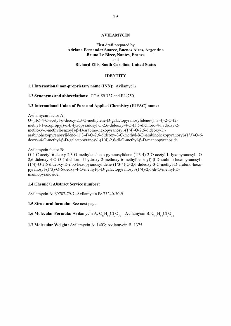
Figure 1: Structural formula of main avilamycin components
Melting point: Avilamycin A: 166-169°C Avilamycin B: 179-182°C
OTHER INFORMATION ON IDENTITY AND PROPERTIES Pure active ingredient: Avilamycin is an orthosomycin antibiotic complex produced by the fermentation of Streptomyces viridochromogenes. Orthosomycin antibiotics are divided into two groups: those that contain an aminocyclitol residue and those that are esters of dichlorisoeverninic acid. Avilamycin is in the latter group as are the evernimicins. Avilamycin complies with the following specifications for the composition of the total factor content.
Avilamycin A: Not less than 60% Avilamycin B: Not more than 18%
Avilamycin A + Avilamycin B: Not less than 70%
Other single Avilamycin factors: Not more than 6%
Typical Avilamycin content is 260 mg activity/g.
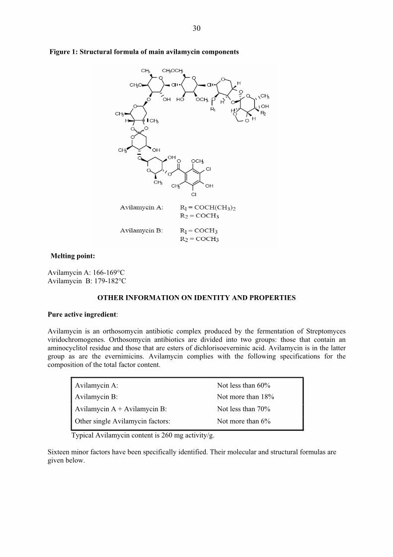
Sixteen minor factors have been specifically identified. Their molecular and structural formulas are given below.
31
Structures of Avilamycin Factors
Factor
R1
R2
R3
R4
R5
R6
R7
R8
Molecular Formula
MolecularWeight
A -CO-CH(CH3)2 -CO-CH3 -OCH3 -Cl -Cl -CH3 -CH3 -OCH3 C61H88Cl2O32 1403
A’ -CO-CH2CH3 -H -OCH3 -Cl -Cl -CH3 -CH3 -OCH3 C58H84Cl2O31 1347
B -CO-CH3 -CO-CH3 -OCH3 -Cl -Cl -CH3 -CH3 -OCH3 C59H84Cl2O32 1375
C -CO-CH(CH3)2 -CHOH-CH3 -OCH3 -Cl -Cl -CH3 -CH3 -OCH3 C61H90Cl2O32 1405
D1 -H -CO-CH3 -OCH3 -Cl -Cl -CH3 -CH3 -OCH3 C57H82Cl2O31 1333
D2 -CO-CH3 -CHOH-CH3 -OCH3 -Cl -Cl -CH3 -CH3 -OCH3 C59H86Cl2O32 1377
E -H -CHOH-
CH3 -OCH3 -Cl -Cl -CH3 -CH3 -OCH3 C57H84Cl2O31 1335
F -CO-CH(CH3)2 -CO-CH3 -OH -H -Cl -CH3 -CH3 -OCH3 C60H87ClO32 1355
G -CO-C4H9 -CO-CH3 -OCH3 -Cl -Cl -CH3 -CH3 -OCH3 C62H90Cl2O32 1417
H -CO-CH(CH3)2 -CO-CH3 -OCH3 -Cl -H -CH3 -CH3 -OCH3 C61H89ClO32 1369
I -CO-CH2CH3 -CO-CH3 -OCH3 -Cl -Cl -CH3 -CH3 -OCH3 C60H86Cl2O32 1389
J -CO-CH(CH3)2 -CO-CH3 -OCH3 -Cl -Cl -H -CH3 -OCH3 C60H86Cl2O32 1389
K -CO-CH(CH3)2 -CO-CH3 -OCH3 -Cl -Cl -CH3 -CH2OH -OCH3 C61H88Cl2O33 1419
L -CO-CH(CH3)2 -CO-H -OCH3 -Cl -Cl -CH3 -CH3 -OCH3 C60H86Cl2O32 1389
M -CO-CH(CH3)2 -CO-CH3 -OCH3 -Cl -Cl -CH3 -H -OCH3 C60H86Cl2O32 1389
N -CO-CH(CH3)2 -CO-CH3 -OCH3 -Cl -Cl -CH3 -CH3 -OH C60H86Cl2O32 1389
Solubility: Solubility in water and organic solvents is expressed in g/L. The solubility of avilamycin factor A has been determined in a variety of solvents at 20°C.
Solvent Solubility (g/L) Water 1
Ethanol 4 Methanol 5
Ethyl acetate 10 Acetone 50 Heptane < 1
Chloroform 100 Refractive index, optical rotation: The optical rotation of a 2.773% solution of factor A in dioxane was � D20 = + 2° ± 1°.
RESIDUES IN FOOD AND THEIR EVALUATION
The residue studies were carried out using avilamycin as a fermentation product, with different degrees of purity or as pure (crystalline) product. The factor composition and purity of avilamycin differed between studies.

32
The Committee evaluated avilamycin to recommend MRLs in poultry, pigs and rabbits at the request of the 17th session of the Codex Committee on Residues of Veterinary Drugs in Foods. Conditions of use Avilamycin is intended for use as a veterinary medicine in chickens, turkeys, pigs and rabbits to control bacterial enteric infections. It exhibits good antimicrobial activity against important veterinary Gram-positive pathogens (e.g., Clostridium perfringens) and has no related molecules in its class in human use. Therefore, avilamycin has been developed for treating necrotic enteritis in poultry, and enteric disease in pig and rabbits. Avilamycin was previously authorised in the European Union (EU) as a feed additive for growth promotion in accordance with Council Directive 70/524/EEC; the substance was incorporated in pig feedstuffs at a concentration of 20 mg/kg feed for animals up to 6 months of age and 40 mg/kg feed for animals up to 4 months of age. It was incorporated into chicken and turkey feedstuffs at a concentration of 10 mg/kg feed. The use of the substance as a feed additive was discontinued in the EU from 1 January 2006. Dosage Table 1: Recommended doses and duration of treatment for Avilamycin in feed
Target Animal Dose in Feed
(mg/kg) Dose Rate
(mg/kg bw/day) Maximum Duration
(days) Pig 100 6-8 21 days Chicken 100 20 21 days Turkey 100 20 21 days Rabbit 80 5 28 days
PHARMACOKINETICS AND METABOLISM
Pharmacokinetics in Laboratory Animals, Humans and Food Animals No classical pharmacokinetic studies have been conducted in any species with avilamycin because avilamycin is not detectable in plasma (LOD = 0.05 mg/kg) following oral administration of avilamycin in feed. In addition, the concentration necessary for kinetic analysis would be well below the toxicologically relevant concentrations and would not be pertinent to human food safety. Metabolism and Residue studies in pigs, poultry and other species (rat) that have been conducted using radiolabelled material are presented below. �Where blood, serum or plasma concentrations were measured in various species following oral doses, avilamycin concentrations were below the limits of detection. For example, in broiler chickens that were fed with a ration containing 22 mg of avilamycin/kg of feed for 25 days, no avilamycin was detected in blood measured by a bio-autographic method (LOD <0.04 mg/kg) or by GC method (LOD <0.1 mg/kg) (West, et al., 1982). Humans Avilamycin has not been developed for human use and therefore, no pharmacokinetic data in humans are available. Laboratory animals

33
Avilamycin is primarily excreted in faeces when administered orally to pig or chickens. In a GLP compliant rat study (Magnussen, 1985a), less than one percent of the oral dose was eliminated in the urine after 72 hours, while 80 to 104% was recovered in the faeces. Pigs In a balance-excretion non GLP-compliant study two cross-bred gilts were administered non-radiolabelled avilamycin at 120 mg per kg in the feed per day (Dalidowicz, et al., 1983). After 7 days administration to approximate steady state conditions, a single bolus dose of 120 mg of [U-14C]avilamycin was administered and excreta were collected at 24-hr intervals for 9 days. During the collection period, the two gilts excreted 96.9% and 99.0% of the dose, respectively, with an average of 93.4% in the faeces and 4.5% in the urine. The bulk of the radioactivity was excreted within the first four days. Another radiolabelled GLP-compliant pig study (Magnussen, et al., 1987) conducted on six crossbred pigs receiving the same dose for either ten or fourteen days showed similar results. Excreted radioactivity reached a plateau after 2-3 days and, on average comprised 8% in urine and 92% in faeces. Approximate concentration in faeces was 120 mg/kg equivalents avilamycin. Results are shown in Table 2. Table 2 : Excreted radioactivity in pigs fed ten days with avilamycin
Collection Period (day) Urine (μCi) Faeces (μCi) 1 0.76 1.12 2 1.06 18.56 3 1.45 17.57 4 1.51 18.83 5 1.53 16.96 6 1.54 18.83 7 2.04 21.09 8 1.69 21.26 9 1.94 20.29
10 1.57 18.16 �Chickens A balance-excretion non-GLP-compliant study was conducted in chickens (Dalidowicz, et al., 1984a). Broiler chickens (2 males/2 females) were administered non-radiolabelled avilamycin at 20 mg of microbiological activity per kg in the feed. After 7 days administration to approximate steady state conditions, a single bolus dose of 4 mg of [U-14C]avilamycin was administered and excreta were collected at 24-hr intervals for 13 days. During the collection period, the birds excreted 92.8%, 99.2%, 96.6% and 84.4% of the dose, respectively. An average of 90% of the radioactivity was excreted within the first 6 days. Data for avilamycin in turkeys and rabbits are not available. Metabolism in Laboratory Animals Position of radiolabel (14C) in Avilamycin. The pivotal residue and metabolism studies for pig and chickens were performed using radiolabelled avilamycin. However, because avilamycin is extensively metabolized, the position of the radiolabel is important in understanding not only the metabolic profile of this substance, but also the correct interpretation of the total radioactive tissue data. Therefore, a discussion of the radiolabel position is necessary prior to the assessment of the specific studies.

34
Two types of radiolabelled avilamycin have been prepared by fermentation using S. viridochromogenes with one of two radiolabelled precursors: Figure 2: Structure of radiolabelled avilamycin
1. [U-14C]avilamycin: Using uniformly labelled glucose, [U-14C-glucose], as the precursor labels
the molecule uniformly in all rings (Donoho et al, 1987). 2. [DIA-14C]avilamycin: Using [2-14C-diethylmalonate] as the precursor places approximately
85% of the radioactivity in the dichloro-isoeverninic acid moiety (DIA; Ring A in Figure 2). The remaining 15% of the radioactivity was not conclusively identified, but is suggested to be associated with the iso-butyrate, propionate or acetate moiety at position R1 on Ring G (Dalidowicz, 1985).
Rats In a GLP- compliant study (Magnussen, 1985a), three male and three female Sprague-Dawley rats weighing 215-265 grams each were dosed by gavage for three consecutive days with [DIA14C]avilamycin (specific activity 0.246 �gCi/mg) at levels equivalent to 100 mg/kg of body weight. Following the initial dose, urine and faeces were collected separately from each animal at 24-hour intervals.. In addition, faeces collected during the 24-hour period following the third dose were extracted and assayed for avilamycin and metabolites. All radioactivity in the selected faeces samples was extractable into ethyl acetate at a neutral and acidic pH. The neutral fraction contained 85-87% of the radioactivity, while the acidic fraction contained 12-14%. TLC analysis showed avilamycins A and B to represent 40-60% of the radioactivity in the neutral fraction, while an unidentified, polar metabolite represented 10-30%. The major radioactive component in the acidic fraction was confirmed as flambalactone (previously identified as the major avilamycin derived residue in pig liver (Magnussen, 1985b). It is formed by cleavage of the ortho ester linking the C and D rings of avilamycin. Flambalactone represented 30-60% of the radioactivity in the acidic fraction. One other metabolite common to both rats and pigs in the acidic fraction representing 10-30% of the radioactivity was later identified as flambic acid (Magnussen, et al., 1987). In other studies flambalactone and flambic acid were found to be inter-convertible.
������
O
OO H COCH3
CH3
OHH
O
O
O
O
HO
H
O
OH OCH3
OO
O
CH3
OHO
O O
CH3
H CH3
O
OO
O OCH3
CH3
Cl
OH
Cl
CH3
O
OHCH3
OH
H3CO
R1
H3CO
A
B
C
D
E F G
H
[U-14C]Avilamycin: All rings labelled
[DIA-14C]Avilamycin 85% Ring A 15% other (possibly R1)
35
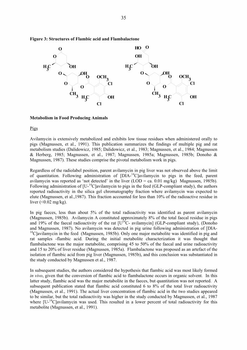
�Figure 3: Structures of Flambic acid and Flambalactone Metabolism in Food Producing Animals Pigs Avilamycin is extensively metabolized and exhibits low tissue residues when administered orally to pigs (Magnussen, et al., 1991). This publication summarizes the findings of multiple pig and rat metabolism studies (Dalidowicz, 1985; Dalidowicz, et al., 1983; Magnussen, et al., 1984; Magnussen & Herberg, 1985; Magnussen, et al., 1987; Magnussen, 1985a; Magnussen, 1985b; Donoho & Magnussen, 1987). These studies comprise the pivotal metabolism work in pigs. Regardless of the radiolabel position, parent avilamycin in pig liver was not observed above the limit of quantitation. Following administration of [DIA-14C]avilamycin to pigs in the feed, parent avilamycin was reported as ‘not detected’ in the liver (LOD = ca. 0.01 mg/kg) Magnussen, 1985b). Following administration of [U-14C]avilamycin to pigs in the feed (GLP-compliant study), the authors reported radioactivity in the silica gel chromatography fraction where avilamycin was expected to elute (Magnussen, et al.,1987). This fraction accounted for less than 10% of the radioactive residue in liver (<0.02 mg/kg). In pig faeces, less than about 5% of the total radioactivity was identified as parent avilamycin (Magnussen, 1985b). Avilamycin A constituted approximately 8% of the total faecal residue in pigs and 19% of the faecal radioactivity of the rat [U14C- avilamycin] (GLP-compliant study), (Donoho and Magnussen, 1987). No avilamycin was detected in pig urine following administration of [DIA-14C]avilamycin in the feed (Magnussen, 1985b). Only one major metabolite was identified in pig and rat samples -flambic acid. During the initial metabolite characterization it was thought that flambalactone was the major metabolite, comprising 45 to 50% of the faecal and urine radioactivity and 15 to 20% of liver residue (Magnussen, 1985a). Flambalactone was proposed as an artefact of the isolation of flambic acid from pig liver (Magnussen, 1985b), and this conclusion was substantiated in the study conducted by Magnussen et al., 1987. In subsequent studies, the authors considered the hypothesis that flambic acid was most likely formed in vivo, given that the conversion of flambic acid to flambalactone occurs in organic solvent. In this latter study, flambic acid was the major metabolite in the faeces, but quantitation was not reported. A subsequent publication stated that flambic acid constituted 6 to 8% of the total liver radioactivity (Magnussen, et al., 1991). The actual liver concentration of flambic acid in the two studies appeared to be similar, but the total radioactivity was higher in the study conducted by Magnussen, et al., 1987 where [U-14C]avilamycin was used. This resulted in a lower percent of total radioactivity for this metabolite (Magnussen, et al., 1991).
O
O O
O O CH3
O
CH3
Cl
OHCl
C H 3
O OHC H 3
O H
OH
O
OOHCH3
OH
OO
O O C H 3
CH3
Cl
O H Cl
CH3
OH

36
No other significant metabolites were identified in pigs or rats, although a few minor peaks were observed. The silica gel chromatographic profiles of extracts from the faeces, urine and livers of rats and pigs treated with [U-14C]avilamycin were qualitatively comparable and quantitatively similar by visual inspection (Donoho & Magnussen, 1987). There was a good correlation between the metabolic profiles of rats and pigs. The faeces extract from [DIA-14C]avilamycin-treated pigs exhibited the same three peaks, but the proportions were different, with the flambic acid-containing peak predominant. Additional TLC analyses of the column fractions from faeces extracts indicated that oligosaccharide-derived metabolites were present in [U-14C]avilamycin samples that were not present in [DIA-14C]avilamycin samples. These metabolites were not further characterized because the corresponding peaks were not present in liver and would thus not pose a food safety risk. The metabolic profiles in liver of treated rats and pigs were essentially the same with the flambic acid as the most abundant metabolite (Donoho & Magnussen, 1987). Parent avilamycin concentration in rat and pig liver were less that 0.05 mg/kg. The pattern of minor metabolites was similar but insufficient for identification. Data are supportive that rats treated with avilamycin have been exposed to the same metabolites that are present in edible tissues of treated pigs. Characterization of residues in fat samples from treated pigs demonstrated that essentially all of the residues in fat are due to the incorporation of radioactivity into the endogenous fatty acids, oleic and stearic acid (Dalidowicz, 1985). No DIA-related residues were detected in fat when assayed by hydrolysis and GC analysis, indicating that parent and DIA-containing metabolites such as flambic acid are not detectable (Magnussen, et al., 1984). Moreover, when the radiolabel is distributed into the carbohydrate moieties of avilamycin (i.e., [U-14C]avilamycin), the total radioactive residues are higher than those when using [DIA-14C]avilamycin, while the amounts of DIA-containing residues remain relatively constant (Magnussen, et al., 1984;, Magnussen, et al.; 1987, Magnussen, et al., 1991) The increased incorporation of carbon-14 into fatty acids when [U-14C]avilamycin was administered is consistent with the avilamycin carbohydrate moieties being extensively metabolized. Chickens, Turkeys and Rabbits No metabolism data available.
TISSUE RESIDUE DEPLETION STUDIES Radiolabelled Residue Depletion Studies Pigs Several GLP-compliant studies following administration of 14C avilamycin were submitted. Two of the studies used [DIA14C] avilamycin and the third used [U-14C] avilamycin. Five crossbred pigs, three gilts and two barrows, weighing approximately 46 kg each were fed at 12-hour intervals for seven days with a ration containing 76mg of [DIA14C] avilamycin per kilogram of feed (equivalent to 80 mg/kg of activity and equal to 4.6-6.1 mg avilamycin/kg bw/day) (Magnussen & Herberg, 1985). Each day, animals received an amount of ration equal to 4% of their body weights. At a practical zero-time withdrawal (six hours) after the final medicated feed ration, one gilt was sacrificed. The remaining animals were then fed non-medicated ration at 12-hour intervals for either three or five days, and one gilt and one barrow sacrificed at the end of each time period. At each sampling time, muscle, liver, kidney, and fat were collected for radiochemical analysis. Results are shown in Table 3.

37
Table 3: Total radiolabel residue (TRR) in pig tissues �
Withdrawal TRR (mg/kg equivalents avilamycin) Days n Muscle Liver Kidney Fat
0 1 NDR1 0.15 0.08 0.07 3 2 NDR NDR 0.02 0.05 5 2 NDR NDR < 0.032 0.05
1: NDR = no detectable residue 2: one animal, 0.025 mg/kg; one animal NDR (0.017 mg/kg)
At zero-time withdrawal, no detectable residue was found in muscle, while the total radiolabel residues in liver, kidney, and fat, expressed as avilamycin equivalents, were 0.15, 0.08, and 0.07 mg/kg, respectively. After a three-day withdrawal period, no residues were detected in either liver or muscle, while residues in kidney and fat were 0.024 and 0.053 mg/kg, respectively. After five days, residue levels in fat were nearly the same as those observed at three days, while levels in kidney were from non-detectable residues to 0.025 mg/kg. �Concentrations of avilamycin-related radioactivity in liver and muscle declined to non-detectable levels within three days after the termination of dosing, while concentrations in kidney declined to near non-detectable levels within five days after the termination of dosing. Radioactivity in fat showed a much slower rate of decline due to the fact that radiolabelled carbon from the 14C-avilamycin molecule had become incorporated into the fatty acid fraction as demonstrated by Dalidowicz, 1985. Authors quoted a fat turnover rate of 14-21 days but provided no evidence to support the comment. In another study conducted by Magnussen, et al, 1984, nine crossbred pigs, weighing approximately 44 kg each, were fed with a ration containing 76.2 mg of [DIA14C]avilamycin per kilogram of feed (equivalent to 80 mg/kg of avilamycin activity and equal to 4.6-6.1 mg avilamycin/kg bw/day) at 12-hour intervals for either four, seven, or ten days. Each day, animals received a ration equal to 4.0% of their body weights, equivalent to a daily dose of approximately 134 mg of DIA14C-avilamycin. All animals were sacrificed at a practical zero-time withdrawal (six hours) after the final feeding. Muscle, liver, kidney, fat, and bile were collected for radiochemical analyses by liquid scintillation counting. Selected tissues were assayed for avilamycin by bio-autography and residues containing the dichloroisoeverninic acid (DIA) moiety. Liver from each animal was extracted to determine levels of non-extractable radioactivity. For total radioactive residues (TTR) results are shown in Table 4. Table 4: Total radiolabel residue (TTR) in pig tissues.
Dosing Total Radioactivity (mg/kg equivalents avilamycin) Interval (days) Muscle Liver Kidney Fat Bile
4 0.01 0.21 0.10 0.05 18.9 7 0.01 0.23 0.10 0.08 19.9
10 0.02 0.22 0.10 0.12 19.8 �After ten days dosing, total mean radiolabel residues in liver, fat, and kidney, expressed as avilamycin equivalents, were 0.22, 0.12, and 0.10 mg/kg, respectively. Residues in muscle were less than 0.016 mg/kg. Steady-state concentrations of radioactivity were attained in muscle, liver, and kidney within four days after the initiation of dosing. A steady-state concentration was not attained in fat during this study. The study mentioned in the metabolism section conducted by Dalidowicz, 1985 demonstrated that radioactivity found in fat was incorporated into the fatty acid portion of triglycerides. These non-active residues were not of toxicological concern.
38
Liver, kidney, and fat from animals dosed for ten days were assayed for avilamycin by bio-autography (Prichard et al, 2006; Method Number AM-AA-CA-R075-AB-755). This method consisted of extracting avilamycin from pig or broiler tissues with acetone. The acetone extract is purified by liquid-liquid partitioning, and the purified extract is spotted on a thin layer chromatographic plate (TLC). After development the TLC plate is subjected to bio-autographic analysis using a Micrococcus flavus overlay. The plate is sprayed to enhance the appearance of the zones of inhibition and the presence or absence of avilamycin is determined by comparison with a reference standard. The method does not determine the concentration of avilamycin, but the LOD was reported at 0.05 mg/kg. Results are presented in Table 5. Table 5: Microbiologically active avilamycin pig tissue residues
Dosing Interval (days)
Microbiological Activity (mg/kg equivalents avilamycin)
Muscle Liver Kidney Fat 10 -- < 0.05 NDR1 NDR1
NDR1: non-detectable residues. No microbiologically active residues of avilamycin were detected in kidney or fat and only traces in liver, but were considerably less than the limit of detection (LOD is < 0.05 mg/kg). Muscle was not assayed due to radioactivity concentrations less than LOD for the bio-autographic assay. Tissue residues containing DIA were analysed by gas chromatography (Formica, G and Giannone, C., 1986). Results are shown in Table 6. Table 6: DIA Residues in selected pig tissues from the 10-day withholding time
DIA Residue (mg/kg equivalents avilamycin) Animal No. Muscle Liver Kidney Fat
130 -- 0.10 < 0.1 NDR 135 -- 0.12 < 0.1 NDR 137 -- 0.17 < 0.1 NDR
Mean -- 0.13 < 0.1 NDR Approximately 50% of TRR in liver was due to DIA-related residues. DIA-related residues were detected in kidney, but below the limit of quantification (LOQ < 0.1 mg/kg). No DIA residues were observed in fat (< 0.1 mg/kg). Liver results are presented in Table 7. Table 7: Pig liver extraction results
Dosing Interval Mean (n=3) Percent of Radioactivity Days Acetone Unextracted
4 79.5 20.5 7 82.2 17.8
10 73.1 26.9 About 18 - 27% of radioactivity was not extractable into acetone for the 4, 7 and 10 day liver samples. Statistical analysis of the extraction data indicated no significant difference between un-extracted radioactivity through 10 days of treatment. �A steady-state, tissue residue study using uniformly labelled 14C avilamycin was conducted by Magnussen, et al., 1987. Six crossbred pigs, four barrows and two gilts, weighing approximately 44 kg each were fed at 12-hour intervals for either ten or fourteen days with a ration containing a nominal concentration of 60 mg of 14C-avilamycin/kg of feed (equivalent to 60 mg activity/kg and to 3.6-4.8 mg/kg bw/day). Each day, animals received an amount of ration equal to 4% of their body

39
weights. Groups of three animals were killed after ten days and fourteen days on treatment. Muscle, liver, kidney, and fat were collected for radiochemical analysis. Liver from each animal was extracted to determine the concentration of non-extractable radioactivity and to characterize the extractable radioactivity. Radioactivity in fat was also characterized. Total radioactivity residues in tissues are presented in Table 8. Table 8: Total radiolabel residues (TRR) in pig tissues
Dosing Interval TTR (mg/kg equivalents avilamycin) Days Muscle Liver Kidney Fat
10 0.09 0.55 0.32 0.26 14 0.14 0.66 0.34 0.55
Radioactive tissue residues are higher in this study than the other two 14C studies because the avilamycin molecule was more uniformly labelled over all rings with 14C for this study. The 14C label in the avilamycin for the other two studies was primarily (85%) in the DIA ring. One-way analysis of variance (ANOVA) indicated no difference between 10 or 14 days for muscle, liver or kidney total radioactive residues. Only the fat radioactive residues were significantly different at 10 and 14 days (P < 0.05). Non-extractable liver residues were 33 - 37% of total liver residues and were not different in the 10- and 14-day treatment groups as are shown in Table 9. Table 9: Percent extraction of radioactivity from pig livers
10-day Group 14-day Group Animal No. 961 960 957 954 955 959 Acetone Extract 34 32 34 32 33 29 Methanol Extract 25 24 26 25 26 28 Acetone/water 7 7 6 8 6 6 Pellet 34 37 33 34 35 37
The GC analysis shown that extractable liver radioactivity consisted of several minor metabolites (<0.1 mg/kg). Flambic acid was present at concentrations up to 0.04 mg/kg. Parent 14C-avilamycin concentrations were less than 0.01-0.02 mg/kg. Chickens In a GLP-compliant conducted study (Dalidowicz, 1986), twelve seven-week-old broiler-type chickens, six male and six female, were fed a standard broiler finishing ration containing 14.16 mg of [DIA14C] avilamycin per kilogram of feed (equivalent to 15 mg of activity /kg and equal to 3 mg/kg bw/day) for either four, seven, or ten days. Medicated ration and water were provided ad libitum throughout the dosing phase. At the end of each designated dosing period, two birds of each sex were deprived of food and water for six hours and then killed. Samples of muscle, liver, abdominal fat, kidney and skin with subcutaneous fat were collected for radiochemical analysis. Results are shown in Table 10.
40
Table 10: Total radiolabel residues (TTR) in chicken tissues
Tissue TTR (mg/kg equivalents of Avilamycin) Method LOD 4 day 7 day 10 day Muscle 0.01 < 0.011 NDR2 NDR Liver 0.01 0.03 0.04 0.02 Skin 0.01 0.023 0.013 0.023
Fat 0.01 0.014 0.03 0.033
Kidney 0.02 NDR NDR NDR 1: Three of four individuals below LOD; LOD value substituted for NDR of individuals 2: NDR: no detectable residue (less than LOD) 3: One of four individuals below LOD; LOD value substituted for NDR of individuals 4: Study director excluded one of four samples as a statistical outlier. Reliable detection and quantitation were demonstrated only for 0.025 mg/kg. After ten days dosing, the mean total radiolabel residues in skin, liver, and fat expressed as avilamycin equivalents, were 0.02, 0.02, and 0.03 mg/kg, respectively. Muscle and kidney samples contained no detectable radiolabel residues. Steady-state concentrations of radioactivity were attained in all tissues within four to seven days after the initiation of dosing. In another GLP-compliant study, twenty-four Highline W-36 laying hens were fed rations containing 30 mg/kg [U14C] avilamycin for fourteen days (Sweeney, et al., 1997). Eggs were collected daily throughout the study. At slaughter, liver, kidney, muscle, fat, skin/fat, and bile were collected. The tissues were assayed for total radioactivity by solubilization and liquid scintillation counting. Results are summarized in the Table 11. Table 11: Total radiolabel residues in chicken tissues �
TTR (mg/kg equivalents of Avilamycin) Liver Kidney Muscle Skin/fat Fat Bile Mean(n=7) 0.08 0.07 NDR NDR 0.03 3.5
NDR: No Detectable Residues Eggs from study day five, ten, twelve, and fourteen were separated into yolk and albumin and analysed for radioactive residues. Residues in albumin were not detectable (<0.07 mg/kg), while the residues in yolk were on the average 0.2 mg/kg at 10 days, 0.21 mg/kg at 12 days and 0.22 mg/kg at fourteen days. One hen had significantly higher liver, kidney, and yolk residues than the other six treated hens. The higher residue values in this hen were attributed to animal-to-animal variation. Turkeys and Rabbits No radiolabelled residue depletion studies data are available. Avilamycin is not a suitable marker residue because it is not detected in tissues of pigs and chickens. Flambic acid, the major metabolite, is not a suitable marker residue because it does not have a reference standard available. Dichloroisoeveninic acid (DIA) is a moiety present in avilamycin, along with flambic acid and other possible metabolites. Measurement of DIA following extraction and hydrolysis of DIA-containing fractions or metabolites provides a satisfactory method for measuring residues of avilamycin, as studies have demonstrated measurable amounts of DIA in liver and kidney. DIA is a useful marker residue because it is not a common chemical structure and where it can be found in related substances, none of them are veterinary drugs. The DIA concentration may be reported as avilamycin equivalents by multiplying the DIA concentration by the molar ratio of avilamycin/DIA (5.6:1).
41
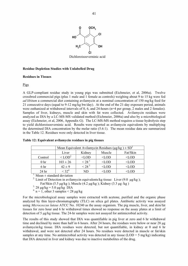
Residue Depletion Studies with Unlabelled Drug Residues in Tissues Pigs A GLP-compliant residue study in young pigs was submitted (Eichmeier, et al, 2006a). Twelve crossbred commercial pigs (plus 1 male and 1 female as controls) weighing about 9 to 15 kg were fed ad libitum a commercial diet containing avilamycin at a nominal concentration of 150 mg/kg feed for 21 consecutive days (equal to 9-12 mg/kg bw/day). At the end of the 21-day exposure period, animals were euthanized at withdrawal intervals of 0, 6, and 24-hours (n=4 per group, 2 males and 2 females). Samples of liver, kidneys, muscle and skin with fat were collected. Avilamycin residues were analyzed as DIA by a LC-MS-MS validated method (Eichmeier, 2006a) and also by a microbiological assay (Eichmeier, et al, 2006, Appendix G). The LC-MS-MS method requires a tissue hydrolysis step to yield dichloroisoeverninic acid. Results were reported as avilamycin equivalents by multiplying the determined DIA concentration by the molar ratio (5.6:1). The mean residue data are summarized in the Table 12. Residues were only detected in liver tissue. Table 12: Equivalent avilamycin residues in pig tissues
Mean Equivalent Avilamycin Residues (�g/kg ) ± SD1
Liver Kidney Muscle Fat/Skin Control < LOD2 <LOD <LOD <LOD
0 hr 103 ± 26 < 28 3 <LOD <LOD 6 hr 42 ± 9 < 28 3 <LOD <LOD
24 hr < 324 ND <LOD <LOD 1: Mean ± standard deviation 2: Limit of Detection in avilamycin equivalents/kg tissue: Liver (9.0 �g/kg );
Fat/Skin (5.5 �g/kg ); Muscle (4.2 �g/kg ); Kidney (3.3 �g /kg ) 3: 28 �g/kg = 5.0 �g/kg DIA 4: n = 1, other 3 samples < 28 �g/kg
For the microbiological assay samples were extracted with acetone, purified and the organic phase analyzed by thin layer-chromatography (TLC) on silica gel plates. Antibiotic activity was assayed using Micrococcus luteus ATCC No. 10240 as the assay organism. The pig muscle, liver, and skin/fat tissues for zero hour and 6 hr withdrawal times showed no response on the assay plates at a limit of detection of 5 �g/kg tissue. The 24-hr samples were not assayed for antimicrobial activity.
The results of this study showed that DIA was quantifiable in pig liver at zero and 6 hr withdrawal time and declined by more than half in 6 hours. After 24 hours, the residues were below or near 28 �g avilamycin/kg tissue. DIA residues were detected, but not quantifiable, in kidney at 0 and 6 hr withdrawal, and were not detected after 24 hours. No residues were detected in muscle or fat/skin samples at any time. No antimicrobial activity was detected in any tissue (LOD = 5 mg/kg) indicating that DIA detected in liver and kidney was due to inactive metabolites of the drug.
Dichloroisoeverninic acid
OH
O OCH3
Cl
OH
Cl
CH3

42
A non GLP-compliant study to determine microbiological activity of avilamycin residues was submitted (Asanuma, et al., 1987a). A preparation of 10% avilamycin/mg (EL-750) was administered to castrated male pigs from 28 to 84 days orally by medicated feed at concentrations of 40 or 400 mg/kg feed (two groups of 8 pigs, one control). Non-medicated feed was provided during withdrawal. The pigs were allowed free access to the feed. Liver, kidney, muscle, fat tissues and small intestine were collected at 42 days (two animals per group) and after 84 days (two animals at each time, 0.1 and 3 days withdrawal). The avilamycin residues were analysed by the microbiological assay method with Micrococcus flavus (LOD = 25 �g/kg). Avilamycin was not detected in major organs or tissue for all sampling points including during medication. In a previously reported non GLP-compliant study, the microbiological activity was studied by Morimoto et al., 1986a. Breeding piglets (22 boars and 22 sows) at approximately 30 days old were fed with medicated feed containing avilamycin (40, 200 or 400 mg/kg) for 12 weeks followed by a 7-day withdrawal. The avilamycin preparation was the same used in the previous mentioned study (EL-750. 10% avilamycin/mg). Plasma, liver, kidney, muscle, fat, and small intestine was collected from groups of 1 male and 1 female at 6 weeks and 12 weeks at 0-hour withdrawal and at 1, 3, 5 and 7days withdrawal time. The residues were analyzed using the TLC/microbiological assay based on inhibition of Micrococcus flavus (LOD = 25 �g/kg). No microbiologically active residues were observed in any samples of plasma, muscle, liver, kidney, or fat (25 �g/kg was detected in two small intestine samples from the 200 mg/kg treatment group, one at 6 weeks and one at 12 weeks; 27 �g/kg was detected in a small intestine sample of the 400 mg/kg group at 6 weeks and 30 �g/kg at 12 weeks). No other residues were detected. The authors concluded that EL-750 is not readily absorbed and only very small amounts of avilamycin are found in tissues, even at a dose of 400 mg/kg in the feed for 12 weeks. Two non GLP-compliant studies simulating the commercial pig industry were reported. In the first study (West and Wellenreiter, 1983), grower-finisher pigs (2 male, 4 female) were fed standard rations containing 0 or 40 mg/kg of avilamycin for a period of 99 days. The pigs were sacrificed after a zero-day (six-hour) or a one-day (30-hour) withdrawal period. Using the bio-autographic technique with Micrococcus flavus as the indicator organism, no microbiologically active residues were detected in the muscle, liver, kidney or fat tissues from any of the six treated or three control pigs analysed at a detection limit of 0.05 mg/kg. In the second study (West, et al, 1983), starter pigs (3 male, 3 female) were fed a standard ration containing 0 or 200 mg/kg of avilamycin for a period of 56 days. The pigs were sacrificed after a zero-day (six-hour) withdrawal time. Using the microbiological assay, no microbiologically active residues were detected in the muscle, liver, kidney or fat tissues from any of the pigs analysed at a detection limit of 0.05 mg/kg. Chickens In a GLP-compliant residue study, a commercial breed of broiler chickens (15 males and 15 females plus 10 males and 10 females as controls) approximately two weeks old weighing from 339-541 g were fed ad libitum a commercial diet containing avilamycin at a nominal concentration of 150 mg/kg feed for 21 consecutive days, equivalent to 30 mg/kg bw/day (Eichmeier, 2006b). At the end of the 21-day exposure period, animals were euthanized at withdrawal intervals of 0, 6, and 24-hours (n=6 per group, 3 males and 3 females). Samples of liver, kidneys, muscle and skin/fat (subcutaneous) were collected. Avilamycin residues were analyzed as DIA by the LC/MS/MS method (Eichmeier, 2006b) and also by the microbiological assay (Eichmeier, et al., 2006, Appendix G) as previously described for the study with pigs (Eichmeier, et al., 2006a). The DIA moiety of avilamycin was quantifiable in chicken liver at zero time withdrawal and declined to below or near 28 μg avilamycin/kg tissue within 6 hours. After 24 hours, the liver residues were
43
below 28 μg avilamycin/kg. DIA residues were detected, but not quantifiable, in kidney and skin/fat at 0 and 6 hours withdrawal, and were not detected after 24 hours. Half of the skin/fat and kidney samples had no detectable residues after 6 hours withdrawal. No DIA residues were detected in muscle samples at any time. No antimicrobial activity was detected in any tissue from the 0-hr and 6-hr treated groups (LOD = 5�g/kg), with the exception of two skin/fat samples from the 6-hr treated group that were attributed to laboratory contamination. The 24-hr samples were not assayed for antimicrobial activity. No antimicrobial activity was detected in any other tissue (LOD = 5 �g/kg). Therefore, DIA detected in liver and kidney was due to inactive metabolites of the drug. Results for the LC-MS-MS analysis are shown in Table 13. Table 13: Avilamycin equivalent residues in chicken tissues
A non GLP-compliant study to determine microbiological activity of avilamycin residues was submitted (Asanuma, et al., 1987b). A preparation of 10 % avilamycin/mg (EL-750) was administered to chickens (40-47 weeks old, 920-1090 g) for 56 days using medicated feed at concentrations of 10 or 200 mg/kg (60 chickens per group). The birds were allowed free access to the feed. The residues of avilamycin were analysed by the microbiological assay method with Micrococcus flavus. Avilamycin was not detected in major organs or tissue for all sampling points including the medication period, thus, avilamycin is not readily absorbed. In an earlier non GLP-compliant study, the microbiological activity was studied by Morimoto, et al., 1986b. A preparation of 10 % avilamycin/mg (EL-750) was administered to broiler chickens from day 1 to 84 weeks orally by medicated feed at concentrations of 20, 100 or 200 mg avilamycin/kg feed (3 groups of 6 males and 6 females, one control group). Non medicated feed was used during the 7 day withdrawal time. Plasma, liver, kidney, muscle, fat, and small intestine were collected at 28 days, 56 days (0-hour withdrawal), and at 1, 3, 5 and 7 days withdrawal. The residues were analyzed using the TLC/microbiological assay based on Micrococcus flavus as the indicator organism. No microbiologically active residues were observed in any samples of plasma, muscle, liver, kidney, or fat. (LOD= 0.05 �g/kg). In a third non GLP-compliant study, broiler chickens (six treated plus two control birds) were fed standard rations containing 0 or 20 mg of avilamycin/kg of feed for a period of 56 days (West, et al., 1983). The chickens were sacrificed after a zero-day (six-hour) withdrawal. Using the bio-autographic technique with Micrococcus flavus as the indicator organism, no microbiologically active residues were detected in the muscle, liver, kidney, or skin with adhering fat tissues from any of the chicken. Turkeys A GLP-compliant residue study in turkeys was submitted (Eichmeier, et al., 2006c) to demonstrate the applicability of the routine analytical residue method for the determination of avilamycin and dichloroisoeverninic acid-containing metabolites in turkey tissues. Domesticated turkeys (Melleagris
Mean Equivalent Avilamycin Residues (�g/kg ) ±SD1
Liver Kidney Muscle Fat/Skin Control <LOD2 <LOD <LOD <LOD 0 hr 67 ±32 < 283 <LOD < 28 6 hr < 304 < 285 <LOD < 282
24 hr < 282 <LOD <LOD <LOD 1: Mean ± Standard Deviation 2: Limit of Detection (in avilamycin equivalents/kg tissue): Liver (3.0 �g/kg );
Fat/Skin (5.0 �g/kg ); Muscle (4.4 �g/kg ); Kidney (4.9 �g/kg ) 3: 5.0 �g/kg DIA =28 �g avilamycin/kg tissue 4 n = 1, other 5 samples < 28�g/kg 5: n=3 at <28�g/kg , n = 3 < LOD
44
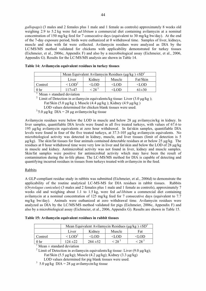
gallopago) (3 males and 2 females plus 1 male and 1 female as controls) approximately 8 weeks old weighing 2.9 to 5.2 kg were fed ad libitum a commercial diet containing avilamycin at a nominal concentration of 150 mg/kg feed for 7 consecutive days (equivalent to 30 mg/kg bw/day). At the end of the 7-day exposure period, birds were euthanized at 0 withdrawal time. Samples of liver, kidneys, muscle and skin with fat were collected. Avilamycin residues were analyzed as DIA by the LC/MS/MS method validated for chickens with applicability demonstrated for turkey tissues (Eichmeier, et al., 2006c, Appendix F) and also by a microbiological assay (Eichmeier, et al., 2006, Appendix G). Results for the LC/MS/MS analysis are shown in Table 14. Table 14: Avilamycin equivalent residues in turkey tissues
Mean Equivalent Avilamycin Residues (�g/kg ) ±SD1
Liver Kidney Muscle Fat/Skin Control < LOD2 <LOD <LOD <LOD 0 hr 117±47 < 28 3 <LOD 61±30 1: Mean ± standard deviation 2: Limit of Detection in avilamycin equivalents/kg tissue: Liver (3.0 �g/kg );
Fat/Skin (5.0 �g/kg ); Muscle (4.4 �g/kg ); Kidney (4.9 �g/kg ) LOD values determined for chicken blank tissues were used. 3:5.0 �g/kg DIA = 28 �g avilamycin/kg tissue
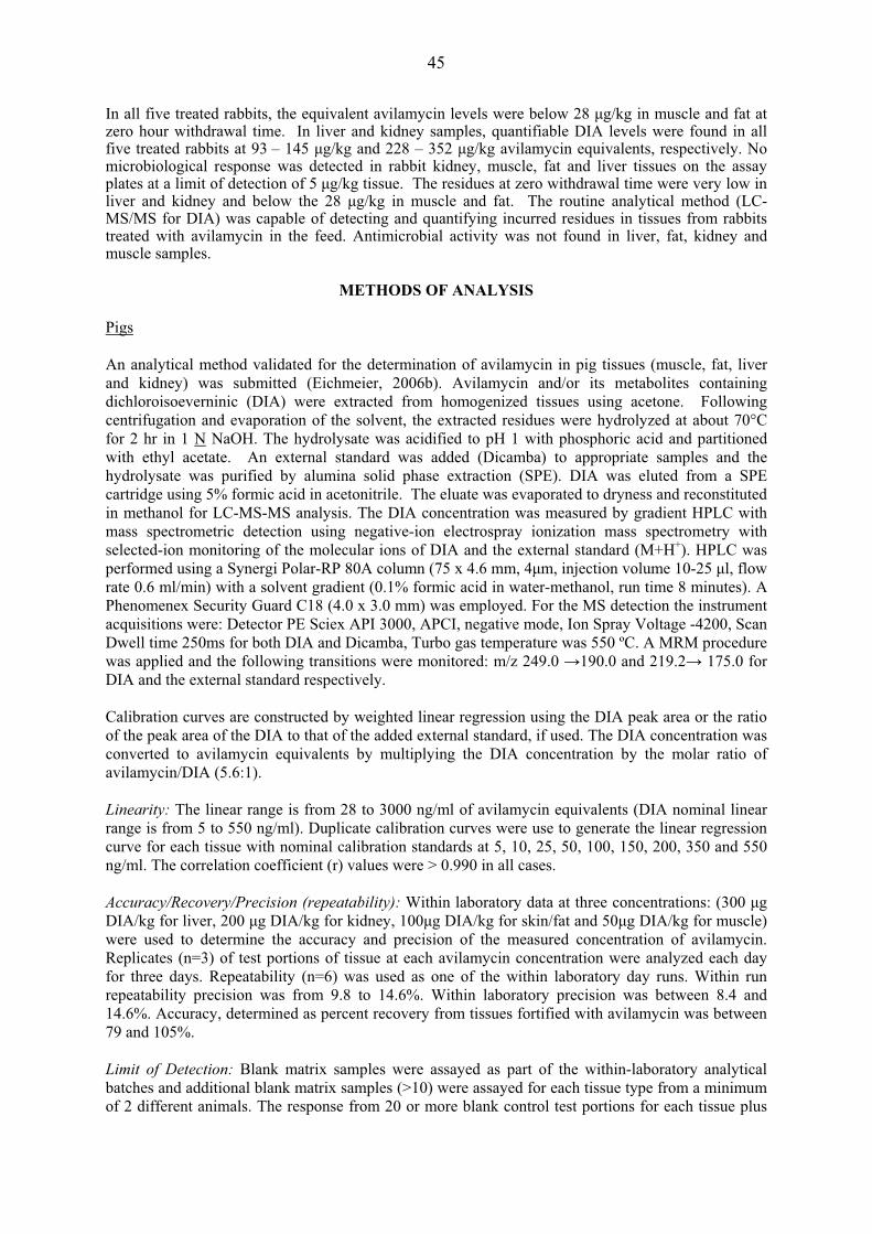
Avilamycin residues were below the LOD in muscle and below 28 �g avilamycin/kg in kidney. In liver samples, quantifiable DIA levels were found in all five treated turkeys, with values of 67.6 to 195 �g/kg avilamycin equivalents at zero hour withdrawal. In fat/skin samples, quantifiable DIA levels were found in four of the five treated turkeys, at 37.3-105 �g/kg avilamycin equivalents. No microbiological activity was detected in kidney, muscle, and liver tissues (limit of detection is 5 �g/kg). The skin/fat tissues for four animals contained detectable residues at or below 25 �g/kg. The residues at 0 hour withdrawal time were very low in liver and fat/skin and below the LOD of 28 �g/kg in muscle and kidney. Antimicrobial activity was not found in liver, kidney and muscle samples. Skin/fat samples were positive for antimicrobial activity which may have been the result of contamination during the in-life phase. The LC-MS/MS method for DIA is capable of detecting and quantifying incurred residues in tissues from turkeys treated with avilamycin in the feed. Rabbits A GLP-compliant residue study in rabbits was submitted (Eichmeier, et al., 2006d) to demonstrate the applicability of the routine analytical LC-MS-MS for DIA residues in rabbit tissues. Rabbits (Orytolagus cuniculus) (3 males and 2 females plus 1 male and 1 female as controls), approximately 7 weeks old and weighing about 1.1 to 1.5 kg, were fed ad libitum a commercial diet containing avilamycin at a nominal concentration of 125 mg/kg feed for 7 consecutive days (equivalent to 7.7 mg/kg bw/day). Animals were euthanized at zero withdrawal time. Avilamycin residues were analyzed as DIA by the LC/MS/MS method validated for pigs (Eichmeier, 2006c, Appendix F) and also by a microbiological assay (Eichmeier, et al., 2006, Appendix G). Results are shown in Table 15. Table 15: Avilamycin equivalent residues in rabbit tissues
Mean Equivalent Avilamycin Residues (�g/kg ) ±SD1
Liver Kidney Muscle Fat Control < LOD2 <LOD <LOD <LOD 0 hr 124 ±22 284 ±52 < 28 3 < 28 3 1 Mean ± standard deviation 2 Limit of Detection in avilamycin equivalents/kg tissue: Liver (9.0 �g/kg);
Fat/Skin (5.5 �g/kg); Muscle (4.2 �g/kg); Kidney (3.3 �g/kg) LOD values determined for pig blank tissues were used. 3 5.0 �g/kg DIA = 28 �g avilamycin/kg tissue
45
In all five treated rabbits, the equivalent avilamycin levels were below 28 �g/kg in muscle and fat at zero hour withdrawal time. In liver and kidney samples, quantifiable DIA levels were found in all five treated rabbits at 93 – 145 �g/kg and 228 – 352 �g/kg avilamycin equivalents, respectively. No microbiological response was detected in rabbit kidney, muscle, fat and liver tissues on the assay plates at a limit of detection of 5 �g/kg tissue. The residues at zero withdrawal time were very low in liver and kidney and below the 28 �g/kg in muscle and fat. The routine analytical method (LC-MS/MS for DIA) was capable of detecting and quantifying incurred residues in tissues from rabbits treated with avilamycin in the feed. Antimicrobial activity was not found in liver, fat, kidney and muscle samples.
METHODS OF ANALYSIS Pigs An analytical method validated for the determination of avilamycin in pig tissues (muscle, fat, liver and kidney) was submitted (Eichmeier, 2006b). Avilamycin and/or its metabolites containing dichloroisoeverninic (DIA) were extracted from homogenized tissues using acetone. Following centrifugation and evaporation of the solvent, the extracted residues were hydrolyzed at about 70°C for 2 hr in 1 N NaOH. The hydrolysate was acidified to pH 1 with phosphoric acid and partitioned with ethyl acetate. An external standard was added (Dicamba) to appropriate samples and the hydrolysate was purified by alumina solid phase extraction (SPE). DIA was eluted from a SPE cartridge using 5% formic acid in acetonitrile. The eluate was evaporated to dryness and reconstituted in methanol for LC-MS-MS analysis. The DIA concentration was measured by gradient HPLC with mass spectrometric detection using negative-ion electrospray ionization mass spectrometry with selected-ion monitoring of the molecular ions of DIA and the external standard (M+H+). HPLC was performed using a Synergi Polar-RP 80A column (75 x 4.6 mm, 4�m, injection volume 10-25 �l, flow rate 0.6 ml/min) with a solvent gradient (0.1% formic acid in water-methanol, run time 8 minutes). A Phenomenex Security Guard C18 (4.0 x 3.0 mm) was employed. For the MS detection the instrument acquisitions were: Detector PE Sciex API 3000, APCI, negative mode, Ion Spray Voltage -4200, Scan Dwell time 250ms for both DIA and Dicamba, Turbo gas temperature was 550 ºC. A MRM procedure was applied and the following transitions were monitored: m/z 249.0 �190.0 and 219.2� 175.0 for DIA and the external standard respectively. Calibration curves are constructed by weighted linear regression using the DIA peak area or the ratio of the peak area of the DIA to that of the added external standard, if used. The DIA concentration was converted to avilamycin equivalents by multiplying the DIA concentration by the molar ratio of avilamycin/DIA (5.6:1). Linearity: The linear range is from 28 to 3000 ng/ml of avilamycin equivalents (DIA nominal linear range is from 5 to 550 ng/ml). Duplicate calibration curves were use to generate the linear regression curve for each tissue with nominal calibration standards at 5, 10, 25, 50, 100, 150, 200, 350 and 550 ng/ml. The correlation coefficient (r) values were > 0.990 in all cases. Accuracy/Recovery/Precision (repeatability): Within laboratory data at three concentrations: (300 �g DIA/kg for liver, 200 �g DIA/kg for kidney, 100�g DIA/kg for skin/fat and 50�g DIA/kg for muscle) were used to determine the accuracy and precision of the measured concentration of avilamycin. Replicates (n=3) of test portions of tissue at each avilamycin concentration were analyzed each day for three days. Repeatability (n=6) was used as one of the within laboratory day runs. Within run repeatability precision was from 9.8 to 14.6%. Within laboratory precision was between 8.4 and 14.6%. Accuracy, determined as percent recovery from tissues fortified with avilamycin was between 79 and 105%. Limit of Detection: Blank matrix samples were assayed as part of the within-laboratory analytical batches and additional blank matrix samples (>10) were assayed for each tissue type from a minimum of 2 different animals. The response from 20 or more blank control test portions for each tissue plus

46
three times the standard deviation determined the limit of detection for each tissue. The claimed avilamycin limits of detection were 5.5�g/kg DIA for fat/skin, 4.2�g/kg for muscle, 3.3�g/kg for kidney and 9.0�g/kg for liver. The Committee reconsidered the data provided and calculated the LOQs considering the representative chromatograms of typical LC-MS-MS spectra of the extracted fortified samples for pigs using the criteria of signal to noise ratio equal to 10. Limit of Quantification: The sponsor adopted the LOQs as the minimum concentration in fortified samples that were shown to satisfy the criteria for recovery and precision, – i.e., the lowest concentration on the calibration curve and rounding these values (150�g DIA/kg for liver, 100�g DIA/kg for kidney, 50�g DIA/kg for skin/fat and 25�g DIA/kg for muscle). Repeatability measurements at concentrations noted below using signal to noise ratios were used to estimate LOD and LOQ at avilamycin equivalents of 100�g/kg in muscle; 750�g/kg in liver; 500�g/kg in kidney; and 250�g/kg in skin/fat. Results are tabulated in table 16. Table16: LOD and LOQ determinations for pig tissues
Tissue Avilamycin (�g/kg)
LOD (�g/kg)
LOQ (�g/kg)
Muscle 100 7.2 24 Liver 750 3.0 10
Kidney 500 1.0 3.3 Skin/Fat 250 6.7 22.4
Selectivity, Specificity and Carry-Over: These parameters were evaluated by extracting and analyzing individual blank pig liver, kidney, muscle and fat/skin samples. No significant response at the retention times of DIA or the external standard (Dicamba), were noted in the tissue blanks. Specificity was also examined by separately analyzing medicated feed additives of monensin, tylosin, tilmicosin, nicarbazin, narasin, salinomycin and clopidol. These reference compounds were processed by the avilamycin method procedure through to analysis by HPLC-MS/MS. None of the medicated feed additives showed a response at the retention time of DIA or Dicamba. Carry-over was evaluated by placing vials of solvent blank (methanol) at several locations in the analysis set after a high calibration standard sample. No carry over was observed in the solvent blank samples. Robustness: Examination of different lots of HPLC columns and the effect of variation of pH (0.2 pH units) after the method hydrolysis step in DIA extraction were used to determine the robustness using liver extracts. The mean accuracy values of the tested extracts were within 20% of each other. Stability: DIA solutions in reconstituted solvent for HPLC-MS/MS analysis for tissues are stable at 4 to 8 ºC for at least 7 days. Avilamycin fortified tissue samples are stable for at least 9 months at -70ºC. Liver, kidney and fat/skin extracts are stable for at least 7 days at room temperature and muscle extracts for at least 19 days. DIA and Dicamba in methanol stored at 4-8ºC are stable for at least 8 and 9 months respectively. Chickens The analytical validated method for the determination of avilamycin in pig tissues (muscle, fat, liver and kidney) for use with chicken tissues was submitted (Eichmeier, 2006a). Identical clean-up steps were used, HPLC/MS/MS conditions and construction of calibration curves described for pig samples were applied. Linearity: Results are described in the pig tissue method.

47
Accuracy/Recovery/Precision (repeatability): Within laboratory data at 0.5 MRL, MRL and 2xMRL of the sponsor proposed MRLs (300 �g/kg for liver, 200 �g/kg for kidney, 100 �g/kg for skin/fat and 50 �g/kg for muscle) were used to determine the accuracy and precision of the measured concentration of avilamycin. Replicates (n=3) of test portions of tissue at each avilamycin concentration were analyzed each day for three days. Repeatability (n=6) was used as one of the within laboratory day runs. Within run repeatability precision ranged from 7.9 - 13.3%. Within laboratory precision ranged between 7.5 to 20.6%. Accuracy, determined as percent recovery from tissues fortified with avilamycin ranged between 82 - 105%. Limit of Detection: Blank matrix samples were assayed as part of the within-laboratory analytical batches and additional blank matrix samples (11) were assayed for each tissue type from a minimum of 2 different animals. The response from 20 or more blank control test portions for each tissue plus three times the standard deviation determined the limit of detection for each tissue. The claimed avilamycin limits of detection were 5.0 �g/kg for fat/skin, 4.4 �g/kg for muscle, 4.9 �g/kg for kidney and 9.0 �g/kg for liver. The Committee reconsidered the data provided and calculated the LOQs considering the representative chromatograms of typical LC-MS-MS spectra of the extracted fortified samples for chickens using the criteria of signal to noise ratio equal to 10. Limit of Quantification: The sponsor adopted the LOQs as the minimum concentration in fortified samples that were shown to satisfy the criteria for recovery and precision, – i.e., the lowest concentration on the calibration curve and rounding these values (150 �g DIA/kg for liver, 100 �g DIA/kg for kidney, 50 �g DIA/kg for skin/fat and 25 �g DIA/kg for muscle). It was deemed that a more appropriate measure of limit of quantification was the comparison of signal to noise ratios of typical LC-MS-MS spectra. Repeatability measurements at concentrations noted below were used to estimate LOD and LOQ as avilamycin equivalents of 100�g/kg in muscle; 750 �g/kg in liver; 500 �g/kg in kidney; and 250 �g/kg in skin/fat. Results are tabulated in table 17. Table17: LOD and LOQ determinations for chicken tissues
Tissue Avilamycin (�g/kg)
LOD (�g/kg)
LOQ (�g/kg)
Muscle 100 5.7 18.8 Liver 750 9.1 30.4
Kidney 500 6.7 22.4 Skin/Fat 250 5.6 18.7
Selectivity, Specificity, Carry-Over and Robustness: These parameters were evaluated with identical procedures as those employed in the pig tissue method with identical results. Stability: DIA tissue extracts are stable at 4 to 8 ºC for at least 7 days in muscle, liver and fat/skin and for at least 6 days in kidney. Avilamycin - fortified tissue samples are stable at -70 ºC for at least 7.5 months in liver, muscle and skin/fat and for at least 9 months in kidney tissue. Liver and fat/skin extracts are stable for at least 7 days and kidney and muscle for at least 6 days at room temperature. Turkeys The same analytical method for the determination of avilamycin in chicken tissues was employed and its applicability in turkey tissues was demonstrated (Eichmeier, et al., 2006c, Appendix F). The method was capable of detecting and quantifying incurred residues in tissues from turkeys treated with avilamycin in feed.
48
Linearity of the DIA calibration curves was acceptable with correlation coefficient (r) values for this study ranging from 0.9959 to 0.9989. Recovery samples were analyzed at tissue fortification levels of 50 �g/kg for each tissue; acceptable recoveries were from 72 to 103%. LOD and LOQ were nearly equivalent to those adopted for chicken tissues. Rabbit The same analytical method for the determination of avilamycin in pig and chicken tissues was demonstrated (Eichmeier et al., 2006d, Appendix F). The method was capable of detecting and quantifying residues incurred in tissues from rabbits treated with avilamycin in the feed. Linearity of the DIA calibration curves was acceptable and correlation coefficient (r) values for this study were 0.9957 to 0.9985. Recovery samples were analyzed at tissue fortification levels of 50 �g/kg for each tissue; acceptable recoveries ranged from 81 to 110%. The LOD and LOQ were nearly equivalent to that determined for chicken tissues
APPRAISAL Avilamycin has not been previously evaluated by the Committee. Avilamycin is an orthosomycin antibiotic complex primarily active against Gram-positive bacteria. The major fermentation product consists of avilamycin A and avilamycin B while 15 minor factors have been identified. Avilamycin is intended for use only as a veterinary medicine in chickens, turkeys, pigs and rabbits to control bacterial enteric infections at a dose of 100 mg/kg feed for 21 days. In rabbits it is administered orally at a dose of 80 mg/kg feed for 28 days. No classical pharmacokinetic studies were conducted in any species with avilamycin because avilamycin is not detectable in plasma following oral administration of avilamycin in feed. Metabolism and residue studies in pigs, poultry and the rat were conducted using radiolabelled material. Where avilamycin residues were measured in blood, serum or plasma following oral doses, they were below the limits of detection. In rats, less than one percent of the oral dose was eliminated in the urine while 80 - 104% was recovered in the faeces. Similar results were observed in food animal species. For example, when avilamycin is administered orally to pigs, 92 - 93% of the residues are recovered in the faeces and 5-8% in the urine. Similar results were found for chickens. Pharmacokinetic data in turkeys and rabbits are not available. However, pharmacokinetic data in rats, pigs and chickens are highly consistent. Owing to the similarity of species, pharmacokinetic data in chickens may be applied to turkeys. The metabolite pattern in urine and faeces of treated pigs was essentially the same as the pattern for rats. Parent avilamycin constituted less than 10% of the faecal radioactivity in pigs. Similarly, the metabolite profiles in livers of treated rats and pigs were essentially the same. Parent avilamycin concentrations in rat and pig livers were less than 0.05 mg/kg. The most abundant metabolite was flambic acid. The pattern of minor metabolites was similar, but none of the minor metabolites were sufficiently abundant for identification. Characterization of residues in fat samples from pigs demonstrated that almost all radioactivity in fat was due to its incorporation into the endogenous fatty acids. No metabolism data are available on turkeys or rabbit. Dichloroisoeverninic acid (DIA) is a moiety present in avilamycin, flambic acid and other possible metabolites that can be released by hydrolysis of avilamycin residues. DIA is proposed as the marker residue. The DIA concentration may be reported as avilamycin equivalents by multiplying the determined DIA concentration by the molar ratio of avilamycin/DIA of 5.6:1. As noted below, the only tissue with measurable residues at six hour withdrawal times is liver, and is the only possible target tissue. Three GLP-compliant radiolabelled residue studies in pigs were submitted. Two of them used avilamycin labelled in the DIA moiety [DIA-14C], and the third used uniformly labelled [U-14C] avilamycin. One GLP -compliant [DIA-14C] radiolabelled study in chickens was submitted. In all
49
studies, animals were slaughtered at a practical zero-time withdrawal of 6 h after the final feeding of medicated ration. In the first study, pigs fed a ration containing [14C-DIA]avilamycin in feed at 12 hour intervals for 7 days, the concentrations of avilamycin-related radioactivity in liver and muscle declined to non-detectable levels within 3 days after the termination of dosing, whereas concentrations in kidney declined to near non-detectable levels within 5 days after the termination of dosing (LOD = 0.025 mg/kg). Radioactivity in fat showed a much slower rate of decline due to [14C]avilamycin being incorporated into the fatty acid fraction. In the second study in pigs using the same radiolabelled compound fed at 12 hour intervals for 4, 7 or 10 days, total radioactive residues in liver, muscle, fat and kidney, expressed as avilamycin equivalents, were 0.22, 0.02, 0.12 and 0.10 mg/kg, respectively. Steady-state concentrations of radioactivity were attained in muscle, liver and kidney within 4 days after the initiation of dosing. A steady-state concentration was not attained in fat; residues were 0.12 mg/kg at 10 days. No residues of parent avilamycin were detected in pig kidney or fat analysed by thin-layer chromatography bio-autography after 10 days of treatment, and only traces were detected in liver. Muscle was not assayed because of very low amounts of radioactivity (LOQ <0.05 mg/kg). Approximately 50% of total radiolabelled residues in liver were DIA-related residues. DIA-related residues were detected in kidney, but were less than the LOQ (<0.1 mg/kg). No DIA residues were observed in fat (<0.1 mg/kg). In the third study, pigs were dosed with [U-14C]avilamycin at 12-h intervals for either 10 or 14 days. After 10 days of treatment, total radioactive residues expressed as avilamycin equivalents in liver, fat, muscle and kidney were 0.55, 0.26, 0.09 and 0.32 mg/kg, respectively. There was no statistical difference in total radioactive residues in muscle, liver or kidney at 10 or 14-day dosing times. Only the radioactive residues in fat were significantly different between 10 and 14 days (P < 0.05). The gas chromatographic analysis showed that extractable liver radioactivity consisted of several minor metabolites (<0.1 mg/kg). Flambic acid was present at concentrations up to 0.04 mg/kg. Parent [14C]avilamycin concentrations were less than 0.01-0.02 mg/kg. Total residues in broiler chickens fed a standard broiler finishing ration containing [DIA-14C] avilamycin in feed for up to 10 days. Total residues at ten days, expressed as avilamycin equivalents, in skin, liver and fat were 0.02, 0.022 and 0.03 mg/kg, respectively. Muscle and kidney samples contained no detectable radiolabel residues. Steady-state concentrations of radioactivity were attained in all tissues within 4-7 days after the initiation of dosing. No radiolabelled depletion studies on turkeys or rabbits are available. One GLP-compliant non-radiolabelled residue depletion study was provided for pigs. Pigs fed a commercial diet containing avilamycin ad libitum for 21 consecutive days. Using a LC/MS/MS validated method and also by a microbiological assay, the DIA moiety of avilamycin was quantifiable in pig liver at 0 and 6 hours withdrawal. After 24 hours, the residues were below 28 �g avilamycin equivalents/kg tissue. DIA residues were detected, but not quantifiable, in kidney at 0 and 6 hour withdrawal and were not detected after 24 hours. No residues were detected in muscle or fat/skin samples at any time. No antimicrobial activity was detected in any tissue by an inhibition assay using Micrococcus luteus as the indicator organism. Thus, DIA residues detected in liver and kidney are due to microbiologically inactive metabolites of the drug. In a non-radiolabelled broiler chickens study, birds were fed a commercial diet containing avilamycin ad libitum for 21 consecutive days (equal to 30 mg/kg bw/day). After a 21-day exposure period, DIA was quantifiable in chicken liver at 0 time withdrawal and declined to 28 μg avilamycin equivalents/kg tissue or less within 6 hours. DIA residues were detected, but not quantifiable, in kidney and skin/fat at 0 and 6 hour withdrawal and were not detected after 24 hours. Skin/fat and kidney samples did not have detectable residues after 6 hours withdrawal. No DIA residues were
50
detected in muscle samples at any time. No antimicrobial activity was detected in any other tissue by the inhibition assay using Micrococcus luteus (LOD = 5 �g/kg), indicating that DIA residues detected in liver and kidney were due to microbiologically inactive metabolites of the drug. In a similar study conducted in turkeys fed a commercial diet containing avilamycin ad libitum for 7 consecutive days, residues at zero withdrawal time were 68-195 �g avilamycin equivalents/kg in liver and 37-105 �g avilamycin equivalents/kg in fat/skin and below 28 �g avilamycin equivalents/kg tissue in muscle and kidney. Antimicrobial activity was not found in liver, kidney and muscle samples. In rabbits fed a commercial diet containing avilamycin ad libitum for 7 consecutive days the residues at zero withdrawal time were very low in liver and kidney (93-145 �g avilamycin equivalents/kg and 228-352 �g avilamycin equivalents/kg, respectively, and below 28 �g avilamycin equivalents/kg tissue in muscle and fat. Antimicrobial activity was not found in liver, fat, kidney and muscle samples. For considering MRLs, an estimate of marker residue (DIA) to total residues was calculated. For pig liver, available data indicate a ratio of 0.5. For the other pig tissues and the other species, this ratio could not be established on an experimental basis owing to the low or non-detectable residue concentrations. A conservative ratio of 0.1 was considered appropriate for recommending MRLs in other species and tissues. Analytical methods for residues of avilamycin in pig and chicken tissues (muscle, skin/fat, liver and kidney) have been developed. The applicability of the methods to turkey and rabbit tissues was demonstrated to measure DIA-avilamycin equivalents. The DIA concentration was measured by gradient HPLC using negative-ion electrospray ionization mass spectrometry and converted to avilamycin equivalents by multiplying the determined DIA concentration by the molar ratio of avilamycin to DIA (5.6:1). The method was validated by the sponsor at three concentrations for all tissues in all species. The sponsor adopted the LOQs as the minimum concentration in fortified samples shown to satisfy the criteria for recovery and precision, however, this is not always the case. The Committee reconsidered the data provided and calculated the LOQs considering the representative chromatograms of the extracted fortified samples for pigs and chickens and using the LOQ criterion of signal to noise ratio equal to 10. The LOQs expressed as DIA determined for pigs are 24, 22.4, 3.3 and 10 �g/kg for muscle, skin/fat, kidney and liver, respectively. The LOQs expressed as DIA for chickens are 18.8, 18.7, 22.4 and 30.4 �g/kg for muscle, skin/fat, kidney and liver, respectively. While the method is satisfactory for measuring avilamycin residues as DIA in a quantitative manner, it requires relatively complex instrumentation that may not be available in all regulatory laboratories. It may be necessary to use alternative methods in these situations.
MAXIMUM RESIDUE LIMITS
The following data have been taken into account in recommending MRLs for avilamycin:
� A toxicological ADI of 0–2 mg/kg bw was established, which is equivalent to a daily intake of 0-120 mg for a 60 kg person.
� Avilamycin is poorly absorbed and extensively metabolized. � Metabolism studies are available in rats and pigs. No metabolism data are available for
chickens, turkeys or rabbits. � DIA was selected as the marker residue and liver is a suitable target tissue. � Residue concentrations of the marker residue were not quantifiable or detected in muscle,
skin/fat and kidney in pigs and chickens at a withdrawal time of 0 h or greater. Low residue
51
concentrations were present in liver of all species studied in the first hours post-treatment, but were not quantifiable or detected after 24 h withdrawal.
� For pig liver, a ratio of marker residue to total residue of 0.5 has been established. For the other pig tissues and the other species, the ratio could not be established on an experimental basis owing to the low or non-detectable residue concentrations. A conservative ratio of 0.1 was adopted.
� No microbiologically active residues were detected in edible tissues of pigs, chickens, turkeys or rabbits.
� A validated routine analytical method for the determination of the marker residue in edible tissues of pigs, chickens, turkeys and rabbits is available.
� A conservative estimate of approximately 10 × LOQ expressed as DIA was used to recommend MRLs for chickens. Pig MRLs have been harmonized with chicken MRLs. Chicken MRLs may be extended to turkeys based on similarity between the species. For rabbits, as a minor species, MRLs were harmonized based on the existing recommended MRLs in major species.
The recommended MRLs are expressed as the marker residue, DIA. Rounded MRL values are 200 μg/kg for muscle, 200 μg/kg for skin/fat, 200 μg/kg for kidney and 300 μg/kg for liver for pigs, chickens, turkeys and rabbits.
The EDI was not determined because of insufficient quantifiable data points with which to calculate the median values of residues (low quantities of residues or absence of quantifiable residues). Using the model diet and the ratio of avilamycin equivalents to DIA, the recommended MRLs would result in a daily intake of 5.3 mg of avilamycin, approximately 4% of the upper bound of the ADI.
REFERENCES Asanuma, K., Shimazaki, S., Wada, N., Satake, A., Igarashi, Y., Funabashi, M., Kato, C., and Tanno, K. (1987a). Residue of EL-750 (avilamycin) in pig. Research Institute for Animal Science in Biochemistry & Toxicology, 2277 Shimokuzawa, Sagamihara-shi, Kanagawa, Japan. Sponsor submitted. Asanuma, K., Fukushima, T., Wada, N., Satake, A., Igarashi, Y., Funabashi, M., Kato, C., and Tanno, K. (1987b). Residue of EL-750 (avilamycin) in chickens. Research Institute for Animal Science in Biochemistry & Toxicology, 2277 Shimokuzawa, Sagamihara-shi, Kanagawa, Japan. Sponsor submitted. Dalidowicz J.E., Thomson T.C., and Herberg, R.J. (1983). 14C Avilamycin Balance-Excretion Study in Pig (GLP-compliant study No. ABC-0229). Eli Lilly and Company, Greenfield, IN, USA. Sponsor submitted. Dalidowicz, J. E., Thomson, T. C., and Herberg, R. J. (1984a). 14C Avilamycin Balance-Excretion Study in Chickens (non-GLP-compliant study). Eli Lilly and Company, Greenfield, Indiana, USA. Sponsor submitted. Dalidowicz, J. E., Thomson, T.C., and Herberg, R.J. (1984). 14C Avilamycin Balance-Excretion Study in Chickens. Unpublished GLP-compliant Study No. ABC-0230 from Lilly Research Laboratories, Eli Lilly and Company, Greenfield, IN, USA. Sponsor submitted. Dalidowicz, J. E. (1985). Characterization of 14C Residues in Fat from Pig Fed 14C Avilamycin (GLP-compliant samples from ABC-0287). Eli Lilly and Company, Greenfield, IN, USA. Sponsor submitted.

52
Dalidowicz, J. E. (1986). 14C Avilamycin Steady State Tissue Residue Study in Broilers Study. Submitted to FAO by Eli Lilly and Company, Greenfield, Indiana, USA. Donoho, A. L., Goebel, G. V., and Babbitt, G. E. (1987). Characterization of 14C Avilamycin Prepared from 14C Glucose Precursor. Submitted to FAO by Eli Lilly and Company, Greenfield, IN, USA. Donoho, A. L., and Magnussen, J. D. (1987). Comparative Metabolism of 14C Avilamycin in Pig and Rats (GLP-compliant study). Eli Lilly and Company, Greenfield, IN, USA. Sponsor submitted. Eichmeier, L.S. (2006a). Validation of an HPLC-MS/MS Method for the Determination of Avilamycin in Chicken Liver, Kidney, Muscle, and Fat/Skin. Report No. 49783, ABC Laboratories, Inc., Columbia, MO, USA (ABC Method 49783-MI). Sponsor submitted. Eichmeier, L.S. (2006b). Validation of an HPLC-MS/MS Method for the Determination of Avilamycin in Pig Liver, Kidney, Muscle, and Fat/Skin. Report No. 49784, ABC Laboratories, Inc., Columbia, MO, USA (ABC Method 49784-MI-01). Sponsor submitted. Eichmeier, L.S., Burnett, T.J., Madsen, T., Pritchard, T.W., Rodewald, J.M., and Turberg, M.J. (2006). Avilamycin Residue Decline Study in Pig (non-clinical GLP-compliant laboratory study), ABC Laboratories, Inc., Columbia, MO, USA. Appendix G pg. 248. Determination of Avilamycin in tissue by TLC and detection using bioautographic techniques. Covance Study N0.7074-126, Eli Lilly and Company, Greenfield, IN, USA. Sponsor submitted. Eichmeier, L.S., Burnett, T.J., Madsen, T., Pritchard, T.W., Rodewald, J.M., and Turberg, M.J. (2006a). Avilamycin Residue Decline Study in Pig (non-clinical GLP-compliant laboratory study), Study 50405, ABC Laboratories, Inc., Columbia, MO, USA. Sponsor submitted. Eichmeier, L.S., Burnett, T.J., Madsen, T., Pritchard, T.W., Rodewald, J.M., and Turberg, M.J. (2006b). Avilamycin Residue Decline Study in Pig and Broiler Chickens (non-clinical GLP-compliant laboratory study), Study 50406, ABC Laboratories, Inc., Columbia, MO, USA. Sponsor submitted. Eichmeier, L.S., Burnett, T.J., Madsen, T., Pritchard, T.W., Rodewald, J.M., and Turberg, M.J. (2006c). Non-clinical laboratory study (GLP-compliant): Avilamycin Residue Decline Study in Turkey Tissues, Study 49785, ABC Laboratories, Inc., Columbia, MO, USA. Submitted to FAO by Eli Lilly and Company, Greenfield, IN, USA. Eichmeier, L.S., Burnett, T.J., Madsen, T., Pritchard, T.W., Rodewald, J.M., and Turberg, M.J. (2006d). Avilamycin Residue Decline Study in Pig (non-clinical GLP-compliant laboratory study), Study 49786, ABC Laboratories, Inc., Columbia, MO, USA. Sponsor submitted. Formica, G., and Giannone, C. (1986). Gas chromatographic determination of avilamycin total residues in pig tissues, fat, blood, faeces, and urine. Journal of the Association of Official Analytical Chemists 69(5), 763-766. Magnussen, J. D., Herberg, R. J., and Thomson, T. C. (1984). 14C-Avilamycin Steady-State Tissue Residue Study in Pig (GLP-compliant study). Submitted to FAO by Eli Lilly and Company, Greenfield, IN, USA. Magnussen, J. D., and Herberg, R. J. (1985).14C-Avilamycin Tissue Withdrawal Study in Pig (GLP-compliant study). Eli Lilly and Company, Greenfield, IN, USA. Sponsor submitted. Magnussen, J.D. (1985a). 14C-Avilamycin Rat Metabolism Study (GLP-compliant study). Eli Lilly and Company, Greenfield, IN, USA. Sponsor submitted.

53
Magnussen, J.D. (1985b). Characterization of 14C Avilamycin Residues in Pig Liver and Excreta, (non-GLP-compliant samples from ABC-0287). Eli Lilly and Company, Greenfield, IN, USA. Sponsor submitted. Magnussen, J. D., Donoho, A. L., Herberg, R. J., and Thomson, T. C. (1987). A Steady-State Tissue Residue Study in Pig Dosed with Uniformly Labeled 14C-Avilamycin (GLP-compliant). Eli Lilly and Company, Greenfield, IN, USA. Sponsor submitted. Magnussen, J. D., Dalidowicz, J.E., Thomson, T. C., and Donoho, A. L. (1991). Tissue residues and metabolism of avilamycin in swine and rats. J. Agric. Food Chem., 39, 306-310. Method Number AM-AA-CA-R075-AB-755. Bioautographic Detection Of Avilamycin Residues In Pig And Broiler Tissues. Submitted to FAO by Eli Lilly and Company, Greenfield, IN, USA. Method Number AM-AA-CA-R093-AA-755. Gas Chromatographic Determination Of Avilamycin And Its Metabolites Containing Dichloro-Isoeverninic Acid In Animal Tissues. Eli Lilly and Company, Greenfield, Indiana, USA. Sponsor submitted. Morimoto, H., Otaki, H., Takagi, H., Suga, K., and Yonemochi, C. (1986a). Investigation of safety and residue of EL-750 in pigs. Japan Scientific Feeds Association, 821 Yoshikura, Narita-shi, Chiba, Japan. Morimoto, H., Otaki, H., Takagi, H., Suga, K., and Yonemochi, C. (1986b). Investigation of safety and residue of EL-750 in broilers. Japan Scientific Feeds Association, 821 Yoshikura, Narita-shi, Chiba, Japan. Prichard, T. (2006). Non clinical laboratory study (GLP-compliant): Validation of a Bioautobiographic Method for Detection of Avilamicyn Residues in Pig and Chicken Tissues. Covance Method 70-74 113, Vol 1. S26 submitted to FDA CVM. Sponsor submitted. Sweeney, D. J., Bewley, G. W., and Fossler, S. C. (1997). Total Residues in Edible Tissues and Eggs of Highline W-36 Hens Dosed with 14C-Avilamycin in Feed (GLP-compliant study). Eli Lilly and Company, Greenfield, IN, USA. Sponsor submitted. West, S.D., Poole, G.M., and Wellenreiter, R.J. (1982). “Determination Of Residues In The Blood Of Broilers Fed Diets Containing Avilamycin.” (non-GLP-compliant study). Eli Lilly and Company, Greenfield, IN, USA. Sponsor submitted. . West, S.D., and Wellenreiter, R. J. (1983). Bioautographic Detection Of Avilamycin Residues In Pig (Grower-Finisher) Tissues (non-GLP-compliant study). Submitted to FAO by Eli Lilly and Company, Greenfield, IN, USA West, S.D., and Weston, J. H. (1983). Bioautographic Detection of Avilamycin Residue in Chicken Tissues (non-GLP-compliant study). Eli Lilly and Company, Greenfield, IN, USA Sponsor submitted. West, S.D., Weston, J. H., and Wellenreiter, R. J. (1983). Bioautographic Detection Of Avilamycin Residue In Pig (Starter Pig) Tissues (non-GLP-compliant study). Eli Lilly and Company, Greenfield, IN, USA. Sponsor submitted.

55
DEXAMETHASONE
First draft prepared by Bruno LE BIZEC, Nantes, France
ADDENDUM
to the Dexamethasone monograph prepared by the 42nd , 43rd and 50th meetings of the Committee and published in FAO Food and Nutrition Paper 41/6, 41/7 and 41/11, respectively
IDENTITY
Chemical name: (11�,16�)-9-fluoro-11,17,21-trihydroxy-16-methylpregna-1,4-diene-3,20-
dione Systematic name: (8S,9R,10S,11S,13S,14S,16R,17R)-9-Fluoro-11,17-dihydroxy-17-(2-
hydroxyacetyl)-10,13,16-trimethyl-6,7,8,9,10,11,12,13,14,15,16,17-dodecahydro-3H-cyclopenta[a]phenanthren-3-one (IUPAC)
Structural formula:
F
O
O
OH
CH3OH
CH3 CH3
OH
H
H
Molecular formula: C22H29FO5 Molecular weight: 392.45 Pure active ingredient: Dexamethasone
INTRODUCTION Dexamethasone is a fluorinated glucocorticosteroid and a potent anti-inflammatory agent used frequently for treatment of inflammatory processes and primary ketosis in domestic food producing animals. Dexamethasone lacks effects on electrolyte balance but is 30-35 times more potent than cortisol as an anti-inflammatory agent. Dexamethasone was evaluated at the forty-second and forty-third meetings of the committee (Wells, 1994a,b). At the forty-second meeting, the Committee established an ADI of 0-0.015 μg/kg of body weight for dexamethasone and recommended the following temporary MRLs: 0.5 μg/kg for muscle, 2.5 μg/kg for liver and 0.5 μg/kg for kidney, expressed as parent drug, in cattle and pigs; and 0.3 μg/l for cows’ milk, expressed as parent drug. As its forty-second meeting, the Committee noted that dexamethasone undergoes extensive metabolism. However, it also noted that the metabolites did not exhibit any biological activity and consequently proposed dexamethasone as the marker residue. The MRLs were designated as temporary because an adequate method to determine compliance with the

56
MRL was not available. At the forty-third meeting, the same temporary MRLs were recommended for horses as were recommended at the forty-second meeting for cattle and pigs. Performance data were requested on the analytical method for evaluation at the forty-eighth meeting of the Committee, but no data were provided. The temporary MRLs for dexamethasone were withdrawn at that meeting due to lack of an adequate analytical method allowing enforcement of the MRLs. At the 50th meeting, the Committee reviewed documentation on an HPLC-MS method (thermospray ionisation, selected ion monitoring) for the control of dexamethasone residues in tissue and milk (Cook and McCormack, 1996; Curl and McCormack, 1996). The chromatograms provided with the report showed some apparent retention time instabilities. Selectivity was not judged adequate because of the partial co-elution of betamethasone (16�-isomer); unambiguous identification of dexamethasone was considered as difficult. Large variation in detector response was reported to occur during analysis. Calculation of quantitative results in incurred samples may not be accurate because non-specific interferences are encountered occasionally. Even if the criteria recommended by the Codex Alimentarius Commission, Volume 3, Residues of Veterinary Drugs in Foods, for accuracy and precision were used and fulfilled, the Committee concluded that the method did not meet the required performance criteria for identification and quantification of incurred residues in tissues and milk.
ANALYTICAL METHODS Two sponsors provided three methods for the quantification of dexamethasone in muscle/kidney, liver and milk. Liquid chromatographic methods based on UV detection were considered unsuitable for residue analysis at sub-μg/kg (or sub-μg/l) concentrations. Methods based on GC-MS (negative chemical ionization) after dexamethasone oxidation are no longer in use. Liquid chromatography electrospray (positive or, better, negative mode) mass spectrometry methods are preferred because they provide improved sensitivity and specificity. The analytical method for muscle and kidney from cattle consists of one common procedure using LC-MS/MS (ESI+). Sample preparation was performed using two solid-phase extraction (SPE) purification steps. The chromatographic method involves gradient elution using a reversed-phase column. No mention of the use of any internal standard was provided for muscle and kidney. The analytical method for liver and milk from cattle consists of two different procedures using LC-MS/MS (ESI�). Sample preparation was performed using one SPE purification step. The chromatographic method involves isocratic elution using a reversed-phase column. A chemical analogue (deltafludrocortisone) was utilized for dexamethasone identification and quantification. The validation of the method for muscle and kidney was conducted with a target residue level set at 1 μg/kg, whereas the MRL recommended previously by the Committee was 0.5 μg/kg. The validation of the method for liver tissue was performed for a target residue level of 2 μg/kg, whereas the previous MRL in liver was 2.5 μg/kg. The validation of the method for milk was performed at a target residue level of 0.3 μg/l, equal to the previously recommended MRL for milk. Sample preparation Muscle, kidney. The sample is denatured in acid and then digested overnight with protease enzyme. After digestion, isopropanol is added to facilitate the extraction of the analytes. The mixture is diluted and passed through a C18 cartridge followed by further clean up a SPE anion exchange column and then onto a C18-SPE cartridge. The analytes are eluted from the SPE cartridges, evaporated to dryness, and the dried extracts are reconstituted in mobile phase for further determination by LC-MS/MS. Milk. Sample preparation is based on protein precipitation using trichloroacetic acid. Clean-up is carried out using solid phase extraction. The final sample solution is analysed by liquid

57
chromatography (LC) with tandem mass spectrometric (MS/MS) detection using negative electrospray ionisation. Liver. After enzymatic hydrolysis of the glucocorticosteroid conjugates, the free and aglycone residues are extracted using methanol. The extracts are then centrifuged, evaporated, dissolved in water and cleaned up by SPE. After SPE treatment, the methanol eluates are evaporated and the dry residue is dissolved in the mobile phase. The samples are analysed by liquid chromatography (LC) with tandem mass spectrometry (MS/MS) using negative electrospray ionisation. Analytical measurement Muscle, kidney. The chromatographic method (HPLC) was based on gradient elution using a C18 (2 x 150 mm; 4 μm) reversed phase column. The mobile phase consisted of acetonitrile and 50/50 - 0.01% formic acid/0.01M ammonium formate. Flow rate was set at 0.2 ml/min. Injection volume was 20 μl, column temperature was set at 40°C. Total run time was 40 min. The detection of dexamethasone was performed by electrospray (ESI, positive mode) ionisation tandem mass sepctrometry (triple quadrupole mass analyser). Capillary was set 2.5 kV, source temperature at 120°C. Selected Reaction Monitoring was employed and dexamethasone was monitored at 393>373 for quantification, and 393>355, 393>147, 393>337 for identification. No internal standard was used for identification (retention time criteria) nor quantification (calibration curve). Liver, milk. The chromatographic method (HPLC) was based on an isocratic elution using a Hypercarb C18 column (2.1 x 100 mm; 5μm) reversed phase column. The mobile phase consisted of a mixture of acetonitrile - 0.1% formic acid (90/10; v/v). Flow rate was set at 0.6 ml/min for screening and 0.22 ml/min for confirmation. Injection volume was 20 μl, column was set at room temperature. The detection of dexamethasone was performed by electrospray (ESI, negative mode) ionisation tandem mass spectrometry (triple quadrupole mass analyser). Capillary was set 2.7 kV, source temperature at 120°C. Selected Reaction Monitoring was employed and dexamethasone was monitored at 437>361 for quantification (collision energy 18 eV), plus 437>345 (CE 25 eV) for identification. Deltafludrocortisone (DFUD) was used as an internal standard. Method validation The validation was conducted at 1 μg/kg for muscle and kidney, 2 μg/kg for liver and 0.3μg/kg for milk. Stability Muscle, kidney. Analytes obtained from tissue extracts are stable over the period of a typical analyses cycle. Analytes are stable under frozen conditions (-20°C) for up to 10 weeks. Standard solutions prepared in methanol using the analytes are stable for up to 1 year. Liver, milk. The stock standard solutions have been found to be stable for at least 22 months for dexamethasone at –20°C. No information was given for the sample extract. Specificity and selectivity Muscle, kidney. The presented method demonstrated its ability to provide non-interfered signals. The technique of acquisition (SRM) used on the triple quadrupole instruments permitted it to eliminate most of the interferences. Betamethasone previously pointed out as a source of potential interfering signal (isobaric compound, close retention time when analysed on reverse phase liquid chromatography) was completely separated from dexamethasone because of the stationary phase. Betamethasone is eluted before dexamethasone and chromatographic peaks are fully separated (e.g. 19.5 min and 20.2 min, respectively). Finally, the method was able to detect truly negative samples uncontaminated with dexamethasone (Boison, et al., 2008).
58
Liver, milk. The method demonstrated its ability to provide non-interference signals for dexamethasone and its internal standard (i.e. deltafludrocortisone) especially when an HPLC column Hypercard 100 x 2.1 mm is used. The SRM acquisition of the signals provided high specificity. Betamethasone a 16-stereoisomer of dexamethasone was fully chromatographically separated eluting after dexamethasone (e.g. 3.5 min and 4.6 min respectively). Finally, the method was able to detect true negative samples uncontaminated with dexamethasone. Deltafludrocortisone eluted at 2.8 min. Accuracy-Trueness Muscle, kidney. Within-day and between day accuracy data generated from the method showed that quantitation can be performed with a trueness below 10% (%RSD). Liver, milk. Trueness was determined by spiking a pooled sample of bovine liver at 0.5, 1.0 and 1.5 times the MRL for dexamethasone. The matrix matched standard curve was prepared from the same pooled sample as the other spiked samples. Trueness was 15% or better at the three levels either in the screening or confirmatory methods both for milk and liver. Accuracy-Precision Muscle, kidney. Within-day and between day accuracy data generated from the method showed that quantitation can be performed with a precision below 10% (%RSD). Liver. The repeatability (within day) for the confirmation set-up was determined by analysing 6 replicates of a pooled bovine liver sample on 3 occasions spiked at 0.5, 1.0 and 1.5 times MRL. In addition, the repeatability (within day) for the confirmation set-up was determined by analysing a total of 20 different samples on three occasions (spike level at 1 MRL). Repeatability was better than 10.3% (CV) in the pool samples and 12.6% (CV) for the different samples. Reproducibility (within-day and inter-day) was better than 20.4% (CV). Milk. The repeatability was determined for 20 different samples spiked at the 1-MRL level on three different occasions. Repeatability was better than 10.7% (CV). Reproducibility (within-day and inter-day) was better than 24.6% (CV). Accuracy-Recovery Muscle, kidney. The mean absolute recovery was calculated at each of the six calibration points in one of two ways; by either comparing the slope of the calibration curve obtained from the matrix fortified sample to the slope of the chemical standard or matrix matched standard curve, or comparing the interpolated concentrations at each of the six calibration points, pooling them together and calculating the mean absolute recovery over the calibration range. Dexamethasone mean recovery was 66 ± 4 %. Liver, milk. The absolute recoveries were determined by comparing the absolute peak area response for six individual blank samples spiked at the MRL before sample preparation with the same six blank samples spiked after sample preparation. In this case no internal standard was used, and quantification was carried out using external absolute response. When diluting the extracts, care was taken to ensure that the suppression/enhancement effects were the same in samples spiked before and samples spiked after the extraction. In liver recovery values were 70.0 ± 10.6 % for the screening method and 69.0 ± 15.5 % for the confirmation process. For milk, lower values have been found, i.e. below 25% for dexamethasone.
59
Limit of detection, quantitation, decision limit, detection capability Muscle, kidney. Linear regression analysis was performed on three sets of calibration standards (i.e., chemical standards, matrix matched standards, and matrix fortified standards). Calibration curves from a minimum of three different days were pooled and analysed. Calibration curves generated from the chemical standards were linear over the calibration range of 0.5 – 10.0 ng g-1 with a correlation coefficient better than 0.996. Similarly, calibration curves generated from matrix fortified and matrix matched standards were also linear over the same analytical range and had a correlation coefficient better than 0.992. The claimed limits of quantification and identification were both 0.4 μg/kg. Limit of decision (CC�, risk �=5%) and detection capability (CC�, risk �=5%) were 1.2 μg/kg and 1.5 μg/kg. The validation has been conducted with a MRL set at 1 μg/kg wheras the recommended MRL by the JECFA was 0.5 μg/kg (the EMEA fixed the MRL at 0.75 μg/kg). Milk. The calibration curve was tested in the range 0-0.6 �g/L. The correlation coefficient (R2) was found to be better than 0.95. The claimed limit of quantification was defined as the lowest validated level which was 0.15 μg/L. Limit of decision (CC�) was 0.45 �g/L. Detection capability (CC�) was 0.57 �g/l. Liver. The calibration curve in bovine liver was tested in the range of 0-4 μg/kg. The correlation coefficient (R2) was found to be better than 0.95. The claimed limit of quantification was defined as the lowest validated level which was 1μg/kg. Limit of decision (CC�) was 2.9 �g/kg. Detection capability (CC�) was 3.7 μg/kg.
APPRAISAL General Muscle, kidney, liver, milk. Liquid chromatographic methods based on UV detection were considered unsuitable for residue analysis at sub μg/kg (or l-1) concentrations. Methods based on GC-MS (negative chemical ionisation) after dexamethasone oxidation are no longer used. Liquid chromatography electrospray (positive or better negative mode) mass spectrometry methods were developed, validated and used. This technology is now available is most laboratories worldwide. Analytical method Muscle, kidney, liver, milk. Tissue sample preparation was performed using at least one (milk, liver) or two (muscle, kidney) SPE purification steps. The chromatographic method involves gradient (liver, kidney) or isocratic (milk, liver) elution using a reversed phase column. Electrospray ionisation was used to produce ions further characterised by selective reaction monitoring. No mention regarding the use of any internal standard is provided for muscle and kidney, whereas a chemical analogue (deltafludrocortisone) was utilised for dexamethasone identification and quantification in liver and milk samples. Method validation Muscle, kidney, liver, milk. Stability of the target analyte was demonstrated in standard solution (all matrices), and biological extract (muscle, kidney). Selectivity was proved as fitting with the needs, especially the efficient chromatographic separation of betamethasone and dexamethasone was made possible. The validation for muscle and kidney was conducted with a target residue level of 1μg/kg wheras the MRL recommended previously by the Committee was 0.5 μg/kg. The validation for liver has been performed with a target residue level of 2 μg/kg wheras the MRL recommended previously by the Committee was 2.5 μg/kg. The validation for milk was performed with a target residue level of 0.3 μg/l equal to the previously recommended MRL for milk. The claimed limits of quantification and identification were 0.4 μg/kg both for muscle and kidney. The claimed limits of quantification were 0.15 μg/kg for milk and 1 μg/kg for liver (LOQ defined as lowest validated level).

60
Limit of decision (CC�, risk �=5%) and detection capability (CC�, risk �=5%) were 1.2 μg/kg and 1.5 μg/kg for muscle and kidney. These performance values fit the expectations of a MRL method at 1.0 μg/kg, but are insufficient considering the MRL as recommended by the Committee at its forty-second and forty-third meetings. Limits of decision and detection capabilities were 0.45 μg/L and 0.57 μg/l for milk (MRL recommended by JECFA at 0.3 μg/l), and 2.9 μg/kg and 3.4 μg/kg for liver (recommended MRL by JECFA at 2.0 μg/kg). These performance values have been calculated in reproducibility conditions, which can be considered as conservative. Any laboratory implementing the methods will characterize an “in-house” decision limit in repeatability conditions. The CC� would then be significantly closer to the MRL. The same applies to the CC�. In summary, the performances of the methods fulfil the minimum performance criteria corresponding to dexamethasone residues in milk and liver at the MRL as recommended by the Committee in its forty-second and forty-third meetings. Conclusion A suitable validated routine method was available for monitoring dexamethasone in bovine milk and liver at 0.3 μg/l and 2.0 μg/kg, respectively. A suitable validated routine method was available for monitoring dexamethasone in bovine muscle and kidney at 1.0 μg/kg, but not at 0.5 μg/kg. No validated method for horses and pigs was provided or could be found, but the method provided for cattle tissue is adequate to be extended to pig and horse tissues.
MAXIMUM RESIDUE LIMITS
In recommending MRLs for dexamethasone, the Committee considered the following factors:
� The established ADI is 0–0.015 μg/kg-bw, equivalent to 0–0.9 μg for a 60-kg person. � The marker residue is dexamethasone. � The appropriate target tissues are liver or kidney and milk. � A suitable validated routine method was available for monitoring dexamethasone in
bovine milk and liver at 0.3 μg/l and 2.0 μg/kg, respectively. � A suitable validated routine method was available for monitoring dexamethasone in
bovine muscle and kidney at 1.0 μg/kg, but not at 0.5 μg/kg. � No validated method for horses and pigs was provided or could be found, but the method
provided for cattle tissue is adequate to be extended to pig and horse tissues. � The recommended MRLs are based on the performances of the analytical methods at
twice the LOQ.
On the basis of the above considerations, the Committee recommended the following MRLs for edible tissues of cattle, pigs and horses, expressed as the marker residue dexamethasone: muscle/kidney, 1.0 μg/kg; liver, 2.0 μg/kg; cow’s milk, 0.3 μg/l. Based on these values for the MRLs, the maximum theoretical intake would be 1μg/day per person. This would be compatible with a maximum ADI of 0.9 μg for a 60-kg person. The Committee noted that at its forty-second meeting it was concluded that dexamethasone is rapidly eliminated from muscle and milk, and that the probability of exposure to residues from these tissues is low.
REFERENCES Boison, J., Fedeniuk, R. and Chrusch J. (2008). A determinative and confirmatory method for 29 antibiotic drug residues in bovine muscle tissues by liquid chromatography-tandem mass spectrometry. Project N° SF 0103 Improved test capability for banned substances in food of animal origin. Submitted to FAO by the Canadian Food Inspection Agency, Saskatoon Laboratory, Centre for Veterinary Drug Residues, Canada.
61
Cherlet, M., De Baere, S. and De Backer, P. (2004). Quantitative determination of dexamethasone in bovine milk by liquid chromatography-atmospheric pressure chemical ionization-tandem mass spectrometry, J. Chromatogr. B 805 57-65. Cook, J and McCormack, A. (1996). Determination of specificity of a dexamethasone assay with respect to other corticosteroids. Final report (1034/14-1012). Corning Hazleton Europe, North Yorkshire, England. Curl, M.G. and McCormack, A. (1996). Development and validation of an analytical method for the determination of dexamethasone in tissues and plasma of cattle, pigs and horses and in milk of cattle. Amended Final Report (7309-806/3). Corning Hazleton Europe, North Yorkshire, England. McDonald, M., Granelli, K. and Sjöberg, P. (2007). 2. Rapid multi-residue method for the quantitative determination and confirmation of glucocorticosteroids in bovine milk using liquid chromatography-electrospray ionization-tandem mass spectrometry, Anal. Chim. Acta 588, 20-25. National Food Administration, SLV K1-f2-v321 (2008-03-27 BGOS). Validation of the method: Analysis of the glucocortocosteroids dexamethasone, betamethasone, flumethasone, prednisolone and 6�-methylprednisolone in bovine liver using LC-MS/MS. Submitted to FAO by the National Food Administration, Uppsala, Sweden. Wells, R. (1994a). Dexamethasone in JECFA Monograph, Residues of some veterinary drugs in animals and foods. FAO Food and Nutrition Paper 41/6, 13-31. Wells, R. (1994b). Dexamethasone in JECFA Monograph, Residues of some veterinary drugs in animals and foods. FAO Food and Nutrition Paper 41/7, 15-16.

62
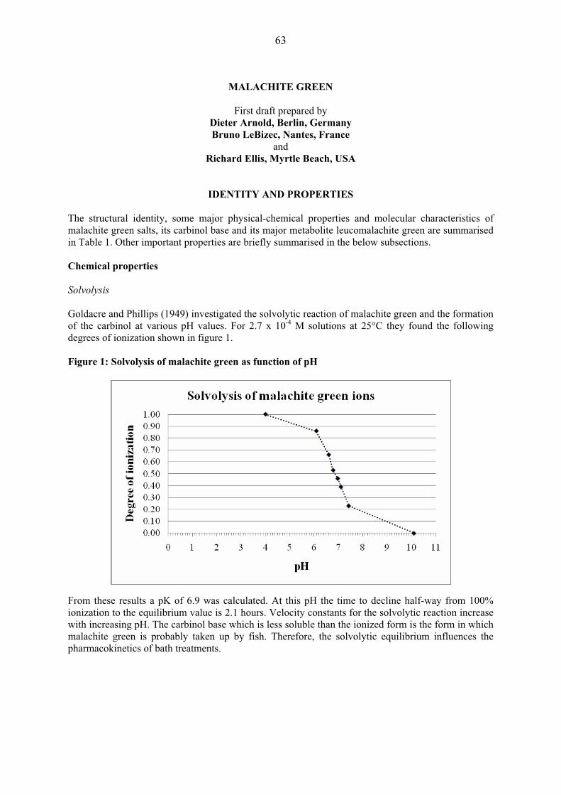
63
MALACHITE GREEN
First draft prepared by
Dieter Arnold, Berlin, Germany Bruno LeBizec, Nantes, France
and Richard Ellis, Myrtle Beach, USA
IDENTITY AND PROPERTIES
The structural identity, some major physical-chemical properties and molecular characteristics of malachite green salts, its carbinol base and its major metabolite leucomalachite green are summarised in Table 1. Other important properties are briefly summarised in the below subsections. Chemical properties Solvolysis Goldacre and Phillips (1949) investigated the solvolytic reaction of malachite green and the formation of the carbinol at various pH values. For 2.7 x 10⁻⁴ M solutions at 25°C they found the following degrees of ionization shown in figure 1. Figure 1: Solvolysis of malachite green as function of pH From these results a pK of 6.9 was calculated. At this pH the time to decline half-way from 100% ionization to the equilibrium value is 2.1 hours. Velocity constants for the solvolytic reaction increase with increasing pH. The carbinol base which is less soluble than the ionized form is the form in which malachite green is probably taken up by fish. Therefore, the solvolytic equilibrium influences the pharmacokinetics of bath treatments.

64
Tab
le 1
: Phy
sica
l-che
mic
al p
rope
rtie
s of m
alac
hite
gre
en, i
ts c
arbi
nol b
ase
and
its m
etab
olite
leuc
omal
achi
te g
reen
.
Subs
tanc
e na
me
Mal
achi
te g
reen
L
euco
mal
achi
te g
reen
M
alac
hite
gre
en c
arbi
nol b
ase
Dep
osito
r-su
pplie
d sy
nony
ms (
PubC
hem
)
Tota
l num
ber:
82
Exam
ples
: M
ethy
lene
gre
en
Gre
nobl
e G
reen
V
icto
ria G
reen
A
nilin
e G
reen
B
enza
l Gre
en
Chi
na G
reen
Fa
st G
reen
B
urm
a G
reen
B
Dia
mon
d G
reen
Bx
Tota
l num
ber:
38
Exam
ples
: Le
uco
mal
achi
te g
reen
M
alac
hite
gre
en le
uco
Mal
achi
te g
reen
leuc
o ba
se
Tetra
met
hyld
iam
inot
riphe
nylm
etha
ne
C.I.
Bas
ic G
reen
4, l
euco
bas
e
Tota
l num
ber:
20
Exam
ples
: So
lven
t Gre
en- 1
M
alac
hite
Gre
en c
arbi
nol
Mal
achi
te G
reen
Car
bino
l bas
e
Che
mic
al A
bstra
cts
Reg
istry
num
ber
1030
9-95
-2
(chl
orid
e: 5
69-6
4-2)
(o
xala
te: 2
437-
29-8
) 12
9-73
-7
510-
13-4
PubC
hem
-CID
11
295
6721
5 10
521
EIN
ECS
209-
322-
8 (c
hlor
ide)
21
9-44
1-7
(oxa
late
)
208-
109-
7
IUPA
C
[4-[
(4-d
imet
hyla
min
ophe
nyl)-
phen
ylm
ethy
liden
e]-1
-cyc
lohe
xa-2
,5-
dien
ylid
ene]
-dim
ethy
laza
nium
4-[(
4-di
met
hyla
min
ophe
nyl)-
phen
ylm
ethy
l]-N
,N-d
imet
hyla
nilin
e bi
s(4-
dim
ethy
lam
inop
heny
l)-ph
enyl
met
hano
l
Mol
ecul
ar fo
rmul
a C
23H
25N
2+
C23
H26
N2
C23
H26
N2O
Fo
rmul
a w
eigh
t 32
9.46
33
0.47
34
6.47
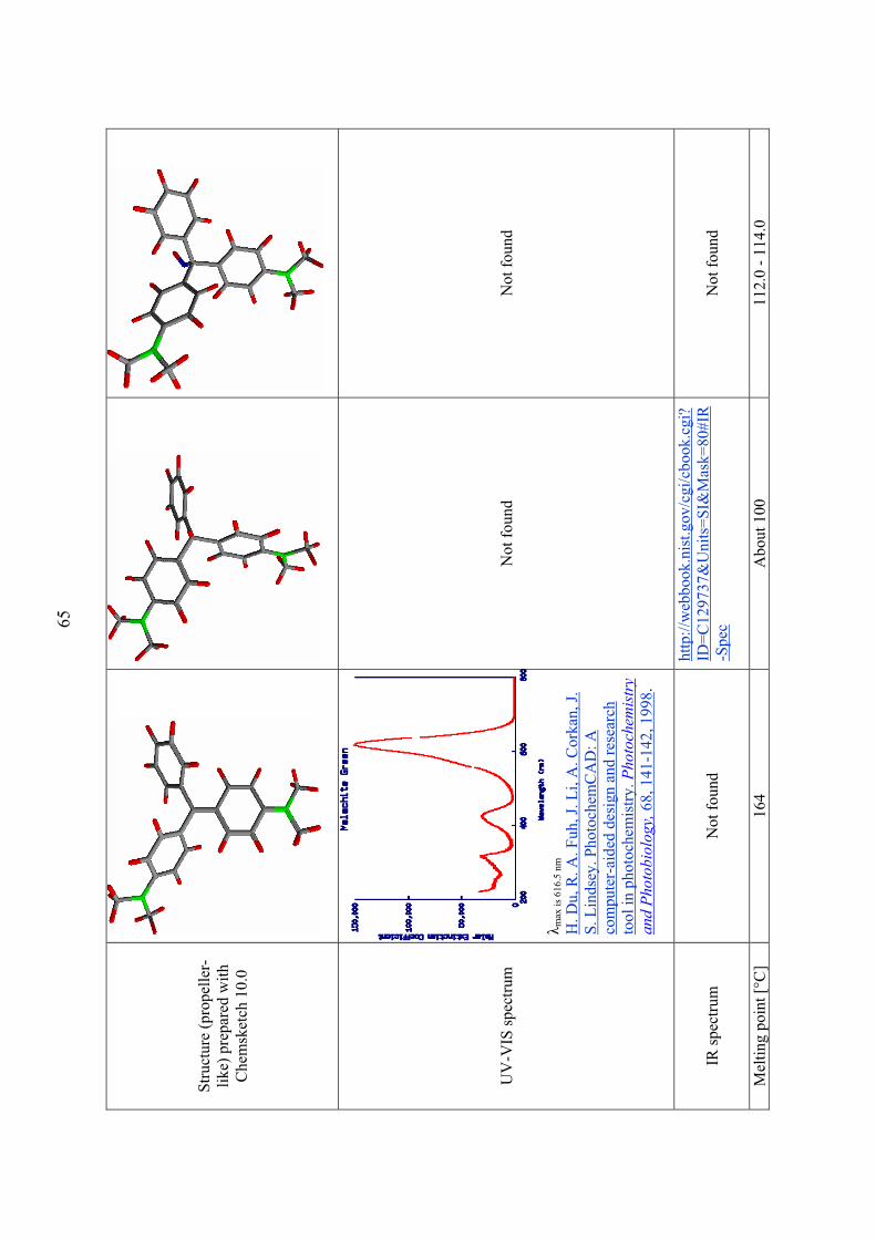
65
Stru
ctur
e (p
rope
ller-
like)
pre
pare
d w
ith
Che
msk
etch
10.
0
UV
-VIS
spec
trum
� m
ax is
616
.5 n
m
H. D
u, R
. A. F
uh, J
. Li,
A. C
orka
n, J.
S.
Lin
dsey
. Pho
toch
emC
AD
: A
com
pute
r-ai
ded
desi
gn a
nd re
sear
ch
tool
in p
hoto
chem
istry
. Pho
toch
emis
try
and
Phot
obio
logy
, 68,
141
-142
, 199
8.
Not
foun
d N
ot fo
und
IR sp
ectru
m
Not
foun
d
http
://w
ebbo
ok.n
ist.g
ov/c
gi/c
book
.cgi
?ID
=C12
9737
&U
nits
=SI&
Mas
k=80
#IR
-Spe
c
Not
foun
d
Mel
ting
poin
t [°C
] 16
4 A
bout
100
11
2.0
- 114
.0
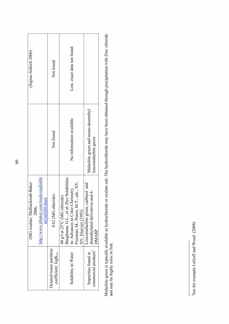
66
(MG
oxa
late
; Mal
linck
rodt
-Bak
er
2006
, ht
tp://
ww
w.jt
bake
r.com
/msd
s/en
glis
hht
ml/m
0286
.htm
)
(Sig
ma-
Ald
rich
2006
)
Oct
anol
/wat
er p
artit
ion
coef
ficie
nt: l
ogK
ow
0.62
(MG
chl
orid
e)
Not
foun
d N
ot fo
und
Solu
bilit
y in
Wat
er
40 g
/l at
25°
C (M
G c
hlor
ide)
B
augh
man
, G.L
., et
al;
Dye
Sol
ubili
ties
in: A
dvan
ces i
n C
olor
Che
mis
try,
Free
man
, M.,
Pete
rs, M
.T.,
eds.,
NY
, N
Y: E
lsev
ier (
1993
)
No
info
rmat
ion
avai
labl
e Lo
w, e
xact
dat
a no
t fou
nd
Impu
ritie
s fou
nd in
co
mm
erci
al p
rodu
cts1
Leuc
omal
achi
te g
reen
, car
bino
l an
d de
met
hyla
ted
deriv
ativ
es a
nd 4
-D
MA
BP
Mal
achi
te g
reen
and
mon
o-de
smet
hyl
leuc
omal
achi
te g
reen
Mal
achi
te g
reen
is ty
pica
lly a
vaila
ble
as h
ydro
chlo
ride
or o
xala
te sa
lt. T
he h
ydro
chlo
ride
may
hav
e be
en o
btai
ned
thro
ugh
prec
ipita
tion
with
Zin
c ch
lorid
e an
d m
ay b
e hi
ghly
toxi
c to
fish
.
1 Se
e fo
r exa
mpl
e Le
Gof
f and
Woo
d (2
008)

67
Other physical-chemical properties Adsorption Adsorption characteristics of malachite green were studied in order to find ways to remove it from waste waters. Traditional methods of wastewater cleaning only partly remove synthetic dyes such as, malachite green and structurally related compounds. The adsorption on a variety of inexpensive and more or less efficient inorganic and organic solid supports was tested as a method for the removal of malachite green from water and wastewater. When testing the adsorption characteristics of malachite green from aqueous solutions frequently only the kinetics of decolourisation of the aqueous solution was measured. Garg, et al. (2003) studied the properties of chemically treated saw dusts (e.g., formaldehyde, sulphuric acid). Gong, et al. (2006) studied citric acid modified rice straw and soybean hulls esterified with phosphoric acid. Hameed and El-Khaiary (2008) used rice straw-derived char. Mittal (2006) determined the adsorption kinetics from waste water onto hen feathers. Janos, et al. (2005) studied the sorption of malachite green from waters onto a naturally occurring kind of weathered and oxidized young brown, acid stable coal called oxihumolite. Tahir and Rauf (2006) used bentonite clay for the removal of malachite green from aqueous solutions. Wang and Ariyanto (2007) studied adsorption of malachite green to natural zeoliths. Carbon-based sorbents show excellent adsorption properties for a considerable number of synthetic dyes. The preparation of carbon sorbents is generally energy consuming and large amount of carbon sorbent is needed for the removal of dyes from large volumes of water. Consequently, according to Forgacs, et al. (2004), the use of commercially available products is fairly expensive. Activated carbon can remove malachite green from water. A system for removal of malachite green from waters used for antifungal treatment in hatcheries was developed and described by Marking, et al. (1989). The removal efficiency was significantly less than 100%. For solutions containing 2 mg/l of malachite green an average of 23.4 mg of malachite green were adsorbed per gram of carbon. Recent studies on the sorption using activated carbon with BET surface areas in the order of 1000 m2/g prepared from different sources of material using various models for the calculation of adsorption isotherms (linearised and non-linear) and for studying the kinetics of adsorption. According to some authors the adsorption of malachite green was best described using a pseudo-second-order model with intra-particle diffusion of malachite green molecules within the carbon particles as a rate-limiting step. The following materials (examples only) were used for the preparation of activated carbon:
Authors
Material used
Singh and Rastogi (2004) Used tea leaves Rahman, et al. (2005) Rice husks Baar (2006) Waste apricot Onal (2006) Waste apricot Onal, et al. (2006, 2007) Lignite, Malik, et al. (2007) Groundnut shell waste. Kumar (2006), Kumar and Sivanesan (2006)
No information on source material
Porkodi and Kumar (2007) Jute fiber. Zhang, et al. (2008) Arundo donax root
Several groups determined thermodynamic parameters (H, G, and S) and concluded that the reaction was endothermic. Malachite green is markedly biosorbed by activated sludge and reduces the rate of oxygen uptake by activated sludge proportionally to its biosorption (Mihara, et al., 2005). The results of the above studies suggest that malachite green and its metabolites and breakdown products may not be
68
completely removed by wastewater treatment and may be present in sufficient amounts in effluents from industry or waste water treatment plants or other sources to cause residues in wild fish. A group of researchers has recently claimed – without providing convincing evidence - that they have published the first example where malachite green was demonstrably taken up by eels caught downstream from treatment plants in the lakes and rivers surrounding Berlin, Germany (Schuetze, et al., 2008). Photodegradation Hydrogen peroxide has been frequently applied to decolorize synthetic dyes including malachite green in waters in the presence of suitable photo catalysts. For example, micro porous solid material prepared by precipitation of phosphotungstic acid and potassium ions, followed by calcinations was proposed for this purpose (Chen, et al., 2006). The formation of active oxygen species such as the radicals O2
-, HO2 and OH are detected during the degradation of dye, and they are proposed to be responsible for the degradation of dyes. In such systems CO2 and small organic acids are the main reaction products. The presence of catalysts such as TiO2 enhances the rate of photodecomposition of malachite green under visible light. Both the superoxide anionic radical and the dye cationic radical are essential to the mineralization of the dyes under visible light-induced photo catalytic conditions (Arpaç, et al., 2007). Kominami, et al. (2003) used TiO2 nano-particles with various physical properties that had been prepared by hydrothermal crystallization in organic media (HyCOM) and post-calcination, for photo catalytic decomposition of malachite green in an aqueous suspension under aerated conditions. Adsorptivity is a decisive factor for the initial bleaching of malachite green on this material which follows pseudo zero-order kinetics. Chen, et al. (2007) have studied the reaction mechanism of malachite green photo degradation on TiO2. They identified a series of N-demethylated intermediates (mono-, di-, tri-, and tetra-demethylated malchite green) under basic reaction conditions of the process. These degradation products are also known to be formed metabolically in bacteria and animals (see below). Under acidic conditions, the whole conjugated chromophore structure of malachite green was cleaved (Chen, et al., 2007). Hydrogen peroxide can effectively decolorize dye wastewaters in the presence of Fe(III) – loaded ion exchange resin (Amberlite IRA 200). The degradation process of Malachite green proceeds via demethylation and phenyl ring openings before CO2 and small molecules are formed. Binding to macromolecules Malachite green can bind to macromolecules. Of interest is the binding to small artificial RNA molecules (aptamers). The complex can exhibit interesting new properties, for example enzymatic activities (Brackett and Dieckmann, 2006). In the binding process the RNA adapts to the ligand (“adaptive binding”), but the ligand itself also undergoes conformational changes ("induced fit") (Nguyen, et al., 2002; Nguyen et al., 2004). The crystal structure of such complexes has been studied using tetramethylrosamine (TMR), a high-affinity analogue of malachite green (Baugh, et al., 2000). The properties and a number of applications of such complexes have been published. One possible use is the determination of malachite green itself, because aptamers are known which enhance malachite green fluorescence by factors in the order > 1000. It is long known that malachite green binds to DNA (Nordén, et al., 1978; Bhasikuttan, et al., 2007). Cationic triarylmethane dyes also have complex-forming properties with proteins (Tacal and Ozer, 2004). A full discussion of all these properties is beyond the scope of this monograph. Industrial uses of malachite green Malachite green is used extensively as a dye for leather, wool, cotton, jute, paper, certain fibres, etc. For such purposes it has been produced in large quantities and extremely variable qualities. About 10-

69
15% of all dyes are directly lost to wastewater in the dyeing process (Parshetti, et al., 2006). Frequently the purity of products used in biological studies has not been reported. In human medicine the carbinol is/has been used as a wound antiseptic and as a treatment of mycotic skin infections, and in staining of tissues and bacteria.
RESIDUES IN FOOD FROM AQUATIC SPECIES AND THEIR EVALUATION Conditions of use in aquatic animals Malachite green has been used as a fish fungicide in closed systems alone or in combination with other chemicals such as formaldehyde for decades. Frequently reported concentrations are about 0.05 - 0.1 mg/kg. It is important to use zinc free preparations in order to avoid metal intoxications of the fish. Therefore, the oxalate was most frequently used. Foster and Woodbury (1936) were reportedly the first to introduce its use as fungicide and antiseptic. It took almost 47 years until researchers considered for the first time the possibility that malachite green could be taken up by fish (Poe and Wilson, 1983). Malachite green has been used for the treatment of eggs of fish and crayfish. Malachite green is also an effective topical and systemic antiprotozoal agent. Reported types of treatment of fish include dip treatment, flush treatment, sustained culture treatment and application in feed. Extremely wide ranges of concentrations and exposure times have been used (review by Alderman 1985). In a review of historical uses of malachite green, Sudova, et al. (2007) discriminate between dip treatments of 10-30 seconds duration and concentrations up to 100 mg/L to treat topical fungal infections, short-term malachite green bath treatments of 60-90 minutes duration at 6.7 mg/L, and long-term bath treatments of six days duration at 0.15 mg/L for salmonids and 0.5 mg/l for cyprinids. They state that this type of treatment was used to control protozoan ectoparasites, particularly the ciliated protozoan Ichthyophthirius multifiliis. Malachite green concentrations can be reduced in multi-component baths, for example in combination with formaldehyde. Treatment with malachite green can produce numerous side-effects in treated fish and fish eggs. An important factor determining therapeutic and toxic effects is the temperature. Batch to batch variation in concentration and purity of the dye and lack of standardization of test conditions have been major confounding factors in the judgment of effectiveness of doses and exposure times. Therefore, it goes beyond the scope of this monograph to make any conclusive statements and comparisons about dosages and other conditions of use. Information on treatment conditions will be given individually in connection with the discussion of pharmacokinetic and residue studies. Malachite green is toxic to fish, in particular to small fry. Bills, et al. (1977) used standard laboratory tests in order to determine the LC50 under various conditions of temperature, pH, and hardness of the treatment bath and of duration of treatment. Fingerling fish of a great variety of species, weighing 0.5 to 1.5g were used for the tests. Increase in exposure time significantly increased the acute toxicity. In short term-exposure (3 and 6 hours) of rainbow trout and channel catfish higher temperatures increased the acute toxicity. At the longest tested exposure time (96 hours) the temperature effect disappeared in rainbow trout. pH and hardness had no significant influence on acute toxicity. As an example, some data obtained with rainbow trout were selected from the original paper and are presented below in Table 2. The original work also provides the 95% conficence intervals of the LC50 which is not given in the below Table 2.

70
Table 2: Acute toxicity of malachite green to rainbow trout
Incubation time [hours] 3 6 24 96
Temperature [°C]
Water hardness pH
LC50 [mg/L 7 soft 7.5 >2 2.3 0.4 0.17
12 soft 6.5 >2 1.0 0.28 0.28 12 soft 7.5 1.4 0.8 0.36 0.25 12 very soft 8 2.0 0.8 0.36 0.29 12 soft 8 2.3 0.8 0.28 0.23 12 hard 8 2.3 1.4 0.35 0.29 12 very hard 8 2.4 0.8 0.28 0.25 12 soft 8.5 2.6 1.0 0.28 0.21 12 soft 9.5 >2 1.3 0.37 0.17 17 soft 7.5 1.4 0.6 0.57 0.28
PHARMACOKINETICS AND METABOLISM Pharmacokinetics in Fish Physiological facts: Relationship between carcass weight and organ weight of fish Some basic physiological facts are necessary to understand the kinetic behavior of malachite green in fish. Schmelzing and Claus (1990) found that in rainbow trout (Oncorhynchus mykiss) absolute organ weights increased with increasing carcass weight while their weights as a proportion of carcass weight decreased. Heart and liver weight were highly correlated with carcass weight, while the correlation between spleen and carcass weight was moderate. According to Corti (1948), muscle meat, liver and kidney make up 70.2, 1.2, and 0.77% of body weight in rainbow trout. The range of body weights (n=7) was 54.1 to 82.3g and corresponding range of muscle meat weight ranged from 61% to 78% of the body weights of the animals. The corresponding figures for eel were 80.9, 1.32, and 0.75 % for muscle meat, liver and kidney, respectively. Fish physiology: Uptake of hydrophobic compounds Fish normally take up hydrophobic compounds via the gills by passive diffusion (Gobas, et al., 1986). Rates of uptake can be a function of: water flow over the gills, blood flow through the gills, diffusion through the aqueous stagnant layer along the gill epithelium, or diffusion through the gill membrane. Hayton and Barron (1990) have summarized: “In general, for any particular chemical, only one of the barriers is operative with the resistance offered by the others being negligible. The rate-limiting barrier is determined by the physico- and biochemical properties of the substance: molecular size, lipophilicity, binding to blood proteins and formed elements. The resistance of each barrier is affected differently by variables such as temperature, molecular size, lipophilicity and body size of the animal. When the resistance offered by the gill barriers is low, uptake may be controlled by transfer to storage tissues, e.g., by blood flow to adipose tissue”. When fish increase their water flow, e.g., with decreasing oxygen concentrations or other types of stress, uptake per unit time frequently increases. Sijm, et al. (1994) studied the influence of blood and water flows on the uptake of some hydrophobic compounds by rainbow trout. The fish used in their experiments had an average weight of 110 ± 12g (n not given). The temperature was 12°C. For all compounds studied the uptake rate constants increased with water flow between 0.045 and 0.52 l/min/kg body weight and remained constant at higher flow. The uptake rate constant was constant for blood flow between 4.4 and 10 ml/min/kg body weight but doubled when the blood flow was

71
increased from 10 to 20 ml/min/kg body weight. From their findings they deduce that water flow will practically limit uptake of hydrophobic chemicals in fish weighing more than 5g. Gill physiology In a study on water flow and gas exchange at the gills of rainbow trout (Salmo Gairdneri) Erickson and McKim (1990) developed a simple flow limited model for the exchange of organic chemicals at fish gills. The mathematical model for the exchange of organic chemicals by fish gills was formulated based solely on the limitations imposed by the flows of water and blood into the gills. The model could be useful for approximate assessments of accumulation of organic chemicals by fish. For large rainbow trout, the model was found to closely follow the magnitude and trends of observed gill uptake rates over a range of octanol/water partition coefficient from 1 to 106 . Davis and Cameron (1971) estimated the volume of water passing over the gills per unit time (ventilation volume). The technique was direct measurement. For this purpose a rubber membrane was stitched round the margin of the mouth of the fish in a way that it separated inspired and expired water. 18 fish of a body weight of 210.3 ± 2.3 g were used at 8.6°C to determine ventilation volume when the animals were quiet. The estimation was repeated 4-11 times with each animal. The lowest individual estimate of ventilation volume observed in one fish was 22.0 ml/min; the average per fish ranged from 26.0 to 49.0 ml/min. The overall average was 37.0 ±7.4 ml/min. The corresponding mean ventilation rate was 74 breaths/min and the mean ventilator stroke volume was 0.5 ml/breath. When the fish struggled or were disturbed maximum values rose as high as 162 ml/min in one animal (average 88.2 ± 43.7). Nichols, et al. (2004) developed a physiologically based toxico-kinetic model for dietary uptake of hydrophobic organic compounds. Malachite green administered via the diet shows unsatisfactory efficacy. Therefore, this model cannot be applied to the data discussed in this monograph. Pharmacokinetic studies in rainbow trout Alderman and Clifton-Hadley (1993) conducted a pharmacokinetic study in rainbow trout (Onkorhynchus mykiss). The dye was administered through uptake from the water bath. The heavily vascularized gill was assumed to be the principal site of malachite green uptake from solution under these conditions The fish used in the main pharmacokinetic experiments of the study were two separate groups of rainbow trout with average body weights of 241 ± 33 g (n not given) for studies conducted at 16°C and 199 ± 20g (n not given) used for studies conducted at 8°C, respectively. A third group of 30 fish of 50g body weight was used for additional experiments in which residues in muscle were determined in individual fish at 16°C (results not given). The pH was 7.6 and total hardness was 13.8° dH. Under these conditions more than 95% of the malachite green was in the carbinol form. The purity of the malachite green was tested by thin layer chromatography. Treatment solutions containing 1.6 mg/l (4.86 μmoles/l) were prepared from a commercial liquid formulation 15 h before use in order that the dye-carbinol equilibrium concentrations could become established. Treatment time was 40 minutes. In the main experiments fluids and tissues of five fish per time point were pooled and stored frozen for analysis. However, one graph of the publication shows individual kinetic data in serum obtained in a separate experiment. Samples of serum and bile were allowed to defrost before analysis. After dilution with buffer at pH 4.0 they were extracted for 24 h into pentan-1-ol (no information on the partitioning of malachite green and its metabolites was provided). After 24 h, samples were centrifuged at 2000 × g for 30 min, giving a clear pentan-1-ol supernatant. All other tissues were allowed to defrost overnight and weighted composites were then blended in 2% pepsin adjusted to pH 2.0 with HCl. The blended samples were kept for 18h at room temperature (20°C) and were shaken thoroughly several times in that time. The samples were then partitioned at room temperature at approximately pH 4 into pentan-1-ol. Following the addition of pentan-1-ol the flasks were shaken

72
vigorously before being left (with further shaking) for a further 24 h. Samples were then shaken thoroughly again before centrifugation at 2000 × g for 30 min at 4°C. Sample extracts were scanned at wave lengths from 540 to 700 nm. Peak absorption for the malachite green dye ion in extracts was 625 nm. Reported recoveries at 10 mg/kg were 19, 82, 75, 68, and 60% for serum, liver, kidney, muscle, and viscera, respectively. The spectrophotometer was calibrated against extracts of malachite green obtained from representative fish tissues spiked with known concentrations of malachite green and after equilibration subjected to the same extraction procedure as described above for the experimental samples. The reported LOD was about 50 μg/kg (data not shown). Some tissues presented general or occasional problems in residue determination including interference of colored co-extracted substances. The paper exhibits a number of weaknesses:
� Concentration of the drug in water was not monitored during the experiment. � The description of the experiment lacks precision. The exact relationship between
total weight of treated fish and weight of the bath is not given. One may speculate that it not exceeded 24 x 5 x 0.241 kg of fish in 725 kg bath.
� Fat was not sampled. � It was not determined to what extent leucomalachite green was extracted and whether
it was re-oxidized to the malachite green. Therefore, the kinetic data of this study can probably not be interpreted.
� Insufficient information on method validation is provided. The study typically does not provide information on results obtained with individual animals; thus no estimate of the biological variability is possible.
� The authors report that malachite green appeared in the serum very rapidly, with concentrations increasing steadily until the fish were removed from the dye (results not shown). In fact, peak concentrations shown in figure 2 of the paper were in the order of 13-13.5 mg/kg at both 8 and 16°C.
� The text states that peak concentrations in muscle were reached 90-120 min after the end of exposure and gives values of 6.81 and 10.79 mg/kg for the peak concentrations reached at 8°C and 16°C, respectively; however, the curve describing the influence of exposure time never exceeds approximately 1 mg/kg for muscle.
� The text states that when a group of small rainbow trout was exposed to malachite green and individual muscle residues examined at 24 h post-exposure, considerable fish-to-fish variations were evident; however neither data nor an estimate of the variance are provided.
� The legend to the figure describing the kinetics in bile and the corresponding text state that bile could only be reliably collected for the first 40 h; however, the corresponding graph shows data points for 48 and 72 hours.
� The analytical method used was inadequate. � The calculated half-lives for serum and tissues cannot be compared because they are
either based on different kinetic models used for curve fitting, or - even when the same model was used - the distribution in time of the data points covered different phases of the kinetics.
� Some variations in treatment parameters and associated effects on results are discussed without providing any data.
� Extrapolations (calculations and results not shown) far beyond the experimental time points and orders of magnitude below the measured concentrations.
The following conclusions may be drawn from the results of the study: Uptake was lower at 8°C than at 16°C; the initial rate of decrease of the optical density at 625 nm was higher at 16°C than at 8°C; the maximum concentrations found in tissues were in the order of (all values in mg/kg):
Temperature/Tissue Serum Liver Kidney Muscle
8 °C 13 8 7.8 16 °C 13.5 16.5 34 10.8

73
However, this statement is only valid if one can assume that the biotransformation of malachite green in this experiment was a slow process compared to the rate of uptake or that leucomalachite green is not picked up by the analytical method. If one assumes that malachite green was rapidly metabolized to leucomalachite green and other molecules (as shown in other studies) which are all extracted, then all results of this study could be meaningless numbers; if leucomalachite green was re-oxidized the above given maximum concentrations could represent an estimate of total residue. A table summarizing pharmacokinetic and residue data in aquatic species was found on the website of the U.S. FDA. The table includes the work of Alborali, et al., published under the title: “The persistence of malachite green in the edible tissue of rainbow trout (Oncorhynchus mykiss)” in the Journal Rivista Italiana di Acquacoltura 32, 45-60. The summaries of the cited findings are given here: Rainbow trout (Oncorhynchus mykiss) with a body weight of 60-80 g were exposed at 18 °C for one hour to a solution of 1 mg/l of malachite green. Residues were determined by HPLC (no details given). The following information contained in the FDA document is provided for gills, kidney, muscle and skin: “Gill: Residues decreased fairly rapidly during the first 320 days reaching 260 by d41, and below 1 ng/g after 7th month. T 1/2 in the 20 d range initially, with later slow decline in the 50d range. Kidney: Max residue on d(ay) 30 = 1650 ng/g, declined to below 1 ng/g after 41 d. Muscle: Residues high for 34 days - above 1,000 ng/g. Decreased to 200 ng at the 4th month, around 100 by 150 d, slowly declining to below 10 ng/g by 9th month. T1/2 in the 40-50 d range. Skin: Residues decreased fairly rapidly during first 20 days, to about 2500 ng/g, d 50= approx 1000, 200 ng/g round the 4th month, below 1 ng/g at day 283. T1/2 in the 50 d range.” Pharmacokinetic studies in channel catfish The pharmacokinetics and metabolism of malachite green in channel catfish (Ictalurus punctatus) were examined by Plakas, et al. (1996) after intravascular dosing or waterborne exposure. The intravascular dosing solution contained 0.8 mg of 14C labeled dye cation per ml of 0.85% aqueous NaCl solution corresponding to a specific activity of 0.925 MBq/ml or 1.16 MBq/mg. For waterborne exposures, the initial dye concentration was 0.8 mg/l corresponding to a specific activity of 0.185 MBq/ml or 0.231 MBq/mg. The channel catfish were 0.5 to 0.7 kg. For the collection of blood and urine the dorsal aorta and urinary bladder were cannulated. � Five animals were dosed intravascularly with [14C] malachite green at a dosage of 0.8 mg/kg
body weight. Blood specimens were collected 2.5, 5, 7.5, 10, 15, 20, 30, and 45 min and 1, 2, 4, 6, 8, and 10 h after drug administration.
� Five animals were transferred to the dosing solution (0.8 mg/l). Blood specimens were taken at 15-min intervals during the 1-h exposure period. At the end of the dosing period, fish were briefly rinsed in a water bath. Blood specimens were taken at 10, 20, 30, and 45 min and at 1, 2, 4, 6, 8, and 10 h after the end of the dosing period.
� To determine the tissue distribution of malachite green and its residues after waterborne exposure, groups of five animals were exposed to [14C]-malachite green solutions (0.8 mg/l) for 1 hour. Animals were killed and tissues collected immediately after dosing (designated 0 h) and at 2, 4, 24, 96, 168, and 336 h (14 days). Additional animals dosed with unlabelled malachite green were sacrificed after 28 and 42 days.
For HPLC determination of malachite green and leucomalachite green, plasma was extracted with acetonitrile. Muscle was subjected to a more complex procedure involving extraction with acetonitrile – acetate buffer, re-extraction, solvent partition, and SPE. HPLC fractions were subjected to post-column oxidation to the malachite green ion. Mean extraction efficiencies for malachite green and leucomalachite green from plasma and muscle were 85 - 95%. Mean recovery of total radioactivity from muscle of treated fish was 88%, however, individual animal data were not provided.

74
The mean of the concentrations of the plasma samples of five animals declined rapidly after intravascular dosing. Simultaneously the corresponding concentrations of leucomalachite green increased rapidly and reached an average maximum concentration of 0.875 �g/ml in the samples taken at 0.75 h after dosing. At this time point the corresponding concentration of the parent malachite green was 0.6 �g/ml. At ten hours the concentrations of leucomalachite green and of parent malachite green were 0.20 and 0.05 �g/ml, respectively. The sum of these two compounds accounted for approximately 70% of the total drug equivalents at each sampling time. The authors fitted a six parameter (three exponential terms) equation to the data obtained at 14 time points of which nine points were collected during the first hour after treatment. They estimated a terminal half life of 6.2 hours for malachite green. During waterborne exposure at 21 oC total radioactivity and the concentrations of both malachite green and leucomalachite green increased very rapidly to 2.77 and 1.56 �g/ml plasma, respectively. Concentrations of malachite green then started declining immediately; however the peak concentration of leucomalachite green was 2.36 �g/ml 1 hour after transfer of the fish to clean water. The authors state that the decline followed a tri-exponential curve (results not shown) and estimated a terminal half life of 4.7 h. After 10 h concentrations of malachite green in plasma had declined to the limit of detection of 0.25 �g/ml. The authors state that the concentrations of leucomalachite green were still 30 times higher. The half life of terminal depletion of leucomalachite green was not estimated. No information on biological variability is available. The water bath conditions were such that the ratio of the ionic and the carbinol form of malachite green was 6:10. Malachite green and its metabolites were widely distributed and concentrated in the tissues. Concentrations of radioactive residues were highest in the excretory tissues and fat and lowest in the muscle and plasma. Concentrations of total residues exceeded the initial concentration of the water bath in all tissues, with the following few exceptions: bile (0 hours), spleen (168 and 336 hours), and muscle (336 hours). The authors’ results, originally in tabular form, are summarized in figure 2 below. The variability of the results expressed as relative standard deviation of the mean of 5 data points ranged from 0.07 to 0.76. Variability was lowest in muscle tissue and highest in skin and bile. The variability increased over time. The high values of many standard deviations suggest that the residue concentrations might not be normally distributed and that the averages calculated by the authors are not the ideal parameters to show a central tendency. Concentrations of residues in skin were always higher than in muscle. The highest ratio was 2.2, observed 24 hours after treatment. The concentrations of malachite green and of leucomalachite green in plasma were 3.29 and 1.94 �g/ml, respectively, immediately after dosing. One day after dosing, malachite green levels were at the LOQ while leucomalachite green levels were 0.11 �g/ml at day 14 after treatment. In muscle, malachite green and leucomalachite green concentrations were 1.18 and 1.45 mg/kg, respectively, at the end of the exposure period and 0.012 and 0.52 mg/kg 14 days after treatment. Results obtained at other time points are not numerically given. The elimination of malachite green in muscle appeared biphasic with a terminal half-life of about 67 h. Concentrations of leucomalachite green were quantifiable for up to 42 days (0.02 mg/kg ). Unidentified metabolites eluting before leucomalachite green during HPLC were found. The sum of the concentrations of these three metabolites reached a maximum of 31.3% of the total residue at 24 hours after treatment. No detailed pharmacokinetic information is provided for the water-borne exposure; however the authors state that the half lives for malachite green and for leucomalachite green were 2.8 and 10 days, respectively. The effect of pH of the exposure solution was studied at pH values of 6, 7, and 8. When catfish were exposed to solutions of 0.8 mg/l of malachite green for one hour, uptakes increased significantly with increasing pH, determined by the concentrations of malachite green and leucomalachite green in plasma and muscle immediately after exposure. This may be due to the change in equilibrium concentrations of the cation and the carbinol and in the rates of conversion of the cation to the carbinol. The well designed and conducted study cannot be used for the derivation of MRLs because statistical evaluations are not possible in the absence of individual animal data.

75
Figure 2: Total residues of malachite green in tissues of channel catfish. Pharmacokinetic studies in juvenile eels Bergwerf, et al. (2004) exposed 450 juvenile eels (Anguilla anguilla) of an average weight of 4.1 g (range 1.5 to 7.4 g) for 24 hours to malachite green in a water bath with temperature varying between 23.0 and 26.5°C and at pH values between 7.0 and 7.8 in 40 L water. A malachite green concentration of 0.1 mg/l was intended. Fish were then cultured in malachite green free water at 21.6 to 27.5°C. During this period pH varied between 6.0 and 7.8. Before, and at various time points during, and up to 100 days after treatment, ten fish and water were sampled (50 ml per time point). The fish did not grow during the study. The whole fish were cut in fine pieces and 2g of cut tissues were blended in buffer and extracted with buffer/acetonitrile mixtures. Partitioning with dichloromethane was followed by solid phase extraction. Brilliant green was added as internal standard before HPLC analysis. Water samples were mixed with buffer, acetonitrile and internal standard prior to analysis. Two reversed phase columns (phenyl-hexyl and C8) were used in series and were eluted with a mixture of 60% (vol/vol) acetonitrile and 40% (vol/vol) of 0.05M ammonium acetate buffer, pH 4.5, at 0.6 ml/min. The eluate was monitored at 620 nm after post column oxidation. Recoveries (n=36) were 61 ± 6% for malachite green and 88 ± 10% for leucomalachite green. Results were recovery-corrected. Analysis of water revealed that the starting bath concentrations were only 0.032 mg/L instead of 0.1 mg/l. This concentration further decreased exponentially during the experiment and fell below the limit of detection at 12 hours. A definitive reason for the low initial concentration could not be found. The further decrease can apparently be explained by the uptake of malachite green by the fish. One can roughly estimate the total amount of malachite green taken up by the fish from the sum of the concentrations of malachite green and leucomalachite green found at a given time point multiplied with number and average weight of fish present in the bath at this time point plus the accumulated amounts removed from the bath by sampling 10 fish at every earlier time point. The figure 3 shows
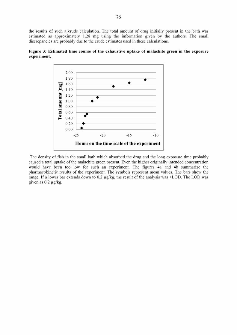
76
the results of such a crude calculation. The total amount of drug initially present in the bath was estimated as approximately 1.28 mg using the information given by the authors. The small discrepancies are probably due to the crude estimates used in these calculations. Figure 3: Estimated time course of the exhaustive uptake of malachite green in the exposure experiment. The density of fish in the small bath which absorbed the drug and the long exposure time probably caused a total uptake of the malachite green present. Even the higher originally intended concentration would have been too low for such an experiment. The figures 4a and 4b summarize the pharmacokinetic results of the experiment. The symbols represent mean values. The bars show the range. If a lower bar extends down to 0.2 μg/kg, the result of the analysis was <LOD. The LOD was given as 0.2 μg/kg.

77
Figure 4a: Concentration changes of malachite green and leucomalachite green during and after bath exposure to 0.032 mg/L of malachite green.
Figure 4a shows the averages of the measurements obtained with 9-10 individual fish at every time point and the range of the individual values. It is not clear how the averages have been calculated at time points where “non-detects” occurred. The latest time point at which the results were not influenced by “non-detects” was 480 hours for malachite green and 1487 hours for leucomalachite green. Malachite green was quantified in the one or other fish up until day 62 following exposure and leucomalachite green was found in the one or other fish over the whole 100 days observation period following exposure. This finding is important in view of the very limited dose administered.
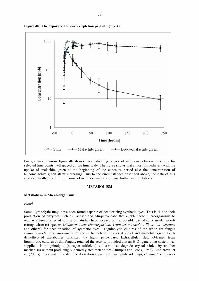
78
Figure 4b: The exposure and early depletion part of figure 4a.
For graphical reasons figure 4b shows bars indicating ranges of individual observations only for selected time points well spaced on the time scale. The figure shows that almost immediately with the uptake of malachite green at the beginning of the exposure period also the concentration of leucomalachite green starts increasing. Due to the circumstances described above, the data of this study are neither useful for pharmacokinetic evaluations nor any further interpretations.
METABOLISM Metabolism in Micro-organisms Fungi Some ligninolytic fungi have been found capable of decolorizing synthetic dyes. This is due to their production of enzymes such as, laccase and Mn-peroxidase that enable these microorganisms to oxidize a broad range of substrates. Studies have focused on the possible use of some model wood-rotting white-rot species (Phanerochaete chrysosporium, Trametes versicolor, Pleurotus ostreatus and others) for decolorization of synthetic dyes. Ligninolytic cultures of the white rot fungus Phanerochaete chrysosporium were shown to metabolize crystal violet and malachite green to N-demethylated metabolites catalyzed by lignin peroxidase. Extracellular fluid obtained from ligninolytic cultures of this fungus, retained the activity provided that an H2O2-generating system was supplied. Non-ligninolytic (nitrogen-sufficient) cultures also degrade crystal violet by another mechanism without producing N-demethylated metabolites (Bumpus and Brock, 1988). Eichlerova, et al. (2006a) investigated the dye decolorization capacity of two white rot fungi, Dichomitus squalens

79
and Ischnoderma resinosum (Eichlerova, et al., 2006b). D. squalens showed high decolorizing capacity. I. resinosum decolorized malachite green to a lower extent up to a concentration of 0.1 g/l. A total of 26 white rot fungi from Argentina were tested for their ability to produce lignin-modifying enzymes and decolorize industrial dyes (Levin, et al., 2004). Ten of the strains decolourised all tested dyes including malachite green. The mycelia were grown on solid malt extract/glucose media containing the dye. All ten strains produced laccase, lignin peroxidase and manganese peroxidase on solid medium. White-rot fungi normally require a lignocellulose substrate. Their use in polluted water streams or soils may be problematic since species of these typical wood colonizers do not exhibit satisfactory growth and competitiveness under such conditions. Litter-decomposing fungi differ from wood-rotting species with respect to their growth substrate, forest litter and soil. They are characterized by higher C:N ratio and microbial activity. Laccase is the most common ligninolytic enzyme among these organisms and Mn-peroxidase is produced only by some species (Baldrian and Snijdr, 2006). Cha, et al. (2001) performed biotransformation experiments of malachite green with cultures of Cunninghamella elegans, a filamentous fungus which had previously been shown to enzymatically catalyse N-demethylation and N-oxidation reactions of a number of chemicals. Metabolites were analysed using HPLC-diode array and HPLC-MS methods. Malachite green was reduced to leucomalachite green and also converted to N-demethylated and N-oxidized metabolites, including primary and secondary arylamines. The mono-, di- and tri-desmethyl derivatives of malachite green and the mono-, di-, tri-, and tetra-desmethyl derivatives of leucomalachite green were found in the supernatant following removal of the mycelium. Malachite green N-oxide was only detected in the mycelia. Identical patterns of metabolites were observed with malachite green and with leucomalachite green as initial substrate. After prolonged incubation only reduced metabolites were found suggesting that parent malachite green and N-demethylated metabolites were reduced by the fungus. Microsomal fractions did not produce reduced metabolites in the absence of NADPH. The cytochrome P450 inhibitor metapyrone completely inhibited the biotransformation reactions. Intestinal bacteria Henderson, et al. (1997) studied the metabolism of malachite green by intestinal microflora from human, rat, mouse, and monkey fecal samples and 14 pure cultures of anaerobic bacteria representative of those found in the human gastrointestinal tract. All complete microfloras were very efficient in reducing malachite green to leucomalachite green (human and rhesus monkey intestinal microfloras, C3H/HEN-MTV mouse intestinal microflora, and Fisher 344 rat intestinal microflora). Of the bacteria commonly found in the human intestinal tract, Clostridium perfringens (ATCC 3624), Escherichia coli (ATCC 25922), and Peptostreptococcus anaerobius (ATCC 27337) converted almost all of the dye to the leuco derivative. The conversion was monitored with HPLC with diode array detection and the structure was confirmed by mass spectrometry. Baker’s yeast (Saccharomyces cerevisiae (MTCC 463) was also shown to effectively decolorize malachite green, primarily through reductive pathways (Jadhav and Goindwar, 2006). A number of other bacteria have been positively tested for decolorizing capacity of malachite green. A complete review would go beyond the scope of this monograph. Metabolism in Laboratory Animals Rats and mice In short term feeding studies, Culp, et al. (1999) have shown that MG is sequentially N-demethylated to secondary and primary aromatic amines in rats and mice both before and after reduction to LMG. Female and male B6C3F1 mice and Fischer 344 rats were fed up to 1200 mg/kg malachite green or 1160 mg/kg leucomalachite green for 28 days. The malachite green used was � 94% pure. Impurities
80
detected were leucomalachite green (1%) and demethlyated derivatives of malachite green (3.5%). Leucomalachite green was � 98% pure. Impurities detected were malachite green and mono-desmethyl leucomalachite green. Livers were extracted using a modification of a published method (Roybal, et al., 1995). The extracts were analysed by HPLC connected to a post-column oxidation chamber and a photodiode array detector. Analyses using HPLC-APCI/MS also were performed. The desmethyl derivatives were synthesized to confirm structures in the samples subjected to APCI/MS. In HPLC-APC/MS analysis of liver extracts from rats treated with leucomalachite green the primarily seen compounds were protonated leucomalachite green, protonated demethylated derivatives and the molecular ions of malachite green N-oxide and demethylated N-oxide. A small, but measurable, amount of malachite green was also found. At higher cone voltages additional collision-induced diagnostic fragments were found that were formed following losses of dimethylaniline-, methyl-, or phenyl- moieties. The appearance of these molecules was consistent with the fragmentation pathways previously published for leucomalachite green (Doerge, et al., 1998a) who observed similar sequential demethylation in a thyroid peroxidase-catalyzed reaction of leucomalachite green. A dose-related increase in leucomalachite green and metabolites was observed in both rat and mouse liver extracts. Similarly, HPLC-APC/MS analysis of liver extracts from rats treated with malachite green detected the molecular ions for malachite green, its mono-, di-, tri-, and tetra-desmethyl derivatives, and malachite green N-oxide. A small, but measurable, amount of leucomalachite green was also detected. Higher cone voltages produced fragments consistent with those previously reported by Doerge, et al., (1998b). These authors incubated leucomalachite green with tyrosin peroxidase, iodide, and tyrosine in the presence of an H2O2 generating system and obtained the mono-, di-, and tri—desmethyl derivatives of leucomalachite green as well as malachite green and malachite green-N-oxide. Concentrations of malachite green and metabolites increased with increasing dose. The formation of both the symmetric and asymmetric di-desmethyl malachite green metabolite could be demonstrated with the symmetrical isomer eluting first. When liver extracts were analysed using HPLC/UV detection, leucomalachite green was the major product detected in rats fed leucomalachite green (accompanied by small amounts of mono- and di-desmethyl-leucomalachite green) and malachite green was the major product detected in the livers of rats and mice fed malachite green (accompanied by mono- and di-desmethyl malachite green and leucomalachite green – and in the case of rats mono- and di-desmethyl-leucomalachite green). 32P-Postlabeling of liver DNA indicated the formation of a DNA adduct, or co-eluting adducts, that increased with increasing dose, in rats and mice fed leucomalachite green or malachite green. Cho, et al. (2003) mention that malachite green and the N-demethylated derivatives of malachite green and leucomalachite green are capable of forming DNA adducts in vivo, with the binding being consistently greater with the ionic MG derivatives. Figure 5 below summarizes the structural elements of the N-desmethyl metabolites of malachite green and leucomalachite green.

81
Figure 5: Structures of malachite green and leucomalachite green.
Malachite green Leucomalachite green
Structures of malachite green, leucomalachite green, and demethylated derivatives
R1 R2 R3 R4 R1 R2 R3 R4
Parent molecule CH3 CH3 CH3 CH3 CH3 CH3 CH3 CH3 Desmethyl- CH3 CH3 CH3 H CH3 CH3 CH3 H
Di-desmethyl- (symmmetric)
CH3 H CH3 H CH3 H CH3 H
Tri-desmethyl- CH3 H H H CH3 H H H Tetra-desmethyl- H H H H H H H H
Metabolism in Food Producing Animals Fish No systematic metabolism study has been performed in fish. However, many of the degradation products formed either under physical-chemical conditions or in in vitro and in vivo studies in bacteria, fungi and laboratory animals have also been found in fish. The potential presence of such molecules in monitoring samples is largely ignored by analysts and only a few methods use protective agents in order to prevent the breakdown of incurred residues during extraction and cleanup.
TISSUE RESIDUE DEPLETION STUDIES Fish Poe and Wilson (1983) reported in a short note to the journal “Progressive Fish Culturist” that they had observed a green color developing after some storage time on the surface of the tissue of skinned frozen fish. The color could be extracted using a procedure for lipid extraction and was identified as malachite green by infrared and visible spectra. They conducted a small series of experiments with channel catfish of body weights ranging from 0.3 to 0.7 kg in which they varied exposure conditions (exposure levels and frequencies). Samples of visceral fat and the carcass were frozen. The green color appeared typically in visceral fat more rapidly than in muscle. When Alderman published his frequently cited article “Malachite green: a review” in 1985, uptake and residues had not yet become a significant issue in the scientific literature.
Eggs and fry Allen and Hunn (1986) reported that malachite green accumulates in the eggs of gravid female salmon after treatment and is detectable in eggs and newly hatched fry. Meinertz, et al. (2005) determined residues of [14C]-malachite green in eggs and fry of rainbow trout after treatment of the eggs. The treatment method was flush treatment (a concentrated solution of malachite green was added to an incubation unit and flushed through with fresh water). At the beginning of the treatment the eggs were approximately 2 days old. Hatching began on day 25 and
R1R2
R3
R4N
N+
R4
R3
H
R2R1
N
N

82
was completed on day 31. Six groups of 250 eggs each were treated in 500-ml glass test aquaria. Before treatment water flow to the aquaria was stopped and the volume was drawn down to 265 ml. Eggs in all aquaria were exposed simultaneously using a distribution manifold. The expected nominal concentration was 1.0 mg/l. Water flow was re-established immediately after treatment. Treatment was performed on days 0, 3, 6, 9, 12, 15, 18, 21, 24, and 31. Water samples were taken at every treatment from one randomly selected aquarium immediately after addition of the treatment solution and 2.5, 5, 10, 15, 20, 25, 30, and 60 minutes after water flow was re-established. Ten eggs were sampled from each test aquarium immediately before each treatment through day 24. Five were prepared for combustion analysis (92.8 to 95.8 combustion efficiency) and the remaining five were used for analysis of malachite green residues by HPLC with post-column oxidation and visible light detection. On day 31, 10 fry were sampled from each aquarium immediately before the final treatment and 0, 6, 12, and 24 hours, and 2, 4, 7, 12, 17, 22, and 28 days after treatment. Mean peak concentrations of chromatic malachite green in water were 0.37 mg/l and were slightly higher than the radioactive concentration equivalents. The intended nominal concentration was not reached and individual measurements were extremely variable at all time points. Untreated eggs contained measurable concentrations of unlabelled malachite green. Pre-treatment radioactive concentration equivalents in eggs and fry increased from day 0 to day 31 to a concentration of 271 ± 42 (n=6) – without exhibiting saturation effects or reaching a steady state - and declined to 55 ± 11 on day 28 after the final treatment. The efficiency of the analytical method to extract radioactivity was 49 to 119 % (average 76%). Leucomalachite green was the predominant residue. OD and radioactivity traces of HPLC separations showed one more polar unknown compound in addition to the known compounds. If measurements in fry were corrected for growth, the elimination half life was 9.7 days. Kinetic depletion studies in fish In addition to the studies described in the section of pharmacokinetics some other studies have been performed. Bauer, et al. (1988) published an article “Uptake and excretion of malachite green in rainbow trout”. They described an HPLC method for the determination of malachite green and leucomalachite green. Recoveries were approximately 75% and all results were recovery corrected. For the experiment they used 156 rainbow trout with an approximate body weight range of 200-300 g. The total weight of all fish was 41.2 kg. The temperature was 9.7 ± 0.1°C. Treatment was performed in a tank with 1000 l water to which 200 mg malachite green was added. The proportions of fish to water corresponded to intensive aquaculture conditions for trout. Duration of treatment was 24 hours. Samples for water analysis were taken every hour. Ten fish were sampled immediately after the end of treatment and groups of six were sacrificed at all other sampling times. The last samples were taken on day 143 after treatment. Homogenized tissue samples were frozen and stored at -30°C prior to analysis. Two parallel smaller experiments were carried out in smaller tanks and with smaller fish densities and a treatment concentration of 0.1 mg/l. for methodological studies. The initial concentration in water of malachite green was 205 mg/m3 and decreased to 5 mg/m3 in 24 hours following an exponential term. Thus 97.6% of the malachite green disappeared and was probably taken up by the fish because in one of the smaller experiments 80% of the malachite green that had disappeared from the water bath was found in the fish. About 33% of the malachite green which was taken up was found in muscle. At the end of the treatment period the total concentration of malachite green plus leucomalachite green was 910 ± 243 μg/kg (n=10). The concentration of the parent drug was 86.3 ± 54.4 μg/kg (n=6). On subsequent days the concentrations of the parent drug rapidly decreased and the between fish variability increased. A graph provided in the original paper shows that the decrease in the concentration of malachite green did not follow a mono-exponential term. However, the group of data points describing the depletion of the leucomalachite green comes closer to a log-linear curve. The concentrations measured in the fatty tissue were very high. Therefore the authors determined the fat content of the muscle samples.

83
For the muscle samples taken during the first 87 days classified according to fat content they found a very high correlation between fat content and concentration of leucomalachite green in muscle and a decrease of the rate of depletion of leucomalachite green in the groups of fish with the highest fat contents. For fish with the highest fat content the elimination half life of leucomalachite green was 43.3 days. The almost complete uptake of the malachite green shows that the compartment fish was still far from any saturation in this experiment. Allen (1990) applied colorimetric analysis to samples of muscle, eggs and fry of malachite green treated Atlantic Salmon (Salmo salar) and Chinook Salmon (Oncorhynchus tshawytscha). Fish had been treated 10 to 47 times with a solution containing 1 mg/kg of malachite green oxalate for one hour. Samples were obtained 1 to 18 days after the last treatment. Residues were extracted with a mixture of 85% ethyl alcohol, 10% formalin and 5% acetic acid. Following extraction in the dark, centrifugation and filtering absorbance at 615 nm was measured in the extracts. The method was not validated. The author states that concentrations of residues in muscle of Atlantic salmon showed no relation with the number of treatments and the concentrations in both species depended only on the elapsed time after the last treatment. Since the methodology is inadequate for the determination of malachite green the numerical results published by the author are most likely of little value. A paper in Thai language (Amornchai Somletchaoen) for which only an English summary is available reports on the persistence of malachite green in tilapia. Sixty juvenile tilapias of an average body weight of 24.1 ± 6.8 g were exposed to malachite green at two therapeutic doses, 0.1 for 24 h and 0.2 for 1 h at a water temperature varying between 23.5 and 26.0°C. The fish were then transferred into clean water and 3 fish were collected at 0, 6, 12, 24, 72, 120, 168 and 360 h post exposure for the determination of malachite green and leucomalachite green residues in muscle tissues. A LC-MS-MS method was used with LOD of 2μg/kg. Following treatment with the high therapeutic dose, highest average concentrations of malachite green and leucomalachite green were 35.6 ± 5.8 and 32.2 ± 17.5 μg/kg, respectively. Malachite green depleted to 0.4 ± 0.15 μg/kg within 24 hours while leucomalachite green was 1.5 ± 0.7 μg/kg after 120 hours. After treatment at the lower dose the highest average concentration of malachite green and leucomalachite green 4.6 ± 1.8 and 30.6 ± 2.6 μg/kg, respectively. The concentration of malachite green was 2.0 ± 0.35 μg/kg at 72 h and not detectable 168 h after treatment. Concentrations of leucomalachite green remained stable between 12 to 72 h after treatment. At 360 h after exposure, the average concentration was 3.5 ± 2.3 μg/kg. A study investigating the metabolic profiles and residues of malachite green in trout tissues was carried out for the United States Food and Drug Administration (Law, 1994). The study was conducted in trout kept in tanks under the following conditions: water temperature (10±2°C), pH (6.0-7.0), hardness (5-10 mg/l), and dissolved oxygen (9±2 mg/l). 14C-Labeled malachite green of a radiochemical purity of 98% was used for the treatment. All experiments and analytical work was carried out under decreased intensity room light. Concentrations in the exposure tanks were maintained by a metering apparatus containing a 14C-MG stock solution at 800 mg/l and delivering 10 ml/min of this solution; the concentration of the treatment solution was 2 mg/kg. Seventy-two randomly selected trout, each weighing about 350g, were divided into 3 groups of 24 fish and put into three 200-l continuous flow exposure tanks containing 2.0 mg/kg 14C-labeled MG (actual concentrations 1.8 ± 0.2 mg/kg, 1.9 ± 0.3 mg/kg and 1.9 ± 0.2 mg/kg, respectively). A water sample (5 ml) was withdrawn from the exposure tanks every 15 min during the 14C-labeled MG exposure period. After a 1-h exposure, the fish were removed to a depuration tank containing flowing, uncontaminated water. At specific time intervals during 14C-labeled MG exposure and depuration two or three trout were removed randomly from each group of fish and sacrificed. The annexes to the study report provided information on concentration of total radioactive residue in tissue homogenates and ratio of malachite green to leucomalachite green concentrations in an organic extract. From these data the concentrations of malachite green and leucomalachite green in the tissues were calculated. The highest concentrations of residues were found in liver and kidney. Significant concentrations of residues were also found in skin.

84
The data of the study by Law (1994) representing the time period between the end of the treatment and 505 hours post treatment were subjected to statistical analysis using one exponential term on the basis of the natural logarithms of the residue contents for curve fitting. The following parameters given in Table 3 were obtained by linear regression: Table 3: Parameters of the linear regression analysis of kinetic residue depletion data in trout muscle.
Parameter MG LMG Intercept - 0.99747 - 0.01994 Slope - 0.02461 - 0.00352 Coefficient of correlation - 0.60012 - 0.73361 Residual variance 0.58312 0.52876
The kinetic data representing the concentrations of malachite green and leucomalachite green between the end of the treatment and 505 hours were subjected to statistical analysis using one exponential term on the basis of the natural logarithms of the residue contents. Depletion half lives were 28 hours for malachite green and 197 hours for leucomalachite green. The kinetic parameters including the variance of the data were used to perform estimates of dietary exposure to malachite green (see below). The results obtained for muscle are summarised in Figures 6 and 7. The data are the same in the two figures. In Figure 6 the time axis is given on a logarithmic scale in order to enable better discrimination of the treatment phase and the phase after treatment. Figure 6: Kinetics of malachite green and leucomalachite green in trout muscle.
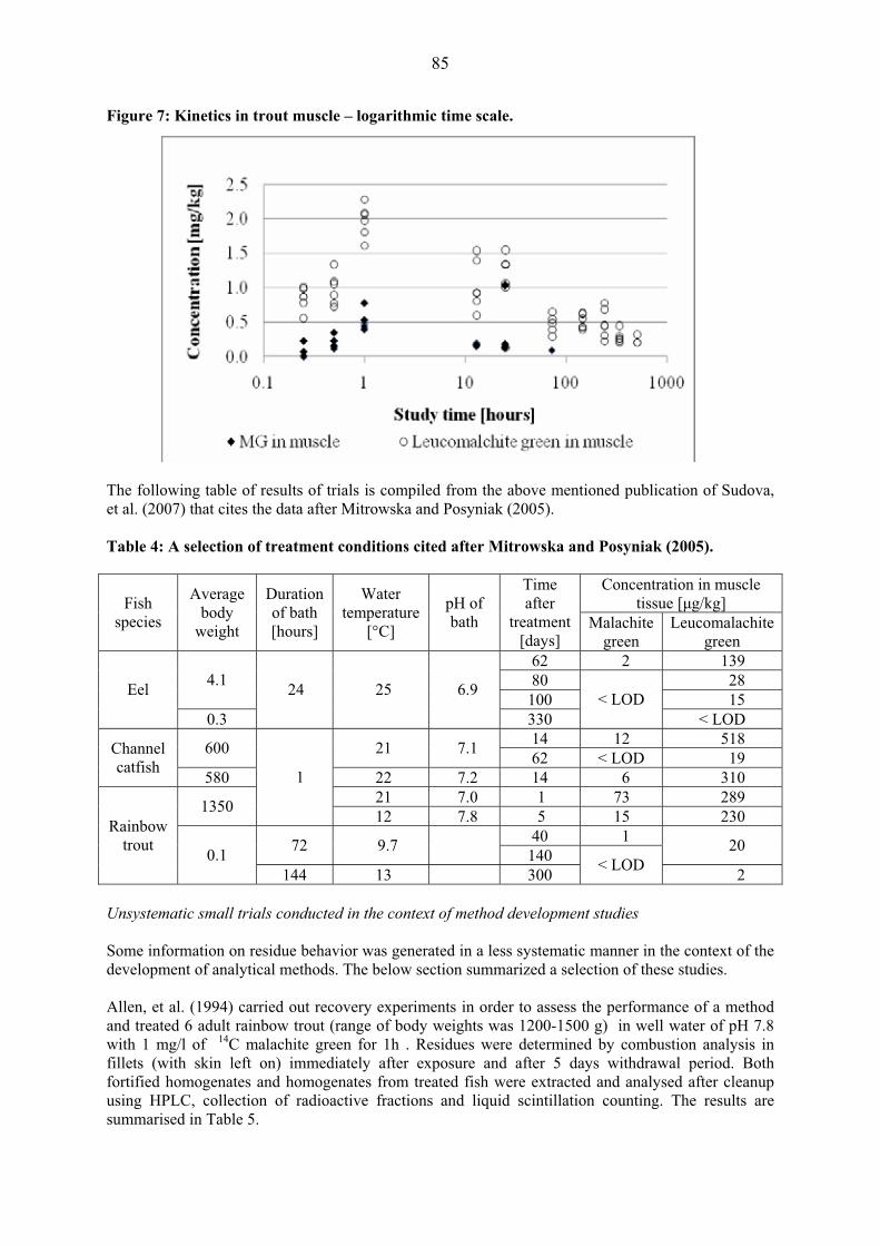
85
Figure 7: Kinetics in trout muscle – logarithmic time scale.
The following table of results of trials is compiled from the above mentioned publication of Sudova, et al. (2007) that cites the data after Mitrowska and Posyniak (2005). Table 4: A selection of treatment conditions cited after Mitrowska and Posyniak (2005).
Concentration in muscle tissue [�g/kg] Fish
species
Average body
weight
Duration of bath [hours]
Water temperature
[°C]
pH of bath
Time after
treatment [days]
Malachite green
Leucomalachite green
62 2 139 80 28 4.1
100 15 Eel
0.3
24 25 6.9
330 < LOD
< LOD 14 12 518 600 21 7.1 62 < LOD 19 Channel
catfish 580 22 7.2 14 6 310 21 7.0 1 73 289 1350
1
12 7.8 5 15 230 40 1 72 9.7 140 20
Rainbow trout 0.1
144 13 300 < LOD 2 Unsystematic small trials conducted in the context of method development studies Some information on residue behavior was generated in a less systematic manner in the context of the development of analytical methods. The below section summarized a selection of these studies. Allen, et al. (1994) carried out recovery experiments in order to assess the performance of a method and treated 6 adult rainbow trout (range of body weights was 1200-1500 g) in well water of pH 7.8 with 1 mg/l of 14C malachite green for 1h . Residues were determined by combustion analysis in fillets (with skin left on) immediately after exposure and after 5 days withdrawal period. Both fortified homogenates and homogenates from treated fish were extracted and analysed after cleanup using HPLC, collection of radioactive fractions and liquid scintillation counting. The results are summarised in Table 5.
86
Table 5: Results of the study of Allen, et al. (1994).
Original concentration [mg/kg]
Found in extract Composition of extract [%]
Material Days after
treatment
Total residue
after combustion
Fortification level with
MG [mg/kg] %1 MG LMG Unknown
Fish 1, muscle 0.8 62 29 45 25
Fish 2, muscle 1.1 85 26 49 24
Fish 3, muscle
0 1.3
1.0 77 34 45 21
Fish 4, muscle 0.5 10
0 3.0 46 51
Fish 5, muscle 0.3 60 3.0 33 64
Fish 6, muscle
5 0.5
0.3 60 1.8 40 59
1 85 84 7 9 Egg homogenate 0.5 98 81 10 9
Fry homogenate 0.65 68 76 11 13
Muscle homogenate
1
66 11 89 1. The values for the six incurred tissues are calculated from the data; they are not given in the original paper. The following results obtained with trout muscle are interesting: � Recoveries from incurred muscle tissues were lower than from fortified homogenates; � A significant fraction of the radioactive residue was of unknown structure. This fraction
increased from approximately 23% immediately after treatment to approximately 58% on day five after treatment.
� Malachite green added to homogenates was largely reduced to leucomalachite green. � The original solution of malachite green remained unchanged if processed by the same
procedure in the absence of tissue homogenate. Andersen, et al. (2005) developed an analytical method based on liquid chromatography in which leucomalachite green is oxidised prior to cleanup with 2,3-dichloro-5,6-dicyano-1,4-benzoquinone (DDQ). Most of the DDQ is consumed by the sample extract. A high excess is needed; however, excess oxidation and formation of breakdown products has to be avoided. The study included the determination of residues in salmon that had been treated with malachite green. Fish (weight not given) were treated in a water bath (volume not given) with 0.01 mg/l malachite green for one hour (temperature not given). The fish were returned to clean water and sampled 2 and 4 hours after the end of exposure. The concentrations (sum of malachite green and leucomalachite green) were found in Table 6. The authors discuss the lower recoveries of spiked malachite green compared to leucomalachite green as a possible explanation of the higher concentrations found at the later time point. Mean recoveries for malachite green were approximately 71% over a concentration range of 1-10 μg/kg. For the same concentrations of leucomalachite green recoveries averaged 95.4%.
87
Table 6: Results of the study of Andersen, et al. (2005).
Hours after treatment Concentration [�g/kg]
2 29.8 35.2 36.3 35.3 4 48.0 47.6 36.9 52.5
The same group published a paper in which a similar method was used, but results were confirmed with LC-MS (Andersen, et al., 2006). The method was validated for catfish, trout, tilapia, basa, salmon, and shrimp. Incurred tissues were also analysed. For this purpose catfish, tilapia, trout, and salmon were exposed to 0.01 mg/l malachite green in water for 1 h. After transfer to clean water samples were analysed at a different time point for each species 16, 16.25, 16.5, and 24 h, respectively, after the end of the treatment. The results are summarised below:
Table 7: Results of the study of Andersen, et al. (2006).
LC-VIS LC-MS mean sd mean sd Fish
species Hours after treatment [�g/kg]
catfish 16 32.2 2.2 31.3 2.7 tilapia 16.25 1.9 0.13 2.1 0.3 trout 16.5 27.1 1.4 28.6 1.1
salmon 24 26.4 0.84 27.4 2 Bergwerf and Scherpenisse (2003) published a method for the determination of malachite green in aquatic animals which is based on HPLC or LC-ESI-MS-MS (for confirmation). The mobile phase was pumped through a pre-column oxidation reactor and effluent was oxidised in a post-column reactor. The authors made the observation that the time interval between spiking of a homogenate and further processing influenced the recovery of malachite green significantly, but had little influence on recoveries of leucomalachite green.
Table 8: Results of the study of Bergwerf and Scherpenisse (2003)
Recovery [%] Time [min] Malachite green Leucomalachite
green 1 81 100
15 63 96 30 60 93 60 54 93
120 45 95
Malachite green was apparently not reduced, but possibly degraded, since the chromatograms showed satellite peaks next to malachite green. The method was optimised for leucomalachite green. Forty-eight samples of trout, eel and salmon were collected at retail level and on fish markets. Approximately 50% were tested positive for leucomalachite green. Doerge, et al. (1998b) described LC methods for the simultaneous quantification of malachite green and leucomalachite green using isotope dilution mass spectrometry. In addition they characterised metabolites derived from malachite green and leucomalachite green found in catfish and trout. Mature catfish of approximately 0.5 kg bw were exposed for one hour in a 40 l tank to 1 mg/kg malachite green at 25°C and pH 7.2. The fish were briefly rinsed and transferred to fresh clean water. Fish were killed 24 hours after dosing and fillets (skin removed) were blended and stored at -60°C prior to

88
analysis. Leucomalachite and malachite concentrations in muscle of treated catfish were 1030 and 590 �g/kg, respectively. Trout were purchased in 1994-1995 from retail outlets in the UK. Blended tissues were spiked with d5-leucomalachite green and 13C6-malachite green. Recoveries of the internal standards were about 34 – 70% (n=12) for malachite green and 64-86 % (n=12) for leucomalachite green. The concentrations of incurred residues ranged from 0.4-3.4 μg/kg for malachite green and 9-96 μg/kg for leucomalachite green. Leucomalachite green was present at much higher concentrations (range 12- to 38-fold). Halme, et al. (2007) proposed an LC-ESI-MS/MS method for confirmation of residues of malachite green and leucomalachite green in trout. D5-leucomalachite green was used as internal standard. They analysed 34 fish monitoring samples of which eight contained malachite green residues. Only the range of the results is given (0.35-1.34 μg/kg of leucomalachite green). Roybal, et al. (1995) developed a method for the determination of malachite green and leucomalachite green by SPE, HPLC, post-column oxidation and detection at 618 nm. In this context they analysed catfish exposed to 1 mg/kg malachite green oxalate for 1h at 21°C and pH 7.0. The treated and rinsed fish were place into separate aquaria equipped with activated carbon filters. Fish were sacrificed and analysed at 0, 2, 4, 8, and 24 hours after placement in individual aquaria. Table 9: Results of the study of Roybal, et al. (1995).
Concentration [μg/kg] Malachite green Leucomalachite green Hours after
treatment mean s.d. mean s.d. Replicates
0 486 23.4 632 23.6 4 2 190 18.8 703 30.8 4 4 187 23.7 748 30.0 4 8 111 12.8 450 30.7 4
24 73.4 7.5 289 19.8 4
Scherpenisse and Bergwerff (2005) published a method for the determination of residues of malachite green in finfish by LC-MS/MS. Recoveries for malachite green were very low in most fish matrices. Recoveries for leucomalachite green were from 86 to 105%. They used the method to analyse nineteen samples including pangasius, salmon, shrimps and trout bought in local shops. Residues were found in three of the samples (trout 24 and 0.15 μg/kg, pangasius 7 μg/kg). Turnipseed, et al. (2005) proposed an analytical method in which leucomalachite green is oxidised to malachite green before the SPE extraction step of cleanup and final LC-MS determination. They used the method to analyse two samples of treated salmon (10 �g/l for 1 hour). They found 34.6 �g/kg in a fish 2h after treatment and 44.3 �g/kg in a fish 4 h after treatment.
ESTIMATION OF DAILY INTAKE
In the open literature, well conducted residue studies suitable to predict the concentration–time course of residues of MG in fish are available for only two species, the rainbow trout and the channel catfish. Only for trout were sufficient individual animal data available to perform a statistical evaluation. Useful information on frequency of occurrence and levels of residues can primarily be obtained from monitoring activities or from well supervised trials conducted under field conditions. The following discussion analyses the selected data for estimation of exposure. In the UK, approximately 400 trout samples were analyzed in three surveys between July 1993 and March 1995. Sixty-seven samples contained malachite green at concentrations of 2-50 �g/kg. The analytical method did not pick up leucomalachite green. In a survey of retail trout in 1996, malachite
89
green was detected in 15 of 208 samples. In 1997 there was only one trout sample out of 137 that contained malachite green. A change in methodology was introduced in 1997 and subsequently malachite green and leucomalachite green were measured. When thirty-one randomly taken samples of the 1997 survey of which 29 were negative were re-analyzed with the new method, seven became positive. The new method was applied to the 27 samples taken in 1998. One contained both malachite green and leucomalachite green. In five samples only leucomalachite green was found (COT, 1999). Thus, the introduction of new methodology increased the number of positives. The individual results of the non-compliant samples are given in the cited COT document. The Veterinary Residues Committee established in 2001 in the UK published the results of all statutory and non-statutory surveillance schemes. For non-compliant samples the individual numerical values found are also given. The presentation of the data was initially such that it was not possible to find out cases in which both residues were found in the same sample. The data are available on the internet. When the results of the 2001-2006 plans were evaluated more than 2300 samples analyzed for malachite green residues have been found. The main fish species covered were trout and salmon, including imports. Occasionally the data are scheduled under “imported farmed fish”. Another useful data set is reported from Denmark. Rasmussen (2007) reported on findings of malachite green in fish in Denmark from 1988 to 2005. 446 plus 95 “targeted samples” were taken. 48 plus 82 “targeted samples” were positive. The author gives individual results for six samples and ranges for the rest of the positives. Unfortunately the individual data were therefore largely not available to the Committee. Of 3277 samples selected from these reports, 222 samples were reported positive for malachite green in the range from 0.2 to about 600 �g/kg fish muscle. For many of the samples it cannot be defined what malachite green means (malachite green, leucomalachite green, or the sum of both or just a number because the method was inadequate). Most likely the true concentrations were higher than the results obtained and a significant fraction of the negatives were probably false negatives. An exhaustive search all public sources of information for data on residues of malachite green is impractical. Many fish species currently moving in international trade and commonly eaten in many parts of the world are not covered. It is not known to what extent results of random sampling and of biased sampling is mixed. It is not known whether any recognised sampling plan has been used in the sampling of lots and which of the individual results given have been obtained from the same lot. The currently published surveillance data are not transparent enough to use it for intake estimates. The more systematically collected data of the UK from the above described activities can be used to estimate a worst-case figure of upper limit of intake of malachite green resulting from illegal uses. These were monitoring data (spanning from 1995 to 2006) published in the United Kingdom on the occurrence of MG and LMG in fish muscle. If both substances were found in a sample a calculated sum could be determined. The estimated mean level found in the positive samples was 30.7 �g/kg fish muscle and the level at the 97.5th percentile to be 138 �g/kg. Assuming the daily consumption of fish to be 300 g per person, the daily exposure to the sum of malachite green and leucomalachite green can be calculated to be 9.2 and 41 �g/per person at the mean and 97.5th percentile, respectively. For a 60-kg person, this would be equivalent to 0.15 �g/kg bw per day and 0.69 �g/kg bw per day, respectively. The Figure 8 below shows the frequency distribution of the levels found in the positive samples out of the 3277 samples selected from the above mentioned programs.

90
Figure 8: Non-representative frequency distribution of illegal residues of malachite green in fish.
The assumption of consumption of 300g of fish contaminated with malachite green and leucomalachite every day for a lifetime was used (a highly conservative assumption). In addition, it was assumed that the concentrations of malachite green and leucomalachite would not change during cooking of the fish. The study by Law (1994) also was suitable to use for performing a dietary exposure estimate. Such an estimate provides some information on the order of magnitude of likely human exposure to residues of malachite green in case the drug would be authorized for treatments comparable to those performed by the authors. Depletion half-lives of 28 hours for malachite green and 197 h for leucomalachite green were determined. The kinetic parameters, including the variance of the data, were used to calculate model intakes for every day of 80 years of a human lifespan, assuming daily consumption of 300g of fish muscle. For this purpose 29220 approximately log-normally distributed random numbers were generated for each time point of interest ranging from the predicted value of the regression line minus four times the residual variance to the same predicted value plus four times the residual variance. These calculations were repeated for a number of assumed slaughter times of the fish, ranging from 1h (end of treatment) to 500 h. The results were expressed in mg malachite green/leucomalachite green/kg of human body weight. The minima, maxima and several percentiles, including the median of these estimated daily intakes, were calculated. The median was used for an assessment of chronic intake. The median daily intake of leucomalachite green declined from 7.3 μg/kg bw at hour 1 to 0.87 μg/kg bw at 500 h. Results of an intake assessment for malachite green and leucomalachite green are shown in Table 11 below. The Committee considered that the assumption of consumption of 300 g of fish contaminated with malachite green and leucomalachite green every day for a lifetime made these estimates highly conservative. In addition, it was assumed that the concentrations of malachite green and leucomalachite green would not change during cooking of the fish. However, that may not be the case.

91
Mitrowska, et al. (2007) investigated the stability of malachite green and leucomalachite green in muscle of treated carp under various conditions of cooking. The initial concentrations of the residues were approximately 200�g/kg. Leucomalachite green was much more stable than the parent compound. Microwaving was the most effective way to partly destroy the incurred residues. The authors published time curves of the degradation. The end results are summarised in Table 10. Table 10: Stability of malachite green and leucomalachite green under various conditions of cooking.
% reduction Sample Procedure Temperature [°C]
Duration [min] MG LMG
Boiling, baking 15 54 0 Residues in carp muscle Microwave 1 61 40
Boiling water 100 0 0 10 49 90 97 150
120 0
Standard solutions Cooking oil
210 10 97 18

92
Tab
le 1
1: R
esul
ts o
f an
inta
ke a
sses
smen
t for
mal
achi
te g
reen
and
leuc
omal
achi
te g
reen
.
Part
I –
Est
imat
ed in
take
at v
ario
us th
eore
tical
slau
ghte
r tim
es o
f fis
h
1h
1.6h
2.
4h3.
8h5.
9h9.
2h14
.3h
22.4
h 34
.9h
54.3
h84
.7h
132.
0h20
5.8h
320.
8h50
0.0h
In
take
of M
alac
hite
gre
en [μ
g/kg
bw
per
day
]
min
0.
2 0.
2 0.
20.
20.
20.
20.
10.
1 0.
10.
10.
00.
00.
00.
00.
0 P5
0 1.
8 1.
8 1.
71.
71.
61.
51.
31.
1 0.
80.
50.
20.
10.
00.
00.
0 P9
0 3.
8 3.
8 3.
73.
63.
43.
12.
82.
3 1.
61.
00.
50.
20.
00.
00.
0 P9
5 4.
7 4.
7 4.
54.
44.
23.
83.
42.
8 2.
01.
30.
60.
20.
00.
00.
0 P9
7.5
5.6
5.6
5.4
5.2
5.0
4.6
4.0
3.3
2.4
1.5
0.7
0.2
0.0
0.0
0.0
P99
6.9
6.9
6.6
6.5
6.1
5.7
5.0
4.1
2.9
1.8
0.9
0.3
0.0
0.0
0.0
max
15
.6
16.2
21
.115
.214
.214
.111
.89.
8 10
.43.
82.
00.
60.
10.
00.
0
E
stim
ated
inta
ke o
f Leu
com
alac
hite
gre
en [μ
g/kg
bw
per
day
]
min
1.
5 1.
4 1.
51.
31.
51.
11.
31.
2 0.
90.
80.
80.
50.
30.
30.
1 P5
0 7.
3 7.
3 7.
27.
16.
96.
76.
46.
0 5.
54.
84.
03.
32.
51.
70.
9 P9
0 12
.6
12.6
12
.312
.212
.011
.711
.310
.7
10.0
8.9
7.8
6.4
4.8
3.3
1.7
P95
14.7
14
.7
14.5
14.6
14.1
13.8
13.3
12.6
11
.710
.79.
37.
75.
84.
02.
1 P9
7.5
16.9
17
.0
16.6
16.8
16.4
15.8
15.3
14.5
13
.512
.511
.09.
16.
94.
62.
4 P9
9 19
.7
19.5
19
.619
.719
.118
.918
.317
.3
16.0
15.0
13.4
10.9
8.3
5.6
3.0
max
48
.5
42.4
36
.039
.736
.234
.638
.038
.9
31.5
32.0
30.3
22.6
16.6
14.3
6.4
E
stim
ated
inta
ke o
f the
sum
of M
alac
hite
gre
en a
nd le
ucom
alac
hite
gre
en [μ
g/kg
bw
per
day
]
min
1.
7 1.
6 1.
71.
41.
71.
31.
41.
3 1.
00.
80.
80.
50.
30.
30.
1 P5
0 9.
1 9.
1 8.
98.
78.
58.
27.
77.
1 6.
35.
34.
33.
42.
51.
70.
9 P9
0 16
.4
16.4
16
.015
.815
.414
.814
.112
.9
11.6
9.9
8.3
6.6
4.9
3.3
1.7
P95
19.4
19
.4
19.0
19.0
18.3
17.6
16.7
15.4
13
.812
.09.
97.
95.
94.
02.
1 P9
7.5
22.5
22
.5
22.0
22.0
21.4
20.4
19.3
17.8
15
.914
.011
.79.
37.
04.
62.
4 P9
9 26
.7
26.4
26
.226
.325
.224
.623
.321
.4
18.9
16.8
14.3
11.2
8.4
5.6
3.0
max
64
.1
58.6
57
.154
.950
.448
.849
.848
.7
42.0
35.8
32.3
23.2
16.7
14.3
6.4
Not
e: F
or e
ase
of re
adin
g an
d fo
rmat
ting
the
data
, the
tabl
e en
tries
are
roun
ded
valu
es u
sing
stan
dard
roun
ding
tech
niqu
es.

93
Results obtained from other surveys Only a few examples of the type of information available from surveys can be given here in order to facilitate the discussion of the limited usefulness of such results in the context of intake assessments. Example 1: The Centre for Food Safety of the government of the Hong Kong special administrative region frequently informs consumers about findings of noncompliant foods. In four separate reports from December 2006 to November 2007, 29 positives were reported from 130 samples of varying sea and fresh water samples collected at import and local markets at concentrations of 14 – 480 μg/kg. Example 2: Reports on monitoring malachite green in aquatic species are available from Australia and New Zealand (FSANZ, 2005). The 60 samples of 7 species of fish were from eight countries of origin. The LOQ for malachite green was 2�g/kg using an LC-MS/MS method. The range of malachite green concentrations was from 4 – 138 �g/kg. Example 3: The Canadian total diet study (1993-2004) collected shrimp and fish samples of various species from various sources to prepare 30 composite samples for analysis of residues of veterinary drugs (Tittlemier, 2007). Fish were baked at 230 °C for approximately 10 minutes. Shrimp were boiled in tap water. It is unlikely that malachite residues are stable under these conditions of sample preparation. It would have been useful to include composite samples of raw fish. The composite samples were frozen and stored at -20 °C until analysis in 2005. The report makes no statement on the stability of residues over such long storage times at relatively high temperatures. The LC-MS/MS methods used for the determination of malachite green and leucomalachite green had a limit of detection of 0.15 �g/kg. Leucomalachite green was found in three of the composite samples (freshwater fish 2002 and 2003, 0.95 and 0.73 �g/kg; shrimp 2002, 1.2 �g/kg).
Results obtained from residue depletion studies Residue depletion studies are only available for the rainbow trout, the channel catfish and tilapia. Only one of all the residue studies discussed above is suitable to predict the time course of residues of malachite green in fish. Table 12 summarises some selected characteristics of four major kinetic and residue studies. Despite the large differences in bath size the ratio of fish weight to bath weight is similar; however, there are large differences in the amounts of drug available per fish. This is most likely a critical factor in studies with prolonged exposure times. The figure is lowest for the study with the longest exposure time. Only one study, conducted by Law (1994), replaced the malachite green taken up from the bath by the fish. The main argument against using the data of the Alderman and Clifton-Hadley study (1993) is that the otherwise well designed study exhibited methodological deficits and used an analytical method that was not valid for the purpose. The Plakas, et al. (1996) study is excellently designed and conducted, but the individual fish residue data were not available. The Bauer, et al. study could have been well used for an observation period similar to the exposure time in the other study; however, individual data were also not available. Overall, only limited conclusions are possible for this study because most of the malachite green was used up by the fish during exposure to the bath. Table 13 highlights some possible impact on results of bath treatment studies regarding the study designs. For the calculations in the table the physiological data for trout discussed in a previous section were used. Three options regarding ventilation volume and breaths/min were calculated for the Alderman and Clifton-Hadley study (1993), one alternative is given for the Bauer, et al. study (1988). From the data presented the design of the Alderman and Clifton-Hadley study was largely acceptable, however, it was not the case for the Bauer et al. study. This is further substantiated in Figure 10.

94
Tab
le 1
2: S
elec
ted
char
acte
rist
ics o
f fou
r ki
netic
res
idue
stud
ies.
Aut
hors
and
yea
r Sp
ecie
s
Volume of the tank used for treatment [L]
Initial concentration of malachite green [mg/L]
Average temperature [° C]
Duration of exposure [min]
Estimated maximum weight of all fish in the tank
Ratio bath weight to fish weight
Initial amount of drug available [ of fish]
Bau
er, e
t al.,
198
8 R
ainb
ow tr
out
1000
0.
21
9.7
1440
41
.2
24.2
7 4.
98
16
Ald
erm
an a
nd C
lifto
n-H
adle
y, 1
993
Rai
nbow
trou
t 72
5 1.
6 8
40
28.9
2 25
.07
40.1
1
Plak
as, e
t al.,
199
6 C
hann
el c
atfis
h 10
0 0.
8 21
60
3.
5 28
.57
22.8
6 La
w, 1
994
Rai
nbow
trou
t 20
0 2.
0
60
8.4
23.8
47
.6
Tab
le 1
3: F
ish-
phys
iolo
gica
l asp
ects
of s
elec
ted
kine
tic r
esid
ue st
udie
s.
Ventilation volume mL/min
Stroke volume [mL]
Breaths/min
Initial MG concentration [mg/L]
Tank volume [L]
Total amount of MG in tank [mg]
Exposure time [min]
Total number of breaths per animal
Number of fish
Stroke volume of all fish together [mL]
Total inspired water during exposure [L]
22
0.5
44
1.6
725
1160
40
17
60
120
60
105.
6 37
0.
5 74
1.
6 72
5 11
60
40
2960
12
0 60
17
7.6
49
0.5
98
1.6
725
1160
40
39
20
120
60
235.
2 22
0.
5 44
0.
205
1000
20
5 14
40
6336
0 15
0 75
47
52

95
For a primitive modelling exercise it was assumed that the inspired water is completely cleared from malachite green which means the amounts inspired with a stroke remain in the fish. On this basis the following two graphs (Figure 10) were prepared modelling the situation of the Bauer, et al. study. The data were generated by dissecting the whole uptake process into the number of elementary steps dictated by the above given total number of breaths per animal.
Figure 10: Modelling of some aspects of the Bauer et al. (1988) study.
Left side: changes in the drug concentration in the bath; right side: uptake during exposure in a limited bath volume and limited amounts of available malachite green. The modelling on the left side of Figure 10 predicts that the bath volume is insufficient for an exposure experiment of 1440 minutes duration involving 150 fish. The right modelling experiment predicts that the uptake of malachite green by the fish will be limited by the amount of available drug and therefore, could lead to the wrong interpretation that the uptake capacities of the fish were saturated. The model predicts a final concentration of 2 mg/m3; the authors reported 5 mg/m3. Also the predicted amounts of residues in the fish are in the same order of magnitude as experimentally determined by the authors. Although the study has provided some remarkable results it is not representative for the treatment of rainbow trout at 0.8 mg/l in a bath. Similar calculations were performed using the information from the Alderman and Clifton-Hadley study (1993) and are summarized in Figure 11 below. The graphs show that the concentration in the water bath decreased by approximately 25% in the worst case model using the highest ventilation volume. If one compares the predicted amounts taken up by the fish it appears that the scenario with the lower ventilation volume fits better to the order of magnitude of initial tissue residue concentrations given by the authors. Thus the study was well designed but suffered from a number of weaknesses discussed in a previous section. It was not possible to perform similar modelling with the information provided in the Plakas et al. study (1996). However, this was also not necessary because the authors were prudent to use a fresh bath for every set of five fish treated and they report that the concentration of malachite green in the bath decreased by only approximately 15% during treatment.
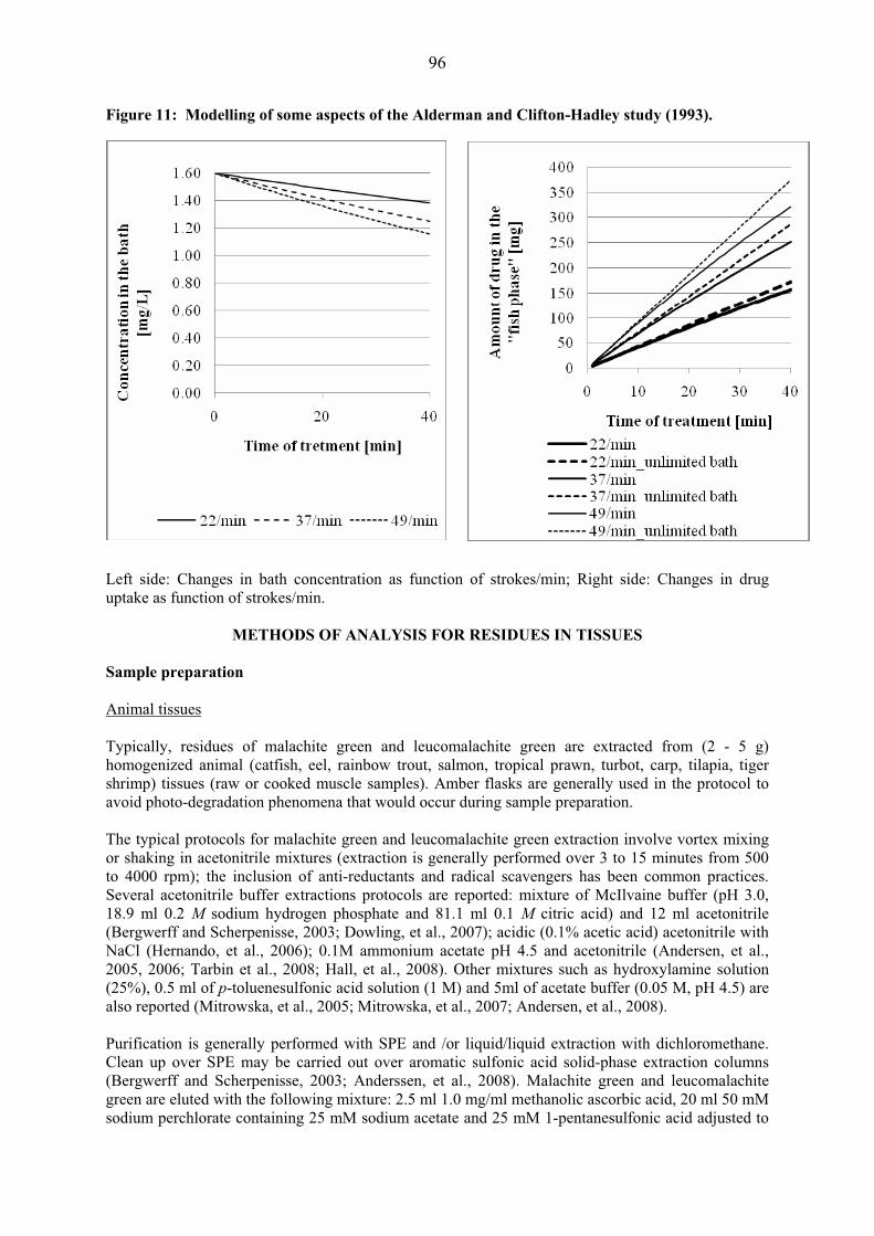
96
Figure 11: Modelling of some aspects of the Alderman and Clifton-Hadley study (1993).
Left side: Changes in bath concentration as function of strokes/min; Right side: Changes in drug uptake as function of strokes/min.
METHODS OF ANALYSIS FOR RESIDUES IN TISSUES Sample preparation
Animal tissues Typically, residues of malachite green and leucomalachite green are extracted from (2 - 5 g) homogenized animal (catfish, eel, rainbow trout, salmon, tropical prawn, turbot, carp, tilapia, tiger shrimp) tissues (raw or cooked muscle samples). Amber flasks are generally used in the protocol to avoid photo-degradation phenomena that would occur during sample preparation.
The typical protocols for malachite green and leucomalachite green extraction involve vortex mixing or shaking in acetonitrile mixtures (extraction is generally performed over 3 to 15 minutes from 500 to 4000 rpm); the inclusion of anti-reductants and radical scavengers has been common practices. Several acetonitrile buffer extractions protocols are reported: mixture of McIlvaine buffer (pH 3.0, 18.9 ml 0.2 M sodium hydrogen phosphate and 81.1 ml 0.1 M citric acid) and 12 ml acetonitrile (Bergwerff and Scherpenisse, 2003; Dowling, et al., 2007); acidic (0.1% acetic acid) acetonitrile with NaCl (Hernando, et al., 2006); 0.1M ammonium acetate pH 4.5 and acetonitrile (Andersen, et al., 2005, 2006; Tarbin et al., 2008; Hall, et al., 2008). Other mixtures such as hydroxylamine solution (25%), 0.5 ml of p-toluenesulfonic acid solution (1 M) and 5ml of acetate buffer (0.05 M, pH 4.5) are also reported (Mitrowska, et al., 2005; Mitrowska, et al., 2007; Andersen, et al., 2008).
Purification is generally performed with SPE and /or liquid/liquid extraction with dichloromethane. Clean up over SPE may be carried out over aromatic sulfonic acid solid-phase extraction columns (Bergwerff and Scherpenisse, 2003; Anderssen, et al., 2008). Malachite green and leucomalachite green are eluted with the following mixture: 2.5 ml 1.0 mg/ml methanolic ascorbic acid, 20 ml 50 mM sodium perchlorate containing 25 mM sodium acetate and 25 mM 1-pentanesulfonic acid adjusted to
97
pH 4.0 with acetic acid, and 27.5 ml acetonitrile (Bergwerff and Scherpenisse, 2003). Other elution conditions have been reported on the same SPE cartridges: 90% (v/v) methanol, 5% (v/v) of 1mg/ml ascorbic acid and 5% (v/v) of 25% (m/v) aqueous NH4OH (Scherpenisse and Bergwerff, 2005). Purification is also reported on Strata SCX (strong cation-exchange) disposable columns with a mixture containing acetonitrile and ammonium hydroxide (25%) (90/10) (Mitrowska, et al., 2005; Tarbin, et al., 2008) or with citrate buffer/acetonitrile (Stubbings, et al., 2005). Few papers report the development of Molecularly Imprinted Polymers (MIP) based SPE for selective purification of malachite green from fish water and fish feed samples. Malachite green is used as template, methacrylic acid (MAA) as the functional monomer and ethylene glycol dimethacrylate (EGDMA) as the linking agent (Li, et al., 2008). Eighty percent cross reactivity with leucomalachite green was observed. The use of malachite green as a template might however lead to a bleeding phenomenon. Solid-Liquid (SLE) extraction methods are also reported for the purification step using Bondesil-NH2, 40 μm particle size (Hernando, et al., 2006). The literature also reports some protocols with an “in situ” quantitative oxidation of leucomalachite green into MG by reaction with 2,3-dichloro-5,6-dicyano-1,4-benzoquinone after the acetonitrile extraction step. Resulting total malachite green is then subsequently purified by solid phase extraction with alumina and propylsulfonic acid phases (Anderssen, et al., 2005, 2006).
HPLC analysis - Screening tests
Current methods for the determination of malachite green and leucomalachite green in fish tissues or water are based on liquid chromatography (LC), mainly with visible (VIS)/fluorescence (FLD) on-line detections. The parent compound has �max at 620 nm, whereas the leuco form has �max at 265 nm, making it difficult to determine malachite green and the leuco form using the same conditions. In practice, the absorbance detector is set at 620 nm (Bergwerff and Scherpenisse, 2003; Mitrowska, et al., 2005, 2007, 2008; Anderssen, et al., 2008) or 618 nm (Anderssen, et al., 2005; Stubbings, et al., 2005) for malachite green detection while the fluorescence detector is set at �ex = 265 nm and �em = 360 nm for leucomalachite green detection (Mitrowska, et al., 2005, 2007, 2008; Anderssen, et al., 2008). Chromatographic separation is reported on phenyl-hexyl analytical columns fitted with corresponding guard columns; the mobile phase consisting in acetonitrile and acetate buffer (0.05 M, pH 4.5) (70:30, v/v) in isocratic conditions (Mitrowska, et al., 2005, 2007, 2008). The use of reversed-phase analytical chromatographic columns is also reported in this context - ODS-2 (Bergwerff and Scherpenisse, 2003), Alltima C18 (Anderssen, et al., 2005, 2008) with acetonitrile based mobile phases such as mixture of sodium perchlorate containing pentanesulfonic acid and acetonitrile in a ratio 2:3 (v/v) (Bergwerff and Scherpenisse, 2003) or ammonium acetate buffer/acetonitrile 50/50 (v/v) (Anderssen, et al., 2005, 2008, Stubbings, et al., 2008).
Simultaneous LC-VIS determination of both forms is possible by post-column oxidation of leucomalachite green to malachite green to convert the colorless leuco form into the chromophore using cartridge containing lead(IV) oxide (PbO2) (Allen and Meinertz, 1991; Allen, et al., 1992; Swarbrick, et al., 1997; Rushing. et al., 1995; Tarbin, et al., 1998; Bergwerff and Scherpenisse, 2003). Post column oxidation protocols are also reported (Valle, et al., 2005). Electrochemical oxidation has been used as an alternative to PbO2 (Rushing, et al., 1997). The determination of both compounds together constitutes a good screening method to confirm the presence of this kind of residue, taking into account that the combined signals will provide a gain of sensitivity. Detection limits reported for LC-VIS measurements are around 1 μg/kg. More recently, screening tests involving mass spectrometric detection have been reported for the simultaneous measurement of malachite green and leucomalachite green. The sum of malachite green
98
and leucomalachite green is determined by liquid chromatography coupled to atmospheric pressure chemical ionisation mass spectrometry (LC-APCI-MS) after post column oxidation (Valle, et al., 2005). Detection limit obtained on spiked salmon samples based on ion at m/z 313 is 0.15 μg/kg. Typical recoveries are in the range 70-85%.
Screening tests involving Surface-Enhanced Raman microfluidic sensors have also been reported for water analysis. This kind of biosensor allows fast and sensitive trace analysis of malachite green (Lee, et al., 2007; Lucotti, et al., 2007). Malachite green molecules are adsorbed onto silver nanoparticles while flowing along the polydimethylsulfoxane (PDMS) channel. A quantitative analysis of malachite green is performed based on the measured peak height at 1615 cm-1 in its SERS spectrum. Corresponding limit of detection was found around 1-2 μg/kg.
Finally, ELISA tests have also been developed for selective detection of malachite green and the related triphenylmethane dyes in fish and fishpond water (LOD = 0.05 μg/L in water) (Yang, et al., 2007). Performance characteristics are noted below.
LOQ: 0.49 μg/kg (malachite green, UV-VIS) (Andersen, et al., 2008) LOD: 0.15 μg/kg (MS detection ion m/z 313 (MG+LMG)), 0.15 μg/kg (MG, UV-VIS, (Andersen, et al., 2008); 1 μg/kg (MG + LMG, UV-VIS) (Bergwerff and Scherpenisse, 2003, Andersen, et al., 2006); 1-2 μg/kg (SERS) (Lee, et al., 2007). CC�: 0.15 μg/kg (MG UV-VIS), 0.13 μg/kg (LMG, FLD) (Mitrowska, et al., 2006) CC�: 0.37 μg/kg (MG UV-VIS), 0.32 μg/kg (LMG, FLD) (Mitrowska, et al., 2006) Linearity: R2 = >0.995 Precision: RSD 7.7-10.9% (MG, UV-VIS) 7.7-8.4%(LMG, FLD) (Mitrowska, et al., 2006) Accuracy: 60-64% (MG, UV-VIS), 89-92% (LMG, FLD) (Mitrowska, et al., 2006) Recovery: 85-90% LMG (Andersen, et al., 2008; Mitrowska, et al. 2005), 60-70% MG (Andersen, et al., 2008, Mitrowska, et al. 2005)
Confirmatory methods For confirmatory purposes analytical procedures utilize detection by mass spectrometry (MS) with liquid or gas chromatography, which does not demand post-column oxidation of leucomalachite green (Turnipseed, et al., 1995a). However, either the PbO2 reactor or the “in situ” oxidations are used with MS, because detection of malachite green is more sensitive comparing it with leucomalachite green (Tarbin, et al., 1998; Bergwerff and Scherpenisse, 2003). Gas chromatography coupled to mass spectrometry (GC-MS) analysis GC-MS analyses were first developed in the mid 1990’s to provide confirmatory methods for leucomalachite green in fish tissues (Turnipseed, et al., 1995b). Selected ion monitoring was performed based on five diagnostic ions (m/z 330, 329, 253, 210 and 165). Liquid chromatography coupled to tandem mass spectrometry (LC-MS/MS) analysis Chromatographic separation is generally performed on phenyl phases using either a gradient of acidic acetonitrile (0.1% FA)/ water or an isocratic mixture of acetonitrile/acetate buffer (70/30, v/v) as mobile phases (Andersen, et al., 2006, 2008, Mitrowska, et al., 2008). C18 phases with 50mM ammonium acetate/acetonitrile or acidic water/acetonitrile as eluents have also been reported (Bergwerff and Scherpenisse, 2003; Scherpenisse and Bergwerff, 2005; Hernando, et al., 2006, Tarbin, et al., 2008). Atmospheric pressure chemical ionization coupled to ion trap (APCI-IT) Atmospheric pressure chemical ionization coupled to ion trap has shown to be a very sensitive and selective technique for the analysis of malachite green which is recovered under [M]+ charged species

99
with a molecular ion at m/z 329 (Valle, et al., 2005). The use of ion trap as mass analyzer is reported since it allows for full collection of product scan data, further increasing the analytical selectivity of the method (Doerge, et al., 1998b). (MS acquisition program = MS2 scan of m/z 329, width 2 amu, relative. collision energy 48-50%, activation Q = 0.25, activation time = 30 ms, mass range 150-350) (Andersen, et al., 2006, 2008). The product ions include m/z 314 (M+-CH3), m/z 313 (M+-H-CH3), m/z 285 (M+-NC2H6), m/z 251 (M+-C2H6), m/z 237 (M+-C6H5-CH3) and m/z 208 (M+-C6H5 NC2H6). High collision energy is needed to obtain significant abundance of these ions.
LC-ESI+-QqQ Analysis with liquid chromatography coupled to an electrospray ionization-triple quadrupole mass spectrometer allows monitoring malachite green as [M]+ with the following transitions: 329.3>165.0, 329.3>208.0, 329.3>313.3 with associated collision energy ranging from 45 to 75 V; leucomalachite green ([M+H]+ is monitored using the following transitions: 331>239 and 331>316 (Bergwerff and Scherpenisse, 2003, Dowling, et al., 2007; Scherpenisse and Bergwerff, 2005; Mitrowska, et al., 2008; Tarbin, et al., 2008).
LC-ESI+-TOF Applications have also been reported with liquid chromatography coupled to time-of-flight mass spectrometry with electrospray ionization (R = 9500 FWHM) for improved selectivity, especially with regard to matrix effect (see figure 15) (Hernando et al., 2006). However, sensitivity performances of the corresponding method are not compliant with the legislation since the minimum residue performance level (MRPL) is exceeded.
LC-ESI+-LTQ One application is reported with a linear ion trap as mass analyzer exhibiting similar performances as observed with ion trap or triple quadrupole technologies (Wu, et al., 2007).
Isotopic internal standards (d5-MG and 13C6-LMG) are available and have been used to overcome problems such as matrix suppression during electrospray ionisation (Hall, et al., 2008)
Performance characteristics of confirmatory methods
LOQ: 0.75 μg/kg (Andersen, et al., 2008)[6 ppb (MG), 3 μg/kg (LMG), TOF (Hernando, et al., 2006)] LOD: 0.2-0.25 μg/kg (Bergwerff and Scherpenisse, 2003, Andersen, et al., 2006, 2008) [2 μg/kg (MG), 1 μg/kg (LMG), TOF (Hernando, et al., 2006)] CC�: 0.07-0.14 μg/kg (MG), 0.05-0.17 μg/kg (LMG) (Scherpenisse and Bergwerff, 2005; Dowling, 2007) [8 μg/kg (MG), 38 μg/kg (LMG) TOF (Hernando, et al., 2006)][1.2 μg/kg (MG) multiresidue (Tarbin, et al., 2008)] CC�: 0.15-0.23 μg/kg (MG),0.08-0.21 μg/kg (LMG) (Scherpenisse, 2005) [13 μg/kg (MG), 65 μg/kg (LMG), TOF (Hernando, et al., 2006)] [2.0 μg/kg (MG) multiresidue (Tarbin, et al., 2008)] Linearity: R2 > 0.995 in the range 0.5 – 10 μg/kg (Andersen, et al., 2008) Accuracy: RSD 10% Recovery: 85-100%
Extraction and Quantification in incurred samples Although the ability to detect malachite green and leucomalachite green at regulated levels has been dramatically improved by the use of LC-MS/MS and SPE clean-up procedures, the analysis of malachite green in fish tissues remains a challenge, essentially due to issues surrounding extraction and analyte stability. This issue has been reported recently in literature with a high accuracy method

100
for quantification of malachite green and leucomalachite green in salmon using exact matching isotope dilution mass spectrometry associated to longer extraction time (16h) (Hall, et al., 2008). Results showed that whilst the total extraction and equilibrium of leucomalachite green was achieved in less than 1h, further malachite green could still be extracted up to 16h. This highlights the difference in chemical behaviour of the two analytes in fish matrix and the necessity for longer extraction time. Further work could concentrate on improving the rate of release of malachite green from fish tissue (e.g., enzymatic digestion). In particular the binding of malachite green to proteins might be an issue in extraction efficiency.
Stability of the analytes in incurred samples Degradation is reported as less than 10% after 12 months storage at -20°C, however, dramatic degradation is observed for malachite green at room temperature (recoveries from 80 to 40 % in 2 hours), and little effects are observed on leucomalachite green. Malachite green and leucomalachite green recoveries are strongly affected by freeze-thawing cycles and storages at +4°C and -20°C. Degradation products The metabolite leucomalachite green is not the endpoint of malachite green transformation and the MRPL fixed at 2 μg/kg and the corresponding sum MG+LMG is an underestimate of the actual presence of malachite green residues. Indeed, several studies have shown that malachite green and leucomalachite green are de-methylated by systematic sequential oxidations (Culp, et al., 1999). (Bergwerff and Scherpenisse, 2008) provide a tabular summary of the structures of residues of malachite green identified in treated rainbow trout. The degradations products may be formed in living fish organisms during enzymatic action but also during photo-oxidative degradation in water (Mitrowska, et al., 2008; Bergwerff and Scherpenisse, 2008). Some identified degradation products in incurred rainbow trout or in water are shown in figure 16 [m/z 315 (N-demethyl-MG), m/z 301 (N,N-didemethyl-MG); m/z 317 (N-demethyl-LMG); m/z 303 (N,N-didemethyl-LMG)] (Mitrowska, et al., 2008; Bergrwerff and Scherpenisse, 2008). In rainbow trout, these derivatives were found to represent about 20% of the total concentration of malachite green residues. Since these demethyl derivatives are also expected, like malachite green and leucomalachite green, to react with DNA, being thus potential carcinogens, the MRPL (2 μg/kg for MG + LMG) (European Commission Decision 2004/25/EC) may therefore be subjected to future revision. Recent literature indicates that malachite green undergoes three main photolytic degradations under natural sunlight irradiation: N-demethylation, hydroxylation and cleavage of the conjugated structure forming benzophenone derivatives (Perez-Estrada, et al., 2008). More than 20 transformation products have thus been identified. These processes involve hydroxyl radical attack on the phenyl ring, the N,N-dimethylamine group and the central carbon atom. The Vibrio fischeri acute toxicity test showed that the solution remains toxic after malachite green has completely disappeared. This toxicity could be assigned, at least in part, to the formation of 4-(dimethylamine)benzophenone (D20), which is considered ‘very toxic to aquatic organisms’ by current EU legislation.
Degradation of malachite green and leucomalachite green reported and explained by photo-oxidative demethylation, might be prevented or at least reduced during sample preparation by addition of ascorbic acid (Mitrowska, et al., 2005, 2008) or N,N,N’,N’-tetramethyl-1,4-phenylenediamine dihydrochloride (TMPD) to the analytical matrix (Bergwerff and Scherpenisse, 2008).
Multi-residue method sensitivity As multi-residue protocols are developed and more reported as a trend in scientific literature (Tarbin, et al., 2008), performances (CC� = 1.2 μg/kg and CC� = 2 μg/kg for malachite green in fish tissues) they may not be compliant, in particular with the MRPL (2 μg/kg MG + LMG).

101
Influence of processing Effects of various cooking methods (boiling, baking, microwaving) on malachite green and leucomalachite green have been investigated in incurred carp muscles as noted previously (Mitrowska, et al., 2007). A decrease in concentration was observed for malachite green: - 54% after 15 min boiling or baking; leucomalachite green was stable under these conditions. Microwaving induced a loss of both malachite green and leucomalachite green after 1 min (- 61% and - 40%, resp.). Malachite green also appeared to be degraded in cooking oil at 150 °C (50 % in 10 min).
REFERENCES
Alderman, D.J. (1985). Malachite green: a review. J. Fish Diseases, 8, 289-298. Alderman, D. J., and Clifton-Hadley, R. S. (1993). Malachite green: a pharmacokinetic study in rainbow trout, Oncorhynchus mykiss (Walbaum). J. Fish Diseases, 16, 297-311. Allen, J.L. (1990). Residues of Malachite Green in Muscle, Eggs, and Fry of Treated Atlantic Salmon and Chinook Salmon. Investigations in Fish Control 101. United States of the Interior, Fish and Wildlife Service. Allen, J.L., and Hunn, J.B. (1986). Fate and distribution studies of some drugs used in aquaculture. Vet. Hum. Toxicol., 28 Suppl. 1, 21-24. (Only available as a PubMed abstract). Allen, J.L., and Meinertz, J.R. (1991). Post column reaction for simultaneous analysis of chromatic and leuco forms of malachite green and crystal violet by high performance liquid chromatography with photometric detection. J. Chromatogr., 536, 217-222. Allen, J.L., Meinertz, J.R., and Gofus, J.E. (1992). Determination of MG and its leuco form in water. J. AOAC International, 75(4), 646-649. Allen, L.J., Gofus, E., and Meinertz, J. R. (1994). Determination of malachite green residues in the eggs, fry, and adult muscle tissue of rainbow trout (Oncorhynchus mykiss). J. AOAC International, 77, 553-557. Amornchai Somjetlertcharoen (Undated). Persistence of malachite green in tilapia (Oreochromis niloticus). Inland Aquatic Animal Health Research Institute, Inland Fisheries Research and Development Bureau, Department of Fisheries. Jatuchak, Bangkok, Thailand. (Only summary in English, Personal communication). Andersen, W.C., Roybal, J.E., and Turnipseed, S.B. (2005). Liquid chromatographic determination of malachite green and leucomalachite green residues in salmon with in situ LMG oxidation. J. AOAC International, 88, 1292-1298. Andersen, W.C., Turnipseed, S.B., and Roybal, J.E. (2006). Quantitative and confirmatory analyses of malachite green and leucomalachite green residues in fish and shrimp. J. Agric. Food Chem., 54, 4517-4523. Andersen, W.C., Turnipseed, S.B., Karbiwnyk, C.M., Lee, R.H., Clark, S.B., Rowe, W.D., Madson, M.R., and Miller, K.E. (2008). Multiresidue method for trimethylmethane dyes in fish: Malachite green, Crystal (Gentian) violet, brilliant green. Anal. Chim. Acta (in press). Arpaç, E., Sayilkan, F., Asiltürk, M., Tatar, P., Kiraz, N., and Sayilkan, H. (2007). Photo catalytic performance of Sn-doped and undoped TiO2 nanostructured thin films under UV and vis-lights. J Hazard. Mater., 140, 69-74.

102
Baldrian, P., and Snajdr, J. (2006). Production of ligninolytic enzymes by litter-decomposing fungi and their ability to decolorize synthetic dyes. Enzyme and Microbial Technology, 39,1023–1029. Ba�ar, C.A. (2006). Applicability of the various adsorption models of three dyes adsorption onto activated carbon prepared from waste apricot. J. Hazard. Mater., 135, 232-241. Bauer, K., Dangschat, H., Knöppler, H.-O., and Neudegger, J. (1988). Archiv für Lebensmittelhygiene, 39, 85-108. Baugh, C., Grate, D., and Wilson, C. (2000). 2.8 Å crystal structure of the malachite green aptamer. J. Mol. Biol., 301, 117-128. Bergwerff, A.A., and Scherpenisse, P. (2003). Determination of residues of malachite green in aquatic animals. J. Chromatogr. B., 788, 351-359. Bergwerf, A.A., Kuiper, R.V., and Scherpenisse, P. (2004). Persistence of residues of malachite green in juvenile eels (Anguilla anguilla). Aquaculture, 233, 55-63. Bergwerff, A.A., and Scherpenisse, P. (2008). Determination of demethylated metabolites of malachite green in finfish. Proceedings from the ERVI Conference,held at Egmond aan Zee, The Netherlands, 19-21st May 2008. Bhasikuttan, A.C., Mohanty, J., and Pal, H. (2007). Interaction of malachite green with guanine-rich single-stranded DNA: Preferential binding to a G-quadruplex. Angew. Chem. Int. Ed Engl., 46, 9305-9307. Bills, T.D., Marking, L., and Cahndler Jr., J.H. (1977). Malachite green: its toxicity to aquatic organisms, persistence and removal with activated carbon. Investigations in Fish Control, 75, 1-6. Brackett, D.M., and Dieckmann, T. (2006). Aptamer to ribozyme: the intrinsic catalytic potential of a small RNA. Chembiochem., 7, 839-843. Bumpus, J. A., and Brock, B. J. (1988). Biodegradation of crystal violet by the white rot fungus Phanerochaete chrysosporium. Appl. Environ. Microbiol.,. 54, 1143–1150. Cha, C.J., Doerge, D.R., and Cerniglia, C.E. (2001). Biotransformation of malachite green by the Fungus Cunninghamella elegans. Appl. Environ. Microbiol. 67:4358-4360. Chen, C., Wang, Q., Lei, P., Song, W., Ma, W., and Zhao, J. (2006). Photodegradation of dye pollutants catalyzed by porous K3PW12O40 under visible irradiation. Environ. Sci. Technol., 40, 3965-3970. Chen, C.C., Lu, C.S., Chung, Y.C., and Jan, J.L. (2007). UV light induced photodegradation of malachite green on TiO2 nanoparticles. J. Hazard. Mater., 141, 520-528. Cho, B.P., Yang, T., Blankenship, L.R., Moody, J.D., Churchwell, M., Beland, F.A., and Culp, S.J. (2003). Synthesis and characterization of N-demethylated metabolites of malachite green and leucomalachite green. Chem. Res. Toxicol., 16, 285-294. European Commission (2004). 2004/25/EC: Commission Decision of 22 December 2003 amending Decision 2002/657/EC as regards the setting of minimum required performance limits (MRPLs) for certain residues in food of animal origin. Official Journal of the European Union L 6, 38-39.
103
Corti, U.A. (1948). Die Matrix der Fische III. Prozentuale Fischorgangewichte. Aquatic Sciences – Research Across Boundaries 11: 305-322. COT (1999). Committee on Toxicity of Chemicals in Food, Consumer Products and the Environment (United Kingdom). Statement on surveillance for malachite green and leucomalachite green in farmed fish. COT statement 1999/4. Culp, S. J., Blankenship, L. R., Kusewitt, D. F., Doerge, D. R., Mulligan, L. T., and Beland, F. A. (1999). Chem. Biol. Interact., 122, 153–170. Davis, J. C., and Cameron, J.N. (1971). Water flow and gas exchange at the gills of rainbow trout, Salmo Gairdneri. J. Exp. Biol., 54, 1-18. Doerge, R., Chang, H.C., Divi, R.L., and Churchwell, M.I. (1998a). Mechanism for inhibition of thyroid peroxidase by leucomalachite green. Chem. Res. Toxicol., 11, 1098–1104. Doerge, D.R., Churxhwell, M.I., Genring, T.A., Pu, Y.M., and Plakas, S.M. (1998b). Analysis of malachite green and metabolites in fish using liquid chromatography atmospheric pressure chemical ionization mass spectrometry. Rapid Com. Mass Spectrom., 12, 1625-1634. Dowling, G., Mulder, P.P., Duffy, C., Regan, L., and Smyth, M.R. (2007). Confirmatory Analysis of malachite green, leucomalchite green, crystal violet and leucocrystal violet in salmon by liquid chromatography coupled to tandem mass spectrometry. Anal. Chim. Acta, 586, 411-419. Eichlerová, I., Homolka, L., and Nerud, F. (2006a). Synthetic dye decolorization capacity of white rot fungus Dichomitus squalens. Bioresour Technol., 97, 2153-2159. Eichlerová, I., Homolka, L., and Nerud, F. (2006b). Evaluation of synthetic dye decolorization capacity in Ischnoderma resinosum. J. Ind. Microbiol. Biotechnol., 33, 759-766. Erickson, R.J., and McKim, J.M. (1990). A simple flow limited model for the exchange of organic chemicals at fish gills. Environ. Toxicol. Chem., 9, 159-165. Forgacs, E., Cserhati, T., and Oros, G. (2004). Removal of synthetic dyes from wastewaters: A review. Environment International, 30, 953– 971. Foster, F.J., and Woodbury, L. (1936). The use of malachite green as a fish fungicide and antiseptic. Prog. Fish. Cult., 18, 7-9. (Article only cited, but not reviewed in this monograph). Food Standards Australia New Zealand (2005). Report on a survey of chemical residues in domestic and imported aquacultured fish. A national survey conducted under the Coordinated Food Survey Plan with participation by food regulatory agencies in all Australian States and Territories. Garg, V.K., Gupta, R., Bala, Y.A., and Kumar, R. (2003). Dye removal from aqueous solution by adsorption on treated saw dust. Bioresour. Technol., 89, 121-124. Gobas, F.A.P.C., Opperhuizen, A., and Hutzinger, O. (1986). Bioconcentration of hydrophobic chemicals in fish: relationship with membrane permeation. Environ. Toxicol. Chem., 5, 637-646. Goldacre, R.J., and Phillips, J.N. (1949). The ionization of basic triphenylmethane dyes. J. Chem. Soc., 1724-1732. Gong, R., Jin, Y., Chen, F., Chen, J., and Liu, Z. (2006). Enhanced malachite green removal from aqueous solution by citric acid modified rice straw. J. Hazard. Mater., 137, 865-870.

104
Hall, Z., Hopley, C., and O’Connor, G. (2008). High accuracy determination of malachite green and leucomalachite green in salmon tissue by exact matching isotope dilution mass spectrometry. J. Chromatogr. B, Analyt. Technol. Biomed. Life Sci., 874(1-2), 95-100. Halme, K., Lindfors, E., and Peltonen, K. (2007). A confirmatory analysis of malachite green residues in rainbow trout with liquid chromatography–electrospray tandem mass spectrometry. Journal of Chromatography B, Analyt. Technol. Biomed. Life Sci., 845(1), 74-79. Hameed, B.H., and El-Khaiary, M.I. (2008). Kinetics and equilibrium studies of malachite green adsorption on rice straw-derived char. J. Hazard. Mater., 153, 701-708. Hayton, W.L., and Barron, M.G. (1990). Rate-limiting barriers to xenobiotic uptake by the gills. Environ. Toxicol. Chem., 9, 151-157. Henderson, A.L., Schmitt, T.C., Heinze, T.M., and Cerniglia, C.E. (1997). Reduction of Malachite Green to Leucomalachite Green by Intestinal Bacteria. Appl. Environ. Microbiol., 63, 4099-4101. Hernando, M.D., Mezcua, M., Suarez-Barcena, J.M., and Fernandez-Alba, A.R. (2006). Liquid chromatography with time of flight mass spectrometry for simultaneous determination of chemotherapeutant residues in salmon. Anal. Chim. Acta, 562, 176-184. Jadhav, J.P., and Govindwar, S.P. (2006). Biotransformation of malachite green by Saccharomyces cerevisiae MTCC 463. Yeast, 23, 315-323. Janos, P., Sedivý, P., Rýznarová, M., and Grötschelová, S. (2005). Sorption of basic and acid dyes from aqueous solutions onto oxihumolite. Chemosphere, 59, 881-886. Kominami, H., Kumamoto, H., Kera, Y., and Ohtani, B. (2003). Photocatalytic decolorization and mineralization of malachite green in an aqueous suspension of titanium(IV) oxide nano-particles under aerated conditions: correlation between some physical properties and their photocatalytic activity. Journal of Photochemistry and Photobiology A. Chemistry, 160: 99-104. Kumar, K.V. (2006) Comparative analysis of linear and non-linear method of estimating the sorption isotherm parameters for malachite green onto activated carbon. J. Hazard. Mater., 136, 197-202 Kumar, K.V., and Sivanesan, S. (2006). Pseudo second order kinetics and pseudo isotherms for malachite green onto activated carbon: comparison of linear and non-linear regression methods. J. Hazard. Mater., 136, 721-726. Law, F.C.P. (1994). Total Residue Depletion and Metabolic Profile of Selected Drugs in Trout. U.S. Food and Drug Administration, Contract number 223-90-7016 Study conducted at Simon Fraser University, Burnby, British Columbia, Canada. Submitted by the United States Food and Drug Administration. Lee, S., Choi, J., Chen, L., Park, B., Kyong, J.B., Seong, G.H., Choo, J., Lee, Y., Shin, K.H., Lee, E.K., Joo, S.W., and Lee, K.H. (2007). Fast and sensitive trace analysis of malachite green using surface enhanced Raman microfluidic sensor. Anal. Chim. Acta, 8, 139-144. LeGoff, T., and Wood, S. (2008). Production of malachite green oxalate and leucomalachite green reference materials certified for purity. Anal. Bioanal. Chem., 391, 2035-2045. Levin, L., Papinutti, L., and Forchiassin, F. (2004). Evaluation of Argentinean white rot fungi for their ability to produce lignin-modifying enzymes and decolorize industrial dyes. Bioresour. Technol., 94, 169-176.

105
Li, Y.-H., Yang, T., Qi, X.-L., Qiao, Y., and Deng, A.-P. (2008). Development of a group selective molecularly imprinted polymers based solid phase extraction of malachite green from fish water a,d fish feed samples. Anal. Chim. Acta,. 624, 317-325. Lucotti, A., Pesapane, A., and Zerbi, G. (2007). Use of a geometry optimized fiber-optic surface enhanced Raman scattering sensor in trace detection. Appl. Spectrosc., 61(3), 260-268. Malik, R., Ramteke, D.S., and Wate, S.R. (2007). Adsorption of malachite green on groundnut shell waste based powdered activated carbon. Waste Manag. 27, 1129-1138. Marking, L.L., Leith, D., and Warren, J. (1989). Demonstration of a system for removing malachite green, Final report, Report to Bonneville Power Administration, Contract No. 1987BP36834, Project No. 198742100, 45 pages (BPA Report DOE/BP-36834-1). Meinertz, J.R., Stehly, G.R., Gingerich, W.H., and Allen, J.L. (2005). Residues of [14C]-malachite green in eggs and fry of rainbow trout, Oncorhynchus mykiss (Walbaum), after treatment of eggs. J. Fish Diseases, 18, 239–248. Mihara, Y., Inoue, T., and Yokota, K. (2005). Relation between oxygen uptake rate and biosorption of activated sludge against chemical substance. Yakugaku Zasshi., 125, 225-229. (Only abstract was reviewed). Mitrowska, K., Posyniak, A., and Zmudzki, J. (2005). Determination of malachite green and leucomalachite green in carp muscle by liquid chromatography with visible and fluorescence detection. J. Chromatogr. A, 1089, 187-192. Mitrowska, K., Posyniak, A., and Zmudzki, J. (2007). The effects of cooking on residues of malachite green and leucomalachite green in carp muscles. Anal. Chim. Acta,. 586, 420-425. Mitrowska, K., Posyniak, A., and Zmudzki, J. (2008). Determination of malachite green and leucomalachite green residues in water using liquid chromatography with visible fluorescence detection and confirmation by tandem mass spectrometry. J. Chromatogr. A, 1207(1-2), 94-100. Mittal, A. (2006). Adsorption kinetics of removal of a toxic dye, Malachite Green, from wastewater by using hen feathers. J. Hazard. Mater., 133, 196-202. Nguyen, D.H., DeFina, S.C., Fink, W.H., and Dieckmann, T. (2002). Binding to an RNA aptamer changes the charge distribution and conformation of malachite green. J. Am. Chem. Soc., 124, 15081-15084. Nguyen, D.H., Dieckmann, T., Colvin, M.E., and Fink, W.H. (2004). Dynamics studies of a malachite green-RNA complex revealing the origin of the red-shift and energetic contributions of stacking interactions. J. Phys. Chem. B, 108, 1279 -1286. Nichols, J.W., Fitzsimmons, P.N., and Whiteman, F.W. (2004). A physiologically based toxicokinetic model for dietary uptake of hydrophobic organic Compounds by Fish. Toxicological Sciences 77: 219-229. Nordén B., Tjerneld, F., and Palm, E. (1978) Linear dichroism studies of binding site structures in solution. Complexes between DNA and basic arylmethane dyes. Biophys. Chem. 8, 1-15. Onal, Y. (2006). Kinetics of adsorption of dyes from aqueous solution using activated carbon prepared from waste apricot. J. Hazard. Mater., 137, 1719-1728.

106
Onal, Y., Akmil-Ba�ar, C., Eren, D., Sarici-Ozdemir, C., and Depci, T. (2006). Adsorption kinetics of malachite green onto activated carbon prepared from Tunçbilek lignite. J. Hazard. Mater., 128, 150-157. Onal, Y., Akmil-Ba�ar, C., and Sarici-Ozdemir, C. (2007) Investigation kinetics mechanisms of adsorption malachite green onto activated carbon. J. Hazard. Mater., 146, 194-203. Parshetti, G., Kalme, S., Saratale, G., and Gowindwar S. (2006). Biodegradation of Malachite Green by Kocuria rosea MTCC 1532. Acta Chim. Slov., 53, 492-498. Perez-Estrada, L.A., Agüera, A., Hernando, M.D., Malato, S., and Fernadez-Alba, A.R. (2008). Photodegradation of malachite green under natural sunlight irradiation: Kinetic and toxicity of the transformation products. Chemosphere, 70, 2068-2075. Plakas, S.M. El Said, K.R., Stehly, G.R., Gingerich, W.H., and Allen, J.L. (1996). Uptake, tissue distribution, and metabolism of malachite green in the channel catfish (Ictalurus punctatus). Can. J. Fish. Aquat. Sci., 53, 1427-1433. Poe, W.E., and Wilson, R.P. (1983). Absorption of malachite green by channel catfish. Prog. Fish Cult., 45, 228-229. Porkodi, K., and Vasanth Kumar, K. (2007). Equilibrium, kinetics and mechanism modeling and simulation of basic and acid dyes sorption onto jute fiber carbon: Eosin yellow, malachite green and crystal violet single component systems. J. Hazard. Mater., 143, 311-327. Rahman, I.A., Saad, B., Shaidan, S., and Sya Rizal, E.S. (2005). Adsorption characteristics of malachite green on activated carbon derived from rice husks produced by chemical-thermal process. Bioresour. Technol., 96, 1578-1583. Rasmussen, A.R.R (2007). Restindhold af malakitgrønt i dambrugs fisk. Dansk Kemi, 88, 39-41. Roybal, J.E., Pfenning, A.P., Munns, R.K., Holland, D.C., Hurlbut, J.A., and Long, A.R. (1995). Determination of malachite green and its metabolite, leucomalachite green, in catfish (Ictalurus punctatus) tissue by liquid chromatography with visible detection. J. AOAC International, 78, 453-457. Rushing, L.G., Webb, S.F., and Thompson Jr., H.C. (1995). Determination of leucogentian violet and gentian violet in catfish tissue by high-performance liquid chromatography with visible detection. J. Chromatogr. B, 674, 125-131. Rushing, L.G., and Hansen, E.B. (1997). Confirmation of malachite green, gentian violet and their leuco analogs in catfish and trout tissue by high-performance liquid chromatography utilizing electrochemistry with ultraviolet-visible diode array detection and fluorescence detection. J. Chromatogr. B, 700, 223-231. Schmelzing, Th.O., and Claus, J. (1990). Relationship between carcass weight and organ weight of cultured rainbow trout (Oncorhynchus mykiss). J. Appl. Ichthyology, 6, 65–72. Schuetze, A., Heberer, T., and Juergensen, S. (2008). Occurrence of residues of the veterinary drug malachite green in eels caught downstream from municipal sewage treatment plants. Chemosphere, 72, 1664-1670. Scherpenisse, P., and Bergwerff, A. (2005). Determination of residues of malachite green in finfish by liquid chromatography tandem mass spectrometry. Anal. Chim. Acta,. 529, 173-177.
107
Sijm, D.T.H.M., Verberne, M.E., Pärt, P., and Opperhuizen, A. (1994). Experimentally determined blood and water flow limitations for uptake of hydrophobic compounds using perfused gills of rainbow trout (Onkorhynchus mykiss): Allomeric applications. Aquatic Toxicology, 30, 325-341. Singh, D.K., and Rastogi, K. (2004). Adsorptive removal of basic dyes from aqueous phase onto activated carbon of used tea leaves: a kinetic and thermodynamic study. J. Environ. Sci. Eng., 46, 293-302. Stubbings, G., Tarbin, J., Cooper, A., Sharman, M., Bigwood, T., and Robb, P. (2005). A multi-residue cation exchange clean-up procedure for basic drugs in produce of animal origin. Anal. Chim. Acta, 547, 262-268. Sudova, E., Machova, J., Svobodova, Z., and Vesely, T. (2007). Negative effects of malachite green and possibilities of its replacement in the treatment of fish eggs and fish: a review. Veterinarni Medicina, 52, 527-539. Swarbrick, A., Murby, E.J. and Hume, P. (1997). Post column electrochemical oxidation of leuco malachite green for the analysis of rainbow trout flesh using HPLC with absorbance detection. J. Liquid Chromatography and Related Technologies, 20, 2269-2280. Tahir, S.S., and Rauf, N. (2006). Removal of a cationic dye from aqueous solutions by adsorption onto bentonite clay. Chemosphere, 63, 1842-1848. Tacal, O., and Ozer, I. (2004). Adduct-forming tendencies of cationic triarylmethane dyes with proteins: metabolic and toxicological implications. J. Biochem. Mol. Toxicol., 18, 253-256. Tarbin, J.A., Barnes, K.A., Bygrave, J., and Farrington, W.H. (1998). Screening and confirmation of trimethylmethane dyes and their leuco metabolites in trout muscle using HPLC-Vis and ESP-LC-MS. Analyst, 123, 2567-2571. Tarbin, J.A., Chan, D. Stubbings G., and Sharman, M. (2008). Multiresidue determination of triarylmethane and phenothiazine dyes in fish tissues by LC-MS/MS. Anal. Chim. Acta, 625, 188-194. Tittlemier, S.A., van de Riet, J., Burns, G., Potter, R., Murphy, C., Rourke, W., Pearce, H., and Dufresne, G. (2007) Analysis of veterinary drug residues in fish and shrimp composites collected during the Canadian Total Diet Study, 1993–2004. Food Additives & Contaminants, 24,14-20. Turnipseed, S.B., Roybal, J.E., Hurlbut, J.A., and Long, A.R. (1995a). GC-MS confirmation of LMG in catfish (Ictalurus punctatus). J. AOAC International, 78: 971-977. Turnipseed, S.B., Roybal, J.E., Rupp, H.R., Hurlbut, J.A., and Long, A.R (1995b). Particle beam liquid chromatography-mass spectrometry of triphenylmethane dyes: application to confirmation of malachite green in incurred catfish tissue J. Chromatogr. B, 670, 55-62. Turnipseed, S.B., Anderson, W.C., and Roybal, J.E. (2005). Determination and confirmation of malachite green and leucomalachite green residues in salmon using liquid chromatography/mass spectrometry with no-discharge atmospheric pressure chemical ionisation. J. AOAC International, 88, 1312-1317. Valle, L., Diaz, C., Zanocco, A.L., and Richter, P. (2005). Determination of the sum of MG and LMG in salmon muscle by liquid chromatography-atmospheric pressure chemical ionisation-mass spectrometry. J. Chromatogr. A, 1067, 101-105.
108
Wang, S., and Ariyanto, E. (2007) Competitive adsorption of malachite green and Pb ions on natural zeolite. J. Colloid Interface Sci., 314, 25-31. Wu, X., Zhang, G., Wu, Y., Hou, X., and Yuan, Z. (2007). Simultaneous determination of MG gentian violet and their leuco-metabolites in aquatic products by HPLC-linear ion trap mass spectrometry. J. Chromatogr. A, 1172, 121-126. Yang, M-C., Fang, J.M., Kuo, T-F., Wang, D-M., Huang, Y-L., Liu, L-Y., Chen, P-H., and Chang, T-H. (2007). Production of antibodies for selective detection of malachite green and the related triphenylmethane dyes in fish and fishpond water J. Agric. Food Chem., 55, 8851-8856. Zhang, J., Li, Y., Zhang, C., and Jing, Y. (2008) Adsorption of malachite green from aqueous solution onto carbon prepared from Arundo donax root. J. Hazard. Mater., 150, 774-782.
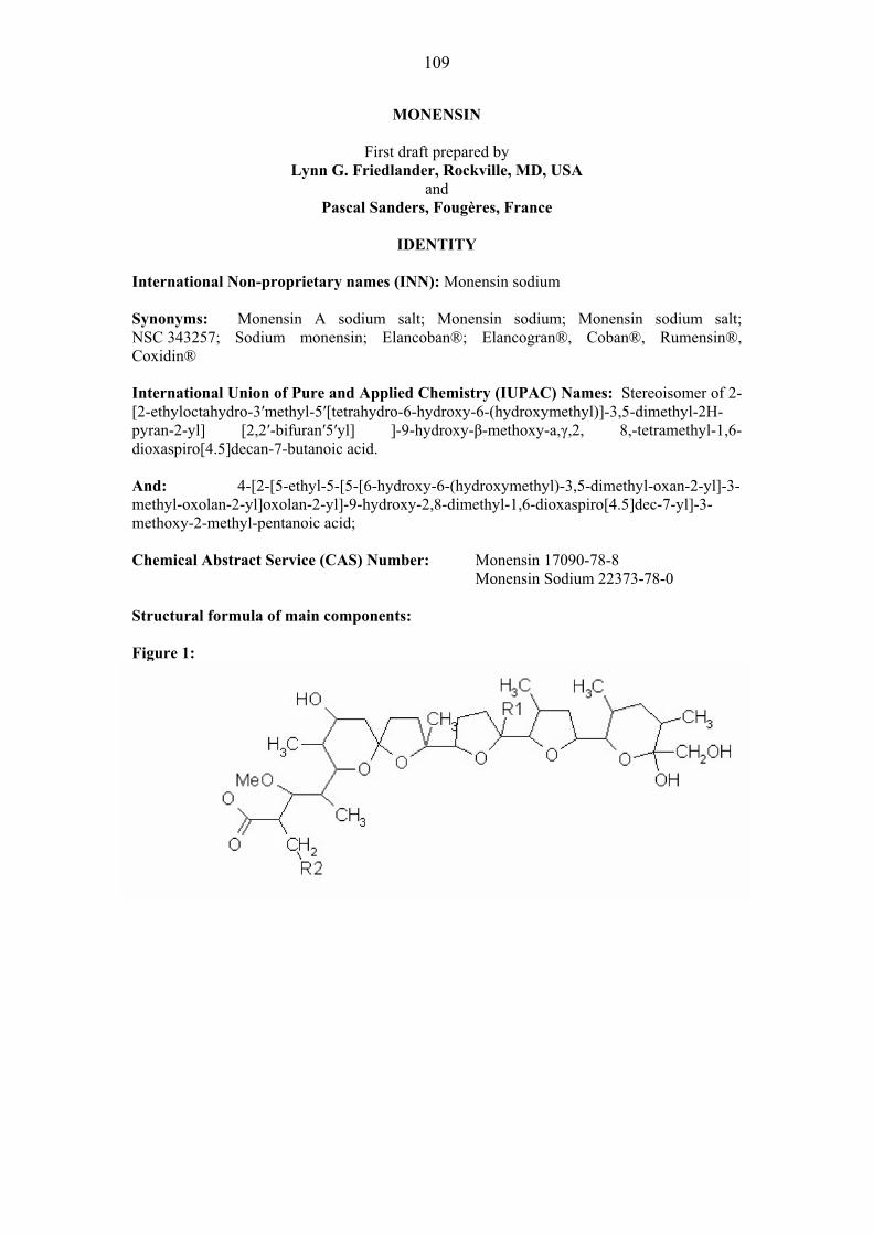
109
MONENSIN
First draft prepared by Lynn G. Friedlander, Rockville, MD, USA
and Pascal Sanders, Fougères, France
IDENTITY
International Non-proprietary names (INN): Monensin sodium Synonyms: Monensin A sodium salt; Monensin sodium; Monensin sodium salt; NSC 343257; Sodium monensin; Elancoban®; Elancogran®, Coban®, Rumensin®, Coxidin® International Union of Pure and Applied Chemistry (IUPAC) Names: Stereoisomer of 2-[2-ethyloctahydro-3�methyl-5�[tetrahydro-6-hydroxy-6-(hydroxymethyl)]-3,5-dimethyl-2H-pyran-2-yl] [2,2�-bifuran�5�yl] ]-9-hydroxy-�-methoxy-a,�,2, 8,-tetramethyl-1,6-dioxaspiro[4.5]decan-7-butanoic acid. And: 4-[2-[5-ethyl-5-[5-[6-hydroxy-6-(hydroxymethyl)-3,5-dimethyl-oxan-2-yl]-3-methyl-oxolan-2-yl]oxolan-2-yl]-9-hydroxy-2,8-dimethyl-1,6-dioxaspiro[4.5]dec-7-yl]-3-methoxy-2-methyl-pentanoic acid; Chemical Abstract Service (CAS) Number: Monensin 17090-78-8
Monensin Sodium 22373-78-0
Structural formula of main components: Figure 1:

110
Table 1: Summary of Monensin Factors.
Monensin Factor R1 R2 R3
A C2H5 H H
B† CH3 H H
C* C2H5 H CH3
D* Not supplied
†Factor B accounts for less than 4% of the total composition. *The relative biological activity of trace factors C and D assayed against Streptococcus faecium are negligible (Haney and Hoehn., 1967; Agtarap and Chamberlin, 1967; Chamberlin and Agtarap, 1970).
Molecular formula: Monensin A (sodium salt): C36H61O11Na Monensin B (sodium salt): C35H59O11Na
Molecular weight: Monensin A (sodium salt): 692 Monensin B (sodium salt): 678
OTHER INFORMATION ON IDENTITY AND PROPERTIES Pure active ingredient: Appearance: Off-white to tan crystalline powder Melting point: 267-269 C (sodium salt); 103-106° C (acid) Solubility: Soluble in ethyl acetate, acetone, chloroform and dimethyl
sulphoxide. Essentially insoluble in water and petroleum ether.
RESIDUES IN FOOD AND THEIR EVALUATION Conditions of use Monensin sodium is used for the control of coccidiosis in chickens, turkeys, and quail. In feedlot and pasture cattle, it is used to improve the efficiency of rumen fermentation, increase rate of weight gain and for the prevention and control of coccidiosis. In feedlot and lactating and non-lactating dairy cattle, it is used to control ketosis. In dry and lactating dairy cows, it is used to increase milk production efficiency (production of marketable solids-corrected milk per unit of feed intake). In calves, non-lactating goats, and sheep it is used for prevention and control of coccidiosis. Dosage Monensin sodium is provided in a complete feed at maximum use concentrations of 125 mg/kg feed for broiler chickens and 120 mg/kg feed for replacement layers. The maximum dose for turkeys is 100 mg/kg feed; for quail, the maximum dose is 73mg/kg feed. To improve the efficiency of rumen fermentation, monensin sodium is provided at a maximum dose of 360 mg/animal/day or 40 mg/kg in complete feed for feedlot cattle and 200 mg/animal/day in a 0.45 kg feed supplement for pasture cattle. For control of ketosis in feedlot cattle, the maximum dose is 480 mg/animal/day. To control ketosis in lactating dairy

111
cattle, monensin is administered as a controlled-release capsule providing a maximum daily dose of 400 mg/animal, released into the rumen. It may be given to dry and lactating dairy cows continuously as a total mixed ration containing 11 to 22 g monensin/ton (12 to 24 mg/kg). In calves, it is provided at a maximum dose of 200 mg/animal/day. In the USA, it is explicitly labelled not to be used in calves raised for veal. In non-lactating goats, it is provided as the sole ration containing 20 g monensin/ton (22 mg/kg). In sheep, it is provided in the total ration at a rate of not less than 11 and not more than 22 g monensin/metric ton.
PHARMACOKINETICS AND METABOLISM Pharmacokinetics in Laboratory Animals Rats The disposition of orally administered [14C] monensin was determined in rats treated for 13 days with feed containing 100 mg unlabelled monensin/kg feed. On day 14, rats received a single oral dose of radiolabelled monensin, 2.15 mg (specific activity 0.0266 �Ci/mg), by gavage. Thereafter, for the remainder of the study (12 days), rats again received unlabelled monensin in the feed. Unlabelled feed was provided ad libitum. Urine and faeces were assayed daily for radioactivity. Within three days after dosing with the labelled drug, 92.0% of the total radioactivity was recovered. The majority of the radioactivity was recovered in the faeces (91.5%) but a small amount was recovered in urine (0.5%). Selected tissues and organs were assayed for radioactivity but the detected radioactivity was not different from that measured in pooled control tissues (Herberg, 1973a). Monensin and monensin metabolites were isolated from the liver and faeces of male and female rats treated orally with 5 mg [14C] monensin/kg body weight (Donoho, 1985). In another study (Howard and Lobb, 1981), tissue distribution and biliary elimination of radioactivity was evaluated in male and female rats following oral administration of [14C] monensin. Radiolabelled monensin (0.614 μCi/mg) was administered orally at doses ranging from 5 to 40 mg/kg body weight in male rats and 2 to 16 mg/kg body weight in female rats. The doses were based on previously determined oral toxicity data (Broddle and Worth, 1976) for male (40.1±3.0 mg/kg) and female (24.3±2.7 mg/kg) rats. After administration, radioactivity was eliminated rapidly and extensively in faeces during a 72-hour collection period. Faecal elimination accounted for 83.6-87.4% and 70.8-87.2% of the dose in male and female rats, respectively. Urinary excretion represented only a minor route of elimination representing 1.0-1.6% and 1.0-1.3% of the dose in male and female rats, respectively. Biliary secretion was the primary excretory pathway following oral administration in rats. There were no differences in the urinary, faecal, or biliary excretions of radioactivity between male and female rats. At toxic doses, there was an initial delay in the excretion of radioactivity in faeces and bile considered secondary to the toxicity. Further support for partial gastrointestinal absorption of monensin in male and female rats was found in studies that showed that 31 to 53% of a radioactive dose (32.8-46.6% in males and 30.7-53.2% in females) of 2 to 40 mg/kg body weight was collected in bile within 72 hours of administration (Howard and Lobb, 1981). Pharmacokinetics in Food Animals Cattle Gastrointestinal absorption of monensin in cattle has been evaluated. In one study, absorption of [14C] monensin in calves was evaluated by measuring radiolabelled residues in bile (Davison, 1984). The amount of radiolabelled material recovered in bile can serve as an estimate of the amount of material absorbed because little monensin is excreted in cattle urine. Two calves, one male and one female, were fitted with bile duct cannulae. Each calf
112
received a single oral dose of 10 mg [14C] monensin/kg body weight in a gelatine capsule. Bile was collected continuously for 72h. Approximately 35 and 37% of the administered radioactivity was recovered in the bile from the male and female calf, respectively. The presence of monensin or monensin metabolites in plasma (Donoho, 1984), liver and milk (Herberg, et al., 1978; Kline and Wicker, 1975; Kennington, et al., 1995) from orally treated animals provides supporting evidence for absorption. Chickens and Turkeys The pharmacokinetic profile of monensin was evaluated in broiler chickens (Atef, et al., 1993) following administration by gavage and intravenously as a single dose of 40 mg/kg body weight. Following intravenous administration, disposition of monensin followed a two-compartment open model. The absorption half-life was 0.6 hours, the volume of distribution was 4.1 L/kg, and the total body clearance was 28.4±0.2 ml/kg/min. The highest serum concentration (4.1±0.05 �g/ml) was reached after 0.4 hours following administration by gavage. The absorption half-life was 0.3 hours and the elimination half-life was 2.1 hours. A somewhat longer terminal elimination half-life (3.1 to 5.6 hours) has been determined recently (Henri, et al, 2008a). In vitro serum protein binding was calculated to be 22.8%. Bioavailability following administration by gavage was 65.1% (Atef, et al., 1993). In chickens, monensin concentrations in serum and tissues were higher after administration by gavage (40 mg/kg body weight) than after feeding a diet containing 120 mg monensin/kg for 2 weeks (average daily consumption was 24 mg monensin based on a daily feed consumption of 200 g of medicated feed). The rate of faecal excretion and quantitation of orally administered monensin in chickens were determined. Chickens received an oral dose of [14C] monensin (7.36 mg, specific activity 0.018 �Ci/mg). Seventy-five percent of the administered dose was eliminated in excreta within 3 days and was eliminated completely within 12 days (Herberg, 1973b). In another study, three chickens were treated ad libitum with feed containing 120 mg unlabelled monensin/kg feed. Chickens were then dosed by oral capsule with a single dose of [14C] monensin. More than 75% of the radioactivity was recovered within 3 days following the dose. Radioactivity in excreta returned to background levels in 4, 5, and 12 days (Herberg, 1975a). In another study (Grundy, et al., 1998), chickens were fed a ration containing 125 mg [14C] monensin/kg feed for 6 days then slaughtered 6h, 1, 3 or 5 days after the treated feed was withdrawn (3 male and 3 female chickens per withdrawal group). Bile contained approximately 87mg monensin/kg after 6h withdrawal. By 5 days withdrawal, bile contained approximately 0.4mg monensin/kg and approximately 76% of the dose had been recovered in excreta. These data indicate that radiolabelled monensin is eliminated rapidly and quantitatively by chickens. In turkeys, the evidence of intestinal absorption is analogous to that for chickens. Monensin and metabolites were found in turkey liver at zero withdrawal following ad libitum access feeding with 110 mg [14C] monensin/kg feed for five days (Donoho, et al., 1982a). The reported terminal elimination half-life in turkeys is 1.4 to 1.6 hours (Henri, et al, unpublished 2008a). Metabolism in Laboratory Animals and Humans Rats Data from studies in laboratory animals indicate that monensin is extensively metabolized prior to excretion. The in vitro metabolism of monensin has been evaluated in several studies (Ershov, et al., 2001; Nebbia, et al., 1999; Nebbia, et al., 2001). In liver microsomes induced by dexamethasone, monensin is metabolized by the 3A family of cytochrome P-450. Inducers of O-demethylation enhance and inhibitors reduced monensin metabolism. Although
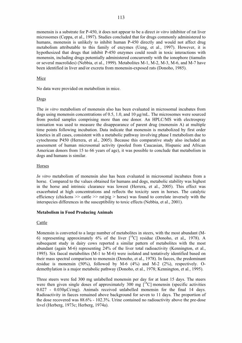
113
monensin is a substrate for P-450, it does not appear to be a direct in vitro inhibitor of rat liver microsomes (Ceppa, et al., 1997). Studies concluded that for drugs commonly administered to humans, monensin is unlikely to inhibit human P-450 directly and would not affect drug metabolism attributable to this family of enzymes (Ueng, et al., 1997). However, it is hypothesized that drugs that inhibit P-450 enzymes could result in toxic interactions with monensin, including drugs potentially administered concurrently with the ionophore (tiamulin or several macrolides) (Nebbia, et al., 1999). Metabolites M-1, M-2, M-3, M-6, and M-7 have been identified in liver and/or excreta from monensin-exposed rats (Donoho, 1985). Mice No data were provided on metabolism in mice. Dogs The in vitro metabolism of monensin also has been evaluated in microsomal incubates from dogs using monensin concentrations of 0.5, 1.0, and 10 �g/mL. The microsomes were sourced from pooled samples comprising more than one donor. An HPLC/MS with electrospray ionisation was used to measure the disappearance of parent drug (monensin A) at multiple time points following incubation. Data indicate that monensin is metabolized by first order kinetics in all cases, consistent with a metabolic pathway involving phase I metabolism due to cytochrome P450 (Herrera, et al., 2005). Because this comparative study also included an assessment of human microsomal activity (pooled from Caucasian, Hispanic and African American donors from 15 to 66 years of age), it was possible to conclude that metabolism in dogs and humans is similar. Horses In vitro metabolism of monensin also has been evaluated in microsomal incubates from a horse. Compared to the values obtained for humans and dogs, metabolic stability was highest in the horse and intrinsic clearance was lowest (Herrera, et al., 2005). This effect was exacerbated at high concentrations and reflects the toxicity seen in horses. The catalytic efficiency (chickens >> cattle >> rat/pig > horse) was found to correlate inversely with the interspecies differences in the susceptibility to toxic effects (Nebbia, et al., 2001). Metabolism in Food Producing Animals Cattle Monensin is converted to a large number of metabolites in steers, with the most abundant (M-6) representing approximately 6% of the liver [14C] residue (Donoho, et al,, 1978). A subsequent study in dairy cows reported a similar pattern of metabolites with the most abundant (again M-6) representing 24% of the liver total radioactivity (Kennington, et al., 1995). Six faecal metabolites (M-1 to M-6) were isolated and tentatively identified based on their mass spectral comparison to monensin (Donoho, et al., 1978). In faeces, the predominant residue is monensin (50%), followed by M-6 (4%) and M-2 (2%), respectively. O-demethylation is a major metabolic pathway (Donoho, et al., 1978; Kennington, et al., 1995). Three steers were fed 300 mg unlabelled monensin per day for at least 15 days. The steers were then given single doses of approximately 300 mg [14C] monensin (specific activities 0.027 - 0.030μCi/mg). Animals received unlabelled monensin for the final 14 days. Radioactivity in faeces remained above background for seven to 11 days. The proportion of the dose recovered was 88.6% - 102.3%. Urine contained no radioactivity above the pre-dose level (Herberg, 1973c; Herberg, 1974a).
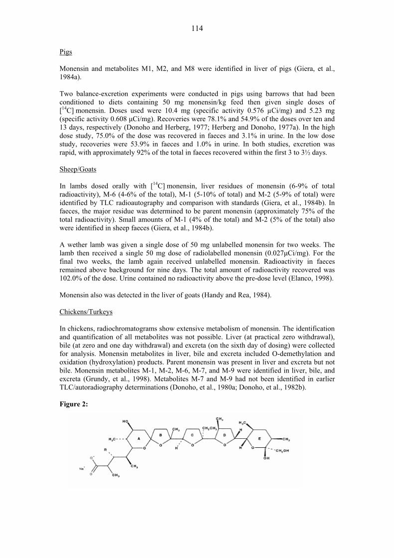
114
Pigs Monensin and metabolites M1, M2, and M8 were identified in liver of pigs (Giera, et al., 1984a). Two balance-excretion experiments were conducted in pigs using barrows that had been conditioned to diets containing 50 mg monensin/kg feed then given single doses of [14C] monensin. Doses used were 10.4 mg (specific activity 0.576 μCi/mg) and 5.23 mg (specific activity 0.608 �Ci/mg). Recoveries were 78.1% and 54.9% of the doses over ten and 13 days, respectively (Donoho and Herberg, 1977; Herberg and Donoho, 1977a). In the high dose study, 75.0% of the dose was recovered in faeces and 3.1% in urine. In the low dose study, recoveries were 53.9% in faeces and 1.0% in urine. In both studies, excretion was rapid, with approximately 92% of the total in faeces recovered within the first 3 to 3½ days. Sheep/Goats In lambs dosed orally with [14C] monensin, liver residues of monensin (6-9% of total radioactivity), M-6 (4-6% of the total), M-1 (5-10% of total) and M-2 (5-9% of total) were identified by TLC radioautography and comparison with standards (Giera, et al., 1984b). In faeces, the major residue was determined to be parent monensin (approximately 75% of the total radioactivity). Small amounts of M-1 (4% of the total) and M-2 (5% of the total) also were identified in sheep faeces (Giera, et al., 1984b). A wether lamb was given a single dose of 50 mg unlabelled monensin for two weeks. The lamb then received a single 50 mg dose of radiolabelled monensin (0.027�Ci/mg). For the final two weeks, the lamb again received unlabelled monensin. Radioactivity in faeces remained above background for nine days. The total amount of radioactivity recovered was 102.0% of the dose. Urine contained no radioactivity above the pre-dose level (Elanco, 1998). Monensin also was detected in the liver of goats (Handy and Rea, 1984). Chickens/Turkeys In chickens, radiochromatograms show extensive metabolism of monensin. The identification and quantification of all metabolites was not possible. Liver (at practical zero withdrawal), bile (at zero and one day withdrawal) and excreta (on the sixth day of dosing) were collected for analysis. Monensin metabolites in liver, bile and excreta included O-demethylation and oxidation (hydroxylation) products. Parent monensin was present in liver and excreta but not bile. Monensin metabolites M-1, M-2, M-6, M-7, and M-9 were identified in liver, bile, and excreta (Grundy, et al., 1998). Metabolites M-7 and M-9 had not been identified in earlier TLC/autoradiography determinations (Donoho, et al., 1980a; Donoho, et al., 1982b). Figure 2:
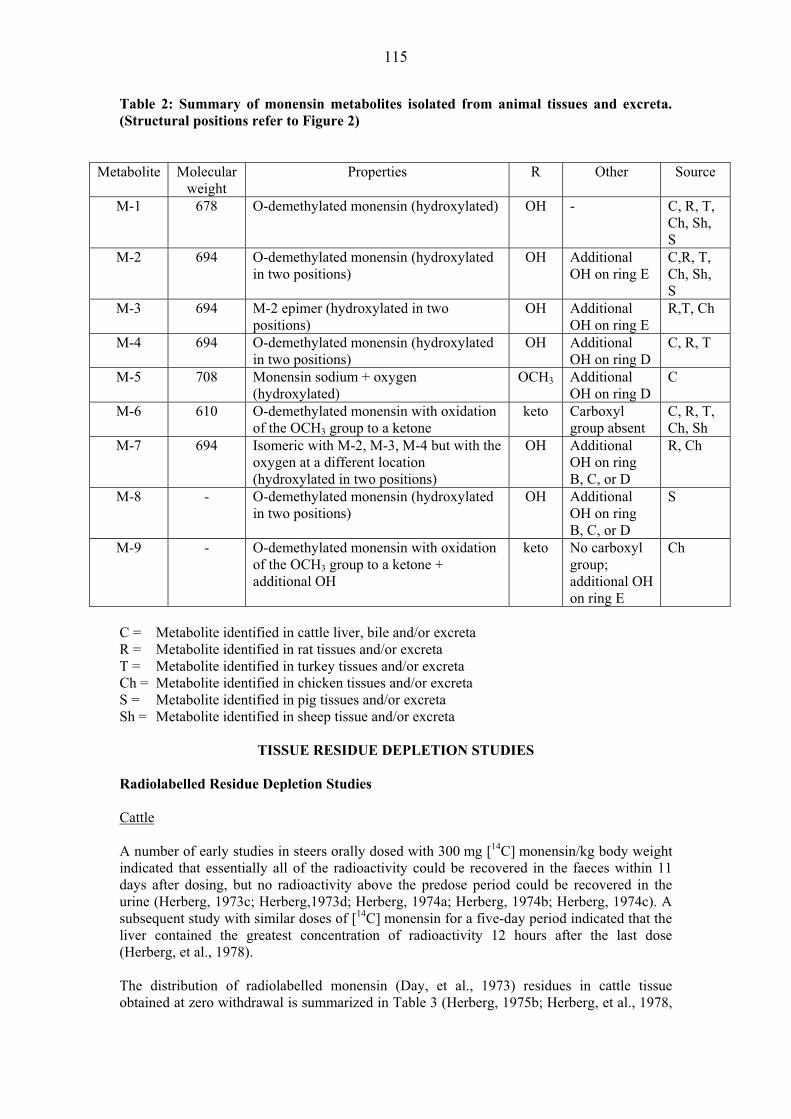
115
Table 2: Summary of monensin metabolites isolated from animal tissues and excreta. (Structural positions refer to Figure 2)
Metabolite Molecular weight
Properties R Other Source
M-1 678 O-demethylated monensin (hydroxylated) OH - C, R, T, Ch, Sh, S
M-2 694 O-demethylated monensin (hydroxylated in two positions)
OH Additional OH on ring E
C,R, T, Ch, Sh, S
M-3 694 M-2 epimer (hydroxylated in two positions)
OH Additional OH on ring E
R,T, Ch
M-4 694 O-demethylated monensin (hydroxylated in two positions)
OH Additional OH on ring D
C, R, T
M-5 708 Monensin sodium + oxygen (hydroxylated)
OCH3 Additional OH on ring D
C
M-6 610 O-demethylated monensin with oxidation of the OCH3 group to a ketone
keto Carboxyl group absent
C, R, T, Ch, Sh
M-7 694 Isomeric with M-2, M-3, M-4 but with the oxygen at a different location (hydroxylated in two positions)
OH Additional OH on ring B, C, or D
R, Ch
M-8 - O-demethylated monensin (hydroxylated in two positions)
OH Additional OH on ring B, C, or D
S
M-9 - O-demethylated monensin with oxidation of the OCH3 group to a ketone + additional OH
keto No carboxyl group; additional OH on ring E
Ch
C = Metabolite identified in cattle liver, bile and/or excreta R = Metabolite identified in rat tissues and/or excreta T = Metabolite identified in turkey tissues and/or excreta Ch = Metabolite identified in chicken tissues and/or excreta S = Metabolite identified in pig tissues and/or excreta Sh = Metabolite identified in sheep tissue and/or excreta
TISSUE RESIDUE DEPLETION STUDIES
Radiolabelled Residue Depletion Studies Cattle A number of early studies in steers orally dosed with 300 mg [14C] monensin/kg body weight indicated that essentially all of the radioactivity could be recovered in the faeces within 11 days after dosing, but no radioactivity above the predose period could be recovered in the urine (Herberg, 1973c; Herberg,1973d; Herberg, 1974a; Herberg, 1974b; Herberg, 1974c). A subsequent study with similar doses of [14C] monensin for a five-day period indicated that the liver contained the greatest concentration of radioactivity 12 hours after the last dose (Herberg, et al., 1978). The distribution of radiolabelled monensin (Day, et al., 1973) residues in cattle tissue obtained at zero withdrawal is summarized in Table 3 (Herberg, 1975b; Herberg, et al., 1978,
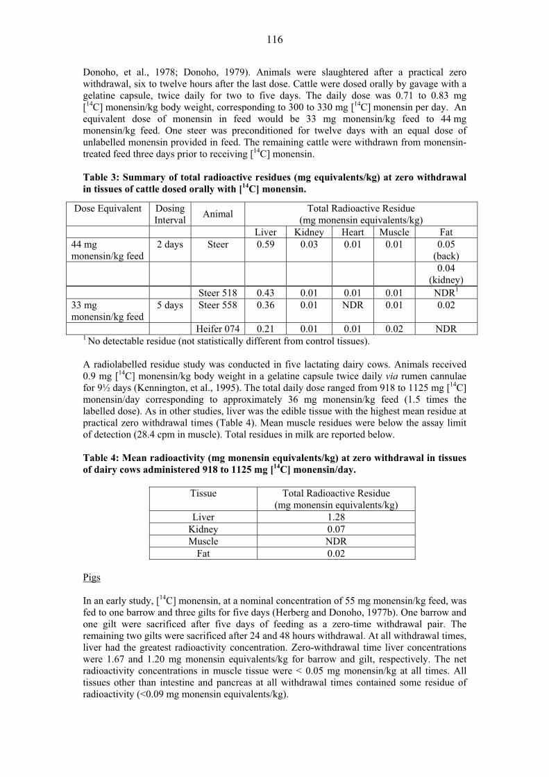
116
Donoho, et al., 1978; Donoho, 1979). Animals were slaughtered after a practical zero withdrawal, six to twelve hours after the last dose. Cattle were dosed orally by gavage with a gelatine capsule, twice daily for two to five days. The daily dose was 0.71 to 0.83 mg [14C] monensin/kg body weight, corresponding to 300 to 330 mg [14C] monensin per day. An equivalent dose of monensin in feed would be 33 mg monensin/kg feed to 44 mg monensin/kg feed. One steer was preconditioned for twelve days with an equal dose of unlabelled monensin provided in feed. The remaining cattle were withdrawn from monensin-treated feed three days prior to receiving [14C] monensin. Table 3: Summary of total radioactive residues (mg equivalents/kg) at zero withdrawal in tissues of cattle dosed orally with [14C] monensin.
Dose Equivalent Dosing Interval Animal Total Radioactive Residue
(mg monensin equivalents/kg) Liver Kidney Heart Muscle Fat
44 mg monensin/kg feed
2 days Steer 0.59 0.03 0.01 0.01 0.05 (back)
0.04 (kidney)
Steer 518 0.43 0.01 0.01 0.01 NDR1
33 mg monensin/kg feed
5 days Steer 558 0.36 0.01 NDR 0.01 0.02
Heifer 074 0.21 0.01 0.01 0.02 NDR 1 No detectable residue (not statistically different from control tissues). A radiolabelled residue study was conducted in five lactating dairy cows. Animals received 0.9 mg [14C] monensin/kg body weight in a gelatine capsule twice daily via rumen cannulae for 9½ days (Kennington, et al., 1995). The total daily dose ranged from 918 to 1125 mg [14C] monensin/day corresponding to approximately 36 mg monensin/kg feed (1.5 times the labelled dose). As in other studies, liver was the edible tissue with the highest mean residue at practical zero withdrawal times (Table 4). Mean muscle residues were below the assay limit of detection (28.4 cpm in muscle). Total residues in milk are reported below. Table 4: Mean radioactivity (mg monensin equivalents/kg) at zero withdrawal in tissues of dairy cows administered 918 to 1125 mg [14C] monensin/day.
Tissue Total Radioactive Residue (mg monensin equivalents/kg)
Liver 1.28 Kidney 0.07 Muscle NDR
Fat 0.02 Pigs In an early study, [14C] monensin, at a nominal concentration of 55 mg monensin/kg feed, was fed to one barrow and three gilts for five days (Herberg and Donoho, 1977b). One barrow and one gilt were sacrificed after five days of feeding as a zero-time withdrawal pair. The remaining two gilts were sacrificed after 24 and 48 hours withdrawal. At all withdrawal times, liver had the greatest radioactivity concentration. Zero-withdrawal time liver concentrations were 1.67 and 1.20 mg monensin equivalents/kg for barrow and gilt, respectively. The net radioactivity concentrations in muscle tissue were < 0.05 mg monensin/kg at all times. All tissues other than intestine and pancreas at all withdrawal times contained some residue of radioactivity (<0.09 mg monensin equivalents/kg).

117
In a preliminary study, one male and one female pig were dosed orally for two and one-half days with [14C] monensin at an equivalent to 50 mg monensin/kg in feed (Herberg and Donoho, 1978). Four hours after the final dose, the pigs were sacrificed and edible tissues were assayed for residual activity. Liver contained the highest net residue, 1.02 mg monensin equivalents/kg in the male and 1.44 mg monensin equivalents/kg in the female. Residues in the other tissues were less than 0.09 mg monensin equivalents/kg. Three grower pigs of each sex were fed [14C] monensin-fortified ration (110 mg monensin/kg feed) for five consecutive days (Giera, et al., 1984a). One male and one female were slaughtered at six hours, three days and five days withdrawal. Liver, kidney, fat, and muscle were assayed for total radioactivity (limit 0.05 mg/kg). Bioautography was used for detection of monensin in those tissues. The bio-autographic method (Rea, 1976) has a limit of detection of 0.025 mg/kg for liver and muscle and 0.05 mg/kg for fat and kidney. Selected samples of liver and faeces were characterized chromatographically (LOD = 0.005 mg/kg). The radioactive residues found in pig tissues are summarized in Table 5. Liver from the male and female zero-time withdrawal animals contained approximately 2.26 mg monensin equivalents/kg total residue. Residues decreased to approximately 0.44 mg monensin equivalents/kg by 5 days withdrawal. Kidney contained approximately 0.17 mg monensin equivalents/kg total residue at zero withdrawal. Residues decreased to approximately 0.05 mg monensin equivalents/kg after 5 days. The radioactive residues in fat and muscle were approximately 0.044 and 0.037 mg monensin equivalents/kg, respectively, at zero withdrawal. After five days of withdrawal, fat and muscle 14C residues were approximately 0.05 and 0.02 mg monensin equivalents/kg, respectively. Monensin was not detected in the tissues of any of the treated animals by a microbiological assay (sensitivity 0.025-0.050 mg/kg) or HPLC assay (sensitivity 0.005 mg/kg). Table 5: Net mg 14C-monensin/kg equivalents in pig tissues following oral administration of radiolabelled monensin in feed (110 mg/kg).
0-Day (6hr) Withdrawal
3-Day Withdrawal
5-Day Withdrawal
F 126 M 127 F 122 M 125 F 121 M 120 Liver 2.08 2.45 0.88 1.03 0.35 0.53
Kidney 0.16 0.18 0.08 0.09 0.04 0.06
Fat 0.04 0.05 0.05 0.05 0.03 0.06
Muscle 0.04 0.04 0.02 0.02 0.02 0.02 Sheep Lambs were fed [14C] monensin equivalent to a feeding level of 15g per ton (16.5 mg/kg) of complete ration (Giera, et al., 1984). Groups of lambs (two wethers and one ewe) were dosed for 3, 5, or 7 days and killed at zero withdrawal (12 hours) after the last dose. Edible tissues were assayed for total radioactivity (assay reliability = 0.1 mg/kg or lower). Liver samples were assayed for parent monensin, and selected samples of liver and faeces were characterized chromatographically. After dosing for 3, 5, or 7 days, liver contained mean residues of 0.36, 0.32, and 0.20 mg/kg radioactivity equivalents, respectively (Table 6). Parent monensin concentrations in liver were determined using bioautography (Kline, et al., 1975) and were less than 0.05 mg/kg. There was no accumulation of residues for either total radioactivity or parent monensin with longer dosing intervals. Residues in kidney and fat were all less than 0.03 mg monensin equivalents/kg and muscle all less than 0.01 mg monensin equivalents/kg.

118
Table 6: Summary of 14C (mg monensin equivalents/kg) in sheep tissues following oral administration of radiolabelled monensin at 15 g/ton in feed.
3 Day Dosing Period 5 Day Dosing Period 7 Day Dosing Period M 578 F 579 M 583 M 580 M 581 F 582 M 577 M 584 F 585 Liver1 0.50 0.18 0.39 0.40 0.29 0.285 0.32 0.19 0.11 Kidney 0.01 0.004 0.01 0.02 0.01 -0.01 0.01 -0.01 0.01 Fat 0.01 0.01 0.01 0.01 0.02 0.01 0.03 0.01 0.01 Muscle <0.01 <0.01 <0.01 <0.01 <0.01 <0.01 <0.01 <0.01 <0.01 Chickens/Turkeys The distribution of radiolabelled residues at zero withdrawal has been extensively evaluated in chickens and turkeys. In several studies, chickens were treated with [14C] monensin in feed at 110-125 mg/kg, ad libitum, for four to six days. The first study (Table 7) established that steady-state equilibrium occurred within four days in chickens receiving 120 mg [14C] monensin/kg in feed (Donoho, et al., 1980a). In all [14C] monensin residue studies, liver tissue had the highest amount of total residues at a practical or true zero withdrawal. Muscle had the least residues at zero withdrawal. This was consistent with the distribution pattern seen in rats (Howard and Lobb, 1981). Table 7: Mean radioactivity (mg monensin equivalents/kg) at zero withdrawal in tissues of chickens fed 110 to 125 mg [14C] monensin/kg feed.
Study No. Dose (in feed)
Dosing Interval
Total Radioactive Residue (mg monensin equivalents/kg)
Liver Kidney Muscle Skin/Fat Fat ABC-00431 120 mg/kg 4 days
6 days 0.56 0.43
0.12 0.12
0.01 0.01
0.09 0.07
0.14 0.07
ABC-00802 120 mg/kg 5 days 0.83 0.22 0.05 0.29 0.23 ABC-00923 110 mg/kg 5 days 0.53 0.18 0.02 0.12 0.07 T1F7597014 125 mg/kg 6 days 0.94 0.20 0.06 0.29 0.48 1Donoho, et al., 1980a; 2Donoho, et al., 1980b; 3Donoho, et al., 1980c; 4Grundy, et al., 1998 The depletion of [14C] monensin is summarized in two studies (Donoho, et al., 1980c; Grundy, et al., 1998). In the first study (Table 8), fat and skin/fat had slowly depleting residues. The relatively high and persistent radioactivity in abdominal fat was due to incorporation of radioactivity into endogenous fatty acids (Grundy, et al., 1998). Total residue in liver was approximately 2-15 times greater than residues in kidney, muscle, abdominal fat, and skin/fat at zero withdrawal. After three days, liver residues depleted to less than the fat and skin residues. Fat was also a slowly depleting tissue in the second study (Table 9, Donoho, et al., 1980c). In the liver, the percentage of bound (unextracted) radioactivity increased from 38 to 69% during the 5-day withdrawal even as the total liver radioactivity decreased. Over the 5-day study, the absolute amount of bound residue in liver decreased from approximately 0.20 mg/kg to 0.07 mg/kg.

119
Table 8: Summary of total radioactive residues (mg monensin equivalents/kg) in edible tissues, fat, and bile of chickens after ad libitum access to 125 mg [14C] monensin/kg feed. Treatment
Group Withdrawal Time (days)
Total Radioactive Residue (mg monensin equivalents/kg)
Liver Kidney Muscle Skin/Fat Abdominal Fat Bile 01 0 0.94 0.20 0.06 0.29 0.48 87 02 1 0.49 0.14 0.05 0.17 0.31 26 03 3 0.27 0.09 0.05 0.23 0.47 1.3 04 5 0.14 0.05 0.04 0.17 0.26 0.4
Table 9: Summary of total radioactive residues (mg monensin equivalents/kg) in tissues of chickens fed 100 mg [14C] monensin/kg in feed. Withdrawal Time (days) Total Radioactive Residue (mg monensin equivalents/kg)
Liver Kidney Muscle Skin Fat 0 0.53 0.18 0.02 0.12 0.07 1 0.27 0.08 0.01 0.05 0.07 2 0.22 0.06 0.01 0.06 0.07 3 0.15 0.05 0.01 0.04 0.05 5 0.11 0.04 0.01 0.03 0.05
In turkeys fed 110 mg [14C] monensin/kg in feed for five days, radiolabelled residues were evaluated in the edible tissues of birds killed 6 hours (practical zero withdrawal) after the removal of medicated feed (Donoho, et al., 1982a). As in the chicken studies, turkey liver and muscle had the most and least amount of radioactive residue, respectively (Table 10). Table 10: Total radioactive residue in turkey tissues at zero withdrawal following treatment with 110 mg [14C] monensin/kg feed for five days.
Tissue Total Radioactive Residue (mg monensin equivalents/kg)
Liver 0.91 Kidney 0.16 Muscle <0.03 Skin/Fat 0.10
Fat 0.14 Cows’ Milk Radiolabelled residues in milk were determined in a study conducted in lactating dairy cows (Kennington, et al., 1995). Cows were treated intra-ruminally via gelatine capsule for 9 days. Within approximately five days, radiolabelled milk residues had reached a steady state and averaged 0.045 mg/kg during the final three days of dosing. No residues of monensin were detected in the milk of any treated animals (LOQ = 0.005 mg/kg in milk). The radioactivity was distributed approximately 30% in the cream, 50% in the whey, and 20% in the casein. Fractionation of the milk and the use of a sensitive LC/ESP-MS/LSC analytical technique allowed for the detection of parent monensin in milk at low concentrations (<0.001 mg/kg). As in chickens (Grundy, et al., 1998), it was determined that a significant proportion (26.5%) of the radioactivity in milk from an animal treated with [14C] monensin was due to the incorporation of the radioactivity into endogenous fatty acids (myristic, oleic, palmitic and stearic acid), rather than resulting from monensin-related residues (Grundy and Bewley, 2003).

120
Residue Depletion Studies with Unlabelled Drug Residues in Tissues Cattle Monensin was fed to cattle at levels of 100 and 500 mg/animal/day for 148 days and at 750 mg/animal/day for 106 days (Kline, 1973). Animals were slaughtered at 0, 48, 120 and 240 hours withdrawal for the 100 and 500 mg/animal/day treatments and at 0 and 48 hours withdrawal for the 750 mg/animal/day treatment. No monensin residues (assay sensitivity 0.05 mg/kg) were detected in animals receiving 100 or 500 mg monensin/day at any withdrawal period. One kidney sample from a zero withdrawal animal in the 750 mg/animal/day treatment group contained a detectable residue. At 48 hours withdrawal, tissues in the 750 mg/animal/day treatment group were free of residual monensin. Lactating dairy cows were treated intra-ruminally with two controlled release capsules (32 g monensin in a hexaglycerol distearate matrix into a plastic tube) and fed a medicated ration containing 24 mg monensin/kg feed for 10 days and then fed 36 mg monensin/kg for 21 days (Bagg and Dick, 1999). After measurement of the monensin release rate from the controlled release capsules, the resulting daily dose ranged from 1537 to 1804 mg monensin per cow. At zero withdrawal, animals were slaughtered and liver and kidney samples were analyzed using a validated HPLC method with post-column derivatization. There were no detectable monensin residues in kidney tissue (<0.025 mg/kg). Monensin residues were detected in 4 of 6 liver samples (detected residues ranged from 0.05 mg/kg to 0.09 mg/kg). To determine monensin residues in the tissues of lactating dairy animals, Holstein cows were fed medicated rations containing 0, 24, or 36 mg monensin/kg feed or with 1.8 mg monensin/kg body weight via gelatine capsule through a rumen fistula (Dick, et al., 1994). At the completion of feeding periods, liver was analyzed using a validated HPLC method with post-column derivatization. No residues were found above the method LOQ of 0.025 mg/kg. The depletion of monensin was determined in the edible tissues (liver, muscle, kidney and fat) of 12 lactating dairy cows after dosing with monensin at 0.9 mg/kg body weight for seven consecutive days (Bassissi and Larvor, 2007). Gelatine capsules containing equal doses were administered at approximately 12-hour intervals. Tissues were collected at 6, 18 and 30 hours after the final dosing. Monensin residues (Table 11) were determined using a validated HPLC-MS/MS method with a LOQ of 1 �g/kg.
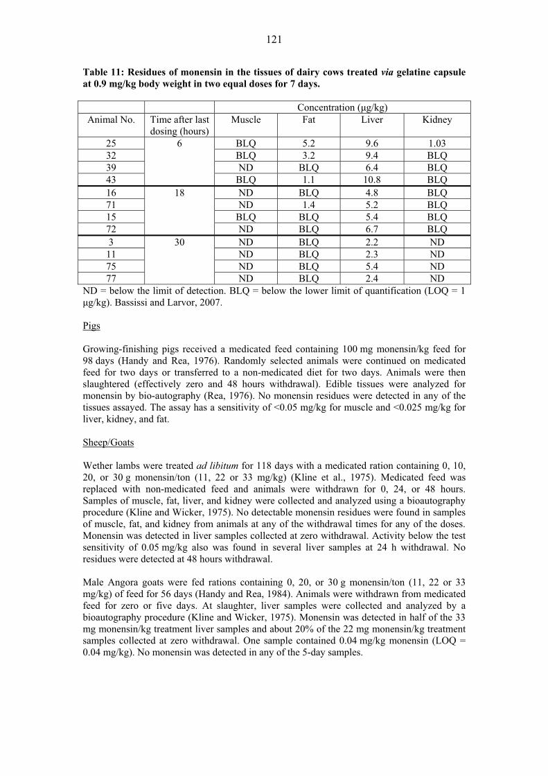
121
Table 11: Residues of monensin in the tissues of dairy cows treated via gelatine capsule at 0.9 mg/kg body weight in two equal doses for 7 days.
Concentration (�g/kg) Animal No. Time after last
dosing (hours) Muscle Fat Liver Kidney
25 BLQ 5.2 9.6 1.03 32 BLQ 3.2 9.4 BLQ 39 ND BLQ 6.4 BLQ 43
6
BLQ 1.1 10.8 BLQ 16 ND BLQ 4.8 BLQ 71 ND 1.4 5.2 BLQ 15 BLQ BLQ 5.4 BLQ 72
18
ND BLQ 6.7 BLQ 3 ND BLQ 2.2 ND
11 ND BLQ 2.3 ND 75 ND BLQ 5.4 ND 77
30
ND BLQ 2.4 ND ND = below the limit of detection. BLQ = below the lower limit of quantification (LOQ = 1 �g/kg). Bassissi and Larvor, 2007. Pigs Growing-finishing pigs received a medicated feed containing 100 mg monensin/kg feed for 98 days (Handy and Rea, 1976). Randomly selected animals were continued on medicated feed for two days or transferred to a non-medicated diet for two days. Animals were then slaughtered (effectively zero and 48 hours withdrawal). Edible tissues were analyzed for monensin by bio-autography (Rea, 1976). No monensin residues were detected in any of the tissues assayed. The assay has a sensitivity of <0.05 mg/kg for muscle and <0.025 mg/kg for liver, kidney, and fat. Sheep/Goats Wether lambs were treated ad libitum for 118 days with a medicated ration containing 0, 10, 20, or 30 g monensin/ton (11, 22 or 33 mg/kg) (Kline et al., 1975). Medicated feed was replaced with non-medicated feed and animals were withdrawn for 0, 24, or 48 hours. Samples of muscle, fat, liver, and kidney were collected and analyzed using a bioautography procedure (Kline and Wicker, 1975). No detectable monensin residues were found in samples of muscle, fat, and kidney from animals at any of the withdrawal times for any of the doses. Monensin was detected in liver samples collected at zero withdrawal. Activity below the test sensitivity of 0.05 mg/kg also was found in several liver samples at 24 h withdrawal. No residues were detected at 48 hours withdrawal. Male Angora goats were fed rations containing 0, 20, or 30 g monensin/ton (11, 22 or 33 mg/kg) of feed for 56 days (Handy and Rea, 1984). Animals were withdrawn from medicated feed for zero or five days. At slaughter, liver samples were collected and analyzed by a bioautography procedure (Kline and Wicker, 1975). Monensin was detected in half of the 33 mg monensin/kg treatment liver samples and about 20% of the 22 mg monensin/kg treatment samples collected at zero withdrawal. One sample contained 0.04 mg/kg monensin (LOQ = 0.04 mg/kg). No monensin was detected in any of the 5-day samples.
122
Chickens
In one study (Callender, et al., 1980), male Hubbard chickens were reared for 45 days on feed containing 110 g monensin/ton (120 mg/kg). The birds were then placed on feed containing 15, 45, or 110 g monensin/ton (16.5, 50, or 120 mg/kg) for five days. At slaughter, abdominal fat, liver, and breast muscle tissues were assayed for monensin residues. No residues of monensin were found in liver and muscle tissue from any of the birds (LOQ = 0.04 mg/kg). Concentrations of monensin <0.04 mg monensin/kg were detected in fat tissue from birds in the 120 mg monensin/kg treatment group. No residues were found in the fat tissues from birds treated with feed containing 16.5 or 50 mg monensin/kg.
In an earlier study, chickens were fed monensin (120 mg monensin/kg) alone or in combination with other feed additives (Callender, 1978). Tissues were analyzed with a bioautography method with a sensitivity of approximately 0.05 mg/kg (Donoho and Kline, 1967). Residues are reported as samples positive/total samples. More than 2000 samples were analyzed. Although a few samples were positive at 0 and 24 hours withdrawal, samples from chickens withdrawn for 48, 72, and 96 hours were all negative. The study concludes that there was little potential for residues to exceed the 0.05 mg/kg concentration when chickens were fed monensin and withheld for 24 hours or more.
In studies (Pankhurst, 1981) where monensin was fed to broiler chickens at the highest recommended level (120 mg monensin/kg feed), the concentrations of monensin were determined with a bioautography method (Donoho and Kline, 1967). Samples were reported as positive/negative for monensin. While a limited number of fat (18/22), muscle and liver (2/12), and kidney (1/16) contained detectable concentrations of monensin at zero withdrawal, all samples at 24 or 48 hours were negative.
In another study (Okada, et al., 1980), chickens received medicated feed containing 80, 100, or 120 mg monensin/kg feed for 9 weeks. Thin-layer bioautography was used to determine the concentrations of monensin in edible tissues (Donoho and Kline, 1967). The minimum detection limits for the method used were 0.01 mg/kg in fat and 0.0125 mg/kg in liver, kidney, and muscle. Concentrations of monensin residues at zero withdrawal were 0.06 to 0.11 mg/kg in fat, undetectable to 0.04 mg/kg in muscle, undetectable to 0.04 mg/kg in liver and undetectable to 0.01 mg/kg in kidney. No detectable residues of monensin were found in fat at 48 hours or longer withdrawal times, or in liver, muscle, and kidney at 24 hours or more after withdrawal. Tissue residue concentrations did not increase proportionally when feed concentrations were increased to 300 and 600 mg monensin/kg feed.
The depletion of monensin from edible tissues was evaluated in broiler chickens (Atef, et al., 1993) following administration by gavage as a single dose of 40 mg monensin/kg body weight. Monensin residues were detected in all tested tissues (liver, kidney, fat, skin, thigh and breast muscle, plus heart) collected 2, 4, 6, and 8 hours after administration. The highest concentrations of monensin residues were found in liver. Twenty-four hours after administration, monensin residues were detected in liver, kidney and fat. Monensin residues were detected only in liver 48 hours after administration.
In a recent study, 30 chickens were treated with monensin sodium in the diet (nominal level of 125 mg monensin/kg in feed) for 42 consecutive days (Walker and McLean, 2007). The birds were sacrificed at specified time points after removal of the medicated diet. Samples of liver, kidney, muscle, and skin with fat were collected for analysis using a validated HPLC method with post-column derivatization and UV detection at 520 nm. The assay LOQ was 0.025 mg/kg. At zero withdrawal, the highest concentration of monensin (factor A) was found in skin with fat (0.02 mg/kg) followed by liver (0.02 mg/kg) and kidney (0.01 mg/kg). No monensin residues were detected in muscle samples. There were no detectable residues in any

123
tissues collected at 12 or 48 hours withdrawal. Samples collected at 48 and 72 hours withdrawal were not analyzed. In another recent study, a sensitive LC-MS/MS method was developed and validated for the quantitation of monensin in the edible tissues of chicken (Chéneau, et al., 2007). In a subsequent depletion study, 68 chickens were treated orally with monensin (121 mg monensin/kg feed) for 33 days. The residues declined rapidly and were only observed in fat at the 18 hour sampling. Unpublished data also are available (Sanders, 2008a; Henri, et al., 2008b). In this study, samples were collected at close intervals. The data are presented in Table 12. Table 12: Tissue residues (�g/kg) in chickens after feeding monensin in the diet at the rate of 125 mg/kg (mean ± SD).
Tissue 0 h 2 h 4 h 6 h 8 h Liver 17.0 ± 6.4 4.9 ± 3.9 3.8 ± 1.8 1.5 ± 0.3 <LOQ Fat 49.1 ± 20.5 29.2 ± 7.0 28.8 ± 10.2 10.5 ± 7.8 5.0 ± 2.1
Muscle 5.8 ± 2.0 6.35 <LOQ 3.4 ± 0.3 <LOQ 10 h 12h 23 h 35 h 71 h
Liver <LOQ <LOQ NA NA NA Fat 9.3 ± 2.5 7.1 ± 5.4 <LOQ <LOQ <LOQ
Muscle <LOQ <LOQ <LOQ ND <LOQ LOQ: 1 μg/kg for liver, 2.5 μg/kg for fat and muscle. NA=not analysed. ND = not detected. Turkeys In a residue depletion study, turkeys received medicated feed containing monensin at 120 mg monensin/kg for approximately 17 weeks. Birds were withdrawn from medicated feed for 0, 24, 48, 72 and 96 hours. Edible tissues were collected at slaughter and analyzed for monensin using a bioautography method (Donoho, 1972). Detectable residues were found in all tissues at zero withdrawal. No residues of monensin were detected in fat and kidney samples beyond 24 hours withdrawal. Muscle and skin were free of residual monensin 48 hours post-treatment. Liver samples were negative at 72 hours (Donoho, 1972). Additional residue data for turkeys are provided in an unpublished study (Sanders, 2008b), Table 13. Table 13: Tissue concentrations of monensin (�g/kg) in turkeys after feeding at the rate of 100 mg/kg monensin (mean ± SD).
Tissue 0 h 2 h 4 h 6 h Liver 3.5 ± 1.2 1.7 ± 0.1 1.8 ± 1.0 1.8 ± 1.0 Fat 40.5 ± 7.4 33.8 ± 19.2 35.6 ± 14.1 42.4 ± 37.1
Muscle 4.5 ± 1.4 4.1 ± 0.0 2.5 ± 0.0 4.3 ± 1.6 8 h 10 h 12 h 24 h
Liver ND ND ND ND Fat 9.8 ± 4.3 6.1 ±3.3 4.2 ± 0.2 3.4 ± 0.6
Muscle ND ND ND ND NA=not analyzed. ND=not detected. Quail Two groups of quail were reared for eight weeks. One group received non-medicated feed while the treatment group received feed containing 80 mg monensin/kg feed continuously throughout the growth period (Handy and Rea, 1985). At the end of the feeding period, the

124
birds were sacrificed with no withdrawal period. Liver tissues were pooled to provide 15 g samples from each group. Monensin residues were determined using a thin-layer bio-autography method with a LOQ of 0.04 mg monensin activity/kg in chicken and bovine tissues (Kline and Wicker, 1975). No monensin was detected in any of the liver samples from monensin-treated birds. Residues in Milk and Eggs Cows’ Milk In a milk residue depletion study, lactating dairy cows were treated with two monensin controlled release capsules and fed a medicated feed containing 36 mg monensin (Bagg and Dick, 1999), resulting in an average daily dose of 1804 mg monensin per cow. There were no detectable residues of monensin in milk (<0.005 mg/kg) while on treatment. In another study, Holstein cows, approximately 80 to 120 days in milk with four functional quarters, were treated with a total mixed ration containing 0, 24, or 36 mg monensin/kg feed or administered a gelatine capsule (through a rumen fistula) containing 1.8 mg monensin/kg body weight. At the completion of feeding periods, the milk was assayed for monensin using a validated HPLC method with post-column derivatization (Dick, et al., 1994). No milk samples contained residues of monensin at or above the LOQ of the method (0.005 mg/kg). Residue depletion in the milk of 12 lactating dairy cows was determined after dosing with monensin at 0.9 mg/kg body weight for seven consecutive days (Bassissi and Larvor, 2007). Gelatine capsules containing equal doses were administered at approximately 12-hour intervals. Milk samples were collected prior to the first treatment, on second milkings and on the third milkings after the last treatment. Monensin residues were determined using a validated HPLC-MS/MS method with a LOQ of 0.25 μg/kg. See Table 14. Table 14: Concentrations of monensin in the milk of individual dairy cows treated via gelatine capsule at 0.9 mg/kg body weight in two equal doses for 7 days.
Monensin residues (�g/kg) Post-final-treatment
Animal number Pre-treatment Milking 1 Milking 2 Milking 3
3 ND 0.54 BLQ NSC 75 ND 0.38 BLQ NSC 77 ND 0.32 BLQ NSC 11 ND 0.39 BLQ NSC 12 ND 0.41 BLQ BLQ 19 ND 0.41 ND ND 52 ND 0.48 0.32 BLQ 76 ND BLQ ND ND
ND = below the limit of detection. BLQ = below the lower limit of quantification (LOQ = 0. 25 μg/kg). NSC = no specimen collected. (Bassissi and Larvor, 2007). Effect on dairy starter cultures Although no data on the effect of monensin on dairy starter cultures were included in the dossier, information is available in the EMEA summary report (EMEA, 2007). According to the summary report, monensin was tested against a panel of dairy starter cultures. Lactobacillus acidophilus La-5 (MIC equal to 1 �g/ml) and Streptococcus thermophilus TH-4 (MIC equal to 2 �g/ml) cultures were found to be the most sensitive to monensin. For all
125
other cultures, monensin MICs were 4 �g/ml or above. The summary notes that Lactobacillus lactis was not included in the test panel. The EMEA established the NOEL of monensin for commercial dairy starter cultures at 0.1 �g/ml (EMEA, 2007).
METHODS OF ANALYSIS FOR RESIDUES IN TISSUES The earliest semi-quantitative method for the analysis of monensin in animal tissues and fluids was based on thin layer chromatography/bio-autography (Donoho and Kline, 1967; Donoho, 1984). It has a limit of detection of 0.025 mg/kg and a routine performance limit of 0.05 mg/kg. It has been refined to have a detection limit of 0.01 mg/kg (Okada, et al., 1980). Residues of monensin in milk and tissues also are analyzed using an HPLC method with post-column derivatization with vanillin and detection at 520 nm (Elanco AM-AA-CR-R174-AA791 for bovine tissues and milk; Elanco AM-AA-CR-R152-AA-791 for poultry tissues). The limit of quantification is 0.025 mg/kg for tissues and 0.005 mg/kg for milk. More recently, a method utilizing extraction with an organic solvent and clean-up on solid-phase extraction columns followed by liquid chromatography tandem mass spectrometry (LC-MS/MS) using C18 columns and electrospray detection methods (Dubois, et al., 2004) has been developed. This multi-residue LC-MS/MS method, which uses dinitrocarbanilide-d8, diclazuril-bis and nigericin as internal standards, is suitable for the determination of monensin residues in whole eggs and residues in bovine, porcine, and avian tissues including muscle, liver and fat with a sensitivity of �0.001 mg/kg. Chéneau, et al. also have validated an HPLC/MS/MS method for analysis of chicken tissues and plasma using a narasin internal standard (Chéneau, et al., 2007), Tables 15 and 16. Table 15: Results of the regression analysis of the data of the standard calibration graphs. Matrix Curve Intercept Slope Quadratic
term Weight R2
Plasma 1 0 9.160E�05 – 1 – 2 0 9.475E�05 – 1 – 3 0 9.125E�05 – 1 – RSD: 2.1% Muscle 1 0 8.785E�05 - 1 - 2 0 8.205E�05 - 1 - 3 0 7.975E�05 - 1 - RSD: 5.0% Fat 1 0 3.058E�05 - 1 - 2 0 2.955E�05 - 1 - 3 0 2.893E�05 - 1 - RSD: 2.8% Liver 1 4.813E�04 1.168E�04 2.457E�09 1/x2 0.9987 2 6.231E�04 1.338E�04 2.073E�09 1/x2 0.9997 3 5.383E�04 1.246E�04 1.784E�09 1/x2 0.9978 RSD: 6.8% Most recently, a validated HPLC-MS/MS method with ESI tandem mass spectrometry was developed in accordance with Good Laboratory Practice and European Guidelines for the establishment of MRLs for residues of veterinary medicinal products in foodstuffs of animal origin (Cordroc’h, 2007). Using liquid phase extraction and a narasin internal standard, separation is achieved with a reversed phase column and gradient elution. The method is summarized in Table 17.

126
Table 16: Detailed results of validation for all matrices (plasma, muscle, fat, and liver).
Mean introduced concentration
(g/L (plasma) or (g/kg)
Trueness Precision Accuracy
Absolute bias
(g/L or g/kg)
Recovery (%)
Repeatability (RSD%)
Intermediate precision (RSD%)
�-Expectation tolerance limit (g/L or g/kg)
Risk (%)
Plasma 2.5 -0.002 99.2 8.5 13.3 [1.85, 3.11] 18.3 5 -0.07 98.5 8.8 8.8 [4.24, 5.62] 2.1
10 -0.30 97.0 3.7 3.7 [9.12, 10.29] <0.1 25 -0.40 98.4 3.4 3.4 [23.27, 25.93] <0.1
100 1.91 101.9 4.5 4.5 [94.87, 109.00] <0.1 Muscle
0.5 -0.21 58.7 10.9 10.9 [0.16, 0.43] 94.1 2.5 0.05 102.2 5.2 5.7 [2.34, 2.77] 0.1 5 -0.59 88.2 4.6 4.6 [4.07, 4.75] 0.6
10 -0.99 90.1 3.2 6.5 [7.78, 10.24] 12.1 100 -1.72 98.3 2.8 7.7 [82.87, 113.70] 10.1 Fat 2.5 0.11 104.3 3.3 4.0 [2.34, 2.88] <0.1 5 0.35 107.0 6.5 8.1 [4.23, 6.47] 6.4
10 0.05 105.4 3.0 4.3 [9.24, 11.85] 0.2 100 0.07 100.7 4.8 4.8 [88.73, 112.60] <0.1 200 -14.00 93.0 1.9 2.8 [168.90,
203.10] <0.1
Liver 1 -0.02 97.9 6.4 6.4 [0.82, 1.14] <0.1
2.5 0.12 105.0 8.3 8.3 [2.12, 3.13] <0.1 5 0.23 104.5 8.9 9.4 [4.05, 6.41] 0.1
10 -0.80 92.0 11.8 11.8 [6.32, 12.09] 1.2 100 0.69 100.7 3.5 3.5 [92.18, 109.20] <0.1

127
Table 17: Summary of performance characteristics for the validated HPLC-MS/MS method (Cordroc’h, 2007).
*Not validated
APPRAISAL Monensin has not been reviewed previously by the Committee. Monensin is a polyether ionophore produced by Streptomyces cinnamonensis. It exhibits both antibacterial and anticoccidial activities. Monensin is used for the control of coccidiosis in poultry, cattle, sheep, and goats. It is used to improve the efficiency of rumen fermentation, increase weight gain and to control ketosis. In dry and lactating dairy cows, it is used to increase milk production. Monensin is metabolized extensively; the metabolic profiles are qualitatively similar across many tested species. The rat appears to be a suitable species for toxicity testing of monensin and its metabolites. Metabolism rates vary by species, with horses showing slow metabolism and high sensitivity to monensin. Animal species have been classified as relatively insensitive (mice and poultry), moderately sensitive (rats, rabbits, pigs, and ruminants) and extremely sensitive (horses). Monensin is eliminated rapidly, primarily in the faeces. Radiolabelled studies were conducted in cattle (including lactating dairy cows), pigs, sheep, chickens, and turkeys. Radiolabelled total residues in muscle were uniformly low, in some studies less than the method LOQ. At zero withdrawal, residues were highest in liver in all species. In most species, at most doses, residues at early withdrawal times were highest in liver, followed by kidney and fat. In
Kidney Liver Fat Muscle Milk Interference and
carry over No interference
Selectivity Selective against tylosin, tilmicosin, tulathromycin, salinomycin, amoxicillin, ampicillin, cloxacillin, benzylpenicillin, cefoperazone and
thiopental Linearity (�g/kg; ng/ml for milk)
1.0 to 250.0 1.0 to 250.0
1.0 to 250.0 1.0 to 50.0 0.25 to 50.0
Regression (weighting factor)
1/x2 1/x2 1/x2 1/x2 1/x2
LOQ (�g/kg; ng/ml for milk)
1.00 1.00 1.00 1.00 0.25
LOD (�g/kg; ng/ml for milk)
0.22 0.24 0.15 0.12 0.06
Within-run precision (%)
3.0 to 6.5 2.5 to 4.2 2.9 to 4.8 0.7 to 5.0 5.8 to 8.2
Between-run precision (%)
4.2 to 9.3 2.5 to 5.2 3.3 to 6.3 3.0 to 5.5 8.7 to 19.3
Accuracy (%) -3.0 to +6.3 -2.0 to +0.2 +1.6 to +8.0 +1.0 to +2.0 -0.7 to +5.1 Stability in extract
during analysis (ca.5°C)
24 hours 48 hours 48 hours 48 hours 48 hours
Stability after freeze-thaw cycles
3 cycles
Stability in extract during analysis
(ca.20°C)
72 days 71 days 64 days 93 days 89 days
50-fold dilution test Precision (%) Accuracy (%)
15.6/19.2*
-17.9/-18.7*
4.5
-11.1
7.9
-11.5
2.9
-12.3
2.6
+1.2

128
chickens, at a dose of 125 mg monensin/kg feed, residues in fat exceeded those in kidney at all withdrawal times and, at withdrawal periods greater than one day, residues in fat also exceeded those in liver. In lactating dairy cows, radiolabelled residues in milk reached steady state (0.045 mg/kg) after five days dosing. There were no detectable residues of monensin (LOQ = 0.005 mg/kg). Using a sensitive LC/ECP-MS/LSC method, parent monensin was detected at low concentrations (<0.001 mg/kg). Much of the detected radioactivity in milk is attributed to incorporation of the radiolabel into endogenous fatty acids. Monensin is an appropriate marker residue for monensin residues in tissues and milk. It represents approximately 5% of the total residues in tissues and 2.7% in milk. Residue depletion studies using unlabelled monensin have been conducted in cattle, pigs, sheep, goats, chickens, turkeys, and quail. In early studies in cattle, few if any detectable monensin residues were found. An early study in Holstein cows treated with monensin in feed or capsules, no detectable residues were found in liver tissues (LOQ = 0.025 mg/kg). In a more recent study, lactating cattle were treated with controlled release capsules and fed a medicated ration. Low but quantifiable residues were found in liver tissues. Kidney residues were less than the LOQ of <0.025 mg/kg. In the most recent study in lactating dairy cows, monensin was administered in gelatine capsules administered twice daily. Tissues were analyzed with an HPLC MS/MS method (LOQ = 0.001 mg/kg). Muscle residues were below the LOQ at all sampling times as were most of the kidney residues. Residues in fat were detectable at 6 hours withdrawal (3 of 4 samples) and 18 hours withdrawal (1 of 4 samples) but not at 30 hours withdrawal. Detectable residues were found in all liver samples at all withdrawal times. No detectable residues were found in pigs treated with monensin. The studies are more than 30 years old and the method had limited sensitivity (LOQ range 0.025-0.050 mg/kg). In sheep and goats, detectable residues of monensin were found in liver at zero withdrawal. Residues also were detected in sheep liver samples collected at 24 hours withdrawal. In goats, only one liver sample at zero withdrawal had quantifiable residues (LOQ = 0.040 mg/kg). There were no detectable residues at withdrawal times greater than 24 hours. Several residue depletion studies were conducted in chickens. In the earliest studies, more than 2000 samples were analyzed. Only a few samples were positive at 0 and 24 hours withdrawal (sensitivity = 0.05 mg/kg). In a subsequent study, no residues were detected in muscle and liver samples. At the highest dose, 110 g monensin/ton, detectable residues were found in fat samples. When chickens were treated with medicated feed containing 120 mg monensin/kg feed, limited numbers of zero withdrawal samples contained detectable residues. All of the samples collected at 24 and 48 hours withdrawal were negative for monensin. In still another study, TLC bio-autography was used to assess monensin residues. While detectable residues were found in fat at zero withdrawal, residues were significantly lower in muscle, liver, and kidney. No detectable residues were found after 24 hours (liver, muscle and kidney) or 48 hours (fat) withdrawal. Following administration by gavage, monensin residues were detected in all tested samples collected at 2, 4, 6, and 8 hours withdrawal. Highest concentrations were found in liver. At 24 hours, monensin residues were detected in liver, kidney and fat. Only the liver contained detectable residues at the 48-hour withdrawal time. In a recent study, monensin residues were determined using a validated HPLC with post-column derivatization and UV detection. At zero withdrawal, highest residues were found in skin with fat followed by liver and kidney. There were no detectable residues in muscle. There were no detectable residues in any tissues collected at 12 or 48 hours withdrawal. In another recent study, an HPLC-MS/MS method was used to measure monensin residues in the edible tissues of chickens. Residues declined rapidly and were detectable only in fat at the 18-hour sampling time. Depletion studies also were conducted in turkeys and quail. In turkeys, no residues of monensin were detected in fat and kidney samples beyond 24 hours withdrawal; muscle and skin were free of residual

129
monensin 48 hours post-treatment; liver samples were negative at 72 hours withdrawal. In quail, no monensin was detected in any of the liver samples from monensin-treated birds using a thin-layer bioautography method. Three milk residue studies were conducted. In the oldest study, none of the milk samples contained residues of monensin at or above the LOQ of the method (0.005 mg/kg). In a more recent study, cows were treated via controlled release capsules and medicated feed. There were no detectable (LOQ = 0.005 mg/kg) residues in milk, even for milk samples collected while on treatment. In the most recent study, cows were treated by gelatine capsule and monensin residues were determined using an HPLC-MS/MS method (LOQ = 0.25 μg/kg). Only in the first milking were residues consistently above the LOQ.
MAXIMUM RESIDUE LIMITS In recommending MRLs for monensin, the Committee considered the following factors:
� An ADI of 0–10 �g/kg bw was established by the Committee based on a chronic toxicological end-point. This ADI is equivalent to up to 600�g monensin for a 60-kg person.
� Monensin is the marker residue in both tissues and milk. � Monensin is extensively metabolized; monensin represents, conservatively, 5% of total
residues in tissues and 2.7% in milk. � Liver contains the highest concentration of total residues at zero withdrawal in all species
tested. In chickens treated at the maximum dose of 125 mg/kg in feed, total residues in abdominal fat exceed those in liver at 3 and 5 days of withdrawal. Liver can serve as the target tissue.
� While residue data in the studies submitted were determined using several methods, newer methods include a validated HPLC method with post-column derivatization and a validated HPLC-MS/MS method. Both of these newer methods are suitable for routine monitoring.
� The MRLs recommended for poultry tissues were based on residue data from the unlabelled residue depletion studies. For cattle, the residue concentrations were determined using the validated HPLC with post-column derivatization method. For chickens and turkeys, the residue concentrations were determined using the validated HPLC-MS/MS method.
� The MRL recommended for cows’ milk was based on unlabelled residue depletion data determined using the validated HPLC-MS/MS method. The recommended milk MRL is 8 times the LOQ (0.25 �g/kg) for that method.
� Because monensin is not currently approved for use in pigs, no MRLs were recommended for monensin residues in pig tissues.
The Committee recommended permanent MRLs for monensin in poultry (chicken, turkey and quail) tissues of 10 μg/kg in liver, kidney and muscle, and 100 �g/kg in fat. The Committee recommended permanent MRLs for monensin in ruminant (cattle, sheep and goat) tissues of 10 μg/kg in kidney and muscle, 20 μg/kg in liver, and 100 μg/kg in fat, and and 2 μg/kg in milk. Residues in all species are determined as monensin. It was not possible to do an intake estimate for monensin because of the small number of residue data points. Using the model diet and marker to total residue ratios of 5% for tissues and 2.7% for milk, the MRLs recommended above would result in an intake of 301 �g/person per day (poultry tissues plus milk) or 321 μg/person per day (ruminant tissues plus milk), which represent 50% and 54% of the upper bound of the ADI, respectively.
REFERENCES Agtarap, A., and Chamberlin, J.W. (1967). Monensin, a new biologically active compound. IV. Chemistry. Antimicrobial Agents Chemother., 7, 359-362.

130
Anon. (undated). Analytical Method AM-AA-CR-R152-AA-791. Determination of monensin in chicken tissues by HPLC using post-column derivitization. Sponsor submitted. Anon. (undated). Analytical Method AM-AA-CR-R174-AA-791. Determination of monensin in edible bovine tissues and milk by high-performance liquid chromatography. Sponsor submitted. Atef, M., Ramadan, A., and abo el-Sooud, K. (1993). Pharmacokinetic profile and tissue distribution of monensin in broiler chickens. Br. Poult. Sci., 34, 195-203. Bagg, R., and Dick, P. (1999). Determination of monensin residues in lactating dairy cattle at zero time withdrawal following administration of two Rumensin controlled release capsules (CRC) and the feeding of Rumensin premix at 36 ppm of the total dry matter intake. Unpublished study No. T1FCA9804 from Elanco, Guelph, Ontario, Canada. Sponsor submitted. Bassissi, F., and Larvor, A. (2007). Non-clinical laboratory study: Residue depletion study of monensin in milk and edible tissue from dairy cattle following oral administration of monensin at 0.9 mg per bodyweight for seven consecutive days. Unpublished GLP study No. A061486 from Avogadro, Parc de Genibrat, Fontenilles, France. Sponsor submitted. Broddle, W.D., and Worth, H.M. (1976). Acute oral toxicity of mycelial monensin sodium, QA-182D, given to rodents. Unpublished study from the Toxicology Division, Lilly Research Laboratories, Eli Lilly and Company, Greenfield, IN, USA. Sponsor submitted. Callender, M.E. (1978). Monensin residues in chicken tissues. Unpublished study No. LCE444, LAE182, CK727, CK710, CK709, CK694, CK649, CK584, CK568, CK567, CK566, CK503, CK502, CK501, LCE58 & LCE59 from Lilly Research Laboratories, Eli Lilly and Company, Greenfield, IN, USA. Sponsor submitted. Callender, M.E., Handy, P.R., and Rea, J.E. (1980). Monensin residues in tissues from chickens fed withdrawal feeds containing different levels of monensin. Unpublished study No. IAAG8109 from Agricultural Analytical Chemistry, Lilly Research Laboratories, Eli Lilly and Company, Greenfield, IN, USA. Sponsor submitted. Ceppa, L., Dacasto, M., Carletti, M., Montesissa, C., and Nebbia, C. (1997). In vitro interactions of monensin with hepatic xenobiotic metabolizing enzymes. Pharmacol. Res., 36, 249-254. Chamberlin, J.W., and Agtarap, A. (1970). Organic Mass Spectrometry. Observations on the mass spectrometry of monensin and related compounds. Organic Mass Spectrometry, 3, 271-285. Chéneau, E., Henri, J., Pirotais, Y., Abjean, J-P., Roudaut, B., Sanders, P., and Laurentie, M. (2007). Liquid chromatography-electrospray tandem mass spectrometric method for quantification of monensin in plasma and edible tissues of chicken used in pharmacokinetic studies: Applying a total error approach. J. Chromatogram., B 850: 15-23. Cordroc’h, S. (2007). Non-clinical laboratory study (GLP): Validation of an HPLC/MSMS method for the assay of monensin A in bovine muscle, liver, kidney, fat and milk. Unpublished Study No. A061485 from Avogardro, Fontnenilles, France and Elanco Animal Health, a division of Eli Lilly and Company, Greenfield, IN. Sponsor submitted. Davison, K.L. (1984). Monensin absorption and metabolism in calves and chickens. J. Agric. Food Chem., 32: 1273-1277. Day, L.E., Chamberlin, J.W., Gordee, E.Z., Chen, S., Gorman, M., Hamill, R.L., Ness, T.,
131
Weeks, R.E., and Stroshane, R. (1973). Biosynthesis of monensin. Antimicrob. Agents Chemother., 4, 410-414. Dick, P., Coleman, M., Vessie, G., and Moran, J.W. (1994). Determination of monensin residues in milk and tissues of dairy cows administered monensin. Unpublished report No. T1FCA9401 from Elanco, London, Ontario and Lilly Research Laboratories, Eli Lilly and Company, Greenfield, IN, USA. Sponsor submitted. Donoho, A.L. (1972). Tissue residue analyses experiment TK-47. Monensin in turkeys. Unpublished study No. TK-47 from Agricultural Analytical Chemistry, Eli Lilly and Company, Greenfield, IN, USA. Sponsor submitted. Donoho, A.L. (1979). Supplementary information for the report: Radioactivity in cattle tissues following oral dosing with 14C monensin (Experiment ABC-0001). Unpublished study from Agricultural Biochemistry, Eli Lilly and Company, Greenfield, IN, USA. Sponsor submitted. Donoho, A.L. (1984). Biochemical studies on the fate of monensin in animals and the environment. J. Animal Sci., 58(6), 1528-1539. Donoho, A.L. (1985). [14C] monensin metabolism study in rats. Unpublished GLP study No. ABC-0053 from Agricultural Biochemistry, Lilly Research Laboratories, Eli Lilly and Company, Greenfield, IN, USA. Sponsor submitted. Donoho, A.L., and Herberg, R.J. (1977). 14C monensin swine balance-excretion study in a barrow. Unpublished study No. 702-Q63-118 from Agricultural Biochemistry, Lilly Research Laboratories, Eli Lilly and Company, Greenfield, IN, USA. Sponsor submitted. Donoho, A.L., and Kline, R.M. (1967). Monensin, a biologically active compound VII. Thin-layer bioautographic assay for monensin in chick tissues. Antimicrobial Agents & Chemother., 7, 763-766. Donoho, A.L., Manthey, J., Occolowitz, J., and Zornes, L. (1978). Metabolism of monensin in the steer and rat. J. Agric. Food Chem., 26, 1090-1095. Donoho, A.L., Herberg, R.J., and Van Duyn, R.L. (1980a). [14C] monensin tissue residue study in chickens. Unpublished GLP study No. ABC-0043 from Lilly Research Laboratories, Eli Lilly and Company, Greenfield, IN, USA. Sponsor submitted. Donoho, A.L., Herberg, R.J., Althus, W.A., and Van Duyn, R.L. (1980b). Monensin and Nicarbazin combination tissue residue and metabolism study in chickens. Unpublished study No. ABC-0080 from Lilly Research Laboratories, Eli Lilly and Company, Greenfield, IN, USA. Sponsor submitted. Donoho, A.L., Herberg, R.J., Zornes, L.L., and Van Duyn, R.L. (1980c). [14C] monensin tissue residue decline study in chickens. Unpublished study No. ABC-0092 from Agricultural Biochemistry, Lilly Research Laboratories, Eli Lilly and Company, Greenfield, IN, USA. Sponsor submitted. Donoho, A.L., Herberg, R.J., and Ruggles, D.E. (1982a). [14C] monensin in turkeys. Unpublished GLP study No. ABC-0151 from Lilly Research Laboratories, Eli Lilly and Company, Greenfield, IN, USA. Sponsor ubmitted. Donoho, A.L., Herberg, R.J., Zornes, L.L., and Van Duyn, R.L. (1982b). Excretion and Tissue Distribution of [14C] monensin in chickens. J. Agric. Food Chem., 30, 909-913.

132
Dubois, M., Pierret, G., and Delahaut, P. (2004). Efficient and sensitive detection of residues of nine coccidiostats in egg and muscle by liquid chromatography-electrospray tandem mass spectrometry. J. Chromatogr. B Analyt. Technol. Biomed. Life Sci., 813, 181-189. Elanco (1998). Appendix 1. Rate and route of excretion in a lamb of orally administered 14C monensin. Unpublished experiment No. B48-3242 from Lilly Research Laboratories, Eli Lilly and Company, Greenfield, IN, USA. Sponsor submitted. EMEA (2007). Monensin (Cattle, including dairy cows). Summary Report. Committee for Veterinary Medicinal Products, EMEA/CVMP/185123/2007-FINAL. Ershov, E., Ballaiche, M., Hanj, V., Soback, S., Gips, M., and Shlosberg, A. (2001). Interaction of fluoroquinolones and certain ionophores in broilers: effect on blood levels and hepatic cytochrome P450 monooxygenase activity. Drug Metab. Drug Interact., 18, 209-219. Giera, D.D., Herberg, R.J., Thomson, T.D., and Handy, P.R. (1984a). 14C-Monensin tissue residue study in swine. Unpublished GLP study No. ABC-0279 from Agricultural Biochemistry, Lilly Research Laboratories, Eli Lilly and Company, Greenfield, IN, USA. Sponsor submitted. Giera, D.D., Herberg, R.J., Thomson, T.D., and Handy, P.R. (1984b). 14C-Monensin tissue residue study in sheep. Unpublished GLP study No. ABC-0270 from Agricultural Biochemistry, Lilly Research Laboratories, Eli Lilly and Company, Greenfield, IN, USA. Sponsor submitted. Grundy, J.S., and Bewley, G.W. (2003). Determination of the total radioactivity and identity of fatty acid esters prepared from a pooled milk sample collected from a [14C] monensin treated dairy cow. Unpublished final report supplement for study No. T1F749401 from Elanco Animal Health, Animal Science Chemistry & Biopharmaceutical Research, Lilly Research Laboratories, Eli Lilly and Company, Greenfield, IN, USA. Sponsor submitted. Grundy, J.S., Bewley, G.W., Buck, J.M., Kiehl, D.E., Krabel, B.J., and Moran, J.W. (1998). [14C] monensin residue decline and metabolism in broiler chickens. Unpublished study No. T1F759701 from Elanco Animal Health, Animal Science Chemistry & Biopharmaceutical Research, Lilly Research Laboratories, Eli Lilly and Company, Greenfield, IN, USA. Sponsor submitted. Handy, P.R., and Rea, J.E. (1976). Tissue residue analysis experiment SW-469 monensin in swine. Unpublished study No. SW-469 from Agricultural Analytical Chemistry, Lilly Research Laboratories, Eli Lilly and Company, Greenfield, IN, USA. Sponsor submitted. Handy, P.R., and Rea, J.E. (1984). Determination of monensin residues in the liver tissue of Angora goats following feeding of monensin at levels of 20 and 30 grams per ton. Unpublished GLP study No. S-AAC-84-01 from Agricultural Analytical Chemistry, Lilly Research Laboratories, Eli Lilly and Company, Greenfield, IN, USA. Sponsor submitted. Handy, P.R., and Rea, J.E. (1985). Determination of monensin residues in the liver tissue of quail treated with monensin in the feed. Unpublished GLP study No. AAC8519 from Agricultural Analytical Chemistry, Lilly Research Laboratories, Eli Lilly and Company, Greenfield, IN, USA. Sponsor submitted. Haney, M.E., Jr., and Hoehn, M.M. (1967). Monensin, a new biologically active compound. 1. Discovery and Isolation. Antimicrobial Agents Chemother., 7, 349-352. Henri, J., Maurice, R., Postollec, G. Dubreil-Chéneau, E., Laurentie, M., and Sanders, P. (2008a). Systemic bioavailability and tissue distribution of coccidiostats in poultry: comparison of monensin in chickens and turkeys and salinomycin in chickens. Vet. Pharmacol. Ther. (personal
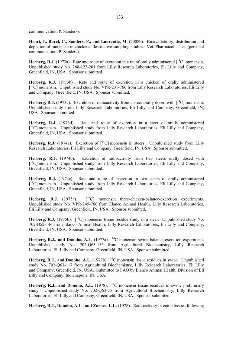
133
communication, P. Sanders). Henri, J., Burel, C., Sanders, P., and Laurentie, M. (2008b). Bioavailability, distribution and depletion of monensin in chickens: destructive sampling studies. Vet. Pharmacol. Ther. (personal communication, P. Sanders). Herberg, R.J. (1973a). Rate and route of excretion in a rat of orally administered [14C] monensin. Unpublished study No. 268-122-201 from Lilly Research Laboratories, Eli Lilly and Company, Greenfield, IN, USA. Sponsor submitted. Herberg, R.J. (1973b). Rate and route of excretion in a chicken of orally administered [14C] monensin. Unpublished study No. VPR-231-766 from Lilly Research Laboratories, Eli Lilly and Company, Greenfield, IN, USA. Sponsor submitted. Herberg, R.J. (1973c). Excretion of radioactivity from a steer orally dosed with [14C] monensin. Unpublished study from Lilly Research Laboratories, Eli Lilly and Company, Greenfield, IN, USA. Sponsor submitted. Herberg, R.J. (1973d). Rate and route of excretion in a steer of orally administered [14C] monensin. Unpublished study from Lilly Research Laboratories, Eli Lilly and Company, Greenfield, IN, USA. Sponsor submitted. Herberg, R.J. (1974a). Excretion of [14C] monensin in steers. Unpublished study from Lilly Research Laboratories, Eli Lilly and Company, Greenfield, IN, USA. Sponsor submitted. Herberg, R.J. (1974b). Excretion of radioactivity from two steers orally dosed with [14C] monensin. Unpublished study from Lilly Research Laboratories, Eli Lilly and Company, Greenfield, IN, USA. Sponsor submitted. Herberg, R.J. (1974c). Rate and route of excretion in two steers of orally administered [14C] monensin. Unpublished study from Lilly Research Laboratories, Eli Lilly and Company, Greenfield, IN, USA. Sponsor submitted. Herberg, R.J. (1975a). [14C] monensin three-chicken-balance-excretion experiments. Unpublished study No. VPR-245-766 from Elanco Animal Health, Lilly Research Laboratories, Eli Lilly and Company, Greenfield, IN, USA. Sponsor submitted. Herberg, R.J. (1975b). [14C] monensin tissue residue study in a steer. Unpublished study No. 702-B52-146 from Elanco Animal Health, Lilly Research Laboratories, Eli Lilly and Company, Greenfield, IN, USA. Sponsor submitted. Herberg, R.J., and Donoho, A.L. (1977a). 14C monensin swine balance-excretion experiment. Unpublished study No. 702-Q63-155 from Agricultural Biochemistry, Lilly Research Laboratories, Eli Lilly and Company, Greenfield, IN, USA. Sponsor submitted. Herberg, R.J., and Donoho, A.L. (1977b). 14C monensin tissue residues in swine. Unpublished study No. 702-Q63-117 from Agricultural Biochemistry, Lilly Research Laboratories, Eli Lilly and Company, Greenfield, IN, USA. Submitted to FAO by Elanco Animal Health, Division of Eli Lilly and Company, Indianapolis, IN, USA. Herberg, R.J., and Donoho, A.L. (1978). 14C monensin tissue residues in swine preliminary study. Unpublished study No. 702-Q63-75 from Agricultural Biochemistry, Lilly Research Laboratories, Eli Lilly and Company, Greenfield, IN, USA. Sponsor submitted. Herberg, R.J., Donoho, A.L., and Zornes, L.L. (1978). Radioactivity in cattle tissues following
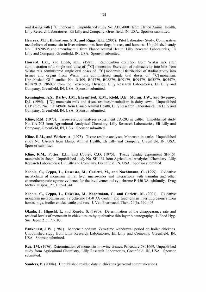
134
oral dosing with [14C] monensin. Unpublished study No. ABC-0001 from Elanco Animal Health, Lilly Research Laboratories, Eli Lilly and Company, Greenfield, IN, USA. Sponsor submitted. Herrera, M.J., Holmstrom, S.D., and Riggs, K.L. (2005). Pilot Laboratory Study: Comparative metabolism of monensin in liver microsomes from dogs, horses, and humans. Unpublished study No. T1F920505 and amendment 1 from Elanco Animal Health, Lilly Research Laboratories, Eli Lilly and Company, Greenfield, IN, USA. Sponsor submitted. Howard, L.C., and Lobb, K.L. (1981). Radiocarbon excretion from Wistar rats after administration of a single oral dose of [14C] monensin; Excretion of radioactivity into bile from Wistar rats administered single oral doses of [14C] monensin; Distribution of Radioactivity into tissues and organs from Wistar rats administered single oral doses of [14C] monensin. Unpublished GLP studies No. R-409, R04779, R08879, R09179, R09579, R05279, R05579, R05879 & R06079 from the Toxicology Division, Lilly Research Laboratories, Eli Lilly and Company, Greenfield, IN, USA. Sponsor submitted. Kennington, A.S., Darby, J.M., Ehrenfried, K.M., Kiehl, D.E., Moran, J.W., and Sweeney, D.J. (1995). [14C] monensin milk and tissue residues/metabolism in dairy cows. Unpublished GLP study No. T1F749401 from Elanco Animal Health, Lilly Research Laboratories, Eli Lilly and Company, Greenfield, IN, USA. Sponsor submitted. Kline, R.M. (1973). Tissue residue analyses experiment CA-203 in cattle. Unpublished study No. CA-203 from Agricultural Analytical Chemistry, Lilly Research Laboratories, Eli Lilly and Company, Greenfield, IN, USA. Sponsor submitted. Kline, R.M., and Wicker, A. (1975). Tissue residue analyses. Monensin in cattle. Unpublished study No. CA-268 from Elanco Animal Health, Eli Lilly and Company, Greenfield, IN, USA. Sponsor submitted. Kline, R.M., Potter, E.L., and Cooley, C.O. (1975). Tissue residue experiment SH-151 monensin in sheep. Unpublished study No. SH-151 from Agricultural Analytical Chemistry, Lilly Research Laboratories, Eli Lilly and Company, Greenfield, IN, USA. Sponsor submitted. Nebbia, C., Ceppa, L., Dascasto, M., Carletti, M., and Nachtmann, C. (1999). Oxidative metabolism of monensin in rat liver microsomes and interactions with tiamulin and other chemotherapeutic agents: evidence for the involvement of cytochrome P-450 3A subfamily. Drug Metab. Dispos., 27, 1039-1044. Nebbia, C., Ceppa, L., Dascasto, M., Nachtmann, C., and Carletti, M. (2001). Oxidative monensin metabolism and cytochrome P450 3A content and functions in liver microsomes from horses, pigs, broiler chicks, cattle and rats. J. Vet. Pharmacol. Ther., 24(6), 399-403. Okada, J., Higuchi, I., and Kondo, S. (1980). Determination of the disappearance rate and residual levels of monensin in chick tissues by qualitative thin-layer bioautography. J. Food Hyg. Soc. Japan 21: 177-183. Pankhurst, J.W. (1981). Monensin sodium. Zero-time withdrawal period on boiler chickens. Unpublished study from Lilly Research Laboratories, Eli Lilly and Company, Greenfield, IN, USA. Sponsor submitted. Rea, JM. (1976). Determination of monensin in swine tissues, Procedure 5801669. Unpublished study from Agricultural Chemistry, Lilly Research Laboratories, Greenfield, IN, USA. Sponsor submitted. Sanders, P. (2008a). Unpublished residue data in chickens (personal communication).
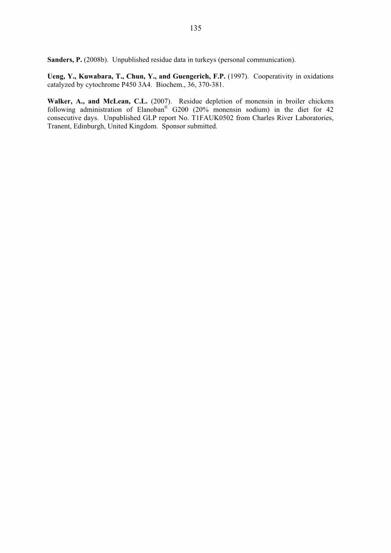
135
Sanders, P. (2008b). Unpublished residue data in turkeys (personal communication). Ueng, Y., Kuwabara, T., Chun, Y., and Guengerich, F.P. (1997). Cooperativity in oxidations catalyzed by cytochrome P450 3A4. Biochem., 36, 370-381. Walker, A., and McLean, C.L. (2007). Residue depletion of monensin in broiler chickens following administration of Elanoban® G200 (20% monensin sodium) in the diet for 42 consecutive days. Unpublished GLP report No. T1FAUK0502 from Charles River Laboratories, Tranent, Edinburgh, United Kingdom. Sponsor submitted.

136