importancia del factor de crecimiento del endotelio ... · sistema vascular complejo, que le...
TRANSCRIPT
89
IMPORTANCIA DEL FACTOR DE CRECIMIENTO DEL ENDOTELIO VASCULARRev Mex Cienc Pecu 2011;3(1):89-111
Importancia del Factor de Crecimiento del EndotelioVascular (VEGF) y de sus receptores en el ciclo ovárico.
Revisión
Role of Vascular Endothelial Growth Factor (VEGF) and itsreceptors during the ovarian cycle. Review
Ana María Rosales Torresa, Adrián Guzmán Sáncheza
RESUMEN
El objetivo de esta revisión fue recopilar y analizar la información más reciente acerca del papel del Factor de Crecimientodel Endotelio Vascular (VEGF, por sus siglas en inglés), sus receptores de membrana (VEGFR1 y VEGFR2) y receptoressolubles (sVEGFR1 y sVEGFR2), durante los procesos involucrados en el ciclo ovárico. La principal función del sistema VEGF(VEGF y sus receptores), es controlar la formación de nuevos vasos sanguíneos y la protección de células endoteliales y de lagranulosa. Es conocido que durante el ciclo ovárico, los cambios vasculares son importantes para controlar el desarrollofolicular, la ovulación y la formación y regresión del cuerpo lúteo (CL). En la selección folicular, VEGF y el receptor VEGFR2incrementan su expresión para favorecer el aporte de nutrientes al folículo. En la ovulación VEGF, VEGFR1 y VEGFR2reducen su expresión para evitar una hemorragia, y se incrementa inmediatamente después para promover la formación devasos sanguíneos y el desarrollo del CL. Finalmente durante la regresión del CL el VEGF y VEGFR2 reducen su expresióncoincidiendo con la muerte de las células que lo forman. Las evidencias revisadas permiten sugerir que VEGF y VEGFR2 sonlos principales promotores de la angiogénesis y protección celular en el desarrollo del folículo y CL, sin embargo los otrosmiembros del sistema VEGF; VEGFR1 y sVEGFR1 y sVEGFR2, parecen desempeñar funciones anti-angiogénicas en losprocesos ováricos mencionados.
PALABRAS CLAVE: Sistema VEGF, Desarrollo folicular, Ovulación, CL.
ABSTRACT
The aim of present review was to compile the more recent information related with the role of VEGF and its receptors duringovarian cycle. The main function of VEGF system (VEGF and its receptors), is to control the new blood vessels formation and theprotection of endothelial and granulosa cells. It is known that during the ovarian cycle, the vascular changes are important to regulatethe follicular and corpus luteum development (CL), as well that the ovulation. The evidences reviewed here shown that duringfollicular selection, VEGF and VEGF receptor-2 (VEGFR2) increase their expression to promote the nutrients supply and follicularcell protection, while in the ovulation VEGF system reduces the expression but this increases rapidly to induce the new blood vesselformation and thus the CL development. Finally, during the CL regression the expression of VEGF and VEGFR2 is reducedcoinciding with the cell death of the gland. The results present herein suggest that VEGF and its receptor 2 are the mainpromoters of angiogenesis and cellular protection during follicular and CL development, however others members of VEGFsystem such as VEGF receptor-1 and the soluble receptors (sVEGFR1 and sVEGFR2) seem to have an anti-angiogenic role.
KEY WORDS: SEGF, Ovulation, CL, Receptors, Folicular development.
Recibido el 10 de diciembre de 2010. Aceptado el 25 de abril de 2011.a Laboratorio de Bioquímica de la Reproducción. Departamento de Producción Agrícola y Animal. Universidad Autónoma Metropolitana-Xochimilco. Calzada del Hueso 1100,
Col. Villa Quietud, Delegación Coyoacán, 04960, México, D.F. Edificio 34. Tel. 5483 7000 ext 3082. [email protected]. Correspondencia al primer autor.
INTRODUCCIÓN
La función ovárica de las hembras depende delestablecimiento y la remodelación continua de un
INTRODUCTION
Ovarian function of females depends on theestablishment and continuous remodelling of a
90
Ana María Rosales Torres, et al. / Rev Mex Cienc Pecu 2012;3(1):89-111
sistema vascular complejo, que le suministre alfolículo y al cuerpo lúteo el oxígeno, los nutrientesy las hormonas que demandan en cada una de lasetapas de su desarrollo; además que le permitan lasalida a los esteroides que se producen en las célulasfoliculares y lúteas para alcanzar sus respectivosórganos blanco. Los folículos que llegan a laovulación tienen una extensa red de capilares quesoportan su crecimiento y maduración(1,2), mientrasque la muerte de las células endoteliales de lateca(3) y la degeneración del lecho capilar delfolículo que conllevan a un suministro vascularinsuficiente, parecen ser disparadores de laatresia(2,4). El análisis del flujo sanguíneo porultrasonido, sugiere que los folículos dominantestienen mejor perfusión sanguínea que lossubordinados(5). El folículo dominante que sedesarrolla bajo el efecto del pico de la hormonaluteinizante (LH), ocasionado por la alta frecuenciade pulsos de esta hormona, se luteinizará, ovularáy se convertirá en un cuerpo lúteo (CL). Duranteeste proceso de transformación, las células de lagranulosa se diferencian a células lúteas grandes(LLC) y las de la teca a células lúteas pequeñas(SLC)(4), capaces de producir progesterona. Estoscambios en la producción de esteroides se dan porel incremento en la expresión de enzimas queconvierten colesterol a progesterona (P450 ssc y3-HSD), y el decremento en la expresión de lasque convierten progesterona a estrógenos (P45017- y P450arom).
Además de las células lúteas, el CL está conformadopor fibroblastos, por células de músculo liso yprincipalmente por células endoteliales, las cualesdurante la formación y desarrollo del CL formanuna red vascular que garantiza el suministro dehormonas, nutrientes y oxígeno. El funcionamientodel CL, se pierde por la muerte programada queocurre en sus células, y por la vasoconstricciónprovocada por la prostaglandina PGF2 producidaen el endometrio, que también conlleva a la muertede las células lúteas y endoteliales(1).
La interacción del factor de crecimiento delendotelio vascular (VEGF), con sus receptores demembrana; VEGFR1 y VEGFR2, se reconoce comoel principal estimulador de la angiogénesis
complex vascular system, which provide to thefollicle and the corpus luteum, the oxygen, nutrientsand hormones that demand in each of the stages ofits development, moreover the vescular systemallowing the output to the steroids produced inluteal and follicular cells to reach their respectivetarget cell. The follicles arriving at ovulation havean extensive network of capillaries that supporttheir growth and maturation(1,2), while the deathof endothelial cells of theca(3) and the degenerationof the capillary bed of the follicle that lead to aninadequate vascular supply, appear to triggers theatresia(2.4). Analysis of ultrasonic blood flow,suggests that the dominant follicles have betterblood perfusion than subordinate follicles(5). Thedominant follicle which develops under the influenceof the peak of the luteinizing hormone (LH), causedby the high-frequency pulses of this hormone, itwill luteinize; it will ovulate and will become acorpus luteum (CL). During this process oftransformation, the granulosa cells differentiate tolarge luteal cell (LLC) and the theca cells to smallluteal cells (SLC)(4), capable of producingprogesterone. These changes in the production ofsteroids are given by the increase in the expressionof enzymes that converted choles terol toprogesterone (P450 ssc and 3-HSD), and thedecrease in the expression of enzymes that convertprogesterone to estrogens (P450 17- andP450arom).
In addition to luteal cells, the CL is formed byfibroblasts, smooth muscle cells and by endothelialcells, which during the formation and developmentof the CL form a vascular network that guaranteesthe supply of hormones, nutrients and oxygen. Thefunction of the CL is lost by apoptosis, and thevasoconstriction caused by prostaglandin F2(PGF2-) produced in the endometrium, which alsoleads to the death of luteal cells and endothelialcells(1).
The interaction of the vascular endotelial growthfactor (VEGF), with its membrane receptors(VEGFR1 and VEGFR2) is recognized as the mainstimulator of angiogenesis (proliferation, migrationand survival of endothelial cells), both in the ovaryand in other tissues(6.7). Recently, soluble forms

91
IMPORTANCIA DEL FACTOR DE CRECIMIENTO DEL ENDOTELIO VASCULAR
(proliferación, migración y sobrevivencia de lascélulas endoteliales), tanto en el ovario como enotros tejidos(6,7). Recientemente se conocen lasformas solubles de los receptores de membranapara VEGF: sVEGFR1 y sVEGFR2, los cualesaparentemente tienen un efecto anti-angiogénico alcapturar al ligando, evitando que tenga interaccióncon los receptores de membrana(8). Nosotros hemosllamado sistema VEGF, al ligando, a los receptoresde membrana y a los receptores solubles, por locual en adelante así será referido. En este trabajose describen las evidencias que existen de laparticipación del sistema VEGF en el desarrollofolicular, ovulación, formación y regresión delcuerpo lúteo.
GeneralidadesAngiogénesis
El brote de un nuevo vaso a partir de uno yaexistente, es el mecanismo más frecuente en laangiogénesis, la cual ocurre en varios procesosfisiológicos, entre los que destacan el desarrolloembrionario, el desarrollo folicular y el desarrollodel CL, así como en procesos patológicos, comoes la formación de tumores. La angiogénesis,involucra varios pasos secuenciales(9); primero, loscomponentes del entorno de la matriz extracelularendotelial son degradados localmente por proteasasproducidas por la propia célula endotelial. A estole sigue la migración quimiotáctica de célulasendoteliales hacia el lugar donde se desarrollará elnuevo vaso sanguíneo. Subsecuentemente en lasección media del vaso en formación, las célulasendoteliales proliferan y se ensamblan para formarel lumen del vaso sanguíneo. El nuevo vaso sufreanastomosis con el vaso adyacente para serperfundido con circulación sanguínea(9,10,11). Hastaeste punto, el capilar recientemente formado, esfrágil y puede ser remodelado. La maduración delnuevo vaso sanguíneo hacia un vaso estable yfuncional, requiere la acumulación de una láminabasal, y el recubrimiento por pericitos y células demúsculo liso para fortalecerlo(12).
Regulación de la angiogénesis
En un inicio, cuando se estudiaron las moléculasque regulan la angiogénesis, se propuso a los
of membrane receptors of VEGF (sVEGFR1 andsVEGFR2 were reported, which apparently havean anti-angiogenic effect because capturing to theligand, avoids an interaction with the receptormembrane(8). We have called VEGF system, tothe ligand, membrane receptors and solublereceptors, which henceforth so will be mentioned.This review describes the evidence existing ofVEGF system’s role on follicular development,ovulation, formation and regression of corpusluteum.
OverviewAngiogenesis
The outbreak of a new vessel from an existingone, is the commonest in angiogenesis, whichoccurs in many physiological processes includingembryonic development, follicular and CLdevelopment, as well as in disease processes, suchas the tumors development. Angiogenesis, involvesseveral sequential steps(9); first, the extracellularmatrix components are locally degraded by proteasesproduced by endothelial cell, this is followed bythe chemotactic migration of endothelial cellstowards the place where the new blood vessel isbuild. Subsequently in the midsection of the vesselin formation, the endothelial cells proliferate andare assembled to form the lumen of the bloodvessel. The new vessel suffers anastomosis withthe adjacent one to be perfused with bloodcirculation(9,10,11). At this point, the recentlyformed capillary is fragile and can be remodeled.The maturation of the new blood vessel towards astable and functional one, requires the accumulationof a basal lamina, and coating for pericytes andsmooth muscle cells to strengthen it(12).
Regulation of angiogenesis
Initially, when molecules that regulate angiogenesiswere studied, it was propossed the fibroblasticgrowth factors (FGF) as key regulators of theprocess, however knockout mice for these genes,did not develope vascular defects(13), so the existenceof other molecules that control the new blood vesselformation was considerered . Since 1989, whenVEGF was discovered, it has been considered asthe main factor involved in angiogenesis(14).
92
Ana María Rosales Torres, et al. / Rev Mex Cienc Pecu 2012;3(1):89-111
factores de crecimiento fibroblásticos (FGF) comolos principales reguladores del proceso; sin embargoratones nocaut para estos genes, no desarrollarondefectos vasculares(13), por lo que se pensó en laexistencia de otras moléculas que controlan laformación de nuevos vasos sanguíneos. Así fuecomo en 1989, se descubrió el VEGF, al que sele ha considerado como el principal factorinvolucrado en la angiogénesis(14).
Dentro de las moléculas que regulan la neovascu-larización, VEGF juega un papel central durante elproceso angiogénico en condiciones tantofisiológicas como patológicas; es un potentemitógeno de células endoteliales derivadas dearterias y venas, aunque también ha mostrado teneruna importante actividad mitótica en otros tiposcelulares(15), entre los que destacan lasneuronas(9,16) y la actividad citoportectora en lascélulas de la granulosa(17).
VEGF
La familia de proteínas del VEGF, incluyen alVEGF-A, VEGF-B, VEGF-C, VEGF-D y el factorde crecimiento placentario (PLGF)(7). Algunos otrosautores incluyen dos miembros más que sonreferidos como VEGF-E(9) y VEGF-F(18). De estafamilia de trascriptos, el más estudiado es elVEGF-A, que es referido simplemente comoVEGF. El VEGF-B y PLGF se unen a VEGFR1y modulan la actividad de VEGF-A en célulasendoteliales durante el desarrollo embrionario(VEGF-B) y durante la angiogénesis patológica(PLGF)(8). Aunque VEGF-C y VEGF-D se unena VEGFR2 en células endoteliales de vasossanguíneos, estos miembros de la familia de VEGFse unen a su receptor (VEGFR3) en tejido linfáticopara promover el desarrollo de vasos linfáticosdurante el desarrollo embrionario(7,18). FinalmenteVEGF-E es un homólogo de VEGF producido porel virus Orf que afecta a cabras y ovejas(9) mientrasque del VEGF-F no se ha caracterizado del todosu función(18).
El VEGF ha sido caracterizado como un factor decrecimiento angiogénico que se une a heparina(19),y que exhibe una alta especificidad para las células
VEGF plays a central role during the angiogenicin both, physiological and pathological conditions;it is a powerful mitogene of endothelial cells derivedfrom arteries and veins, although he has also shownto have mitotic activity in other cell types(15), asneurons(9,16) and citoportector activity in granulosacells(17).
VEGF
The VEGF protein family, include VEGF-A,VEGF-B, VEGF-C, VEGF-D and placental growthfactor (PLGF)(7). Some other authors include twoother members referred as VEGF-E(9) and VEGF-F(18). From this trascryipt family, the most studiedis the VEGF-A, which is referred only as VEGF.The VEGF-B and PLGF bind to VEGFR1 andmodulate of VEGF-A activity in endothelial cellsduring embryonic development (VEGF-B) andpathological angiogenesis (PLGF)(8). AlthoughVEGF-C and VEGF-D bind to VEGFR2 in endothelialcells of blood vessels, these members of the VEGFfamily bind to its receptor (VEGFR3) in lymphtissue to promote the development of lymphaticvessels during embryonic development(7,18). FinallyVEGF-E is a homologue of VEGF produced bythe virus Orf which affects goats and sheep(9),whereas VEGF-F function has not been fullycharacterized(18).
The VEGF has been characterized as an angiogenicgrowth factor that binds to heparin(19), and exhibitsa high specificity for endothelial cells(20) of arteries,veins and lymphatic vessels(21). VEGF seems tohave three forms of participation in the normalvascular development, first is able to initiateangiogenesis promoting proliferation andquimioatracción of endothelial cells(22). In cellscultures from human umbilical vein endothelial cell(HUVEC), the addition of 3 or 10 ng/ml of VEGFto the culture medium, increases both, proliferationand migration of these cells. This effect is alsoreflected as an increase in the vascular network ofalveoli treated with VEGF(23). These results areconsistent by other authors in porcine aorticendothelial cells (PAE)(24) and human umbilicalvein (HUVEC)(25). Second, maintaining theviability of vessel blood immature, and third

93
IMPORTANCIA DEL FACTOR DE CRECIMIENTO DEL ENDOTELIO VASCULAR
endoteliales(20) de arterias, venas y vasoslinfáticos(21). El VEGF parece tener tres formasde participación en el desarrollo vascular normal,primero es capaz de iniciar la angiogénesispromoviendo la proliferación y quimioatracción delas células endoteliales(22). En cultivo de célulasde la vena umbilical de humano, la adición de 3o 10 ng/ml de VEGF al medio de cultivo,incrementa tanto la proliferación como la migraciónde dichas células. Este efecto se ve también reflejadocomo un incremento en la red vascular de alvéolostratados con VEGF(23). Estos resultados coincidencon lo reportado anteriormente por otros autoresen células endoteliales de la aorta porcina (EAP)(24)
y de la vena umbilical de humano (HUVEC)(25).Segundo, mantiene la viabilidad de vasos sanguíneosinmaduros y tercero facilita el recubrimiento delvaso con pericitos(26). Con relación a la viabilidadde vasos sanguíneos inmaduros, VEGF participaen la supervivencia de las células endotelialesinduciendo la expresión de proteínas antiapoptóticastales como Bcl-2 y A1 (26). La actividadantiapoptótica de VEGF está mediada por laactivación de la vía del fofatidilinositol 3- cinasa/akt(27).
La expresión del VEGF está modulada por un sinnúmero de factores de crecimiento y citocinas. Losoncogenes, v-ras, K-ras, v-raf, src fos y v-yes, sonfuertes inductores de la expresión de VEGF; sinembargo se sabe que la hipoxia y la hipoglicemiason los principales inductores de la síntesis deVEGF(20,28). Por ejemplo, en las regiones hipóxicasde algunos tumores existe un incremento en laexpresión del ARNm de VEGF(19). Se ha reportadoque el factor inductor de hipoxia 1(HIF-1) favorecela expresión del VEGF y de óxido nítrico (NO),y que al inhibir al HIF-1 por el FK228 (un potenteinhibidor de la diacetilaza histona) se frena laexpresión del VEGF en respuesta a la hipoxia tantoa nivel transcripcional como traduccional(29). Porotro lado el gen supresor del ciclo celular, p53juega un papel importante en la inhibición de laangiogénesis y la expresión de VEGF. La proteínaproducto del gen p53 es la principal inductora dela apoptosis y participa en la secreción de latrombospondina 1 (TSP 1), una glicoproteínaendógena que inhibe la angiogénesis(30). Mutaciones
facilitates the coating of the vessel with pericytes(26).With regard to the immature blood vessels viability,VEGF participates in the endothelial cells survival,by inducing the expression of antiapoptotic proteinssuch as Bcl-2 and A1(26). The antiapoptotic VEGFactivity is mediated by the activation of the pathwayof fofatidilinositol 3-kinase/akt(27).
The expression of VEGF is modulated by severalgrowth factors and cytokines. The oncogenes v-ras,K, v-raf, src fos and v-yes, increase the expressionof VEGF; however, it is known that hypoxia andhypoglycemia are the main stimuli of VEGFsynthesis(20,28). For example, in the hypoxicregions of some tumors there is an increase in theexpression of VEGF RNAm(19). It is reported thathipoxia-inducible Factor-1 (HIF-1) promote theexpression of VEGF and nitric oxide (NO), andthe HIF-1 inhibition by the FK228 (a potentinhibitor of diacetil histone) reduce the expressionof VEGF in response to hipoxia, both at transcriptionaland translational level(29). On the other hand thecell cycle suppressor gene p53 inhibits theangiogenesis and the expression of VEGF. Theprotein product of the p53 gene is the mainapoptosis inductor and participates in the secretionof the thrombospondin 1 (TSP 1), an endogenousglycoprotein that inhibits angiogenesis (30).Mutations that inactivate the p53 gene, stimulatecell proliferation and cause an increase in theexpression of VEGF(31). Although the TSP 1 appearsto be the most important molecule to inhibit theexpression of VEGF, there are others such as theangiostatin, endostatin, prolactin, interferons (INF-,INF-), plaquetary factor 4 (PF-4), interleukins(IL 12 IL 4), inhibitors of metalloproteinases (TIMP1, TIMP 2), testosterone, somatostatin, melatonin,and metoxiestradiol, which can also block the neoformation of vessels through the inhibition ofVEGF(31,32).
VEGF isoforms and angiogenesis
Five angiogenic isoforms derived from a singlegene of VEGF have been identified in human:VEGF121, VEGF145, VEGF165, VEGF189 andVEGF206. These isoforms are a result of splicing-alternative process of RNAm of VEGF, which

94
Ana María Rosales Torres, et al. / Rev Mex Cienc Pecu 2012;3(1):89-111
que inactivan al gen p53, estimulan la proliferacióncelular y ocasionan un incremento en la expresiónde VEGF(31). Aunque la TSP 1 parece ser lamolécula más importante para inhibir la expresiónde VEGF, existen otras como la angiostatina,endostatina, prolactina, interferones (INF INF), factor plaquetario 4 (PF 4), interleucinas (IL12 IL 4), inhibidoras de metaloproteinasas (TIMP1, TIMP 2), testosterona, somatostatina, melatoninay metoxiestradiol, que también pueden bloquear laneo formación de vasos sanguíneos por medio dela inhibición de VEGF(31,32).
Isoformas del VEGF y angiogénesis
En humanos se han identificado cinco isoformasangiogénicas derivadas de un solo gen de VEGF;VEGF121, VEGF145, VEGF165, VEGF189 yVEGF206. Estas isoformas son resultado delproceso de maduración alternativo por corte yempalme del ARNm de VEGF, que contiene ochoexones(33). Tres tipos de DNA complementario(cDNA) del VEGF humano (hVEGF) se aislaronde una biblioteca de cDNA preparada de célulasleucémicas HL60. Los cDNAs codifican para unaproteína con 95 % de similitud al VEGF bovinoque tiene 165 aminoácidos a la que se le denominaVEGF165; de las otras dos proteínas una contiene121 aminoácidos (VEGF121) y la otra 189(VEGF189). La cuarta isoforma de VEGF fueidentificada en bibliotecas de cDNA de hígado fetal,y la proteína madura está formada por 206aminoácidos (VEGF206). Como mencionamos,estas cuatro isoformas de VEGF son originadasdel corte y empalme alternativo del ARNminmaduro, y los cambios están relacionados conlos exones 6 y 7 del gen. Una quinta isoforma, elVEGF145 ha sido identificada en el endometrio ymiometrio humano(34,35). Las isoformas VEGF121y VEGF165 son más ácidas y no se unen fácilmentea la heparina, y por lo tanto llegan fácilmentehacia la célula endotelial. En contraste, las isoformasVEGF189 y el VEGF206 son básicas y se unen ala heparina con mayor facilidad, permaneciendocompletamente secuestradas en la matrizextracelular(21). Las isoformas 189 y 206 por acciónde la plasmina, pueden dar lugar a la formación deuna proteína de 110 aminoácidos (VEGF110) capaz
contains eight exons(33). Three types of complementaryDNA (cDNA) of human VEGF (hVEGF) isolatedfrom a cDNA library of leukemia cells HL60. ThecDNAs encoding a protein with 95 % similarity tobovine VEGF has 165 amino acids which is calledVEGF165; two other proteins, one contains 121amino acids (VEGF121) and the other 189(VEGF189). The fourth isoform of VEGF wasidentified in fetal liver cDNA libraries, and themature protein is composed of 206 amino acids(VEGF206). As mentioned, these four isoforms ofVEGF are originated from splicing-alternative ofthe immature RNAm, and the changes are relatedto the exon 6 and 7 of the gene. A fifth isoform,the VEGF145, has been identified in the humanendometrium and myometrium (34,35). TheVEGF121 and VEGF165 isoforms are more acidicand not easily bind to heparin, and therefore comeeasily to the endothelial cell. In contrast, VEGF189and the VEGF206 isoforms are basic and bind toheparin, remaining completely sequestered in theextracellular matrix(21). Isoforms 189 and 206, byplasmin action, may lead to the formation of a 110amino acids (VEGF110) protein capable of bindingto VEGFR1 and VEGFR2 in endothelial cell(7).This phenomenon is important during angiogenesis,since acid isoforms begin angiogenic process,promote the activation of proteases that degradethe matrix, and thus the release of basic isoformsthat are linked to the extracellular matrix to enhancethe proliferation and migration of endothelialcells(36). Finally, VEGF145 is mainly expressedby tumoral cells of the female reproductivesystem(37). Is worth mentioning that in all animals,including cattle, all proteins, product of the isoformsof VEGF, have one fewer amino acid(2).
VEGF Receptors
The biological activity of VEGF is mediatedprimarily by two tyrosine kinase receptor:VEGFR-1 or Flt-1 (fms-like tyrosine kinase-1), andVEGFR-2 or Flk-1 (fetal liver kinase-1); howeverother receptors such as the VEGFR-3 or Flt-4, theneuropilin 1 and 2 (NPR-1 and NPR-2 respectively)which also binds VEGF(9,16,20,33). The receptorsof major importance by his affinity with the ligandare: VEGFR1 and VEGFR2, both receptors are
95
IMPORTANCIA DEL FACTOR DE CRECIMIENTO DEL ENDOTELIO VASCULAR
de unirse a VEGFR1 y VEGFR2 en la célulaendotelial(7). Este fenómeno es de vital importanciadurante la angiogénesis, ya que las isoformas ácidasinician el proceso angiogénico, promoviendo laactivación de proteasas que degradan la matriz, ycon ello la liberación de las isoformas básicas quese encuentran secuestradas en la matriz extracelularpara potenciar la proliferación y migración decélulas endoteliales(36). Finalmente VEGF145 esexpresado principalmente por células cancerosasdel sistema reproductor femenino(37). Cabemencionar que en todos los animales, incluyendolos bovinos, todas las proteínas producto de lasisoformas de VEGF tienen un aminoácido menos(2).
Receptores del VEGF
La actividad biológica del VEGF está mediadaprincipalmente por dos receptores tipo tirosinacinasa: el VEGFR-1 ó Flt-1 (fms-like tyrosinekinase-1, por sus siglas en inglés), y el VEGFR-2ó Flk-1 (fetal liver kinase-1, por sus siglas eninglés), sin embargo se han reportado otros receptorestales como el VEGFR-3 o Flt-4, la neuropilina 1 y2 (NRP-1 y NRP-2 respectivamente) a los cualestambién se une el VEGF(9,16,20,33). Los receptoresde mayor importancia por su afinidad con el ligandoson: VEGFR1 y VEGFR2, ambos receptores secaracterizan por tener siete dominios extracelularesde unión similares a inmunoglobulinas, una solaregión transmembranal y un dominio tirosina cinasaintracelular(7,35).
El VEGF se une al VEGFR1 con una constante dedisociación (Kd) de 10-20 pM, por lo que seconsidera que el factor tiene una alta afinidad paraeste receptor(38) en comparación con el VEGFR2que tiene una Kd de 75 a 125 pM(24). Sin embargo,la capacidad de autofosforilación de VEGFR2 es almenos 10 veces mayor que la del VEGFR1, locual indica que el VEGFR2 tiene una mayorcapacidad en la traducción de la señal deVEGF(24,25). Se considera que VEGFR2 es elmayor mediador de los efectos mitogénicos,angiogénicos y de permeabilidad de VEGF(36). Encontraste, VEGF unido al VEGFR-1 también se lehan atribuido efectos proliferativos(25), quimiotácticosy de protección de células endoteliales(36), aunque
characterized by seven union extracellular domainslike immunoglobulin, a single transmembrane regionand a tyrosine kinase intracellular domain(7,35).
VEGF joins the VEGFR1 with a constant ofdissociation (Kd) 10-20 pM, so it is consideredthat the factor has a high affinity for this receptor(38)
in comparison with the VEGFR2 with a Kd of 75to 125 pM(24). However, the fosforilation ofVEGFR2 is at least 10 times greater than VEGFR1,which indicates that the VEGFR2 has a greatercapacity in the translation of the signal ofVEGF(24,25). VEGFR2 is considered the greatestmediator of mitogenic, angiogénicos andpermeability effects of VEGF(36). In contrast,VEGF bind to VEGFR-1 also have proliferativeeffects(25), chemotactic and protection of endothelialcells(36), although with the limited ability ofsignaling, is generally considered a “decoy”receptor(25).
This evidence suggests that both receptors triggerdifferent effects in response to the ligand. Thus theVEGFR1 may regulate angiogenesis by makingless available the l igand to VEGFR2(25).Additionally, by splicing.alternative of the immatureRNAm of VEGFR-1 and VEGFR-2, are synthesizedsoluble forms of both receptors (sVEGFR-1 andsVEGFR-2), these receptors proteins lose theintracellular and transmembrane domain, andtherefore can not translate signs; however, maintainthe domains of union to the ligand, retaining thesame affinity for VEGF that membrane receptors(39).Because of this, some authors suggest that there iscompetition for the ligand between soluble receptorsand those of the membrane, which gives the firstanti-angiogenic effect(40).
Participation of VEGF in the folliculardevelopmentFolicular development
The follicular development that occurs in theestrous/menstrual cycle occurs in the form of waves,formed by cohorts or groups of follicles that duringthe initial recruitment, are elected to continue itsgrowth in response to gonadotropin (cyclicrecruitment)(41,42). In each wave, recruited follicles
96
Ana María Rosales Torres, et al. / Rev Mex Cienc Pecu 2012;3(1):89-111
por su limitada capacidad de señalización, esgeneralmente considerado un receptor “anzuelo”(25).
Estas evidencias sugieren que ambos receptoresdesencadenan efectos diferentes en respuesta alligando. Así, el VEGFR1 puede regular laangiogénesis haciendo poco disponible el ligando aVEGFR2(25). Adicionalmente por corte y empalmede los ARNm inmaduros de VEGFR-1 yVEGFR-2, se producen formas solubles de ambosreceptores (sVEGFR-1 y sVEGFR-2), las proteínasde estos receptores pierden el dominiotransmembranal e intracelular con lo cual no puedentraducir señales, sin embargo, mantienen losdominios de unión al ligando, conservando la mismaafinidad por VEGF, que los receptores demembrana(39). Por lo anterior algunos autoressugieren que existe una competencia por el ligandoentre los receptores solubles y los de membrana,que les confiere a los primeros un efectoantiangiogénico(40).
Participación de VEGF en el desarrollo folicular
Desarrollo folicular
El desarrollo folicular que se presenta en el cicloestral/menstrual, ocurre en forma de ondas u olas,conformadas por cohortes o grupos de folículosque durante el reclutamiento inicial, son elegidospara continuar su crecimiento en respuesta agonadotropinas (reclutamiento cíclico)(41,42). Encada ola, se establece entre los folículos reclutadosuna competencia por la dominancia, en la cual unoo varios folículos (dependiendo de la especie) de lacohorte adquiere el desarrollo competente que lepermitirá seguir creciendo en un ambiente de bajasconcentraciones de gonadotropinas, al tiempo enque sus compañeros de cohorte sufren atresia. Elfolículo dominante modifica el patrón de crecimientode los folículos subordinados en ambos ovarios,mediante la producción de estradiol e inhibina queactúan en forma endocrina, autocrina o paracrinapara autopotenciar su desarrollo e inhibir el de lossubordinados. En las especies monotocas el folículodominante de la primera ola del ciclo, emergemientras está activo el CL, de tal manera que laprogesterona secretada por esta glándula transitoria,reduce la frecuencia de pulsos de LH, evitando su
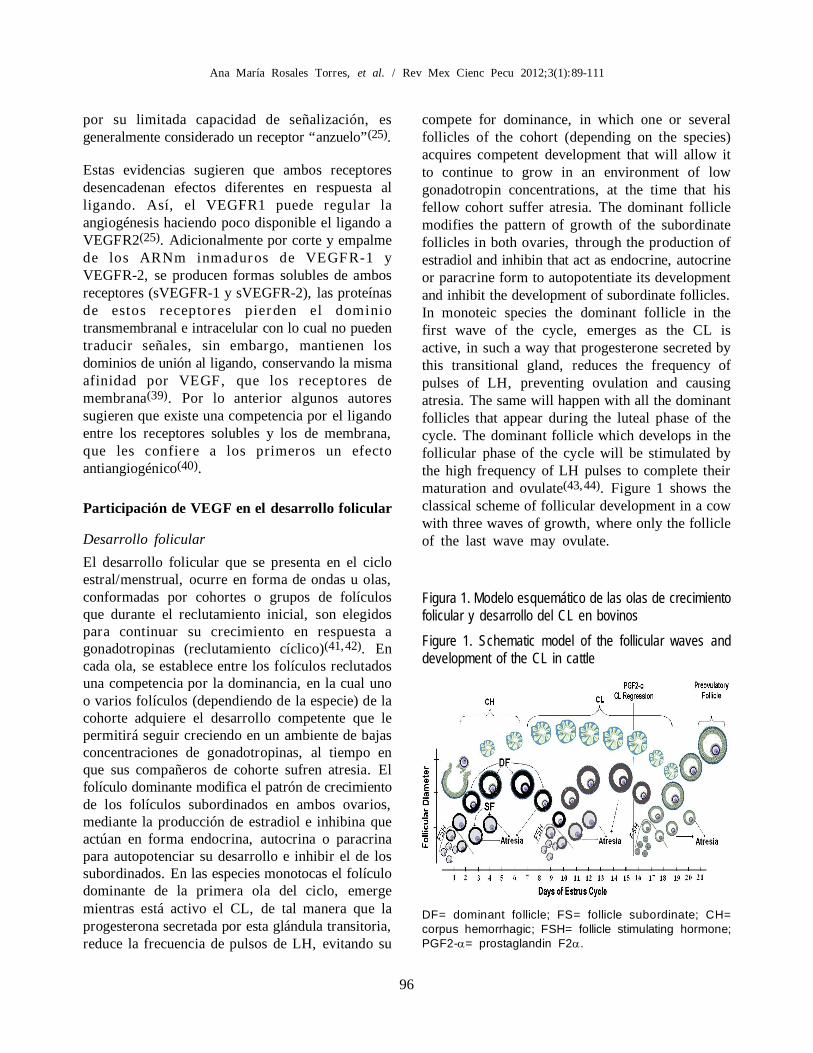
compete for dominance, in which one or severalfollicles of the cohort (depending on the species)acquires competent development that will allow itto continue to grow in an environment of lowgonadotropin concentrations, at the time that hisfellow cohort suffer atresia. The dominant folliclemodifies the pattern of growth of the subordinatefollicles in both ovaries, through the production ofestradiol and inhibin that act as endocrine, autocrineor paracrine form to autopotentiate its developmentand inhibit the development of subordinate follicles.In monoteic species the dominant follicle in thefirst wave of the cycle, emerges as the CL isactive, in such a way that progesterone secreted bythis transitional gland, reduces the frequency ofpulses of LH, preventing ovulation and causingatresia. The same will happen with all the dominantfollicles that appear during the luteal phase of thecycle. The dominant follicle which develops in thefollicular phase of the cycle will be stimulated bythe high frequency of LH pulses to complete theirmaturation and ovulate(43,44). Figure 1 shows theclassical scheme of follicular development in a cowwith three waves of growth, where only the follicleof the last wave may ovulate.
Figura 1. Modelo esquemático de las olas de crecimientofolicular y desarrollo del CL en bovinosFigure 1. Schematic model of the follicular waves anddevelopment of the CL in cattle
DF= dominant follicle; FS= follicle subordinate; CH=corpus hemorrhagic; FSH= follicle stimulating hormone;PGF2-= prostaglandin F2.

97
IMPORTANCIA DEL FACTOR DE CRECIMIENTO DEL ENDOTELIO VASCULAR
ovulación y provocándole atresia. Lo mismoocurrirá con todos los folículos dominantes queaparezcan durante la fase lútea del ciclo. El folículodominante que se desarrolle en la fase folicular delciclo será estimulado por la frecuencia alta de pulsosde LH para terminar su maduración y ovular(43,44).En la Figura 1 se muestra el esquema clásico dedesarrollo folicular en una vaca con tres olas decrecimiento, donde sólo el folículo de la última olapodrá ovular.
VEGF y sus receptores durante la selección ydominancia folicular
En el ovario de las hembras de los mamíferos, losfolículos primordiales y primarios reciben oxígenoy nutrientes por difusión pasiva desde los vasossanguíneos del estroma, debido a que en estosestadios, los folículos carecen de irrigación propia.La red capilar individual, se inicia en los folículossecundarios en cuanto aparece la capa celular de lateca(45). Durante la formación de los folículospreantrales, hay un gran incremento no sólo en eltotal de la vasculatura, sino también en la densidadvascular dado que aproximadamente el 40 % delas células que proliferan en la teca en este estadio,son de origen endotelial(46). En el estado antral delos folículos, la capa vascular está formada pordos redes concéntricas de vasos sanguíneos, una deellas ubicada directamente por fuera de la membranabasal y la otra en la teca externa(47). Estas redescapilares, en un folículo íntegro no penetran haciala granulosa(48,49,50), por lo que esta capa celularpermanece avascular durante todo el desarrollofolicular(51).
El establecimiento del plexo capilar de la tecainterna, coincide con el período de rápido crecimientoy diferenciación de los folículos, por lo que se hapropuesto que un factor que condiciona la seleccióndel folículo dominante, además de su producciónde estradiol y respuesta a gonadotropinas, es lacapacidad de desarrollar una mayor red vascular,así como un incremento en la permeabilidadvascular(22). Se ha puesto en evidencia que losfolículos dominantes tienen una mayor y mejorirrigación que los folículos subordinados, y queestos últimos presentan una rápida degeneración de
VEGF and its receptors during selection andfollicular dominance
In the ovary of female mammals, primary andprimordial follicles receive oxygen and nutrientsby passive diffusion from the blood vessels of thestroma, because in these stages, the follicles haveno own irrigation. The individual capillary network,begins in secondary follicles as son as the celllayer of theca appears(45). During the formation ofpreantral follicles, there is an increase not only inthe total vascularity, but the vascular density also,because approximately 40 % of the cells which areproliferating in the theca at this stage are ofendothelial origin(46). In antral follicles, the vascularlayer consists of two concentric networks of bloodvessels, one of them located directly outside of thebasement membrane and the other in externaltheca(47). These capillary networks in an intactfol l icle, do not penetrate towards thegranulosa(48,49,50), so this cell layer remainsavascular throughout the follicular development(51).
The establishment of the capillary plexus of internaltheca coincides with the period of rapid growthand differentiation of the follicles; it has beenproposed that a factor that influences the selectionof the dominant follicle, in addition to its responseto gonadotropin and estradiol production, is theability to develop a greater vascular network, Ithas been proved that the dominant follicles have agreater and better irrigation to subordinate follicles,and the latter presented a rapid degeneration of thevasculature of the theca as part of the process ofatresia(52). In this way VEGF and their membranereceptors have shown to have an important role asregulators of angiogenesis of the follicle(45) and asprotector of the granulosa cells(17).
The VEGF is produced by theca, granulosa andluteal cells. In the follicle, the granulosa cells arethe main source of VEGF, which seems to createa gradient with direction towards the basalmembrane, by encouraging the contribution ofoxygen, nutrients and hormones to granulosacells(35,53). VEGF can act as a factor of survivalfor the granulosa cells and thereby suppresing atresiaof the antral follicles(54); Greenaway et al(17)
demonstrated that VEGF has a citoprotector effect,
98
Ana María Rosales Torres, et al. / Rev Mex Cienc Pecu 2012;3(1):89-111
la vasculatura de la teca como parte del proceso deatresia(52). De esta forma el VEGF y sus receptoresde membrana han mostrado tener un papelimportante como reguladores de la angiogénesisdel folículo(45) y como protectores de las célulasde la granulosa(17).
El VEGF es producido por células de la teca,granulosa y lúteas. En el folículo, las células de lagranulosa son las principales productoras de VEGF,lo cual parece crear un gradiente con direcciónhacia la membrana basal, favoreciendo el aportede oxígeno, nutrientes y hormonas a las células dela granulosa(35,53). El VEGF puede actuar comoun factor de sobrevivencia para las células de lagranulosa y con ello suprimir la atresia de losfolículos antrales(54), al respecto Greenaway etal(17) demostraron que VEGF ejerce un efectocitoprotector, evitando la apoptosis de célulasendoteliales y de la granulosa en cultivo. Por otrolado, durante la selección del folículo dominante,los de mayor tamaño y concentración de estradiol(estrógeno-activos) corresponden a los que tambiéntienen una vasta vascularización y una mayorconcentración de VEGF en sus compartimientos(55).En ratas inmaduras de 21 días de edad, tratadascon eCG y hCG, la administración de fragmentostrasncripcionalmente activos del gen de VEGF,incrementa el número de ovocitos ovulados, elnúmero de folículos antrales grandes y el defolículos preovulatorios, reduciendo así el porcentajede folículos en atresia(56,57). Resultados similaresse reportaron en ratones(58).
Investigaciones con el uso de inhibidores de VEGFen primates y humanos han permitido destacar lasfunciones más importantes de VEGF(59). Eltratamiento con un antagonista de VEGF en ovariosde mono tití reduce el volumen folicular, así comoel índice de proliferación de granulosa, teca ycélulas endoteliales de folículos desde el estadosecundario temprano hasta el de dominancia. Elefecto sobre las células endoteliales se ve reflejadoen una reducción de la vasculatura de los folículostratados con el antagonista de VEGF(60).
Resultados de Quintana et al(61) y otros autores(62,63)
muestran que las gonadotropinas (FSH y LH) y
preventing the apoptosis of endothelial cells andgranulosa cells in culture. On the other hand, duringthe selection of the dominant follicle, the folliclesof larger size and concentration of estradiol (active-estrógens) correspond to those who also have avast vascularisation and a greater concentration ofVEGF in their compartments(55). In immature ratsof 21 d of age treated with eCG and hCG, theadministration of active trasncripcional fragmentsof VEGF gene, increases the number of ovulatedoocites, the number of large antral follicles and thepreovulatory follicles, thereby reducing thepercentage of follicles in atresia(56,57). Similarresults were reported in mice(58).
Use of inhibitors of VEGF in primates and humanshave made it possible to highlight the mostimportant functions of VEGF(59). Treatment witha VEGF antagonist in Rhesus Monkey ovariesreduces the follicular volume, as well as the rateof proliferation of granulosa, theca and endothelialfollicles cells; since early secondary stage until thedominant follicle. The effect on endothelial cells isreflected in a reduction of follicles vasculature(60).
Quintana et al(61) and other authors results(62,63),show that the gonadotropins (FSH and LH) andtheir homologous: human chorionic gonadotropin(hCG) and equine chorionic gonadotropin (eCG),stimulates the production of VEGF in granulosacells. Barboni et al(64) demonstrated in sows, thattreatment with 1,250 UI of eCG increasesproduction of VEGF, as well as the transcriptionof its gene, in cells of the granulose of folliclesgreater to 5 mm in diameter(65). The administrationof 1 ng/ml of VEGF to cultures cells from thegranulosa cells of follicles of cattle with a diameterof 4 to 8 mm, increases cell proliferation, whichwas exacerbated when FSH was also added (10 ng/ml)(66).
Little is known about the participation and regulationof the different isoforms of VEGF in the folliculardevelopment; the most abundant in all mammalsseem to be VEGF120 and VEGF164. It has beenproposed that in cells culture of the bovinegranulosa, estradiol (1 ng/ml) in combination withprogesterone (10 ng/ml) increase the expression of
99
IMPORTANCIA DEL FACTOR DE CRECIMIENTO DEL ENDOTELIO VASCULAR
sus homólogas; la gonadotropina coriónica humana(hCG) y la gonadotropina coriónica equina (eCG),estimulan la producción de VEGF en células de lagranulosa. Barboni et al(64) demostraron en cerdas,que el tratamiento con 1,250 UI eCG incrementala producción de VEGF, así como la transcripciónde su gen en células de la granulosa de folículosmayores de 5 mm de diámetro (65). Laadministración de 1 ng/ml de VEGF al cultivo decélulas de la granulosa provenientes de folículosde bovino con un diámetro de 4 a 8 mm, incrementala proliferación celular, la cual se exacerbó cuandoademás se adicionó FSH (10 ng/ml)(66).
Se conoce poco sobre la participación y regulaciónde las diferentes isoformas de VEGF en eldesarrollo folicular; las más abundantes en todoslos mamíferos parecen ser VEGF120 y VEGF164.Se ha propuesto que en cultivos de células de lagranulosa de bovinos, estradiol (1 ng/ml) encombinación con progesterona (10 ng/ml) regulana la alta la expresión del ARNm de VEGF164 ya la baja el ARNm de VEGF120, mientras que laprogesterona (10 ng/ml) sola, regula a la baja elARNm de VEGF164 y a la alta el ARNm deVEGF120(67). Sin embargo en células de lagranulosa de folículos seleccionados (estrógenodominantes) de bovinos, la expresión de VEGF120y VEGF164 es mayor que en aquéllos noseleccionados(68). En folículos de ovejas, Rosaleset al(69) reportan la expresión del ARNm deVEGF120, VEGF164 y VEGF205 en granulosas ytecas de folículos sanos y atrésicos. La expresiónde ARNm de VEGF120 y VEGF164 se redujo conun patrón muy similar conforme avanzó el gradode atresia, siendo en las células de la granulosa, encomparación con la teca donde se observó la menorexpresión del ARNm de ambas isoformas. En elcaso del ARNm de la isoforma VEGF205, se pudoobservar en tecas y granulosa de folículos sanos,sin embargo durante la atresia prácticamente dejóde expresarse en las células de la granulosa, ysolamente se detectó expresión en células de lateca.
En cuanto a los receptores de membrana paraVEGF, se ha demostrado la presencia de ambosreceptores en tecas y granulosa de folículos
RNAm of VEGF164 and reduce the RNAm ofVEGF120, while the progesterone (10 ng/ml) alone,reduce the RNAm of VEGF164 and increase theRNAm of VEGF120(67). However in granulosacells of selected follicles (estrogen dominants) theexpression of VEGF120 and VEGF164 is higherthan in those not selected(68). In sheep follicles,Rosales et al(69) reported the expression of RNAmof VEGF120, VEGF164 and VEGF205 in granulosaand theca cells of healthy and atretic follicles. Theexpression of RNAm of VEGF120 and VEGF164was reduced with a very similar pattern as thedegree of atresia advanced. The cells of thegranulosa, in comparison with the theca, had lowerexpression of RNAm of both isoforms. In the caseof the RNAm of VEGF205 isoform, it was observedin thecas and granulosa of healthy follicles, howeverduring atresia virtually ceased from expresing inthe granulosa cells, and was only detected in thecacells.
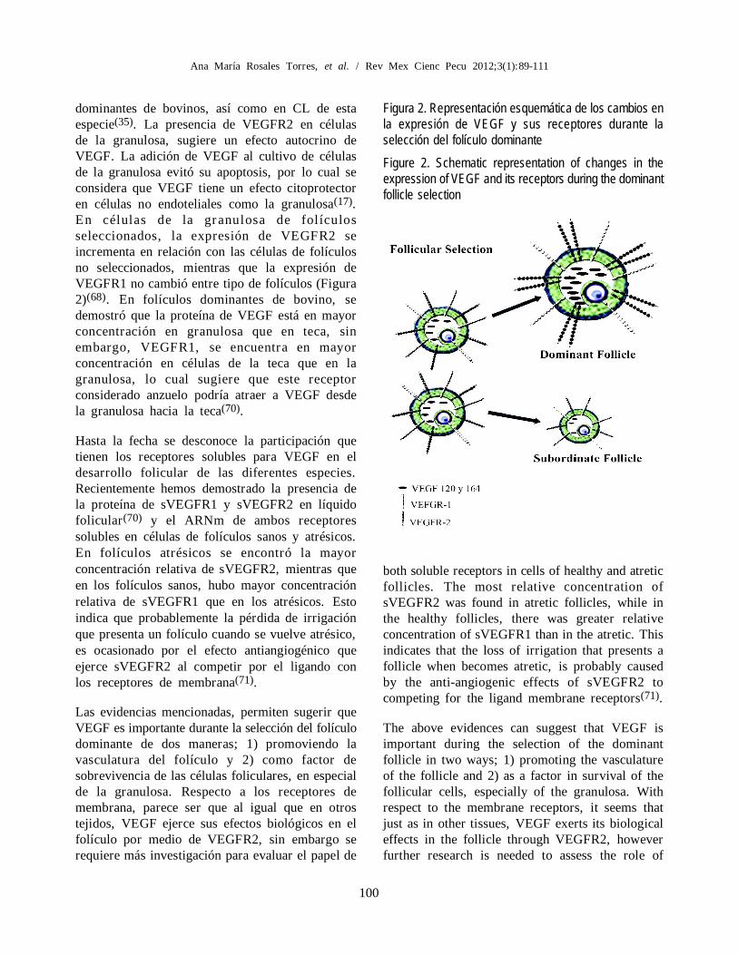
With regard to VEGF membrane receptors, it hasbeen demonstrated the presence of both receptorson thecas and granulosa cells of bovine dominantfollicles, and CL from this species(35). The presenceof VEGFR2 in granulosa cells, suggests an autocrineeffect of VEGF. The addition of VEGF to thegranulosa cells culture prevented its apoptosis, soit is considered that VEGF has a citoprotectoreffect in no endothelial cells, as the granulosa(17).In the granulosa cells of selected follicle, expressionof VEGFR2 increases in relation with the expresionin not selected follicles, whereas VEGFR1expression did not change between type of follicles(Figure 2)(68). Dominant follicles of bovines,showed that VEGF protein is in greaterconcentration in granulosa than in theca; however,VEGFR1, is more concentrated in theca than inthe granulosa cells, suggesting that this decoyreceptor might attract VEGF from the granulosatowards theca(70).
To date the participation of the soluble VEGFreceptors in the follicular development of differentspecies is unknown. We have recently demonstratedthe presence of the protein of sVEGFR1 andsVEGFR2 in follicular fluid(70) and the RNAm of
100
Ana María Rosales Torres, et al. / Rev Mex Cienc Pecu 2012;3(1):89-111
both soluble receptors in cells of healthy and atreticfollicles. The most relative concentration ofsVEGFR2 was found in atretic follicles, while inthe healthy follicles, there was greater relativeconcentration of sVEGFR1 than in the atretic. Thisindicates that the loss of irrigation that presents afollicle when becomes atretic, is probably causedby the anti-angiogenic effects of sVEGFR2 tocompeting for the ligand membrane receptors(71).
The above evidences can suggest that VEGF isimportant during the selection of the dominantfollicle in two ways; 1) promoting the vasculatureof the follicle and 2) as a factor in survival of thefollicular cells, especially of the granulosa. Withrespect to the membrane receptors, it seems thatjust as in other tissues, VEGF exerts its biologicaleffects in the follicle through VEGFR2, howeverfurther research is needed to assess the role of
dominantes de bovinos, así como en CL de estaespecie(35). La presencia de VEGFR2 en célulasde la granulosa, sugiere un efecto autocrino deVEGF. La adición de VEGF al cultivo de célulasde la granulosa evitó su apoptosis, por lo cual seconsidera que VEGF tiene un efecto citoprotectoren células no endoteliales como la granulosa(17).En células de la granulosa de fol ículosseleccionados, la expresión de VEGFR2 seincrementa en relación con las células de folículosno seleccionados, mientras que la expresión deVEGFR1 no cambió entre tipo de folículos (Figura2)(68). En folículos dominantes de bovino, sedemostró que la proteína de VEGF está en mayorconcentración en granulosa que en teca, sinembargo, VEGFR1, se encuentra en mayorconcentración en células de la teca que en lagranulosa, lo cual sugiere que este receptorconsiderado anzuelo podría atraer a VEGF desdela granulosa hacia la teca(70).
Hasta la fecha se desconoce la participación quetienen los receptores solubles para VEGF en eldesarrollo folicular de las diferentes especies.Recientemente hemos demostrado la presencia dela proteína de sVEGFR1 y sVEGFR2 en líquidofolicular(70) y el ARNm de ambos receptoressolubles en células de folículos sanos y atrésicos.En folículos atrésicos se encontró la mayorconcentración relativa de sVEGFR2, mientras queen los folículos sanos, hubo mayor concentraciónrelativa de sVEGFR1 que en los atrésicos. Estoindica que probablemente la pérdida de irrigaciónque presenta un folículo cuando se vuelve atrésico,es ocasionado por el efecto antiangiogénico queejerce sVEGFR2 al competir por el ligando conlos receptores de membrana(71).
Las evidencias mencionadas, permiten sugerir queVEGF es importante durante la selección del folículodominante de dos maneras; 1) promoviendo lavasculatura del folículo y 2) como factor desobrevivencia de las células foliculares, en especialde la granulosa. Respecto a los receptores demembrana, parece ser que al igual que en otrostejidos, VEGF ejerce sus efectos biológicos en elfolículo por medio de VEGFR2, sin embargo serequiere más investigación para evaluar el papel de
Figura 2. Representación esquemática de los cambios enla expresión de VEGF y sus receptores durante laselección del folículo dominanteFigure 2. Schematic representation of changes in theexpression of VEGF and its receptors during the dominantfollicle selection

101
IMPORTANCIA DEL FACTOR DE CRECIMIENTO DEL ENDOTELIO VASCULAR
VEGFR1, VEGFR2, sVEGFR1 and sVEGFR2 inthe follicular development.
Participation of VEGF in ovulation
Ovulation is the process by which the oocyte restartsthe meiotic activity and is released from the follicleto the oviduct to be fertilized, for this a pore in theapical wall of the follicle is created. This eventinvolves remodelling and differentiation of follicularcells to form the corpus luteum(72). For a follicleto be ovulated, it must develop from primordialfollicle until preovulatory follicle, process in whichthe oocyte, the granulosa and theca cells acquirefunctional characteristics, which allow to carry outthe ovulation(73). FSH and LH are the mainhormones responsible for stimulating the growthand maturation of the follicle. LH is the hormonethat triggers all the mechanisms of ovulation, whichinclude the proteolysis of the follicular wall for therelease of the oocyte, activation of the oocytemeiosis and luteinization of follicular cells(74).
The LH bond to its receptor in the plasmamembrane of the granulose cells, triggers, via cyclicadenosine monophosphate (cAMP), the activationof Mitogen-activated protein kinase (MAPK), alsoknown kinases regulated by extracellular signs 1and 2 (ERK1, 2); which at the nuclear level causethe decrease in the synthesis of E2 and cellproliferation, and the increase in the synthesis ofP4 by modifying the transcription and translationof esteroidogenic enzymes. Other effects caused bythe bond of LH to its receptor are the expansionof the cumulus, the rupture of the follicle stimulationand CL formation(72-75).
After the preovulatory LH peak, there is an increasein the movement toward the ovary, accompaniedby vasodilatation and increased vascular permeabilityin the preovulatory follicle. These vascular changescause edema in internal theca, which causes anedematous condition in the follicle, which persiststhrough the follicular rupture(75,76,77). Blood flowto the follicle is reduced at the apex while increasesat its base to facilitate follicular rupture(77).Induction of ovulation with LH or hCG increasesthe release of histamine from the ovary and theconcentration of eicosanoids, leukotrienes, platelets
VEGFR1, VEGFR2, sVEGFR1 y sVEGFR2 en eldesarrollo folicular.
Participación de VEGF en la ovulación
La ovulación es el proceso mediante el cual elovocito reinicia la actividad meiótica y es liberadodel folículo hacia el oviducto para ser fecundado,para esto se crea un poro en la pared apical delfolículo. Este evento implica remodelación ydiferenciación de las células foliculares para formarel cuerpo lúteo(72). Para que un folículo sea ovuladodebe desarrollarse desde folículo primordial hastafolículo preovulatorio, proceso en el cual el ovocito,las células de la granulosa y células de la tecaadquieren características funcionales que permitenllevar a cabo la ovulación(73). La FSH y LH sonlas principales hormonas encargadas de estimularel crecimiento y la maduración del folículo. La LHes la hormona que desencadena todos losmecanismos de ovulación, entre los que seencuentran la proteólisis de la pared folicular paraliberar el ovocito, la activación de la meiosis delovocito y la luteinización de las célulasfoliculares(74).
La unión de LH a su receptor en la membranaplasmática de las células de la granulosa,desencadena, por medio del adenosin monofosfatocíclico (cAMP) la activación de cinasas activadaspor mitógenos (MAPK), también conocidas comocinasas reguladas por señales extracelulares 1 y 2(ERK1, 2); las cuales a nivel nuclear ocasionan ladisminución en la síntesis de E2 y proliferacióncelular, y el incremento en la síntesis de P4 pormodificar la trascripción y traducción de enzimasesteroidogénicas. Otro de los efectos ocasionadospor la unión de LH a su receptor son la expansióndel cumulus, la estimulación de la ruptura delfolículo y la formación del CL(72-75).
Después del pico preovulatorio de LH hay unincremento de la circulación hacia el ovario que seacompaña de vasodilatación e incremento en lapermeabilidad vascular en el folículo preovulatorio.Estos cambios vasculares causan edema en la tecainterna, lo que provoca una condición edematosaen todo el folículo, la cual persiste a través de laruptura folicular(75,76,77). El flujo sanguíneo hacia

102
Ana María Rosales Torres, et al. / Rev Mex Cienc Pecu 2012;3(1):89-111
activator factor and bradiquinas, which are associatedwith vascular processes inside the follicle(78). Afterthe preovulatory LH peak, the follicular cells and theextracellular matrix of the follicular apex becomethinner, and the basement membrane is degradedby proteolysis, likewise the follicle is remodeledthrough a rapid angiogenesis and infiltration ofnew blood vessels, theca and cells of the immunesystem in the follicular antrum, while the follicularcells differentiate into luteal cells(72).
In mare’s ovulatory follicles, there is an increasein the number of blood vessels in the thecas 36 hafter treatment with hCG. It is very likely that thegonadotropins, in special LH, is responsible fornot only triggering the physiological, biochemical,hormonal and mechanical processes that triggerovulation, but also the hormone responsible tomodulate the angiogenesis in the ovulatory follicle,that somehow stop temporarily the flow of bloodin the follicle at the time of ovulation, and therebyavoiding a likely hemorrhage. There is evidence indifferent species that support this idea. In folliclesof pigs, the endothelial area and endothelialproliferation are greater in periovulatory folliclescompared with early periovulatory follicles (18 hafter hCG). However in late periovulatory follicles(36 h after hCG) the endothelial area and endothelialproliferation rate, increase to values greater orsimilar to those observed in the preovulatoryfollicles(46).
Vascular changes during ovulation, as well as inother angiogenic processes, seem to be mediatedby VEGF and its receptors. In Rhesus(80) andmacaques(81) the intra-folicular injection of sVGFR1in preovulatory follicles, disrupts ovulation andCL function. In another experiment in whichfollicular development was induced in prepuberrats with 10 IU eGC and 48 h after 20 IU of hCG,and ovaries were disected at 0,6,12,18 and 24 hafter hCG treatment, could be demonstrated thatthese hormones cause different responses in theexpression of VEGF and its receptors. In the ovariesof these animals, treatment with eCG increasedRNAm of VEGF120 VEGF164 and VEGFR1, butnot changed the expression of VEGFR2; howeverhCG has not modified the expression of RNAm of
el folículo se reduce en el ápex mientras que seincrementa en la base de éste para facilitar la rupturafolicular(77). La inducción de la ovulación con LHo hCG incrementa la liberación de histamina porparte del ovario y la concentración de eicosanoides,leucotrienos, factor activador de plaquetas ybradiquinas, las cuales están asociadas con losprocesos vasculares dentro del folículo(78). Despuésdel pico preovulatorio de LH, las capas celularesdel folículo y de la matriz extracelular del ápexfolicular se hacen más delgadas y la membranabasal es degrada por proteólisis, así mismo todo elfolículo es remodelado por medio de una rápidaangiogénesis e infiltración de nuevos vasossanguíneos, células de la teca y del sistemainmunológico en el antro folicular, mientras quelas células foliculares se diferencian en célulaslúteas(72).
En folículos ovulatorios de yeguas, después de36 h del tratamiento con hCG hay un incrementoen el número de vasos sanguíneos en las tecas(79).Es muy probable que las gonadotropinas, en especialLH, sea la responsable no sólo de desencadenarlos procesos fisiológicos, bioquímicos, hormonalesy mecánicos que disparan la ovulación, sino tambiénla hormona responsable de modular o regular laangiogénesis en el folículo ovulatorio, para que dealguna manera detenga de manera transitoria laafluencia sanguínea en el folículo al momento dela ovulación, y con ello evitar una probablehemorragia (existen evidencias bibliográficas endiferentes especies que apoyan esta idea).
En folículos de cerdos, el área endotelial y laproliferación endotelial son mayores en folículospreovulatorios en comparación con folículosperiovulatorios tempranos (18 h después de hCG).Sin embargo en folículos periovulatorios tardíos(36 h después de hCG) el área endotelial y la tasade proliferación endotelial se incrementan hastavalores mayores o similares a los observados enlos folículos preovulatorios(46).
Los cambios vasculares durante la ovulación, aligual que en otros procesos angiogénicos, parecenestar mediados por el VEGF y sus receptores. Enmono Rhesus(80) y macacos(81) la inyección intra-
103
IMPORTANCIA DEL FACTOR DE CRECIMIENTO DEL ENDOTELIO VASCULAR
the first three genes in any of the post-treatmenttimes, while RNAm of VEGFR2, significantlydecreased since 12 h pos hCG treatment(82). Thisresult is congruent if we remember that VEGFR2is the receptor of major signaling capacity, then,even when VEGF and VEGFR1 did not altered itsexpression approaching ovulation, the reduction ofVEGFR2 expression is eneough to prevent bleeding.There is other evidence supporting the above:induction of ovulation with eCG (1,250 IU) andhCG (750 IU) in sows, shows that around ovulation(18 h after hCG) protein of VEGF produced by thegranulosa and vascular area of the follicle cells arereduced in comparison with preovulatory follicles(60 h from eCG stimulation); however, at 36 hafter treatment with hCG, VEGF and vascular areaincreases again(47). In culture of human cells ofluteinized granulosa, the use of a GnRH antagonistreduces the secretion of VEGF by these cellswithout affecting the production of gonadalsteroids(83). In preovulatory bovine follicles, theRNAm expression of VEGF120, VEGF164 andVEGF188 isoforms is almost linearly reduces since4 to 25 h (near ovulation) after GnRH application,but its expression dramatically increasing in thenewly formed CL (60 h from GnRH). In this samework, there was no changes in the expression ofVEGFR1 during the first 25 h after GnRHapplication, but the expression of VEGFR2 wasreduced at this time, and increased in the newlyformed CL(84). A work done by our research group,which compared the expression of RNAm ofVEGF120, VEGF164 and receptors VEGFR1,VEGFR2 and sVEGFR1 between dominant folliclesof d 6 of the cycle with preovulatory follicles (18 hafter treatment with GnRH), revealed that theexpression of RNAm of VEGF164, VEGFR-1,VEGFR-2 was reduced significantly in preovulatoryfollicles, but increased the expression of RNAm ofsVEGFR1, which proposes that in cattle when thefollicle is approaching to ovulation, there is anantiangiogenic transitional process (greaterexpression of the soluble receptor than membranereceptors), probably in order to prevent bleedingduring ovulation(85).
It seems clear that near ovulation, there is areduction in angiogenic activity of VEGF and its
folicular de sVGFR1 en folículos preovulatorios,altera la ovulación y la función del CL. En otroexperimento en el que se indujo el desarrollofolicular a ratas pre púberes con 10 UI eGC y 48h después 20 UI de hCG, y se disecaron los ovarios0,6,12,18 y 24 h después del tratamiento con hCG,se pudo demostrar que estas hormonas provocandiferentes respuestas en la expresión de VEGF ysus receptores. En los ovarios de estos animales;el tratamiento con eCG incrementó el ARNm deVEGF120 VEGF164 y VEGFR1, pero no modificóla expresión de VEGFR2; sin embargo hCG nomodificó la expresión del ARNm de los tresprimeros genes en ninguno de los tiempospostratamiento, mientras que el ARNm deVEGFR2, disminuyó significativamente desde las12 h de la aplicación de hCG(82). Este resultado esrazonable si recordamos que VEGFR2 es el receptorcon mayor capacidad de señalización, entonces auncuando VEGF y VEGFR1 no modifiquen suexpresión al acercarse la ovulación, basta quedisminuya de manera significativa el receptor conmayor funcionalidad para que ocurra este proceso.Existen otras evidencias al respecto que sustentanlo anterior; la inducción de la ovulación con eCG(1,250 UI) y hCG (750 UI) en cerdas, muestra quealrededor de la ovulación (18 h después de hCG)la proteína de VEGF producida por las células dela granulosa y el área vascular del folículo sereducen en comparación con folículos preovulatorios(60 h de la estimulación con eCG), sin embargo,a las 36 h del tratamiento con hCG, VEGF y elárea vascular se incrementa nuevamente(47). Encultivo de células de granulosa luteinizadas dehumano, el uso de un antagonista de GnRH reducela secreción de VEGF por estas células sin afectarla producción de esteroides gonadales(83). Enfolículos preovulatorios de bovinos, la expresiónde ARNm de las isoformas 120, 164 y 188 deVEGF se reduce casi linealmente desde las 4 yhasta las 25 h (cerca de la ovulación) después dela aplicación de GnRH, incrementándosedramáticamente en el CL recién formado (60 h deGnRH). En este mismo trabajo no se observaroncambios en la expresión de VEGFR1 durante lasprimeras 25 h después de la aplicación de GnRH,pero la expresión de VEGFR2, sí se redujo a estetiempo, y se incrementó en el CL recién formado(84).
104
Ana María Rosales Torres, et al. / Rev Mex Cienc Pecu 2012;3(1):89-111
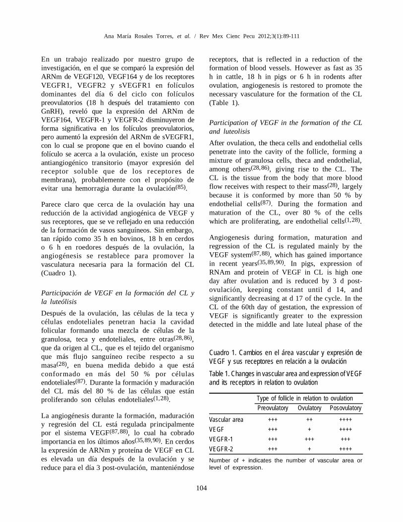
receptors, that is reflected in a reduction of theformation of blood vessels. However as fast as 35h in cattle, 18 h in pigs or 6 h in rodents afterovulation, angiogenesis is restored to promote thenecessary vasculature for the formation of the CL(Table 1).
Participation of VEGF in the formation of the CLand luteolisis
After ovulation, the theca cells and endothelial cellspenetrate into the cavity of the follicle, forming amixture of granulosa cells, theca and endothelial,among others(28,86), giving rise to the CL. TheCL is the tissue from the body that more bloodflow receives with respect to their mass(28), largelybecause it is conformed by more than 50 % byendothelial cells(87). During the formation andmaturation of the CL, over 80 % of the cellswhich are proliferating, are endothelial cells(1,28).
Angiogenesis during formation, maturation andregression of the CL is regulated mainly by theVEGF system(87,88), which has gained importancein recent years(35,89,90). In pigs, expression ofRNAm and protein of VEGF in CL is high oneday after ovulation and is reduced by 3 d post-ovulación, keeping constant until d 14, andsignificantly decreasing at d 17 of the cycle. In theCL of the 60th day of gestation, the expression ofVEGF is significantly greater to the expressiondetected in the middle and late luteal phase of the
En un trabajo realizado por nuestro grupo deinvestigación, en el que se comparó la expresión delARNm de VEGF120, VEGF164 y de los receptoresVEGFR1, VEGFR2 y sVEGFR1 en folículosdominantes del día 6 del ciclo con folículospreovulatorios (18 h después del tratamiento conGnRH), reveló que la expresión del ARNm deVEGF164, VEGFR-1 y VEGFR-2 disminuyeron deforma significativa en los folículos preovulatorios,pero aumentó la expresión del ARNm de sVEGFR1,con lo cual se propone que en el bovino cuando elfolículo se acerca a la ovulación, existe un procesoantiangiogénico transitorio (mayor expresión delreceptor soluble que de los receptores demembrana), probablemente con el propósito deevitar una hemorragia durante la ovulación(85).
Parece claro que cerca de la ovulación hay unareducción de la actividad angiogénica de VEGF ysus receptores, que se ve reflejado en una reducciónde la formación de vasos sanguíneos. Sin embargo,tan rápido como 35 h en bovinos, 18 h en cerdoso 6 h en roedores después de la ovulación, laangiogénesis se restablece para promover lavasculatura necesaria para la formación del CL(Cuadro 1).
Participación de VEGF en la formación del CL yla luteólisis
Después de la ovulación, las células de la teca ycélulas endoteliales penetran hacia la cavidadfolicular formando una mezcla de células de lagranulosa, teca y endoteliales, entre otras(28,86),que da origen al CL, que es el tejido del organismoque más flujo sanguíneo recibe respecto a sumasa(28), en buena medida debido a que estáconformado en más del 50 % por célulasendoteliales(87). Durante la formación y maduracióndel CL más del 80 % de las células que estánproliferando son células endoteliales(1,28).
La angiogénesis durante la formación, maduracióny regresión del CL está regulada principalmentepor el sistema VEGF(87,88), lo cual ha cobradoimportancia en los últimos años(35,89,90). En cerdosla expresión de ARNm y proteína de VEGF en CLes elevada un día después de la ovulación y sereduce para el día 3 post-ovulación, manteniéndose
Cuadro 1. Cambios en el área vascular y expresión deVEGF y sus receptores en relación a la ovulaciónTable 1. Changes in vascular area and expression of VEGFand its receptors in relation to ovulation
Type of follicle in relation to ovulationPreovulatory Ovulatory Posovulatory
Vascular area +++ ++ ++++VEGF +++ + ++++VEGFR-1 +++ +++ +++VEGFR-2 +++ + ++++Number of + indicates the number of vascular area orlevel of expression.

105
IMPORTANCIA DEL FACTOR DE CRECIMIENTO DEL ENDOTELIO VASCULAR
estrous cycle(91,92). Similar results have beenreported by Boomyaprakob et al(89) as regards theRNAm of VEGF in CL pigs during the 5 to 15 dof the estrous cycle; also these authors show thatthe expression of mRNA of VEGFR1, increasesgradually from d 4 to the d 15 of the cycle, whereasmRNA of VEGFR2 remains high during the first13 d of initiated oestrus and significantly reducesat d 15(89). However, in buffalo, the expression ofmRNA of VEGF and VEGFR2 does not presentmajor changes during formation, maturation andlysis of the CL, while VEGFR1 mRNA, is reducedin linear form during luteolysis(93). In cattle VEGFand VEGFR2 mRNA is higher during the formationof the CL (d 3-4 of estrous cycle) and there is areduction as the CL matures and is approachingthe luteolysis, while in VEGFR1 mRNA does notpresent changes in his expresion during the estrouscycle(35). The results analysed so far, suggest thatVEGF and VEGFR2 are the main controllers ofthe formation of new blood vessels during the CLdevelopment in pigs and cattle, while in buffaloVEGFR1 seems to be more involved in the lysisof the CL.
Intravenous treatment with a VEGF antagonist(VEGFtrap 0.25 mg/kg) in the early and middleluteal phase of macaques, reduces secretion ofprogesterone by the CL, and increases the secretionof FSH(81). In the Rhesus monkey treatment withthis same antagonist (25 mg/kg) during the middleluteal phase, reduces the weight of the ovary, theCL area and increases the activity of Caspase 3 incomparison with middle luteal phase CL withouttreatment(94). Intra-luteal injection of a VEGFantibody (8.3 mg/100 µl/injection three times aday) in cows from d 1 to 7 after ovulation, reducesthe concentration of progesterone in plasma andthe volume of the CL, however does not affect theexpression of VEGF mRNA and their membranereceptors(95). These data support the importanceof VEGF in the development and maintenance ofthe CL functionality.
With regard to the the soluble receptors sVEGFR1and sVEGFR2, there is only evidence of thepresence of mRNA of sVEGFR1 in sows CL(96),without clearity of their role. Our group’s
constante hasta día 14 y disminuyendo significati-vamente el día 17 del ciclo. En el CL del día 60 degestación, la expresión de VEGF es significativamentemayor a la expresión detectada en la fase lútea mediay tardía del ciclo estral(91,92). Resultados similareshan sido reportados por Boomyaprakob et al(89)
respecto al ARNm de VEGF en CL de cerdos durantelos días 5 al 15 del ciclo estral; además estos autoresdemuestran que la expresión de ARNm de VEGFR1,se incrementa paulatinamente del día 4 al día 15 delciclo, mientras que el ARNm de VEGFR2 se mantieneelevado durante los primeros 13 días de iniciado elestro y se reduce significativamente el día 15(89).No obstante, en búfalos, la expresión de ARNm deVEGF y VEGFR2 no presenta cambios importantesdurante la formación, maduración y lisis del CL,mientras que el ARNm de VEGFR1, se reduce enforma líneal durante la luteolisis(93). En bovinos elARNm de VEGF y VEGFR2 es elevado durante laformación del CL (día 3-4 del ciclo estral) y sereduce conforme madura y se acerca la luteolisis,mientras que en ARNm de VEGFR1 no presentacambios en su expresión a lo largo del ciclo estral(35).Los resultados analizados hasta este momento,sugieren que VEGF y VEGFR2 son los principalesencargados de controlar la formación de nuevosvasos sanguíneos durante el desarrollo del CL encerdos y bovinos, mientras que en búfalos VEGFR1parece estar más involucrado en la lisis del CL.
El tratamiento intravenoso con un antagonista deVEGF (VEGFtrap 0.25 mg/kg) en la fase lúteatemprana y media de macacos, reduce la secreción deprogesterona por el CL, e incrementa la secreción deFSH(81). En el mono titi el tratamiento con estemismo antagonista (25 mg/kg) durante la fase lúteamedia, reduce el peso del ovario, el área del CL eincrementa la actividad de caspasa 3 en comparacióncon CL de fase lútea media sin tratamiento(94). Lainyección intra-luteal de un anticuerpo contra VEGF(8.3 mg/100 µl/inyección tres veces por día) en vacasdesde el día 1 hasta el día 7 después de la ovulación,reduce la concentración de progesterona en plasma yel volumen del CL, sin embargo no se afecta laexpresión de ARNm de VEGF y la de sus receptoresde membrana(95). Estos datos sustentan la importanciade VEGF en el desarrollo y mantenimiento de lafuncionalidad del CL.
106
Ana María Rosales Torres, et al. / Rev Mex Cienc Pecu 2012;3(1):89-111
Con respecto a la los receptores solubles sVEGFR1y sVEGFR2, sólo existe la evidencia de la presenciade ARNm de sVEGFR1 en CL de ciclo estral y degestación en cerdas(96), sin que se tenga claro cuál essu función. Resultados preliminares de nuestro grupoindican que la expresión de sVEGFR1, no se modificadurante la fase lútea, en tanto que la expresión delARNm de sVEGFR2, se incrementa en la faselútea tardía con respecto a la fase lútea temprana(97).
Como se ha establecido, el sistema VEGF esimportante para la formación de la red vascular delCL que le permita ser funcional, sin embargo sedesconocen los mecanismos que regulan la expresiónde VEGF para que pueda estimular la angiogénesisdel CL. Si bien es cierto que existen evidencias dela participación de LH como moduladora de laexpresión de VEGF durante el desarrollo foliculary del CL(98-101), es ampliamente conocido queVEGF es regulado principalmente por hipoxia(28),
ya que durante la ruptura del folículo provocada enla ovulación, hay sangrado y presencia devasculatura inmadura que la propician(95) En cultivode células lúteas, la reducción en la tensión deoxigeno de 20 a 3 %, incrementa la expresión deARNm, y de la proteína de VEGF y HIF-1á desdelas 6 y hasta las 24 h después del tratamiento(102).
La regresión del CL implica una reducción en sutamaño, la cual es acompañada de la muerte de lascélulas que lo forman, incluyendo a las célulasendoteliales(103), por lo que se considera que existeuna angioregresión durante el proceso de luteolisis.En bovinos la lisis inducida del CL con PGF2-,no reduce la expresión de ARNm de VEGF yVEGFR1 durante las primeras 48 h después deltratamiento, sin embargo la expresión del ARNmde VEGFR2 se reduce paulatinamente desde las2 h después de la aplicación de PGF2-(104).Resultados similares se encontraron en ovejas, enlos cuales además se muestra que la expresión deVEGF y sus receptores se correlacionanpositivamente con la expresión de BS-1 y lectina(marcadores de células endoteliales) durante la lisisinducida del CL(105).
Todo lo anterior sugiere que el sistema VEGF esnecesario no sólo para la angiogénesis inicial del
preliminary results indicate that the expression ofsVEGFR1 does not change during the luteal phase,whereas the expression of mRNA of sVEGFR2,increases in the late luteal phase with regard to theearly luteal phase(97).
As it has been established, the VEGF system isimportant for the formation of the vascular networkof the CL that allows it to be functional; howeverthe mechanisms that regulate the expression ofVEGF to stimulate angiogenesis of the CL areunknown. Even when there is evidence that LH isinvolved in modulation of the expression of VEGFduring follicular and CL development(98-101), iswidely known that VEGF is mainly regulated byhypoxia(28). During rupture of the follicle atovulation, there is bleeding, and presence ofimmature vasculature favoring hypoxia(95). In lutealcells cultures, the reduction in the tension of oxygenfrom 20 to 3 %, increases the expression of mRNAand protein of VEGF and HIF-1á since 6 to 24 hafter treatment(102).
The CL regression implies a reduction in its size,which is accompanied by the death of its cells,including the endothelial cells(103), so there is aangioregresion during the process of luteolysis. Incattle the lysis induction of CL with PGF2-alpha,does not reduce the expression of VEGF mRNAand VEGFR1 during the first 48 h after treatment,however expression of mRNA of VEGFR2 isreduced gradually since the 2 h after PGF2-alphaapplication(104). Similar results were found in sheep,which also shows that expression of VEGF and itsreceptor were correlate positively with theexpression of BS-1 and lectin (markers ofendothelial cells) during CL regression(105).
All the above suggests that the VEGF system isnecessary not only for the initial angiogenesis ofthe CL during its development, but also for themaintenance of its function. In addition, duringCL lysis, the VEGF system seems to be destabilizedto prevent its action and promote regression of theCL. Table 2 shows changes in the relativeexpression of VEGF and its receptors during theearly, middle, late luteal phase and the regressionof the CL.

107
IMPORTANCIA DEL FACTOR DE CRECIMIENTO DEL ENDOTELIO VASCULAR
CL durante su desarrollo, sino también para elmantenimiento de su función. En adición, durantela lisis del CL el sistema VEGF parece serdesestabilizado para evitar su acción y favorecer laregresión del CL. En el Cuadro 2 se muestran loscambios en la expresión relativa de VEGF y susreceptores durante la fase lútea temprana, media,tardía y en la regresión del CL.
CONCLUSIONES
Aun cuando falta mucho por conocer sobre laparticipación del sistema VEGF en el desarrollo delos folículos, la ovulación y la formación del cuerpolúteo, lo que hasta ahora se conoce permite afirmarque los componentes de este complejo sistema,deben mantener un fino equilibrio para asegurar laintegridad y funcionamiento de las estructurasováricas. Las evidencias indican que en los procesosováricos mencionados, las mayores modificacionesen la expresión del sistema VEGF, ocurren en losreceptores de membrana y solubles, más que en elligando. Adicionalmente, es necesario seguirtrabajando para conocer con mayor profundidadlos mecanismos por los cuales los diferentescompartimentos foliculares y el CL regulan laexpresión de los componentes del sistema VEGF.
AGRADECIMIENTOS
Al Consejo nacional de Ciencia y Tecnología porel apoyo otorgado #24735.
LITERATURA CITADA
1. Tamanini C, De Ambrogi M. Angiogenesis in developing follicleand corpus luteum. Reprod Domest Anim 2004;39:206-216.
2. Kaczmarek MM, Schams D, Ziecik AJ. Role of vascularendothelial growth factor in ovarian physiology - an overview.Reprod Biol 2005;5:111-136.
3. Clark LJ, Irving-Rodgers HF, Dharmarajan AM, Rodgers RJ.Theca interna: the other side of bovine follicular atresia. BiolReprod 2004;71:1071-1078.
4. Feranil J, Isobe N, Nakao T. Apoptosis in the antral folliclesof swamp buffalo and cattle ovary: TUNEL and caspase-3histochemistry. Reprod Domest Anim 2005;40:111-116.
CONCLUSIONS
Even though much remains to learn about theparticipation of the VEGF system in the developmentof follicles, ovulation and the formation of the corpusluteum, which hitherto known allows affirm that thecomponents of this complex system, must maintaina fine balance to ensure the integrity and operationof the ovarian structures. Evidence indicates that inthe ovarian processes already mentioned, majorchanges in the expression of VEGF system occur inthe membrane receptors and soluble receptors, ratherthan on the ligand. In addition, it is necessary tocontinue to learn in greater depth the mechanisms bywhich different follicular compartments and the CLregulate the expression of VEGF system components.
ACKNOWLEDGMENTS
The National Council of science and technologyfor the given # 24735 support.
End of english version
Cuadro 2. Cambios en la expresión de VEGF y susreceptores durante el desarrollo y regresión del CLTable 2. Changes in the expression of VEGF and itsreceptors during development and regression of the CL
Phase of corpus luteum developmentEarly Medium Late Regression
VEGF ++++ ++ ++ ++VEGFR1 ++ ++ +++ ++VEGFR2 ++++ ++++ +++ +sVEGFR1 ++ ++ ++ ++sVEGFR2 ++ ++ +++ +++
Number of + indicates the level of expression.
5. Acosta TJ, Hayashi KG, Ohtani M, Miyamoto A. Local changesin blood flow within the preovulatory follicle wall and earlycorpus luteum in cows. Reproduction 2003;125:759-767.
6. Ferrara N. Role of vascular endothelial growth factor in
108
Ana María Rosales Torres, et al. / Rev Mex Cienc Pecu 2012;3(1):89-111
regulation of physiological angiogenesis. Am J Physiol CellPhysiol 2001;280:1358-1366.
7. Redmer DA, Doraiswamy V, Bortnem BJ, Fisher K, Jablonka-Shariff A, Grazul-Bilska AT, Reynolds LP. Evidence for a roleof capillary pericytes in vascular growth of the developingovine corpus luteum. Biol Reprod 2001;65:879-889.
8. Sallinen H, Anttila M, Narvainen J, Koponen J, HamalainenK, Kholova I, Heikura T, et al. Antiangiogenic gene therapywith soluble VEGFR-1, -2, and -3 reduces the growth of solidhuman ovarian carcinoma in mice. Mol Ther 2009;17:278-284.
9. Jussila L, Alita lo K. Vascular growth factors andlymphangiogenesis. Physiol Rev 2002;82:673-700.
10. Risau W. Mechanisms of angiogenesis. Nature. 1997;386:671-674.
11. Abulafia O, Sherer DM. Angiogenesis of the ovary. Am JObstet Gynecol 2000;(1 Pt 1):240-246.
12. Hellström M, Gerhardt H, Kalén M, Li X, Eriksson U, WolburgH, Betsholtz C. Lack of pericytes leads to endothelial hyperplasiaand abnormal vascular morphogenesis. J Cell Biol 2001;153:543-553.
13. Miller DL, Ortega S, Bashayan O, Basch R, Basilico C.Compensation by fibroblast growth factor 1 (FGF1) does notaccount for the mild phenotypic defects observed in FGF2 nullmice. Mol Cell Biol 2000;20:2260-2268.
14. Ferrara N, Henzel WJ. Pituitary follicular cells secrete a novelheparin-binding growth factor specific for vascular endothelialcells. Biochem Biophys Res Commun 1989;161:851-858.
15. Vieira JM, Ruhrberg C, Schwarz Q. VEGF receptor signalingin vertebrate development. Organogenesis 2010;6:97-106.
16. Petrova TV, Makinen T, Alitalo K. Signaling via vascularendothelial growth factor receptors. Exp Cell Res 1999;253:117-130.
17. Greenaway J, Connor k, Pedersen H, Coomber B, Lamarre J,Petrik J. Vascular endothelial growth factor and its receptor,Flk-1/KDR, are cytoprotective in the extravascular compartmentof the ovarian follicle. Endocrinology 2004;145:2896-2905.
18. Augustin PD. Vascular morphogenesis in the ovary. Bailliere´sClinical Obstet Gynecol;2000;(14):867-882.
19. Espinosa CR, Rosado GA. Angiogénesis en la fisiologíareproductiva. Desarrollo folicular, formación y mantenimientodel cuerpo lúteo. Ginecol Obstet Méx 2002;70:17-27.
20. Neufeld G, Cohen T, Gengrinovitch S, Poltorak Z. Vascularendothelial growth factor (VEGF) and its receptors. FASEB J.1999;3:9-22.
21. Ferrara N, Davis-Smyth T. The biology of vascular endothelialgrowth factor. Endocr Rev 1997;18:4-25.
22. Reynolds LP, Redmer DA. Expression of the angiogenic factors,basic fibroblast growth factor and vascular endothelial growthfactor, in the ovary. J Anim Sci 1998;76:1671-1681.
23. Fierro IM, Kutok JL, Serhan CN. Novel lipid mediatorregulators of endothelial cell proliferation and migration: aspirin-triggered-15R-lipoxin A(4) and lipoxin A(4). J Pharmacol ExpTher 2002;300:385-392.
24. Waltenberger J, Claesson-Welsh L, Siegbahn A, Shibuya M,Heldin CH. Different signal transduction properties of KDRand Flt1, two receptors for vascular endothelial growth factor.J Biol Chem 1994;269:26988-26995.
25. Zeng H, Dvorak HF, Mukhopadhyay D. Vascular permeabilityfactor (VPF)/vascular endothelial growth factor (VEGF)
peceptor-1 down-modulates VPF/VEGF receptor-2-mediatedendothelial cell proliferation, but not migration, throughphosphatidylinositol 3-kinase-dependent pathways. J Biol Chem2001;276:26969-26979.
26. Gerber HP, Dixit V, Ferrara N. Vascular endothelial growthfactor induces expression of the antiapoptotic proteins Bcl-2and A1 in vascular endothelia l cells. J Biol Chem1998;27:13313-13316.
27. Gerber HP, McMurtrey A, Kowalski J, Yan M, Keyt BA,Dixit V, Ferrara N. Vascular endothelial growth factor regulatesendothelial cell survival through the phosphatidylinositol 3'-kinase/Akt signal transduction pathway. Requirement for Flk-1/KDR activation. J Biol Chem 1998;273:30336-30343.
28. Fraser HM, Wulff C. Angiogenesis in the corpus luteum. ReprodBiol Endocrinol 2003;10:1:88.
29. Lee M, Hwang JT, Lee HJ, Jung SN, Kang I, Chi SG, KimSS, Ha J. AMP-activated protein kinase activity is critical forhypoxia-inducible factor-1 transcriptional activity and its targetgene expression under hypoxic conditions in DU145 cells. JBiol Chem 2003;278:39653-39661.
30. Dameron KM, Volpert OV, Tainsky MA, Bouck N. Control ofangiogenesis in fibroblasts by p53 regulation of thrombospondin-1. Science 1994;265:1582-1584.
31. Stepien HM, Kolomecki K, Pasieka Z, Komorowski J, StepienT, Kuzdak K. Angiogenesis of endocrine gland tumours—newmolecular targets in diagnostics and therapy. Eur J Endocrinol2002;146:143-51.
32. Huss WJ, Hanrahan CF, Barrios RJ, Simons JW, GreenbergNM. Angiogenesis and prostate cancer: identification of amolecular progression switch. Cancer Res 2001;61:2736-2743.
33. Jia H, Jezequel S, Löhr M, Shaikh S, Davis D, Soker S,Selwood D, Zachary I. Peptides encoded by exon 6 of VEGFinhibit endothelial cell biological responses and angiogenesisinduced by VEGF. Biochem Biophys Res Commun2001;283:164-173.
34. Otani N, Minami S, Yamoto M, Shikone T, Otani H, NishiyamaR, Otani T, Nakano R. The vascular endothelial growth factor/fms-like tyrosine kinase system in human ovary during themenstrual cycle and early pregnancy. J Clin Endocrinol Metab1999;84:3845-3851.
35. Berisha B, Schams D, Kosmann M, Amselgruber W, EinspanierR. Expression and tissue concentration of vascular endothelialgrowth factor, its receptors, and localization in the bovinecorpus luteum during estrous cycle and pregnancy. Biol Reprod2000;63:1106-1114.
36. Ferrara N. Vascular Endothelial Growth Factor: Basic Scienceand Clinical Progress. Endocrine Reviews 2004;25:581-611.
37. Elias AP, Dias S. Microenvironment changes (in pH) affectVEGF alternative splicing. Cancer Microenviron 2008;1:131-139.
38. de Vries C, Escobedo JA, Ueno H, Houck K, Ferrara N,Williams LT. The fms-like tyrosine kinase, a receptor forvascular endothelial growth factor. Science 1992;255:989-991.
39. Robinson CJ, Stringer SE. The splice variants of vascularendothelial growth factor (VEGF) and their receptors. J CellSci 2001;114:853-865.
40. Sallinen H, Anttila M, Narvainen J, Koponen J, HamalainenK, Kholova I, Heikura T, et al. Antiangiogenic gene therapywith soluble VEGFR-1, -2, and -3 reduces the growth of solidhuman ovarian carcinoma in mice. Mol Ther 2009;17:278-284.
109
IMPORTANCIA DEL FACTOR DE CRECIMIENTO DEL ENDOTELIO VASCULAR
41. Evans JLH, Ireland ME, Winn P, Lonergan GW, Smith PM.Coussens, Ireland JJ. Identification of genes involved in apoptosisand dominant follicle development during follicular waves incattle. Biol Reprod 2004;70:1475-1484.
42. Webb R, Campbell BK, Garverick HA, Gong JG, GutierrezCG, Armstrong DG. Molecular mechanisms regulating follicularrecruitment and selection. Reproduction in Domestic RuminantsIV. J Reprod Fertil 1999;54(Suppl 1):33-48.
43. Mihm M, Austin EJ. The final stages of dominant follicleselection in cattle. Domest Anim Endocrinol 2002;23:155-166.
44. Ireland JJ, Mihm M, Austin E, Diskin MG, Roche JF. Historicalperspective of turnover of dominant follicles during the bovineestrous cycle: key concepts, studies, advancements, and terms.J Dairy Sci 2000;83:1648-1658.
45. Wulff C, Wiegand SJ, Saunders PT, Scobie GA, Fraser HM.Angiogenesis during follicular development in the primate andits inhibition by treatment with truncated Flt-1-Fc (vascularendothelial growth factor Trap(A40)). Endocrinology2001;142:3244-3254.
46. Martelli A, Bernabò N, Berardinelli P, Russo V, Rinaldi C, DiGiacinto O, Mauro A, Barboni B. Vascular supply as adiscriminating factor for pig preantral follicle selection.Reproduction 2009;137:45-58.
47. Martelli A, Berardinelli P, Russo V, Mauro A, Bernabò N,Gioia L, Mattioli M, Barboni B. Spatio-temporal analysis ofvascular endothelial growth factor expression and blood vesselremodeling in pig ovarian follicles during the periovulatoryperiod. J Mol Endocrinol 2006;36:107-119.
48. Goede V, Schmidt T, Kimmina S, Kozian D, Augustin HG.Analysis of blood vessel maturation processes during cyclicovarian angiogenesis. Lab Invest 1998;78:1385-1394.
49. Tempel-Brami C, Neeman M. Non-invasive analysis of ratovarian angiogenesis by MRI. Mol Cell Endocrinol 2002;187:19-22.
50. Wulff C, Wilson H, Wiegand SJ, Rudge JS, Fraser HM.Prevention of thecal angiogenesis, antral follicular growth, andovulation in the primate by treatment with vascular endothelialgrowth factor Trap R1R2. Endocrinology 2002;143:2797-2807.
51. Tamanini C, De Ambrogi M. Angiogenesis in developing follicleand corpus luteum. Reprod Domest Anim 2004;39:206-216.
52. Macchiarelli G, Jiang JY, Nottola SA, Sato E. Morphologicalpatterns of angiogenesis in ovarian follicle capillary networks.A scanning electron microscopy study of corrosion cast. MicroscRes Tech 2006;69:459-468.
53. Greenaway J, Gentry PA, Feige JJ, LaMarre J, Petrik JJ.Thrombospondin and vascular endothelial growth factor arecyclically expressed in an inverse pattern during bovine ovarianfollicle development. Biol Reprod 2005;72:1071-1078.
54. Shimizu T, Jiang JY, Iijima K, Miyabayashi K, Ogawa Y,Sasada H, Sato E. Induction of follicular development by directsingle injection of vascular endothelial growth factor genefragments into the ovary of miniature gilts. Biol Reprod2003;69:1388-1393.
55. Grazul-Bilska AT, Navanukraw C, Johnson ML, VonnahmeKA, Ford SP, Reynolds LP, Redmer DA. Vascularity andexpression of angiogenic factors in bovine dominant follicles ofthe first follicular wave. J Anim Sci 2007;85:1914-1922.
56. Shimizu T, Iijima K, Ogawa Y, Miyazaki H, Sasada H, SatoE. Gene injections of vascular endothelial growth factor andgrowth differentiation factor-9 stimulate ovarian folliculardevelopment in immature female rats. Fertil Steril 2008;89(Suppl5):1563-1570.
57. Shimizu T, Iijima K, Miyabayashi K, Ogawa Y, Miyazaki H,Sasada H, Sato E. Effect of direct ovarian injection of vascularendothelial growth factor gene fragments on folliculardevelopment in immature female ra ts. Reproduction2007;134:677-682.
58. Quintana R, Kopcow L, Sueldo C, Marconi G, Rueda NG,Barañao RI. Direct injection of vascular endothelial growthfactor into the ovary of mice promotes follicular development.Fertil Steril 2004;82(Suppl 3):1101-1105.
59. Zimmermann RC, Xiao E, Bohlen P, Ferin M. Administrationof antivascular endothelial growth factor receptor 2 antibody inthe early follicular phase delays follicular selection anddevelopment in the rhesus monkey. Endocrinology2002;143:2496-2502.
60. Taylor PD, Wilson H, Hillier SG, Wiegand SJ, Fraser HM.Effects of inhibition of vascular endothelial growth factor attime of selection on follicular angiogenesis, expansion,development and atresia in the marmoset. Mol Hum Reprod2007;13:729-736.
61. Quintana R, Kopcow L, Marconi G, Sueldo C, Speranza G,Barañao RI. Relationship of ovarian stimulation response withvascular endothelial growth factor and degree of granulosa cellapoptosis. Hum Reprod 2001;16:1814-1818.
62. Redmer DA, Dai Y, Li J, Charnock-Jones DS, Smith SK,Reynolds LP, Moor RM. Characterization and expression ofvascular endothelial growth factor (VEGF) in the ovine corpusluteum. J Reprod Fertil 1996;108:157-165.
63. Neulen J, Raczek S, Pogorzelski M, Grunwald K, Yeo TK,Dvorak HF, Weich HA, Breckwoldt M. Secretion of vascularendothelial growth factor/vascular permeability factor fromhuman lute inized granulosa cells is human chorionicgonadotrophin dependent. Mol Hum Reprod 1998;4:203-206.
64. Barboni B, Turriani M, Galeati G, Spinaci M, Bacci ML,Forni M, Mattioli M. Vascular endothelial growth factorproduction in growing pig antral follicles. Biol Reprod2000;63(3):858-64.
65. Mattioli M, Barboni B, Turriani M, Galeati G, Zannoni A,Castellani G, Berardinelli P, Scapolo PA. Follicle activationinvolves vascular endothelial growth factor production andincreased blood vessel extension. Biol Reprod 2001;65:1014-1019.
66. Doyle LK, Walker CA, Donadeu FX. VEGF modulates theeffects of gonadotropins in granulosa cells. Domest AnimEndocrinol 2010;38:127-137.
67. Shimizu T, Miyamoto A. Progesterone induces the expressionof vascular endothelial growth factor (VEGF) 120 and Flk-1,its receptor, in bovine granulosa cells. Anim Reprod Sci2007;102:228-237.
68. Shimizu T, Jayawardana BC, Tetsuka M, Miyamoto A.Differential effect of follicle-stimulating hormone and estradiolon expressions of vascular endothelial growth factor (VEGF)120, VEGF164 and their receptors in bovine granulosa cells.J Reprod Dev 2007;53:105-112.
69. Rosales-Torres AM, Alonso I, Vergara M, Romano MC,Castillo-Juárez H, Ávalos A, Rosado A, Gutiérrez CG. Vascularendothelial growth factor isoforms 120, 164 and 205 are reducedwith atresia in ovarian follicles of sheep. Anim Reprod Sci2010;122;111-117.
70. Pinzón EC, Vergara M, Gutiérrez CG, Fierro F, Rosales AF.Factor de crecimiento endotelio vascular (VEGF) y susreceptores Flk1, Flt1,sFlk1 y sFlt1 en folículos dominantes de
110
Ana María Rosales Torres, et al. / Rev Mex Cienc Pecu 2012;3(1):89-111
bovinos [resumen]. Reunión Anual Academia de Investigaciónen Biología de la Reproducción. Universidad Veracruzana.México. 2010:25.
71. Macias RV, Vergara M, Gutiérrez CG, Fierro F, Rosales AM.Identificación de RNAm de los receptores solubles para elfactor de crecimiento del endotelio vascular (VEGF) en célulasfoliculares de bovinos [resumen]. Reunión Anual Academia deInvestigación en Biología de la Reproducción. UniversidadVeracruzana. México. 2010;22.
72. Russell DL, Robker RL. Molecular mechanisms of ovulation:co-ordination through the cumulus complex. Hum ReprodUpdate 2007;13:289-312.
73. Richards JS, Russell DL, Ochsner S, Hsieh M, Doyle KH,Falender AE, Lo YK, Sharma SC. Novel signaling pathwaysthat control ovarian follicular development, ovulation, andluteinization. Recent Prog Horm Res 2002;57:195-220.
74. Duggavathi R, Murphy BD. Development. Ovulation signals.Science 2009;324:890-891.
75. Espey LL. Current status of the hypothesis that mammalianovulation is comparable to an inflammatory reaction. Biol Reprod1994;50:233-238.
76. Espey LL. Ovulation as an inflammatory reaction—a hypothesis.Biol Reprod 1980;22:73-106.
77. Acosta TJ, Miyamoto A. Vascular control of ovarian function:ovulation, corpus luteum formation and regression. Anim ReprodSci 2004;82-83:127-140.
78. Gerdes U, Gåfvels M, Bergh A, Cajander S. Localized increasesin ovarian vascular permeability and leucocyte accumulationafter induced ovulation in rabbits. J Reprod Fertil 1992;95:539-550.
79. Kerban A, Doré M, Sirois J. Characterization of cellular andvascular changes in equine follicles during hCG-inducedovulation. J Reprod Fertil 1999;117:115-123.
80. Hazzard TM, Xu F, Stouffer RL. Injection of soluble vascularendothelial growth factor receptor 1 into the preovulatory follicledisrupts ovulation and subsequent luteal function in rhesusmonkeys. Biol Reprod 2002;67:1305-1312.
81. Fraser HM, Wilson H, Morris KD, Swanston I, Wiegand SJ.Vascular endothelial growth factor Trap suppresses ovarianfunction at all stages of the luteal phase in the macaque. J ClinEndocrinol Metab 2005;90:5811-5118.
82. Miyabayashi K, Shimizu T, Kawauchi C, Sasada H, Sato E.Changes of mRNA expression of vascular endothelial growthfactor, angiopoietins and their receptors during the periovulatoryperiod in eCG/hCG-treated immature female rats. J Exp ZoolA Comp Exp Biol 2005;303:590-597.
83. Asimakopoulos B, Nikolettos N, Nehls B, Diedrich K, Al-Hasani S, Metzen E. Gonadotropin-releasing hormoneantagonists do not influence the secretion of steroid hormonesbut affect the secretion of vascular endothelial growth factorfrom human granulosa luteinized cell cultures. Fertil Steril2006;86:636-641.
84. Berisha B, Steffl M, Welter H, Kliem H, Meyer HH, SchamsD, Amselgruber W. Effect of the luteinizing hormone surge onregulation of vascular endothelial growth factor and extracellularmatrix-degrading proteinases and their inhibitors in bovinefollicles. Reprod Fertil Dev 2008;20:258-268.
85. Méndez HJ, Vergara OM, Fierro FF, Gutiérrez CG, RosalesAM. Expresión del VEGF y sus receptores en folículosdominantes de bovino y su modificación por la aplicación delíquido folicular. Reunión Anual Academia de Investigación en
Biología de la Reproducción. Universidad Veracruzana.2010;300-309.
86. Fraser HM, Lunn SF. Regulation and manipulation ofangiogenesis in the primate corpus luteum. Reproduction2001;121:355-362.
87. Miyamoto A, Shirasuna K, Sasahara K. Local regulation ofcorpus luteum development and regression in the cow: Impactof angiogenic and vasoactive factors. Domest Anim Endocrinol2009;37:159-169.
88. Robinson RS, Woad KJ, Hammond AJ, Laird M, Hunter MG,Mann GE. Focus on vascular function in female reproductionangiogenesis and vascular function in the ovary. Reproduction2009;138;869-881.
89. Boonyaprakob U, Gadsby JE, Hedgpeth V, Routh P, AlmondGW. Expression and localization of vascular endothelial growthfactor and its receptors in pig corpora lutea during the oestrouscycle. Reproduction 2003;126:393-405.
90. Galeati G, Forni M, Spinaci M, Zannoni A, Govoni N, RibeiroLA, Seren E, Tamanini C. Fasting influences steroidogenesis,vascular endothelial growth factor (VEGF) levels and mRNAsexpression for VEGF, VEGF receptor type 2 (VEGFR-2),endothelin-1 (ET-1), endothelin receptor type A (ET-A) andendothelin converting enzyme-1 (ECE-1) in newly formed pigcorpora lutea. Domest Anim Endocrinol 2005;28:272-284.
91. Ribeiro LA, Turba ME, Zannoni A, Bacci ML, Forni M.Gelatinases, endonuclease and vascular endothelial growth factorduring development and regression of swine luteal tissue. BMCDev Biol 2006;6:58.
92. Ribeiro LA, Turba ME, Bernardini C, Zannoni A, Bacci ML,Forni M. Matrix metalloproteinases -2 and -9 in swine lutealtissue angiogenesis and angioregression. Vet Res Commun2007;31(Suppl 1):193-196.
93. Papa PC, Moura CE, Artoni LP, Fátima LA, Campos DB,Marques JE Jr, Baruselli PS, et al. VEGF system expressionin different stages of estrous cycle in the corpus luteum of non-treated and superovulated water buffalo. Domest AnimEndocrinol 2007;33:379-389.
94. Fraser HM, Wilson H, Wulff C, Rudge JS, Wiegand SJ.Administration of vascular endothelial growth factor Trap duringthe ‘post-angiogenic’ period of the luteal phase causes rapidfunctional luteolysis and selective endothelial cell death in themarmoset. Reproduction 2006;132:589-600.
95. Yamashita H, Kamada D, Shirasuna K, Matsui M, Shimizu T,Kida K, Berisha B, Schams D, Miyamoto A. Effect of localneutralization of basic fibroblast growth factor or vascularendothelial growth factor by a specific antibody on thedevelopment of the corpus luteum in the cow. Mol Reprod Dev2008;75:1449-156.
96. Kaczmarek MM, Kiewisz J, Schams D, Ziecik AJ. Expressionof VEGF-receptor system in conceptus during peri-implantationperiod and endometrial and luteal expression of soluble VEGFR-1 in the pig. Theriogenology 2009;71:1298-306.
97. Macías VR1, Guzmán SA, Vergara OM, Fierro FF, GutiérrezCG, Rosales-Torres A. Expresión de las isoformas VEGF120ay VEGF164a y sus receptores de membrana y solubles encuerpo lúteo de bovino. [Aceptado para su publicación] Reuniónde la Academia de Investigación en Biología de la Reproducción.Juriquilla, Qro, México.
98. Taylor PD, Hillier SG, Fraser HM. Effects of GnRH antagonisttreatment on follicular development and angiogenesis in theprimate ovary. J Endocrinol 2004;183:1-17.
111
IMPORTANCIA DEL FACTOR DE CRECIMIENTO DEL ENDOTELIO VASCULAR
99. Duncan WC, van den Driesche S, Fraser HM. Inhibition ofvascular endothelial growth factor in the primate ovary up-regulates hypoxia-inducible factor-1alpha in the follicle andcorpus luteum. Endocrinology 2008;149:3313-320.
100. Neulen J, Yan Z, Raczek S, Weindel K, Keck C, Weich HA,Marmé D, Breckwoldt M. Human chorionic gonadotropin-dependent expression of vascular endothelial growth factor/vascular permeability factor in human granulosa cells: importancein ovarian hyperstimulation syndrome. J Clin Endocrinol Metab1995;80:1967-1971.
101. Hazzard TM, Molskness TA, Chaffin CL, Stouffer RL. Vascularendothelial growth factor (VEGF) and angiopoietin regulationby gonadotrophin and steroids in macaque granulosa cells duringthe peri-ovulatory interval. Mol Hum Reprod 1999;5:1115-1121.
102. Nishimura R, Okuda K. Hypoxia is important for establishingvascularization during corpus luteum formation in cattle. JReprod Dev 2010;56:110-116.
103. Rosales AM. Guzmán A. Apoptosis in follicular atresia andluteal regression. Review. Téc Pecu Méx 2008;46:159-182.
104. Neuvians TP, Berisha B, Schams D. Vascular endothelial growthfactor (VEGF) and fibroblast growth factor (FGF) expressionduring induced luteolysis in the bovine corpus luteum. MolReprod Dev 2004;67:389-395.
105. Vonnahme KA, Redmer DA, Borowczyk E, Bilski JJ, LutherJS, Johnson ML, Reynolds LP, Grazul-Bilska AT. Vascularcomposition, apoptosis, and expression of angiogenic factors inthe corpus luteum during prostaglandin F2alpha-inducedregression in sheep. Reproduction 2006;131:1115-1126.