iii reunión de jóvenes investigadores en coloides e interfases abstract_j… · iii reunión de...
TRANSCRIPT
III Reunión de Jóvenes Investigadores en Coloides e Interfases
Madrid, October 13th –October 14th, 2016
Facultad de Ciencias Químicas
Universidad Complutense de Madrid

Scientific Committee
Francisco Ortega Gómez (Universidad Complutense de Madrid) Jacqueline Forcada (Universidad del País Vasco)
Miguel Ángel Cabrerizo Vílchez (Universidad de Granada) Mercedes Velázquez Salicio (Universidad de Salamanca)
María Luisa Moyá Morán (Universidad de Sevilla) Antonio Fernández Barbero (Universidad de Almería)
Jorge Pérez Juste (Universidad de Vigo) Francesc Mas Pujadas (Universidad de Barcelona)
Local Organization
Eduardo Guzmán Solís-Conference Chair Andrés Guerrero Martínez
Lionel Perrin Email: [email protected]
Venue: Facultad de Ciencias Químicas
Universidad Complutense de Madrid
Ciudad Universitaria s/n
28040-Madrid (Spain)

SPONSORS

III Reunión de Jóvenes Investigadores en Coloides e Interfases (JICI III, Madrid 2016)
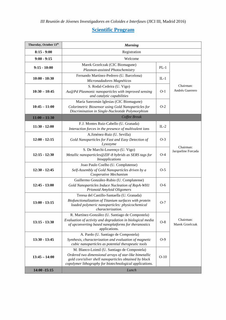
Scientific Program
Thursday, October 13th Morning
8:15 - 9:00 Registration
9:00 - 9:15 Welcome
9:15 - 10:00 Marek Grzelczak (CIC Biomagune) Plasmon-assisted Photochemistry PL-1
Chairman: Andrés Guerrero
10:00 - 10:30 Fernando Martínez-Pedrero (U. Barcelona)
Micronadadores Magnéticos IL-1
10:30 – 10:45 S. Rodal-Cedeira (U. Vigo)
Au@Pd Plasmonic nanoparticles with improved sensing and catalytic capabilities
O-1
10:45 – 11:00 María Sanromán Iglesias (CIC Biomagune)
Colorimetric Biosensor using Gold Nanoparticles for Discrimination in Single-Nucleotide Polymorphism
O-2
11:00 – 11:30 Coffee Break
11:30 - 12:00 F.J. Montes Ruiz-Cabello (U. Granada)
Interaction forces in the presence of multivalent ions IL-2
Chairman: Jacqueline Forcada
12:00 - 12:15 A.Jiménez-Ruiz (U. Sevilla)
Gold Nanoparticles for Fast and Easy Detection of Lysozyme
O-3
12:15 - 12:30 S. De Marchi-Lourenço (U. Vigo)
Metallic nanoparticles@ZIF-8 hybrids as SERS tags for bioapplications
O-4
12:30 - 12:45 Joao Paulo Coelho (U. Complutense)
Self-Assembly of Gold Nanoparticles driven by a Cooperative Mechanism
O-5
12:45 - 13:00 Guillermo González-Rubio (U. Complutense)
Gold Nanoparticles Induce Nucleation of RepA-WH1 Prionoid Amyloid Oligomers
O-6
Chairman: Marek Grzelczak
13:00 - 13:15
Teresa del Castillo-Santaella (U. Granada) Biofunctionalization of Titanium surfaces with protein
loaded polymeric nanoparticles: physicochemical characterization.
O-7
13:15 - 13:30
R. Martínez-González (U. Santiago de Compostela) Evaluation of activity and degradation in biological media
of upconverting based nanoplatforms for theranostics applications.
O-8
13:30 - 13:45 A. Pardo (U. Santiago de Compostela)
Synthesis, characterization and evaluation of magnetic cubic nanoparticles as potential therapeutic tools
O-9
13:45 – 14:00
M. Blanco-Loimil (U. Santiago de Compostela) Ordered two dimensional arrays of star-like bimetallic gold core/silver shell nanoparticles obtained by block
copolymer lithography for biotechnological applications.
O-10
14:00 -15:15 Lunch

III Reunión de Jóvenes Investigadores en Coloides e Interfases (JICI III, Madrid 2016)
Thursday, October 13th Afternoon
15:15 - 16:00 Rubén Alvárez-Asencio (IMDEA Nanoscience)
Nanotribology, Surface Interactions and Characterization: Unconventional Applications of AFM
PL-2
Chairman: Armando Maestro
16:00 - 16:30 Noemí Encinas (MPI of Polymer, Germany)
Super-liquid repellent surfaces with reduced protein, bacteria and cells adsorption
IL-3
16:30 – 16:45 Ana Mateos-Maroto (U. Complutense)
Liposomes as templates for polymer nanocapsules O-11
16:45 – 17:00 A. Aguilera-Garrido (U. Granada)
Albumin-Covered Lipid Nanocapsules Exhibit Enhanced Uptake Performance by Breast-Tumor Cells
O-12
17:00 – 17:30 Coffee Break
17:30 - 17:45 Anna May-Masnou (ICMAB-CSIC)
Au/TiO2 nanoparticles on bacterial cellulose membranes for water splitting in gas phase
O-13
Chairman: Rubén Alvárez
Asencio 17:45 – 18:00
Pablo F. Garrido (U. Santiago de Compostela) STAND: Determinación del Número de Agregación
Micelar por Tensión Superficial. O-14
18:00 – 18:15 Rubén Ahijado-Guzmán (U. Complutense)
Plasmonic Tip-to-Tip Assembled Nanorods for Enhanced Photothermal Cancer Therapy
O-15
18:15 – 18:30
S. Gómez-Graña (U. Vigo) Self-Assembled Gold Nanooctahedra Through
Microevaporators for Highly Efficient SERS-active Substrates.
O-16
Chairman:
Carlos Rey Castro
18:30 - 18:45
Yoran Beldengrün (IQAC-CSIC) Cross-Linked microgels, produced by water-in-water-
emulsions, as delivery system for enzymes O-17
18:45 – 19:00
Irene Adroher-Benítez (U. Granada) Efecto de las interacciones electroestéricas en la carga
efectiva de microgeles termosensibles: teoría y experimentos.
O-18
20:30 - ….. Social Dinner
III Reunión de Jóvenes Investigadores en Coloides e Interfases (JICI III, Madrid 2016)
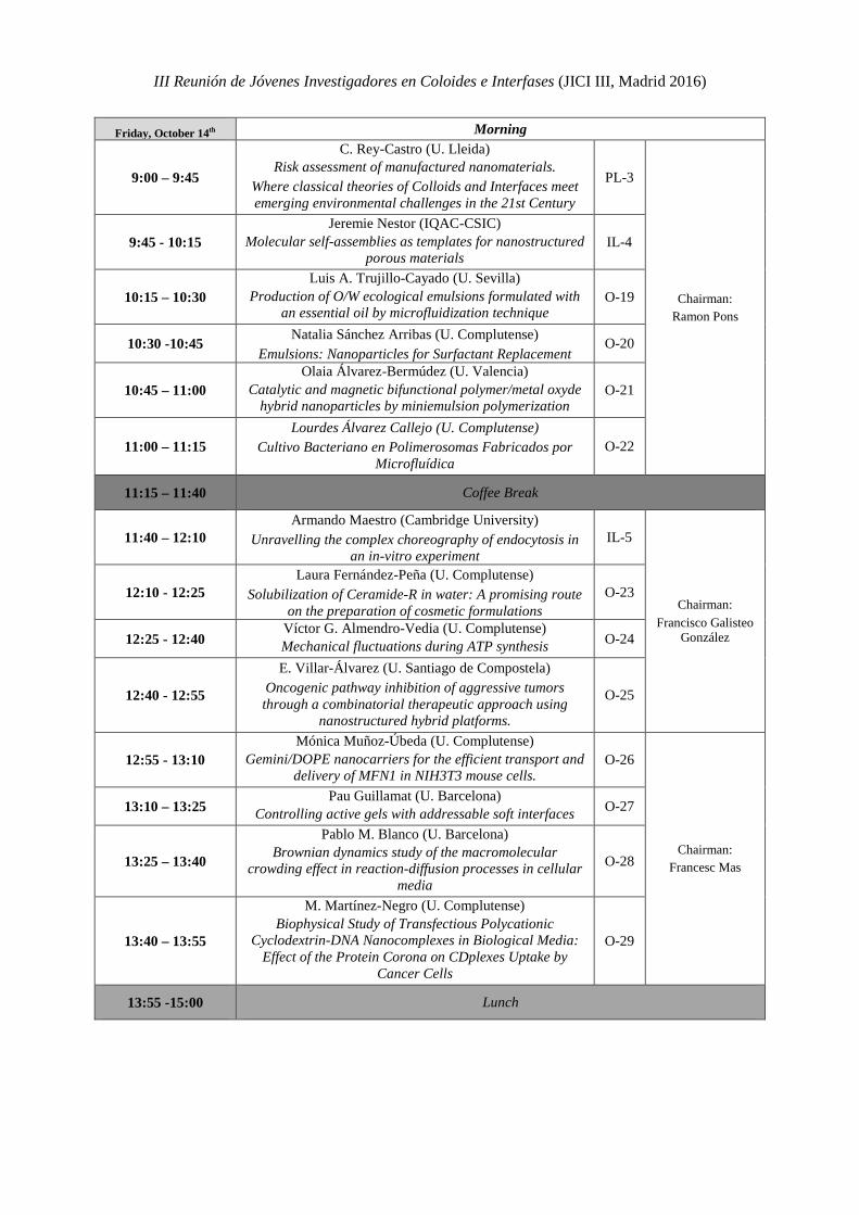
Friday, October 14th Morning
9:00 – 9:45
C. Rey-Castro (U. Lleida) Risk assessment of manufactured nanomaterials.
Where classical theories of Colloids and Interfaces meet emerging environmental challenges in the 21st Century
PL-3
Chairman: Ramon Pons
9:45 - 10:15 Jeremie Nestor (IQAC-CSIC)
Molecular self-assemblies as templates for nanostructured porous materials
IL-4
10:15 – 10:30 Luis A. Trujillo-Cayado (U. Sevilla)
Production of O/W ecological emulsions formulated with an essential oil by microfluidization technique
O-19
10:30 -10:45 Natalia Sánchez Arribas (U. Complutense) Emulsions: Nanoparticles for Surfactant Replacement
O-20
10:45 – 11:00 Olaia Álvarez-Bermúdez (U. Valencia)
Catalytic and magnetic bifunctional polymer/metal oxyde hybrid nanoparticles by miniemulsion polymerization
O-21
11:00 – 11:15 Lourdes Álvarez Callejo (U. Complutense)
Cultivo Bacteriano en Polimerosomas Fabricados por Microfluídica
O-22
11:15 – 11:40 Coffee Break
11:40 – 12:10 Armando Maestro (Cambridge University)
Unravelling the complex choreography of endocytosis in an in-vitro experiment
IL-5
Chairman: Francisco Galisteo
González
12:10 - 12:25 Laura Fernández-Peña (U. Complutense)
Solubilization of Ceramide-R in water: A promising route on the preparation of cosmetic formulations
O-23
12:25 - 12:40 Víctor G. Almendro-Vedia (U. Complutense) Mechanical fluctuations during ATP synthesis O-24
12:40 - 12:55
E. Villar-Álvarez (U. Santiago de Compostela) Oncogenic pathway inhibition of aggressive tumors through a combinatorial therapeutic approach using
nanostructured hybrid platforms.
O-25
12:55 - 13:10 Mónica Muñoz-Úbeda (U. Complutense)
Gemini/DOPE nanocarriers for the efficient transport and delivery of MFN1 in NIH3T3 mouse cells.
O-26
Chairman: Francesc Mas
13:10 – 13:25 Pau Guillamat (U. Barcelona)
Controlling active gels with addressable soft interfaces O-27
13:25 – 13:40
Pablo M. Blanco (U. Barcelona) Brownian dynamics study of the macromolecular
crowding effect in reaction-diffusion processes in cellular media
O-28
13:40 – 13:55
M. Martínez-Negro (U. Complutense) Biophysical Study of Transfectious Polycationic
Cyclodextrin-DNA Nanocomplexes in Biological Media: Effect of the Protein Corona on CDplexes Uptake by
Cancer Cells
O-29
13:55 -15:00 Lunch

III Reunión de Jóvenes Investigadores en Coloides e Interfases (JICI III, Madrid 2016)
Friday, October 14th Afternoon
15:00 - 15:15 Sergio Ángel Ortega (U. Complutense)
Reconstitución artificial mediante microfluídica de un sistema de amplificación de DNA y expresión genética
O-30
Chairman: Francisco Ortega
15:15 - 15:30 Samuel Salinas (U. Complutense)
The Mechanics of the Cell Membrane of Escherichia coli During the Division Cell Cycle
O-31
15:30 - 15:45
Lionel Perrin (U. Complutense) Effect of silver nanoparticles on the wetting and the evaporation of water sessile droplets for different
substrates
O-32
15:45 – 16:00
Javier Tajuelo (UNED) Diagrama de fases de los módulos dinámicos de
monocapas de Langmuir de ácidos grasos con distinta longitud de cadena hidrófoba.
O-33
16:00 – 16:15 David López Díaz (U. Salamanca)
Óxidos de Grafeno: una familia de materiales con propiedades modulables.
O-34
Chairman:
Ramón G. Rubio
16:15 – 16:30 Leonor Pérez-Fuentes (U. Granada)
Behaviour of milk allergenic proteins at hydrophobic interfaces.
O-35
16:30 - 16:45 Andrew Akanno (U. Complutense)
Interactions in Polyelectrolyte-Surfactant Mixtures in Bulk and at the Air-Water Interface
O-36
16:45 - 17:00 J.M. Pages-Casas (U. Barcelona) Electric field control of phoretic nematic colloids O-37
17:00 – 17:30 Coffe Break
17:30 - 17:45 V. Domínguez (U. Santiago de Compostela)
Effect of Cationic Gemini Surfactant on DPPC Liposomes O-38 Chairman:
David López Díaz 17:45 – 18:00
F. J. Ostos Formation of surfactant-cyclodextrins host-guest
complexes. Effect of the inclusion of a functional group at the end of the surfactant tail
O-39
18:00 - ….. Concluding Remarks
PLENARY LECTURES

III Reunión de Jóvenes Investigadores en Coloides e Interfases (JICI III, Madrid 2016) Plenary Lectures PL-1
Plasmon-assisted Photochemistry
Marek Grzelczak
1. CIC biomaGUNE, Paseo de Miramón 182, 20009 Donostia - San Sebastían, Spain. 2. Ikerbasque, Basque Foundation for Science, 48013 Bilbao, Spain
[email protected] Spurred by outstanding optical properties, metallic nanoparticles gain progressive attention as photocatalysts.1 The large absorption cross-sections and chemical stability allow metallic nanoparticles efficiently harvest light over the UV-Vis-NIR spectral range. The light, while interacting with metallic nanoparticles invokes the surface plasmon resonance that decaying provokes enhancement of the optical near field, generation of hot carriers, or increase local temperature.2 As a consequence, the rate of the chemical reactions increases with the proximity to particles surface independently on the mechanistic pathway.
We disscuss here the use of gold nanoparticles with multiple shapes (rods, cube, stars) and surface functionalization (Pd, Pt) toward photoregeneration of relevant biomolecules - cofactors, in particular, nicotinamide adenine dinucleotide - using visible and IR light.3,4 The photocatalytic activity depends not only on the degree of the shape anisotropy but also on the spatial distribution of co-catalyst on the particle surface. Plasmonic nanoparticles exhibit excellent activity either deposited in the form of plasmonic films on the glass substrate or distributed homogenously in the three-dimensional hydrogel matrix.
Acknowledgements This work was supported by the Spanish Ministry of Economy and Competitiveness (MAT2013-49375-EXP), and BBVA Foundation – “Primera convocatoria de ayudas fundación BBVA a investigadores, innovadores y creadores culturales”. References 1 M. L. Brongersma, N. J. Halas and P. Nordlander, Nature Nanotech., 10, 25–34, 2015 2 G. Baffou and R. Quidant, Chem. Soc. Rev., 43, 3898–3907, 2014 3 A. Sánchez-Iglesias, A. Chuvilin and M. Grzelczak, Chem. Commun., 51, 5330–5333, 2015 4 A. Sánchez-Iglesias, J. Barroso, D. Martinez-Solis, J. M. T. Varela, F. O. Basteiro, V. Pavlov, A. Chuvilin and
M. Grzelczak, J. Mater. Chem. A, 4, 7045 – 7052, 2016
III Reunión de Jóvenes Investigadores en Coloides e Interfases (JICI III, Madrid 2016) Plenary Lectures PL-2
Nanotribology, Surface Interactions and Characterization: Unconventional Applications of AFM
Álvarez-Asencio R.1,*, Luengo G. S.2, Carlos M. Pina3 and Rutland M.W.4 1. Instituto Madrileño de Estudios Avanzados, IMDEA Nanociencia, Madrid, Spain
2. L’Oréal Research and Innovation, Aulnay-Sous-Bois, France 3. Departamento de Cristalografía y Mineralogía, Facultad de Ciencias Geológicas, Universidad
Complutense de Madrid, Madrid, Spain 4. School of Chemical Science and Engineering, Department of Chemistry, KTH Royal
Institute of Technology, Stockholm, Sweden *[email protected]
When two surfaces achieve contact, then contact phenomena such as adhesion, friction and wear can occur, which are of great interest in many disciplines, including physics, physical chemistry, material chemistry, and life and health sciences. These phenomena are largely determined by the nature and magnitude of the surface forces such as van der Waals, capillary and hydration forces. Moreover these forces are length-dependent, and therefore when the system scales down, their contribution scales up, dominating the interaction between the surfaces.
The goal of our research work is to investigate fundamental contact phenomena in terms of the surface forces that regulate their properties. The primary tool applied is the atomic force microscopy (AFM), which (with all of its sub-techniques) offers the possibility to study such forces with high resolution virtually between all types of materials and intervening media.
One of our interests is to understand the long ranged interactions presented in air between different industrially relevant materials and how these interactions are shielded when the systems are immersed in an ionic liquid.[1] We have also investigated by AFM nanomechanical surface mapping, the influence of the microstructure on adhesion and corrosion initiation of a FeCrVN tool alloy.[2] The mechanical properties of stratum corneum (SC), which is the outermost layer of the skin, were also of interest in our research. A novel probe has been designed with a single hair fibre in order to understand how the skin deforms locally in response to the interaction with such a fibre probe.[3] An important achievement in our work is the development of a new AFM technique - tribological property mapping. This technique provides friction coefficient and contact adhesion maps with information that can be attributed to surface microstructure.[4] Finally, I will present our latest work where we used friction force microscopy to study friction anisotropy of a semiconductor organic polymer crystal and its relation to its surface microstructure. References 1. Álvarez-Asencio R., Cranston E. D., Atkin R., Rutland M.W. Langmuir 2012, 28:9967-9976. 2. Alvarez-Asencio R., Sababi M., Pan J., Ejnermark S., Ekman L., Rutland MW. Corrosion Science 2014, 89:236-241. 3. Alvarez-Asencio R., Wallquist V., Kjellin M., Rutland M. W., Camacho A., Nordgren N., Luengo G. S. Journal of the Mechanical Behavior of Biomedical Materials 2016, 54:185-193. 4. Alvarez-Asencio R., Pan J., Thormann E., Rutland M. W. Tribology Letters 2013, 50:387-395.
III Reunión de Jóvenes Investigadores en Coloides e Interfases (JICI III, Madrid 2016) Plenary Lectures PL-3
Risk assessment of manufactured nanomaterials. Where classical theories of Colloids and Interfaces meet emerging
environmental challenges in the 21st Century
C. Rey-Castro 1,*, C.A. David1, F. Mas2, J.Galceran1, and J. Puy1
1. Departament de Química and AGROTECNIO. Universitat de Lleida (Spain) 2. Department of Materials Science and Physical Chemistry & Research Institute of Theoretical and
Computational Chemistry (IQTCUB) of Barcelona University (UB), Barcelona (Spain) *contact e-mail: [email protected]
The advances and developments achieved in nanotechnology during the last years are leading to an outstanding growth in the amount of commercial products based on engineered nanomaterials (NMs) that are currently arriving to the market. This fact is pushing EU governments and agencies to adapt their regulations on human health and environmental risk assessment of chemical substances (such as REACH) to the specific characteristics of NMs. Due to the virtually infinite number of possible combinations of chemical composition, core and surface structure, size, shape, polydispersity, etc. of NMs, its classification and regulation remains a big challenge. Several questions arise such as: How should we define NMs from a regulatory point of view? Are the existing guidelines for toxicological testing of “conventional” chemicals still valid for NMs? How should we define the dose of NM in these tests? To what extent are the current models for environmental fate and behaviour of pollutants usable for NMs? What does “NM solubility” mean? The answer to these and similar questions is still controversial, due to the structural and compositional complexity of NMs. The role of Physical Chemists is particularly valuable in this field, thanks to the substantial body of knowledge on the properties of colloidal and interfacial systems accumulated over the last century. The purpose of this talk is to show a few examples where classical theories (Smoluchowski, Stokes, Ostwald-Freundlich, Langmuir, etc.) are useful in the development of models for the risk assessment of NMs, illustrated with experimental data from ZnO NMs obtained in our lab over the last years [1-6]. Acknowledgements The authors thank support from European Union Seventh Framework Programme (FP7-NMP.2012.1.3-3) under grant agreement no. 310584 (NANoREG), and from the Spanish Ministry of Education and Science (Projects CTM2012-39183 and CTM2013-48967). References [1] C. David, J. Galceran, C. Rey-Castro, J. Puy, et al., J. Phys. Chem. C, 116 (2012) 11758. [2] Q. S. Mu, C. A. David, J. Galceran, C. Rey-Castro, et al., Chem. Res. Toxicol., 27 (2014) 558. [3] J. Galceran, M. Lao, C. David, E. Companys, C. Rey-Castro, et al., J. Electroanal. Chem., 722-723 (2014) 110. [4] N. Adam, C. Schmitt, J. Galceran, E. Companys, et al., Nanotoxicology, 8 (2014) 709. [5] R. Tantra, H. Bouwmeester, E. Bolea, C. Rey-Castro, C. A. David et al., Nanotoxicology, 10 (2016) 173-184. [6] R. Tantra, H. Bouwmeester, E. Bolea, C. Rey-Castro, C. A. David et al., Chapter 5. Solubility. In Nanomaterial Characterization: An Introduction. R. Tantra (Editor). John Wiley & Sons, 2016.


INVITED LECTURES

III Reunión de Jóvenes Investigadores en Coloides e Interfases (JICI III, Madrid 2016) Invited Lectures IL-1
Micronadadores Magnéticos
Fernando Martínez-Pedrero1,2*, Andrejs Cebers3, Ignacio Pagonabarraga1,2, Pietro Tierno1,2
1.Departamento de Física de la Materia Condensada,Universidad de Barcelona, Barcelona, España. 2. Institut de Nanociencia i Nanotecnologia,IN2UB, Universidad de Barcelona, Barcelona, España.
3. Facultad de Física y Matemáticas,Universidad de Letonia, Riga, Letonia. *[email protected]
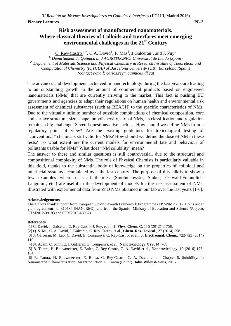
El control del movimiento de micropartículas que se hallan sumergidas en un medio es un campo aun por explorar. A estas escalas, las fuerzas inerciales son despreciables en comparación con las viscosas, y las ecuaciones de Navier-Stokes son reversibles en el tiempo, lo que obliga a incluir algún tipo de asimetría o flexibilidad en el sistema si lo que se quiere es inducir el desplazamiento de las micropartículas. En este trabajo presentamos diferentes nadadores magnéticos, constituidos por partículas paramagnéticas y ferromagnéticas, sujetos a campos constantes, oscilatorios o rotantes, y que se propulsan aprovechando la proximidad de diferentes interfases. Estos sistemas, que se disponen en diferentes geometrías, permiten una manipulación precisa de la materia a esta escala, lo que resulta fundamental en diferentes aplicaciones biológicas o microfluidicas.
Figure 1. "Gusano" constituido por 6 partículas superparamagnéticas, sujetas a un campo rotatorio, elípticamente polarizado, que se traslada sobre una superficie de vidrio en presencia de micropartículas de sílice.
Acknowledgements F.M.P. y P.T. agradecen la financiación proveniente del Consejo Europeo de Investigación (Proyecto número 335040). A.C. agradece la financiación proveniente del Programa Nacional de Investigación No. 2014.10-4/VPP- 3/21. I.P agradece la financiación proveniente del Proyecto FIS2011-22603 de MINECO (España). References [1] Martinez-Pedrero, F., Ortiz-Ambriz, A., Pagonabarraga I., Tierno P., Phys. Rev. Lett. 115, 138301, 2015. [2] Martinez-Pedrero, F., Tierno P., Phys. Rev. Applied 3, 051003, 2015. [3] Martinez-Pedrero, F., Cebers A., Tierno P., Soft Matter, 12, 3688-3695, 2016.
III Reunión de Jóvenes Investigadores en Coloides e Interfases (JICI III, Madrid 2016) Invited Lectures IL-2
Interaction forces in the presence of multivalent ions
F.J. Montes Ruiz-Cabello*
1Departament of Applied Physics,University of Granada, Granada, Spain *[email protected]
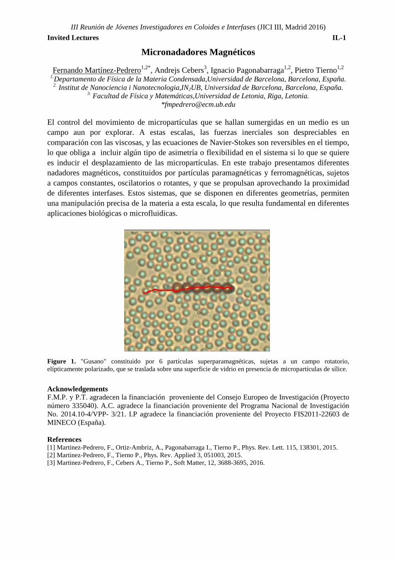
Interaction forces in the presence of multivalent ions have been a subject of strong interest for the interesting phenomena revealed in these systems. This problem has been widely studied from the theoretical and computational point of view. However, experimentally it has not been much addressed mainly due to technical limitations and the difficulty to apply existing theories to analyze and quantify the experimental force profiles. In this presentation I will show our contribution to this field by the study of direct forces measurement between similar and dissimilar colloidal particles in the presence of multivalent counterions and coions1, 2, 3. These measurements were carried out with the colloidal probe technique based on AFM. Our results point out that the force profiles can be quantified with the classical Poisson Boltzmann theory down to few nanometers of separation distances. At shorter distances, we observed additional attractive forces not captured by the DLVO theory which origin might be attributed to ion-ion correlations or patch charge interactions.
Figure 1. Interaction forces at different salt concentrations of K4FeCN6 between two oppositely charged latex particles and their prediction based on DLVO theory. A charge reversal of the positive particle is evidenced from these force profile upon salt addition. These predictions were calculated from the independent fittings of the two symmetric (identical particles) force profiles. Acknowledgements This research was supported by the projects: MAT2014-60615R funded by MICINN, the project P12-FQM-1443 funded by “Junta de Andalucía” References 1. Sinha, P.; Szilagyi, I.; Montes Ruiz-Cabello, F. J.; Maroni, P.; Borkovec, M. J. Phys. Chem. Lett. 2013, 4, 648-652. 2. Montes Ruiz-Cabello, F. J.; Moazzami-Gudarzi, M.; Elzbieciak-Wodka, M.; Maroni, P.; Labbez, C.; Borkovec, M.; Trefalt, G. Soft Matter 2015, 11, 1562-71. 3. Montes Ruiz-Cabello, F. J.; Trefalt, G.; Maroni, P.; Borkovec, M. Langmuir 2014, 30, 4551-4555.
III Reunión de Jóvenes Investigadores en Coloides e Interfases (JICI III, Madrid 2016) Invited Lectures IL-3
Super-liquid repellent surfaces with reduced protein, bacteria and cells adsorption.
Noemí Encinas 1,*, Maxime Paven1, Lars Schmüser1, Daniel Graham2, David G. Castner2,
Tobias Weidner1, Doris Vollmer1, Hans Jürgen Butt1
1.Physics at Interfaces Department,Max Planck Institute for Polymer Research, Mainz 1. Department of Chemical Engineering, University of Washington, Seattle,USA
*[email protected] Biofouling denotes the contamination related to the accumulation of microorganisms attached to a solid surface [1] and can develop thick layers within a few hours on nearly every surface. Biofouling causes economic losses in the order of billions of euro (reduced efficiency on heat transfer systems, fuel consumption due to friction on ship hulls or corrosion of pipelines) [2]. Furthermore, it is one of the major causes of nosocomial infections and mortality in the world when associated to food packaging, or water reservoirs or medical devices [3, 4]. Cells inside a biofilm are enclosed in an extracellular polymeric matrix that preserves against hostile environments, increasing the antibiotic resistance to a factor of 103 compared to the free-floating cells [5]. Thus, avoiding the attachment of the first layer, biofilm formation will be delayed or even prevented.
Our approach is focused in the use of super-liquid repellent surfaces characterized by a mobile fluid layer between the liquid and solid interfaces. These coatings structured on the micro- or nanometer scale and tuned chemistry by functionalizing with perfluorinated groups can offer a route to prevent bacterial attachment and biofilm growth [6,7]. We evaluated the adsorption of proteins (bovine albumin and complex human serum plasma) on superamphiphobic and superhydrophobic surfaces through X-ray photoelectron spectroscopy (XPS), time-of-flight secondary ion mass spectrometry (ToF-SIMS) and laser scanning confocal microscopy (LSCM). The ease of fabrication and low detected concentrations in the range of 4 ng/cm2 make these surfaces attractive candidates for biomaterials design.
Acknowledgements N.E. thanks the Marie Skłodowska-Curie fellowship 660523-NoBios-ESR. L.S. and T.W. thank supporting from the Max Planck Graduate Center with the Johannes Gutenberg-Universität Mainz (MPGC), the European Commission (CIG grant #322124) and the Deutsche Forschungsgemeinschaft (WE4478/4-1) for financial support. HJB wishes to thank the ERC for the advanced grant 340391-SUPRO. DJG and DGC were supported by grant EB-002027 from the United States National Institutes of Health. References [1] Costerton, J. W.; Stewart, P. S.; Greenberg, E. P. Science 284, 1318-1322, 1999. [2] Melo L.F; Bott T.R. Exp Therm Fluid Sci 14, 375–381, 1997 [3] Klevens, R. M. et al.; Public Health Rep 122, 160-166, 2007. [4] Davies D. Nat. Rev. Drug Discov. 2, 114, 2003. [5] Stewart P.S., Costerton W.J. The Lancet. 358, 135-138, 2001. [6] Deng X., Mammen L., Butt H.-J., Vollmer D., Science 335, 67-70, 2012. [7] Paven M., Papadopoulos P., Schöttler S., Deng X., Mailänder V., Vollmer D., Butt H.-J., Nature Communication 4, 2013.
III Reunión de Jóvenes Investigadores en Coloides e Interfases (JICI III, Madrid 2016) Invited Lectures IL-4
Molecular self-assemblies as templates for nanostructured porous materials
Jeremie Nestor*, Jordi Esquena, Conxita Solans Institute for Advanced Chemistry of Catalonia, IQAC-CSIC, and CIBER de Bioingeniería,
Biomateriales y Nanomedicina (CIBER-BBN), c/Jordi Girona, 18-26, Barcelona, Spain *[email protected]
In the past decades, molecular self-assembly has attracted considerable attention to the academia and industry as a tool for the bottom up synthesis of nanostructured materials. Studies have shown how a well-designed system can be used as a very efficient soft template for the preparation of advanced nanostructured materials, such as hierarchically organized porous materials, controlling surface area and pore size.
Highly concentrated emulsions (HIPE) stabilized by a liquid crystalline phase is an excellent template for nanostructured materials. HIPE are characterized by an internal phase volume fraction larger than 0.74, which is the maximum packing of monodisperse undistorted spherical droplets. Consequently, these emulsions have a compact foam-like structure, which consist in deformed and/or polydispersed droplets, separated by a thin film of continuous phase. In our previous studies, silica porous materials were obtained by hydrolysing tetraethyl orthosilicate (TEOS) in the external phase of O/W highly concentrated emulsions, where the external phase was a liquid crystal. However, ethanol released by TEOS hydrolysis produced emulsion instability and also obstructed the formation of ordered mesopores [1].
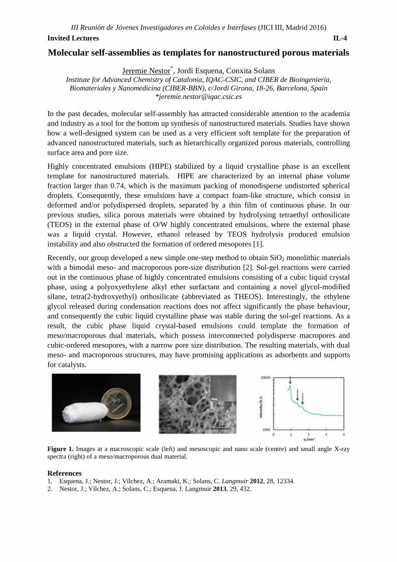
Recently, our group developed a new simple one-step method to obtain SiO2 monolithic materials with a bimodal meso- and macroporous pore-size distribution [2]. Sol-gel reactions were carried out in the continuous phase of highly concentrated emulsions consisting of a cubic liquid crystal phase, using a polyoxyethylene alkyl ether surfactant and containing a novel glycol-modified silane, tetra(2-hydroxyethyl) orthosilicate (abbreviated as THEOS). Interestingly, the ethylene glycol released during condensation reactions does not affect significantly the phase behaviour, and consequently the cubic liquid crystalline phase was stable during the sol-gel reactions. As a result, the cubic phase liquid crystal-based emulsions could template the formation of meso/macroporous dual materials, which possess interconnected polydisperse macropores and cubic-ordered mesopores, with a narrow pore size distribution. The resulting materials, with dual meso- and macroporous structures, may have promising applications as adsorbents and supports for catalysts.
Figure 1. Images at a macroscopic scale (left) and mesoscopic and nano scale (centre) and small angle X-ray spectra (right) of a meso/macroporous dual material. References 1. Esquena, J.; Nestor, J.; Vílchez, A.; Aramaki, K.; Solans, C. Langmuir 2012, 28, 12334. 2. Nestor, J.; Vílchez, A.; Solans, C.; Esquena, J. Langmuir 2013, 29, 432.
III Reunión de Jóvenes Investigadores en Coloides e Interfases (JICI III, Madrid 2016) Invited Lectures IL-5
Unravelling the complex choreography of endocytosis in an in-vitro experiment
Armando Maestro*
Cavendish Laboratory, University of Cambridge, JJ Thompson Avenue, CB3 0EH Cambridge, UK CMR Cambridge Institute for Medical Research, Hills Rd, CB2 0XY, Cambridge, UK
Clathrin-mediated endocytosis is the main mechanism by which proteins are controllably removed from the plasma membrane, and is thus vital to cellular life. It is a beautiful example of controlled molecular choreography, involving scales of components from the molecular (10’s Å) to the vesicle (100’s nm), and long-range processes of membrane-mediated interaction also coming into play. This complexity has up to now prevented a full understanding of this key cellular process, despite the fact that very precise knowledge exists on specific aspects of molecular detail. In-vitro experiments have been very insightful so far, but remain lacking a physical description of this collective mechanism that relies on protein aggregation and self-assembly coupled to membrane processes that involve non-equilibrium thermodynamic conditions. Our final goal is to disentangle the molecular mechanism by which the recruitment of clathrin by the membrane is triggered by the binding of endocytic adaptor proteins to particular lipid domains: the ones constituted by phosphatidyl inositol PtdIns4,5P2. By fluorescence microscopy and surface experiments monitoring the lateral pressure of the lipid monolayer, we addressed in a controlled in-vitro experiment how the uptake and change of conformation of adaptor proteins, concretely the heterotetrameric AP2 complex, that triggers the recruitment of clathrin is regulated by the asymmetry in the distribution of PiP2 in the plane of the monolayer. Besides, by exploring the relaxation response after applying an external deformation (for instance, shear) the dynamical behaviour related to the creation of a clathrin coat has been identified.


ORAL COMMUNICATIONS

TOPIC 1: NANOPARTICLES

III Reunión de Jóvenes Investigadores en Coloides e Interfases (JICI III, Madrid 2016) Nanoparticles O-1
Au@Pd Plasmonic nanoparticles with improved sensing and catalytic capabilities.
S. Rodal-Cedeira1*, V. Montes-García1, M. De Clercq2, H. Heidari2, S. Bals2,
L. Polavarapu1, I. Pastoriza-Santos1, J. Pérez-Juste1
1Departamento de Química Física, Universidade de Vigo, Campus Universitario, Vigo, Spain. 2EMAT, University of Antwerp, Groenenborgerlaan 171, B-2020 Antwerp, Belgium.
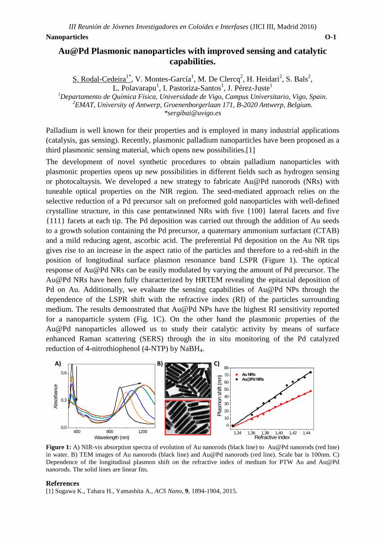
*[email protected] Palladium is well known for their properties and is employed in many industrial applications (catalysis, gas sensing). Recently, plasmonic palladium nanoparticles have been proposed as a third plasmonic sensing material, which opens new possibilities.[1] The development of novel synthetic procedures to obtain palladium nanoparticles with plasmonic properties opens up new possibilities in different fields such as hydrogen sensing or photocaltaysis. We developed a new strategy to fabricate Au@Pd nanorods (NRs) with tuneable optical properties on the NIR region. The seed-mediated approach relies on the selective reduction of a Pd precursor salt on preformed gold nanoparticles with well-defined crystalline structure, in this case pentatwinned NRs with five {100} lateral facets and five {111} facets at each tip. The Pd deposition was carried out through the addition of Au seeds to a growth solution containing the Pd precursor, a quaternary ammonium surfactant (CTAB) and a mild reducing agent, ascorbic acid. The preferential Pd deposition on the Au NR tips gives rise to an increase in the aspect ratio of the particles and therefore to a red-shift in the position of longitudinal surface plasmon resonance band LSPR (Figure 1). The optical response of Au@Pd NRs can be easily modulated by varying the amount of Pd precursor. The Au@Pd NRs have been fully characterized by HRTEM revealing the epitaxial deposition of Pd on Au. Additionally, we evaluate the sensing capabilities of Au@Pd NPs through the dependence of the LSPR shift with the refractive index (RI) of the particles surrounding medium. The results demonstrated that Au@Pd NPs have the highest RI sensitivity reported for a nanoparticle system (Fig. 1C). On the other hand the plasmonic properties of the Au@Pd nanoparticles allowed us to study their catalytic activity by means of surface enhanced Raman scattering (SERS) through the in situ monitoring of the Pd catalyzed reduction of 4-nitrothiophenol (4-NTP) by NaBH4.
Figure 1: A) NIR-vis absorption spectra of evolution of Au nanorods (black line) to Au@Pd nanorods (red line) in water. B) TEM images of Au nanorods (black line) and Au@Pd nanorods (red line). Scale bar is 100nm. C) Dependence of the longitudinal plasmon shift on the refractive index of medium for PTW Au and Au@Pd nanorods. The solid lines are linear fits. References [1] Sugawa K., Tahara H., Yamashita A., ACS Nano, 9, 1894-1904, 2015.
400 800 12000,0
0,3
0,6
Abso
rban
ce
Wavelength (nm)1,34 1,36 1,38 1,40 1,42 1,44
0
10
20
30
40
50
60
70
80
Au NRs Au@Pd NRs
Plas
mon
shi
ft (n
m)
Refractive index
A) B) C)
III Reunión de Jóvenes Investigadores en Coloides e Interfases (JICI III, Madrid 2016) Nanoparticles O-2
Colorimetric Biosensor using Gold Nanoparticles for Discrimination in Single-Nucleotide Polymorphism
María Sanromán Iglesias1,2*, Charles Lawrie2, Marek Grzelczak1,3, Luis M. Liz Marzán1,3
1CIC biomaGUNE, Paseo de Miramón 182, 20009 Donostia-San Sebastián, Spain 2Oncology Area, Biodonostia Research Institute, Donostia-San Sebastián, Spain
3Ikerbasque, Basque Foundation for Science, 48013 Bilbao, Spain *[email protected]
Single-nucleotide polymorphism (SNP) is a random replacement of a nucleotide in a given genetic location that occurs in human genome at every few hundred of bases across the genome. These replacements alter functioning of proteins, leading to cancer, cardiovascular or neurodegenerative diseases. Therefore, the ability of sensitive detection of specific SNPs has considerable value in diagnosis, prediction of patient’s responses to treatments, and risk of relapse of diseases. LSPR-based detection methods offer some significant advantages: applicability to a wide range of analytes, ease of use, elimination of the use of toxic organic solvents, point-of-care applications, as well as high sensitivity in the detection of some biological species [1].
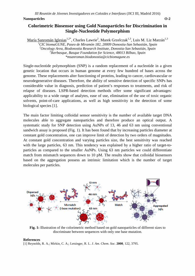
The main factor limiting colloidal sensor sensitivity is the number of available target DNA molecules able to aggregate nanoparticles and therefore produce an optical output. A systematic study for SNP detection using AuNPs of 13, 46 and 63 nm using conventional sandwich assay is proposed (Fig. 1). It has been found that by increasing particles diameter at constant gold concentration, one can improve limit of detection by two orders of magnitudes. At constant gold concentration and varying particles size, the best sensitivity was reached with the large particles, 63 nm. This tendency was explained by a higher ratio of target-to-particles as compared to the smaller AuNPs. Using 63 nm particles we could differentiate match from mismatch sequences down to 10 pM. The results show that colloidal biosensors based on the aggregation possess an intrinsic limitation which is the number of target molecules per particles.
Fig. 1: Illustration of the colorimetric method based on gold nanoparticles of different sizes to
discriminate between sequences with only one base mutation.
References [1] Reynolds, R. A.; Mirkin, C. A.; Letsinger, R. L. J. Am. Chem. Soc. 2000, 122, 3795.
III Reunión de Jóvenes Investigadores en Coloides e Interfases (JICI III, Madrid 2016) Nanoparticles O-3
Gold Nanoparticles for Fast and Easy Detection of Lysozyme
A.Jiménez-Ruiz1,*, R. Prado-Gotor1, J. Carnerero1, P. Castillo1
1 Departamento de Química Física, Universidad de Sevilla, 41012 Sevilla,España *[email protected]
Colorimetric and spectroscopic properties of gold nanoparticles have been known for the best part of a century, since a fast and reliable synthetic route giving rise to stable colloids was developed by J. Turkevich and coworkers in the 1950s.1,2 Turkevich’s citrate-capped gold nanoparticles, stabilized by electrostatic repulsion forces and devoid from any functionalization still remain a staple in the field. The original synthetic route would be streamlined in the coming years in order to better control the size, shape and stability of the resulting colloid, but the core steps would remain the same.
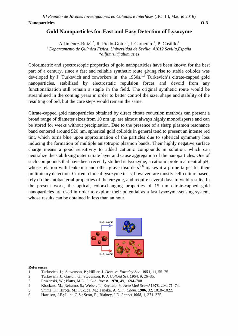
Citrate-capped gold nanoparticles obtained by direct citrate reduction methods can present a broad range of diameter sizes from 10 nm up, are almost always highly monodisperse and can be stored for weeks without precipitation. Due to the presence of a sharp plasmon resonance band centered around 520 nm, spherical gold colloids in general tend to present an intense red tint, which turns blue upon approximation of the particles due to spherical symmetry loss inducing the formation of multiple anisotropic plasmon bands. Their highly negative surface charge means a good sensitivity to added cationic compounds in solution, which can neutralize the stabilizing outer citrate layer and cause aggregation of the nanoparticles. One of such compounds that have been recently studied is lysozyme, a cationic protein at neutral pH, whose relation with leukemia and other grave disorders3–6 makes it a prime target for their preliminary detection. Current clinical lysozyme tests, however, are mostly cell-culture based, rely on the antibacterial properties of the enzyme, and require several days to yield results. In the present work, the optical, color-changing properties of 15 nm citrate-capped gold nanoparticles are used in order to explore their potential as a fast lysozyme-sensing system, whose results can be obtained in less than an hour.
References 1. Turkevich, J.;. Stevenson, P.; Hillier, J. Discuss. Faraday Soc. 1951, 11, 55–75. 2. Turkevich, J.; Garton, G.; Stevenson, P. J. Colloid Sci. 1954, 9, 26–35. 3. Pruzanski, W.; Platts, M.E. J. Clin. Invest. 1970, 49, 1694–708. 4. Klockars, M.; Reitamo, S.; Weber, T.; Kerttula, Y. Acta Med Scand 1978, 203, 71–74. 5. Shima, K.; Hirota, M.; Fukuda, M.; Tanaka, A. Clin. Chem. 1986, 32, 1818–1822. 6. Harrison, J.F.; Lunt, G.S.; Scott, P.; Blainey, J.D. Lancet 1968, 1, 371–375.
III Reunión de Jóvenes Investigadores en Coloides e Interfases (JICI III, Madrid 2016) Nanoparticles O-4
Metallic nanoparticles@ZIF-8 hybrids as SERS tags for bioapplications.
S. De Marchi-Lourenço1, I. Pastoriza-Santos1,*and J. Pérez-Juste1,* 1. Colloid Chemistry Group. Dpto de Química Física, Facultade de Química,
Universidade de Vigo, Spain * [email protected], [email protected]
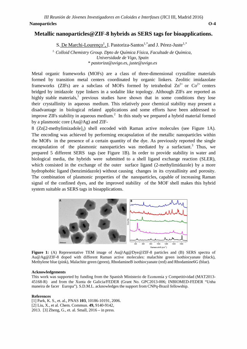
Metal organic frameworks (MOFs) are a class of three-dimensional crystalline materials formed by transition metal centers coordinated by organic linkers. Zeolitic imidazolate frameworks (ZIFs) are a subclass of MOFs formed by tetrahedral Zn2+ or Co2+ centers bridged by imidazole type linkers in a sodalite like topology. Although ZIFs are reported as highly stable materials,1 previous studies have shown that in some conditions they lose their crystallinity in aqueous medium. This relatively poor chemical stability may present a disadvantage in biological related applications and some efforts have been addressed to improve ZIFs stability in aqueous medium.2 In this study we prepared a hybrid material formed by a plasmonic core (Au@Ag) and ZIF- 8 (Zn[2-methylimizadole]2) shell encoded with Raman active molecules (see Figure 1A). The encoding was achieved by performing encapsulation of the metallic nanoparticles within the MOFs in the presence of a certain quantity of the dye. As previously reported the single encapsulation of the plasmonic nanoparticles was mediated by a surfactant.3 Thus, we prepared 5 different SERS tags (see Figure 1B). In order to provide stability in water and biological media, the hybrids were submitted to a shell ligand exchange reaction (SLER), which consisted in the exchange of the outer surface ligand (2-methylimidazole) by a more hydrophobic ligand (benzimidazole) without causing changes in its crystallinity and porosity. The combination of plasmonic properties of the nanoparticles, capable of increasing Raman signal of the confined dyes, and the improved stability of the MOF shell makes this hybrid system suitable as SERS tags in bioapplications.
Figure 1: (A) Representative TEM image of Au@Ag@Dye@ZIF-8 particles and (B) SERS spectra of Au@Ag@ZIF-8 doped with different Raman active molecules: malachite green isothiocyanate (black), Methylene blue (pink), Malachite green (green), RhodamineB isothiocyanate (red) and Rhodamine6G (blue).
Acknowledgements This work was supported by funding from the Spanish Ministerio de Economı́a y Competitividad (MAT2013-45168-R) and from the Xunta de Galicia/FEDER (Grant No. GPC2013-006; INBIOMED-FEDER “Unha maneira de facer Europa”). S.D.M.L. acknowledges the support from CNPq-Brazil fellowship.
References [1] Park, K. S., et. al., PNAS 103, 10186-10191, 2006. [2] Liu, X., et al. Chem. Commun. 49, 9140-9142, 2013. [3] Zheng, G., et. al. Small, 2016 – in press.
III Reunión de Jóvenes Investigadores en Coloides e Interfases (JICI III, Madrid 2016) Nanoparticles O-5
Self-Assembly of Gold Nanoparticles driven by a Cooperative Mechanism
Joao Paulo Coelho1, Gloria Tardajos1, Vladimir Stepanenko2, Alexander Roedle3, Gustavo Fernández3, and Andrés Guerrero-Martínez*1
1Departamento de Química Física I, Universidad Complutense de Madrid, Avda. Complutense s/n 28040 Madrid, Spain.
2 Institut fur Organishe Chemie and Center of Nanosystems Chemistry, University of Würzburg, Am Hubland, 97074 Würzburg, Germany.
3 Organisch-Chemisches Institut, Westfälische Wilhelms-Universität Münster,Corrensstrasse 40, 48149 Münster, Germany.
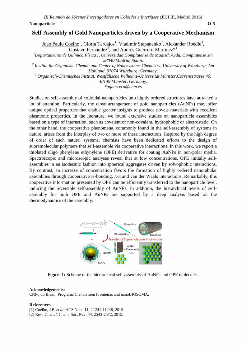
Studies on self-assembly of colloidal nanoparticles into highly ordered structures have attracted a lot of attention. Particularly, the close arrangement of gold nanoparticles (AuNPs) may offer unique optical properties that enable greater insights to produce novels materials with excellent plasmonic properties. In the literature, we found extensive studies on nanoparticle assemblies based on a type of interaction, such as covalent or non-covalent, hydrophobic or electrostatic. On the other hand, the cooperative phenomena, commonly found in the self-assembly of systems in nature, arises from the interplay of two or more of these interactions. Inspired by the high degree of order of such natural systems, chemists have been dedicated efforts to the design of supramolecular polymers that self-assemble via cooperative interactions. In this work, we report a thiolated oligo phenylene ethynylene (OPE) derivative for coating AuNPs in non-polar media. Spectroscopic and microscopic analyses reveal that at low concentrations, OPE initially self-assembles in an isodesmic fashion into spherical aggregates driven by solvophobic interactions. By contrast, an increase of concentration favors the formation of highly ordered nanotubular assemblies through cooperative H-bonding, π-π and van der Waals interactions. Remarkably, this cooperative information presented by OPE can be efficiently transferred to the nanoparticle level, inducing the reversible self-assembly of AuNPs. In addition, the hierarchical levels of self-assembly for both OPE and AuNPs are supported by a deep analysis based on the thermodynamics of the assembly.
Figure 1: Scheme of the hierarchical self-assembly of AuNPs and OPE molecules.
Acknowledgements: CNPq do Brasil, Programa Ciencia sem Fronteiras and nanoBIOSOMA. References [1] Coelho, J.P. et al. ACS Nano 11, 11241-11248, 2015. [2] Rest, C. et al. Chem. Soc. Rev. 44, 2543-2572, 2015.
III Reunión de Jóvenes Investigadores en Coloides e Interfases (JICI III, Madrid 2016) Nanoparticles O-6
Gold Nanoparticles Induce Nucleation of RepA-WH1 Prionoid Amyloid Oligomers
Guillermo González-Rubio1,2 , Cristina Fernández3, Judith Langer2, Gloria Tardajos1,
Luis M. Liz-Marzán2,4,5, Rafael Giraldo*,3, and Andrés Guerrero-Martínez*,1 1Departamento de Química Física I Universidad Complutense de Madrid Avda. Complutense s/n,
E28040, Madrid, Spain. 2BioNanoPlasmonics Laboratory CIC biomaGUNE Donostia - San Sebastián E20009
3Department of Cellular and Molecular Biology Centro de Investigaciones Biológicas – CSIC Madrid E28040, Spain.
4Ikerbasque Basque Foundation for Science. Bilbao, E48013, Spain. 5Biomedical Research Networking Center in Bioengineering, Biomaterials, and Nanomedicine,
CIBER-BBN, Spain. *[email protected]/[email protected]
Protein amyloidogenesis is a hot topic in protein science due to the role of amyloid
aggregates, especially oligomers, in the etiology of a number of devastating human degenerative diseases.1 However, the mechanisms that determine the formation of amyloid oligomers remain elusive due to the high complexity of the amyloidogenesis process.2 The unique properties of metal nanoparticles offer an alternative not fully explored yet for the study of such diseases. We functionalized gold nanorods with the metal chelating group ANTACo as a way to immobilize the soluble dimer of hexa-histidine tagged (H6) RepA-WH1 protein, a model synthetic bacterial prionoid, and induce their oligomerization on the surface of the AuNRs. In a physically separated event such oligomers are able to induce the growth of amyloid fibers in the presence of untagged soluble RepA-WH1. SERS spectra of H6-RepA-WH1 obtained with spherical citrate-AuNP as Raman enhancers provide evidence for structural changes in the protein compatible with a progressive increase in β-sheet, as expected for amyloidogenesis.3
Acknowledgements Research at RG laboratory was supported by MINECO grants CSD2009-00088 and BIO2012-30852. Research at AGM and LMLM laboratories was supported by MINECO (MAT2014- 59678-R and MAT2013-46101-R), Madrid Regional Government (S2013/MIT-2807) and the “I Convocatoria de Ayudas Fundación BBVA a Investigadores, Innovadores y Creadores Culturales (14_CBB_147)” grants. AGM and GGR acknowledge, respectively, receipt of Ramón y Cajal and FPI Fellowships from the Spanish MINECO. References [1] Westermark, P.; Benson, M. D.; Buxbaum, J. N.; Cohen, A. S.; Frangione, B.; Ikeda, S.; Masters, C. K.;
Merlini, G.; Saraiva, M. J.; Sipe, J. D. Amyloid, 9, 197–200, 2002 [2] Eichner, T.; Radford, S. E. Mol. Cell, 43, 8–18, 2011. [3] González-Rubio, G.; Fernández, C.; Langer, J.; Tardajos, G.; Liz-Marzán, L. M.; Giraldo, R., Guerrero-Martínez, A. Submitted (2016)
III Reunión de Jóvenes Investigadores en Coloides e Interfases (JICI III, Madrid 2016) Nanoparticles O-7
Biofunctionalization of Titanium surfaces with protein loaded polymeric nanoparticles: physicochemical characterization.
Teresa del Castillo-Santaella1,*, José Manuel Peula-García2, Inmaculada Ortega-Oller3,
Miguel Padial-Molina3, Pablo Galindo-Moreno3, Ana Belén Jódar-Reyes1 1 Departament of Applied Physics,University of Granada, Granada, Spain 2 Departament of Applied Physics II,University of Málaga, Málaga, Spain
3 Department of Oral Surgery and Implant Dentistry, University of Granada, Granada, Spain. *[email protected]
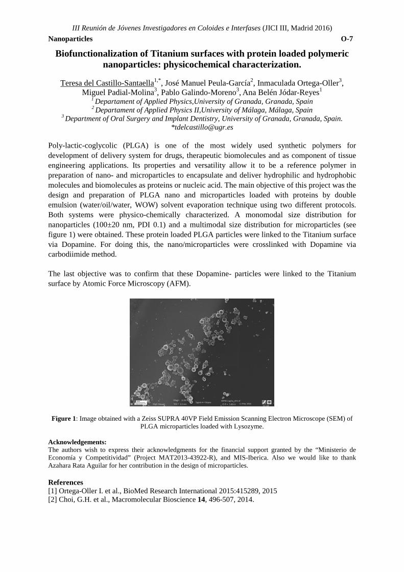
Poly-lactic-coglycolic (PLGA) is one of the most widely used synthetic polymers for development of delivery system for drugs, therapeutic biomolecules and as component of tissue engineering applications. Its properties and versatility allow it to be a reference polymer in preparation of nano- and microparticles to encapsulate and deliver hydrophilic and hydrophobic molecules and biomolecules as proteins or nucleic acid. The main objective of this project was the design and preparation of PLGA nano and microparticles loaded with proteins by double emulsion (water/oil/water, WOW) solvent evaporation technique using two different protocols. Both systems were physico-chemically characterized. A monomodal size distribution for nanoparticles (100±20 nm, PDI 0.1) and a multimodal size distribution for microparticles (see figure 1) were obtained. These protein loaded PLGA particles were linked to the Titanium surface via Dopamine. For doing this, the nano/microparticles were crosslinked with Dopamine via carbodiimide method.
The last objective was to confirm that these Dopamine- particles were linked to the Titanium surface by Atomic Force Microscopy (AFM).
Figure 1: Image obtained with a Zeiss SUPRA 40VP Field Emission Scanning Electron Microscope (SEM) of PLGA microparticles loaded with Lysozyme.
Acknowledgements: The authors wish to express their acknowledgments for the financial support granted by the “Ministerio de Economía y Competitividad” (Project MAT2013-43922-R), and MIS-Iberica. Also we would like to thank Azahara Rata Aguilar for her contribution in the design of microparticles. References [1] Ortega-Oller I. et al., BioMed Research International 2015:415289, 2015 [2] Choi, G.H. et al., Macromolecular Bioscience 14, 496-507, 2014.
III Reunión de Jóvenes Investigadores en Coloides e Interfases (JICI III, Madrid 2016) Nanoparticles O-8
Evaluation of activity and degradation in biological media of upconverting based nanoplatforms for theranostics applications.
R. Martínez-González*, A. Pardo, E. Villar-Álvarez, M. Blanco-Loimil, P. Taboada, S. Barbosa.
Grupo de Física de Coloides y Polímeros, Departamento de Física de la Materia Condensada, Universidade de Santiago de Compostela, Santiago de Compostela 15782, Spain
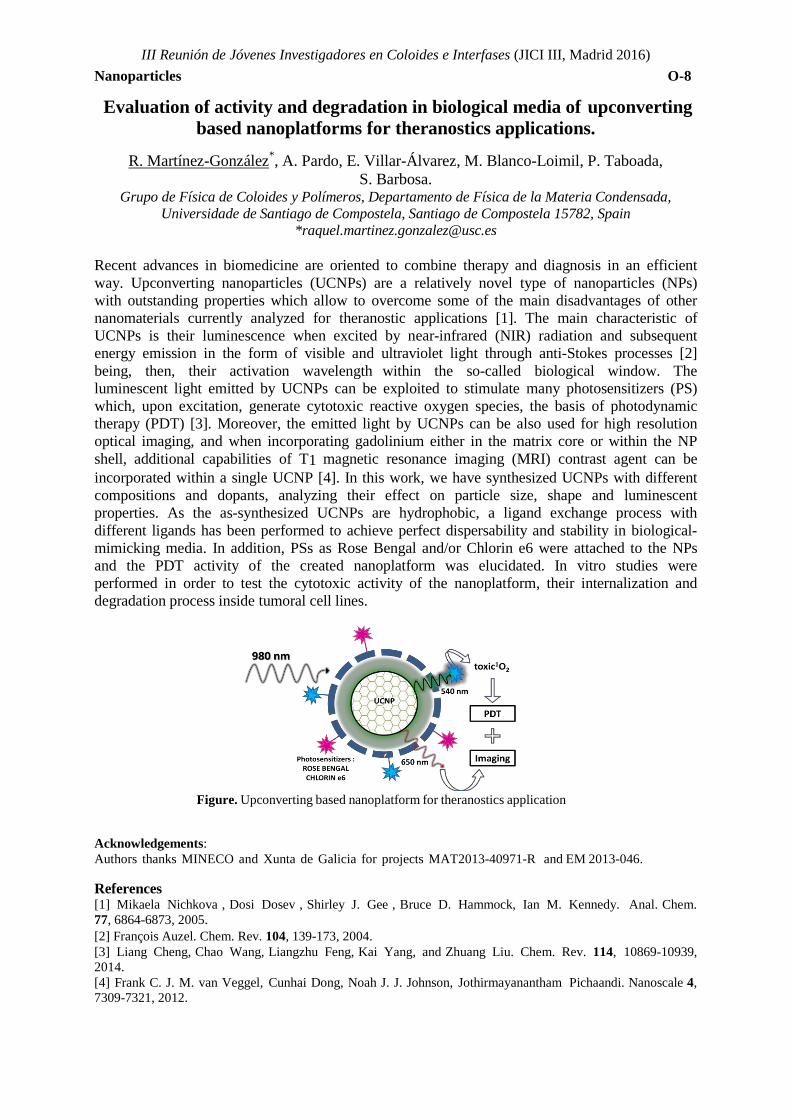
*[email protected] Recent advances in biomedicine are oriented to combine therapy and diagnosis in an efficient way. Upconverting nanoparticles (UCNPs) are a relatively novel type of nanoparticles (NPs) with outstanding properties which allow to overcome some of the main disadvantages of other nanomaterials currently analyzed for theranostic applications [1]. The main characteristic of UCNPs is their luminescence when excited by near-infrared (NIR) radiation and subsequent energy emission in the form of visible and ultraviolet light through anti-Stokes processes [2] being, then, their activation wavelength within the so-called biological window. The luminescent light emitted by UCNPs can be exploited to stimulate many photosensitizers (PS) which, upon excitation, generate cytotoxic reactive oxygen species, the basis of photodynamic therapy (PDT) [3]. Moreover, the emitted light by UCNPs can be also used for high resolution optical imaging, and when incorporating gadolinium either in the matrix core or within the NP shell, additional capabilities of T1 magnetic resonance imaging (MRI) contrast agent can be incorporated within a single UCNP [4]. In this work, we have synthesized UCNPs with different compositions and dopants, analyzing their effect on particle size, shape and luminescent properties. As the as-synthesized UCNPs are hydrophobic, a ligand exchange process with different ligands has been performed to achieve perfect dispersability and stability in biological- mimicking media. In addition, PSs as Rose Bengal and/or Chlorin e6 were attached to the NPs and the PDT activity of the created nanoplatform was elucidated. In vitro studies were performed in order to test the cytotoxic activity of the nanoplatform, their internalization and degradation process inside tumoral cell lines.
Figure. Upconverting based nanoplatform for theranostics application
Acknowledgements: Authors thanks MINECO and Xunta de Galicia for projects MAT2013-40971-R and EM 2013-046. References [1] Mikaela Nichkova , Dosi Dosev , Shirley J. Gee , Bruce D. Hammock, Ian M. Kennedy. Anal. Chem. 77, 6864-6873, 2005. [2] François Auzel. Chem. Rev. 104, 139-173, 2004. [3] Liang Cheng, Chao Wang, Liangzhu Feng, Kai Yang, and Zhuang Liu. Chem. Rev. 114, 10869-10939, 2014. [4] Frank C. J. M. van Veggel, Cunhai Dong, Noah J. J. Johnson, Jothirmayanantham Pichaandi. Nanoscale 4, 7309-7321, 2012.
III Reunión de Jóvenes Investigadores en Coloides e Interfases (JICI III, Madrid 2016) Nanoparticles O-9
Synthesis, characterization and evaluation of magnetic cubic nanoparticles as potential therapeutic tools
A. Pardo*, R. Martínez-González, E. Villar-Ávarez, M. Blanco-Loimil, S. Barbosa,
P. Taboada and V. Mosquera
Grupo de Física de Coloides y Polímeros, Departamento de Física de la Materia Condensada, Universidad de Santiago de Compostela, Santiago de Compostela 15782, Spain.
In last decades, magnetic nanoparticles (MNPs) have been intensively studied due to their potential technological and biomedical applications [1]. Many of the synthetic techniques used to obtain magnetic nanoparticles have serious limitations in terms of costs and versatility, being thermal decomposition [2] one of the most robust and reproducible methods to obtain MNPs with a high crystallinity while simultaneously achieving a great control over their shape and size.
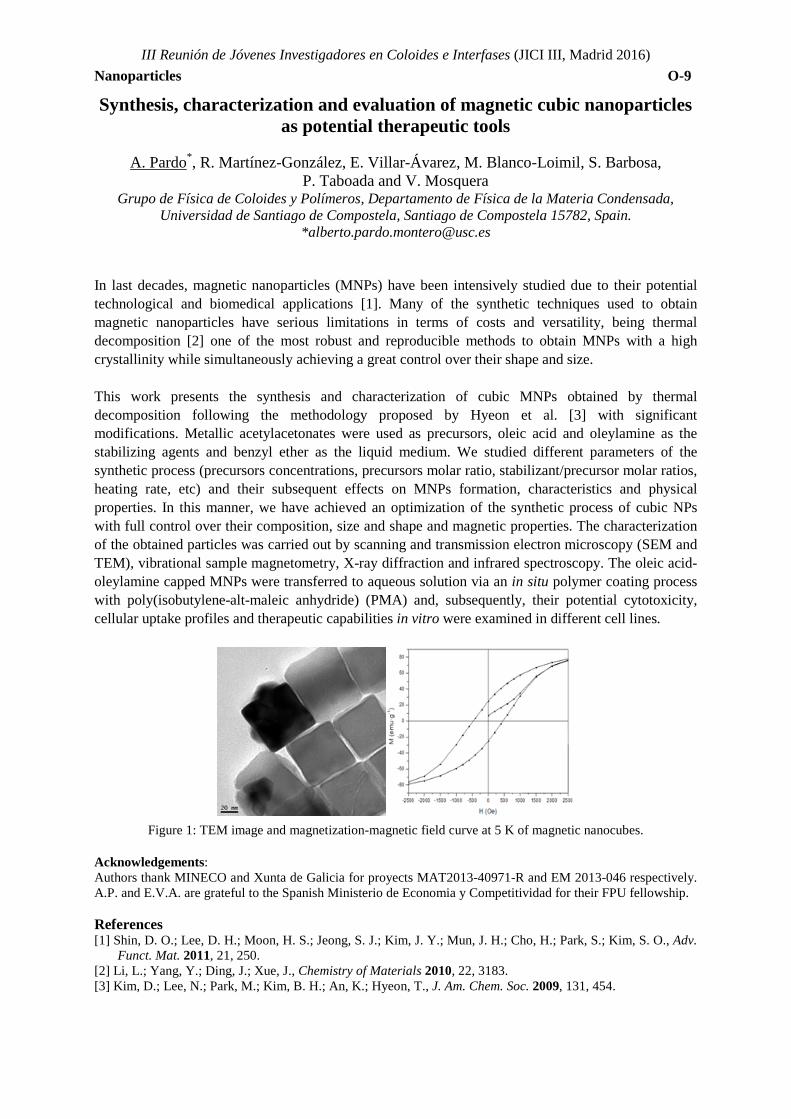
This work presents the synthesis and characterization of cubic MNPs obtained by thermal decomposition following the methodology proposed by Hyeon et al. [3] with significant modifications. Metallic acetylacetonates were used as precursors, oleic acid and oleylamine as the stabilizing agents and benzyl ether as the liquid medium. We studied different parameters of the synthetic process (precursors concentrations, precursors molar ratio, stabilizant/precursor molar ratios, heating rate, etc) and their subsequent effects on MNPs formation, characteristics and physical properties. In this manner, we have achieved an optimization of the synthetic process of cubic NPs with full control over their composition, size and shape and magnetic properties. The characterization of the obtained particles was carried out by scanning and transmission electron microscopy (SEM and TEM), vibrational sample magnetometry, X-ray diffraction and infrared spectroscopy. The oleic acid-oleylamine capped MNPs were transferred to aqueous solution via an in situ polymer coating process with poly(isobutylene-alt-maleic anhydride) (PMA) and, subsequently, their potential cytotoxicity, cellular uptake profiles and therapeutic capabilities in vitro were examined in different cell lines.
Figure 1: TEM image and magnetization-magnetic field curve at 5 K of magnetic nanocubes.
Acknowledgements: Authors thank MINECO and Xunta de Galicia for proyects MAT2013-40971-R and EM 2013-046 respectively. A.P. and E.V.A. are grateful to the Spanish Ministerio de Economia y Competitividad for their FPU fellowship. References [1] Shin, D. O.; Lee, D. H.; Moon, H. S.; Jeong, S. J.; Kim, J. Y.; Mun, J. H.; Cho, H.; Park, S.; Kim, S. O., Adv.
Funct. Mat. 2011, 21, 250. [2] Li, L.; Yang, Y.; Ding, J.; Xue, J., Chemistry of Materials 2010, 22, 3183. [3] Kim, D.; Lee, N.; Park, M.; Kim, B. H.; An, K.; Hyeon, T., J. Am. Chem. Soc. 2009, 131, 454.
III Reunión de Jóvenes Investigadores en Coloides e Interfases (JICI III, Madrid 2016) Nanopartículas O-10
Ordered two dimensional arrays of star-like bimetallic gold core/silver shell nanoparticles obtained by block copolymer lithography for biotechnological
applications.
M. Blanco-Loimil1,*, R. Martínez-González1, A. Pardo1, E. Villar-Álvarez1, S. Barbosa1, P. Taboada1, V. Mosquera1.
1. Grupo de Física de Coloides y Polímeros, Departamento de Física de la Materia Condensada, Universidad de Santiago de Compostela, Santiago de Compostela 15782, Spain.
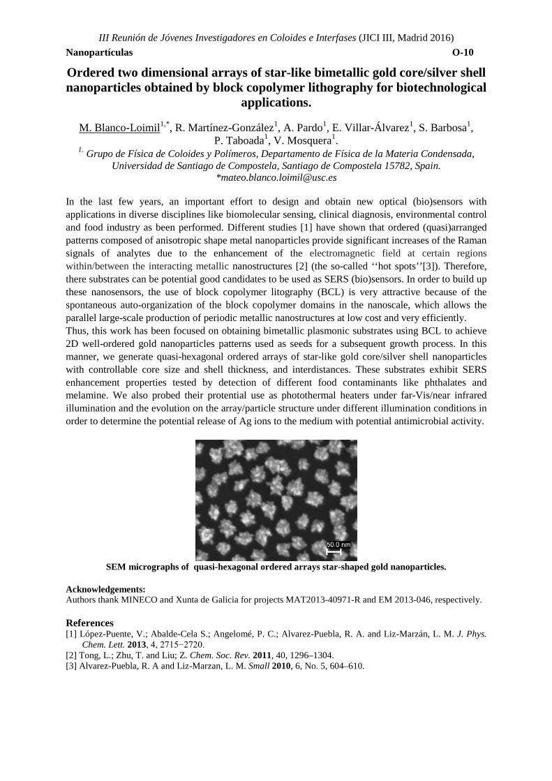
*[email protected] In the last few years, an important effort to design and obtain new optical (bio)sensors with applications in diverse disciplines like biomolecular sensing, clinical diagnosis, environmental control and food industry as been performed. Different studies [1] have shown that ordered (quasi)arranged patterns composed of anisotropic shape metal nanoparticles provide significant increases of the Raman signals of analytes due to the enhancement of the electromagnetic field at certain regions within/between the interacting metallic nanostructures [2] (the so-called ‘‘hot spots’’[3]). Therefore, there substrates can be potential good candidates to be used as SERS (bio)sensors. In order to build up these nanosensors, the use of block copolymer litography (BCL) is very attractive because of the spontaneous auto-organization of the block copolymer domains in the nanoscale, which allows the parallel large-scale production of periodic metallic nanostructures at low cost and very efficiently. Thus, this work has been focused on obtaining bimetallic plasmonic substrates using BCL to achieve 2D well-ordered gold nanoparticles patterns used as seeds for a subsequent growth process. In this manner, we generate quasi-hexagonal ordered arrays of star-like gold core/silver shell nanoparticles with controllable core size and shell thickness, and interdistances. These substrates exhibit SERS enhancement properties tested by detection of different food contaminants like phthalates and melamine. We also probed their protential use as photothermal heaters under far-Vis/near infrared illumination and the evolution on the array/particle structure under different illumination conditions in order to determine the potential release of Ag ions to the medium with potential antimicrobial activity.
SEM micrographs of quasi-hexagonal ordered arrays star-shaped gold nanoparticles.
Acknowledgements: Authors thank MINECO and Xunta de Galicia for projects MAT2013-40971-R and EM 2013-046, respectively. References [1] López-Puente, V.; Abalde-Cela S.; Angelomé, P. C.; Alvarez-Puebla, R. A. and Liz-Marzán, L. M. J. Phys.
Chem. Lett. 2013, 4, 2715−2720. [2] Tong, L.; Zhu, T. and Liu; Z. Chem. Soc. Rev. 2011, 40, 1296–1304. [3] Alvarez-Puebla, R. A and Liz-Marzan, L. M. Small 2010, 6, No. 5, 604–610.
TOPIC 2: APPLICATIONS

III Reunión de Jóvenes Investigadores en Coloides e Interfases (JICI III, Madrid 2016) Applications O-11
Liposomes as templates for polymer nanocapsules
Ana Mateos-Maroto*1; Marta Ruano Aldea2; Eduardo Guzmán1; Ramón G. Rubio1,3,
Francisco Ortega1
1. Departamento de Química Física I,Universidad Complutense de Madrid, Madrid, Spain 2.Leitat Technological Center, Barcelona, Spain
3Instituto Pluridisciplinar, Universidad Complutense de Madrid, Madrid, Spain *[email protected]
Nanostructured materials have been shown a wide range of applications in many technological fields. Among the nanostructured materials, polyelectrolyte multilayers obtained by the layer-by-layer (LbL) self-assembly technology has become a promising solution to many problems raised for modern biotechnology, in applications such as sensors, functional coatings, etc1. The LbL technique can be used to form polyelectrolyte microcapsules by coating a colloidal template such as micro- or nano-particle, followed by the subsequent dissolution of this template1. These polyelectrolyte multilayer microcapsules have several potential applications such as drug delivery carriers2 and DNA encapsulation3. Following the aforementioned approach, it is possible to coat charged liposomes, avoiding the core dissolution step that may involve problems of degradation and presence of impurities on the LbL layers. We have used mixtures of the lipid dioleoylphosphatidylcholine (DOPC) and the charged surfactant dioctadecyl-dimethylammonium bromide (DODAB) to prepare liposomes of 50 nm radius by extrusion. We have coated them by the sequential adsorption of the anionic polymer poly-(styrenesulfonate, sodium salt) (PSS) and a cationic one, poly-(diallyldimethylammonium) (PDADMAC), and up to 6 polyelectrolyte layers have been assembled, which forms a robust coating. The characterization of the capsules formed was carried out by ζ-potential and dynamic light scattering measurements. Acknowledgements This work was funded by MINECO under grants FIS-212-38231-C02-01 and F2014-62005-EXP. References [1] Decher, G.; Schlenoff, B., Multilayer Thin Films: Sequential Assembly of Nanocomposite Materials, ed
Wiley-VCH, 2012 [2] Sukhorukov, G. B.; Rogach, A. L.; Garstka, M.; Springer, S.; Parak, W. J.; Muñoz-Javier, A.; Kreft, O.;
Skirtach, A. G.; Susha, A. S.; Ramaye, Y.; Palankar, R.; Winterhalter, M., Small 3(6), 944-955, 2007 [3] Schukin, D. G.; Patel, A. A.; Sukhorukov, G. B.; Lvov, Y. M., J. Am. Chem. Soc., 126(11), 3374-3375, 2004
III Reunión de Jóvenes Investigadores en Coloides e Interfases (JICI III, Madrid 2016) Applications O-12
Albumin-Covered Lipid Nanocapsules Exhibit Enhanced Uptake Performance by Breast-Tumor Cells
A. Aguilera-Garrido1, F. Galisteo-González1, M. J. Gálvez-Ruiz1, J.A. Molina-Bolívar2, S. A. Navarro3, H. Boulaiz3, A. Ramírez4, J. A. Marchal3
1Department of Applied Physics, University of Granada, Spain 2Department of Applied Physics II, Engineering School, University of Málaga, Spain
3Biopathology and Medicine Regenerative Institute (IBIMER), Biosanitary Institute of Granada (ibs.GRANADA), University of Granada, Spain.
4Department of Health Sciences, University of Jaén, Spain *[email protected]
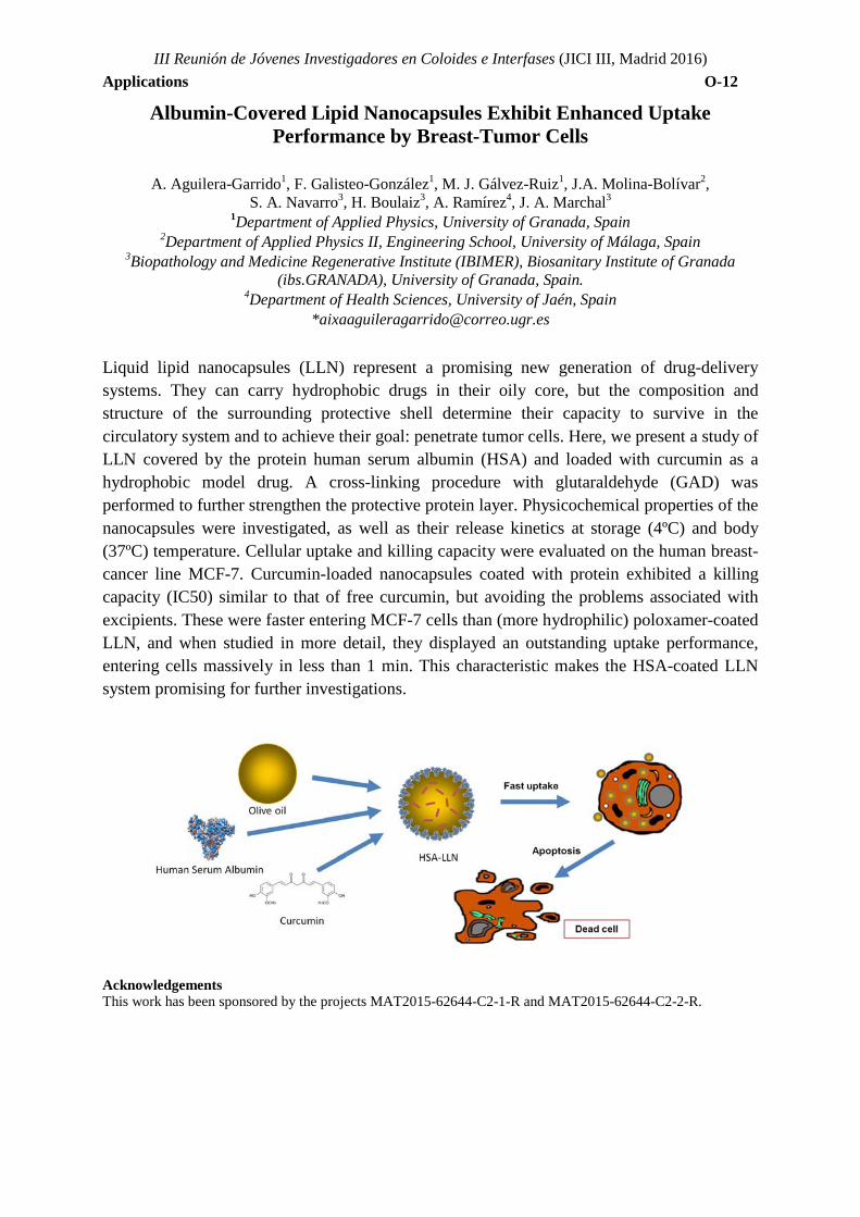
Liquid lipid nanocapsules (LLN) represent a promising new generation of drug-delivery systems. They can carry hydrophobic drugs in their oily core, but the composition and structure of the surrounding protective shell determine their capacity to survive in the circulatory system and to achieve their goal: penetrate tumor cells. Here, we present a study of LLN covered by the protein human serum albumin (HSA) and loaded with curcumin as a hydrophobic model drug. A cross-linking procedure with glutaraldehyde (GAD) was performed to further strengthen the protective protein layer. Physicochemical properties of the nanocapsules were investigated, as well as their release kinetics at storage (4ºC) and body (37ºC) temperature. Cellular uptake and killing capacity were evaluated on the human breast-cancer line MCF-7. Curcumin-loaded nanocapsules coated with protein exhibited a killing capacity (IC50) similar to that of free curcumin, but avoiding the problems associated with excipients. These were faster entering MCF-7 cells than (more hydrophilic) poloxamer-coated LLN, and when studied in more detail, they displayed an outstanding uptake performance, entering cells massively in less than 1 min. This characteristic makes the HSA-coated LLN system promising for further investigations.
Acknowledgements This work has been sponsored by the projects MAT2015-62644-C2-1-R and MAT2015-62644-C2-2-R.
III Reunión de Jóvenes Investigadores en Coloides e Interfases (JICI III, Madrid 2016) Applications O-13
Au/TiO2 nanoparticles on bacterial cellulose membranes for water splitting in gas phase
Anna May-Masnou1, Lluís Soler2, Anna Laromaine1, Jordi Llorca2, Anna Roig1
1Institut de Ciència de Materials de Barcelona (ICMAB-CSIC), Bellaterra 2Institut de Tècniques Energètiques (INTE-UPC), Barcelona
The production of hydrogen from water and sunlight under ambient conditions is one of the desired routes to obtain hydrogen. One technique to achieve it is photocatalytic water splitting using nano-TiO2, a potential photocatalyst due to its band gap size, its resistance to corrosion, and its high surface area.
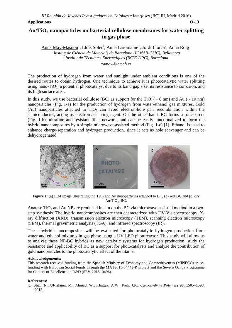
In this study, we use bacterial cellulose (BC) as support for the TiO2 (∼ 8 nm) and Au (∼ 10 nm) nanoparticles (Fig. 1-a) for the production of hydrogen from water/ethanol gas mixtures. Gold (Au) nanoparticles attached to TiO2 can avoid electron-hole pair recombination within the semiconductor, acting as electron-accepting agent. On the other hand, BC forms a transparent (Fig. 1-b), ultrafine and resistant fiber network, and can be easily functionalized to form the hybrid nanocomposites by a simple microwave-assisted method (Fig. 1-c) [1]. Ethanol is used to enhance charge-separation and hydrogen production, since it acts as hole scavenger and can be dehydrogenated.
Figure 1: (a)TEM image illustrating the TiO2 and Au nanoparticles attached to BC, (b) wet BC and (c) dry Au/TiO2_BC.
Anatase TiO2 and Au NP are produced in situ on the BC via microwave-assisted method in a two-step synthesis. The hybrid nanocomposites are then characterized with UV-Vis spectroscopy, X-ray diffraction (XRD), transmission electron microscopy (TEM), scanning electron microscopy (SEM), thermal gravimetric analysis (TGA), and infrared spectroscopy (IR).
These hybrid nanocomposites will be evaluated for photocatalytic hydrogen production from water and ethanol mixtures in gas phase using a UV LED photoreactor. This study will allow us to analyse these NP-BC hybrids as new catalytic systems for hydrogen production, study the resistance and applicability of BC as a support for photocatalysts and analyze the contribution of gold nanoparticles to the photocatalytic effect of the titania.
Acknowledgements: This research received funding from the Spanish Ministry of Economy and Competitiveness (MINECO) in co-funding with European Social Funds through the MAT2015-64442-R project and the Severo Ochoa Programme for Centers of Excellence in R&D (SEV-2015- 0496). References: [1] Shah, N.; Ul-Islama, M.; Ahmad, W.; Khattak, A.W.; Park, J.K.. Carbohydrate Polymers 98, 1585–1598,
2013.
III Reunión de Jóvenes Investigadores en Coloides e Interfases (JICI III, Madrid 2016) Applications O-14
STAND: Determinación del Número de Agregación Micelar por Tensión Superficial.
Pablo F. Garrido1,*, Pilar Brocos1, Alfredo Amigo1, Roberto Rosende1, Luis García-Río2,
Jesús Gracia-Fadrique,3 Ángel Piñeiro1
1Depto de Física Aplicada, Facultade de Física, Universidade de Santiago de Compostela, 15782 Santiago de Compostela, España.
2Depto de Química Física, Centro Singular de Investigación en Química Biolóxica e Materiais Moleculares (CIQUS), Facultade de Química, Universidade de Santiago de Compostela, 15782
Santiago de Compostela, España. 3Depto de Fisicoquímica, Facultad de Química, Universidad Nacional Autónoma de México, Ciudad
Universitaria, 04510 México D.F., México. *E-mail: [email protected]
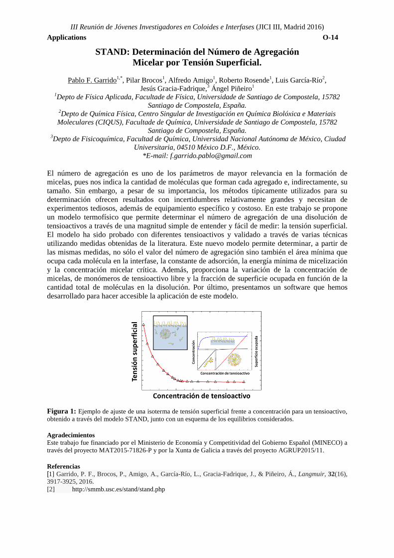
El número de agregación es uno de los parámetros de mayor relevancia en la formación de micelas, pues nos indica la cantidad de moléculas que forman cada agregado e, indirectamente, su tamaño. Sin embargo, a pesar de su importancia, los métodos típicamente utilizados para su determinación ofrecen resultados con incertidumbres relativamente grandes y necesitan de experimentos tediosos, además de equipamiento específico y costoso. En este trabajo se propone un modelo termofísico que permite determinar el número de agregación de una disolución de tensioactivos a través de una magnitud simple de entender y fácil de medir: la tensión superficial. El modelo ha sido probado con diferentes tensioactivos y validado a través de varias técnicas utilizando medidas obtenidas de la literatura. Este nuevo modelo permite determinar, a partir de las mismas medidas, no sólo el valor del número de agregación sino también el área mínima que ocupa cada molécula en la interfase, la constante de adsorción, la energía mínima de micelización y la concentración micelar crítica. Además, proporciona la variación de la concentración de micelas, de monómeros de tensioactivo libre y la fracción de superficie ocupada en función de la cantidad total de moléculas en la disolución. Por último, presentamos un software que hemos desarrollado para hacer accesible la aplicación de este modelo.
Figura 1: Ejemplo de ajuste de una isoterma de tensión superficial frente a concentración para un tensioactivo, obtenido a través del modelo STAND, junto con un esquema de los equilibrios considerados. Agradecimientos Este trabajo fue financiado por el Ministerio de Economía y Competitividad del Gobierno Español (MINECO) a través del proyecto MAT2015-71826-P y por la Xunta de Galicia a través del proyecto AGRUP2015/11. Referencias [1] Garrido, P. F., Brocos, P., Amigo, A., García-Río, L., Gracia-Fadrique, J., & Piñeiro, Á., Langmuir, 32(16), 3917-3925, 2016. [2] http://smmb.usc.es/stand/stand.php
III Reunión de Jóvenes Investigadores en Coloides e Interfases (JICI III, Madrid 2016) Applications O-15
Plasmonic Tip-to-Tip Assembled Nanorods for Enhanced Photothermal Cancer Therapy
Rubén Ahijado-Guzmán1*, Guillermo Gonzalez-Rubio1, 2, Gloria Tardajos1, Luis Liz-Marzan2 and Andrés Guerrero-Martínez1
1Departamento de Química Física I, Universidad Complutense de Madrid, Avda. Complutense s/n, 28040, Madrid, Spain.
2BioNanoPlasmonics Laboratory, CIC biomaGUNE, Paseo de Miramon 182, 20009 Donostia - San Sebastián, Spain.
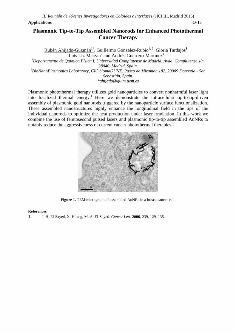
*[email protected] Plasmonic photothermal therapy utilizes gold nanoparticles to convert nonharmful laser light into localized thermal energy.1 Here we demonstrate the intracellular tip-to-tip-driven assembly of plasmonic gold nanorods triggered by the nanoparticle surface functionalization. These assembled nanostructures highly enhance the longitudinal field in the tips of the individual nanorods to optimize the heat production under laser irradiation. In this work we combine the use of femtosecond pulsed lasers and plasmonic tip-to-tip assembled AuNRs to notably reduce the aggressiveness of current cancer photothermal therapies.
Figure 1. TEM micrograph of assembled AuNRs in a breast cancer cell.
References 1. I. H. El-Sayed, X. Huang, M. A. El-Sayed. Cancer Lett. 2006, 239, 129–135.
III Reunión de Jóvenes Investigadores en Coloides e Interfases (JICI III, Madrid 2016) Applications O-16
Self-Assembled Gold Nanooctahedra Through Microevaporators for Highly Efficient SERS-active Substrates.
S. Gómez-Graña1, C. Fernández-López,1 L. Polavarapu,1 J.-B. Salmon,2 J. Leng,2 I. Pastoriza-
Santos,1 J. Pérez-Juste 1 1. Department of Physical Chemistry, University of Vigo, Vigo, Spain
2 Laboratoire du Futur, Université de Bordeaux, UMR 5258, F-33600 Pessac, France *[email protected]
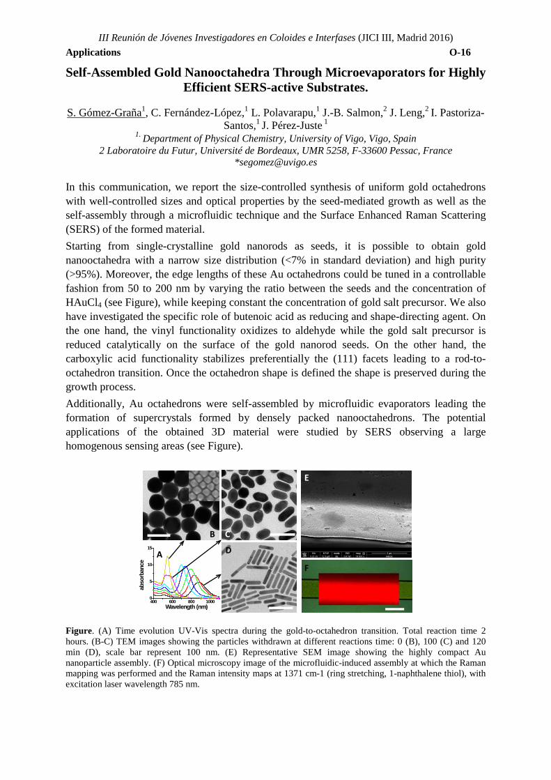
In this communication, we report the size-controlled synthesis of uniform gold octahedrons with well-controlled sizes and optical properties by the seed-mediated growth as well as the self-assembly through a microfluidic technique and the Surface Enhanced Raman Scattering (SERS) of the formed material. Starting from single-crystalline gold nanorods as seeds, it is possible to obtain gold nanooctahedra with a narrow size distribution (<7% in standard deviation) and high purity (>95%). Moreover, the edge lengths of these Au octahedrons could be tuned in a controllable fashion from 50 to 200 nm by varying the ratio between the seeds and the concentration of HAuCl4 (see Figure), while keeping constant the concentration of gold salt precursor. We also have investigated the specific role of butenoic acid as reducing and shape-directing agent. On the one hand, the vinyl functionality oxidizes to aldehyde while the gold salt precursor is reduced catalytically on the surface of the gold nanorod seeds. On the other hand, the carboxylic acid functionality stabilizes preferentially the (111) facets leading to a rod-to-octahedron transition. Once the octahedron shape is defined the shape is preserved during the growth process. Additionally, Au octahedrons were self-assembled by microfluidic evaporators leading the formation of supercrystals formed by densely packed nanooctahedrons. The potential applications of the obtained 3D material were studied by SERS observing a large homogenous sensing areas (see Figure).
Figure. (A) Time evolution UV-Vis spectra during the gold-to-octahedron transition. Total reaction time 2 hours. (B-C) TEM images showing the particles withdrawn at different reactions time: 0 (B), 100 (C) and 120 min (D), scale bar represent 100 nm. (E) Representative SEM image showing the highly compact Au nanoparticle assembly. (F) Optical microscopy image of the microfluidic-induced assembly at which the Raman mapping was performed and the Raman intensity maps at 1371 cm-1 (ring stretching, 1-naphthalene thiol), with excitation laser wavelength 785 nm.
400 600 800 10000
5
10
15
abso
rban
ce
Wavelength (nm)
B
A
C
E
F
D
TOPIC 3: POLYMERS AND GELS


III Reunión de Jóvenes Investigadores en Coloides e Interfases (JICI III, Madrid 2016) Polymer and gels O-17
Cross-Linked microgels, produced by water-in-water-emulsions, as delivery system for enzymes
Yoran Beldengrün*, Cristina Miquel Espigulé, Jordi Aragón-Artigas, Jordi Esquena Moret
Institute of Advanced Chemistry of Catalonia (IQAC-CSIC), Department of Chemical and Biomolecular Nanotechnology. CIBER on Bioengineering, Biomaterials and Nanomedicine (CIBER-
BBN), Barcelona, Spain, *[email protected]
The research work has focused on the preparation and characterization of microgel particles, obtained by cross-linking in the disperse phase of water-in-water (W/W) emulsions, with the final aim of studying the microgels as carriers for the delivery of enzymes. Two different systems have been investigated.
The first system was composed of water, gelatin and maltodextrin, because of its excellent biocompatibility and the easy formation of W/W emulsions in this system, due to mutual immiscibility between gelatin and maltodextrin. Stable gelatin-in-maltodextrin emulsions have been prepared by dispersing the gelatin aqueous phase into the maltodextrin aqueous phase. Genipin, a biocompatible cross-linker, has been added to cross-link into the gelatin-based aqueous droplets, and thus obtaining stable microgel dispersions of 10-20 µm (Fig.1a). The microgels have been purified and freeze dried, which lead to leaf-like structured macroporous particles (Fig. 1b). The influence of cross-linker concentration, ions and pH on microgel properties have been tested. High genipin concentrations (10 mM) increase bond formation between polymer chains, leading to rigid particles with low swelling ratios.
The second studied system was composed of water, carboxymethylcellulose (CMC) and bovine serum albumin (BSA). This system, which also formed W/W emulsions, allowed to obtain microgels, around 5-10 µm, by emulsifying CMC droplets dispersed into a BSA aqueous phase. CMC exhibited low swelling at acidic pH or if ionically crosslinked by Fe3+.
Enzyme activity within gelatin macrogel cross-linked with genipin has been evaluated, as preliminary tests before encapsulation of the enzyme inside the microgels. The enzyme did not lose activity in presence of previously crosslinked microgels. The efficiency of enzyme encapsulation into microgels, and their release and stability, are subject to current study.
Microscopic image (a) of gelatin microgel dispersion and scanning electron microscope image after freeze-drying the sample (b)
Acknowledgements Financial support from the People Programme (Marie Curie Actions) of the European Union’s Seventh Framework Programme FP7/2007-2013/ under REA grant agreement n°606713 (BIBAFOODS project).
(a) (b)
III Reunión de Jóvenes Investigadores en Coloides e Interfases (JICI III, Madrid 2016) Polymers and gels O-18
Efecto de las interacciones electroestéricas en la carga efectiva de microgeles termosensibles: teoría y experimentos.
Irene Adroher-Benítez1,*, Silvia Ahualli1, Delfi Bastos-González1, José Ramos2,
Jacqueline Forcada,2 Arturo Moncho-Jordá1
1.Departamento de Física Aplicada, Facultad de Ciencias, Universidad de Granada, Campus Fuentenueva S/N, 18071 Granada, Spain
2.Grupo de Ingeniería Química, Facultad de Ciencias Químicas, Universidad del País Vasco/EHU, 20080 San Sebastián, Spain
3.Instituto Carlos de Física Teórica y Computacional, Universidad de Granada, Campus Fuentenueva S/N, 18071 Granada, Spain
*[email protected] Los microgeles son nanopartículas formadas por cadenas de polímero entrecruzadas que se encuentran dispersas en un medio, normalmente acuoso. Su principal característica es que pueden expandirse y compactarse en respuesta a un gran número de estímulos externos, como la temperatura, el pH o la concentración salina. Esta cualidad supone una ventaja para un gran número de aplicaciones biotecnológicas. En concreto, en los últimos años está creciendo el interés por desarrollar nuevos sistemas de transporte y encapsulación de fármacos. Para este tipo de aplicaciones biomédicas los microgeles eléctricamente cargados son de especial utilidad. Por este motivo, es de vital importancia comprender las interacciones que entran en juego cuando un microgel se encuentra en presencia de iones en el medio. En el presente trabajo hemos estudiado el efecto de la valencia de los contraiones y de la concentración salina en la carga efectiva de microgeles termosensibles. En primer lugar, hemos obtenido experimentalmente la carga efectiva de los microgeles a partir de medidas de movilidad electroforética. Posteriormente, estos datos han sido comparados con los calculados teóricamente mediante el formalismo de ecuaciones integrales de Ornstein-Zernike junto a la relación de cierre HNC. Los resultados muestran que la carga original del microgel es apantallada debido al efecto combinado de la condensación y de la permeación de contraiones en el interior de la partícula. Además de la interacción electrostática, la exclusión de volumen ejercida por la red polimérica juega un papel determinante en la concentración local de iones, especialmente en el caso de microgeles compactados. Esta contribución estérica es responsable del significativo aumento de la carga efectiva del microgel observada experimentalmente cuando la partícula se compacta a temperaturas mayores de la temperatura crítica de transición. Asimismo, hemos observado que el microgel es electroneutro en sus regiones más internas, lo cual indica que la carga efectiva del microgel procede de la región superficial del mismo. Agradecimientos Ministerio de Economía y Competitividad, Plan Nacional de Investigación, Desarrollo e Innovación (I+D+i) (MAT2012-36270-C04-01, -02 y -04), European Regional Founding, Junta de Andalucía (CTS-6270), proyecto posdoctoral de la Universidad de Granada (Plan Propio 2014), Instituto Carlos I de Física Teórica y Computacional de la Universidad de Granada. References [1] Adroher-Benítez, I; Ahualli, S; Bastos-González, D; Ramos, J; Forcada, J; and Moncho-Jordá, A., J. Polym.
Sci. Part B Polym. Phys. (aceptado).

TOPIC 4: DISPERSED SYSTEMS

III Reunión de Jóvenes Investigadores en Coloides e Interfases (JICI III, Madrid 2016) Dispersed Systems O-19
Production of O/W ecological emulsions formulated with an essential oil by microfluidization technique
Luis A. Trujillo-Cayado1, Álvaro Avila1, M. Carmen García1, M. Carmen Alfaro1,*J. Muñoz1
1.Departament of Chemical Engineering, Faculty of Chemistry, University of Seville, Seville, Spain * [email protected]
Essential oils are very interesting natural biosolvents obtained from plants, whose composition and biological properties depend on their constituents. It is important to highlight their properties against a broad spectrum of microorganisms. For this reason, these solvents are widely used nowadays in pharmaceutical, sanitary, cosmetic, agricultural and food industries. In many of these formulations they are presented as emulsions. The objective of this work was to investigate the effect of the energy per volume applied to a primary emulsion which subsequently fed a Microfluidizer device on the droplet size distribution, mean diameters and stability of final emulsions. O/W emulsions were formulated using 30 wt % thyme oil, as dispersed phase, and 3 wt % wheat biomass-derived surfactant. Two rotor-stator devices, Silverson L5M and Ultraturrax T50, at 2000, 4000 and 6000 rpm were used to prepare the primary emulsions. Afterwards, a high pressure homogenizer (Microfluidizer model M110P, equipped with an F12Y interaction chamber) was used at 2500 psi and 1-pass. Laser diffraction and multiple light scattering were the techniques used in order to reach the aforementioned targets. Sauter mean diameter depends on the energy input and also on the rotor-stator device used to prepare the coarse primary emulsion. We conclude that the 30 wt% thyme oil in water emulsion based on a primary emulsion prepared using the Silverson L5M rotor-stator with an emulsor-mesh applying low energy, exhibited the longest physical stability and the lowest mean diameters. Acknowledgements The financial support received (Project CTQ2015-70700-P) from the Spanish Ministerio de Economía y Competitividad (MINECO) and from the FEDER Programme (European Union) is kindly acknowledged.
III Reunión de Jóvenes Investigadores en Coloides e Interfases (JICI III, Madrid 2016) Dispersed Systems O-20
Emulsions: Nanoparticles for Surfactant Replacement
Natalia Sánchez Arribas*1; Alejandro Lucía2; Eduardo Guzmán1; Francisco Ortega,1 Ramón G. Rubio1,3
1. Departamento de Química Física I,Universidad Complutense de Madrid, Madrid, Spain 2. Centro de Investigaciones de Plagas e Insecticidas (UNIDEF/CITEDEF/CONICET), Buenos Aires,
(Argentina) 3Instituto Pluridisciplinar, Universidad Complutense de Madrid, Madrid, Spain
In recent years, the design and fabricación of multifunctional capsules with controlled chemical composition and physical properties has focused the interest of academy and industrial research. In particular, in the biomedical field, the formulation of novel polymer-based encapsulation platforms for the early-stage disease diagnostic and effective delivery of bioactive agents represents one of the most rapidly advancing areas of science. The stabilization of oil and water droplets by solid particles, which are generally referred to as Pickering emulsions, has been known for over one century, being a good alternative for replacing classical surfactant-stabilized emulsions. Many studies have already been devoted of the study of this type of systems, on both fundamental properties of particles at fluid interfaces and their emerging applications.
In this project, we study the stabilization of emulsions with nanoparticles in which the oil phase is an essential oil with insecticide activity. The aim is to formulate oil in water emulsions (o/w) stabilized by silica nanoparticles (SiO2) with different hydrophobicity degrees. The hydrophobicity of the nanoparticles (NPs) was tuned using Pluronic F-127, a triblock copolymer with two blocks of poly(ethylene oxide) (PEO) and central block of poly(propilene oxide) (PPO), favouring the emulsion stabilization. All the emulsions were prepared by mild shaking, and characterized by direct observation, Dynamic Light Scattering (DLS), ζ potential and turbidity measurements, which allowed us to optimize the emulsion stabilization. The emulsions within the stability region were found to be optically transparent, whereas the phase separate mixtures presented a wide range of nature, including solid precipitates, phase separate liquids, among others. From the point of view of the applications, the monophasic emulsions are preferred for the design of vectors agains different pests. A last aspect studied was the efficacy of some of the emulsions prepared as insecticide against lice (Pediculus humanus capitis), being the death rate obtained around (60 ± 10)%, which demonstrate a good effectiveness. The use of biocompatible components for the preparation of emulsions, both stabilizing agents and dispersed phases, is important for their future applications in the design and fabrication of platforms for load and release active ingredients in many field, thus presenting many future applications, e.g. the encapsulation of insecticides, opening new routes for the fabrication of biosustainable formulations for pest controlling.
Acknowledgements This work was funded by MINECO under grants FIS-212-38231-C02-01 and F2014-62005-EXP
III Reunión de Jóvenes Investigadores en Coloides e Interfases (JICI III, Madrid 2016) Dispersed Systems O-21
Catalytic and magnetic bifunctional polymer/metal oxyde hybrid nanoparticles by miniemulsion polymerization
Olaia Álvarez-Bermúdez1,2,*, Katharina Landfester2, Rafael Muñoz-Espí1
1.Institute of Material Science (ICMUV), University of Valencia, Paterna, Spain 2.Max Planck Institute for Polymer Research, Mainz, Germany
*[email protected] The latest trends in nanotechnology are driven towards the development of multifunctional materials. In this fashion, complex polymer–inorganic hybrids are of great interest as a result of the addition of inorganic functionalities to the biocompatibility, stability or processability features of polymers. Versatile hybrid systems are designed by integration of several inorganic components with specific functionalities [1]. This work is focused on the development of bifunctional nanoparticles that include two metal oxides in a polymer matrix of poly(methyl methacrylate) (PMMA): magnetite in the core and a second catalytically active metal oxide (either titanium(IV) oxide or cerium(IV) oxide) located on the surface. Morphology plays an essential role in the properties of hybrid nanoparticles and its relevance increases with the complexity of the system. Miniemulsion polymerization has been selected as the synthetic route to overcome structural limitations. While other techniques are restricted to hybrids with the inorganic particles at the surface or to single inorganic particles covered by a polymer shell, miniemulsion polymerization allows the incorporation of functional inorganic nanoparticles in the polymer with different locations in the final materials. Structure in hybrid colloidal systems is mostly governed by self-assembly processes driven by a minimization of the overall interfacial energy [2], which we address through the chemical modification of the surface of the inorganic components with alkoxysilanes. In previous work, magnetite has been encapsulated within PMMA with a homogeneous distribution or Janus-like morphologies by using silanes with polymerizable moieties or long alkyl chains without polymerizable units, respectively [3]. In our bifunctional nanoparticles, each metal oxide is specifically functionalized for compatibilization with the hydrophobic monomer, which is polymerized afterwards by free-radical miniemulsion polymerization, so that a selective location of the inorganic components is obtained. We are working on the magnetic characterization of the final materials and on their catalytic applications for the degradation of dyes and for model oxidation reactions. In summary, we generate magnetically separable nanoparticles, with magnetite encapsulated within PMMA and a catalytically active metal oxide (TiO2 or CeO2) accessible on the surface of the polymer. We investigate the surface functionalization of the metal oxides for the selective migration of each component regarding to its specific functionalities within the polymer nanoparticles.
Acknowledgements: The authors thank the Max Planck Society as well as the Spanish Ministry of Economy and Competitiveness (Ramón y Cajal grant to R. Muñoz-Espí, grant No. RYC-2013-13451) for financial support.
References [1] Fischer, V., Bannwarth, M. B., Jakob, G., Landfester, K., Muñoz-Espí, R., J. Phys. Chem. C 117, 5999–6005, 2013. [2] Asua, J. M., Macromolecular Chemistry and Physics 215, 548–464, 2014. [3] Schoth, A., Keith, A. D., Landfester, K., Muñoz-Espí, R., RSC Adv. 2016, DOI: 10.1039/c6ra08896a
III Reunión de Jóvenes Investigadores en Coloides e Interfases (JICI III, Madrid 2016) Dispersed Systems O-22
Cultivo Bacteriano en Polimerosomas Fabricados porMicrofluídica
Lourdes Álvarez Callejo, Lara H. Moleiro, Laura R. Arriaga and Francisco Monroy
Departamento de Química Física I, Universidad Complutense de Madrid, 28040, Madrid, España, *[email protected]
Las técnicas de análisis microbiológico actuales requieren el cultivo de bacterias. Convencionalmente, dicho cultivo se realiza en medio sólido, semisólido o líquido, adaptando la metodología utilizada a las características metabólicas particulares de cada organismo. Además, la tasa de crecimiento varía en función del estado del medio en el que se encuentren. El crecimiento en medio líquido es más rápido que en medio sólido.1 Sin embargo el cultivo en medio líquido no permite la obtención de unidades formadoras de colonias (UFCs) aisladas. El reciente desarrollo de la tecnología microfluidica ha posibilitado la creación de gotas de emulsión con tamaños uniformes, capaces de encapsular de manera controlada y gran eficiencia cualquier material de interés. Además, dicha tecnología no solo permite fabricar gotas de emulsión sencilla, de agua en aceite o aceite en agua, sino que también permite la creación de emulsiones múltiples. Un ejemplo de emulsión doble es una gota de agua recubierta por un caparazón de aceite y dispersada en agua. El compartimento acuoso puede emplearse para encapsular bacterias en su medio acuoso de cultivo, mientras que el caparazón de aceite las protege y aísla del resto de gotas de emulsión que también encapsulan de manera eficiente una cantidad controlada de bacterias. Eligiendo el aceite y el tensioactivo adecuados, dichas gotas de emulsión pueden servir como plantillas para formar vesículas de polímero o polimerosomas de tamaño controlado y uniforme.2 Eligiendo la concentración de bacterias y la velocidad de flujo del fluido acuoso interno adecuados, el polimerosoma puede encapsular eficientemente una única UFC, de manera que el cultivo provenga de dicha UFC aislada. Esta aproximación, nos permite hacer un seguimiento del cultivo a nivel de bacteria individual, proporciona un mayor rendimiento que el cultivo en medio sólido, y nos permite estudiar cambios puntuales en las condiciones ambientales de una manera rápida. Por tanto, esperamos que la utilización de polimerosomas como soporte para el cultivo bacteriano se convierta en una nueva forma de microbiología.
Agradecimientos Este trabajo ha sido realizado en el laboratorio del Profesor Francisco Monroy en el Departamento de Química Física I de la UCM Referencias [1] Zhang, Y., et al. Biomaterials 34, 4564-4572 (2013). [2] do Nascimento, D. F., et al. Langmuir 32, 5350-5355 (2016).
TOPIC 5: BIOCOLLOIDS

III Reunión de Jóvenes Investigadores en Coloides e Interfases (JICI III, Madrid 2016) Biocolloids O-23
Solubilization of Ceramide-R in water: A promising route on the preparation of cosmetic formulations
Laura Fernández-Peña1,*, Sara Llamas2, Eduardo Guzmán1, Francisco Ortega1,
Ramón G. Rubio1,3. 1.Departamento de Química Física I, Universidad Complutense de Madrid, Ciudad Universitaria s/n
28040-Madrid, Spain. 2. CNR-Istituto per l’Energetica e le Interfasi, via de Marini 6, 16149 Genova, Italy
.3.Instituto Pluridisciplinar, Universidad Complutense de Madrid, Av. Juan XIII,2. 28040-Madrid, Spain
*[email protected] Ceramides consist of a sphingosine covalently bound to a fatty acid by an amide linkage. They are involved in the regulation of diverse cellular functions such as cell grown, differentiation, senescence, apoptosis and immune responses.1 Ceramides are also structural elements of many cells and tissues. The outermost layer of the skin, the stratum corneum, is formed by a wide range of lipids, proteins and cells called corneocytes2. Among the intercellular lipids, ceramides are the main one, accounting for 40-50% of total intracellular lipids3. Human hair also contains ceramides but in this case, in trace amounts. Ceramides play a major role in the water-retaining property and barrier function of hair as well as skin1. This makes them promising candidates for the development of new cosmetic formulations. However their low solubility in water may be seen as an important drawback because shampoo and conditioner formulations are most frequently one-phase aqueous products. On the other hand, the insolubility of ceramides can become an advantage to increase the deposition of polymer-ceramide complexes onto the hair surface. Thus, it can enhance the conditioning and repairing performance of cosmetic formulations. This work has been focused on the development of a variety of strategies in order to increase the solubility of ceramide-R in water. For this purpose, ceramide-R has been mixed with different cosmetic surfactants, in order to form vesicles or lamellar phases that allow us to enhance the incorporation of ceramide-R into water based formulation. Furthermore, the dispersion of ceramide-R in oil in water emulsions has also been explored. Acknowledgements This work was funded by L’Oréal S.A. and by MINECO under grants FIS-212-38231-C02-01 and F2014-62005-EXP References [1] Lee, Y.S; Choi, K.; Choi, M.H.; Ji, S.Y.; Yoo, J.M.; Lee, Y.M; Hong, J.T.; Yun, Y.P.; Yoo, H.S.; Arch Pharm Res 32, 1795-1801, 2009. doi: 10.1007/s12272-009-2219-5 [2] Itaya, Y.; Tokudome, Y.; Biochemical and Biophysical Research Communications 473,114-117, 2016. doi:10.1016/j.bbrc.2016.03.061 [3] Tian, Y.; Guo, Y.; Zhang, W.; Journal of Dispersion Science and Technology 37, 1115-1122, 2016
III Reunión de Jóvenes Investigadores en Coloides e Interfases (JICI III, Madrid 2016) Biocolloids O-24
Mechanical fluctuations during ATP synthesis
Víctor G. Almendro-Vedia1,2*, Paolo Natale1,2, Francisco Monroy1, Iván López-Montero1,2 1. Department of Physical Chemistry I (UCM), Madrid, Spain
2. Fundación Investigación Biomédica Hospital 12 de Octubre, Madrid, Spain *[email protected]
The ATP synthases are multiprotein complexes present in the plasma membranes of bacteria, in the thylakoid membranes of chloroplasts, and in the inner membranes of mitochondria1. They are able to produce a source of chemical energy by the synthesis of ATP from ADP and Pi. The presence of a transmembrane protonmotive force, Δp, caused by a ΔpH or a membrane potential Δψ, triggers a mechanical rotary mechanism of some of its subunits, through which, protons are pumped across the membrane and adenosine triphosphate is generated. This rotation may affect somehow the movement of the lipid membrane. Despite having achieved reconstitutions of the complex in lipid systems such as SUVs2 (small unilamellar vesicles) and GUVs3 (giant unilamellar vesicles) that relation has not been studied yet. In this work, we extract and purify F0F1-ATPsynthase from E. coli and we incorporate it into vesicles made of E. coli total lipid extract. By using confocal microscopy we monitored the changes of ΔpH inside of the vesicles due to proton pumping during ATP synthesis. Also, phase contrast microscopy is been used to perfom analysis of membrane flickering to study the variation of the mechanical properties of the system along the process. Acknowledgements VGAV acknowledges European research funding programmes (ERC-2013-StG-338133). References 1 Walker JE, Biochemical Society Transactions 41, 1-16, 2013. 2 Richad P, The Journal of Biological Chemistry, 270, 21571-21578, 1995. 3 Xiaolong L, Biochimica et Biophysica Acta 1770, 1620–1626, 2007.

III Reunión de Jóvenes Investigadores en Coloides e Interfases (JICI III, Madrid 2016) Biocolloids O-25
Oncogenic pathway inhibition of aggressive tumors through a combinatorial therapeutic approach using nanostructured hybrid
platforms.
E.Villar-Álvarez 1,*, Irene Golán-Cancela2, Mateo Blanco-Loimil1, Raquel Martínez-González1, Alberto Pardo1, Silvia Barbosa1, J. A Costoya2 Pablo Taboada1.
1.Departamento de Física de la Materia Condensada, Universidad de Santiago de Compostela, Campus Vida, 15782, Santiago de Compostela, Spain
2 Molecular Oncology Laboratory MOL, CIMUS; IDIS Departamento de Fisiología, Universidad de Santiago de Compostela, Av de Barcelona s/n 15782, Santiago de Compostela, Spain.
The human epidermal growth factor receptor (HER) family of tyrosine kinases is deregulated in multiple cancers either through amplification, over-expression, or mutation, causing their carcinogenic effect [1]. HER3 and HER2 oncogenes are commonly over-expressed in the most aggressive forms of cancer. In particular, HER3 amplification is present in breast, colon and gastric tumors [1,2,3], and a high expression is positively associated with the presence of lymph node metastases [2]. In addition, HER2 has been found in approximately 30% of human breast cancers and has been shown to render cancer cells more resistant to chemotherapy [3,4]. However, the mutant HER3 oncogenic activity is dependent on HER2 signaling [1]. Expression of HER2 alone does not significantly enhance drug resistance, which is related to other HER receptors like HER3 [4]. In recent studies, HER2 has been found highly phosphorylated in cells coexpressing at the same time HER2 with HER3, as for NIH 3T3 or SKBR3 cells [4] Thus, HER3 presence may be correlated with bioresponses to chemotreatments that target HER2, providing a route for resistance to anti-HER drugs and drug targeting strategies [3] Oncogenic tyrosine kinases have proven to be promising targets for the development of highly effective anticancer drugs [5]. Some studies have shown the benefits of an effective combinatorial treatment using anti-HER antibodies with small molecule inhibitors which effectively blocks mutant HER3 [1]. Herein, we propose a nanoplataform with multitherapeutic capability against HER2 and HER3 receptors. This nanocarrier consist of a hybrid gold nanoshell [6] functionalized with the antibody Trastuzumab and a silencing RNA (siRNA) against HER3. We observe that HER3, HER2 and, consequently, the PI3K/Akt signaling route are inhibited in SKBR3 cells (positive HER 3 and HER2 cells) and MDA-MB-231 cells (only HER2 positive) in vitro as derived from Western blotting. We noted HER3 knocking down involves induction of apoptosis and cellular death for several Trastuzumab/siRNA molar ratios studied. Acknowledgements Authors thank MINECO and Xunta de Galicia for financial support. Eva Villar and Alberto Pardo thank to MECD for their FPU fellowship. References [1] Jaiswal, B. S., Kljavin, N. M., Stawiski, E. W., Chan, E., Parikh, C., Durinck, S., Sauvage, F. J. De., Cancer
Cell, 23(5), 603–617., 2013. [2] Lemoine, N.R. ,Barnes, D.M., Hollywood, D.P., Hughes, , C.M., Smith, P., Dublin, E., Prigent, S.A.,
Gullick, W.J., Hurst, H.C., Br. J. Cancer , 66, 1116-1121, 1992. [3] Desbois-Mouthon, C. Gastroenterologie Clinique et Biologique, 34 (4-5), 255–259, 2010. [4]Chen, X., Yeung, T. K., & Wang, Z. Biochem. Biophys. Res. Comm., 277(3), 757–763, 2010. [5] Sergina, N. V., Rausch, M., Wang, D., Blair, J., Hann, B., Shokat, K.M., Moasser, M.M., Nature, 445(7126),
437-4, 2007. [6] (a) Topete, A., Alatorre-Meda, M., Iglesias, P., Villar-Alvarez, E. M., Barbosa, S., Costoya, J. A., Mosquera,
V. ACS Nano, 8(3), 2725–2738, 2014. (b) Topete, A., Alatorre-Meda, M., Villar-Alvarez, E. M., Carregal-Romero, S., Barbosa, S., Parak, W. J., Mosquera, V. Healthcare Materials, 3(8), 1309–1325, 2014.
III Reunión de Jóvenes Investigadores en Coloides e Interfases (JICI III, Madrid 2016) Biocolloids O-26
Gemini/DOPE nanocarriers for the efficient transport and delivery of MFN1 in NIH3T3 mouse cells.
Mónica Muñoz-Úbeda1*, Paolo Natale1, Andrés Tolosa-Díaz1, Elena Junquera,2
Emilio Aicart2, Iván López-Montero1 1. Department of Physical Chemistry I (UCM) and Fundación Investigación Biomédica Hospital 12 de
Octubre, Madrid, Spain 2. Colloidal Chemistry and Supramolecular Group, Department of Physical Chemistry I
(UCM),Madrid, Spain *[email protected]
Mitochondria form a highly dynamic network of organelles which constantly fuse and divide1. Mitochondrial fusion requires the coordinated fusion of the outer and inner membranes. Three central players have been identified in mitochondrial fusion: mitofusin 1 and mitofusin 2 (outer mitochondrial membrane fusion) and OPA1 (inner mitochondrial membrane fusion). Imbalances in membrane fusion pathways give rise to important mitochondrial diseases (MD). MD are severe chronic diseases, often fatal, to which there is no cure to date.2 Lipoplexes may constitute powerful tools for designing efficient therapeutic agents against MD. In particular, lipoplexes are biocompatible, biodegradable, non-toxic and stealth, thus potentially used in gene therapy. In this work, we synthesize different lipoplexes constituted by mixed cationic liposomes (CL), composed by a synthetic cationic gemini lipid and a zwitterionic phospholipid (DOPE)3 together with the pTL2 plasmid, coding for the Mitofusin 1. We present the physicochemical characterization of lipoplexes in terms of hydrodynamic diameter, polydispersity, surface charge density and stability. We also monitored the uptake of lipoplexes in Mouse Embryonic Fibroblasts (NIH3T3) by using confocal microscopy and flow cytometry. Preliminary results show good viability, low cytotoxicity rates and high transfection efficiencies.
Acknowledgements MMU aknowledges the Sara Borrell program (Spanish Institute of Health Carlos III, CD15/00190). References [1] Legros F, Lombes A, Frachon P, Rojo M. Mol Biol. Cell 13, 4343-4354, 2002. [2] Ferlini A, Scotton C, Novelli G. Public Health Genomics 16, 313-21, 2014. [3] (a) Muñoz-Úbeda M. et al, J. Am. Chem. Soc. 133, 18014-18017, 2011. (b) Misra S. K. et al,
Biomacromolecules 14, 3951-3963, 2013. (c) Kumar K. et al, J. Mat. Chem. B 3, 1495-1506, 2015.
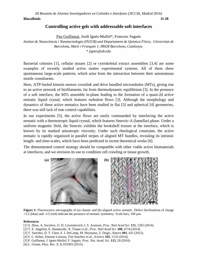
III Reunión de Jóvenes Investigadores en Coloides e Interfases (JICI III, Madrid 2016) Biocolloids O-28
Controlling active gels with addressable soft interfaces
Pau Guillamat, Jordi Ignés-Mullol*, Francesc Sagués Institut de Nanociència i Nanotecnologia (IN2UB) and Departament de Química Física, Universitat de
Barcelona, Martí i Franquès 1, 08028 Barcelona, Catalunya. * [email protected]
Bacterial colonies [1], cellular tissues [2] or cytoskeletal extract assemblies [3,4] are some examples of recently studied active matter experimental systems. All of them show spontaneous large-scale patterns, which arise from the interaction between their autonomous motile constituents. Here, ATP-fueled kinesin motors crosslink and drive bundled microtubules (MTs), giving rise to an active network of biofilaments, far from thermodynamic equilibrium [3]. In the presence of a soft interface, the MTs assemble in-plane leading to the formation of a quasi-2d active nematic liquid crystal, which features turbulent flows [3]. Although the morphology and dynamics of these active nematics have been studied in flat [3] and spherical [4] geometries, there was still lack of true control capabilities. In our experiments [5], the active flows are easily commanded by interfacing the active nematic with a thermotropic liquid crystal, which features Smectic-A (lamellar) phase. Under a uniform magnetic field, the Smectic exhibits the bookshelf texture at the interface, which is known by its marked anisotropic viscosity. Under such rheological constraint, the active nematic is rapidly organized in parallel stripes of aligned MT bundles, revealing its intrinsic length- and time-scales, which have been predicted in recent theoretical works [6]. The demonstrated control strategy should be compatible with other viable active biomaterials at interfaces, and we envision its use to condition cell crawling or tissue growth.
(a) (b) Figure 1: Fluorescence micrographs of (a) chaotic and (b) aligned active nematic. Defect disclinations of charge +1/2 (blue) and -1/2 (red) indicate the presence of nematic symmetry. Scale bars, 100 µm. References [1] S. Zhou, A. Socolow, O. D. Lavrentovich, I. S. Aranson, Proc. Natl Acad Sci. 111, 1265 (2014). [2] T. E. Angelini, E. Hannezobr, X. Trepat et al., Proc. Natl Acad Sci. 108, 4714 (2014). [3] T. Sanchez, D. T. Chen, S. J. DeCamp, M. Heymann, Z. Dogic, Nature 491, 431 (2012). [4] F. C. Keber, Etienne Loiseau, Tim Sanchez et al., Science 345, 1135 (2014). [5] P. Guillamat, J. Ignés-Mullol, F. Sagués. Proc. Nat. Acad. Sci. 113, 20 (2016) [6] L. Giomi, Phys. Rev. X, 5, 031003 (2015).
III Reunión de Jóvenes Investigadores en Coloides e Interfases (JICI III, Madrid 2016) Biocolloids O-28
Brownian dynamics study of the macromolecular crowding effect in reaction-diffusion processes in cellular media
Pablo M. Blanco1,*, Mireia Via1 ,Sergio Madurga1 , Josep L. Garcés2, Eudald Vilaseca1 and
Francesc Mas1 1. Material Science and Physical Chemistry Department, Barcelona University (UB) & Institute of
Theorical and Computational Chemistry (IQTCUB), Barcelona, Spain 2. Chemistry Department, Lleida University (UdL), Lleida, Spain.
Although a particular protein concentration in cell is small (from 1-1000 nmol/L), total protein concentration is really high (80-400 g/L). This means about 20-40% of total volume of cell is occupied by macromolecules. Macromolecular crowding is generated by macromolecular cosolutes that are nominally inert with respect to the reaction of interest [1]. Macromolecular crowding affects the thermodynamics and kinetics of processes taking place in the biological medium due to nonspecific interactions [1]. Since classical in vitro experiments are usually done in quasi-ideal conditions (< 1g/L) there is a lack of information of reaction rates, diffusion processes, equilibrium properties and mechanisms of biological reactions in realistic media [2,3]. Diffusion and reaction processes can be studied using on-lattice [4,5] as well as off-lattice algorithms. In this scope, we have developed a Brownian Dynamics reaction-diffusion code to study enzyme kinetics that follows the Michaelis-Menten mechanism. The biomolecules are modelled explicitly as individual, non-overlapping coarse-grained hard spheres that are transported via stochastic dynamics. Species undergo reactions with other chemical species when they accomplish the Monte Carlo criteria; according to a given reaction probability related to the macroscopic rate constants. In order to describe accurately Brownian motion is important to take into account the hydrodynamic interactions [6]. These interactions originate when a Brownian particle collides with a solvent particle which, in turn, also collides with other solvent molecules This correlation of the particle's movement through the liquid leads to a decrease of the diffusion coefficient of the species in the reaction medium. In this study, the effect of non-specific interaction, excluded volume, obstacle size and hydrodynamic interactions in the diffusion and reactions processes is analysed. References [1] Zhou, H.X.; Rivas, G.; Minton: A.P.: Annu Rev Biophys 37, 375, 2008. [2] Pastor, I.; Vilaseca, E.; Madurga, S.;Garcés, J.L. ; Cascante, M.; Mas, F. : J. Phys.Chem. B 114, 4028, 2010 & erratum ibid 114,12182, 2010; Vilaseca,E.; Pastor, I.; Isvoran, A.; Madurga, S.; Garcés, J.L.; Mas, F.: Theor. Chem. Acc. 128, 795, 2011. [3] Pastor, I.; Pitulice, L.;Balcells, C.;Vilaseca, E.; Madurga, S.; Isvoran, A.;Mas, F: Biophys.Chem., 185, 8, 2014. [4] Pitulice, L.; Vilaseca, E.; Pastor, I.; Madurga, S.; Garcés, J.L.; Isovran, A.; Mas, F.: Mathematical Biosciences, 2014, 72, 2014. [5] Vilaseca, E.; Isvoran,A.; Madurga, S.; Pastor, I.; Garcés, J.L.; Mas, F.: PhysChem-ChemPhysm 13, 7396, 2011. [6] Ermak, D.L. and McCammon, J.A.: J.Chem. Phys. 69, 1352, 1978.
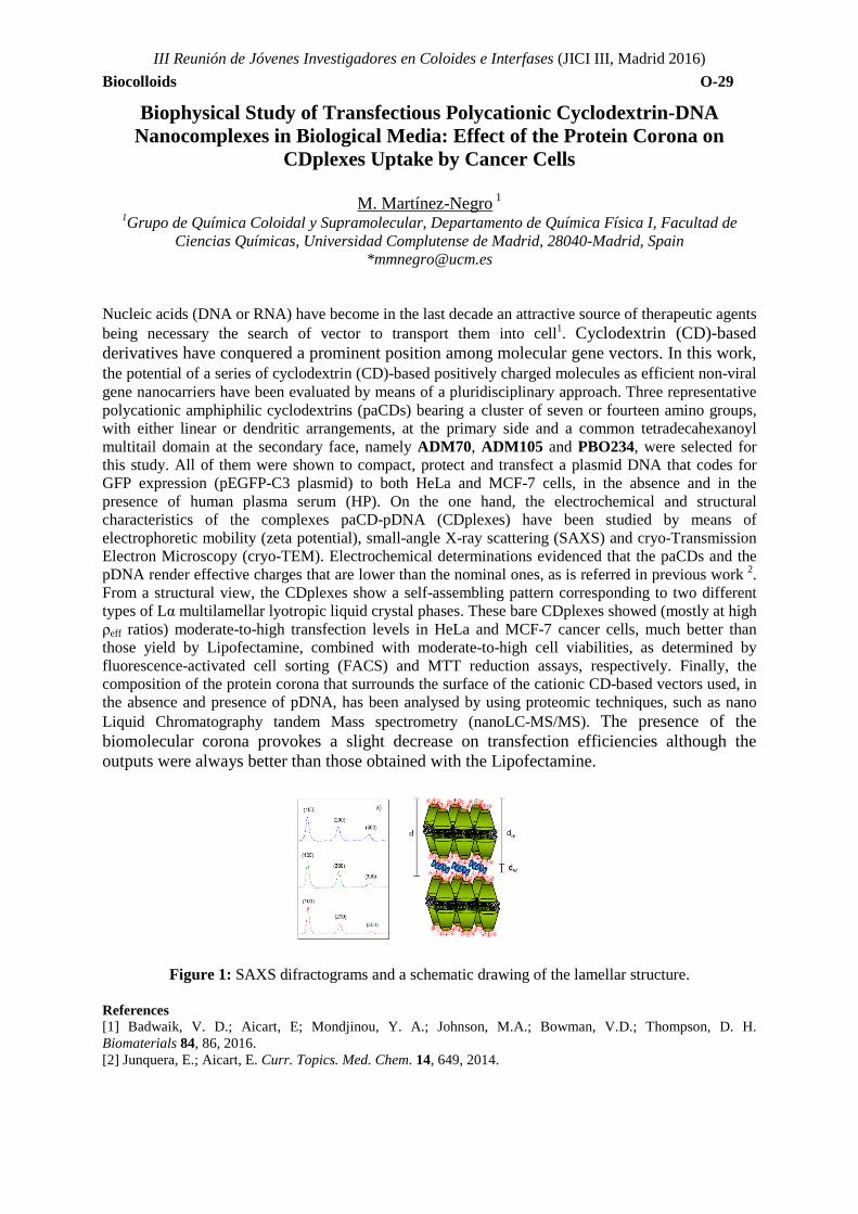
III Reunión de Jóvenes Investigadores en Coloides e Interfases (JICI III, Madrid 2016) Biocolloids O-29
Biophysical Study of Transfectious Polycationic Cyclodextrin-DNA Nanocomplexes in Biological Media: Effect of the Protein Corona on
CDplexes Uptake by Cancer Cells
M. Martínez-Negro 1
1Grupo de Química Coloidal y Supramolecular, Departamento de Química Física I, Facultad de Ciencias Químicas, Universidad Complutense de Madrid, 28040-Madrid, Spain
Nucleic acids (DNA or RNA) have become in the last decade an attractive source of therapeutic agents being necessary the search of vector to transport them into cell1. Cyclodextrin (CD)-based derivatives have conquered a prominent position among molecular gene vectors. In this work, the potential of a series of cyclodextrin (CD)-based positively charged molecules as efficient non-viral gene nanocarriers have been evaluated by means of a pluridisciplinary approach. Three representative polycationic amphiphilic cyclodextrins (paCDs) bearing a cluster of seven or fourteen amino groups, with either linear or dendritic arrangements, at the primary side and a common tetradecahexanoyl multitail domain at the secondary face, namely ADM70, ADM105 and PBO234, were selected for this study. All of them were shown to compact, protect and transfect a plasmid DNA that codes for GFP expression (pEGFP-C3 plasmid) to both HeLa and MCF-7 cells, in the absence and in the presence of human plasma serum (HP). On the one hand, the electrochemical and structural characteristics of the complexes paCD-pDNA (CDplexes) have been studied by means of electrophoretic mobility (zeta potential), small-angle X-ray scattering (SAXS) and cryo-Transmission Electron Microscopy (cryo-TEM). Electrochemical determinations evidenced that the paCDs and the pDNA render effective charges that are lower than the nominal ones, as is referred in previous work 2. From a structural view, the CDplexes show a self-assembling pattern corresponding to two different types of Lα multilamellar lyotropic liquid crystal phases. These bare CDplexes showed (mostly at high ρeff ratios) moderate-to-high transfection levels in HeLa and MCF-7 cancer cells, much better than those yield by Lipofectamine, combined with moderate-to-high cell viabilities, as determined by fluorescence-activated cell sorting (FACS) and MTT reduction assays, respectively. Finally, the composition of the protein corona that surrounds the surface of the cationic CD-based vectors used, in the absence and presence of pDNA, has been analysed by using proteomic techniques, such as nano Liquid Chromatography tandem Mass spectrometry (nanoLC-MS/MS). The presence of the biomolecular corona provokes a slight decrease on transfection efficiencies although the outputs were always better than those obtained with the Lipofectamine.
Figure 1: SAXS difractograms and a schematic drawing of the lamellar structure.
References [1] Badwaik, V. D.; Aicart, E; Mondjinou, Y. A.; Johnson, M.A.; Bowman, V.D.; Thompson, D. H. Biomaterials 84, 86, 2016. [2] Junquera, E.; Aicart, E. Curr. Topics. Med. Chem. 14, 649, 2014.
III Reunión de Jóvenes Investigadores en Coloides e Interfases (JICI III, Madrid 2016) Biocolloids O-30
Reconstitución artificial mediante microfluídica de un sistema de
amplificación de DNA y expresión genética
Sergio Ángel Ortega1*, Lara H. Moleiro, Laura R. Arriaga and Francisco Monroy .Departamento de Química Física I, Universidad Complutense de Madrid, 28040, Madrid, España,
*[email protected] La biología sintética es una disciplina que utiliza principios de ingeniería para autosensamblar componentes biológicos. Una de sus aplicaciones es la fabricación de células artificiales,1 un campo que debe sus avances a los recientes desarrollos en tecnologías de amplificación y expresión génica fuera de células vivas, y a los desarrollos en biofísica de membranas. El reciente desarrollo de la tecnología microfluidica permite la fabricación de vesículas con tamaños y composiciones uniformes y perfectamente controladas.2 Encapsulando funcionalmente dentro de este tipo de vesículas un sistema de amplificación basado en la DNA polimerasa de phi29,3 amplificamos DNA mediante el mecanismo de círculo rodador con un alto rendimiento. La amplificación es cuantificada mediante microscopia de fluorescencia a través de la asociación de una sonda fluorescente llamada Evagreen que se asocia al DNA que va siendo amplificado. Esperamos que esta tecnología nos permita estudiar en el futuro la estocasticidad asociada a los procesos de amplificación y expresión de proteínas. Agradecimientos Este trabajo ha sido realizado en el laboratorio del Profesor Francisco Monroy en el Departamento de Química Física I de la UCM con la ayuda de una beca del programa Exina UCM Manuel Álvarez López. Referencias [1] McDaniel, R., & Weiss, R. (2005). Current Opinion in Biotechnology, 16(4), 476-483. [2] Arriaga, L. R., Datta, S. S., Kim, S. H., Amstad, E., Kodger, T. E., Monroy, F., & Weitz, D. A.
(2014). Small, 10(5), 950-956. [3] Blanco, L., Bernad, A., Lázaro, J. M., Martin, G., Garmendia, C., & Salas, M. (1989). Journal of Biological
Chemistry, 264(15), 8935-8940.
III Reunión de Jóvenes Investigadores en Coloides e Interfases (JICI III, Madrid 2016) Biocolloids O-31
The Mechanics of the Cell Membrane of Escherichia coli During the
Division Cell Cycle
Samuel Salinas1,2*, Aurora Hernández-Machado3, Carlos Ruiz2 and Francisco Monroy1
1Departamento de Quimica Física I, Universidad Complutense de Madrid, Ciudad Universitaria s/n, 28040 Madrid, Spain
2Bioquímica y biofísica de fluidos y membranas, CINVESTAV Monterrey, Vía del Conocimiento 201, Parque de Investigación e Innovación Tecnológica, 66600 Apodaca NL, Mexico
3Departamento de Estructura y Constituyentes de la Materia, Universidad de Barcelona, Avenida Diagonal 647, 08028 Barcelona, Spain
Taking advantage of the flickering motions of the cell membrane [1], we analyze the temporal evolution of Escherichia coli during the cell division using optimized methods of cell contour segmentation implemented with fast camera video-microscopy devices. Through a statistical and biophysical analysis, we study the activity of the internal mechanism of cell division through the effect that exerts on the membrane’s fluctuations. As a result, we are able to track high activity points which leads to formation of septum, create maps of tensions and study the diffusion of the membrane since the first stages of division. This approach reveals new possibilities for studying microscopic effects of biological systems in vitro.
References [1] Pécréaux J, Döbereiner HG, Prost J, Joanny JF, Bassereau P (2004) Refined contour analysis of giant
unilamellar vesicles. Eur Phys J E 13: 277–290. doi:10.1140/epje/i2004-10001-9.

TOPIC 6: INTERFACES AND SURFACES

III Reunión de Jóvenes Investigadores en Coloides e Interfases (JICI III, Madrid 2016)
Interfaces and Surfaces O-32
Effect of silver nanoparticles on the wetting and the evaporation of water sessile droplets for different substrates
L.Perrin1, A.Pajor-Swierzy2, E. Guzmán1, F. Ortega1, R.G. Rubio1,3, S. Magdassi2, M.G. Velarde3
1 Department of Chemistry, University Complutense of Madrid, Madrid, Spain, 2Casali Center for applied Chemistry, Hebrew University of Jerusalem, Jerusalem, Israël.
3Pluridisciplinar Institut, University Complutense of Madrid, Madrid, Spain. *[email protected].
The evaporation of drop on surfaces is present in a lot of industrial and medical applications like printed electronics, spraying of pesticides, spray cooling, DNA mapping. Despite this strong interest, the dynamic of evaporation of complex liquid remains not fully understood. The lifetime of a sessile droplet occurs in several steps. It starts with a stage of spreading followed by 3 different stages of evaporation. A 1st stage, where the contact line remains constant with a contact angle that decreases from an advancing to a receding contact angle. Then a 2nd stage of evaporation where the contact line reduces with constant contact angle and finally a 3rd stage where these parameters decrease simultaneously until the droplet completely disappears. The effect of silver nanoparticles on the wetting and evaporation of water sessile droplet is studied in the ambient conditions of temperature and relative humidity. A droplet of complex liquid is deposited on a substrate and the evolution of contact line, height and contact angle are simultaneously determined thanks to a droplet shape analyzer. Special care is taken regarding the volume of the droplet to ensure that the effect of gravity is negligible so that the droplet presents a spherical cap. The values of the advancing and receding contact angles for several complex liquids on different substrates are estimated. Also, the experimental results are compared with a theoretical model developed by S. Semenov et al. [1] to describe the 1st stage of evaporation and show good agreement. Acknowledgements The study has been prepared in the framework Marie Curie Initial Training Network “Complex Wetting Phenomena” (CoWet). References [1] S. Semenov, V.M. Starov, R.G. Rubio, H. Agogo, M.G. Velarde, Evaporation of water sessile droplet: Universal behaviour in presence of contact angle hysteresis. Colloids and Surfaces A: Physicochem. Eng. Aspects 2011, 391, 135-144.
III Reunión de Jóvenes Investigadores en Coloides e Interfases (JICI III, Madrid 2016)
Interfaces and Surfaces O-33
Diagrama de fases de los módulos dinámicos de monocapas de Langmuir de ácidos grasos con distinta longitud de cadena hidrófoba.
Javier Tajuelo1,*, Juan Manuel Pastor2, Francisco Ortega3, Ramón G. Rubio3,
Eduardo Guzmán3 and Miguel Ángel Rubio1
1.Departamento de Física Fundamental, Universidad Nacional de Eduación a Distancia UNED, 28040, Madrid
2.Grupo de Sistemas Complejos, Univeridad Politécnica de Madrid (ETSIAAB), 28040, Madrid 3.Departamento de Química Física I, Universidad Complutense de Madrid, 28040, Madrid
*[email protected] Las monocapas de Langmuir son películas de espesor monomolecular formadas por surfactantes insolubles en una interfase fluido-fluido. Además de ser tensioactivas, estas monocapas confieren a la interfase unas determinadas propiedades viscoelásticas que dependerán de su concentración y temperatura, por lo que pueden emplearse para modificar la estabilidad de películas delgadas o de sistemas con elevada área interfacial. Estas propiedades explican la presencia de monocapas de Langmuir en numerosos procesos industriales o en sistemas biológicos, así como el interés en desarrollar técnicas reológicas más precisas que permitan el estudio de sus módulos dinámicos en un amplio rango, puesto que éstos pueden variar en órdenes de magnitud en respuesta a cambios de temperatura y concentración. Tras explicar brevemente el funcionamiento y virtudes del Reómetro Interfacial de Cizalla de Pinza Magnética diseñado y construido para caracterizar este tipo de sistemas [1, 2], expondremos los resultados obtenidos para monocapas de Langmuir de ácidos grasos. Veremos cómo los distintos tipos de ordenamiento molecular observados con anterioridad mediante el estudio de las isotermas concentración-presión superficial y distintas técnicas ópticas [3], se manifiestan también en las propiedades mecánicas de la monocapa. Veremos cómo pueden distinguirse las distintas fases con incrementos en los valores de los módulos dinámicos y, finalmente, exploraremos la influencia de la longitud de la cadena hidrofóbica del ácido graso.
Agradecimientos Los autores agradecen la financiación del MINECO a través del proyecto FIS2013-47350-C5-5-R. J.T. agradece a la UNED la beca UNED-FPI (2013). Referencias [1] J. Tajuelo et al., Journal of Rheology (to be published) (2016). [2] M. A. Rubio et al., Patent pending P201531113 (2015). [3] V. M. Kaganer et al., Rev. Mod. Phys. 3, 779-819 (1999)
III Reunión de Jóvenes Investigadores en Coloides e Interfases (JICI III, Madrid 2016)
Interfaces and Surfaces O-34
Óxidos de Grafeno: una familia de materiales con propiedades modulables.
David López-Díaz1,*, Marta Holgado1 y M. Mercedes Velázquez1
1. Departamento de Química Física, Universidad de Salamanca, Plaza de los Caídos s/n, 37008 Salamanca
En los últimos años el grafeno se ha convertido en uno de los materiales más estudiados debido a sus excelentes propiedades. Sin embargo, su gran dificultad de obtención a gran escala ha desviado el interés hacia sus derivados. Uno de los derivados de grafeno que ha suscitado un mayor interés es el óxido de grafeno. Este material se obtiene mediante oxidación química de grafito [1, 2] o de otros derivados de carbono como las nanofibras de carbono [2, 3] utilizando los procesos de Hummers o Staundemaier. La presencia de grupos oxigenados en su estructura permite anclar distintos tipos de moléculas y nanopartículas formando nanocomposites de propiedades modulables que pueden utilizarse como componentes biosensores, células fotovoltaicas, catalizadores, liberadores de fármacos, etc. Sin embargo, cuando se analizan los resultados obtenidos para un mismo composite por distintos autores, a menudo se observan discrepancias significativas. Desde nuestro punto de vista una de las razones de estas discrepancias, estriba en la diferente composición química del óxido de grafeno utilizado en la preparación del nanocomposite. A pesar de que el óxido de grafeno se conoce desde el año 1859, su estructura sigue siendo aún motivo de controversia [2, 3]. La más aceptada consiste en una lámina monoatómica de carbono sp2 que se encuentra formando un teselado hexagonal en la que se encuentran unidos grupos alcohol y epóxido en el centro de la misma y grupos cetona y ácidos carboxílicos en los extremos. El porcentaje de estos grupos dentro de cada lámina parece depender del tipo de material utilizado en el proceso de oxidación, de las impurezas producidas en dicho proceso que se encuentran adsorbidas en las láminas y del proceso de oxidación empleado. Sin embargo, no existe hasta el momento un estudio sistemático de la influencia de estos factores sobre la composición química del óxido de grafeno y en consecuencia, sobre sus propiedades. Por esta razón es necesario realizar un estudio sistemático de las propiedades y estructura de óxidos de grafeno obtenidos con diferentes materiales grafíticos y diferentes protocolos de oxidación y reducción. Con este objetivo se ha abordado el estudio de la composición química y propiedades estructurales de óxidos de grafeno obtenidos a partir de distintos tipos de grafito y nanofibras de carbono y utilizando protocolos de oxidación y de reducción diferentes [3-5]. Por otra parte, puesto que este material se utiliza en muchas aplicaciones depositado sobre sólidos, se ha estudiado el proceso de formación de monocapas de Langmuir de los distintos óxidos así como del proceso de transferencia de esas monocapas a un sólido utilizando las metodologías de Langmuir-Blodgett y Langmuir-Schaefer [2, 3, 6]. Los resultados muestran una gran dependencia de la estructura química y de las propiedades de los óxidos de grafeno, del tipo de materiales de partida y los protocolos de oxidación. Este hecho que podría verse a simple vista como una limitación constituye una útil herramienta para obtener nanocomposites de óxido de grafeno con propiedades modulables en función de la aplicación en la que van a ser empleados. Agradecimientos Los autores agradecen al Ministerio de Economía y Competitividad (PT-2012-0429-420000) la financiacion del trabajo y al Prof. José Luis García Fierro la realización y discusión de XPS. Referencias [1] Martín-García B, Velázquez M M, et al. ChemPhysChem. 13, 3682-3690, 2012. [2] Hidalgo R S, López-Díaz D, et al. Langmuir. 31, 2697-2705, 2015. [3] Lopez-Diaz D, Velazquez M M, et al. Chemphyschem. 14, 4002-4009, 2013. [4] Claramunt S, Varea A, et al. The Journal of Physical Chemistry C. 119, 10123-10129, 2015. [5] J. Orna, D. López-Díaz, et al. 2013 In: eNanonewsletter, pp 33-37 [6] M. M. Velázquez, T. Alejo, et al. 2016 Recent Advances In Graphene Research, ed P K Nayak
III Reunión de Jóvenes Investigadores en Coloides e Interfases (JICI III, Madrid 2016)
Interfaces and Surfaces O-35
Behaviourof milk allergenic proteins at hydrophobic interfaces.
Leonor Pérez-Fuentes1,*, Carlos Drummond2, Jordi Faraudo3 and Delfi Bastos-González1
1.Grupo de Física de Fluidos y Biocoloides, Departamento de Física Aplicada, Universidad de Granada, Avda. Fuentenueva, nº 2, 18001, Granada
2.Centre de Recherche Paul Pascal, CNRS, UPR 8641, F3300, Pessac, France 3.Institut de Ciència de Materials de Barcelona, ICMAB-CSIC, Campus de la UAB, E-08193
Bellaterra, Barcelona *[email protected]
Nowadays, food allergies are very common in our society and their impact is growing among the population, especially for children [1]. Most of the food allergies are produced by proteins, thus the knowledge of protein behaviour is essential to develop new detection and extraction techniques of the allergenic proteins from food. Therefore, the development of biosensors or other tools requires a previous characterization of the proteins of interest in solution and when they interact (are adsorbed) with other interfaces. The present work is focused on the study of milk allergenic proteins (β-casein and β-lactoglobulin), as well as, Bovine Serum Albumin (BSA) as reference protein. For that, we have combined Molecular Dynamics (MD) simulations; experimental techniques as Quartz Crystal Microbalance (QCM) and electrophoretic mobility (μe) measurements; and theoretical models to study protein adsorption onto hydrophobic substrates. Protein adsorption is a complex process broadly studied, but not completely understood, due to protein-surface interaction is the result of many contributions [2,3]. It seems that the hydrophobic effect is the main driving force in this kind of interaction, although the electrostatic force may improve the attraction depending on the pH conditions. In our simulations, we study the role of each one of these interactions in protein adsorption. In addition, QCM experiments give us information about the importance of the protein-protein interaction in this process, which is the key for the degree of covering and the formation of monolayers or multilayers. Another interesting issue is that protein adsorption generates highly charged surfaces. However, this behaviour is not reflected in μe measurements of protein-coated microparticles, whose values are lower in absolute value than the corresponding ones for free-protein interfaces [4]. We have concluded by theoretical models [5], that counterion accumulation in the proximities of the interface is responsible for this experimentally observed effect. The present study means a great progress to understand protein adsorption, which is a key process in the development of new applications. Acknowledgements The authors wish to thank the financial support granted by the projects CTS-6270 (Junta de Andalucía, Spain), MAT2012-36270-C04-02 (Ministerio de Economía y Competitividad, Spain) and COST Action MP1303: Understanding and Controlling Nano and Mesoscale Friction.
References [1] Pons, R. E.; Klein, C. L. and Anklam, E., Food Additi. Contam. 21:1, 1-31, 2007. [2] Norde, W. and Lyklema, J., J. Biomat. Sci. 2:3, 183-202, 1991. [3] Rabe, M.; Verdes, D. and Seeger, S., Adv. Colloid Interf. Sci. 162, 87-106, 2011. [4] Santander-Ortega, M. J.; Bastos-González, D. and Ortega-Vinuesa, J. L., Colloids Surf. B. 60, 80-88, 2007. [5] Hansen, J. P. and McDonald, I. R., Theory of Simple Liquids: With Applications to Soft Matter
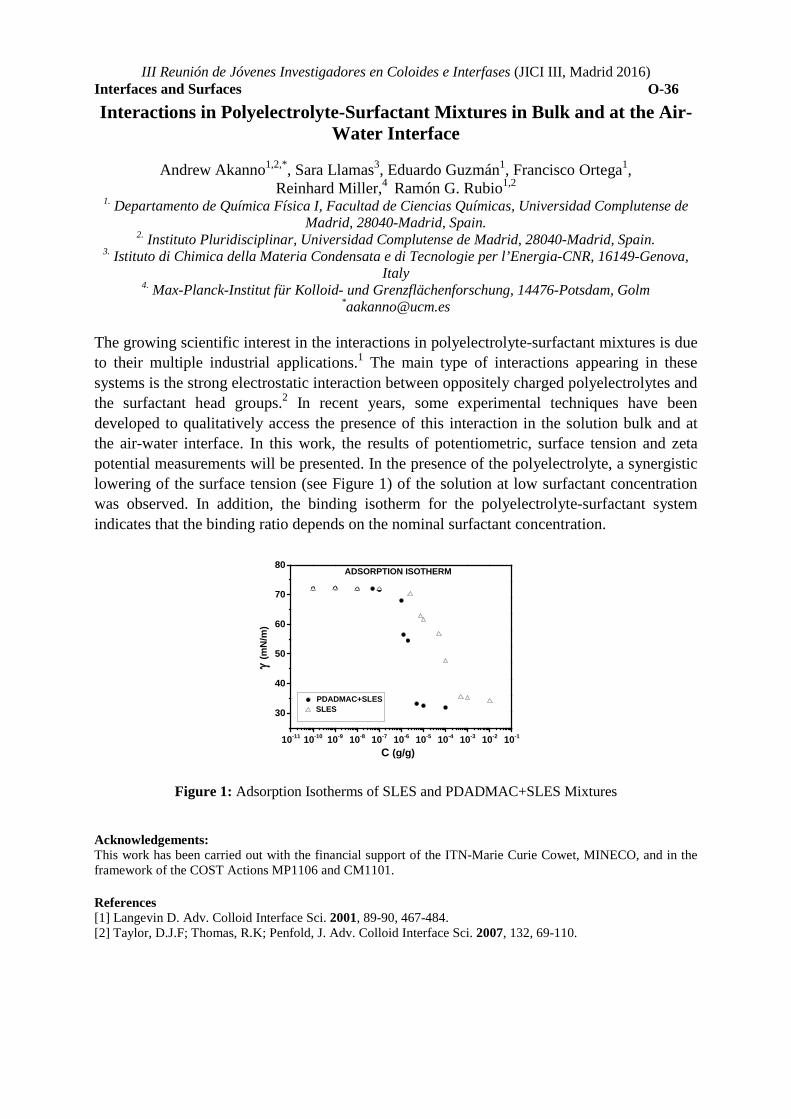
III Reunión de Jóvenes Investigadores en Coloides e Interfases (JICI III, Madrid 2016)
Interfaces and Surfaces O-36
Interactions in Polyelectrolyte-Surfactant Mixtures in Bulk and at the Air-Water Interface
Andrew Akanno1,2,*, Sara Llamas3, Eduardo Guzmán1, Francisco Ortega1,
Reinhard Miller,4 Ramón G. Rubio1,2 1. Departamento de Química Física I, Facultad de Ciencias Químicas, Universidad Complutense de
Madrid, 28040-Madrid, Spain. 2. Instituto Pluridisciplinar, Universidad Complutense de Madrid, 28040-Madrid, Spain.
3. Istituto di Chimica della Materia Condensata e di Tecnologie per l’Energia-CNR, 16149-Genova, Italy
4. Max-Planck-Institut für Kolloid- und Grenzflächenforschung, 14476-Potsdam, Golm *[email protected]
The growing scientific interest in the interactions in polyelectrolyte-surfactant mixtures is due to their multiple industrial applications.1 The main type of interactions appearing in these systems is the strong electrostatic interaction between oppositely charged polyelectrolytes and the surfactant head groups.2 In recent years, some experimental techniques have been developed to qualitatively access the presence of this interaction in the solution bulk and at the air-water interface. In this work, the results of potentiometric, surface tension and zeta potential measurements will be presented. In the presence of the polyelectrolyte, a synergistic lowering of the surface tension (see Figure 1) of the solution at low surfactant concentration was observed. In addition, the binding isotherm for the polyelectrolyte-surfactant system indicates that the binding ratio depends on the nominal surfactant concentration.
10-11 10-10 10-9 10-8 10-7 10-6 10-5 10-4 10-3 10-2 10-1
30
40
50
60
70
80
PDADMAC+SLES SLES
γ (m
N/m
)
C (g/g)
ADSORPTION ISOTHERM
Figure 1: Adsorption Isotherms of SLES and PDADMAC+SLES Mixtures
Acknowledgements: This work has been carried out with the financial support of the ITN-Marie Curie Cowet, MINECO, and in the framework of the COST Actions MP1106 and CM1101. References [1] Langevin D. Adv. Colloid Interface Sci. 2001, 89-90, 467-484. [2] Taylor, D.J.F; Thomas, R.K; Penfold, J. Adv. Colloid Interface Sci. 2007, 132, 69-110.
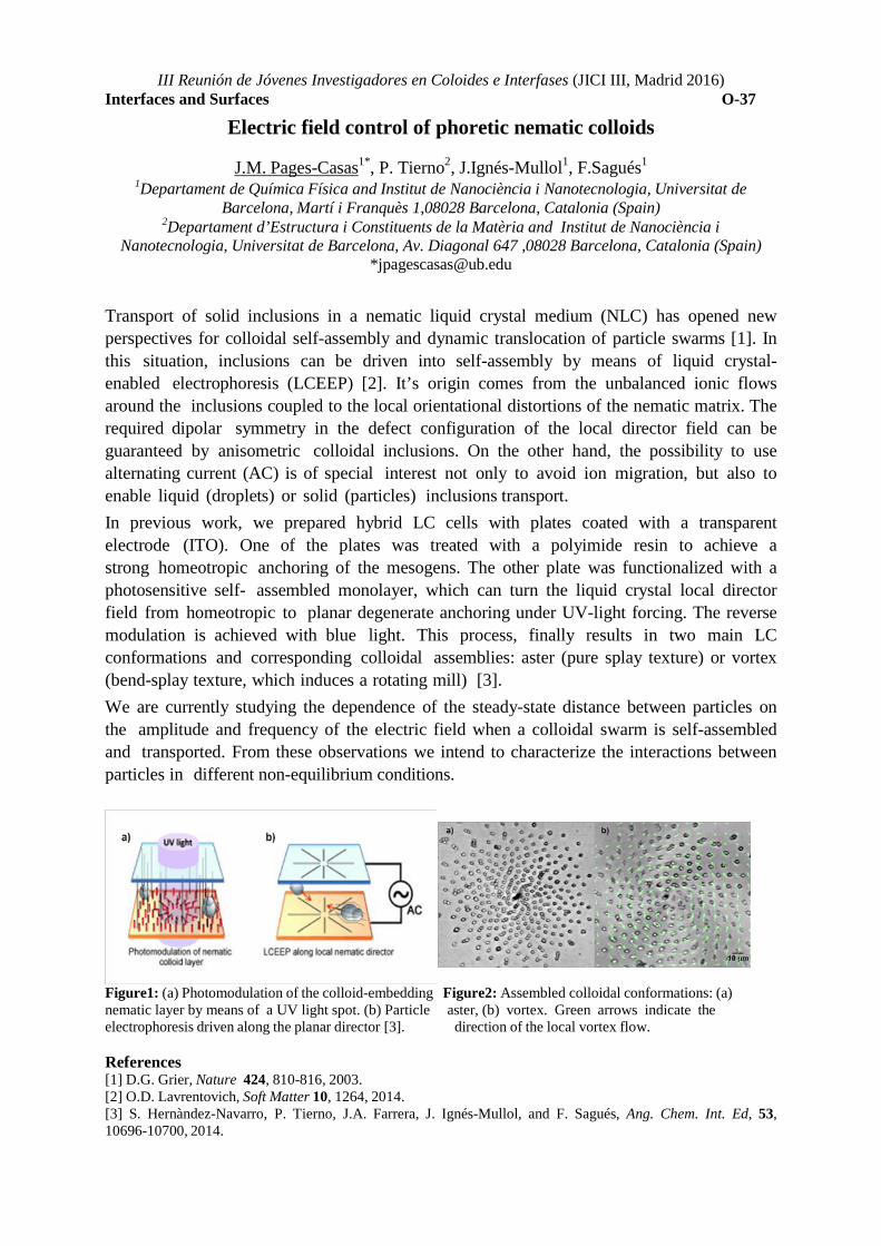
III Reunión de Jóvenes Investigadores en Coloides e Interfases (JICI III, Madrid 2016)
Interfaces and Surfaces O-37
Electric field control of phoretic nematic colloids
J.M. Pages-Casas1*, P. Tierno2, J.Ignés-Mullol1, F.Sagués1
1Departament de Química Física and Institut de Nanociència i Nanotecnologia, Universitat de Barcelona, Martí i Franquès 1,08028 Barcelona, Catalonia (Spain)
2Departament d’Estructura i Constituents de la Matèria and Institut de Nanociència i Nanotecnologia, Universitat de Barcelona, Av. Diagonal 647 ,08028 Barcelona, Catalonia (Spain)
Transport of solid inclusions in a nematic liquid crystal medium (NLC) has opened new perspectives for colloidal self-assembly and dynamic translocation of particle swarms [1]. In this situation, inclusions can be driven into self-assembly by means of liquid crystal-enabled electrophoresis (LCEEP) [2]. It’s origin comes from the unbalanced ionic flows around the inclusions coupled to the local orientational distortions of the nematic matrix. The required dipolar symmetry in the defect configuration of the local director field can be guaranteed by anisometric colloidal inclusions. On the other hand, the possibility to use alternating current (AC) is of special interest not only to avoid ion migration, but also to enable liquid (droplets) or solid (particles) inclusions transport. In previous work, we prepared hybrid LC cells with plates coated with a transparent electrode (ITO). One of the plates was treated with a polyimide resin to achieve a strong homeotropic anchoring of the mesogens. The other plate was functionalized with a photosensitive self- assembled monolayer, which can turn the liquid crystal local director field from homeotropic to planar degenerate anchoring under UV-light forcing. The reverse modulation is achieved with blue light. This process, finally results in two main LC conformations and corresponding colloidal assemblies: aster (pure splay texture) or vortex (bend-splay texture, which induces a rotating mill) [3]. We are currently studying the dependence of the steady-state distance between particles on the amplitude and frequency of the electric field when a colloidal swarm is self-assembled and transported. From these observations we intend to characterize the interactions between particles in different non-equilibrium conditions.
Figure1: (a) Photomodulation of the colloid-embedding Figure2: Assembled colloidal conformations: (a) nematic layer by means of a UV light spot. (b) Particle aster, (b) vortex. Green arrows indicate the electrophoresis driven along the planar director [3]. direction of the local vortex flow. References [1] D.G. Grier, Nature 424, 810-816, 2003. [2] O.D. Lavrentovich, Soft Matter 10, 1264, 2014. [3] S. Hernàndez-Navarro, P. Tierno, J.A. Farrera, J. Ignés-Mullol, and F. Sagués, Ang. Chem. Int. Ed, 53, 10696-10700, 2014.

TOPIC 7: CHARACTERIZATION

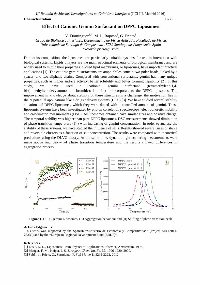
III Reunión de Jóvenes Investigadores en Coloides e Interfases (JICI III, Madrid 2016) Characterization O-38
Effect of Cationic Gemini Surfactant on DPPC Liposomes
V. Domínguez1,*, M. L. Raposo1, G. Prieto1
1Grupo de Biofísica e Interfases. Departamento de Física Aplicada. Facultade de Física. Universidade de Santiago de Compostela. 15782 Santiago de Compostela, Spain
*[email protected] Due to its composition, the liposomes are particularly suitable systems for use in interaction with biological systems. Lipids bilayers are the main structural elements of biological membranes and are widely used to mimic their properties. Closed lipid membranes, or liposomes, have important practical applications [1]. The cationic gemini surfactants are amphiphiles contain two polar heads, linked by a spacer, and two aliphatic chains. Compared with conventional surfactants, gemini has many unique properties, such as higher surface activity, better solubility and better forming capability [2]. In this study, we have used a cationic gemini surfactant (tetramethylene-1,4-bis(dimethyltetradecylammonium bromide); 14-4-14) to incorporate to the DPPC liposomes. The improvement in knowledge about stability of these structures is a challenge, the motivation lies in theirs potential applications like a drugs delivery systems (DDS) [3]. We have studied several stability situations of DPPC liposomes, which they were doped with a controlled amount of gemini. These liposomic systems have been investigated by photon correlation spectroscopy, electrophoretic mobility and calorimetric measurements (DSC). All liposomes obtained have similar sizes and positive charge. The temporal stability was higher than pure DPPC liposomes. DSC measurements showed diminution of phase transition temperature (TC) with increasing of gemini concentration. In order to analyse the stability of these systems, we have studied the influence of salts. Results showed several sizes of stable and reversible clusters as a function of salt concentration. The results were compared with theoretical predictions using the DLVO theory. At the same time, dynamic light scattering measurements were made above and below of phase transition temperature and the results showed differences in aggregation process.
Figure 1. DPPC/gemini Liposomes, (A) Aggregation behaviour and (B) Shifting of phase transition peak Acknowledgements: This work was supported by the Spanish “Ministerio de Economía y Competitividad” (Project MAT2011-26330) and by the “European Regional Development Fund (ERDF)”. References [1] Lasic, D. D., Liposomes: From Physics to Applications. Elsevier, Amsterdam. 1993. [2] Menger, F. M., Keiper, J. S. J. Angew. Chem. Int. Ed. 39, 1906-1920, 2000. [3] Sabín, J., Prieto, G., Sarmiento, F. Soft Matter 8, 3212-3222, 2012.
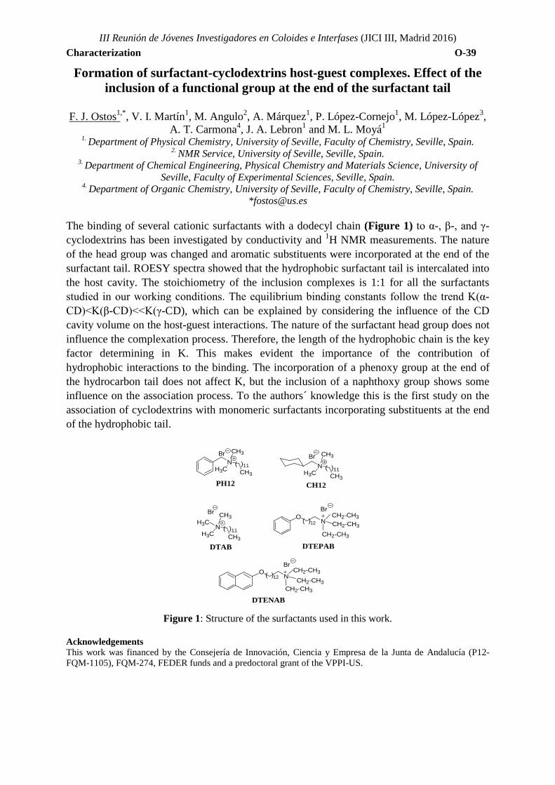
III Reunión de Jóvenes Investigadores en Coloides e Interfases (JICI III, Madrid 2016) Characterization O-39
NH3C
CH3
CH3
Br( )11 N
H3C
CH3
CH3
Br
( )11
PH12 CH12
NH3C
CH3
CH3
Br
( )11H3C
DTAB DTEPAB
ON
Br
DTENAB
( )12 CH2-CH3
CH2-CH3
CH2-CH3
ON( )12 CH2-CH3
CH2-CH3
CH2-CH3
Br
Formation of surfactant-cyclodextrins host-guest complexes. Effect of the inclusion of a functional group at the end of the surfactant tail
F. J. Ostos1,*, V. I. Martín1, M. Angulo2, A. Márquez1, P. López-Cornejo1, M. López-López3,
A. T. Carmona4, J. A. Lebron1 and M. L. Moyá1
1. Department of Physical Chemistry, University of Seville, Faculty of Chemistry, Seville, Spain. 2. NMR Service, University of Seville, Seville, Spain.
3. Department of Chemical Engineering, Physical Chemistry and Materials Science, University of Seville, Faculty of Experimental Sciences, Seville, Spain.
4. Department of Organic Chemistry, University of Seville, Faculty of Chemistry, Seville, Spain. *[email protected]
The binding of several cationic surfactants with a dodecyl chain (Figure 1) to α-, β-, and γ-cyclodextrins has been investigated by conductivity and 1H NMR measurements. The nature of the head group was changed and aromatic substituents were incorporated at the end of the surfactant tail. ROESY spectra showed that the hydrophobic surfactant tail is intercalated into the host cavity. The stoichiometry of the inclusion complexes is 1:1 for all the surfactants studied in our working conditions. The equilibrium binding constants follow the trend K(α-CD)<K(β-CD)<<K(γ-CD), which can be explained by considering the influence of the CD cavity volume on the host-guest interactions. The nature of the surfactant head group does not influence the complexation process. Therefore, the length of the hydrophobic chain is the key factor determining in K. This makes evident the importance of the contribution of hydrophobic interactions to the binding. The incorporation of a phenoxy group at the end of the hydrocarbon tail does not affect K, but the inclusion of a naphthoxy group shows some influence on the association process. To the authors´ knowledge this is the first study on the association of cyclodextrins with monomeric surfactants incorporating substituents at the end of the hydrophobic tail.
Figure 1: Structure of the surfactants used in this work. Acknowledgements This work was financed by the Consejería de Innovación, Ciencia y Empresa de la Junta de Andalucía (P12-FQM-1105), FQM-274, FEDER funds and a predoctoral grant of the VPPI-US.
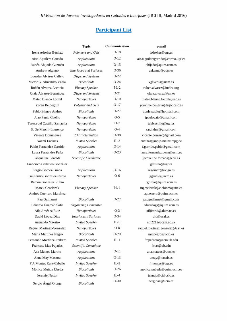
III Reunión de Jóvenes Investigadores en Coloides e Interfases (JICI III, Madrid 2016)
Participant List
Topic Communication e-mail
Irene Adroher Benítez Polymers and Gels O-18 [email protected]
Aixa Aguilera Garrido Applications O-12 [email protected]
Rubén Ahijado Guzmán Applications O-15 [email protected]
Andrew Akanno Interfaces and Surfaces O-36 [email protected]
Lourdes Alvárez Callejo Dispersed Systems O-22
Víctor G. Almendro Vedia Biocolloids O-24 [email protected]
Rubén Álvarez Asencio Plenary Speaker PL-2 [email protected]
Olaia Álvarez-Bermúdez Dispersed Systems O-21 [email protected]
Mateo Blanco Loimil Nanoparticles O-10 [email protected]
Yoran Beldegrun Polymer and Gels O-17 [email protected]
Pablo Blanco Andrés Biocolloids O-27 [email protected]
Joao Paulo Coelho Nanoparticles O-5 [email protected]
Teresa del Castillo Santaella Nanoparticles O-7 [email protected]
S. De Marchi-Lourenço Nanoparticles O-4 [email protected]
Vicente Domínguez Characterization O-38 [email protected]
Noemí Encinas Invited Speaker IL-3 [email protected]
Pablo Fernández Garrido Applications O-14 [email protected]
Laura Fernández Peña Biocolloids O-23 [email protected]
Jacqueline Forcada Scientific Committee [email protected] Francisco Gallisteo González
Sergio Gómez-Graña Applications O-16 [email protected]
Guillermo González-Rubio Nanoparticles O-6 [email protected]
Ramón González Rubio [email protected]
Marek Grzelczak Plenary Speaker PL-1 [email protected]
Andrés Guerrero Martínez [email protected]
Pau Guillamat Biocolloids O-27 [email protected]
Eduardo Guzmán Solís Organizing Committee [email protected]
Aila Jiménez Ruiz Nanoparticles O-3 [email protected]
David López Díaz Interfaces y Surfaces O-34 [email protected]
Armando Maestro Invited Speaker IL-5 [email protected]
Raquel Martínez-González Nanoparticles O-8 [email protected]
María Martínez Negro Biocolloids O-29 [email protected]
Fernando Martínez-Pedrero Invited Speaker IL-1 [email protected]
Francesc Mas Pujadas Scientific Committee [email protected]
Ana Mateos Maroto Applications O-11 [email protected]
Anna May Masnou Applications O-13 [email protected]
F.J. Montes Ruiz-Cabello Invited Speaker IL-2 [email protected]
Mónica Muñoz Ubeda Biocolloids O-26 [email protected]
Jeremie Nestor Invited Speaker IL-4 [email protected]
Sergio Ángel Ortega Biocolloids O-30 [email protected]
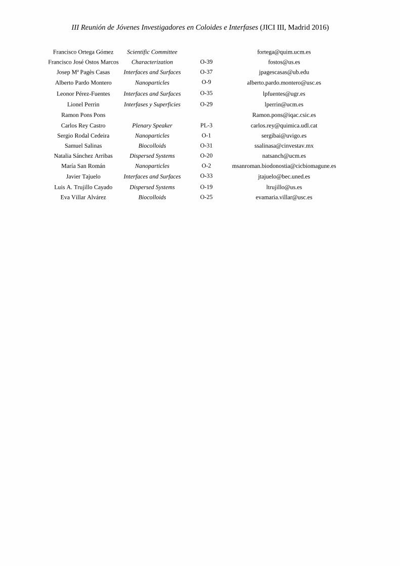
III Reunión de Jóvenes Investigadores en Coloides e Interfases (JICI III, Madrid 2016)
Francisco Ortega Gómez Scientific Committee [email protected]
Francisco José Ostos Marcos Characterization O-39 [email protected]
Josep Mª Pagès Casas Interfaces and Surfaces O-37 [email protected]
Alberto Pardo Montero Nanoparticles O-9 [email protected]
Leonor Pérez-Fuentes Interfaces and Surfaces O-35 [email protected]
Lionel Perrin Interfases y Superficies O-29 [email protected]
Ramon Pons Pons
Carlos Rey Castro Plenary Speaker PL-3 [email protected]
Sergio Rodal Cedeira Nanoparticles O-1 [email protected]
Samuel Salinas Biocolloids O-31 [email protected]
Natalia Sánchez Arribas Dispersed Systems O-20 [email protected]
María San Román Nanoparticles O-2 [email protected]
Javier Tajuelo Interfaces and Surfaces O-33 [email protected]
Luis A. Trujillo Cayado Dispersed Systems O-19 [email protected]
Eva Villar Alvárez Biocolloids O-25 [email protected]

