efectos del propranolol sobre el deterioro cognitivo y las...
TRANSCRIPT

Universidad de Navarra
Facultad de Farmacia
Efectos del Propranolol Sobre el Deterioro Cognitivo y
las Patologías Amiloide y Tau en Distintos Modelos
Experimentales de Enfermedad de Alzheimer
Marta Dobarro Bugarín


Efectos del Propranolol Sobre el Deterioro Cognitivo y las Patologías
Amiloide y Tau en Distintos Modelos Experimentales de
Enfermedad de Alzheimer
Memoria presentada por Dª Marta Dobarro Bugarín para aspirar al grado de Doctor
por la Universidad de Navarra
El presente trabajo ha sido realizado bajo mi dirección en el Departamento de
Farmacología y autorizo su presentación ante el Tribunal que lo ha de juzgar.
Pamplona, Junio de 2012
Fdo: Dra. María Javier Ramírez Gil


EEll vveerrddaaddeerroo ééxxiittoo rreessiiddee eenn eell eessffuueerrzzoo ppoorr llooggrraarr aallggoo……
EEll úúnniiccoo ffrraaccaassoo eess nnoo iinntteennttaarrlloo
((AAnnóónniimmoo))


AA mmiiss ppaaddrreess
AA mmii aabbuueellaa


Hace ya más de cuatro años que tomé la decisión de volver a Pamplona para
embarcarme en un mundo para mí desconocido, el mundo de la ciencia. Lo recuerdo como
un momento lleno de dudas y de miedo a equivocarme. Ahora que esta etapa ha llegado a
su fin, puedo decir que tomé la decisión acertada y que ha merecido la pena. Además de
todo lo que he aprendido, me llevo un montón de buenos momentos. Por eso quiero
agradecer en estas páginas a todos los que habéis formado parte de mi vida estos años, el
apoyo, la amistad y el cariño con el que me habéis tratado. Gracias por unos años
inolvidables.
Quisiera agradecer a la Dra. Edurne Cenarruzabeitia por abrirme las puertas del
Departamento de Farmacología. Y a Berta, por el ánimo y el cariño con que me has tratado
durante todo este tiempo y por tu constante preocupación por todos nosotros.
Gracias Mariaja. Has sido, sin lugar a dudas, una de las personas más importantes,
y sin tu ayuda no habría sido posible. Además de convertirme en doctora, estos años han
supuesto para mí un montón de cosas más. Y ha sido gracias a ti, por darme esta
oportunidad y hacerme un hueco en tu equipo cuando parecía imposible. Gracias por no
perder nunca la paciencia, por tus consejos científicos y personales, por tu cariño, y por la
seguridad y confianza que has sabido transmitirme cuando he estado a punto de
derrumbarme. En definitiva, gracias por tu trabajo a la hora de dirigir esta tesis y por el
gran esfuerzo final que ha hecho posible que todo salga bien. Muchísimas gracias.
Gracias al gran equipo que me acogió con tanto cariño en los primeros meses,
cuando todo era nuevo para mí. Porque siempre es más agradable trabajar acompañada,
pero sobre todo si es de personas como vosotros. A Maite, por ayudarme tanto sin pedir
nada a cambio, incluso ahora desde la distancia. Eres un ejemplo de trabajo y esfuerzo y
gracias a ti he aprendido todo lo que he necesitado para llegar hasta aquí. Barbi, gracias
por un millón de cosas. Por todos esos “momenticos”. Siempre has dejado tus problemas a
un lado cuando te he necesitado y no sé qué habría hecho sin las interminables horas de
terapia en nuestra covacha rosa. Gracias por ser mi compañera de piso y mi amiga, pero
sobre todo, por ser como una hermana para mí. Evitxu, gracias por el cariño que me has

dado desde el primer día y por tu ayuda en el trabajo. Por tus visitillas, por tu risa
contagiosa y por estar ahí siempre que he necesitado desahogarme.
Gracias también al resto de la gente que contribuyó a hacerme sentir como en casa
en tan poco tiempo. A Alvarito, gracias de tu lil’ thing. Por compartir tus “movidas”
conmigo. No me olvido de que tenemos una visita pendiente. A Elena, por tu alegría y
generosidad con todo el mundo, y porque siempre eres capaz de ponerte en el lugar de los
demás. Y a Natalia, por transmitirme tu tranquilidad y serenidad en los momentos clave.
Un agradecimiento especial a mis compis de cuartito durante estos últimos meses.
Por todas las horas que hemos pasado juntos. Gracias por aguantarme, soy consciente de
que no siempre ha sido fácil. Esta recta final sin vosotros habría sido, sin duda, mucho más
dura y aburrida. A Gorka, por darme siempre un punto de vista diferente, por tu paciencia y
tus bromas (no sé con quién te vas a meter ahora…). Por decirme siempre las cosas como
las piensas. Y por supuesto, por estos últimos meses que te he tenido explotado… mil
gracias. A Elitxu. Tú eres la responsable de que aterrizara en el depar y siempre te lo
agradeceré. Gracias por todo lo que hemos vivido juntas, antes y durante la tesis. Por tu
pasión por las pequeñas cosas, y por estar siempre dispuesta a compartir con todos tu
inmenso corazón. A Lucía, por tu amistad, tu compañía en esos viajes interminables y por
estar siempre dispuesta a escuchar. Creo que todo el mundo debería aprender algo de ti y
de tu capacidad para darle la vuelta a los momentos difíciles con sentido del humor. ¡Te voy
a echar mucho de menos!
Por supuesto, muchas gracias a todos los demás. A Lourdes, mi sampeochilla, al fin
y al cabo tú también has formado parte de este cuartito de“pretésicas”. Gracias por tus
mensajitos constantes de ánimo y por todas esas risas en el animalario. A Patxi, por estar
siempre dispuesto a echarme una mano cuando lo he necesitado. Gracias Merche, por tu
amabilidad y compañerismo. A Manuel y Xabi, por darle un aire nuevo al laboratorio. Os
deseo lo mejor a los 3 en esta nueva experiencia que comenzáis. A Luis, por compartir
nuestros últimos agobios por el pasillo. A Vero, porque enseguida fuiste una más y por
compartir con nosotros algunos de los mejores momentos del departamento. A María,
gracias por tu alegría y tu ilusión. A pesar de llevar tan poco tiempo has sido un gran apoyo
durante estos meses. A Mikel, gracias por soportarme siempre con tan buen humor. Ya sé
que en el fondo no te parezco tan borde. Y por supuesto a Bea, gracias por volver y darnos
la oportunidad de conocerte. Has sido como una doctoranda más y en muchas ocasiones
has sido capaz de entenderme mejor que nadie.
Quisiera dar las gracias al resto de personas que llevan tanto tiempo formando el
depar de Farma, haciendo que el trabajo diario sea más llevadero. A Rosa, por tu sonrisa,
amabilidad y muestras de interés. A Popi, por tratarme siempre con cariño y por los
momentos compartidos en el “club de fumadores”. A Pili, por tu alegría constante y por
estar siempre al pie del cañón, pendiente de cada uno de nosotros. A Mari Luz, gracias por
esas miles de muestras que sé que te han vuelto loca. Y porque para mí has sido como una
madre y una amiga a la vez. El depar no sería lo mismo sin ti. Gracias a Sandra, Guadalupe
e Idoia por los momentos de desconexión al mediodía. Gracias a vosotras quedarse a comer
siempre da menos pereza. Y no me olvido Sandri, de todas esas membranas, incubaciones y
demás. Muchísimas gracias.
Me gustaría agradecer también a todas aquellas personas que han pasado por el
departamento: masterianos, alumnos internos… porque todos habéis dejado vuestra huella
de alguna manera.
No puedo olvidarme de la gente que ha estado conmigo día a día fuera del
laboratorio, esforzándose por entender todas mis preocupaciones aún sin saber muy bien
de qué les estaba hablando.
A Andrea, por todos esos meses que compartimos sofá y ser capaz de aguantar
todas nuestras chapas casi sin rechistar. Y por aportar, junto con Tepy, ese punto de locura
de la pareja cómico-violenta. Gracias a vosotros también.
A mis niñas de Ferrol, por ser las mejores amigas del mundo. Por todo lo que hemos
compartido desde pequeñas. Da igual los meses que pasen sin vernos, porque cuando nos
juntamos es como si no hubiera pasado el tiempo.

A ti David, que te ha tocado vivir esta tesis de cerca el último año. Gracias por
escucharme y animarme cuando lo he necesitado. Por conseguir ver siempre el lado
positivo de las cosas, haciéndolo todo más fácil. Pero sobre todo gracias por ser capaz de
sacarme siempre una sonrisa. Para mí ha sido muy importante tenerte a mi lado y espero
que siga siendo así por mucho tiempo.
Por último me gustaría dar las gracias a mi familia. Porque aunque nos veamos
menos de lo que me gustaría, siempre he recibido vuestro cariño. Especialmente a mis
padres, porque gracias a vosotros nunca me he sentido sola. Sois lo más importante y esta
tesis es también vuestra. No habría llegado hasta aquí sin vuestro apoyo incondicional
durante todos estos años. Gracias por vuestra confianza, vuestros ánimos y por recordarme
día a día que soy capaz de muchas cosas. Nunca podré agradecéroslo lo suficiente. Y a ti,
Juls, por todas esas cosas que nos hacen tan diferentes y de las que tengo tanto que
aprender: tu seguridad, tu perseverancia y tu paciencia. Estoy segura de que tu empeño y
entusiasmo te harán llegar muy lejos en todo lo que te propongas. Pero sobre todo, gracias
por estar ahí. Porque pase lo que pase sé que siempre voy a poder contar contigo.
Gracias a todos…
La realización de este trabajo no hubiera sido posible sin la financiación del Departamento de
Industria del Gobierno de Navarra y el proyecto de Caja Navarra “Tú eliges, Tú decides”, y sin la ayuda de la
Asociación de Amigos de la Universidad de Navarra.


Abreviaturas más empleadas
A: péptido -amiloide
AICD: fragmento carboxi-terminal intracelular de la proteína precursora del -amiloide
AINEs: fármacos antiinflamatorios no esteroideos
ApoE: apolipoproteína E
APP: proteína precursora del -amiloide
BACE1: -secretasa
BDNF: factor neurotrófico derivado de cerebro
BHE: barrera hematoencefálica
C83: fragmento carboxilo terminal de 83 aminoácidos
C99: fragmento carboxilo terminal de 99 aminoácidos
CaMKII: quinasa II dependiente de Ca2+/calmodulina
Cdk5: quinasa dependiente de ciclina 5
DM2: diabetes mellitus tipo 2
EA: enfermedad de Alzheimer, Alzheimer’s disease (AD)
ECS: estrés crónico suave
FAD: enfermedad de Alzheimer de origen familiar
GC: glucocorticoides
GSK3β: quinasa glicógeno sintasa 3β
HFD: dieta hipercalórica, high fat diet
IDE: enzima degradadora de insulina, insulin degrading enzyme
IR: receptor de insulina, insulin receptor
IRS: sustrato del receptor de la insulina, insulin receptor substrate
JNK: quinasa N-terminal de la c-Jun
LCR: líquido cefalorraquídeo
LTP: potenciación sináptica a largo plazo
MAP: proteína asociada a microtúbulos
MARK: quinasa reguladora de la afinidad de microtúbulos
MCI: deterioro cognitivo leve, mild cognitive impairment
MS: separación materna, maternal separation

NMDA: receptor N-metil-D-aspartato
NORT: test de reconocimiento de nuevo objeto
OMS: Organización Mundial de la Salud
ONF: ovillo neurofibrilar
PHF: filamentos helicoidales pareados
PI3K: fosfoinositol 3 quinasa
PKA: quinasa dependiente de adenosín monofosfato cíclico tipo A
PKC: quinasa dependiente de adenosín monofosfato cíclico tipo C
PS: presenilina
pTau: tau fosforilada
ARNm: ARN mensajero
SAMP8: ratones con tendencia a la senescencia acelerada, senescence accelerated mice
prone-8
SAMR1: ratones resistentes a la senescencia acelerada, senescence accelerated mice
resistant-1
SNC: Sistema Nervioso Central

ÍÍnnddiiccee

Capítulo I: Introducción ........................................................................................1
1. Enfermedad de Alzheimer ..................................................................................................... 5
1.1. Definición y epidemiología .................................................................................... 5
1.2. Factores de riesgo.................................................................................................. 7
1.3. Patogénesis.......................................................................................................... 12
1.4. Etiología ............................................................................................................... 14
1.4.1. Hipótesis de la cascada del amiloide...................................................... 15
1.4.2. Hipótesis de la proteína Tau................................................................... 18
1.4.2. Patología sináptica ................................................................................. 20
2. Tratamiento de la Enfermedad de Alzheimer ..................................................................... 22
2.1. Terapias actuales ................................................................................................. 23
2.2. Nuevas terapias ................................................................................................... 24
3. Hipertensión y Enfermedad de Alzheimer .......................................................................... 27
4. Modelos animales para el estudio de la Enfermedad de Alzheimer................................... 29
4.1. Modelos animales de EA familiar ........................................................................ 30
4.1.1. Modelos transgénicos de APP ................................................................ 30
4.1.2. Modelos transgénicos de PS................................................................... 31
4.1.3. Modelos transgénicos de Tau ................................................................ 32
4.1.4. Dobles y triples transgénicos.................................................................. 33
4.1.5. Modelos transgénicos 5xFAD APP/PS1 .................................................. 33
4.2. Modelos animales de envejecimiento................................................................. 34
4.3. Modelos animales de EA esporádica ................................................................... 34
4.3.1. Modelos animales genéticos.................................................................. 35
4.3.2. Modelos animales metabólicos.............................................................. 35
4.3.3. Modelos animales de estrés................................................................... 36
Bibliografía............................................................................................................................... 40

Capítulo II: Planteamiento y Objetivos................................................................. 77
Bibliografía............................................................................................................................... 83
Capítulo III: Propranolol restores cognitive deficits and improves amyloid and
Tau pathologies in a senescence-accelerated mouse model ................................. 85
Resumen.................................................................................................................................. 89
Abstract ................................................................................................................................... 90
1. Introduction......................................................................................................................... 91
2. Material and Methods......................................................................................................... 92
2.1. Animals ................................................................................................................ 92
2.2. Drug treatment and experimental design ........................................................... 93
2.3. Behavioral experiments....................................................................................... 93
2.3.1. Locomotor activity.................................................................................. 93
2.3.2. Novel object recognition test ................................................................. 93
2.4. Biochemical measurements ................................................................................ 94
2.4.1. Tissue and blood collection .................................................................... 94
2.4.2. Western blotting .................................................................................... 94
2.4.3. Insulin levels ........................................................................................... 96
2.4.4. A levels ................................................................................................. 96
2.5. Data analysis and statistics .................................................................................. 96
3. Results ................................................................................................................................. 96
3.1. Effect of propranolol treatment on memory deficits in SAMP8 ......................... 96
3.2. Effects of propranolol on Aβ processing and clearance ...................................... 98
3.3. Effects of propranolol on Tau pathology............................................................. 99
3.4. Effects of propranolol on synaptic plasticity ..................................................... 101
4. Discussion .......................................................................................................................... 103
References............................................................................................................................. 107

Capítulo IV: Propranolol reduces cognitive deficits, amyloid and Tau pathology
in Alzheimer’s transgenic mice .......................................................................... 115
Resumen................................................................................................................................ 119
Abstract ................................................................................................................................. 120
1. Introduction....................................................................................................................... 121
2. Material and Methods....................................................................................................... 122
2.1. Animals .............................................................................................................. 122
2.2. Drug treatment and experimental design ......................................................... 123
2.3. Behavioural experiments................................................................................... 123
2.3.1. Locomotor activity................................................................................ 124
2.3.2. Object recognition................................................................................ 124
2.3.2. Fear conditioning test........................................................................... 124
2.4. Biochemical measurements .............................................................................. 125
2.4.1. Tissue and blood collection .................................................................. 125
2.4.2. Western blotting .................................................................................. 125
2.4.3. Aβ levels ............................................................................................... 126
2.5. Neuronal primary culture .................................................................................. 127
2.6. Data analysis and statistics ................................................................................ 127
3. Results ............................................................................................................................... 128
3.1. Propranolol restored cognitive function in Tg2576 mice .................................. 128
3.2. Propranolol decreases Aβ burden and protects against Aβ toxicity ................. 129
3.3. Effects of propranolol on markers of synaptic plasticity................................... 131
3.4. Effects of propranolol on Tau pathology........................................................... 133
4. Discussion .......................................................................................................................... 134
References............................................................................................................................. 139

Capítulo V: Propranolol reduces cognitive deficits, amyloid β levels, Tau
phosphorylation and insulin resistance in response to chronic corticosterone
administration .................................................................................................. 147
Resumen................................................................................................................................ 151
Abstract ................................................................................................................................. 152
1. Introduction....................................................................................................................... 153
2. Material and Methods....................................................................................................... 154
2.1. Animals .............................................................................................................. 154
2.2. Drug treatment and experimental design ......................................................... 155
2.3. Behavioral tests ................................................................................................. 155
2.3.1. Locomotor activity................................................................................ 155
2.3.2. Cognitive testing: novel object recognition test (NORT)...................... 156
2.4. Tissue and blood collection ............................................................................... 156
2.5. Insulin measurements ....................................................................................... 156
2.6. Western blotting................................................................................................ 157
2.7. Aβ levels ............................................................................................................. 158
2.8. Data analysis and statistics ................................................................................ 158
3. Results ............................................................................................................................... 158
3.1. Effects of propranolol on corticosterone-treated mice: cognitive studies ....... 158
3.2. Propranolol decreases Aβ burden in corticosterone-treated mice ................... 160
3.3. Effects of propranolol on corticosterone treatment on insulin and insulin-
triggered are reversed by propranolol treatment: implications in Tau pathology 161
4. Discussion .......................................................................................................................... 164
4.1. Effects on cognition ........................................................................................... 164
4.2. Propranolol decreases Aβ burden, probably through an increased IDE
expression................................................................................................................. 165
4.3. Corticosterone treatments lead to alterations in the insulin receptor
pathway that are reversed by propranolol............................................................... 165
References............................................................................................................................. 168

Capítulo VI: Discusión........................................................................................ 177
1. Efectos del propranolol sobre los procesos cognitivos ........................................ 186
2. Efectos del propranolol sobre la patología amiloide............................................ 186
3. Efectos del propranolol sobre la patología de Tau............................................... 188
4. Efectos del propranolol sobre la patología sináptica ........................................... 190
5. Conclusión general................................................................................................ 192
Bibliografía ........................................................................................................................ 193
Capítulo VII: Conclusiones ................................................................................. 207


CCaappííttuulloo II


Capítulo 1
Introducción

Introducción
5
1. Enfermedad de Alzheimer
1.1. Definición y epidemiología
La enfermedad de Alzheimer (EA), la causa más frecuente de demencia en los
ancianos, es una patología degenerativa cerebral irreversible, caracterizada por el
deterioro gradual de neuronas focalizada principalmente en el hipocampo y la corteza
cerebral, cuya causa no es del todo conocida. Desde un punto de vista clínico, la EA se
caracteriza por un deterioro progresivo de las funciones cognitivas. El síntoma inicial más
frecuente es una pérdida de memoria a la que se añaden, a medida que avanza la
enfermedad, un deterioro en otras capacidades como el lenguaje, el razonamiento y la
orientación temporo-espacial, además de cambios en la personalidad y la conducta.
Es una enfermedad con una importante trascendencia económica y social, puesto
que cursa con una pérdida gradual de la capacidad del paciente para valerse por sí mismo.
Este hecho hace que en las últimas fases de la enfermedad, el paciente dependa
totalmente de sus familiares y/o sus cuidadores, que deben enfrentarse diariamente a
problemas como la desorientación, la agresividad o su aseo.
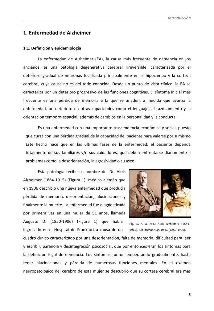
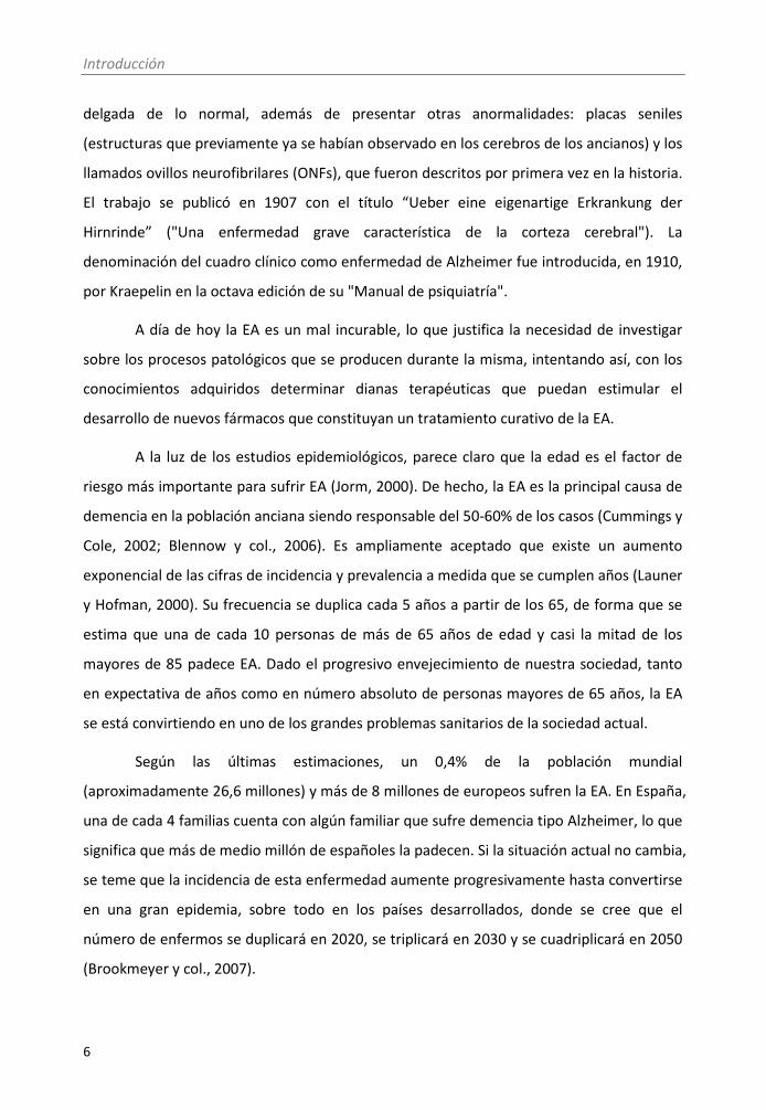
Esta patología recibe su nombre del Dr. Alois
Alzheimer (1864-1915) (Figura 1), médico alemán que
en 1906 describió una nueva enfermedad que producía
pérdida de memoria, desorientación, alucinaciones y
finalmente la muerte. La enfermedad fue diagnosticada
por primera vez en una mujer de 51 años, llamada
Auguste D. (1850-1906) (Figura 1) que había
ingresado en el Hospital de Frankfurt a causa de un
cuadro clínico caracterizado por una desorientación, falta de memoria, dificultad para leer
y escribir, paranoia y desintegración psicosocial, que por entonces eran los síntomas para
la definición legal de demencia. Los síntomas fueron empeorando gradualmente, hasta
tener alucinaciones y pérdida de numerosas funciones mentales. En el examen
neuropatológico del cerebro de esta mujer se descubrió que su corteza cerebral era más
Fig. 1. A la izda.: Alois Alzheimer (1864-
1915). A la drcha: Auguste D. (1850-1906).
Introducción
6
delgada de lo normal, además de presentar otras anormalidades: placas seniles
(estructuras que previamente ya se habían observado en los cerebros de los ancianos) y los
llamados ovillos neurofibrilares (ONFs), que fueron descritos por primera vez en la historia.
El trabajo se publicó en 1907 con el título “Ueber eine eigenartige Erkrankung der
Hirnrinde” ("Una enfermedad grave característica de la corteza cerebral"). La
denominación del cuadro clínico como enfermedad de Alzheimer fue introducida, en 1910,
por Kraepelin en la octava edición de su "Manual de psiquiatría".
A día de hoy la EA es un mal incurable, lo que justifica la necesidad de investigar
sobre los procesos patológicos que se producen durante la misma, intentando así, con los
conocimientos adquiridos determinar dianas terapéuticas que puedan estimular el
desarrollo de nuevos fármacos que constituyan un tratamiento curativo de la EA.
A la luz de los estudios epidemiológicos, parece claro que la edad es el factor de
riesgo más importante para sufrir EA (Jorm, 2000). De hecho, la EA es la principal causa de
demencia en la población anciana siendo responsable del 50-60% de los casos (Cummings y
Cole, 2002; Blennow y col., 2006). Es ampliamente aceptado que existe un aumento
exponencial de las cifras de incidencia y prevalencia a medida que se cumplen años (Launer
y Hofman, 2000). Su frecuencia se duplica cada 5 años a partir de los 65, de forma que se
estima que una de cada 10 personas de más de 65 años de edad y casi la mitad de los
mayores de 85 padece EA. Dado el progresivo envejecimiento de nuestra sociedad, tanto
en expectativa de años como en número absoluto de personas mayores de 65 años, la EA
se está convirtiendo en uno de los grandes problemas sanitarios de la sociedad actual.
Según las últimas estimaciones, un 0,4% de la población mundial
(aproximadamente 26,6 millones) y más de 8 millones de europeos sufren la EA. En España,
una de cada 4 familias cuenta con algún familiar que sufre demencia tipo Alzheimer, lo que
significa que más de medio millón de españoles la padecen. Si la situación actual no cambia,
se teme que la incidencia de esta enfermedad aumente progresivamente hasta convertirse
en una gran epidemia, sobre todo en los países desarrollados, donde se cree que el
número de enfermos se duplicará en 2020, se triplicará en 2030 y se cuadriplicará en 2050
(Brookmeyer y col., 2007).

Introducción
7
Actualmente se considera la EA como la tercera enfermedad más costosa detrás de
las enfermedades cardiovasculares y el cáncer. Se calcula que el coste medio anual por
paciente de EA en España es de entre 12000 y 24000 € (Durán, 2000; Atance, 2004), por lo
que toda terapia encaminada a ralentizar la progresión puede repercutir significativamente
en los costes de la misma.
En la mayoría de los casos se desconocen las causas que producen la EA. Se estima
que entre un 1 y un 5% de los casos, la enfermedad se puede considerar que es de origen
hereditario (“Enfermedad de Alzheimer familiar” o FAD por sus siglas en inglés), en el que
la enfermedad se presenta antes de los 65 años. En estos casos, el factor genético parece
ser el principal responsable de la enfermedad. Los casos restantes, en los que la
enfermedad aparece después de los 65 años de edad, se consideran “EA esporádica o de
aparición tardía”.
En cuanto a la EA familiar, hasta el momento se han identificado 3 mutaciones que
la causan: en el cromosoma 21, concretamente en el gen que codifica la proteína
precursora del β-amiloide (APP); en el cromosoma 14, en el gen que codifica las
presenilinas 1 y 2 (PS1 y PS2); y en el cromosoma 1, en el gen que codifica la PS2. En los 3
casos, las mutaciones tienen como consecuencia un aumento en la producción del péptido
Aβ (ver punto 1.4.1. de la Introducción).
1.2. Factores de riesgo
En la EA esporádica se han descrito múltiples factores de riesgo que a continuación
pasan a ser brevemente descritos:
- Edad:
El envejecimiento es, según la OMS, el conjunto de modificaciones morfológicas y
fisiológicas que aparecen con el paso del tiempo en los seres vivos, y supone, entre otras
cosas, una disminución de la capacidad de respuesta a los distintos agentes nocivos que
inciden sobre el individuo. Dicho proceso de envejecimiento se caracteriza por una pérdida
funcional generalizada que aumenta tanto el riesgo de mortalidad como el de padecer
ciertas enfermedades entre las que se encuentran las enfermedades neurodegenerativas,

Introducción
8
siendo una de las más frecuentes la EA. Los radicales libres, el genoma y la acumulación de
distintas sustancias producidas por el metabolismo celular son, entre otros, distintos
factores que se han identificado como posibles responsables del envejecimiento del
individuo. En base a estas consideraciones, la edad puede considerarse como el factor de
riesgo más importante de la EA, puesto que como se ha comentado en el punto 1.1., la
mayor prevalencia de la EA se encuentra en personas mayores de 65 años, edad a partir de
la cual el riesgo de padecer la enfermedad se duplica cada 5 años.
- Factores de riesgo genético:
Existe una predisposición genética para sufrir la enfermedad en ciertos genotipos,
principalmente el polimorfismo para la apolipoproteína E (ApoE). En concreto, el haplotipo
E4 (ApoE4) triplica el riesgo de padecer la enfermedad en el caso de los heterocigotos y lo
multiplica por 10 en el caso de los homocigotos, respecto a los haplotipos ApoE2 Y ApoE3
(Huang, 2006; He y col., 2007).
- Factores de riesgo ambiental:
Salvo en el caso de la EA familiar, donde una única causa se relaciona con la
aparición de la enfermedad (el componente genético), se podría considerar a la EA como
una enfermedad de origen multifactorial, que no se debe a una única causa sino a diversos
factores que contribuyen a su aparición y desarrollo.
Enfermedad cardíaca y cerebrovascular:
La enfermedad cardiovascular está asociada a un mayor riesgo de padecer EA,
especialmente en personas con aterosclerosis (Newman y col., 2005). El fallo cardíaco y la
fibrilación auricular también podrían estar asociados a la demencia. De las misma manera,
los infartos cerebrales múltiples y recurrentes se han asociado a déficit cognitivo y a la
aparición de EA (Purandare y col., 2006).
Hipertensión
Hay argumentos de índole epidemiológico y experimental que dan base racional a
la posible interacción entre la hipertensión y la EA. Además, el tratamiento con

Introducción
9
antihipertensivos parece reducir la aparición de deterioro cognitivo y el riesgo de
demencia. Este punto va a ser desarrollado en el apartado 3 de la presente Introducción.
Hiperlipidemia:
Diversos estudios sugieren que puede haber factores asociados con el
metabolismo lipídico en la génesis o desarrollo del EA (Whitmer y col., 2005). Una cifra de
colesterol elevado (> 6,5 mmol/L), asociada o no a hipertensión arterial, hacia los 50 años
es factor de riesgo de deterioro cognitivo leve (MCI) hacia los 70 (Kivipelto y col., 2001a).
En cuanto a los ácidos grasos, la ingesta moderada o elevada de grasas insaturadas es un
factor protector, mientras que la ingesta moderada de grasas saturadas incrementa el
riesgo de padecer EA (Kalmijn y col., 1997; Morris y col., 2003), sobre todo en aquellos
individuos ApoE4 (Laitinen y col., 2006). Estos ácidos grasos podrían conducir al desarrollo
de la enfermedad por diversos mecanismos como la aterosclerosis o la inflamación.
Obesidad y diabetes:
Comienza a demostrarse la relación entre obesidad y diabetes con el deterioro
cognitivo y la demencia (Trakas y col., 2001), quizá asociado a la relación que puede existir
entre la obesidad y la enfermedad cardiovascular. Un alto índice de masa corporal en la
edad adulta se relaciona con un incremento en el riesgo de padecer demencia en la vejez
(Kivipelto y col., 2005).
Por otro lado, un gran número de investigaciones han relacionado la demencia
(Stewart y Liolitsa, 1999), incluyendo la EA (Biessels y Kappelle, 2005), con la hiperglicemia
y la resistencia a la insulina (Steen y col., 2005; De la Monte y col., 2006). Una elevada
proporción de afectados por la EA muestran altos niveles de insulina y baja utilización de
glucosa, perfil que es característico de una resistencia a la insulina (Craft y col., 1996).
Numerosos estudios epidemiológicos han demostrado que el riesgo de desarrollar EA es
mayor entre los pacientes con diabetes mellitus de tipo 2 (DM2) (Leibson y col., 1997; Ott y
col., 1999; Luchsinger y col., 2001; Arvanitakis y col., 2004; Biessels y Kappelle, 2005), por
lo que pudiera sugerirse que la hiperinsulinemia y la resistencia a la insulina pueden ser un
factor de riesgo para el desarrollo de la EA.
Introducción
10
Estrés:
Hay cada vez más estudios que demuestran una posible relación entre el estrés y
los glucocorticoides (GC, sistema efector de la respuesta al estrés en el organismo), con un
aumento en el riesgo para padecer EA. La implicación de los GC en los procesos de
aprendizaje y memoria está ampliamente aceptada. El estrés y los GC tienen un gran
impacto sobre la ejecución cognitiva. El papel de los GC en el SNC puede ser tanto
neuroprotector como neurodegenerativo, dependiendo de la concentración de los mismos
(Abraham y col., 2001). Niveles basales normales de GC son necesarios para la
consolidación de la memoria a largo plazo y los aumentos puntuales en la secreción de los
GC durante el aprendizaje refuerzan la memoria espacial (Brinks y col., 2007; Sandi y
Pinelo-Nava, 2007). En cambio, la exposición crónica a niveles altos de GC produce
alteraciones que eventualmente generan disfunción, atrofia y muerte neuronal en el
hipocampo. Como consecuencia, se provoca una reducción del aprendizaje espacial y la
memoria, tareas que dependen en parte del buen funcionamiento del hipocampo (De
Kloet y col., 1999; Roozendaal y col., 2003).
Se ha encontrado un aumento en la EA de los niveles de cortisol en sangre, líquido
cefalorraquídeo (LCR) y saliva (Martignoni y col., 1990; Hartmann y col., 1997; Weiner y col.,
1997; Hatfield y col., 2004; Elgh y col., 2006; Hoogendijk y col., 2006; Popp y col., 2009; Gil-
Bea y col., 2010). De hecho, se ha sugerido que los niveles elevados de cortisol y el estrés
podrían contribuir al desarrollo y mantenimiento de la enfermedad. Esta hipótesis ha sido
apoyada por estudios que demuestran que situaciones de estrés o niveles elevados de GC
exacerban la sintomatología en los modelos transgénicos de EA (Green y col., 2006; Jeong y
col., 2006). Por todo ello, se ha sugerido que los niveles de cortisol en enfermos de EA
podrían tener relevancia pronóstica. En relación con este hecho, Csernansky y col. (2006)
demostraron que niveles elevados de cortisol en sangre durante la fase clínica de pre-
demencia predice un deterioro cognitivo más rápido. Por otro lado, se han observado
niveles elevados de cortisol en LCR en pacientes con EA pero no en personas con MCI, lo
que sugiere que el aumento de esta hormona en LCR se relaciona con la progresión de la
enfermedad (Popp y col., 2009).

Introducción
11
Depresión:
Además de que la depresión puede ser síntoma prodrómico de la EA, parece existir
una asociación positiva entre historia de depresión y posterior desarrollo de EA (Jorm,
2000). Estudios epidemiológicos señalan que los pacientes afectados por la EA tienen una
alta prevalencia de depresión siendo ésta entre el 10 y el 50% (Butters y col., 2008). Dado
que los síntomas depresivos de relevancia clínica se encuentran aproximadamente en un
20% de los pacientes enfermos de EA (Reifler y col. 1982; Patterson y col., 1990; Starkstein
y col., 1997), se sugiere que ambas enfermedades puedan compartir algunos mecanismos
patofisiológicos.
También existen estudios que indican que tener historia previa de depresión puede
doblar o hasta cuadruplicar el riesgo de desarrollar demencia de tipo Alzheimer (Zalsman y
col., 2000; Pfenning y col., 2007). Además, pacientes deprimidos con MCI presentan un
mayor riesgo de desarollar EA que sujetos no deprimidos únicamente con MCI (Modrego y
Fernandez, 2004). También existen estudios mostrando que los enfermos de EA con
antecedentes de depresión muestran un aumento de algunos marcadores
neuropatológicos de la EA (Rapp y col., 2006). Por otro lado, a pesar de que en la depresión
no se ha observado una elevada pérdida neuronal, varios estudios han mostrado un
incremento en los marcadores pro-apoptóticos, una disminución en los marcadores anti-
apoptóticos y alteraciones en los niveles de BDNF en pacientes con depresión (Chen y col.,
2001) y en modelos experimentales de depresión (Luo y col., 2004; Bachis y col., 2008).
- Inflamación:
Mientras que en cerebros de personas de avanzada edad sin patología se pueden
encontrar ligeros signos de neuroinflamación, en aquellos con EA este factor aparece con
mucha mayor intensidad, observándose una significativa activación de la microglía. Existen
evidencias de que el péptido Aβ, principal componente de las placas seniles, puede activar
la microglía (Paresce y col., 1996; Yan y col., 1998). Además, diversos estudios han
observado la activación de astrocitos y de la microglía sin presencia de placas seniles tanto
en modelos animales de EA (Heneka y col., 2005) como en humanos (Cagnin y col., 2001),

Introducción
12
Cerebro en fasesavanzadas de la EA
del hipocampo
Retracciónsevera de la
corteza
Expansiónventricular
severa
Encogimientosevero delhipocampo
Cerebro en fasesavanzadas de la EA
del hipocampo
Retracciónsevera de la
corteza
Expansiónventricular
severa
Encogimientosevero delhipocampo
Cerebro sano
Cortezacerebral
Hipocampo
Cerebro sano
Cortezacerebral
Hipocampo
Cerebro en fasesavanzadas de la EA
del hipocampo
Retracciónsevera de la
corteza
Expansiónventricular
severa
Encogimientosevero delhipocampo
Cerebro en fasesavanzadas de la EA
del hipocampo
Retracciónsevera de la
corteza
Expansiónventricular
severa
Encogimientosevero delhipocampo
Cerebro sano
Cortezacerebral
Hipocampo
Cerebro sano
Cortezacerebral
Hipocampo
demostrando la importancia, al menos al comienzo de la enfermedad, de los oligómeros de
Aβ en la activación de la glía.
- Otros factores:
Otros factores importantes en la EA son el sexo, ya que se aprecia un riesgo mayor
de padecer la enfermedad en las mujeres (en proporción aproximada de 3 a 1), en
particular entre la población mayor de 85 años (Di Carlo y col., 2002; Schmidt y col., 2008),
el tabaquismo, la intoxicación crónica por ciertos metales como el cobre, el nivel de
educación, los traumatismos craneoencefálicos graves, etc.
1.3. Patogénesis
Macroscópicamente, cerebros post-mortem de EA revelan una profunda atrofia
cerebral que afecta,
principalmente, a áreas
cerebrales relacionadas
con el aprendizaje y la
memoria como las
cortezas temporal,
parietal y frontal, el
hipocampo y la
amígdala (Figura 2).
La reducción en el volumen cerebral se debe, principalmente, a una importante
degeneración neuronal y sináptica (Mattson, 2004).
Histopatológicamente, los cerebros
de pacientes de EA muestran dos
estructuras patológicas características: las
placas seniles extracelulares y los ONFs
intracelulares (Figura 3). Placas y ovillos
son estructuras proteicas anormales que
se depositan en la corteza y el hipocampo a medida que aparece la neurodegeneración
Fig. 3. Principales marcadores histopatológicos de la EA.
A) Placas seniles B) Ovillos neurofibrilares
Fig. 2. Caracterísicas macroscópicas de la EA, atrofia cerebral. Ilustración de Bob
Morreale adaptada al castellano (American Health Assistance Foundation
www.ahaf.org/alzheimers).

Introducción
13
asociada a la EA, y son la base del diagnóstico definitivo de la enfermedad (Small y col.,
1997; Selkoe, 2004).
Placas seniles
Las placas seniles son depósitos extracelulares compuestos principalmente por el
péptido beta amiloide (Aβ). Este péptido procede del procesamiento amiloidogénico de la
proteína precursora de amiloide (APP, ver punto 1.4. de la presente Introducción) tras la
acción de las enzimas β-secretasa y γ-secretasa, y forma agregados insolubles que se
acumulan y depositan.
Ovillos neurofibrilares
Los ONFs son acúmulos filamentosos intracelulares de proteína Tau en estado
hiperfosforilado. La proteína Tau forma parte de una familia de proteínas asociadas a
microtúbulos (microtubule-associated proteins, MAPs) y cuando esta proteína se
hiperfosforila (pTau) adopta conformaciones anómalas y se acumula (Goedert y col., 1991).
En los cerebros de los pacientes con EA, las placas seniles y los ONFs no están
distribuidos al azar sino que siguen unos patrones bien establecidos. Atendiendo a un
orden temporal, una de las primeras características histopatológicas que se desarrolla en
los cerebros con EA es la aparición de ONFs en un grupo de neuronas localizadas en la
porción medial del lóbulo temporal, concretamente en la corteza entorrinal y en la
formación hipocampal, viéndose afectadas las funciones de aprendizaje y memoria. A
medida que la enfermedad avanza, la presencia de los ONFs se extiende a áreas asociativas
y estructuras subcorticales, lo que conduce a una acentuación del deterioro cognitivo del
paciente, mientras que las áreas motoras tienden a permanecer libres de éstos hasta los
estadios más avanzados. Curiosamente, la distribución de las placas seniles es más
aleatoria, ya que, aunque también se localizan en áreas asociativas, su densidad es
relativamente menor en regiones clínicamente relevantes como el hipocampo (Braak y
Braak, 1995) mientras que pueden ser muy abundantes en regiones clínicamente silentes
(Arnold y col., 1991). Por tanto, se puede considerar que, en general, la distribución de los
ONFs se correlaciona mejor que la de placas seniles con las alteraciones clínicas
características de esta demencia (Arriagada y col., 1992).

Introducción
14
Otro factor característico en esta enfermedad, además de los dos rasgos descritos,
es la pérdida significativa de neuronas y sinapsis en áreas vulnerables tales como regiones
límbicas y corticales (Scheff y Price, 2006), lo que contribuye a la aparición de los síntomas
clínicos debido a la desconexión de las neuronas entre las distintas áreas cerebrales (Terry
y col., 1981). Al igual que en el caso de los ONFs, la pérdida neuronal y sináptica se
correlaciona de forma muy precisa con la evolución de las alteraciones clínicas observadas
en la EA (Matsui y col., 2006). La posible relación entre la muerte neuronal y los ONFs
merece ser comentada. Diferentes grupos de investigación han puesto de manifiesto que
en el hipocampo, la estructura cerebral más afectada en la EA, existe una correlación entre
el número de ONFs y el número de neuronas dañadas, postulando que la acumulación de
pTau en el interior de la neurona induce la formación de ONFs, lo que provocaría un fallo
global en el funcionamiento celular y posterior muerte neuronal (Braak y col., 1994). La lisis
celular sería por tanto la responsable de la liberación de los ovillos en el espacio
extracelular (Smith, 2002). En este sentido, se puede admitir que el número y la
distribución de estos ovillos se correlacionan tanto con el grado de pérdida de neuronas y
sinapsis como con el deterioro cognitivo en los pacientes.
Otras características de la EA son el aumento del estrés oxidativo a nivel cerebral,
los fenómenos de inflamación o las alteraciones en los procesos de expresión génica
(Cummings y Cole, 2002). Todas ellas son, sin embargo, procesos comunes en diferentes
enfermedades neurodegenerativas.
1.4. Etiología
Pese a todo el conocimiento de algunos de los factores de riesgo asociados al
desarrollo de la EA (ver punto 1.2. de la presente Introducción), las causas de la
enfermedad no han sido completamente esclarecidas. Existen dos teorías principales que
intentan dar explicación al desarrollo de la enfermedad. La principal de todas ellas es la
“hipótesis de la cascada del amiloide”, que postula que el evento inicial que lleva
finalmente a la neurodegeneración y a la demencia, es el desequilibrio entre la producción
y el aclaramiento del péptido Aβ (Hardy y Selkoe, 2002; Meilandt y col., 2009; Crews y
Masliah, 2010). Por otro lado, existe la teoría de que la hiperfosforilación de Tau y la

Introducción
15
degeneración neurofibrilar es la responsable de los trastornos cognitivos que se producen
en la EA (Iqbal y col., 2009). La patología sináptica también ha sido implicada en la génesis
de la EA.
1.4.1. Hipótesis de la cascada del amiloide
Como acaba de ser mencionado, una de las hipótesis que intentan explicar las
causas de la EA es la hipótesis de la cascada amiloide, que postula que en la EA se produce
un desequilibrio entre la producción y el aclaramiento del péptido Aβ.
El Aβ es producido por el corte de la proteína precursora del amiloide (APP). La APP
es una proteína transmembrana muy ubicua a nivel celular que posee una porción
intracelular pequeña que contiene el extremo carboxilo terminal, una región
transmembrana y una porción extracelular relativamente larga (Figueiredo-Pereira y col.,
1999). Durante su procesamiento se libera Aβ directamente al espacio extracelular o al
intracelular que después puede ser secretado al exterior. El procesamiento de la APP
puede seguir dos vías metabólicas: la no amiloidogénica y la amiloidogénica (Figura 4).
Fig. 4. Procesamiento de la APP. Procesamiento no amiloidogénico (a la izda): la enzima α-secretasa corta la
porción intermedia de la región del péptido Aβ, dando origen a una proteína larga que es excretada al medio
extracelular (αAPPs), y un péptido de 83 aminoácidos (C83). Este es procesado por la γ-secretasa produciendo
el péptido p3 y el fragmento C-terminal del APP (AICD). Procesamiento amiloidogénico (a la drcha): la β-
secretasa (BACE1) realiza un corte en el extremo amino terminal de la región del péptido Aβ, generando un
fragmento distal (βAPPs), y un péptido de 99 aminoácidos (C99). Este fragmento sufre un nuevo corte por la γ-
secretasa, generándose el AICD y el péptido Aβ. (Adaptado de Cummings, 2004).
En condiciones no patológicas se produce principalmente el procesamiento no
amiloidogénico de la APP, donde la enzima α-secretasa corta la porción intermedia

Introducción
16
correspondiente a la región del péptido Aβ, concretamente en la Lys16, dando origen a una
proteína larga que se compone de prácticamente toda la región extracelular de la APP y
que es excretada al medio extracelular nada más ser cortada (αAPPs), y un péptido de 83
aminoácidos denominado C83 (Yamazaki y col., 1996). Este último es procesado
posteriormente por acción de la γ-secretasa produciendo un péptido conocido como p3 y
el fragmento C-terminal de la proteína precursora del amiloide (AICD, amyloid precursor
protein intracellular domain) (Lammich y col., 1999). Se ha descrito que tanto el fragmento
αAPPs como el C83 poseen efectos beneficiosos en cultivos neuronales que se asocian a la
supervivencia celular, interacción célula-matriz intersticial, crecimiento neurítico,
formación sináptica y plasticidad neuronal (Shivers y col., 1988; Jin y col., 1994; Perez y col.,
1997; Meziane y col., 1998).
Una pequeña proporción de la APP sufre un procesamiento amiloidogénico, dando
lugar al péptido Aβ. BACE1 realiza un corte en el extremo amino terminal de la región del
péptido Aβ (Sinha y col., 1999; Vassar y col., 1999; Yang y col., 1999), generando dos
fragmentos: uno distal, que segrega al medio extracelular, conocido como βAPPs, y un
péptido de 99 aminoácidos unido a la membrana denominado C99, que contiene todo el
péptido Aβ y el extremo carboxilo de la APP. Este fragmento es internalizado rápidamente
hasta el compartimento lisosomal donde sufre una nueva digestión proteolítica por la γ-
secretasa, generándose un péptido de 55-57 aminoácidos (AICD) y el péptido Aβ
(Figueiredo-Pereira y col., 1999), que puede variar entre 38 y 43 aminoácidos (Younkin,
1998; Selkoe, 2001a; Selkoe, 2001b). En humanos, la especie mayoritaria de Aβ es aquella
que contiene 40 aminoácidos (Aβ40) pero, aunque de manera minoritaria (10-15%), la γ-
secretasa produce también péptidos de 42 aminoácidos (Aβ42). En cambio, en
determinadas líneas de modelos animales de EA, la forma que se produce
mayoritariamente es la de 42 aminoácidos (Sabbagh y col., 2008). Se ha descrito que la
relación entre la forma de 42 aminoácidos y la de 40 puede ser importante en la etiología
de la enfermedad (Mayeux, 2003; Wiltfang y col., 2007).
La hipótesis de la cascada amiloide se sustenta en el componente genético de la EA,
puesto que se ha visto que las mutaciones que presentan los pacientes que padecen EA
familiar, ya sea a nivel de la APP o de las presenilinas (proteínas que forman parte del
Introducción
17
complejo -secretasa), conducen a una mayor producción del péptido Aβ de 42
aminoácidos. Además, es conocido que las personas con síndrome de Down, donde el gen
para la APP se encuentra duplicado, desarrollan placas de amiloide a una edad muy precoz.
También se ha observado en pacientes con EA familiar una duplicación en el locus de la
APP (Rovelet-Lecrux y col., 2006).
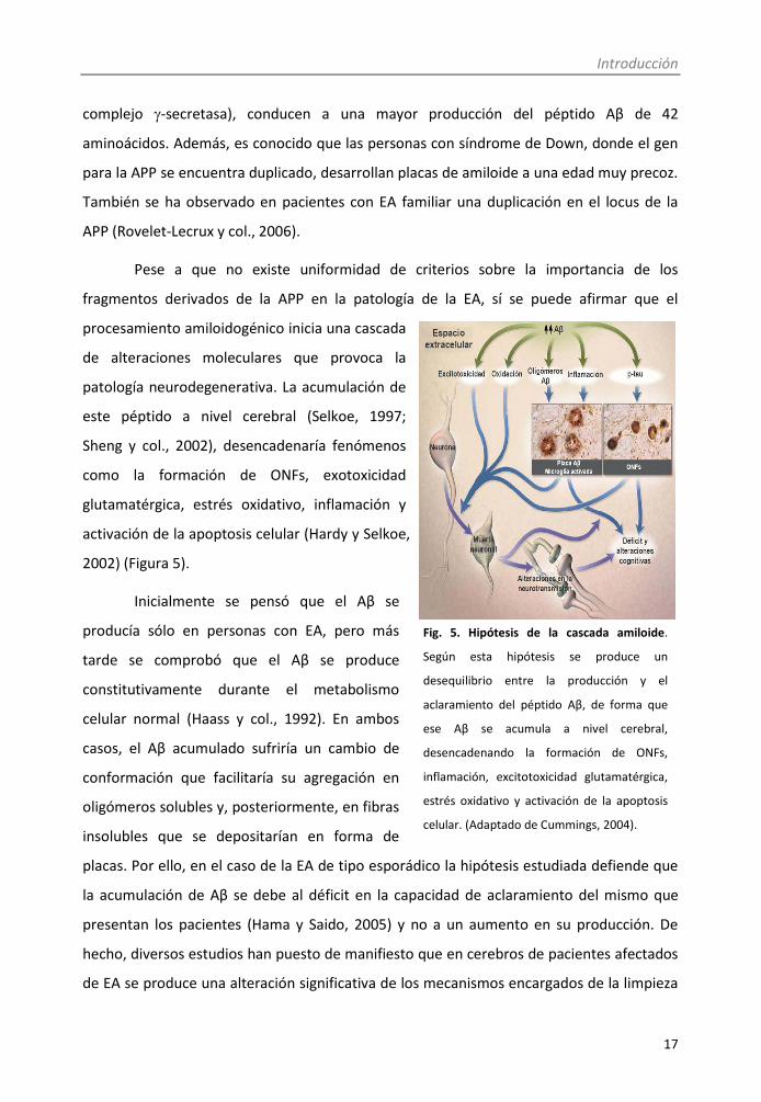
Pese a que no existe uniformidad de criterios sobre la importancia de los
fragmentos derivados de la APP en la patología de la EA, sí se puede afirmar que el
procesamiento amiloidogénico inicia una cascada
de alteraciones moleculares que provoca la
patología neurodegenerativa. La acumulación de
este péptido a nivel cerebral (Selkoe, 1997;
Sheng y col., 2002), desencadenaría fenómenos
como la formación de ONFs, exotoxicidad
glutamatérgica, estrés oxidativo, inflamación y
activación de la apoptosis celular (Hardy y Selkoe,
2002) (Figura 5).
Inicialmente se pensó que el Aβ se
producía sólo en personas con EA, pero más
tarde se comprobó que el Aβ se produce
constitutivamente durante el metabolismo
celular normal (Haass y col., 1992). En ambos
casos, el Aβ acumulado sufriría un cambio de
conformación que facilitaría su agregación en
oligómeros solubles y, posteriormente, en fibras
insolubles que se depositarían en forma de
placas. Por ello, en el caso de la EA de tipo esporádico la hipótesis estudiada defiende que
la acumulación de Aβ se debe al déficit en la capacidad de aclaramiento del mismo que
presentan los pacientes (Hama y Saido, 2005) y no a un aumento en su producción. De
hecho, diversos estudios han puesto de manifiesto que en cerebros de pacientes afectados
de EA se produce una alteración significativa de los mecanismos encargados de la limpieza
Fig. 5. Hipótesis de la cascada amiloide.
Según esta hipótesis se produce un
desequilibrio entre la producción y el
aclaramiento del péptido Aβ, de forma que
ese Aβ se acumula a nivel cerebral,
desencadenando la formación de ONFs,
inflamación, excitotoxicidad glutamatérgica,
estrés oxidativo y activación de la apoptosis
celular. (Adaptado de Cummings, 2004).

Introducción
18
de proteínas anómalas tales como el péptido Aβ y la proteína Tau (Selkoe, 2000). Los
mecanismos implicados en la eliminación del péptido Aβ han sido estudiados
exhaustivamente, encontrándose diversas vías implicadas, entre las que se encuentra la
degradación enzimática.
Las principales enzimas con actividad endopeptidasa implicadas en la degradación
del péptido Aβ se caracterizan por ser metaloproteasas dependientes de zinc, entre las que
se encuentra la enzima degradadora de insulina (IDE o insulisina). La enzima IDE actúa
extracelularmente y se encuentra presente además de en cerebro, en hígado, riñón y
músculo (Authier y col., 1996). El IDE juega un papel crucial en el aclaramiento cerebral del
Aβ (Kurochkin y Goto, 1994; McDermott y Gibson, 1997; Qiu y col., 1998) ya que, aunque el
sustrato principal de esta metaloproteasa es la insulina, IDE también se encarga de
degradar el péptido Aβ, impidiendo así que se acumule, retrasando la formación de placas
seniles y por lo tanto, protegiendo de la EA. Estudios histopatológicos han demostrado que
en cerebros de pacientes de EA hay una menor expresión de este enzima. Pero la evidencia
más consistente acerca del papel de IDE en el procesamiento de Aβ se deriva de estudios
realizados en ratones knockout. Estos ratones deficientes en IDE presentan elevados
niveles de Aβ en el cerebro. Además, se ha observado que ratones heterocigóticos IDE −/+
presentan niveles de Aβ intermedios entre los hallados en animales no transgénicos y los
encontrados en animales homocigóticos IDE −/− (Farris y col., 2003).
Teniendo en cuenta todo esto, el IDE podría ser una importante diana terapéutica
para el tratamiento de la EA, ya sea sobreexpresándolo mediante terapia génica, o
aumentando su expresión/activación.
1.4.2. Hipótesis de la proteína Tau
La proteína Tau se expresa de manera muy abundante en los axones de las
neuronas. Está localizada en el citosol neuronal donde se une a la tubulina, entre otras
proteínas, formando los microtúbulos de transporte celular. Este hecho le confiere un
papel fundamental, participando de este modo en la formación y mantenimiento de
dendritas, axones y, en general, de la estructura neuronal (Weingarten y col., 1975; Drubin
y Kirschner, 1986; Caceres y Kosik, 1990).
Introducción
19
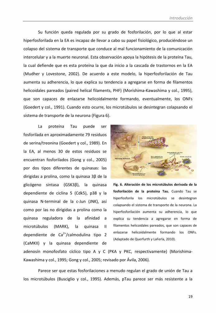
Su función queda regulada por su grado de fosforilación, por lo que al estar
hiperfosforilada en la EA es incapaz de llevar a cabo su papel fisiológico, produciéndose un
colapso del sistema de transporte que conduce al mal funcionamiento de la comunicación
intercelular y a la muerte neuronal. Esta observación apoya la hipótesis de la proteína Tau,
la cual defiende que es esta proteína la que da inicio a la cascada de trastornos en la EA
(Mudher y Lovestone, 2002). De acuerdo a este modelo, la hiperfosforilación de Tau
aumenta su adherencia, lo que explica su tendencia a agregarse en forma de filamentos
helicoidales pareados (paired helical filaments, PHF) (Morishima-Kawashima y col., 1995),
que son capaces de enlazarse helicoidalmente formando, eventualmente, los ONFs
(Goedert y col., 1991). Cuando esto ocurre, los microtúbulos se desintegran colapsando el
sistema de transporte de la neurona (Figura 6).
La proteína Tau puede ser
fosforilada en aproximadamente 79 residuos
de serina/treonina (Goedert y col., 1989). En
la EA, al menos 30 de estos residuos se
encuentran fosforilados (Gong y col., 2005)
por dos tipos diferentes de quinasas: las
dirigidas a prolina, como la quinasa 3β de la
glicógeno sintasa (GSK3β), la quinasa
dependiente de ciclina 5 (Cdk5), p38 y la
quinasa N-terminal de la c-Jun (JNK), así
como por las no dirigidas a prolina como la
quinasa reguladora de la afinidad a
microtúbulos (MARK), la quinasa II
dependiente de Ca2+/calmodulina tipo 2
(CaMKII) y la quinasa dependiente de
adenosín monofosfato cíclico tipo A y C (PKA y PKC, respectivamente) (Morishima-
Kawashima y col., 1995; Gong y col., 2005; revisado por Ávila, 2006).
Parece ser que estas fosforilaciones a menudo regulan el grado de unión de Tau a
los microtúbulos (Busciglio y col., 1995). Además, pTau parece ser más resistente a la
Fig. 6. Alteración de los microtúbulos derivada de la
fosforilación de la proteína Tau. Cuando Tau se
hiperfosforila los microtúbulos se desintegran
colapsando el sistema de transporte de la neurona. La
hiperfosforilación aumenta su adherencia, lo que
explica su tendencia a agregarse en forma de
filamentos helicoidales pareados, que son capaces de
enlazarse helicoidalmente formando los ONFs.
(Adaptado de Querfurth y LaFerla, 2010).

Introducción
20
proteolisis por diferentes proteasas, lo que contribuiría a la acumulación de ésta en las
neuronas con el consiguiente efecto tóxico (Shimura y col., 2004).
Finalmente, cabe destacar una idea propuesta en los últimos años que sugiere que
la acumulación de Aβ puede provocar la fosforilación de Tau y, aunque ambos contribuyen
a la fisiopatología de la enfermedad, esta última es quien principalmente produce el
deterioro de estructuras y de las funciones neuronales. De hecho, se ha observado que
cantidades elevadas de Aβ aumentan la formación de lesiones por Tau fibrilar en modelos
animales de EA (Lewis y col., 2001; Oddo y col., 2003), apoyando la hipótesis de que las
patologías amiloide y Tau aparecen cronológicamente en este orden. No obstante, el nexo
entre Aβ y la patología Tau sigue sin estar totalmente esclarecido.
1.4.3. Patología sináptica
Como ha sido comentado, la pérdida de sinapsis, junto con la presencia de ONFs,
es la característica fisiopatológica de la EA que mejor se correlaciona con el grado de déficit
cognitivo (Terry, 2000). La degeneración sináptica que se produce en la EA se caracteriza
por una pérdida progresiva de terminales axónicas (Scheff y col., 2007), una disminución en
la expresión de proteínas pre y postsinápticas (Almeida y col., 2005), alteraciones en la
estructura de espinas dendríticas (Knobloch y Mansuy, 2008) e incluso, la pérdida de las
mismas (Blanpied y Ehlers, 2004). Además de la gran pérdida sináptica, también se
observan cambios compensatorios en las terminales restantes (DeKosky y Scheff, 1990;
Scheff y col., 1993; Scheff y Price 1993).
A pesar de que no están claros los mecanismos patológicos que conducen al daño
sináptico en la EA, además de la relación existente entre la aparición de ONFs y el daño
neuronal (ver punto 1.2.), el Aβ parece jugar un papel clave. La acumulación del péptido Aβ
intraneuronal parece ser uno de los principales responsables de la disfunción sináptica que
se produce en la EA, y que se correlaciona con el déficit en los fenómenos de aprendizaje y
memoria que se observan en diferentes modelos animales de la EA. Por ejemplo, los
ratones transgénicos para APP han proporcionado evidencias de que la disfunción sináptica
relacionada con Aβ puede ocasionar déficits en el aprendizaje y la memoria (Westerman y
col., 2002). Existen pruebas claras de los efectos directos de Aβ en mecanismos de

Introducción
21
plasticidad sináptica demostrando que la actividad neuronal puede inducir el
procesamiento de la APP para generar Aβ y que el péptido Aβ a su vez puede reducir la
transmisión sináptica excitatoria. Diferentes investigaciones han revelado que las formas
solubles del mismo son capaces de deprimir la transmisión sináptica a través de
mecanismos similares a la depresión a largo plazo, induciendo la fosforilación de los
receptores glutamatérgicos tipo AMPA y su endocitosis (Hsieh y col., 2006).
A continuación se describen dos de los marcadores de plasticidad sináptica que
más consistentemente han sido relacionados con la patología de EA:
Sinaptofisina
La liberación de neurotransmisores es un proceso altamente regulado y que
comprende varios pasos: la dirección de las vesículas sinápticas a las zonas presinápticas
activas, el anclaje de las vesículas a la membrana celular, la fusión entre la membrana
vesicular y la plasmática y el reciclaje de la membrana vesicular (Sudhof, 2000). En ese
proceso participan las proteínas de las membranas vesiculares (De Camilli y Jahn, 1990), de
entre las cuales una de las más abundantes es la sinaptofisina (Rehm y col., 1986). La
sinaptofisina fue la primera proteína de vesícula sináptica clonada y desde su
descubrimiento en 1985 (Wiedenmann y Franke, 1985), muchos laboratorios la han usado
como un marcador de neuroplasticidad que permite estudiar la distribución y el número de
las sinapsis y/o la integridad general de la expresión de proteínas sinápticas. No se conoce
su papel exacto en todo ese proceso de exocitosis de los neurotransmisores, pero se ha
visto que es fundamental para el control de la liberación de estas sustancias, y como
consecuencia también para procesos como la LTP, el aprendizaje y la memoria, ya que se
ha visto que ratones knockout para esta proteína, presentan una fuerte disminución en los
procesos de plasticidad sináptica (Spiwoks-Becker y col., 2001; Valtorta y col., 2004).
Diversos estudios han demostrado una pérdida de la proteína sinaptofisina en
cerebros de EA (Wakabayashi y col., 1994; Love y col., 2006). Según lo observado, esta
pérdida ocurre inicialmente en el giro dentado del hipocampo y se extiende a la corteza
frontal a medida que la enfermedad se hace más severa (Masliah y col., 1992).

Introducción
22
BDNF
Las neurotrofinas son moléculas que promueven el desarrollo y la supervivencia de
las neuronas (Bibel y Barde, 2000). De entre todas las neurotrofinas, el BDNF (brain-derived
neurotrophic factor) destaca como regulador de la plasticidad sináptica, la supervivencia y
diferenciación neuronal y también como diana clave para el desarrollo de nuevos
tratamientos para desórdenes neuronales (Binder y Scharfman, 2004). Se ha descrito que
el BDNF parece ser fundamental para la supervivencia continua y el mantenimiento del
fenotipo de neurona madura, de manera que se estima que los cambios en los niveles y la
distribución podrían ser importantes en la patogénesis de enfermedades
neurodegenerativas.
Teniendo en cuenta que el BDNF regula la potenciación sináptica a largo plazo (LTP)
y otras formas de plasticidad (procesos que tienen un papel clave en la formación y el
almacenamiento de memoria y están afectados en la EA), una reducción en los niveles
endógenos de esta neurotrofina podría subyacer a la patofisiología de la EA.
Los primeros estudios que hablan de la deficiencia de BDNF en la EA datan de los
años 90, en los cuales se demostró una disminución selectiva de la expresión de ARNm en
el hipocampo de los pacientes con EA (Phillips y col., 1991). Estudios posteriores mostraron
también disminuciones de esta expresión en el neocórtex y el núcleo basal de Meynert,
fuente principal de inervación colinérgica de la corteza cerebral (Murer y col., 1999; Murer
y col., 2001; Tapia-Arancibia y col., 2008). Más tarde se pudo comprobar que no solo el
ARNm, sino también los niveles de proteína tanto madura como inmadura (proBDNF)
disminuían cuando se medían en el giro dentado, la corteza frontal o la corteza parietal de
enfermos de EA (Narisawa-Saito y col., 1996; Lee y col., 2005; Peng y col., 2005).
2. Tratamiento de la Enfermedad de Alzheimer
Los tratamientos disponibles actualmente para la EA son tratamientos sintomáticos.
Estos tratamientos tienen como objetivo la mejora de la cognición pero no actúan sobre la
etiología de la enfermedad. Sin embargo, dadas las importantes repercusiones humanas,

Introducción
23
sociales y económicas de la enfermedad, es evidente la necesidad de buscar nuevos
tratamientos curativos.
2.1. Terapias actuales
Los fármacos que en la actualidad se emplean para el tratamiento sintomático de
la EA son:
Inhibidores de la acetilcolinesterasa
En la actualidad se utilizan tres: donezepilo, rivastigmina y galantamina. Se ha
postulado que la degeneración de las neuronas colinérgicas, sobre todo en el prosencéfalo
basal, causa una disminución de los niveles de acetilcolina en las terminales presinápticas
de hipocampo y neocortex, lo que produciría alteraciones en la memoria (Terry y
Buccafusco, 2003). Los fármacos inhibidores de la acetilcolinesterasa aumentan la
disponibilidad de acetilcolina en la hendidura sináptica al inhibir la enzima encargada de su
degradación.
Memantina
Es un antagonista débil y no competitivo del receptor de glutamato N-metil-D-
aspartato (NMDA). En la EA, alteraciones de la actividad glutamatérgica produce un
aumento de los receptores de NMDA, lo que lleva a una disfunción neuronal. La
memantina bloquea los canales NMDA de manera que modula los efectos patológicos de
los niveles elevados de glutamato pero permitiendo al receptor activarse ante señales
fisiológicas normales.
Fármacos para el tratamiento de alteraciones conductuales
Además de las características típicas de la EA, la sintomatología cognitiva viene
acompañada por otras alteraciones conductuales, como pueden ser la agresión, agitación
psicomotora, depresión, ansiedad, insomnio ó psicosis (alucinaciones y delirios). Por tanto,
es frecuente que estos pacientes reciban ansiolíticos, hipnóticos, neurolépticos y/o
antidepresivos.

Introducción
24
Hay que señalar que ninguno de los actuales fármacos detiene la degeneración de
las neuronas o revierte la progresión de la enfermedad. Actualmente todos los esfuerzos
en la investigación de nuevos fármacos se basan en las distintas hipótesis sobre la etiología
de la enfermedad que han sido postuladas, como por ejemplo la deposición de placas de
amiloide, la formación de ONFs y la neuroinflamación.
2.2. Nuevas terapias
En los últimos años se ha sugerido la posible utilidad de diferentes grupos de
fármacos en el tratamiento de la EA, por lo que se están realizando cada vez más estudios
que se encuentran en diferentes fases de desarrollo (Tabla 1).
Tabla 1. Fases del desarrollo de terapias actuales para la EA (Adaptado de Haas, 2012)
Mecanismo de acción Nº fármacos Fase clínica
Inhibidores de β-secretasa 3 I, II, III
Inhibidores de γ-secretasa 7 I, II, III
Moduladores de γ-secretasa 3 I, II, III
Potenciadores de α-secretasa 12 I, II
Fármacos que actúan sobre receptores serotonérgicos 5 I, II
Fármacos que afectan a la acetilcolina 4 II
Inhibidores de la agregación de Aβ 10 I, II
Inmunoterapia contra Aβ 11 I, II, III
Inhibidores de fosforilación de Tau 3 II, III
Potenciadores de la desfosforilación de Tau 3 I, II
Inhibidores de agregación de Tau/degradadores de ovillos 3 II, III
Anti-inflamatorios/Neuroprotectores 9 II, III
Suplementos nutricionales 10 II, III, IV
Antagonistas de receptores de histamina 3 I, II
Antagonistas de receptores adrenérgicos 1 II

Introducción
25
Algunas de las terapias farmacológicas potencialmente útiles para el tratamiento
de la EA son:
Moduladores de las secretasas
Se observó que ratones knockout de BACE1 no producen Aβ ni desarrollan un
fenotipo clínico (Luo y col., 2001). Además, los inhibidores de la β-secretasa disminuyen los
niveles cerebrales de Aβ, lo que podría reducir la patología (Chang y col., 2004). Sobre los
inhibidores de la γ-secretasa hay dudas debido a los efectos adversos ya que también
actúan sobre otros sustratos como Notch (Wolfe, 2008). Sin embargo, los nuevos
inhibidores generados han demostrado no afectar a la señalización de Notch (Petit y col.,
2001), así como buena tolerancia en ensayos clínicos en fase I (Siemers y col., 2005).
Inmunoterapia contra el péptido Aβ
La primera vez que se informó de esta posible terapia se demostró que la
inmunización activa con A fibrilar en un modelo animal de EA disminuía la agregación del
péptido (Schenk y col., 1999). Los efectos pueden estar mediados por anticuerpos que
reconocen A y que pueden unirse bien al péptido presente en placas, lo que induciría la
limpieza de dicho A a través de la microglía (Schenk y col., 2004), o bien a formas solubles
del péptido, promoviendo la eliminación al exterior del cerebro.
Fármacos inhibidores de la fosforilación de Tau
Los candidatos que reducen la fosforilación de Tau al inhibir quinasas tales como
Cdk5 y GSK3β, están en distintas fases de ensayos clínicos. Sin embargo, ya que esta
fosforilación depende de múltiples quinasas y fosfatasas (Avila, 2006), la inhibición de una
única quinasa podría no ser suficiente para normalizar la fosforilación de Tau.
Por otro lado, los resultados de diversos estudios epidemiológicos sugieren la
posible utilidad de otros fármacos que actúan sobre diversos factores de riesgo
ambientales en el tratamiento de la EA:
Fármacos anti-inflamatorios
Como ya ha sido mencionado, la EA tiene un fuerte componente inflamatorio
(McGeer y McGeer, 2007). Diversos estudios epidemiológicos han relacionado el uso de
Introducción
26
fármacos antiinflamatorios no esteroideos (AINEs) con un menor riesgo de desarrollar EA
(Aisen, 2002).
Fármacos que disminuyen el colesterol
En un estudio de Sparks y col. de 1994, se observó que ratones alimentados con
una dieta muy alta en colesterol desarrollaban acumulaciones intracelulares de Aβ. Sin
embargo, a día de hoy estudios epidemiológicos no han podido concretar si el empleo de
fármacos que disminuyen el colesterol, más concretamente las estatinas, son capaces de
modificar los niveles de dicho péptido, así como de mejorar el estado cognitivo de los
pacientes (Hoglund y col., 2005; Sparks y col., 2005). Esto ha llevado a la realización de
estudios más amplios que se encuentran en desarrollo actualmente.
Fármacos antihipertensivos
Este punto se desarrolla en el apartado 3. de la Introducción.
Estrógenos
Estudios epidemiológicos han relacionado la suplementación postmenopáusica con
estrógenos con un descenso en el riesgo de desarrollar EA (Tang y col., 1996). Estudios en
animales también sugieren que los estrógenos podrían tener diversos efectos beneficiosos
sobre la función neuronal (Almeida y Flicker, 2005). Sin embargo, ensayos clínicos no
demuestran un menor riesgo de padecer la enfermedad (Shumaker y col., 2004).
Antioxidantes
Estudios basados en la ingesta de dietas ricas en antioxidantes, como la vitamina E,
demostraron que éstos disminuían el riesgo de padecer EA (Engelhart y col., 2002). Sin
embargo, otros ensayos clínicos randomizados no han demostrado ningún efecto de la
vitamina E sobre la progresión de la enfermedad (Petersen y col., 2005).

Introducción
27
3. Hipertensión y Enfermedad de Alzheimer
Cada vez se acumulan más evidencias acerca de la asociación entre las cifras
elevadas de presión arterial en la edad media de la vida y la aparición de la EA en las
edades más avanzadas (Kivipelto y col., 2001b).
La hipertensión produce desmielinización isquémica (Tuhrim y Levine, 2002),
pérdida neuronal hipocampal (Kril y col., 2002), así como una disminución de la integridad
vascular de la barrera hemato-encefálica (BHE), dando como resultado una extravasación
de proteínas al tejido cerebral (Kalaria, 2010) que produce apoptosis, una reducción de la
función sináptica y un aumento de la acumulación de Aβ, desembocando todo ello en la
aparición del deterioro cognitivo (Deane y col, 2004).
En base a todo esto, se considera que la conexión entre hipertensión arterial y EA
puede estar en la angiopatía amiloide que ésta produce (Vinters, 2001; Greenberg, 2002).
Como en la EA suelen encontrarse lesiones vasculares, ambas patologías podrían tener un
efecto sinérgico sobre la intensidad de la demencia. Por ello, es importante controlar los
factores de riesgo vascular como la hipertensión, ya que podría contribuir a la prevención
de la demencia (McCullagh y col., 2001; Maher y Schubert, 2009; Purandare, 2009). De
hecho, diversos estudios epidemiológicos han encontrado que una reducción de la presión
arterial podría ejercer un efecto protector sobre el deterioro cognitivo (Fogari y col., 2003,
2006).
Por otro lado, estudios clínicos sugieren que pacientes con hipertensión tratada
presentan menos lesiones neuropatológicas de EA (placas seniles y ONFs) que los
normotensos (Hoffman y col., 2009), datos que concuerdan con otras investigaciones que
también demuestran que la hipertensión podría estar involucrada en la acumulación de
depósitos de Aβ y la formación de ONFs (Lee y col., 2003; Bomboi y col., 2010). Además, se
ha descrito un aumento de la presión arterial sistólica en pacientes con deterioro cognitivo
leve y pacientes con EA instaurada en comparación con los pacientes control, existiendo
una correlación entre esta hipertensión y el déficit cognitivo (Ciobica y col., 2011).
A pesar de estas evidencias, en la actualidad existe cierta controversia acerca del
efecto de la presión arterial elevada sobre la función cerebral, ya que aunque hay estudios
Introducción
28
que sugieren una conexión entre el deterioro cognitivo y la hipertensión arterial (Posner y
col., 2002, Luchsinger y col., 2005), y que la hipertensión correlaciona negativamente con
las funciones cognitivas, especialmente con la memoria, la atención y las funciones
ejecutivas (Hanon y col., 2005; Vicario y col., 2005), otros autores no encuentran ninguna
relación entre ambos (in’t Veld y col., 2001; Qiu y col., 2003; Beeri y col., 2009).
Apoyando el papel de la hipertensión en el desarrollo de la EA, son cada vez más
numerosos los trabajos que demuestran que el uso de ciertos fármacos antihipertensivos
parece disminuir la incidencia de la EA. Por ejemplo, Lopez-Arrieta y Birks (2002)
describieron que el nimodipino, un antagonista de canales de calcio, disminuye la
incidencia de EA en pacientes con hipertensión. El ensayo Syst-Eur mostró que
tratamientos a largo plazo con el antihipertensivo nitrendipino redujo el riesgo de
demencia, y en particular de EA, en pacientes hipertensos y tras seguimiento de 2 años
(Forette y col., 2002). Guo y col (1999) describieron que el uso de β-bloqueantes protege a
sujetos mayores de desarrollar EA. Aún más, el estudio Cache County señaló que el uso de
medicación antihipertensiva se asocia a una incidencia reducida de EA en los mayores de
65 años (Khachaturian y col., 2006).
Entre estos fármacos se encuentra el propranolol, un antagonista β-adrenérgico
competitivo y no selectivo. A través de su acción sobre receptores β1 produce una
disminución de la frecuencia cardíaca, del gasto cardíaco y de la secreción de renina,
contribuyendo todas estas acciones a un efecto antihipertensivo. El propranolol se usa
para el control de arritmias cardíacas, hipertensión arterial esencial y renal, y para el
control de la ansiedad y la taquicardia. Las dosis utilizadas habitualmente en el tratamiento
de la hipertensión arterial en humanos es de 160 mg/día-320 mg/día.
Estudios in vitro han demostrado que el propranolol es capaz de disminuir
significativamente los niveles de Aβ en cultivos primarios de neuronas derivadas de un
modelo transgénico de EA. Además, estudios in vivo han descrito que este fármaco es
capaz de revertir el déficit cognitivo asociado al estrés (Roozendaal y col., 2004; Aisa y col.,
2007). A pesar de estos resultados, algunos autores sugieren que la administración de
propranolol afecta negativamente a la formación y consolidación de la memoria
Introducción
29
(Murchison y col., 2004; Miranda y col., 2011; Moncada y col., 2011), datos que no
concuerdan con otras publicaciones que señalan que la administración de propranolol no
afecta al rendimiento cognitivo (Roozendaal y col., 2004; Kenton y col., 2008).
Por último, cabe destacar un trabajo del grupo de Wang y col. del año 2007, que
estudió a nivel experimental la hipótesis de que algunos fármacos antihipertensivos
podrían influir en la expresión de marcadores de EA mediante mecanismos no relacionados
con su acción antihipertensiva. En concreto, en este trabajo estudiaron el efecto sobre la
producción de Aβ de 55 fármacos antihipertensivos con distintos mecanismos de acción.
De todos los fármacos estudiados, 7 fueron capaces de disminuir in vitro la carga de β-
amiloide. De acuerdo a la “hipótesis de la cascada del amiloide”, es el desequilibrio entre la
producción y el aclaramiento del péptido Aβ lo que desencadena la neurodegeneración en
la EA (Glenner y Wong, 1984; Hardy y Allsop, 1991; Hardy y Selkoe, 2002). Por lo tanto,
cualquier fármaco capaz de reducir la carga de Aβ es un potencial candidato para el
tratamiento de la enfermedad.
4. Modelos animales para el estudio de la Enfermedad de Alzheimer
Como ya se ha comentado, la etiología de la EA no está del todo clara y no existen
tratamientos efectivos. Debido a la gran prevalencia y al diagnóstico tardío, el desarrollo de
modelos animales ha sido una de las prioridades científicas en los últimos años, con la
finalidad tanto de poder comprender los mecanismos patogénicos de la enfermedad como
de probar diferentes estrategias terapéuticas. Existen numerosos modelos animales para el
estudio de la EA, aunque hasta ahora no se ha conseguido ninguno que sea capaz de
reproducir totalmente la naturaleza multifactorial de la enfermedad, ni todas las
características patológicas que se observan en los humanos.
A continuación se describen diferentes tipos de modelos que han sido
desarrollados hasta el momento, y dentro de cada grupo los más utilizados en la actualidad.

Introducción
30
4.1. Modelos animales de EA familiar
Las mutaciones genéticas causantes de la EA de tipo familiar han conducido a la
generación de diferentes líneas de ratones transgénicos que se emplean como modelos
animales de EA (Janus y Westaway, 2002; Phinney y col., 2003; Walsh y Selkoe, 2004a). A
pesar de que ninguno de ellos reúne todas las características neuropatológicas de la
enfermedad, algunos presentan conjuntamente varias de ellas como los depósitos de
amiloide, la astrocitosis, la microgliosis, la hiperfosforilación anormal de Tau y los déficits
de memoria (Phinney y col., 2003).
4.1.1. Modelos transgénicos de APP
Ratones PDAPP
El primer modelo animal transgénico para la EA, fue desarrollado por Games y col.
en 1995. Estos ratones, denominados PDAPP, contienen la mutación indiana (V717F) bajo
el control del promotor PDGF. Este modelo expresa niveles de APP hasta 18 veces mayores
que los niveles endógenos de ratones añosos, y el tamaño de las fibras de amiloide
presentes en el cerebro es análogo al que se detecta en los pacientes de EA (Masliah y col.,
2001; Kobayashi y Chen, 2005). A los 3 meses muestran una pérdida sináptica significativa
dependiente de la edad, así como atrofia hipocampal (Dodart y col., 1999), mientras que a
partir de los 6 meses se observa la presencia de depósitos de Aß en el hipocampo (Games y
col., 1995; Irizarry y col., 1997) y trastornos de memoria en la mayoría de las pruebas
conductuales con respecto a los animales control, que aumentan con la edad y la carga
amiloide (Chen y col., 2000; Arendash y col., 2001; Morgan, 2003).
Ratones Tg2576
Dentro de los animales transgénicos de APP, la línea de ratones Tg2576, es uno de
los más utilizados y contrastados. Este ratón sobreexpresa la isoforma de 695 aminoácidos
del gen de la APP humana conteniendo la doble mutación sueca (K670M/N671L) bajo el
control de un prión de promotor de hámster (Hsiao y col., 1996). El modelo murino Tg2576
muestra signos de daño en el hipocampo a los 9-10 meses de edad, hecho que queda
reflejado en el deterioro cognitivo que muestran en diferentes pruebas de
comportamiento (Chapman y col., 1999; Westerman y col., 2002). La aparición de placas
Introducción
31
seniles comienza entorno a los 9 meses de edad (Irizarry y col., 1997) y aumenta
exponencialmente con la edad, de forma que a los 16 meses la carga de amiloide alcanza el
3,6% en la región CA1 del hipocampo, el 6% del giro dentado y el 8,5% de la corteza
entorrinal. Los animales de esta edad, muestran así mismo una importante activación
microglial que se correlaciona cualitativa y cuantitativamente con las placas neuríticas
(Frautschy y col., 1998). Otro dato interesante es que a pesar de que a los 16 meses de
edad los animales ya muestran un elevado número de neuritas degeneradas asociadas a
placas de Aβ, los estudios estereológicos llevados a cabo en este modelo de la EA no
muestran pérdida neuronal (Irizarry y col., 1997). Por tanto, la pérdida de memoria y de la
capacidad de aprendizaje de estos animales se debería al daño sináptico que experimentan
y no a una reducción en el número de neuronas.
Ratones TgCRND8
El modelo murino TgCRND8 sobreexpresa la doble mutación sueca y la mutación
indiana (K670N/M671L+V717F) bajo el control de un prión de promotor de hámster, y es el
modelo animal de APP más agresivo puesto que desarrolla densas placas seniles a la
temprana edad de 3 meses que se correlacionan con importantes déficits cognitivos
(Chishti y col., 2001) y la activación de astrocitos y microglía alrededor de dichas placas
(Dudal y col., 2004).
4.1.2. Modelos transgénicos de PS
Todos los modelos basados en la carencia de PS1 o PS2 demuestran deterioro
cognitivo en diferentes pruebas conductuales, pero no presentan alteraciones muy severas
si se comparan con las observadas en los Tg2576. Asimismo, en ratones con ausencia de
PS1 se encuentran niveles elevados de Aβ42 pero no se acumula formando placas seniles ni
hay manifestaciones de alteraciones conductuales (Kobayashi y Chen, 2005; Spires y
Hyman, 2005).
Sin embargo, cuando se cruzan ratones portadores de un gen APP mutante con
ratones portadores de un gen PS1 mutante (APP+PS1) aparecen los marcadores
neuropatológicos asociados a la EA, además de cambios de conducta, con mucha mayor
rapidez que cuando son portadores de una sola de estas variedades transgénicas (Bales y

Introducción
32
col., 1997), y este fue uno de los primeros modelos que mostró una fuerte correlación
entre el desarrollo de Aβ42 y el déficit cognitivo (Holcomb y col., 1999; Arendash y col.,
2001).
El problema de todos estos modelos descritos hasta el momento es
fundamentalmente que en humanos la mutación de APP o de alguna de las PS es suficiente
para que tenga lugar el desarrollo completo de la EA, incluyendo déficit cognitivo
progresivo, placas, ovillos y pérdida neuronal en el cerebro. Sin embargo, cuando estas
mutaciones se expresan en los ratones transgénicos, se desarrollan algunas de estas
características de la enfermedad, pero no todas.
4.1.3. Modelos transgénicos de Tau
La mayoría de modelos transgénicos, tanto en estudios iniciales como actuales, se
han concentrado en la expresión de APP y de genes mutantes de presenilina, ambos
asociados con formas familiares de EA, hallando un aumento de la producción de amiloide
en el cerebro. Debido a que en estos modelos no se han podido demostrar presencia de
ONFs o neurodegeneración, surgió la necesidad de obtener un enfoque diferente creando
ratones transgénicos que expresaran la proteína Tau humana (Götz y col., 1995).
Se ha visto que ciertas mutaciones de la proteína Tau ligadas al cromosoma 17
están relacionadas con enfermedades como parkinson o demencia fronto-temporal, pero
ninguna es capaz de producir la patología característica de la EA (Gendrun y Petrucelli,
2009; Iqbal y col., 2009). Ratones que sobreexpresan estas mutaciones presentan ONFs,
muerte neuronal y déficits conductuales (Lewis y col., 2001; Yoshiyama y col., 2007;
Rosenmann y col., 2008), pero estos modelos presentan bastantes limitaciones, ya que en
algunos casos estos déficits se manifiestan en ausencia de ovillos o pérdida de neuronas
(Taniguchi y col., 2005). Además, la distribución de los ONFs en la mayoría de ellos es muy
diferente de la observada en la EA ya que son muy escasos en las regiones hipocampales y
corticales (Zilka y col., 2009). Es importante señalar que tampoco se encontraron depósitos
amiloides en estos animales (Hamilton, 1998).
Introducción
33
4.1.4. Dobles y triples transgénicos
Ratones TAPP
Este modelo animal surgió por la necesidad de encontrar un modelo que expresara
tanto placas como ONFs, combinando mutaciones típicas de FAD y mutaciones de Tau
ligadas al cromosoma 17 (FTDP-17). Fue desarrollado mediante el cruzamiento de ratones
Tg2576 con otra linea conocida como JNPL3 (Lewis y col., 2001), consiguiendo de esta
manera la presencia de ambos marcadores neuropatológicos a los 8 meses, lo que permite
examinar los efectos de Aß en la formación de los ONFs.
Ratones 3xTg-AD
Otro modelo desarrollado expresa la mutación sueca de APP (K670N/M671L), y
una mutación de Tau en el cromosoma 17 (P301L) combinadas con una mutación en la PS1
(M146V) (Oddo y col., 2003), dando lugar a un triple transgénico denominado 3xTg-AD.
Estos ratones desarrollan depósitos amiloides a los 6 meses de edad y una
patología neurofibrilar que comienza a los 10 meses en el hipocampo y que se extiende
progresivamente a la corteza. Además, presentan déficits tanto en la plasticidad sináptica
como en la memoria y el aprendizaje que correlacionan con la acumulación de Aβ
intraneuronal (Oddo y col., 2003; Billings y col., 2005).
4.1.5. Modelos transgénicos 5xFAD APP/PS1
Este modelo expresa la proteína humana APP con las mutaciones sueca, florida y
london juntas con una mutación de PS1 (M146L/L286V) reguladas por el promotor Thy1,
constituyendo uno de los modelos más agresivos y tempranos de patología amiloide. Estos
animales se caracterizan por presentar depósitos amiloides visibles a la edad de 2 meses, y
una astrogliosis y microgliosis proporcionales a los niveles de Aβ42, así como una pérdida
neuronal muy robusta. Por otro lado, en las pruebas conductuales se observan déficits en
las meorias espacial y contextual a partir de los 4-6 meses de edad (Oakley, 2006; Ohno y
col., 2007).

Introducción
34
4.2. Modelos animales de envejecimiento
Tal y como aparece explicado en el punto 1.1. del presente trabajo, la edad es el
factor de riesgo más importante para sufrir EA. De ahí que sea tan importante el estudiar la
relación entre el envejecimiento y las características fisiopatológicas de la enfermedad.
Los ratones SAMP8 (senescence accelerated mice prone-8), denominados así por
presentar un envejecimiento acelerado son considerados en la actualidad un modelo
animal robusto para el estudio de patologías asociadas a la edad, incluyendo la EA (Sureda
y col., 2006; Pallas y col., 2008; Liu y col., 2010). Estos ratones fueron generados a finales
de los años 70 a partir de la cepa AKR/J (Takeda y col, 1981), al observarse que a través de
su mantenimiento convencional con cruzamientos entre hermanos, algunas camadas
poseían características indicativas de envejecimiento acelerado similares a las que se
observan en humanos de edad avanzada, como por ejemplo menor actividad física,
pérdida de pelo, lordocifosis o acortamiento de vida, además de algunos trastornos de tipo
neurológico (Hamamoto y col., 1984; Takeda y col., 1994), mientras que otras camadas se
mostraban resistentes al envejecimiento acelerado (senescence accelerated mice
resistant-1, SAMR1) y presentaban un patrón de envejecimiento normal (Takeda, 1999).
Lo más interesante es que estos ratones SAMP8 presentan déficit en el aprendizaje
y la memoria a partir de los 5 meses de edad, así como depósitos cerebrales de Aβ
(Tomobe y Nomura, 2009; Del Valle y col., 2010) y un aumento en la expresión de Tau
hiperfosforilada (Canudas y col., 2005; Orejana y col., 2011).
Debido a todos estos antecedentes y a la naturaleza multifactorial de la EA, los
SAMP8 se están empezando a considerar uno de los modelos animales más
representativos para su estudio, ya que son capaces de mimetizar la complejidad de esta
enfermedad mucho mejor que los modelos transgénicos (Pang et al., 2006).
4.3. Modelos animales de EA esporádica
Debido a que entre un 95 y un 99% de los casos la EA no se debe a causas
hereditarias sino que se considera una enfermedad esporádica, además del desarrollo de
Introducción
35
animales transgénicos para FAD, se ha intentado reproducir el componente esporádico de
la enfermedad utilizando otro tipo de modelos animales.
4.3.1. Modelos animales genéticos
Numerosos estudios han confirmado que la posesión del alelo ApoE4 constituye un
importante factor de riesgo para el desarrollo de EA (Bertram y col., 2007). Muchos de
estos estudios han utilizado ratones que sobreexpresan APP cruzados con knockout para
ApoE y observaron que la cantidad de Aβ en el cerebro decrece considerablemente, lo que
indica un papel importante de ApoE en la formación de depósitos de amiloide (DeKosky,
2002).
Por otro lado, ratones transgénicos de ApoE4 muestran un incremento en la
fosforilación de Tau neuronal (Tesseur y col., 2000; Brecht y col., 2004) y alteraciones
sinápticas y cognitivas (Zhong y col., 2008; Ramaswamy y col., 2005).
4.3.2. Modelos animales metabólicos
Estudios epidemiológicos realizados acerca de la relación entre insulina y EA
señalan que individuos diabéticos tienen un 70% más de riesgo de desarrollar demencia y
EA que los no diabéticos (Xu y col., 2007). Por ello, modelos animales desarrollados para el
estudio de la diabetes están empezando a utilizarse actualmente para el estudio de la EA.
Ratones ob/ob y db/db
En los modelos más utilizados para el estudio de la DM los animales presentan
alteraciones en la leptina, una hormona sintetizada por los adipocitos que juega un
importante papel en el metabolismo y la sensación de saciedad (Zhang y col., 1994; Shore,
2007). Los ratones ob/ob, descubiertos en 1949 (Ingalls y col., 1950), son el resultado de
una mutación espontánea en el gen que codifica la leptina debido a una sustitución en el
codón 105. En los ratones db/db, aunque presentan las mismas características, la causa se
debe a una mutación en el dominio citoplasmático de un receptor de la leptina, Ob-Rb
(Nishina y col., 1994; Schreyer y col., 1998). Debido a esta ausencia de leptina, los ratones
de ambos modelos no reciben la señal de saciedad e ingieren alimento en exceso,
presentando como consecuencia obesidad a partir de las 4 semanas de vida,
Introducción
36
hiperinsulinemia, hiperglicemia, aumento de la corticosterona sérica, e incremento de los
triglicéridos y el colesterol en ayunas (Leibel y col., 1997; Nishina y col., 1994). Se han
observado en estos ratones además procesos neurodegenerativos como pérdida sináptica
y neuronal así como Tau hiperfosforilado (Kim y col., 2009).
Es de señalar un estudio del 2010 donde se observó que el fenotipo diabético es
mucho más severo en ratones ob/ob combinados con otros transgénicos para APP que en
los ratones únicamente deficientes en leptina, y que a las 8 semanas de edad manifiestan
alteraciones en el aprendizaje que no aparecen en los modelos individuales hasta varios
meses más tarde (Takeda y col., 2010). Estos resultados apoyan la hipótesis de que la
resistencia a la insulina es un posible factor de riesgo para el desarrollo de la EA acelerando
su aparición.
Ingesta hipercalórica (high fat diet, HFD)
La ingesta de una dieta hipercalórica provoca a lo largo del tiempo una obesidad y
resistencia a la insulina típica de DM2. Se ha visto que animales sometidos a una dieta de
estas características presentan alteraciones en la vía de señalización de la insulina, y que
ratones Tg2576 alimentados con HFD durante meses muestran aumentos significativos de
Aβ, actividad γ-secretasa y Tau, mientras que los niveles de IDE y BDNF se encuentran
disminuidos (Ho y col., 2004; Kohjima y col., 2010).
El efecto agravante de la DM2 sobre la patología de la EA también ha sido
comprobada con el modelo HFD en un estudio con el triple transgénico 3xTg-AD donde se
observaron incrementos en los niveles de Tau soluble y un aumento de Aβ40 y Aβ42 (Julien y
col., 2010).
4.3.3. Modelos animales de estrés
Existe una fuerte evidencia, proveniente tanto de estudios realizados en animales
de experimentación como en estudios clínicos, de que la exposición al estrés crónico puede
tener un papel importante en la etiología de diversas patologías neurodegenerativas
(Kessler, 1997; Heim y Nemeroff, 1999; McEwen, 2002; Miller y O’Callaghan, 2005). En
concreto, el estrés se considera como un factor de riesgo para el desarrollo y progresión de
la EA (ver punto 1.1.), ya que se ha visto que es capaz de acelerar la aparición de placas de

Introducción
37
Aβ y el deterioro cognitivo en modelos animales de EA (Green y col., 2006; Jeong y col.,
2006; Solas y col., 2010). Estudios recientes sugieren además que un estrés crónico
conduce a la hiperfosforilación de Tau (Solas y col., 2010; Briones y col., 2012), y que
factores estresantes aumentan la agregación de esta proteína hiperfosforilada con
respecto a animales transgénicos de Tau que no han sido sometidos a ningún tipo de estrés
(Sotiropoulos y col., 2011). Un trabajo de Briones y col. (2012) mostró que de todos los
animales expuestos a estrés experimentalmente, un subgrupo de ellos presentó anhedonia
(disminución de la preferencia por la sacarosa) mientras que otro subgrupo se clasificó
como resistente al estrés por no presentar ningún síntoma anhedónico. Sólo en el grupo
anhedónico se observaron déficits cognitivos y alteraciones de los maracadores
neuropatológicos de la EA que, por el contrario, no aparecían en el grupo resistente. Por lo
tanto, estos modelos animales constituyen también una herramienta importante para
estudiar el fenómeno de la resistencia/sensibilidad al estrés en humanos, sugiriendo que
los individuos sensibles al estrés son más propensos a la aparición de trastornos de la
memoria, incremento de Aβ, hiperfosforilación de Tau y alteraciones de neuroplasticidad,
lo que podría contribuír al desarrollo de la EA (Briones y col., 2012).
Diversos modelos experimentales han sido utilizados para estudiar los efectos del
estrés sobre el organismo. A continuación se describen dos de los modelos de estrés
crónico que han sido validados y son ampliamente utilizados en la actualidad.
Separación materna
El modo en que hemos sido criados y el afecto que recibimos en la infancia pueden
influir en el comportamiento y en la salud mental. La separación materna (MS) se
desarrolló como un modelo para mimetizar los efectos de traumas vitales en la infancia
(abuso infantil, pérdida parental temprana, carencia de protección, nutrición o apoyo
emocional), en el posterior desarrollo de patologías neuropsicológicas.
En los últimos años diversos estudios han sugerido que un estrés en épocas
tempranas de la vida, como es una MS, podría estar interfiriendo con la maduración y el
desarrollo del hipocampo (Huot y col., 2002; Aisa y col., 2009), de manera que la
exposición a niveles elevados de GC durante esta época podría conducir a una mayor

Introducción
38
vulnerabilidad de desarrollar enfermedades psiquiátricas, así como déficits cognitivos
(Huot y col., 2002; Aisa y col., 2009, Solas y col., 2010). De hecho, un estudio reciente ha
relacionado la MS con un incremento de los niveles de Aβ hipocampales, como
consecuencia de un aumento del procesamiento amiloidogénico de APP y una disminución
en la expresión de IDE (Solas y col., 2010).
Estrés crónico suave
El estrés crónico suave (ECS) se desarrolló como un modelo para mimetizar los
factores ambientales adversos que contribuyen a la inducción de los desórdenes
depresivos en humanos, y es capaz de reproducir cambios en el comportamiento que
perduran durante meses. Se considera uno de los mejores modelos en cuento a su validez
predictiva, constructiva y aparente (Willner y Mitchell, 2002).
Estudios recientes señalan que este modelo es capaz de inducir un deterioro en la
memoria de reconocimiento (Orsetti y col., 2006; Elizalde y col., 2008) y en la memoria
espacial (Song y col., 2006), así como una disminución de la proliferación celular y de la
neurogénesis en el giro dentado del hipocampo (Mineur y col., 2007; Kong y col., 2009).
Algunos trabajos también han mostrado cómo el ECS induce un aumento de la apoptosis
hipocampal y cortical (Bachis y col., 2008; Silva y col., 2008) y diversas modificaciones en
numerosos marcadores de plasticidad neuronal. Por ejemplo, el ECS produce una
regulación a la baja del factor neurotrófico BDNF (Duman y Monteggia, 2006; Li y col.,
2008).
Todos estos antecedentes son la base por la cual cada vez hay más estudios que
hablan de una posible relación causa-efecto entre el estrés y el desarrollo de
enfermedades neuropsicopatológicas asociadas a la edad (Heim y Nemeroff, 1999;
McEwen, 2002; Miller y O’Callaghan, 2005). A pesar de que los estudios acerca de este
tema no son todavía muy numerosos, cada vez es mayor el interés en conocer los
mecanismos responsables de la interacción entre el estrés, la memoria y la EA.
En resumen, el desarrollo de diferentes modelos animales en los últimos años ha
supuesto un gran avance para el estudio de la EA, ya que a pesar de que ningún animal
muestra todas las características de la enfermedad, y de que estos modelos no son

Introducción
39
perfectos, han desempeñado un papel crucial en el progresivo conocimiento de la
fisiopatología de la EA. Además, la posibilidad de combinar diferentes animales
transgénicos entre si o la evaluación de diferentes fármacos en cada uno de ellos, abren un
amplio abanico de posibilidades que pueden servir para un mejor estudio de los
mecanismos implicados en la alteración de las vías de la EA, así como para la búsqueda de
mejores estrategias terapéuticas y preventivas para el deterioro cognitivo.
Introducción
40
Bibliografía
Abraham IM, Harkany T, Horvath KM, Luiten PG. (2001). Action of glucocorticoids on
survival of nerve cells: promoting neurodegeneration or neuroprotection? Journal of
Neuroendocrinology 13: 749–760.
Aisa B, Elizalde N, Tordera R, Lasheras B, Del Río J, Ramírez MJ. (2009). Effects of neonatal
stress on markers of synaptic plasticity in the hippocampus: implications for spatial
memory. Hippocampus 19: 1222-1231.
Aisa B, Tordera R, Lasheras B, Del Río J, Ramírez MJ. (2007). Cognitive impairment
associated to HPA axis hyperactivity after maternal separation in rats.
Psychoneuroendocrinology 32: 256-266.
Aisen PS. (2002). The potential of anti-inflammatory drugs for the treatment of Alzheimer's
disease. Lancet Neurology 1: 279-284.
Almeida CG, Tampellini D, Takahashi RH, Greengard P, Lin MT, Snyder EM, Gouras GK.
(2005). Beta-amyloid accumulation in APP mutant neurons reduces PSD-95 and GluR1 in
synapses. Neurobiology of Disease 20: 187-198.
Almeida OP, Flicker L. (2005). Association between hormone replacement therapy and
dementia: is it time to forget? International Psychogeriatric 17: 155-164.
Arendash GW, King DL, Gordon MN, Morgan D, Hatcher JM, Hope CE, Diamond DM. (2001).
Progressive, age-related behavioral impairments in transgenic mice carrying both mutant
amyloid precursor protein and presenilin-1 transgenes. Brain Research 891: 42-53.
Arnold SE, Hyman BT, Flory J, Damasio AR, Van Hoesen GW. (1991). The topographical and
neuroanatomical distribution of neurofibrillary tangles and neuritic plaques in the cerebral
cortex of patients with Alzheimer's disease. Cerebral Cortex 1: 103-116.

Introducción
41
Arriagada PV, Growdon JH, Hedley-Whyte ET, Hyman BT. (1992). Neurofibrillary tangles
but not senile plaques parallel duration and severity of Alzheimer's disease. Neurology 42:
631-639.
Arvanitakis Z, Wilson RS, Bienias JL, Evans DA, Bennett DA. (2004). Diabetes mellitus and
risk of Alzheimer disease and decline in cognitive function. Archives of Neurology 61: 661-
666.
Atance Martínez JC, Yusta Izquierdo A, Grupeli Gardel BE. (2004). Costs study in
Alzheimer's disease. Revista Clínica Española 204: 64-69.
Authier F, Posner BI, Bergeron JJ. (1996). Insulin-degrading enzyme. Clinical and
Investigative Medicine 19: 149-60.
Avila J. (2006). Tau phosphorylation and aggregation in Alzheimer's disease pathology.
FEBS Letters 580: 2922-2927.
Bachis A, Cruz MI, Nosheny RL, Mocchetti I. (2008) Chronic unpredictable stress promotes
neuronal apoptosis in the cerebral cortex. Neuroscience Letters 442: 104-108.
Bales KR, Verina T, Dodel RC, Du YS, Altstiel L, Bender M, Hyslop P, Johnstone EM, Little SP,
Cummins DJ, Piccardo P, Ghetti B, Paul SM. (1997). Lack of apolipoprotein E dramatically
reduces amyloid β-peptide deposition. Nature Genetics 17: 263-264.
Beeri MS, Ravona-Springer R, Silverman JM, Haroutunian V. (2009). The effects of
cardiovascular risk factors on cognitive compromise. Dialogues in Clinical Neuroscience 11:
201-212.
Introducción
42
Bertram L, Hsiao M, McQueen MB, Parkinson M, Mullin K, Blacker D, Tanzi RE. (2007). The
LDLR locus in Alzheimer's disease: a family-based study and meta-analysis of case-control
data. Neurobiology of Aging 28: 18e1-4.
Bibel M, Barde YA. (2000). Neurotrophins: key regulators of cell fate and cell shape in the
vertebrate nervous system. Genes & Development 14: 2919-2937.
Biessels GJ, Kappelle LJ. (2005). Increased risk of Alzheimer's disease in Type II diabetes:
insulin resistance of the brain or insulin-induced amyloid pathology? Biochemical Society
Transactions 33: 1041-1044.
Billings LM, Oddo S, Green KN, McGaugh JL, LaFerla FM. (2005). Intraneuronal Abeta
causes the onset of early Alzheimer’s disease-related cognitive deficits in transgenic mice.
Neuron 45: 675-688.
Binder DK, Scharfman HE. (2004). Brain-derived neurotrophic factor. Growth Factors 22:
123-131.
Blanpied TA, Ehlers MD. (2004). Microanatomy of dendritic spines: emerging principles of
synaptic pathology in psychiatric and neurological disease. Biological Psychiatry 55: 1121-
1127.
Blennow K, de Leon MJ, Zetterberg H. (2006). Alzheimer's disease. Lancet 368: 387-403.
Bomboi G, Castello L, Cosen ino F, Giubilei F, Orzi F, Volpe M. (2010). Alzheimer's disease
and endothelial dysfunction. Journal of the Neurological Sciences 31: 1-8.
Braak E, Braak H, Mandelkow EM. (1994). A sequence of cytoskeleton changes related to
the formation of neurofibrillary tangles and neuropil threads. Acta Neuropathologica 87:
554-567.

Introducción
43
Braak H, Braak E. (1995). Staging of Alzheimer's disease-related neurofibrillary changes.
Neurobiology of Aging 16: 271-278.
Brecht WJ, Harris FM, Chang S, Tesseur I, Yu GQ, Xu Q, Dee Fish J, Wyss-Coray T, Buttini M,
Mucke L, Mahley RW, Huang Y. (2004). Neuron-specific apolipoprotein e4 proteolysis is
associated with increased tau phosphorylation in brains of transgenic mice. Journal of
Neuroscience 24: 2527-2534.
Brinks V, van der Mark MH, de Kloet ER, Oitzl MS. (2007). Differential MR/GR activation in
mice results in emotional states beneficial or impairing for cognition. Neural Plasticity :
90163.
Briones A, Gagno S, Martisova E, Dobarro M, Aisa B, Solas M, Tordera R, Ramírez MJ.
(2012). Stress-induced anhedonia is associated with an increase in Alzheimer's disease-
related markers. British Journal of Pharmacology 165: 897-907.
Brookmeyer R, Johnson E, Ziegler-Graham K, Arrighi HM. (2007). Forecasting the global
burden of Alzheimer's disease. Alzheimers Dementia 3: 186-191.
Busciglio J, Lorenzo A, Yeh J, Yankner BA. (1995). Beta-amyloid fibrils induce tau
phosphorylation and loss of microtubule binding. Neuron 14: 879-888.
Butters MA, Klunk WE, Mathis CA, Price JC, Ziolko SK, Hoge JA, Tsopelas ND, Lopresti BJ,
Reynolds CF 3rd, DeKosky ST, Meltzer CC. (2008). Imaging Alzheimer pathology in late-life
depression with PET and Pittsburgh Compound-B. Alzheimer Disease and Associated
Disorders 22: 261-268.
Caceres A, Kosik KS. (1990). Inhibition of neurite polarity by tau antisense oligonucleotides
in primary cerebellar neurons. Nature 343: 461-463.

Introducción
44
Cagnin A, Brooks DJ, Kennedy AM, Gunn RN, Myers R, Turkheimer FE, Jones T, Banati RB.
(2001). In-vivo measurement of activated microglia in dementia. Lancet 358: 461-467.
Canudas AM, Gutierrez-Cuesta J, Rodríguez MI, Acuña-Castroviejo D, Sureda FX, Camins A,
Pallàs M. (2005). Hyperphosphorylation of microtubule-associated protein tau in
senescence-accelerated mouse (SAM). Mechanisms of Ageing and Development 126: 1300-
1304.
Chang W.P., Koelsch G., Wong S., Downs D., Da H., Weerasena V., Gordon B.,
Devasamudram T., Bilcer G., Ghosh A.K., Tang J. (2004). In vivo inhibition of Abeta
production by memapsin 2 (beta-secretase) inhibitors. Journal of Neurochemistry. 89:
1409-1416.
Chapman PF, White GL, Jones MW, Cooper-Blacketer D, Marshall VJ, Irizarry M, Younkin L,
Good MA, Bliss TV, Hyman BT, Younkin SG, Hsiao KK. (1999). Impaired synaptic plasticity
and learni ng in aged amyloid precursor protein transgenic mice. Nature Neuroscience 2:
271-276.
Chen B, Dowlatshahi D, MacQueen GM, Wang JF, Young LT. (2001). Increased hippocampal
BDNF immunoreactivity in subjects treated with antidepressant medication. Biological
Psychiatry 50: 260-265.
Chen G, Chen KS, Knox J, Inglis J, Bernard A, Martin SJ, Justice A, McConlogue L, Games D,
Freedman SB, Morris RG. (2000). A learning deficit related to age and beta-amyloid plaques
in a mouse model of Alzheimer’s disease. Nature 408: 975-979.
Introducción
45
Chishti MA, Yang DS, Janus C, Phinney AL, Horne P, Pearson J, Strome R, Zuker N, Loukides
J, French J, Turner S, Lozza G, Grilli M, Kunicki S, Morissette C, Paquette J, Gervais F,
Bergeron C, Fraser PE, Carlson GA, George-Hyslop PS, Westaway D. (2001). Early-onset
amyloid deposition and cognitive deficits in transgenic mice expressing a double mutant
form of amyloid precursor protein 695. Journal of Biological Chemistry 276: 21562-21570.
Chun W, Johnson GV. (2007). The role of tau phosphorylation and cleavage in neuronal cell
death. Frontiers in Bioscience 12: 733-756.
Ciobica A, Padurariu M, Bild W, Stefanescu C. (2011). Cardiovascular risk factors as
potential markers for mild cognitve impairments and Alzheimer’s disease. Psychiatria
Danubina 4: 340-346.
Craft S, Newcomer J, Kanne S, Dagogo-Jack S, Cryer P, Sheline Y, Luby J, Dagogo-Jack A,
Alderson A. (1996). Memory improvement following induced hyperinsulinemia in
Alzheimer's disease. Neurobiology of Aging 17: 123-130.
Crews L, Masliah E, (2010). Molecular mechanisms of neurodegeneration in Alzheimer's
disease. Human Molecular Genetics 19: 12-20.
Csernansky JG, Dong H, Fagan AM, Wang L, Xiong C, Holtzman DM, Morris JC. (2006).
Plasma cortisol and progression of dementia in subjects with Alzheimer-type dementia.
American Journal of Psychiatry 163: 2164-2169.
Cummings JL. (2004). Alzheimer's disease. New England Journal of Medicine 351: 56-67.
Cummings JL, Cole G. (2002). Alzheimer disease. JAMA 287: 2335-2338.
De Camilli P, Jahn R. (1990). Pathways to regulated exocytosis in neurons. Annual Review
of Physiology 52: 625-645.

Introducción
46
De Kloet ER, Oitzl MS, Joels M. (1999). Stress and cognition: are corticosteroids good or
bad guys? Trends in Neurosciences 22: 422-426.
De la Monte SM, Tong M, Lester-Coll N, Plater M, Jr., Wands JR. (2006). Therapeutic rescue
of neurodegeneration in experimental type 3 diabetes: relevance to Alzheimer's disease.
Journal of Alzheimer’s Disease 10: 89-109.
Deane R, Wu Z, Zlokovic BV. (2004). RAGE (yin) versus LRP (yang) balance regulates
Alzheimer amyloid β-peptide clearance through transport across the blood–brain barrier.
Stroke 35: 2628-2631.
DeKosky S, Scheff S. (1990). Synapse loss in frontal cortex biopsies in Alzheimer's disease:
Correlation with cognitive severity. Annals of Neurology 27: 457-464.
DeKosky ST. (2002). Neurobiología y biología molecular de la enfermedad de Alzheimer.
Revista de Neurología 35: 752-760.
Del Valle J, Duran-Vilaregut J, Manich G, Casadesús G, Smith MA, Camins A, Pallàs M,
Pelegrí C, Vilaplana J. (2010). Early amyloid accumulation in the hippocampus of SAMP8
mice. Journal of Alzheimer’s Disease 19: 1303-1315.
Di Carlo A, Baldereschi M, Amaducci L, Lepore V, Bracco L, Maggi S, Bonaiuto S,
Perissinotto E, Scarlato G, Farchi G, Inzitari D. (2002). Incidence of dementia, Alzheimer's
disease, and vascular dementia in Italy. The ILSA Study. Journal of the American Geriatrics
Society 50: 41-48.
Dodart JC, Meziane H, Mathis C, Bales KR, Paul SM, Ungerer A. (1999). Behavioral
disturbances in transgenic mice overexpressing the V717F beta-amyloid precursor protein.
Behavioral Neuroscience 113: 982-990.

Introducción
47
Drubin DG, Kirschner MW. (1986). Tau protein function in living cells. Journal of Cell
Biology 103: 2739-2746.
Dudal S, Krzywkowski P, Paquette J, Morissette C, Lacombe D, Tremblay P, Gervais F.
(2004). Inflammation occurs early during the Abeta deposition process in TgCRND8 mice.
Neurobiology of Aging 25: 861-871.
Duman RS, Monteggia LM. (2006). A neurotrophic model for stress-related mood disorders.
Biological Psychiatry 59: 1116-1127.
Durán MA. (2000). Los costes invisibles de la enfermedad. Madrid: Fundación BBV.
Elgh E, Lindqvist Astot A, Fagerlund M, Eriksson S, Olsson T, Nasman B. (2006). Cognitive
dysfunction, hippocampal atrophy and glucocorticoid feedback in Alzheimer’s disease.
Biological Psychiatry 59: 155-161.
Elizalde N, Gil-Bea FJ, Ramírez MJ, Aisa B, Lasheras B, Del Rio J, Tordera RM. (2008). Long-
lasting behavioral effects and recognition memory deficit induced by chronic mild stress in
mice: effect of antidepressant treatment. Psychopharmacology 199: 1-14.
Engelhart MJ, Geerlings MI, Ruitenberg A, van Swieten JC, Hofman A, Witteman JC,
Breteler MM. (2002). Dietary intake of antioxidants and risk of Alzheimer disease. JAMA
287: 3223-3229.
Fagard R, Gil-Extremera B, Laks T, Kobalava Z, Sarti C, Tuomilehto J, Vanhanen H, Webster J,
Yodfat Y, Birkenhager WH. (2002). The prevention of dementia with antihypertensive
treatment: new evidence from the Systolic Hypertension in Europe (Syst-Eur) study.
Archives of Internal Medicine 162: 2046-2052.
Introducción
48
Farris W, Mansourian S, Chang Y, Lindsley L, Eckman EA, Frosch MP, Eckman CB, Tanzi RE,
Selkoe DJ, Guenette S. (2003). Insulin-degrading enzyme regulates the levels of insulin,
amyloid beta-protein, and the beta-amyloid precursor protein intracellular domain in vivo.
Proceedings of the National Academy of Sciences of the United States of America 100:
4162-4167.
Figueiredo-Pereira ME, Efthimiopoulos S, Tezapsidis N, Buku A, Ghiso J, Mehta P, Robakis
NK. (1999). Distinct secretases, a cysteine protease and a serine protease, generate the C
termini of amyloid beta-proteins Abeta1-40 and Abeta1-42, respectively. Journal of
Neurochemistry 72: 1417-1422.
Fogari R, Mugellini A, Zoppi A, Derosa G, Pasotti C, Fogari E, Preti P. (2003). Influence of
losartan and atenolol on memory function in very elderly hypertensive patients. Journal of
Human Hypertension 17: 781-785.
Fogari R, Mugellini A, Zoppi A, Lazzari P, Destro M, Rinaldi A, Preti P. (2006). Effect of
telmisartan/hydrochlorothiazide vs lisinopril/hydrochlorothiazide combination on
ambulatory blood pressure and cognitive function in elderly hypertensive patients. Journal
of Human Hypertension 20: 177-185.
Forette F, Seux ML, Staessen JA, Thijs L, Babarskiene MR, Babeanu S, Bossini A, Fagard R,
Gil-Extremera B, Laks T, Kobalava Z, Sarti C, Tuomilehto J, Vanhanen H, Webster J, Yodfat Y,
Birkenhager WH. (2002). The prevention of dementia with antihypertensive treatment:
new evidence from the Systolic Hypertension in Europe (Syst-Eur) study. Archives of
Internal Medicine 162: 2046-2052.
Frautschy S, Yang F, Irrizarry M, Hyman B, Saido TC, Hsiao K, Cole GM. (1998). Microglial
response to amyloid plaques in APPsw transgenic mice. American Journal of Pathology 152:
307-317.

Introducción
49
Games D, Adams D, Alessandrini R, Barbour R, Berthelette P, Blackwell C, Carr T, Clemens J,
Donaldson T, Gillespie F, Guido T, Hagoplan S, Johnson-Wood K, Kan K, Lee M, Leibowitz P,
Lieberburg I, Little S, Masliah E, McConlogue L, Montoya-Zavala M, Mucke L, Paganini L,
Penniman E, Power M, Schenk D, Seubert P, Snyder B, Soriano F, Tan H, Vitale J,
Wadsworth S, Wolozin B, Zhao J. (1995). Alzheimer-type neuropathology in transgenic
mice over-expressing V717F β-amyloid precursor protein. Nature 373: 523-527.
Gendron TF, Petrucelli L. (2009). The role of tau in neurodegeneration. Molecular
Neurodegeneration 11: 4-13.
Gil-Bea FJ, Aisa B, Solomon A, Solas M, del Carmen Mugueta M, Winblad B, Kivipelto M,
Cedazo-Mínguez A, Ramírez MJ. (2010). HPA axis dysregulation associated to
apolipoprotein E4 genotype in Alzheimer's disease. Journal of Alzheimer’s Disease 22: 829-
838.
Glenner GG, Wong CW. (1984). Alzheimer's disease and Down's syndrome: sharing of a
unique cerebrovascular amyloid fibril protein. Biochemical and Biophysical Research
Communications 122: 1131-1135.
Goedert M, Spillantini MG, Crowther RA. (1991). Tau proteins and neurofibrillary
degeneration. Brain Pathology 1: 279-286.
Goedert M, Spillantini MG, Potier MC, Ulrich J, Crowther RA. (1989). Cloning and
sequencing of the cDNA encoding an isoform of microtubule-associated protein tau
containing four tandem repeats: differential expression of tau protein mRNAs in human
brain. The EMBO Journal 8: 393-399.
Goedert M, Spillantini MG., Crowther RA. (1991). Tau proteins and neurofibrillary
degeneration. Brain Pathology 1: 279-286.
Introducción
50
Gong CX, Liu F, Grundke-Iqbal I, Iqbal K. (2005). Post-translational modifications of tau
protein in Alzheimer's disease. Journal of Neural Transmission 112: 813-838.
Götz J, Probst A, Spillantini MG, Schäfer T, Jakes R, Bürki K, Goedert M. (1995).
Somatodendritic localization and hyperphosphorylation of tau protein in transgenic mice
expressing the longest human brain tau isoform. The EMBO Journal 14: 1304-1313.
Green KN, Billings LM, Roozendaal B, McGaugh JL, La Ferla FM. (2006). Glucocorticoids
increase amyloid-beta and Tau pathology in a mouse model of Alzheimer’s disease. Journal
of Neuroscience 26: 9047-9056.
Greenberg SM. (2002). Cerebral amyloid angiopathy and dementia. Two amyloids are
worse than one. Neurology 58: 1587-1588.
Guo Z, Viitanen M, Winblad B, Fratiglioni L. (1999). Low blood pressure and incidence of
dementia in a very old sample: dependent on initial cognition. Journal of the American
Geriatrics Society 47: 723-726.
Haas C. (2012). Strategies, Development, and Pitfalls of Therapeutic Options for
Alzheimer’s Disease. Journal of Alzheimer’s Disease 28: 241-281.
Haass C, Schlossmacher MG, Hung AY, Vigo-Pelfrey C, Mellon A, Ostaszewski BL, Lieberburg
I, Koo EH, Schenk D, Teplow DB, Selkoe DJ. (1992). Amyloid beta-peptide is produced by
cultured cells during normal metabolism. Nature 359: 322-325.
Hama E, Saido TC. (2005). Etiology of sporadic Alzheimer's disease: somatostatin, neprilysin,
and amyloid beta peptide. Medical Hypotheses 65: 498-500.
Introducción
51
Hamamoto H, Honma A, Irino M, Matsushita T, Toda K, Matsumura M, Takeda T. (1984).
Grading score system: A method for evaluation of the degree of senescence in senescence
accelerated mouse (SAM). Mechanisms of Ageing and Development 26: 91-102.
Hamilton RL. (1998). Neuropatología experimental de la enfermedad de Alzheimer:
modelos animales. Revista de Neurología 27: s80-84.
Hanon O, Seux ML, Lenoir H, Latour F, Rigaud AS, Forette F. (2005). Cognitive functions and
hypertension. Archives des Maladies du Coeur Vaisseaux 98: 133-139.
Hardy J, Allsop D. (1991). Amyloid deposition as the central event in the aetiology of
Alzheimer's disease. Trends in Pharmacological Sciences 12: 383-388.
Hardy J, Selkoe DJ. (2002). The amyloid hypothesis of Alzheimer's disease: progress and
problems on the road to therapeutics. Science 297: 353-356.
Hartmann A, Veldhuis JD, Deuschle M, Standhardt H, Heuser I. (1997). Twenty-four hour
cortisol release profiles in patients with Alzheimer’s and Parkinson’s disease compared to
normal controls: ultradian secretory pulsatility and diurnal variation. Neurobiology of Aging
18: 285-289.
Hatfield CF, Herbert J, van Someren EJ, Hodges JR, Hastings MH. (2004). Disrupted daily
activity/rest cycles in relation to daily cortisol rhythms of home-dwelling patients with
early Alzheimer's dementia. Brain 127: 1061-1074.
He X, Cooley K, Chung CH, Dashti N, Tang J. (2007). Apolipoprotein receptor 2 and X11
alpha/beta mediate apolipoprotein E-induced endocytosis of amyloid-beta precursor
protein and beta-secretase, leading to amyloid-beta production. Journal of Neuroscience
27: 4052-4060.

Introducción
52
Heim C, Nemeroff CB. (1999). The impact of early adverse experiences on brain systems
involved in the pathophysiology of anxiety and affective disorders. Biological Psychiatry 46:
1509-1522.
Heneka MT, Sastre M, Dumitrescu-Ozimek L, Dewachter I, Walter J, Klockgether T, Van
Leuven F. (2005). Focal glial activation coincides with increased BACE1 activation and
precedes amyloid plaque deposition in APP[V717I] transgenic mice. Journal of
Neuroinflammation 2: 22.
Ho L, Qin W, Pompl PN, Xiang Z, Wang J, Zhao Z, Peng Y, Cambareri G, Rocher A, Mobbs CV,
Hof PR, Pasinetti GM. (2004). Diet-induced insulin resistance promotes amyloidosis in a
transgenic mouse model of Alzheimer's disease. FASEB Journal 18: 902-904.
Hoffman LB, Schmeidler J, Lesser GT, Beeri MS, Purohit DP, Grossman HT, Haroutunian V.
(2009). Less Alzheimer disease neuropathology in medicated hypertensive than
nonhypertensive persons. Neurology 72: 1720-1726.
Hoglund K, Thelen KM, Syversen S, Sjogren M, von Bergmann K, Wallin A, Vanmechelen E,
Vanderstichele H, Lutjohann D, Blennow K. (2005). The effect of simvastatin treatment on
the amyloid precursor protein and brain cholesterol metabolism in patients with
Alzheimer's disease. Dementia and Geriatric Cognitive Disorders 19: 256-265.
Holcomb LA, Gordon MN, Jantzen P, Hsiao K, Duff K, Morgan D. (1999). Behavioral changes
in transgenic mice expressing both amyloid precursor protein and presenilin-1 mutations:
lack of association with amyloid deposits. Behavior Genetics 29: 177-185.
Hoogendijk WJ, Meynen G, Endert E, Hofman MA, Swaab DF. (2006). Increased
cerebrospinal fluid cortisol level in Alzheimer's disease is not related to depression.
Neurobiology of Aging 27: 780-781.

Introducción
53
Hsiao K, Chapman P, Nilsen S, Eckman C, Harigaya Y, Younkin S, Yang F, Cole G. (1996).
Correlative memory deficits, Aβ elevation and amyloid plaques in transgenic mice. Science
274: 99-102.
Hsieh H, Boehm J, Sato C, Iwatsubo T, Tomita T, Sisodia S, Malinow R. (2006). AMPAR
removal underlies Abeta-induced synaptic depression and dendritic spine loss. Neuron 52:
831-843.
Huang Y. (2006). Apolipoprotein E and Alzheimer disease. Neurology 66: 79-85.
Huot RL, Plotsky PM, Lenox RH, McNamara RK. (2002). Neonatal maternal separation
reduces hippocampal mossy fiber density in adult Long Evans rats. Brain Research 950: 52-
63.
Ingalls AM, Dickie MM, Snell GD. (1950) Obese, a new mutation in the house mouse.
Journal of Heredity 41: 317-318.
in't Veld BA, Ruitenberg A, Hofman A, Stricker BH, Breteler MM. (2001). Antihypertensive
drugs and incidence of dementia: the Rotterdam Study. Neurobiology of Aging 22: 407-412.
Iqbal K, Liu F, Gong CX, Alonso AC, Grundke-Iqbal I. (2009). Mechanisms of tau-induced
neurodegeneration. Acta Neuropathologica 118: 53-69.
Irizarry MC, McNamara M, Fedorchak K, Hsiao K, Hyman BT. (1997). APPSw transgenic
mice develop age-related A beta deposits and neuropil abnormalities, but no neuronal loss
in CA1. Journal of Neuropathology & Experimental Neurology 56: 965-973.
Janus C, Westaway D. (2001). Transgenic mouse models of Alzheimer's disease. Physiology
& Behavior 73: 873-886.

Introducción
54
Jeong YH, Park CH, Yoo J, Shin KY, Ahn SM, Kim HS, Lee SH, Emson PC, Suh YH. (2006).
Chronic stress accelerates learning and memory impairments and increases amyloid
deposition in APPV717I-CT100 transgenic mice, an Alzheimer’s disease model. FASEB
Journal 20: 729-731.
Jin LW, Ninomiya H, Roch JM, Schubert D, Masliah E, Otero DA, Saitoh T. (1994). Peptides
containing the RERMS sequence of amyloid beta/A4 protein precursor bind cell surface and
promote neurite extension. Journal of Neuroscience 14: 5461-5470.
Jorm, A. (2000). Risk factors for Alzheimer´s disease. En: Dementia, second edition. J.
O´Brien, D. Ames, A. Burns (editores). London, Arnold, pp 383-390.
Julien C, Tremblay C, Phivilay A, Berthiaume L, Emond V, Julien P, Calon F. (2010). High-fat
diet aggravates amyloid-beta and tau pathologies in the 3xTg-AD mouse model.
Neurobiology of Aging 31: 1516-1531.
Kalaria RN. (2010). Vascular basis for brain degeneration: faltering controls and risk factors
for dementia. Nutrition Reviews 68: s74-87.
Kalmijn S, Launer LJ, Ott A, Witteman JC, Hofman A, Breteler MM. (1997). Dietary fat
intake and the risk of incident dementia in the Rotterdam Study. Annals of Neurology 42:
776-782.
Kenton L, Boon F, Cain DP. (2008). Combined but not individual administration of beta-
adrenergic antagonists impairs water maze adquisition in the rat.
Neuropsychopharmacology 33: 1298-1311.
Kessler RC. (1997). The effects of stressful life events on depression. Annual Review of
Psychology 48: 191-214.

Introducción
55
Khachaturian AS, Zandi PP, Lyketsos CG, Hayden KM, Skoog I, Norton MC, Tschanz JT,
Mayer LS, Welsh-Bohmer KA, Breitner JC. (2006). Antihypertensive medication use and
incident Alzheimer disease: the Cache County Study. Archives of Neurology 63: 686-692.
Kim B, Backus C, Oh S, Hayes JM, Feldman EL. (2009). Increased tau phosphorylation and
cleavage in mouse models of type 1 and type 2 diabetes. Endocrinology 150: 5294-5301.
Kipivelto M, Helkala EL, Laakso MO, Hanninen T, Hallikainen M, Alhainen K, Soininen H,
Tuomilehto J, Nissinen A. (2001a). Midlife vascular risk factors and Alzheimer´s disease in
later life: longitudinal, population based study. British Medical Journal 322: 1447-1451.
Kivipelto M, Helkala EL, Hänninen T, Laakso MP, Hallikainen M, Alhainen K, Soininen H,
Tuomilehto J, Nissinen A. (2001b). Midlife vascular risk factors and late-life mild cognitive
impairment: A population-based study. Neurology 56: 1683-1689.
Kivipelto M, Ngandu T, Fratiglioni L, Viitanen M, Kåreholt I, Winblad B, Helkala EL,
Tuomilehto J, Soininen H, Nissinen A. (2005). Obesity and vascular risk factors at midlife
and the risk of dementia and Alzheimer disease. Archives of Neurology 62: 1556-1560.
Knobloch M, Mansuy IM. (2008). Dendritic spine loss and synaptic alterations in
Alzheimer's disease. Molecular Neurobiology 37: 73-82.
Kobayashi DT, Chen KS. (2005). Behavioral phenotypes of amyloid-based genetically
modified mouse models of Alzheimer's disease. Genes, Brain, and Behavior 4: 173-196.
Kohjima M, Sun Y, Chan L. (2010). Increased food intake leads to obesity and insulin
resistance in the tg2576 Alzheimer's disease mouse model. Endocrinology 151: 1532-1540.
Introducción
56
Kong H, Sha LL, Fan Y, Xiao M, Ding JH, Wu J, Hu G. (2009). Requirement of AQP4 for
antidepressive efficiency of fluoxetine: implication in adult hippocampal neurogenesis.
Neuropsychopharmacology 34: 1263-1276.
Kril JJ, Harding AJ, Halliday GM. (2002). Patients with vascular dementia dua to
microvascular pathology have significant hippocampal neuronal loss. Journal of Neurology,
Neurosurgery and Psychiatry 72: 747-751.
Kurochkin IV, Goto S. (1994). Alzheimer's beta-amyloid peptide specifically interacts with
and is degraded by insulin degrading enzyme. FEBS Letters 345: 33-37.
Laitinen MH, Ngandu T, Rovio S, Helkala EL, Uusitalo U, Viitanen M, Nissinen A, Tuomilehto
J, Soininen H, Kivipelto M. (2006). Fat intake at midlife and risk of dementia and
Alzheimer's disease: a population-based study. Dementia and Geriatric Cognitive Disorders
22: 99-107.
Lammich S, Kojro E, Postina R, Gilbert S, Pfeiffer R, Jasionowski M, Haass C, Fahrenholz F.
(1999). Constitutive and regulated alpha-secretase cleavage of Alzheimer's amyloid
precursor protein by a disintegrin metalloprotease. Proceedings of the National Academy
Sciences of the United States of America 96: 3922-3927.
Launer LJ, Hofman A. (2000). Frequency and impact of neurologic diseases in the elderly of
Europe: A collaborative study of population-based cohorts. Neurology 54: 1-8.
Lee BC, Mintun M, Buckner RL, Morris JC. (2003). Imaging of Alzheimer's disease. Journal of
Neuroimaging 13: 199-214.
Lee J, Fukumoto H, Orne J, Klucken J, Raju S, Vanderburg CR, Irizarry MC, Hyman BT,
Ingelsson M. (2005). Decreased levels of BDNF protein in Alzheimer temporal cortex are
independent of BDNF polymorphisms. Experimental Neurology 194: 91-96.
Introducción
57
Leibel RL, Chung WK, Chua SC Jr. (1997). The molecular genetics of rodent single gene
obesities. Journal of Biological Chemistry 272: 31937-31940.
Leibson CL, Rocca WA, Hanson VA, Cha R, Kokmen E, O'Brien PC, Palumbo PJ. (1997). Risk
of dementia among persons with diabetes mellitus: a population-based cohort study.
American Journal of Epidemiology 145: 301-308.
Lewis J, Dickson D, Lin WL, Chisholm L, Corral A, Jones G, Yen SH, Sahara N, Skipper L, Yager
D, Eckman C, Hardy J, Hutton M, McGowan E. (2001). Enhanced neurofibrillary
degeneration in transgenic mice expressing mutant tau and APP. Science 293: 1487-1491.
Li S, Wang C., Wang W, Dong H, Hou P, Tang Y. (2008). Chronic mild stress impairs
cognition in mice: from brain homeostasis to behavior. Life Sciences 82: 934-942.
Liu W, Liu C, Zhu J, Shu P, Yin B, Gong Y, Qiang B, Yuan J, Peng X. (2010). MicroRNA-16
targets amyloid precursor protein to potentially modulate Alzheimer’s-associated
pathogenesis in SAMP8 mice. Neurobiology of Aging 33: 522-534.
Lopez-Arrieta JM, Birks J. (2002). Nimodipine for primary degenerative, mixed and vascular
dementia. Cochrane database of systematic reviews (Online) CD000147.
Love S, Siew LK, Dawbarn D, Wilcock GK, Ben-Shlomo Y, Allen SJ. (2006). Premorbid effects
of APOE on synaptic proteins in human temporal neocortex. Neurobiology of Aging 27:
797-803.
Luchsinger JA, Reitz C, Honig LS, Tang MX, Shea S, Mayeux R. (2005). Aggregation of
vascular risk factors and risk of incident Alzheimer disease. Neurology 65: 545-551.

Introducción
58
Luchsinger JA, Tang MX, Stern Y, Shea S, Mayeux R. (2001). Diabetes mellitus and risk of
Alzheimer's disease and dementia with stroke in a multiethnic cohort. American Journal of
Epidemiology 154: 635-641.
Luo C, Xu H, Li XM. (2004). Post-stress changes in BDNF and Bcl-2 immunoreactivities in
hippocampal neurons: effect of chronic administration of olanzapine. Brain Research 1025:
194-202.
Luo Y, Bolon B, Kahn S, Bennett BD, Babu-Khan S, Denis P, Fan W, Kha H, Zhang J, Gong Y,
Martin L, Louis JC, Yan Q, Richards WG, Citron M, Vassar R. (2001). Mice deficient in BACE1,
the Alzheimer's beta-secretase, have normal phenotype and abolished beta-amyloid
generation. Nature Neuroscience 4: 231-232.
Maher PA, Schubert DR. (2009). Metabolic links between diabetes and Alzheimer's disease.
Expert Review of Neurotherapeutics 9:617-630.
Martignoni E, Petraglia F, Costa A, Bono G, Genazzani AR, Nappi G. (1990). Dementia of the
Alzheimer type and hypothalamus-pituitary-adrenocortical axis: changes in cerebrospinal
fluid corticotrophin releasing factor and plasma cortisol levels. Acta Neurologica
Scandinavica 81: 452-456.
Masliah E, Ellisman M, Carragher B, Mallory M, Young S, Hansen L, DeTeresa R, Terry RD.
(1992). Three-dimensional analysis of the relationship between synaptic pathology and
neuropil threads in Alzheimer disease. Journal of Neuropathology & Experimental
Neurology 51: 404-414.
Masliah E, Sisk A, Mallory M, Games D. (2001). Neurofibrillary pathology in transgenic mice
overexpressing V717F beta-amyloid precursor protein. Journal of Neuropathology &
Experimental Neurology 60: 357-368.

Introducción
59
Matsui T, Ramasamy K, Ingelsson M, Fukumoto H, Conrad C, Frosch MP, Irizarry MC, Yuan J,
Hyman BT. (2006). Coordinated expression of caspase 8, 3 and 7 mRNA in temporal cortex
of Alzheimer disease: relationship to formic acid extractable abeta42 levels. Journal of
Neuropathology & Experimental Neurology. 65: 508-515.
Mattson MP. (2004). Pathways towards and away from Alzheimer's disease. Nature 430:
631-639.
Mayeux R. (2003). Epidemiology of neurodegeneration. Annual Review of Neuroscience 26:
81-104.
McCullagh CD, David Craig, Stephen P, Mcllroy A, Peter P. (2001) Risk factors for dementia.
Advances in Psychiatric Treatment 7:24-31.
McDermott JR, Gibson AM. (1997). Degradation of Alzheimer's beta-amyloid protein by
human and rat brain peptidases: involvement of insulin-degrading enzyme. Neurochemical
Research 22: 49-56.
McEwen BS. (2002). Sex, stress and the hippocampus: allostasis, allostatic load and the
aging process. Neurobiology of Aging 23: 921-939.
McGeer PL, McGeer EG. (2007). NSAIDs and Alzheimer disease: epidemiological, animal
model and clinical studies. Neurobiology of Aging 28: 639-647.
Meilandt WJ, Cisse M, Ho K, Wu T, Esposito LA, Scearce-Levie K, Cheng IH, Yu G, Mucke L.
(2009). Neprilysin overexpression inhibits plaque formation but fails to reduce pathogenic
Aβ oligomers and associated cognitive deficits in human amyloid precursor protein
transgenic mice. Journal of Neuroscience 29: 1977-1986.

Introducción
60
Meziane H, Dodart JC, Mathis C, Little S, Clemens J, Paul SM, Ungerer A. (1998). Memory-
enhancing effects of secreted forms of the beta-amyloid precursor protein in normal and
amnestic mice. Proceedings of the National Academy of Sciences of the United States of
America 95: 12683-12688.
Miller DB, O'Callaghan JP. (2005). Aging, stress and the hippocampus. Ageing Research
Reviews 4: 123-140.
Mineur YS, Belzung C, Crusio WE. (2007). Functional implications of decreases in
neurogenesis following chronic mild stress in mice. Neuroscience 150: 251-259.
Miranda MI, Sabath E, Nuñez-Jaramillo L, Purón-Sierra L. (2011). Β-Adrenergic receptors in
the insular cortex are differentially involved in aversive vs. incidental context memory
formation. Learning & memory 18: 502-507.
Modrego P, Fernandez J. (2004). Depression in patients with mild cognitive impairment
increases the risk of developing dementia of Alzheimer type: a prospective cohort study.
Archives of Neurology 61: 1290-1293.
Moncada D, Ballarini F, Martinez MC, Frey JU, Viola H. (2011). Identification of transmitter
systems and learning tag molecules involved in behavioral tagging during memory
formation. Proceedings of the National Academy of Sciences of the United States of
America 108: 12931-12936.
Morgan D. (2003). Learning and memory deficits in APP transgenic mouse models of
amyloid deposition. Neurochemical Research 28: 1029-1034.
Morishima-Kawashima M, Hasegawa M, Takio K, Suzuki M, Yoshida H, Watanabe A, Titani
K, Ihara Y. (1995). Hyperphosphorylation of tau in PHF. Neurobiology of Aging 16: 365-371.

Introducción
61
Morris MC, Evans DA, Bienias JL, Tangney CC, Bennett DA, Aggarwal N, Schneider J, Wilson
RS. (2003). Dietary fats and the risk of incident Alzheimer disease. Archives of Neurology 60:
194-200.
Mudher A, Lovestone S. (2002). Alzheimer's disease-do tauists and baptists finally shake
hands? Trends in Neurosciences 25: 22-26.
Murchison CF, Zhang XY, Zhang WP, Ouyang M, Lee A, Thomas SA. (2004). A distinct role
for norepinephrine in memory retrieval. Cell 117: 131-143.
Murer MG., Boissiere F, Yan Q, Hunot S, Villares J, Faucheux B, Agid Y, Hirsch E, Raisman-
Vozari R. (1999). An immunohistochemical study of the distribution of brain-derived
neurotrophic factor in the adult human brain, with particular reference to Alzheimer's
disease. Neuroscience 88: 1015-1032.
Murer MG., Yan Q, Raisman-Vozari R. (2001). Brain-derived neurotrophic factor in the
control human brain, and in Alzheimer's disease and Parkinson's disease. Progress in
Neurobiology 3: 71-124.
Narisawa-Saito M, Wakabayashi K, Tsuji S, Takahashi H, Nawa H. (1996). Regional
specificity of alterations in NGF, BDNF and NT-3 levels in Alzheimer's disease. NeuroReport
7: 2925-2928.
Newman AB, Fitzpatrick AL, Lopez O, Jackson S, Lyketsos C, Jagust W, Ives D, Dekosky ST,
Kuller LH. (2005). Dementia and Alzheimer's disease incidence in relationship to
cardiovascular disease in the Cardiovascular Health Study cohort. Journal of the American
Geriatrics Society 53: 1101-1107

Introducción
62
Nishina PM, Lowe S, Wang J, Paigen B. (1994). Characterization of plasma lipids in
genetically obese mice: the mutants obese, diabetes, fat, tubby, and lethal yellow.
Metabolism 43: 549-553.
Oakley H, Cole SL, Logan S, Maus E, Shao P, Craft J, Guillozet-Bongaarts A, Ohno M,
Disterhoft J, Van Eldik L, Berry R, Vassar R. (2006). Intraneuronal beta-amyloid aggregates,
neurodegeneration, and neuron loss in transgenic mice with five familial Alzheimer’s
disease mutations: potential factors in amyloid plaque formation. Journal of Neuroscience
26: 10129-10140.
Oddo S, Caccamo A, Shepherd JD, Murphy MP, Golde TE, Kayed R, Metherate R, Mattson
MP, Akbari Y, LaFerla FM. (2003). Triple-transgenic model of Alzheimer's disease with
plaques and tangles: intracellular Abeta and synaptic dysfunction. Neuron 39: 409-421.
Ohno M, Cole SL, Yasvoina M, Zhao J, Citron M, Berry R, Disterhoft JF, Vassar R. (2007).
BACE1 gene deletion prevents neuron loss and memory deficits in 5XFAD APP/PS1
transgenic mice. Neurobiology of Disease 26: 134-145.
Orejana L, Barros-Miñones L, Jordán J, Puerta E, Aguirre N. (2011). Sildenafil ameliorates
cognitive deficits and tau pathology in a senescence-accelerated mouse model.
Neurobiology of Aging 33: 625.e11-20.
Orsetti M, Colella L, Dellarole A, Luigi Canonico P, Ghi P. (2006). Modification of spatial
recognition memory and object discrimination after chronic administration of haloperidol,
amitriptyline, sodium valproate or olanzapine in normal and anhedonic rats. International
Journal of Neuropsychopharmacology 7: 1-13.
Ott A, Stolk RP, van Harskamp F, Pols HA, Hofman A, Breteler MM. (1999). Diabetes
mellitus and the risk of dementia: The Rotterdam Study. Neurology 53: 1937-1942.

Introducción
63
Pallas M, Camins A, Smith MA, Perry G, Lee HG, Casadesus G. (2008). From aging to
Alzheimer’s disease: unveiling “the switch” with the senescence-accelerated mouse model
(SAMP8). Journal of Alzheimer’s Disease 15: 615-624.
Pang KCH, Miller JP, Fortress A, McAuley D. (2006). Age-related disruptions of circadian
rhythm and memory in the senescence-accelerated mouse (SAM8). Age 28: 283-296.
Paresce DM, Ghosh RN, Maxfield FR. (1996). Microglial cells internalize aggregates of the
Alzheimer's disease amyloid beta-protein via a scavenger receptor. Neuron 17: 553-565.
Patterson MB, Schnell AH, Martin RJ, Mendez MF, Smyth KA, Whitehouse PJ. (1990).
Assessment of behavioral and affective symptoms in Alzheimer's disease. Journal of
Geriatric Psychiatry and Neurology 3: 21-30.
Peng S, Wuu J, Mufson EJ, Fahnestock M. (2005). Precursor form of brain-derived
neurotrophic factor and mature brain-derived neurotrophic factor are decreased in the
pre-clinical stages of Alzheimer's disease. Journal of Neurochemistry 93: 1412-1421.
Perez RG, Zheng H, Van der Ploeg LH, Koo EH. (1997). The beta-amyloid precursor protein
of Alzheimer's disease enhances neuron viability and modulates neuronal polarity. Journal
of Neuroscience 17: 9407-9414.
Petersen RC, Thomas RG, Grundman M, Bennett D, Doody R, Ferris S, Galasko D, Jin S, Kaye
J, Levey A, Pfeiffer E, Sano M, van Dyck CH, Thal LJ. (2005). Vitamin E and donepezil for the
treatment of mild cognitive impairment. New England Journal of Medicine 352: 2379-2388.
Petit A, Bihel F, Alves da Costa C, Pourquie O, Checler F, Kraus JL. (2001). New protease
inhibitors prevent gamma-secretase-mediated production of Abeta40/42 without affecting
Notch cleavage. Nature Cell Biology 3: 507-511.
Introducción
64
Pfennig A, Littmann E, Bauer M. (2007). Neurocognitive impairment and dementia in mood
disorders. Journal of Neuropsychiatry and Clinical Neurosciences 19: 373-382.
Phillips HS, Hains JM, Armanini M, Laramee GR, Johnson SA, Winslow JW. (1991). BDNF
mRNA is decreased in the hippocampus of individuals with Alzheimer's disease. Neuron 7:
695-702.
Phinney AL, Horne P, Yang J, Janus C, Bergeron C, Westaway D. (2003). Mouse models of
Alzheimer's disease: the long and filamentous road. Neurological Research 25: 590-600.
Popp J, Schaper K, Kölsch H, Cvetanovska G, Rommel F, Klingmüller D, Dodel R, Wüllner U,
Jessen F. (2009). CSF cortisol in Alzheimer's disease and mild cognitive impairment.
Neurobiology of Aging 30: 498-500.
Posner HB, Tang MX, Luchsinger J, Lantigua R, Stern Y, Mayeux R. (2002). The relationship
of hypertension in the elderly to AD, vascular dementia, and cognitive function. Neurology
58: 1175-1181.
Purandare N, Burns A, Daly KJ, Hardicre J, Morris J, Macfarlane G. McCollum C. (2006).
Cerebral emboli as a potential cause of Alzheimer's disease and vascular dementia: case-
control study. British Medical Journal 332: 1119-1124.
Purandare N. (2009). Preventing dementia: role of vascular risk factors and cerebral emboli.
British Medical Bulletin 91: 49-59.
Querfuth HW, LaFerla FM. (2010). Alzheimer’s disease. New England Journal of Medicine
28: 319-344.

Introducción
65
Qiu C, von Strauss E, Fastbom J, Winblad B, Fratiglioni L. (2003). Low blood pressure and
risk of dementia in the Kungsholmen project: a 6-year follow-up study. Archives of
Neurology 60: 223-228.
Qiu WQ, Walsh DM, Ye Z, Vekrellis K, Zhang J, Podlisny MB, Rosner MR, Safavi A, Hersh LB,
Selkoe DJ. (1998). Insulin-degrading enzyme regulates extracellular levels of amyloid beta-
protein by degradation. The Journal of Biological Chemistry 273: 32730-32738.
Ramaswamy G, Xu Q, Huang Y, Weisgraber KH. (2005). Journal of Neuroscience 25: 10658-
10663.
Rapp MA, Schnaider-Beeri M, Grossman HT, Sano M, Perl DP, Purohit DP, Gorman JM,
Haroutunian V. (2006). Increased hippocampal plaques and tangles in patients with
Alzheimer disease with a lifetime history of major depression. Archives of General
Psychiatry 63: 161-167.
Rehm H, Wiedenmann B, Betz H. (1986). Molecular characterization of synaptophysin, a
major calcium-binding protein of the synaptic vesicle membrane. The EMBO Journal 5: 535-
541.
Reifler BV, Larson E, Hanley R. (1982). Coexistence of cognitive impairment and depression
in geriatric outpatients. American Journal of Psychiatry 139: 623-626.
Roozendaal B, Griffith QK, Buranday J, De Quervain DJ, McGaugh JL. (2003). The
hippocampus mediates glucocorticoid-induced impairment of spatial memory retrieval:
dependence on the basolateral amygdala. Proceedings of the National Academy of Sciences
of the United States of America 100: 1328-1333.

Introducción
66
Roozendaal B, Hahn EL, Nathan SV, de Quervain DJ, McGaugh JL. (2004). Glucocorticoid
effects on memory retrieval require concurrent noradrenergic activity in the hippocampus
and basolateral amygdala. Journal of Neuroscience 24: 8161-8169.
Rosenmann H, Grigoriadis N, Eldar-Levy H, Avital A, Rozenstein L, Touloumi O, Behar L,
Ben-Hur T, Avraham Y, Berry E, Segal M, Ginzburg I, Abramsky O. (2008). A novel transgenic
mouse expressing double mutant tau driven by its natural promoter exhibits tauopathy
characteristics. Experimental Neurology 212: 71-84.
Rovelet-Lecrux A, Hannequin D, Raux G, Le Meur N, Laquerriere A, Vital A, Dumanchin C,
Feuillette S, Brice A, Vercelletto M, Dubas F, Frebourg T, Campion D. (2006). APP locus
duplication causes autosomal dominant earlyonset Alzheimer disease with cerebral
amyloid angiopathy. Nature Genetics 38: 24-26.
Sabbagh MN, Walker DG, Reid RT, Stadnick T, Anand K, Lue LF. (2008). Absence of effect of
chronic nicotine administration on amyloid beta peptide levels in transgenic mice
overexpressing mutated human APP (Sw, Ind). Neuroscience Letters 448: 217-220.
Sandi C, Pinelo-Nava MT. (2007). Stress and memory: behavioral effects and
neurobiological mechanisms. Neural Plasticity 78970.
Scheff SW, Price DA, Schmitt FA, DeKosky ST, Mufson EJ. (2007). Synaptic alterations in CA1
in mild Alzheimer disease and mild cognitive impairment. Neurology 68: 1501-1508.
Scheff SW, Price DA. (1993). Synapse loss in the temporal lobe in Alzheimer's disease.
Annals of Neurology 33: 190-199.
Scheff SW, Price DA. (2006). Alzheimer's disease-related alterations in synaptic density:
neocortex and hippocampus. Journal of Alzheimer’s Disease 9: 101-115.

Introducción
67
Scheff SW, Sparks L, Price DA. (1993). Quantitative assessment of synaptic density in the
entorhinal cortex in Alzheimer's disease. Annals of Neurology 34: 356-361.
Schenk D, Barbour R, Dunn W, Gordon G, Grajeda H, Guido T, Hu K, Huang J, Johnson-
Wood K, Khan K, Kholodenko D, Lee M, Liao Z, Lieberburg I, Motter R, Mutter L, Soriano F,
Shopp G, Vasquez N, Vandevert C, Walker S, Wogulis M, Yednock T, Games D, Seubert P.
(1999). Immunization with amyloid-beta attenuates Alzheimer-disease-like pathology in
the PDAPP mouse. Nature 400: 173-177.
Schenk D, Hagen M, Seubert P. (2004). Current progress in beta-amyloid immunotherapy.
Current Opinion in Immunology 16: 599-606.
Schmidt R, Kienbacher E, Benke T, Dal-Bianco P, Delazer M, Ladurner G, Jellinger K,
Marksteiner J, Ransmayr G, Schmidt H, Stogmann E, Friedrich J, Wehringer C. (2008). Sex
diffe rences in Alzheimer's disease. Neuropsychiatry 22: 1-15.
Schreyer SA, Chua SC Jr, LeBoeuf RC. (1998). Obesity and diabetes in TNF-alpha receptor-
deficient mice. Journal of Clinical Investigation 102: 402-411.
Selkoe DJ. (1997). Images in neuroscience. Alzheimer's disease: from genes to
pathogenesis. American Journal of Psychiatry 154: 1198.
Selkoe DJ. (2000). Toward a comprehensive theory for Alzheimer's disease. Hypothesis:
Alzheimer's disease is caused by the cerebral accumulation and cytotoxicity of amyloid
beta-protein. Annals of the New York Academy of Sciences 924: 17-25.
Selkoe DJ. (2001a). Alzheimer's disease: genes, proteins, and therapy. Physiological
Reviews 81: 741-766.
Selkoe DJ. (2001b). Clearing the brain's amyloid cobwebs. Neuron 32: 177-180.

Introducción
68
Selkoe DJ. (2004). Cell biology of protein misfolding: the examples of Alzheimer's and
Parkinson's diseases. Nature Cell Biology 6: 1054-1061.
Sheng JG, Price DL, Koliatsos VE. (2002). Disruption of corticocortical connections
ameliorates amyloid burden in terminal fields in a transgenic model of Abeta amyloidosis.
Journal of Neuroscience 22: 9794-9799.
Shimura H, Schwartz D, Gygi SP, Kosik KS. (2004). CHIP-Hsc70 complex ubiquitinates
phosphorylated tau and enhances cell survival. Journal of Biological Chemistry 279: 4869-
4876.
Shivers BD, Hilbich C, Multhaup G, Salbaum M, Beyreuther K, Seeburg PH. (1988).
Alzheimer's disease amyloidogenic glycoprotein: expression pattern in rat brain suggests a
role in cell contact. The EMBO Journal 7: 1365-1370.
Shore SA. (2007). Obesity and asthma: lessons from animal models. Journal of Applied
Physiology 102: 516-528.
Shumaker SA, Legault C, Kuller L, Rapp SR, Thal L, Lane DS, Fillit H, Stefanick ML, Hendrix SL,
Lewis CE, Masaki K, Coker LH. (2004). Conjugated equine estrogens and incidence of
probable dementia and mild cognitive impairment in postmenopausal women: Women's
Health Initiative Memory Study. JAMA 291: 2947-2958.
Siemers E, Skinner M, Dean RA, Gonzales C, Satterwhite J, Farlow M, Ness D, May PC.
(2005). Safety, tolerability, and changes in amyloid beta concentrations after
administration of a gamma-secretase inhibitor in volunteers. Clinical Neuropharmacology
28: 126-132.

Introducción
69
Silva R, Mesquita AR, Bessa J, Sousa JC, Sotiropoulos I, Leao P, Almeida OF, Sousa N. (2008).
Lithium blocks stress-induced changes in depressive-like behaviour and hippocampal cell
fate: the role of glycogen-synthase-kinase-3beta. Neuroscience 152: 656-669.
Sinha S, Anderson JP, Barbour R, Basi GS, Caccavello R, Davis D, Doan M, Dovey HF, Frigon
N, Hong J, Jacobson-Croak K, Jewett N, Keim P, Knops J, Lieberburg I, Power M, Tan H,
Tatsuno G, Tung J, Schenk D, Seubert P, Suomensaari SM, Wang S, Walker D, Zhao J,
McConlogue L, John V. (1999). Purification and cloning of amyloid precursor protein beta-
secretase from human brain. Nature 402: 537-540.
Small GW, Rabins PV, Barry PP, Buckholtz NS, DeKosky ST, Ferris SH, Finkel SI, Gwyther LP,
Khachaturian ZS, Lebowitz BD, McRae TD, Morris JC, Oakley F, Schneider LS, Streim JE,
Sunderland T, Teri LA, Tune LE. (1997). Diagnosis and treatment of Alzheimer disease and
related disorders. Consensus statement of the American Association for Geriatric
Psychiatry, the Alzheimer's Association, and the American Geriatrics Society. JAMA 278:
1363-1371.
Smith AD. (2002). Imaging the progression of Alzheimer pathology through the brain.
Proceedings of the National Academy Sciences of the United States of America 99: 4135-
4137.
Solas M, Aisa B, Mugueta MC, Del Río J, Tordera RM, Ramírez MJ. (2010). Interactions
between age, stress and insulin on cognition: implications for Alzheimer's Disease.
Neuropsychopharmacology 35: 1664-1673.
Song L, Che W, Min-Wei W, Murakami Y, Matsumoto K. (2006). Impairment of the spatial
learning and memory induced by learned helplessness and chronic mild stress.
Pharmacology, Biochemistry, and Behavior 83: 186-193.

Introducción
70
Sotiropoulos I, Catania C, Pinto LG, Silva R, Pollerberg GE, Takashima A, Sousa N, Almeida
OF. (2011). Stress acts cumulatively to precipitate Alzheimer's disease-like Tau pathology
and cognitive deficits. Journal of Neuroscience 31: 7840-7847.
Sparks DL, Sabbagh MN, Connor DJ, Lopez J, Launer LJ, Browne P, Wasser D, Johnson-
Traver S, Lochhead J, Ziolwolski C. (2005). Atorvastatin for the treatment of mild to
moderate Alzheimer disease: preliminary results. Archives of Neurology 62: 753-757.
Sparks DL, Scheff SW, Hunsaker JC 3rd, Liu H, Landers T, Gross DR. (1994). Induction of
Alzheimer-like beta-amyloid immunoreactivity in the brains of rabbits with dietary
cholesterol. Experimental Neurology 126: 88-94.
Spires T, Hyman BT. (2005). Transgenic models of Alzheimer’s disease: learning from
animals. Journal of American Society for Experimental NeuroTherapeutics 2: 423-437.
Spiwoks-Becker I, Vollrath L, Seeliger MW, Jaissle G, Eshkind LG, Leube RE. (2001). Synaptic
vesicle alterations in rod photoreceptors of synaptophysin-deficient mice. Neuroscience
107: 127-142.
Starkstein SE, Chemerinski E, Sabe L, Kuzis G, Petracca G, Tesón A, Leiguarda R. (1997).
Prospective longitudinal study of depression and anosognosia in Alzheimer's disease.
British Journal of Psychiatry 171: 47-52.
Steen E, Terry BM, Rivera EJ, Cannon JL, Neely TR, Tavares R, Xu XJ, Wands JR, de la Monte
SM. (2005). Impaired insulin and insulin-like growth factor expression and signaling
mechanisms in Alzheimer's disease--is this type 3 diabetes? Journal of Alzheimer’s Disease
7: 63-80.
Stewart R, Liolitsa D. (1999). Type 2 diabetes mellitus, cognitive impairment and dementia.
Diabetic Medicine 16: 93-112.

Introducción
71
Stokin GB, Lillo C, Falzone TL, Brusch RG, Rockenstein E, Mount SL, Raman R, Davies P,
Masliah E, Williams DS, Goldstein LS. (2005). Axonopathy and transport deficits early in the
pathogenesis of Alzheimer's disease. Science 307: 1282-1288.
Sudhof TC. (2000). The synaptic vesicle cycle revisited. Neuron 28: 317-320.
Sureda FX, Gutierrez-Cuesta J, Romeu M, Mulero M, Canudas AM, Camins A, Mallol J,
Pallàs M. (2006). Changes in oxidative stress parameters and neurodegeneration markers
in the brain of the senescence-accelerated mice SAMP-8. Experimental Gerontology 41:
360-367.
Takeda S, Sato N, Uchio-Yamada K, Sawada K, Kunieda T, Takeuchi D, Kurinami H,
Shinohara M, Rakugi H, Morishita R. (2010). Diabetes-accelerated memory dysfunction via
cerebrovascular inflammation and Abeta deposition in an Alzheimer mouse model with
diabetes. Preceedings of the National Academy of Sciences of the United States of America
107: 7036-7041.
Takeda T, Hosokawa M, Higuchi K, Hosono M, Akiguchi I, Katoh H. (1994). A novel murine
model of aging, Senescence-Accelerated Mouse (SAM). Archives of Gerontology and
Geriatrics 19: 185-192.
Takeda T, Hosokawa M, Takeshita S, Irino M, Higuchi K, Matsushita T, Tomita Y, Yasuhira K,
Hamamoto H, Shimizu K, Ishii M, Yamamuro T. (1981). A new murine model of accelerated
senescence. Mechanisms of Ageing and Development 17: 183-194.
Takeda T. (1999). Senescence-accelerated mouse (SAM): a biogerontological resource in
aging research. Neurobiology of Aging 20: 105-110.

Introducción
72
Tang MX, Jacobs D, Stern Y, Marder K, Schofield P, Gurland B, Andrews H, Mayeux R.
(1996). Effect of oestrogen during menopause on risk and age at onset of Alzheimer's
disease. Lancet 348: 429-432.
Taniguchi T, Doe N, Matsuyama S, Kitamura Y, Mori H, Saito N, Tanaka C. (2005).
Transgenic mice expressing mutant (N279K) human tau show mutation dependent
cognitive deficits without neurofibrillary tangle formation. FEBS Letters 579: 5704-5712.
Tapia-Arancibia L, Aliaga E, Silhol M, Arancibia S. (2008). New insights into brain BDNF
function in normal aging and Alzheimer disease. Brain Research Reviews 59: 201–220.
Terry AV, Jr., Buccafusco JJ. (2003). The cholinergic hypothesis of age and Alzheimer's
disease-related cognitive deficits: recent challenges and their implications for novel drug
development. Journal of Pharmacology and Experimental Therapeutics 306: 821-827.
Terry RD, Peck A, DeTeresa R, Schechter R, Horoupian DS. (1981). Some morphometric
aspects of the brain in senile dementia of the Alzheimer type. Annals of Neurology. 10:
184-192.
Terry RD. (2000). Cell death or synaptic loss in Alzheimer disease. Journal of
Neuropathology & Experimental Neurology 59: 1118-1119.
Tesseur I, Van Dorpe J, Spittaels K, Van den Haute C, Moechars D, Van Leuven F. (2000).
Expression of human apolipoprotein E4 in neurons causes hyperphosphorylation of protein
tau in the brains of transgenic mice. American Journal of Pathology 156: 951-964.
Tomobe K, Nomura Y. (2009). Neurochemistry, neuropathology, and heredity in SAMP8: a
mouse model of senescence. Neurochemical Research 34: 660-669.
Introducción
73
Trakas K, Oh PI, Singh S, Risebrough N, Shear NH. (2001). The health status of obese
individuals in Canada. International Journal of Obesity and Related Metabolic Disorders 25:
662-668.
Tuhrim S, Levine SR. (2002). Hypertension + MRI changes = impaired cognition. Journal of
Neurology, Neurosurgery and Psychiatry 72: 690-691.
Valtorta F, Pennuto M, Bonanomi D, Benfenati F. (2004). Synaptophysin: leading actor or
walk-on role in synaptic vesicle exocytosis? BioEssays 26: 445-453.
Vassar R, Bennett BD, Babu-Khan S, Kahn S, Mendiaz EA, Denis P, Teplow DB, Ross S,
Amarante P, Loeloff R, Luo Y, Fisher S, Fuller J, Edenson S, Lile J, Jarosinski MA, Biere AL,
Curran E, Burgess T, Louis JC, Collins F, Treanor J, Rogers G, Citron M. (1999). Beta-
secretase cleavage of Alzheimer's amyloid precursor protein by the transmembrane
aspartic protease BACE. Science 286: 735-741.
Vicario A, Martinez CD, Baretto D, Diaz Casale A, Nicolosi L. (2005). Hypertension and
cognitive decline: impact on executive function. Journal of Clinical Hypertension
(Greenwich) 7: 598-604.
Vinters HV. (2001). Cerebral amyloid angiopathy: a microvascular link between parechymal
and vascular dementia. Annals of Neurology 49: 691-692.
Wakabayashi K, Honer WG, Masliah E. (1994). Synapse alterations in the hippocampal-
entorhinal formation in Alzheimer's disease with and without Lewy body disease. Brain
Research 667: 24-32.
Walsh DM, Selkoe DJ. (2004). Deciphering the molecular basis of memory failure in
Alzheimer's disease. Neuron 44: 181-193.

Introducción
74
Wang J, Ho L, Chen L, Zhao Z, Zhao W, Qian X, Humala N, Seror I, Bartholomew S,
Rosendorff C, Pasinetti GM. (2007). Valsartan lowers brain beta-amyloid protein levels and
improves spatial learning in a mo use model of Alzheimer disease. Journal of Clinical
Investigation 117: 3393-3402.
Weiner MF, Vobach S, Olsson K, Svetlik D, Risser RC. (1997). Cortisol secretion and
Alzheimer's disease progression. Biological Psychiatry 42: 1030-1038.
Weingarten MD, Lockwood AH, Hwo SY, Kirschner MW. (1975). A protein factor essential
for microtubule assembly. Proceedings of the National Academy Sciences of the United
States of America 72: 1858-1862.
Westerman MA, Cooper-Blacketer D, Mariash A, Kotilinek L, Kawarabayashi T, Younkin LH,
Carlson GA, Younkin SG Ashe KH. (2002). The relationship between Abeta and memory in
the Tg2576 mouse model of Alzheimer's disease. Journal of Neuroscience 22: 1858-1867.
Whitmer RA, Sidney S, Selby J, Johnston SC, Yaffe K. (2005). Midlife cardiovascular risk
factors and risk of dementia in late life. Neurology 64: 277-281.
Wiedenmann B, Franke WW. (1985). Identification and localization of synaptophysin, an
integral membrane glycoprotein of Mr 38,000 characteristic of presynaptic vesicles. Cell 41:
1017-1028.
Willner P, Mitchell PJ. (2002). The validity of animal models of predisposition to depression.
Behavioural Pharmacology 13: 169-188.

Introducción
75
Wiltfang J, Esselmann H, Bibl M, Hull M, Hampel H, Kessler H, Frolich L, Schroder J, Peters
O, Jessen F, Luckhaus C, Perneczky R, Jahn H, Fiszer M, Maler JM, Zimmermann R,
Bruckmoser R, Kornhuber J, Lewczuk P. (2007). Amyloid beta peptide ratio 42/40 but not
Abeta 42 correlates with phospho-Tau in patients with low- and high-CSF Abeta 40 load.
Journal of Neurochemistry 101: 1053-1059.
Wolfe MS. (2008). Inhibition and modulation of gamma-secretase for Alzheimer's disease.
Neurotherapeutics 5: 391-398.
Xu W, Qiu C, Winblad B, Fratiglioni L. (2007). The effect of borderline diabetes on the risk
of dementia and Alzheimer’s disease. Diabetes 56: 211-216.
Yamazaki T, Koo EH, Selkoe DJ. (1996). Trafficking of cell-surface amyloid betaprotein
precursor. II. Endocytosis, recycling and lysosomal targeting detected by
immunolocalization. Journal of Cell Science 109: 999-1008.
Yan SD, Stern D, Kane MD, Kuo YM, Lampert HC, Roher AE. (1998). RAGE-Abeta
interactions in the pathophysiology of Alzheimer's disease. Restorative Neurology and
Neuroscience 12: 167-173.
Yang AJ, Chandswangbhuvana D, Shu T, Henschen A, Glabe CG. (1999). Intracellular
accumulation of insoluble, newly synthesized abetan-42 in amyloid precursor protein-
transfected cells that have been treated with Abeta1-42. Journal of Biological Chemistry 274:
20650-20656.
Yoshiyama Y, Higuchi M, Zhang B, Huang SM, Iwata N, Saido TC, Maeda J, Suhara T,
Trojanowski JQ, Lee VM. (2007). Synapse loss and microglial activation precede tangles in a
P301S tauopathy mouse model. Neuron 53: 337-351.
Introducción
76
Younkin SG. (1998). The role of Abeta 42 in Alzheimer's disease. Journal of Physiology Paris
92: 289-292.
Zalsman G, Aizenberg D, Sigler M, Nahshony E, Karp L, Weizman A. (2000). Increased risk
for dementia in elderly psychiatric in patients with late-onset major depression. Journal of
Nervous and Mental Disease 188: 242-243.
Zhang Y, Proenca R, Maffei M, Barone M, Leopold L, Friedman JM. (1994) Positional cloning
of the mouse obese gene and its human homologue. Nature 372: 425-432.
Zhong N, Scearce-Levie K, Ramaswamy G, Weisgraber KH. (2008). Apolipoprotein E4
domain interaction: synaptic and cognitive deficits in mice. Alzheimer’s & Dementia 4: 179-
192.
Zilka N, Korenova M, Novak M. (2009). Misfolded tau protein and disease modifying
pathways in transgenic rodent models of human tauopathies. Acta Neuropathologica 118:
71-86.

CCaappííttuulloo IIII


Capítulo 2
Planteamiento y Objetivos


Planteamiento y objetivos
81
Aunque gran número de trabajos han tratado de esclarecer el papel de diversos
factores de riesgo, tanto genéticos como ambientales, en el desarrollo de las alteraciones
neuropatológicas características de la EA, la etiología de la enfermedad no se conoce con
exactitud y no existen tratamientos efectivos.
De entre todas las nuevas terapias que se han desarrollado en los últimos años, y
en base a los resultados obtenidos en estudios epidemiológicos, se ha propuesto que los
fármacos empleados en el tratamiento de la hipertensión podrían ser potencialmente
útiles en la terapia de la EA (Hanon y col., 2006; Khachaturian y col., 2006; Peila y col.,
2006). Sin embargo, existen también estudios que no apoyan la eficacia de los fármacos
antihipertensivos en la EA (Prince y col., 1998; In't Veld y col., 2001; Lithell y col., 2004). Por
lo tanto, en la actualidad existe una cierta controversia en cuanto a su utilidad para el
tratamiento de la EA. Esta ausencia de consenso puede deberse a la existencia de efectos
diferenciales entre los fármacos antihipertensivos para actuar sobre los marcadores
etiopatogénicos en la EA.
Un trabajo de Wang y col. (2007) evaluó experimentalmente 55 fármacos
antihipertensivos con distinto mecanismo de acción. Se encontró que solo 7 de ellos fueron
capaces de disminuir significativamente los acúmulos de Aβ40 y Aβ42 en cultivos primarios
de neuronas cortico-hipocampales derivadas de un modelo transgénico de EA. Estos
resultados permiten sugerir que algunos fármacos antihipertensivos podrían influir en la
EA a través de mecanismos que afectan a la neuropatología β-amiloide.
En base a estas premisas, en el presente trabajo se pretende estudiar cómo
algunos fármacos antihipertensivos pueden actuar sobre la EA, a través de mecanismos
que afectan a la proteína β-amiloide independientemente de su actividad antihipertensiva.
En concreto, se estudiará el propranolol, un antagonista β-adrenérgico que se utiliza
habitualmente en el tratamiento de la hipertensión, arritmia cardiaca, angina de pecho o
ansiedad aguda. Se ha demostrado experimentalmente que este fármaco es capaz de
revertir el déficit cognitivo asociado al estrés y niveles elevados de glucocorticoides
(Roozendaal y col., 2004; Aisa y col., 2007) y de disminuir los niveles de Aβ in vitro (Wang y
col., 2007). Por lo tanto, se propone que el propranolol, a dosis más bajas que las utilizadas
Planteamiento y objetivos
82
como antihipertensivo, podría mejorar el deterioro cognitivo y la patología amiloide de la
EA.
El objetivo general del presente trabajo es estudiar los efectos del propranolol en
distintos modelos experimentales de EA. Debido a que hasta ahora no se ha conseguido un
buen modelo experimental que recopile todas las características de la enfermedad, en el
presente trabajo se emplearon 3 modelos experimentales distintos de EA:
1. Un modelo de envejecimiento (modelo de los ratones SAMP8).
2. Un modelo genético (modelo transgénico Tg2576).
3. Un modelo de factor de riesgo ambiental (modelo de estrés por elevación de los
niveles de glucocorticoides).
En todos los casos, los parámetros que se van a valorar son:
1. El estado cognitivo.
2. Marcadores de EA: niveles de Aβ. Mecanismos implicados: producción (BACE1) y
aclaramiento (IDE) de Aβ.
3. Marcadores de EA: estado de fosforilación de Tau. Actividad de quinasas
asociadas a su fosforilación (JNK1 y GSK-3β).
4. Marcadores de plasticidad sináptica: BDNF y sinaptofisina.
Planteamiento y objetivos
83
Bibliografía
Aisa B, Tordera R, Lasheras B, Del Río J, Ramírez MJ. (2007). Cognitive impairment
associated to HPA axis hyperactivity after maternal separation in rats.
Psychoneuroendocrinology 32: 256-266.
Hanon O, Pequignot R, Seux ML, Lenoir H, Bune A, Rigaud AS, Forette F, Girerd X. (2006).
Relationship between antihypertensive drug therapy and cognitive function in elderly
hypertensive patients with memory complaints. Journal of Hypertension 24: 2101-2107.
in't Veld BA, Ruitenberg A, Hofman A, Stricker BH, Breteler MM. (2001). Antihypertensive
drugs and incidence of dementia: the Rotterdam Study. Neurobiology of Aging 22: 407-412.
Khachaturian AS, Zandi PP, Lyketsos CG, Hayden KM, Skoog I, Norton MC, Tschanz JT,
Mayer LS, Welsh-Bohmer KA, Breitner JC. (2006). Antihypertensive medication use and
incident Alzheimer disease: the Cache County Study. Archives of Neurology 63: 686-692.
Lithell H, Hansson L, Skoog I, Elmfeldt D, Hofman A, Olofsson B, Trenkwalder P, Zanchetti A;
SCOPE Study Group. (2004). The Study on COgnition and Prognosis in the Elderly (SCOPE);
outcomes in patients not receiving add-on therapy after randomization. Journal of
Hypertension 22: 1605-1612.
Peila R, White LR, Masaki K, Petrovitch H, Launer LJ. (2006). Reducing the risk of dementia:
efficacy of long-term treatment of hypertension. Stroke 37: 1165-1170.
Prince M, Rabe-Hesketh S, Brennan P. (1998). Do antiarthritic drugs decrease the risk for
cognitive decline? An analysis based on data from the MRC treatment trial of hypertension
in older adults. Neurology 50: 374-379.

Planteamiento y objetivos
84
Roozendaal B, Hahn EL, Nathan SV, de Quervain DJ, McGaugh JL. (2004). Glucocorticoid
effects on memory retrieval require concurrent noradrenergic activity in the hippocampus
and basolateral amygdala. Journal of Neuroscience 24: 8161-8169.
Wang J, Ho L, Chen L, Zhao Z, Zhao W, Qian X, Humala N, Seror I, Bartholomew S,
Rosendorff C, Pasinetti GM. (2007). Valsartan lowers brain beta-amyloid protein levels and
improves spatial learning in a mo use model of Alzheimer disease. Journal of Clinical
Investigation 117: 3393-3402.

CCaappííttuulloo IIIIII

Chapter 3
Propranolol restores cognitive deficits and improves amyloid
and Tau pathologies in a senescence-accelerated mouse
model


Propranolol in a senescence mouse model
89
Resumen
La edad está asociada con el deterioro de la función cognitiva y con un aumento
del riesgo de padecer desórdenes neurodegenerativos. La hipertensión es el factor de
riesgo modificable más frecuente de morbilidad cardiovascular y mortalidad a nivel
mundial, y datos clínicos sugieren que es, además, un factor de riesgo para la EA.
En el presente estudio se evaluó si el propranolol podría ser capaz de mejorar el
deterioro cognitivo y el aumento de los marcadores relacionados con la EA que muestran
los ratones de un modelo animal de envejecimiento acelerado (SAMP8). La administración
de propranolol (5 mg/kg durante 3 semanas) a los ratones SAMP8 de 6 meses de edad fue
capaz de mejorar el déficit cognitivo mostrado por estos animales en la prueba de
reconocimiento del nuevo objeto. En el hipocampo de estos ratones se han encontrado
aumentos de los niveles de Aβ42, que constituyen el componente principal de las placas
observadas en la EA, acompañados por un aumento de la expresión de la enzima β-
secretasa (BACE1) y una disminución de la enzima degradadora de insulina (IDE). Todos
estos efectos se vieron revertidos por el tratamiento crónico con propranolol. Además, la
hiperfosforilación de Tau (en el epítopo PHF1) que muestran los ratones SAMP8 a esta
edad se vio disminuida también en el hipocampo de los ratones tratados con propranolol,
un efecto debido posiblemente a una disminución de la expresión de JNK1. Además, el
propranolol también aumentó la fosforilación de Akt en los ratones SAMP8, lo cual está
relacionado con un aumento de la fosforilación de la enzima GSK3β (forma inactiva),
contribuyendo por tanto a la reducción de la hiperfosforilación de Tau. La patología
sináptica en los ratones SAMP8, reflejada en los descensos de sinaptofisina y BDNF, fue
también contrarrestada por el tratamiento con propranolol. Se propone que el propranolol
podría ser beneficioso para la disfunción cerebral relacionada con la edad, y podría ser un
potencial candidato para el tratamiento de diferentes enfermedades neurodegenerativas.
Propranolol in a senescence mouse model
90
Abstract
Aging is associated with a deterioration of cognitive performance and with
increased risk of neurodegenerative disorders. Hypertension is the most-prevalent
modifiable risk factor for cardiovascular morbidity and mortality worldwide, and clinical
data suggest that hypertension is a risk factor for Alzheimer´s disease (AD). In the present
study we tested whether propranolol could ameliorate the cognitive impairments and
increases in AD-related markers shown by the senescence-accelerated mouse prone-8
(SAMP8). Propranolol administration (5 mg/kg for 3 weeks) to 6-month-old SAMP8 mice
attenuated cognitive memory impairments shown by these mice in the novel object
recognition test. In the hippocampus of SAMP8 mice it has been found increases in Aβ42
levels, the principal constituent of amyloid plaques observed in AD, accompanied by both
an increased expression of the cleaving enzyme BACE1 and a decreased expression of the
degrading enzyme IDE. All these effects were reversed by propranolol treatment. Tau
hyperphosphorylation (PHF-1 epitope) shown by SAMP8 mice at this age was also
decreased in the hippocampus of propranolol-treated mice, an effect probably related to a
decrease in JNK1 expression. Interestingly, propranolol also phosphorylated Akt in SAMP8
mice, which was associated with an increase of glycogen synthase kinase-3β
phosphorylation, contributing therefore to the reductions in Tau hyperphosphorylation.
Synaptic pathology in SAMP8 mice, as shown by decreases in synaptophysin and BDNF,
was also counteracted by propranolol treatment. Overall, propranolol might be beneficial
in age-related brain dysfunction and could be an emerging candidate for the treatment of
other neurodegenerative diseases.
Keywords:
Aging; Aβ levels; IDE; BACE1; JNK1; hippocampus; SAMP8

Propranolol in a senescence mouse model
91
1. Introduction
Aging in humans, as well as in experimental animals, is associated with a
deterioration of cognitive performance particularly of learning and memory and with
increased risk of neurodegenerative disorders (Barzilai et al., 2006; Mehta, 2007).
Undoubtedly, identifying the key markers and how they interact to turn benign aging into
pathologic seems a crucial step for the development of therapeutic strategies to prevent or
slow down the progression of aging and neurodegenerative diseases such as Alzheimer´s
disease (AD).
Besides ageing, which is the most obvious risk factor for the AD, clinical data
suggest that hypertension is a risk factor for AD (Luchsinger et al., 2005). Hypertension is
the most-prevalent modifiable risk factor for cardiovascular morbidity and mortality
worldwide. Hypertension is highly prevalent among older adults (≥65 years), and aging of
the population will substantially increase the prevalence of this condition (Pimenta and
Oparil, 2012). Antihypertensive treatments have been associated with lower incidence of
clinically diagnosed AD and better cognitive function (Guo et al., 1999; Hajjar et al., 2005;
Hoffman et al., 2009; Khachaturian et al., 2006). Propranolol, a β-adrenergic antagonist
that is commonly use in the treatment of hypertension, cardiac arrhythmia, angina pectoris
or acute anxiety, has been shown experimentally to reverse cognitive deficits associated to
stress (Aisa et al., 2007; Roozendal et al., 2004). Furthermore, recent evidence suggests
that propranolol is able to decrease Aβ levels in vitro (Wang et al., 2007).
The senescence-accelerated mouse (SAM) is comprised of 14 different strains
derived from selective inbreeding of the AKR/J strain (Takeda et al., 1981). One of these
substrains, the senescence-accelerated mouse prone-8 (SAMP8) strain manifests
irreversible advancing senescence and shares similar characteristics with aged humans
such as a reduced lifespan, lordosis, hair loss, and reduced physical activity (Hamamoto et
al., 1984; Takeda et al., 1994), whereas the senescence-accelerated mice resistant-1
(SAMR1) strain presents a normal aging pattern (Takeda, 1999). Interestingly, SAMP8 mice
also exhibit age-related learning and memory deficits, as well as amyloid-like deposits in
the brain (Del Valle et al., 2010; Tomobe and Nomura, 2009) and increased expression of

Propranolol in a senescence mouse model
92
hyperphosphorylated Tau (Canudas et al., 2005; Orejana et al., 2011). Given such features,
SAMP8 mice have been proposed as suitable rodent model for studying age-associated
pathologies (Tomobe and Nomura, 2009), or even as an AD animal model (Liu et al., 2010;
Pallas et al., 2008).
The aim of the present study was to explore whether propranolol could reverse the
memory impairments shown by SAMP8 mice as well as the underlying mechanisms. In
particular, it is hypothesised here that propranolol treatment may protect from amyloid,
Tau and synaptic pathology present in the SAMP8 model of accelerated ageing. Therefore,
in the present study it has been checked the effects of propranolol on SAMP8 mice
regarding cognitive status in the novel object recognition test, Aβ levels mechanisms of
processing (BACE1 levels) and clearance (IDE expression), phosphorylation of tau and
kinases implicated (JNK1 and GSK3β) and markers of synaptic plasticity (synaptophysin and
BDNF levels).
2. Material and Methods
2.1 Animals
Experiments were carried out in 16 male SAMP8 and 16 SAMR1 mice (30–35 g, 6
months old at the beginning of treatment). A pilot study was also carried out in 16 male
SAMP8 and 16 SAMR1 mice (31–35 g, 9 months old at the beginning of treatment). Animals
were obtained from Harlan (Harlan Iberica, Barcelona, Spain). Animals were housed (5 per
cage) in constant conditions of humidity and temperature (22 ± 1 °C) with a 12-hour/12
hour light-dark cycle (lights on at 7:00 hours). Food and water were available ad libitum. All
the procedures followed in this work were in compliance with the European Community
Council Directive of 24 November 1986 (86/609/EEC) and were approved by the Ethical
Committee of the University of Navarra. All efforts were made to minimise animal suffering,
and to reduce the number of animals. Behavioral experiments were conducted between
9:00 a.m. and 1:00 p.m. Animals were randomly assigned to control and treatment groups
(n= 8 per group age, genotype and treatment).
Propranolol in a senescence mouse model
93
2.2 Drug treatment and experimental design
To study the effect of propranolol on cognitive impairment, SAMP8 and SAMR1
mice were treated once daily with propranolol (5 mg/kg i.p.) or saline for 3 consecutive
weeks.
Based on the fact that the recommended dose of propranolol for the treatment of
hypertension in humans is 160–320 mg/day, and using FDA criteria ([human equivalent
dose in mg/kg = animal dose in mg/kg × (animal weight in kg/human weight in kg)0.33]
http://www.fda.gov/cber/gdlns/dose.htm) and formula described by Reagan-Shaw et al.
(2007) for converting drug equivalent dosages across species area [human equivalent dose
in mg/kg = animal dose in mg/kg ×*Km mice (=3) / Km human (=37)], it can be calculated
that the dose used in the present study is below the recommended human equivalent
dosage range.
2.3 Behavioural experiments
After three weeks of treatment, and during two consecutive days, behavioral
experiments were conducted between 9:00 a.m. and 1:00 p.m. Propranolol was
administered 20 h before any behavioural test.
2.3.1 Locomotor activity
Horizontal locomotor activity was measured for 30 min in an open field, which
consisted of four square arenas (65 x 65 x 45 cm3) made of black wood, using a video
tracking system (Ethovision 3.0, Noldus Information Tech-nology B.V., The Netherlands), in
a softly illuminated room. Total path length (cm) was analysed.
2.3.2 Novel Object Recognition Test
The novel object recognition test (NORT) was adapted from Ennaceur and Delacour
(1988). The open field consisted of a square open field (65 x 65 x 45 cm3) made of black
wood. During the first trial of the experiment (sample trial), two objects (prisms, conical
tubes weighted with colored sand or small objects were constructed using Lego® blocks)
similar in shape, size, colour, texture, etc., equidistant from the sides (10 cm) were placed
within the chamber. The animal was placed into the centre of the open-field and allowed

Propranolol in a senescence mouse model
94
to freely explore for 5 min. It was considered that the animal was exploring the object
when the head of the mice was oriented toward the object with its nose within 2 cm of the
object. One hour later a second trial (test trial) took place, in which one object was
replaced by a different one, and exploration was scored for 5 min. In order to eliminate
olfactory stimuli, chamber and objects were cleaned after testing each animal. To avoid
preference for one of the objects, the order of the objects was balance between testing
animals. Results were expressed as percentage of time spent with the novel object with
respect to the total exploration time (discrimination index).
2.4 Biochemical measurements
2.4.1 Tissue and blood collection
Fasting mice were sacrificed by decapitation between 8:00 and 10:00 am. Brains
were removed and dissected on dry ice to obtain the hippocampus. Trunk blood was
placed in EDTA tubes, centrifuged at 12,500 g (15 min, 4° C), and plasma was frozen until
insulin levels were determined.
2.4.2 Western blotting
Total extract preparations from the hippocampus of mice were homogenized in a
Tris buffer (pH 7.2, 4ºC) of the following composition: Tris 50mM HCl (Trizma®
hydrochloride, Sigma, St. Louis, MO, USA), NaCl 150 mM (Sigma, St. Louis, MO, USA), 1%
Nonidet P-40 (Roche), EGTA 150 mM (Sigma, St. Louis, MO, USA), 1:100 inhibitor proteases
and phosphatases cocktail (Complete™ Protease Inhibitor Cocktail, Roche). After a 13000
rpm centrifugation (20 min), the pellet was discarded and protein levels were measured by
the Bradford method in the supernatant (cytosolic fraction). Samples (20 µg of protein)
were separated by electrophoresis on a sodium dodecyl sulphate-polyacrylamide gel.
Membranes were probed overnight at 4 °C with the corresponding primary antibodies
(Table 1).
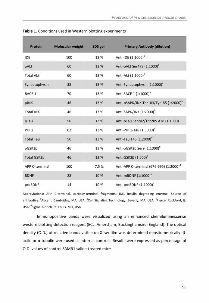
Propranolol in a senescence mouse model
95
Table 1. Conditions used in Western blotting experiments
Abbreviations: APP C-terminal, carboxy-terminal fragments; IDE, insulin degrading enzyme. Source of
antibodies:aAbcam, Cambridge, MA, USA;
bCell Signaling Technology, Beverly, MA, USA;
cPierce, Rockford, IL,
USA;dSigma-Aldrich, St. Louis, MO, USA.
Immunopositive bands were visualized using an enhanced chemiluminescense
western blotting-detection reagent (ECL; Amersham, Buckinghamsire, England). The optical
density (O.D.) of reactive bands visible on X-ray film was determined densitometrically. β-
actin or α-tubulin were used as internal controls. Results were expressed as percentage of
O.D. values of control SAMR1 saline-treated mice.
Protein Molecular weight SDS gel Primary Antibody (dilution)
IDE 100 13 % Anti-IDE (1:1000)a
pAkt 60 13 % Anti-pAkt Ser473 (1:1000)b
Total Akt 60 13 % Anti-Akt (1:1000)b
Synaptophysin 38 13 % Anti-Synaptophysin (1:1000)a
BACE 1 70 13 % Anti BACE 1 (1:1000)a
pJNK 46 13 % Anti-pSAPK/JNK Thr183/Tyr185 (1:2000)b
Total JNK 46 13 % Anti-SAPK/JNK (1:2000)b
pTau 50 13 % Anti-pTau Ser202/Thr205 AT8 (1:1000)c
PHF1 62 13 % Anti-PHF1-Tau (1:3000)a
Total Tau 50 13 % Anti-Tau T46 (1:2000)d
pGSK3β 46 13 % Anti-pGSK3β Ser9 (1:1000)b
Total GSK3β 46 13 % Anti-GSK3β (1:500)b
APP C-terminal 100 7,5 % Anti-APP C-terminal (676-695) (1:2000)d
BDNF 28 10 % Anti-mBDNF (1:1000)a
proBDNF 14 10 % Anti-proBDNF (1:1000)a

Propranolol in a senescence mouse model
96
2.4.3 Insulin levels
Insulin was measured in 10 µl plasma samples using the Sensitive Mouse Insulin
enzimeimmunoassay Kit (EZRMI-13K, Millipore, Billerica, MA).
2.4.4 Aβ levels
Aβ levels were determined with BC05 antibody, specific for the C-terminal of Aβ42,
using a commercially available high-sensitive ELISA kit (Wako Pure Chemical Industries,
Tokyo, Japan) following manufacturer instructions.
2.5 Data analysis and statistics
Data were analysed by SPSS for Windows, release 15.0. Normality was checked by
Shapiro-Wilks’s test (p>0.05). Behavioural and biochemical data were analysed by two-way
analysis of variance ANOVA (strain*treatment), followed by Student's t-test adjusted by
Bonferroni correction. Data are presented as mean ±SEM. The level of significance for all
analyses was set at p < 0.05.
3. Results
3.1 Effect of propranolol treatment on memory deficits in SAMP8
Two-way ANOVA indicated a significant interaction between strain and treatment
on the measure of discrimination between new and familiar objects (F1.31=7.166, p<0.05;
n=8). Further analysis revealed that SAMP8 animals showed a significant memory
impairment (Student’s t-test, p<0.01) that was reversed by propranolol treatment
(Student’s t-test, p<0.05) (Figure 1a).
The effects observed in NORT do not seem to be associated to differences in
locomotor activity, as total distance travelled in the open field (Table 2) was not affected
by either strain (F1.31 = 1.703, p = 0.203) or treatment (F1.31 = 2.115, p = 0.301). In addition,
as shown in Table 2, there was no difference in the discrimination index in the sample trial
in the NORT associated to strain (F1.31=0.75, p=0.387) or treatment (F1.31=0.16, p=0.692).
Total amount of time spent in exploring in the NORT was similar between groups in the

Propranolol in a senescence mouse model
97
sample trial (Table 2, (F1.31=1.96, p=0.172 associated to strain, F1.31=0.634, p=0.433
associated to treatment).
Table 2. Locomotor activity and exploration time and object preference in the sample
phase of the novel object recognition test (NORT).
Locomotor activity Total exploration Discrimination
(cm) time (s) index
6 months
SAMR1 Saline 5715,57±1734,42 60,10±7,12 54,02±6,96
SAMR1 Propranolol 5807,33±1719,61 57,49±3,02 53,62±3,82
SAMP8 Saline 6081,14±1485,43 67,31±8,17 52,87±4,21
SAMP8 Propranolol 5236,46±1732,32 57,04±12,13 51,02±4,53
9 months
SAMR1 Saline 5087,66±1025,71 23,92±3,45 46,30±5,07
SAMR1 Propranolol 4715,05±1464,46 28,27±4,18 51,82±3,93
SAMP8 Saline 4085,59±1433,18 27,43±5,07 52,26±4,76
SAMP8 Propranolol 4630,59±1310,17 28,32±3,64 45,055±3,52
Discrimination index: time exploring one of the objects/total exploration time x 100. There were no significant
differences (two-way ANOVA strain*treatment) in any of the measurements.
As it has been showing that drastic memory deficits comparing SAMR1 and SAMP8
mice could be observed in older animals, the effects of propranolol in the NORT were also
tested in a pilot experiment in 9 months old mice. A significant interaction between strain
and treatment on the discrimination index in the NORT was found (F1.31=3.404, p<0.05,
n=8). Further analysis revealed a significant memory deficit in older SAP8 mice (Student’s t-
test, p<0.01) that was reversed by propranolol treatment (Student’s t-test, p<0.05) (Figure
1b). Again, these effects observed in NORT do not seem to be associated to differences in
locomotor activity, as total distance travelled in the open field (F1.31 = 1.097, p = 0.306 for
strain, F1.31 = 0.028, p = 0869 for treatment), discrimination index in the sample trial (F1.31 =
0.058, p = 0.812 for strain, F1.31 = 0.062, p = 0.805 for treatment) or total amount of time
spent in exploring in the sample trial of the NORT (F1.31 = 1.553, p = 0.272 for strain, F1.31 =

Propranolol in a senescence mouse model
98
0.096, p = 0.759 for treatment) was similar between groups. Regardless of treatment, total
time exploring objects (F1.60 = 25.115, p = 0.301 for strain, F1.60 <0.000 for age) was
significantly lower in older mice. These results are shown in Table 2.
Figure 1. Effects of propranolol treatment on cognition in the NORT in 6 month old (a) and 9 months old (b)
mice. Data presented as discrimination index: time exploring the new object /total exploration time x 100. Two-
way ANOVA (strain x treatment), significant interaction,*p<0.05 significant differences vs control (saline)
SAMR1 mice,#p<0.05 significant differences vs control (saline) SAMP8 mice.
3.2 Effects of propranolol on Aβ processing and clearance
SAMP8 mice exhibited significantly higher hippocampal levels of Aβ42 (two-way
ANOVA, significant interaction F1.27=4.686, p<0.05; n=6–8; Student’s t-test, p<0.05, Figure
2a) and this effect was reversed by propranolol treatment (Student’s t-test, p<0.05). These
increases in Aβ were not related to an increase in APP protein levels (two-way ANOVA,
F1.27=1.998, p > 0.05; n=6–8, Figure 2b). Levels of BACE1, the β-secretase responsible for
the cleavage of APP to form Aβ, were significantly increased in the hippocampus of SAMP8
mice (two-way ANOVA, F1.27=5.264, p<0.05; n=6-8). Further analysis showed that
propranolol was able to reverse this increase (Student’s t-test, p<0.05, Figure 2c).
On the other hand, as shown in Figure 2d, the expression of IDE, a metalloprotease
that plays a crucial role in Aβ clearance in the brain, was significantly lower in SAMP8 mice
[main effect of strain (F1.27=5.812, p<0.05; n=6-8)], and propranolol treatment was able to
increase IDE expression [main effect of treatment (F1.27=4.851, p<0.05; n=6-8). Insulin
levels were also measured as one of the key factors regulating IDE expression. No
differences in plasmatic insulin levels among groups were detected. Levels of insulin in
Saline
Propranolol
0
25
50
75
100
*
#
SAMR 1 SAMP 8
Dis
crim
inat
ion
ind
ex
0
25
50
75
100
*
#
SAMR 1 SAMP 8D
iscr
imin
atio
nin
de
x
(a) (b)Saline
Propranolol
0
25
50
75
100
*
#
SAMR 1 SAMP 8
Dis
crim
inat
ion
ind
ex
0
25
50
75
100
*
#
SAMR 1 SAMP 8D
iscr
imin
atio
nin
de
x
(a) (b)

Propranolol in a senescence mouse model
99
plasma were as follows: 3.32±0.48 for SAMR1, 3.50±0.98 for SAMR1 propranolol,
3.49±1.03 for SAMP8 and 2.81±1.03 for SAMP8 propranolol.
Figure 2. Effects of propranolol on Aβ processing and clearance. (a) Aβ levels (pmol/mg); (b) APP protein levels
(105 kDa) levels; (c) BACE1 levels (70 kDa) ; (d) IDE levels (110 kDa) levels. Panels b, c and d show percentage of
optical density (O.D.) values of control (saline) SAMR1 mice and representative picture of blotting. Satistical
analysis by two-way ANOVA (strain x treatment),*p<0.05 interaction, significant differences vs control (saline)
SAMR1 mice,#p<0.05 interaction, significant differences vs control (saline) SAMP8 mice,
†p<0.05 main effect of
strain,§p<0.05 main effect of treatment.
3.3 Effects of propranolol on Tau pathology
Immunoblot analysis showed that SAMP8 mice displayed hyperphosphorylation of
Tau at pSer396/404, using the PHF-1 antibody (pTau normalized to total Tau levels) [two-
way ANOVA, main effect of strain, F1.27=4.304, p<0.05; n=6-8] and propranolol treatment
0
100
200
300
400
500
600
700
800
Aß
42
leve
ls(p
mo
l/m
g)
*
#
SAMR 1 SAMP 8
(a) APP
ß-actin
salprop propsal
SAMR1 SAMP8
0
25
50
75
100
125
150
SAMR 1 SAMP 8%
O.D
.
(b)
0
25
50
75
100
125
150
†/§
§
%O
.D. †
SAMR 1 SAMP 8
IDE
ß-actin
salprop propsal
SAMR1 SAMP8
(d)
Saline
Propranolol
0
25
50
75
100
125
150
175
200
225*
#
SAMR 1 SAMP 8
%O
.D.
(c) BACE1
ß-actin
salprop propsal
SAMR1 SAMP8
0
100
200
300
400
500
600
700
800
Aß
42
leve
ls(p
mo
l/m
g)
*
#
SAMR 1 SAMP 8
(a)
0
100
200
300
400
500
600
700
800
Aß
42
leve
ls(p
mo
l/m
g)
*
#
SAMR 1 SAMP 8
(a) APP
ß-actin
salprop propsal
SAMR1 SAMP8
0
25
50
75
100
125
150
SAMR 1 SAMP 8%
O.D
.
(b) APP
ß-actin
salprop propsal
SAMR1 SAMP8
APP
ß-actin
salprop propsal
SAMR1 SAMP8
0
25
50
75
100
125
150
SAMR 1 SAMP 8%
O.D
.
(b)
0
25
50
75
100
125
150
†/§
§
%O
.D. †
SAMR 1 SAMP 8
IDE
ß-actin
salprop propsal
SAMR1 SAMP8
(d)
0
25
50
75
100
125
150
†/§
§
%O
.D. †
SAMR 1 SAMP 8
IDE
ß-actin
salprop propsal
SAMR1 SAMP8
IDE
ß-actin
salprop propsal
SAMR1 SAMP8
(d)
Saline
Propranolol
Saline
Propranolol
0
25
50
75
100
125
150
175
200
225*
#
SAMR 1 SAMP 8
%O
.D.
(c) BACE1
ß-actin
salprop propsal
SAMR1 SAMP8
0
25
50
75
100
125
150
175
200
225*
#
SAMR 1 SAMP 8
%O
.D.
(c) BACE1
ß-actin
salprop propsal
SAMR1 SAMP8
BACE1
ß-actin
salprop propsal
SAMR1 SAMP8

Propranolol in a senescence mouse model
100
was able to decrease this phosphorylation (main effect of treatment F1.27=5.806, p<0.05;
n=6-8) (Figure 3a).
The expression of several kinases implicated in Tau pathology was checked. As
shown in Figure 3b, there was an interaction between strain and treatment (two-way
ANOVA, F1.27=9.593, p<0.01; n=6-8) regarding to phosphorylated JNK1 levels (pJNK1)
normalized to total JNK1. Western blot analysis revealed that pJNK1 levels were
significantly increased in saline SAMP8 mice compared with SAMR1 mice (Student’s t-test,
p<0.05). Importantly, propranolol treatment was able to reverse these effects on pJNK1
expression (Student’s t-test, p<0.05).
Figure 3. Effects of propranolol on Tau pathology. (a) Phosphorylated Tau at Ser396 and Ser404 levels (PHF-1
62 kDa) normalized to total Tau, (b) Phosphorylated JNK1 levels (active form 46-54 kDa) normalized to total
JNK. Each panel shows percentage of optical density (O.D.) values of control (saline) SAMR1 mice and
representative picture of the blotting. Statistical analysis by two way ANOVA (strain x treatment)*p<0.01
interaction, significant differences vs control (saline) SAMR1 mice,#p<0.05 interaction, significant differences vs
control (saline) SAMP8 mice,†main effect of strain,
§p<0.05 main effect of treatment.
Moreover, propranolol treatment was able to increase the expression of
phosphorylated GSK3β (pGSK3β in Ser9) normalized to total GSK3ß (two-way ANOVA, main
effect of treatment, F1.27=7.945, p<0.05; n=6-8) (Figure 4a). Levels of pAkt were also
measured, as GSK3β phosphorylated at Ser9 is a downstream target of activated Akt (pAkt).
There was a significant interaction (two-way ANOVA, F1.27=5.652, p<0.05; n=6-8) between
strain and treatment in pAkt expression normalized to total Akt (Figure 4b). Further
total tau
ß-actin
PHF1
salprop propsal
SAMR1 SAMP8
0
25
50
75
100
125
150
175
§
†/§
%O
.D.
†
SAMR 1 SAMP 8
(a)
0
25
50
75
100
125
150
175**
#
%O
.D.
SAMR 1 SAMP 8
pJNK
total JNK
ß-actin
salprop propsal
SAMR1 SAMP8(b)
Saline
Propranolol
total tau
ß-actin
PHF1
salprop propsal
SAMR1 SAMP8
total tau
ß-actin
PHF1
salprop propsal
SAMR1 SAMP8
0
25
50
75
100
125
150
175
§
†/§
%O
.D.
†
SAMR 1 SAMP 8
(a)
0
25
50
75
100
125
150
175**
#
%O
.D.
SAMR 1 SAMP 8
pJNK
total JNK
ß-actin
salprop propsal
SAMR1 SAMP8
pJNK
total JNK
ß-actin
salprop propsal
SAMR1 SAMP8(b)
Saline
Propranolol
Saline
Propranolol
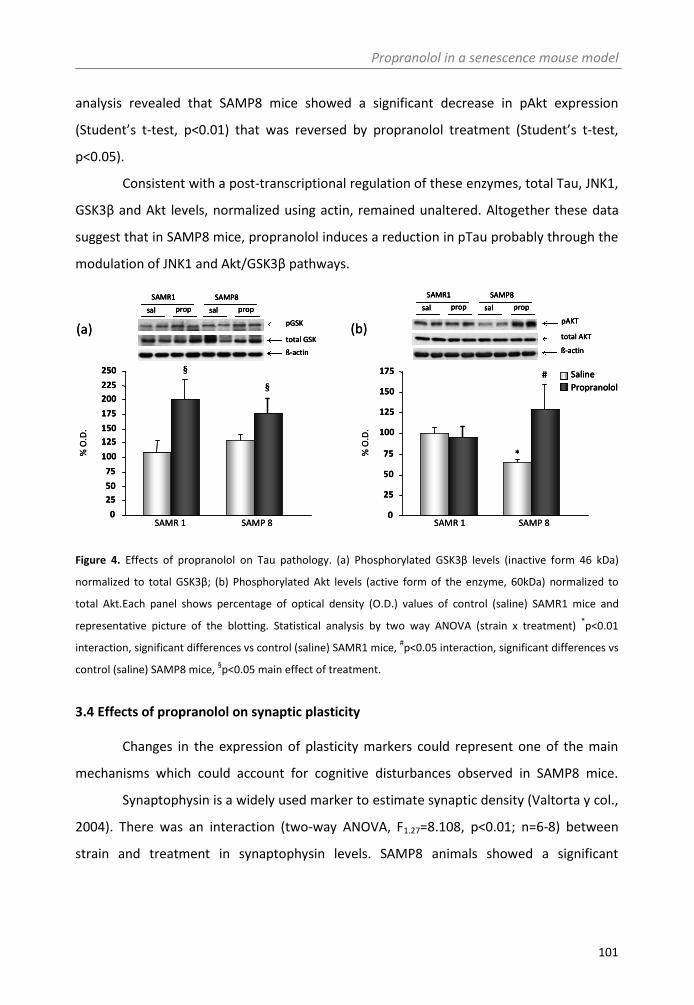
Propranolol in a senescence mouse model
101
analysis revealed that SAMP8 mice showed a significant decrease in pAkt expression
(Student’s t-test, p<0.01) that was reversed by propranolol treatment (Student’s t-test,
p<0.05).
Consistent with a post-transcriptional regulation of these enzymes, total Tau, JNK1,
GSK3β and Akt levels, normalized using actin, remained unaltered. Altogether these data
suggest that in SAMP8 mice, propranolol induces a reduction in pTau probably through the
modulation of JNK1 and Akt/GSK3β pathways.
Figure 4. Effects of propranolol on Tau pathology. (a) Phosphorylated GSK3β levels (inactive form 46 kDa)
normalized to total GSK3β; (b) Phosphorylated Akt levels (active form of the enzyme, 60kDa) normalized to
total Akt.Each panel shows percentage of optical density (O.D.) values of control (saline) SAMR1 mice and
representative picture of the blotting. Statistical analysis by two way ANOVA (strain x treatment)*p<0.01
interaction, significant differences vs control (saline) SAMR1 mice,#p<0.05 interaction, significant differences vs
control (saline) SAMP8 mice,§p<0.05 main effect of treatment.
3.4 Effects of propranolol on synaptic plasticity
Changes in the expression of plasticity markers could represent one of the main
mechanisms which could account for cognitive disturbances observed in SAMP8 mice.
Synaptophysin is a widely used marker to estimate synaptic density (Valtorta y col.,
2004). There was an interaction (two-way ANOVA, F1.27=8.108, p<0.01; n=6-8) between
strain and treatment in synaptophysin levels. SAMP8 animals showed a significant
0
25
50
75
100
125
150
175
200
225
250 §
%O
.D.
§
SAMR 1 SAMP 8
total GSK
ß-actin
pGSK
salprop propsal
SAMR1 SAMP8
(a)
0
25
50
75
100
125
150
175
*
#%
O.D
.
SAMR 1 SAMP 8
pAKT
total AKT
ß-actin
salprop propsal
SAMR1 SAMP8
(b)
Saline
Propranolol
0
25
50
75
100
125
150
175
200
225
250 §
%O
.D.
§
SAMR 1 SAMP 8
total GSK
ß-actin
pGSK
salprop propsal
SAMR1 SAMP8
total GSK
ß-actin
pGSK
salprop propsal
SAMR1 SAMP8
(a)
0
25
50
75
100
125
150
175
*
#%
O.D
.
SAMR 1 SAMP 8
pAKT
total AKT
ß-actin
salprop propsal
SAMR1 SAMP8
pAKT
total AKT
ß-actin
salprop propsal
SAMR1 SAMP8
(b)
Saline
Propranolol
Saline
Propranolol

Propranolol in a senescence mouse model
102
%O
.D.
0
25
50
75
100
125
150
**
#
synaptophysin
ß-actin
salprop propsal
SAMR1 SAMP8
SAMR 1 SAMP 8
(a)
SAMR 1 SAMP 80
25
50
75
100
125
150
*
#
Rat
iom
BD
NF/
pro
BD
NF
(b)
Saline
Propranolol
mBDNF
proBDNF
ß-actin
salprop propsal
SAMR1 SAMP8
%O
.D.
0
25
50
75
100
125
150
**
#
synaptophysin
ß-actin
salprop propsal
SAMR1 SAMP8
synaptophysin
ß-actin
salprop propsal
SAMR1 SAMP8
SAMR 1 SAMP 8
(a)
SAMR 1 SAMP 80
25
50
75
100
125
150
*
#
Rat
iom
BD
NF/
pro
BD
NF
SAMR 1 SAMP 80
25
50
75
100
125
150
*
#
Rat
iom
BD
NF/
pro
BD
NF
(b)
Saline
Propranolol
Saline
Propranolol
mBDNF
proBDNF
ß-actin
salprop propsal
SAMR1 SAMP8
mBDNF
proBDNF
ß-actin
salprop propsal
SAMR1 SAMP8
decrease in synaptophysin levels (Student’s t-test; p<0.01) that was reversed by
propranolol treatment (Student’s t-test, p<0.05) (Figure 5a).
The neurotrophic factor BDNF helps the survival of existing neurons, facilitates the
growth of new neurons and synapses, and low BDNF levels are associated with cognitive
deficits. Decreased BDNF levels (mature, mBDNF) were found associated to strain
(F1.25=7.727, p<0.05; n=6-8), while propranolol treatment increased mBDNF expression
(F1.25=5.370, p<0.05; n=6-8). BDNF is secreted to the extracellular matrix in a proBDNF form,
which negatively affects survival and synaptic plasticity. However, proBDNF is mainly
cleaved to mature BDNF, which eventually has positive effects on survival, growth, synaptic
plasticity and memory formation. Therefore, the ratio proBDNF/mBDNF has been
measured (Figure 5b). There was a significant interaction between strain and treatment
(two-way ANOVA, F1.27=5.221, p<0.05; n=6-8), and a significant decrease in the pro-
BDNF/mBDNF ratio was found in SAMP8 mice (Student’s t-test; p<0.01) that was reversed
by propranolol treatment (Student’s t-test, p<0.05).
Figure 5. Effects of propranolol on synaptic plasticity. (a) Synaptophysin levels (38 kDa) levels; (b) Ratio
proBDNF to mature (m)BDNF. No differences were found in the proBDNF normalized to actin. Statistical
analysis by two-way ANOVA (strain x treatment),*p<0.01 interaction, significant differences vs control (saline)
SAMR1 mice,#p<0.05 interaction, significant differences vs control (saline) SAMP8 mice.

Propranolol in a senescence mouse model
103
4. Discussion
In the present study it has been shown that propranolol, a β-adrenergic antagonist
widely used as an antihypertensive drug, exerts a protective effect on memory
impairments shown by SAMP8 mice in the NORT. Moreover, propranolol reversed the
main neuropathological markers shown by this senescence-accelerated mouse model, such
as Tau hyperphosphorylation and β-amyloid pathology. It is discussed below that these
effects could be mediated by the modulation of JNK1 activity.
SAMP8 mice show at early as 5-6-month-old of age memory impairments, Aβ
deposition and increased Tau phosphorylationin the hippocampus when compared with
age matched SAMR1 mice (Pallas et al., 2008). SAMP8 mice are considered nowadays as a
robust model for studying age-associated pathologies, including AD (Liu et al., 2012; Pallas
et al., 2008; Sureda et al., 2006). It has even been suggested that SAMP8 mice may more
closely represent the complexity of the disease than other transgenic mouse lines because
of the multifactorial nature of AD (Pang et al., 2006). As already described, 6-month-old
SAMP8 mice presented learning and memory impairments in the NORT when compared
with age matched SAMR1 mice, an effect that was reversed by propranolol. Previous works
have reported the ability of propranolol to reverse cognitive deficits associated to
deleterious insults, such as chronic stress (Aisa et al., 2007). However, to our knowledge,
this is the first report describing pro-cognitive actions of propranolol in an experimental
model of ageing. It is important to note that propranolol was used in the present study at a
lower dose than the commonly prescribed for the treatment of hypertension in humans
(Shannon et al., 2007), as calculated by the formula described by Reagan-Shaw et al. (2007).
Therefore, the results presented here seem to be independent from the antihypertensive
effect of propranolol. In an attempt to elucidate the mechanisms underlying the beneficial
effects of propranolol we focused our study on both β-amyloid and Tau pathology, the two
main pathological hallmarks in AD.
AD is characterized by the progressive accumulation in the brain of senile plaques
composed of beta-amyloid protein (Aβ). According to the amyloid hypothesis,
accumulation of Aβ in the brain is the primary influence leading to AD pathogenesis, and
Propranolol in a senescence mouse model
104
this accumulation it is proposed to result from an imbalance between Aβ production and
Aβ clearance. Del Kumar et al. (2000) and Valle et al (2010) showed that from as early as 6
months onward, SAMP8 mice show Aβ deposition in the hippocampus that increases in
number and extent with age. However, plaques cannot be found until the age of 18
months (Morley et al., 2000). Aβ protein is generated from APP, which can be cleaved by
BACE1 and α-secretase to produce C99 and C83. These C-terminal fragments can then be
cleaved by γ-secretase to produce Aβ and p3, respectively (Dhikav and Anand, 2007). In
APP transgenic mice, BACE1 overexpression increases Aβ formation (Fukomoto et al.,
2002), whereas BACE1 knockout mice produce little or no Aβ peptide (Bodendorf et al.,
2002; Luo et al., 2001), indicating that BACE1 expression plays a critical role in Aβ peptide
biosynthesis. On the other hand, the major protease involved in Aβ degradation in brain
tissue is the insulin degrading enzyme (IDE) (Kurochkin et al., 1994; McDermott et al., 1997;
Qiu et al., 1998; Vekrellis et al., 2000). It has been hypothesized that an age-associated
decline in Aβ-degrading protease activity would contribute to Aβ accumulation, particularly
in patients with late-onset AD. Early studies supported such a relationship and revealed
reduced IDE mRNA and protein immunolabeling in AD compared with control brains
(Miners et al., 2006; Russo et al., 2005; Wang et al., 2010; Akiyama et al., 2001). It has also
been shown that knockout IDE mice present a lower degradation of Aβ (Farris et al., 2003).
In our hands, it seems that the increased Aβ levels in SAMP8 mice could be derived from
both an increased formation through a BACE1 up regulation and a decreased degradation
associated to a lower IDE expression, and all these effects were reversed by propranolol.
Supporting this hypothesis, increased levels of Aβ were not accompanied by changes in the
expression of APP, suggesting an increased amyloidogenic processing of APP.
In addition to accumulation of Aβ, abnormal hyperphosphorylation of Tau is a
critical event in the cascade leading to AD (Blennow et al., 2006). Tau
hyperphosphorylation has been described in 5-month-old SAMP8 mice and it has been
directly implicated in neurofibrillary degeneration and accelerated aging process in this
senescence mouse model (Canudas et al., 2005; García-Matas et al., 2008; Sureda et al.,
2006). Data based on several lines of evidence from transgenic mice and humans indicate a
strong correlation between the extent of Tau pathology and cognitive dysfunction
Propranolol in a senescence mouse model
105
(Arriadaga et al., 1992; Ashe and Zahs, 2010; Guillozet et al., 2003). Among kinases
implicated in Tau pathology, data on activation of JNK1 seems to be of particular interest.
JNK1 is a member of the MAPK family that plays a key role in neuronal plasticity,
regeneration, cell death and regulation of cellular senescence (Kögel et al., 2005,
Maruyama et al., 2009). Activation of JNK1 phosphorylates Tau at sites recognized by the
presently used PHF-1 antibody (Bellucci et al., 2007). It is to note that JNK1 phosphorylates
the insulin receptor substrate 1 (IRS1) at an inhibitory site that can block signal
transduction by the insulin receptor (Sabio et al, 2008), therefore purportedly altering
insulin-related intracellular pathways, namely the Akt-GSK-Tau. This Akt-GSK route is
required for the induction of long-term potentiation and depression, basic processes
underlying learning and memory (Van der Heide et al., 2006). Accordingly, it has been
found, compared to controls, that treatment with propranolol in SAMP8 mice leads to
decreased JNK1 activity accompanied by increased pAkt, increased pGSK-3β (inactive form)
and decreased pTau expression. Even more, it has been described that JNK1 mediates
BACE1 upregulation (Tamagno et al., 2005; 2009). Therefore, modulation of JNK1 activity in
SAMP8 mice or by propranolol treatment may explain the effects on both Aβ and tau
pathology. However, the mechanisms by which propranolol could affect JNK1 activity
remains to be elucidated.
As previously reported, SAMP8 mice displayed decreased levels of synaptopshysin
(Cheng et al., 2011) or BDNF (Tajes et al., 2010; Li et al., 2009), which could contribute to
cognitive deficits. BDNF has been described to be highly protective for different neuronal
phenotypes and can prevent neuronal cell death in different experimental paradigms in
vivo as well as in vitro (Barde, 1994; Siegel and Chauhan, 2000). BDNF levels decline with
age and are lower in AD brains that in those without dementia. In various mouse, rat, and
primate models, BDNF therapy has reverted synaptic damage and reversed learning and
memory deficits (see review by Nagahara and Tuszynsk, 2011). Not only that, but
synaptophysin seem to be a presynaptic vesicle protein partially regulated by BDNF
(Tartaglia et al., 2001; Valtorta et al., 2004), therefore reduced synaptophysin expression
may result from a reduced BDNF function. However, hippocampal BDNF is secreted to the
extracellular matrix in a proBDNF form. ProBDNF is active via p75NTR receptors and

Propranolol in a senescence mouse model
106
negatively affects survival and synaptic plasticity. ProBDNF is mainly cleaved to mature
BDNF to activate TrkB receptors, which eventually has positive effects on survival, growth,
synaptic plasticity and memory formation. Therefore, the ratio proBDNF/mBDNF could be
a more accurate form to anticipate BDNF outcomes. It is to note that it could have been
expected that β-receptor blockade would have led to decreased BDNF levels (Flores et al.,
2010). Therefore, the present increase in the ratio suggests that propranolol could
modulate the cleavage of pro-BDNF to give rise to the mature form.
As final comments, it is worth mentioning that this is not the first work showing
that semi-chronic 3-4 weeks treatments are enough to reverse deficits/alterations in
SAMP8 mice (Orejana et al., 2012). One of the reason that could explain the success of
semi-chronic treatments in reversing pathology in SAMP8 mice is the fact that this is a
model of accelerated outcomes (pathology starts to show early in life) and the fact that
treatment started when pathology was not so established and deleterious that it is difficult
to reverse. These hypotheses are related to the currently spreading notion that
diagnosis/pharmacological treatment of Alzheimer´s disease starts too late in life (Haas et
al., 2012). Interestingly, a pilot experiment was also performed in the present work in
which propranolol was also able to reverse the cognitive deficits observed even in older (9
months old at the beginning of treatment) SAMP8 mice. At this age, the most significant
memory changes between SAMR1 and SAMP8 could be observed (Pallas et al., 2008).
In summary, it is proposed here that Aβ-, Tau-, synaptic- and cognitive pathology in
SAMP8 mice could be related to JNK1 activation, as already suggested in Tg2576/PS1
double transgenic mouse model for AD, in which the activation of JNK1 has been
associated with age-dependent amyloid plaque deposition, Tau phosphorylation, and the
loss of synaptophysin (Kyriakis, 1999; Pearson et al., 2006). Interestingly, all these
pathological features could be reversed by propranolol treatment. Therefore, the present
work could constitute the proof of concept for the use of propranolol for the treatment of
pathologies associate to ageing.

Propranolol in a senescence mouse model
107
References
Aisa B, Tordera R, Lasheras B, Del Rio J, Ramirez MJ. (2007). Cognitive impairment
associated to HPA axis hyperactivity after maternal separation in rats.
Psychoneuroendocrinology 32: 256-266.
Akiyama H, Kondo H, Ikeda K, Kato M, McGeer PL. (2001). Immunohistochemical
localization of neprilysin in the human cerebral cortex: inverse association with
vulnerability to amyloid beta-protein (Abeta) deposition. Brain Research 902: 277-281.
Arriagada PV, Growdon JH, Hedley-Whyte ET, Hyman BT. (1992). Neurofibrillary tangles
but not senile plaques parallel duration and severity of Alzheimer’s disease. Neurology 42:
631-639.
Ashe KH, Zahs KR. (2010). Probing the biology of Alzheimer’s disease in mice. Neuron 66:
631-645.
Barde YA. (1994). Neurotrophic factors: an evolutionary perspective. Journal of
Neurobiology 25: 1329-1333.
Barzilai N, Atzmon G, Derby CA, Bauman JM, Lipton RB. (2006). A genotype of exceptional
longevity is associated with preservation of cognitive function. Neurology 67: 2170-2175.
Bellucci A, Rosi MC, Grossi C, Fiorentini A, Luccarini I, Casamenti F. (2007). Abnormal
processing of tau in the brain of aged TgCRND8 mice. Neurobiology of Disease 27: 328-338.
Blennow K, de Leon MJ, Zetterberg H. (2006). Alzheimer's disease. Lancet 368: 387-403.
Bodendorf U, Danner S, Fischer F, Stefani M, Sturchler-Pierrat C, Wiederhold KH,
Staufenbiel M, Paganetti P. (2002). Expression of human beta-secretase in the mouse brain
increases the steady-state level of betaamyloid. Journal of Neurochemistry 80: 799-806.

Propranolol in a senescence mouse model
108
Canudas AM, Gutierrez-Cuesta J, Rodríguez MI, Acuña-Castroviejo D, Sureda FX, Camins A,
Pallàs M. (2005). Hyperphosphorylation of microtubule-associated protein tau in
senescence-accelerated mouse (SAM). Mechanisms of Ageing and Development 126: 1300-
1304.
Cheng XR, Yang Y, Zhou WX, Zhang YX. (2011). Expression of VGLUTs contributes to
degeneration and acquisition of learning and memory. Neurobiology of Learning and
Memory 95: 361-375.
Del Valle J, Duran-Vilaregut J, Manich G, Casadesús G, Smith MA, Camins A, Pallàs M,
Pelegrí C, Vilaplana J. (2010). Early amyloid accumulation in the hippocampus of SAMP8
mice. Journal of Alzheimer’s Disease 19: 1303-1315.
Dhikav V, Anand KS. (2007). Glucocorticoids may initiate Alzheimer's disease: a potential
therapeutic role for mifepristone (RU-486). Medical Hypotheses 68: 1088-1092.
Ennanceur A, Delacour J. (1988). A new one trial-test for neurobiological studies of
memory in rats. 1: Behavioral data. Behavioural Brain Research 31: 47-59.
Farris W, Mansourian S, Chang Y, Lindsley L, Eckman EA, Frosch MP, Eckman CB, Tanzi RE,
Selkoe DJ, Guenette S. (2003). Insulin-degrading enzyme regulates the levels of insulin,
amyloid beta-protein, and the beta-amyloid precursor protein intracellular domain in vivo.
Proceedings of the National Academy of Sciences of the United States of America 100:
4162-4167.
Flores O, Núñez H, Pérez H, Morgan C, Soto-Moyano R, Valladares L, Burgos H, Olivares R,
Hernández A. (2010). beta-Adrenoceptor blockade depresses molecular and functional
plasticities in the rat neocortex. Brain Research Bulletin 82: 284-288.

Propranolol in a senescence mouse model
109
Fukumoto H, Cheung BS, Hyman BT, Irizarry MC. (2002). Beta-secretase protein and
activity are increased in the neocortex in Alzheimer disease. Archives of Neurology 59:
1381-1389.
García-Matas S, Gutierrez-Cuesta J, Coto-Montes A, Rubio-Acero R, Díez-Vives C, Camins A,
Pallàs M, Sanfeliu C, Cristòfol R. (2008). Dysfunction of astrocytes in senescence-
accelerated mice SAMP8 reduces their neuroprotective capacity. Aging Cell 7: 630-640.
Guillozet AL, Weintraub S, Mash DC, Mesulam MM. (2003). Neurofibrillary tangles, amyloid,
and memory in aging and mild cognitive impairment. Archives of Neurology 60: 729-736.
Guo Z, Viitanen M, Winblad B, Fratiglioni L. (1999). Low blood pressure and incidence of
dementia in a very old sample: dependent on initial cognition. Journal of the American
Geriatrics Society 47: 723-726.
Hajjar I, Keown M, Frost B. (2005). Antihypertensive agents for aging patients who are at
risk for cognitive dysfunction. Current Hypertension Reports 7: 466-473.
Hamamoto H, Honma A, Irino M, Matsushita T, Toda K, Matsumura M, Takeda T. (1984).
Grading score system: A method for evaluation of the degree of senescence in senescence
accelerated mouse (SAM). Mechanisms of Ageing and Development 26: 91-102.
Hoffman LB, Schmeidler J, Lesser GT, Beeri MS, Purohit DP, Grossman HT, Haroutunian V.
(2009). Less Alzheimer disease neuropathology in medicated hypertensive than
nonhypertensive persons. Neurology 72: 1720-1726.
Khachaturian AS, Zandi PP, Lyketsos CG, Hayden KM, Skoog I, Norton MC, Tschanz JT,
Mayer LS, Welsh-Bohmer KA, Breitner JC. (2006). Antihypertensive medication use and
incident Alzheimer disease: the Cache County Study. Archives of Neurology 63: 686-692.
Propranolol in a senescence mouse model
110
Kögel D, Schomburg R, Copanaki E, Prehn JH. (2005). Regulation of gene expression by the
amyloid precursor protein: inhibition of the JNK/cJun pathway. Cell Death & Differentiation
12: 1-9.
Kyriakis JM. (1999). Making the connection: coupling of stress-activated ERK/MAPK
(extracellular-signal-regulated kinase/mitogen-activated protein kinase) core signalling
modules to extracellular stimuli and biological responses. Biochemical Society Symposia 64:
29-48.
Li Q, Zhao HF, Zhang ZF, Liu ZG, Pei XR, Wang JB, Li Y. (2009). Long-term green tea catechin
administration prevents spatial learning and memory impairment in senescence-
accelerated mouse prone-8 mice by decreasing Abeta1-42 oligomers and upregulating
synaptic plasticity-related proteins in the hippocampus. Neuroscience 163: 741-749.
Liu W, Liu C, Zhu J, Shu P, Yin B, Gong Y, Qiang B, Yuan J, Peng X. (2010). MicroRNA-16
targets amyloid precursor protein to potentially modulate Alzheimer’s-associated
pathogenesis in SAMP8 mice. Neurobiology of Aging 33: 522-534.
Luchsinger JA, Reitz C, Honig LS, Tang MX, Shea S, Mayeux R. (2005). Aggregation of
vascular risk factors and risk of incident Alzheimer disease. Neurology 65: 545-551.
Luo Y, Bolon B, Kahn S, Bennett BD, Babu-Khan S, Denis P, Fan W, Kha H, Zhang J, Gong Y,
Martin L, Louis JC, Yan Q, Richards WG, Citron M, Vassar R. (2001). Mice deficient in BACE1,
the Alzheimer's beta-secretase, have normal phenotype and abolished beta-amyloid
generation. Nature Neuroscience 4: 231-232.
Maruyama J, Naguro I, Takeda K, Ichijo H. (2009). Stress-activated MAP kinase cascades in
cellular senescence. Current Medicinal Chemistry 16, 1229-1235.

Propranolol in a senescence mouse model
111
McDermott JR, Gibson AM. (1997). Degradation of Alzheimer's beta-amyloid protein by
human and rat brain peptidases: involvement of insulin-degrading enzyme. Neurochemical
Research 22: 49-56.
Mehta PD. (2007). Amyloid beta protein as a marker or risk factor of Alzheimer’s disease.
Current Alzheimer Research 4: 359-363.
Miners JS, Van Helmond Z, Chalmers K, Wilcock G, Love S, Kehoe PG. (2006). Decreased
expression and activity of neprilysin in Alzheimer disease are associated with cerebral
amyloid angiopathy. Journal of Neuropathology & Experimental Neurology 65: 1012-1021.
Orejana L, Barros-Miñones L, Jordán J, Puerta E, Aguirre N. (2011). Sildenafil ameliorates
cognitive deficits and tau pathology in a senescence-accelerated mouse model.
Neurobiology of Aging 33: 625.e11-20.
Pallas M., Camins A, Smith MA, Perry G, Lee HG, Casadesus G. (2008). From aging to
Alzheimer’s disease: unveiling “the switch” with the senescence-accelerated mouse model
(SAMP8). Journal of Alzheimer’s Disease 15: 615-624.
Pang KCH, Miller JP, Fortress A, McAuley D. (2006). Age-related disruptions of circadian
rhythm and memory in the senescence-accelerated mouse (SAM8). Age 28: 283-296.
Pearson AG, Byrne UT, MacGibbon GA, Faull RL, Dragunow M. (2006). Activated c-Jun is
present in neurofibrillary tangles in Alzheimer's disease brains. Neuroscience Letters 398:
246-250.
Pimenta E, Oparil S. (2012). Management of hypertension in the elderly. Nature Reviews
Cardiology 9: 286-296.

Propranolol in a senescence mouse model
112
Qiu WQ, Walsh DM, Ye Z, Vekrellis K, Zhang J, Podlisny MB, Rosner MR, Safavi A, Hersh LB,
Selkoe DJ. (1998). Insulin-degrading enzyme regulates extracellular levels of amyloid beta-
protein by degradation. Journal of Biological Chemistry 273: 32730-32738.
Reagan-Shaw S, Nihal M, Ahmad N. (2007). Dose translation from animal to human studies
revisited. FASEB Journal 22: 659-661.
Roozendaal B, Hahn EL, Nathan SV, de Quervain DJ, McGaugh JL. (2004). Glucocorticoid
effects on memory retrieval require concurrent noradrenergic activity in the hippocampus
and basolateral amygdala. Journal of Neuroscience 24: 8161-8169.
Russo R, Borghi R, Markesbery W, Tabaton M, Piccini A. (2005). Neprylisin decreases
uniformly in Alzheimer’s disease and in normal aging. FEBS Letters 579: 6027-6030.
Sabio G, Das M, Mora A, Zhang Z, Jun JY, Ko HJ, Barret T, Kim JK, Davis RJ. (2008). A stress
signaling pathway in adipose tissue regulates hepatic insulin resistance. Science 322: 1539-
1543.
Siegel GJ, Chauhan NB. (2000). Neurotrophic factors in Alzheimer's and Parkinson's disease
brain. Brain Research Reviews 33: 199-227.
Sureda FX, Gutierrez-Cuesta J, Romeu M, Mulero M, Canudas AM, Camins A, Mallol J,
Pallàs M. (2006). Changes in oxidative stress parameters and neurodegeneration markers
in the brain of the senescence-accelerated mice SAMP-8. Experimental Gerontology 41:
360-367.
Tajes M, Gutierrez-Cuesta J, Folch J, Ortuño-Sahagun D, Verdaguer E, Jiménez A, Junyent F,
Lau A, Camins A, Pallàs M. (2010). Neuroprotective role of intermittent fasting in
senescence-accelerated mice P8 (SAMP8). Experimental Gerontology 45: 702-710.
Propranolol in a senescence mouse model
113
Takeda T. (1999). Senescence-accelerated mouse (SAM): a biogerontological resource in
aging research. Neurobiology of Aging 20: 105-110.
Takeda T, Hosokawa M, Higuchi K, Hosono M, Akiguchi I, Katoh H. (1994). A novel murine
model of aging, Senescence-Accelerated Mouse (SAM). Archives of Gerontology and
Geriatrics 19: 185-192.
Takeda T, Hosokawa M, Takeshita S, Irino M, Higuchi K, Matsushita T, Tomita Y, Yasuhira K,
Hamamoto, H, Shimizu K, Ishii M, Yamamuro T. (1981). A new murine model of accelerated
senescence. Mechanisms of Ageing and Development 17: 183-194.
Tamagno E, Guglielmotto M, Giliberto L, Vitali A, Borghi R, Autelli R, Danni O, Tabaton M.
(2009). JNK and ERK1/2 pathways have a dual opposite effect on the expression of BACE1.
Neurobiology of Aging 30: 1563-1573.
Tamagno E, Parola M, Bardini P, Piccini A, Borghi R, Guglielmotto M, Santoro G, Davit A,
Danni O, Smith MA, Perry G, Tabaton M. (2005). Beta-site APP cleaving enzyme up-
regulation induced by 4-hydroxynonenal is mediated by stress-activated protein kinases
pathways. Journal of Neurochemistry 92: 628-636.
Tartaglia N, Du J, Tyler WJ, Neale E, Pozzo-Miller L, Lu B. (2001). Protein synthesis-
dependent and -independent regulation of hippocampal synapses by brain-derived
neurotrophic factor. Journal of Biological Chemistry 276: 37585-37593.
Tomobe K, Nomura Y. (2009). Neurochemistry, neuropathology, and heredity in SAMP8: a
mouse model of senescence. Neurochemical Research 34: 660-669.
Valtorta F, Pennuto M, Bonanomi D, Benfenati F. (2004). Synaptophysin: leading actor or
walk-on role in synaptic vesicle exocytosis? BioEssays 26: 445-453.

Propranolol in a senescence mouse model
114
van der Heide LP, Ramakers GM, Smidt MP. (2006). Insulin signaling in the central nervous
system: learning to survive. Progress in Neurobiology 79: 205-221.
Vekrellis K, Ye Z, Qiu WQ, Walsh D, Hartley D, Chesneau V, Rosner MR, Selkoe DJ. (2000).
Neurons regulate extracellular levels of amyloid beta-protein via proteolysis by insulin-
degrading enzyme. Journal of Neuroscience 20: 1657-1665.
Wang J, Ho L, Chen L, Zhao Z, Zhao W, Qian X, Humala N, Seror I, Bartholomew S,
Rosendorff C, Pasinetti GM. (2007). Valsartan lowers brain beta-amyloid protein levels and
improves spatial learning in a mouse model of Alzheimer disease. Journal of Clinical
Investigation 117: 3393-3402.

CCaappííttuulloo IIVV

Chapter 4
Propranolol reduces cognitive deficits, amyloid and Tau
pathology in Alzheimer´s transgenic mice

Propranolol in a transgenic mouse model
119
Resumen
La eficacia de los fármacos antihipertensivos en la EA es controvertida. En este
trabajo se ha evaluado si algunos antihipertensivos podrían actúar sobre la EA mediante
mecanismos independientes de su actividad antihipertensiva. Se han estudiado los efectos
del tratamiento con el antihipertensivo propranolol sobre la cognición y los marcadores
relacionados con la EA en el modelo animal Tg2576 de EA. El propranolol, a una dosis
menor que la usada como antihipertensivo (5mg/kg, 6 semanas) es capaz de atenuar las
alteraciones cognitivas mostradas por los ratones Tg2576 a los 9 meses de edad en la
prueba de reconocimiento del nuevo objeto y en el test del miedo condicionado. El
propranolol fue capaz de contrarrestar los niveles de Aβ42 presentes en el hipocampo de
los ratones Tg2576. Este efecto se vio acompañado por un aumento en la expresión de la
enzima degradadora IDE. Cambios en marcadores de la patología sináptica, reflejada en
descensos de la fosforilación de Akt y en la expresión de BDNF en los ratones Tg2576,
fueron también contrarrestados por el tratamiento con propranolol. La hiperfosforilación
de Tau mostrada por estos animales también disminuyó en el hipocampo de los ratones
tratados con propranolol, un efecto probablemente debido a un aumento de la
fosforilación de GSK-3β (forma inactiva) y a un descenso de la expresión de JNK1. En
general, estos resultados refuerzan la idea de que el propranolol podría considerarse un
potencial agente terapéutico para la EA.

Propranolol in a transgenic mouse model
120
Abstract
The efficacy of antihypertensive agents in Alzheimer´s disease (AD) is controversial.
It has been tested here whether some antihypertensive drugs might influence AD through
mechanisms independent of blood pressure-lowering activity. The effects of treatment
with the antihypertensive propranolol on cognition and AD-related markers have been
studied in the Tg2576 mouse model of AD. Propranolol, at a lower dose than that used as
antihypertensive (5 mg/kg, 6 weeks), attenuated cognitive impairments shown by 9 month
old Tg2576 mice in the novel object recognition and fear conditioning tests. Propranolol
was also able to counteract the increases in hippocampal levels of Aβ42 present in Tg2576
mice. This effect was accompanied by an increased expression of the degrading enzyme
IDE. Changes in markers of synaptic pathology, as shown by decreases in phosphorylation
of Akt and in the expression of BDNF in Tg2676 mice, were also counteracted by
propranolol treatment. Tau hyperphosphorylation shown by Tg2576 mice was also
decreased in the hippocampus of propranolol-treated mice, an effect probably related to
an increase of GSK-3β phosphorylation (inactive form) and a decreased JNK1 expression.
Overall, these data further strengthen the potential of propranolol as a therapeutic agent
for Alzheimer’s disease.
Keywords:
Antihypertensive, synaptophysin, BDNF, JNK1, IDE, BACE, Akt, GSK-3β, risk factor

Propranolol reduces cognitive deficits,amyloid and tau pathology inAlzheimer’s transgenic mice
Marta Dobarro1, Gorka Gerenu1 and María J Ramírez1,21Dpto Farmacologia, Universidad de Navarra, Pamplona, Spain2Division of Neurosciences, CIMA, Pamplona, Spain
Abstract
The efficacy of antihypertensive agents in Alzheimer’s disease (AD) is controversial. It has been testedhere whether some antihypertensive drugs might influence AD through mechanisms independentof blood pressure-lowering activity. The effects of treatment with the antihypertensive propranolol oncognition and AD-related markers have been studied in the Tg2576 mouse model of AD. Propranolol,at a lower dose than that used as antihypertensive (5mg/kg, 6 wk), attenuated cognitive impairmentsshown by Tg2576 mice aged 9 months in the novel object recognition and fear conditioning tests.Propranolol was also able to counteract the increases in hippocampal levels of Aβ42 present in Tg2576mice. This effect was accompanied by an increased expression of insulin degrading enzyme. Changes inmarkers of synaptic pathology, as shown by decreases in phosphorylation of Akt and in the expressionof BDNF in Tg2676 mice, were also counteracted by propranolol treatment. Tau hyperphosphorylationshown by Tg2576 mice was also decreased in the hippocampus of propranolol-treated mice, an effect prob-ably related to an increase of GSK3β phosphorylation (inactive form) and a decreased JNK1 expression.Overall, these data further strengthen the potential of propranolol as a therapeutic agent for AD.
Received 19 February 2013; Reviewed 14 March 2013; Revised 5 April 2013; Accepted 9 May 2013;First published online 17 June 2013
Key words: Antihypertensive, BACE, BDNF, insulin degrading enzyme, JNK1.
Introduction
Alzheimer’s disease (AD) is the most common neuro-degenerative disorder and the first cause of dementiain the elderly (Cummings and Cole, 2002). AD isassociated with progressive memory loss and cognitiveimpairments and at the molecular level by the presenceof neurofibrillary tangles, composed of hyperphos-phorylated τ fibrils and β-amyloid (Aβ)-containingplaques (Hardy, 2006). Loss of synapses and degener-ation of the neurons are also key pathological eventsin AD (Selkoe, 2002). Over the last few years, severaldrugs such as Aβ modulators, γ-secretase inhibitors,antioxidants or anti-inflammatory molecules, haveemerged as potential therapeutic candidates for thetreatment of AD after showing promising results in
animal models. However, none of them have provento be effective.
Identification of risk factors of AD is important fordeveloping therapeutic strategies. Several longitudinalstudies suggest that blood pressure levels are increaseddecades before the onset of AD and higher bloodpressure seems to accelerate the rate of cognitive de-cline in patients with early-onset AD (Bellew et al.,2004; Luchsinger et al., 2005; Skoog and Gustafson,2006). Furthermore, epidemiological evidence sug-gests that hypertensionmay promote the onset and pro-gression of Aβ and τ neuropathology (Sparks et al., 1996;Petrovitch et al., 2000). Antihypertensive treatmentshave been associated with lower incidence of clinicallydiagnosed AD and better cognitive function (Hajjaret al., 2005; Khachaturian et al., 2006; Hoffman et al.,2009). For example, Guo et al. (1999) reported thatthe use of specific cardiovascular agents, especiallyβ-blockers and calcium-receptor antagonists, protectedelderly hypertensive subjects from developing AD.However, despite these encouraging reports, otherstudies failed to support the efficacy of antihypertensive
Address for correspondence: Dr M. J. Ramírez, DepartmentPharmacology, School of Medicine, University of Navarra,Irunlarrea 1, 31008 Pamplona, Spain.Tel.: 34 948 425600 Fax: 34 948 425649Email: [email protected]
International Journal of Neuropsychopharmacology (2013), 16, 2245–2257. © CINP 2013doi:10.1017/S1461145713000631
ARTICLE

agents in AD dementia (Prince et al., 1998; in’t Veldet al., 2001; Lithell et al., 2004). Thus, at present thereis inconsistent evidence regarding the influence ofantihypertensive drugs on AD incidence and/orpathogenesis.
This lack of consensus may reflect, in part, the differ-ent AD-modifying features among antihypertensivedrugs. Wang et al. (2007) performed a high-throughputscreening study assessing 55 antihypertensive drugsrepresenting all pharmacological classes of currentlyavailable antihypertensives. They found that only afew of the antihypertensive agents were able to sig-nificantly reduce the accumulation of Aβ40 and Aβ42in primary embryonic cortico-hippocampal neuroncultures derived from a transgenic mouse AD model,suggesting that some antihypertensive drugs mightinfluence AD through mechanisms affecting Aβneuropathology.
Based on this premise, the present work aimsto study whether some antihypertensive drugsmight influence AD through mechanisms affectingAβ neuro pathology independent of blood pressure-lowering activity. We have focussed on propranolol, aβ-adrenergic antagonist that is commonly used in thetreatment of hypertension, cardiac arrhythmia, anginapectoris or acute anxiety. Propranolol has been shownexperimentally to reverse cognitive deficits associatedwith stress (Roozendaal et al., 2004; Aisa et al., 2007)and to decrease Aβ levels in vitro (Wang et al., 2007).Propranolol has been previously used as a thera-peutic agent for the treatment of disruptive behaviours(mainly aggression and agitation) in AD (Shankleet al., 1995; Peskind et al., 2005). It is proposed herethat propranolol, at lower doses than those used asantihypertensive, could ameliorate memory impair-ments and Aβ pathology in the Tg2576 transgenicmouse model of AD.
Method
Animals
Experiments were carried out in 16 female Tg2576 ADtransgenic mice (23–26 g), that express the human695-aa isoform of amyloid precursor protein (APP)containing the Swedish double mutation [APPswe;(APP695) Lys670?Asn, Met671?Leu]. The mice wereon an inbred C57BL/6/SJL genetic background. In theTg2576 AD mouse model, Aβ peptide content in thebrain accumulates exponentially between ages 7 and12 months (Hsiao et al., 1996). As our hypothesis isthat propranolol is able to interfere with Aβ pathology,mice aged 8 months have been used. As a control we
used a group of 16 age- and strain-matched non-transgenic mice. Animals were housed (five per cage)in constant conditions of humidity and temperature(22±1 °C) with a 12-h/12-h light–dark cycle (lightson 07:00 hours). Food and water were available adlibitum. All the procedures followed in this work werein compliance with the European Community CouncilDirective of 24 November 1986 (86/609/EEC) and wereapproved by the Ethical Committee of the Universityof Navarra. Behavioural experiments were conductedbetween 09:00 and 13:00 hours. Animalswere randomlyassigned to control and treatment group.
Drug treatment and experimental design
To study the effect of propranolol (propranolol hydro-chloride; Sigma-Aldrich, USA) on cognitive impair-ment, female Tg2576 mice aged 8 months and theirrespective controls (non-transgenic mice), were treatedonce daily with propranolol (5 mg/kg i.p.) or saline for6 consecutive wk (n=8 per genotype and treatment).
In the final week of treatment the novel object recog-nition test (NORT) and the fear conditioning test wereperformed. During the behavioural studies, to avoidany purported effect of an acute drug administration,propranolol was administered after the behaviouraltesting (20 h before the following behavioural session).
Behavioural tests
Behavioural experiments were conducted between09:00 and 13:00 hours. Animals were randomlyassigned to control and treatment group. Tg2576mice aged 9 months have shown impairments in rec-ognition memory, but spatial working memory isnot altered (Hsiao et al., 1996; Reed et al., 2010).Therefore, the tests chosen to study the effects of pro-pranolol on cognition have been the NORT and fearconditioning.
Locomotor activity
Horizontal locomotor activity was measured for30min in an open field, which consisted of four squarearenas (40×50×50 cm) using a video tracking system(Ethovision 3.0; Noldus Information Technology BV,The Netherlands), in a softly illuminated room. Totalpath length (cm) was analysed.
Object recognition
The object recognition test was adapted from Ennaceurand Delacour (1988). The open field consisted of asquare open field (40×50×50 cm). During the firsttrial of the experiment, two objects similar in shape,
2246 M. Dobarro et al.

size, colour, texture etc., equidistant from the sides(10 cm) were placed within the chamber. The animalwas placed into the centre of the open field andallowed to freely explore for 5min. It was consideredthat the animal was exploring the object when thehead of the mouse was oriented towards the objectwith its nose within 2 cm of the object. One hourlater a second trial took place, in which one objectwas replaced by a different one and exploration wasscored for 5min. In order to eliminate olfactory stimuli,chamber and objects were cleaned after testing eachanimal. To avoid preference for one of the objects,the order of the objects was balanced between testinganimals. Results were expressed as percentage oftime spent with the novel object with respect to thetotal exploration time (discrimination index).
Fear conditioning test
The conditioning procedure was carried out in aStartFear system (Panlab S.L., Spain) that allows re-cording and analysis of the signal generated by theanimal movement through a high sensitivity weighttransducer system. The analogical signal is transmittedto the FREEZING and STARTLE software modulesthrough the load cell unit for recording purposesand posterior analysis in terms of activity/immobility.The conditioning box is housed inside a soundproofchamber, which minimized external stimulation duringthe conditioning and retention tests. The box was pro-vided with a house light that supplied dim illumi-nation and with a floor grid through which footshocks could be administered.
On habituation day, the mice were placed in theconditioning chamber for 5min. On training day, themice were placed in the conditioning chamber for2min before the onset of a tone at 2800 Hz, 85 dB[conditioned stimulus (CS)], which lasted for 30 s.The final 2 s of the CS was paired with a 0.2 mA footshock [unconditioned stimulus (US)]. Finally, 10 safter the CS–US, mice were returned to their homecages. Memory was evaluated 24 h after training.Mice were returned to the conditioning chamber andallowed to explore the context for 3min, duringwhich freezing behaviour was recorded (contextualfear conditioning). Freezing was defined as lack ofmovement except that required for breathing for atleast 2 s and was analysed to give the percentagetime freezing during exposure to the chamber. Underthese experimental conditions the fear conditioningtest is a contextual learning task dependent on thehippocampus (Leach et al., 2012; Ricobaraza et al.,2012). The conditioning apparatus was controlled by
the experimenter with specific software (Packwin;Panlab SL) running on a personal computer.
Neuronal primary culture
Hippocampal tissues from C57BL/6J mice embryosaged 19 d were homogenized in serum-free Neuro-Basal medium with 2% B27 supplement (Invitrogen,USA). Cells from each embryo were seeded separatelyin dishes that were pre-coated with poly-D-lysine MW300000 (0.17mg/ml; Sigma-Aldrich, USA) in PBS.Cells were grown for 10 d and the culture medium waschanged every fourth day. On day 10, 1 μM propranololwas added to a fresh and pre-warm medium and cellswere collected 24 h after the treatment.
Cell viability was examined by means of the3-(4,5-dimethylthiazol-2-yl)-2,5-diphenyl tetrazoliumbromide (MTT) assay, which depends on the reductionof the tetrazolium salt MTT to formazan by livingcells. Hippocampal/cortical mixed cells were culturedin 48-well plates and treated overnight with 8 μMAβ25–35 fragments (Sigma-Aldrich, USA) and followedby an overnight incubation with propranolol (1 μM).After treatment, MTT was dissolved in PBS (5mg/ml)and added (10 μl per 100 μl medium) to all wells ofthe assay and incubated for 2 h at 37 °C. The MTT sol-ution was aspirated and DMSO was added to the cells.Aliquots were transferred to a 96-well plate and absor-bances were measured at 595 nm in a plate reader.Results were expressed as percentages of the respectivevalue obtained for cells treated with 10mM sodiumphosphate buffer at pH 7.25.
Tissue collection
At the end of the behavioural studies, fasting micewere killed between 08:00 and 10:00 hours. Brainswere removed and dissected on ice to obtain the hippo-campus, or frozen immediately and stored at −80 °C,according to random assignment.
Western blotting
Cytosolic extract preparations from the hippocampusof mice were homogenized in a cold lysis buffer withprotease inhibitors (0.2 M NaCl, 0.1 M Hepes, 10% gly-cerol, 200mM NaF, 2mM Na4P2O7, 5mM EDTA,1mM EGTA, 2mM DTT, 0.5mM PMSF, 1mM Na3VO4,1mM benzamidine, 10 μg/ml leupeptin, 400 U/ml apro-tinin). Samples (20 μg) were separated by electrophor-esis on a sodium dodecyl sulphate–polyacrylamidegel. Membranes were probed overnight at 4 °C withthe corresponding primary antibodies (Table 1). Immuno-positive bands were visualized using an enhanced
Propranolol in AD transgenic mice 2247

chemiluminescense Western blotting-detection reagent(ECL; UK). The optical density (OD) of reactive bandsvisible on X-ray film was determined densitometri-cally. β-Actin or α-tubulin was used as internal control.Results were expressed as percentage of OD values ofcontrol (non-transgenic saline) mice.
Aβ levels
For analysis of soluble Aβ40 and Aβ42 burden, thehippocampus was homogenized in a buffer containing5 M guanidine HCl and 50mMTris–HCl, pH 8, proteaseinhibitors (Complete Protease Inhibitor Cocktail,Roche, Spain) and phosphatase inhibitors (0.1mM
Na3VO4, 1mM NaF). Aβ levels were measured usinga sensitive sandwich ELISA kit (Biosource, USA) fol-lowing the manufacturer’s instructions.
Data analysis and statistics
Data were analysed by SPSS for Windows, release 15.0.Normality was checked by Shapiro–Wilks’s test(p>0.05). Behavioural and biochemical data wereanalysed by Student’s t test or two-way analysis ofvariance (ANOVA; genotype×treatment). Post hoc com-parisons were conducted, if appropriate, using Tukey’sprotected least significance test. Data are presented as
mean±S.E.M. The level of significance for all analysestesting was set at p<0.05.
Results
Propranolol restored cognitive function inTg2576 mice
Two-way ANOVA indicated a significant genotype×treatment interaction on the measure of discriminationbetween new and familiar objects (F1.31=9.191, p<0.01).Further analysis revealed that Tg2576 animals showeda significant learning impairment (Tukey’s test, p<0.05)that was reversed by propranolol treatment (Tukey’stest, p<0.05) (Fig. 1a).
The effects observed in NORT do not seem to beassociated to differences in locomotor activity, asthere was no difference in the total amount of timespent exploring two identical objects between non-transgenic and Tg2576 groups in the NORT (two-wayANOVA, F1.31=0.013, p>0.05; Supplementary Fig. S1).Further supporting this fact, total distance travelledin the open field (F1.31 =0.906, p>0.05; SupplementaryFig. S1) was not affected by either genotype ortreatment.
Mice were next given the fear conditioning test (con-textual learning), another hippocampus-dependent
Table 1. Conditions used in Western blotting experiments
Protein SDS–polyacrylamide gel Molecular weight Primary antibody (dilution)
IDE 13% 100 kDa Anti-IDE (1:1000)a
pAkt 13% 60 kDa Anti-pAkt (1:1000)b
Total Akt 13% 60 kDa Anti-Akt (1:1000)b
Synaptophysin 13% 38 kDa Anti-Synaptophysin (1:500)a
BACE1 13% 70 kDa Anti-BACE 1 (1:1000)a
pJNK 13% 46 kDa Anti-pSAPK/JNK Thr183/Tyr185 (1:1000)b
Total JNK 13% 46 kDa Anti-SAPK/JNK (1:1000)b
pτ (AT8) 13% 50 kDa Anti-pτ Ser202/Thr205 AT8 (1:500)c
pτ (PHF1) 13% 62 kDa Anti-PHF1-τ (1:500)a
Total τ 13% 50 kDa Anti-τ T46 (1:1000)d
pGSK3β 13% 46 kDa Anti-pGSK3β Ser9 (1:1000)b
Total GSK3β 13% 46 kDa Anti-GSK3β (1:1000)b
BDNF 10% 28 kDa Anti-mBDNF (1:1000)a
proBDNF 10% 14 kDa Anti-proBDNFa
APP C-terminal 7.5% 100 kDa Anti-APP C-terminal (676–695; 1:2000)d
ADAM17 7.5% 37 kDa Anti-ADAM17 (1:1000)a
SDS, Sodium dodecyl sulphate; IDE, insulin degrading enzyme; p, phosphorylated; SAPK, stress activated protein kinase; m,mature; JNK, c-Jun N-terminal kinase; GSK, glycogen synthase kinase; BDNF, brain-derived neurotrophic factor; APP, amyloidprecursor protein.a Abcam, USA.b Cell Signalling Technology, USA.c Pierce, USA.d Sigma-Aldrich, USA.
2248 M. Dobarro et al.
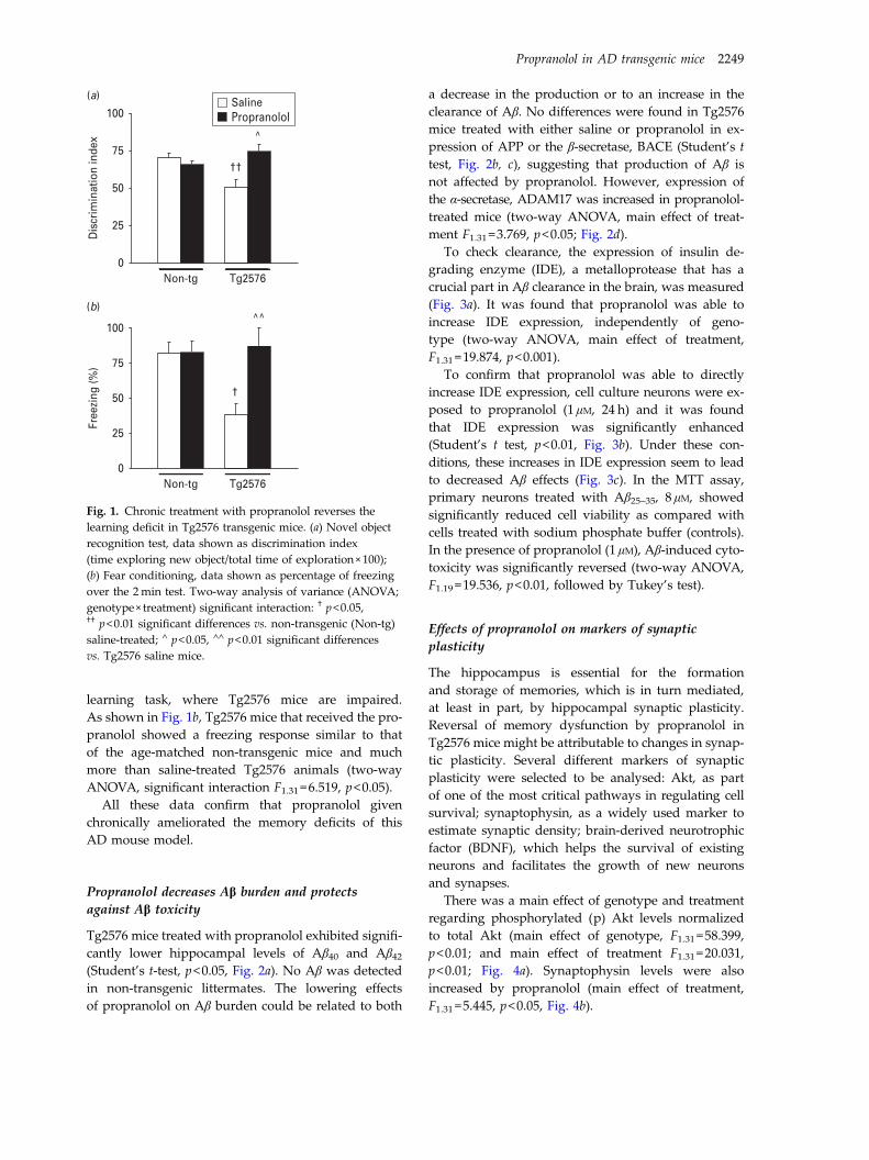
learning task, where Tg2576 mice are impaired.As shown in Fig. 1b, Tg2576 mice that received the pro-pranolol showed a freezing response similar to thatof the age-matched non-transgenic mice and muchmore than saline-treated Tg2576 animals (two-wayANOVA, significant interaction F1.31=6.519, p<0.05).
All these data confirm that propranolol givenchronically ameliorated the memory deficits of thisAD mouse model.
Propranolol decreases Aβ burden and protectsagainst Aβ toxicity
Tg2576 mice treated with propranolol exhibited signifi-cantly lower hippocampal levels of Aβ40 and Aβ42(Student’s t-test, p<0.05, Fig. 2a). No Aβ was detectedin non-transgenic littermates. The lowering effectsof propranolol on Aβ burden could be related to both
a decrease in the production or to an increase in theclearance of Aβ. No differences were found in Tg2576mice treated with either saline or propranolol in ex-pression of APP or the β-secretase, BACE (Student’s ttest, Fig. 2b, c), suggesting that production of Aβ isnot affected by propranolol. However, expression ofthe α-secretase, ADAM17 was increased in propranolol-treated mice (two-way ANOVA, main effect of treat-ment F1.31 =3.769, p<0.05; Fig. 2d).
To check clearance, the expression of insulin de-grading enzyme (IDE), a metalloprotease that has acrucial part in Aβ clearance in the brain, was measured(Fig. 3a). It was found that propranolol was able toincrease IDE expression, independently of geno-type (two-way ANOVA, main effect of treatment,F1.31 =19.874, p<0.001).
To confirm that propranolol was able to directlyincrease IDE expression, cell culture neurons were ex-posed to propranolol (1 μM, 24 h) and it was foundthat IDE expression was significantly enhanced(Student’s t test, p<0.01, Fig. 3b). Under these con-ditions, these increases in IDE expression seem to leadto decreased Aβ effects (Fig. 3c). In the MTT assay,primary neurons treated with Aβ25–35, 8 μM, showedsignificantly reduced cell viability as compared withcells treated with sodium phosphate buffer (controls).In the presence of propranolol (1 μM), Aβ-induced cyto-toxicity was significantly reversed (two-way ANOVA,F1.19 =19.536, p<0.01, followed by Tukey’s test).
Effects of propranolol on markers of synapticplasticity
The hippocampus is essential for the formationand storage of memories, which is in turn mediated,at least in part, by hippocampal synaptic plasticity.Reversal of memory dysfunction by propranolol inTg2576 mice might be attributable to changes in synap-tic plasticity. Several different markers of synapticplasticity were selected to be analysed: Akt, as partof one of the most critical pathways in regulating cellsurvival; synaptophysin, as a widely used marker toestimate synaptic density; brain-derived neurotrophicfactor (BDNF), which helps the survival of existingneurons and facilitates the growth of new neuronsand synapses.
There was a main effect of genotype and treatmentregarding phosphorylated (p) Akt levels normalizedto total Akt (main effect of genotype, F1.31 =58.399,p<0.01; and main effect of treatment F1.31=20.031,p<0.01; Fig. 4a). Synaptophysin levels were alsoincreased by propranolol (main effect of treatment,F1.31 =5.445, p<0.05, Fig. 4b).
100
(a)SalinePropranolol
75††
†
^
^^
50
25
Dis
crim
inat
ion
inde
x
0Non-tg Tg2576
100
(b)
75
50
25
Free
zing
(%)
0Non-tg Tg2576
Fig. 1. Chronic treatment with propranolol reverses thelearning deficit in Tg2576 transgenic mice. (a) Novel objectrecognition test, data shown as discrimination index(time exploring new object/total time of exploration×100);(b) Fear conditioning, data shown as percentage of freezingover the 2min test. Two-way analysis of variance (ANOVA;genotype×treatment) significant interaction: † p<0.05,†† p<0.01 significant differences vs. non-transgenic (Non-tg)saline-treated; ∧ p<0.05, ∧∧ p<0.01 significant differencesvs. Tg2576 saline mice.
Propranolol in AD transgenic mice 2249
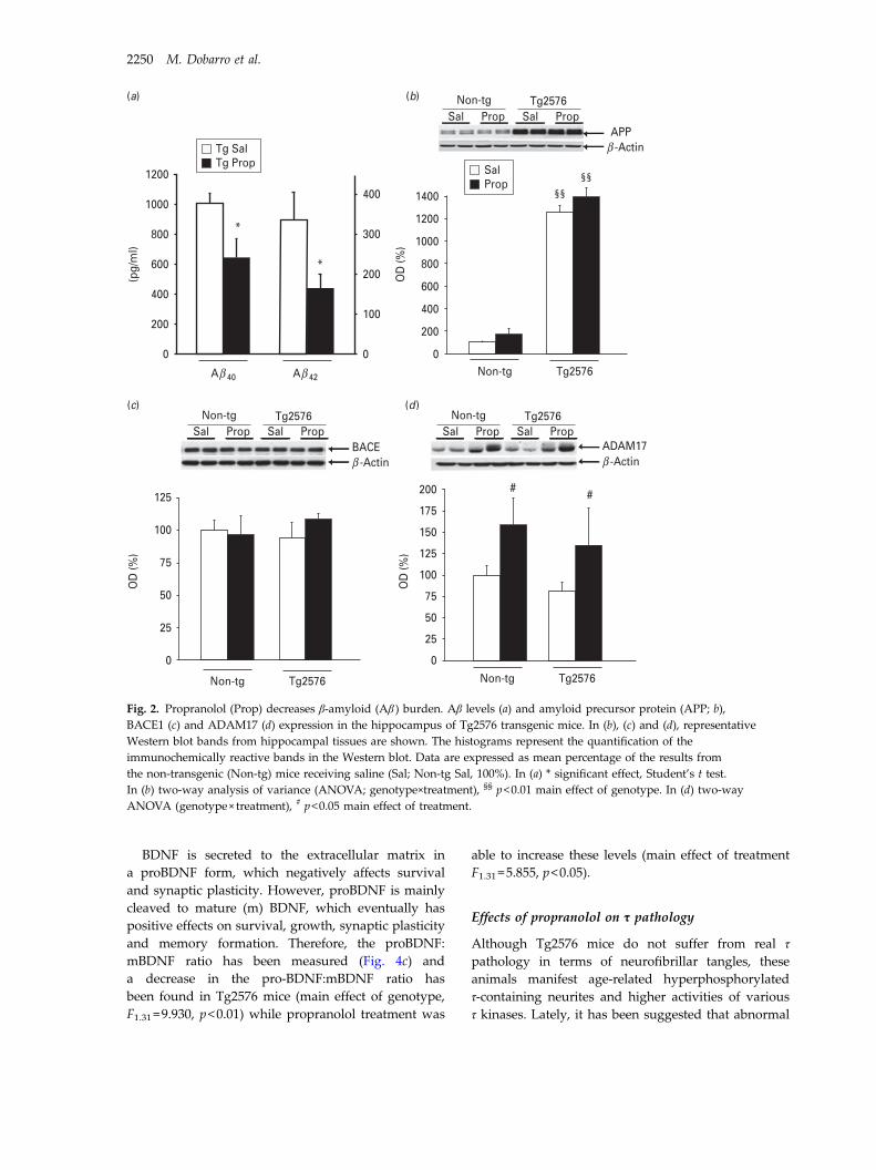
BDNF is secreted to the extracellular matrix ina proBDNF form, which negatively affects survivaland synaptic plasticity. However, proBDNF is mainlycleaved to mature (m) BDNF, which eventually haspositive effects on survival, growth, synaptic plasticityand memory formation. Therefore, the proBDNF:mBDNF ratio has been measured (Fig. 4c) anda decrease in the pro-BDNF:mBDNF ratio hasbeen found in Tg2576 mice (main effect of genotype,F1.31 =9.930, p<0.01) while propranolol treatment was
able to increase these levels (main effect of treatmentF1.31=5.855, p<0.05).
Effects of propranolol on τ pathology
Although Tg2576 mice do not suffer from real τpathology in terms of neurofibrillar tangles, theseanimals manifest age-related hyperphosphorylatedτ-containing neurites and higher activities of variousτ kinases. Lately, it has been suggested that abnormal
Tg SalTg Prop
1200
*
*
1000
800
600
400
400
300
200
100
0
200
(a) (b)
(c) (d)
(pg/
ml)
OD
(%)
0
0
25
50
75
100
125
1200
1400
1000
800
600
400
200
0
Aβ 40 Aβ 42 Non-tg Tg2576
OD
(%)
OD
(%)
175
200 # #
150
125
75
100
50
25
0
Non-tg Tg2576Non-tg Tg2576
Non-tgSal Prop
Tg2576Sal Prop
BACEβ -Actin
Non-tgSal Prop
Tg2576Sal Prop
Non-tgSal Prop
Tg2576Sal Prop
APPβ -Actin
ADAM17β -Actin
SalProp
§§§§
Fig. 2. Propranolol (Prop) decreases β-amyloid (Aβ ) burden. Aβ levels (a) and amyloid precursor protein (APP; b),BACE1 (c) and ADAM17 (d) expression in the hippocampus of Tg2576 transgenic mice. In (b), (c) and (d), representativeWestern blot bands from hippocampal tissues are shown. The histograms represent the quantification of theimmunochemically reactive bands in the Western blot. Data are expressed as mean percentage of the results fromthe non-transgenic (Non-tg) mice receiving saline (Sal; Non-tg Sal, 100%). In (a) * significant effect, Student’s t test.In (b) two-way analysis of variance (ANOVA; genotype×treatment), §§ p<0.01 main effect of genotype. In (d) two-wayANOVA (genotype× treatment), # p<0.05 main effect of treatment.
2250 M. Dobarro et al.

τ phosphorylation is a secondary event to Aβ path-ology in AD. In addition, considering the significanceof the Akt–glycogen synthase kinase (GSK)3β–τ path-way and given the results found measuring Akt, weanalysed next the levels of τ phosphorylation in themice hippocampi. Immunoblot analysis showed a sig-nificant genotype×treatment interaction regarding pτat pSer202/Thr205, (pτ normalized to total τ levels;two-way ANOVA, F1.31 =6.869, p<0.05). Further analy-sis revealed that Tg2576 animals showed significanthyperphosphorylation of pτ that was reversed by pro-pranolol treatment (Tukey’s test, p<0.05, Fig. 5a).Similarly, pτ at Ser396/404, using the PHF1 antibody(Fig. 5b), was also altered in Tg2576 mice (two-wayANOVA, significant genotype×treatment interaction,F1.31 =4.651, p<0.05) and the increases in pτ levelsshowed by Tg2576 were reverted by propranolol(Tukey’s test, p<0.05).
The expression of several kinases implicated in τpathology was checked. As shown in Fig. 5c, therewas also a significant interaction in pGSK3β expression(pGSK3β in Ser9, inactive form) normalized to totalGSK3β (two-way ANOVA, F1.31=4.349, p<0.05).Further analysis revealed that Tg2576 animals showeddecreased levels of pGSK3β (Tukey’s test, p<0.05) thatwas reversed by propranolol treatment (Tukey’s test,p<0.05). Moreover, there was a genotype×treatmentinteraction (two-way ANOVA, F1.31=6.828, p<0.05)with regard to p- c-Jun N-terminal kinase (JNK)1 levelsnormalized to total JNK1. Western blot analysisrevealed that pJNK1 levels were significantly increasedin saline Tg2576 mice compared with non-transgenicmice (Tukey’s test, p<0.05) and propranolol treatmentwas able to reverse these effects on pJNK1 expression(Tukey’s test, p<0.05; Fig. 5d).
Consistent with a post-transcriptional regulationof these enzymes, total τ, JNK1 and GSK3β levels,normalized using actin, remained unaltered.
Discussion
The main finding in this study is the reversal ofmemory deficits in the Tg2576 mouse model of ADusing propranolol, a brain penetrating non selective
(a)
(b)
(c)
SalNon-tg
200 ## ##
**
††
^^
175
150
125
100
75
50
25
0
0
25
50
75
100
125
150
PropTg2576
Non-tg Tg2576
Sal Prop
Sal Prop
IDEβ -Actin
IDEβ -Actin
SalProp
SalProp
OD
(%)
OD
(%)
0
25
50
75
100
125 ControlPropAβAβ+prop
Sur
viva
l (%
)
Fig. 3. Propranolol (Prop) increases β-amyloid (Aβ )clearance and toxicity. In (a) increased levels of insulindegrading enzyme (IDE) expression in the hippocampusof propranolol treated mice might reflect an increasedclearance of Aβ in the hippocampus of Tg2576 transgenicmice. In neuronal primary cultures IDE expression wasalso increased after propranolol treatment (b) and thistreatment protected against Aβ25–35 toxicity (c). In all casesrepresentative Western blot bands from hippocampal tissuesare shown. The histograms represent the quantification ofthe immunochemically reactive bands in the Western blot.Data are expressed as mean percentage of the results fromthe non-transgenic (Non-tg) mice receiving saline
(Sal; Non-tg Sal, 100%). In (a), two-way analysis of variance(ANOVA; genotype×treatment), ## p<0.01 main effect oftreatment. In cell culture experiments, (b) Student’s t test,** p<0.01, significant differences vs. control. In (c) two-wayANOVA (Aβ×Prop), significant interaction, †† p<0.01,significant differences vs. control; ∧∧ p<0.01, significantdifferences vs. Aβ25–35 toxicity.
Propranolol in AD transgenic mice 2251

β-adrenergic antagonist widely used as an antihyper-tensive drug. Previous work has reported the abilityof propranolol to reverse cognitive deficits associatedwith deleterious insults such as chronic stress (Aisaet al., 2007). However, to our knowledge, this is oneof the first reports describing pro-cognitive actions ofpropranolol in an experimental model of AD.
Because the overall goal of our study was to test thehypothesis that propranolol may influence AD-type Aβpathogenesis independent of blood pressure-loweringactivity, we treated Tg2576 mice with 5mg/kg pro-pranolol. Based on the fact that the recommendeddose of propranolol for the treatment of hypertensionin humans is 160–320mg/d and using US Food andDrug Administration criteria [human equivalentdose in mg/kg=animal dose in mg/kg×(animal weightin kg/human weight in kg)0.33; http://www.fda.gov/cber/gdlns/dose.htm] and the formula described byReagan-Shaw et al. (2007) for converting drug equi-valent dosages across species area [human equivalentdose in mg/kg animal dose in mg/kg×Km mice(=3)/Km human (=37)], it can be calculated thatthe dose used in the present study is below therecommended human equivalent dosage range.Therefore, propranolol was used in the present studyat a lower dose than that commonly prescribed for thetreatment of hypertension in humans (Reagan-Shawet al., 2007) and the results presented here seem to beindependent from the antihypertensive effect of propra-nolol. In an attempt to elucidate the mechanisms under-lying the beneficial effects of propranolol and basedon previously published work (Wang et al., 2007), wefocused our study on Aβ and τ pathology, the twomain pathological hallmarks in AD and in markers ofsynaptic plasticity.
A key issue was to determine the optimal age ofTg2576 mice for this study. Tg2576 mice show rapidincreases in Aβ levels starting at age 6 months andAβ peptide content in the brain accumulates expo-nentially between ages 7 and 12 months. Followingour hypothesis that propranolol was able to alter Aβburden, Tg2576 mice aged 8 months (at the beginningof treatment) were chosen in order to be able to detectsignificant increases in Aβ content in saline treatedmice. Tg2576 mice aged 9 months (at the end of treat-ment) shown impairments in recognition memory(Mouri et al., 2007; Taglialatela et al., 2009) but spatialworking memory is not altered until age 12–15 months(Hsiao et al., 1996; Reed et al., 2010). Therefore, thetests chosen to study the effects of propranolol oncognition have been the NORT, to assess recognitionmemory, and fear conditioning, to check contextualhippocampal dependent memory. Interestingly, in
Non-tgSal Prop
Tg2576Sal Prop
pAKTTotal Aktβ -Actin
mBDNFproBDNFβ -Actin
Synaptophysinβ -Actin
SalProp
200175150125
##
##
§§
§§/##
§§/##
§§
1007550250
0
20
50
75
100
125
150
OD
(%)
OD
(%)
Non-tg
Non-tg
Tg2576
Tg2576Sal Prop Sal Prop
Non-tg
Non-tg
Tg2576
Tg2576Sal Prop Sal Prop
#
0
25
50
75
100
125
Rat
io m
BD
NF:
proB
DN
F
Non-tg Tg2576
(a)
(b)
(c)
Fig. 4. Effects of propranolol (Prop) on markers ofsynaptic plasticity in the hippocampus. (a) Phosphorylated(p)Akt levels, (b) synaptophysin levels, (c) proBDNFmature (m)BDNF:proBDNF ratio. Representative Westernblot bands from hippocampal tissues are shown.The histograms represent the quantification of theimmunochemically reactive bands in the Western blot.Data are expressed as mean percentage of the resultsfrom the non-transgenic (Non-tg) mice receiving saline(Sal; Non-tg Sal, 100%). Two-way analysis of variance(ANOVA; genotype× treatment): §§ p<0.01, main effect ofgenotype; # p<0.05, ## p<0.01, main effect of treatment.
2252 M. Dobarro et al.
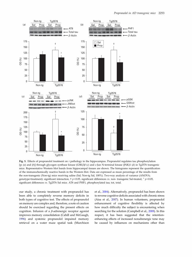
our study, a chronic treatment with propranolol hasbeen able to completely reverse memory deficits inboth types of cognitive test. The effects of propranololonmemory are complex and, therefore, a note of cautionshould be exercised regarding the present effects oncognition. Infusion of a β-adrenergic receptor agonistimproves memory consolidation (Cahill and McGaugh,1996) and systemic propranolol impaired memoryretrieval on a water maze spatial task (Murchison
et al., 2004). Alternatively, propranolol has been shownto reverse cognitive deficits associatedwith chronic stress(Aisa et al., 2007). In human volunteers, propranololenhancement of cognitive flexibility is affected byhow much difficulty the subject is encountering whensearching for the solution (Campbell et al., 2008). In thisrespect, it has been suggested that the retention-enhancing effects of increased noradrenergic tone maybe caused by influences on mechanisms other than
(a)Non-tg Tg2576
Sal PropAT8
Sal Prop
Total tauβ -Actin
(b)Non-tg
Sal PropTg2576
Sal PropPHF1Total tauβ -Actin
(c)Non-tg
Sal PropTg2576
Sal ProppJNKJNKtotβ -Actin
(d)Non-tg
Sal PropTg2576
Sal ProppGSKGSKtotβ -Actin
SalProp
175
150
125
100
75
50
25
0Non-tg Tg2576
OD
(%)
†
^
200 125
100
75
50
25
0
175
150
125
100
75
50
25
0Non-tg Tg2576
OD
(%)
OD
(%)
†
^
Non-tg Tg2576
†
^
175
150
125
100
75
50
25
0Non-tg Tg2576
OD
(%)
†
^
Fig. 5. Effects of propranolol treatment on τ pathology in the hippocampus. Propranolol regulates tau phosphorylation[p; (a) and (b)] through glycogen synthase kinase (GSK)3β (c) and c-Jun N-terminal kinase (JNK)1 (d) in Tg2576 transgenicmice. Representative Western blot bands from hippocampal tissues are shown. The histograms represent the quantificationof the immunochemically reactive bands in the Western blot. Data are expressed as mean percentage of the results fromthe non-transgenic (Non-tg) mice receiving saline (Sal; Non-tg Sal, 100%). Two-way analysis of variance (ANOVA;genotype×treatment): significant interaction, † p<0.05, significant differences vs. non- transgenic Sal-treated, ∧ p<0.05,significant differences vs. Tg2576 Sal mice. AT8 and PHF1, phosphorylated tau; tot, total.
Propranolol in AD transgenic mice 2253

memory retrieval e.g. nonspecific effects on arousal andattention (Aston-Jones et al., 2000).
Given the genotypic nature of Tg2576 mouse, Aβshould be a major player in the memory dysfunctiondeveloped by this transgenic mouse. According tothe amyloid cascade hypothesis (Hardy and Selkoe,2002), Aβ is the starting point for a sequence of patho-genic events, such as τ hyperphosphorylation andneuroinflammation, that contribute to synaptic dys-function and ultimately cause dementia. Therefore,although lately controversial in literature reports, itcould be suggested that the reduction of Aβ burdenby propranolol would consequently contribute to ame-liorate AD symptoms. Accumulation of Aβ is proposedto result from an imbalance between Aβ productionand Aβ clearance. Although changes in the amyloido-genic processing of APP cannot be completelyexcluded from the present work, the lack of effect ofpropranolol treatment on BACE1 expression seems toindicate that Aβ production is not altered by proprano-lol. However, the effects of propranolol on ADAM17suggest that the non-amyloidogenic processing ofAPP could be facilitated in propranolol-treated mice.Alternatively, the brain possesses robust intrinsic Aβclearance mechanisms (Tanzi et al., 2004). Aβ peptidesare proteolytically degraded within the brain mainlyby neprilysin (Iwata et al., 2000) and IDE (Kurochkinand Goto, 1994). It is unclear whether increased nepri-lysin levels may be beneficial to Aβ pathology, as thisenzyme neither degrades Aβ oligomers nor restoresmemory function in an AD mouse model (Meilandtet al., 2009). Therefore, we have focused on IDEexpression. The results found both in the Tg2576mice and in cell culture experiments suggest thatpropranolol, by increasing IDE expression, is able toprevent the cellular damage induced by Aβ.
Although the Tg2576 mouse is a model of ADamyloidosis and Tg2576 mice do not suffer from realτ pathology in terms of neurofibrillar tangles, thepresence of hyperphosphorylated τ epitopes (Ser199,Thr231/Ser235, Ser396 and Ser413) and higher activitiesof various τ kinases has been consistently reported(Tomidokoro et al., 2001; Sasaki et al., 2002; Ferreret al., 2005; Cuadrado-Tejedor et al., 2011 and shownin Fig. 4). In fact, Aβ amyloidosis can activate the τkinase pathway involving GSK3β (via its phosphoryl-ation at Tyr216) and, subsequently, τ is phosphorylatedat sites that promote its accumulation and deposition(Tomidokoro et al., 2001; Otth et al., 2002). Amongkinases implicated in τ pathology, data on activationof JNK1 seems to be of particular interest. JNK1 isa member of the mitogen-activated protein kinasefamily that plays a key role in neuronal plasticity,
regeneration and cell death (Kögel et al., 2005,Maruyama et al., 2009). Activation of JNK1 phosphory-lates τ at sites recognized by the presently used PHF1antibody (Bellucci et al., 2007). It is of note that JNK1phosphorylates the insulin receptor substrate-1 at aninhibitory site that can block signal transduction bythe insulin receptor (Sabio et al., 2008), therefore pur-portedly altering insulin-related intracellular path-ways, namely the Akt–GSK3β–τ. This Akt–GSK3βroute is required for the induction of long-term poten-tiation and depression, basic processes underlyinglearning and memory (van der Heide et al., 2006).Accordingly, it has been found, compared to controls,that treatment with propranolol in Tg2576 mice leadsto decreased JNK1 activity accompanied by increasedpAkt, increased pGSK-3β (inactive form) and de-creased pτ expression. Together these data suggestthat the decrease in the kinase activity of GSK3β andJNK1 due to propranolol was able to prevent thesigns of tauopathy observed in the hippocampus ofTg2576 mice, possibly contributing to the reinstate-ment of cognitive function as data, based on severallines of evidence from transgenic mice and humans,indicating a strong correlation between the extent ofτ pathology and cognitive dysfunction (Guillozetet al., 2003; Hanger et al., 2009; Ashe and Zahs,2010). Interestingly, propranolol seems to restore theGSK3β or JNK1 pathways when they are hyperacti-vated but not under normal conditions. In fact, thesepathways were not affected by propranolol whennon-transgenic mice were analysed.
The reversion by propranolol of changes in markersof synaptic pathology in Tg2576 mice should be alsoconsidered. BDNF has been described to be highlyprotective for different neuronal phenotypes and canprevent neuronal cell death in different experimentalparadigms in vivo as well as in vitro (Barde, 1994;Siegel and Chauhan, 2000). BDNF levels decline withage and are lower in AD brains that in those withoutdementia. BDNF delivery ameliorated age-related cog-nitive impairment in aged primates and mice evenwhen the treatment was initiated after disease onset(Nagahara et al., 2009). Not only that, but synaptophy-sin seem to be a pre-synaptic vesicle protein partiallyregulated by BDNF (Tartaglia et al., 2001; Valtortaet al., 2004) and therefore reduced synaptophysinexpression may result from a reduced BDNF function.However, hippocampal BDNF is secreted to the extra-cellular matrix in a proBDNF form. ProBDNF is activevia p75 neurotrophin receptors and negatively affectssurvival and synaptic plasticity. ProBDNF is mainlycleaved to mature BDNF to activate TrkB receptors,which eventually has positive effects on survival,
2254 M. Dobarro et al.

growth, synaptic plasticity and memory formation.Therefore, the proBDNF:mBDNF ratio could be amore accurate form to anticipate BDNF outcomes.It is of note that it could have been expected thatβ-receptor blockade would have led to decreasedBDNF levels (Flores et al., 2010). Therefore, the presentincrease in the ratio suggests that propranolol couldmodulate the cleavage of proBDNF to give rise to themature form. An alternative is to consider the effectsof propranolol on ADAM17 expression. Besides APP,many other membrane-bound pro-ligands with trophicor survival properties, such as epidermal growthfactor, neuregulin 1β or transforming growth factor-α,could be the substrate of ADAM17 (Dang et al.,2011). Thus, the possibility is that increases ofADAM17 might be increasing levels of these neuro-trophic factors. In this respect, it has been describedthat cultured neurons from Tg2576 mice showed areduced viability after treatment with an inhibitorof ADAM17 (Gil-Bea et al., 2012). In addition, it isnoted that propranolol was able to increase pAkt,synaptophysin and BDNF also in non-transgenicmice. Even though the mechanism responsible forincreases by propranolol in these synaptic markerscannot be unravelled from the present results, it ispossible to speculate about the usefulness of this com-pound for the treatment of any pathology associatedwith a decreased synaptic function.
Concluding remarks
Collectively, our evidence suggest that some of thedrugs, namely propranolol, currently prescribed forthe treatment of high blood pressure might also benefi-cially modulate AD by mechanisms independentof changes in blood pressure (Zhao et al., 2009).Propranolol may have AD-modifying activity andmay protect against progressive Aβ/τ-related memorydeficits. Although further studies would help to fullyunderstand the mechanisms by which propranololexert its effects on AD-related pathology, the presentpreclinical work should foster future clinical studieson the use of propranolol for the treatment of AD.
Acknowledgements
This work has been supported by a grant from FIS(10/01748) and ‘Tu eliges, Tu decides’ projects of CAN.
Conflicts of Interest
None.
Supplementary material
For supplementary material accompanying this paper,visit http://dx.doi.org/10.1017/S1461145713000631.
References
Aisa B, Tordera R, Lasheras B, Del Río J, Ramírez MJ (2007)Cognitive impairment associated to HPA axis hyperactivityafter maternal separation in rats.Psychoneuroendocrinology 32:256–266.
Ashe KH, Zahs KR (2010) Probing the biology of Alzheimer’sdisease in mice. Neuron 66:631–645.
Aston-Jones G, Rajkowski J, Cohen J (2000) Locus coeruleusand regulation of behavioral flexibility and attention.Prog Brain Res 126:165–182.
Barde YA (1994) Neurotrophins: a family of proteinssupporting the survival of neurons. Prog Clin Biol Res390:45–56.
Bellew KM, Pigeon JG, Stang PE, Fleischman W, Gardner RM,Baker WW (2004) Hypertension and the rate of cognitivedecline in patients with dementia of the Alzheimer type.Alzheimer Dis Assoc Disord 18:208–213.
Bellucci A, Rosi MC, Grossi C, Fiorentini A, Luccarini I,Casamenti F (2007) Abnormal processing of tau in the brainof aged TgCRND8 mice. Neurobiol Dis 27:328–338.
Cahill L, McGaugh JL (1996) Modulation of memory storage.Curr Opin Neurobiol 6:237–242.
Campbell HL, Tivarus ME, Hillier A, Beversdorf DQ(2008) Increased task difficulty results in greater impactof noradrenergic modulation of cognitive flexibility.Pharmacol Biochem Behav 88:222–229.
Cuadrado-Tejedor M, Hervias I, Ricobaraza A, Puerta E,Pérez-Roldán JM, García-Barroso C, Franco R, Aguirre N,García-Osta A (2011) Sildenafil restores cognitive functionwithout affecting Aβ burden in an Alzheimer’s diseasemouse model. Neurobiol Aging 164:2029–2041.
Cummings JL, Cole G (2002) Alzheimer disease. J AmMed Assoc 287:2335–2338.
Dang M, Dubbin K, D’Aiello A, Hartmann M, Lodish H,Herrlich A (2011) Epidermal growth factor (EGF) ligandrelease by substrate-specific a disintegrin and metallo-proteases (ADAMs) involves different protein kinase C(PKC) isoenzymes depending on the stimulus. J BiolChem 286:17704–17713.
Ennaceur A, Delacour J (1988) A new one trial-test forneurobiological studies of memory in rats. 1: behavioraldata. Behav Brain Res 31:47–59.
Ferrer I, Gomez-Isla T, Puig B, Freixes M, Ribé E, Dalfó E,Avila J (2005) Current advances on different kinasesinvolved in tau phosphorylation, and implications inAlzheimer’s disease and tauopathies. Curr AlzheimerRes 2:3–18.
Flores O, Núñez H, Pérez H, Morgan C, Soto-Moyano R,Valladares L, Burgos H, Olivares R, Hernández A (2010)beta-Adrenoceptor blockade depresses molecular and
Propranolol in AD transgenic mice 2255

functional plasticities in the rat neocortex. Brain Res Bull82:284–288.
Gil-Bea FJ, Gerenu G, Aisa B, Kirazov LP, Schliebs R,Ramírez MJ (2012) Cholinergic denervation exacerbatesamyloid pathology and induces hippocampal atrophy inTg2576 mice. Neurobiol Dis 48:439–346.
Guillozet AL, Weintraub S, Mash DC, Mesulam MM (2003)Neurofibrillary tangles, amyloid, and memory in aging andmild cognitive impairment. Arch Neurol 60:729–736.
Guo Z, Viitanen M, Winblad B, Fratiglioni L (1999) Lowblood pressure and incidence of dementia in a very oldsample: dependent on initial cognition. J Am Geriatr Soc47:723–726.
Hajjar I, Keown M, Frost B (2005) Antihypertensive agents foraging patients who are at risk for cognitive dysfunction.Curr Hypertens Rep 7:466–473.
Hanger DP, Seereeram A, Noble W (2009) Mediators oftau phosphorylation in the pathogenesis of Alzheimer’sdisease. Expert Rev Neurother 9:1647–1666.
Hardy J (2006) Has the amyloid cascade hypothesis forAlzheimer’s disease been proved? Curr Alzheimer Res3:71–73.
Hardy J, Selkoe DJ (2002) The amyloid hypothesis ofAlzheimer’s disease: progress and problems on the roadto therapeutics. Science 297:353–356.
Hoffman LB, Schmeidler J, Lesser GT, Beeri MS, Purohit DP,Grossman HT, Haroutunian V (2009) Less Alzheimerdisease neuropathology in medicated hypertensive thannon hypertensive persons. Neurology 72:1720–1726.
Hsiao K, Chapman P, Nilsen S, Eckman C, Harigaya Y,Younkin S, Yang F, Cole G (1996) Correlative memorydeficits, Abeta elevation, and amyloid plaques in transgenicmice. Science 274:99–102.
in’t Veld BA, Ruitenberg A, Hofman A, Stricker BH,Breteler MM (2001) Antihypertensive drugs andincidence of dementia: the Rotterdam study. NeurobiolAging 22:407–412.
Iwata N, Tsubuki S, Takaki Y, Watanabe K, Sekiguchi M,Hosoki E, Kawashima-Morishima M, Lee HJ, Hama E,Sekine-Aizawa Y, Saido TC (2000) Identification of themajor Abeta1-42-degrading catabolic pathway in brainparenchyma: suppression leads to biochemical andpathological deposition. Nat Med 6:143–150.
Khachaturian AS, Zandi PP, Lyketsos CG, Hayden KM,Skoog I, Norton MC, Tschanz JT, Mayer LS, Welsh-BohmerKA, Breitner JC (2006) Antihypertensive medication useand incident Alzheimer disease: the Cache County Study.Arch Neurol 63:686–692.
Kögel D, Schomburg R, Copanaki E, Prehn JH (2005)Regulation of gene expression by the amyloid precursorprotein: inhibition of the JNK/cJun pathway. Cell DeathDiffer 12:1–9.
Kurochkin IV, Goto S (1994) Alzheimer’s beta-amyloidpeptide specifically interacts with and is degraded byinsulin degrading enzyme. FEBS Lett 345:33–37.
Leach PT, Poplawski SG, Kenney JW, Hoffman B,Liebermann DA, Abel T, Gould TJ (2012)
Gadd45b knockout mice exhibit selective deficits inhippocampus-dependent long-term memory. Learn Mem19:319–324.
Lithell H, Hansson L, Skoog I, Elmfeldt D, Hofman A,Olofsson B, Trenkwalder P, Zanchetti A, SCOPE StudyGroup (2004) The study on COgnition and prognosis inthe elderly (SCOPE); outcomes in patients not receivingadd-on therapy after randomization. J Hypertens22:1605–1612.
Luchsinger JA, Reitz C, Honig LS, Tang MX, Shea S,Mayeux R (2005) Aggregation of vascular risk factorsand risk of incident Alzheimer disease. Neurology65:545–551.
Maruyama J, Naguro I, Takeda K, Ichijo H (2009) Stress-activated MAP kinase cascades in cellular senescence.Curr Med Chem 16:1229–1235.
Meilandt WJ, Cisse M, Ho K, Wu T, Esposito LA, Scearce-Levie K, Cheng IH, Yu GQ, Mucke L (2009) Neprilysinoverexpression inhibits plaque formation but fails to reducepathogenic Aβ oligomers and associated cognitive deficitsin human amyloid precursor protein transgenic mice.J Neurosci 29:1977–1986.
Mouri A, Noda Y, Hara H, Mizoguchi H, Tabira T,Nabeshima T (2007) Oral vaccination with a viral vectorcontaining Abeta cDNA attenuates age-related Abetaaccumulation and memory deficits without causinginflammation in a mouse Alzheimer model. FASEBJ 163:2135–2148.
Murchison CF, Zhang XY, Zhang WP, Ouyang M, Lee A,Thomas SA (2004) A distinct role for norepinephrine inmemory retrieval. Cell 117:131–143.
Nagahara AH, Merrill DA, Coppola G, Tsukada S,Schroeder BE, Shaked GM, Wang L, Blesch A, Kim A,Conner JM, Rockenstein E, Chao MV, Koo EH,Geschwind D, Masliah E, Chiba AA, Tuszynski MH(2009) Neuroprotective effects of brain-derived neuro-trophic factor in rodent and primate models of Alzheimer’sdisease. Nat Med 15:331–337.
Otth C, Concha II, Arendt T, Stieler J, Schliebs R,González-Billault C, Maccioni RB (2002) AbetaPP inducescdk5-dependent tau hyperphosphorylation in transgenicmice Tg2576. J Alzheimers Dis 4:417–430.
Peskind ER, Tsuang DW, Bonner LT, Pascualy M, Riekse RG,Snowden MB, Thomas R, Raskind MA (2005)Propranolol for disruptive behaviors in nursing homeresidents with probable or possible Alzheimer disease:a placebo-controlled study. Alzheimer Dis Assoc Disord19:23–28.
Petrovitch H, White LR, Izmirilian G, Ross GW, Havlik RJ,Markesbery W (2000) Midlife blood pressure and neuriticplaques, neurofibrillary tangles, and brain weight at death:the HAAS. Honolulu-Asia aging study. Neurobiol Aging21:57–62.
Prince M, Rabe-Hesketh S, Brennan P (1998) Do antiarthriticdrugs decrease the risk for cognitive decline? An analysisbased on data from the MRC treatment trial of hypertensionin older adults. Neurology 50:374–379.
2256 M. Dobarro et al.

Reagan-Shaw S, Nihal M, Ahmad N (2007) Dose translationfrom animal to human studies revisited. FASEB J22:659–661.
Reed MN, Liu P, Kotilinek LA, Ashe KH (2010) Effect sizeof reference memory deficits in the Morris water mazein Tg2576 mice. Behav Brain Res 212:115–120.
Ricobaraza A, Cuadrado-Tejedor M, Marco S, Pérez-Otaño I,García-Osta A (2012) Phenylbutyrate rescues dendriticspine loss associated with memory deficits in a mousemodel of Alzheimer disease. Hippocampus 22:1040–1050.
Roozendaal B, Hahn EL, Nathan SV, de Quervain DJ,McGaugh JL (2004) Glucocorticoid effects on memoryretrieval require concurrent noradrenergic activity in thehippocampus and basolateral amygdala. J Neurosci24:8161–8169.
Sabio G, Das M, Mora A, Zhang Z, Jun JY, Ko HJ, Barrett T,Kim JK, Davis RJ (2008) A stress signaling pathway inadipose tissue regulates hepatic insulin resistance. Science322:1539–1543.
Sasaki A, Shoji M, Harigaya Y, Kawarabayashi T, Ikeda M,Naito M, Matsubara E, Abe K, Nakazato Y (2002) Amyloidcored plaques in Tg2576 transgenic mice are characterizedby giant plaques, slightly activated microglia, and the lackof paired helical filament-typed, dystrophic neurites.Virchows Arch 441:358–367.
Selkoe DJ (2002) Alzheimer’s disease is a synaptic failure.Science 298:789–791.
Shankle WR, Nielson KA, Cotman CW (1995) Low-dosepropranolol reduces aggression and agitationresembling that associated with orbitofrontal dysfunctionin elderly demented patients. Alzheimer Dis Assoc Disord9:233–237.
Siegel GJ, Chauhan NB (2000) Neurotrophic factors inAlzheimer’s and Parkinson’s disease brain. Brain ResRev 33:199–227.
Skoog I, Gustafson D (2006) Update on hypertensionand Alzheimer’s disease. Neurol Res 28:605–611.
Sparks DL, Scheff SW, Liu H, Landers T, Danner F,Coyne CN, Hunsaker JC III (1996) Increased density ofsenile plaques (SP), but not neurofibrillary tangles (NFT),in non-demented individuals with the apolipoproteinE4 allele: comparison to confirmed Alzheimer’s diseasepatients. J Neurol Sci 138:97–104.
Taglialatela G, Hogan D, Zhang WR, Dineley KT (2009)Intermediate- and long-term recognition memory deficits inTg2576 mice are reversed with acute calcineurin inhibition.Behav Brain Res 200:95–99.
Tanzi RE, Moir RD, Wagner SL (2004) Clearance ofAlzheimer’s Abeta peptide: the many roads to perdition.Neuron 43:605–608.
Tartaglia N, Du J, Tyler WJ, Neale E, Pozzo-Miller L, Lu B(2001) Protein synthesis-dependent and -independentregulation of hippocampal synapses by brain-derivedneurotrophic factor. J Biol Chem 276:37585–37593.
Tomidokoro Y, Ishiguro K, Harigaya Y, Matsubara E,Ikeda M, Park JM, Yasutake K, Kawarabayashi T,Okamoto K, Shoji M (2001) Abeta amyloidosis inducesthe initial stage of tau accumulation in APP(Sw) mice.Neurosci Lett 299:169–172.
Valtorta F, Pennuto M, Bonanomi D, Benfenati F (2004)Synaptophysin: leading actor or walk-on role in synapticvesicle exocytosis? Bioessays 26:445–453.
van der Heide LP, Ramakers GM, Smidt MP (2006) Insulinsignaling in the central nervous system: learning to survive.Prog Neurobiol 79:205–221.
Wang J, Ho L, Chen L, Zhao Z, Zhao W, Qian X, Humala N,Seror I, Bartholomew S, Rosendorff C, Pasinetti GM (2007)Valsartan lowers brain beta-amyloid protein levels andimproves spatial learning in a mouse model of Alzheimerdisease. J Clin Invest 117:3393–3402.
Zhao W, Wang J, Ho L, Ono K, Teplow DB, Pasinetti GM(2009) Identification of antihypertensive drugs whichinhibit amyloid-beta protein oligomerization. J AlzheimersDis 16:49–57.
Propranolol in AD transgenic mice 2257


CCaappííttuulloo VV


Chapter 5
Propranolol reduces cognitive deficits, amyloid β levels, Tau
phosphorylation and insulin resistance in response to chronic
corticosterone administration

Propranolol in a model of sporadic EA
151
Resumen
La exposición crónica a glucocorticoides puede llevar no solo a una situación de
resistencia a la insulina o a déficits cognitivos, si no que además se considera un factor de
riesgo para patologías como la Enfermedad de Alzheimer (EA). El propranolol es un
antagonista β-adrenérgico comúnmente utilizado en el tratamiento de la hipertensión y de
la ansiedad aguda. Se han evaluado en este trabajo los efectos de la administración del β-
bloqueante propranolol (5 mg/kg) en un modelo de administración crónica de
corticosterona (100 µg/ml, 4 semanas) en agua de bebida. La administración de
corticosterona produce alteraciones cognitivas en el test de reconocimiento del nuevo
objeto que fueron revertidas por el propranolol. El aumento de los niveles de Aβ en el
hipocampo de los ratones tratados con corticosterona fue contrarrestado por el
tratamiento con propranolol, posiblemente mediante un aumento de la expresión de IDE.
El tratamiento crónico con corticosterona induce respuestas características de resistencia a
la insulina, como aumentos periféricos de los niveles de insulina, descensos de la activación
de los receptores de insulina (pIR) y disminución de las vías intracelulares asociadas (pAkt).
Estos efectos parecen estar relacionados con descensos de la expresión de c-Jun N quinasa
terminal (JNK1). El propranolol fue capaz de contrarrestar todos estos efectos inducidos
por la corticosterona. Una de las principales quinasas responsables de la fosforilación de
Tau, la glucógeno sintasa 3β (GSK3β), que es inactivada mediante la fosforilación de pAkt,
se encontró que estaba disminuída tras el tratamiento con corticosterona y aumentada
tras el tratamiento con propranolol. Además, se encontraron aumentos en la expresión de
Tau fosforilada. En general, estos resultados refuerzan la idea de que el propranolol podría
considerarse un potencial agente terapéutico para las patologías asociadas a la interacción
glucocorticoides/insulino-resistencia y el desarrollo de procesos celulares relevantes para
la EA.
Propranolol in a model of sporadic EA
152
Abstract
Chronic exposure to glucocorticoids might result not only in insulin resistance or
cognitive deficits, but it is also considered as a risk factor for pathologies such as
Alzheimer’s disease. Propranolol is a β-adrenergic antagonist commonly used in the
treatment of hypertension or acute anxiety. It has been tested here the effects of the β-
blocker propranolol (5 mg/kg) in a model of chronic corticosterone administration (100
µg/ml, 4 weeks) in drinking water. Corticosterone administration led to cognitive
impairment in the novel object recognition test that was reversed by propanolol. Increased
levels of Aβ in the hippocampus of corticosterone treated mice were counteracted by
propranolol treatment, purportedly through an increased IDE expression. Chronic
corticosterone treatment induced responses characteristic of insulin resistance, as
increased peripheral insulin levels, decreased activation of the insulin receptor (pIR) and
decreased associated intracellular pathways (pAkt). These effects might be related to a
decreased c-Jun N terminal kinase 1 (JNK1) expression. Again, propranolol was able to
counteract all corticosterone-induced effects. One of the main kinases involved in Tau
phosphorylation, glycogen synthase kinase 3β (GSK3β), which is inactivated by
phosphorylation by pAkt, was found to be decreased after corticosterone and increased
after propranolol treatment. Concomitant changes in pTau expression were found. Overall,
these data further strengthen the potential of propranolol as a therapeutic agent for
pathlogies associated to the interaction glucocorticoids-insulin resistance and the
development of relevant cellular processes for Alzheimer´s disease.
Keywords:
Aβ, Tau, JNK1, IDE, BACE1, Akt, GSK-3β, hippocampus
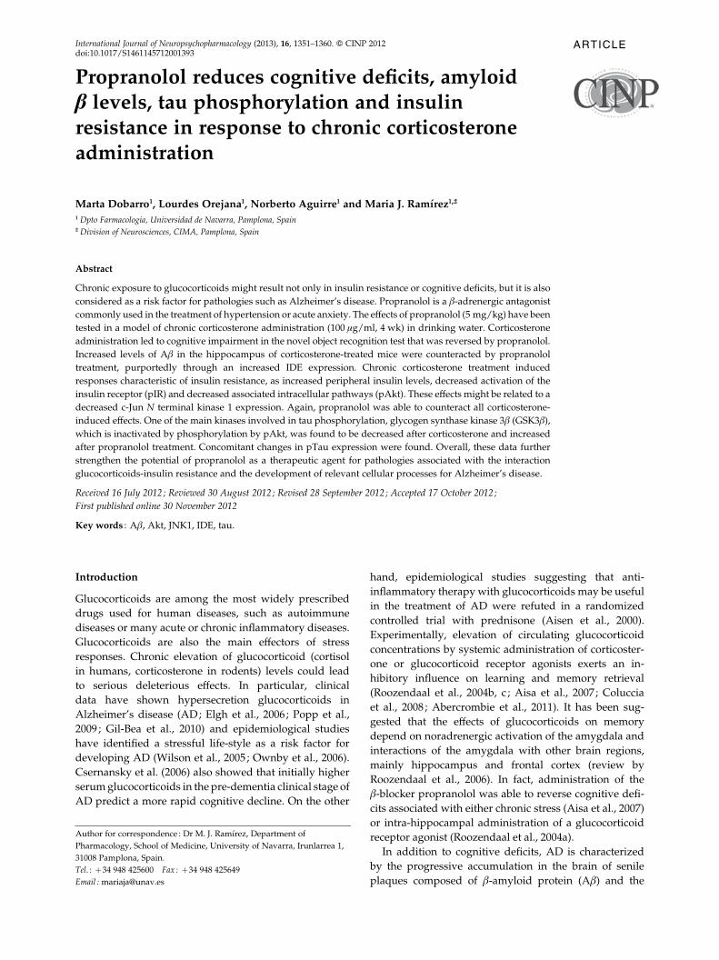
Propranolol reduces cognitive deficits, amyloidb levels, tau phosphorylation and insulinresistance in response to chronic corticosteroneadministration
Marta Dobarro1, Lourdes Orejana1, Norberto Aguirre1 and Maria J. Ramırez1,2
1 Dpto Farmacologia, Universidad de Navarra, Pamplona, Spain2 Division of Neurosciences, CIMA, Pamplona, Spain
Abstract
Chronic exposure to glucocorticoids might result not only in insulin resistance or cognitive deficits, but it is also
considered as a risk factor for pathologies such as Alzheimer’s disease. Propranolol is a b-adrenergic antagonist
commonly used in the treatment of hypertension or acute anxiety. The effects of propranolol (5 mg/kg) have been
tested in a model of chronic corticosterone administration (100 mg/ml, 4 wk) in drinking water. Corticosterone
administration led to cognitive impairment in the novel object recognition test that was reversed by propranolol.
Increased levels of Ab in the hippocampus of corticosterone-treated mice were counteracted by propranolol
treatment, purportedly through an increased IDE expression. Chronic corticosterone treatment induced
responses characteristic of insulin resistance, as increased peripheral insulin levels, decreased activation of the
insulin receptor (pIR) and decreased associated intracellular pathways (pAkt). These effects might be related to a
decreased c-Jun N terminal kinase 1 expression. Again, propranolol was able to counteract all corticosterone-
induced effects. One of the main kinases involved in tau phosphorylation, glycogen synthase kinase 3b (GSK3b),
which is inactivated by phosphorylation by pAkt, was found to be decreased after corticosterone and increased
after propranolol treatment. Concomitant changes in pTau expression were found. Overall, these data further
strengthen the potential of propranolol as a therapeutic agent for pathologies associated with the interaction
glucocorticoids-insulin resistance and the development of relevant cellular processes for Alzheimer’s disease.
Received 16 July 2012 ; Reviewed 30 August 2012 ; Revised 28 September 2012 ; Accepted 17 October 2012 ;
First published online 30 November 2012
Key words : Ab, Akt, JNK1, IDE, tau.
Introduction
Glucocorticoids are among the most widely prescribed
drugs used for human diseases, such as autoimmune
diseases or many acute or chronic inflammatory diseases.
Glucocorticoids are also the main effectors of stress
responses. Chronic elevation of glucocorticoid (cortisol
in humans, corticosterone in rodents) levels could lead
to serious deleterious effects. In particular, clinical
data have shown hypersecretion glucocorticoids in
Alzheimer’s disease (AD; Elgh et al., 2006 ; Popp et al.,
2009 ; Gil-Bea et al., 2010) and epidemiological studies
have identified a stressful life-style as a risk factor for
developing AD (Wilson et al., 2005 ; Ownby et al., 2006).
Csernansky et al. (2006) also showed that initially higher
serumglucocorticoids in the pre-dementia clinical stage of
AD predict a more rapid cognitive decline. On the other
hand, epidemiological studies suggesting that anti-
inflammatory therapy with glucocorticoids may be useful
in the treatment of AD were refuted in a randomized
controlled trial with prednisone (Aisen et al., 2000).
Experimentally, elevation of circulating glucocorticoid
concentrations by systemic administration of corticoster-
one or glucocorticoid receptor agonists exerts an in-
hibitory influence on learning and memory retrieval
(Roozendaal et al., 2004b, c ; Aisa et al., 2007; Coluccia
et al., 2008 ; Abercrombie et al., 2011). It has been sug-
gested that the effects of glucocorticoids on memory
depend on noradrenergic activation of the amygdala and
interactions of the amygdala with other brain regions,
mainly hippocampus and frontal cortex (review by
Roozendaal et al., 2006). In fact, administration of the
b-blocker propranolol was able to reverse cognitive defi-
cits associated with either chronic stress (Aisa et al., 2007)
or intra-hippocampal administration of a glucocorticoid
receptor agonist (Roozendaal et al., 2004a).
In addition to cognitive deficits, AD is characterized
by the progressive accumulation in the brain of senile
plaques composed of b-amyloid protein (Ab) and the
Author for correspondence : Dr M. J. Ramırez, Department of
Pharmacology, School of Medicine, University of Navarra, Irunlarrea 1,
31008 Pamplona, Spain.
Tel. : +34 948 425600 Fax : +34 948 425649
Email : [email protected]
International Journal of Neuropsychopharmacology (2013), 16, 1351–1360. f CINP 2012doi:10.1017/S1461145712001393
ARTICLE

presence of neurofibrillary tangles composed of hyper-
phosphorilated tau. Increased corticosterone levels pro-
voke misprocessing of amyloid precursor peptide in the
rat hippocampus, resulting in increased levels of Ab
(Ploj et al., 2003 ; Catania et al., 2009) and to potentiate
the deleterious effects of Ab on neuronal survival
(Sotiropoulos et al., 2008a, b). Not only that, but in
experimental models of AD, stress or glucocorticoids
exacerbated AD-like neuropathology (Green et al., 2006;
Jeong et al., 2006; Kang et al., 2007 ; Carroll et al., 2011).
Interestingly, propranolol has been shown to decrease Ab
levels in vitro (Wang et al., 2007).
The present work aims to study whether propranolol
could ameliorate memory impairments and amyloid
pathology in a model of chronic glucocorticoid treatment.
Propranolol is a b-adrenergic antagonist commonly
used in the treatment of hypertension or acute anxiety.
Epidemiological evidence supports the use of specific
cardiovascular agents, especially b-blockers, to protected
elderly hypertensive subjects from developing AD (Guo
et al., 1999). It is proposed here that propranolol, at lower
doses than those used as antihypertensive, could ameli-
orate not only memory impairments and amyloid patho-
logy but also tau hyperphosphorylation in a situation of
chronic corticosterone administration.
Method
Animals
Experiments were carried out in 32 male AKR/J mice
(30–35 g, aged 6 months at the beginning of treatment).
AKR/J mice are relatively resistant to aortic lesion
formation on a semi-synthetic high-fat diet and are hy-
poresponsive to diets containing high levels of fat and
cholesterol. In terms of behavioural studies, AKR/J mice
are well known as these mice were selectively bred to
establish a senescence prone (SAMP) strain, having
inherited early onset and irreversible advancement of
senescence and a resistant (SAMR) strain (Flood and
Morley, 1998). The strain used for the present study is the
SAMR1 strain, characterized by normal ageing (Takeda,
1999). This strain is frequently used as a control in studies
using SAMP8 mice of accelerated senescence and in the
field of AD (Morley et al., 2012). Animals were housed
(five per cage) in constant conditions of humidity and
temperature (22¡1 xC) with a 12 h/12 h light–dark cycle
(lights on 07:00 hours). Food and water were available
ad libitum. All the procedures followed in this work
were in compliance with the European Community
Council Directive of 24 November 1986 (86/609/EEC)
and were approved by the Ethical Committee of the
University of Navarra. All efforts were made to minimize
animal suffering and to reduce the number of animals.
Behavioural experiments were conducted between
09:00 and 13:00 hours. Animals were randomly assigned
to the different experimental groups (n=8 per group).
Drug treatment and experimental design
For 4 wk, mice received drinking water with a solution
containing 100 mg/ml corticosterone (Sigma-Aldrich,
USA) in 1% ethanol. This approach was used as a non-
invasive way of altering plasma corticosterone levels
while retaining some integrity in the diurnal rhythm
present in normal animals. It has been shown that this
approach has advantages over methods involving con-
stant corticosterone pellets or corticosterone injections
(Karatsoreos et al., 2010). Propranolol (5 mg/kg; Dobarro
et al., 2012) was dissolved in saline and administered
(i.p.) once per day during the whole treatment with cor-
ticosterone. Based on the fact that the recommended
dose of propranolol for the treatment of hypertension
in humans is 160–320 mg/d and, using the formula
described by Reagan-Shaw et al. (2007) for converting
drug equivalent dosages across species [human equiva-
lent dose in mg/kg=animal dose in mg/kgr*km
mice (=3)/km human (=37)], it can be calculated
that the dose used in the present study is below the
recommended human equivalent dosage range. There-
fore, there were four experimental groups : control
saline ; corticosterone ; propranolol ; corticosterone+propranolol. There were no differences in the initial
weight or weight gain of animals among groups.
Behavioural tests
After the 4 wk of treatments, and over 3 consecutive
days, behavioural experiments were conducted between
09:00 and 13:00 hours. During behavioural testing, cor-
ticosterone was kept in the drinking water. To avoid
purported effects of an acute administration, propranolol
administration took place 20 h before any behavioural
test.
Locomotor activity
Horizontal locomotor activity was measured for 30 min
in an open field, which consisted of four square arenas
(65r65r45 cm3) made of black wood, using a video
tracking system (Ethovision 3.0 ; Noldus Information
Technology BV, The Netherlands), in a softly illuminated
room. Total path length (cm) was analysed.
Cognitive testing : novel object recognition test (NORT)
The NORT was performed as previously described (Aisa
et al., 2007) and it was adapted from Ennaceur and
Delacour (1988). The open field consisted of a square
open field (65r65r45 cm3) made of black wood. During
the first trial of the experiment (sample trial), two objects
(prisms, conical tubes weighted with coloured sand or
small objects were constructed using Lego1 blocks)
similar in shape, size, colour, texture, etc., equidistant
from the sides (10 cm) were placed within the chamber.
The animal was placed into the centre of the open field
and allowed to freely explore for 5 min. It was considered
1352 M. Dobarro et al.

that the animal was exploring the object when the head of
the mouse was oriented toward the object with its nose
within 2 cm of the object. Mice were video recorded
(Pinaccle Studio 9.0 ; Pinnacle Systems, USA) and the total
time spent exploring was measured. One or 24 h later,
a second trial (test trials) took place, in which one object
was replaced by a different one and exploration was
scored for 5 min. In order to eliminate olfactory stimuli,
the chamber and objects were cleaned after testing
each animal. To avoid preference for one of the objects,
the order of the objects was a balance between testing
animals. Results were expressed as percentage of time
spent with the novel object with regard to the total ex-
ploration time (discrimination index).
Tissue and blood collection
To avoid interfering effects of acute treatments, fasting
mice were killed by decapitation between 08:00 and
10:00 hours, 20 h after cessation of corticosterone or
propranolol treatment. Brains were removed and dis-
sected on dry ice to obtain the hippocampus. Trunk blood
was placed in EDTA tubes, centrifuged at 12 500 g
(15 min, 4 xC) and plasma was frozen until insulin levels
were determined.
Insulin measurements
Insulin was measured in 10 ml plasma samples using
the Sensitive Mouse Insulin Enzyme Immunoassay Kit
(EZRMI-13K; Millipore, USA).
Western blotting
Cytosolic extract preparations from the hippocampus of
mice were obtained by homogenization of hippocampal
tissue in a Tris buffer (pH 7.2, 4 xC) of the following
composition : Tris 50 mM HCl (Trizma1 hydrochloride ;
Sigma), 150 mM NaCl (Sigma), 1% Nonidet P-40 (Roche,
Switzerland), 150 mM EGTA (Sigma), 1 :100 inhibitor
proteases and phosphatases cocktail (CompleteTM
Protease Inhibitor Cocktail ; Roche). After a 13000 rpm
(10 000 g) centrifugation (20 min), the pellet was dis-
carded and protein levels were measured by the Bradford
method in the supernatant (cytosolic fraction). Samples
were separated by electrophoresis on a sodium dodecyl
sulphate-polyacrylamide gel. Membranes were probed
overnight at 4 xC with the corresponding primary anti-
bodies (Table 1). Immunopositive bands were visualized
using an enhanced chemiluminescence Western blotting
detection reagent (ECL; Amersham, UK). The optical
density (OD) of reactive bands visible on X-ray film was
determined densitometrically. b-actin was used as inter-
nal controls. Results were expressed as percentage of OD
values of control saline-treated mice.
Ab levels
As previously described (Suzuki et al., 1994 ; Julien
et al., 2010), Ab42 levels were determined with BC05
antibody, specific for the C-terminal of Ab42, using a
commercially available high-sensitive ELISA kit (sensi-
tivity 0.024 pmol/l) following manufacturer instructions
(Wako Pure Chemical Industries, Japan).
Data analysis and statistics
Data were analysed by SPSS for Windows, release 15.0.
Normality was checked by Shapiro–Wilks’s test (p>0.05).
Behavioural and biochemical data were analysed by
two-way analysis of variance (ANOVA; corticoster-
onerpropranolol), followed by Scheffe’s test.
Table 1. Conditions used in Western blotting experiments
Protein molecular SDS-polyacrylamide primary antibody weight gel (dilution)
IDE 100 kDa 13% Anti-IDE (1 :1000)a
pAkt 60 kDa 13% Anti-pAkt Ser473 (1 :1000)b
Total Akt 60 kDa 13% Anti-Akt (1 :1000)b
BACE 1 70 kDa 13% Anti BACE 1 (1 :1000)a
pJNK 46 kDa 13% Anti-pSAPK/JNK Thr183/Tyr185 (1 :2000)b
Total JNK 46 kDa 13% Anti-SAPK/JNK (1 :2000)b
pTau (PHF1) 62 kDa 13% Anti-PHF1-Tau (1 :3000)a
Total tau 50 kDa 13% Anti-tau T46 (1 :2000)c
pGSK3b 46 kDa 13% Anti-pGSK3b Ser9 (1 :1000)b
Total GSK3b 46 kDa 13% Anti-GSK3b (1 :500)b
Neprilysin 86 kDa 13% Anti-Neprilysin (1 :1000)d
pIR (Y1361) 90 kDa 7.5% Anti-pIR (Y1361) (1 : 500)a
Total IR 90 kDa 7.5% Anti-IR (Y1361) (1 : 500)b
IDE, Insulin degrading enzyme; pJNK, phosphorylated c-Jun N terminal kinase ;
pGSK3b, phosphorylated glycogen synthase kinase 3b ; IR, insulin receptor.
Source of antibodies : aAbcam, USA; bCell Signaling Technology, USA;c Sigma-Aldrich, USA: dMillipore Corporation, USA.
Propranolol and corticosterone treatment 1353
Results
Effects of propranolol on corticosterone-treated mice :
cognitive studies
As shown in Fig. 1, recognition memory was significantly
impaired by a chronic treatment with corticosterone and
this cognitive impairment was completely reversed by
propranolol (F1,31=4.287, p<0.05, Scheffe’s test, p<0.05).
Similarly, in the 24 h paradigm, a more demanding
test in terms of cognition, there was a significant corti-
costeronerpropranolol treatment interaction (F1,31=4.175, p<0.05) and cognitive impairment induced by
corticosterone administration was reversed by propra-
nolol treatment (Scheffe’s test, p<0.05).
The effects observed in NORT do not seem to be
associated with differences in locomotor activity, as total
distance travelled in the open field (Table 2) was not
affected by either corticosterone (F1,31=2.028, p>0.05) or
propranolol treatment (F1,31=0.814, p>0.05). In addition,
as shown in Table 2, there was no difference in the
discrimination index in the sample trial in the NORT
associated with corticosterone (F1,31=0.852, p>0.05) or
propranolol treatment (F1,31=0.876, p>0.05).
Propranolol decreases Ab burden in corticosterone-
treated mice
Mice treated with corticosterone exhibited signifi-
cantly increased hippocampal levels of Ab42, and
propranolol treatment was able to reverse this
effect (two-way ANOVA, significant corticosteronerpropranolol interaction; F1,27=4.536, p<0.05), as can be
seen in Fig. 2a.
The lowering effects of propranolol on Ab burden
could be related to both a decrease in the production or to
an increase in the clearance of Ab. No differences were
found among the different experimental groups in the
expression of BACE1, the b-secretase responsible for Ab
cleavage from amyloid precursor protein (APP; two way
ANOVA; F1,27=0.255, p>0.05, Fig. 2b), suggesting that
production of Ab is not affected by either corticosterone
or propranolol treatment. The expression of the two
major proteases responsible for Ab clearance in the brain,
insulin degrading enzyme (IDE) and neprilysin, were
measured (Fig. 2c, d). It was found that corticosterone
administration lowered IDE expression (two way
ANOVA, main effect of corticosterone; F1,27=6.627,
p<0.05), while propranolol treatment was able to in-
crease IDE expression, independently of corticosterone
(main effect of treatment ; F1,27=5.865, p<0.05).
Neprilysin expression was not affected by either corti-
costerone or propranolol treatment.
Effects of propranolol on corticosterone treatment
on insulin and insulin-triggered are reversed by
propranolol treatment : implications in tau pathology
Peripheral insulin levels were increased after corticoster-
one administration and propranolol was able to reverse
this increase (two way ANOVA, significant interaction;
F1,31=4.931, p<0.05, Scheffe’s test, p<0.05, Fig. 3a)
Decreased activation of insulin receptor (pIR) was found
in corticosterone-treated mice and propranolol was again
able to reverse this effect (two way ANOVA, significant
interaction; F1,31=5.986, p<0.05, Scheffe’s test, p<0.01,
Fig. 3b).
One of the main intracellular pathways mediated
by insulin-receptor activation is the PI3K/Akt/GSK3b
pathway. There was a main effect of corticosterone and
propranolol treatment regarding activation of Akt (pAkt ;
main effect of corticosterone ; F1,27=4.637, p<0.05 ; main
effect of propranolol ; F1,27=7.274, p<0.05 ; Fig. 3c).
GSK3b, which is inactivated by phosphorylation by pAkt,
was found to be decreased after corticosterone treatment.
Propranolol increased expression of the inactive form of
GSK3b (two way ANOVA, main effect of corticosterone;
F1,27=5.049, p<0.05 ; main effect of propranolol F1,27=7.262, p<0.05 ; Fig. 3d). GSK3b is responsible for
phosphorylating Ser396/Ser404 of tau protein (PHF-1 anti-
body; Davies, 2000). A significant corticosteronerpropranolol treatment interaction was found (Fig. 3e)
SalineProp
Cort
1 h
**
24 h
100
75
50
25
0
Dis
crim
inat
ion
inde
xProp+Cort
Fig. 1. Treatment with propranolol (Prop) reverses the cognitive
deficit in corticosterone (Cort) mice in the novel object
recognition test. Data shown as discrimination index (time
exploring new object/total time of explorationr100). Two-way
analysis of variance (CortrProp), significant interaction,
*p<0.05 vs. control saline-treated.
Table 2. Locomotor activity and object preference in the sample
phase of the novel object recognition test
Locomotor activity (cm) Discrimination index
Saline 4917.29¡306.14 52.92¡7.02
Prop 5011.75¡389.93 53.62¡3.79
Cort 3941.38¡407.59 47.44¡7.21
Prop+Cort 4577.80¡544.68 51.61¡6.08
Prop, Propranolol ; Cort, corticosterone.
Discrimination index : time exploring one of the objects/total
exploration timer100. There were no significant differences
(two-way analysis of variance CortrProp) in any of the
measurements.
1354 M. Dobarro et al.

and the increased expression of pTau in corticosterone-
treated mice was reversed by propranolol (two-way
ANOVA, significant interaction ; F1,31=4.191, p<0.05,
Scheffe’s test, p<0.05).
As for the mechanisms responsible for insulin
resistance in corticosterone-treated mice, it has been
described that the stress-activated kinase c-Jun N ter-
minal kinase 1 (JNK1) phosphorylates the insulin recep-
tor substrate 1 at an inhibitory site that can block signal
transduction by the insulin receptor. As shown in Fig. 3 f,
there was a significant increase in levels of phosphory-
lated JNK1 (pJNK1) in corticosterone-treated mice
(main effect of corticosterone ; F1,27=7.755, p<0.01), while
treatment with propranolol was able to decrease pJNK1
expression (main effect of propranolol ; F1,27=5.438,
p<0.05).
Consistent with a post-transcriptional regulation of
these enzymes, total insulin receptor, Akt, GSK3b, tau
and JNK1 levels, normalized using actin, remained un-
altered.
Discussion
Propranolol, a b-adrenergic antagonist that is commonly
used in the treatment of hypertension, cardiac arrhyth-
mia, angina pectoris or acute anxiety has been shown
experimentally to decrease Ab levels in vitro (Wang et al.,
2007). Due to the purported involvement of glucocorti-
coids in AD, the present work aimed to test whether
propranolol could ameliorate memory impairments and
amyloid pathology in a model of chronic glucocorticoid
treatment. The experimental model chosen was taken
from that described by Karatsoreos et al. (2010). This is a
non-invasive method of administration of corticosterone
in drinking water that has been described to model many
of the physiological and behavioural effects observed in
hypercortisolaemia in humans.
Effects on cognition
Chronic elevation of circulating corticosterone con-
centrations by stress, systemic administration of
1250
1000
750
500
250 25
50
75
100
125
0 0
Aβ4
2 le
vels
(pm
ol/m
g)
(a)
(c) (d )
(b)
β-actin
*
%O
D
Sal Prop Cort Prop+Cort
BACESalProp
CortProp+Cort
25
50
75
100
125
150
0
%O
D
25
50
75
100
125
150
0
%O
Dβ-actin
Sal Prop Cort Prop+Cort Sal Prop Cort Prop+Cort
IDEβ-actinNeprilysin
#
†/#
†
Fig. 2. Propranolol (Prop) decreases Ab burden in corticosterone (Cort)-treated mice. (a) Increased levels of Ab in the hippocampus of
Cort-treated mice were counteracted by Prop treatment ; (b) expression of BACE1; (c) expression of insulin degrading enzyme (IDE) ;
(d) expression of neprilysin. (b–d), Representative Western blot bands from hippocampal tissues are shown. The histograms represent
the quantification of the immunochemically reactive bands in the Western blot. Data are expressed as mean percentage of the results
from control mice receiving saline (Sal). Statistical analysis using two-way analysis of variance (CortrProp), * p<0.05 vs. control
Sal-treated, # p<0.05, main effect of Cort, # p<0.05 main effect of Prop. OD, Optical density.
Propranolol and corticosterone treatment 1355

corticosterone or glucocorticoid receptor agonists
exerts an inhibitory influence on learning and memory
(Roozendaal et al., 2004b, c ; Aisa et al., 2007;
Abercrombie et al., 2011). As the hippocampus is im-
portant for mediating glucocorticoid effects on memory
and receives a dense input of noradrenergic terminals
(Schroeter et al., 2000), glucocorticoids may interact
with hippocampal noradrenergic mechanisms in influ-
encing memory (Kim and Diamond, 2002 ; Maroun and
Richter-Levin, 2003). It has been shown that systemic
administration of propranolol blocks the impairing
effect of corticosterone on working memory (Fuster,
1991). Moreover, propranolol, infused into the hippo-
campus, prevents the impairing effect of concurrent intra-
hippocampal administration of a glucocorticoid receptor
agonist on memory retrieval (Roozendaal et al., 2004a).
Propranolol has also been shown to reverse cognitive
deficits associated with chronic stress (Aisa et al., 2007).
Our finding that propranolol administration alone did
not affect cognitive performance appears to support other
(c)
(e) (f )
25
25 25
50
5050
75
7575
100
100
100
125
125
125
150
150
150175
200
0
0 0
%O
D%
OD
%O
D# #
#
†/#†/#
††/#
††
††
Sal Prop Cort Prop+Cort Sal Prop Cort Prop+Cort
β-actin
β-actin β-actin
β-actin
AKTtot
Tau tot JNKtot
pJNKPHF1
GSK3β tot
pGSK3βpAKT
2
3
4
5
6
7
1
0
Insu
lin le
vels
(mg/
ml)
(a)
*
*
25
50
75
100
125
0
%O
D
(b)β-actin
Sal Prop Cort Prop+Cort
IRtot
pIRSalProp
CortProp+Cort
(d )
25
50
75
100
125
0
%O
D
Sal Prop Cort Prop+Cort Sal Prop Cort Prop+Cort
Fig. 3. Propranolol (Prop) reverses the effects of corticosterone (Cort) on insulin related pathways. (a) Insulin levels ; (b) activation
of insulin receptor (pIR) ; (c) pAkt expression; (d) phosphorylated glycogen synthase kinase 3b (pGSK3b ; inactive form) ; (d) pTau
(PHF-1 antibody) ; (f) phosphorylated c-Jun N terminal kinase 1 (pJNK1). The histograms represent the quantification of the
immunochemically reactive bands in the Western blot. Data are expressed as mean percentage of the results from control mice
receiving saline (Sal). Statistical analysis using two-way analysis of variance (CortrProp), * p<0.05 vs. control Sal-treated, # p<0.05,## p<0.01 main effect of Cort, # p<0.05 main effect of Prop. OD, Optical density.
1356 M. Dobarro et al.

previous studies in literature (Roozendaal et al., 2004a),
although it seems in conflict with others reporting sys-
temic propranolol impaired memory retrieval on a water
maze spatial task (Murchison et al., 2004). In this regard,
it has been suggested that the retention-enhancing effects
of increased noradrenergic tone may be caused by influ-
ences on mechanisms other than memory retrieval, e.g.,
non-specific effects on arousal and attention (Aston-Jones
et al., 2000).
Propranolol decreases Ab burden, probably through
an increased IDE expression
AD is characterized by the progressive accumulation in
the brain of senile plaques composed of Ab. The principal
constituent of amyloid plaques observed in AD is Ab,
which is generated from APP. APP can be cleaved by
b-secretase (BACE1) and a-secretase to produce C99 and
C83. These C-terminal fragments can then be cleaved
by c-secretase to produce Ab and p3, respectively.
According to the amyloid hypothesis (Hardy and Selkoe,
2002), accumulation of Ab in the brain is the primary in-
fluence leading to AD pathogenesis. It is proposed that
this accumulation results from an imbalance between
Ab production and Ab clearance. In our hands, due to the
lack of effect of either corticosterone or propranolol
treatment on BACE1 expression, it seems that the in-
creased Ab levels in corticosterone-treated mice could be
derived from a decreased degradation. Two enzymes are
considered to be mainly responsible for Ab degradation
in brain tissue, IDE and neprilysin (Farris et al., 2003;
Leissring et al., 2003). Our results suggest that the low-
ering effects of propranolol of Ab levels are associated
with an increased IDE expression. Early studies sup-
ported such a relationship and revealed reduced IDE
mRNA and protein immunolabelling in AD compared to
control brains (Akiyama et al., 2001 ; Russo et al., 2005;
Miners et al., 2006 ; Wang et al., 2010). It has also been
shown that knockout IDE mice present a lower degra-
dation of Ab (Farris et al., 2003). In this same line, it could
be suggested that the mechanism by which propranolol
could decrease Ab burden is through an increase in IDE
expression.
Corticosterone treatment leads to alterations in
the insulin receptor pathway that are reversed by
propranolol
It is becoming increasingly evident that alterations in
glucocorticoids can result in numerous metabolic chan-
ges. There are numerous connections between alterations
in glucocorticoids and incidence of the metabolic syn-
drome, which leads to greater risk of diabetes. Exposure
to elevated levels of glucocorticoids produces physio-
logical responses that are characteristic of type 2 diabetes,
such as peripheral insulin resistance, and corticosterone
administration has been shown to impair insulin signal-
ling in the rat hippocampus (Piroli et al., 2007).
Glucocorticoid excess has been described to result in in-
sulin resistance by blunting insulin’s action to suppress
hepatic glucose production and stimulate peripheral
glucose utilization (Amatruda et al., 1985). Accordingly,
chronic exposure to glucocorticoids induces physiologi-
cal responses that are characteristic of insulin resistance,
shown presently as increased peripheral insulin levels,
decreased activation of the insulin receptor (pIR) and
decreased associated intracellular pathways (pAkt).
Among the different mechanisms that have been sug-
gested to be responsible for this insulin resistance
(Amatruda et al., 1985), our results point to a central role
of JNK1, a member of the MAPK family (Kyriakis, 1999).
JNK1 plays a key role in central stress signalling path-
ways implicated in neuronal plasticity and cell death
(Kogel et al., 2005 ; Maruyama et al., 2009). JNK1 may
directly induce insulin resistance (Weston and Davis,
2007), as JNK1 phosphorylates the insulin receptor sub-
strate 1 at an inhibitory site that can block signal trans-
duction by the insulin receptor (Sabio et al., 2008). In
addition, Akt and tau have been shown to be important
substrates of JNK1 (Bogoyevitch and Kobe, 2006). On the
other hand, the increased IDE expression could result in a
normalization of insulin levels in corticosterone-treated
mice after propranolol. Therefore, propranolol, either by
acting upon pJNK or by increasing IDE expression, could
result in an activation of insulin receptor associated
pathways.
The main intracellular pathway associated with insulin
receptor activation is the Akt-GSK3b-tau pathway.
Abnormal hyperphosphorylation of tau is a critical event
in the cascade leading to AD pathology. Glucocorticoids
are the physiological effectors of responses to stress.
Previous studies have reported the association between
the hyperphosphorylation of tau and stress and stress-
activated kinases (Sotiropoulos et al., 2011). Repeated
stress exposure elicited cumulative effects on pTau and
its sequestration in an insoluble, and potentially patho-
genic, form (Rissman et al., 2007). Solas et al. (2010)
showed that stressed aged rats showed significant in-
creases in both pTau levels and activation of GSK3b. It
has also been described recently that chronic mild stress
produces an increase in hippocampal phospho-tau levels
on a background of AD susceptibility (Cuadrado-Tejedor
et al., 2012). From our results it seems that, through
dysregulation of GSK3b, a dysfunction of the insulin re-
ceptor facilitates tau hyperphosphorylation (Schubert
et al., 2004), which forms neurofibrillary tangles in AD.
Supporting our data, Sotiropoulos et al. (2011) have re-
cently shown that chronic stress and glucocorticoids
induce abnormal hyperphosphorylation of tau. In ad-
dition to GSK3b, among kinases implicated in tau path-
ology, data on activation of JNK1 seems of particular
interest. Activation of JNK1 phosphorylates tau at
sites recognized by PHF-1 antibody (Bellucci et al., 2007).
Therefore, both GSK3b and JNK1 could be contributing to
the hyperphosphorylation of tau. The present results do
Propranolol and corticosterone treatment 1357

not preclude changes in other kinases or changes
phosphorylation sites other than those recognized by the
PHF-1 antibody. Based on clinical studies reporting a
strong correlation between the extent of tau hyperphos-
phorylation and severity of impairments of memory
(Ewers et al., 2007; van der Vlies et al., 2009), it could be
speculated that the effects of propranolol on tau phos-
phorylation could contribute to the reversal of memory
deficits in corticosterone-treated mice.
Conclusions
As a final comment, it should be noted that propranolol
was used in the present study at a lower dose than com-
monly used for the treatment of hypertension. Therefore,
the results presented here seem to be independent from
the antihypertensive effect of propranolol. Based on the
present results, it could be concluded that propranolol
not only reversed cognitive deficits and increased Ab
levels associated with chronic corticosterone adminis-
tration, it also reversed tau pathology, probably through
a modulation of JNK1 activity. It is becoming increasingly
evident that alterations in glucocorticoids can result in
numerous metabolic changes and there are connections
between alterations in glucocorticoids and incidence of
metabolic syndrome (Vogelzangs and Penninx, 2007;
Muhtz et al., 2009), which leads to greater risk of diabetes.
Taking into account that both glucocorticoids and dia-
betes/insulin resistance are considered to be important
risk factors for developing AD, the present results en-
courage further studies of approaches to the treatment of
such pathologies based on propranolol.
Acknowledgements
This work has been supported by a grant from FIS
(10/01748) and ‘Tu eliges, Tu decides’ projects of CAN.
Statement of Interest
None.
References
Abercrombie HC, Jahn AL, Davidson RJ, Kern S, Kirschbaum C,
Halverson J (2011). Cortisol’s effects on hippocampal
activation in depressed patients are related to alterations in
memory formation. J Psychiatr Res 45 :15–23.
Aisa B, Tordera R, Lasheras B, Del Rıo J, Ramırez MJ (2007).
Cognitive impairment associated to HPA axis hyperactivity
after maternal separation in rats. Psychoneuroendocrinology
32 :256–266.
Aisen PS, Davis KL, Berg JD, Schafer K, Campbell K, Thomas
RG, Weiner MF, Farlow MR, Sano M, Grundman M, Thal LJ
(2000). A randomized controlled trial of prednisone in
Alzheimer’s disease. Alzheimer’s disease cooperative study.
Neurology 54 :588–593.
Akiyama H, Kondo H, Ikeda K, Kato M, McGeer PL (2001).
Immunohistochemical localization of neprilysin in the human
cerebral cortex : inverse association with vulnerability to
amyloid beta-protein (Abeta) deposition. Brain Res
902 :277–281.
Amatruda JM, Livingston JN, Lockwood DH (1985). Cellular
mechanisms in selected states of insulin resistance : human
obesity, glucocorticoid excess, and chronic renal failure.
Diabetes Metab Rev 1 :293–317.
Aston-Jones G, Rajkowski J, Cohen J (2000). Locus coeruleus and
regulation of behavioral flexibility and attention. Prog Brain
Res 126 :165–182.
Bellucci A, Rosi MC, Grossi C, Fiorentini A, Luccarini I,
Casamenti F (2007). Abnormal processing of tau in the brain
of aged TgCRND8 mice. Neurobiol Dis 27 :328–338.
Bogoyevitch M.A, Kobe B (2006). Uses for JNK: the many and
varied substrates of the c-Jun N-terminal kinases. Microbiol
Mol Biol Rev 70 :1061–1095.
Carroll JC, Iba M, Bangasser DA, Valentino RJ, James MJ,
Brunden KR, Lee VM, Trojanowski JQ (2011). Chronic stress
exacerbates tau pathology, neurodegeneration, and cognitive
performance through a corticotropin-releasing factor receptor-
dependent mechanism in a transgenic mouse model of
tauopathy. J Neurosci 31 :14436–14449.
Catania C, Sotiropoulos I, Silva R, Onofri C, Breen KC, Sousa N,
Almeida OF (2009). The amyloidogenic potential and
behavioral correlates of stress. Mol Psychiatry 14 :95–105.
Coluccia D, Wolf OT, Kollias S, Roozendaal B, Forster A,
de Quervain DJ (2008). Glucocorticoid therapy-induced
memory deficits : acute vs. chronic effects. J Neurosci
28 :3474–3478.
Csernansky JG, Dong H, Fagan AM, Wang L, Xiong C,
Holtman DM, Morris JC (2006). Plasma cortisol and
progression of dementia in subjects with Alzheimer-type
dementia. Am J Psychiatry 163 :2164–2169.
Cuadrado-Tejedor M, Ricobaraza A, Frechilla D, Franco R,
Perez-Mediavilla A, Garcia-Osta A (2012). Chronic mild stress
accelerates the onset and progression of the Alzheimer’s
disease phenotype in Tg2576 mice. J Alzheimers Dis
28 :567–578.
Davies P (2000). Characterization and use of monoclonal
antibodies to tau and paired helical filament tau. Methods
Mol Med 32 :361–373.
Dobarro M, Orejana L, Aguirre N, Ramırez MJ (2012).
Propranolol restores cognitive deficits and improves
amyloid and tau pathologies in a senescence-accelerated
mouse model. Neuropharmacology 64 :137–144.
Elgh E, Lindqvist Astot A, Fagerlund M, Eriksson S, Olsson T,
Nasman B (2006). Cognitive dysfunction, hippocampal
atrophy and glucocorticoid feedback in Alzheimer’s disease.
Biol Psychiatry 59 :155–161.
Ennaceur A, Delacour J (1988). A new one trial-test for
neurobiological studies of memory in rats. 1 : behavioral data.
Behav Brain Res 31 :47–59.
Ewers M, Buerger K, Teipel SJ, Scheltens P, Schroder J,
Zinkowski RP, Bouwman FH, Schonknecht P,
Schoonenboom NS, Andreasen N, Wallin A, DeBernardis JF,
Kerkman DJ, Heindl B, Blennow K, Hampel H (2007).
Multicenter assessment of CSF-phosphorylated tau for the
prediction of conversion of MCI. Neurology 69 :2205–2212.
Farris W, Mansourian S, Chang Y, Lindsley L, Eckman EA,
Frosch MP, Eckman CB, Tanzi RE, Selkoe DJ, Guenette S
(2003). Insulin-degrading enzyme regulates the levels of
insulin, amyloid b-protein, and the b-amyloid precursor
1358 M. Dobarro et al.

protein intracellular domain in vivo. Proc Natl Acad Sci USA
100 :4162–4167.
Flood JF, Morley JE (1998). Learning and memory in the SAMP8
mouse. Neurosci Biobehav Rev 22 :1–20.
Fuster JM (1991). The prefrontal cortex and its relation to
behavior. Prog Brain Res 87 :201–211.
Gil-Bea FJ, Aisa B, Solomon A, Solas M, del Carmen Mugueta M,
Winblad B, Kivipelto M, Cedazo-Mınguez A, Ramırez MJ
(2010). HPA axis dysregulation associated to apolipoprotein
E4 genotype in Alzheimer’s disease. J Alzheimers Dis
22 :829–838.
Green KN, Billings LM, Roozendaal B, McGaugh JL, LaFerla FM
(2006). Glucocorticoids increase amyloid-beta and tau
pathology in a mouse model of Alzheimer’s disease. J
Neurosci 26 :9047–9056.
Guo Z, Viitanen M, Winblad B, Fratiglioni L (1999). Low blood
pressure and incidence of dementia in a very old sample :
dependent on initial cognition. J Am Geriatr Soc 47 :723–726.
Hardy J, Selkoe DJ (2002). The amyloid hypothesis of
Alzheimer’s disease : progress and problems on the road to
therapeutics. Science 297 :353–356.
Jeong YH, Park CH, Yoo J, Shin KY, Ahn SM, Kim HS, Lee SH,
Emson PC, Suh YH (2006). Chronic stress accelerates learning
and memory impairments and increases amyloid deposition
in APPV717I-CT100 transgenic mice, an Alzheimer’s disease
model. FASEB J 20 :729–731.
Julien C, Tremblay C, Phivilay A, Berthiaume L, Emond V,
Julien P, Calon F (2010). High-fat diet aggravates amyloid-beta
and tau pathologies in the 3rTg-ADmouse model. Neurobiol
Aging 31 :1516–1531.
Kang JE, Cirrito JR, Dong H, Csernansky JG, Holtzman DM
(2007). Acute stress increases interstitial fluid amyloid-beta
via corticotropin-releasing factor and neuronal activity. Proc
Natl Acad Sci USA 104 :10673–10678.
Karatsoreos IN, Bhagat SM, Bowles NP, Weil ZM, Pfaff DW,
McEwen BS (2010). Endocrine and physiological changes in
response to chronic corticosterone : a potential model of the
metabolic syndrome in mouse. Endocrinology 151 :2117–2127.
Kim JJ, Diamond DM (2002). The stressed hippocampus,
synaptic plasticity and lost memories. Nat Rev Neurosci
3 :453–462.
Kogel D, Schomburg R, Copanaki E, Prehn JH (2005). Regulation
of gene expression by the amyloid precursor protein :
inhibition of the JNK/c-Jun pathway. Cell Death Differ 12 :1–9.
Kyriakis JM (1999). Signaling by the germinal center kinase
family of protein kinases. J Biol Chem 274:5259–5262.
Leissring MA, Farris W, Chang AY, Walsh DM, Wu X, Sun X,
Frosch MP, Selkoe DJ (2003). Enhanced proteolysis of beta-
amyloid in APP transgenic mice prevents plaque formation,
secondary pathology, and premature death. Neuron
40 :1087–1093.
Maroun M, Richter-Levin G (2003). Exposure to acute stress
blocks the induction of long-term potentiation of the
amygdala-prefrontal cortex pathway in vivo. J Neurosci
23 :4406–4409.
Maruyama J, Naguro I, Takeda K, Ichijo H (2009). Stress-
activated MAP kinase cascades in cellular senescence. Curr
Med Chem 16 :1229–1235.
Miners JS, Van Helmond Z, Chalmers K, Wilcock G, Love S,
Kehoe PG (2006). Decreased expression and activity of
neprilysin in Alzheimer disease are associated with cerebral
amyloid angiopathy. J Neuropathol Exp Neurol 65 :1012–1021.
Morley JE, Farr SA, Kumar VB, Armbrecht HJ (2012). The
SAMP8 mouse : a model to develop therapeutic
interventions for Alzheimer’s disease. Curr Pharm Des
18 :1123–1130.
Muhtz C, Zyriax BC, Klahn T, Windler E, Otte C (2009).
Depressive symptoms and metabolic risk : effects of
cortisol and gender. Psychoneuroendocrinology
34 :1004–1011.
Murchison CF, Zhang XY, Zhang WP, Ouyang M, Lee A,
Thomas SA (2004). A distinct role for norepinephrine in
memory retrieval. Cell 117 :131–143.
Ownby RL, Crocco E, Acevedo A, John V, Loewenstein D (2006).
Depression and risk for Alzheimer disease : systematic review,
meta-analysis, and meta regression analysis. Arch Gen
Psychiatry 63 :530–538.
Piroli GG, Grillo CA, Reznikov LR, Adams S, McEwen DS,
Charron MJ, Reagan LP (2007). Corticosterone impairs
insulin-stimulated translocation of GLUT4 in the rat
hippocampus. Neuroendocrinology 85 :71–80.
Ploj K, Roman E, Nylander I (2003). Long-term effects of
maternal separation on ethanol intake and brain opioid and
dopamine receptors in male Wistar rats. Neuroscience
121 :787–799.
Popp J, Schaper K, Kolsch H, Cvetanovska G, Rommel F,
Klingmuller D, Dodel R, Wullner U, Jessen F (2009). CSF
cortisol in Alzheimer’s disease and mild cognitive
impairment. Neurobiol Aging 30 :498–500.
Reagan-Shaw S, Nihal M, Ahmad N (2007). Dose translation
from animal to human studies revisited. FASEB J 22 :659–661.
Rissman RA, Lee KF, Vale W, Sawchenko PE (2007).
Corticotropin-releasing factor receptors differentially
regulate stress-induced tau phosphorylation. J Neurosci
27 :6552–6562.
Roozendaal B, de Quervain DJ, Schelling G, McGaugh JL
(2004a). A systemically administered beta-adrenoceptor
antagonist blocks corticosterone-induced impairment of
contextual memory retrieval in rats. Neurobiol Learn Mem
81 :150–154.
Roozendaal B, Hahn EL, Nathan SV, de Quervain DJ,
McGaugh JL (2004b). Glucocorticoid effects on memory
retrieval require concurrent noradrenergic activity in the
hippocampus and basolateral amygdala. J Neurosci
24 :8161–8169.
Roozendaal B, McReynolds JR, McGaugh JL (2004c). The
basolateral amygdala interacts with the medial prefrontal
cortex in regulating glucocorticoid effects on workingmemory
impairment. J Neurosci 24 :1385–1392.
Roozendaal B, Okuda S, de Quervain DJ, McGaugh JL (2006).
Glucocorticoids interact with emotion-induced noradrenergic
activation in influencing different memory functions.
Neuroscience 138 :901–910.
Russo R, Borghi R, Markesbery W, Tabaton M, Piccini A (2005).
Neprylisin decreases uniformly in Alzheimer’s disease and in
normal aging. FEBS Lett 579 :6027–6030.
Sabio G, Das M, Mora A, Zhang Z, Jun JY, Ko HJ, Barrett T, Kim
JK, Davis RJ (2008). A stress signaling pathway in adipose
tissue regulates hepatic insulin resistance. Science
322 :1539–1543.
Schroeter S, Apparsundaram S, Wiley RG, Miner LH, Sesack SR,
Blakely RD (2000). Immunolocalization of the cocaine- and
antidepressant-sensitive l-norepinephrine transporter. J Comp
Neurol 420 :211–232.
Propranolol and corticosterone treatment 1359

Schubert M, Gautam D, Surjo D, Ueki K, Baudler S, Schubert D,
Kondo T, Alber J, Galldiks N, Kustermann E, Arndt S,
Jacobs AH, Krone W, Kahn CR, Bruning JC (2004). Role for
neuronal insulin resistance in neurodegenerative diseases.
Proc Natl Acad Sci USA 101 :3100–3105.
Solas M, Aisa B, Mugueta MC, Del Rıo J, Tordera RM,
Ramırez MJ (2010). Interactions between age, stress and
insulin on cognition : implications for Alzheimer’s disease.
Neuropsychopharmacology 35 :1664–1673.
Sotiropoulos I, Catania C, Riedemann T, Fry JP, Breen KC,
Michaelidis TM, Almeida OF (2008a). Glucocorticoids trigger
Alzheimer disease-like pathobiochemistry in neuronal cells
expressing human tau. J Neurochem 107 :385–397.
Sotiropoulos I, Cerqueira JJ, Catania C, Takashima A, Sousa N,
Almeida OF (2008b). Stress and glucocorticoid footprints in
the brain-the path from depression to Alzheimer’s disease.
Neurosci Biobehav Rev 32 :1161–1173.
Sotiropoulos I, Catania C, Pinto LG, Silva R, Pollerberg GE,
Takashima A, Sousa N, Almeida OF (2011). Stress
acts cumulatively to precipitate Alzheimer’s disease-like tau
pathology and cognitive deficits. J Neurosci 31 :7840–7847.
Suzuki N, Cheung TT, Cai XD, Odaka A, Otvos Jr. L, Eckman C,
Golde TE, Younkin SG (1994). An increased percentage of
long amyloid beta protein secreted by familial amyloid
beta protein precursor (beta APP717) mutants. Science
264 :1336–1340.
Takeda T (1999). Senescence-accelerated mouse (SAM) : a
biogerontological resource in aging research. Neurobiol Aging
20 :105–110.
van der Vlies AE, Verwey NA, Bouwman FH, Blankenstein MA,
Klein M, Scheltens P, van der Flier WM (2009). CSF
biomarkers in relationship to cognitive profiles in Alzheimer
disease. Neurology 72 :1056–1061.
Vogelzangs N, Penninx BW (2007). Cortisol and insulin in
depression and metabolic syndrome.
Psychoneuroendocrinology 32 :856.
Wang J, Ho L, Chen L, Zhao Z, Zhao W, Qian X, Humala N,
Seror I, Bartholomew S, Rosendorff C, Pasinetti GM (2007).
Valsartan lowers brain beta-amyloid protein levels and
improves spatial learning in a mouse model of Alzheimer
disease. J Clin Invest 117 :3393–3402.
Wang F, Shu C, Jia L, Zuo X, Zhang Y, Zhou A, Qin W, Song H,
Wei C, Zhang F, Hong Z, Tang M, Wang DM, Jia J (2010).
Exploration of 16 candidate genes identifies the association of
IDE with Alzheimer’s disease in Han Chinese. Neurobiol
Aging 33 :1–9.
Weston CR, Davis RJ (2007). The JNK signal transduction
pathway. Curr Opin Cell Biol 19 :142–149.
Wilson RS, Barnes LL, Bennett DA, Li Y, Bienias JL,
Mendes de Leon CF, Evans DA (2005). Proneness to
psychological distress and risk of Alzheimer disease in a
biracial community. Neurology 64 :380–382.
1360 M. Dobarro et al.

CCaappííttuulloo VVII


Capítulo 6
Discusión


Discusión
181
La enfermedad de Alzheimer (EA) es un trastorno neurodegenerativo progresivo
que afecta a 37 millones de personas a nivel mundial (OMS, 2005). Debido al
envejecimiento de la población, se estima que su incidencia superará los 100 millones de
afectados en el año 2050. En términos económicos, la EA se considera la tercera
enfermedad más costosa detrás de las enfermedades cardiovasculares y el cáncer, con un
coste medio anual calculado por paciente de EA en España de aproximadamente 19.000 €.
Este gasto crece de forma exponencial a medida que la enfermedad progresa por lo que
cualquier terapia que consiguiese ralentizar la progresión de la enfermedad podría
repercutir significativamente en los costes de la misma. Se ha estimado (Ziegler-Graham y
col., 2008) que si fuera posible retrasar el desarrollo de la enfermedad un año, la
prevalencia se reduciría en 12 millones menos de casos a nivel mundial en el año 2050.
Todo esto, unido a la ausencia de un tratamiento curativo para la enfermedad, justifica la
necesidad de investigar sobre los procesos patológicos que se producen durante la misma,
intentando así, con los conocimientos adquiridos determinar dianas terapéuticas que
puedan estimular el desarrollo de nuevos fármacos que constituyan un tratamiento
curativo de la EA.
Además de la edad, que es el principal factor de riesgo para desarrollar la EA, la
implicación de distintos factores de riesgo ambientales en el desarrollo de la enfermedad
ha sido descrita, entre los que destacan los cardiovasculares. La hipertensión arterial es el
factor de riesgo cardiovascular de mayor prevalencia, ya que el 45% de la población
mundial presenta cifras de presión arterial iguales o superiores a 140 mmHg (Lawes y col.,
2006). Como se describe ampliamente en el punto 2 de la Introducción, estudios
epidemiológicos señalan la existencia de una relación inversa entre la presión arterial y la
función cognitiva (Starr y col., 1993; Seux y col., 1998; Hanon y col., 2005; Peila y col.,
2006), y cifras elevadas de presión arterial en el individuo adulto y la aparición de EA en las
edades más avanzadas (Kivipelto y col., 2001). Además, desde un punto de vista
neuropatológico, existen datos que demuestran que la hipertensión podría estar
involucrada en la acumulación de depósitos de Aβ y la formación de ONFs (Lee y col., 2003;
Bomboi y col., 2010). Se ha postulado que la disminución endotelial y microvascular propia
de esta situación se asocia a un peor aclaramiento del péptido Aβ (Knopman, 2009).
Discusión
182
Apoyando la implicación de la hipertensión, hay estudios que muestran una relación entre
la administración de tratamiento antihipertensivo y una menor probabilidad de presentar
deterioro cognitivo, incluyendo EA (Murray y col., 2002; Peila y col., 2006; Hanon y col.,
2006; Khachaturian y col., 2006). Sin embargo, este es un punto controvertido, ya que
también hay trabajos que no logran encontrar esta relación (Guo y col., 1999; in’t Veld y
col., 2001; Hebert y col., 2004).
Esta falta de consenso puede estar relacionada con un efecto diferencial de los
distintos fármacos antihipertensivos sobre la patología de la EA. Relacionado con esto,
cabe destacar el trabajo de Wang y col. (2007), en el que se estudiaron 55 fármacos
antihipertensivos con distintos mecanismos de acción. De entre todos ello, solo 7 fueron
capaces de disminuir in vitro la carga de β-amiloide, entre ellos el propranolol (Wang y col.,
2007). Por lo tanto, pudiera sugerirse que algunos fármacos antihipertensivos podrían ser
capaces de revertir la patología amiloide.
El propranolol es antagonista β-adrenérgico no selectivo ampliamente utilizado
para tratar la presión arterial elevada, ansiedad y otras patologías cardiovasculares. La
hipótesis de trabajo fue que el propranolol es capaz de actuar sobre la patología amiloide y
por ende, sobre la patología cognitiva en la enfermedad de Alzheimer, y que además, sus
efectos son independientes del efecto antihipertensivo.
Siguiendo esta hipótesis, la dosis utilizada en este trabajo corresponde a una dosis
por debajo de las utilizadas habitualmente en el tratamiento de la hipertensión arterial en
humanos (160 mg/día-320 mg/día). La dosis de propranolol utilizada (5 mg/kg) equivaldría
a una dosis de 24,3 mg/día en una persona adulta de unos 60 kg, según la siguiente
fórmula (Reagan-Shaw y col., 2007):
Figura 1. Fórmula de conversión de dosis animales a dosis equivalentes humanas. DHE: dosis equivalentehumana. Km de ratón=3; Km de humano=37 (Adaptado de Reagan-Shaw y col., 2007).
DHE (mg/kg) = Dosis animal (mg/kg) xKm animal
Km humanoDHE (mg/kg) = Dosis animal (mg/kg) x
Km animal
Km humano
Km animal
Km humano

Discusión
183
Por otro lado, y debido a que hasta ahora no se ha conseguido un buen modelo
experimental que recopile todas las características de la EA, en el presente trabajo se
emplearon 3 modelos experimentales distintos: un modelo de envejecimiento (modelo de
los ratones SAMP8), un modelo genético (modelo transgénico Tg2576) y un modelo de
factor de riesgo ambiental (modelo de estrés por elevación de los niveles de
glucocorticoides). La edad de los animales se decidió en función de la aparición de los
primeros síntomas de la enfermedad según ha sido descrito en la bibliografía para cada
uno de los modelos. Esta elección está de acuerdo con una idea muy extendida en la
actualidad que afirma que tanto el diagnóstico como el comienzo del tratamiento
farmacológico de la EA tienen lugar demasiado tarde en los pacientes afectados (Haas y
col., 2012). Por lo tanto, en los distintos modelos experimentales se eligió la edad en que
comienza la patología amiloide y cognitiva. A continuación se describen las características
más importantes de los distintos modelos experimentales empleados.
Modelo animal de envejecimiento: ratones de senescencia acelerada (SAMP8)
Los ratones SAMP8 son considerados en la actualidad un modelo animal robusto
para el estudio de patologías asociadas a la edad, incluyendo la EA (Pallas y col., 2008; Liu y
col., 2010; Sureda y col., 2006), ya que son capaces de mimetizar la naturaleza
multifactorial de la enfermedad mucho mejor que los modelos transgénicos (Pang y col.,
2006). Diversos trabajos han descrito que estos ratones SAMP8 presentan déficit en el
aprendizaje y la memoria a partir de los 5 meses de edad, así como depósitos cerebrales de
Aβ (Tomobe y Nomura, 2009; Del Valle y col., 2010) que van aumentando con la edad en
número y extensión (Del Valle y col., 2010). De la misma manera se ha descrito la presencia
de Tau hiperfosforilada en el ratón SAMP8 a los 5 meses de edad, y esta hiperfosforilación
se ha relacionado directamente con la degeneración neurofibrilar y la aceleración del
envejecimiento característico de estos ratones (Canudas y col., 2005; Sureda y col., 2006;
García-Matas y col., 2008). Sin embargo no hay constancia de la aparición de placas antes
de los 18 meses (Morley y col., 2000). Por estas razones, y tal como se ha explicado
anteriormente en la presente discusión, se han elegido la edad de 6 meses para el presente
trabajo, edad a partir de la cual empieza a depositarse el péptido Aβ y aparecen los

Discusión
184
primeros síntomas del deterioro cognitivo. Se ha llevado a cabo un estudio piloto adicional
para comprobar si el propranolol mantenía su capacidad de revertir también los déficits
cognitivos observados en ratones SAMP8 en edades más avanzadas (9 meses), en las que
las alteraciones de memoria son significativamente mayores (Pallas y col., 2008).
Modelo animal transgénico de EA: ratones Tg2576
Tal y como se ha explicado en el punto 1.2 de la Introducción, se estima que entre
un 1 y un 5% de los casos de EA es de origen hereditario, y en estos casos el factor genético
parece ser el principal responsable de la enfermedad. Hasta el momento se han
identificado 3 mutaciones que la causan: en el cromosoma 21, concretamente en el gen
que codifica la proteína precursora del β-amiloide (APP); en el cromosoma 14, en el gen
que codifica las presenilinas 1 y 2 (PS1 y PS2); y en el cromosoma 1, en el gen que codifica
la PS2. En los 3 casos, las mutaciones tienen como consecuencia un aumento en la
producción del péptido Aβ.
El modelo transgénico Tg2576 constituye uno de los modelos animales más
utilizados y contrastados actualmente para el estudio de EA. Este ratón sobreexpresa la
isoforma de 695 aminoácidos del gen de la APP humana conteniendo la doble mutación
sueca (K670M/N671L), y muestra signos de daño en el hipocampo a los 9 meses de edad,
hecho que queda reflejado en el deterioro cognitivo que muestran en diferentes pruebas
de comportamiento (Chapman y col., 1999; Westerman y col., 2002). La aparición de placas
seniles comienza igualmente entorno a los 9 meses de edad (Irizarry y col., 1997) y
aumenta exponencialmente con la edad, de forma que a los 16 meses la carga de amiloide
alcanza una amplia parte del hipocampo. En base a todo esto, para el estudio de los
ratones Tg2576 se han elegido animales de 9 meses, edad en la cual muestran alteraciones
de memoria tanto en el NORT (Mouri y col., 2007; Taglialatela y col., 2009) como en el test
de miedo condicionado (Jacobsen y col., 2006; Perez-Cruz y col., 2011), así como aumentos
significativos del péptido Aβ.

Discusión
185
Modelo animal de estrés: administración crónica de corticosterona
Existe una fuerte evidencia, proveniente tanto de estudios realizados en animales
de experimentación como en estudios clínicos, de que la exposición al estrés crónico puede
tener un papel importante en la etiología de diversas patologías neurodegenerativas
(Kessler, 1997; Heim y Nemeroff, 1999; McEwen, 2002; Miller y O’Callaghan, 2005). En
concreto, el estrés se considera como un factor de riesgo para el desarrollo y progresión de
la EA (ver punto 1.1 de la presente Introducción), ya que se ha visto que es capaz de
acelerar la aparición de placas de Aβ y el deterioro cognitivo en modelos animales de EA
(Green y col., 2006; Jeong y col., 2006; Solas y col., 2010). Estudios recientes sugieren
además que un estrés crónico conduce a la hiperfosforilación de Tau (Solas y col., 2010;
Briones y col., 2012). Las respuestas al estrés en el organismo se caracterizan por una
elevación de los glucocorticoides (corticosterona en modelos murinos) circulantes, y esta
elevación es la considerada causante y origen de los efectos deletéreos del estrés.
El modelo de administración crónica de corticosterona elegido es el descrito por
Karatsoreos y col. (2010). Según este modelo, se administró diariamente y durante 4
semanas en el agua de bebida una solución con 100 µg/ml de corticosterona. Según lo
descrito (Karatsoreos y col., 2010) este procedimiento consigue alterar los niveles
plasmáticos de corticosterona de una manera no invasiva conservando la integridad del
ritmo presente en los animales normales, al contrario que otras formas de administración
de corticosterona (inyecciones o pellets).
En conjunto, a la vista de los resultados obtenidos en el presente trabajo utilizando
estos 3 modelos experimentales que se acaban de describir, se podría concluir que el
propranolol ejerce un efecto protector frente a las alteraciones cognitivas presentes en los
diferentes modelos, revirtiendo además los principales marcadores neuropatológicos de la
EA (patología β-amiloide e hiperfosforilación de Tau). El mecanismo propuesto, aunque no
ha podido ser del todo esclarecido, parece implicar una modulación tanto de la actividad
de JNK1, como del enzima IDE.

Discusión
186
Los principales resultados obtenidos pasan a ser discutidos con detalle a
continuación.
1. Efectos del propranolol sobre los procesos cognitivos
De acuerdo con trabajos previamente publicados, se han encontrado en los 3
modelos utilizados alteraciones cognitivas en la prueba de reconocimiento del nuevo
objeto, y estos déficits observados fueron revertidos tras el tratamiento crónico con
propranolol. En el caso de los ratones SAMP8, esta reversión tuvo lugar tanto en los
animales de 6 meses como en los de 9 meses de edad, donde la patología está
completamente instaurada. Los mismos resultados se observaron en el test del miedo
condicionado al que fueron sometidos los ratones Tg2576, donde la mejora cognitiva
quedó reflejada por un aumento significativo del tiempo de inmovilidad en los animales
tratados con respecto a los salinos. En los animales sometidos a estrés, el propranolol fue
capaz de revertir los déficits inducidos por la corticosterona en la prueba del NORT
utilizando el protocolo de 24 horas.
2. Efectos del propranolol sobre la patología amiloide
La EA se caracteriza por la acumulación progresiva en el cerebro de placas seniles,
compuestas principalmente por Aβ. De acuerdo con la hipótesis de la cascada del amiloide,
esta acumulación de Aβ es la causa principal que conduce a la patogénesis de la EA, y se
postula que se debe a un desequilibrio entre la producción y el aclaramiento de Aβ. El Aβ
se genera a partir de la proteína APP, que puede ser cortada por distintas secretasas BACE1
y α-secretasa, para producir C99 y C83. Estos fragmentos pueden ser cortados a su vez por
la γ-secretasa para producir Aβ y P3 respectivamente. En ratones transgénicos para APP, la
sobreexpresión de BACE1 aumenta la formación de Aβ (Fukomoto y col., 2002), mientras
que en los ratones knockout para BACE1 la producción de Aβ es prácticamente nula (Luo y
col., 2001; Bodendorf y col., 2002). Estos datos indican que la expresión de BACE1 juega un
papel fundamental en la biosíntesis del péptido Aβ.

Discusión
187
Por otro lado, IDE es la principal proteasa involucrada en la degradación de Aβ
(Kurochkin et al., 1994; McDermott et al., 1997; Qiu et al., 1998; Vekrellis et al., 2000). Se
ha sugerido que existe una disminución de la actividad de esta proteasa asociada a la edad,
lo que podría contribuir a la acumulación de β-amiloide, particularmente en pacientes con
EA tardía. Además, ratones knockout de IDE muestran una menor degradación de Aβ
(Farris y col., 2003).
Figura 2. Efectos del propranolol sobre la patología amiloide. En los tres modelos el propranolol
aumenta la expresión de IDE, lo que podría estar contribuyendo a un mayor aclaramiento de Aβ en el
hipocampo. Además, en los ratones SAMP8 el propranolol también es capaz de contrarrestar el aumento de los
niveles de BACE1 encontrados en estos animales. BACE1: β-secretasa; Aβ: péptido β-amiloide; APP: proteína
precursora del β-amiloide; IDE: enzima degradadora de insulina.
En los 3 modelos animales utilizados se encontraron niveles elevados de Aβ42 con
respecto a los controles, que fueron reducidos significativamente como consecuencia del
tratamiento con propranolol. Se estudió si esta reducción de la carga amiloide puede tener
lugar por una disminución de la producción de Aβ ó por un incremento de su aclaramiento.
Aunque no se puede excluir completamente un aumento en el procesamiento
amiloidogénico, el hecho de que tanto los ratones Tg2576 como los estresados no
muestren cambios en los niveles de BACE1, junto con que no se encontraron cambios en la
expresión del APP, parece indicar que el propranolol no altera la producción de β-amiloide.
No ocurre lo mismo en los ratones SAMP8, donde sí se observan cambios en la expresión
de esta secretasa que el propranolol fue capaz de contrarrestar. Por otro lado, para
Aβ
Aβ
AβAβ Aβ
APP
BACE1
IDE
SAMP8 CorticosteronaTg2576
Aβ
Aβ
AβAβ Aβ
APP
BACE1
IDE
SAMP8 CorticosteronaTg2576

Discusión
188
comprobar el aclaramiento de Aβ, se midieron los niveles de IDE y los resultados obtenidos
fueron muy semejantes en todos los casos: el propranolol fue capaz de aumentar la
expresión de IDE, lo que podría estar contribuyendo a un mayor aclaramiento de Aβ en el
hipocampo.
3. Efectos del propranolol sobre la patología de Tau
Junto con la acumulación de Aβ, la hiperfosforilación anormal de Tau es un evento
crucial en la cascada que conduce a la patología de la EA (Grundke-Iqbal y col., 1986;
Schneider y col., 1999; Kimura y col., 2007). Aunque tal y como se ha explicado en el punto
4 de la Introducción, ninguno de los modelos utilizados en el presente trabajo muestra una
patología real de Tau en términos de ONFs, la existencia de Tau hiperfosforilada en
diversos epítopos ha sido ampliamente descrita en ratones SAMP8 (Canudas y col., 2005;
Orejana y col., 2011), ratones Tg2576 (Tomidokoro y col., 2001; Sasaki y col., 2002; Ferrer y
col., 2005; Cuadrado-Tejedor y col., 2011) y en animales sometidos a estrés (Korneyev,
1998; El-faramawy y col., 2009), así como una mayor actividad de varias quinasas de Tau.
De hecho, los aumentos en los niveles de β-amiloide pueden activar la vía de las quinasas
de Tau mediante la fosforilación de GSK3β en Tyr216 y como consecuencia se fosforila Tau
en determinados sitios que promueven su depósito y acumulación (Tomidokoro y col.,
2001; Otth y col., 2002). Estudios clínicos muestran una fuerte correlación entre la
hiperfosforilación de Tau y la gravedad de los trastornos de la memoria (Ewers y col., 2007;
van der Vlies y col., 2009).
En cuanto a las quinasas implicadas en la fosforilación de Tau, la activación de JNK1
parece ser de particular interés. JNK1 es un miembro de la familia de las MAPK que juega
un papel fundamental en la plasticidad neuronal, muerte celular y regulación de la
senescencia celular (Kögel y col., 2005, Maruyama y col., 2009). La activación de JNK1
fosforila Tau en sitios reconocidos por el anticuerpo PHF-1 (Ser396/Ser404) (Bellucci y col.,
2007), pero además se ha descrito que esta enzima también fosforila al sustrato 1 del
receptor de la insulina (IRS1) en un sitio inhibitorio, de manera que es capaz de bloquear la
cascada de señalización iniciada a través de dicho receptor (Sabio y col., 2008), alterando

Discusión
189
como consiguiente la vía de Akt-GSK-Tau. Esta ruta Akt-GSK es indispensable para inducir la
LTP, un proceso básico para el aprendizaje y la memoria (Van der Heide et al., 2006). En
concordancia, en nuestros ratones tratados hemos encontrado que el tratamiento con
propranolol lleva a una disminución de los niveles de JNK1, acompañados tanto por
aumentos en pAkt y pGSK-3β (forma inactiva), como por descensos de la expresión de
pTau. Aún más, también se ha descrito que JNK1 media una regulación al alza de BACE1
(Tamango y col., 2005; 2009). Por lo tanto, la modulación de JNK1 por parte del
propranolol en los ratones SAMP8, podría explicar los efectos encontrados tanto sobre la
patología amiloide como sobre la neurofibrilar. Sin embargo, los mecanismos mediante los
cuales el propranolol podría afectar a JNK1 no han sido esclarecidos.
Figura 3. Efectos del propranolol sobre la patología de Tau. El propranolol es capaz de restablecer las
alteraciones de la vía Akt-GSK-Tau observadas en los tres modelos animales posiblemente mediante la
modulación de JNK1. Como consecuencia, se observa en todos los casos una disminución de los niveles de pTau.
IR: receptor de insulina; IRS: sustrato del receptor de insulina; JNK1: quinasa N-terminal de la c-Jun; GSK3β:
quinasa glicógeno sintasa 3β.
Otra consecuencia de la modulación de la actividad de JNK1 está relacionada con la
insulina y su receptor. Este punto se ha estudiado y desarrollo particularmente en el
modelo experimental de administración crónica de corticosterona. Una exposición a
niveles elevados de glucocorticoides produce respuestas carácterísticas de la diabetes
P
P
P
P
P
P
IRS
IRS
IRS
IRSP
P
P
P
P
P
pAkt pGSK3β pTau
pJNK1
IRP
P
P
PP
PP
PP
P
P
P
P
P
P
IRS
IRS
IRS
IRSP
P
P
PP
PP
PP
P
P
P
P
P
P
pAkt pGSK3β pTau
pJNK1
IR

Discusión
190
mellitus de tipo 2 como pueden ser la resistencia a la insulina. Así, se ha encontrado en
ratones a los que se les ha administrado corticosterona parece existir una situación de
resistencia a insulina, reflejada en un aumento de los niveles de insulina periférica, un
descenso en la activación de los receptores de insulina (pIR) y la alteración por
consiguiente vía de señalización
Debido a la importante relación que existe entre la insulina y la vía de fosforilación
de Tau a través de la ruta IR-Akt-GSK, la señalización de la insulina en el SNC se ha
convertido en el centro de atención del estudio de la patofisiología de la EA. Datos
experimentales han demostrado la importancia de la insulina en el control de la
fosforilación de Tau, y en la protección frente a la acumulación de Aβ (Zhao y Townsend,
2008). Según diversos estudios epidemiológicos existe una asociación entre la resistencia a
la insulina con la consiguiente hiperinsulinemia y el deterioro cognitivo en ancianos (Stolk y
col., 1997a y 1997b), y un mayor riesgo de desarrollar EA (Arvanitakis y col., 2004; Haan,
2006).
Entre los diferentes mecanismos que han sido sugeridos como posibles
responsables de esta insulinoresistencia (Amatruda y col., 1985), nuestros resultados
apuntan una vez más a JNK1, ya que esta enzima es capaz de fosforilar IRS1, inhibiendo de
esta forma la señalización mediada por el receptor de insulina. Por otro lado, el aumento
de la expresión de IDE podría conducir a la normalización de los niveles de insulina
observada en los ratones tratados con propranolol. Por lo tanto el propranolol, bien
actuando sobre pJNK o bien aumentando la expresión de IDE, podría estar modulando la
activación de los receptores de insulina y sus vías asociadas.
4. Efectos del propranolol sobre la patología sináptica
A pesar de que no están claros los mecanismos patológicos que conducen al daño
sináptico en la EA, además de la relación existente entre la aparición de ONFs y el daño
neuronal (ver punto 1.2. de la Introducción), el Aβ parece jugar un papel clave. La
acumulación del péptido Aβ intraneuronal parece ser uno de los principales responsables
de la disfunción sináptica que se produce en la EA, y que se correlaciona con el déficit en

Discusión
191
los fenómenos de aprendizaje y memoria que se observan en diferentes modelos animales
de la EA.
Se ha descrito que el BDNF parece ser fundamental para la supervivencia continua
y el mantenimiento del fenotipo de neurona madura, y se ha visto que es capaz de
prevenir la muerte neuronal en experimentos in vitro e in vivo (Barde, 1994; Siegel y
Chauhan, 2000). Los niveles de BDNF disminuyen con la edad y son menores en cerebros
de pacientes con EA que en aquellos que no presentan demencia. En varios modelos de
ratones, ratas y primates la terapia con BDNF fue capaz de revertir tanto los daños
sinápticos como los déficits en la memoria y el aprendizaje (Nagahara y col., 2009).
El BDNF del hipocampo es secretado a la matriz extracelular en forma de proBDNF
que actuando a nivel de receptores p75NTR afecta negativamente a la supervivencia
celular y la plasticidad sináptica. Sin embargo, este proBDNF es procesado para dar lugar
principalmente BDNF maduro cuya señalización está mediada por su alta afinidad a
receptores TrkB, y que finalmente actuará positivamente sobre la supervivencia y
crecimiento celular y sobre la plasticidad sináptica y la memoria. Por lo tanto, el cociente
proBDNF/mBDNF podría constituir una herramienta bastante precisa para determinar
anticipadamente cuáles van a ser los efectos esperados del BDNF.
El otro marcador escogido fue la sinaptofisina. Esta proteína sináptica es
fundamental para los procesos de memoria, aprendizaje y LTP, y se encuentra disminuida
en cerebros de EA (Wakabayashi y col., 1994; Love y col., 2006). Varios autores sugieren
que la sinaptofisina parece estar regulada parcialmente por el BDNF (Tartaglia y col., 2001;
Valtorta y col., 2004), por lo que una cambios de la expresión de la sinaptofisina podría
estar relacionados con cambios de la función del BDNF.
De acuerdo con otros estudios publicados, en los ratones SAMP8 se detectaron
descensos en los niveles de sinaptofisina (Cheng y col., 2011) y BDNF (Tajes y col., 2010; Li
y col., 2009) que podrían estar contribuyendo al déficit cognitivo. El tratamiento con
propranolol fue capaz de revertir estos descensos. No ocurrió lo mismo en los ratones
Tg2576, donde no se observaron diferencias significativas para estos marcadores sinápticos.
Sin embargo, el propranolol produce un incremento tanto del BDNF como de la

Discusión
192
sinaptofisina independientemente del genotipo, de manera que la mejora cognitiva
observada en estos animales durante las pruebas conductuales podría verse favorecida, en
parte, por estos aumentos.
Por último señalar que aunque atendiendo a la bibliografía existente, en un
principio se podría esperar que el bloqueo de los β-receptores puede llevar a una
disminución de los niveles de BDNF (Flores y col., 2010), nuestros resultados muestran en
ambos casos un aumento significativo del ratio proBDNF/mBDNF correspondiente a los
animales tratados, lo que sugiere que el propranolol podría modular el procesamiento de
proBDNF a mBDNF.
5. Conclusión general
A la vista de todos estos resultados, se puede sugerir que el propranolol es capaz
no solo de revertir los déficits cognitivos y el aumento de los niveles de Aβ asociados a la
administración de corticosterona, al envejecimiento o a una sobreexpresión de APP, sino
que también es capaz de revertir la patología de Tau en los 3 modelos. Es importante
recordar que la dosis utilizada en todos los casos es inferior a la que se utiliza para el
tratamiento de la hipertensión, por lo que todos estos efectos son independientes de la
actividad antihipertensiva del propranolol.
Aunque no se ha podido esclarecer totalmente su posible mecanismo de acción,
los datos obtenidos parecen señalar a una modulación de JNK que podría repercutir en la
fosforilación de Tau mediante diferentes vías de señalización, así como a un aumento de
IDE que favorecería el aclaramiento del péptido β-amiloide.
En conclusión, el propranolol parece estar ejerciendo un papel protector frente a la
neuropatología de la enfermedad, lo que lo convierte en un potencial candidato para el
tratamiento de la EA.
Discusión
193
Bibliografía
Amatruda JM, Livingston JN, Lockwood DH. (1985). Cellular mechanisms in selected states
of insulin resistance: human obesity, glucocorticoid excess, and chronic renal failure.
Diabetes/Metabolism Research and Reviews 1: 293-317.
Arvanitakis Z, Wilson RS, Bienias JL, Evans DA, Bennett DA. (2004). Diabetes mellitus and
risk of Alzheimer disease and decline in cognitive function. Archives of Neurology 61: 661-
666.
Barde YA. (1994). Neurotrophic factors: an evolutionary perspective. Journal of
Neurobiology 25: 1329-1333.
Bellucci A, Rosi MC, Grossi C, Fiorentini A, Luccarini I, Casamenti F. (2007). Abnormal
processing of tau in the brain of aged TgCRND8 mice. Neurobiology of Disease 27: 328-338.
Bodendorf U, Danner S, Fischer F, Stefani M, Sturchler-Pierrat C., Wiederhold KH,
Staufenbiel M, Paganetti P. (2002). Expression of human beta-secretase in the mouse brain
increases the steady-state level of beta-amyloid. Journal of Neurochemistry 80: 799-806.
Bomboi G, Castello L, Cosen ino F, Giubilei F, Orzi F, Volpe M. (2010). Alzheimer's disease
and endothelial dysfunction. Journal of the Neurological Sciences 31: 1-8.
Briones A, Gagno S, Martisova E, Dobarro M, Aisa B, Solas M, Tordera R, Ramírez M. (2012).
Stress-induced anhedonia is associated with an increase in Alzheimer's disease-related
markers. British Journal of Pharmacology 165: 897-907.
Canudas AM, Gutierrez-Cuesta J, Rodríguez MI, Acuña-Castroviejo D, Sureda FX, Camins A,
Pallàs M. (2005). Hyperphosphorylation of microtubule-associated protein tau in
senescence-accelerated mouse (SAM). Mechanisms of Ageing and Development 126: 1300-
1304.

Discusión
194
Canudas AM, Gutierrez-Cuesta J, Rodríguez MI, Acuña-Castroviejo D, Sureda FX, Camins A,
Pallàs M. (2005). Hyperphosphorylation of microtubule-associated protein tau in
senescence-accelerated mouse (SAM). Mechanisms of Ageing and Development 126: 1300-
1304.
Chapman PF, White GL, Jones MW, Cooper-Blacketer D, Marshall VJ, Irizarry M, Younkin L,
Good MA, Bliss TV, Hyman BT, Younkin SG, Hsiao KK. (1999). Impaired synaptic plasticity
and learni ng in aged amyloid precursor protein transgenic mice. Nature Neuroscience 2:
271-276.
Cheng XR, Yang Y, Zhou WX, Zhang YX. (2011). Expression of VGLUTs contributes to
degeneration and acquisition of learning and memory. Neurobiology of Learning and
Memory 95: 361-375.
Cuadrado-Tejedor M, Hervias I, Ricobaraza A, Puerta E, Pérez-Roldán JM, García-Barroso C,
Franco R, Aguirre N, García-Osta A. (2011). Sildenafil restores cognitive function without
affecting Aß burden in an Alzheimer´s disease mouse model. Neurobiology of Aging 164:
2029-2041.
Del Valle J, Duran-Vilaregut J, Manich G, Casadesús G, Smith MA, Camins A, Pallàs M,
Pelegrí C, Vilaplana J. (2010). Early amyloid accumulation in the hippocampus of SAMP8
mice. Journal of Alzheimer’s Disease 19: 1303-1315.
El-faramawy YA, El-banouby MH, Sergeev P, Mortagy AK, Amer MS, Abdel-tawab AM.
(2009). Changes in glutamate decarboxylase enzyme activity and tau-protein
phosphorylation in the hippocampus of old rats exposed to chronic mild stress: reversal
with the neuronal nitric oxide synthase inhibitor 7-nitroindazole. Pharmacology,
Biochemistry, and Behavior 91: 339-344.
Discusión
195
Ewers M, Buerger K, Teipel SJ, Scheltens P, Schröder J, Zinkowski RP, Bouwman FH,
Schönknecht P, Schoonenboom NS, Andreasen N, Wallin A, DeBernardis JF, Kerkman DJ,
Heindl B, Blennow K, Hampel H. (2007). Multicenter assessment of CSF-phosphorylated
Tau for the prediction of conversion of MCI. Neurology 69: 2205-2212.
Farris W, Mansourian S, Chang Y, Lindsley L, Eckman EA, Frosch MP, Eckman CB, Tanzi RE,
Selkoe DJ, Guenette S. (2003). Insulin-degrading enzyme regulates the levels of insulin,
amyloid beta-protein, and the beta-amyloid precursor protein intracellular domain in vivo.
Proceedings of the National Academy of Sciences of the United States of America 100:
4162-4167.
Ferrer I, Gomez-Isla T, Puig B, Freixes M, Ribé E, Dalfó E, Avila J. (2005). Current advances
on different kinases involved in tau phosphorylation, and implications in Alzheimer's
disease and tauopathies. Current Alzheimer Research 2: 3-18.
Flores O, Núñez H, Pérez H, Morgan C, Soto-Moyano R, Valladares L, Burgos H, Olivares R,
Hernández A. (2010). beta-Adrenoceptor blockade depresses molecular and functional
plasticities in the rat neocortex. Brain Research Bulletin 82: 284-288.
García-Matas S, Gutierrez-Cuesta J, Coto-Montes A, Rubio-Acero R, Díez-Vives C, Camins A,
Pallàs M, Sanfeliu C, Cristòfol R. (2008). Dysfunction of astrocytes in senescence-
accelerated mice SAMP8 reduces their neuroprotective capacity. Aging Cell 7: 630-640.
Green KN, Billings LM, Roozendaal B, McGaugh JL, La Ferla FM. (2006). Glucocorticoids
increase amyloid-beta and Tau pathology in a mouse model of Alzheimer’s disease. Journal
of Neuroscience 26: 9047-9056.
Discusión
196
Grundke-Iqbal I, Iqbal K, Tung YC, Quinlan M, Wisniewski HM, Binder LI. (1986). Abnormal
phosphorylation of the microtubule-associated protein Tau (Tau) in Alzheimer cytoskeletal
pathology. Proceedings of the National Academy of Sciences of the United States of
America 83: 4913-4917.
Guo Z, Viitanen M, Winblad B, Fratiglioni L. (1999). Low blood pressure and incidence of
dementia in a very old sample: dependent on initial cognition. Journal of the American
Geriatrics Society 47: 723-726.
Haan MN. (2006). Therapy Insight: type 2 diabetes mellitus and the risk of late-onset
Alzheimer's disease. Nature Clinical Practice Neurology 2: 159-166.
Haas C. (2012).Strategies, Development, and Pitfalls of Therapeutic Options for Alzheimer’s
Disease. Journal of Alzheimer’s Disease 28: 241-281.
Hanon O, Pequignot R, Seux ML, Lenoir H, Bune A, Rigaud AS, Forette F, Girerd X. (2006).
Relationship between antihypertensive drug therapy and cognitive function in elderly
hypertensive patients with memory complaints. Journal of Hypertension 24: 2101-2107.
Hanon O, Seux ML, Lenoir H, Latour F, Rigaud AS, Forette F. (2005). Cognitive functions and
hypertension. Archives des Maladies du Coeur Vaisseaux 98: 133-139.
Hebert LE, Scherr PA, Bennett DA, Bienias JL, Wilson RS, Morris MC, Evans DA. (2004).
Blood pressure and late-life cognitive function change: a biracial longitudinal population
study. Neurology 62: 2021-2024.
Heim C, Nemeroff CB. (1999). The impact of early adverse experiences on brain systems
involved in the pathophysiology of anxiety and affective disorders. Biological Psychiatry 46:
1509-1522.

Discusión
197
in't Veld BA, Ruitenberg A, Hofman A, Stricker BH, Breteler MM. (2001). Antihypertensive
drugs and incidence of dementia: the Rotterdam Study. Neurobiology of Aging 22: 407-412.
Irizarry MC, McNamara M, Fedorchak K, Hsiao K, Hyman BT. (1997). APPSw transgenic
mice develop age-related A beta deposits and neuropil abnormalities, but no neuronal loss
in CA1. Journal of Neuropathology & Experimental Neurology 56: 965-973.
Jacobsen JS, Wu CC, Redwine JM, Comery TA, Arias R, Bowlby M, Martone R, Morrison JH,
Pangalos MN, Reinhart PH, Bloom FE. (2006). Early-onset behavioral and synaptic deficits in
a mouse model of Alzheimer's disease. Proceedings of the National Academy of Sciences of
the United States of America 103: 5161-5166.
Jeong YH, Park CH, Yoo J, Shin KY, Ahn SM, Kim HS, Lee SH, Emson PC, Suh YH. (2006).
Chronic stress accelerates learning and memory impairments and increases amyloid
deposition in APPV717I-CT100 transgenic mice, an Alzheimer’s disease model. FASEB
Journal 20: 729-731.
Karatsoreos IN, Bhagat SM, Bowles NP, Weil ZM, Pfaff DW, McEwen BS. (2010). Endocrine
and physiological changes in response to chronic corticosterone: a potential model of the
metabolic syndrome in mouse. Endocrinology 151: 2117-2127.
Kessler RC. (1997). The effects of stressful life events on depression. Annual Review of
Psychology 48: 191-214.
Khachaturian AS, Zandi PP, Lyketsos CG, Hayden KM, Skoog I, Norton MC, Tschanz JT,
Mayer LS, Welsh-Bohmer KA, Breitner JC. (2006). Antihypertensive medication use and
incident Alzheimer disease: the Cache County Study. Archives of Neurology 63: 686-692.
Discusión
198
Kimura T, Yamashita S, Fukuda T, Park JM, Murayama M, Mizoroki T, Yoshiike Y, Sahara N,
Takashima A. (2007). Hyperphosphorylated TAU in parahippocampal cortex impairs place
learning in aged mice expressing wild-type human TAU. The EMBO Journal 26: 5143-5152.
Kivipelto M, Helkala EL, Hänninen T, Laakso MP, Hallikainen M, Alhainen K, Soininen H,
Tuomilehto J, Nissinen A. (2001). Midlife vascular risk factors and late-life mild cognitive
impairment: A population-based study. Neurology 56: 1683-1689.
Knopman DS. (2009). Hypertension and late-life dementia. A real link? Neurology 72: 1716-
1717.
Kögel D, Schomburg R, Copanaki E, Prehn JH. (2005). Regulation of gene expression by the
amyloid precursor protein: inhibition of the JNK/c-Jun pathway. Cell Death Differentiation
12: 1-9.
Korneyev AY. (1998) Stress-induced tau phosphorylation in mouse strains with different
brain Erk 1 + 2 immunoreactivity. Neurochemical research 23: 1539-1543.
Kurochkin IV, Goto S. (1994). Alzheimer's beta-amyloid peptide specifically interacts with
and is degraded by insulin degrading enzyme. FEBS Letters 345: 33-37.
Lawes CMM, Vander Hoorn S, Law MR, Elliott P, MacMahon S, Rodgers A. (2000). Blood
pressure and the global burden of disease 2000. Part 1: estimates of blood pressure levels.
Journal of Hypertension 24: 413-422.
Lee BC, Mintun M, Buckner RL, Morris JC. (2003). Imaging of Alzheimer's disease. Journal of
Neuroimaging 13: 199-214.

Discusión
199
Li Q, Zhao HF, Zhang ZF, Liu ZG, Pei XR, Wang JB, Li Y. (2009). Long-term green tea catechin
administration prevents spatial learning and memory impairment in senescence-
accelerated mouse prone-8 mice by decreasing Abeta1-42 oligomers and upregulating
synaptic plasticity-related proteins in the hippocampus. Neuroscience 163: 741-749.
Liu W, Liu C, Zhu J, Shu P, Yin B, Gong Y, Qiang B, Yuan J, Peng X. (2010). MicroRNA-16
targets amyloid precursor protein to potentially modulate Alzheimer’s-associated
pathogenesis in SAMP8 mice. Neurobiology of Aging 33: 522-534.
Love S, Siew LK, Dawbarn D, Wilcock GK, Ben-Shlomo Y, Allen SJ. (2006). Premorbid effects
of APOE on synaptic proteins in human temporal neocortex. Neurobiology of Aging 27:
797-803.
Luo Y, Bolon B, Kahn S, Bennett BD, Babu-Khan S, Denis P, Fan W, Kha H, Zhang J, Gong Y,
Martin L, Louis JC, Yan Q, Richards WG, Citron M, Vassar R. (2001). Mice deficient in BACE1,
the Alzheimer's beta-secretase, have normal phenotype and abolished beta-amyloid
generation. Nature Neuroscience 4: 231-232.
Maruyama J, Naguro I, Takeda K, Ichijo H. (2009). Stress-activated MAP kinase cascades in
cellular senescence. Current Medicinal Chemistry 16: 1229-1235.
McDermott JR, Gibson AM. (1997). Degradation of Alzheimer's beta-amyloid protein by
human and rat brain peptidases: involvement of insulin-degrading enzyme. Neurochemical
Research 22: 49-56.
McEwen BS. (2002). Sex, stress and the hippocampus: allostasis, allostatic load and the
aging process. Neurobiology of Aging 23: 921-939.
Miller DB, O'Callaghan JP. (2005). Aging, stress and the hippocampus. Ageing Research
Reviews 4: 123-140.

Discusión
200
Morley JE, Kumar VB, Bernardo AE, Farr SA, Uezu K, Tumosa N, Flood JF. (2000). Beta-
amyloid precursor polypeptide in SAMP8 mice affects learning and memory. Peptides 21:
1761-1767.
Mouri A, Noda Y, Hara H, Mizoguchi H, Tabira T, Nabeshima T. (2007). Oral vaccination
with a viral vector containing Abeta cDNA attenuates age-related Abeta accumulation and
memory deficits without causing inflammation in a mouse Alzheimer model. FASEB Journal
163: 2135-2148.
Murray MD, Lane KA, Gao S, Evans RM, Unverzagt FW, Hall KS, Hendrie H. (2002).
Preservation of cognitive function with antihypertensive medications: a longitudinal
analysis of a community-based sample of African Americans. Archives of Internal Medicine
162: 2090-2096.
Nagahara AH, Merrill DA, Coppola G, Tsukada S, Schroeder BE, Shaked GM, Wang L, Blesch
A, Kim A, Conner JM, Rockenstein E, Chao MV, Koo EH, Geschwind D, Masliah E, Chiba AA,
Tuszynski M.H. (2009). Neuroprotective effects of brain-derived neurotrophic factor in
rodent and primate models of Alzheimer's disease. Nature Medicine 15: 331-337.
Orejana L, Barros-Miñones L, Jordán J, Puerta E, Aguirre N. (2011). Sildenafil ameliorates
cognitive deficits and tau pathology in a senescence-accelerated mouse model.
Neurobiology of Aging 33: 625.e11-20.
Otth C, Concha II, Arendt T, Stieler J, Schliebs R, González-Billault C, Maccioni RB. (2002).
AbetaPP induces cdk5-dependent tau hyperphosphorylation in transgenic mice Tg2576.
Journal of Alzheimer’s Disease 4: 417-430.
Pallas M, Camins A, Smith MA, Perry G, Lee HG, Casadesus G. (2008). From aging to
Alzheimer’s disease: unveiling “the switch” with the senescence-accelerated mouse model
(SAMP8). Journal of Alzheimer’s Disease 15: 615-624.
Discusión
201
Pang KCH, Miller JP, Fortress A, McAuley D. (2006). Age-related disruptions of circadian
rhythm and memory in the senescence-accelerated mouse (SAM8). Age 28: 283-296.
Peila R, White LR, Masaki K, Petrovitch H, Launer LJ. (2006). Reducing the risk of dementia:
efficacy of long-term treatment of hypertension. Stroke 37: 1165-1170.
Perez-Cruz C, Nolte MW, van Gaalen MM, Rustay NR, Termont A, Tanghe A, Kirchhoff F,
Ebert U. (2011). Reduced spine density in specific regions of CA1 pyramidal neurons in two
transgenic mouse models of Alzheimer's disease. Journal of Neuroscience 31: 3926-3934.
Qiu WQ, Walsh DM, Ye Z, Vekrellis K, Zhang J, Podlisny MB, Rosner MR, Safavi A, Hersh LB,
Selkoe DJ. (1998). Insulin-degrading enzyme regulates extracellular levels of amyloid beta-
protein by degradation. Journal of Biological Chemistry 273: 32730-32738.
Reagan-Shaw S, Nihal M, Ahmad N. (2008). Dose translation from animal to human studies
revisited. FASEB Journal 22, 659-661.
Sabio G, Das M, Mora A, Zhang Z, Jun JY, Ko HJ, Barrett T, Kim JK, Davis RJ. (2008). A stress
signaling pathway in adipose tissue regulates hepatic insulin resistance. Science 322: 1539-
1543.
Sasaki A, Shoji M, Harigaya Y, Kawarabayashi T, Ikeda M, Naito M, Matsubara E, Abe K,
Nakazato Y. (2002). Amyloid cored plaques in Tg2576 transgenic mice are characterized by
giant plaques, slightly activated microglia, and the lack of paired helical filament-typed,
dystrophic neurites. Virchows Archiv 441: 358-367.
Schneider A, Biernat J, von Bergen M, Mandelkow E, Mandelkow EM. (1999).
Phosphorylation that detaches TAU protein from microtubules (Ser262, Ser214) also
protects it against aggregation into Alzheimer paired helical filaments. Biochemistry 38:
3549-3558.
Discusión
202
Seux ML, Thijs L, Forette F, Staessen JA, Birkenhäger WH, Bulpitt CJ, Girerd X, Jääskivi M,
Vanhanen H, Kivinen P, Yodfat Y, Vänskä O, Antikainen R, Laks T, Webster JR, Hakamäki T,
Lehtomäki E, Lilov E, Grigorov M, Janculova K, Halonen K, Kohonen-Jalonen P, Kermowa R,
Nachev C, Tuomilehto J. (1998). Correlates of cognitive status of old patients with isolated
systolic hypertension: the Syst-Eur Vascular Dementia Project. Journal of Hypertension 16:
963-969.
Siegel GJ, Chauhan NB. (2000). Neurotrophic factors in Alzheimer's and Parkinson's disease
brain. Brain Research Reviews 33: 199-227.
Solas M, Aisa B, Mugueta MC, Del Río J, Tordera RM, Ramírez MJ. (2010). Interactions
between age, stress and insulin on cognition: implications for Alzheimer's Disease.
Neuropsychopharmacology 35: 1664-1673.
Starr JM, Whalley LJ, Inch S, Shering PA. (1993). Blood pressure and cognitive function in
healthy old people. Journal of the American Geriatrics Society 41: 753-756.
Stolk RP, Breteler MM, Ott A, Pols HA, Lamberts SW, Grobbee DE, Hofman A. (1997a).
Insulin and cognitive function in an elderly population. The Rotterdam Study. Diabetes Care
20: 792-795.
Stolk RP, Pols HA, Lamberts SW, de Jong PT, Hofman A, Grobbee DE. (1997b). Diabetes
mellitus, impaired glucose tolerance, and hyperinsulinemia in an elderly population. The
Rotterdam Study. American Journal of Epidemiology 145: 24-32.
Sureda FX, Gutierrez-Cuesta J, Romeu M, Mulero M, Canudas AM, Camins A, Mallol J,
Pallàs M. (2006). Changes in oxidative stress parameters and neurodegeneration markers
in the brain of the senescence-accelerated mice SAMP-8. Experimental Gerontology 41:
360-367.

Discusión
203
Taglialatela G, Hogan D, Zhang WR, Dineley KT. (2009). Intermediate- and long-term
recognition memory deficits in Tg2576 mice are reversed with acute calcineurin inhibition.
Behavioural Brain Research 200: 95-99.
Tajes M, Gutierrez-Cuesta J, Folch J, Ortuño-Sahagun D, Verdaguer E, Jiménez A, Junyent F,
Lau A, Camins A, Pallàs M. (2010). Neuroprotective role of intermittent fasting in
senescence-accelerated mice P8 (SAMP8). Experimental Gerontology 45: 702-710.
Tamagno E, Guglielmotto M, Giliberto L, Vitali A, Borghi R, Autelli R, Danni O, TabatonM.
(2009). JNK and ERK1/2 pathways have a dual opposite effect on the expression of BACE1.
Neurobiology of Aging 30: 1563-1573.
Tamagno E, Parola M, Bardini P, Piccini A, Borghi R, Guglielmotto M, Santoro G, Davit A,
Danni O, Smith MA, Perry G, Tabaton M. (2005). Beta-site APP cleaving enzyme up-
regulation induced by 4-hydroxynonenal is mediated by stress-activated protein kinases
pathways. Journal of Neurochemistry 92: 628-636.
Tartaglia N, Du J, Tyler WJ, Neale E, Pozzo-Miller L, Lu B. (2001). Protein synthesis-
dependent and -independent regulation of hippocampal synapses by brain-derived
neurotrophic factor. Journal of Biological Chemistry 276: 37585-37593.
Tomidokoro Y, Ishiguro K, Harigaya Y, Matsubara E, Ikeda M, Park JM, Yasutake K,
Kawarabayashi T, Okamoto K, Shoji M. (2001). Abeta amyloidosis induces the initial stage
of tau accumulation in APP(Sw) mice. Neuroscience Letters 299: 169-172.
Tomobe K, Nomura Y. (2009). Neurochemistry, neuropathology, and heredity in SAMP8: a
mouse model of senescence. Neurochemical Research 34: 660-669.
Valtorta F, Pennuto M, Bonanomi D, Benfenati F. (2004). Synaptophysin: leading actor or
walk-on role in synaptic vesicle exocytosis? BioEssays 26: 445-453.

Discusión
204
van der Heide LP, Ramakers GM, Smidt MP. (2006). Insulin signaling in the central nervous
system: learning to survive. Progress in Neurobiology 79: 205-221.
van der Vlies AE, Verwey NA, Bouwman FH, Blankenstein MA, Klein M, Scheltens P, van der
Flier WM. (2009). CSF biomarkers in relationship to cognitive profiles in Alzheimer disease.
Neurology 72: 1056-1061.
Vekrellis K, Ye Z, Qiu WQ, Walsh D, Hartley D, Chesneau V, Rosner MR, Selkoe DJ. (2000).
Neurons regulate extracellular levels of amyloid beta-protein via proteolysis by insulin-
degrading enzyme. Journal of Neuroscience 20: 1657-1665.
Wakabayashi K, Honer WG, Masliah E. (1994). Synapse alterations in the hippocampal-
entorhinal formation in Alzheimer's disease with and without Lewy body disease. Brain
Research 667: 24-32.
Wang J, Ho L, Chen L, Zhao Z, Zhao W, Qian X, Humala N, Seror I, Bartholomew S,
Rosendorff C, Pasinetti GM. (2007). Valsartan lowers brain beta-amyloid protein levels and
improves spatial learning in a mo use model of Alzheimer disease. Journal of Clinical
Investigation 117: 3393-3402.
Westerman MA, Cooper-Blacketer D, Mariash A, Kotilinek L, Kawarabayashi T, Younkin LH,
Carlson GA, Younkin SG, Ashe KH. (2002). The relationship between Abeta and memory in
the Tg2576 mouse model of Alzheimer's disease. Journal of Neuroscience 22: 1858-1867.
Zhao WQ, Townsend M. (2008). Insulin resistance and amyloidogenesis as common
molecular foundation for type 2 diabetes and Alzheimer's disease. Biochimica et Biophysica
Acta 1792: 482-496.

Discusión
205
Ziegler-Graham K, Brookmeyer R, Johnson E, Arrighi HM. (2008). Worldwide variation in
the doubling time of Alzheimer's disease incidence rates. Alzheimer’s & Dementia 4: 316-
323.


CCaappííttuulloo VVIIII


Capítulo 7
Conclusiones


Conclusiones
211
Los resultados obtenidos en el presente trabajo permiten deducir las siguientes
conclusiones:
1. Se han encontrado alteraciones cognitivas en la prueba de reconocimiento del nuevo
objeto en los tres modelos experimentales de EA utilizados. En los ratones transgénicos
Tg2576 los trastornos de memoria se observaron también en la prueba del miedo
condicionado. En todos los casos, el tratamiento crónico con propranolol (5 mg/kg, i.p.) fue
capaz de contrarrestar estos déficits cognitivos.
2. El tratamiento farmacológico con propranolol fue capaz de disminuir el incremento de
los niveles de Aβ observados en todos los modelos animales en el hipocampo, efecto
asociado posiblemente a un aumento de su aclaramiento debido al aumento de la
expresión de la enzima degradadora de insulina (IDE). En los ratones SAMP8 se observó
además un aumento de la expresión de la enzima BACE1, lo que sugiere un aumento en el
procesamiento amiloidogénico, que fue revertido por el propranolol.
3. En todos los modelos experimentales se observó aumento de la proteína Tau fosforilada
en el hipocampo. Descensos de la expresión de pAkt y pGSK-3β (forma inactiva) fueron
encontrados también, lo que pudiera contribuir al aumento en la fosforilación de Tau. El
tratamiento con propranolol revierte todos estos efectos.
4. La expresión de pJNK1 está aumentada en los tres modelos experimentales utilizados, lo
que pudiera asociarse al aumento en la fosforilación de Tau. Propranolol de nuevo fue
capaz de revertir las alteraciones en los niveles de pJNK1. Además, dado que pJNK1 podría
estar regulando al alza los niveles de BACE1, la modulación de pJNK1 por parte del
propranolol en el modelo de envejecimiento SAMP8 podría explicar los efectos
encontrados tanto sobre la patología amiloide como la sobre la patología Tau.
Conclusiones
212
5. La administración crónica de corticosterona indujo en los ratones cambios indicativos de
una situación de resistencia a insulina, como son un aumento en los niveles de insulina
periférica y una disminución en la expresión del receptor de insulina y de las vías
intracelulares asociadas (disminución de pAkt) en el hipocampo. El propranolol fue capaz
de revertir esta situación de resistencia a insulina, bien por sus efectos sobre pJNK1 o bien
por el aumento en la expresión de IDE.
6. Tanto en el modelo de envejecimiento (ratones SAMP8) como en el modelo transgénico
Tg2576 se observaron descensos en los niveles de sinaptofisina y del cociente
proBDNF/BDNF, lo que sugiere que en estos modelos hay alteraciones en la plasticidad
sináptica. Propranolol fue capaz de revertir todos estos efectos.
7. La dosis de propranolol utilizada en todos los casos es inferior a la que se utiliza para el
tratamiento de la hipertensión, por lo que los efectos obtenidos parecen ser
independientes de la actividad antihipertensiva de este fármaco.
8. En resumen, el tratamiento crónico con propranolol fue capaz de revertir los efectos
sobre la patología amiloide, Tau y sináptica producidos por el envejecimiento, la
sobreexpresión de APP mutada ó la elevación de los glucocorticoides. Todos estos efectos
podría contribuir a la mejora cognitiva por el tratamiento con propranolol encontrada en
los tres modelos experimentales. El presente trabajo apoya la idea de que el propranolol
podría constituir un potencial candidato para el tratamiento de la EA.
