dicoumarol enhances gemcitabine-induced cytotoxicity in ......dicoumarol enhances...
TRANSCRIPT
ORIGINAL ARTICLE
Dicoumarol enhances gemcitabine-induced cytotoxicity in high NQO1-expressing cholangiocarcinoma cells
Benjaporn Buranrat, Auemduan Prawan, Upa Kukongviriyapan, Sarinya Kongpetch, Veerapol Kukongviriyapan
Benjaporn Buranrat, Auemduan Prawan, Sarinya Kong-petch, Veerapol Kukongviriyapan, Department of Pharmacol-ogy, Faculty of Medicine, and Liver Fluke and Cholangiocarcino-ma Research Center, Khon Kaen University, Khon Kaen 40002, Thailand Upa Kukongviriyapan, Department of Physiology, Faculty of Medicine, Khon Kaen University, Khon Kaen 40002, Thailand Author contributions: Buranrat B, Prawan A and Kukongviri-yapan V designed the research; Buranrat B and Kongpetch S performed the research; Buranrat B, Prawan A, Kukongviriya-pan U, Kongpetch S and Kukongviriyapan V analyzed the data; Buranrat B and Kukongviriyapan V wrote the paper; all authors gave final approval of the manuscript.Supported by Thailand Research Fund, National Science and Technology Development Agency, research funding from Khon Kaen University; the Royal Golden Jubilee Ph.D. Program (to Kongpetch S); the Office of the Commission on Higher Educa-tion (to Buranrat B)Correspondence to: Veerapol Kukongviriyapan, Associate Professor, Department of Pharmacology, Faculty of Medicine, Khon Kaen University, Khon Kaen 40002, Thailand. [email protected]: +66-43-348397 Fax: +66-43-348397 Received: January 29, 2010 Revised: March 5, 2010Accepted: March 12, 2010Published online: May 21, 2010
AbstractAIM: To investigate whether dicoumarol, a potent in-hibitor of NAD(P)H quinone oxidoreductase-1 (NQO1), potentiates gemcitabine to induce cytotoxicity in chol-angiocarcinoma cells (CCA) and the role of reactive oxygen generation in sensitizing the cells.
METHODS: Four human cell lines with different NQO1 activity were used; the human CCA cell lines, KKU-100, KKU-OCA17, KKU-M214, and Chang liver cells. NQO1 ac-tivity and mRNA expression were determined. The cells were pretreated with dicoumarol at relevant concentra-tions before treatment with gemcitabine. Cytotoxicity was determined by staining with fluorescent dyes. Oxi-dant formation was examined by assay of cellular glu-
tathione levels and reactive oxygen species production by using dihydrofluorescein diacetate. Measurement of mitochondrial transmembrane potential was performed by using JC-1 fluorescent probe. Western blotting analy-sis was performed to determine levels of survival related proteins.
RESULTS: Dicoumarol markedly enhanced the cyto-toxicity of gemcitabine in KKU-100 and KKU-OCA17, the high NQO1 activity and mRNA expressing cells, but not in the other cells with low NQO1 activity. Dicoumarol induced a marked decrease in cellular redox of gluta-thione in KKU-100 cells, in contrast to KKU-M214 cells. Dicoumarol at concentrations that inhibited NQO1 activ-ity did not alter mitochondrial transmembrane potential and production of reactive oxygen species. Gemcitabine alone induced activation of NF-κB and Bcl-XL protein expression. However, gemcitabine and dicoumarol com-bination induced increased p53 and decreased Bcl-XL levels in KKU-100, but not in KKU-M214 cells.
CONCLUSION: NQO1 may be important in sensitizing cells to anticancer drugs and inhibition of NQO1 may be a strategy for the treatment of CCA.
© 2010 Baishideng. All rights reserved.
Key words: NAD(P)H quinone oxidoreductase-1; Dicou-marol; Cholangiocarcinoma; Chemotherapy; Oxidative stress
Peer reviewer: Ismail Matalka, MD, FRCPath, Professor, De-partment of Pathology and Laboratory Medicine, King Abdullah University Hospital and School of Medicine, Jordan University of Science and Technology, Irbid, Jordan
Buranrat B, Prawan A, Kukongviriyapan U, Kongpetch S, Ku-kongviriyapan V. Dicoumarol enhances gemcitabine-induced cytotoxicity in high NQO1-expressing cholangiocarcinoma cells. World J Gastroenterol 2010; 16(19): 2362-2370 Avail-able from: URL: http://www.wjgnet.com/1007-9327/full/v16/i19/2362.htm DOI: http://dx.doi.org/10.3748/wjg.v16.i19.2362
2362 May 21, 2010|Volume 16|Issue 19|WJG|www.wjgnet.com
World J Gastroenterol 2010 May 21; 16(19): 2362-2370 ISSN 1007-9327 (print)
© 2010 Baishideng. All rights reserved.
Online Submissions: http://www.wjgnet.com/[email protected]:10.3748/wjg.v16.i19.2362
Buranrat B et al . Dicoumarol enhances cell killing in cholangiocarcinoma
INTRODUCTIONNAD(P)H quinone oxidoreductase-1 (NQO1 or DT-diaphorase) is a ubiquitous flavoprotein localized widely in body tissues. It is an obligated two-electron reductase that reduces quinones to hydroquinones, thus bypassing the toxic semiquinone intermediates, and these resultant hydroquinones are thus ready for further conjugation and excretion[1]. Several functions of NQO1 have been proposed including xenobiotic detoxification, superoxide scavenging, maintenance of endogenous antioxidant, modulation of p53 and proteasomal degradation[2-4]. It is conceivable that NQO1 functions primarily to protect normal cells from oxidant stress and electrophilic attack. A number of experimental models and epidemiological studies support the concept that intake of dietary phyto-chemicals confers a cancer chemoprevention effect and these chemicals have been shown to induce increased ex-pression of phase Ⅱ drug detoxifying enzymes including NQO1[5,6]. The cytoprotective role of NQO1 is supported by reports that disruption of the NQO1 gene or genetic polymorphism increase the risk of chemical-induced toxicity and carcinogenesis[7,8]. The expressions of NQO1 and antioxidant enzymes are recognized as an adaptive response to chemical stress[6,9]. On the other hand, analy-sis of several solid tumors found an over-expression of the NQO1 gene in cancers of the liver, thyroid, breast, colon, lung, and pancreas[10,11]. Under these circumstances, NQO1 probably functions to protect cancer cells by eliminating oxidant species and making cells resistant to anticancer drugs that induce oxidative injury[12].
Inhibition of NQO1 activity by dicoumarol has been shown to suppress urogenital and pancreatic cancer cell growth and potentiate cytotoxicity of cisplatin and doxo-rubicin[13,14]. The inhibition of NQO1 with dicoumarol was suggested to stimulate formation of superoxide, oxi-dative stress and subsequent suppression of pancreatic cancer cell growth and induction of apoptosis[15,16]. How-ever, dicoumarol has been shown to induce formation of reactive oxygen species (ROS) independently from NQO1 activity by inhibition of the mitochondrial electron trans-port chain[17]. Therefore, the question as to whether inhi-bition of NQO1 renders cancer cells more sensitive to chemotherapeutic agents is still not clear. More study is necessary to define the role of NQO1 in cancer cells. Sensitizing cancer cells to be more susceptible to radio-therapy or chemotherapy may be an important strategy to overcome resistance in cancer chemotherapy.
Cholangiocarcinomas (CCA) are rare types of liver cancers arising from the biliary duct system. Surgical resection with a histologically free margin is the only chance for cure[18]. Unfortunately, only few patients are eligible for surgery. Furthermore, current chemotherapy and radiotherapy regimens do not substantially improve survival in CCA patients[18,19]. Gemcitabine has been the most important nucleoside analog which has wide spectrum activity against various solid and hematological tumors[20]. It shows some efficacy and is well tolerated in CCA patients[21]. It is essential to gain insight into molec-
ular mechanisms by which cancer cells operate to survive and evade the attack by chemotherapeutic agents.
Because NQO1 appears to be a potential target for exploitation in cancer chemotherapy, we investigated whether dicoumarol, a potent inhibitor of NQO1, sen-sitized CCA cells to respond to the cytotoxicity of gem-citabine. Furthermore, we examined whether dicouma-rol- and gemcitabine-induced cell killing was associated with ROS generation, mitochondrial dysfunction and apoptotic protein expression.
MATERIALS AND METHODSHuman cell line culturesThree human CCA cell lines established in our institute, KKU-100, KKU-OCA17 and KKU-M214, were derived from human intrahepatic CCA tissues with the histologi-cal types of poorly differentiated, well differentiated and moderately differentiated adenocarcinoma, respective-ly[22,23]. Chang liver cells were also used in the study. CCA cells and Chang liver cells were routinely cultured as pre-viously described[24] in Ham’s F12 media, supplemented with 4 mmol/L L-glutamine, 12.5 mmol/L N-2-hydroxy-ethylpiperazine-N’-2-ethanesulfonic acid (HEPES), at pH 7.3, 100 U/mL penicillin 100 μg/mL streptomycin sulfate and 10% fetal calf serum. The media was renewed every 3 d, trypsinized with 0.25% trypsin-EDTA and subcul-tured in the same media. NQO1 activity assayNQO1 assay was performed essentially according to a previously published method[25]. Cells were seeded onto 96-well cultured plates overnight. Cells were then lysed with 50 μL of 0.8% digitonin in 2 mmol/L EDTA at room temperature for 10 min. The assay was performed using menadione and MTT [3-(4,5-dmethylthiazol-2-yl)-2,5-diphenyltetrazolium bromide, a tetrazole] in the substrate coupling reaction and measured as rate-kinetics in a micro-plate reader at a wavelength of 620 nm. The initial velocity of reaction was calculated as NQO1 activ-ity using the extinction coefficient of formazan of MTT of 11 300 L/mol per cm and correction factor for the light path. In the determination of the enzymatic prop-erty of NQO1 in each cell line, cells were incubated with varying concentrations of dicoumarol for 10 min before carrying out assays as above.
Cytotoxicity assayCCA cells (KKU-100, KKU-OCA17, and KKU-M214) and Chang liver cells were seeded onto 96-well cultured plates at a density of 5 × 10³ cells/well (KKU-M214 and Chang cells) or 7.5 × 10³ cells/well (KKU-100 and KKU-OCA17) for an overnight, then media was renewed with fresh media containing test compound and further incu-bated for the indicated times. The cytotoxicity was deter-mined by fluorescence microscopy[26]. In brief, cells were washed once with phosphate-buffered saline (PBS) and the following added: 4 μL mixture of acridine orange, ethid-ium bromide (each 1 μg/mL) and trace amount of he-
2363 May 21, 2010|Volume 16|Issue 19|WJG|www.wjgnet.com
moglobin. The cells were examined using a Nikon Eclipse TS100 inverted microscope with excitation and emission filters of 480 and 535 nm, respectively. The microphoto-graphs were taken at predetermined three areas per well in triplicate wells per concentration with a Nikon Coolpix digital camera. The numbers of viable and non-viable cells were counted. The viable cells were colored green with intact nuclei. The non-viable cells included necrotic and apoptotic cells which showed bright orange staining and green fluorescence, with appearance of cell shrinkage and condensation and fragmentation of the nuclei, respectively. The antiproliferation value was calculated as = (number of viable cells in control wells - viable cells in treatment wells)/(number of viable cells in control wells) × 100.
Determinations of glutathione and glutathione disulfideTotal glutathione assay was performed essentially accord-ing to Tietze methods[27]. Glutathione disulfide (GSSG) was assayed by the previously described method[28] using 1-methyl-2-vinylpyridinium triflate (M2VP) as a glutathi-one scavenger. Cell cultures were trypsinized and washed three times with cold PBS buffer and centrifuged at 1500 g, 4℃ for 10 min and resuspended in PBS buf-fer. Cell suspension of 100 μL was reacted with M2VP (33 mmol/L in DI) or without M2VP. The solution was mixed gently and stored frozen at -20℃ until analysis. An aliquot of cell suspension was saved for protein de-termination by Bradford’s dye binding assay. Measurement of mitochondrial transmembrane potentialTo measure the change in mitochondrial transmembrane potential (∆Ψm), the lipophilic cation fluorescent dye JC-1 (5,5’,6,6’-tetrachloro-1,1’,3,3’-tetraethyl-benzimidazolylcar-bocyanine iodide) was used. After treatment with dicou-marol, gemcitabine or the combination at defined period of times, cultured cells were loaded with JC-1 mitochon-drial membrane potential assay kit (Clayman Chemical, Ann Arbor, Michigan) by incubation for 30 min at 37℃. After that, cultured cells were rinsed, incubated in JC-1 assay buffer and mitochondrial transmembrane potential was analyzed by a fluorescent plate reader. In healthy mi-tochondria, JC-1 forms J-aggregates which display strong fluorescent intensity with excitation and emission wave-length at 560 and 595 nm, respectively. In depolarized mi-tochondria, JC-1 exists as J monomers which show strong fluorescence with excitation and emission wavelength at 485 and 535 nm, respectively. The shift down in ratio of fluorescent intensity of JC-1 aggregates to fluorescent intensity of monomers is used as an indicator of depolar-ization of ∆Ψm.
Determination of formation of reactive oxygen speciesReactive oxygen species (ROS) levels generated from cultured cells were determined by incubating the cells in Hank’s buffer supplemented with 15 mmol/L HEPES containing 1.2 μg/mL dihydrofluorescein diacetate (H2-DHFDA) (Sigma-Aldrich, St. Louis, MO) for 30 min at 37℃. H2-DHFDA was taken up into the cells, hydro-lyzed and oxidized to the fluorescent product DHF by
ROS. The fluorescent signal was determined by a fluo-rescent plate reader with a setting of the excitation and emission wavelengths at 485 and 520 nm, respectively.
RNA isolation and reverse transcription-polymerase chain reactionTotal RNA was extracted from liver tissues and the four cell lines using Trizol®LS reagent following the manu-facturer’s instructions. Total RNA (3 μg) was reverse-transcribed in 20 μL containing 0.5 μg of oligo(dT)15 primer, 20 U of RNasin® ribonuclease inhibitor and 200 U of ImProm-Ⅱ™ reverse transcriptase in 10 × PCR buffer, 3 mmol/L MgCl2, and 1 mmol/L dNTPs. The first-strand cDNA was synthesized at conditions of 42℃ for 60 min. The reverse transcription products served as a template for real-time PCR. PCR amplification was performed using specific primers for the NQO1 and the internal control using FDFT1. The PCR primer sequences were as follows: NQO1; forward primers: 5'GGCAGAAGAG-CACTGATCGTA3', NQO1; reverse primers: 5'TGAT-GGGATTGAAGTTCATGGC3', GenBank accession number BC007659.2, FDFT1; forward primers: 5'TTTA-ACTTCTGTGCTATTCCAC3', FDFT1; reverse primers: 5'TCTCCAGTCTGAACATAGTC3', GenBank accession number NM_004462.3. The real-time fluorescence PCR, based on SYBR Green, was carried out in a final volume of 20 μL containing 1 × SYBR Green PCR Master Mix (DyNAmo™ Flash SYBR® Green qPCR Kit), 0.5 μmol/L of each NQO1 or FDFT1 primer. Thermal cycling was performed for each gene in duplicate on cDNA samples in 96-well reaction plates using the ABI 7500 Sequence Detection system (Applied Biosystems). A negative con-trol was included in the experimental runs. The negative control was set up by substituting the template with de-ionized H2O and this routinely had a high Ct value which represented the lower detection limit. Real-time PCR was conducted with the following cycling conditions: 95℃ for 10 min, followed by 40 cycles of 95℃ for 15 s, 55℃ for 30 s and 72℃ for 45 s. To verify the purity of the products, a melting curve analysis was produced after each run. Upon completion of 40 PCR amplification cycles, there was a dissociation step of ramping temperature from 60℃ to 95℃ steadily for 20 min, while the fluorescence signal was continually monitored, for melting curve analysis. The rela-tive expression ratio (R) of target genes is calculated based on efficiency (E) and Ct deviation and expressed in com-parison to a reference gene. The corresponding real time PCR efficiencies were calculated according to the equation E = 10[-1/slope]. All data were analyzed using Sequence De-tector Software Version 1.4 (Applied Biosystems).
Western blotting analysis of whole cell and nuclear protein extractsThe whole cell lysates and nuclear protein were prepared according to a previous report[29]. Treated KKU-100 and KKU-M214 cells were washed with PBS, collected and lysed at 4℃ with cell lysis buffer [20 mmol/L Tris-HCl (pH 7.5), 150 mmol/L NaCl, 1 mmol/L Na2EDTA, 1 mmol/L EGTA, 1% Triton, 2.5 mmol/L sodium py-
2364 May 21, 2010|Volume 16|Issue 19|WJG|www.wjgnet.com
Buranrat B et al . Dicoumarol enhances cell killing in cholangiocarcinoma
rophosphate, 1 mmol/L β-glycerophosphate, 1 mmol/L Na3VO4, 1 μg/mL leupeptin, 1 mmol/L dithiothreitol, 0.1 mmol/L phenylmethylsulfonyl fluoride (PMSF)] with vigorous shaking. Following centrifugation at 10 000 g for 15 min, supernatant was collected and stored at -80℃ until use. Nuclear protein was prepared by lysis of cul-tured cells with hypotonic buffer A [10 mmol/L HEPES-KOH pH 7.9, 1.5 mmol/L MgCl2, 10 mmol/L KCl, 0.5 mmol/L DTT, 0.2 mmol/L PMSF, 0.1 mmol/L EGTA], incubated in an ice bath for 15 min and then 1% NP-40 was added, cells were centrifuged at 12 000 g, 4℃ for 15 min, the nuclear pellet was resuspended in ice-cold buf-fer B (20 mmol/L HEPES-KOH pH 7.9, 25% glycerol, 1.5 mmol/L MgCl2, 420 mmol/L NaCl, 0.2 mmol/L EDTA, 0.5 mmol/L DTT, 0.2 mmol/L PMSF, and 1 mmol/L EGTA), followed by incubation at 0℃ for 45 min. After vortex mixing, the suspension was centrifuged at 12 000 g, 4℃ for 30 min. The supernatant containing nuclear proteins was stored at -80℃ for the NF-κB Western immunoblot analysis.
The protein samples were mixed with 5 × loading dye buffer, heated at 95℃ for 5 min and proteins were separated by electrophoresis in 10% SDS-polyacrylamide gel. Proteins were transferred to polyvinylidene difluoride (PVDF) membranes at 50 V for 2 h. The PVDF mem-branes were blocked for 1 h at room temperature with 5% (w/v) skimmed milk powder in Tris buffer saline (TBS) with 0.1% Tween-20. PVDF membrane was incubated overnight at 4℃ with primary antibodies of rabbit poly-clonal IgG NF-κB p65 subunits (dilution 1:500) (sc-109: Santa Cruz Biotechnology), mouse monoclonal IgG Bcl-XL (1:1000) (sc-8392), rabbit polyclonal IgG Bax (1:2000) (sc-493), rabbit polyclonal IgG cyclin D1 (1:1500) (sc-718), and mouse monoclonal IgG1 β-actin (1:2500) (sc-8432) diluted with TBS. The primary antibody was then re-moved and the blots were extensively washed with TBS/Tween-20. Blots were then incubated for 2 h at room temperature with the secondary antibody horseradish peroxidase goat anti-mouse IgG (sc-2005) and goat anti-rabbit IgG (sc-2004) at 1:5000 dilutions in TBS buffer. After removal of the secondary antibody and washes in TBS buffer, the blots were incubated in the ECL substrate solution (Supersignal® West Pico Chemiluminescent Sub-strate). Densities of the specific bands of NF-κB, Bcl-XL, Bax, cyclin D1, and β-actin were visualized and captured by Imagequant 350 (GE Healthcare).
Statistical analysisData are expressed as mean ± SE of duplicate assays from three independent experiments. An analysis of vari-ance with repeated measurement was used to determine significant differences between each experimental group. The level of significance was set at P < 0.05.
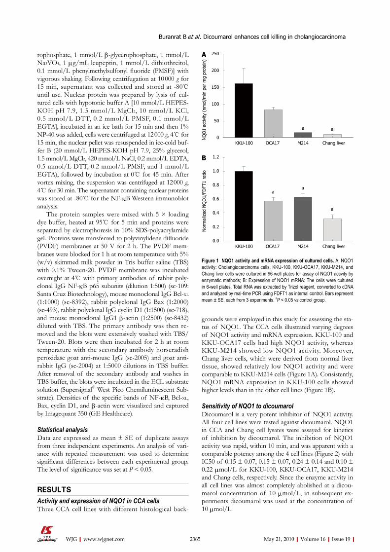
RESULTSActivity and expression of NQO1 in CCA cellsThree CCA cell lines with different histological back-
grounds were employed in this study for assessing the sta-tus of NQO1. The CCA cells illustrated varying degrees of NQO1 activity and mRNA expression. KKU-100 and KKU-OCA17 cells had high NQO1 activity, whereas KKU-M214 showed low NQO1 activity. Moreover, Chang liver cells, which were derived from normal liver tissue, showed relatively low NQO1 activity and were comparable to KKU-M214 cells (Figure 1A). Consistently, NQO1 mRNA expression in KKU-100 cells showed higher levels than in the other cell lines (Figure 1B).
Sensitivity of NQO1 to dicoumarolDicoumarol is a very potent inhibitor of NQO1 activity. All four cell lines were tested against dicoumarol. NQO1 in CCA and Chang cell lysates were assayed for kinetics of inhibition by dicoumarol. The inhibition of NQO1 activity was rapid, within 10 min, and was apparent with a comparable potency among the 4 cell lines (Figure 2) with IC50 of 0.15 ± 0.07, 0.15 ± 0.07, 0.24 ± 0.14 and 0.10 ± 0.22 μmol/L for KKU-100, KKU-OCA17, KKU-M214 and Chang cells, respectively. Since the enzyme activity in all cell lines was almost completely abolished at a dicou-marol concentration of 10 μmol/L, in subsequent ex-periments dicoumarol was used at the concentration of 10 μmol/L.
2365 May 21, 2010|Volume 16|Issue 19|WJG|www.wjgnet.com
250
200
150
100
50
0NQ
O1
activ
ity (
nmol
/min
per
mg
prot
ein)
KKU-100 OCA17 M214 Chang liver
A
a a
1.2
1.0
0.8
0.6
0.4
0.2
0.0
Nor
mal
ized
NQ
O1/
FDFT
1 ra
tio
KKU-100 OCA17 M214 Chang liver
B
Figure 1 NQO1 activity and mRNA expression of cultured cells. A: NQO1 activity: Cholangiocarcinoma cells, KKU-100, KKU-OCA17, KKU-M214, and Chang liver cells were cultured in 96-well plates for assay of NQO1 activity by enzymatic methods; B: Expression of NQO1 mRNA: The cells were cultured in 6-well plates. Total RNA was extracted by Trizol reagent, converted to cDNA and analyzed by real-time PCR using FDFT1 as internal control. Bars represent mean ± SE, each from 3 experiments. aP < 0.05 vs control group.
aa
a
Buranrat B et al . Dicoumarol enhances cell killing in cholangiocarcinoma
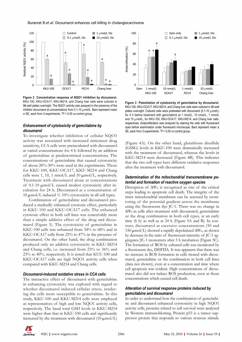
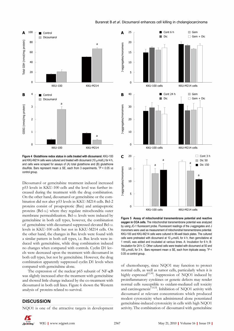
Enhancement of cytotoxicity of gemcitabine by dicoumarolTo investigate whether inhibition of cellular NQO1 activity was associated with increased anticancer drug sensitivity, CCA cells were preincubated with dicoumarol at varied concentrations for 4 h followed by an addition of gemcitabine at predetermined concentrations. The concentrations of gemcitabine that caused cytotoxicity of about 20%-30% were used in the experiments. Those for KKU-100, KKU-OCA17, KKU-M214 and Chang cells were 1, 10, 1 nmol/L and 10 μmol/L, respectively. Treatment with dicoumarol alone at concentrations of 0.1-10 μmol/L caused modest cytotoxicity after in-cubation for 24 h. Dicoumarol at a concentration of 10 μmol/L induced ≤ 10% cytotoxicity in all cell types.
Combination of gemcitabine and dicoumarol pro-duced a markedly enhanced cytotoxic effect, particularly in KKU-100 and KKU-OCA17 cells. The enhanced cytotoxic effect in both cell lines was conceivably more than a simple additive effect of the drug and dicou-marol (Figure 3). The cytotoxicity of gemcitabine in KKU-100 cells was enhanced from 34% to 68% and in KKU-OCA17 cells from 25% to 47% in the presence of dicoumarol. On the other hand, the drug combination produced only an additive cytotoxicity in KKU-M214 and Chang cells, i.e. increased from 23% to 36% and 23% to 40%, respectively. It is noted that KUU-100 and KKU-OCA17 cells are high NQO1 activity cells when compared with KKU-M214 and Chang cells.
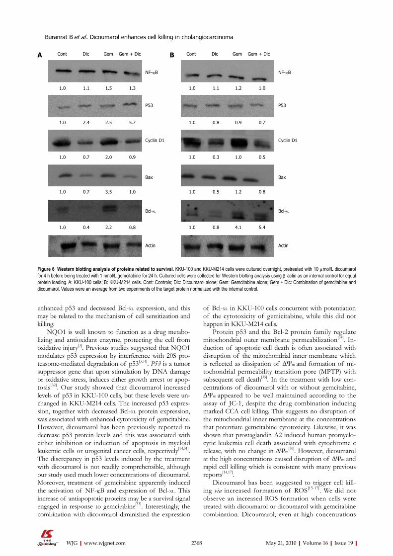
Dicoumarol-induced oxidative stress in CCA cellsThe interactive effect of dicoumarol with gemcitabine in enhancing cytotoxicity was explored with regard to whether dicoumarol induced cellular stress, render-ing the cells more susceptible to gemcitabine. In this study, KKU-100 and KKU-M214 cells were employed as representatives of high and low NQO1 activity cells, respectively. The basal total GSH levels in KKU-M214 were higher than that in KKU-100 cells and significantly increased by the treatment with dicoumarol (10 μmol/L)
(Figure 4A). On the other hand, glutathione disulfide (GSSG) levels in KKU-100 were dramatically increased with the treatment of dicoumarol, whereas the levels in KKU-M214 were decreased (Figure 4B). This indicates that the two cell types have different oxidative responses after the treatment with dicoumarol.
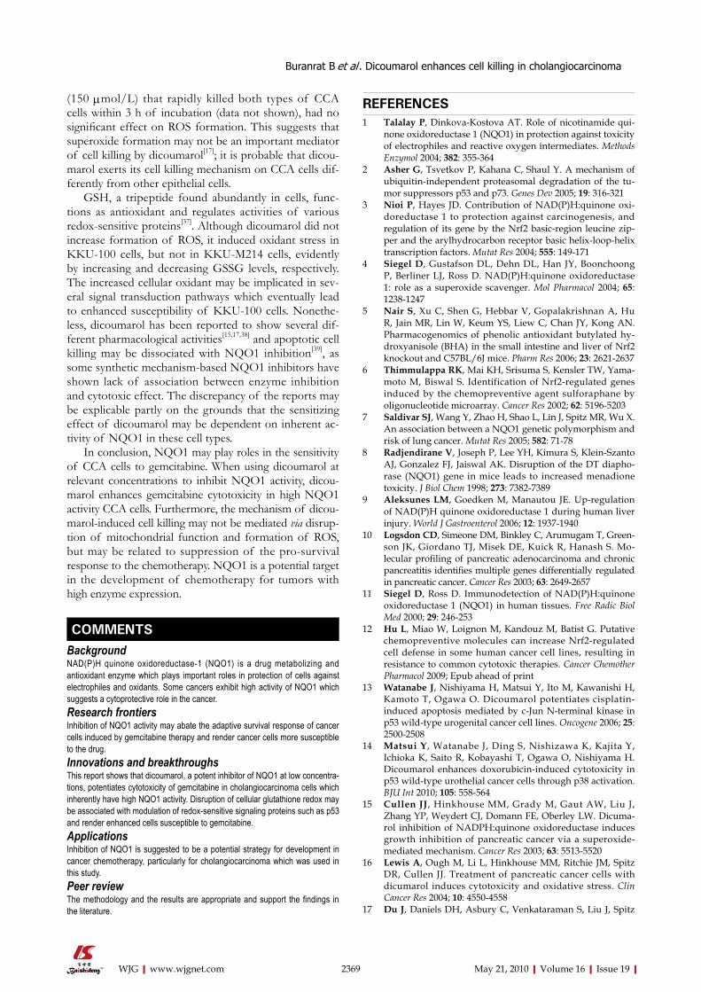
Determination of the mitochondrial transmembrane po-tential and formation of reactive oxygen speciesDisruption of ∆Ψm is recognized as one of the critical steps leading to apoptotic cell death. The integrity of the inner mitochondrial membrane can be assessed by moni-toring of the potential gradient across the membrane using the fluorescent dye JC-1. There was no change in ∆Ψm in cells after treatment with dicoumarol, gemcitabine or the drug combination in both cell types, at an early time (6 h) as well as at 24 h (Figure 5A and B). In con-trast, dicoumarol at excessive concentrations (50 and 150 μmol/L) showed a rapidly depolarized ∆Ψm, as shown by decrease in the ratio of fluorescent intensity of JC-1 ag-gregates/JC-1 monomers after 3 h incubation (Figure 5C). The formation of ROS by cultured cells was monitored by a fluorescent dye, DHFDA. It was apparent that there was no increase in ROS formation in cells treated with dicou-marol, gemcitabine or the combination in both cell lines (data not shown), even at a concentration and time where cell apoptosis was evident. High concentrations of dicou-marol also did not induce ROS production, even at those concentrations which caused cell death.
Alteration of survival response proteins induced by gemcitabine and dicoumarolIn order to understand how the combination of gemcitabi-ne and dicoumarol enhanced cytotoxicity in high NQO1 activity cells, proteins related to cell survival were analyzed by Western immunoblotting. Protein p53 is a tumor sup-pressor protein that responds to various noxious stimuli.
2366 May 21, 2010|Volume 16|Issue 19|WJG|www.wjgnet.com
120
100
80
60
40
20
0
Resi
dual
NQ
O1
activ
ity (
%)
KKU-100 OCA17 M214 Chang liver
a
a
a
a
a
a
a
a
a
a a
Control 1 μmol/L Dic
0.1 μmol/L Dic 10 μmol/L Dic
Figure 2 Concentration response of NQO1 inhibition by dicoumarol. KKU-100, KKU-OCA17, KKU-M214, and Chang liver cells were cultured in 96-well plates overnight. The NQO1 activity was assayed in the presence of the inhibitor dicoumarol at concentrations from 0.1-10 μmol/L. Bars represent mean ± SE, each from 3 experiments. aP < 0.05 vs control group.
100
80
60
40
20
0
Antip
rolif
erat
ion
(%)
Gem 1 nmol/L 10 nmol/L 1 nmol/L 10 μmol/L
KKU-100 OCA17 M214 Chang liver
Gem only 1 μmol/L Dic
0.1 μmol/L Dic 10 μmol/L Dic
a a
a
a
Figure 3 Potentiation of cytotoxicity of gemcitabine by dicoumarol. KKU-100, KKU-OCA17, KKU-M214, and Chang liver cells were cultured in 96-well plates overnight. Cultured cells were pretreated with dicoumarol (0.1-10 μmol/L) for 4 h before treatment with gemcitabine at 1 nmol/L, 10 nmol/L, 1 nmol/L and 10 μmol/L, for KKU-100, KKU-OCA17, KKU-M214, and Chang liver cells, respectively. Antiproliferation was analyzed by staining the cells with fluorescent dyes before examination under fluorescent microscope. Bars represent mean ± SE, each from 3 experiments. aP < 0.05 vs control group.
Buranrat B et al . Dicoumarol enhances cell killing in cholangiocarcinoma
Dicoumarol or gemcitabine treatment induced increased p53 levels in KKU-100 cells and the level was further in-creased during the treatment with the drug combination. On the other hand, dicoumarol or gemcitabine or the com-bination did not alter p53 levels in KKU-M214 cells. Bcl-2 proteins consist of proapoptotic (Bax) and antiapoptotic proteins (Bcl-XL) where they regulate mitochondria outer membrane permeabilization. Bcl-XL levels were induced by gemcitabine in both cell types, however, the combination of gemcitabine with dicoumarol suppressed elevated Bcl-XL levels in KKU-100 cells but not in KKU-M214 cells. On the other hand, the changes in Bax levels were found with a similar pattern in both cell types, i.e. Bax levels were in-duced with gemcitabine, while drug combination induced no changes when compared with controls. Cyclin D1 lev-els were decreased upon the treatment with dicoumarol in both cell types, but not by gemcitabine. However, the drug combination apparently suppressed cyclin D1 levels when compared with gemcitabine alone.
The expression of the nuclear p65 subunit of NF-κB was slightly increased after the treatment with gemcitabine and showed little change induced by the co-treatment with dicoumarol in both cell lines. Figure 6 shown the Western analysis of proteins related to survival.
DISCUSSIONNQO1 is one of the attractive targets in development
of chemotherapy, since NQO1 may function to protect normal cells, as well as tumor cells, particularly when it is highly expressed[12,30]. Suppression of NQO1 induced by proinflammatory cytokines or genetic defects may render normal cells susceptible to oxidant-mediated cell toxicity and carcinogenesis[7,8,22]. Inhibition of NQO1 activity with dicoumarol at relevant concentrations which produced modest cytotoxicity when administered alone potentiated gemcitabine-induced cytotoxicity in cells with high NQO1 activity. The combination of dicoumarol with gemcitabine
2367 May 21, 2010|Volume 16|Issue 19|WJG|www.wjgnet.com
100
80
60
40
20
0
Tota
l GSH
(nm
ol/m
g pr
otei
n)
KKU-100 KKU-M214
a
Control
DicoumarolA
4
3
2
1
0
GSS
G (
nmol
/mg
prot
ein)
KKU-100 KKU-M214
Control
DicoumarolB
a
a
Figure 4 Glutathione redox status in cells treated with dicoumarol. KKU-100 and KKU-M214 cells were cultured and treated with dicoumarol (10 μmol/L) for 4 h, and cells were scraped for assays of (A) total glutathione and (B) glutathione disulfide. Bars represent mean ± SE, each from 3 experiments. aP < 0.05 vs control group.
Figure 5 Assay of mitochondrial transmembrane potential and reactive oxygen in CCA cells. The mitochondrial transmembrane potential was analyzed by using JC-1 fluorescent probe. Fluorescent readings of the J-aggregates and J monomers were used as measurement of mitochondrial transmembrane potential. KKU-100 and KKU-M214 cells were cultured in 96-well black plates. The cultured cells were pretreated with dicoumarol at 10 μmol/L for 4 h, then gemcitabine at 1 nmol/L was added and incubated at various times. A: Incubation for 6 h; B: Incubation for 24 h; C: Other cultured cells were treated with dicoumarol at 50 and 150 μmol/L for 3 h. Bars represent mean ± SE, each from triplicate assay. aP < 0.05 vs control group.
Cont 6 h Gem
Dic Gem + Dic
25
20
15
10
5
0
J-ag
greg
ates
/mon
omer
s
KKU-100 cells KKU-M214 cells
A
Cont 24 h Gem
Dic Gem + Dic
40
30
20
10
0
J-ag
greg
ates
/mon
omer
s
KKU-100 cells KKU-M214 cells
B
20
15
10
5
0
J-ag
greg
ates
/mon
omer
s
KKU-100 cells KKU-M214 cells
C Cont 3 h
Dic 50
Dic 150 a
aa
a
Buranrat B et al . Dicoumarol enhances cell killing in cholangiocarcinoma
enhanced p53 and decreased Bcl-XL expression, and this may be related to the mechanism of cell sensitization and killing.
NQO1 is well known to function as a drug metabo-lizing and antioxidant enzyme, protecting the cell from oxidative injury[3]. Previous studies suggested that NQO1 modulates p53 expression by interference with 20S pro-teasome-mediated degradation of p53[3,31]. P53 is a tumor suppressor gene that upon stimulation by DNA damage or oxidative stress, induces either growth arrest or apop-tosis[32]. Our study showed that dicoumarol increased levels of p53 in KKU-100 cells, but these levels were un-changed in KKU-M214 cells. The increased p53 expres-sion, together with decreased Bcl-XL protein expression, was associated with enhanced cytotoxicity of gemcitabine. However, dicoumarol has been previously reported to decrease p53 protein levels and this was associated with either inhibition or induction of apoptosis in myeloid leukemic cells or urogenital cancer cells, respectively[14,31]. The discrepancy in p53 levels induced by the treatment with dicoumarol is not readily comprehensible, although our study used much lower concentrations of dicoumarol. Moreover, treatment of gemcitabine apparently induced the activation of NF-κB and expression of Bcl-XL. This increase of antiapoptotic proteins may be a survival signal engaged in response to gemcitabine[33]. Interestingly, the combination with dicoumarol diminished the expression
of Bcl-XL in KKU-100 cells concurrent with potentiation of the cytotoxicity of gemicitabine, while this did not happen in KKU-M214 cells.
Protein p53 and the Bcl-2 protein family regulate mitochondrial outer membrane permeabilization[34]. In-duction of apoptotic cell death is often associated with disruption of the mitochondrial inner membrane which is reflected as dissipation of ∆Ψm and formation of mi-tochondrial permeability transition pore (MPTP) with subsequent cell death[35]. In the treatment with low con-centrations of dicoumarol with or without gemcitabine, ∆Ψm appeared to be well maintained according to the assay of JC-1, despite the drug combination inducing marked CCA cell killing. This suggests no disruption of the mitochondrial inner membrane at the concentrations that potentiate gemcitabine cytotoxicity. Likewise, it was shown that prostaglandin A2 induced human promyelo-cytic leukemia cell death associated with cytochrome c release, with no change in ∆Ψm
[36]. However, dicoumarol at the high concentrations caused disruption of ∆Ψm and rapid cell killing which is consistent with many previous reports[14,17].
Dicoumarol has been suggested to trigger cell kill-ing via increased formation of ROS[15-17]. We did not observe an increased ROS formation when cells were treated with dicoumarol or dicoumarol with gemcitabine combination. Dicoumarol, even at high concentrations
2368 May 21, 2010|Volume 16|Issue 19|WJG|www.wjgnet.com
Figure 6 Western blotting analysis of proteins related to survival. KKU-100 and KKU-M214 cells were cultured overnight, pretreated with 10 μmol/L dicoumarol for 4 h before being treated with 1 nmol/L gemcitabine for 24 h. Cultured cells were collected for Western blotting analysis using β-actin as an internal control for equal protein loading. A: KKU-100 cells; B: KKU-M214 cells. Cont: Controls; Dic: Dicoumarol alone; Gem: Gemcitabine alone; Gem + Dic: Combination of gemcitabine and dicoumarol. Values were an average from two experiments of the target protein normalized with the internal control.
1.0 1.1 1.5 1.3
Cont Dic Gem Gem + DicA Cont Dic Gem Gem + DicB
1.0 1.1 1.2 1.0
1.0 2.4 2.5 5.7 1.0 0.8 0.9 0.7
1.0 0.7 2.0 0.9 1.0 0.3 1.0 0.5
1.0 0.7 3.5 1.0 1.0 0.5 1.2 0.8
1.0 0.4 2.2 0.8 1.0 0.8 4.1 5.4
NF-κB
P53
Cyclin D1
Bax
Bcl-XL
Actin
NF-κB
P53
Cyclin D1
Bax
Bcl-XL
Actin
Buranrat B et al . Dicoumarol enhances cell killing in cholangiocarcinoma
(150 μmol/L) that rapidly killed both types of CCA cells within 3 h of incubation (data not shown), had no significant effect on ROS formation. This suggests that superoxide formation may not be an important mediator of cell killing by dicoumarol[17]; it is probable that dicou-marol exerts its cell killing mechanism on CCA cells dif-ferently from other epithelial cells.
GSH, a tripeptide found abundantly in cells, func-tions as antioxidant and regulates activities of various redox-sensitive proteins[37]. Although dicoumarol did not increase formation of ROS, it induced oxidant stress in KKU-100 cells, but not in KKU-M214 cells, evidently by increasing and decreasing GSSG levels, respectively. The increased cellular oxidant may be implicated in sev-eral signal transduction pathways which eventually lead to enhanced susceptibility of KKU-100 cells. Nonethe-less, dicoumarol has been reported to show several dif-ferent pharmacological activities[15,17,38] and apoptotic cell killing may be dissociated with NQO1 inhibition[39], as some synthetic mechanism-based NQO1 inhibitors have shown lack of association between enzyme inhibition and cytotoxic effect. The discrepancy of the reports may be explicable partly on the grounds that the sensitizing effect of dicoumarol may be dependent on inherent ac-tivity of NQO1 in these cell types.
In conclusion, NQO1 may play roles in the sensitivity of CCA cells to gemcitabine. When using dicoumarol at relevant concentrations to inhibit NQO1 activity, dicou-marol enhances gemcitabine cytotoxicity in high NQO1 activity CCA cells. Furthermore, the mechanism of dicou-marol-induced cell killing may not be mediated via disrup-tion of mitochondrial function and formation of ROS, but may be related to suppression of the pro-survival response to the chemotherapy. NQO1 is a potential target in the development of chemotherapy for tumors with high enzyme expression.
COMMENTSBackgroundNAD(P)H quinone oxidoreductase-1 (NQO1) is a drug metabolizing and antioxidant enzyme which plays important roles in protection of cells against electrophiles and oxidants. Some cancers exhibit high activity of NQO1 which suggests a cytoprotective role in the cancer. Research frontiersInhibition of NQO1 activity may abate the adaptive survival response of cancer cells induced by gemcitabine therapy and render cancer cells more susceptible to the drug.Innovations and breakthroughsThis report shows that dicoumarol, a potent inhibitor of NQO1 at low concentra-tions, potentiates cytotoxicity of gemcitabine in cholangiocarcinoma cells which inherently have high NQO1 activity. Disruption of cellular glutathione redox may be associated with modulation of redox-sensitive signaling proteins such as p53 and render enhanced cells susceptible to gemcitabine.ApplicationsInhibition of NQO1 is suggested to be a potential strategy for development in cancer chemotherapy, particularly for cholangiocarcinoma which was used in this study.Peer reviewThe methodology and the results are appropriate and support the findings in the literature.
REFERENCES1 Talalay P, Dinkova-Kostova AT. Role of nicotinamide qui-
none oxidoreductase 1 (NQO1) in protection against toxicity of electrophiles and reactive oxygen intermediates. Methods Enzymol 2004; 382: 355-364
2 Asher G, Tsvetkov P, Kahana C, Shaul Y. A mechanism of ubiquitin-independent proteasomal degradation of the tu-mor suppressors p53 and p73. Genes Dev 2005; 19: 316-321
3 Nioi P, Hayes JD. Contribution of NAD(P)H:quinone oxi-doreductase 1 to protection against carcinogenesis, and regulation of its gene by the Nrf2 basic-region leucine zip-per and the arylhydrocarbon receptor basic helix-loop-helix transcription factors. Mutat Res 2004; 555: 149-171
4 Siegel D, Gustafson DL, Dehn DL, Han JY, Boonchoong P, Berliner LJ, Ross D. NAD(P)H:quinone oxidoreductase 1: role as a superoxide scavenger. Mol Pharmacol 2004; 65: 1238-1247
5 Nair S, Xu C, Shen G, Hebbar V, Gopalakrishnan A, Hu R, Jain MR, Lin W, Keum YS, Liew C, Chan JY, Kong AN. Pharmacogenomics of phenolic antioxidant butylated hy-droxyanisole (BHA) in the small intestine and liver of Nrf2 knockout and C57BL/6J mice. Pharm Res 2006; 23: 2621-2637
6 Thimmulappa RK, Mai KH, Srisuma S, Kensler TW, Yama-moto M, Biswal S. Identification of Nrf2-regulated genes induced by the chemopreventive agent sulforaphane by oligonucleotide microarray. Cancer Res 2002; 62: 5196-5203
7 Saldivar SJ, Wang Y, Zhao H, Shao L, Lin J, Spitz MR, Wu X. An association between a NQO1 genetic polymorphism and risk of lung cancer. Mutat Res 2005; 582: 71-78
8 Radjendirane V, Joseph P, Lee YH, Kimura S, Klein-Szanto AJ, Gonzalez FJ, Jaiswal AK. Disruption of the DT diapho-rase (NQO1) gene in mice leads to increased menadione toxicity. J Biol Chem 1998; 273: 7382-7389
9 Aleksunes LM, Goedken M, Manautou JE. Up-regulation of NAD(P)H quinone oxidoreductase 1 during human liver injury. World J Gastroenterol 2006; 12: 1937-1940
10 Logsdon CD, Simeone DM, Binkley C, Arumugam T, Green-son JK, Giordano TJ, Misek DE, Kuick R, Hanash S. Mo-lecular profiling of pancreatic adenocarcinoma and chronic pancreatitis identifies multiple genes differentially regulated in pancreatic cancer. Cancer Res 2003; 63: 2649-2657
11 Siegel D, Ross D. Immunodetection of NAD(P)H:quinone oxidoreductase 1 (NQO1) in human tissues. Free Radic Biol Med 2000; 29: 246-253
12 Hu L, Miao W, Loignon M, Kandouz M, Batist G. Putative chemopreventive molecules can increase Nrf2-regulated cell defense in some human cancer cell lines, resulting in resistance to common cytotoxic therapies. Cancer Chemother Pharmacol 2009; Epub ahead of print
13 Watanabe J, Nishiyama H, Matsui Y, Ito M, Kawanishi H, Kamoto T, Ogawa O. Dicoumarol potentiates cisplatin-induced apoptosis mediated by c-Jun N-terminal kinase in p53 wild-type urogenital cancer cell lines. Oncogene 2006; 25: 2500-2508
14 Matsui Y, Watanabe J, Ding S, Nishizawa K, Kajita Y, Ichioka K, Saito R, Kobayashi T, Ogawa O, Nishiyama H. Dicoumarol enhances doxorubicin-induced cytotoxicity in p53 wild-type urothelial cancer cells through p38 activation. BJU Int 2010; 105: 558-564
15 Cullen JJ, Hinkhouse MM, Grady M, Gaut AW, Liu J, Zhang YP, Weydert CJ, Domann FE, Oberley LW. Dicuma-rol inhibition of NADPH:quinone oxidoreductase induces growth inhibition of pancreatic cancer via a superoxide-mediated mechanism. Cancer Res 2003; 63: 5513-5520
16 Lewis A, Ough M, Li L, Hinkhouse MM, Ritchie JM, Spitz DR, Cullen JJ. Treatment of pancreatic cancer cells with dicumarol induces cytotoxicity and oxidative stress. Clin Cancer Res 2004; 10: 4550-4558
17 Du J, Daniels DH, Asbury C, Venkataraman S, Liu J, Spitz
2369 May 21, 2010|Volume 16|Issue 19|WJG|www.wjgnet.com
COMMENTS
Buranrat B et al . Dicoumarol enhances cell killing in cholangiocarcinoma
DR, Oberley LW, Cullen JJ. Mitochondrial production of re-active oxygen species mediate dicumarol-induced cytotoxic-ity in cancer cells. J Biol Chem 2006; 281: 37416-37426
18 Khan SA, Thomas HC, Davidson BR, Taylor-Robinson SD. Cholangiocarcinoma. Lancet 2005; 366: 1303-1314
19 Uttaravichien T, Bhudhisawasdi V, Pairojkul C, Pugkhem A. Intrahepatic cholangiocarcinoma in Thailand. J Hepatobiliary Pancreat Surg 1999; 6: 128-135
20 Mini E, Nobili S, Caciagli B, Landini I, Mazzei T. Cellular pharmacology of gemcitabine. Ann Oncol 2006; 17 Suppl 5: v7-v12
21 Valle JW, Wasan H, Johnson P, Jones E, Dixon L, Swindell R, Baka S, Maraveyas A, Corrie P, Falk S, Gollins S, Lofts F, Ev-ans L, Meyer T, Anthoney A, Iveson T, Highley M, Osborne R, Bridgewater J. Gemcitabine alone or in combination with cisplatin in patients with advanced or metastatic cholangio-carcinomas or other biliary tract tumours: a multicentre ran-domised phase II study - The UK ABC-01 Study. Br J Cancer 2009; 101: 621-627
22 Prawan A, Buranrat B, Kukongviriyapan U, Sripa B, Kukong-viriyapan V. Inflammatory cytokines suppress NAD(P)H:qui-none oxidoreductase-1 and induce oxidative stress in cholan-giocarcinoma cells. J Cancer Res Clin Oncol 2009; 135: 515-522
23 Sripa B, Leungwattanawanit S, Nitta T, Wongkham C, Bhu-dhisawasdi V, Puapairoj A, Sripa C, Miwa M. Establishment and characterization of an opisthorchiasis-associated chol-angiocarcinoma cell line (KKU-100). World J Gastroenterol 2005; 11: 3392-3397
24 Buranrat B, Prawan A, Sripa B, Kukongviriyapan V. Inflam-matory cytokines suppress arylamine N-acetyltransferase 1 in cholangiocarcinoma cells. World J Gastroenterol 2007; 13: 6219-6225
25 Prochaska HJ, Santamaria AB. Direct measurement of NAD(P)H:quinone reductase from cells cultured in microti-ter wells: a screening assay for anticarcinogenic enzyme inducers. Anal Biochem 1988; 169: 328-336
26 Ribble D, Goldstein NB, Norris DA, Shellman YG. A simple technique for quantifying apoptosis in 96-well plates. BMC Biotechnol 2005; 5: 12
27 Tietze F. Enzymic method for quantitative determination of nanogram amounts of total and oxidized glutathione: appli-cations to mammalian blood and other tissues. Anal Biochem 1969; 27: 502-522
28 Somparn N, Kukongviriyapan U, Tassaneeyakul W, Jetsrisu-parb A, Kukongviriyapan V. Modification of CYP2E1 and CYP3A4 activities in haemoglobin E-beta thalassemia pa-tients. Eur J Clin Pharmacol 2007; 63: 43-50
29 Jang JH, Surh YJ. Bcl-2 attenuation of oxidative cell death is associated with up-regulation of gamma-glutamylcysteine ligase via constitutive NF-kappaB activation. J Biol Chem 2004; 279: 38779-38786
30 Dinkova-Kostova AT, Talalay P. Persuasive evidence that quinone reductase type 1 (DT diaphorase) protects cells against the toxicity of electrophiles and reactive forms of oxygen. Free Radic Biol Med 2000; 29: 231-240
31 Asher G, Lotem J, Cohen B, Sachs L, Shaul Y. Regulation of p53 stability and p53-dependent apoptosis by NADH quinone oxidoreductase 1. Proc Natl Acad Sci USA 2001; 98: 1188-1193
32 Bouchet BP, de Fromentel CC, Puisieux A, Galmarini CM. p53 as a target for anti-cancer drug development. Crit Rev Oncol Hematol 2006; 58: 190-207
33 Fahy BN, Schlieman MG, Virudachalam S, Bold RJ. Inhibi-tion of AKT abrogates chemotherapy-induced NF-kappaB survival mechanisms: implications for therapy in pancreatic cancer. J Am Coll Surg 2004; 198: 591-599
34 Chao DT, Korsmeyer SJ. BCL-2 family: regulators of cell death. Annu Rev Immunol 1998; 16: 395-419
35 Chipuk JE, Bouchier-Hayes L, Green DR. Mitochondrial out-er membrane permeabilization during apoptosis: the inno-cent bystander scenario. Cell Death Differ 2006; 13: 1396-1402
36 Lee SY, Ahn JH, Ko KW, Kim J, Jeong SW, Kim IK, Kim J, Kim HS. Prostaglandin A2 activates intrinsic apoptotic pathway by direct interaction with mitochondria in HL-60 cells. Prostaglandins Other Lipid Mediat 2010; 91: 30-37
37 Schafer FQ, Buettner GR. Redox environment of the cell as viewed through the redox state of the glutathione disulfide/glutathione couple. Free Radic Biol Med 2001; 30: 1191-1212
38 Madari H, Panda D, Wilson L, Jacobs RS. Dicoumarol: a unique microtubule stabilizing natural product that is syn-ergistic with Taxol. Cancer Res 2003; 63: 1214-1220
39 Dehn DL, Siegel D, Zafar KS, Reigan P, Swann E, Moody CJ, Ross D. 5-Methoxy-1,2-dimethyl-3-[(4-nitrophenoxy)-methyl]indole-4,7-dione, a mechanism-based inhibitor of NAD(P)H:quinone oxidoreductase 1, exhibits activity against human pancreatic cancer in vitro and in vivo. Mol Cancer Ther 2006; 5: 1702-1709
S- Editor Wang JL L- Editor Logan S E- Editor Ma WH
2370 May 21, 2010|Volume 16|Issue 19|WJG|www.wjgnet.com
Buranrat B et al . Dicoumarol enhances cell killing in cholangiocarcinoma