¿cuál seria su abordaje quirúrgico? -...
TRANSCRIPT
Acude a nuestra consulta unpaciente varón de 74 años de edad,remitido por su odontólogo por pre-sentar una tumoración que abom-ba en paladar blando. La lesión noera dolorosa ni producía ningunaotra sintomatología acompañante.
En cuanto a sus antecedentespersonales destaca tan sólo una aler-gia a los antibióticos beta-lactámicosy a la estreptomicina.
A la exploración física se apre-ciaba una tumoración que abombay deforma el paladar blando dere-cho, de aproximadamente 4x4 cen-tímetros de diámetro, de consisten-cia dura, desplazable y no doloro-sa a la palpación. En la exploracióncervical no había hallazgos patoló-gicos.
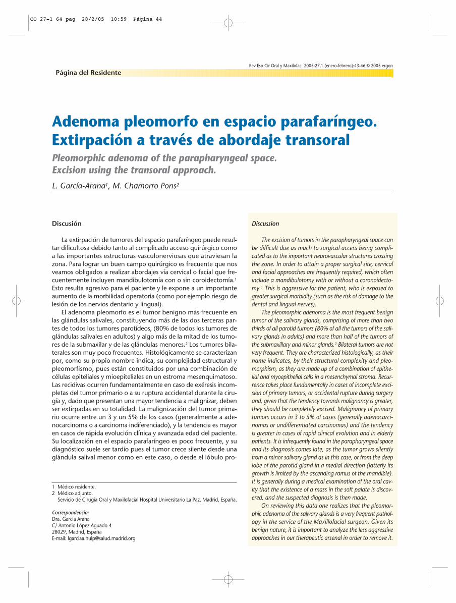
Para completar el estudio diag-nóstico y de extensión solicitamosuna PAAF de la lesión y una TC facialy cervical con contraste intraveno-so. El diagnóstico microscópico dela PAAF (Fig. 1) fue de glándula sali-val con imágenes sugerentes de ade-noma pleomorfo. Y la TC (Fig. 2) fueinformada como lesión extendidadesde el plano del paladar durohasta el del suelo de la boca en eleje craneocaudal y de aproximada-mente 4 centímetros de diámetrosanteroposterior y transversal. Lalesión produce desplazamiento haciala izquierda del espacio parafarín-geo derecho, e impronta la vía aéreay los espacios retrofaríngeo y pre-vertebral derechos. Se aprecia un desplazamiento lateral de los músculospterigoideos. Es difícil situar la lesión en el espacio parotídeo o en el espa-cio mucoso-submucoso orofaríngeo. Aparecen ganglios linfáticos inespe-cíficos en todas las cadenas cervicales.
Con los resultados de la PAAF y TC realizamos el diagnóstico de pre-sunción de adenoma pleomorfo de espacio parafaríngeo derecho, pro-gramando su extirpación quirúrgica.
A 74 year-old male patient was referredto our service by his dentist due to atumor-like bulge in his soft palate. Thelesion was painless and it was not pro-ducing any other accompanying symp-tomatology. With regard to his personal history,only an antibiotic allergy to beta-lac-tam and streptomycin was of signif-icance.On physical exploration a tumor-likebulge was noted that was deformingthe right soft palate. It measured 4 x 4centimeters approximately in diameter.It was hard, movable and not painfulon palpation. No pathological findingswere noted on examination of his neck.In order to complete the diagnosticand extension study, we requesteda FNA of the lesion and a CT scan ofthe face and neck with intravenouscontrast. The microscopical diagno-sis of the FNA (Fig. 1) was of a sali-vary gland with images suggestingPleomorphic adenoma. And in the CTscan (Fig. 2) it was reported as alesion extending from the plane of thehard palate to the floor of the mouthalong the craniocaudal axis, and mea-suring approximately 4 centimetersin the anteroposterior and transversediameter. The lesion was causing theright parapharyngeal space to be dis-placed towards the left, and it wasaffecting the airway and the rightretropharyngeal and prevertebralspaces. A sideways displacement ofthe pterygoid muscles could be
observed. Locating the lesion in the parotid space or in themucosal-submucosal oropharyngeal space was difficult. Non-specific lymph nodes appeared along all the cervical chains.
With the results of the FNA and the CT scan the pre-sumed diagnosis of Pleomorphic Adenoma of the right para-pharyngeal space was made, and surgical excision was pro-grammed.
Página del Residente
¿Cuál seria su abordaje quirúrgico?
¿What surgical approach should be used?
Rev Esp Cir Oral Maxilofac 2005;27,1 (enero-febrero):43-46 © 2005 ergon
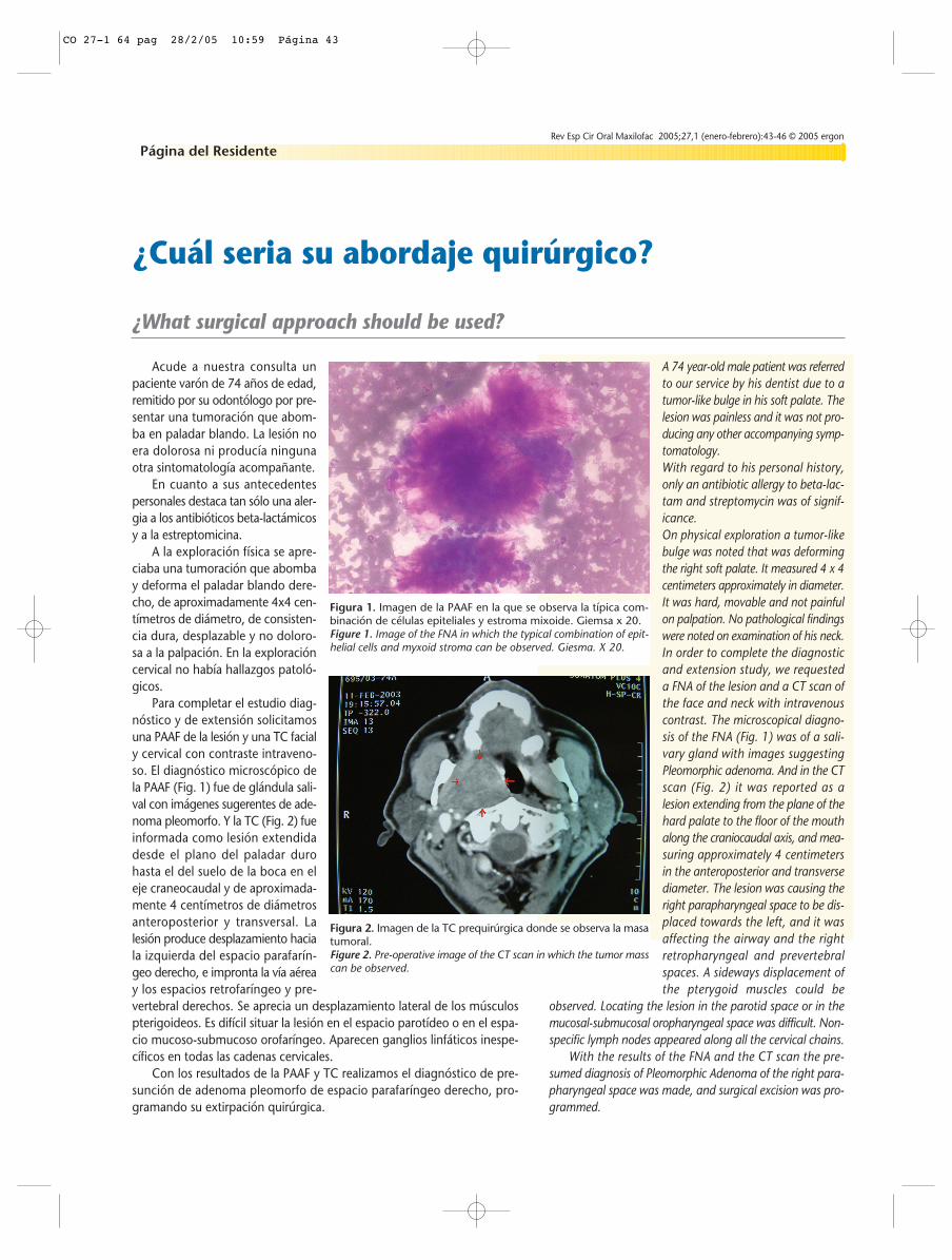
Figura 1. Imagen de la PAAF en la que se observa la típica com-binación de células epiteliales y estroma mixoide. Giemsa x 20.Figure 1. Image of the FNA in which the typical combination of epit-helial cells and myxoid stroma can be observed. Giesma. X 20.
Figura 2. Imagen de la TC prequirúrgica donde se observa la masatumoral.Figure 2. Pre-operative image of the CT scan in which the tumor masscan be observed.
CO 27-1 64 pag 28/2/05 10:59 Página 43
Discusión
La extirpación de tumores del espacio parafaríngeo puede resul-tar dificultosa debido tanto al complicado acceso quirúrgico comoa las importantes estructuras vasculonerviosas que atraviesan lazona. Para lograr un buen campo quirúrgico es frecuente que nosveamos obligados a realizar abordajes vía cervical o facial que fre-cuentemente incluyen mandibulotomía con o sin coroidectomía.1
Esto resulta agresivo para el paciente y le expone a un importanteaumento de la morbilidad operatoria (como por ejemplo riesgo delesión de los nervios dentario y lingual).
El adenoma pleomorfo es el tumor benigno más frecuente enlas glándulas salivales, constituyendo más de las dos terceras par-tes de todos los tumores parotídeos, (80% de todos los tumores deglándulas salivales en adultos) y algo más de la mitad de los tumo-res de la submaxilar y de las glándulas menores.2 Los tumores bila-terales son muy poco frecuentes. Histológicamente se caracterizanpor, como su propio nombre indica, su complejidad estructural ypleomorfismo, pues están constituidos por una combinación decélulas epiteliales y mioepiteliales en un estroma mesenquimatoso.Las recidivas ocurren fundamentalmente en caso de exéresis incom-pletas del tumor primario o a su ruptura accidental durante la ciru-gía y, dado que presentan una mayor tendencia a malignizar, debenser extirpadas en su totalidad. La malignización del tumor prima-rio ocurre entre un 3 y un 5% de los casos (generalmente a ade-nocarcinoma o a carcinoma indiferenciado), y la tendencia es mayoren casos de rápida evolución clínica y avanzada edad del paciente.Su localización en el espacio parafaríngeo es poco frecuente, y sudiagnóstico suele ser tardío pues el tumor crece silente desde unaglándula salival menor como en este caso, o desde el lóbulo pro-
Discussion
The excision of tumors in the parapharyngeal space canbe difficult due as much to surgical access being compli-cated as to the important neurovascular structures crossingthe zone. In order to attain a proper surgical site, cervicaland facial approaches are frequently required, which ofteninclude a mandibulotomy with or without a coronoidecto-my.1 This is aggressive for the patient, who is exposed togreater surgical morbidity (such as the risk of damage to thedental and lingual nerves).
The pleomorphic adenoma is the most frequent benigntumor of the salivary glands, comprising of more than twothirds of all parotid tumors (80% of all the tumors of the sali-vary glands in adults) and more than half of the tumors ofthe submaxillary and minor glands.2 Bilateral tumors are notvery frequent. They are characterized histologically, as theirname indicates, by their structural complexity and pleo-morphism, as they are made up of a combination of epithe-lial and myoepithelial cells in a mesenchymal stroma. Recur-rence takes place fundamentally in cases of incomplete exci-sion of primary tumors, or accidental rupture during surgeryand, given that the tendency towards malignancy is greater,they should be completely excised. Malignancy of primarytumors occurs in 3 to 5% of cases (generally adenocarci-nomas or undifferentiated carcinomas) and the tendencyis greater in cases of rapid clinical evolution and in elderlypatients. It is infrequently found in the parapharyngeal spaceand its diagnosis comes late, as the tumor grows silentlyfrom a minor salivary gland as in this case, or from the deeplobe of the parotid gland in a medial direction (latterly itsgrowth is limited by the ascending ramus of the mandible).It is generally during a medical examination of the oral cav-ity that the existence of a mass in the soft palate is discov-ered, and the suspected diagnosis is then made.
On reviewing this data one realizes that the pleomor-phic adenoma of the salivary glands is a very frequent pathol-ogy in the service of the Maxillofacial surgeon. Given itsbenign nature, it is important to analyze the less aggressiveapproaches in our therapeutic arsenal in order to remove it.
Adenoma pleomorfo en espacio parafaríngeo.Extirpación a través de abordaje transoralPleomorphic adenoma of the parapharyngeal space. Excision using the transoral approach.
L. García-Arana1, M. Chamorro Pons2
Página del Residente
1 Médico residente.2 Médico adjunto.
Servicio de Cirugía Oral y Maxilofacial Hospital Universitario La Paz, Madrid, España.
Correspondencia:Dra. García AranaC/ Antonio López Aguado 428029, Madrid, EspañaE-mail: [email protected]
Rev Esp Cir Oral y Maxilofac 2005;27,1 (enero-febrero):43-46 © 2005 ergon
CO 27-1 64 pag 28/2/05 10:59 Página 44
Rev Esp Cir Oral Maxilofac 2005;27,1 (enero-febrero):43-46 © 2005 ergon 45L. García-Arana y cols.
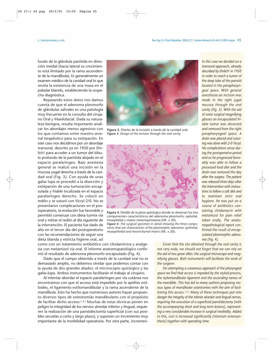
In this case we decided on atransoral approach, alreadydescribed by Ehrlich2 in 1950in order to reach a tumor ofthe deep lobe of the parotidlocated in the parapharyn-geal space. With generalanesthesia an incision wasmade in the right jugalmucosa through the oralcavity (Fig. 3). With the aidof some surgical magnifyingglasses an encapsulated fri-able tumor was dissectedand removed from the rightparapharyngeal space. Adrain was placed and sutur-ing was done with 2-0 Vicryl.No complications arose dur-ing the postoperative periodand as he progressed favor-ably was able to follow aprocessed food diet and thedrain was removed the dayafter the surgery. The patientwas released three days afterthe intervention with instruc-tions to follow a soft diet andto maintain strict oralhygiene. He was put on acourse of antibiotics con-taining clindamycin withmetamizol for pain relieftaken orally. The anato-mopathological report con-firmed the result of encap-sulated pleomorphic adeno-ma (Fig. 4).
Given that the site obtained through the oral cavity isnot very wide, we should not forget that we can rely onthe aid of two great allies: the surgical microscope and mag-nifying glasses. Both instruments will facilitate the work ofthe surgeon.
On attempting a cutaneous approach of the pharyngealspace we find that access is impeded by the styloid process,the stylomandibular ligament and the ascending ramus ofthe mandible. This has led to many authors proposing var-ious types of mandibular osteotomies with the aim of facil-itating this access.1,4,5 Many of these techniques put intodanger the integrity of the inferior alveolar and lingual nerves,requiring the execution of a superficial parotidectomy (withthe accompanying short and long term lesions), represent-ing a very considerable increase in surgical morbidity. Addedto this, cost is increased significantly (titanium osteosyn-thesis) together with operating time.
fundo de la glándula parótida en direc-ción medial (hacia lateral su crecimien-to está limitado por la rama ascenden-te de la mandíbula). Es generalmente unexamen médico de la cavidad oral lo querevela la existencia de una masa en elpaladar blando, estableciendo la sospe-cha diagnóstica.
Repasando estos datos nos damoscuenta de que el adenoma pleomorfode glándulas salivales es una patologíamuy frecuente en la consulta del ciruja-no Oral y Maxilofacial. Dada su natura-leza benigna, resulta importante anali-zar los abordajes menos agresivos conlos que contamos entre nuestro arse-nal terapéutico para su extirpación. Eneste caso nos decidimos por un abordajetransoral, descrito ya en 1950 por Ehr-lich2 para acceder a un tumor del lóbu-lo profundo de la parótida alojado en elespacio parafaríngeo. Bajo anestesiageneral se realizó una incisión en lamucosa yugal derecha a través de la cavi-dad oral (Fig. 3). Con ayuda de unasgafas lupa se procedió a la disección yextirpación de una tumoración encap-sulada y friable localizada en el espacioparafaríngeo derecho. Se colocó unredón y se suturó con Vicryl 2/0. No sepresentaron complicaciones en el pos-toperatorio, la evolución fue favorable ypermitió comenzar con dieta turmix víaoral y retirar el redón al día siguiente dela intervención. El paciente fue dado dealta en el tercer día del postoperatoriocon las recomendaciones de seguir unadieta blanda y estricta higiene oral, asícomo con un tratamiento antibiótico con clindamicina y analge-sia con metamizol vía oral. El informe anatomopatológico confir-mó el resultado de adenoma pleomorfo encapsulado (Fig. 4).
Dado que el campo obtenido a través de la cavidad oral no esdemasiado amplio, no debemos olvidar que podemos contar conla ayuda de dos grandes aliados: el microscopio quirúrgico y lasgafas-lupa. Ambos instrumentos facilitarán el trabajo al cirujano.
Al intentar abordar el espacio parafaríngeo por vía cutánea nosencontramos con que el acceso está impedido por la apófisis esti-loides, el ligamento estilomandibular y la rama ascendente de lamandíbula. Esto ha hecho que numerosos autores hayan propues-to diversos tipos de osteotomías mandibulares con el propósitode facilitar dicho acceso.1,4,5 Muchas de estas técnicas ponen enpeligro la integridad de los nervios alveolar inferior y lingual, requie-ren la realización de una parotidectomía superficial (con sus posi-bles secuelas a corto y largo plazo), y suponen un incremento muyimportante de la morbilidad operatoria. Por otra parte, incremen-
Figura 3. Diseño de la incisión a través de la cavidad oral.Figure 3. Design of the incision through the oral cavity.
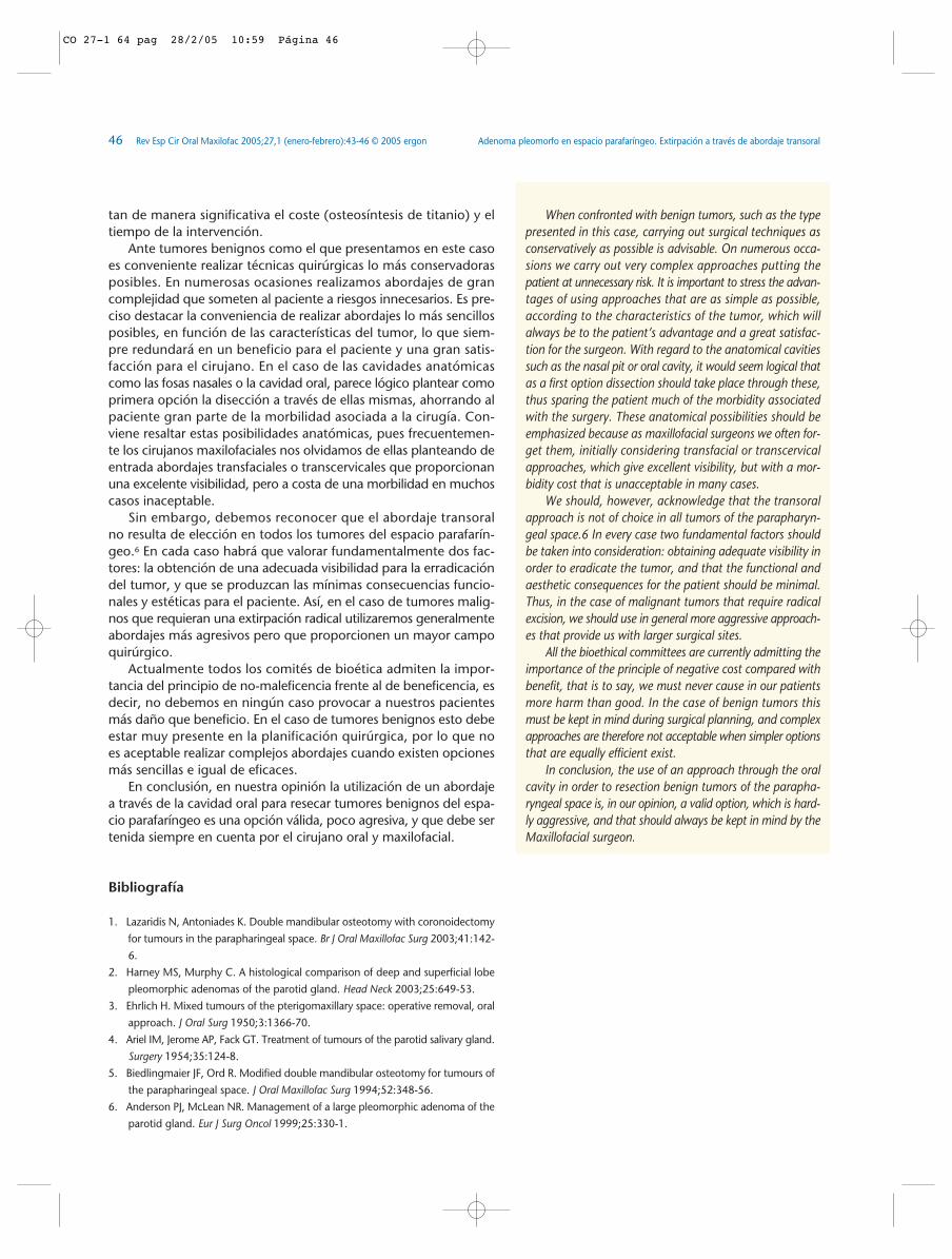
Figura 4. Detalle de la pieza quirúrgica donde se observan los trescomponentes característicos del adenoma pleomorfo: epitelial,mioepitelial y matriz mesenquimatosa (HE. x 20).Figure 4. The surgical specimen in detail showing the three compo-nents that are characteristic of the pleomorphic adenoma: epithelial,myoepithelial and mesenchymal matrix (HE. x 20).
CO 27-1 64 pag 28/2/05 10:59 Página 45
Adenoma pleomorfo en espacio parafaríngeo. Extirpación a través de abordaje transoral46 Rev Esp Cir Oral Maxilofac 2005;27,1 (enero-febrero):43-46 © 2005 ergon
tan de manera significativa el coste (osteosíntesis de titanio) y eltiempo de la intervención.
Ante tumores benignos como el que presentamos en este casoes conveniente realizar técnicas quirúrgicas lo más conservadorasposibles. En numerosas ocasiones realizamos abordajes de grancomplejidad que someten al paciente a riesgos innecesarios. Es pre-ciso destacar la conveniencia de realizar abordajes lo más sencillosposibles, en función de las características del tumor, lo que siem-pre redundará en un beneficio para el paciente y una gran satis-facción para el cirujano. En el caso de las cavidades anatómicascomo las fosas nasales o la cavidad oral, parece lógico plantear comoprimera opción la disección a través de ellas mismas, ahorrando alpaciente gran parte de la morbilidad asociada a la cirugía. Con-viene resaltar estas posibilidades anatómicas, pues frecuentemen-te los cirujanos maxilofaciales nos olvidamos de ellas planteando deentrada abordajes transfaciales o transcervicales que proporcionanuna excelente visibilidad, pero a costa de una morbilidad en muchoscasos inaceptable.
Sin embargo, debemos reconocer que el abordaje transoralno resulta de elección en todos los tumores del espacio parafarín-geo.6 En cada caso habrá que valorar fundamentalmente dos fac-tores: la obtención de una adecuada visibilidad para la erradicacióndel tumor, y que se produzcan las mínimas consecuencias funcio-nales y estéticas para el paciente. Así, en el caso de tumores malig-nos que requieran una extirpación radical utilizaremos generalmenteabordajes más agresivos pero que proporcionen un mayor campoquirúrgico.
Actualmente todos los comités de bioética admiten la impor-tancia del principio de no-maleficencia frente al de beneficencia, esdecir, no debemos en ningún caso provocar a nuestros pacientesmás daño que beneficio. En el caso de tumores benignos esto debeestar muy presente en la planificación quirúrgica, por lo que noes aceptable realizar complejos abordajes cuando existen opcionesmás sencillas e igual de eficaces.
En conclusión, en nuestra opinión la utilización de un abordajea través de la cavidad oral para resecar tumores benignos del espa-cio parafaríngeo es una opción válida, poco agresiva, y que debe sertenida siempre en cuenta por el cirujano oral y maxilofacial.
Bibliografía
1. Lazaridis N, Antoniades K. Double mandibular osteotomy with coronoidectomy
for tumours in the parapharingeal space. Br J Oral Maxillofac Surg 2003;41:142-
6.
2. Harney MS, Murphy C. A histological comparison of deep and superficial lobe
pleomorphic adenomas of the parotid gland. Head Neck 2003;25:649-53.
3. Ehrlich H. Mixed tumours of the pterigomaxillary space: operative removal, oral
approach. J Oral Surg 1950;3:1366-70.
4. Ariel IM, Jerome AP, Fack GT. Treatment of tumours of the parotid salivary gland.
Surgery 1954;35:124-8.
5. Biedlingmaier JF, Ord R. Modified double mandibular osteotomy for tumours of
the parapharingeal space. J Oral Maxillofac Surg 1994;52:348-56.
6. Anderson PJ, McLean NR. Management of a large pleomorphic adenoma of the
parotid gland. Eur J Surg Oncol 1999;25:330-1.
When confronted with benign tumors, such as the typepresented in this case, carrying out surgical techniques asconservatively as possible is advisable. On numerous occa-sions we carry out very complex approaches putting thepatient at unnecessary risk. It is important to stress the advan-tages of using approaches that are as simple as possible,according to the characteristics of the tumor, which willalways be to the patient’s advantage and a great satisfac-tion for the surgeon. With regard to the anatomical cavitiessuch as the nasal pit or oral cavity, it would seem logical thatas a first option dissection should take place through these,thus sparing the patient much of the morbidity associatedwith the surgery. These anatomical possibilities should beemphasized because as maxillofacial surgeons we often for-get them, initially considering transfacial or transcervicalapproaches, which give excellent visibility, but with a mor-bidity cost that is unacceptable in many cases.
We should, however, acknowledge that the transoralapproach is not of choice in all tumors of the parapharyn-geal space.6 In every case two fundamental factors shouldbe taken into consideration: obtaining adequate visibility inorder to eradicate the tumor, and that the functional andaesthetic consequences for the patient should be minimal.Thus, in the case of malignant tumors that require radicalexcision, we should use in general more aggressive approach-es that provide us with larger surgical sites.
All the bioethical committees are currently admitting theimportance of the principle of negative cost compared withbenefit, that is to say, we must never cause in our patientsmore harm than good. In the case of benign tumors thismust be kept in mind during surgical planning, and complexapproaches are therefore not acceptable when simpler optionsthat are equally efficient exist.
In conclusion, the use of an approach through the oralcavity in order to resection benign tumors of the parapha-ryngeal space is, in our opinion, a valid option, which is hard-ly aggressive, and that should always be kept in mind by theMaxillofacial surgeon.
CO 27-1 64 pag 28/2/05 10:59 Página 46