caracterización de los eventos inmunológicos diferenciales...
TRANSCRIPT

Caracterización de los eventos inmunológicos
diferenciales en pacientes con cáncer de pulmón de célula pequeña tratados con quimioterapia
con o sin ipilimumab
Max Hardy-Werbin
ADVERTIMENT. La consulta d’aquesta tesi queda condicionada a l’acceptació de les següents condicions d'ús: La difusió d’aquesta tesi per mitjà del servei TDX (www.tdx.cat) i a través del Dipòsit Digital de la UB (diposit.ub.edu) ha estat autoritzada pels titulars dels drets de propietat intelꞏlectual únicament per a usos privats emmarcats en activitats d’investigació i docència. No s’autoritza la seva reproducció amb finalitats de lucre ni la seva difusió i posada a disposició des d’un lloc aliè al servei TDX ni al Dipòsit Digital de la UB. No s’autoritza la presentació del seu contingut en una finestra o marc aliè a TDX o al Dipòsit Digital de la UB (framing). Aquesta reserva de drets afecta tant al resum de presentació de la tesi com als seus continguts. En la utilització o cita de parts de la tesi és obligat indicar el nom de la persona autora. ADVERTENCIA. La consulta de esta tesis queda condicionada a la aceptación de las siguientes condiciones de uso: La difusión de esta tesis por medio del servicio TDR (www.tdx.cat) y a través del Repositorio Digital de la UB (diposit.ub.edu) ha sido autorizada por los titulares de los derechos de propiedad intelectual únicamente para usos privados enmarcados en actividades de investigación y docencia. No se autoriza su reproducción con finalidades de lucro ni su difusión y puesta a disposición desde un sitio ajeno al servicio TDR o al Repositorio Digital de la UB. No se autoriza la presentación de su contenido en una ventana o marco ajeno a TDR o al Repositorio Digital de la UB (framing). Esta reserva de derechos afecta tanto al resumen de presentación de la tesis como a sus contenidos. En la utilización o cita de partes de la tesis es obligado indicar el nombre de la persona autora. WARNING. On having consulted this thesis you’re accepting the following use conditions: Spreading this thesis by the TDX (www.tdx.cat) service and by the UB Digital Repository (diposit.ub.edu) has been authorized by the titular of the intellectual property rights only for private uses placed in investigation and teaching activities. Reproduction with lucrative aims is not authorized nor its spreading and availability from a site foreign to the TDX service or to the UB Digital Repository. Introducing its content in a window or frame foreign to the TDX service or to the UB Digital Repository is not authorized (framing). Those rights affect to the presentation summary of the thesis as well as to its contents. In the using or citation of parts of the thesis it’s obliged to indicate the name of the author.
Caracterización de los eventos inmunológicos diferenciales en pacientes con cáncer de pulmón de célula pequeña tratados
con quimioterapia con o sin ipilimumab
Memoria presentada por Max Hardy-Werbin para optar al grado de Doctor por la Universidad de Barcelona
Programa de Doctorado en Biomedicina Barcelona, 2019
Tesis realizada bajo la dirección de la Dra. Edurne Arriola Aperribay, del departamento de Oncología Médica del Hospital del Mar, y bajo la tutorización de la Dra. María
Teresa Mampel Astals, del departamento de Bioquímica y Biomedicina Molecular de la Universidad de Barcelona
Dra Edurne Arriola Aperribay
Dra María Teresa Mampel Astals
Max Hardy-Werbin


La Dra. Edurne Arriola Aperribay, jefa de sección del departamento de Oncología Médica del Hospital del Mar e Investigadora Principal del Programa de Cáncer del Instituto Hospital del Mar de investigaciones médicas Certifica Que la tesis doctoral titulada “Caracterización de los eventos inmunológicos diferenciales en pacientes con cáncer de pulmón de célula pequeña tratados con quimioterapia con o sin ipilimumab”, presentada por Max Hardy-Werbin para acceder al título de Doctor en Biomedicina por la Universidad de Barcelona, se ha llevado a cabo bajo su supervisión y cumple los requisitos formales y científicos para ser defendida delante del tribunal correspondiente. El trabajo se ha realizado en el laboratorio de terapia molecular del cáncer, perteneciente al programa de cáncer, en el Instituto Hospital del Mar de Investigaciones Médicas. Barcelona, 2019
Dra Edurne Arriola Aperribay


« Qui cherche l’infini n’a qu’à fermer les yeux. » ― Milan Kundera
« We are what we pretend to be, so we must be careful about what we pretend to be. » ― Kurt Vonnegut


FINANCIACIÓN


Este proyecto de tesis se ha llevado a cabo en el marco del proyecto FIS P16/00591 otorgado por el Instituto de Salud Carlos III y gracias a la colaboración de la Fundación Cellex.


SINOPSIS


Small cell lung cancer (SCLC) is the most aggressive type of lung cancer. More than
half of patients are diagnosed at extensive stage, where platinum-based chemotherapy
has been the systemic standard treatment since the mid ‘80s. Although robust and often
dramatic clinical responses are achieved after first-line treatment, disease progression
takes place soon and it is usually resistant to available treatments. In this scenario,
outcomes remain poor, with a median overall survival that rarely exceeds one year.
SCLC is characterized by the presence of autoinmmunity, reflected by the incidence of
autoimmune paraneoplastic immune. This fact, as well as the high tumor mutational
burden found in this disease suggest that immune modulation might be a promising
strategy in SCLC. Anti-CTLA-4 antibodies and anti-PD-1/L1 antibodies have shown
activity and durable responses. In fact, an anti-PD-L1 agent has been recently approved
as first line treatment in addition to standard chemotherapy.
The aim of this project was to identify the differential immunological events observed
in patients treated with chemotherapy and ipilimumab (anti-CTLA-4 antibody) vs. those
in patients treated with standard chemotherapy, and in those patients treated with
ipilimumab with long survival vs. those with short survival. To this end we had
availability of serial samples from patients treated with ipilimumab + chemotherapy and
chemotherapy alone over time. We assessed cytokine and autoantibody profiles in
serum samples, and peripheral lymphocyte populations by flow cytometry.
We found that unlike autoantibodies, serum cytokines and specific lymphocyte
peripheral subpopulations were modulated after CTLA-4 blockade. We were able to
verify the prognostic role of autoantibodies, and to confirm the predictive role of
cytokines. Lastly, we detected particular patterns of peripheral T and NK cells
populations linked to survival and toxicity in SCLC.

With this project we were able to depict a comprehensive scene of the relevant
immunological events in SCLC undergoing standard treatment and immunotherapy,
which may aid us to better select patients who could benefit from immunotherapy and
thus to design appropriate clinical trials.

ÍNDICE


INTRODUCCIÓN ......................................................................................................... 1 EPIDEMIOLOGÍA Y CLASIFICACIÓN DEL CÁNCER DE PULMÓN ......................................................... 3 CARACTERÍSTICAS GENERALES DEL CPCP ................................................................................ 5 ESTADIFICACIÓN DEL CPCP ................................................................................................. 6 FACTORES PRONÓSTICOS EN EL CPCP .................................................................................... 7 TRATAMIENTO CLÁSICO DEL CPCP ........................................................................................ 7 SUPERVIVENCIA ................................................................................................................. 9 EL SISTEMA INMUNOLÓGICO ................................................................................................ 9 INMUNOTERAPIA ............................................................................................................. 10
CTLA4 ...................................................................................................................... 10 PD-1 ........................................................................................................................ 12
TOXICIDAD DE LOS INHIBIDORES DE PUNTO DE CONTROL .......................................................... 15 RACIONAL DEL ROL DE LA INMUNOTERAPIA EN EL CPCP ........................................................... 16
Síndromes paraneoplásicos .................................................................................... 16 Relación con hábito tabáquico ............................................................................... 19
INMUNOTERAPIA EN EL CPCP ............................................................................................ 20 BIOMARCADORES DE RESPUESTA A INMUNOTERAPIA ............................................................... 25
Biomarcadores asociados con eficacia ................................................................... 25 Biomarcadores asociados con toxicidad ................................................................ 32
IMPORTANCIA DE LOS MARCADORES EN SANGRE PERIFÉRICA EN CPCP ........................................ 33 ENSAYO CLÍNICO ICE ........................................................................................................ 34
HIPÓTESIS Y OBJETIVOS ........................................................................................... 37 HIPÓTESIS ...................................................................................................................... 39 OBJETIVO PRINCIPAL ........................................................................................................ 39
MATERIALES Y MÉTODOS ........................................................................................ 41 POBLACIONES DEL ESTUDIO ............................................................................................... 43 ANÁLISIS DE BIOMARCADORES ............................................................................................ 44
RESULTADOS ........................................................................................................... 47 PRIMERA PARTE: ANÁLISIS DE AUTOANTICUERPOS ................................................................... 51 SEGUNDA PARTE: ANÁLISIS DE CITOQUINAS ........................................................................... 65 TERCERA PARTE: FENOTIPADO DE PBMCS ............................................................................ 85
DISCUSIÓN .............................................................................................................. 93 ESTUDIO Y ANÁLISIS DE AUTOANTICUERPOS NEURONALES ......................................................... 96 ESTUDIO Y ANÁLISIS DE CITOQUINAS .................................................................................. 103 FENOTIPADO DE LAS POBLACIONES LINFOCITARIAS PERIFÉRICAS ............................................... 108
CONCLUSIONES ..................................................................................................... 113 BIBLIOGRAFÍA ........................................................................................................ 117


Introducción
1
1. INTRODUCCIÓN

Tesis doctoral – Max Hardy-Werbin
2
Introducción
3
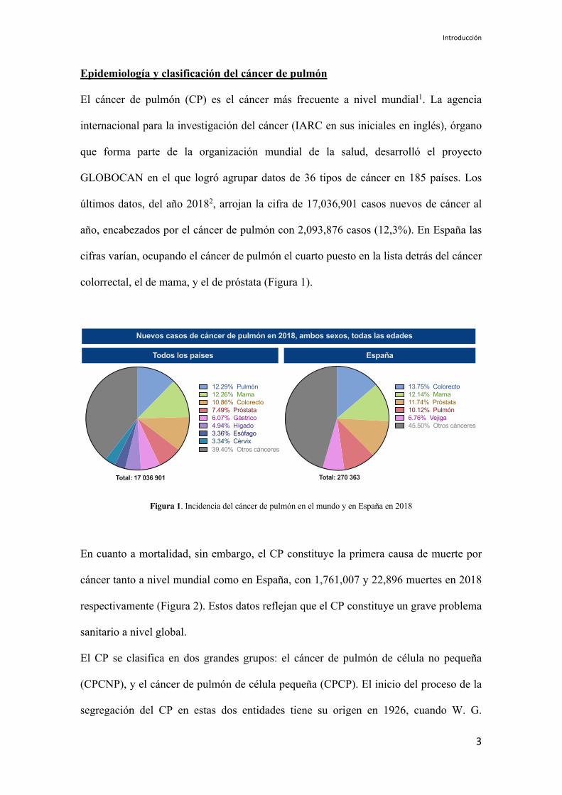
Epidemiología y clasificación del cáncer de pulmón
El cáncer de pulmón (CP) es el cáncer más frecuente a nivel mundial1. La agencia
internacional para la investigación del cáncer (IARC en sus iniciales en inglés), órgano
que forma parte de la organización mundial de la salud, desarrolló el proyecto
GLOBOCAN en el que logró agrupar datos de 36 tipos de cáncer en 185 países. Los
últimos datos, del año 20182, arrojan la cifra de 17,036,901 casos nuevos de cáncer al
año, encabezados por el cáncer de pulmón con 2,093,876 casos (12,3%). En España las
cifras varían, ocupando el cáncer de pulmón el cuarto puesto en la lista detrás del cáncer
colorrectal, el de mama, y el de próstata (Figura 1).
Figura 1. Incidencia del cáncer de pulmón en el mundo y en España en 2018
En cuanto a mortalidad, sin embargo, el CP constituye la primera causa de muerte por
cáncer tanto a nivel mundial como en España, con 1,761,007 y 22,896 muertes en 2018
respectivamente (Figura 2). Estos datos reflejan que el CP constituye un grave problema
sanitario a nivel global.
El CP se clasifica en dos grandes grupos: el cáncer de pulmón de célula no pequeña
(CPCNP), y el cáncer de pulmón de célula pequeña (CPCP). El inicio del proceso de la
segregación del CP en estas dos entidades tiene su origen en 1926, cuando W. G.
12.29% Pulmón12.26% Mama10.86% Colorecto7.49% Próstata6.07% Gástrico4.94% Hígado3.36% Esófago3.34% Cérvix39.40% Otros cánceres
Total: 17 036 901
13.75% Colorecto12.14% Mama11.74% Próstata10.12% Pulmón6.76% Vejiga45.50% Otros cánceres
Total: 270 363
Nuevos casos de cáncer de pulmón en 2018, ambos sexos, todas las edades
Todos los países España

Tesis doctoral – Max Hardy-Werbin
4
Barnard reportó por primera vez que los llamados sarcomas “oat-cell” del mediastino,
eran realmente tumores bronquiales3. Posteriormente fue Azzopardi en 19594 quien
distinguió este tipo de tumor de los adenocarcinomas y de los carcinomas de células
escamosas de pulmón, estableciendo al CPCP como una entidad propia.
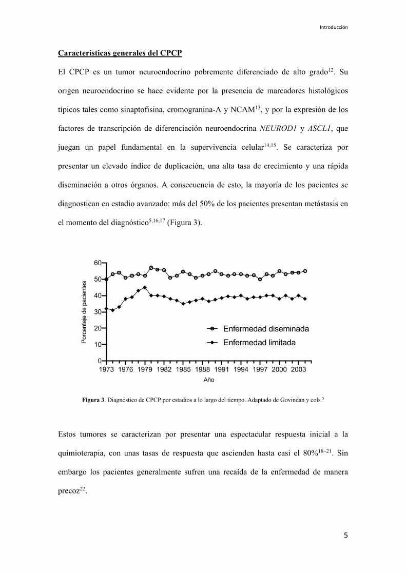
El CPCP constituye entre un 13 y un 15% de todos los casos de CP5,6 y está
estrechamente ligado al consumo tabáquico7,8. En 2014, Varghese y cols. publicaron la
serie de casos de CPCP en pacientes no fumadores más grande hasta la fecha9. En este
estudio se incluyeron 1040 pacientes con CPCP y solo el 2% (23 pacientes) de ellos
eran no fumadores. Sin embargo, en población exclusivamente asiática se han reportado
series con cifras de pacientes no fumadores con CPCP de entre un 13% y un 23%10,11.
Aunque la proporción del CPCP ha disminuido entre un 4 y un 5% en los últimos 20
años del siglo XX5, su incidencia ha ido en aumento de la mano del incremento global
de la incidencia del CP2. A esto se le suma el hecho de que la incidencia del diagnóstico
del CPCP se ha incrementado en las mujeres, llevando la ratio mujer : hombre a casi 1 a
15,6 (Figura 2).
Figura 2. Distribución del CPCP por sexo a lo largo del tiempo. Adaptado de Govindan y cols.5
1973 1976 1979 1982 1985 1988 1991 1994 1997 2000 20030
10
20
30
40
50
60
70
80
90
100
Año
Porc
enta
je d
e pa
cien
tes
Mujeres
Hombres
Introducción
5
Características generales del CPCP
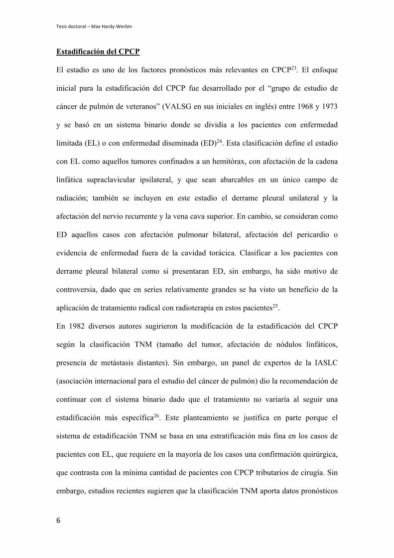
El CPCP es un tumor neuroendocrino pobremente diferenciado de alto grado12. Su
origen neuroendocrino se hace evidente por la presencia de marcadores histológicos
típicos tales como sinaptofisina, cromogranina-A y NCAM13, y por la expresión de los
factores de transcripción de diferenciación neuroendocrina NEUROD1 y ASCL1, que
juegan un papel fundamental en la supervivencia celular14,15. Se caracteriza por
presentar un elevado índice de duplicación, una alta tasa de crecimiento y una rápida
diseminación a otros órganos. A consecuencia de esto, la mayoría de los pacientes se
diagnostican en estadio avanzado: más del 50% de los pacientes presentan metástasis en
el momento del diagnóstico5,16,17 (Figura 3).
Figura 3. Diagnóstico de CPCP por estadios a lo largo del tiempo. Adaptado de Govindan y cols.5
Estos tumores se caracterizan por presentar una espectacular respuesta inicial a la
quimioterapia, con unas tasas de respuesta que ascienden hasta casi el 80%18–21. Sin
embargo los pacientes generalmente sufren una recaída de la enfermedad de manera
precoz22.
1973 1976 1979 1982 1985 1988 1991 1994 1997 2000 20030
10
20
30
40
50
60
Año
Por
cent
aje
de p
acie
ntes
Enfermedad diseminada
Enfermedad limitada
Tesis doctoral – Max Hardy-Werbin
6
Estadificación del CPCP
El estadio es uno de los factores pronósticos más relevantes en CPCP23. El enfoque
inicial para la estadificación del CPCP fue desarrollado por el “grupo de estudio de
cáncer de pulmón de veteranos” (VALSG en sus iniciales en inglés) entre 1968 y 1973
y se basó en un sistema binario donde se dividía a los pacientes con enfermedad
limitada (EL) o con enfermedad diseminada (ED)24. Esta clasificación define el estadio
con EL como aquellos tumores confinados a un hemitórax, con afectación de la cadena
linfática supraclavicular ipsilateral, y que sean abarcables en un único campo de
radiación; también se incluyen en este estadio el derrame pleural unilateral y la
afectación del nervio recurrente y la vena cava superior. En cambio, se consideran como
ED aquellos casos con afectación pulmonar bilateral, afectación del pericardio o
evidencia de enfermedad fuera de la cavidad torácica. Clasificar a los pacientes con
derrame pleural bilateral como si presentaran ED, sin embargo, ha sido motivo de
controversia, dado que en series relativamente grandes se ha visto un beneficio de la
aplicación de tratamiento radical con radioterapia en estos pacientes25.
En 1982 diversos autores sugirieron la modificación de la estadificación del CPCP
según la clasificación TNM (tamaño del tumor, afectación de nódulos linfáticos,
presencia de metástasis distantes). Sin embargo, un panel de expertos de la IASLC
(asociación internacional para el estudio del cáncer de pulmón) dio la recomendación de
continuar con el sistema binario dado que el tratamiento no variaría al seguir una
estadificación más específica26. Este planteamiento se justifica en parte porque el
sistema de estadificación TNM se basa en una estratificación más fina en los casos de
pacientes con EL, que requiere en la mayoría de los casos una confirmación quirúrgica,
que contrasta con la mínima cantidad de pacientes con CPCP tributarios de cirugía. Sin
embargo, estudios recientes sugieren que la clasificación TNM aporta datos pronósticos

Introducción
7
precisos que podrían modificar la estrategia terapéutica17,27 y se recomienda su
aplicación.
Factores pronósticos en el CPCP
Existen algunos factores que pueden estimar a priori el curso de la evolución del CPCP.
Se ha comprobado en diversos estudios que un buen estado funcional previo
(performance status 0-2), la edad inferior a 70 años, el sexo femenino y el estadio
localizado confieren un buen pronóstico al CPCP28–33. Otros estudios aislados han
identificado también como marcadores pronósticos relevantes la concentración sérica de
lactato deshidrogenasa (LDH), de albúmina y de creatinina, así como la cantidad de
sitios metastásicos34,35.
Tratamiento clásico del CPCP
La quimioterapia ha sido clásicamente el pilar fundamental en el tratamiento del CPCP.
A lo largo del tiempo, diferentes esquemas de tratamiento en monoterapia y con
combinación de fármacos han demostrado eficacia en el CPCP. Sin embargo, el
esquema más ampliamente utilizado y el que es considerado de elección es el doblete de
platino (cisplatino o carboplatino), con etopósido36 (PE), que reemplazó a los esquemas
previos basados en antraciclinas, dada su superioridad en cuanto a respuesta y mejor
perfil de toxicidad37. Sin embargo, en cuanto a eficacia, diversos ensayos clínicos
aleatorizados38–40 y un meta-análisis con más de 660 pacientes41 demostraron que ambos
esquemas presentan una eficacia equivalente, con similares tasas de respuesta,
supervivencia libre de progresión (SLP) y supervivencia global (SG).
En los casos con EL, el tratamiento de elección es la quimioterapia (PE) concomitante
con radioterapia42,43.
Tesis doctoral – Max Hardy-Werbin
8
Como se ha comentado previamente, una de las características del CPCP es su gran
respuesta inicial frente al tratamiento con quimioterapia. Sin embargo esto se continúa
de una rápida progresión al tratamiento, presentando una alta resistencia a tratamientos
subsiguientes44. Una vez que el paciente ha finalizado la primera línea de tratamiento, se
pueden distinguir tres tipos de escenarios45:
i) aquellos que mantienen una respuesta durante más de 3 meses
(enfermedad platino-sensible);
ii) aquellos que no consiguen respuesta alguna al tratamiento (enfermedad
refractaria);
iii) aquellos que, tras una respuesta inicial, progresan durante los primeros 3
meses tras completar la primera línea de tratamiento (enfermedad
platino-resistente).
En el último caso, la tasa de respuesta con terapias subsiguientes es de menos del 10%.
En los casos de pacientes que progresan tras mantener una respuesta duradera de más de
3 meses, se puede plantear reintroducir el mismo tratamiento inicial dada la sensibilidad
que se asume al platino. El único fármaco aprobado en segunda línea en CPCP en
nuestro medio por más de 20 años ha sido el topotecan46.
En estadio avanzado, recientemente un ensayo clínico fase 3 demostró un beneficio en
términos de SG y SLP con atezolizumab, un agente inmunoestimulador (anti-PD-L1) en
combinación con carboplatino y etopósido y mantenimiento con atezolizumab en
primera línea 47. Este estudio ha llevado a la aprobación del fármaco en Estados Unidos
y en Europa, y significa el primer gran cambio en el manejo de esta enfermedad en
primera línea.

Introducción
9
Por último, a diferencia del CPCNP, en el CPCP las terapias dirigidas no han
demostrado ningún beneficio en los diferentes ensayos clínicos realizados por lo que
ningún inhibidor molecular ha llegado a la clínica48,49.
Supervivencia
El CPCP es una enfermedad con mal pronóstico y con una supervivencia
extremadamente corta si no se trata50. En los últimos 40 años, solo dos ensayos clínicos
aleatorizados compararon el uso de tratamiento quimioterápico frente a placebo en
pacientes con CPCP51,52. En estos se pudo ver que la SG en pacientes que no recibían
tratamiento era de 2 a 3 meses en pacientes con ED y de 5 meses en pacientes con EL.
En EL, aquellos pacientes que reciben tratamiento con quimioterapia (PE) combinado
con radioterapia logran obtener una mediana de supervivencia de 24 a 30 meses, con
una supervivencia a los 5 años de entre el 25 y el 30%53. En ED, sin embargo, los
pacientes que realizan una primera línea de tratamiento presentan una mediana de
supervivencia de 7-12 meses, con una supervivencia a los 5 años que se sitúan en torno
al 3%6,16. Los pacientes que progresan tras haber completado una primera línea de
tratamiento suelen tener una SG que no supera los 4 o 5 meses de vida.
El sistema inmunológico
El sistema inmunológico se compone de dos sistemas íntimamente interconectados: el
sistema inmune innato y el sistema inmune adaptativo54. El primero se basa en un
sistema de protección rápida frente a elementos extraños, pero que no es específico ni
guarda memoria. Las células involucradas son macrófagos, granulocitos, células
dendríticas y células natural killer (NK). El segundo, por el contrario, ejecuta una
respuesta inmune frente a antígenos específicos que es más robusta y que confiere
Tesis doctoral – Max Hardy-Werbin
10
memoria inmunológica; esta se lleva a cabo básicamente con los linfocitos B y T. Estos
dos sistemas actúan de manera conjunta, entre otras cosas, para detectar la presencia de
un proceso tumoral incipiente y destruirlo antes de que sea clínicamente aparente55.
Prácticamente todas las células nucleadas expresan en su superficie el complejo mayor
de histocompatibilidad (CMH). El CMH tipo I se encarga de exhibir en la superficie
celular pequeños péptidos derivados de epítopos (tanto patógenos como tumorales),
para presentárselos a los linfocitos T CD8+. Una vez que la célula T ha reconocido el
antígeno presentado por el CMH tipo I, esta se activará, proliferará, y destruirá aquellas
células que presenten ese antígeno reconocido. EL CMH tipo II, por otro lado, se
expresa en células presentadoras de antígenos (macrófagos, células dendríticas), y
exhiben péptidos en la superficie para presentárselos a los linfocitos T CD4+56–58. De
esta manera se activarán los linfocitos T CD4+ helper, que a su vez reclutarán otras
poblaciones inmunológicas al tumor mediante la secreción sobretodo de citoquinas, y de
los linfocitos T citotóxicos (CD8+) que atacarán de manera directa y específica a las
células tumorales a través de la unión de su receptor (TCR) y el complejo mayor de
histocompatibilidad de las células tumorales59,60.
Inmunoterapia
La última década se ha visto marcada por el estudio de los llamados “puntos de control
inmunológico” y por el desarrollo de los fármacos que logran bloquearlos para permitir
al sistema inmune ejercer sus funciones sobre el tumor 61.
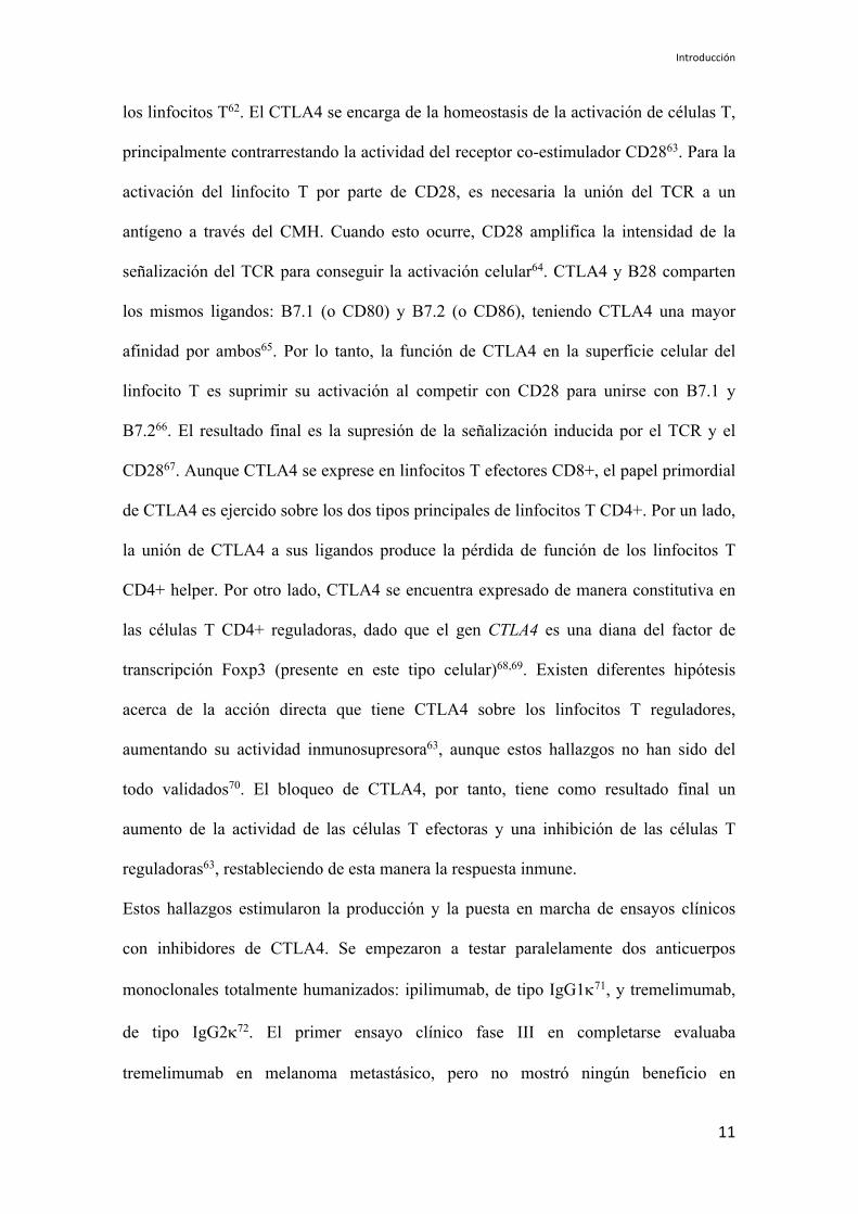
CTLA4
El primer punto de control inmunológico que se intentó bloquear fue el CTLA4
(cytotoxic T-lymphocyte antigen 4), un receptor con función inhibitoria, expresado en
Introducción
11
los linfocitos T62. El CTLA4 se encarga de la homeostasis de la activación de células T,
principalmente contrarrestando la actividad del receptor co-estimulador CD2863. Para la
activación del linfocito T por parte de CD28, es necesaria la unión del TCR a un
antígeno a través del CMH. Cuando esto ocurre, CD28 amplifica la intensidad de la
señalización del TCR para conseguir la activación celular64. CTLA4 y B28 comparten
los mismos ligandos: B7.1 (o CD80) y B7.2 (o CD86), teniendo CTLA4 una mayor
afinidad por ambos65. Por lo tanto, la función de CTLA4 en la superficie celular del
linfocito T es suprimir su activación al competir con CD28 para unirse con B7.1 y
B7.266. El resultado final es la supresión de la señalización inducida por el TCR y el
CD2867. Aunque CTLA4 se exprese en linfocitos T efectores CD8+, el papel primordial
de CTLA4 es ejercido sobre los dos tipos principales de linfocitos T CD4+. Por un lado,
la unión de CTLA4 a sus ligandos produce la pérdida de función de los linfocitos T
CD4+ helper. Por otro lado, CTLA4 se encuentra expresado de manera constitutiva en
las células T CD4+ reguladoras, dado que el gen CTLA4 es una diana del factor de
transcripción Foxp3 (presente en este tipo celular)68,69. Existen diferentes hipótesis
acerca de la acción directa que tiene CTLA4 sobre los linfocitos T reguladores,
aumentando su actividad inmunosupresora63, aunque estos hallazgos no han sido del
todo validados70. El bloqueo de CTLA4, por tanto, tiene como resultado final un
aumento de la actividad de las células T efectoras y una inhibición de las células T
reguladoras63, restableciendo de esta manera la respuesta inmune.
Estos hallazgos estimularon la producción y la puesta en marcha de ensayos clínicos
con inhibidores de CTLA4. Se empezaron a testar paralelamente dos anticuerpos
monoclonales totalmente humanizados: ipilimumab, de tipo IgG1k71, y tremelimumab,
de tipo IgG2k72. El primer ensayo clínico fase III en completarse evaluaba
tremelimumab en melanoma metastásico, pero no mostró ningún beneficio en
Tesis doctoral – Max Hardy-Werbin
12
comparación con quimioterapia72. Ipilimumab, sin embargo, fue el primer inhibidor de
CTLA4 en demostrar un beneficio en términos de supervivencia en pacientes con
melanoma, y a consecuencia de esto fue aprobado en 2011 por la FDA73.
Figura 4. Unión de CTLA4 con su ligando
PD-1
El otro punto de control inhibitorio que ha surgido como un candidato prometedor para
reactivar el sistema inmune es el PD-1 (programmed death-1). PD-1 es un receptor que
se expresa en la superficie de los linfocitos T y B una vez han sido activados74,75, y
posee dos ligandos: PD-L1 y PD-L276. Su función principal es el mantenimiento de la
tolerancia inmunológica periférica, para mantener la respuesta de células T dentro de un
rango fisiológico. PD-L1 y PD-L2 se expresan en presencia de mediadores
inflamatorios como IFNg77. Además, el PD-1 se encuentra ampliamente expresado en
los linfocitos T reguladores, y se ha reportado que en presencia de su ligando podría
aumentar su proliferación78. Cuando PD-1 se une a uno de sus ligandos, envía una señal

Introducción
13
inhibidora para atenuar la activación celular al bloquear la señalización del TCR79. El
bloqueo de PD-1, o PD-L1, por tanto, contribuiría a una reactivación de los linfocitos T
citotóxicos CD8+.
Para conseguir el bloqueo farmacológico de la unión de PD-1 con su ligando PD-L1 se
han desarrollado tanto fármacos anti-PD-1 (nivolumab, pembrolizumab) como anti-PD-
L1 (atezolizumab, durvalumab, avelumab). El primero en ser aprobado por parte de la
FDA fue nivolumab para melanoma metastásico en el año 201480 y posteriormente para
CPCNP en 201581.
Figura 5. Unión de PD-1 con sus ligandos
A continuación, se detalla una tabla con todos los inhibidores de punto de control
aprobados hasta octubre de 2019.

Tesis doctoral – Max Hardy-Werbin
14
Diana Fármaco Indicación Año de aprobación FDA CTLA-4 Ipilimumab
Melanoma 2011
Melanoma (adyuvancia) 2015 Melanoma pediátrico 2017
Ipi + nivo Melanoma BRAF wt 2015 Melanoma any BRAF 2016
Cáncer renal 2018 Cáncer colo-rectal MSI-high 2018
PD-1 Nivolumab Melanoma 2014 CPCNP 2015
Cáncer renal 2015 Linfoma de Hodgkin 2016
Ca escamoso de cabeza y cuello 2016 Carcinoma urotelial 2017
Cáncer colo-rectal MSI-high 2017 Carcinoma hepatocelular 2017 Melanoma (adyuvancia) 2017
CPCP tercera línea 2018 Pembrolizumab Melanoma 2014
CPCNP 2015 Ca escamoso de cabeza y cuello 2016
Linfoma de Hodgkin 2017 Carcinoma urotelial 2017
Cáncer gástrico y de la UGE 2017 Cáncer colo-rectal MSI-high 2017
Cáncer de cérvix 2018 Linfoma B de células grandes 2018
CPCNP (+ quimio) 2018 Carcinoma hepatocelular 2018
Carcinoma de células de Merkel 2018 Melanoma (adyuvancia) 2018 Cáncer renal (+ axitinib) 2019
Ca escamoso de cabeza y cuello 2019 CPCP segunda línea 2019
Carcinoma de esófago 2019 Carcinoma de endometrio (+lenvatinib) 2019
PD-L1 Atezolizumab Carcinoma urotelial 2016 CPCNP 2016
Cáncer de mama triple negativo 2019 CPCP primera línea (+ quimio) 2019
Avelumab Carcinoma de células de Merkel 2017 Carcinoma urotelial 2017
Cáncer renal 2019 Durvalumab Carcinoma urotelial 2017
CPCNP 2018
Tabla 1. Inhibidores de punto de control. Se describen los diferentes fármacos aprobados, y sus indicaciones por año de aprobación. MSI, microsatellite instability; wt, wild-type; Ca, carcinoma; UGE, unión gastro-esofágica.
Introducción
15
Toxicidad de los inhibidores de punto de control
A pesar de que los nuevos inhibidores de punto de control han cambiado el paradigma
del tratamiento del cáncer con resultados sin precedentes, no están exentos de toxicidad.
Los efectos adversos (EA) relacionados con estos tratamientos ocurren por un defecto
de la tolerancia inmune a consecuencia de la pérdida de la inhibición de las células T.
Estos EA pueden aparecer en cualquier órgano, pero lo hacen de manera más frecuente
a nivel digestivo, dermatológico, hepático o endocrino. En la mayoría de los casos, los
EA relacionados con la inmunoterapia son reversibles y manejables cuando se hace un
diagnóstico precoz, pero en algunos casos pueden llegar a ser fatales82–84.
Los puntos de control inmunológicos (CTLA4, PD-1) modulan la respuesta inmune a
diferentes niveles: mientras que CTLA4 controla el alcance de la respuesta inmune en
estadios tempranos de la activación de células T85, PD-1 actúa en etapas más tardías,
limitando la activación T en tejidos periféricos86,87. Estas diferencias podrían, en parte,
explicar los diferentes perfiles de toxicidad de cada uno de los inhibidores, según sea su
diana terapéutica. De hecho, la incidencia de EA severos (grados 3-5) en relación al uso
de ipilimumab puede llegar al 30% mientras no asciende del 15% con los agentes anti-
PD-1; cuando se combinan ambas terapias la cifra puede aumentar hasta un 55%88.
Los inhibidores de CTLA4 se han relacionado más con colitis, hipofisitis y rash,
mientras que los anti-PD-1 con neumonitis, alteraciones tiroides, artralgias y vitíligo89–
91. Cabe destacar que se ha observado que la toxicidad secundaria al bloqueo de CTLA4
es dosis dependiente, cosa que no ocurre con el bloqueo de PD-189.
Se han reportado casos de toxicidad neurológica, aunque estos EA ocurren en menos del
5% de los pacientes89, siendo el síntoma más frecuente la cefalea. Se pueden dar casos
de neuropatía periférica (sensitiva o motora), pero en menos del 1% de los
pacientes92,93. Otros cuadros neurológicos que se han reportado en forma de toxicidad

Tesis doctoral – Max Hardy-Werbin
16
secundaria a la inmunoterapia son casos de miastenia gravis y síndrome de Guillain-
Barré94,95. Por último, casos de toxicidad a nivel central incluyen meningitis aséptica,
encefalitis autoinmune, encefalopatía posterior reversible y mielitis transversa89,92,96,97.
Racional del rol de la inmunoterapia en el CPCP
Algunas características propias del CPCP lo hacen un candidato prometedor para
evaluar tratamientos con inmunoterapia98.
Síndromes paraneoplásicos
Existe evidencia acumulada que propone que el sistema inmunológico tiene un papel
fundamental en la fisiopatología del CPCP99–101. Se ha considerado un tumor muy
inmunogénico, y esto se debe sobre todo a la alta prevalencia de los síndromes
paraneoplásicos (SP), en especial los SP neurológicos (SPN) 102. Estos se producen
como consecuencia de la generación de una respuesta inmune contra antígenos
neuronales que se expresan de manera ectópica en las células tumorales pero de manera
fisiológica en el sistema nervioso central99,103,104. El motivo fundamental por el que el
CPCP está implicado en este mecanismo es que, al tener características
neuroendocrinas, expresa diversos antígenos neuronales105. El primero en identificar la
naturaleza inmunomediada de estos síndromes paraneoplásicos neurológicos fue el Dr.
Posner en la década de los ’80s al hallar en el líquido cefalorraquídeo de los pacientes
con cuadros neurológicos atípicos anticuerpos que se unían a antígenos onconeuronales
localizados tanto en el tumor como en las células de Purkinje106. El mecanismo
propuesto consiste en que las células tumorales apoptóticas son fagocitadas por células
dendríticas que migran hacia los ganglios linfáticos, donde se producirá una activación
tanto de células B como de células T. Los linfocitos B sufrirán posteriormente una
Introducción
17
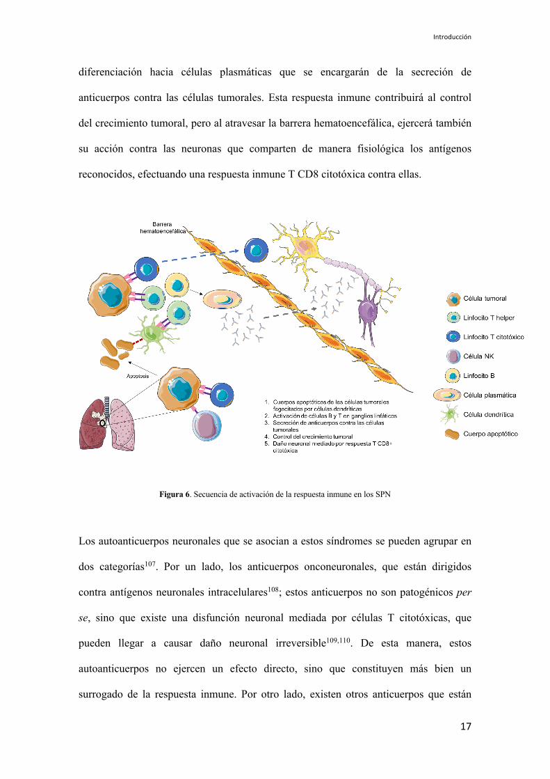
diferenciación hacia células plasmáticas que se encargarán de la secreción de
anticuerpos contra las células tumorales. Esta respuesta inmune contribuirá al control
del crecimiento tumoral, pero al atravesar la barrera hematoencefálica, ejercerá también
su acción contra las neuronas que comparten de manera fisiológica los antígenos
reconocidos, efectuando una respuesta inmune T CD8 citotóxica contra ellas.
Figura 6. Secuencia de activación de la respuesta inmune en los SPN
Los autoanticuerpos neuronales que se asocian a estos síndromes se pueden agrupar en
dos categorías107. Por un lado, los anticuerpos onconeuronales, que están dirigidos
contra antígenos neuronales intracelulares108; estos anticuerpos no son patogénicos per
se, sino que existe una disfunción neuronal mediada por células T citotóxicas, que
pueden llegar a causar daño neuronal irreversible109,110. De esta manera, estos
autoanticuerpos no ejercen un efecto directo, sino que constituyen más bien un
surrogado de la respuesta inmune. Por otro lado, existen otros anticuerpos que están
Tesis doctoral – Max Hardy-Werbin
18
dirigidos hacia proteínas ancladas en la membrana neuronal o que residen en el
complejo sináptico; estos anticuerpos generan un daño directo, provocando disfunción
neuronal al contactar directamente con el antígeno involucrado111,112.
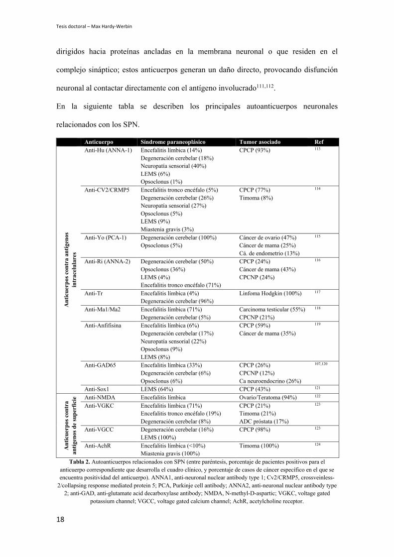
En la siguiente tabla se describen los principales autoanticuerpos neuronales
relacionados con los SPN.
Anticuerpo Síndrome paraneoplásico Tumor asociado Ref
Ant
icue
rpos
con
tra
antíg
enos
in
trac
elul
ares
Anti-Hu (ANNA-1) Encefalitis límbica (14%) Degeneración cerebelar (18%) Neuropatía sensorial (40%) LEMS (6%) Opsoclonus (1%)
CPCP (93%) 113
Anti-CV2/CRMP5 Encefalitis tronco encéfalo (5%) Degeneración cerebelar (26%) Neuropatía sensorial (27%) Opsoclonus (5%) LEMS (9%) Miastenia gravis (3%)
CPCP (77%) Timoma (8%)
114
Anti-Yo (PCA-1) Degeneración cerebelar (100%) Opsoclonus (5%)
Cáncer de ovario (47%) Cáncer de mama (25%) Cá. de endometrio (13%)
115
Anti-Ri (ANNA-2) Degeneración cerebelar (50%) Opsoclonus (36%) LEMS (4%) Encefalitis tronco encéfalo (71%)
CPCP (24%) Cáncer de mama (43%) CPCNP (24%)
116
Anti-Tr Encefalitis límbica (4%) Degeneración cerebelar (96%)
Linfoma Hodgkin (100%) 117
Anti-Ma1/Ma2 Encefalitis límbica (71%) Degeneración cerebelar (5%)
Carcinoma testicular (55%) CPCNP (21%)
118
Anti-Anfifisina Encefalitis límbica (6%) Degeneración cerebelar (17%) Neuropatía sensorial (22%) Opsoclonus (9%) LEMS (8%)
CPCP (59%) Cáncer de mama (35%)
119
Anti-GAD65 Encefalitis límbica (33%) Degeneración cerebelar (6%) Opsoclonus (6%)
CPCP (26%) CPCNP (12%) Ca neuroendocrino (26%)
107,120
Anti-Sox1 LEMS (64%) CPCP (43%) 121
Ant
icue
rpos
con
tra
antíg
enos
de
supe
rfic
ie Anti-NMDA Encefalitis límbica Ovario/Teratoma (94%) 122
Anti-VGKC Encefalitis límbica (71%) Encefalitis tronco encéfalo (19%) Degeneración cerebelar (8%)
CPCP (21%) Timoma (21%) ADC próstata (17%)
123
Anti-VGCC Degeneración cerebelar (16%) LEMS (100%)
CPCP (98%) 123
Anti-AchR Encefalitis límbica (<10%) Miastenia gravis (100%)
Timoma (100%) 124
Tabla 2. Autoanticuerpos relacionados con SPN (entre paréntesis, porcentaje de pacientes positivos para el anticuerpo correspondiente que desarrolla el cuadro clínico, y porcentaje de casos de cáncer específico en el que se encuentra positividad del anticuerpo). ANNA1, anti-neuronal nuclear antibody type 1; Cv2/CRMP5, crossveinless-
2/collapsing response mediated protein 5; PCA, Purkinje cell antibody; ANNA2, anti-neuronal nuclear antibody type 2; anti-GAD, anti-glutamate acid decarboxylase antibody; NMDA, N-methyl-D-aspartic; VGKC, voltage gated
potassium channel; VGCC, voltage gated calcium channel; AchR, acetylcholine receptor.
Introducción
19
Como se ha comentado previamente, el CPCP es el tumor que más se asocia a los SPN.
En un estudio longitudinal multicéntrico se incluyeron 968 pacientes diagnosticados de
un SPN107, de los cuales un 92% se asoció a un tumor. El tumor que más
frecuentemente se asoció fue el CPCP, acumulando casi un 40% de los casos. Dentro de
los pacientes diagnosticados con CPCP, la incidencia de SPN se ha estimado en series
pequeñas, obteniendo una frecuencia del 2-3%125–127. Sin embargo, un estudio más
detallado involucrando neurólogos de Gozzard y cols., evaluó la aparición de SPN en
264 pacientes con CPCP, hallando una prevalencia del 9.1%128, sugiriendo que existen
numerosos casos que no se diagnostican. En la mayoría de los casos (entre en 65 y el
70%) los síntomas neurológicos secundarios al síndrome paraneoplásico aparecen antes
de la detección del tumor129,130.
Relación con hábito tabáquico
Se estima que el cáncer de pulmón asociado a tabaco se desarrolla tras haber fumado
aproximadamente 50 paquetes-año (esto es, por ejemplo, 1 paquete diario durante 50
años, o bien 2 paquetes diarios durante 25 años)131. Las alteraciones más habituales que
diferencian la población no fumadora de la fumadora son mutaciones en TP53 y KRAS.
Asimismo el espectro mutacional en estos pacientes es mucho más amplio132–134.
En 2010 se publicó un estudio 135 donde realizaban secuenciación masiva del ADN de
una línea celular derivada de una metástasis de médula ósea de CPCP, para analizar las
firmas mutacionales generadas por la exposición a los carcinógenos del humo del
tabaco. Identificaron más de 22,000 mutaciones somáticas, de las cuales 134 se
encontraban en exones codificantes. Posteriormente en 2013 se reportó que el CPCP
presentaba una de las cargas mutacionales tumorales (TMB, en sus cifras en inglés) más
elevadas (después del melanoma, del CPCNP y del cáncer de vejiga), con dos firmas

Tesis doctoral – Max Hardy-Werbin
20
mutacionales validadas, una de las cuales se encontraba presente también en otros
tumores asociados al tabaco136.
Por último, en 2015 se publicaron los datos de secuenciación masiva donde se analizó el
perfil genómico de 110 muestras de CPCP. En este estudio se corroboró la elevada
TMB, con una media de 8.62 mutaciones no-sinónimas por megabase de ADN
analizado137.
En diferentes estudios en cáncer urotelial, melanoma y CPCNP se ha observado una
asociación entre una elevada TMB y una mayor eficacia a la inmunoterapia138–144. En el
contexto del CPCP, el ensayo clínico Checkmate 032145 evaluó la eficacia de nivolumab
en monoterapia y en combinación con ipilimumab en enfermedad metastásica
refractaria a tratamiento de primera línea. En un subanálisis posterior de este estudio 146
se realizó la secuenciación del exoma de las muestras de los pacientes para ver si en
CPCP se cumplía la relación entre la carga mutacional y la eficacia de la inmunoterapia.
Se observó que en los pacientes tanto tratados con nivolumab en monoterapia como con
la combinación con ipilimumab, que presentaban un TMB elevado, se obtenía una
respuesta objetiva superior (21.3% y 46.2%, respectivamente) que en aquellos pacientes
con una TMB media (6.8% y 16%, respectivamente) o baja (4.8% y 22.2%,
respectivamente).
En conclusión, el CPCP se relaciona íntimamente con el consumo tabáquico, que a su
vez le confiere una alta carga mutacional, factor que se relaciona con una mejor
respuesta al tratamiento con inmunoterapia en los datos retrospectivos disponibles.
Inmunoterapia en el CPCP
En los últimos diez años se ha evaluado la eficacia de la inmunoterapia en el CPCP con
diferentes fármacos y en diferentes esquemas de tratamiento. El primer ensayo clínico

Introducción
21
(EC) que lo hizo incluía tanto pacientes con CPCP como con CPCNP con enfermedad
avanzada, pero contaba con capacidad para poder analizar ambas cohortes de pacientes
por separado147. Se randomizaba a los pacientes para que recibieran quimioterapia
(platino + paclitaxel) con placebo o con ipilimumab en dos modos diferentes:
concomitante con la quimioterapia, o de manera secuencial. Se observó que la adición
de ipilimumab (se manera secuencial) ofrecía un aumento en la SG en pacientes que
recibían ipilimumab tras la quimioterapia, pero la diferencia con el brazo control no
alcanzó la significación estadística. Posteriormente, el fase III confirmatorio, específico
para CPCP fue negativo al no poder demostrar un beneficio ni en SG ni en SLP al
añadir ipilimumab a la quimioterapia estándar (PE)148. Sin embargo, casi paralelamente
a este último ensayo clínico, un EC fase II 149 de un único brazo que evaluaba
ipilimumab en combinación con PE en primera línea de tratamiento, reportó una SG que
ascendía hasta los 17 meses. Estos ensayos plantearon la premisa de que habría algunos
pacientes con CPCP que se benefician de la inmunoterapia con ipilimumab.
En 2013 se comenzaron a reclutar pacientes para el Checkmate-032145. En este ensayo
se trataron un total de 216 pacientes con nivolumab en monoterapia o con la
combinación de nivolumab e ipilimumab en dos diferentes dosis en pacientes con
enfermedad refractaria al doblete de platino. En un análisis preliminar se observó una
SG a los dos años de un 14% en la cohorte de pacientes tratados con nivolumab en
monoterapia, y de un 26% en pacientes tratados con nivolumab + ipilimumab150. Esto
llevó a la inclusión de nivolumab con o sin ipilimumab como una recomendación de
tratamiento de segunda línea en las guías americanas de oncología de la NCCN en 2017.
Posteriormente, basándose en las respuestas duraderas observadas en este mismo EC en
el subgrupo de 109 pacientes tratados con nivolumab a 3mg/kg (tasa de respuesta del
11.9% y una duración de respuesta de 6 meses o más en el 77% de los pacientes, y de

Tesis doctoral – Max Hardy-Werbin
22
12 meses o más en el 62% de los pacientes), se obtuvo la aprobación de nivolumab en
tercera línea por parte de la FDA.
Posteriormente se testó nivolumab en otros dos ensayos clínicos, ambos negativos. El
Checkmate-331 fue un EC fase III que evaluó nivolumab en segunda línea de
tratamiento en comparación con el tratamiento estándar (topotecan)151 y no pudo
cumplir el objetivo primario de SG. Sin embargo, la presencia de algunos largos
supervivientes con respuestas duraderas a nivolumab apoyaba el papel de este fármaco
en segunda línea de tratamiento. El otro EC fue el Checkmate 451152 que evaluó el
papel de la inmunoterapia en mantenimiento tras una primera línea de tratamiento.
Consistía en dos brazos de tratamiento tras quimioterapia estándar: nivolumab +
ipilimumab vs nivolumab monoterapia. No hubo diferencias entre ambos brazos.
Pembrolizumab se ha evaluado en cuatro EC en CPCP. El primero fue un basket trial153
en el que se trataron 16 pacientes con CPCP PD-L1+ con pembrolizumab en
monoterapia en segunda línea de tratamiento. Se obtuvo una tasa de respuesta del 25%
con respuesta duraderas de más de 4 meses. Tras este estudio se llevó a cabo el Keynote
158154, evaluando pembrolizumab en monoterapia y estratificando los pacientes según
la expresión de PD-L1. En los pacientes con PD-L1 positivo se observó una SG de 14.6
meses y una tasa de respuesta del 37.5%, mientras que en el subgrupo de pacientes PD-
L1 negativo la SG fue de 7.7 meses, con una tasa de respuesta del 6%. Posteriormente
un pequeño fase II155 con 26 pacientes con CPCP testó pembrolizumab en segunda línea
de tratamiento en combinación con paclitaxel obteniendo una tasa de respuesta del 23%.
Por último, se llevó a cabo un EC fase II con pembrolizumab de mantenimiento en
aquellos pacientes que no habían progresado a PE en primera línea. Este fue negativo,
con una SLP de 1.4 meses156. Recientemente, al analizar los 83 pacientes con CPCP
incluidos en los EC Keynote 028 y Keynote 158, se observó una tasa de respuesta del

Introducción
23
29%, con un 61% de los pacientes que mostraron una duración de respuesta superior a
18 meses. Estos datos motivaron la aprobación de Pembrolizumab en segunda línea de
tratamiento en CPCP por parte de la FDA157.
El anti-PD-L1 atezolizumab fue evaluado en dos ensayos clínicos en CPCP. El primero
fue un fase II que comparaba atezolizumab vs topotecán en segunda línea de tratamiento
y fue negativo, con una TR del 2.3% vs 10% respectivamente158. El segundo EC fue el
IMpower-13347, un fase III que evaluaba la adición de atezolizumab a quimioterapia
estándar (PE) frente a placebo en primera línea de tratamiento. Se observó una SG de
12.3 meses en los pacientes que habían recibido atezolizumab + quimioterapia y de 10
meses en aquellos que había recibido quimioterapia sola, y esta diferencia fue
estadísticamente significativa; la SLP fue de 5.2 meses vs 4.3 meses respectivamente.
La tasa de respuesta fue de 60.5% vs 64.4% respectivamente. Este resultado llevó a la
aprobación de este esquema de tratamiento como primera línea en CPCP por parte de la
FDA y EMA, consiguiendo cambiar el estándar de tratamiento por primera vez en más
de 35 años.
El otro anti-PD-L1 que se está testando en CPCP es durvalumab; actualmente se ha
llevado a cabo un EC fase III de tres brazos comparando durvalumab +/- tremelimumab
(anti-CTLA4) concomitante con quimioterapia vs quimioterapia sola en primera línea
de tratamiento. Recientemente se han reportado los datos correspondientes a la
comparativa entre el grupo de durvalumab + quimioterapia vs quimioterapia sola159. En
el brazo de la combinatoria se observo una SG de 13 meses, frente a 10.3 meses en el
grupo de quimioterapia sola, y está diferencia fue significativamente estadística; la
supervivencia media a los 18 meses fue de 34% vs 25% respectivamente.

Tesis doctoral – Max Hardy-Werbin
24
Ensayo clínico Fase Línea Tratamiento N Resultados Reck y cols. NCT00527735 2008
II 1ra Ipilimumab + Platino + Paclitaxel Placebo + Platino + Paclitaxel 130
OS 12.9m vs 9.9 P=0.13
Arriola y cols. NCT01331525 2011
II 1ra Ipilimumab + PE 39 PFS 6.9m, OS 17m
Reck y cols. NCT01450761 2011
III 1ra Ipilimumab + Platino-Etopósido Placebo + Platino-Etopósido 1132
OS 11m vs 10.0 P=0.37
Checkmate-032 NCT01928394 2013
I/II 2/3era Nivolumab +/- Ipilimumab (1 or 3 mg/kg) 216
ORR 11.9% nivo, 23% nivo-ipi 3, 18% nivo-ipi 1
Checkmate-331 NCT02481830 2015
III 2da Nivolumab vs topotecán/amrubicina 569
OS 7.5 vs 8.4 P=0.11
Checkmate-451 NCT02538666 2015
III 1era Mantenimiento con nivolumab 1mg/kg + ipilimumab 3mg/kg or nivolumab 1mg/kg vs placebo
834
OS 9.2 vs 9.6 (nivo/ipi vs placebo) OS 10.4 vs 9.6 (nivo vs placebo)
Keynote-028 NCT02054806 2014
Ib 2da Pembrolizumab en CPCP PD-L1+ 16 ORR 25%
Keynote-158 NCT02628067 2015
II 2da Pembrolizumab 107 ORR 18.7% (37.5% en los PD-L1+ y 6% en los PD-L1-)
MISP-MK3475 NCT02551432 2015
II 2da Pembrolizumab + Paclitaxel 26 ORR 23.1%
Gadgeel y cols. NCT02359019 2015
II 1era Pembrolizumab mantenimiento 45 Negative PFS 1.4m, OS 9.6m
IFCT-1603 NCT03059667 2017
II 2da Atezolizumab Topotecán 73
ORR 20.9% vs 65%
IMpower-133 NCT02763579 2016
III 1era Atezolizumab + Platino-Etopósido Placebo + Platino-Etopósido 403
OS 12,3 vs 10,3 p=0.007
Caspian NCT03043872 2017
III 1era
Durvalumab + Tremelimumab + PE Durvalumab + PE Placebo + PE
988
OS 13 vs 10.3m p=0.0047
Tabla 3. Ensayos clínicos en inmunoterapia en CPCP

Introducción
25
Biomarcadores de respuesta a inmunoterapia
Los inhibidores de punto de control han demostrado ser eficaces y aportar un beneficio
en CPCP tanto en monoterapia y en combinación, como asociados a quimioterapia. Sin
embargo, por un lado, este beneficio se observa solo en un grupo de pacientes, y por el
otro, en determinados casos se han asociado a un perfil de toxicidad específico: por eso
la determinación de marcadores tanto predictivos de respuesta como de toxicidad es
fundamental. En particular, ipilimumab solo o en combinación con nivolumab, ha
demostrado en los diversos estudios añadir beneficio y contribuir a la existencia de
largos supervivientes. Sin embargo, su desarrollo en esta patología está en duda debido
a las dificultades de identificar los pacientes que se benefician de esta estrategia.
Biomarcadores asociados con eficacia
Existen múltiples biomarcadores potenciales de beneficio frente a ipilimumab y agentes
anti-PD-1/PD-L1 que se han reportado en diferentes estudios.
Recuento de células sanguíneas circulantes
Linfocitos. En diferentes estudios se ha observado que en pacientes tratados con anti-
CTLA4 se produce un aumento de los linfocitos circulantes160, y que esto se asocia con
mejores respuestas al tratamiento en pacientes con melanoma71,161. De hecho en un
estudio realizado en cooperación entre Holanda y el Reino Unido, se observó que cifras
elevadas de linfocitos circulantes antes de empezar el tratamiento, se relacionaban con
una SG más prolongada162. En otro estudio, en pacientes con melanoma tratados con
ipilimumab se observó que una disminución del ratio neutrófilo/linfocito por debajo de
la media durante el curso de tratamiento, se relacionaba con una mejor SG163.
Tesis doctoral – Max Hardy-Werbin
26
Neutrófilos. Se ha observado que en pacientes con melanoma tratados con ipilimumab,
cifras elevadas de neutrófilos antes de iniciar el tratamiento se correlacionan con una
peor SG164,165.
Eosinófilos. Los datos referentes a la relación entre el recuento de eosinófilos y eficacia
de la inmunoterapia difieren entre los diferentes inhibidores. En una cohorte de
pacientes con melanoma tratados con ipilimumab se observó que un recuento bajo de
eosinófilos antes de iniciar el tratamiento se asociaba con una SG superior166. Sin
embargo en pacientes tratados con pembrolizumab, un recuento bajo de eosinófilos se
relacionó con una peor supervivencia167.
Fenotipado de PBMCs (células sanguíneas circulantes periféricas)
Células T CD4+ ICOShi . ICOS es un receptor co-estimulador presente en las células
T168. Se ha observado un aumento de células T CD4+ ICOShi tanto en sangre periférica
como en el microambiente tumoral en pacientes con cáncer de vejiga, de mama y de
próstata tratados con inhibidores de CTLA4169–171. En una pequeña cohorte de 6
pacientes con cáncer de vejiga, se ha reportado que niveles mantenidos de CD4+ ICOShi
durante el tratamiento con ipilimumab se asociaba con mejores respuestas y una SG más
prolongada172.
Células T reguladoras. Los datos relativos a la modulación de los porcentaje de Tregs
y su asociación con eficacia en pacientes tratados con ipilimumab son
contradictorios173. Por un lado, algunos estudios demostraron un descenso de Tregs
tanto circulatorios como peritumorales tras tratamiento con ipilimumab170,174,175,
mientras que otros no evidenciaron esta variación172,176,177. Por otro lado, si bien algunos
estudios reportaron que el descenso de los Tregs tras tratamiento con ipilimumab se
Introducción
27
asociaba a una mejor supervivencia178, otros estudios asociaban un aumento de la SLP
con un aumento en el número de estos179.
Monocitos CD16+. En un análisis de las PBMCs de 29 pacientes con melanoma
tratados con ipilimumab se evaluó si la cantidad de monocitos tenía relación con datos
de eficacia180. Se observó que ipilimumab era capaz de estimular monocitos CD16+ (y
no los monocitos clásicos CD14+CD16-) que a su vez eran capaces de provocar una
depleción de las células T CD4+ reguladoras. Estos hallazgos se relacionaron con una
mejor respuesta al tratamiento.
Células mieloides supresoras. Las células mieloides supresoras (MDSC, en sus
iniciales en inglés) constituyen una estirpe celular heterogénea que se expande bajo
estados patológicos como puede ser una infección o el cáncer y que tienen una función
supresora, sobretodo sobre células T181. Se ha demostrado en diversos estudios que en
pacientes tratados con ipilimumab, existe una reducción en el recuento de las MDSCs
tras el tratamiento179,182. En pacientes con melanoma tratados con el anti-CTLA4 se
observó que en aquellos pacientes donde había una reducción de MDSCs tras
tratamiento tenían una SG183,184 y una SLP más prolongadas179.
Diversidad del repertorio de células T
En un estudio con 12 pacientes con melanoma metastásico tratados con ipilimumab se
analizó el espectro de receptores TCR y se observó que una mayor diversidad de células
T antes del tratamiento se relacionaba con una mayor tasa de respuesta185. Otro estudio
que evaluaba parámetros similares en pacientes con melanoma y cáncer de próstata,
reportó que una estabilidad en el clonotipo del receptor TCR, antes y después del
tratamiento con anti-CLTA4 se asociaba con una mejor supervivencia186.

Tesis doctoral – Max Hardy-Werbin
28
Respuesta celular y humoral frente a antígenos tumorales
El antígeno NY-ESO-1 es un antígeno tumoral que se encuentra en diferentes tumores
sólidos187. Se ha observado que la presencia de anticuerpos dirigidos contra NY-ESO-1
en pacientes con melanoma antes de iniciar tratamiento con ipilimumab, se asociaba con
una mejor respuesta al tratamiento. Por otro lado, los pacientes que eran seropositivos
para el antígeno, alcanzaban una mayor SG si se detectaban células T CD8+ dirigidas
frente a este188.
Receptor soluble CD25
El CD25 está presente en las Tregs y es la proteína receptora de IL-2189. Se ha
observado que pacientes tratados con ipilimumab eran resistentes al tratamiento cuando
presentaban niveles altos de CD25 soluble pre tratamiento190.
Citoquinas
Las citoquinas cumplen un papel fundamental en el estado de activación inmunitaria y
algunas de ellas han demostrado tener un papel pronóstico en CPCNP191,192. En
pacientes con melanoma tratados con ipilimumab, se ha observado que unos valores
basales elevados de CXCL11 se asociaban a peores respuestas193.
Lactato deshidrogenasa (LDH)
En diferentes estudios en pacientes con melanoma metastásico se ha visto que una LDH
sérica elevada antes de iniciar el tratamiento con anti-CTLA4 se asociaba a una
resistencia al tratamiento161,162,194.

Introducción
29
Proteína C Reactiva (PCR)
La PCR es un marcador de carga tumoral y se ha relacionado con el pronóstico de
diversos tipos de cánceres195. En un estudio retrospectivo se observó que pacientes con
melanoma tratados con ipilimumab (n=196) presentaban una mejor OS si se detectaba
una PCR dentro de cifras normales antes de iniciar el tratamiento. En otro estudio se
observó que una descenso de las cifras de PCR en relación al inicio del tratamiento con
ipilimumab se relacionaban con una mejor SLP y OS en melanoma avanzado178.
Linfocitos infiltrantes
La presencia de linfocitos infiltrantes de tumor es un factor de buen pronóstico en varios
tipos de cánceres196,197. En pacientes con melanoma metastásico, un aumento en el
recuento de linfocitos peritumorales se ha relacionado con una mejor tasa de respuesta
en pacientes tratados con ipilimumab198 y con pembrolizumab199. En biopsias post-
tratamiento con ipilimumab, un aumento de granzima B (proteasas citotóxicas
contenidas en gránulos de linfocitos T CD8 efectores) se relacionó con respuesta al
tratamiento en pacientes con melanoma metastásico199.
Carga mutacional tumoral
El término ‘carga mutacional tumoral’ (TMB) hace referencia al número de mutaciones
no-sinónimas presentes en el exoma de un tumor136. La cantidad de éstas es
proporcional a la generación de péptidos inmunogénicos y por eso se relaciona con la
respuesta de los inhibidores de puntos de control inmunológicos en los tumores. Los
primeros estudios que demostraron una relación entre una elevada TMB y respuesta a
inmunoterapia evaluaba pacientes con melanoma metastásico y observaron que aquellos
pacientes que presentaban un beneficio duradero a ipilimumab presentaban una TMB

Tesis doctoral – Max Hardy-Werbin
30
pre-tratamiento superior200,201. Posteriormente se vieron asociaciones similares en
pacientes con CPCNP tratados con Pembrolizumab202. Tras estas observaciones
iniciales en melanoma y CPCNP, se fueron reportando asociaciones significativas entre
una elevada TMB y respuesta a inhibidores de punto de control en diversos tipos de
cánceres 141,203. Concretamente en CPCP, como se ha mencionado previamente, se ha
observado que la eficacia de nivolumab con y particularmente con ipilimumab
aumentaba en pacientes con una elevada TMB146.
Expresión de PD-L1
Expresión tumoral. El mecanismo de acción de los fármacos anti-PD-1 y anti-PD-L1
ha llevado a varios grupos a intentar determinar si la expresión de PD-L1 en el tumor se
correlacionaba con la eficacia de los inhibidores. Se ha reportado una relación entre la
expresión de PD-L1 y la respuesta frente a inhibidores de PD-1 en melanoma y CPCNP
204,205. Aunque se ha estimado que la expresión de PD-L1 en células tumorales en CPCP
es baja, diferentes estudios han reportado porcentajes que van desde 2.5 a 51.8%206–209.
En el Checkmate 032145, donde se evaluó nivolumab en monoterapia o en combinación
con ipilimumab en CPCP con enfermedad diseminada refractario a tratamiento, se
observó una expresión de PD-L1 del 17%. En este estudio no se observó una
correlación entre PD-L1 en el tumor y respuesta a inhibidores de punto de control. Sin
embargo, en un fase II con pembrolizumab en mantenimiento en CPCP con enfermedad
diseminada156, se observó que aquellos pacientes con expresión de PD-L1 tenían una
SG más prolongada; en este estudio en lugar de llevar a cabo la evaluación de PD-L1
mediante el sistema de puntuación de células tumorales (tumor proportion score)205, se
utilizó un sistema de puntuación combinado (combined proportion score), que se basa
en el número de células PD-L1 positivas (incluyendo tumor, linfocitos y macrófagos),

Introducción
31
en relación al número total de células tumorales210. Esto podría explicar las diferencias
en el valor predictivo de PD-L1.
Expresión PD-L1 en linfocitos circulantes. En pacientes con melanoma tratados con
ipilimumab, una expresión elevada de PD-L1 en células T CD4+ y CD8+ se asociaron
con una peor SLP y SG211.
Indolamina 2,3-dioxigenasa (IDO)
La IDO es una enzima que degrada el triptófano212; se ha observado que tiene una
acción inmunosupresora al disminuir la proliferación de células T213. Se ha reportado
una asociación entre la expresión de IDO en tumor y respuesta a ipilimumab en
melanoma metastásico198.
Perfiles de expresión génica
Se ha observado que el tratamiento con anti-CTLA4 induce un perfil de expresión
génica relacionado con la proliferación de células T 186. Al analizar el transcriptoma de
45 pacientes con melanoma, se observó que un patrón relacionado con genes inmunes
era predictivo de respuesta a ipilimumab214.
Microbiota intestinal
En los últimos años se ha documentado que la microbiota intestinal tiene un efecto
sobre la respuesta anti-tumoral215. En modelos de ratones con sarcoma, melanoma y
cáncer de colon se ha observado que el bloqueo de CTLA-4 no era efectivo en aquellos
ratones libres de gérmenes, o bien en aquellos ratones que habían sido tratados con
antibióticos de amplio espectro previamente216. Esta falta de respuesta anti-tumoral se
solventaba por transferencia de Bacteroides Fragilis o mediante células T específicas
Tesis doctoral – Max Hardy-Werbin
32
frente a este germen. Sin embargo, en un estudio con 26 pacientes con melanoma
metastásico tratados con ipilimumab analizaron la composición de la microbiota y
observaron que los pacientes con un predominio de Faecalibacterium genus en las
muestras previas a iniciarse el tratamiento tenían una mejor SLP y SG que aquellos con
un predominio de Bacterioides217.
Toxicidad secundaria a inmunoterapia como predictor de respuesta
Se han publicado diferentes trabajos que sugieren que la aparición de EA relacionados
con la inmunoterapia se podría asociar a respuesta al tratamiento con ipilimumab218–221.
Sin embargo, esta asociación no es directa, y existen muchos pacientes que responden al
tratamiento sin sufrir EA222.
Biomarcadores asociados con toxicidad
Interleukina-17 (IL-17)
En un estudio con 52 pacientes con melanoma tratados con ipilimumab, se observaron
13 casos de colitis. En estos pacientes, los valores de IL-17 fueron significativamente
superiores durante el evento de colitis, y la resolución del cuadro se asoció a una
normalización de sus valores223.
Perfil de expresión génica
Un aumento de la expresión de CD177 y CEACAM1, marcadores de activación de
neutrófilos, en pacientes con melanoma tratados con ipilimumab, se asoció a EA
gastrointestinales inmunomediados224.

Introducción
33
Microbiota intestinal
En un estudio de 26 pacientes con melanoma metastásico, se ha observado que aquellos
pacientes con una microbiota enriquecida con Firmicutes antes de iniciar el tratamiento
con ipilimumab, tenían una probabilidad más alta de desarrollar colitis217.
Importancia de los marcadores en sangre periférica en CPCP
Uno de los mayores problemas en el diagnóstico del CPCP es el acceso limitado al
tejido tumoral, dado que se diagnostica a partir de pequeñas biopsias o citologías en más
del 90% de los casos225. Tras el diagnóstico inicial, el tejido remanente la mayoría de las
veces es escaso, lo cual constituye una limitación para análisis subsiguientes. Una
evidencia de este hecho es el tejido disponible para análisis de biomarcadores en
ensayos clínicos con inmunoterapia en CPCP. Por ejemplo, Hellmann y cols.146
analizaron la TMB en muestras de tumor del EC Checkmate 032145: solo el 61% de los
pacientes tenían disponible muestras pareadas de tumor y sangre, y de estos solo el 86%
fue apto para análisis (por lo tanto, sólo el 53% del total). Por este motivo, la búsqueda
de biomarcadores en sangre periférica tiene un especial interés en el CPCP.
Con el estudio de muestras de sangre periférica se logra también, desde una perspectiva
técnica, la obtención de muestras de una manera poco cruenta y de manera repetida en
el tiempo a lo largo de la enfermedad. Por otro lado, desde una perspectiva más
académica, se puede obtener una visión amplia del efecto del tumor sobre el sistema
inmunológico.

Tesis doctoral – Max Hardy-Werbin
34
Ensayo clínico ICE
La relevancia de este ensayo clínico dentro del actual proyecto radica en que sus
resultados fueron los que llevaron a la formulación de la hipótesis que se describe a
continuación. Las muestras de los pacientes del ensayo clínico ICE forman parte del
sustrato que se analizará en el trabajo de la tesis actual y serán un punto de partida
fundamental para realizar las comparaciones correspondientes e inferir las conclusiones.
El ensayo clínico ICE (Ipilimumab-Carboplatino-Etoposido) 149 fue el primer estudio en
el que se evaluaron marcadores de respuesta a inmunoterapia en CPCP. Fue un ensayo
fase II de un brazo único que incluyó 42 pacientes con CPCP con enfermedad
diseminada que aún no habían recibido tratamiento. El objetivo era evaluar la eficacia y
toxicidad de la combinación de ipilimumab (10 mg/kg) con carboplatino y etopósido.
Posteriormente se llevó a cabo un análisis exploratorio para la búsqueda de
biomarcadores tanto de respuesta como de toxicidad, mediante la evaluación de
autoanticuerpos en suero (autoanticuerpos anti-membrana y anti-nucleares).
Se observó que con la combinación de quimioterapia con ipilimumab se obtenía un
beneficio, pero a cambio de una gran toxicidad. Se observaron EA grado 3 relacionados
con ipilimumab en el 69% de los pacientes; el 8% de los pacientes experimento un EA
neurológico relacionado con ipilimumab. Dos pacientes sufrieron síntomas neurológicos
que imitaban un SPN, en forma de encefalopatía y síndrome cerebelar, respectivamente.
Se observó que aquellos pacientes que habían presentado EA de grado 3 o superior
relacionados con ipilimumab tenían una peor supervivencia, aunque esta asociación no
alcanzó la significación estadística.
En este estudio se ha identificado un subgrupo de pacientes largos supervivientes (>12
meses, incluyendo pacientes con supervivencias superiores a 30 meses) que contrasta

Introducción
35
con la mediana de supervivencia de 9 meses en esta población tratada con
quimioterapia.
Por último, en el análisis de biomarcadores se observó que en los pacientes donde se
detectaba por lo menos la presencia de un autoanticuerpo en suero, tenían una tendencia
a una mejor respuesta.

Hipótesis y objetivos

Hipótesis y objetivos
37
2. HIPÓTESIS Y OBJETIVOS

Tesis doctoral – Max Hardy-Werbin
38

Hipótesis y objetivos
39
Hipótesis
El tratamiento con ipilimumab ha demostrado eficacia en forma de largos
supervivientes en pacientes con CPCP avanzado. Sin embargo, su aplicación clínica en
esta enfermedad está en duda debido a los ensayos clínicos negativos en población no
seleccionada. La identificación de biomarcadores de beneficio de ipilimumab en CPCP
nos permitirá la adecuada selección de pacientes para dicho tratamiento, pudiendo
ofrecer ipilimumab a los pacientes que se beneficiarán de manera relevante y
minimizando de esta manera un coste en toxicidad e ineficacia en los pacientes que no
se benefician. El uso de muestras de sangre periférica permitirá llevar a cabo esta
valoración de una manera eficiente, cómoda y con posibilidad de aplicación en el
seguimiento y en la detección de resistencias.
Objetivo principal
Caracterización de los eventos inmunológicos diferenciales en pacientes con CPCP
tratados con quimioinmunoterapia (carboplatino + etopósido + ipilimumab) con
beneficio del tratamiento vs aquellos sin beneficio en muestras seriadas, y su
comparación con pacientes tratados con quimioterapia sola (carboplatino + etopósido).
Objetivos concretos:
1. Evaluación de la prevalencia de los AAN en CPCP y estudio de su modulación
en pacientes tratados con quimioinmunoterapia y con quimioterapia sola
2. Análisis de la correlación de la presencia de AAN con beneficio del tratamiento,
toxicidad y supervivencia global.
Tesis doctoral – Max Hardy-Werbin
40
3. Evaluación de los niveles basales séricos de citoquinas inmunomoduladoras en
CPCP y estudio de la modulación de estas en pacientes tratados con
quimioinmunoterapia y con quimioterapia sola.
4. Análisis de la asociación de los niveles de citoquinas y sus variaciones con
beneficio del tratamiento, toxicidad y supervivencia global.
5. Evaluación de las diferentes subpoblaciones linfocitarias periféricas en CPCP y
su modulación en pacientes tratados con quimioinmunoterapia y con
quimioterapia sola.
6. Análisis de la asociación entre la modulación de las subpoblaciones periféricas
de linfocitos T y células NK, y supervivencia en CPCP.
7. Evaluación de la asociación entre el fenotipo linfocitario periférico y toxicidad
por ipilimumab en CPCP.

Materiales y métodos
3. MATERIALES Y MÉTODOS

Tesis doctoral – Max Hardy-Werbin
42
Materiales y métodos
43
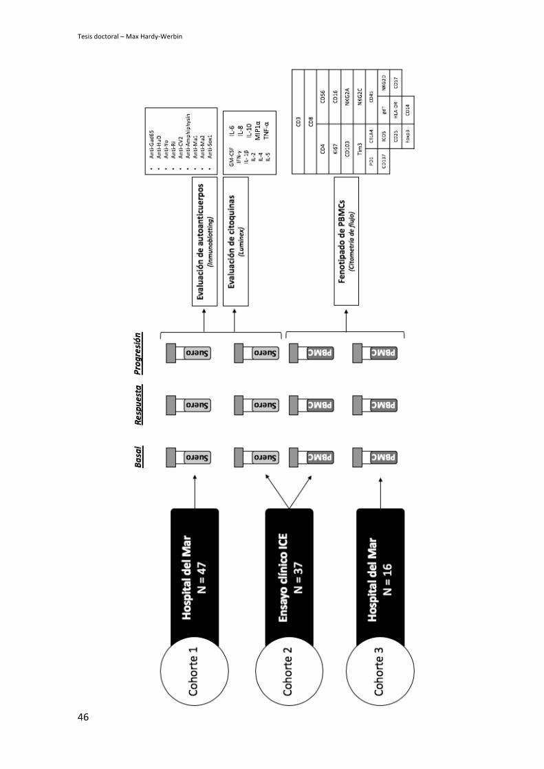
Poblaciones del estudio
Cohorte hospital del Mar (cohorte 1)
En esta cohorte se incluyen 47 pacientes con CPCP tratados con quimioterapia
convencional, reclutados en el contexto de proyectos de investigación del Hospital del
Mar. Se han recogido muestras de sangre periférica previo al inicio del tratamiento, en
el momento de la respuesta al tratamiento y a la progresión de la enfermedad. Se
dispone de muestras de suero y plasma.
Cohorte ensayo clínico ICE (cohorte 2)
El ensayo ICE, incluye 37 pacientes con CPCP diseminado tratados con carboplatino +
etopósido + ipilimumab. En el ensayo se han recogido muestras de sangre periférica
previo al inicio del tratamiento con ipilimumab y secuencialmente durante la evolución
de la enfermedad. Se dispone de muestras de suero, de plasma y de PBMCs.
Cohorte prospectiva hospital del Mar (cohorte 3)
Desde octubre de 2016 en el hospital del Mar se ha iniciado la recogida de muestras
seriadas de suero, plasma y PBMCs de pacientes con CPCP metastásico tratados con
quimioterapia convencional, en el momento del diagnóstico, a la respuesta al
tratamiento y a la progresión de la enfermedad. En el momento de la elaboración de este
documento se han reclutado 31 pacientes, y se han obtenido datos de supervivencia de
16 de ellos.

Tesis doctoral – Max Hardy-Werbin
44
Análisis de biomarcadores
Detección de autoanticuerpos en sangre periférica
Se ha utilizado la técnica de inmunoensayo en línea que permite la detección simultánea
y en una sola reacción de anticuerpos contra numerosos antígenos mediante un Kit
comercial (RAVO kit™) que permite identificar una batería de autoanticuerpos
típicamente detectables en CPCP (anti-HuD, anti-Yo, anti-Ri, anti-CV2/CRMP5, anti-
Amphiphysin, anti-Ma1, anti-Ma2, SOX1 yGAD65).
Estudio de citoquinas en sangre periférica
Se ha utilizado un kit de ensayo multiplex de Merck Millipore (Cytokine Human
Magnetic 10-Plex Panel) para la plataforma de tecnología Luminex xMAP que
cuantifica GM-CSF, IFNγ, IL-1β, IL-2, IL-4, IL-5, IL-6, IL-8, IL-10, TNFα y MIP1a
en suero o plasma humanos.
Análisis fenotípico de PBMCs
Se ha diseñado un panel de marcadores para el fenotipado de PBMCs mediante
citometría de flujo. Se aislaron las células circulantes inmunológicas para centrarse en la
detección de poblaciones linfocitarias efectoras, reguladoras y NK, mediante un panel
de anticuerpos que incluye CD3, CD4, CD8, CD45, Foxp3, CD25, CD103, Tim-3,
Ki67, HLA-DR, CD56, NKG2C, NKG2A, NKG2D, CD137, PD-1, CTLA, CD57 y
gdT.
La tinción se llevó a cabo en PBMCs criopreservadas. Tras la descongelación las
células se incubaron con un marcador de viabilidad durante 30 minutos. Tras el bloque
con inmunoglobulina G humana durante 20 minutos, se llevó a cabo la tinción de
marcadores de membrana durante otros 20 minutos. Tras cada incubación se realizaron
Materiales y métodos
45
dos lavados con PBS 1X. Posteriormente se fijaron las células mediante el kit de
permeabilización Foxp3 Fix/Perm para realizar la tinción de los marcadores
intranucleares durante 30 minutos. Tras cada incubación se realizaron dos lavados con
buffer especial de lavado para tinciones intranucleares según las recomendaciones del
fabricante. La detección de las células se llevó a cabo con tecnología de citometría
digital equipada con 4 láseres y 16 detectores (BD LSRFortessa analyser). Para la
adquisición se utilizó el software Diva, y para el análisis de datos, el FlowJo X.
Tesis doctoral – Max Hardy-Werbin
46

Resultados
4. RESULTADOS

Tesis doctoral – Max Hardy-Werbin
48

Resultados
49
Esta memoria de tesis doctoral titulada “Caracterización de los eventos inmunológicos
diferenciales en pacientes con cáncer de pulmón de célula pequeña tratados con
quimioterapia con o sin ipilimumab”, se presenta como compendio de publicaciones en
las que el doctorando Max Hardy-Werbin figura como primer autor y son fruto directo
del proyecto de tesis.
Articulo 1:
Hardy-Werbin M, et al. Assessment of neuronal autoantibodies in patients with small
cell lung cancer treated with chemotherapy with or without ipilimumab.
Oncoimmunology. 2017 Nov 27;7(2):e1395125
DOI: 10.1080/2162402X.2017.1395125
PMID: 29308329
Índice de calidad: IF 2017 = 5.503; 1Q Oncología; 1Q Inmunología
Artículo 2:
Hardy-Werbin M, et al. Serum cytokine levels as predictive biomarkers of benefit from
ipilimumab in small cell lung cancer. Oncoimmunology. 2019 Mar 27;8(6):e1593810.
DOI: 10.1080/2162402X.2019.1593810
PMDI: 31069160
Índice de cualidad: IF 2018 = 5.333; 1Q Oncología; 1Q Inmunología
Dra. Edurne Arriola Aperribay

Tesis doctoral – Max Hardy-Werbin
50
Resultados
51
Primera parte: análisis de autoanticuerpos
Artículo 1.
Título:
Assessment of neuronal autoantibodies in patients with small cell lung cancer treated
with chemotherapy with or without ipilimumab
Autores:
M. Hardy-Werbin, O. Arpí, A. Taus, P. Rocha, D. Joseph-Pietras, L. Nolan, S. Danson,
R. Griffiths, M. Lopez-Botet, A. Rovira, J. Albanell, C. H. Ottensmeier, E. Arriola
Resúmen:
El objetivo de esta primera parte del proyecto era evaluar las diferencias en la presencia
o no de autoanticuerpos neurológicos (AAN) entre pacientes tratados con quimioterapia
sola (cohorte 1) y con quimioterapia e ipilimumab (cohorte 2), y evaluar la relación de
estos hallazgos con beneficio en supervivencia.
Del total de los pacientes evaluados, 12 presentaban CPCP con enfermedad localizada
al diagnóstico de la enfermedad. De estos, el 75% tenían la presencia de ANN en las
muestras basales, frente a un 50% de pacientes con enfermedad diseminada (p = 0.096).
Esta tendencia se replicó para la presencia de anti-Sox1 de manera individual (58.3% vs
33.3%; p = 0.092), pero no para la presencia de anti-HuD y anti-Yo.
Al estudiar la asociación de AAN con datos de supervivencia, se observó que aquellos
pacientes en los que se detectaba la presencia de al menos un AAN al diagnóstico
presentaban una mayor supervivencia global [14.8 meses (m) vs 12.1 m; p= 0.089]. Esta
asociación se mantuvo al analizar individualmente los pacientes tratados con

Tesis doctoral – Max Hardy-Werbin
52
quimioterapia (15.1 m vs 11.7 m; p =0.032), probablemente debido a la prevalencia en
esta cohorte de pacientes con enfermedad localizada.
Posteriormente se segregaron los pacientes entre aquellos con reactividad a 1 AAN
frente aquellos con reactividad frente a 2 AAN o más. Se observó que la presencia de
sólo 1 AAN frente a 2 o más en pacientes tratados con quimioinmunoterapia, se
asociaba con una supervivencia libre de progresión superior (7.3 m vs 5.5 m; p= 0.005).
La detección de múltiples reactividades de AAN, sin embargo, no se asoció a ningún
tipo de reacción adversa provocada por ipilimumab (colitis, astenia, rash) ni a eventos
neurológicos.
Por último, se analizó la variación del título sérico de AAN en las muestras seriadas y
su asociación con beneficio del tratamiento. De los 45 pacientes con detección basal de
AAN, el 93% presentó una respuesta inicial al tratamiento, y en el 60% de estos se
evidenció una bajada del título de NAA. Del resto de los pacientes que no presentaron
una respuesta inicial al tratamiento, todos sufrieron un ascenso en el título del
anticuerpo (p=0.080). En cuanto a supervivencia, un aumento en el título de AAN en
pacientes tratados con quimioterapia convencional se relacionó con un aumento de la
supervivencia libre de progresión (12 m vs 6.9m; p=0.016), y un descenso o
estabilización del título de AAN al final del tratamiento se relacionó con un aumento de
la supervivencia global (18.5m vs 12.3m; p=0.049), independientemente del tratamiento
recibido. No se observaron cambios diferenciales en el título de AAN con la adición de
ipilimumab al tratamiento en las muestras seriadas.

ORIGINAL RESEARCH
Assessment of neuronal autoantibodies in patients with small cell lung cancer treatedwith chemotherapy with or without ipilimumab
M. Hardy-Werbina,i, O. Arp!ıa, A. Tausb, P. Rochab, D. Joseph-Pietrase, L. Nolanf, S. Dansong, R. Griffithsh, M. Lopez-Botetc,A. Roviraa, J. Albanella,b, C. H. Ottensmeierd,e, and E. Arriolaa,b
aCancer Research Program, IMIM (Hospital del Mar Medical Research Institute), Barcelona, Spain; bMedical Oncology Department, Hospital del Mar-CIBERONC, Barcelona, Spain; cImmunology unit, IMIM (Hospital del Mar Medical Research Institute), Barcelona, Spain; dCancer Science Unit, Faculty ofMedicine, University of Southampton, Southampton, United Kingdom; eNIHR Experimental Cancer Medicine Centre, Southampton, United Kingdom;fMedical Oncology Department, University Hospital Southampton, Southampton, United Kingdom; gSheffield Experimental Cancer Medicine Centre,Weston Park Hospital, Sheffield, United Kingdom; hMedical Oncology Department, The Clatterbridge Cancer Centre NHS Foundation Trust, Wirral,United Kingdom; iUniversitat de Barcelona, Barcelona, Spain
ARTICLE HISTORYReceived 23 August 2017Accepted 17 October 2017
ABSTRACTSmall-cell lung cancer (SCLC) is often associated with paraneoplastic syndromes. To assess the role of anti-neuronal autoantibodies (NAAs) as biomarkers of treatment outcome, we assessed NAAs in serial samplesfrom SCLC patients treated with chemoimmunotherapy compared to chemotherapy alone. We evaluated2 cohorts: in cohort 1 (C1), 47 patients received standard platinum/etoposide, and in cohort 2 (C2), 38patients received ipilimumab, carboplatin and etoposide. Serum samples at baseline and subsequent timepoints were analyzed for the presence of NAAs. NAAs were detected at baseline in 25 patients (53.2%) inC1 and in 20 patients (52.6%) in C2 (most frequently anti-Sox1). NAA at baseline was associated withlimited disease (75% vs 50%; p: 0.096) and better overall survival (15.1 m vs 11.7 m; p: 0.032) in C1.Thirteen patients (28.9%) showed 2 or more reactivities before treatment; this was associated with worsePFS (5.5 m vs 7.3 m; p: 0.005) in patients treated with chemoimmunotherapy. NAA titers decreased aftertherapy in 68.9% patients, with no differential patterns of change between cohorts. Patients whose NAAtiter decreased after treatment, showed longer OS [18.5 m (95% CI: 15.8 – 21.2)] compared with thosewhose NAA increased [12.3 m (95% CI: 8.1 – 16.5; p 0.049)], suggesting that antibody levels correlate totumor load. Our findings reinforce the role of NAAs as prognostic markers and tumor activity/burden inSCLC, warrant further investigation in their predictive role for immunotherapy and raise concern over theuse of immunotherapy in patients with more than one anti-NAA reactivity.
KEYWORDSanti-Hu; anti-Yo;autoantibodies; anti-SOX-1;ipilimumab; paraneoplasticsyndrome; Small cell lungcancer
Introduction
Small-cell lung cancer (SCLC) accounts for approximately 15%of all lung cancer.1,2 Although robust and often dramatic clini-cal responses are achieved with platinum-based first-line treat-ment, recurrence/progression usually occurs early and isresistant to available treatments.3 Outcomes in patients withstage IV disease remain poor with a median overall survivalthat rarely exceeds one year.1,4
Immunotherapy has led to a paradigm-shift in the treatmentof solid tumors.5,6 SCLC seems a particularly attractive targetfor immunotherapy. Paraneoplastic neurological syndromes(PNSs) are common in SCLC and are characterized by anti-cancer immune responses, which become clinically visible byoff-tumor immune consequences against neuronal antigens,ectopically expressed by cancer cells and physiologically by thenormal nervous system.7 The better cancer specific survivaloutcomes in patients with autoimmune disease points to a pro-tective effect, linked to these cross-reactive immuneresponses.8,9 Moreover, high mutational load has been linked
to benefit from immunotherapy10 and SCLC falls into this cate-gory,2,11,12 most likely as a result of the tight association withtobacco smoking. Therefore, harnessing the immune system tofight SCLC seems a potentially promising strategy, althoughthe best approach is currently uncertain.
Ipilimumab is a fully human immunoglobulin G1 monoclo-nal antibody that blocks CTLA-413, and has been tested inSCLC. A phase II trial,14 showed a trend to improved overallsurvival with phased ipilimumab added to a platinum doublet.However, the confirmatory phase III trial 15 comparing ipilimu-mab and chemotherapy vs chemotherapy did not confirm bet-ter overall survival after chemoimmunotherapy. More recently,anti-PD-1 agents have also been tested in SCLC. Nivolumab(alone and in combination with ipilimumab) showed antitumoractivity in SCLC patients with relapsed disease.16 In a phase Itrial, Pembrolizumab use led to objective responses greaterthan 30% of patients.17
Nonetheless, the majority of patients with SCLC do notbenefit from checkpoint inhibitors and the absence of
CONTACT Dr. E. Arriola [email protected] Medical Oncology Department, Hospital del Mar, Passeig Mar!ıtim de la Barceloneta, 25–29, 08003Barcelona, Spain.
Supplemental data for this article can be accessed on the publisher’s website.© 2017 Taylor & Francis Group, LLC
ONCOIMMUNOLOGY2017, VOL. 0, NO. 0, e1395125 (9 pages)https://doi.org/10.1080/2162402X.2017.1395125
Dow
nloa
ded
by [8
3.35
.6.5
] at 1
3:32
27
Nov
embe
r 201
7

predictive biomarkers for patient selection may have con-tributed to negative trial results.15 Additionally, clinicallyrelevant autoimmune toxicity has been reported, includingsevere neurological PNS14,16,18 increasing the level of cau-tion that is needed when investigating immunotherapy inSCLC. PD-L1 immunohistochemical expression, which ispredictive for benefit from modulation of PD1/PDL1 inNon-Small Cell Lung cancer, does not appear to correlatewith outcome in SCLC.16,17 A confounding effect for theevaluation of PD-L1 status in the tumor is limited access totissue due to small biopsy material or cytology diagnosticspecimens, a well-recognized problem in SCLC.19 Therefore,the need to develop predictive biomarkers remains anurgent priority in SCLC.
With the aim of identifying potential biomarkers, we previ-ously performed an exploratory analysis of the phase II ICEtrial with a single cohort of patients treated with chemotherapyand ipilimumab. In this single arm study, positivity of autoanti-bodies at baseline was associated with improved outcomes.18
In the current study we added a ‘control’ cohort treated withstandard chemotherapy in order to assess the predictive vs.prognostic role of the neuronal autoantibodies. We also ana-lyzed changes in autoantibody detection during treatment toevaluate the potential utility of serial sampling.
Results
Patients’ characteristics and outcomes
We included 85 SCLC patients: 47 treated with standard chemo-therapy (cohort 1 herein) and 38 with standard chemotherapy plusipilimumab (cohort 2 herein). Patients’ characteristics are shownin Table 1. The main differences between both cohorts were thatcohort 1 (C1) included more men (74.5% vs 65.8%), 25% patientshad limited stage and 15% patients presented with performancestatus (PS) 2 versus none in cohort 2. None of the patients had anyneurological symptoms at diagnosis.
Objective response rate (ORR) was 93.6% in C1 and 81.6%in C2. Median progression free survival (PFS) was 6.8 months(m) (95% CI: 6.1-7.6) in C1 and 6.9 m (95% CI: 6.3-7.6) in C2(p D 0.847). Overall survival (OS) was 13.3 m (95% CI: 10.1-16.5) in C1 vs 17 m (95% CI: 4.5-29.5) in C2 (p D 0.987).
When we selected patients with stage IV in C1, OS was 11.7 m(95% CI: 7.6 – 15.7).
We assessed the impact of clinical variables on survival.Table 2 illustrates the association between clinical variables andOS combining both cohorts. PS:0 vs. PS>0 (p D 0.019), limitedstage (p D 0.003) and response to first line chemotherapy(p<0.001) were associated with a significantly improved sur-vival (Table 2).
Neuronal autoantibody detection
Neuronal autoantibodies (NAA) were detected at baseline in 25patients (53.2%) in C1 and in 20 patients (52.6%) in C2. Inboth cohorts, the most prevalent autoantibody was anti-Sox1(38.3% in C1 and 34.2% in C2), followed by anti-HuD andanti-Yo (Table 3, Fig. 1 – left).
Globally, 13 patients out of the 45 with baseline NAA(28.9%) showed 2 or more reactivities at any time-point (Sup-plementary Table 1); of these, 11 patients (24.5%), had 2 ormore types of autoantibody at baseline. Eight patients (17.8%)in C1 showed 2 or more reactivities; one of these patients waspositive at baseline for anti-Sox1, but became also positive foranti-Yo after 3 cycles of chemotherapy. Five patients (11.1%) inC2 showed 2 or more reactivities; one of these patients was pos-itive at baseline for anti-Yo, and became also positive for anti-Gad65 after 8 weeks of treatment. Anti-Yo and Anti-Sox1detection were associated with another NAA in 7/12 patients(58.3%) and 9/32 patients (28.1%), respectively. One patientshowed reactivity for 4 NAA: anti-Sox1, anti-amphiphysin,anti-Ri and anti-HuD. In Fig. 1 (right) we illustrate the differ-ent multi-reactivities of NAA in patients of both cohorts.
Association between neuronal autoantibodies and clinicalcharacteristics and outcomes
We next evaluated the association of the detection of NAA withclinical variables. Of 12 patients with limited stage, 9 patients(75%) had detectable NAAs at baseline versus 36/72 (50%) ofpatients with stage IV disease (p D 0.096). This trend was alsoobserved for anti-Sox1 where 7/12 (58.3%) of patients with
Table 1. Patients’ characteristics.
Cohort 1 ChemotherapyN D 47
Cohort 2 Chemotherapy CIpilimumab N D 38
GenderFemale 12 (25.5%) 13 (34.2%)Male 35 (74.5%) 25 (65.8%)
AgeMean 62.6 62.5Range (45 – 86) (44 – 84)
PS0 26 (55,3%) 11 (28.9%)1 14 (29,8%) 21 (55.3%)2 7 (14,9%) 0NA 0 6 (15.8%)
StageI-III 12 (25,5%) 0IV 35 (74,5%) 38 (100%)
PS: performance status; NA: not available
Table 2. Univariate analysis for overall survival including both cohorts (Log-Ranktest).
95% Confidence interval
Clinical variables OS (months) Lower Upper p-value
GenderMale 12.1 7.7 16.5 0.960Female 15.4 11.4 19.3
Age< 60 15.4 10.3 20.4 0.651! 60y 12.2 9.5 15.7
PS0 15.4 11.7 19.1 0.0191 or 2 9.2 6.6 11.8
StageI-III 23.7 11.9 35.6 0.003IV 12.1 8.9 15.3
Response to 1st line treatmentNo 6.2 5.4 7.1 < 0.001Yes 15.1 12 18.2
PS: Performance status; OS: Overall survival; y: years
e1395125-2 M. HARDY-WERBIN ET AL.
Dow
nloa
ded
by [8
3.35
.6.5
] at 1
3:32
27
Nov
embe
r 201
7

limited stage had detectable anti-Sox1 at baseline compared to24/72 (33.3%) patients with stage IV (p D 0.092). The presenceof anti-HuD and anti-Yo did not show any correlation withstage. There was no association between sex, age or PS anddetection of neither any baseline NAA nor specificautoantibodies.
We assessed the association of NAAs with clinical outcomes.In patients with pre-treatment NAAs we observed a trend forbetter overall survival when both cohorts were analyzed together[14.8 m (95% CI: 10.6-19) vs 12.1 m (95% CI: 8.2-16); p: 0.089].In patients treated with chemotherapy alone, this association wassignificant [15.1 m (95% CI: 10.2-20) vs 11.7 m (95%CI: 7.5-15.9); p D 0.032] (Fig. 2). This association was lost when theanalysis was restricted to stage IV patients and was not observedin patients from C2. No effect from baseline autoantibody pres-ence was observed on objective response rates or PFS (Table 4).
We then tried to gain insight whether the breadth ratherthan simply examining presence or absence of reactivity againstNAAs might link to outcomes. We segregated patients withreactivity to exactly one, as opposed to >1 NAA. Unexpectedly,the presence of only 1 NAA vs >1 NAA was associated withbetter PFS [7.3 m (95% CI: 5.9-8.6) vs 5.5 m (95% CI: 2.8-8.2);p D 0.005] in patients treated with chemo-immunotherapy(Fig. 3); a trend was also visible in OS [12.3 m (95% CI: 4.7-19.9) vs 7.9 m (95% CI: 4.9-11); p D 0.180]. No such associa-tion was observed in the chemotherapy only cohort. The detec-tion of multiple vs. single reactivities was not associated withipilimumab-related adverse events in cohort 2 (colitis, fatigueand rash) nor neurological events.
We then evaluated the association between NAA against indi-vidual antigens and clinical outcomes. In patients treated withipilimumab and chemotherapy, OS was 7.8 m (95% CI 2.2-13.4)when anti-Yo was positive, and 17 m (8.7-25.4) when it was neg-ative (p D 0.015); PFS was 4.3 m (95% CI: 2.5-6) when anti-Yowas positive, and 6.9 m (95% CI: 6.4-7.5) when anti-Yo was neg-ative (p D 0.081). No other associations were observed betweenother individual NAAs and clinical outcomes.
In the multivariate analysis in C2 for OS, including poor PSand lack of response to first line treatment, having 2 or more vs1 NAA, remained independently associated with poor outcome(HR: 3.4; p-value: 0.033).
Titers of NAA change upon response to treatment
We quantified the titers of NAAs at each time point (Supple-mentary Fig. 1). Overall in 31 (68.9%) of the 45 patients with
Table 3. Neuronal autoantibody detection at baseline.
Cohort 1 ChemotherapyN D 47
Cohort 2 ChemotherapyC Ipilimumab N D 38
Anti-Sox1Positive 18 (38.3%) 13 (34.2%)Negative 29 (61.7%) 24 (63.2%)NA 0 1 (2.6%)
Anti-YoPositive 8 (17%) 3 (7.9%)Negative 39 (83%) 34 (89.5%)NA 0 1 (2.6%)
Anti-HuDPositive 4 (8.5%) 4 (10.5%)Negative 43 (91.5%) 33 (86.8%)NA 0 1 (2.6%)
Anti-Gad65Positive 3 (6.4%) 2 (5.3%)Negative 44 (93.6%) 35 (92.1%)NA 0 1 (2.6%)
Anti-RiPositive 2 (4.3%) 0Negative 45 (95.7%) 37 (100%)NA 0 1 (2.6%)
Anti-CV2Positive 0 1 (2.6%)Negative 47 (100%) 36 (94.7%)NA 0 1 (2.6%)
Anti-AmphiphysinPositive 0 1 (2.6%)Negative 47 (100%) 36 (94.7%)NA 0 1 (2.6%)
Anti-Ma1Positive 0 0Negative 47 (100%) 37 (100%)NA 0 1 (2.6%)
Anti-Ma2Positive 0 0Negative 47 (100%) 37 (100%)NA 0 1 (2.6%)
NA: not available
Figure 1. Neuronal autoantibodies prevalence in patients with SCLC. On the left, percentage and absolute numbers of neuronal autoantibody detected at baseline in eachcohort. In the center, Benn’s diagrams illustrating the different NAA multi reactivities in patients from both cohorts. On the right we show the combination of NAA with 2,3 or 4 reactivities. Numbers at the intersection of Benn’s diagrams are number of patients with that combination.
ONCOIMMUNOLOGY e1395125-3
Dow
nloa
ded
by [8
3.35
.6.5
] at 1
3:32
27
Nov
embe
r 201
7
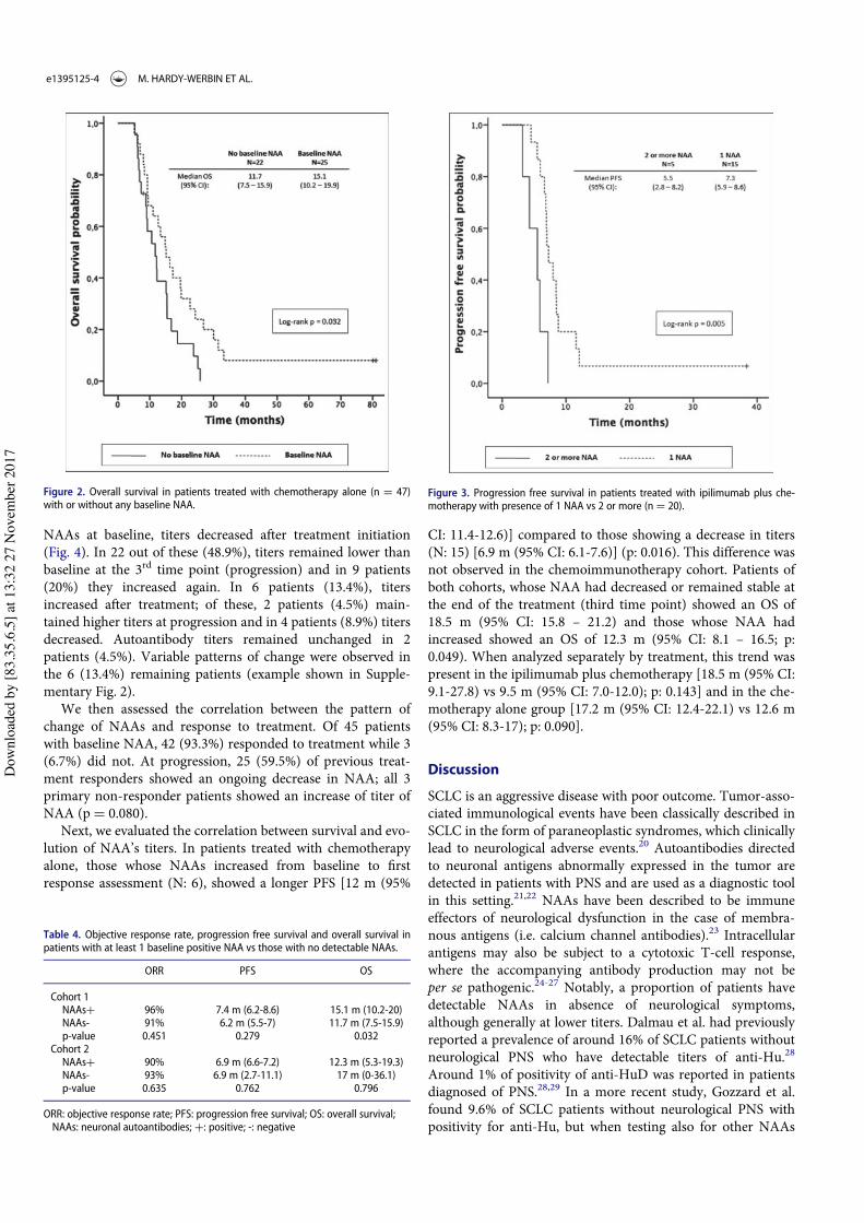
NAAs at baseline, titers decreased after treatment initiation(Fig. 4). In 22 out of these (48.9%), titers remained lower thanbaseline at the 3rd time point (progression) and in 9 patients(20%) they increased again. In 6 patients (13.4%), titersincreased after treatment; of these, 2 patients (4.5%) main-tained higher titers at progression and in 4 patients (8.9%) titersdecreased. Autoantibody titers remained unchanged in 2patients (4.5%). Variable patterns of change were observed inthe 6 (13.4%) remaining patients (example shown in Supple-mentary Fig. 2).
We then assessed the correlation between the pattern ofchange of NAAs and response to treatment. Of 45 patientswith baseline NAA, 42 (93.3%) responded to treatment while 3(6.7%) did not. At progression, 25 (59.5%) of previous treat-ment responders showed an ongoing decrease in NAA; all 3primary non-responder patients showed an increase of titer ofNAA (p D 0.080).
Next, we evaluated the correlation between survival and evo-lution of NAA’s titers. In patients treated with chemotherapyalone, those whose NAAs increased from baseline to firstresponse assessment (N: 6), showed a longer PFS [12 m (95%
CI: 11.4-12.6)] compared to those showing a decrease in titers(N: 15) [6.9 m (95% CI: 6.1-7.6)] (p: 0.016). This difference wasnot observed in the chemoimmunotherapy cohort. Patients ofboth cohorts, whose NAA had decreased or remained stable atthe end of the treatment (third time point) showed an OS of18.5 m (95% CI: 15.8 – 21.2) and those whose NAA hadincreased showed an OS of 12.3 m (95% CI: 8.1 – 16.5; p:0.049). When analyzed separately by treatment, this trend waspresent in the ipilimumab plus chemotherapy [18.5 m (95% CI:9.1-27.8) vs 9.5 m (95% CI: 7.0-12.0); p: 0.143] and in the che-motherapy alone group [17.2 m (95% CI: 12.4-22.1) vs 12.6 m(95% CI: 8.3-17); p: 0.090].
Discussion
SCLC is an aggressive disease with poor outcome. Tumor-asso-ciated immunological events have been classically described inSCLC in the form of paraneoplastic syndromes, which clinicallylead to neurological adverse events.20 Autoantibodies directedto neuronal antigens abnormally expressed in the tumor aredetected in patients with PNS and are used as a diagnostic toolin this setting.21,22 NAAs have been described to be immuneeffectors of neurological dysfunction in the case of membra-nous antigens (i.e. calcium channel antibodies).23 Intracellularantigens may also be subject to a cytotoxic T-cell response,where the accompanying antibody production may not beper se pathogenic.24-27 Notably, a proportion of patients havedetectable NAAs in absence of neurological symptoms,although generally at lower titers. Dalmau et al. had previouslyreported a prevalence of around 16% of SCLC patients withoutneurological PNS who have detectable titers of anti-Hu.28
Around 1% of positivity of anti-HuD was reported in patientsdiagnosed of PNS.28,29 In a more recent study, Gozzard et al.found 9.6% of SCLC patients without neurological PNS withpositivity for anti-Hu, but when testing also for other NAAs
Figure 2. Overall survival in patients treated with chemotherapy alone (n D 47)with or without any baseline NAA.
Table 4. Objective response rate, progression free survival and overall survival inpatients with at least 1 baseline positive NAA vs those with no detectable NAAs.
ORR PFS OS
Cohort 1NAAsC 96% 7.4 m (6.2-8.6) 15.1 m (10.2-20)NAAs- 91% 6.2 m (5.5-7) 11.7 m (7.5-15.9)p-value 0.451 0.279 0.032
Cohort 2NAAsC 90% 6.9 m (6.6-7.2) 12.3 m (5.3-19.3)NAAs- 93% 6.9 m (2.7-11.1) 17 m (0-36.1)p-value 0.635 0.762 0.796
ORR: objective response rate; PFS: progression free survival; OS: overall survival;NAAs: neuronal autoantibodies; C: positive; -: negative
Figure 3. Progression free survival in patients treated with ipilimumab plus che-motherapy with presence of 1 NAA vs 2 or more (n D 20).
e1395125-4 M. HARDY-WERBIN ET AL.
Dow
nloa
ded
by [8
3.35
.6.5
] at 1
3:32
27
Nov
embe
r 201
7
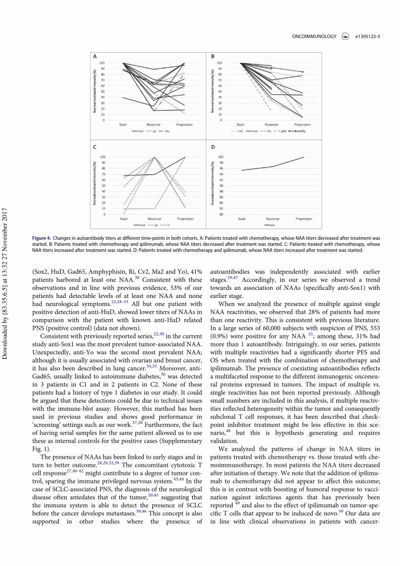
(Sox2, HuD, Gad65, Amphyphisin, Ri, Cv2, Ma2 and Yo), 41%patients harbored at least one NAA.30 Consistent with theseobservations and in line with previous evidence, 53% of ourpatients had detectable levels of at least one NAA and nonehad neurological symptoms.22,28-33 All but one patient withpositive detection of anti-HuD, showed lower titers of NAAs incomparison with the patient with known anti-HuD relatedPNS (positive control) (data not shown).
Consistent with previously reported series,22,30 in the currentstudy anti-Sox1 was the most prevalent tumor-associated NAA.Unexpectedly, anti-Yo was the second most prevalent NAA;although it is usually associated with ovarian and breast cancer,it has also been described in lung cancer.34,35 Moreover, anti-Gad65, usually linked to autoimmune diabetes,36 was detectedin 3 patients in C1 and in 2 patients in C2. None of thesepatients had a history of type 1 diabetes in our study. It couldbe argued that these detections could be due to technical issueswith the immune-blot assay. However, this method has beenused in previous studies and shows good performance in‘screening’ settings such as our work.37,38 Furthermore, the factof having serial samples for the same patient allowed us to usethese as internal controls for the positive cases (SupplementaryFig. 1).
The presence of NAAs has been linked to early stages and inturn to better outcome.28,29,32,39 The concomitant cytotoxic Tcell response27,40-42 might contribute to a degree of tumor con-trol, sparing the immune privileged nervous system.43,44 In thecase of SCLC-associated PNS, the diagnosis of the neurologicaldisease often antedates that of the tumor,20,45 suggesting thatthe immune system is able to detect the presence of SCLCbefore the cancer develops metastases.39,46 This concept is alsosupported in other studies where the presence of
autoantibodies was independently associated with earlierstages.29,47 Accordingly, in our series we observed a trendtowards an association of NAAs (specifically anti-Sox1) withearlier stage.
When we analyzed the presence of multiple against singleNAA reactivities, we observed that 28% of patients had morethan one reactivity. This is consistent with previous literature.In a large series of 60,000 subjects with suspicion of PNS, 553(0.9%) were positive for any NAA 21; among these, 31% hadmore than 1 autoantibody. Intriguingly, in our series, patientswith multiple reactivities had a significantly shorter PFS andOS when treated with the combination of chemotherapy andipilimumab. The presence of coexisting autoantibodies reflectsa multifaceted response to the different inmunogenic onconeu-ral proteins expressed in tumors. The impact of multiple vs.single reactivities has not been reported previously. Althoughsmall numbers are included in this analysis, if multiple reactiv-ities reflected heterogeneity within the tumor and consequentlysubclonal T cell responses, it has been described that check-point inhibitor treatment might be less effective in this sce-nario,48 but this is hypothesis generating and requiresvalidation.
We analyzed the patterns of change in NAA titers inpatients treated with chemotherapy vs. those treated with che-moimmunotherapy. In most patients the NAA titers decreasedafter initiation of therapy. We note that the addition of ipilimu-mab to chemotherapy did not appear to affect this outcome;this is in contrast with boosting of humoral response to vacci-nation against infectious agents that has previously beenreported 49 and also to the effect of ipilimumab on tumor-spe-cific T cells that appear to be induced de novo.50 Our data arein line with clinical observations in patients with cancer-
Figure 4. Changes in autoantibody titers at different time-points in both cohorts. A: Patients treated with chemotherapy, whose NAA titers decreased after treatment wasstarted. B: Patients treated with chemotherapy and ipilimumab, whose NAA titers decreased after treatment was started. C: Patients treated with chemotherapy, whoseNAA titers increased after treatment was started. D: Patients treated with chemotherapy and ipilimumab, whose NAA titers increased after treatment was started.
ONCOIMMUNOLOGY e1395125-5
Dow
nloa
ded
by [8
3.35
.6.5
] at 1
3:32
27
Nov
embe
r 201
7

associated PNS, where in treatment-responders antibodies candecrease or even normalize.51,52 In a series of 50 cancer patientswith PCD and at least one NAA, Shams’ili et al. report resolu-tion of PCD in 9/42 patients (21.4%) after successful tumortreatment.53 In 5 neurological responder patients where theauthors evaluated changes in NAA-titer no consistent patternwas observed. Additional immunosupressants for PNS mayhave also confounded the interpretation in this study. Inpatients with anti-Yo related PNS, antibody titers decreasedafter surgical removal of the tumor without resolution of theneurological symptoms.54,55 Overall NAA levels appear todecrease following removal of tumor antigen; the observationof worsening PNS with disease recurrence32,56 and increase riskof relapse in HPVC patients whose HPV antibodies persist57
both support this conclusion. Whether levels of NAA areimportant clinically or simply ‘bystanders’ of cellular immuneconsequences of treatment is uncertain.
An intriguing question arises over the location of the anti-body forming cells that drive the production of NAA. Morewidely in oncology, for example in patients with thymoma,improvement of paraneoplastic myasthenia is observed after,and is the clinical aim of surgical thymectomy. These data pointto the production of antibody within the tumor microenviron-ment and there is indeed an emerging literature that identifiesB-cells and tertiary lymphatic structures as common constitu-ents in the cancer mass in a wide variety of solid tumors.58-61 Incontrast, persistence of the neurological paraneoplasia in somecases even after radiological complete removal of cancer bytreatment both in thymomas and SCLC suggests that the anti-body forming cells may not be located in the cancer tissue(only) in all cases. In line with this, one immunohistochemicalstudy assessing the presence of T and B-cells in SCLC surgicalspecimens, demonstrated a small numbers of B-cells infiltratingthe tissue with 40% of cases with absence of this cell subtype,62
although this might change in extensive disease. Moreover, wecannot rule out here that the SCLC chemotherapy may havealso removed B-cells that are producing the NAA irrespectiveof the effect on tumor load.
Consistent with the concept that removal of immunogen islinked with a decrease in antibody titer, we observed a trend toa longer OS in those patients whose titer of NAA decreasedprogressively after treatment with chemotherapy; this was irre-spective of whether ipilimumab was administered.
Intriguingly however, our data also raise the possibilityof a quite different immunological consequence: subgroupanalysis of the small number of patients with increasingtiters after initiation of chemotherapy identified a longerthan median PFS; this raises the question of whetherenhanced immune response due to intracellular antigenexposure after cytotoxic chemotherapy may lead to a betterT-cell response and in turn to increased antibody produc-tion. A dataset in melanoma reported by the Wolchokgroup is consistent with this concept, as the presence ordevelopment of antibodies and T-cell responses to NyEso1appeared to identify a group of patients, who did well afteripilimumab treatment.63 We conclude that further detailedstudies are needed, which link the humoral responses andcellular responses in cancer patients, both after standardtreatment and immunotherapy.
In summary, we were able to confirm the prognostic role ofNAA probably related with disease control in early stagederived from tumor directed immune response. To our knowl-edge, we demonstrate for the first time that treatment with ipili-mumab combined with chemotherapy in SCLC does not inducedifferential changes in NAA titers and their levels appear tomainly reflect the disease burden. The predictive role of NAAsand B cell immune response warrants further investigation inSCLC.
Patients and methods
Study populations
We retrospectively analyzed the data and serum from 85 SCLCpatients from two independent cohorts. The first cohortincluded 47 patients with SCLC treated with first-line standardchemotherapy (platinum – etoposide), recruited withinresearch projects in Hospital del Mar, Barcelona, and previ-ously reported.64-66 We included patients with early and locallyadvanced disease as well as stage IV patients, in order to inves-tigate the impact of stage in the detection of NAAs. From thisseries we had a control population, from age- and sex-matchedhealthy donors (N: 30) to the study population. The secondcohort were patients included in the ICE trial,18 where 38chemo-na€ıve stage IV SCLC patients were treated with ipilimu-mab in combination with carboplatin-etoposide. Serum sam-ples at baseline and subsequent time points (before, during andafter treatment) were collected from each patient. Completeclinical data and follow up information was available for everypatient.
Sample analysis was approved for the cohort 1 by the localethics committee and for cohort 2 within the trial ethicscommittee.
Sampling
Serum samples were collected for C1 before treatment initiation(baseline), at first response assessment and at progression. ForC2, samples were collected at baseline, at 3 and 6 months whenfeasible. All patients in C1 had 3 serial serum samples (baseline,first response assessment and progression timepoints). Out ofthe 38 patients in C2, 20 had 2, 11 had 3, and 6 had 4 serialtimepoints (all included baseline). One patient in C2 had noavailable serum samples. Serum from 3 healthy volunteers wasused as negative controls and serum from a SCLC patient witha known anti-HuD associated PNS as a positive control.
Whole blood samples were collected by standard venipunc-ture techniques using serum separator tubes. Samples wereallowed to clot for 30 minutes at room temperature before cen-trifugation for 10 minutes at 1000 g at 4!C. Following centrifu-gation, the supernatant (serum) was immediately removed andassayed immediately or aliquoted and stored frozen at ¡80!Cuntil further use.
Assessment of autoantibodies
A commercial immunoblotting assay (Ravo PNS-Blot, RavoDiagnostika) consisting of a membrane strip coated with the
e1395125-6 M. HARDY-WERBIN ET AL.
Dow
nloa
ded
by [8
3.35
.6.5
] at 1
3:32
27
Nov
embe
r 201
7

recombinant intracellular neuronal antigens Gad65, Sox1, Ma1,Ma2, Amphiphysin, CRMP5, Ri, Yo and HuD was used. Patientand control sera were diluted 1:200 as established in the currentversion of the instructions for use (01/2017) and processed fol-lowing manufacturer’s protocol.
Incubated line blots were scanned and processed forquantification with ImageJ 1.51 h (NIH). A distinctive bandcorresponding to the actual position of the antigen in theline blot was considered as a positive signal. Backgroundwas subtracted from band intensity. This value was normal-ized with the positive control and the percentage of varia-tion between each time-point was calculated. A percentageof variation of more than 5% was considered as change(increase/decrease) of titer of NAA. Prism 6.0 (GraphPad)was used for plot construction.
Statistical analysis
Statistical analysis was carried out using SPSS 22.0 (SPSS Inc,Chicago, IL, USA) and Stata/MP 14 (StataCorp LLC, Texas,USA) together with the Statistical Assessment Service fromIMIM (Hospital del Mar). To analyze associations between cat-egorical variables we used the Chi-square test or the Fisher’sexact test as appropriate. Overall survival was plotted byKaplan–Meier method and curves were compared by the log-rank test. Multivariate analysis was performed with the Coxregression method. All tests were conducted at the two-sided0.05 level of significance.
Abbreviations
CTLA-4 Cytotoxic T-lymphocyte antigen 4EDTA Ethylenediaminetetraacetic acidICE Ipilimumab-Carboplatin-EtoposideNAA Neuronal auto-antibodyORR Objective response rateOS Overall survivalPCD Paraneoplastic cerebellar degenerationPD-1 Programmed cell death protein 1PFS Progression free survivalPNS Paraneoplastic neurological syndromepRB Retinoblastoma proteinPS Performance statusSCLC Small cell lung cancer
Disclosure statement
E.A. and C.O. have received honoraria for consultancy and lectures fromBristol-Myers Squibb outside of the current work. The rest of authorsreport no conflict of interest.
Funding details
This work was supported in part by a grant from (1) Fundaci!o La Marat!ode TV3 (666/C/2013); (2) ISCiii/FEDER (CIBERONC CB16/12/00241,RD12/0036/0051, PIE15/00008, PI15/00146, PI16/00591, PI13/00140); (3)Xarxa de Bancs de Tumors (XBTC); (4) Ag#encia de Gesti!o d’Ajuts Univer-sitaris i de Recerca (AGAUR), Generalitat de Catalunya (2014SGR740), (5)Fundaci!o Cellex and (6) Cancer Research UK grant: C491/A12135
References
1. Oze I, Hotta K, Kiura K, Ochi N, Takigawa N, Fujiwara Y, TabataM, Tanimoto M. Twenty-seven years of phase III trials forpatients with extensive disease small-cell lung cancer: disappoint-ing results. PLoS One. 2009;4:e7835. doi:10.1371/journal.pone.0007835. PMID:19915681.
2. Byers LA, Rudin CM. Small cell lung cancer: Where do we go fromhere? Cancer. 2014;121:664–72.
3. Hanna N, Bunn PA, Langer C, Einhorn L, Guthrie T, Beck T, AnsariR, Ellis P, Byrne M, Morrison M, et al. Randomized phase III trialcomparing irinotecan/cisplatin with etoposide/cisplatin in patientswith previously untreated extensive-stage disease small-cell lung can-cer. J Clin Oncol. 2006;24:2038–43.
4. Puglisi M, Dolly S, Faria A, Myerson JS, Popat S, O’Brien MER. Treat-ment options for small cell lung cancer – do we have more choice? BrJ Cancer. 2010;102:629–38. doi:10.1038/sj.bjc.6605527.
5. Hodi FS, O’Day SJ, McDermott DF, Weber RW, Sosman JA, HaanenJB, Gonzalez R, Robert C, Schadendorf D, Hassel JC, et al. Improvedsurvival with ipilimumab in patients with metastatic melanoma. NEngl J Med. 2010;363:711–23. doi:10.1056/NEJMoa1003466.
6. Brahmer J, Reckamp K, Baas P, Crin#o L, Eberhardt W, Poddubskaya E,Antonia S, Pluzanski A, Vokes E, Holgado E, et al. Nivolumab versusDocetaxel in Advanced Squamous-Cell Non–Small-Cell Lung Cancer.N Engl J Med. 2015;373:1627–39. doi:10.1056/NEJMoa1504627.
7. Darnell RB, Posner JB. Paraneoplastic syndromes and the nervous sys-tem. N Engl J Med. 2003;3:287–8.
8. Maddison P, Newsom-Davis J, Mills K, Souhami R. Favourable prog-nosis in Lambert-Eaton myasthenic syndrome and small-cell lung car-cinoma. Lancet. 1999;353:117–8. doi:10.1016/S0140-6736(05)76153-5.PMID:10209997.
9. Wirtz PW, Lang B, Graus F, Van Den Maagdenberg AMJM, Saiz A,De Koning Gans PA, Twijnstra A, Verschuuren JJGM. P/Q-type cal-cium channel antibodies, Lambert-Eaton myasthenic syndrome andsurvival in small cell lung cancer. J Neuroimmunol. 2005;164:161–5.doi:10.1016/j.jneuroim.2005.04.001.
10. Rizvi NA, Hellmann MD, Snyder A, Kvistborg P, Makarov V, HavelJJ, Lee W, Yuan J, Wong P, Ho TS, et al. Cancer immunology. Muta-tional landscape determines sensitivity to PD-1 blockade in non-smallcell lung cancer. Science. 2015;348:124–8. doi:10.1126/science.aaa1348. PMID:26068851.
11. Daniel VC, Marchionni L, Hierman JS, Rhodes JT, Devereux WL,Rudin CM, Yung R, Parmigiani G, Dorsch M, Peacock CD, et al. Aprimary xenograft model of small-cell lung cancer reveals irreversiblechanges in gene expression imposed by culture in vitro. Cancer Res.2009;69:3364–73. doi:10.1158/0008-5472.CAN-08-4210.
12. Miller CW, Simon K, Aslo A, Kok K, Yokota J, Buys CHCM, Terada M,Koeffler HP. p53 Mutations in Human Lung Tumors. Cancer Res.1992;143:1695–8.
13. Ku GY, Yuan J, Page DB, Schroeder SEA, Panageas KS, Carvajal RD,Chapman PB, Schwartz GK, Allison JP, Wolchok JD. Single-institu-tion experience with ipilimumab in advanced melanoma patients inthe compassionate use setting: lymphocyte count after 2 doses corre-lates with survival. Cancer. 2010;116:1767–75. doi:10.1002/cncr.24951.
14. Reck M, Bondarenko I, Luft A, Serwatowski P, Barlesi. F, Chacko R,Sebastian M, Lu H, Cuillerot JM, Lynch TJ. Ipilimumab in combina-tion with paclitaxel and carboplatin as first-line therapy in extensive-disease-small-cell lungcancer: Results from a randomized, double-blind, multicenter phase 2 trial. Ann Oncol. 2013;24:75–83.doi:10.1093/annonc/mds213.
15. Reck M, Luft A, Szczesna A, Havel L, Kim SW, Akerley W, PietanzaMC, Wu YL, Zielinski C, Thomas M, et al. Phase III randomized trialof ipilimumab plus etoposide and platinum versus placebo plus etopo-side and platinum in extensive-stage small-cell lung cancer. J ClinOncol. 2016;34:3740–8. doi:10.1200/JCO.2016.67.6601.
16. Antonia SJ, L!opez-Martin JA, Bendell J, Ott PA, Taylor M, Eder JP,J€ager D, Pietanza MC, Le DT, de Braud F, et al. Nivolumab alone andnivolumab plus ipilimumab in recurrent small-cell lung cancer(CheckMate 032): a multicentre, open-label, phase 1/2 trial. LancetOncol. 2016;17:883–95. doi:10.1016/S1470-2045(16)30098-5.
ONCOIMMUNOLOGY e1395125-7
Dow
nloa
ded
by [8
3.35
.6.5
] at 1
3:32
27
Nov
embe
r 201
7
17. Ott PA, Elez E, Hiret S, Kim D-W, Morosky A, Saraf S, Piperdi B,Mehnert JM. Pembrolizumab in Patients With Extensive-Stage Small-Cell Lung Cancer: Results From the Phase Ib KEYNOTE-028 Study. JClin Oncol 2017;:JCO. 2017.72.506.
18. Arriola E,WheaterM,Galea I, CrossN,MaishmanT,HamidD, Stanton L,Cave J, Geldart T, Mulatero C, et al. Outcome and Biomarker Analysisfrom a Multicenter Phase 2 Study of Ipilimumab in Combination withCarboplatin and Etoposide as First-Line Therapy for Extensive-StageSCLC. J ThoracOncol. 2016;11:1511–21. doi:10.1016/j.jtho.2016.05.028.
19. Travis WD. Pathology and Diagnosis of Neuroendocrine Tumors:Lung Neuroendocrine. Thorac Surg Clin. 2014;24:257–66.doi:10.1016/j.thorsurg.2014.04.001.
20. Darnell RB, Posner JB. Paraneoplastic syndromes affecting the ner-vous system. Semin Oncol. 2006;33:270–98. doi:10.1053/j.seminoncol.2006.03.008.
21. Pittock SJ, Kryzer TJ, Lennon VA. Paraneoplastic antibodies coexistand predict cancer, not neurological syndrome. Ann Neurol.2004;56:715–9. doi:10.1002/ana.20269.
22. Titulaer MJ, Klooster R, Potman M, Sabater L, Graus F, Hegeman IM,Thijssen PE, Wirtz PW, Twijnstra A, Smitt PAES, et al. SOX antibod-ies in small-cell lung cancer and Lambert-Eaton myasthenic syn-drome: frequency and relation with survival. J Clin Oncol.2009;27:4260–7. doi:10.1200/JCO.2008.20.6169.
23. Fukunaga H, Engel AG, Lang B, Newsom-Davis J, Vincent A. Passivetransfer of Lambert-Eaton myasthenic syndrome with IgG from manto mouse depletes the presynaptic membrane active zones. Pnas.1983;80:7636–40. doi:10.1073/pnas.80.24.7636.
24. Bach JF, Koutouzov S, Van Endert PM. Are there unique autoantigenstriggering autoimmune diseases? Immunol Rev. 1998;164:139–55.doi:10.1111/j.1600-065X.1998.tb01216.x.
25. Kai W. Wucherpfennig. Auroimmunity in the Central Nervous Sys-tem: Mechanisms of Antigen Presentation and Recognition. Clin.Immunol. Immunopathol. 1994;72:293–306.
26. De Beukelaar JW, Smitt PAS, Hop WC, Kraan J, Hooijkaas H, VerjansGMGM, Gratama JW. Imbalances in circulating lymphocyte subsetsin Hu antibody associated paraneoplastic neurological syndromes.Eur J Neurol. 2007;14:1383–91. doi:10.1111/j.1468-1331.2007.01986.x.
27. Albert ML, Darnell JC, Bender a, Francisco LM, Bhardwaj N, DarnellRB. Tumor-specific killer cells in paraneoplastic cerebellar degenera-tion. Nat Med. 1998;4:1321–4. doi:10.1038/3315.
28. Dalmau J, Furneaux HM, Gralla RJ, Kris MG, Posner JB. Detection ofthe anti-Hu antibody in the serum of patients with small cell lung can-cer–a quantitative western blot analysis. Ann Neurol. 1990;27:544–52.doi:10.1002/ana.410270515.
29. Graus F, Dalmou J, Re~n!e R, Tora M, Malats N, Verschuuren JJ, Carde-nal F, Vi~nolas N, Garcia del Muro J, Vadell C, et al. Anti-Hu antibod-ies in patients with small-cell lung cancer: association with completeresponse to therapy and improved survival. J Clin Oncol.1997;15:2866–72. doi:10.1200/JCO.1997.15.8.2866.
30. Gozzard P, Woodhall M, Chapman C, Nibber A, Waters P, Vincent A,Lang B, Maddison P. Paraneoplastic neurologic disorders in small celllung carcinoma: A prospective study. Neurology. 2015;85:235–9.doi:10.1212/WNL.0000000000001721.
31. Sodeyama N, Ishida K, Jaeckle KA, Zhang L, Azuma A, YamadaM, Mizusawa H, Wada Y. Pattern of epitopic reactivity of theanti-Hu antibody on HuD with and without paraneoplastic syn-drome. J Neurol Neurosurg Psychiatry. 1999;66:97–9. doi:10.1136/jnnp.66.1.97.
32. Atakan S, Bayiz H, Sak S, Poyraz A, Vural B, YildirimAS, Demirag F, GureAO. Autologous anti-SOX2 antibody responses reflect intensity but notfrequency of antigen expression in small cell lung cancer. BMCClin Pathol.2014;14:24. doi:10.1186/1472-6890-14-24. PMID:24940116.
33. Tsou JA, Kazarian M, Ankur Patel M, Janice S., Galler M., Laird-Off-ringa A, Carpenter CL, J. SL. Low level anti-Hu reactivity: a riskmarker for small cell lung cancer? Cancer Detect Prev. 2010;32:292–9.doi:10.1016/j.cdp.2008.06.006.
34. Hasadsri L, Lee J, Wang BH, Yekkirala L, Wang M. Anti-yo associatedparaneoplastic cerebellar degeneration in a man with large cell cancerof the lung. Case Rep Neurol Med. 2013;2013:725936. PMID:24167748.
35. Peterson K, Rosenblum MK, Kotanides H, Posner JB. Paraneoplasticcerebellar degeneration. I. A clinical analysis of 55 anti-Yo antibody-positive patients. Neurology. 1992;42:1931–7.
36. Winter WE, Schatz DA. Autoimmune markers in diabetes. ClinChem. 2011;57:168–75. doi:10.1373/clinchem.2010.148205.
37. Gozzard P, Chapman C, Vincent A, Lang B, Maddison P. NovelHumoral Prognostic Markers in Small- Cell Lung Carcinoma : A Pro-spective Study. 2015;430:1–14.
38. Berger B, Bischler P, Dersch R, Hottenrott T, Rauer S, Stich O. “Non-classical” paraneoplastic neurological syndromes associated with well-characterized antineuronal antibodies as compared to “classical” syn-dromes – More frequent than expected. J Neurol Sci. 2015;352:58–61.doi:10.1016/j.jns.2015.03.027.
39. Verschuuren JJ, Perquin M, ten Velde G, De Baets M, Vriesman PB,Twijnstra A. Anti-Hu antibody titre and brain metastases before andafter treatment for small cell lung cancer. J Neurol Neurosurg Psychia-try. 1999;67:353–7. doi:10.1136/jnnp.67.3.353.
40. Carpentier AF, Rosenfeld MR, Delattre JY, Whalen RG, Posner JB,Dalmau J. DNA vaccination with HuD inhibits growth of a neuroblas-toma in mice. Clin Cancer Res. 1998;4:2819–24.
41. Musunuru K, Darnell RB. Paraneoplastic neurologic disease antigens:RNA-Binding Proteins and Signaling Proteins in Neuronal Degeneration.Annu RevNeurosci. 2001;24:239–62. doi:10.1146/annurev.neuro.24.1.239.
42. Tanaka M, Maruyama Y, Sugie M, Motizuki H, Kamakura K, TanakaK. Cytotoxic T cell activity against peptides of Hu protein in anti-Husyndrome. J Neurol Sci. 2002;201:9–12. doi:10.1016/S0022-510X(02)00157-0.
43. Dalmau J, Graus F, Cheung N-K V., Rosenblum MK, Ho A, Ca~nete A,Delattre J-Y, Thompson SJ, Posner JB. Major histocompatibility pro-teins, anti-Hu antibodies, and paraneoplastic encephalomyelitis inneuroblastoma and small cell lung cancer. Cancer. 1995;75:99–109.doi:10.1002/1097-0142(19950101)75.:1%3c99::AID-CNCR2820750117%3e3.0.CO;2-I.
44. DeLuca I, Blach#ere NE, Santomasso B, Darnell RB. Tolerance to theneuron-specific paraneoplastic HuD antigen. PLoS One. 2009;4:e5739.doi:10.1371/journal.pone.0005739. PMID:19492067.
45. Posner JB, Dalmau J. Paraneoplastic syndromes. Curr Opin Immunol.1997;9:723–9. doi:10.1016/S0952-7915(97)80055-6.
46. Winter SF, Sekido Y, Minna JD, McIntire D, Johnson BE, GazdarAF, Carbone DP. Antibodies against autologous tumor cell pro-teins in patients with small-cell lung cancer: association withimproved survival. J Natl Cancer Inst. 1993;85:2012–8.doi:10.1093/jnci/85.24.2012.
47. Maddison P, Thorpe A, Silcocks P, Robertson JFR, Chapman CJ.Autoimmunity to SOX2, clinical phenotype and survival in patientswith small-cell lung cancer. Lung Cancer. 2010;70:335–9. doi:10.1016/j.lungcan.2010.03.002.
48. McGranahan N, Furness AJS, Rosenthal R, Ramskov S, Lyngaa R,Saini SK, Jamal-Hanjani M, Wilson GA, Birkbak NJ, Hiley CT, et al.Clonal neoantigens elicit T cell immunoreactivity and sensitivity toimmune checkpoint blockade. Science. 2016;351:1463–9. doi:10.1126/science.aaf1490.
49. Weber JS, Hamid O, Chasalow SD, Wu DY, Parker SM, Galbraith S,Gnjatic S, Berman D. Ipilimumab Increases Activated T Cells andEnhances Humoral Immunity in Patients With Advanced Melanoma.J Immunother. 2012;35:89–97. doi:10.1097/CJI.0b013e31823aa41c.
50. Kvistborg P, Philips D, Kelderman S, Hageman L, Ottensmeier C,Joseph-Pietras D, Welters MJP, van der Burg S, Kapiteijn E, MichielinO, et al. Anti-CTLA-4 therapy broadens the melanoma-reactiveCD8C T cell response. Sci Transl Med. 2014;6:254ra128. doi:10.1126/scitranslmed.3008918. PMID:25232180.
51. Morita M, Fukuhara T, Takahashi H, Maemondo M. Small cell lungcancer and progressive retinopathy. BMJ Case Rep. 2014;2014:2–5.doi:10.1136/bcr-2014-205888.
52. Hiasa Y, Kunishige M, Mitsui T, Kondo S, Kuriwaka R, Shigekiyo S,Kanematsu T, Satake N, Bando Y, Kondo A, et al. Complicated para-neoplastic neurological syndromes: A report of two patients withsmall cell or non-small cell lung cancer. Clin Neurol Neurosurg.2003;106:47–9. doi:10.1016/S0303-8467(03)00059-3.
e1395125-8 M. HARDY-WERBIN ET AL.
Dow
nloa
ded
by [8
3.35
.6.5
] at 1
3:32
27
Nov
embe
r 201
7
53. Shams’ili S, Grefkens J, de Leeuw B, van den Bent M, Hooijkaas H, vander Holt B, Vecht C, Sillevis Smitt P. Paraneoplastic cerebellar degen-eration associated with antineuronal antibodies: analysis of 50patients. Brain. 2003;126:1409–18. doi:10.1093/brain/awg133.
54. Greenlee JE, Dalmau J, Lyons T, Clawson S, Smith RH, Pirch HR.Association of anti-Yo (type I) antibody with paraneoplastic cerebellardegeneration in the setting of transitional cell carcinoma of the blad-der: detection of Yo antigen in tumor tissue and fall in antibody titersfollowing tumor removal. Ann Neurol. 1999;45:805–9. doi:10.1002/1531-8249(199906)45.:6%3c805::AID-ANA18%3e3.0.CO;2-G.
55. Selby KJ, Warner J, Klempner S, Konstantinopoulos PA, Hecht JL, PhD. Case Report Anti-Yo Antibody Associated With Occult FallopianTube Carcinoma. 2011;:536–8.
56. Nagashima T, Mizutani Y, Kawahara H, Maguchi S, Terayama Y, Shi-nohara T, Orba Y, Chuma T, Mano Y, Itoh T, et al. Anti-Hu paraneo-plastic syndrome presenting with brainstem-cerebellar symptoms andLambert–Eaton myasthenic syndrome. Neuropathology. 2003;23:230–8. doi:10.1046/j.1440-1789.2003.00501.x.
57. Hanna GJ, Sridharan V, Margalit DN, La Follette SK, Chau NG., Rabi-nowits G, Lorch JH, Haddad RI, Tishler RB, Anderson KS, et al. Sali-vary and serum HPV antibody levels before and after definitivetreatment in patients with oropharyngeal squamous cell carcinoma.Cancer Biomark. 2017;19:129–36. doi:10.3233/CBM-160071.
58. Wood O, Woo J, Seumois G, Savelyeva N, McCann KJ, Singh D, JonesT, Peel L, Breen MS, Ward M, et al. Gene expression analysis of TILrich HPV-driven head and neck tumors reveals a distinct B-cell signa-ture when compared to HPV independent tumors. Oncotarget.2016;7:56781–97. doi:10.18632/oncotarget.10788.
59. Pag#es F, Galon J, Dieu-Nosjean M-C, Tartour E, Saut#es-Fridman C,Fridman W-H. Immune infiltration in human tumors: a prognosticfactor that should not be ignored. Oncogene. 2010;29:1093–102.doi:10.1038/onc.2009.416.
60. Fridman WH, Galon J, Dieu-Nosjean M-C, Cremer I, Fisson S, Dam-otte D, Pag#es F, Tartour E, Saut#es-Fridman C. Immune infiltration inhuman cancer: prognostic significance and disease control. Curr TopMicrobiol Immunol. 2011;344:1–24.
61. Poschke I, Faryna M, Bergmann F, Flossdorf M, Lauenstein C, Hermes J,Hinz U, Hank T, Ehrenberg R, VolkmarM, et al. Identification of a tumor-reactive T-cell repertoire in the immune infiltrate of patients with resect-able pancreatic ductal adenocarcinoma. Oncoimmunology. 2016;5:e1240859. doi:10.1080/2162402X.2016.1240859. PMID:28123878.
62. Eerola AK, Soini Y, P€a€akk€o P. A high number of tumor-infiltratinglymphocytes are associated with a small tumor size, low tumor stage,and a favorable prognosis in operated small cell lung carcinoma. ClinCancer Res. 2000;6:1875–81.
63. Yuan J, Adamow M, Ginsberg BA, Rasalan TS, Ritter E, Gallardo HF.Integrated NY-ESO-1 antibody and CD8C T-cell responses correlatewith clinical benefit in advanced melanoma patients treated with ipili-mumab. Pnas. 2011;108. doi:10.1073/pnas.1110814108.
64. Ca~nadas I, Taus !A, Villanueva X, Arp!ı O, Pijuan L, Rodr!ıguez Y,Men!endez S, Mojal S, Rojo F, Albanell J, et al. Angiopoietin-2 is a neg-ative prognostic marker in small cell lung cancer. Lung Cancer.2015;90:302–6. doi:10.1016/j.lungcan.2015.09.023.
65. Ca~nadas I, Taus A, Gonz!alez I, Villanueva X, Gimeno J, Pijuan L,D!omine M, S!anchez-Font A, Vollmer I, Men!endez S, et al. Highcirculating hepatocyte growth factor levels associate with epithelialto mesenchymal transition and poor outcome in small cell lungcancer patients. Oncotarget. 2014;5:5246–56. doi:10.18632/oncotarget.2124.
66. Ca~nadas I, Rojo F, Taus !A, Arp!ı O, Arum!ı-Ur!ıa M, Pijuan L, Men!en-dez S, Zazo S, D!omine M, Salido M, et al. Targeting epithelial-to-mes-enchymal transition with Met inhibitors reverts chemoresistance insmall cell lung cancer. Clin Cancer Res. 2014;20:938–50. doi:10.1158/1078-0432.CCR-13-1330.
ONCOIMMUNOLOGY e1395125-9
Dow
nloa
ded
by [8
3.35
.6.5
] at 1
3:32
27
Nov
embe
r 201
7

Tesis doctoral – Max Hardy-Werbin
62

Resultados
63
Material suplementario correspondiente al artículo 1
Supplementary Figure 1. Example of quantification of autoantibodies in consecutives time-points. By means of image analysis (ImageJ 1.51h), we quantified the titers of
NAA at each time point for every patient as the following examples. On the left side of each plot, a section of an immunoblot assay showing the reactivity against a specific
autoantibody in different time-points of a given patient. On the right, plot showing the variation of intensity of the band normalized to the positive control.
Supplementary Figure 2. Plot showing an example of one patient (out of 6) with different patterns of change of different NAAs.

Tesis doctoral – Max Hardy-Werbin
64

Resultados
65
Segunda parte: análisis de citoquinas
Artículo 2.
Título:
Serum cytokine levels as predictive biomarkers of benefit from ipilimumab in small cell
lung cancer
Autores:
Max Hardy-Werbin, Pedro Rocha, Oriol Arpi, Álvaro Taus, Lara Nonell, Xavier Durán,
Xavier Villanueva, Deborah Joseph-Pietras, Luke Nolan, Sarah Danson, Richard
Griffiths, Miguel Lopez-Botet, Ana Rovira, Joan Albanell, Christian Ottensmeier &
Edurne Arriola
Resúmen:
En esta parte del proyecto se ha analizado la detección de citoquinas en las distintas
muestras seriadas de ambas cohortes para encontrar diferencias entre pacientes tratados
con o sin ipilimumab.
Al comparar la detección de citoquinas en voluntarios sanos con enfermos con CPCP
(antes de iniciar el tratamiento), se observó que el nivel de la mayoría de las citoquinas,
excepto IL-6 estaban disminuidos.
Al evaluar los cambios en los niveles séricos de citoquinas a lo largo del tratamiento
comparativamente entre una cohorte y otra se observó que en la cohorte de pacientes
tratados con ipilimumab (cohorte 2), tras iniciar el tratamiento todas las citoquinas se
elevaban, mientras que en la cohorte de pacientes tratados con quimioterapia (cohorte
1), si bien mayoría de los pacientes presentaban un descenso de los títulos de citoquinas

Tesis doctoral – Max Hardy-Werbin
66
(GM-CSF, IFNg, MIP1a, IL-1b, IL-2, IL-6, IL-8), otras se elevaban o se mantenían
constantes (IL-4, IL-5, IL-10, TNFa).
Posteriormente se analizó la asociación entre la concentración de citoquinas antes de
iniciar el tratamiento y los datos de supervivencia. Se observó que una concentración
elevada de IL-8 se correlacionaba con una peor supervivencia global en ambas cohortes
(9.2m vs 16.8, p= 0.028 en la cohorte 1; y 5.3m vs 17m, p= 0.031 en la cohorte 2).
Asimismo, se vio que los pacientes tratados con quimioinmunoterapia que presentaban
valores basales elevados de IL-2 tenían una supervivencia mayor (30.5m vs 8m, p=
0.015) mientras que esta diferencia no se evidenciaba en aquellos pacientes tratados con
quimioterapia convencional. De manera similar, pacientes tratados con
quimioinmunoterapia que presentaban valores elevados de IL-6 y TNFa antes de iniciar
el tratamiento, presentaban una supervivencia desfavorable (9.5m vs 18.5m, p= 0.026; y
7.8m vs 18.5m, p= 0.004 respectivamente).
Por ultimo, se evaluó si los cambios en los niveles de las citoquinas a lo largo del
tratamiento se podrían relacionar con la supervivencia. Se observó que aquellos que
presentaban un aumento de la IL-4 una vez iniciado el tratamiento tenían una peor
supervivencia si se habían tratado con quimioterapia convencional (9.5 m vs 16.3 m; p
= 0.001), y una mejor supervivencia si se habían tratado con quimioinmunoterapia (18.5
m vs 8.8 m; p = 0.042).

ORIGINAL RESEARCH
Serum cytokine levels as predictive biomarkers of benefit from ipilimumab in smallcell lung cancerMax Hardy-Werbina,b, Pedro Rochac, Oriol Arpia, Álvaro Tausc, Lara Nonelld, Xavier Duráne, Xavier Villanuevac,Deborah Joseph-Pietrasf, Luke Nolang, Sarah Dansonh, Richard Griffithsi, Miguel Lopez-Botetj,k, Ana Roviraa,c,Joan Albanella,c,k, Christian Ottensmeierf,l*, and Edurne Arriolaa,c*aCancer Research Program, IMIM (Institut Hospital del Mar d’Investigacions Mèdiques), Barcelona, Spain; bUniversitat de Barcelona, Barcelona, Spain;cMedical Oncology Department, Hospital del Mar-CIBERONC, Barcelona, Spain; dMicroarrays analysis service, IMIM (Institut Hospital del Mard’Investigacions Mèdiques), Barcelona, Spain; eStatistics department, IMIM (Institut Hospital del Mar d’Investigacions Mèdiques), Barcelona, Spain;fNIHR Experimental Cancer Medicine Centre, Southampton, UK; gMedical Oncology Department, University Hospital Southampton, Southampton, UK;hSheffield Experimental Cancer Medicine Centre, Weston Park Hospital, Sheffield, UK; iThe Clatterbridge Cancer Centre NHS Foundation Trust, Wirral,UK; jImmunology unit, IMIM (Institut Hospital del Mar d’Investigacions Mèdiques), Barcelona, Spain; kUniversitat Pompeu Fabra, Barcelona, Spain;lCancer Science Unit, Faculty of Medicine, University of Southampton, Southampton, UK
ABSTRACTBackground. Immunotherapy has shown efficacy in small cell lung cancer (SCLC), but only a subset ofpatients benefits. Surrogate biomarkers are urgently needed. Our aim was to evaluate serum Th1, Th2,and proinflammatory cytokines in two cohorts of SCLC patients before and during treatment withchemotherapy with or without ipilimumab and to correlate them with survival.Patients and methods. Two cohorts of SCLC patients were studied: patients treated with chemotherapy(n = 47), and patients treated with chemotherapy plus ipilimumab (n = 37). Baseline, on-treatment andafter-treatment serum samples were evaluated for the presence of IL-1beta, IL-2, IL-4, IL-5, IL-6, IL-8, IL-10, IFN-gamma, TNF-alpha, GM-CSF, and Mip-1alpha using a Luminex assay. Differential changes incytokines between cohorts were analyzed. Associations between cytokine levels and their changes withoverall survival were evaluated.Results. Patients treated with ipilimumab showed a global increase of all cytokines after treatmentinitiation. A high level of IL-8 at baseline was associated with worse prognosis regardless of treatment.Baseline increased IL-2 levels predicted sensitivity to ipilimumab, while high IL-6 and TNF-alpha pre-dicted resistance. An on-treatment increase in IL-4 levels in patients treated with immune-chemotherapywas associated with a better overall survival.Conclusions. The addition of ipilimumab to standard chemotherapy in SCLC modulates the serum levelsof cytokines. Baseline levels and their change over time relate to overall survival. Blood-based biomar-kers are convenient for patients, and our results support prospective validation of cytokines as predictivebiomarkers for ipilimumab in SCLC.
ARTICLE HISTORYReceived 19 December 2018Revised 22 February 2019Accepted 28 February 2019
KEYWORDSsmall cell lung cancer;ipilimumab; cytokines;immunotherapy; biomarkers
Introduction
Small cell lung cancer (SCLC) is the most aggressive type of lungcancer. Platinum-based chemotherapy has been the standard ofcare for the last three decades and unfortunately varying combi-natorial systemic approaches have not improved survival.1,2 Thesubstantial incidence of autoimmune paraneoplastic immuneevents3 and the high tumor mutational burden4 suggest thatimmune modulation is a promising strategy in SCLC.5
Consistent with these concepts, immune checkpoint inhi-bitors have shown some activity in SCLC.6–10 Ipilimumab,a fully human immunoglobulin G1 monoclonal that blocksCTLA-4,11 showed a trend to improved overall survival (OS)when combined with standard chemotherapy in a phase IItrial.6 Although the confirmatory phase III failed to confirman improvement in OS,2 combination of anti-CTLA4 and
anti-PD1 agents showed a significant antitumor activity inSCLC patients in second line of treatment, particularly whenipilimumab is included in the regime.7
However, two more recent studies of the combination inthe maintenance and second line settings have failed todemonstrate benefit over standard approaches.12,13 Thesefailed trials have not used any biomarkers for selection ofpatients with higher likelihood of benefit and unfortunately,this may preclude these drugs to get to the clinic. Despite this,there is a subset of patients who benefit from immunotherapyand have long-term outcomes when this strategy is used.2,9
Predictive biomarkers to select patients who will benefit fromimmunotherapy are therefore urgently needed. In SCLC addi-tionally, the limited tissue available for biomarker studies14
makes blood-based tests particularly interesting and relevant.
CONTACT Edurne Arriola [email protected] Medical Oncology Department, Hospital del Mar, Passeig Marítim de la Barceloneta, 25–29,Barcelona 08003, Spain*These authors contributed equally to this work
Supplemental data for this article can be accessed on the publisher’s website.
ONCOIMMUNOLOGYhttps://doi.org/10.1080/2162402X.2019.1593810
© 2019 Taylor & Francis Group, LLC

Cytokines are soluble molecular messengers with a crucialrole in immune response signaling.15 While Th1 cytokines(IL-2, IFNγ and TNFα) elicit cell-mediated responses, Th2cytokines (IL-4, IL-5, and IL-10) direct the T-cell responseaway from a protective Th1 phenotype.16,17 The Th1/Th2cytokine balance is disrupted in malignant tumors18-20 favor-ing an immunosuppressive microenvironment. There are pre-liminary data supporting a prognostic role of inflammatorycytokines such as IL-6 and IL-8 in NSCLC.21,22 However, thebiological impact of cytokine levels has to date not beenevaluated in SCLC.
We analyzed serum Th1, Th2 and inflammatory cytokinesin two independent cohorts of SCLC patients treated withstandard chemotherapy with or without the anti-cytotoxicT-lymphocyte antigen-4 (CTLA-4) monoclonal antibody ipi-limumab. Our goal was to evaluate if baseline levels of cyto-kines or changes induced by therapy would predict benefitfrom the addition of ipilimumab in SCLC and allow insightsinto the immunological consequence of the addition ofa checkpoint inhibitor.
Results
Patients’ characteristics and outcomes
We included 84 SCLC patients. Cohort 1 consisted of 47 patientstreated with platinum – etoposide; cohort 2 included 37 patientstreated with platinum – etoposide in combination with ipilimu-mab 10mg/kg. Patients’ characteristics are summarized inSupplementary table S1. Cohort 1 included more men (74.5%vs 64.9%), patients with performance status (PS) = 2 (7% vs 0%)and patients with limited disease (25% vs 0%). Median progres-sion-free survival (PFS) was 6.8 months (m) in cohort 1 and6.9 m in cohort 2; median overall survival (OS) was 13.3 m incohort 1 and 17 m in cohort 2.
Serum Th1, Th2, and pro-inflammatory cytokines arelower in SCLC patients than in a cohort of healthyindividuals
We evaluated pre-treatment serum cytokine levels in SCLCpatients (both cohorts combined) in comparison to healthyvolunteers. IL-1β, IL-5, Mip-1α, and TNFα were significantlylower in SCLC patients compared with healthy volunteers(Figure 1; Supplementary Table S2). The remaining cytokinesexcept IL-6 were also numerically lower in SCLC, but thedifference was not statistically significant. When we restrictedthe analyses to patients with extensive disease, serum levels ofIL-1β, IL-4, IL-5, and Mip-1α were significantly lower inSCLC patients when compared with healthy volunteers.
Baseline cytokine levels correlate with age, PS, and stage
We assessed the correlation between the level of cytokines(both cohorts) and clinical features. Baseline levels of TNFαwere significantly higher in patients over 60 years old; IL-5 wassignificantly higher in patients with PS 0 vs PS 1/2, and IL-2was significantly higher in female patients compared with male(Supplementary Figure S1). IL-4 and Mip-1α were significantly
lower in patients with extensive disease when compared tothose with limited disease (Supplementary Figure S2).
Ipilimumab globally increases Th1, Th2, andinflammatory cytokines
We next studied how cytokine levels changed once treatment hadbeen started in each cohort. Patients treated with chemotherapyalone, showed a decrease of GM-CSF, IFNγ, Mip-1α, IL-1β, IL-2,IL-6 and IL-8 median concentration from baseline to tumorresponse; TNFα, IL-5, and IL-10 showed an increase frombaselineto tumor response; and IL-4 levels showed no significant changes.Patients treated with immunochemotherapy showed a globalincrease of all cytokines assessed (IL-1β, IL-2, IL-4, IL-5, IL-6,IL-8, IL-10, IFNγ, TNFα, Mip-1α, and GM-CSF) (Figure 2(a–b))(Supplementary Table S3). These differences between cohortswere statistically significant for all cytokines except for GM-CSFand IL-8.
We calculated the percentages of cytokine variation fromthe first to the second time-point using previously log2 trans-formed data. A heatmap of the changes in cytokine levels aftertreatment, compared to baseline is displayed in Figure 2(c). Inpatients treated with immunochemotherapy, the dominanteffect is an increase in cytokine levels in contrast toa reduction after chemotherapy alone. Consistent with this,a principal component analysis (PCA) of fold change ofcytokine levels after treatment reveals that patients clusteraccording to the treatment received (chemotherapy alone vsimmunotherapy) (Figure 2(d)).
Baseline IL-8 levels are an unfavorable prognostic markerin SCLC regardless of treatment
We inquired whether basal levels of cytokines correlated withsurvival in SCLC. In both cohorts, patients with serum IL-8concentration above the cut-off had a worse OS. In cohort 1,patients with baseline IL-8 above cut-off had a median OS of9.2 m vs 16.8 m of those with lower levels (p = 0.028); incohort 2, patients with baseline IL-8 above cut-off hada median OS of 5.3 m vs 17 m of those with lower levels(p = 0.031) (Figure 3). When restricting the analyses to theadvanced disease population we obtained the same outcome.
Baseline IL-2 levels predict sensitivity to ipilimumab,while IL-6 and TNFα predict resistance
We evaluated the potential predictive role of cytokines inpatients treated with ipilimumab. Patients treated with immu-nochemotherapy with a serum IL-2 concentration above cut-offat baseline, had a median OS of 30.5 m while those with lowerlevels had a median OS of 8m (p = 0.015) (Figure 4(a)). Incontrast, patients with a serum IL-6 above cut-off had a medianOS of 9.5 m while those with lower levels had 18.5 m (p = 0.026)(Figure 4(b)). Patients with a serum TNFα concentration abovecut-off had a median OS of 7.8 m while those with lower levelshad 18.5 m (p = 0.004) (Figure 4(c)). These associations werenot observed when patients were treated with chemotherapyalone. When we restricted the analyses to the advanced diseasepopulation, all these results were sustained. Similar results were
e1593810-2 M. HARDY-WERBIN ET AL.

found when the median cytokine serum level was used as a cut-off (Supplementary Table S4). The multivariate analyses showedthat high levels of IL-2 were independently associated withsensitivity to ipilimumab and high levels of IL-6 and TNFαwere independently associated with resistance to ipilimumab(Supplementary Table S5).
Changes in IL-4 levels during treatment link to outcomein SCLC
We hypothesized that quantitative changes in cytokine levelsduring treatment could be associated with survival. Patientstreated with chemotherapy alone whose IL-4 increased more
Figure 1. Baseline Th1, Th2, and pro-inflammatory cytokines are lower in SCLC patients than in healthy individuals.Dot plots showing the difference in cytokine titters between healthy volunteers (n = 30) and patients with small cell lung cancer (n = 84). Top of the grey box showsthe median value. All cytokines but IL-6 were decreased in SCLC patients when compared to a healthy population, although only in the case of IL-1β (p = 0.014), IL-5(p = 0.0013), Mip-1α (p = 0.0001) and TNFα (p = 0.042 ) these differences were statistically significant. Error bars show the interquartile range. GM-CSF, Granulocyte-macrophage colony-stimulating factor; IFN, interferon; IL, interleukin; MIP, Macrophage Inflammatory Protein; TNF, Tumor necrosis factor. *p < 0.05 , ****p < 0.0001.
ONCOIMMUNOLOGY e1593810-3
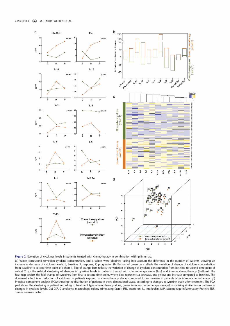
Figure 2. Evolution of cytokines levels in patients treated with chemotherapy in combination with ipilimumab.(a) Values correspond tomedian cytokine concentration, and p values were obtained taking into account the difference in the number of patients showing anincrease vs decrease of cytokines levels. B, baseline; R, response; P, progression (b) Bottom of green bars reflects the variation of change of cytokine concentrationfrom baseline to second time-point of cohort 1. Top of orange bars reflects the variation of change of cytokine concentration from baseline to second time-point ofcohort 2. (c) Hierarchical clustering of changes in cytokine levels in patients treated with chemotherapy alone (top) and immunochemotherapy (bottom). Theheatmap depicts the fold-change of cytokines from first to second time-point, where blue represents a decrease, and yellow and increase compared to baseline. Thedominant effect is of reduction of cytokines in patients exposed to chemotherapy alone, compared to an increase in patients after immunochemotherapy. (d)Principal component analysis (PCA) showing the distribution of patients in three-dimensional space, according to changes in cytokine levels after treatment. The PCAplot shows the clustering of patient according to treatment type (chemotherapy alone, green; immunochemotherapy, orange), visualizing similarities in patterns inchanges in cytokine levels. GM-CSF, Granulocyte-macrophage colony-stimulating factor; IFN, interferon; IL, interleukin; MIP, Macrophage Inflammatory Protein; TNF,Tumor necrosis factor.
e1593810-4 M. HARDY-WERBIN ET AL.

than 23% from baseline to response, had a significant worseOS (9.5 m vs 16.3 m; p = 0.001). This finding was maintainedwhen analyzing only the advanced patients, although it loststatistical significance (p = 0.063). However, those treatedwith immunochemotherapy whose IL-4 increased more than32% had a significant better OS (18.5 m vs 8.8 m; p = 0.042)(Figure 5).
Discussion
To the best of our knowledge, this is the first study to assesshow ipilimumab affects serum levels of immunomodulatorycytokines in SCLC. Our access to two cohorts of patients whowere treated with either immunochemotherapy or chemother-apy alone allowed us to assess the biological effect of theaddition of ipilimumab and to interrogate these data in thelight of clinical outcomes.
An intriguing result is the observation of concordantlylower serum levels of multiple cytokines in SCLC patientscompared to healthy controls. In the literature we onlyfound three patients that had been evaluated in this way;23
a functional study on whole blood stimulated in vitro revealeda lower cytokine release in cells from SCLC patients, perhapsoffering an insight into the underpinning biology of ourobservation.24 More data are available on circulating IL-6levels: a study of 72 patients with SCLC identified that inboth limited and extensive disease, elevated IL6 levels couldbe detected, consistent with our observation.25
The modulation of cytokine levels after treatment withimmunochemotherapy has not been previously reported inSCLC. We found that addition of ipilimumab increased concen-trations of the evaluated cytokines globally and appeared tocounteract the effect of chemotherapy that typically decreasedcytokines. It is recognized that SCLC cells may be the source ofcytokines and therefore successful treatment could reduce levelsas observed for the majority of cytokines. This might not be themain source, except for IL-6, as the pretreatment levels inpatients are already lower than in healthy controls. Immunecells are also sources of cytokines and can be affected by che-motherapy-induced apoptosis, likely contributing to the
observed decrease. Notably, however, patients treated with che-motherapy alone showed stabilization or increase in levels ofTNFα and Th2 cytokines. These data suggest that the importantcompartment of cells contributing to the presence of thesecytokines might not be affected by chemotherapy, for example,M2 macrophages in the tumor microenvironment.26,27 In theabsence of paired samples of tumor tissue, we were unable toevaluate this directly. Ipilimumab has previously been reportedto increase secretion of IFN-γ, IL-2R, IL-12, and IL-13 fromPBMC in vitro exposure,28 consistent with its proposed releaseof activated T-cells from inhibition.
Next, we explored the prognostic and predictive role ofcytokine levels at baseline and during treatment. IL-8 issecreted by malignant cells and tumor stroma cells; anti-IL-8antibodies have shown activity in vitro and in vivo29 and it isbeing currently tested in clinical trials (NCT02536469). Wefound that high baseline levels of IL-8 were associated withworse OS regardless of treatment type. This is consistent withprevious literature and is probably a surrogate of tumorburden as it showed a profound decrease withchemotherapy.30 Interestingly, IL-8 was minimally affectedby the addition of ipilimumab.
To evaluate the predictive value of cytokine levels after immu-nochemotherapy, we analyzed the changes in serum concentra-tions of each cytokine and compared the effect on outcome inboth cohorts. Although the cohorts have differences in baselineclinical characteristics, the possibility of comparing the effects ofthe combination to chemotherapy alone (standard treatment inSCLC up to date) provided the opportunity to individualize theeffects related to ipilimumab. Only associations that were sig-nificant for the ipilimumab treated cohort and were differentfrom those observed in the chemotherapy only arm, were con-sidered predictive of ipilimumab-linked effects. For instance,serum IL-2 behaved as a predictor of benefit to ipilimumab,and elevated baseline levels identified patients witha significant longer OS. No such difference was observed inpatients treated with chemotherapy alone. IL-2 is a cytokinethat promotes the proliferation of T cells, supporting the initia-tion and maintenance of immune response.31 Moreover, it sti-mulates the proliferation of natural killer cells and enhances
Figure 3. Baseline IL-8 may be an unfavorable prognostic marker of response to ipilimumab in SCLC.Patients treated either with chemotherapy alone or with ipilimumab harboring a high baseline IL-8 had a worse OS than those with a low baseline IL-8. mOS: medianoverall survival.
ONCOIMMUNOLOGY e1593810-5

their activity.32 As the regulation of T-cell activation throughbinding of CTLA4 to B7 may affect IL-2 secretion,33 the releaseof this blockade with ipilimumab would increase IL-2 concen-tration enhancing the immune response and could explain theobserved better outcome. In contrast, IL-6 and TNFα behaved aspredictors of resistance to ipilimumab: patients with higherbaseline concentrations treated with immunochemotherapyhad a shorter OS. Our data are consistent with observations inother solid cancers: IL-6 has been associated with tumor
progression in lung cancer34 and to a lack of benefit fromipilimumab in melanoma.35,36 Moreover, it has been tested asa target in cancer in vivo.37 TNFα has pro-tumorigenic activity incancer38 and has been linked to MAPK inhibitor resistance inmelanoma when secreted by macrophages. Increased serumTNFα might reflect an immunosuppressive tumor microenvir-onment explaining the observed associated resistance to ipili-mumab. Although these mechanisms seem plausible, theyrequire further validation.
Figure 4. Baseline concentrations of cytokines may predict benefit from ipilimumab in SCLC patients.(a) IL-2 appears to predict specific benefit from ipilimumab. (b) Patients with higher levels of IL-6 present worse OS when treated with chemoimmunotherapy but notwith chemotherapy alone, suggesting a lack of benefit from ipilimumab in this subgroup. (c) Patients with higher levels of TNFα had a worse OS. This difference insurvival was not replicated in patients treated with chemotherapy alone.
e1593810-6 M. HARDY-WERBIN ET AL.

As serial sampling was available in both cohorts, we eval-uated if changes in the cytokine serum levels could predict forbenefit from ipilimumab. Our results showed that IL-4 levelswere not significantly modified in patients treated with che-motherapy alone. In patients treated with chemotherapy inwhom IL-4 increased we observed a worse overall survival. Itis possible that this may be reflecting an effect on macrophageM2 polarization.39 Interestingly, IL-4 increased in the ipili-mumab treated cohort and patients experiencing this increasehad a better outcome. This increase has been observed in micetreated with ipilimumab but an association with outcome isnot observed after ipilimumab monotherapy.40,41 However,the evidence of the pro or antitumoral role of IL-4 in theliterature is contradictory and its function seems to depend onIL-4 levels and its association with other immunologicalmodulators.42 Globally, our results are novel and hypothesisgenerating, but warrant prospective validation.
In conclusion, we have observed differential impact ofipilimumab in serum cytokines in patients with SCLC.Baseline levels and changes on treatment might serve as con-venient predictive biomarkers of benefit from adding ipilimu-mab to chemotherapy in a disease where tumor biomarkersstudies are challenging.
Patients and methods
Patients and study design
We retrospectively evaluated two independent cohorts ofSCLC patients whose outcomes we have previouslyreported.43 Patients from cohort 1 were recruited betweenNovember of 2009 and January of 2014 at the Hospital delMar, Barcelona and treated with platinum plus etoposide.44,45
Cohort 2 included patients recruited to a phase II trial ofipilimumab at 10mg/kg, platinum and etoposide (ICE-trial).9
We included a control donor population of healthy, age- andsex-matched individuals (n = 30). Sample collection and dataanalyses were approved by the local ethics committee of theparticipating institutions and informed consent of each studyparticipant was obtained.
Sample collection
Serum samples were sequentially collected in each cohort: forcohort 1 at baseline (before starting treatment), at first responseevaluation (at 3 months approximately) and at progression; forcohort 2 at baseline, at 3 and 6 months. Whole blood sampleswere collected by standard venipuncture techniques usingserum separator tubes. Samples were allowed to clot for 30min at room temperature before centrifugation for 10 min at1000 g at 4 C. Following centrifugation, the supernatant(serum) was immediately removed and assayed immediatelyor aliquoted and stored frozen at −80 C until further use.
Cytokine assessment
Serum samples of all patients and healthy donors were eval-uated using a commercially Milliplex map Human HighSensitivity T Cell magnetic bead panel (Millipore, Billerica,MA, USA) coupled with the Luminex xMAP platform. Wemeasured a panel of Th1 (IFNγ, IL-2, TNFα), Th2 (IL-4, IL-5,IL-10), and inflammatory cytokines (GM-CSF, IL-1β, IL-6,IL-g8) plus MIP-1α in accordance with the manufacturer’sinstructions. Data were analyzed using five-parametric curvefitting and assay controls included kit standards andMultiplex controls. Intra-assay variabilities were less than12%. Duplicate measurements with a variability higher than35% were excluded. These experiments were supervised bytechnical personnel of the Luminex Core Facility at IMIM.
Cytokine cut-off calculation
To evaluate the association of cytokines levels with survival,we evaluated the impact of different cut-off methods, includ-ing medians (Supplementary Table S4) and ROC curves(Supplementary Table S6 and Supplementary Figure S3).Finally, as the endpoint for comparison was overall survival,we used the web-based software Cut-off Finder,46 previouslyused in the literature.47,48 This method takes into account thisendpoint outcome: for each cytokine, we identified the thresh-old level at which a log-rank test allowed segregation of
Figure 5. Modulation of IL-4 during treatment in SCLC could predict the outcome.Patients treated with chemotherapy alone whose IL-4 increased more than 23% from first to second time-point had a shorter OS, while those treated withimmunochemotherapy whose IL-4 increased more than 32% had a longer OS.
ONCOIMMUNOLOGY e1593810-7

patients into groups with good and poor outcomes(Supplementary Table S7). Then, we calculated the percentageof cytokine median concentration variation from firstto second time-point and considered >5% positive or negativevariations as significant changes (Supplementary Table S8).
Statistical analyses
Statistical analysis was carried out using Stata/MP 14 (StataCorpLLC, Texas, USA) and Prism 7.0c (GraphPad Software, Inc.).Baseline values of cytokines were compared among cohorts andhealthy volunteers using the non-parametric Mann–WhitneyU-test. Overall survival, measured from the date of start oftreatment until date of death or last visit, was plotted by theKaplan–Meier method and curves were compared with the log-rank test. All tests were conducted at the two-sided test witha 0.05 level of significance. R (v 3.4.3) was used to log2 transformdata, compute cytokine variation and to generate heat maps(using package gplots) and principal component analysis (PCA).
Disclosure statement
E.A. and C.O. have received honoraria for consultancy and lectures fromBristol–Myers Squibb outside of the current work. The remainingauthors report no conflict of interest.
Disclosure of Potential Conflicts of Interest
No potential conflicts of interest were disclosed.
Ethics approval and consent to participate:
Sample collection and data analyses were approved by the local ethicscommittee of the participating institutions and informed consent of eachstudy participant. The study was conducted in accordance with theEuropean Good Clinical Practice requirements (Declaration of Helsinki).
Funding
This work was supported in part by a grant from (1) Fundació La Marató deTV3 (666/C/2013); (2) ISCiii/FEDER (CIBERONC CB16/12/00241, RD12/0036/0051, PIE15/00008, PI15/00146, PI16/00591, PI13/00140); (3) Xarxa deBancs de Tumors (XBTC); (4) Agència de Gestió d’Ajuts Universitaris i deRecerca (AGAUR), Generalitat de Catalunya (2014SGR740), (5) FundacióCellex and (6) Cancer Research UK grant: C491/A12135.
References
1. Oze I, Hotta K, Kiura K, Ochi N, Takigawa N, Fujiwara Y, Tabata M,Tanimoto M. Twenty-seven years of phase III trials for patients withextensive disease small-cell lung cancer: disappointing results. PLoSOne. 2009;4(11):e7835. doi:10.1371/journal.pone.0007835.
2. Reck M, Luft A, Szczesna A, Havel L, Kim SW, Akerley W,Pietanza MC, Wu YL, Zielinski C, Thomas M, et al. Phase IIIrandomized trial of ipilimumab plus etoposide and platinumversus placebo plus etoposide and platinum in extensive-stagesmall-cell lung cancer. J Clin Oncol. 2016;34(31):3740–3748.doi:10.1200/JCO.2016.67.6601.
3. Darnell RB, Posner JB. Paraneoplastic syndromes and the nervoussystem. N Engl J Med. 2003;3(4):287–288. doi:10.1056/NEJMra023009.
4. Miller CW, Simon K, Aslo A, Kok K, Yokota J, Buys CH,Terada M, Koeffler HP. p53 mutations in human lung tumors.Cancer Res. 1992;52:1695–1698.
5. Rizvi NA,HellmannMD, Snyder A, Kvistborg P,MakarovV,Havel JJ,LeeW, Yuan J,Wong P,HoTS, et al.Mutational landscape determinessensitivity to PD-1 blockade in non-small cell lung cancer. Science.2015;348(6230):124–128. doi:10.1126/science.aaa1348.
6. Reck M, Bondarenko I, Luft A, Serwatowski P, Barlesi F,Chacko R, Sebastian M, Lu H, Cuillerot JM, Lynch TJ.Ipilimumab in combination with paclitaxel and carboplatin asfirst-line therapy in extensivedisease-small-cell lungcancer: resultsfrom a randomized, double-blind, multicenter phase 2 trial. AnnOncol. 2013;24(1):75–83. doi:10.1093/annonc/mds213.
7. Antonia SJ, López-Martin JA, Bendell J, Ott PA, Taylor M,Eder JP, Jäger D, Pietanza MC, Le DT, de Braud F, et al.Nivolumab alone and nivolumab plus ipilimumab in recurrentsmall-cell lung cancer (CheckMate 032): a multicentre, open-label,phase 1/2 trial. Lancet Oncol. 2016;17(7):883–895. doi:10.1016/S1470-2045(16)30098-5.
8. Ott PA, Elez E, Hiret S, Kim D-W, Morosky A, Saraf S, Piperdi B,Mehnert JM. Pembrolizumab in patients with extensive-stagesmall-cell lung cancer: results from the phase Ib KEYNOTE-028study. J Clin Oncol. 2017;35(34):3823–3829. doi:10.1200/JCO.2017.72.5069.
9. Arriola E, Wheater M, Galea I, Cross N, Maishman T, Hamid D,Stanton L, Cave J, Geldart T, Mulatero C, et al. Outcome andbiomarker analysis from a multicenter phase 2 study of ipilimu-mab in combination with carboplatin and etoposide as first-linetherapy for extensive-stage SCLC. J Thorac Oncol. 2016;11(9):1511–1521. doi:10.1016/j.jtho.2016.05.028.
10. Horn L, Mansfield AS, Szczęsna A, Havel L, Krzakowski M,Hochmair MJ, F H, Losonczy G, Johnson ML, Nishio M, et al.First-line atezolizumab plus chemotherapy in extensive-stagesmall-cell lung cancer. N Engl J Med 2018;379:2220–2229.doi:10.1056/NEJMoa1809064. NEJMoa1809064.
11. Ku GY, Yuan J, Page DB, Schroeder SEA, Panageas KS,Carvajal RD, Chapman PB, Schwartz GK, Allison JP,Wolchok JD. Single-institution experience with ipilimumab inadvanced melanoma patients in the compassionate use setting:lymphocyte count after 2 doses correlates with survival. Cancer.2010;116(7):1767–1775. doi:10.1002/cncr.24951.
12. Bristol-myers squibb announces checkmate −451 study did notmeet primary endpoint of overall survival with opdivo plus yervoyvs. placebo as a maintenance therapy in patients with extensive-stage small cell lung cancer after completion of first-line; [accessed 2018 Dec 18]. https://news.bms.com/press-release/corporatefinancial-news/bristol-myers-squibb-announces-checkmate-451-study-did-not-mee.
13. Bristol-Myers Squibb announces phase 3 checkmate −331 studydoes not meet primary endpoint of overall survival with opdivoversus chemotherapy in patients with previously treated relapsedsmall cell lung cancer; [accessed 2018 Dec 18]. https://news.bms.com/press-release/corporatefinancial-news/bristol-myers-squibb-announces-phase-3-checkmate-331-study-doe.
14. Travis WD. Pathology and diagnosis of neuroendocrine tumors:lung neuroendocrine. Thorac Surg Clin. 2014;24(3):257–266.doi:10.1016/j.thorsurg.2014.04.001.
15. Lee S, Margolin K. Cytokines in cancer immunotherapy. Cancers(Basel). 2011;3(4):3856–3893. doi:10.3390/cancers3043856.
16. Paul WE, Seder RA. Lymphocyte responses and cytokines. Cell.1994;76(2):241–251. doi:10.1016/0092-8674(94)90332-8.
17. HuangM,Wang J, Lee P, Sharma S,Mao JT,Meissner H, Uyemura K,Modlin R, Wollman J, Dubinett SM. Human non-small cell lungcancer cells express a type 2 cytokine pattern. Cancer Res. 1995;55(17):3847–3853. doi:10.1016/0169-5002(96)85851-X.
18. Hong CC, Yao S, McCann SE, Dolnick RY, Wallace PK, Gong Z,Quan L, Lee KP, Evans SS, Repasky EA, et al. Pretreatment levels ofcirculating Th1 and Th2 cytokines, and their ratios, are associated withER-negative and triple negative breast cancers. Breast Cancer ResTreat. 2013;139(2):477–488. doi:10.1007/s10549-013-2549-3.
e1593810-8 M. HARDY-WERBIN ET AL.

19. Lucey DR, Clerici M, Shearer GM. Type 1, and Type 2 cytokinedysregulation in human infectious, neoplastic, and inflammatorydiseases. Clin Microbiol Rev. 1996;9(4):532–562. doi:10.1128/CMR.9.4.532.
20. Lippitz BE. Cytokine patterns in patients with cancer:A systematic review. Lancet Oncol. 2013;14(6):218–228.doi:10.1016/S1470-2045(12)70582-X.
21. Pine SR, Mechanic LE, Enewold L, Chaturvedi AK, Katki HA,Zheng YL, Bowman ED, Engels EA, Caporaso NE, Harris CC.Increased levels of circulating interleukin 6, interleukin 8,c-reactive protein, and risk of lung cancer. J Natl Cancer Inst.2011;103(14):1112–1122. doi:10.1093/jnci/djr216.
22. Sanmamed MF, Perez-Gracia JL, Schalper KA, Fusco JP,Gonzalez A, Rodriguez-Ruiz ME, Oñate C, Perez G, Alfaro C,Martín-Algarra S, et al. Changes in serum interleukin-8 (IL-8)levels reflect and predict response to anti-PD-1 treatment inmelanoma and non-small-cell lung cancer patients. Ann Oncol.2017;28(8):1988–1995. doi:10.1093/annonc/mdx190.
23. Matanić D, Beg-Zec Z, Stojanović D, Matakorić N, Flego V, Milevoj-Ribić F. Cytokines in patients with lung cancer. Scand J Immunol.2003;57(2):173–178. doi:10.1046/j.1365-3083.2003.01205.x.
24. Fischer JR, Schindel M, Stein N, Lahm H, Gallati H,Krammer PH, Drings P. Selective suppression of cytokine secre-tion in patients with small-cell lung cancer. Ann Oncol Off J EurSoc Med Oncol. 1995;6(9):921–926. doi:10.1093/oxfordjournals.annonc.a059360.
25. Wójcik E, Jakubowicz J, Skotnicki P, Sas-Korczyńska B, Kulpa JK.IL-6 and VEGF in small cell lung cancer patients. Anticancer Res.2010;30:1773–1778.
26. Kitajima M, Ziegler SF. Cutting edge: identification of the thymicstromal lymphopoietin-responsive dendritic cell subset critical forinitiation of type 2 contact hypersensitivity. J Immunol. 2013;191(10):4903–4907. doi:10.4049/jimmunol.1302175.
27. Dijkgraaf EM, Heusinkveld M, Tummers B, Vogelpoel LTC,Goedemans R, Jha V, Nortier JWR, Welters MJP, Kroep JR, VanDer Burg SH. Chemotherapy alters monocyte differentiation tofavor generation of cancer-supporting m2 macrophages in thetumor microenvironment. Cancer Res. 2013;73(8):2480–2492.doi:10.1158/0008-5472.CAN-12-3542.
28. Yano H, Thakur A, Tomaszewski EN, Choi M, Deol A, Lum LG.Ipilimumab augments antitumor activity of bispecificantibody-armed T cells. J Transl Med. 2014;12(1):1–11. doi:10.1186/1479-5876-12-191.
29. Huang S, Mills L, B M, Tellez C, McCarty M, Yang X-D,Gudas JM, Bar-Eli M. Fully humanized neutralizing antibodiesto interleukin-8 (ABX-IL8) inhibit angiogenesis, tumor growth,and metastasis of human melanoma. Am J Pathol. 2002;161(1):125–134. doi:10.1016/S0002-9440(10)64164-8.
30. Young RJ, Tin AW, Brown NJ, Jitlal M, Lee SM, Woll PJ. Analysis ofcirculating angiogenic biomarkers from patients in two phase III trialsin lung cancer of chemotherapy alone or chemotherapy andthalidomide. Br J Cancer. 2012;106(6):1153–1159. doi:10.1038/bjc.2012.50.
31. Dembic Z. Cytokines of the Immune System. Cytokines ImmuneSyst. 2015:143–239. doi:10.1016/B978-0-12-419998-9.00006-7.
32. Henney CS, Kuribayashi K, Kern DE, Gillis S. Interleukin-2 aug-ments natural killer cell activity. Nature. 1981;291(5813):335–338.doi:10.1038/291335a0.
33. Krummel MF, Allison JP. CTLA-4 engagement inhibits IL-2 accu-mulation and cell cycle progression upon activation of resting T cells.J Exp Med. 1996;183(6):2533–2540. doi:10.1084/jem.183.6.2533.
34. Chang CH, Hsiao CF, Yeh YM, Chang GC, Tsai YH, Chen YM,Huang MS, Chen HL, Li YJ, Yang PC, et al. Circulatinginterleukin-6 level is a prognostic marker for survival in advancednonsmall cell lung cancer patients treated with chemotherapy.Int J Cancer. 2013;132(9):1977–1985. doi:10.1002/ijc.27892.
35. Damuzzo V, Solito S, Pinton L, Carrozzo E, Valpione S, Pigozzo J,Arboretti Giancristofaro R, Chiarion-Sileni V,Mandruzzato S. Clinicalimplication of tumor-associated and immunological parameters inmelanoma patients treated with ipilimumab. Oncoimmunology.2016;5(12):e1249559. doi:10.1080/2162402X.2016.1249559.
36. Bjoern J, Juul Nitschke N, Zeeberg Iversen T, Schmidt H, Fode K,Svane IM. Immunological correlates of treatment and response instage IV malignant melanoma patients treated with Ipilimumab.Oncoimmunology. 2016;5(4):e1100788. doi:10.1080/2162402X.2015.1100788.
37. Gopinathan G, Milagre C, Pearce OMT, Reynolds LE, Hodivala-Dilke K, Leinster DA, Zhong H, Hollingsworth RE, Thompson R,Whiteford JR, et al. Interleukin-6 stimulates defectiveangiogenesis. Cancer Res. 2015;75(15):3098–3107. doi:10.1158/0008-5472.CAN-15-1227.
38. Martínez-Reza I, Díaz L, García-Becerra R. Preclinical and clinicalaspects of TNF-α and its receptors TNFR1 and TNFR2 in breastcancer. J Biomed Sci. 2017;24(1):1–8. doi:10.1186/s12929-017-0398-9.
39. Rahal OM, Wolfe AR, Mandal PK, Larson R, Tin S, Jimenez C,Zhang D, Horton J, Reuben JM, McMurray JS, et al. BlockingInterleukin (IL)4- and IL13-mediated phosphorylation of STAT6(Tyr641) decreases M2 polarization of macrophages and protectsagainst macrophage-mediated radioresistance of inflammatorybreast cancer. Int J Radiat Oncol Biol Phys. 2018;100(4):1034–1043. doi:10.1016/j.ijrobp.2017.11.043.
40. Redmond WL, Linch SN, Kasiewicz MJ. Combined targeting ofcostimulatory (OX40) and coinhibitory (CTLA-4) pathways elicitspotent effector t cells capable of driving robust antitumorimmunity. Cancer Immunol Res. 2014;2(2):142–153.doi:10.1158/2326-6066.CIR-13-0031-T.
41. Linch SN, Redmond WL. Combined OX40 ligation plus CTLA-4blockade: more than the sum of its parts. Oncoimmunology.2014;3(3):11–13. doi:10.4161/onci.28245.
42. Li Z, Chen L, Qin Z. Paradoxical roles of IL-4 in tumor immunity.Cell Mol Immunol. 2009;6(6):415–422. doi:10.1038/cmi.2009.53.
43. Hardy-Werbin M, Arpí O, Taus A, Rocha P, Joseph-Pietras D,Nolan L, Danson S, Griffiths R, Lopez-Botet M, Rovira A, et al.Assessment of neuronal autoantibodies in patients with small celllung cancer treated with chemotherapy with or withoutipilimumab. Oncoimmunology. 2018;7(2):e1395125. doi:10.1080/2162402X.2017.1395125.
44. Cañadas I, Taus Á, Villanueva X, Arpí O, Pijuan L,Rodríguez Y, Menéndez S, Mojal S, Rojo F, Albanell J, et al.Angiopoietin-2 is a negative prognostic marker in small celllung cancer. Lung Cancer. 2015;90(2):302–306. doi:10.1016/j.lungcan.2015.09.023.
45. Cañadas I, Taus A, González I, Villanueva X, Gimeno J, Pijuan L,Dómine M, Sánchez-Font A, Vollmer I, Menéndez S, et al. Highcirculating hepatocyte growth factor levels associate with epithelialto mesenchymal transition and poor outcome in small cell lungcancer patients. Oncotarget. 2014;5(14):5246–5256. doi:10.18632/oncotarget.2124.
46. Budczies J, Klauschen F, Sinn BV, Gyorffy B, Schmitt WD, Darb-Esfahani S, Cutoff Finder: DC, Comprehensive A. Straightforwardweb application enabling rapid biomarker cutoff optimization. PLoSOne. 2012;7(12):1–7. doi:10.1371/journal.pone.0051862.
47. Lin F, Marcelo KL, Rajapakshe K, Coarfa C, Dean A, Wilganowski N,Robinson H, Sevick E, Bissig K-D, Goldie LC, et al. The camKK2/camKIV relay is an essential regulator of hepatic cancer. Hepatology.2015;62(2):505–520. doi:10.1002/hep.27832.
48. Depeille P, Henricks LM, Rah VDV, Lemmens E, Wang C-Y,Matli M, Werb Z, Haigis KM, Donner D, Warren R, et al.RasGRP1 opposes proliferative EGFR–SOS1–ras signals andrestricts intestinal epithelial cell growth. Nat Cell Biol. 2015;17(6):804–815. doi:10.1038/ncb3175.
ONCOIMMUNOLOGY e1593810-9

Tesis doctoral – Max Hardy-Werbin
76

Resultados
77
Material suplementario correspondiente al artículo 2:
Supplementary figure S1. Baseline levels of TNFa were significantly increased in
patients over 60 years old [9.15 pg/mL vs 6.84 pg/mL (p=0.0067)]; IL-5 was
significantly increased in patients with PS 1 or 2 vs PS 0 [2.83 pg/mL vs 1.18 pg/mL
(p=0.047)]; and IL-2 was significantly increased in female patients compared with male
[2.76 pg/mL vs 1.78 pg/mL (p=0.037)].
Supplementary figure S2. IL-4 and Mip-1a were significantly decreased in patients
with extensive disease when compared to those with limited disease.

Tesis doctoral – Max Hardy-Werbin
78
Supplementary figure S3. Among other methods, ROC curves were calculated to
evaluate its impact as a cut off parameter (see Supplementary Table S6).

Resultados
79
Supplementary table S1. Patients’ characteristics
Characteristics Cohort 1 Chemotherapy N = 47
Cohort 2 Chemotherapy + Ipilimumab N = 37
Sex, n (%) Female Male
12 (25.5) 35 (74.5)
13 (35.1) 24 (64.9)
Age, mean (range) 62.6 (45 – 86)
62.6 (44 – 84)
PS, n (%) 0 1 2 NA
26 (55,3) 14 (29,8) 7 (14,9) 0
11 (29.7) 20 (54.1) 0 6 (16.2)
Stage, n (%) I-III IV
12 (25,5) 35 (74,5)
0 37 (100)
Smoking status, n (%) Smokers Former smokers Never smokers NA
33 (70.2%) 9 (19.1%) 0 5 (10.6%)
4 (10.8%) 6 (16.2%) 0 27 (72.9%)
Mean serum albumin level (g/L)
39.8
38.4
NA, not available; PS, performance status
Supplementary table S2. Median cytokine concentration in healthy volunteers and SCLC patients (both cohorts).
Healthy volunteers SCLC patients (global) p-value Median
(pg/mL) Q1 – Q3 Median (pg/mL) Q1 – Q3
GM-CSF 47.22 29.6 – 86.76 39.23 17.01 – 61.31 0.209 IFNg 11.32 4.6 – 16.48 8.45 4.35 – 12.54 0.139 IL-10 13.42 6.73 – 22.37 10.97 5.1 – 18.19 0.181 IL-1b 3.14 1.72 – 4.82 1.97 1.17 – 3.14 0.014 IL-2 2.34 1.77 – 3.9 1.945 1.22 – 3.25 0.156 IL-4 42.98 26.38 – 79.45 32.54 17.75 – 61.33 0.070 IL-5 2.95 1.65 – 4.51 1.82 0.75 – 3.6 0.013 IL-6 3.69 2.57 – 6.06 5.27 2.9 – 8.9 0.107 IL-8 14.66 6.93 – 29.31 12.99 8.76 – 25.25 0.715 MIP-1a 17.79 14.25 – 30.49 12.12 8.87 – 16.08 0.0001 TNFa 10.17 8.29 – 12.87 8.355 6.36 – 11.66 0.042
GM-CSF, Granulocyte-macrophage colony-stimulating factor; IFN, interferon; IL, interleukin; MIP, Macrophage Inflammatory Protein; TNF, Tumor necrosis factor. Q1, first quartile; Q3, third quartile; significant p-values in bold.
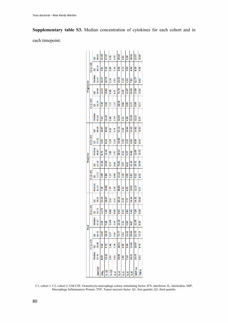
Tesis doctoral – Max Hardy-Werbin
80
Supplementary table S3. Median concentration of cytokines for each cohort and in
each timepoint.
C1, cohort 1; C2, cohort 2; GM-CSF, Granulocyte-macrophage colony-stimulating factor; IFN, interferon; IL, interleukin; MIP, Macrophage Inflammatory Protein; TNF, Tumor necrosis factor. Q1, first quartile; Q3, third quartile.

Resultados
81
Supplementary table S4. Baseline cut-off values calculated using median cytokine
concentrations. The Hazard Ratio for survival was calculated using the median as a cut-
off for each cohort.
Cohort 1 Median HR 95% CI p-value GM-CSF 37.76 0.68 0.35 – 1.3 0.24 IFNg 8.69 0.59 0.32 – 1.1 0.096 IL-10 13.15 1.07 0.57 – 1.98 0.84 IL-1b 2.34 0.65 0.34 – 1.25 0.2 IL-2 2.65 0.76 0.41 – 1.44 0.4 IL-4 48.99 0.69 0.37 – 1.26 0.22 IL-5 1.84 0.58 0.31 – 1.07 0.075 IL-6 7.10 1.17 0.64 – 2.14 0.62 IL-8 19.21 1.79 0.99 – 3.26 0.052 MIP-1a 13.45 1.16 0.64 – 2.11 0.63 TNFa 8.51 1.12 0.61 – 2.02 0.72 Cohort 2 Median HR 95% CI p-value GM-CSF 41.37 0.73 0.26 – 2.05 0.55 IFNg 5.58 0.81 0.31 – 2-12 0.66 IL-10 7.28 1.43 0.56 – 3.67 0.45 IL-1b 1.245 0.81 0.3 – 2.21 0.68 IL-2 1.59 0.4 0.14 – 1.17 0.083 IL-4 22.59 0.8 0.26 – 2.46 0.69 IL-5 1.36 0.95 0.36 – 2.48 0.92 IL-6 4.06 2.54 0.84 – 7.73 0.089 IL-8 8.87 1.44 0.59 – 3.55 0.42 MIP-1a 11.05 0.9 0.37 – 2.18 0.82 TNFa 8.36 2.72 1.02 – 7.25 0.037
GM-CSF, Granulocyte-macrophage colony-stimulating factor; IFN, interferon; IL, interleukin; MIP, Macrophage Inflammatory Protein; TNF, Tumor necrosis factor; HR, Hazard Ratio; CI, confidence interval.
Supplementary Table S5. Multivariate analyses including the most relevant cytokines.
Cohort 1 Cohort 2 HR 95% CI p-value HR 95% CI p-value IL2 high 1.06 0.55 – 2.05 0.848 0.01 0.00 – 0.17 0.001 IL6 high 0.86 0.34 – 2.18 0.758 32.03 2.64 – 387.66 0.006 TNFa high 0.77 0.29 – 2.02 0.591 5.09 1.02 – 25.3 0.047 PS 1.76 0.94 – 3.29 0.075 0.48 0.09 – 2.51 0.383 Stage 4.26 1.68 – 10.79 0.002 1 - -

Tesis doctoral – Max Hardy-Werbin
82
Supplementary table S6. Baseline cut-off values calculated using ROC curves.
Cytokine Cut-off Cohort 1 - Chemotherapy (pg/mL)
Cohort 2 - Chemoimmunotherapy (pg/mL)
GM-CSF 50.69 59.71 IFNg 9.45 16.45 IL-10 13.15 13.42 IL-1b 5.07 1.65 IL-2 2.76 1.63 IL-4 54.53 31.46 IL-5 3.04 0.31 IL-6 7.1 4.95 IL-8 15.94 17.2 MIP-1a 14.77 7.99 TNFa 8.72 8.73
GM-CSF, Granulocyte-macrophage colony-stimulating factor; IFN, interferon; IL, interleukin; MIP, Macrophage Inflammatory Protein; TNF, Tumor necrosis factor.
Supplementary table S7. Baseline cut-off values for each cytokine and each cohort,
using the web-based software Cutoff Finder.
Cytokine Cut-off Cohort 1 - Chemotherapy (pg/mL)
Cohort 2 - Chemoimmunotherapy (pg/mL)
GM-CSF 51.08 17.87 IFNg 9.03 10.24 IL-10 14.84 6.08 IL-1b 2.15 1.735 IL-2 3.165 1.65 IL-4 41.04 24.44 IL-5 2.92 2.97 IL-6 4.2 3.85 IL-8 18.02 13.82 MIP-1a 14.42 7.72 TNFa 6.555 9.16
GM-CSF, Granulocyte-macrophage colony-stimulating factor; IFN, interferon; IL, interleukin; MIP, Macrophage Inflammatory Protein; TNF, Tumor necrosis factor.

Resultados
83
Supplementary table S8. Cut-off values for each cytokine and each cohort, considering
the percentage of cytokine level change from first to second time-point.
Cytokine Cut-off Cohort 1 – Chemotherapy (% of change)
Cohort 2 - Chemoimmunotherapy (% of change)
GM-CSF 6.823 25.58 IFNg -10,2 51.06 IL-10 26.43 185.2 IL-1b -2.385 52.63 IL-2 -4.704 -2.877 IL-4 23.34 32.57 IL-5 -24.29 44.63 IL-6 -4-292 -2.471 IL-8 -42.2 -27.46 MIP-1a -16.73 3.332 TNFa -5.966 42.82
GM-CSF, Granulocyte-macrophage colony-stimulating factor; IFN, interferon; IL, interleukin; MIP, Macrophage Inflammatory Protein; TNF, Tumor necrosis factor.

Tesis doctoral – Max Hardy-Werbin
84

Resultados
85
Tercera parte: fenotipado de PBMCs
Características de las poblaciones de estudio
En esta parte del proyecto se han incluido 53 pacientes con CPCP: 37 pacientes de la
cohorte 2 (ensayo clínico ICE), tratados con quimioterapia e ipilimumab, y 16 pacientes
de la cohorte 3 (recogidos de manera prospectiva en el Hospital del Mar), tratados con
quimioterapia sola. El 100% de los pacientes de las dos cohortes tenían enfermedad
diseminada al momento del diagnóstico (Tabla 4.1). La cohorte 3 incluyó una población
con mayor proporción de hombres, con una edad media similar, y con pacientes con
performance status (PS) 2 y 3, a diferencia de la cohorte 2, que solo incluyó pacientes
con PS 0 y 1. La mediana de SG fue de 17m en la cohorte 2 y de 8.2m en la cohorte 3;
la mediana de SLP fue de 6.8 meses (m) en la cohorte 2 y de 6.1m en la cohorte 3.
Características Cohorte 2
Quimioterapia + Ipilimumab N = 37
Cohorte 3 Quimioterapia
N = 16 Sexo, n (%) Femenino Masculino
13 (35.1) 24 (64.9)
5 (31.4) 11 (68.6)
Edad, media (rango) 62.6 (44 – 84) 65.5 (56 – 83) PS, n (%) 0 1 2 3 NA
11 (29.7) 20 (54.1)
0 0
6 (16.2)
1 (6.25) 12 (75) 2 (12.5) 1 (6.25)
0 Estadio, n (%) I-III IV
0
37 (100)
0
16 (100) Tabaquismo, n (%) Fumadores Ex-fumadores No fumadores NA
4 (10.8%) 6 (16.2%)
0 27 (72.9%)
9 (56.25%) 7 (43.75%)
0 0
Tabla 4.1. Características clínicas de los pacientes
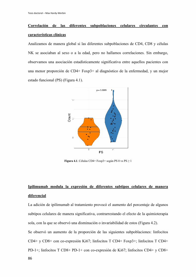
Tesis doctoral – Max Hardy-Werbin
86
Correlación de las diferentes subpoblaciones celulares circulantes con
características clínicas
Analizamos de manera global si las diferentes subpoblaciones de CD4, CD8 y células
NK se asociaban al sexo o a la edad, pero no hallamos correlaciones. Sin embargo,
observamos una asociación estadísticamente significativa entre aquellos pacientes con
una menor proporción de CD4+ Foxp3+ al diagnóstico de la enfermedad, y un mejor
estado funcional (PS) (Figura 4.1).
Figura 4.1. Células CD4+ Foxp3+ según PS 0 vs PS ≥ 1
Ipilimumab modula la expresión de diferentes subtipos celulares de manera
diferencial
La adición de ipilimumab al tratamiento provocó el aumento del porcentaje de algunos
subtipos celulares de manera significativa, contrarrestando el efecto de la quimioterapia
sola, con la que se observó una disminución o invariabilidad de estos (Figura 4.2).
Se observó un aumento de la proporción de las siguientes subpoblaciones: linfocitos
CD4+ y CD8+ con co-expresión Ki67; linfocitos T CD4+ Foxp3+; linfocitos T CD4+
PD-1+; linfocitos T CD8+ PD-1+ con co-expresión de Ki67; linfocitos CD4+ y CD8+
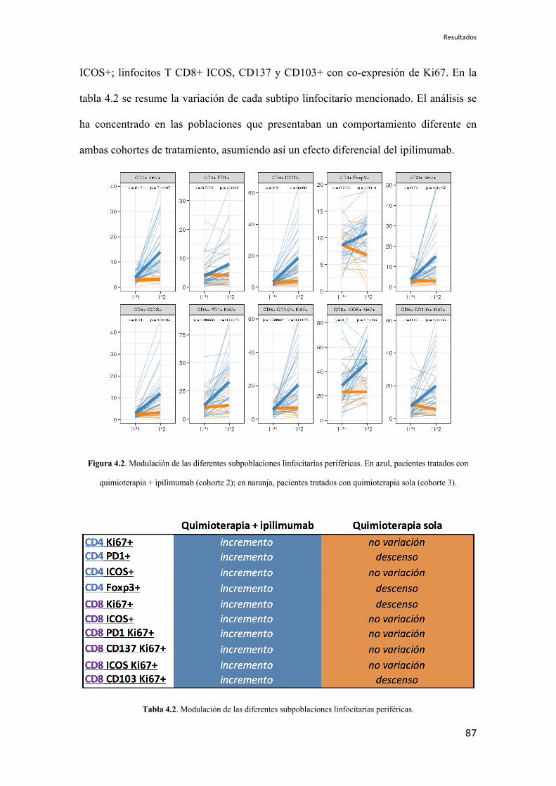
Resultados
87
ICOS+; linfocitos T CD8+ ICOS, CD137 y CD103+ con co-expresión de Ki67. En la
tabla 4.2 se resume la variación de cada subtipo linfocitario mencionado. El análisis se
ha concentrado en las poblaciones que presentaban un comportamiento diferente en
ambas cohortes de tratamiento, asumiendo así un efecto diferencial del ipilimumab.
Figura 4.2. Modulación de las diferentes subpoblaciones linfocitarias periféricas. En azul, pacientes tratados con
quimioterapia + ipilimumab (cohorte 2); en naranja, pacientes tratados con quimioterapia sola (cohorte 3).
Tabla 4.2. Modulación de las diferentes subpoblaciones linfocitarias periféricas.
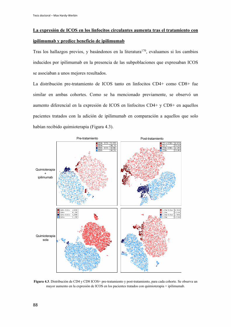
Tesis doctoral – Max Hardy-Werbin
88
La expresión de ICOS en los linfocitos circulantes aumenta tras el tratamiento con
ipilimumab y predice beneficio de ipilimumab
Tras los hallazgos previos, y basándonos en la literatura170, evaluamos si los cambios
inducidos por ipilimumab en la presencia de las subpoblaciones que expresaban ICOS
se asociaban a unos mejores resultados.
La distribución pre-tratamiento de ICOS tanto en linfocitos CD4+ como CD8+ fue
similar en ambas cohortes. Como se ha mencionado previamente, se observó un
aumento diferencial en la expresión de ICOS en linfocitos CD4+ y CD8+ en aquellos
pacientes tratados con la adición de ipilimumab en comparación a aquellos que solo
habían recibido quimioterapia (Figura 4.3).
Figura 4.3. Distribución de CD4 y CD8 ICOS+ pre-tratamiento y post-tratamiento, para cada cohorte. Se observa un mayor aumento en la expresión de ICOS en los pacientes tratados con quimioterapia + ipilimumab.
Pre-tratamiento Post-tratamiento
Quimioterapia +
ipilimumab
Quimioterapia sola

Resultados
89
Para evaluar cómo modificaba el tratamiento la expresión del receptor, calculamos el
porcentaje de variación de la expresión de ICOS entre la muestra pre y post-tratamiento
para cada paciente. Utilizando estos valores, mediante el paquete de R ‘survminer’
identificamos el valor que mejor segregaba dos poblaciones teniendo en cuenta la SG.
De esta manera, observamos que aquellos pacientes tratados con ipilimumab además de
quimioterapia, que sufrían un aumento proporcional en la expresión de ICOS por
encima del punto de corte, tanto en CD4+ (mayor al 310%) como en CD8+ (mayor al
142%), tenían una mejor supervivencia (18.5m vs 7.8m, p=0.012 y 18.5m vs 6.9m,
p=0.0068 respectivamente) (Figura 4.4). Esta asociación no se observó en los pacientes
tratados con quimioterapia sola.
CD4+ ICOS+
CD8+ ICOS+
Figura 4.4. A: pacientes tratados con quimioterapia + ipilimumab con una mayor expansión de CD4+ ICOS+, presentan una mejor supervivencia. B: pacientes tratados con quimioterapia + ipilimumab con una mayor expansión
de CD8+ ICOS+ tras el tratamiento, presentan una mejor supervivencia.
Pre-tratamiento Post-tratamiento
Quimioterapia +
ipilimumab
Quimioterapia sola
Pre-tratamiento Post-tratamiento
Quimioterapia +
ipilimumab
Quimioterapia sola
A
B

Tesis doctoral – Max Hardy-Werbin
90
El estado madurativo de las células NK puede predecir la respuesta a ipilimumab
en CPCP
Además de la caracterización de las diferentes subpoblaciones de los linfocitos T en
sangre periférica, basándonos en trabajos previos del grupo226,227 se llevó a cabo el
estudio de las células NK, entendiéndose éstas como aquellas células CD3- CD56+.
Este análisis incluyó diversos marcajes de receptores para definir distintos subtipos de
células NK (NKG2A, CD16, NKG2C, NKG2D) y sus estados de diferenciación
(CD57).
Observamos que aquellos pacientes tratados son quimioterapia + ipilimumab que
presentaban un porcentaje de CD57+ en el total de células NK por encima del 45%
antes de iniciar el tratamiento, presentaban una SG de 18.5m, mientras que aquellos con
un porcentaje de CD57+ por debajo del 45% presentaban una SG de 7.8m (p=0.002)
(Figura 4.5). Esta asociación no se mantuvo en los pacientes tratados con quimioterapia
sola, lo que indica que podría ser un marcador predictivo de beneficio de ipilimumab.
Figura 4.5. Un mayor porcentaje pre-tratamiento de células NK CD57+ se asocia con una mayor supervivencia global en pacientes tratados con quimioterapia + ipilimumab
.

Resultados
91
El fenotipado de sangre periférica podría predecir toxicidad por ipilimumab en
CPCP
Dada la elevada tasa de efectos adversos por ipilimumab reportada en los diferentes
ensayos clínicos, investigamos si existía alguna distribución específica de
subpoblaciones celulares periféricas que pudiera predecir la aparición de estos. Entre los
pacientes incluidos en nuestro estudio tratados con quimioinmunoterapia, un 70% había
sufrido al menos un efecto adverso grado 3; el desarrollo de efectos adversos se asoció a
una baja expresión de PD-1+ en los linfocitos CD8+ antes de empezar el tratamiento (p
= 0.012) (Figura 4.6).
Figura 4.6. Porcentaje de expresión de PD-1 en los linfocitos CD8+ en pacientes que no sufrieron ningún efecto adverso grado 3 secundario a ipilimjumab (izquierda) vs aquellos que sufrieron al menos un efecto adverso grado 3
(derecha).

Tesis doctoral – Max Hardy-Werbin
92
El 62% de los pacientes padecieron efectos adversos intestinales en forma de colitis.
Aquellos con un porcentaje de linfocitos CD4+ superior antes de iniciar el tratamiento
fueron más propensos a desarrollar colitis de cualquier grado (p = 0.0089) (Figura 4.7).
Figura 4.7. Porcentaje de linfocitos CD4+ en pacientes que no sufrieron colitis de ningún tipo secundaria a ipilimumab (izquierda) vs aquellos que sufrieron al menos un episodio de colitis de cualquier grado (derecha).
La co-expresión de CD137+ en los linfocitos CD4+ se asoció con la aparición de colitis
más severa (grado 3 y 4) (p = 0.016) (Figura 4.8).
Figura 4.8. Porcentaje de expresión de CD137 en los linfocitos CD4+ en pacientes que no sufrieron colitis o sufrieron algún episodio de colitis leve (hasta grado 2) (izquierda) vs aquellos que sufrieron colitis grave (grado 3 o
4) (derecha).

Discusión
5. DISCUSIÓN

Tesis doctoral – Max Hardy-Werbin
94

Discusión
95
El CPCP es una enfermedad sumamente agresiva y presenta un pronóstico infausto. Los
avances en el tratamiento en enfermedad metastásica han sido escasos y se han
caracterizado por sucesivos fracasos de los ensayos clínicos en los que se ha buscado
una alternativa al tratamiento con platino en primera línea. La llegada de la
inmunoterapia ha dado paso a una nueva era en el tratamiento del cáncer; esto tiene una
especial relevancia en el CPCP dada la falta de tratamientos efectivos en esta
enfermedad. Particularmente, ipilimumab fue el primer fármaco inmunoestimulador que
se testó en CPCP, demostrando ser efectivo y obteniendo largos supervivientes. Sin
embargo, la mayoría de los pacientes no responden al tratamiento. Por lo tanto, la
búsqueda de biomarcadores predictivos que puedan ayudar a la selección de pacientes
que se puedan beneficiar del tratamiento es fundamental. En este proyecto de tesis se ha
intentado abordar esta necesidad.
Dada la dificultad del acceso a muestras de tejido en CPCP, nos hemos centrado en el
estudio de marcadores periféricos: además de tratarse de una técnica poco cruenta,
permite la toma seriada muestras para poder estudiar no sólo los eventos inmunológicos
estáticos sino ver como estos se modulan a lo largo del tiempo con el tratamiento.
El trabajo se ha centrado en tres áreas. En primer lugar, se han analizado
autoanticuerpos neuronales (AAN) en suero, característicos de los SPN, que presentan
una alta prevalencia en el CPCP. En segundo lugar, se ha evaluado la presencia de
citoquinas séricas, tanto pro-inflamatorias como supresoras. En tercer y último lugar, se
ha llevado a cabo la caracterización del fenotipo periférico de diferentes poblaciones
inmunológicas.

Tesis doctoral – Max Hardy-Werbin
96
Estudio y análisis de autoanticuerpos neuronales
En el CPCP pueden darse una serie de eventos inmunológicos asociados al tumor, que
se manifiestan en forma de los llamados síndromes paraneoplásicos y se observan en la
clínica como eventos adversos neurológicos102. En pacientes con síndromes
paraneoplásicos neurológicos (SPN) se pueden detectar en sangre autoanticuerpos
dirigidos contra antígenos neuronales que se expresan de manera aberrante en el tumor.
Estos autoanticuerpos neuronales (AAN) pueden tener un papel como una herramienta
diagnóstica en SPNs relacionados con CPCP104. En un estudio, Titulaer y cols.121
analizaron la presencia de anticuerpos frente a múltiples miembros de la familia de Sox
y otros AAN (anti-HuD entre ellos) en pacientes con CPCP con y sin diagnóstico de
SPN, así como en pacientes con síndromes paraneoplásicos sin tumor. Observaron que
la detección de anticuerpos anti-Sox podía tener un valor diagnóstico para discriminar
síndromes paraneoplásicos asociados a CPCP de aquellos que no están asociados al
tumor. Por otro lado, recientemente se ha reportado que la detección a modo de cribado
de autoanticuerpos específicos asociados a antígenos tumorales podría tener un papel
relevante en la detección precoz tanto de CPCNP como CPCP228.
El daño neuronal a consecuencia de la autoinmunidad provocada en los SPN en el
CPCP puede tener diferentes mecanismos de acción. En el caso de los autoanticuerpos
contra antígenos de superficie, son los propios anticuerpos anti-antígenos de membrana
los efectores de la disfunción celular causante de la clínica neurológica123,229–232. Sin
embargo, se ha reportado que los AAN intracelulares no son patogénicos per se 233,234,
sino que es una respuesta mediada por células T la responsable del daño
neurológico109,235. En este sentido, y de manera congruente con este concepto, existe
una proporción de pacientes con CPCP en los que se detectan AAN en suero en
ausencia de síntomas neurológicos. En 1990 Dalmau y cols.236 analizaron la presencia

Discusión
97
de anti-HuD en el suero de 50 sujetos sanos y 69 pacientes con CPCP, de los cuales 25
tenían algún SPN asociado y el resto no. En el 16% de los pacientes con CPCP y sin
SPN asociado se detectaron niveles positivos de anti-HuD. En estos pacientes el título
de anticuerpo era inferior al de aquellos en los que se detectó anti-HuD en presencia de
síntomas neurológicos. En un estudio similar con 196 pacientes sin SPN, también se
observó una prevalencia de anti-HuD en suero del 16% en ausencia de síntomas
neurológicos237. En un estudio más reciente, Gozzard y cols.128 reportaron un 9.6% de
pacientes con CPCP y sin evidencia de SPN que tenía positivización de anti-HuD en
suero; sin embargo al testar otros autoanticuerpos frente a otros antígenos (Sox2,
Gad65, Amphyphisina, Ri, Cv2, Ma2, Yo), más del 40% de los pacientes tenían
positividad para al menos uno de ellos. En nuestro estudio ningún paciente presentaba
síntomas neurológicos al diagnóstico de la enfermedad y el 9.5% de ellos tenía niveles
detectables en suero de anti-HuD y el 53% de ellos tenían positividad para al menos un
autoanticuerpo. Para evaluar si existían diferencias en el título de anticuerpos entre los
pacientes con y sin SPN, se incluyó en el estudio como control positivo a un paciente
con CPCP diagnosticado de una encefalitis límbica paraneoplásica con positividad de
anti-HuD en suero. Todos los pacientes de nuestro estudio con detección de anti-HuD
presentaron niveles inferiores al control positivo menos uno de ellos.
El AAN más prevalente en nuestro estudio fue anti-Sox1, observándose en un 37% de
los pacientes, dato que fue consistente con otras series reportadas121,128. Generalmente,
tras Sox1, el autoanticuerpo más frecuente es anti-HuD. Sin embargo, en nuestra serie,
el segundo anticuerpo más prevalente era el dirigido contra el antígeno Yo. Este
autoanticuerpo se detecta más habitualmente al cáncer de ovario o al cáncer de
mama115, aunque también se ha descrito asociado a cáncer de pulmón238. Por otro lado,
anti-Gad65, que en general está asociado a pacientes con diabetes mellitus239, se detectó

Tesis doctoral – Max Hardy-Werbin
98
en 5 pacientes en nuestro estudio (3 en la cohorte 1, tratados con quimioterapia; 2 en la
cohorte 2, tratados con quimioinmunoterapia), y ninguno de estos pacientes tenía
antecedentes de diabetes tipo 1. Al tratarse de un pequeño número de pacientes, y con
poca probabilidad de positividad según la clínica reportada, podría argumentarse que se
tratara de un error de la técnica de medición del anticuerpo. Sin embargo, este método
se ha utilizado en estudios previos240,241 y ha demostrado ser eficiente como herramienta
de detección como se ha hecho en nuestro proyecto. Por otro lado, el tener diferentes
muestras seriadas dentro de la misma serie de pacientes, objetivándose de manera
repetida la positividad frente al anticuerpo, ha servido como control interno,
descartando razonablemente el error técnico.
La presencia de AAN se ha asociado al diagnóstico de CPCP en estadio limitado y
como consecuencia a mejores cifras de supervivencia. En el reporte de Dalmau y cols.
citado previamente236, todos los pacientes con CPCP sin SPN y con detección de anti-
HuD en suero habían sido diagnosticados con enfermedad limitada, mientras que entre
aquellos que eran negativos para anti-HuD, más de la mitad tenían enfermedad
diseminada. Posteriormente, Verschuuren y cols.242 reportaron una serie de pacientes
con CPCP con un 17% de prevalencia para la detección de anti-HuD en suero; de estos,
más de la mitad se habían diagnosticado con enfermedad limitada. En una serie de 200
pacientes con encefalomielitis asociada a anti-HuD, se diagnosticó un CPCP en 111 de
ellos, y de estos, el 75% se encontraba en estadio limitado129. En una serie más reciente
de 59 pacientes con CPCP donde Atakan y cols.243 analizaron la presencia de anti-Sox1,
observaron que de los 17 pacientes seropositivos para Sox-2, 12 tenían enfermedad
limitada. Por último, en el ensayo clínico ICE149 donde se analizaron las mismas
muestras de nuestra cohorte de pacientes tratados con la combinatoria con ipilimumab
(pero para la detección de un panel diferente de autoanticuerpos), también se observó

Discusión
99
una relación entre la seropostividad de autoanticuerpos y beneficio al tratamiento. Los
datos de nuestro estudio fueron consistentes con todas estas series: en el 75% de los
pacientes con enfermedad limitada se detectó al menos un NAA en suero, y esta
asociación tuvo una tendencia hacia la significación estadística para el global de todos
los NAA así como de manera individual para anti-Sox1. En estos casos, la existencia de
una respuesta citotóxica de células T concomitante234,244,245 podría haber contribuido en
parte al control del crecimiento tumoral, pero al mismo tiempo podría haber llegado a
sortear la barrera inmunológica del sistema nervioso, característica por brindar una
protección diferencial246. Además, en pacientes con CPCP que presentan SPN, los
síntomas neurológicos a menudo se presentan antes de la detección del tumor102,247,248
sugiriendo que el sistema inmune es capaz de detectar y controlar la presencia del
cáncer incluso antes del desarrollo de las metástasis.
Al analizar un panel con múltiples autoanticuerpos, se detectó la presencia en suero no
solo de anticuerpos de manera individual sino también la coexistencia de diferentes
reactividades. En un 28% de pacientes en nuestro estudio se detectó la presencia de más
de un AAN. Estos datos son comparables son los de series reportadas previamente. En
un estudio donde se analizó de manera retrospectiva el suero de 60 mil pacientes con
sospecha de SPN para la detección de autoanticuerpos, en 553 (el 0.9%) se detectó
algún AAN, y de estos, el 31% presentaban reactividad para más de uno104.
Posteriormente analizamos el impacto en la supervivencia de la detección de un solo
AAN frente a la detección de más de un AAN en los pacientes con CPCP. De manera
sorprendente, observamos que aquellos pacientes con detección de múltiples AAN
tenían una peor SLP y SG que aquellos con una sola reactividad cuando se trataban con
la combinación de quimioterapia e ipilimumab. La coexistencia de múltiples
autoanticuerpos refleja una respuesta inmunológica diversificada frente a diferentes

Tesis doctoral – Max Hardy-Werbin
100
antígenos onconeuronales expresados en los tumores, que a su vez se puede explicar por
una mayor heterogeneidad tumoral: esto conllevaría a su vez una respuesta inmune
subclonal de células T. En este escenario se ha reportado que se obtienen peores
resultados con el tratamiento con inhibidores de punto de control en tumores con
predominio de neoantígenos subclonales249, lo cual podría apoyar nuestros resultados.
Un estudio reciente con modelos singénicos de ratón para simular diferentes estados de
heterogeneidad en melanoma, corrobora este concepto y refuerza la importancia del
estudio de la heterogeneidad intratumoral por su impacto en la respuesta a la
inmunoterapia250.
Uno de los objetivos de esta parte del proyecto era analizar la presencia de una
modulación diferencial en el título de los AAN en los pacientes tratados con ipilimumab
además de quimioterapia. En la mayoría de los pacientes en ambas cohortes el título de
AAN se vio reducido tras el inicio del tratamiento. Dado que la mayoría de los
pacientes presentan una respuesta inicial al tratamiento en el CPCP, esta bajada del
autoanticuerpo podría interpretarse como un reflejo de la carga tumoral. Sin embargo, la
adición de ipilimumab al tratamiento no afectó estos resultados. Estos hallazgos difieren
de otros resultados reportados donde se observaba un aumento de la respuesta humoral
frente a antígenos tumorales en pacientes con melanoma tratados con ipilimumab177.
Por otro lado, nuestros datos donde se observa una reducción de AAN tras el inicio del
tratamiento van a favor de otros estudios donde en pacientes con síndromes
paraneoplásicos asociados a CPCP se observaba una reducción o normalización del
título de anticuerpos con el tratamiento efectivo de la neoplasia de base251,252. En una
serie de 50 pacientes con degeneración cerebelar paraneoplásica y positivización para
por lo menos un autoanticuerpo, se consiguió la resolución del síndrome neurológico en
el 21% de los pacientes de los pacientes tras objetivarse que el tratamiento antitumoral
Discusión
101
había sido efectivo253. En este estudio se evaluó la modulación del título de anticuerpos
antes y después del tratamiento en 5 pacientes que presentaron del cuadro neurológico,
pero no se observaron cambios; es probable que el tratamiento concomitante con
inmunosupresores para el SPN pueda haber influido en la interpretación de los
resultados. En otros casos con pacientes diagnosticados de SPN con seropositividad de
anti-Yo, los títulos del anticuerpo se redujeron tras la extirpación quirúrgica del tumor,
con resolución posterior del cuadro254,255. A grandes rasgos, parece que con la retirada
del antígeno, el título del autoanticuerpo desciende: el empeoramiento de un SPN tras la
progresión tumoral243,256, o el aumento de riesgo de recaída en pacientes con tumores
con tumores de cabeza y cuello con virus del papiloma humano (VPH) positivo y con
persistencia del anticuerpo en suero257, refuerzan esta idea.
La localización exacta de los grupos celulares donde se sintetizan los AAN es
desconocida. En pacientes con timoma, por ejemplo, el cuadro de miastenia
paraneoplásica desaparece tras la timectomía quirúrgica258; asimismo, se han observado
casos de neuropatía óptica asociados a la presencia de AAN anti-Cv2 que mejoran tras
la extirpación quirúrgica del CPCP248. Esto sugiere que la producción del anticuerpo se
lleva a cabo en el microambiente tumoral; de hecho, se han identificado clústeres de
linfocitos B y estructuras linfáticas terciarias como constituyentes de la masa tumoral en
diversos tipos de cáncer259–262. Por lo contrario, la persistencia del cuadro neurológico
así como la positividad de los títulos de AAN en algunos casos incluso tras una
respuesta completa al tratamiento (como ha sucedido en el control positivo en nuestro
estudio) sugiere que el nicho productor de autoanticuerpos pueda estar localizado de
manera extratumoral. En consonancia con esta idea, en un estudio que evaluaba
mediante inmunohistoquímica la presencia de células B y T en piezas quirúrgicas de
CPCP, se observó la presencia de escasas células B y se demostró la ausencia de estas
Tesis doctoral – Max Hardy-Werbin
102
en el 40% de los casos263; sin embargo este estudio se centraba en CPCP en estadio
localizado y los hallazgos podrían variar en enfermedad metastásica. En última
instancia, no se puede descartar que sea la quimioterapia la responsable de eliminar las
células productoras de AAN. En relación con este concepto en el que la retirada del
antígeno se asocia con una disminución con la concentración de anticuerpo en suero, en
nuestro estudio observamos una tendencia hacia una SG mas prolongada en aquellos
pacientes donde el título del AAN descendió progresivamente tras el tratamiento con la
quimioterapia, independientemente de si se había añadido ipilimumab.
En un análisis de subgrupos se observó que aquellos pacientes tratados con
quimioterapia que sufrían un incremento en el título de AAN tras el inicio del
tratamiento, alcanzaban una SLP superior. Estos resultados proponen un escenario
inmunológico diferente. Una posible explicación es que el agente citotóxico haya
provocado una mayor exposición antigénica, llevando a una estimulación de la
respuesta inmune con una activación de células T superior y una mayor producción de
autoanticuerpos. En una publicación del grupo de Wolchok en pacientes con melanoma
tratados con ipilimumab188, la presencia o la aparición de anticuerpos y células T frente
al antígeno tumoral NY-ESO1 identificó un subgrupo de pacientes que respondían bien
al tratamiento con ipilimumab. Estos hallazgos son generadores de hipótesis: son
necesarios estudios para evaluar el vínculo entre las respuestas inmunes celulares y
humorales en pacientes con cáncer tras el uso tanto de quimioterapia como
inmunoterapia.
En esta parte del proyecto pudimos confirmar el papel pronóstico de los autoanticuerpos
en CPCP, probablemente con relación a un control inmune precoz de la enfermedad en
estadios limitados. Por otro lado, hemos observado que el tratamiento con ipilimumab
no modula de manera diferencial la secreción de AAN. La participación de estos

Discusión
103
anticuerpos en la respuesta inmune antitumoral como efectores protagonistas o como
una simple imagen del reflejo de la respuesta celular como consecuencia del tratamiento
es incierta y requiere mas investigación al respecto.
Estudio y análisis de citoquinas
Las citoquinas son mensajeros moleculares solubles que tienen un papel crucial en la
señalización de la respuesta inmune264. Existen diferentes tipos de citoquinas, con
diferentes roles: mientras que las citoquinas Th1 (IL-2, IFNg y TNFa) desencadenan
una respuesta inmune celular, las citoquinas Th2 (IL-4, IL-5 e IL-10) desvían la
respuesta de células T hacia un fenotipo supresor265,266. En el cáncer, el equilibrio entre
las citoquinas Th1 y Th2 se encuentra alterado, favoreciendo un ambiente
inmunosupresor267–269.
En general en la literatura se han reportado cifras basales mas elevadas de citoquinas en
suero o plasma en pacientes con diferentes tipos de cáncer al compararlos con
voluntarios sanos270–278. Sin embargo, en nuestro estudio todas las citoquinas evaluadas
menos IL-6, presentaban un valor basal sérico más bajo en comparación a los sujetos
libres de enfermedad. En un estudio se analizó la concentración de citoquinas (IL-6,
TNFa e IL-1b) en suero y en el lavado bronco-alveolar de 26 pacientes con tumores
torácicos, de los cuales 3 habían sido diagnosticados con CPCP279. Los pacientes con
CPCP presentaban niveles de citoquinas basales inferiores a aquellos pacientes con
otros tipos de histologías y a pacientes con patología pulmonar benigna. Previamente,
en un estudio funcional con sangre total de 58 pacientes con CPCP se observó una
secreción de citoquinas reducida tras estimulación in vitro280. Estos datos podrían
apoyar en parte nuestros hallazgos. Respecto a la IL-6, hay mas información disponible:
de manera consistente con nuestras observaciones, en un estudio con 72 pacientes con

Tesis doctoral – Max Hardy-Werbin
104
CPCP ser reportó que tanto en enfermedad limitada como en enfermedad diseminada, se
detectaban niveles elevados de IL-6 circulante281.
La modulación del nivel de citoquinas en pacientes con CPCP tratados con ipilimumab,
no había sido reportado previamente. En nuestro estudio observamos que en la cohorte
de pacientes tratados con ipilimumab + quimioterapia, la concentración sérica de todas
las citoquinas evaluadas se incrementó tras iniciarse el tratamiento, siendo capaz de
contrarrestar el efecto de la quimioterapia que a su vez redujo los niveles de citoquinas
en la cohorte de pacientes tratados solo con quimioterapia. Existen datos donde se ha
observado que las células tumorales pueden secretar citoquinas al estroma y al torrente
circulatorio269,282, cosa que también ocurre concretamente en el CPCP. Es por esto que
tras la respuesta al tratamiento se podría observar una reducción en los niveles de la
mayoría de estas. Sin embargo, estas células podrían no ser las únicas productoras de
citoquinas dado que en las muestras basales antes del tratamiento los niveles ya eran
más bajos que en los controles con los voluntarios sanos, a excepción de IL-6. Existen
componentes del sistema inmunológico que también son fuentes de citoquinas269,283, y
estos a su vez pueden verse afectados por la quimioterapia, contribuyendo también al
descenso de su concentración en sangre periférica. Un hallazgo interesante de nuestro
estudio es que en los pacientes tratados con quimioterapia sola se observó una
estabilización o elevación en los niveles las citoquinas Th2 y TNFa. Esto sugiere que el
compartimento predominante que contribuye a la secreción de estas citoquinas pueda no
haberse afectado por la quimioterapia, por ejemplo, macrófagos de tipo M2 en el
microambiente tumoral284,285. Lamentablemente, al no tener muestras pareadas de
tumor, no se pudo evaluar este fenómeno directamente. Previamente, se ha observado
que ipilimumab puede incrementar la secreción de diversas citoquinas desde de

Discusión
105
linfocitos circulantes in vitro286, lo cual concuerda con el mecanismo propuesto de
liberación de citoquinas desde células T activadas.
Para investigar si el nivel basal de las citoquinas podría tener un papel pronóstico en
CPCP, analizamos las muestras pretratamiento en ambas cohortes de pacientes y
observamos que niveles elevados de IL-8 se asociaban a una peor SG. Este hallazgo fue
similar en pacientes tratados tanto con quimioterapia sola como con la combinación con
ipilimumab. Nuestros hallazgos son consistentes con resultados previos similares287,288 y
demuestran que el nivel de la concentración de IL-8 podría tener un rol como reflejo de
la carga tumoral dado que se ha objetivado un claro descenso tras el tratamiento con
quimioterapia288. En nuestro estudio, curiosamente la IL-8 se vio afectada mínimamente
con la adición de ipilimumab al tratamiento. La IL-8, identificada inicialmente como un
factor quimiotáctico de neutrófilos289, puede ser secretada por las células tumorales o
por células del estroma tumoral en diversos tipos de tumores290,291. Anticuerpos anti-IL-
8 han demostrado actividad in vitro e in vivo292. Recientemente un ensayo clínico fase
I293 demostró que el anticuerpo monoclonal anti-IL-8 ‘HuMax-IL8’ (BMS-986253) es
seguro y tolerable en tumores sólidos. Actualmente existen otros ensayos clínicos en
reclutamiento activo con esta molécula en combinación con anti-PD-1 (NCT03689699,
NCT03400332).
El hecho de tener muestras de dos cohortes de pacientes similares pero que habían
recibido tratamientos diferentes, nos permitió comparar los efectos diferenciales, en
términos de supervivencia, de manera que pueda reflejar el efecto de la adición de
ipilimumab al tratamiento estándar del CPCP. De esta manera, evaluamos el papel de
las citoquinas como posibles marcadores predictivos de respuesta a la
quimioinmunoterapia. Las asociaciones de citoquinas con supervivencia que eran
significativas en la cohorte de pacientes tratados con la adición de ipilimumab, y que se

Tesis doctoral – Max Hardy-Werbin
106
replicaban en la cohorte tratada con quimioterapia sola fueron consideradas como
predictivas del efecto directo del ipilimumab. En este contexto, la IL-2 se comportó
como un predictor de beneficio frente a ipilimumab al identificar que pacientes con
niveles basales elevados presentaban una SG más prolongada cuando se trataban con la
combinatoria, mientras que no presentaban este beneficio al tratarse con quimioterapia
sola. La IL-2 es una citoquina que promueve la proliferación de células T, contribuye a
la iniciación y mantenimiento de la respuesta inmune294, y estimula la proliferación y
actividad de células Natural Killers (NK)295. La regulación de la activación de los
linfocitos T a través de la unión de CTLA-4 con B7 puede afectar la secreción de IL-
2296, y por lo tanto la acción directa de ipilimumab al bloquear esta unión podría
aumentar la liberación de IL-2, estimulando la respuesta inmune; esto podría explicar
los mejores resultados obtenidos en este contexto. La IL-2 se ha utilizado clásicamente
en el tratamiento de tumores sólidos, obteniendo respuesta duraderas en una minoría de
pacientes con melanoma y cáncer renal297. En CPCP, en un ensayo clínico fase II con
pacientes con enfermedad diseminada refractaria a platino, el 17% de ellos obtuvo una
respuesta completa con IL-2 a altas dosis298. Sin embargo, la toxicidad y falta de
beneficio en otros estudios hicieron que se pierda el interés en esta molécula como
tratamiento del CPCP299. De modo contrario, la IL-6 y el TNFa actuaron como
predictores de resistencia a ipilimumab: niveles elevados de ambas citoquinas en
pacientes tratados con quimioinmunoterapia se asociaron a una peor SG, hecho que no
sucedió con los pacientes tratados con quimioterapia sola. Estos datos son similares a
otras observaciones en CPCP281,300 y en otros tipos de tumores sólidos. Los niveles de
IL-6 se han asociado con progresión tumoral en CPCNP301–304 y a falta de beneficio tras
tratamiento con ipilimumab en melanoma305,306. Inhibidores de IL-6 han demostrado
tanto in vitro como in vivo la capacidad de detener la proliferación celular a través de la
Discusión
107
vía de señalización STAT3 en CPCP 299 y en cáncer de mama299, y han sido efectivos
para revertir la resistencia a la quimioterapia en cáncer gástrico307. TNFa, por otro lado,
ha demostrado tener actividad pro-tumoral en cáncer y su secreción desde macrófagos
se ha asociado a resistencia frente a inhibidores de MAP-quinasas en melanoma308. Las
concentraciones elevadas de TNFa podrían ser el reflejo de un microambiente tumoral
inmunosupresor, lo que explicaría la resistencia observada al tratamiento con
ipilimumab. Estos conceptos parecen congruentes con el mecanismo de acción de las
moléculas implicadas, pero sin embargo requieren validación.
Por último, evaluamos si la modulación de los niveles de citoquinas a lo largo del
tratamiento podría servir para predecir la respuesta al tratamiento; esto fue posible
gracias a la disponibilidad de muestras seriadas. En la cohorte de pacientes tratados con
quimioterapia sola, los valores medios de IL-4 no se modificaron a lo largo del
tratamiento. Sin embargo, en el subgrupo de pacientes donde se observó una elevación
en la concentración de IL-4 tras el tratamiento, se observó una peor SG. Hemos
hipotetizado que este hallazgo podría explicarse por una probable polarización de los
macrófagos hacia un fenotipo M2, efecto característico de IL-4309. Por otro lado, en la
cohorte de pacientes tratado con la adición de ipilimumab a la quimioterapia, los valores
medios de IL-4 se elevaron de manera considerable tras el tratamiento.
Sorprendentemente, aquellos pacientes que experimentaron ese cambio en la IL-4
presentaron una SG mas prolongada. En un estudio con ratones con actividad
inmunogénica restringida, se observó un ascenso de IL-4 tras tratamiento combinado
con un anti-CTLA4 y un agonista de OX40310,311. Con esta combinatoria mejoraba la
tasa de supervivencia, pero el efecto desaparecía con el tratamiento en monoterapia; al
bloquear la IL-4 la SG de los ratones aumentaba considerablemente, recalcando el
efecto deletéreo de esta citoquina. De todas maneras, la evidencia del papel pro y
Tesis doctoral – Max Hardy-Werbin
108
antitumoral de la IL-4 es contradictoria y su función parece depender de sus niveles y su
asociación con otros moduladores inmunológicos312. A nivel global, estos hallazgos son
novedosos y abren las puertas para investigación prospectiva en el campo.
En esta parte del proyecto hemos podido observar el impacto diferencial que tiene
ipilimumab en la secreción de citoquinas en el CPCP, y como su concentración basal y
su modulación con el tratamiento son herramientas útiles como biomarcadores de
beneficio de ipilimumab.
Fenotipado de las poblaciones linfocitarias periféricas
La caracterización del fenotipo de los linfocitos circulantes de manera seriada es de gran
interés ya que podría reflejar los cambios en las poblaciones inmunes que
indirectamente podrían traducir cambios en el microambiente tumoral. Este hecho es
especialmente relevante en el CPCP, donde la disponibilidad de tejido para el estudio de
la respuesta inmune en el tumor es muy limitada. Tener la disponibilidad de muestras
seriadas, nos permitió evaluar los cambios a lo largo del tratamiento.
Al evaluar asociaciones entre las diferentes poblaciones linfocitarias y las características
clínicas de los pacientes observamos que la proporción de células T CD4+ Foxp3+
antes de iniciar el tratamiento, se asociaba al estado funcional de los pacientes.
Concretamente observamos que aquellos pacientes con un mejor estado funcional (PS
por sus siglas en inglés, ‘performance status’) al diagnóstico de la enfermedad, tenían
una menor proporción basal de Tregs. Las asociaciones de este tipo en la literatura son
escasas; en un estudio donde buscaban marcadores predictivos de respuesta en el
infiltrado tumoral de linfomas, Kim y cols.313 reportaron que niveles bajos de Tregs en
ganglios linfáticos afectos, se asociaban a un peor PS en pacientes con linfoma.

Discusión
109
Las variaciones de las diferentes subpoblaciones linfocitarias circulantes tras
tratamiento con ipilimumab no han sido reportadas en CPCP. En nuestro estudio
observamos que ipilimumab provocó un incremento en el porcentaje de la mayoría de
los subtipos de linfocitos estudiados y no así la quimioterapia sola. Entre las diferentes
subpoblaciones de linfocitos CD4+ y CD8+, observamos que en la mayoría de ellas
(PD-1, ICOS y CD137) aumentaba la subpoblación que expresaba Ki67 tras el
tratamiento con ipilimumab. Este no fue el caso de los linfocitos CD8+ que expresaban
ICOS+, donde se observó una bajada de aquellos que co-expresaban ICOS y Ki67. Por
último, observamos que en los pacientes tratados con ipilimumab, tras el tratamiento se
producía una expansión de los linfocitos T CD4 Foxp3+. Kwek y cols.314 reportaron un
incremento en los niveles de los linfocitos CD4 efectores y reguladores, y de los
linfocitos CD8 y CD4+ con expresión de PD-1 en pacientes con cáncer de próstata
tratados con ipilimumab.
ICOS (del inglés, inducible co-stimulator) es un receptor específico de las células T
activadas, y se encuentra estructuralmente relacionado con el receptor de activación
CD28 y CTLA-4168,315. Su papel en la respuesta inmunitaria ha sido motivo de
controversia ya que se han reportado hallazgos contradictorios. Existen datos que
señalan que la expresión de ICOS en los linfocitos podría conferirles un fenotipo
supresor de la respuesta inmune. Se ha observado que en ratones deficientes para la
expresión de ICOS, la secreción de la citoquina tipo Th2 IL-10 estaba deprimida316. La
IL-10 liberada por células T reguladoras produce una supresión de las células T
efectoras317. Al mismo tiempo, se ha reportado que como resultado de la interacción de
ICOS con su ligando en células dendríticas, se produce la diferenciación de células T
CD4+ en estadios precoces de maduración hacia células T reguladoras productoras de
IL-10318. Sin embargo, estudios posteriores pusieron en entredicho el papel

Tesis doctoral – Max Hardy-Werbin
110
inmunosupresor de ICOS, al asociarlo con procesos autoinmunes319 y con la
supervivencia y expansión de células T efectoras320. El rol que juega ICOS en cáncer es
incierto, y su papel en CPCP no ha sido examinado en profundidad. En nuestro estudio
observamos que la adición de ipilimumab al tratamiento aumentó el porcentaje de
expresión de ICOS en linfocitos tanto CD4+ como CD8+. Al tener una cohorte de
pacientes tratados sólo con quimioterapia pudimos observar que este efecto no se
replicaba en estos pacientes, indicando que el efecto observado es atribuible a
ipilimumab. Al correlacionar estos hallazgos con los datos de supervivencia,
observamos que un aumento en la expresión de ICOS tanto en células CD4+ como
CD8+ tras el tratamiento con ipilimumab, se asociaba a un aumento en la SG de manera
significativa. Liakou y cols. reportaron un marcado aumento de la expresión de ICOS en
células CD4+ en pacientes con cáncer de vejiga tras tratarse con ipilimumab170. En este
estudio, al tener la disponibilidad de biopsias pareadas, pudieron objetivar los mismos
cambios en la expresión de ICOS en tejido. Estos hallazgos evidencian que uno de los
efectos del tratamiento con ipilimumab en CPCP es un incremento del porcentaje de
linfocitos T CD4+ que expresan ICOS, y que este incremento tiene un impacto positivo
en la supervivencia. Son necesarios estudios mecanísticos y análisis de muestras de
tejido pareadas para determinar la relevancia biológica de ICOS en este escenario.
Las células natural killer (NK) forman parte de la inmunidad innata321 y son capaces de
reconocer células tumorales y provocar su destrucción a través de una muerte citotóxica
celular mediada por anticuerpos322, o mediante el reclutamiento de citoquinas pro-
inflamatorias323. CD57 es un receptor de membrana que se encuentra prácticamente
ausente en las células NK del recién nacido324 y que aumenta progresivamente con la
edad325, lo que apunta a que se trata de un marcador de diferenciación de células NK.
En nuestro estudio observamos que aquellos pacientes con un porcentaje elevado de

Discusión
111
células NK CD57+ antes de empezar el tratamiento, tenían una mejor SG cuando se
trataban con ipilimumab + quimioterapia. En un metaanálisis se evaluó el papel
pronóstico de linfocitos CD57+ (tanto células T CD8+ como células NK) infiltrantes de
tumor. Observaron que la infiltración por linfocitos CD57+ se asociaba de manera
significativa a una mejor SG en varios tipos de cáncer, entre ellos el CPCNP326. De
manera contraria, en un estudio reciente se observó que un elevado número de células
NK CD57+ circulantes se relacionaba con resistencia al tratamiento (trastuzumab
neoadyuvante) en pacientes con cáncer de mama HER2+226. El tratamiento con
ipilimumab en nuestro estudio no alteró de manera significativa el porcentaje de las
células NK CD57+ a lo largo del tratamiento.
Por último, analizamos si existía alguna asociación entre las características del fenotipo
celular periférico previo al tratamiento con ipilimumab y la toxicidad frente a este.
Observamos que un porcentaje bajo de células T CD8+ expresando PD-1 se asociaba
con tener al menos un tipo de toxicidad grado 3 durante el curso de tratamiento con
ipilimumab. Además, un recuento elevado de CD4+ se asociaba con el desarrollo de
colitis de cualquier grado, y dentro de estos, un porcentaje elevado de células positivas
para el receptor de co-activación CD137 se asociaba con una colitis más severa. No se
han reportado muchos datos respecto a la relación entre marcadores periféricos y
toxicidad por ipilimumab. Khoja y cols.327 analizaron el recuento de los subtipos
leucocitarios en sangre en pacientes con melanoma metastásico tratados con
ipilimumab, pero no encontraron ninguna asociación con toxicidad. En un trabajo
presentado en 2017 en el congreso americano de oncología, Schindler y cols.328
evaluaron 156 pacientes en el mismo escenario y observaron que el aumento del número
de eosinófilos a lo largo del tratamiento estaba significativamente asociado a la
aparición de cualquier tipo de efecto adverso. Nuestros hallazgos relativos al desarrollo

Tesis doctoral – Max Hardy-Werbin
112
de colitis de alto grado en pacientes con un porcentaje elevado de CD4+ CD137+ en
sangre, son congruentes con el rol de CD137 reportado en las colitis inmunomediadas,
donde la interacción del receptor con su ligando contribuye a la persistencia de la
inflamación en la mucosa intestinal329. En un modelo de ratón con colitis mediada por
transferencia de células T CD4+, se observó que el patrón de citoquinas generadas
cuando los linfocitos T CD4+ provenían de ratones deficientes para CD137 incluía
citoquinas Th2 inmunoreguladoras, mientras que tenía un predominio de citoquinas pro-
inflamatorias Th1 cuando estos eran transferidos desde ratones con presencia de
CD137330. En nuestro estudio, al valorar la modulación de las subpoblaciones celulares
periféricas tras el tratamiento con ipilimumab, la mayoría de los pacientes no sufrieron
una elevación de la proporción de los linfocitos CD4+ CD137+, sino que el marcador,
en promedio, se mantuvo estable.
En resumen, hemos observado que el estudio de las subpoblaciones linfocitarias
periféricas podría ser de utilidad para predecir tanto la respuesta como la toxicidad
frente a ipilimumab en CPCP, aunque son necesarios estudios funcionales y cohortes
más extensas de pacientes para la validación de los resultados. En la siguiente fase del
proyecto se llevarán a cabo análisis del resto de subpoblaciones linfocitarias estudiadas
y se asociarán los resultados con los datos previos de la concentración de
autoanticuerpos y citoquinas, para obtener una imagen adicional de los eventos
inmunológicos en el CPCP tratado con quimioterapia con o sin ipilimumab.

Conclusiones
113
6. CONCLUSIONES

Tesis doctoral – Max Hardy-Werbin
114

Conclusiones
115
1. Los AAN se pueden detectar en más de la mitad de los pacientes con CPCP,
incluso en ausencia de síntomas neurológicos. El tratamiento con ipilimumab
combinado con quimioterapia no induce cambios diferenciales en los títulos de
AAN a lo largo del tratamiento. La variación del título de AAN en suero a lo
largo del tratamiento actúa como un reflejo de la carga tumoral en CPCP.
2. La presencia de AAN se asocia a un mejor pronóstico en CPCP. La presencia de
múltiples reactividades frente a AAN podría estar relacionada con una peor
respuesta al tratamiento con quimioinmunoterapia.
3. En el CPCP existen unos niveles basales bajos de citoquinas Th1, Th2 y pro-
inflamatorias en comparación a sujetos sanos. La adición de ipilimumab al
tratamiento con quimioterapia en CPCP produce un impacto en los niveles de
citoquinas séricas provocando un incremento global de estas, que no se observa
con la quimioterapia sola.
4. Niveles basales elevados de IL-2 predicen sensibilidad a ipilimumab, mientras
que niveles elevados de IL-6 y TNFa son biomarcadores predictivos de
resistencia.
5. La adición de ipilimumab al tratamiento con quimioterapia en CPCP induce la
expansión de distintas subpoblaciones linfocitarias periféricas.

Tesis doctoral – Max Hardy-Werbin
116
6. Tanto una expansión de los linfocitos CD4/CD8 ICOS+ como un porcentaje
elevado de células NK CD57+ antes de iniciar el tratamiento, se asocian a una
mejor supervivencia en pacientes con CPCP tratados con quimioinmunoterapia.
7. La determinación del fenotipo linfocitario periférico en pacientes con CPCP
tratados con ipilimumab puede ayudar a predecir toxicidad inmunomediada,
especialmente frente a la aparición de colitis severas.

Bibliografía
117
7. BIBLIOGRAFÍA

Tesis doctoral – Max Hardy-Werbin
118

Bibliografía
119
1. Ferlay J, Soerjomataram I, Dikshit R, Eser S, Mathers C, Rebelo M, et al. Cancer incidence and mortality worldwide: Sources, methods and major patterns in GLOBOCAN 2012. Int J Cancer [Internet]. 2015 Mar 1;136(5):E359–86.
2. Bray F, Ferlay J, Soerjomataram I, Siegel RL, Torre LA, Jemal A. Global cancer statistics 2018: GLOBOCAN estimates of incidence and mortality worldwide for 36 cancers in 185 countries. CA Cancer J Clin [Internet]. 2018;68(6):394–424.
3. Barnard WG. The nature of the “oat-celled sarcoma” of the mediastinum. J Pathol Bacteriol. 1926;29(3):241–4.
4. JG A. Oat-cell carcinoma of the bronchus. J Pathol Bacteriol. 1959;Oct;78:513. 5. Govindan R, Page N, Morgensztern D, Read W, Tierney R, Vlahiotis A, et al. Changing
epidemiology of small-cell lung cancer in the United States over the last 30 years: Analysis of the surveillance, epidemiologic, and end results database. J Clin Oncol. 2006;24(28):4539–44.
6. Howlader N, Noone AM, Krapcho M, Miller D, Bishop K, Kosary CL, Yu M, Ruhl J, Tatalovich Z, Mariotto A, Lewis DR, Chen HS, Feuer EJ CK. SEER Cancer Statistics Review, 1975-2014, National Cancer Institute. Bethesda, MD.
7. A KS. Effect of cigarette smoking on major histological types of lung cancer: a meta-analysis. Lung Cancer. 2000;31(2–3):139–48.
8. Ou SHI, Ziogas A, Zell JA. Prognostic factors for survival in extensive stage small cell lung cancer (ED-SCLC): The importance of smoking history, socioeconomic and marital statuses, and ethnicity. J Thorac Oncol [Internet]. 2009;4(1):37–43.
9. Varghese AM, Zakowski MF, Yu HA, Won HH, Riely GJ, Krug LM, et al. Small-Cell Lung Cancers in Patients Who Never Smoked Cigarettes. J Thorac Oncol [Internet]. 2014;9(6):892–6.
10. Sun J-M, Choi Y-L, Ji JH, Ahn JS, Kim K-M, Han J, et al. Small-cell lung cancer detection in never-smokers: clinical characteristics and multigene mutation profiling using targeted next-generation sequencing. Ann Oncol Off J Eur Soc Med Oncol [Internet]. 2015 Jan;26(1):161–6.
11. Liu X, Jiang T, Li W, Li X, Zhao C, Shi J, et al. Characterization of never-smoking and its association with clinical outcomes in Chinese patients with small-cell lung cancer. Lung Cancer [Internet]. 2018;115(November 2017):109–15.
12. Travis W.D., Brambilla E., Muller-Hermelink H.K. HCC (Eds. . World Health Organization Classification of Tumours. Pathology and genetics of tumours of the lung, pleura, thymus and heart. Lyon; 2004.
13. Karachaliou N, Pilotto S, Lazzari C, Bria E, de Marinis F, Rosell R. Cellular and molecular biology of small cell lung cancer: an overview. Transl Lung Cancer Res [Internet]. 2016;5(1):2–15.
14. Mollaoglu G, Guthrie MR, Böhm S, Brägelmann J, Can I, Ballieu PM, et al. MYC Drives Progression of Small Cell Lung Cancer to a Variant Neuroendocrine Subtype with Vulnerability to Aurora Kinase Inhibition. Cancer Cell [Internet]. 2017 Feb 13;31(2):270–85.
15. Borromeo MD, Savage TK, Kollipara RK, He M, Augustyn A, Osborne JK, et al. ASCL1 and NEUROD1 Reveal Heterogeneity in Pulmonary Neuroendocrine Tumors and Regulate Distinct Genetic Programs. Cell Rep [Internet]. 2016 Aug 2;16(5):1259–72.
16. Nicholson AG, Chansky K, Crowley J, Beyruti R, Kubota K, Turrisi A, et al. The international association for the study of lung cancer lung cancer staging project: Proposals for the revision of the clinical and pathologic staging of small cell lung cancer
Tesis doctoral – Max Hardy-Werbin
120
in the forthcoming eighth edition of the tnm classification for lung cancer. J Thorac Oncol. 2016;11(3):300–11.
17. M S, T F, S K, Y H, M I, M K, et al. Prognostic significance of the 8th edition of the TNM classification for patients with extensive disease small cell lung cancer. Cancer Manag Res [Internet]. 2018;10:6039–47.
18. Farago AF, Keane FK. Current standards for clinical management of small cell lung cancer. Transl Lung Cancer Res. 2018;7(1):69–79.
19. Socinski MA, Smit EF, Lorigan P, Konduri K, Reck M, Szczesna A, et al. Phase III study of pemetrexed plus carboplatin compared with etoposide plus carboplatin in chemotherapy-naive patients with extensive-stage small-cell lung cancer. J Clin Oncol. 2009;27(28):4787–92.
20. Evans WK, Shepherd FA, Feld R, Osoba D, Dang P, Deboer G. VP-16 and cisplatin as first-line therapy for small-cell lung cancer. J Clin Oncol [Internet]. 1985 Nov;3(11):1471–7.
21. Pujol J-L, Carestia, Daurès J-P. Is there a case for cisplatin in the treatment of small-cell lung cancer? A meta-analysis of randomized trials of a cisplatin-containing regimen versus a regimen without this alkylating agent. Br J Cancer. 2002;83(1):8–15.
22. Hanna N, Bunn PA, Langer C, Einhorn L, Guthrie T, Beck T, et al. Randomized phase III trial comparing irinotecan/cisplatin with etoposide/cisplatin in patients with previously untreated extensive-stage disease small-cell lung cancer. J Clin Oncol [Internet]. 2006 May 1;24(13):2038–43.
23. Argiris A, Murren JR. Staging and clinical prognostic factors for small-cell lung cancer. Cancer J [Internet]. 7(5):437–47.
24. M Z. Keynote address on biostatistics and data retrieval. Cancer Chemother Rep 3. 1973;Mar;4(2):3.
25. Livingston RB, McCracken JD, Trauth CJ, Chen T. Isolated pleural effusion in small cell lung carcinoma: Favorable prognosis. A review of the Southwest Oncology Group experience. Chest [Internet]. 1982;81(2):208–11.
26. Haddadin S, Perry MC. History of small-cell lung cancer. Clin Lung Cancer [Internet]. 2011;12(2):87–93.
27. Tendler S, Grozman V, Lewensohn R, Tsakonas G, Viktorsson K, De Petris L. Validation of the 8th TNM classification for small-cell lung cancer in a retrospective material from Sweden. Lung Cancer [Internet]. 2018;120:75–81.
28. Wolf M, Holle R, Hans K, Drings P, Havemann K. Analysis of prognostic factors in 766 patients with small cell lung cancer (SCLC): The role of sex as a predictor for survival. Br J Cancer [Internet]. 1991 Dec;63(6):986–92.
29. Sculier J-P, Chansky K, Crowley JJ, Van Meerbeeck J, Goldstraw P, International Staging Committee and Participating Institutions. The Impact of Additional Prognostic Factors on Survival and their Relationship with the Anatomical Extent of Disease Expressed by the 6th Edition of the TNM Classification of Malignant Tumors and the Proposals for the 7th Edition. J Thorac Oncol [Internet]. 2008 May;3(5):457–66.
30. Rawson N, Peto J. An overview of prognostic factors in small cell lung cancer. Br J Cancer [Internet]. 1990 Apr;61(4):597–604.
31. Cerny T, Anderson H, Bramwell V, Thatcher N, Blair V. Pretreatment prognostic factors and scoring system in 407 small-cell lung cancer patients. Int J Cancer [Internet]. 1987 Feb 15;39(2):146–9.
32. Albain KS, Crowley JJ, LeBlanc M, Livingston RB. Determinants of improved outcome
Bibliografía
121
in small-cell lung cancer: an analysis of the 2,580-patient Southwest Oncology Group data base. J Clin Oncol [Internet]. 1990 Sep;8(9):1563–74.
33. Lassen U, Osterlind K, Hansen M, Dombernowsky P, Bergman B, Hansen HH. Long-term survival in small-cell lung cancer: posttreatment characteristics in patients surviving 5 to 18+ years--an analysis of 1,714 consecutive patients. J Clin Oncol [Internet]. 1995 May;13(5):1215–20.
34. Souhami RL, Bradbury I, Geddes DM, Spiro SG, Harper PG, Tobias JS. Prognostic significance of laboratory parameters measured at diagnosis in small cell carcinoma of the lung. Cancer Res [Internet]. 1985 Jun;45(6):2878–82.
35. Foster NR, Mandrekar SJ, Schild SE, Nelson GD, Rowland KM, Deming RL, et al. Prognostic factors differ by tumor stage for small cell lung cancer. Cancer [Internet]. 2009 Jun 15;115(12):2721–31.
36. Evans WK, Shepherd FA, Feld R, Osoba D, Dang P, Deboer G. VP-16 and cisplatin as first-line therapy for small-cell lung cancer. J Clin Oncol [Internet]. 1985 Nov;3(11):1471–7.
37. Pujol J-L, Carestia L, Daurès J-P. Is there a case for cisplatin in the treatment of small-cell lung cancer? A meta-analysis of randomized trials of a cisplatin-containing regimen versus a regimen without this alkylating agent. Br J Cancer [Internet]. 2000 Jul;83(1):8–15.
38. Skarlos D V., Samantas E, Kosmidis P, Fountzilas G, Angelidou M, Palamidas P, et al. Randomized comparison of etoposide-cisplatin vs. etoposide-carboplatin and irradiation in small-cell lung cancer. Ann Oncol [Internet]. 1994 Sep;5(7):601–7.
39. Okamoto H, Watanabe K, Kunikane H, Yokoyama A, Kudoh S, Asakawa T, et al. Randomised phase III trial of carboplatin plus etoposide vs split doses of cisplatin plus etoposide in elderly or poor-risk patients with extensive disease small-cell lung cancer: JCOG 9702. Br J Cancer [Internet]. 2007 Jul 19;97(2):162–9.
40. Hatfield LA, Huskamp HA, Lamont EB. Survival and Toxicity After Cisplatin Plus Etoposide Versus Carboplatin Plus Etoposide for Extensive-Stage Small-Cell Lung Cancer in Elderly Patients. J Oncol Pract. 2016;12(7):666–73.
41. Rossi A, Di Maio M, Chiodini P, Rudd RM, Okamoto H, Skarlos DV, et al. Carboplatin- or Cisplatin-Based Chemotherapy in First-Line Treatment of Small-Cell Lung Cancer: The COCIS Meta-Analysis of Individual Patient Data. J Clin Oncol [Internet]. 2012 May 10;30(14):1692–8.
42. Warde P, Payne D. Does thoracic irradiation improve survival and local control in limited-stage small-cell carcinoma of the lung? A meta-analysis. J Clin Oncol [Internet]. 1992 Jun;10(6):890–5.
43. Pignon J-P, Arriagada R, Ihde DC, Johnson DH, Perry MC, Souhami RL, et al. A Meta-Analysis of Thoracic Radiotherapy for Small-Cell Lung Cancer. N Engl J Med [Internet]. 1992 Dec 3;327(23):1618–24.
44. Schneider BJ. Management of recurrent small cell lung cancer. J Natl Compr Canc Netw [Internet]. 2008 Mar;6(3):323–31.
45. Owonikoko TK, Behera M, Chen Z, Bhimani C, Curran WJ, Khuri FR, et al. A systematic analysis of efficacy of second-line chemotherapy in sensitive and refractory small-cell lung cancer. J Thorac Oncol [Internet]. 2012 May;7(5):866–72.
46. O’Brien MER, Ciuleanu T-E, Tsekov H, Shparyk Y, Čučeviá B, Juhasz G, et al. Phase III Trial Comparing Supportive Care Alone With Supportive Care With Oral Topotecan in Patients With Relapsed Small-Cell Lung Cancer. J Clin Oncol [Internet]. 2006 Dec 1;24(34):5441–7.
Tesis doctoral – Max Hardy-Werbin
122
47. Horn L, Mansfield AS, Szczęsna A, Havel L, Krzakowski M, Hochmair MJ, et al. First-Line Atezolizumab plus Chemotherapy in Extensive-Stage Small-Cell Lung Cancer. N Engl J Med [Internet]. 2018;379(23):NEJMoa1809064.
48. Ready NE, Pang HH, Gu L, Otterson GA, Thomas SP, Miller AA, et al. Chemotherapy With or Without Maintenance Sunitinib for Untreated Extensive-Stage Small-Cell Lung Cancer: A Randomized, Double-Blind, Placebo-Controlled Phase II Study—CALGB 30504 (Alliance). J Clin Oncol [Internet]. 2015 May 20;33(15):1660–5.
49. Arnold AM, Seymour L, Smylie M, Ding K, Ung Y, Findlay B, et al. Phase II study of vandetanib or placebo in small-cell lung cancer patients after complete or partial response to induction chemotherapy with or without radiation therapy: National Cancer Institute of Canada Clinical Trials Group Study BR.20. J Clin Oncol. 2007;25(27):4278–84.
50. Pelayo Alvarez M, Gallego Rubio Ó, Bonfill Cosp X, Agra Varela Y. Chemotherapy versus best supportive care for extensive small cell lung cancer. Cochrane Database Syst Rev. 2009;(4).
51. Kokron O, Micksche M, Titscher R, Wrba H. Ifosfamid versus Ifosfamid + CCNU in der Behandlung des inoperablen kleinzelligen Bronchuskarzinoms. Oncol Res Treat [Internet]. 1982 Apr;5(2):56–9.
52. Kokron O, Titscher R, Micksche M, Cerni C, Wrba H. [Clinical experiences with Holoxan in small cell carcinoma of the bronchus (author’s transl)]. Osterr Z Onkol [Internet]. 1977 Nov 15;4(5–6):103–6.
53. Faivre-Finn C, Snee M, Ashcroft L, Appel W, Barlesi F, Bhatnagar A, et al. Concurrent once-daily versus twice-daily chemoradiotherapy in patients with limited-stage small-cell lung cancer (CONVERT): an open-label, phase 3, randomised, superiority trial. Lancet Oncol [Internet]. 2017 Aug;18(8):1116–25.
54. Nicholson LB. The immune system. Essays Biochem [Internet]. 2016;60(3):275–301. 55. Vesely MD, Kershaw MH, Schreiber RD, Smyth MJ. Natural Innate and Adaptive
Immunity to Cancer. Annu Rev Immunol. 2011;29(1):235–71.
56. Klein J, Sato A. The HLA system. Second of two parts. N Engl J Med [Internet]. 2000 Sep 14;343(11):782–6.
57. Klein J, Sato A. The HLA system. First of two parts. N Engl J Med [Internet]. 2000 Sep 7;343(10):702–9.
58. Parham P, Ohta T. Population Biology of Antigen Presentation by MHC Class I Molecules. Science (80- ) [Internet]. 1996 Apr 5;272(5258):67–74.
59. Horn L, Reck M, Spigel DR. The Future of Immunotherapy in the Treatment of Small Cell Lung Cancer. Oncologist [Internet]. 2016;21(8):910–21.
60. Steer HJ, Lake RA, Nowak AK, Robinson BWS. Harnessing the immune response to treat cancer. Oncogene [Internet]. 2010;29(48):6301–13.
61. Couzin-Frankel J. Breakthrough of the year 2013. Cancer immunotherapy. Science [Internet]. 2013 Dec 20;342(6165):1432–3.
62. Hoos A, Ibrahim R, Korman A, Abdallah K, Berman D, Shahabi V, et al. Development of ipilimumab: Contribution to a new paradigm for cancer immunotherapy. Semin Oncol [Internet]. 2010;37(5):533–46.
63. Pardoll DM. The blockade of immune checkpoints in cancer immunotherapy. Nat Rev Cancer [Internet]. 2012;12(4):252–64.
64. Acuto O, Michel F. CD28-mediated co-stimulation: a quantitative support for TCR signalling. Nat Rev Immunol [Internet]. 2003 Dec;3(12):939–51.

Bibliografía
123
65. Hathcock K, Laszlo G, Dickler H, Bradshaw J, Linsley P, Hodes R. Identification of an alternative CTLA-4 ligand costimulatory for T cell activation. Science (80- ) [Internet]. 1993 Nov 5;262(5135):905–7.
66. Linsley PS, Greene JL, Brady W, Bajorath J, Ledbetter JA, Peach R. Human B7-1 (CD80) and B7-2 (CD86) bind with similar avidities but distinct kinetics to CD28 and CTLA-4 receptors. Immunity [Internet]. 1994 Dec;1(9):793–801.
67. Rudd CE, Taylor A, Schneider H. CD28 and CTLA-4 coreceptor expression and signal transduction. Immunol Rev [Internet]. 2009 May;229(1):12–26.
68. Fontenot JD, Gavin MA, Rudensky AY. Foxp3 programs the development and function of CD4+CD25+ regulatory T cells. Nat Immunol [Internet]. 2003 Apr 3;4(4):330–6.
69. Hori S, Nomura T, Sakaguchi S. Control of Regulatory T Cell Development by the Transcription Factor Foxp3. Science (80- ) [Internet]. 2003 Feb 14;299(5609):1057–61.
70. Walker LSK, Sansom DM. Confusing signals: Recent progress in CTLA-4 biology. Trends Immunol [Internet]. 2015;36(2):63–70.
71. Ku GY, Yuan J, Page DB, Schroeder SEA, Panageas KS, Carvajal RD, et al. Single-institution experience with ipilimumab in advanced melanoma patients in the compassionate use setting: lymphocyte count after 2 doses correlates with survival. Cancer. 2010 Apr 1;116(7):1767–75.
72. Comin-Anduix B, Escuin-Ordinas H, Ibarrondo FJ. Tremelimumab: Research and clinical development. Onco Targets Ther. 2016;9:1767–76.
73. Hodi FS, O’Day SJ, McDermott DF, Weber RW, Sosman JA, Haanen JB, et al. Improved Survival with Ipilimumab in Patients with Metastatic Melanoma. N Engl J Med [Internet]. 2010 Aug 19;363(8):711–23.
74. Ishida Y, Agata Y, Shibahara K, Honjo T. Induced expression of PD-1, a novel member of the immunoglobulin gene superfamily, upon programmed cell death. EMBO J [Internet]. 1992 Nov;11(11):3887–95.
75. Agata Y, Kawasaki A, Nishimura H, Ishida Y, Tsubat T, Yagita H, et al. Expression of the PD-1 antigen on the surface of stimulated mouse T and B lymphocytes. Int Immunol [Internet]. 1996 May;8(5):765–72.
76. Latchman Y, Wood CR, Chernova T, Chaudhary D, Borde M, Chernova I, et al. PD-L2 is a second ligand for PD-1 and inhibits T cell activation. Nat Immunol [Internet]. 2001 Mar;2(3):261–8.
77. Freeman GJ, Long AJ, Iwai Y, Bourque K, Chernova T, Nishimura H, et al. Engagement of the Pd-1 Immunoinhibitory Receptor by a Novel B7 Family Member Leads to Negative Regulation of Lymphocyte Activation. J Exp Med [Internet]. 2000 Oct 2;192(7):1027–34.
78. Francisco LM, Salinas VH, Brown KE, Vanguri VK, Freeman GJ, Kuchroo VK, et al. PD-L1 regulates the development, maintenance, and function of induced regulatory T cells. J Exp Med [Internet]. 2009 Dec 21;206(13):3015–29.
79. Yokosuka T, Takamatsu M, Kobayashi-Imanishi W, Hashimoto-Tane A, Azuma M, Saito T. Programmed cell death 1 forms negative costimulatory microclusters that directly inhibit T cell receptor signaling by recruiting phosphatase SHP2. J Exp Med [Internet]. 2012 Jun 4;209(6):1201–17.
80. Robert C, Long G V., Brady B, Dutriaux C, Maio M, Mortier L, et al. Nivolumab in Previously Untreated Melanoma without BRAF Mutation. N Engl J Med [Internet]. 2015 Jan 22;372(4):320–30.
81. Rizvi NA, Mazières J, Planchard D, Stinchcombe TE, Dy GK, Antonia SJ, et al. Activity
Tesis doctoral – Max Hardy-Werbin
124
and safety of nivolumab, an anti-PD-1 immune checkpoint inhibitor, for patients with advanced, refractory squamous non-small-cell lung cancer (CheckMate 063): a phase 2, single-arm trial. Lancet Oncol [Internet]. 2015 Mar;16(3):257–65.
82. Hodi FS, O’Day SJ, McDermott DF, Weber RW, Sosman JA, Haanen JB, et al. Improved survival with ipilimumab in patients with metastatic melanoma. N Engl J Med [Internet]. 2010 Aug 19;363(8):711–23.
83. Kwon ED, Drake CG, Scher HI, Fizazi K, Bossi A, van den Eertwegh AJM, et al. Ipilimumab versus placebo after radiotherapy in patients with metastatic castration-resistant prostate cancer that had progressed after docetaxel chemotherapy (CA184-043): a multicentre, randomised, double-blind, phase 3 trial. Lancet Oncol [Internet]. 2014 Jun;15(7):700–12.
84. Robert C, Schachter J, Long G V., Arance A, Grob JJ, Mortier L, et al. Pembrolizumab versus Ipilimumab in Advanced Melanoma. N Engl J Med [Internet]. 2015 Jun 25;372(26):2521–32.
85. Krummel MF, Allison JP. CTLA-4 engagement inhibits IL-2 accumulation and cell cycle progression upon activation of resting T cells. J Exp Med [Internet]. 1996 Jun 1;183(6):2533–40.
86. Boussiotis VA. Molecular and Biochemical Aspects of the PD-1 Checkpoint Pathway. Longo DL, editor. N Engl J Med [Internet]. 2016 Nov 3;375(18):1767–78.
87. Dong H, Strome SE, Salomao DR, Tamura H, Hirano F, Flies DB, et al. Tumor-associated B7-H1 promotes T-cell apoptosis: A potential mechanism of immune evasion. Nat Med [Internet]. 2002 Aug 24;8(8):793–800.
88. Larkin J, Hodi FS, Wolchok JD. Combined Nivolumab and Ipilimumab or Monotherapy in Untreated Melanoma. N Engl J Med [Internet]. 2015 Sep 24;373(13):1270–1.
89. Friedman CF, Proverbs-Singh TA, Postow MA. Treatment of the Immune-Related Adverse Effects of Immune Checkpoint Inhibitors. JAMA Oncol [Internet]. 2016 Oct 1;2(10):1346.
90. Khoja L, Day D, Wei-Wu Chen T, Siu LL, Hansen AR. Tumour- and class-specific patterns of immune-related adverse events of immune checkpoint inhibitors: a systematic review. Ann Oncol [Internet]. 2017 Oct 1;28(10):2377–85.
91. Postow MA, Sidlow R, Hellmann MD. Immune-related adverse events associated with immune checkpoint blockade. N Engl J Med. 2018;378(2):158–68.
92. Puzanov I, Diab A, Abdallah K, Bingham CO, Brogdon C, Dadu R, et al. Managing toxicities associated with immune checkpoint inhibitors: consensus recommendations from the Society for Immunotherapy of Cancer (SITC) Toxicity Management Working Group. J Immunother Cancer [Internet]. 2017 Dec 21;5(1):95.
93. Weber JS, Kähler KC, Hauschild A. Management of Immune-Related Adverse Events and Kinetics of Response With Ipilimumab. J Clin Oncol [Internet]. 2012 Jul 20;30(21):2691–7.
94. Naidoo J, Page DB, Li BT, Connell LC, Schindler K, Lacouture ME, et al. Toxicities of the anti-PD-1 and anti-PD-L1 immune checkpoint antibodies. Ann Oncol [Internet]. 2015 Sep 14;26(12):mdv383.
95. Weber JS, Yang JC, Atkins MB, Disis ML. Toxicities of Immunotherapy for the Practitioner. J Clin Oncol [Internet]. 2015 Jun 20;33(18):2092–9.
96. Cuzzubbo S, Javeri F, Tissier M, Roumi A, Barlog C, Doridam J, et al. Neurological adverse events associated with immune checkpoint inhibitors: Review of the literature. Eur J Cancer [Internet]. 2017 Mar;73:1–8.

Bibliografía
125
97. Michot JM, Bigenwald C, Champiat S, Collins M, Carbonnel F, Postel-Vinay S, et al. Immune-related adverse events with immune checkpoint blockade: a comprehensive review. Eur J Cancer [Internet]. 2016 Feb;54:139–48.
98. Pavan A, Attili I, Pasello G, Guarneri V, Conte PF, Bonanno L. Immunotherapy in small-cell lung cancer: from molecular promises to clinical challenges. J Immunother Cancer. 2019;7(1):1–13.
99. Darnell RB, Posner JB. Paraneoplastic syndromes and the nervous system. N Engl J Med. 2003;3(4):287–8.
100. Maddison P, Newsom-Davis J, Mills K, Souhami R. Favourable prognosis in Lambert-Eaton myasthenic syndrome and small-cell lung carcinoma. Lancet. 1999;353:117–8.
101. Wang W, Hodkinson P, McLaren F, Mackean MJ, Williams L, Howie SEM, et al. Histologic assessment of tumor-associated CD45+ cell numbers is an independent predictor of prognosis in small cell lung cancer. Chest [Internet]. 2013;143(1):146–51.
102. Darnell RB, Posner JB. Paraneoplastic syndromes affecting the nervous system. Semin Oncol [Internet]. 2006 Jun;33(3):270–98.
103. Gozzard P, Maddison P. Republished: Which antibody and which cancer in which paraneoplastic syndromes? Postgrad Med J [Internet]. 2011 Jan 1;87(1023):60–70.
104. Pittock SJ, Kryzer TJ, Lennon VA. Paraneoplastic antibodies coexist and predict cancer, not neurological syndrome. Ann Neurol. 2004;56(5):715–9.
105. Benatar M, Blaes F, Johnston I, Wilson K, Vincent A, Beeson D, et al. Presynaptic neuronal antigens expressed by a small cell lung carcinoma cell line. J Neuroimmunol [Internet]. 2001 Feb 1;113(1):153–62.
106. Sagar SM. Neurologic complications of cancer By Jerome B. Posner, MD Philadelphia, FA Davis, 1995 482 pp, illustrated, $99.00. Ann Neurol [Internet]. 1996 Feb;39(2):279–279.
107. Höftberger R, Rosenfeld MR, Dalmau J. Update on neurological paraneoplastic syndromes. Curr Opin Oncol. 2015;27(6):489–95.
108. Lancaster E, Dalmau J. Neuronal autoantigens-pathogenesis, associated disorders and antibody testing. Nat Rev Neurol [Internet]. 2012;8(7):380–90.
109. Zhang L, Qian W, Chen Q, Yin L, Li B, Wang H. Imbalance in circulating T lymphocyte subsets contributes to Hu antibody-associated paraneoplastic neurological syndromes. Cell Immunol [Internet]. 2014 Aug;290(2):245–50.
110. Pignolet BS, Gebauer CM, Liblau RS. Immunopathogenesis of paraneoplastic neurological syndromes associated with anti-Hu antibodies. Oncoimmunology [Internet]. 2013 Dec 27;2(12):e27384.
111. Planagumà J, Leypoldt F, Mannara F, Gutiérrez-Cuesta J, Martín-García E, Aguilar E, et al. Human N-methyl D-aspartate receptor antibodies alter memory and behaviour in mice. Brain [Internet]. 2015 Jan;138(1):94–109.
112. Petit-Pedrol M, Armangue T, Peng X, Bataller L, Cellucci T, Davis R, et al. Encephalitis with refractory seizures, status epilepticus, and antibodies to the GABAA receptor: a case series, characterisation of the antigen, and analysis of the effects of antibodies. Lancet Neurol [Internet]. 2014 Mar;13(3):276–86.
113. Lucchinetti CF, Kimmel DW, Lennon VA. Paraneoplastic and oncologic profiles of patients seropositive for type 1 antineuronal nuclear autoantibodies. Neurology [Internet]. 1998 Mar;50(3):652–7.
114. Yu Z, Kryzer TJ, Griesmann GE, Kim K, Benarroch EE, Lennon VA. CRMP-5 neuronal autoantibody: marker of lung cancer and thymoma-related autoimmunity. Ann Neurol

Tesis doctoral – Max Hardy-Werbin
126
[Internet]. 2001 Feb;49(2):146–54.
115. Peterson K, Rosenblum MK, Kotanides H, Posner JB. Paraneoplastic cerebellar degeneration. I. A clinical analysis of 55 anti-Yo antibody-positive patients. Neurology [Internet]. 1992 Oct;42(10):1931–7.
116. Pittock SJ, Lucchinetti CF, Lennon VA. Anti-neuronal nuclear autoantibody type 2: Paraneoplastic accompaniments. Ann Neurol [Internet]. 2003 May;53(5):580–7.
117. Bernal F, Shams’ili S, Rojas I, Sanchez-Valle R, Saiz A, Dalmau J, et al. Anti-Tr antibodies as markers of paraneoplastic cerebellar degeneration and Hodgkin’s disease. Neurology [Internet]. 2003 Jan 28;60(2):230–4.
118. Dalmau J, Graus F, Villarejo A, Posner JB, Blumenthal D, Thiessen B, et al. Clinical analysis of anti-Ma2-associated encephalitis. Brain [Internet]. 2004 Jun 16;127(8):1831–44.
119. Pittock SJ, Lucchinetti CF, Parisi JE, Benarroch EE, Mokri B, Stephan CL, et al. Amphiphysin autoimmunity: Paraneoplastic accompaniments. Ann Neurol [Internet]. 2005 Jul;58(1):96–107.
120. Ariño H, Höftberger R, Gresa-Arribas N, Martínez-Hernández E, Armangue T, Kruer MC, et al. Paraneoplastic neurological syndromes and glutamic acid decarboxylase antibodies. JAMA Neurol. 2015;72(8):874–81.
121. Titulaer MJ, Klooster R, Potman M, Sabater L, Graus F, Hegeman IM, et al. SOX antibodies in small-cell lung cancer and Lambert-Eaton myasthenic syndrome: frequency and relation with survival. J Clin Oncol [Internet]. 2009 Sep 10;27(26):4260–7.
122. Dalmau J, Gleichman AJ, Hughes EG, Rossi JE, Peng X, Lai M, et al. Anti-NMDA-receptor encephalitis: case series and analysis of the effects of antibodies. Lancet Neurol [Internet]. 2008 Dec;7(12):1091–8.
123. Tan KM, Lennon VA, Klein CJ, Boeve BF, Pittock SJ. Clinical spectrum of voltage-gated potassium channel autoimmunity. Neurology [Internet]. 2008 May 13;70(20):1883–90.
124. Vernino S, Lennon VA. Autoantibody Profiles and Neurological Correlations of Thymoma. Clin Cancer Res [Internet]. 2004 Nov 1;10(21):7270–5.
125. Payne M, Bradbury P, Lang B, Vincent A, Han C, Newsom-Davis J, et al. Prospective Study into the Incidence of Lambert Eaton Myasthenic Syndrome in Small Cell Lung Cancer. J Thorac Oncol [Internet]. 2010 Jan;5(1):34–8.
126. Wirtz PW, Lang B, Graus F, van den Maagdenberg AMJM, Saiz A, de Koning Gans PA, et al. P/Q-type calcium channel antibodies, Lambert–Eaton myasthenic syndrome and survival in small cell lung cancer. J Neuroimmunol [Internet]. 2005 Jul;164(1–2):161–5.
127. Elrington GM, Murray NM, Spiro SG, Newsom-Davis J. Neurological paraneoplastic syndromes in patients with small cell lung cancer. A prospective survey of 150 patients. J Neurol Neurosurg Psychiatry [Internet]. 1991 Sep;54(9):764–7.
128. Gozzard P, Woodhall M, Chapman C, Nibber A, Waters P, Vincent A, et al. Paraneoplastic neurologic disorders in small cell lung carcinoma: A prospective study. Neurology [Internet]. 2015 Jul 21;85(3):235–9.
129. Graus F, Keime-Guibert F, Reñe R, Benyahia B, Ribalta T, Ascaso C, et al. Anti-Hu-associated paraneoplastic encephalomyelitis: analysis of 200 patients. Brain [Internet]. 2001 Jun;124(Pt 6):1138–48.
130. Giometto B. Paraneoplastic Neurologic Syndrome in the PNS Euronetwork Database. Arch Neurol. 2010;67(3):330.

Bibliografía
127
131. Lubin JH, Alavanja MCR, Caporaso N, Brown LM, Brownson RC, Field RW, et al. Cigarette smoking and cancer risk: Modeling total exposure and intensity. Am J Epidemiol. 2007;166(4):479–89.
132. DeMarini DM. Genotoxicity of tobacco smoke and tobacco smoke condensate: a review. Mutat Res Mutat Res [Internet]. 2004 Nov;567(2–3):447–74.
133. Pfeifer GP, Denissenko MF, Olivier M, Tretyakova N, Hecht SS, Hainaut P. Tobacco smoke carcinogens, DNA damage and p53 mutations in smoking-associated cancers. Oncogene [Internet]. 2002 Oct 15;21(48):7435–51.
134. Hecht SS. Progress and Challenges in Selected Areas of Tobacco Carcinogenesis. Chem Res Toxicol [Internet]. 2008 Jan;21(1):160–71.
135. Pleasance ED, Stephens PJ, O’Meara S, McBride DJ, Meynert A, Jones D, et al. A small-cell lung cancer genome with complex signatures of tobacco exposure. Nature. 2010;463(7278):184–90.
136. Alexandrov LB, Nik-Zainal S, Wedge DC, Aparicio SAJR, Behjati S, Biankin A V., et al. Signatures of mutational processes in human cancer. Nature. 2013;500(7463):415–21.
137. George J, Lim JS, Jang SJ, Cun Y, Ozretia L, Kong G, et al. Comprehensive genomic profiles of small cell lung cancer. Nature. 2015;524(7563):47–53.
138. M.D. Galsky1, A. Saci2, P.M. Szabo2, A. Azrilevich3, C. Horak2, A. Lambert4, A. Siefker-Radtke5, A. Necchi6 PS. Impact of Tumor Mutation Burden on Nivolumab Efficacy in Second-Line Urothelial Carcinoma Patients: Exploratory Analysis of the Phase II CheckMate 275. In: Annals of Oncology [Internet]. 2017.
139. Retz M, Bedke J, Herrmann E, Grimm M-O, Zimmermann U, Müller L, et al. 845OPhase III randomized, sequential, open-label study to evaluate the efficacy and safety of sorafenib-pazopanib versus pazopanib-sorafenib in the treatment of metastatic renal cell carcinoma (SWITCH-II). Ann Oncol [Internet]. 2017 Sep 1;28(suppl_5).
140. Snyder A, Makarov V, Merghoub T, Yuan J, Zaretsky JM, Desrichard A, et al. Genetic Basis for Clinical Response to CTLA-4 Blockade in Melanoma. N Engl J Med [Internet]. 2014 Dec 4;371(23):2189–99.
141. Rosenberg JE, Hoffman-Censits J, Powles T, van der Heijden MS, Balar A V, Necchi A, et al. Atezolizumab in patients with locally advanced and metastatic urothelial carcinoma who have progressed following treatment with platinum-based chemotherapy: a single-arm, multicentre, phase 2 trial. Lancet [Internet]. 2016 May 7;387(10031):1909–20.
142. Rizvi NA, Hellmann MD, Snyder A, Kvistborg P, Makarov V, Havel JJ, et al. Cancer immunology. Mutational landscape determines sensitivity to PD-1 blockade in non-small cell lung cancer. Science [Internet]. 2015 Apr 3;348(6230):124–8.
143. Le DT, Durham JN, Smith KN, Wang H, Bartlett BR, Aulakh LK, et al. Mismatch repair deficiency predicts response of solid tumors to PD-1 blockade. Science (80- ) [Internet]. 2017 Jul 28;357(6349):409–13.
144. Carbone DP, Reck M, Paz-Ares L, Creelan B, Horn L, Steins M, et al. First-Line Nivolumab in Stage IV or Recurrent Non–Small-Cell Lung Cancer. N Engl J Med [Internet]. 2017 Jun 22;376(25):2415–26.
145. Antonia SJ, López-Martin JA, Bendell J, Ott PA, Taylor M, Eder JP, et al. Nivolumab alone and nivolumab plus ipilimumab in recurrent small-cell lung cancer (CheckMate 032): a multicentre, open-label, phase 1/2 trial. Lancet Oncol. 2016 Jul;17(7):883–95.
146. Hellmann MD, Callahan MK, Awad MM, Calvo E, Ascierto PA, Atmaca A, et al. Tumor Mutational Burden and Efficacy of Nivolumab Monotherapy and in Combination with Ipilimumab in Small-Cell Lung Cancer. Cancer Cell [Internet]. 2018;1–9.
Tesis doctoral – Max Hardy-Werbin
128
147. Reck M, Bondarenko I, Luft A, Serwatowski P, Barlesi. F, Chacko R, et al. Ipilimumab in combination with paclitaxel and carboplatin as first-line therapy in extensivedisease-small-cell lungcancer: Results from a randomized, double-blind, multicenter phase 2 trial. Ann Oncol. 2013;24(1):75–83.
148. Reck M, Luft A, Szczesna A, Havel L, Kim SW, Akerley W, et al. Phase III randomized trial of ipilimumab plus etoposide and platinum versus placebo plus etoposide and platinum in extensive-stage small-cell lung cancer. J Clin Oncol. 2016;34(31):3740–8.
149. Arriola E, Wheater M, Galea I, Cross N, Maishman T, Hamid D, et al. Outcome and Biomarker Analysis from a Multicenter Phase 2 Study of Ipilimumab in Combination with Carboplatin and Etoposide as First-Line Therapy for Extensive-Stage SCLC. J Thorac Oncol. 2016 Sep;11(9):1511–21.
150. Hellmann MD, Ott PA, Zugazagoitia J, Ready NE, Hann CL, De Braud FG, et al. Nivolumab (nivo) ± ipilimumab (ipi) in advanced small-cell lung cancer (SCLC): First report of a randomized expansion cohort from CheckMate 032. J Clin Oncol [Internet]. 2017 May 20;35(15_suppl):8503–8503.
151. M. Reck, D. Vicente, T. Ciuleanu, S. Gettinger, S. Peters, L. Horn, C. Audigier-Valette, N. Pardo, O. Juan-Vidal, Y. Cheng, H. Zhang, M. Shi, J. Wolf, S.J. Antonia, K. Nakagawa, G. Selvaggi, C. Baudelet, H. Chang DRS. Efficacy and safety of nivolumab (nivo) monotherapy versus chemotherapy (chemo) in recurrent small cell lung cancer (SCLC): Results from CheckMate 331. In: Annals of Oncology (2018) 29 (suppl_10): x39-x43 101093/annonc/mdy511.
152. T.K. Owonikoko1, H.R. Kim2, R. Govindan3, N. Ready4, M. Reck5, S. Peters6, S.R. Dakhil7, A. Navarro8, J. Rodriguez-Cid9, M. Schenker10, J.S. Lee11, V. Gutierrez12, I. Percent13, D. Morgensztern14, J. Fairchild15, C. Baudelet15 KP. Nivolumab (nivo) plus ipilimumab (ipi), nivo, or placebo (pbo) as maintenance therapy in patients (pts) with extensive disease small cell lung cancer. In: Annals of Oncology (2019) 30 (suppl_2): ii77-ii80 101093/annonc/mdz094.
153. Patrick Alexander Ott, Maria Elena Elez Fernandez, Sandrine Hiret, Dong-Wan Kim, Rebecca Anne Moss, Tammy Winser, Sammy Yuan, Jonathan D. Cheng, Bilal Piperdi JMM. Pembrolizumab (MK-3475) in patients (pts) with extensive-stage small cell lung cancer (SCLC): Preliminary safety and efficacy results from KEYNOTE-028. In: J Clin Oncol 33, 2015 (suppl; abstr 7502).
154. Hyun Cheol Chung, Jose A. Lopez-Martin, Stephen Chuan-Hao Kao, Wilson H. Miller, Willeke Ros, Bo Gao, Aurelien Marabelle, Maya Gottfried, Alona Zer, Jean-Pierre Delord, Nicolas Penel, Shadia Ibrahim Jalal, Lei Xu, Susan Zeigenfuss, Scott K. Pruitt SAP-P. Phase 2 study of pembrolizumab in advanced small-cell lung cancer (SCLC): KEYNOTE-158. In: Journal of Clinical Oncology 36, no 15_suppl (May 20 2018) 8506-8506.
155. YuJung Kim, Bhumsuk Keam, Chan-Young Ock, Sanghoon Song, Miso Kim, Se Hyun Kim, Ki Hwan Kim, Jin-Soo Kim, Tae Min Kim, Dong-Wan Kim, Jong Seok Lee, Dae Seog HeoYuJung Kim, Bhumsuk Keam, Chan-Young Ock, Sanghoon Song, Miso Kim, Se Hyun Kim, Ki Hwan Kim, Ji DSH. A phase II study of pembrolizumab and paclitaxel in refractory extensive disease small cell lung cancer. In: J Clin Oncol 36, 2018 (suppl; abstr 8575).
156. Gadgeel SM, Pennell NA, Fidler MJ, Halmos B, Bonomi P, Stevenson J, et al. Phase II Study of Maintenance Pembrolizumab in Patients with Extensive-Stage Small Cell Lung Cancer (SCLC). J Thorac Oncol [Internet]. 2018;13(9):1393–9.
157. FDA approves pembrolizumab for metastatic small cell lung cancer. https://www.fda.gov/drugs/resources-information-approved-drugs/fda-approves-pembrolizumab-metastatic-small-cell-lung-cancer [Internet].

Bibliografía
129
158. Pujol JL, Greillier L, Audigier-Valette C, Moro-Sibilot D, Uwer L, Hureaux J, et al. A Randomized Non-Comparative Phase II Study of Anti-Programmed Cell Death-Ligand 1 Atezolizumab or Chemotherapy as Second-Line Therapy in Patients With Small Cell Lung Cancer: Results From the IFCT-1603 Trial. J Thorac Oncol [Internet]. 2019;14(5):903–13.
159. Paz-Ares L, Dvorkin M, Chen Y, Reinmuth N, Hotta K, Trukhin D, et al. Durvalumab plus platinum–etoposide versus platinum–etoposide in first-line treatment of extensive-stage small-cell lung cancer (CASPIAN): a randomised, controlled, open-label, phase 3 trial. Lancet [Internet]. 2019 Oct;6736(19).
160. Wolchok JD, Neyns B, Linette G, Negrier S, Lutzky J, Thomas L, et al. Ipilimumab monotherapy in patients with pretreated advanced melanoma : a randomised , double-blind , multicentre , phase 2 , dose-ranging study. :155–64.
161. Delyon J, Mateus C, Lefeuvre D, Lanoy E, Zitvogel L, Chaput N, et al. Experience in daily practice with ipilimumab for the treatment of patients with metastatic melanoma: an early increase in lymphocyte and eosinophil counts is associated with improved survival. Ann Oncol [Internet]. 2013 Jun;24(6):1697–703.
162. Kelderman S, Heemskerk B, van Tinteren H, van den Brom RRH, Hospers GAP, van den Eertwegh AJM, et al. Lactate dehydrogenase as a selection criterion for ipilimumab treatment in metastatic melanoma. Cancer Immunol Immunother [Internet]. 2014 May;63(5):449–58.
163. Di Giacomo AM, Calabrò L, Danielli R, Fonsatti E, Bertocci E, Pesce I, et al. Long-term survival and immunological parameters in metastatic melanoma patients who responded to ipilimumab 10 mg/kg within an expanded access programme. Cancer Immunol Immunother [Internet]. 2013 Jun;62(6):1021–8.
164. Valpione S, Martinoli C, Fava P, Mocellin S, Campana LG, Quaglino P, et al. Personalised medicine: Development and external validation of a prognostic model for metastatic melanoma patients treated with ipilimumab. Eur J Cancer [Internet]. 2015 Sep;51(14):2086–94.
165. Ferrucci PF, Ascierto PA, Pigozzo J, Del Vecchio M, Maio M, Antonini Cappellini GC, et al. Baseline neutrophils and derived neutrophil-to-lymphocyte ratio: prognostic relevance in metastatic melanoma patients receiving ipilimumab. Ann Oncol [Internet]. 2016 Apr;27(4):732–8.
166. Katja Schindler , Kaan Harmankaya , Michael Andrew Postow , Sophie Frantal , Danielle Bello , Charlotte Eielson AriyanOlivier Alain Michielin , Christoph Hoeller , Hubert Pehamberger JDW. Pretreatment levels of absolute and relative eosinophil count to improve overall survival (OS) in patients with metastatic melanoma under treatment with ipilimumab, an anti CTLA-4 antibody. In: ASCO. 2017.
167. Weide B, Martens A, Hassel JC, Berking C, Postow MA, Bisschop K, et al. Baseline Biomarkers for Outcome of Melanoma Patients Treated with Pembrolizumab. Clin Cancer Res [Internet]. 2016 Nov 15;22(22):5487–96.
168. Hutloff A, Dittrich AM, Beier KC, Eljaschewitsch B, Kraft R, Anagnostopoulos I, et al. ICOS is an inducible T-cell co-stimulator structurally and functionally related to CD28. Nature [Internet]. 1999 Jan 21;397(6716):263–6.
169. Chen H, Liakou CI, Kamat A, Pettaway C, Ward JF, Ng D, et al. Anti-CTLA-4 therapy results in higher CD4 ؉ ICOS hi T cell frequency and IFN- ␥ levels in both nonmalignant and malignant prostate tissues. 2009;106(8).
170. Liakou CI, Kamat A, Tang DN, Chen H, Sun J, Troncoso P, et al. CTLA-4 blockade increases IFNgamma-producing CD4+ICOShi cells to shift the ratio of effector to regulatory T cells in cancer patients. Proc Natl Acad Sci U S A [Internet]. 2008 Sep
Tesis doctoral – Max Hardy-Werbin
130
30;105(39):14987–92.
171. Vonderheide RH, Lorusso PM, Khalil M, Gartner EM, Khaira D, Soulieres D, et al. Tremelimumab in combination with exemestane in patients with advanced breast cancer and treatment-associated modulation of inducible costimulator expression on patient T cells. Clin Cancer Res. 2010;16(13):3485–94.
172. Carthon BC, Wolchok JD, Yuan J, Kamat A, Tang DSN, Sun J, et al. Preoperative CTLA-4 Blockade : Tolerability and Immune Monitoring in the Setting of a Presurgical Clinical Trial. :2861–71.
173. Weide B, Di Giacomo AM, Fonsatti E, Zitvogel L. Immunologic Correlates in the Course of Treatment With Immunomodulating Antibodies. Semin Oncol [Internet]. 2015;42(3):448–58.
174. Cha E, Klinger M, Hou Y, Cummings C, Ribas A, Faham M, et al. Improved survival with T cell clonotype stability after anti-CTLA-4 treatment in cancer patients. Sci Transl Med. 2014;6(238):1–9.
175. Attia P, Phan GQ, Maker A V., Robinson MR, Quezado MM, Yang JC, et al. Autoimmunity Correlates With Tumor Regression in Patients With Metastatic Melanoma Treated With Anti–Cytotoxic T-Lymphocyte Antigen-4. J Clin Oncol [Internet]. 2005 Sep 1;23(25):6043–53.
176. Maker A V., Phan GQ, Attia P, Yang JC, Sherry RM, Topalian SL, et al. Tumor Regression and Autoimmunity in Patients Treated With Cytotoxic T Lymphocyte–Associated Antigen 4 Blockade and Interleukin 2: A Phase I/II Study. Ann Surg Oncol [Internet]. 2005 Dec 21;12(12):1005–16.
177. Weber JS, Hamid O, Chasalow SD, Wu DY, Parker SM, Galbraith S, et al. Ipilimumab Increases Activated T Cells and Enhances Humoral Immunity in Patients With Advanced Melanoma. J Immunother [Internet]. 2012 Jan;35(1):89–97.
178. Simeone E, Gentilcore G, Giannarelli D, Grimaldi AM, Caracò C, Curvietto M, et al. Immunological and biological changes during ipilimumab treatment and their potential correlation with clinical response and survival in patients with advanced melanoma. 2014;675–83.
179. Tarhini AA, Edington H, Butterfield LH, Lin Y, Shuai Y, Tawbi H, et al. Immune Monitoring of the Circulation and the Tumor Microenvironment in Patients with Regionally Advanced Melanoma Receiving Neoadjuvant Ipilimumab. 2014;9(2).
180. Romano E, Kusio-kobialka M, Foukas PG, Baumgaertner P, Meyer C. Ipilimumab-dependent cell-mediated cytotoxicity of regulatory T cells ex vivo by nonclassical monocytes in melanoma patients. 2015;1–6.
181. Gabrilovich DI, Nagaraj S. Myeloid-derived suppressor cells as regulators of the immune system. Nat Rev Immunol [Internet]. 2009 Mar;9(3):162–74.
182. Pico de Coaña Y, Poschke I, Gentilcore G, Mao Y, Nyström M, Hansson J, et al. Ipilimumab treatment results in an early decrease in the frequency of circulating granulocytic myeloid-derived suppressor cells as well as their Arginase1 production. Cancer Immunol Res [Internet]. 2013 Sep;1(3):158–62.
183. Martens A, Wistuba-hamprecht K, Foppen MG, Yuan J, Postow MA, Wong P, et al. Baseline Peripheral Blood Biomarkers Associated with Clinical Outcome of Advanced Melanoma Patients Treated with Ipilimumab. 2016;22(12):2908–18.
184. Kitano S, Postow MA, Ziegler CGK, Kuk D, Panageas KS, Cortez C, et al. Computational Algorithm-Driven Evaluation of Monocytic Myeloid-Derived Suppressor Cell Frequency for Prediction of Clinical Outcomes. 2014;4(August):812–22.
185. Postow MA, Manuel M, Wong P, Yuan J, Dong Z, Liu C, et al. Peripheral T cell

Bibliografía
131
receptor diversity is associated with clinical outcomes following ipilimumab treatment in metastatic melanoma. ??? [Internet]. 2015;3–7.
186. Das R, Verma R, Sznol M, Boddupalli S, Gettinger SN, Kluger H, et al. Combination Therapy with Anti − CTLA-4 and Anti − PD-1 Leads to Distinct Immunologic Changes In Vivo. 2016;
187. Gnjatic S, Nishikawa H, Jungbluth AA, Güre AO, Ritter G, Jäger E, et al. NY-ESO-1: Review of an Immunogenic Tumor Antigen. Adv Cancer Res. 2006;95(06):1–30.
188. Yuan J, Adamow M, Ginsberg BA, Rasalan TS, Ritter E, Gallardo HF, et al. Integrated NY-ESO-1 antibody and CD8+ T-cell responses correlate with clinical benefit in advanced melanoma patients treated with ipilimumab. Proc Natl Acad Sci U S A [Internet]. 2011 Oct 4;108(40):16723–8.
189. Waldmann H, Cobbold S. Regulatory T Cells: Context Matters. Immunity [Internet]. 2009;30(5):613–5.
190. Hannani D, Vétizou M, Enot D, Rusakiewicz S, Chaput N, Klatzmann D, et al. Anticancer immunotherapy by CTLA-4 blockade: obligatory contribution of IL-2 receptors and negative prognostic impact of soluble CD25. Cell Res [Internet]. 2015 Feb 13;25(2):208–24.
191. Pine SR, Mechanic LE, Enewold L, Chaturvedi AK, Katki HA, Zheng YL, et al. Increased levels of circulating interleukin 6, interleukin 8, c-reactive protein, and risk of lung cancer. J Natl Cancer Inst. 2011;103(14):1112–22.
192. Sanmamed MF, Perez-Gracia JL, Schalper KA, Fusco JP, Gonzalez A, Rodriguez-Ruiz ME, et al. Changes in serum interleukin-8 (IL-8) levels reflect and predict response to anti-PD-1 treatment in melanoma and non-small-cell lung cancer patients. Ann Oncol. 2017;28(8):1988–95.
193. Koguchi Y, Hoen HM, Bambina SA, Rynning MD, Fuerstenberg RK, Curti BD, et al. Serum Immunoregulatory Proteins as Predictors of Overall Survival of Metastatic Melanoma Patients Treated with Ipilimumab. Cancer Res [Internet]. 2015 Dec 1;75(23):5084–92.
194. Wilgenhof S, Four S Du, Vandenbroucke F, Everaert H, Salmon I, Liénard D, et al. Single-center experience with ipilimumab in an expanded access program for patients with pretreated advanced melanoma. J Immunother. 2013;36(3):215–22.
195. Shrotriya S, Walsh D, Bennani-Baiti N, Thomas S, Lorton C. C-Reactive Protein Is an Important Biomarker for Prognosis Tumor Recurrence and Treatment Response in Adult Solid Tumors: A Systematic Review. Zhang L, editor. PLoS One [Internet]. 2015 Dec 30;10(12):e0143080.
196. Ruffini E, Asioli S, Filosso PL, Lyberis P, Bruna MC, Macrì L, et al. Clinical Significance of Tumor-Infiltrating Lymphocytes in Lung Neoplasms. Ann Thorac Surg [Internet]. 2009;87(2):365–72.
197. Eriksen AC, Sørensen FB, Lindebjerg J, Hager H, dePont Christensen R, Kjær-Frifeldt S, et al. The Prognostic Value of Tumor-Infiltrating lymphocytes in Stage II Colon Cancer. A Nationwide Population-Based Study. Transl Oncol [Internet]. 2018;11(4):979–87.
198. Hamid O, Schmidt H, Nissan A, Ridolfi L, Aamdal S, Hansson J, et al. A prospective phase II trial exploring the association between tumor microenvironment biomarkers and clinical activity of ipilimumab in advanced melanoma. 2011;(90404):1–16.
199. Tumeh PC, Harview CL, Yearley JH, Shintaku IP, Taylor EJM, Robert L, et al. PD-1 blockade induces responses by inhibiting adaptive immune resistance. Nature [Internet]. 2014;515(7528):568–71.

Tesis doctoral – Max Hardy-Werbin
132
200. Snyder A, Makarov V, Merghoub T, Yuan J, Zaretsky JM, Desrichard A, et al. Genetic Basis for Clinical Response to CTLA-4 Blockade in Melanoma. N Engl J Med [Internet]. 2014;2189–99.
201. Van Allen EM, Miao D, Schilling B, Shukla SA, Blank C, Zimmer L, et al. Genomic correlates of response to CTLA-4 blockade in metastatic melanoma. Science (80- ) [Internet]. 2015 Oct 9;350(6257):207–11.
202. Rizvi NA, Hellmann MD, Snyder A, Kvistborg P, Makarov V, Havel JJ, et al. Mutational landscape determines sensitivity to PD-1 blockade in non – small cell lung cancer. 2016;348(6230):124–9.
203. Hanna GJ, Lizotte P, Cavanaugh M, Kuo FC, Shivdasani P, Frieden A, et al. Frameshift events predict anti–PD-1/L1 response in head and neck cancer. JCI Insight [Internet]. 2018 Feb 22;3(4).
204. Topalian SL, Hodi FS, Brahmer JR, Gettinger SN, Smith DC, McDermott DF, et al. Safety, Activity, and Immune Correlates of Anti–PD-1 Antibody in Cancer. New. 2012;339–54.
205. Garon EB, Rizvi NA, Hui R, Leighl N, Balmanoukian AS, Eder JP, et al. Pembrolizumab for the Treatment of Non–Small-Cell Lung Cancer. N Engl J Med. 2015;372(21):2018–28.
206. Miao L, Lu Y, Xu Y, Zhang G, Huang Z, Gong L, et al. PD-L1 and c-MET expression and survival in patients with small cell lung cancer. Oncotarget [Internet]. 2017 Aug 15;8(33):53978–88.
207. Takada K, Toyokawa G, Okamoto T, Akamine T, Takamori S, Katsura M, et al. An Immunohistochemical Analysis of PD-L1 Protein Expression in Surgically Resected Small Cell Lung Cancer Using Different Antibodies and Criteria. Anticancer Res [Internet]. 2016 Jul;36(7):3409–12.
208. Yu H, Batenchuk C, Badzio A, Boyle TA, Czapiewski P, Chan DC, et al. PD-L1 Expression by Two Complementary Diagnostic Assays and mRNA In Situ Hybridization in Small Cell Lung Cancer. J Thorac Oncol [Internet]. 2017 Jan;12(1):110–20.
209. Yasuda Y, Ozasa H, Kim YH. PD-L1 Expression in Small Cell Lung Cancer. J Thorac Oncol [Internet]. 2018;13(3):e40–1.
210. Kulangara K, Hanks DA, Waldroup S, Peltz L, Shah S, Roach C, et al. Development of the combined positive score (CPS) for the evaluation of PD-L1 in solid tumors with the immunohistochemistry assay PD-L1 IHC 22C3 pharmDx. J Clin Oncol [Internet]. 2017 May 20;35(15_suppl):e14589–e14589.
211. Jacquelot N, Roberti MP, Enot DP, Rusakiewicz S, Ternès N, Jegou S, et al. Predictors of responses to immune checkpoint blockade in advanced melanoma. Nat Commun. 2017;8(1).
212. Sugimoto H, Oda SI, Otsuki T, Hino T, Yoshida T, Shiro Y. Crystal structure of human indoleamine 2,3-dioxygenase: Catalytic mechanism of O 2 incorporation by a heme-containing dioxygenase. Proc Natl Acad Sci U S A. 2006;103(8):2611–6.
213. Mbongue J, Nicholas D, Torrez T, Kim N-S, Firek A, Langridge W. The Role of Indoleamine 2, 3-Dioxygenase in Immune Suppression and Autoimmunity. Vaccines [Internet]. 2015 Sep 10;3(3):703–29.
214. Ji R-R, Chasalow SD, Wang L, Hamid O, Schmidt H, Cogswell J, et al. An immune-active tumor microenvironment favors clinical response to ipilimumab. Cancer Immunol Immunother [Internet]. 2012 Jul 7;61(7):1019–31.
215. Buder-Bakhaya K, Hassel JC. Biomarkers for clinical benefit of immune checkpoint inhibitor treatment-A review from the melanoma perspective and beyond. Front
Bibliografía
133
Immunol. 2018;9(JUN).
216. Vétizou M, Pitt JM, Daillère R, Lepage P, Waldschmitt N, Flament C, et al. Anticancer immunotherapy by CTLA-4 blockade relies on the gut microbiota. Science [Internet]. 2015 Nov 27;350(6264):1079–84.
217. Chaput N, Lepage P, Coutzac C, Soularue E, Le Roux K, Monot C, et al. Baseline gut microbiota predicts clinical response and colitis in metastatic melanoma patients treated with ipilimumab. Ann Oncol [Internet]. 2017 Jun 1;28(6):1368–79.
218. Attia P, Phan GQ, Maker A V., Robinson MR, Quezado MM, Yang JC, et al. Autoimmunity Correlates With Tumor Regression in Patients With Metastatic Melanoma Treated With Anti–Cytotoxic T-Lymphocyte Antigen-4. J Clin Oncol [Internet]. 2005 Sep 1;23(25):6043–53.
219. Tarhini A, Lo E, Minor DR. Releasing the Brake on the Immune System: Ipilimumab in Melanoma and Other Tumors. Cancer Biother Radiopharm [Internet]. 2010 Dec;25(6):601–13.
220. Downey SG, Klapper JA, Smith FO, Yang JC, Sherry RM, Royal RE, et al. Prognostic Factors Related to Clinical Response in Patients with Metastatic Melanoma Treated by CTL-Associated Antigen-4 Blockade. Clin Cancer Res [Internet]. 2007 Nov 15;13(22):6681–8.
221. Weber J. Ipilimumab: controversies in its development, utility and autoimmune adverse events. Cancer Immunol Immunother [Internet]. 2009 May 6;58(5):823–30.
222. Ascierto PA, Simeone E, Sileni V, Pigozzo J, Maio M, Altomonte M, et al. Clinical experience with ipilimumab 3 mg/kg: real-world efficacy and safety data from an expanded access programme cohort. J Transl Med [Internet]. 2014 May 7;12(1):116.
223. Callahan MK, Yang A, Tandon S, Xu Y, Subudhi SK, Roman RA, et al. Evaluation of serum IL-17 levels during ipilimumab therapy: Correlation with colitis. J Clin Oncol [Internet]. 2011 May 20;29(15_suppl):2505–2505.
224. Shahabi V, Berman D, Chasalow SD, Wang L, Tsuchihashi Z, Hu B, et al. Gene expression profiling of whole blood in ipilimumab-treated patients for identification of potential biomarkers of immune-related gastrointestinal adverse events. J Transl Med [Internet]. 2013 Mar 22;11(1):75.
225. Travis WD. Pathology and Diagnosis of Neuroendocrine Tumors: Lung Neuroendocrine. Thorac Surg Clin. 2014;24(3):257–66.
226. Muntasell A, Servitja S, Cabo M, Bermejo B, Pérez-Buira S, Rojo F, et al. High Numbers of Circulating CD57 + NK Cells Associate with Resistance to HER2-Specific Therapeutic Antibodies in HER2 + Primary Breast Cancer . Cancer Immunol Res. 2019;7(8):1280–92.
227. Muntasell A, Ochoa MC, Cordeiro L, Berraondo P, López-Díaz de Cerio A, Cabo M, et al. Targeting NK-cell checkpoints for cancer immunotherapy. Curr Opin Immunol [Internet]. 2017 Apr;45:73–81.
228. Du Q, Yu R, Wang H, Yan D, Yuan Q, Ma Y, et al. Significance of tumor-associated autoantibodies in the early diagnosis of lung cancer. Clin Respir J [Internet]. 2018 Jun;12(6):2020–8.
229. Fukunaga H, Engel AG, Lang B, Newsom-Davis J, Vincent A. Passive transfer of Lambert-Eaton myasthenic syndrome with IgG from man to mouse depletes the presynaptic membrane active zones. Pnas. 1983;80(24):7636–40.
230. Tomimitsu H, Arimura K, Nagado T, Watanabe O, Otsuka R, Kurono A, et al. Mechanism of action of voltage-gated K+ channel antibodies in acquired neuromyotonia. Ann Neurol [Internet]. 2004 Sep;56(3):440–4.
Tesis doctoral – Max Hardy-Werbin
134
231. Nagado T, Arimura K, Sonoda Y, Kurono A, Horikiri Y, Kameyama A, et al. Potassium current suppression in patients with peripheral nerve hyperexcitability. Brain [Internet]. 1999 Nov;122(11):2057–66.
232. Sinha S, Newsom-Davis J, Mills K, Byrne N, Lang B, Vincent A. Autoimmune aetiology for acquired neuromyotonia (Isaacs’ syndrome). Lancet (London, England) [Internet]. 1991 Jul 13;338(8759):75–7.
233. Kai W. Wucherpfennig. Auroimmunity in the Central Nervous System: Mechanisms of Antigen Presentation and Recognition. Vol. 72, Clinical Immunology and Immunopathology. 1994. p. 293–306.
234. Albert ML, Darnell JC, Bender a, Francisco LM, Bhardwaj N, Darnell RB. Tumor-specific killer cells in paraneoplastic cerebellar degeneration. Nat Med. 1998;4(11):1321–4.
235. Pignolet BS, Gebauer CM, Liblau RS. Immunopathogenesis of paraneoplastic neurological syndromes associated with anti-Hu antibodies: A beneficial antitumor immune response going awry. Oncoimmunology [Internet]. 2013;2(12):e27384.
236. Dalmau J, Furneaux HM, Gralla RJ, Kris MG, Posner JB. Detection of the anti-Hu antibody in the serum of patients with small cell lung cancer--a quantitative western blot analysis. Ann Neurol [Internet]. 1990 May;27(5):544–52.
237. Graus F, Dalmou J, Reñé R, Tora M, Malats N, Verschuuren JJ, et al. Anti-Hu antibodies in patients with small-cell lung cancer: association with complete response to therapy and improved survival. J Clin Oncol. 1997;15(8):2866–72.
238. Hasadsri L, Lee J, Wang BH, Yekkirala L, Wang M. Anti-yo associated paraneoplastic cerebellar degeneration in a man with large cell cancer of the lung. Case Rep Neurol Med [Internet]. 2013;2013:725936.
239. Winter WE, Schatz DA. Autoimmune markers in diabetes. Clin Chem. 2011;57(2):168–75.
240. Gozzard P, Chapman C, Vincent A, Lang B, Maddison P. Novel Humoral Prognostic Markers in Small- Cell Lung Carcinoma : A Prospective Study. 2015;430:1–14.
241. Berger B, Bischler P, Dersch R, Hottenrott T, Rauer S, Stich O. “Non-classical” paraneoplastic neurological syndromes associated with well-characterized antineuronal antibodies as compared to “classical” syndromes - More frequent than expected. J Neurol Sci [Internet]. 2015;352(1–2):58–61.
242. Verschuuren JJ, Perquin M, ten Velde G, De Baets M, Vriesman PB, Twijnstra A. Anti-Hu antibody titre and brain metastases before and after treatment for small cell lung cancer. J Neurol Neurosurg Psychiatry [Internet]. 1999 Sep;67(3):353–7.
243. Atakan S, Bayiz H, Sak S, Poyraz A, Vural B, Yildirim AS, et al. Autologous anti-SOX2 antibody responses reflect intensity but not frequency of antigen expression in small cell lung cancer. BMC Clin Pathol. 2014;14(1):24.
244. Tanaka M, Maruyama Y, Sugie M, Motizuki H, Kamakura K, Tanaka K. Cytotoxic T cell activity against peptides of Hu protein in anti-Hu syndrome. J Neurol Sci. 2002;201(1–2):9–12.
245. Carpentier AF, Rosenfeld MR, Delattre JY, Whalen RG, Posner JB, Dalmau J. DNA vaccination with HuD inhibits growth of a neuroblastoma in mice. Clin Cancer Res. 1998;4(11):2819–24.
246. DeLuca I, Blachère NE, Santomasso B, Darnell RB. Tolerance to the neuron-specific paraneoplastic HuD antigen. PLoS One [Internet]. 2009 Jun 3;4(6):e5739.
247. Posner JB, Dalmau J. Paraneoplastic syndromes. Curr Opin Immunol [Internet]. 1997

Bibliografía
135
Oct;9(5):723–9.
248. Nakajima M, Uchibori A, Ogawa Y, Miyazaki T, Ichikawa Y, Kaneko K, et al. CV2/CRMP5-antibody-related Paraneoplastic Optic Neuropathy Associated with Small-cell Lung Cancer. Intern Med [Internet]. 2018 Jun 1;57(11):1645–9.
249. McGranahan N, Furness AJS, Rosenthal R, Ramskov S, Lyngaa R, Saini SK, et al. Clonal neoantigens elicit T cell immunoreactivity and sensitivity to immune checkpoint blockade. Science [Internet]. 2016 Mar 25;351(6280):1463–9.
250. Wolf Y, Bartok O, Patkar S, Eli GB, Cohen S, Litchfield K, et al. UVB-Induced Tumor Heterogeneity Diminishes Immune Response in Melanoma. Cell. 2019;1–17.
251. Morita M, Fukuhara T, Takahashi H, Maemondo M. Small cell lung cancer and progressive retinopathy. BMJ Case Rep. 2014;2014:2–5.
252. Hiasa Y, Kunishige M, Mitsui T, Kondo S, Kuriwaka R, Shigekiyo S, et al. Complicated paraneoplastic neurological syndromes: A report of two patients with small cell or non-small cell lung cancer. Clin Neurol Neurosurg. 2003;106(1):47–9.
253. Shams’ili S, Grefkens J, de Leeuw B, van den Bent M, Hooijkaas H, van der Holt B, et al. Paraneoplastic cerebellar degeneration associated with antineuronal antibodies: analysis of 50 patients. Brain [Internet]. 2003 Jun;126(Pt 6):1409–18.
254. Greenlee JE, Dalmau J, Lyons T, Clawson S, Smith RH, Pirch HR. Association of anti-Yo (type I) antibody with paraneoplastic cerebellar degeneration in the setting of transitional cell carcinoma of the bladder: detection of Yo antigen in tumor tissue and fall in antibody titers following tumor removal. Ann Neurol [Internet]. 1999 Jun;45(6):805–9.
255. Selby KJ, Warner J, Klempner S, Konstantinopoulos PA, Hecht JL. Anti-Yo antibody associated with occult fallopian tube carcinoma. Int J Gynecol Pathol [Internet]. 2011 Nov;30(6):536–8.
256. Nagashima T, Mizutani Y, Kawahara H, Maguchi S, Terayama Y, Shinohara T, et al. Anti-Hu paraneoplastic syndrome presenting with brainstem-cerebellar symptoms and Lambert–Eaton myasthenic syndrome. Neuropathology. 2003;23(April):230–8.
257. Hanna GJ, Sridharan V, Margalit DN, La Follette SK, Chau NG., Rabinowits G, et al. Salivary and serum HPV antibody levels before and after definitive treatment in patients with oropharyngeal squamous cell carcinoma. Cancer Biomark [Internet]. 2017;19(2):129–36.
258. Romi F. Thymoma in Myasthenia Gravis: From Diagnosis to Treatment. Autoimmune Dis [Internet]. 2011;2011:1–5.
259. Wood O, Woo J, Seumois G, Savelyeva N, McCann KJ, Singh D, et al. Gene expression analysis of TIL rich HPV-driven head and neck tumors reveals a distinct B-cell signature when compared to HPV independent tumors. Oncotarget [Internet]. 2016 Aug 30;7(35):56781–97.
260. Pagès F, Galon J, Dieu-Nosjean M-C, Tartour E, Sautès-Fridman C, Fridman W-H. Immune infiltration in human tumors: a prognostic factor that should not be ignored. Oncogene [Internet]. 2010 Feb 25;29(8):1093–102.
261. Fridman WH, Galon J, Dieu-Nosjean M-C, Cremer I, Fisson S, Damotte D, et al. Immune infiltration in human cancer: prognostic significance and disease control. Curr Top Microbiol Immunol [Internet]. 2011;344(May 2010):1–24.
262. Poschke I, Faryna M, Bergmann F, Flossdorf M, Lauenstein C, Hermes J, et al. Identification of a tumor-reactive T-cell repertoire in the immune infiltrate of patients with resectable pancreatic ductal adenocarcinoma. Oncoimmunology [Internet]. 2016;5(12):e1240859.
Tesis doctoral – Max Hardy-Werbin
136
263. Eerola AK, Soini Y, Pääkkö P. A high number of tumor-infiltrating lymphocytes are associated with a small tumor size, low tumor stage, and a favorable prognosis in operated small cell lung carcinoma. Clin Cancer Res [Internet]. 2000 May;6(5):1875–81.
264. Lee S, Margolin K. Cytokines in cancer immunotherapy. Cancers (Basel). 2011;3(4):3856–93.
265. Paul WE, Seder RA. Lymphocyte responses and cytokines. Cell. 1994;76(2):241–51.
266. Huang M, Wang J, Lee P, Stiantila S, Mao JT, Meissner H, et al. Human non-small cell lung cancer cells express a type 2 cytokine pattern. Lung Cancer. 1996;14(1):154.
267. Hong CC, Yao S, McCann SE, Dolnick RY, Wallace PK, Gong Z, et al. Pretreatment levels of circulating Th1 and Th2 cytokines, and their ratios, are associated with ER-negative and triple negative breast cancers. Breast Cancer Res Treat. 2013;139(2):477–88.
268. Lucey DR, Clerici M, Shearer GM. Type 1, and Type 2 cytokine dysregulation in human infectious, neoplastic, and inflammatory diseases. Clin Microbiol Rev. 1996;9(4):532–62.
269. Lippitz BE. Cytokine patterns in patients with cancer: A systematic review. Lancet Oncol. 2013;14(6):218–28.
270. Islas-vazquez L, Prado-garcia H, Aguilar-cazares D, Meneses-flores M, Galicia-velasco M, Romero-garcia S, et al. Treg Cells is Increased and Overexpresses LAP TGF-Beta in Lung Adenocarcinoma Patients. 2015;2015.
271. Song X-Y, Zhou S-J, Xiao N, Li Y-S, Zhen D-Z, Su C-Y, et al. Research on the relationship between serum levels of inflammatory cytokines and non-small cell lung cancer. Asian Pac J Cancer Prev [Internet]. 2013;14(8):4765–8.
272. Pan YW, Zhou ZG, Wang M, Dong JQ, Du KP, Li S, et al. Combination of IL-6, IL-10, and MCP-1 with traditional serum tumor markers in lung cancer diagnosis and prognosis. Genet Mol Res [Internet]. 2016;15(4):2–10.
273. Yurkovetsky ZR, Kirkwood JM, Edington HD, Marrangoni AM, Velikokhatnaya L, Winans MT, et al. Multiplex analysis of serum cytokines in melanoma patients treated with interferon-alpha2b. Clin Cancer Res [Internet]. 2007 Apr 15;13(8):2422–8.
274. Yamazaki K, Yano T, Kameyama T, Suemitsu R, Yoshino I, Sugio K. Clinical significance of serum TH1/TH2 cytokines in patients with pulmonary adenocarcinoma. Surgery. 2002;131(1):S236–41.
275. Pusztai L, Mendoza TR, Reuben JM, Martinez MM, Willey JS, Lara J, et al. Changes in plasma levels of inflammatory cytokines in response to paclitaxel chemotherapy. Cytokine [Internet]. 2004 Feb 7;25(3):94–102.
276. Dimberg J, Ström K, Löfgren S, Zar N, Lindh M, Matussek A. DNA promoter methylation status and protein expression of interleukin-8 in human colorectal adenocarcinomas. Int J Colorectal Dis [Internet]. 2012 Jun 24;27(6):709–14.
277. Mroczko B, Groblewska M, Wereszczynska-Siemiatkowska U, Kedra B, Konopko M, Szmitkowski M. The diagnostic value of G-CSF measurement in the sera of colorectal cancer and adenoma patients. Clin Chim Acta [Internet]. 2006 Sep;371(1–2):143–7.
278. Johdi NA, Mazlan L, Sagap I, Jamal R. Profiling of cytokines, chemokines and other soluble proteins as a potential biomarker in colorectal cancer and polyps. Cytokine [Internet]. 2017 Nov;99:35–42.
279. Matanić D, Beg-Zec Z, Stojanović D, Matakorić N, Flego V, Milevoj-Ribić F. Cytokines in patients with lung cancer. Scand J Immunol. 2003;57(2):173–8.
280. Fischer JR, Schindel M, Stein N, Lahm H, Gallati H, Krammer PH, et al. Selective

Bibliografía
137
suppression of cytokine secretion in patients with small-cell lung cancer. Ann Oncol Off J Eur Soc Med Oncol. 1995 Nov;6(9):921–6.
281. Wójcik E, Jakubowicz J, Skotnicki P, Sas-Korczyńska B, Kulpa JK. IL-6 and VEGF in small cell lung cancer patients. Anticancer Res. 2010;30(5):1773–8.
282. Rincon M. Interleukin-6: From an inflammatory marker to a target for inflammatory diseases. Trends Immunol [Internet]. 2012;33(11):571–7.
283. Wehner R, Bitterlich A, Meyer N, Kloß A, Schäkel K, Bachmann M, et al. Impact of chemotherapeutic agents on the immunostimulatory properties of human 6-sulfo LacNAc+ (slan) dendritic cells. Int J Cancer. 2013;132(6):1351–9.
284. Kitajima M, Ziegler SF. Cutting Edge: Identification of the Thymic Stromal Lymphopoietin-Responsive Dendritic Cell Subset Critical for Initiation of Type 2 Contact Hypersensitivity. J Immunol. 2013;191(10):4903–7.
285. Dijkgraaf EM, Heusinkveld M, Tummers B, Vogelpoel LTC, Goedemans R, Jha V, et al. Chemotherapy alters monocyte differentiation to favor generation of cancer-supporting m2 macrophages in the tumor microenvironment. Cancer Res. 2013;73(8):2480–92.
286. Yano H, Thakur A, Tomaszewski EN, Choi M, Deol A, Lum LG. Ipilimumab augments antitumor activity of bispecific antibody-armed T cells. J Transl Med. 2014;12(1):1–11.
287. Young RJ, Tin AW, Brown NJ, Jitlal M, Lee SM, Woll PJ. Analysis of circulating angiogenic biomarkers from patients in two phase III trials in lung cancer of chemotherapy alone or chemotherapy and thalidomide. Br J Cancer. 2012;106(6):1153–9.
288. Young RJ, Tin AW, Brown NJ, Jitlal M, Lee SM, Woll PJ. Analysis of circulating angiogenic biomarkers from patients in two phase III trials in lung cancer of chemotherapy alone or chemotherapy and thalidomide. Br J Cancer. 2012;106(6):1153–9.
289. Walz A, Peveri P, Aschauer H, Baggiolini M. Purification and amino acid sequencing of NAF, a novel neutrophil-activating factor produced by monocytes. Biochem Biophys Res Commun [Internet]. 1987 Dec;149(2):755–61.
290. Waugh DJJ, Wilson C. The Interleukin-8 Pathway in Cancer. Clin Cancer Res [Internet]. 2008 Nov 1;14(21):6735–41.
291. Zarogoulidis P, Katsikogianni F, Tsiouda T, Sakkas A, Katsikogiannis N, Zarogoulidis K. Interleukin-8 and Interleukin-17 for Cancer. Cancer Invest [Internet]. 2014 May 28;32(5):197–205.
292. Huang S, Mills L, Mian B, Tellez C, McCarty M, Yang X-D, et al. Fully Humanized Neutralizing Antibodies to Interleukin-8 (ABX-IL8) Inhibit Angiogenesis, Tumor Growth, and Metastasis of Human Melanoma. Am J Pathol [Internet]. 2002 Jul;161(1):125–34.
293. Bilusic M, Heery CR, Collins JM, Donahue RN, Palena C, Madan RA, et al. Phase I trial of HuMax-IL8 (BMS-986253), an anti-IL-8 monoclonal antibody, in patients with metastatic or unresectable solid tumors. J Immunother cancer [Internet]. 2019 Sep 5;7(1):240.
294. Dembic Z. Cytokines of the Immune System. Cytokines Immune Syst. 2015;143–239. 295. Henney CS, Kuribayashi K, Kern DE, Gillis S. Interleukin-2 augments natural killer cell
activity. Nature. 1981;291(5813):335–8.
296. Krummel MF, Allison JP. CTLA-4 engagement inhibits IL-2 accumulation and cell cycle progression upon activation of resting T cells. J Exp Med. 1996;183(6):2533–40.
297. Rosenberg SA. IL-2: The First Effective Immunotherapy for Human Cancer. J Immunol
Tesis doctoral – Max Hardy-Werbin
138
[Internet]. 2014 Jun 15;192(12):5451–8.
298. Clamon G, Herndon J, Perry MC, Ozer H, Kreisman H, Maher T, et al. Interleukin-2 Activity in Patients With Extensive Small-Cell Lung Cancer: A Phase II Trial of Cancer and Leukemia Group B. JNCI J Natl Cancer Inst [Internet]. 1993 Feb 17;85(4):316–20.
299. Clamon G, Herndon J, Akerley W, Green M. Subcutaneous interleukin-2 as initial therapy for patients with extensive small cell lung cancer: a phase II trial of Cancer and Leukemia Group B. Lung Cancer [Internet]. 1998 Jan;19(1):25–9.
300. Ding X, Zhang J, Liu D, Xu W, Lu D, Zhang L-P, et al. Serum expression level of IL-6 at the diagnosis time contributes to the long-term prognosis of SCLC patients. J Cancer [Internet]. 2018;9(5):792–6.
301. Chang CH, Hsiao CF, Yeh YM, Chang GC, Tsai YH, Chen YM, et al. Circulating interleukin-6 level is a prognostic marker for survival in advanced nonsmall cell lung cancer patients treated with chemotherapy. Int J Cancer. 2013;132(9):1977–85.
302. Kita H, Shiraishi Y, Watanabe K, Suda K, Ohtsuka K, Koshiishi Y, et al. Does postoperative serum interleukin-6 influence early recurrence after curative pulmonary resection of lung cancer? Ann Thorac Cardiovasc Surg. 2011;17(5):454–60.
303. Ryan BM, Pine SR, Chaturvedi AK, Caporaso N, Harris CC. A Combined Prognostic Serum Interleukin-8 and Interleukin-6 Classifier for Stage 1 Lung Cancer in the Prostate, Lung, Colorectal, and Ovarian Cancer Screening Trial. J Thorac Oncol [Internet]. 2014;9(10):1494–503.
304. Silva EM, Mariano VS, Aguiar Pastrez PR, Pinto MC, Castro AG, Syrjanen KJ, et al. High systemic IL-6 is associated with worse prognosis in patients with non-small cell lung cancer. PLoS One. 2017;12(7):1–12.
305. Damuzzo V, Solito S, Pinton L, Carrozzo E, Valpione S, Pigozzo J, et al. Clinical implication of tumor-associated and immunological parameters in melanoma patients treated with ipilimumab. Oncoimmunology. 2016;5(12):e1249559.
306. Bjoern J, Juul Nitschke N, Zeeberg Iversen T, Schmidt H, Fode K, Svane IM. Immunological correlates of treatment and response in stage IV malignant melanoma patients treated with Ipilimumab. Oncoimmunology. 2016;5(4):e1100788.
307. Ham I-H, Oh HJ, Jin H, Bae CA, Jeon S-M, Choi KS, et al. Targeting interleukin-6 as a strategy to overcome stroma-induced resistance to chemotherapy in gastric cancer. Mol Cancer [Internet]. 2019 Dec 30;18(1):68.
308. Martínez-Reza I, Díaz L, García-Becerra R. Preclinical and clinical aspects of TNF-α and its receptors TNFR1 and TNFR2 in breast cancer. J Biomed Sci. 2017;24(1):1–8.
309. Rahal OM, Wolfe AR, Mandal PK, Larson R, Tin S, Jimenez C, et al. Blocking Interleukin (IL)4- and IL13-Mediated Phosphorylation of STAT6 (Tyr641) Decreases M2 Polarization of Macrophages and Protects Against Macrophage-Mediated Radioresistance of Inflammatory Breast Cancer. Int J Radiat Oncol Biol Phys. 2018 Mar 15;100(4):1034–43.
310. Linch SN, Redmond WL. Combined OX40 ligation plus CTLA-4 blockade: More than the sum of its parts. Oncoimmunology. 2014;3(3):11–3.
311. Redmond WL, Linch SN, Kasiewicz MJ. Combined Targeting of Costimulatory (OX40) and Coinhibitory (CTLA-4) Pathways Elicits Potent Effector T Cells Capable of Driving Robust Antitumor Immunity. Cancer Immunol Res. 2014;2(2):142–53.
312. Li Z, Chen L, Qin Z. Paradoxical roles of IL-4 in tumor immunity. Cell Mol Immunol. 2009;6(6):415–22.
313. Kim WY, Jeon YK, Kim TM, Kim JE, Kim YA, Lee SH, et al. Increased quantity of
Bibliografía
139
tumor-infiltrating FOXP3-positive regulatory T cells is an independent predictor for improved clinical outcome in extranodal NK/T-cell lymphoma. Ann Oncol. 2009;20(10):1688–96.
314. Kwek SS, Lewis J, Zhang L, Weinberg V, Greaney SK, Harzstark AL, et al. Preexisting Levels of CD4 T Cells Expressing PD-1 Are Related to Overall Survival in Prostate Cancer Patients Treated with Ipilimumab. Cancer Immunol Res. 2015;3(9):1008–16.
315. Dong C, Juedes AE, Temann U-A, Shresta S, Allison JP, Ruddle NH, et al. ICOS co-stimulatory receptor is essential for T-cell activation and function. Nature [Internet]. 2001 Jan 4;409(6816):97–101.
316. Yoshinaga SK, Whoriskey JS, Khare SD, Sarmiento U, Guo J, Horan T, et al. T-cell co-stimulation through B7RP-1 and ICOS. Nature [Internet]. 1999 Dec 16;402(6763):827–32.
317. Kohyama M, Sugahara D, Sugiyama S, Yagita H, Okumura K, Hozumi N. Inducible costimulator-dependent IL-10 production by regulatory T cells specific for self-antigen. Proc Natl Acad Sci [Internet]. 2004 Mar 23;101(12):4192–7.
318. Ito T, Yang M, Wang Y-H, Lande R, Gregorio J, Perng OA, et al. Plasmacytoid dendritic cells prime IL-10–producing T regulatory cells by inducible costimulator ligand. J Exp Med [Internet]. 2007 Jan 22;204(1):105–15.
319. Di Yu D, Tan AH-M, Hu X, Athanasopoulos V, Simpson N, Silva DG, et al. Roquin represses autoimmunity by limiting inducible T-cell co-stimulator messenger RNA. Nature [Internet]. 2007 Nov 8;450(7167):299–303.
320. Burmeister Y, Lischke T, Dahler AC, Mages HW, Lam K-P, Coyle AJ, et al. ICOS Controls the Pool Size of Effector-Memory and Regulatory T Cells. J Immunol [Internet]. 2008 Jan 15;180(2):774–82.
321. Vivier E, Raulet DH, Moretta A, Caligiuri MA, Zitvogel L, Lanier LL, et al. Innate or Adaptive Immunity? The Example of Natural Killer Cells. Science (80- ) [Internet]. 2011 Jan 7;331(6013):44–9.
322. Malmberg K-J, Carlsten M, Björklund A, Sohlberg E, Bryceson YT, Ljunggren H-G. Natural killer cell-mediated immunosurveillance of human cancer. Semin Immunol [Internet]. 2017 Jun;31:20–9.
323. Fauriat C, Long EO, Ljunggren H-G, Bryceson YT. Regulation of human NK-cell cytokine and chemokine production by target cell recognition. Blood [Internet]. 2010 Mar 18;115(11):2167–76.
324. Abo T, Miller CA, Balch CM. Characterization of human granular lymphocyte subpopulations expressing HNK-1 (Leu-7) and Leu-11 antigens in the blood and lymphoid tissues from fetuses, neonates and adults. Eur J Immunol [Internet]. 1984 Jul;14(7):616–23.
325. Le Garff-Tavernier M, Béziat V, Decocq J, Siguret V, Gandjbakhch F, Pautas E, et al. Human NK cells display major phenotypic and functional changes over the life span. Aging Cell [Internet]. 2010 Apr 29;9(4):527–35.
326. Hu G, Wang S. Prognostic role of tumor-infiltrating CD57-positive lymphocytes in solid tumors: A meta-analysis. Oncotarget. 2018;9(8):8111–9.
327. Ni L, Lu J. Interferon gamma in cancer immunotherapy. Cancer Med [Internet]. 2018 Sep;7(9):4509–16.
328. Schindler K, Harmankaya K, Kuk D, Mangana J, Michielin O, Hoeller C, et al. Correlation of absolute and relative eosinophil counts with immune-related adverse events in melanoma patients treated with ipilimumab. J Clin Oncol [Internet]. 2014 May 20;32(15_suppl):9096–9096.

Tesis doctoral – Max Hardy-Werbin
140
329. Maerten P, Geboes K, Hertogh G De, Shen C, Cadot P, Bullens DM., et al. Functional expression of 4-1BB (CD137) in the inflammatory tissue in Crohn’s disease. Clin Immunol [Internet]. 2004 Sep;112(3):239–46.
330. Maerten P, Kwon BS, Shen C, De Hertogh G, Cadot P, Bullens DMA, et al. Involvement of 4-1BB (CD137)-4-1BBligand interaction in the modulation of CD4+ T cell-mediated inflammatory colitis. Clin Exp Immunol [Internet]. 2006 Feb;143(2):228–36.

Bibliografía
141