tesi doctoral francesc balaguer - ubdiposit.ub.edu/dspace/bitstream/2445/42217/1/fbp_tesi.pdfarce...
TRANSCRIPT
CARACTERITZACIÓ I IDENTIFICACIÓ DE LES FORMES HEREDITÀRIES
DE CÀNCER COLORECTAL
Tesi presentada per Francesc Balaguer Prunés per optar al grau de
Doctor en Medicina
Directors:
Antoni Castells i Garangou
Sergi Castellví Bel
Barcelona, 2008

2

3
AUTORITZACIÓ DEL DIRECTOR DE TESI
EL DR. ANTONI CASTELLS I GARANGOU, CAP DE SERVEI DE
GASTROENTEROLOGIA DE L´HOSPITAL CLÍNIC DE BARCELONA,
CERTIFICA:
Que la memòria que du per títol “CARACTERITZACIÓ I IDENTIFICACIÓ
DE LES FORMES HEREDITÀRIES DE CÀNCER COLORECTAL”,
presentada per Francesc Balaguer Prunés per optar al grau de Doctor en
Medicina, ha sigut realitzada sota la meva direcció. Un cop finalitzada
autoritzo la seva presentació per a ser jutjada pel tribunal corresponent.
I per a que quedi constància als efectes oportuns, firmo la present a
Barcelona, a febrer de 2008.
Dr. Antoni Castells i Garangou

4

5
AUTORITZACIÓ DEL DIRECTOR DE TESI EL DR. SERGI CASTELLVÍ BEL, INVESTIGADOR SENIOR DE L’INSTITUT
D’INVESTIGACIONS BIOMÈDIQUES AUGUST PÍ I SUNYER (IDIBAPS) DE
BARCELONA,
CERTIFICA:
Que la memòria que du per títol “CARACTERITZACIÓ I IDENTIFICACIÓ
DE LES FORMES HEREDITÀRIES DE CÀNCER COLORECTAL”,
presentada per Francesc Balaguer Prunés per optar al grau de Doctor en
Medicina, ha sigut realitzada sota la meva direcció. Un cop finalitzada
autoritzo la seva presentació per a ser jutjada pel tribunal corresponent.
I per a que quedi constància als efectes oportuns, firmo la present a
Barcelona, a febrer de 2008.
Dr. Sergi Castellví Bel

6

7
�
�
�
�
�
�
�
�
�
�
�
�
�
�
�
�
�
Dedicada al meu avi
�

8

9
ÍNDEX
Agraïments...............................................................................................13
Presentació..............................................................................................19
Ajuts al grup d’investigació....................................................................21
Abreviatures.............................................................................................25
Antecedents del tema.............................................................................27
1. Epidemiologia del càncer colorectal...............................................29
2. Formes de càncer colorectal hereditari..........................................33
2.1.Síndrome de Lynch...................................................................34
2.1.1. Característiques clíniques...............................................35
2.1.2. Característiques moleculars............................................35
2.1.3. Identificació de la síndrome de Lynch.............................40
2.1.4. Estratègies de cribratge..................................................47
2.1.5. Tractament......................................................................50
2.1.6. Vigilància post-resecció...................................................50
2.2. Càncer colorectal associat a MYH...........................................52
2.2.1. MYH i el sistema per escisió de bases............................52
2.2.2. MYH i càncer colorectal..................................................55
3. El projecte EPICOLON.....................................................................59
Justificació i objectius de la tesi............................................................61
Justificació general...............................................................................63
Justificació i objectius de l’estudi 1......................................................67
Justificació i objectius de l’estudi 2......................................................69

10

11
Publicacions derivades de la tesi doctoral...........................................71
Comunicacions a congressos................................................................73
Articles.....................................................................................................77
Article 1. Validation and extension of the PREMM1,2 model in a
population-based cohort of colorectal cancer patients.
Article 2. Clinical criteria for the identification of MYH mutation carriers
in patients with newly diagnosed colorectal cancer: a prospective,
multicenter, case-control, population-based study.
Discussió..................................................................................................83
Conclusions.............................................................................................93
Bibliografia...............................................................................................99

12

13
AGRAÏMENTS

14

15
A en Toni Castells, per la seva confiança, suport, optimisme i amistat. És un
plaer formar part d’un grup liderat per tu.
A en Sergi Castellví, per la seva paciència, constància i insistència que tant
m’han ajudat durant aquest temps.
A tots els membres del grup EPICOLON sense els quals aquesta tesi no
existiria.
Als meus companys de despatx i amics, Teresa Ocaña, Victòria Gonzalo i
María Dolores Giráldez, amb els que es fa més fàcil i agradable treballar.
A la resta de companys del Servei de Gastroenterologia, Faust Feu, Julià
Panés, Salvador Navarro i Ignasi Elizalde.
A les infermeres, auxiliars i secretàries del Servei de Gastroenterologia, en
especial a la Diana i la Mercè, per la seva constant ajuda.
A tots els companys d’endoscòpia, en especial a la Maria Pellisé i al Josep
Llach, per ser les persones a través de les que vaig entrar al Servei de
Gastroenterologia.
A totes les infermeres d’endoscòpia, tant del torn de matí com de la tarda,
per l’ajuda i l’esforç en la recollida de mostres.
A en Francisco Rodríguez, per mostrar-me el camí.
Als meus companys de residència, Alex Forner i Montse Aceituno amb els
que vaig compartir quatre anys molt intensos.
A la Jenifer Muñoz, per aguantar-me i ajudar-me tant i tant al laboratori.
A la Judith Balmaña i la Sapna Syngal, amb les que hem format un gran
equip i espero es perllongui durant molt de temps.

16

17
A Josep Oriola, Cèlia Bàdenas, Joan Anton Puig, Roberto Mazzara i Yvonne
Arce per ajudar-me en el reclutament del controls.
A en Javier Pérez Gisbert, sense el que no hauria pogut finalitzar el projecte
del gen MYH.
A la resta de personal de l’Institut de Malalties Digestives, amb els que hem
compartit tants moments durant els últims anys.
Al tot el grup de laboratori de malaltia imflamatòria intestinal, per ser un grup
tan agradable.
A tots els companys de la Clínica d’Alt Risc de Càncer Colorectal, amb els
que hem creat un gran equip i fem una feina tan interesant.
Al meu padrí, pels bons consells, reflexions i insistència interminable en fer
les coses ben fetes.
A tota la meva familia, en especial als meus pares, per sempre confiar en mí.
A la Laia i el Marc, que són la meva vida.

18
19
PRESENTACIÓ
La present Tesi Doctoral està estructurada seguint les directrius de la
normativa per a la presentació de tesi doctorals com a compendi de
publicacions, aprovada per el Consell del Departament de Medicina de la
Universitat de Barcelona el 17 de maig de 1997, i els acords de la Comissió
de Doctorat de la Facultat de Medicina del 19 d’abril de 2006.
Els estudis que formen aquesta Tesi Doctoral pertanyen a una mateixa
línia d´investigació, dirigida a aprofundir en la caracterització i identificació de
les formes hereditàries de càncer colorectal. Els resultats dels estudis han
aportat informació rellevant i novedosa en aquest camp, i han sigut recollits
en dos articles originals, publicats en revistes d´àmplia difusió internacional
amb un factor d´impacte global de 12,45 punts.

20

21
AJUTS AL GRUP D’INVESTIGACIÓ
Els treballs que constitueixen la base de la present Tesi Doctoral han
sigut efectuats amb el suport dels següents ajuts i beques personals i al grup
d’investigació:
�� Premi fi de residència "Emili Letang" de l’Hospital Clínic i Provincial
de Barcelona el Juliol de 2005 pel projecte d’investigació: "Implicació
del gen MYH en les formes hereditàries, familiars i esporádiques del
càncer colorectal".
�� Beca de formació de personal investigador de l’Institut
d’Investigacions Biomèdiques August Pi i Sunyer (IDIBAPS) per
alumnes de tercer cicle de la Universitat de Barcelona obtinguda el
Desembre de 2005.
�� Contracte post-formació sanitària especialitzada (post-MIR) del
Fondo de Investigación Sanitaria (FIS) de l’Instituto Carlos III
(Ministerio de Sanidad y Consumo) obtingut el Març de 2006 amb el
número CM05/00011 amb una duració de 3 anys.
�� Beca del Fondo de Investigación Sanitaria (FIS) de l’Instituto Carlos III
(Ministerio de Sanidad y Consumo) obtingut el Juny de 2001 amb el
número FIS 01/0104-02 amb una duració de 3 anys per al projecte
“Utilidad del estudio del fenómeno de inestabilidad de microsatélites en
el cribado del cáncer colorrectal hereditario” (IP: Antoni Castells).
�� Beca de Merck Sharp & Dhome obtinguda el Març de 2001 amb una
duració de 4 anys per al projecte “Cox-2 expression in colorectal cancer
exhibiting altered DNA mismatch repair mechanism” (IP: Antoni Castells).

22

23
�� Beca del Fondo de Investigación Sanitaria (FIS) de l’Instituto Carlos III
(Ministerio de Sanidad y Consumo) obtinguda el Desembre de 2004
amb el número FIS 05/0071 amb una duració de 3 anys per al projecte
“Polimorfismos en genes candidatos involucrados en cáncer colorrectal
familiar: caracterizacion de componentes geneticos comunes y de baja
penetrancia de susceptibilidad para el desarrollo y prediccion de la
respuesta al tratamiento del cancer colorrectal” (IP: Sergi Castellví)

24

25
ABREVIATURES
CCR: càncer colorectal
CCHNP: càncer colorectal hereditari no poliposi
PAF: poliposi adenomatosa familiar
APC: adenomatous polyposis coli
DCC: deleted in colorectal cancer
ADN: àcid desoxiribonucleic
TGF-beta: factor de creixement tumoral beta
COX-2: ciclooxigenasa 2
IHQ: immunohistoquímica
IMS: inestabilitat de microsatèl·lits

26

27
ANTECEDENTS DEL TEMA

28

29
1. Epidemiologia del càncer colorectal
El càncer colorectal (CCR) és una de les neoplàsies més freqüents als
països occidentals. Al nostre país, el CCR és la segona neoplàsia més
freqüent en homes i dones darrera del càncer de pulmó i de mama,
respectivament. Si es consideren ambdós sexes conjuntament ocupa el
primer lloc en incidència, estimant-se en torn a 25.000 nous casos per any, i
representa la segona causa de mort per càncer1. La supervivència ha millorat
en els últims anys, sent la supervivència mitjana als 5 anys comparable a la
dels països europeus (49,5% per càncer de còlon i 43% per a càncer de
recte)2. Malgrat aquests avanços, a Espanya el CCR causa aproximadament
l’11% de les defuncions per càncer en homes i el 15% en dones3. Les taxes
brutes de mortalitat per càncer de còlon i recte l’any 2000 varen ser 24,50
(4.726 defuncions) i 8,93 (1.722 defuncions) per 100.000, respectivament, en
homes, i 19,97 (4.029 defuncions) i 5,72 (1.155 defuncions) per 100.000,
respectivament, en dones3.
Els factors dietètics, hereditaris i l’estil de vida són factors etiològics
reconeguts en el desenvolupament de CCR. En quant a la dieta, les primeres
evidències del seu efecte sobre el desenvolupament del CCR deriven de la
observació d’importants diferències en la incidència d’aquesta neoplàsia entre
diverses àrees geogràfiques (augment en relació amb la dieta occidental).
Malgrat la constatació d’aquest fet des de fa dècades, encara no ha sigut
possible determinar de manera inequívoca quins aliments o nutrients en són
els principals responsables. Tanmateix, diversos estudis mostren una
associació inversa entre el consum de fibra, vegetals i fruita, i el risc de CCR4-
7, mentre que es detecta una relació directa amb el consum de carn vermella8

30
i greixos9. En quant a l’estil de vida s’estima que l’exercici físic regular redueix
el risc de CCR en un 40%10, presentant el consum de tabac11 i d’alcohol9 una
relació directa amb el risc de desenvolupar CCR.
El paper dels factors hereditaris en el desenvolupament del CCR està
força ben establert, de manera que sabem que aquests juguen un paper
primordial en una part significativa dels CCR. Actualment diferenciem tres
grans grups en funció dels antecedents familiars: el CCR esporàdic, definit
per la absència d’antecedents familiars de CCR; el CCR familiar, definit com
la presència d’antecedents familiars de CCR sense que es cumpleixin els
criteris diagnostics o moleculars de les formes hereditàries; i el CCR
hereditari, en el que coneixem la causa genètica (Figura 1).
Figura 1. Classificació del CCR.
Així, en aproximadament el 3-5% dels casos, el CCR apareix en el
context d’una malaltia hereditària12(Taula 1). Des d’un punt de vista clínic i
pràctic, aquest grup es divideix en càncer hereditari polipòsic (ex: PAF,
69%
26% 1%1% 2%
CCR esporàdic
Malaltia imflamatòria intestinal
Poliposi adenomatosa familiar
Síndrome de Lynch
CCR MYH
CCR familiar

31
síndrome de Peutz-Jeghers, poliposi juvenil) i no polipòsic (principalment la
síndrome de Lynch, i recentment algunes formes de CCR associat al gen
MYH). En un percentatge menor de casos (inferior a l’1%), el CCR complica
una malaltia inflamatòria intestinal de llarga evolució. Per últim, en una
proporció encara no ben definida (10-30% de tots els casos de CCR),
existeixen diversos graus d’agregació familiar d’aquesta neoplàsia, sense
arribar a complir els criteris establerts per a les formes hereditàries
esmentades prèviament13, 14, anomentant-se globalment CCR familiar. És ben
conegut que l’edat de diagnòstic del CCR i el nombre de familiars afectes són
les variables que s’han vist associades a un major risc de presentar un CCR
en els diferents estudis15-19.
Taula 1. Principals síndromes hereditàries associades al càncer colorectal
(CCR).
Síndrome Gen causant Risc de CCR Poliposi adenomatosa familiar clàssica APC 100%20 Poliposi adenomatosa familiar atenuada APC 80%20 Poliposi i CCR associats al gen MYH MYH ~100%21 Síndrome de Lynch MLH1, MSH2,
MSH6, PMS2 ~80%22
Síndrome de Peutz-Jeghers STK11 ~40%23 Poliposi juvenil SMAD4
BMPR1A 10-40%24
Malgrat que les síndromes hereditàries, especialment la PAF i la
síndrome de Lynch, representen una proporció reduïda del total de
neoplàsies colorectals, tenen una gran importància des d’un punt de vista
fisiopatològic, clínic i terapèutic12. En primer lloc, els coneixements adquirits
en relació als factors que participen en el desenvolupament d’aquestes
malalties hereditàries han permès conèixer els mecanismes implicats en el
32
CCR esporàdic, ja que alguns dels gens que es troben mutats a nivell
germinal a la PAF i la síndrome de Lynch també tenen un paper clau en
aquesta darrera situació25. En segon lloc, la identificació dels gens
responsables ha permès establir el diagnòstic presimptomàtic dels individus
portadors de mutacions en aquests gens i, per tant, en risc de desenvolupar
la malaltia, amb la conseqüent repercussió en les estratègies de cribratge26.
Per últim, el diagnòstic molecular de les formes hereditàries possibilita
l’adopció de mesures terapèutiques més radicals, diferents de les emprades a
les formes esporàdiques, la qual cosa hauria de tenir un impacte favorable en
el pronòstic d’aquests malalts.
Independentment de la naturalesa hereditària o esporàdica, el
desenvolupament del CCR contempla en la majoria dels casos la seqüència
adenoma-carcinoma25. Així, múltiples estudis epidemiològics i d’intervenció
han permès caracteritzar la història natural d’aquesta neoplàsia, la qual
s’origina a la majoria de casos a partir d’una lesió premaligna, l’adenoma o
pòlip adenomatós27. Des d’un punt de vista fisiopatològic, a la actualitat està
ben establert que existeixen dues vies patogèniques ben diferenciades28. La
primera d’elles, coneguda com a via supressora o de inestabilitat
cromosòmica, implicada en el desenvolupament de la majoria de tumors
esporàdics, comporta l’activació de determinants oncogens (KRAS) i la
inhibició de gens supressors (DCC, APC, SMAD4, TP53)25. L’acúmul
d’aquestes alteracions moleculars, independentment de l’ordre en que s’han
adquirit, és el responsable de la transformació neoplàstica. A banda
d’aquesta via, existeix un segon mecanisme que consisteix en l’acúmul
d’errors durant la replicació de l’ADN com a conseqüència de la presència de

33
mutacions en gens responsables de la seva reparació (MSH2, MLH1, PMS1,
PMS2, MSH6)12. Aquests errors s’acumulen de manera predominant en
fragments repetitius d’ADN (microsatèl·lits) repartits al llarg de tot el genoma,
el que comporta l’aparició de mutacions en diversos gens diana. Aquesta via
mutadora o d’inestabilitat de microsatèl·lits està implicada en la síndrome de
Lynch i en el 15-20% dels CCR12. En relació amb aquesta última situació,
recentment, s’ha descrit una tercera via de carcinogènesi en el CCR
anomenada CIMP (CpG island methylator phenotype)29, 30, caracteritzada per
la hipermetilació del promotor de determinats gens (principalment gens
supresors de tumors), amb la conseqüent silenciació transcripcional. Aquest
fenotip metilador s’ha associat a un epidemiologia (més freqüent en dones),
una histologia (la anomenada via aserrada) i unes característiques
moleculars (més freqüència de mutacions somàtiques al gen BRAF) diferents.
No obstant, encara existeix controvèrsia principalment en quant a la seva
definició, donat que no existeix consens en quant al panell de marcadors
moleculars a utilitzar.
2. Formes hereditàries de càncer colorectal no associat a poliposi.
Les formes herditàries de CCR es divideixen en síndromes
polipòsiques, entre les que es troben la PAF, la síndrome de Peutz-Jeghers,
la poliposi juvenil, la poliposi hiperplàstica i les poliposis associades al gen
PTEN, i síndromes no polipòsiques. Mentre que en les primeres el CCR
sempre esdevé en el contexte d’una poliposi múltiple, ja sigui adenomatosa,
hamartomatosa o hiperplàstica, en les síndromes no polipòsiques el CCR es
desenvolupa en absència d’un contexte clínica de poliposi. Clàssicament, el

34
càncer colorectal hereditari no polipòsic ha sigut sinònim de síndrome de
Lynch. No obstant, el millor coneixement d’aquesta síndrome, ha permès
establir que no només existeix un risc augmentat de desenvolupar CCR, sinó
també altres neoplàsies. Aquest fet, juntament amb el descobriment d’altres
formes de càncer hereditari no polipòsic (com són algunes formes de CCR
associat al gen MYH), han fet que es tendeixi a parlar més de síndrome de
Lynch31.
Actualment estan establertes dues formes de càncer hereditari no
associat a poliposi: la síndrome de Lynch i alguns casos de CCR associat al
gen MYH.
2.1.Síndrome de Lynch o càncer colorectal hereditari no polipòsic
(CCHNP)
El CCHNP o síndrome de Lynch és una malaltia hereditària amb patró
autosòmic dominant deguda a mutacions germinals en els gens reparadors
de l’ADN. Malgrat tractar-se de la forma de CCR hereditari més freqüent, en
la actualitat s’accepta que aquesta entitat representa entre l’1% i el 5% del
total de casos de CCR22, 32, 33. S’ha de tenir present que existeixen importants
variacions en relació a l’estimació de la seva incidència fruit del limitat nombre
d’estudis poblacionals i, probablement, de diversitats geogràfiques. No
obstant, el factor més determinant per a les discordances en quant a la seva
freqüència de presentació és la dificultat per a establir-ne la seva definició i,
conseqüentment, el seu diagnòstic.

35
2.1.1. Característiques clíniques
La síndrome de Lynch es caracteritza pel desenvolupament precoç de
CCR, habitualment abans dels 50 anys d’edat, localitzar-se preferentment al
còlon dret, i tenir una elevada tendència a presentar neoplàsies sincròniques
o metacròniques, bé en el propi còlon, o en altres òrgans (endometri,
estómac, sistema urinari, ovari, vies biliars, budell prim22, 34, 35). Menys
freqüentment, poden presentar-se tumors cerebrals (glioblastomes) o cutanis
(queratoacantomes, adenomes sebacis o adenocarcinomes sebacis),
combinacions que reben el nom de síndrome de Turcot i síndrome de Muir-
Torre, respectivament, i constitueixen variants de la síndrome de Lynch36.
Histològicament, el CCR presenta unes característiques relativament
constants i específiques com són la infiltració limfocitària, la presència de
cèl·lules en anell de segell, una pobre diferenciació cel·lular o creixement
medul·lar12, 37-39.
2.1.2. Característiques moleculars
La causa de la síndrome de Lynch és la presència de mutacions
germinals en els gens reparadors dels errors de replicació de l’ADN40. Fins al
moment actual es coneixen 5 gens que formen part d’aquesta maquinaria
reparadora: MSH2 localitzat a la regió cromosòmica 2p16, MLH1 localitzat a
la regió cromosòmica 3p21, PMS1 i PMS2 localitzats a les regions
cromosòmiques 2q31 i 7q11, respectivament, i MSH6 localitzat a la regió
cromosòmica 2p16. Es coneixen unes 300 mutacions germinals en aquests
gens, de les quals un 59% és troben en el gen MLH1 i un 38% en el gen
MSH2, éssent rares les mutacions a la resta de gens34, 35, 41, 42.
36
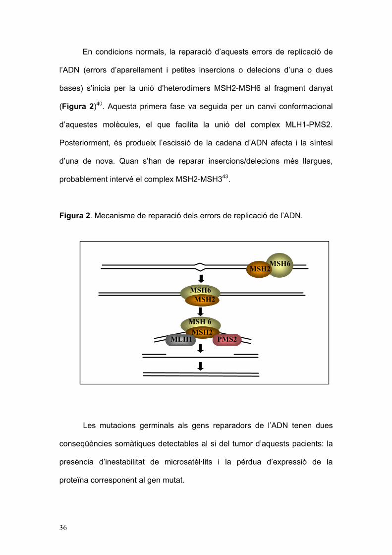
En condicions normals, la reparació d’aquests errors de replicació de
l’ADN (errors d’aparellament i petites insercions o delecions d’una o dues
bases) s’inicia per la unió d’heterodímers MSH2-MSH6 al fragment danyat
(Figura 2)40. Aquesta primera fase va seguida per un canvi conformacional
d’aquestes molècules, el que facilita la unió del complex MLH1-PMS2.
Posteriorment, és produeix l’escissió de la cadena d’ADN afecta i la síntesi
d’una de nova. Quan s’han de reparar insercions/delecions més llargues,
probablement intervé el complex MSH2-MSH343.
Figura 2. Mecanisme de reparació dels errors de replicació de l’ADN.
Les mutacions germinals als gens reparadors de l’ADN tenen dues
conseqüències somàtiques detectables al si del tumor d’aquests pacients: la
presència d’inestabilitat de microsatèl·lits i la pèrdua d’expressió de la
proteïna corresponent al gen mutat.
MSH6 MSH2
MSH6MSH2
MSH 6MSH2
MLH1 PMS2

37
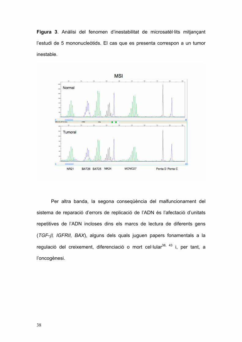
a) Inestabilitat de microsatèl·lits
Des del punt de vista molecular, el malfuncionament del sistema de
reparació d’errors de replicació de l’ADN es tradueix en l’acúmul de
mutacions somàtiques a dos nivells. Per una banda, a nivell de microsatèl·lits,
que són petits fragments repetitius d’ADN distribuïts al llarg de tot el
genoma43, 44 i majoritàriament localitzats en ADN intrònic i que, per tant, la
seva afectació presumiblement no té un significat patològic. El fenòmen
d’inestablititat de microsatèl·lits (Figura 3) constitueix un marcador fenotípic
de la síndrome de Lynch26, estant present en més del 95% dels CCR en
aquests pacients. No obstant això, no es tracta d’un biomarcador específic ja
que fins un 15% dels CCR esporàdics poden presentar inestabilitat de
microsatèl·lits degut a hipermetilació somàtica del promotor del gen MLH1,
amb la conseqüent silenciació gènica45.
38
Figura 3. Anàlisi del fenomen d’inestabilitat de microsatèl·lits mitjançant
l’estudi de 5 mononucleòtids. El cas que es presenta correspon a un tumor
inestable.
Per altra banda, la segona conseqüència del malfuncionament del
sistema de reparació d’errors de replicació de l’ADN és l’afectació d’unitats
repetitives de l’ADN incloses dins els marcs de lectura de diferents gens
(TGF-�, IGFRII, BAX), alguns dels quals juguen papers fonamentals a la
regulació del creixement, diferenciació o mort cel·lular38, 43 i, per tant, a
l’oncogènesi.
39
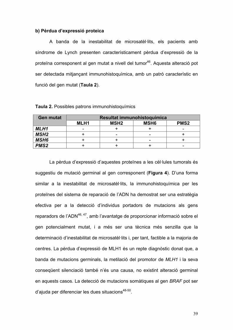
b) Pèrdua d’expressió proteica
A banda de la inestabilitat de microsatèl·lits, els pacients amb
síndrome de Lynch presenten característicament pèrdua d’expressió de la
proteïna corresponent al gen mutat a nivell del tumor46. Aquesta alteració pot
ser detectada mitjançant immunohistoquímica, amb un patró característic en
funció del gen mutat (Taula 2).
Taula 2. Possibles patrons immunohistoquímics
Resultat immunohistoquímica Gen mutat MLH1 MSH2 MSH6 PMS2
MLH1 - + + - MSH2 + - - + MSH6 + + - + PMS2 + + + -
La pèrdua d’expressió d’aquestes proteïnes a les cèl·lules tumorals és
suggestiu de mutació germinal al gen corresponent (Figura 4). D’una forma
similar a la inestabilitat de microsatèl·lits, la immunohistoquímica per les
proteïnes del sistema de reparació de l’ADN ha demostrat ser una estratègia
efectiva per a la detecció d’individus portadors de mutacions als gens
reparadors de l’ADN46, 47, amb l’avantatge de proporcionar informació sobre el
gen potencialment mutat, i a més ser una tècnica més senzilla que la
determinació d’inestabilitat de microsatèl·lits i, per tant, factible a la majoria de
centres. La pèrdua d’expressió de MLH1 és un repte diagnòstic donat que, a
banda de mutacions germinals, la metilació del promotor de MLH1 i la seva
conseqüent silenciació també n’és una causa, no existint alteració germinal
en aquests casos. La detecció de mutacions somàtiques al gen BRAF pot ser
d’ajuda per diferenciar les dues situacions48-50.
40
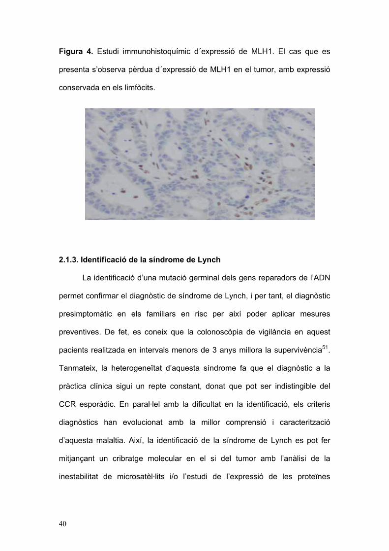
Figura 4. Estudi immunohistoquímic d´expressió de MLH1. El cas que es
presenta s’observa pèrdua d´expressió de MLH1 en el tumor, amb expressió
conservada en els limfòcits.
2.1.3. Identificació de la síndrome de Lynch
La identificació d’una mutació germinal dels gens reparadors de l’ADN
permet confirmar el diagnòstic de síndrome de Lynch, i per tant, el diagnòstic
presimptomàtic en els familiars en risc per així poder aplicar mesures
preventives. De fet, es coneix que la colonoscòpia de vigilància en aquest
pacients realitzada en intervals menors de 3 anys millora la supervivència51.
Tanmateix, la heterogeneïtat d’aquesta síndrome fa que el diagnòstic a la
pràctica clínica sigui un repte constant, donat que pot ser indistingible del
CCR esporàdic. En paral·lel amb la dificultat en la identificació, els criteris
diagnòstics han evolucionat amb la millor comprensió i caracterització
d’aquesta malaltia. Així, la identificació de la síndrome de Lynch es pot fer
mitjançant un cribratge molecular en el si del tumor amb l’anàlisi de la
inestabilitat de microsatèl·lits i/o l’estudi de l’expressió de les proteïnes

41
corresponents per immunohistoquímica, en combinació o no amb criteris
clínics (Taula 3).
Taula 3. Criteris clínics de la síndrome de Lynch
Criteris d’Amsterdam II52 1. Mínim 3 individus amb CCR o tumor associat al CCHNP (endometri,
intestí prim, urèter o pelvis renal), un dels familiars és de primer grau dels altres dos, i
2. Mínim dues generacions consecutives afectes, i 3. Mínim un cas diagnosticat abans dels 50 anys, i 4. Exclusió del diagnòstic de PAF, i 5. Confirmació dels diagnòstics amb informes anatomopatològics
Criteris revisats de Bethesda53 1. CCR diagnosticat abans dels 50 anys, o 2. CCR sincrònic o metacrònic, o un altre tumor associat a CCHNP1,
independentment de l'edat de diagnòstic, o 3. CCR amb histologia de tumor amb IMS2 diagnosticat abans dels 60 anys,
o 4. CCR amb un o més familiars de primer grau amb un tumor associat a
CCHNP1, un dels càncers diagnosticats abans dels 50 anys, o 5. CCR amb 2 o més familiars de primer o segon grau amb un tumor
associat a CCHNP1, independentment de l'edat 1Tumors associats a CCHNP: CCR, endometri, estómac, ovari, pàncrees, urèter i pelvis renal, tracte biliar, cerebral (glioblastoma), adenomes sebacis i queratoacantomes, i intestí prim 2Presència de limfòcits infiltrants de tumor, reacció Crohn-like, diferenciació mucinosa/anell de segell, o medul·lar
Des de la descripció per primera vegada de la síndrome de Lynch fins
a la actualitat, els criteris diagnòstics han anat evolucionat juntament amb el
millor coneixement del fenotip de la malaltia i la identificació dels gens
causants, així com dels fenòmens moleculars que s’observen en el si del
tumor (inestabilitat de microsatèl·lits i pèrdua d’expressió proteica). Així, els
criteris clínics clàssics, els criteris d’Amsterdam52, fonamentals en el seu
moment per identificar els gens causants, avui en dia es consideren ja una
extratègia obsoleta com a eina per a la identificació. Això és degut a que

42
només un 45%-65% de les famílies que acompleixen els criteris d’Amsterdam
presenten mutacions germinals en alguns dels gens reparadors de l’ADN54. A
més, en una proporció no menyspreable de famílies que acompleixen
aquests criteris o que tenen una marcada història familiar de CCR no és
possible identificar aquestes mutacions55-57. Contràriament, famílies que no
acompleixen els criteris d’Amsterdam poden presentar mutacions germinals
en aquests gens58, 59. Aquestes situacions indicarien que, per una banda,
probablement existeixen altres gens responsables de corregir errors de
replicació de l’ADN desconeguts fins el moment i que, per altra, els criteris
clínics no són suficientment sensibles per a detectar totes les famílies amb
síndrome de Lynch. Per aquest motiu, es van disenyar els criteris de
Bethesda60, uns criteris més sensibles però menys específics, modificats el
1999 i revisats el 200453, amb la intenció d’identificar pacients amb una
elevada probabilitat de ser portadors de mutacions als gens reparadors de
l’ADN, als quals estaria indicat determinar la presència del fenòmen
d’inestabilitat de microsatèl·lits o immunohistoquímica per avaluar pèrdua
d’expressió proteica en el si del tumor. En els pacients amb alteració del
sistema de reparació de l’ADN al tumor (definit com presència d’inestabilitat
de microsatèl·lits i/o pèrdua d’expressió), hauria d’investigar-se la presència
de mutacions en els gens reparadors de l’ADN53.
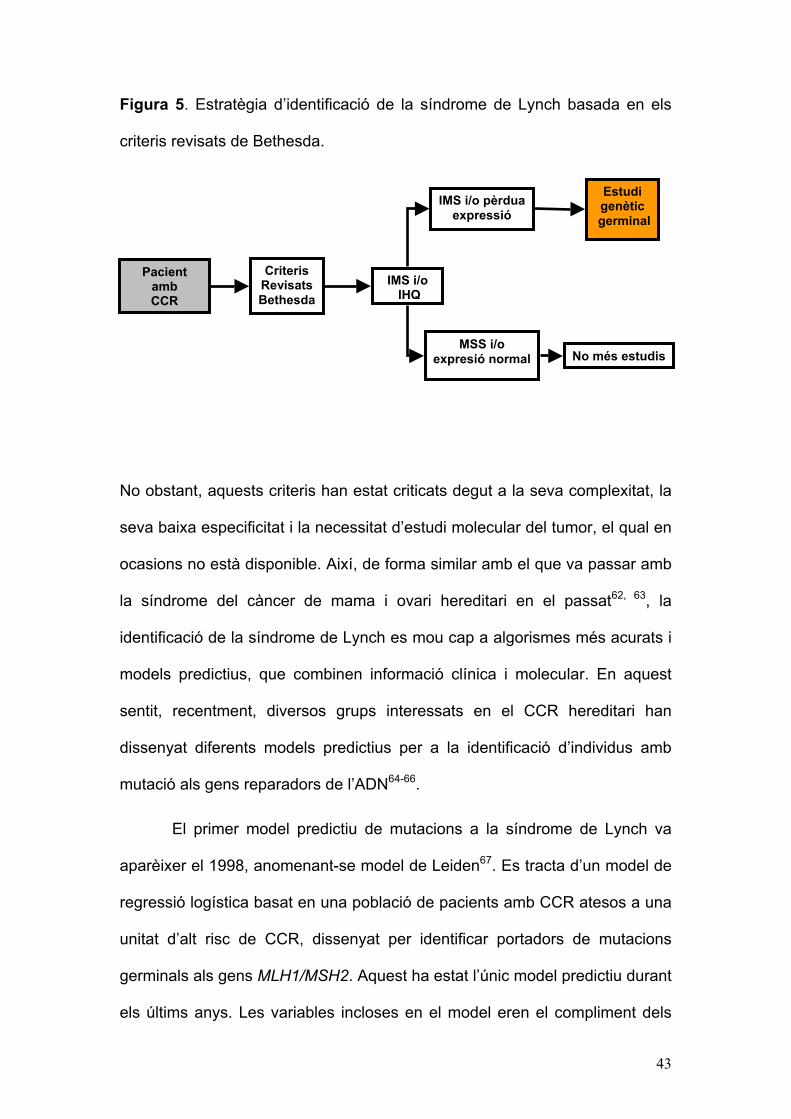
Aquesta estratègia ha permès millorar substancialment el diagnòstic
de la síndrome61, i de fet, constitueix l’estratègia més acceptada actualment
(Figura 5).
43
Figura 5. Estratègia d’identificació de la síndrome de Lynch basada en els
criteris revisats de Bethesda.
No obstant, aquests criteris han estat criticats degut a la seva complexitat, la
seva baixa especificitat i la necessitat d’estudi molecular del tumor, el qual en
ocasions no està disponible. Així, de forma similar amb el que va passar amb
la síndrome del càncer de mama i ovari hereditari en el passat62, 63, la
identificació de la síndrome de Lynch es mou cap a algorismes més acurats i
models predictius, que combinen informació clínica i molecular. En aquest
sentit, recentment, diversos grups interessats en el CCR hereditari han
dissenyat diferents models predictius per a la identificació d’individus amb
mutació als gens reparadors de l’ADN64-66.
El primer model predictiu de mutacions a la síndrome de Lynch va
aparèixer el 1998, anomenant-se model de Leiden67. Es tracta d’un model de
regressió logística basat en una població de pacients amb CCR atesos a una
unitat d’alt risc de CCR, dissenyat per identificar portadors de mutacions
germinals als gens MLH1/MSH2. Aquest ha estat l’únic model predictiu durant
els últims anys. Les variables incloses en el model eren el compliment dels
Criteris Revisats Bethesda
IMS i/o IHQ
MSS i/o expresió normal No més estudis
Estudi genètic
germinal IMS i/o pèrdua
expressió
Pacient amb CCR

44
criteris d’Amsterdam, l’edat mitjana dels diagnòstics de CCR, i la presència
d’algún càncer d’endometri a la familia. No obstant, aquest model encara
incorpora variables complexes (criteris d’Amsterdam) i va ser desenvolupat
en una població relativament petita d’individus d’alt risc. A més, no
contemplava la realització d’estudis moleculars al si del tumor (inestabilitat de
microsatèl·lits o immunohistoquímica).
Més recentment, han aparegut 3 nous models per a la identificació de
portadors de mutacions als gens reparadors de l’ADN (Taula 4). El primer,
anomenat PREMM1,2, esdevé d’una anàlisi de regressió logística realitzat a
una de les cohorts més grans publicades fins al moment d’individus en risc de
tenir una síndrome de Lynch65. Els autors d’aquest estudi van realitzar
l’anàlisi mutacional de MLH1/MSH2 a una cohort de 1914 pacients als que
se’ls havia demanat l’estudi genètic en base a característiques clíniques als
laboratoris Myriad Genetic Lab. a EE.UU. En base a característiques
personals i familiars, es va dissenyar un model que va ser adequadament
validat a l’estudi, i que es pot consultat a internet (http://www.dfci.org/premm).
El model prediu la probabilitat de ser portador de mutacions als gens
MLH1/MSH2 amb una àrea sota la corba ROC (receiver operating
characteristic) de 0,80 (interval de confiança del 95%: 0,76-0,84). No obstant,
la validació d’aquest model en una població diferent a la que va ser dissenyat,
especialment a pacients amb CCR, no s’ha realitzat fins ara.
Els altres dos models inclouen informació molecular per tal de refinar
la predicció de la probabilitat de ser portador d’una mutació. El primer és
tracta d’un model desenvolupat al Regne Unit a una gran cohort de pacients
amb CCR de base poblacional amb una edat inferior a 55 anys66. Aquest
45
model consisteix en dues fases: la primera es basa exclusivament en
variables clíniques (edat, sexe, localització del tumor, presència de CCR
sincrònic o metacrònic, història familiar de CCR o càncer d’endometri, i edat
del familiar amb CCR més jove), disponible a internet
(http://www1.hgu.mrc.ac.uk/softdata/mmrpredict.php); i la segona, basada en
dades d’inestabilitat de microsatèl·lits o immunohistoquímica del tumor. L’àrea
sota la corba ROC d’aquest model, que prediu mutacions germinals a MLH1,
MSH2 i també MSH6, va ser de 0,82 (IC 95%, 0,72-0,91). No obstant, la
aplicació d’aquest model en una població major de 55 anys o en aquells amb
altres neoplàsies associades a la síndrome de Lynch no ha estat avaluada.
L’últim model és un model mendelià per tal de determinar la probabilitat de
ser portador de mutacions germinals als gens MLH1, MSH2 i MSH6 basat en
dades clíniques, moleculars, i la prevalença i penetrança de les mutacions
d’aquests gens a la població, segons els estudis epidemiològics
disponibles64. Aquest model bayesià, utilitza el software gratuit CaGene
(http://www3.utsouthwestern.edu/cancergene) i permet establir la probabilitat
de ser portador de mutació tant a pacients amb CCR com a familiars sans.
L’àrea sota la corba ROC va ser de 0,83 (IC 95%, 0,78-0,88). La aplicabilitat
d’aquest model a la pràctica clínica encara no s’ha avaluat.

Taula 4. Models predictius per a la identificació de la síndrome de Lynch Model Població Gens
Tumors inclosos
Mètode Resultats
Barnetson66 870 pacients amb CCR <55 anys de base poblacional
Mutacions puntuals a MLH1, MSH2, MSH6 i grans reordenaments a MLH1, MSH2
Còlon i endometri Model en dos fases: 1: Anàlisi de regressió logística multivariat. 2: Refinament de la predicció amb informació molecular del tumor (IMS i IHQ).
Predictors: Edat, sexe, localització del tumor, múltiples tumors, edat més jove de CCR a la família (dicotomitzada a 50 anys), presència de càncer d’endometri a familiars de primer grau. AUC= 0,82 (IC 95% 0,72-0,91)
PREMM1,265 1914 individus
sotmesos a estudi genetic per sospita clínica de síndrome de Lynch
Mutacions puntuals i grans reordenaments a MLH1, MSH2
Còlon, endometri, altres neoplàsies associades a la síndrome de Lynch, adenomas colònics
Anàlisi de regressió logística multivariat
Predictors: Proband: nombre de CRC, adenomes, càncer d’endometri, altres neoplàsies associades a la síndrome de Lynch, edat al diagnòstic del CCR, adenomas i cancer d’endometri. Familiars: CCR, cancer d’endometri, altres neoplàsies associades a la síndrome de Lynch, edat al diagnostic del CCR i endometri als familiars de primer i segon grau AUC=0,80 (IC 95% 0,77-0,83)
MMRpro64 Validació a 279 individus de 226 famílies amb diagnòstic clínic
Mutacions puntuals i grans reordenaments a MLH1, MSH2 i MSH6
Còlon i endometri Anàlisi mendelià i bayesià incorporant la penetrança i prevalença de les mutacions, i els valors predictius de l’estudi molecular del tumor amb IMS o IHQ. Té en compte el nombre de familiars i els individus no afectes.
AUC de la validació=0,83 (IC 95% 0,78-0,88)
AUC: area sota la corba ROC; IMS: inestabilitat de microsatèl·lits; IHQ: immunohistoquímica.

2.1.4.Estratègia de cribratge
L’anàlisi genètic dels gens reparadors de l’ADN permet el diagnòstic
presimptomàtic dels familiars en risc26, 68, 69. Així, aquesta anàlisi mutacional
hauria d’oferir-se als familiars de primer grau (pares, germans i fills)
d’individus portadors d’una mutació germinal en alguns d’aquests gens. Amb
aquesta estratègia s’aconsegueix optimitzar la relació cost-eficàcia del
cribratge de la síndrome de Lynch70, de manera que el seguiment endoscòpic
pot centrar-se únicament en aquells membres portadors de mutacions.
El seguiment colonoscòpic està dirigit a la identificació i resecció de
pòlips adenomatosos, així com a la detecció de carcinomes en fases inicials
del seu desenvolupament71. En els darrers anys s’ha demostrat que la
vigilància periòdica mitjançant colonoscòpies millora el pronòstic dels
individus pertanyents a famílies amb aquesta síndrome, al haver-se
demostrat que la realització en individus que pertanyen a famílies amb
síndrome de Lynch d’una colonoscòpia cada 3 anys durant un període de 15
anys s’associa a una disminució del 62% en la incidència de CCR (p=0,02) i
del 66% en la mortalitat global (p=0,003) en relació a la no realització de
cribratge51. L’intèrval idoni entre exploracions no està ben establert, encara
que el fet que alguns estudis hagin descrit l’aparició de CCR als dos o tres
anys d’haver-se realitzat una colonoscòpia negativa51, 72, 73 i la més ràpida
progressió des d’adenoma a carcinoma a la síndrome de Lynch35 justificaria
un intèrval d’1 ó 2 anys entre exploracions. Encara que sense evidències
directes es recomana iniciar el cribratge endoscòpic a partir dels 20-25 anys o
10 anys abans de l’edat de diagnòstic del CCR en el familiar afecte més jove,
escollint la opció més precoç26, 68, 74 (Figura 6).

48
En quant a les neoplàsies extracolòniques associades a la síndrome
de Lynch en individus portadors de mutacions en els gens responsables, el
registre de càncer de Finlàndia ha permès estimar la seva incidència: 60%
per el càncer d’endometri, 13% per al d’estómac, 12% per al d’ovari, 4% per
al de vies urinàries, 3,7% per al cerebral, 3,3% per al de pelvis renal, i 2% per
al de vies biliars75. Tant els individus que han desenvolupat CCR como els
familiars en risc tenen una major probabilitat de presentar una neoplàsia
extracolònica, el que podria justificar el cribratge de les mateixes76. Malgrat
tot, a diferència del que succeeix en relació amb el CCR, no està demostrada
la eficàcia d’aquestes estratègies60, 77, 78, encara que s’assumeix que el
benefici podria ser major en aquelles famílies en les que existeix una major
agregació d’una determinada neoplàsia extracolònica76. Al ser el càncer
d’endometri la neoplàsia extracolònica més freqüent, la majoria de grups
recomanen la realització d’una ultrasonografia pèlvica anual o bienal a partir
dels 25-35 anys d’edat76, 77, 79 (Figura 6).
En resum, les recomanacions actuals en els pacients pertanyents a
famílies amb sospita de síndrome de Lynch contemplen fonamentalment la
realització de l’anàlisi mutacional dels gens reparadors de l’ADN (MLH1,
MSH2, MSH6, PMS2)26, 80. En els individus portadors de mutacions o en
aquells casos en els que no és possible determinar-ne la seva presència,
estarà indicat el cribratge endoscòpic. A més, en funció del predomini de
neoplàsies d’un altre origen en el si d’una determinada família, és convenient
efectuar altres exploracions dirigides a descartar la seva presència.

49
Figura 6. Cribratge, vigilància i tractament de la síndrome de Lynch
Síndrome de Lynch
Cribratge de CCRCribratge de
neoplàsia endometri / ovari
Exploració: Colonoscòpia Edat d’inici: 20-25 anys. Periodicitat: En funció de l’edat: <40 anys: cada 2 anys. >40 anys: anual
No cribratge específic
SI NO
Exploració: ecografia ginecològica transvaginal + citologia endometrial + CA125 Edat d’inici: 30-35 anys ó 10 anys abans del cas més jove (el primer que passi). Periodicitat: 1-2 anys.
Antecedent familiar de > 2 neoplàsies
urotelials o gàstriques
Neoplàsia urotelial: Exploració: citologia d’orina + ecografia reno-vesical. Edat d’inici: 30-35 anys ó 10 anys abans del cas més jove (el primer que passi). Periodicitat: 1-2 anys.
Neoplàsia gàstrica: Exploració: fibrogastroscòpia Edat d’inici: 30-35 anys ó 10 anys abans del cas més jove (el primer que passi). Periodicitat: 1-2 anys.
Colectomia total
- Adenoma irresecable per endoscòpia - Múltiples adenomes. - CCR - Pretensió de reducció de risc
- Neoplàsia endometri/ovari - Pretensió de reducció de risc (no desig reproductiu) en especial si:
a) Història familiar de neoplàsia d’endometri, ó
b) Pacient portador de mutació a MSH6.
Histerectomia + /-salpingo-oforectomia
Vigilància: FCS anual
50
2.1.5. Tractament
Donat que els pacients amb síndrome de Lynch presenten un risc
incrementat de desenvolupar tumors metacrònics71, tenen una ràpida
progressió des d’adenoma a carcinoma35 i que en moltes ocasions el
carcinoma s’origina en lesions planes difícils de tractar endoscòpicament81,
alguns grups recomanen la realització d’una resecció extensa (colectomia
total o proctocolectomia) per al tractament de les neoplàsies colorectals76.
L’edat, la presència de comorbilitat, la opinió del pacient, així com la
localització del tumor són factors a tenir en compte en la decisió terapèutica76
(Figura 6).
No existeixen dades que recomanin la realització d’una colectomia
profilàctica en pacients en risc o en portadors de mutacions en els gens
responsables de la síndrome de Lynch76.
2.1.6. Vigilància post-resecció
Es ben conegut el fet que després del tractament quirúrgic del CCR en
pacients amb la síndrome de Lynch existeix un elevat risc de lesions
metacròniques, havent-se observat que la meitat dels pacients sotmesos a
una ressecció colònica segmentaria presenten una segona neoplàsia
colorectal als 10 anys34. A més, el risc de desenvolupar un carcinoma de
recte en els pacients tractats mitjançant colectomia total és del 12% després
d’un període de seguiment de 12 anys82. Aquesta situació justifica la
vigilància endoscòpica després de la cirurgia82.

51
Finalment, és important assenyalar que, a l’actualitat, no existeixen
dades que demostrin la utilitat de les estratègies de quimioprofilaxi en
aquesta síndrome34, 83.

52
2.2. Càncer colorectal associat al gen MYH
2.2.1. MYH i la via del sistema per escisió de bases
Al-Tassan i col·laboradors84 van investigar una família britànica en la
que tres germans presentaven adenomes colorectals múltiples i CCR. La
seqüenciació total del gen APC, causant de la poliposi adenomatosa familiar,
a l’ADN germinal de dos dels germans afectes junt amb l’anàlisi d’haplotips i
d’expressió gènica van excloure un defecte gènic en aquest gen. L’anàlisi de
la inestabilitat de microsatèl·lits a l’ADN extret d’onze neoplàsies d’aquesta
família també va excloure un posible defecte en la reparació de l’ADN. No
obstant, el patró de mutacions observades en els tumors d’aquests pacients
va facilitar la identificació del gen causant. En aquest sentit, la seqüenciació
completa d’APC a cada un dels 11 tumors va revelar 18 mutacions
somàtiques, 15 de les quals eren transversions G:C�T:A. Aquest tipus de
mutacions correspon només a un 10% de les identificades somàticament en
el gen APC, essent les mutacions de pèrdua de pauta de lectura (frameshift) i
la pèrdua d’heterozigositat (pèrdua al·lèlica) les que més freqüentment
causen la inactivació somàtica del gen APC en els tumors colorectals. La
comparació de les troballes en aquesta família britànica amb una base de
dades amb més de 800 mutacions somàtiques al gen APC en pacients amb
CCR esporàdic o associat a la PAF va confirmar l’increment altament
significatiu de transversions G:C�T:A en aquesta família. Els compostos
derivats de l’oxígen reactiu amb potencial lesiu per el ADN tenen un paper
important en processos com l’envelliment, el càncer i les malalties
neurodegeneratives. Als humans, s’estima que la freqüència del dany oxidatiu
a l’ADN és de 10.000 lesions per cèl·lula i dia85. La 8-oxo-7,8-

53
dihidro2’deoxiguanosina (8-oxodG) és un dels productes nocius mès estables
del dany oxidatiu a l’ADN i la seva concentració és alta en el càncer de
mama, pulmó i ronyó. Aquest compost es mal aparella amb residus d’adenina
provocant les mutacions G�T i C�A a la nova cadena en la replicació de
l’ADN (Figura 7). La incorporació d’8-oxodG a l’ADN té lloc a través de
l’oxidació directa dels residus de guanina en la cadena motlle o gràcies a la
incorporació d’8-oxodG procedent del conjunt de nucleòtids lliures. Les
mutacions apareixen després de la replicació de l’ADN degut a que les
polimerases incorporen adenosines a 8-oxodG a un ritme molt major del que
incorporen citosines.
A Escherichia coli existeixen tres enzims que ajuden a protegir la
cèl·lula dels efectes mutagènics de la oxidació de la guanina. La glicosilasa
d‘ADN MutM elimina la base oxidada dels parells de base 8-oxodG:C a l’ADN
de doble cadena, la glicosilasa d’ADN MutY escindeix les adenines
incorporades per error i aparelladas amb 8-oxodG durant la replicació, i la 8-
oxodGTPasa MutT impideix la incorporació d’8-oxodGTP a la cadena naixent
d’ADN durant la replicació. Els gens homòlegs de mutM, mutY y mutT als
humans han estat identificats i es denominen OGG186, MYH87 i
MTH1/NUDT188, respectivament.

54
Figura 7. Esquema del sistema per escisió de bases.
Per determinar si un defecte heredat en la via de reparació d’8-oxodG
era responsable del patró de mutacions somàtiques G:C�T:A a la familia
estudiada, Al-Tassan i col·laboradors van seqüenciar per complet els gens
OGG1, MYH i MTH1 a l’ADN extret de sang d’un dels familiars afectes.
D’aquesta forma es van identificar dues mutacions que canvien de forma no
conservativa dos aminoàcids del gen MYH (Y165C i G382D), essent la resta
de germans afectes també heterocigots compostos per aquestes dues
mutacions. La resta de familiars no afectes eren o bé heterocigots per una
d’elles o no portadors, suggerint un patró d’herència autosòmic recessiu.

55
2.2. MYH i càncer colorectal
El gen MYH està localitzat en el braç curt del cromosoma 1 entre p32.1
i p34.3, té una longitud de 7.100 bases i conté 16 exons que codifiquen una
proteïna de 535 aminoàcids amb un 41% d’homologia amb la proteïna
homòloga MutY de l’E. coli89. Els seus dominis funcionals han estats descrits
previament i s’esquematitzen en la Figura 8.
Figura 8. Esquema de la proteïna MYH amb els dominis funcionals descrits
fins el moment, i algunes de les mutacions ètniques més prevalents.
A l’estudi d’Al-Tassan i col·laboradors l’han seguit diversos estudis90-94
principalment focalitzats en pacients amb adenomes múltiples que han
demostrat que les mutacions bial·lèliques germinals al gen MYH s’associen a
una forma de poliposi adenomatosa familiar, amb un nombre de pòlips que
oscil·la entre 5-100. De fet, s’ha observat que les mutacions germinals

56
bial·lèliques al gen MYH són la causa de entre el 25-60% de les poliposi amb
fenotip atenuat (<100 pòlips) i fins un 10% de la poliposi adenomatosa clàsica
(>100-1000 pòlips). Així, actualment la forma de poliposi adenomatosa
causada per mutacions al gen MYH es coneix com Poliposi Associada al gen
MYH (PAM), essent la primera forma de síndrome polipósica associada al
CCR amb un patró d’herència recessiu.
Una altra qüestió important detectada pels estudis mencionats ha estat
l’existència de mutacions ètniques o adscrites a un orígen geogràfic concret
(Figura 8). Així, les mutacions G382D i Y165C corresponen a les
majoritàriament detectades a la població caucàsica, mentres que altres
mutacions s’han descrit a poblacions com la pakistanesa93, la indú93, la
italiana94, la portuguesa 95 o la japonesa.
Per altra banda, estudis recents han demostrat que les mutacions
bial·lèliques al gen MYH també predisposen al desenvolupament de CCR,
seguint un patró d’herència recesiva, amb un risc de CCR associat de més de
93 vegades el de la població general21. Així, diversos estudi de base
poblacional han mostrat que les mutacions en aquest són la causa de fins un
1% de tots els CCR, amb una penetrança del 100% als 60 años21, 96-98. A la
majoria dels casos, s’acompanya d’un nombre variable de pòlips sincrònics
(5-100). No obstant, cal destacar que en més d’un 30% dels pacients amb
mutacions bial·lèliques que han desenvolupat un CCR no presenten pòlips
sincrònics, pel que la presència d’aquests com a possible aproximació per a
la detecció de portadors de mutacions és poc sensible21. De fet, tot i que s’ha
suggerit que la presència de pòlips sincrònics i una edat jove al diagnòstic de
CCR podrien ser característiques útils per a la detecció dels pacients amb

57
CCR portadors de mutacions a MYH, no s’ha avaluat la seva eficàcia en una
cohort de pacients amb CCR de base poblacional.
La predisposició al CCR en aquells individus portadors de mutacions
monoal·lèliques a MYH és un tema actualment controvertit. S’ha suggerit un
augment del risc de CCR seguint un patró autosòmic dominant en base a la
detecció, en alguns estudis, d’una major agregació familiar de CCR en
aquests individus99. Estudis més recents han demostrat un augment del risc
de CCR a edats avançades en relació amb la heterocigosi, recolçant la
hipòtesi de que el gen MYH podria actuar com a gen de susceptibilitat per al
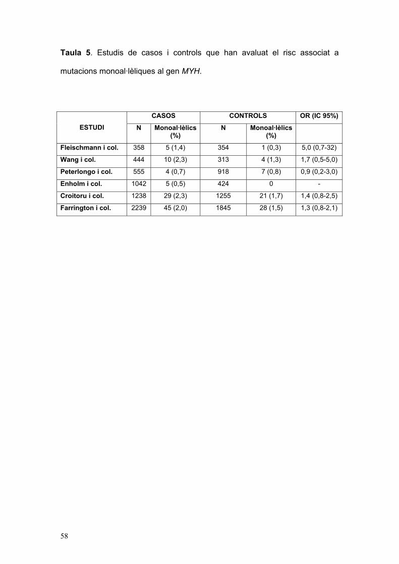
CCR de baixa penetrança21. No obstant, cap dels estudis de casos i controls
realitzats fins el moment ha pogut demostrar de forma indvidual un augment
del risc en els individus amb mutació monoal·lèlica al gen MYH (Taula 5), i
una meta-anàlisi recent ha demostrat un augment de risc no significatiu
associat als heterocigots (risc relatiu de 1,3 (IC 95%: 1,0-1,7; p=0,09)100.
Totes aquestes discrepàncies posen de manifest un efecte lleu de les
mutacions del gen en heterocigosi, pel que calen nous estudis amb
aproximacions metodològiques adequades (meta-anàlisi, estudis de cohorts
familiars) de tal d’esbrinar definitivament el risc associat en aquesta situació.
58
Taula 5. Estudis de casos i controls que han avaluat el risc associat a
mutacions monoal·lèliques al gen MYH.
CASOS CONTROLS OR (IC 95%)
ESTUDI N Monoal·lèlics (%)
N Monoal·lèlics (%)
Fleischmann i col. 358 5 (1,4) 354 1 (0,3) 5,0 (0,7-32)
Wang i col. 444 10 (2,3) 313 4 (1,3) 1,7 (0,5-5,0)
Peterlongo i col. 555 4 (0,7) 918 7 (0,8) 0,9 (0,2-3,0)
Enholm i col. 1042 5 (0,5) 424 0 -
Croitoru i col. 1238 29 (2,3) 1255 21 (1,7) 1,4 (0,8-2,5)
Farrington i col. 2239 45 (2,0) 1845 28 (1,5) 1,3 (0,8-2,1)
59
3. L’estudi EPICOLON
A finals de 1999 es va començar a gestar un dels projectes
cooperatius més ambiciosos portats a terme a l’Estat Espanyol en els últims
anys, anomenat EPICOLON. Es tractava d’un estudi epidemiològic,
prospectiu i multicèntric d’àmbit estatal i de base poblacional, impulsat des de
la Asociación Española de Gastroenterología, i en el que van participar més
de 25 hospitals distribuïts per tota la geografia espanyola. La coordinació
d’aquest estudi es va portar a terme pels grups d’oncologia digestiva de
l’Hospital Clínic, a càrrec del Dr. Antoni Castells, i dels Hospitals del Mar,
Germans Trias i Pujol de Badalona, i Hospital General Universitario de
Alicante. En aquest projecte, des de novembre de 2000 fins octubre de 2001
es van enregistrar prop de 2000 pacients amb CCR, dels quals es va recollir
una detallada història personal i familiar centrada en els antecedents
neoplàstics, així com mostres de teixit tumoral i no tumoral de la majoria
d’ells.
Aquest projecte ha realitzat aportacions rellevants al coneixement de la
síndrome de Lynch i altres formes hereditàries de CCR, en especial pel que
respecte a epidemiologia, diagnòstic, caracterització molecular i pronòstic46,
61, 101-105.
La Unitat de CCR de l’Hospital Clínic de Barcelona és dipositària de
gran part de les mostres obtingudes en el si del projecte EPICOLON. Això
permet disposar, de forma immediata, d’un gran nombre de mostres en el que
poder realitzar estudis de recerca, dirigits a aprofundir en les caracterització
de les formes hereditàries de CCR.

60

61
JUSTIFICACIÓ I OBJECTIUS

62

63
Justificació general
El CCR és una de les neoplàsies més prevalents als països
occidentals i un dels tumors on els factors genètics juguen un paper més
fonamental en el seu desenvolupament. Així, el anomenat CCR hereditari,
entès com aquelles formes degudes a l’alteració en gens d’alta penetrança,
suposa entre el 3 i el 5% de tots els CCR106. Tot i que suposen un
percentatge baix de forma global, l’elevada prevalença del CCR i les
conseqüències catastròfiques de les síndromes hereditàries converteixen el
diagnòstic d’aquests individus en un objectiu fonamental en la pràctica clínica,
la qual cosa justifica l’existència de centres especialitzats en forma de
clíniques d’alt risc de CCR. En aquest sentit, el diagnòstic d’una síndrome
hereditària, a banda de l’interès científic, té importants conseqüències
potencialment beneficioses. Per una banda, permet la vigilància estricta del
pacient afecte, no només en relació al CCR sino també a altres neoplàsies
associades a cada síndrome34. Per altra banda, el diagnòstic genètic permet
el diagnòstic presimptomàtic dels familiars portadors de la malaltia, i així
focalitzar els recursos en aquests, lliurant-se del seguiment aquells que no
tenen la mutació.
Per tant, el primer pas consisteix en identificar els individus o pacients
que potencialment poden tenir una malaltia hereditària, per tal de realitzar en
ells els estudi moleculars adients. Les síndromes hereditàries es divideixen
des d’un punt de vista fenotípic i pràctic en síndromes polipòsiques,
fonamentalment la poliposi adenomatosa familiar, la poliposi associada al gen
MYH, la síndrome de Peutz-Jeghers i altres, i en síndromes no polipòsiques,
encapçalades per la síndrome de Lynch i pel CCR associat al gen MYH.
64
Mentre que a la pràctica clínica el diagnòstic de les síndromes polipòsiques
és senzill donada la seva expressivitat clínica, el diagnòstic de les formes no
polipòsiques és un repte constant per al clínic, donat que en moltes ocasions
pot ser indistingible del càncer esporàdic.
La síndrome de Lynch constitueix la forma més freqüent de càncer
hereditari. La instauració dels criteris d’Amsterdam107 va ser cabdals per a
definir el CCHNP i identificar la seva base molecular. Posteriorment, aquests
criteris varen ser modificats (criteris d’Amsterdam II) per considerar-se els
originals massa restrictius52. Més tard, a l’any 1996, el National Cancer
Institute va proposar els criteris de Bethesda, els quals tenien com objectiu
identificar aquells pacients amb una major probabilitat de ser portadors de
mutacions i que haurien de ser avaluats mitjançant estudi d’inestabilitat de
microsatèl·lits60. Aquests criteris van ser modificats el 1999 i finalment
revisats el 2004, i actualment constitueixen l’estratègia més acceptada per tal
d’identificar els pacients amb CCR portadors de mutacions als gens
reparadors de l’ADN. Així, en els pacients amb CCR que compleixen algún
dels 5 criteris revisats de Bethesda, està indicada la realització d’un cribratge
molecular del tumor per tal de detectar evidència d’alteració del sistema de
reparació de l’ADN (inestabilitat de microsatèl·lits i/o immunohistoquímica de
les proteïnes corresponents). No obstant això, aquests criteris han estat
criticats deguts a la seva complexitat i, conseqüentment, l’escassa aplicació
que han tingut, la seva baixa especificitat i la necessitat d’estudi molecular del
tumor, que en ocasions no és possible. Així, recentment han aparegut varios
models predictius que, basats en anàlisis de regressió logística, permeten
estimar la probabilitat de ser portador d’una mutació als gens reparadors,

65
amb combinació o no amb l’estudi de l’alteració de la reparació de l’ADN en el
si del tumor64-66. Aquests models aporten diferents avantatges en relació als
criteris revisats de Bethesda53, principalment la facilitat d’ús (tots ells estan
disponibles a la xarxa de forma gratuïta en un forma de fàcil ompliment) i,
sobretot, la quantificació del risc, de forma que en funció de la magnitud del
risc, l’actitud clínica preventiva i l’estratègia molecular podria ser diferent.
L’eficàcia d’aquests models per a la detecció de la síndrome de Lynch en una
població de pacients amb CCR de base poblacional no s’ha estudiat fins el
moment.
Per altra banda, la identificació del CCR associat al gen MYH suposa
un repte clínic encara més gran que a la síndrome de Lynch donat que es
tracta d’una síndrome de recent descripció i en la que la informació sobre les
manifestacions fenotípiques i el risc associat a la presència de mutacions en
aquest gen és molt reduïda. Així, en relació amb el seu fenotip, s’ha descrit
que en més d’un 30% dels portadors de mutacions bial·lèliques, el CCR es
desenvolupa en absència de pòlips sincrònics i a una edat jove21. Per altra
banda, el risc de CCR associat a la presència d’una mutació a un sol al·lel és
controvertida. Per tot això, calen més estudis en poblacions de CCR no
seleccionades per a caracteritzar millor la síndrome i establir el risc de CCR
associat a la presència de mutacions.
L’estudi EPICOLON va suposar el recull de més de 1200 casos de
CCR arreu de l’Estat Espanyol dels que es disposa d’informació clínica
exhaustiva així com de mostres per a realitzar estudis moleculars a fi de
caracteritzar i identificar les síndromes hereditàries, i representa el fonament
d’aquesta Tesi Doctoral.

66
67
Justificació i objectius de l’estudi 1
Validation and extension of the PREMM1,2 model in a population-based cohort
of colorectal cancer patients (Gastroenterology. 2008 Jan;134(1):39-46).
La síndrome de Lynch és la forma més frequent de CCR hereditari,
suposant entre l’1-5% del total de casos108. Es caracteritza pel
desenvolupament d’aquesta neoplàsia i d’altres (principalment, endometri) a
edats joves. La síndrome té un patró d’herència dominant amb penetrança
variable, i la seva causa és la presència d’una mutació germinal als gens
reparadors de l’ADN, principalment MLH1 i MSH2 (>90% dels casos)109 però
també MSH6110 i PMS241.
La heterogeneitat de la síndrome fa que el diagnòstic suposi un repte
per al clínic. Així, el diagnòstic es pot fer mitjançant criteris clínics, criteris
moleculars (inestabilitat de microsatèl·lits i/o immunohistoquímica per a les
proteines reparadores de l’ADN), o la combinació d’ambdós. Actualment,
l’estratègia més acceptada és la realització d’estudis moleculars en aquells
pacients amb CCR que compleixin algún dels criteris revisats de Bethesda46,
53, i l’estudi genètic només en aquells que presenten evidència d’alteració del
sistema de reparació de l’ADN amb les tècniques esmentades.
A l’igual del que va succeir amb la síndrome de mama i ovari
hereditària, la identificació de la síndrome de Lynch està canviant cap el
disseny de models predictius quantitatius que permetin estimar la probabilitat
de ser portador de mutacions germinals als gens reparadors de l’ADN. El
model PREMM1,2, recentment desenvolupat, permet predir la probabilitat de
ser portador de mutació germinal als gens MLH1 i MSH2 en base a dades
personals i a la història familiar65. El model va ser desenvolupat utilitzant una

68
gran cohort d’individus sotmesos a estudi genètic de MLH1/MSH2 en base a
dades clíniques (personals i familiars). Mentres que el model discriminava de
forma acurada els individus portadors de mutació en la cohort d’estudi, la
seva eficàcia i utilitat en una població no seleccionada de pacients amb CCR
era desconeguda. A més, l’eficàcia del model PREMM1,2 en combinació amb
l’estudi molecular del tumor tampoc havia estat mai avaluada.
Per tant, utilitzant les dades del projecte EPICOLON, el primer estudi
d’aquesta Tesi Doctoral va anar dirigit a avaluar l’eficàcia del model
PREMM1,2 en una població no seleccionada de pacients amb CCR, en
combinació o no amb l’estudi d’alteració del sistema de reparació de l’ADN en
el si del tumor.

69
Justificació i objectius de l’estudi 2
Identification of MYH mutation carriers in colorectal cancer: a multicenter,
case-control, population-based study (Clin Gastroenterol Hepatol 2007;5:
379-87).
Recentment s’ha demostrat la implicació del sistema per escisió de
bases en la carcinogènesi colorectal84. Així, està ben establert que les
mutacions germinals bial·lèliques al gen MYH predisposen al
desenvolupament d’adenomes múltiples i CCR, amb un patró d’herència
recessiu84, 96-99, 111, 112. No obstant, la influència sobre el risc de CCR de les
mutacions monoal·lèliques és controvertit, havent-se suggerit un lleuger
augment del risc amb un patró d’herència dominant, comportant-se com un
gen de baixa penetrança en aquesta situació99, 113.
Per altra banda, al tractar-se d’una síndrome de recent descripció, es
desconeix encara amb precisió el fenotip de la malaltia. El CCR associat al
gen MYH s’ha observat habitualment en el contexte d’una poliposi
adenomatosa amb un fenotip atenuat. No obstant, en més dels 30% dels
casos, el CCR es desenvolupa en absència de pòlips sincrònics21. Això fa
que el diagnòstic d’aquesta forma de CCR hereditarri suposi un repte
diagnòstic, donat que no disposem d’uns criteris establerts per la seva
identificació.
Per tot això, el segon estudi d’aquesta Tesi Doctoral va dirigit a avaluar
la implicació de les mutacions al gen MYH en el CCR, utilizant una cohort de
pacients amb CCR de base poblacional com és EPICOLON.
Els objectius específics d’aquest estudi van ser:

70
1. Establir la prevalença de les mutacions germinals al gen MYH als pacients
amb CCR a Espanya.
2. Avaluar la implicació de les mutacions monoal·lèliques al gen MYH en la
agregació familiar d’aquesta neoplàsia i/o neoplàsies associades.
3. Establir el risc de CCR associat a la presència de mutacions al gen MYH.
4. Establir factors predictius per a la detecció de pacients amb CCR
portadors de mutacions al gen MYH.
71
PUBLICACIONS ORIGINALS
Els resultats dels estudis que constitueixen la base de la present Tesi
Doctoral han sigut recollits en les següents publicacions:
1. “Validation and extension of the PREMM1,2 model in a population-based
cohort of colorectal cancer patients”.
Balaguer F, Balmaña J, Castellví-Bel S, Steyerberg EW, Andreu M, Llor X,
Jover R, Syngal S, Castells A, for the Gastrointestinal Oncology Group of
the Spanish Gastroenterological Association
Gastroenterology. 2008 Jan;134(1):39-46
Factor d’impacte: 12,457
2. “Identification of MYH mutation carriers in colorectal cancer: a multicenter,
case-control, population-based study”.
Balaguer F, Castellví-Bel S, Castells A, Andreu M, Muñoz J, Gisbert JP,
Llor X, Jover R, de Cid R, Gonzalo V, Bessa X, Xicola RM, Pons E,
Alenda C, Payá A, Piqué JM, for the Gastrointestinal Oncology Group of
the Spanish Gastroenterological Association.
Clinical Gastroenterology Hepatology. 2007 Mar;5(3):379-87.
Factor d’impacte: pendent d’assignació al juny de 2008

72

73
COMUNICACIONS A CONGRESSOS

74

75
Els resultats dels treballs que constitueixen la base de la present Tesi
Doctoral han sigut presentats en els congressos internacionals que es
relacionen a continuació:
1. Balaguer F, Castellví-Bel S, Gonzalo V, Castells A and the GI Oncology
Group of the Spanish Gastroenterology Association.
“Biallelic and monoallelic germline MYH mutations and colorectal cancer:
correlation with familiy history of CRC and related cancers”.
Annual Meeting of the American Gastroenterological Association
(Digestive Disease Week), Maig de 2006.
2. Castellví-Bel S, Balaguer F, Gonzalo V, Castells A, for the Gastrointestinal
Oncology Group of the Spanish Gastroenterology Association.
“Biallelic and monoallelic MYH mutations and colorectal cancer”.
Human Genome Organisation’s 11th Human Genome Meeting. Helsinki,
Juny de 2006.
3. Balaguer F, Balmaña J, Castellví-Bel S, Steyerberg EH, Stoffel EM,
Andreu M, Gonzalo V, Ocaña T, Syngal S, Castells A.
“Clinical application of the PREMM1,2 model for identification of Lynch
syndrome in colorectal cancer (CRC) patients”.
World Gastrointestinal Congress 2007. Barcelona, Juny de 2007.

76

77
ARTICLES

78

79
ARTICLE 1
“Validation and extension of the PREMM1,2 model in a population-based
cohort of colorectal cancer patients”
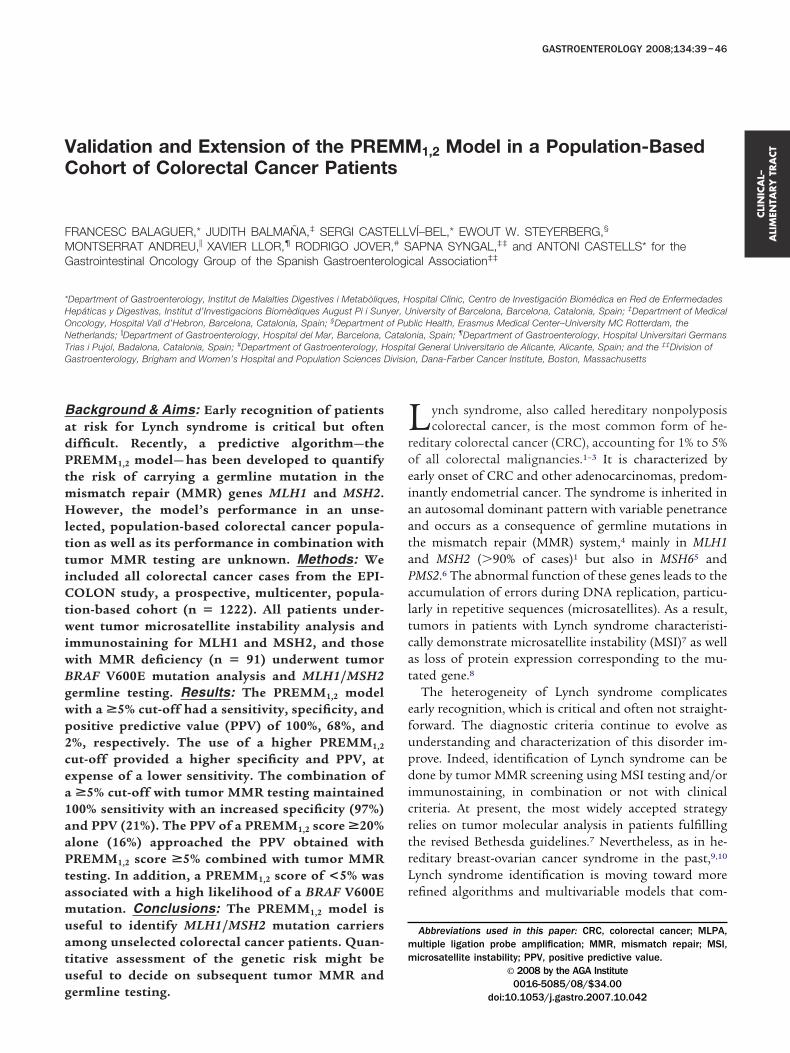
Validation and Extension of the PREMM1,2 Model in a Population-BasedCohort of Colorectal Cancer Patients
FRANCESC BALAGUER,* JUDITH BALMAÑA,‡ SERGI CASTELLVÍ–BEL,* EWOUT W. STEYERBERG,§
MONTSERRAT ANDREU,� XAVIER LLOR,¶ RODRIGO JOVER,# SAPNA SYNGAL,‡‡ and ANTONI CASTELLS* for theGastrointestinal Oncology Group of the Spanish Gastroenterological Association‡‡
*Department of Gastroenterology, Institut de Malalties Digestives i Metabòliques, Hospital Clínic, Centro de Investigación Biomédica en Red de EnfermedadesHepáticas y Digestivas, Institut d’Investigacions Biomèdiques August Pi i Sunyer, University of Barcelona, Barcelona, Catalonia, Spain; ‡Department of MedicalOncology, Hospital Vall d’Hebron, Barcelona, Catalonia, Spain; §Department of Public Health, Erasmus Medical Center–University MC Rotterdam, theNetherlands; �Department of Gastroenterology, Hospital del Mar, Barcelona, Catalonia, Spain; ¶Department of Gastroenterology, Hospital Universitari GermansTrias i Pujol, Badalona, Catalonia, Spain; #Department of Gastroenterology, Hospital General Universitario de Alicante, Alicante, Spain; and the ‡‡Division ofGastroenterology, Brigham and Women’s Hospital and Population Sciences Division, Dana-Farber Cancer Institute, Boston, Massachusetts
Background & Aims: Early recognition of patientsat risk for Lynch syndrome is critical but oftendifficult. Recently, a predictive algorithm—thePREMM1,2 model— has been developed to quantifythe risk of carrying a germline mutation in themismatch repair (MMR) genes MLH1 and MSH2.However, the model’s performance in an unse-lected, population-based colorectal cancer popula-tion as well as its performance in combination withtumor MMR testing are unknown. Methods: Weincluded all colorectal cancer cases from the EPI-COLON study, a prospective, multicenter, popula-tion-based cohort (n � 1222). All patients under-went tumor microsatellite instability analysis andimmunostaining for MLH1 and MSH2, and thosewith MMR deficiency (n � 91) underwent tumorBRAF V600E mutation analysis and MLH1/MSH2germline testing. Results: The PREMM1,2 modelwith a >5% cut-off had a sensitivity, specificity, andpositive predictive value (PPV) of 100%, 68%, and2%, respectively. The use of a higher PREMM1,2
cut-off provided a higher specificity and PPV, atexpense of a lower sensitivity. The combination ofa >5% cut-off with tumor MMR testing maintained100% sensitivity with an increased specificity (97%)and PPV (21%). The PPV of a PREMM1,2 score >20%alone (16%) approached the PPV obtained withPREMM1,2 score >5% combined with tumor MMRtesting. In addition, a PREMM1,2 score of <5% wasassociated with a high likelihood of a BRAF V600Emutation. Conclusions: The PREMM1,2 model isuseful to identify MLH1/MSH2 mutation carriersamong unselected colorectal cancer patients. Quan-titative assessment of the genetic risk might beuseful to decide on subsequent tumor MMR andgermline testing.
Lynch syndrome, also called hereditary nonpolyposiscolorectal cancer, is the most common form of he-
reditary colorectal cancer (CRC), accounting for 1% to 5%of all colorectal malignancies.1–3 It is characterized byearly onset of CRC and other adenocarcinomas, predom-inantly endometrial cancer. The syndrome is inherited inan autosomal dominant pattern with variable penetranceand occurs as a consequence of germline mutations inthe mismatch repair (MMR) system,4 mainly in MLH1and MSH2 (�90% of cases)1 but also in MSH65 andPMS2.6 The abnormal function of these genes leads to theaccumulation of errors during DNA replication, particu-larly in repetitive sequences (microsatellites). As a result,tumors in patients with Lynch syndrome characteristi-cally demonstrate microsatellite instability (MSI)7 as wellas loss of protein expression corresponding to the mu-tated gene.8
The heterogeneity of Lynch syndrome complicatesearly recognition, which is critical and often not straight-forward. The diagnostic criteria continue to evolve asunderstanding and characterization of this disorder im-prove. Indeed, identification of Lynch syndrome can bedone by tumor MMR screening using MSI testing and/orimmunostaining, in combination or not with clinicalcriteria. At present, the most widely accepted strategyrelies on tumor molecular analysis in patients fulfillingthe revised Bethesda guidelines.7 Nevertheless, as in he-reditary breast-ovarian cancer syndrome in the past,9,10
Lynch syndrome identification is moving toward morerefined algorithms and multivariable models that com-
Abbreviations used in this paper: CRC, colorectal cancer; MLPA,multiple ligation probe amplification; MMR, mismatch repair; MSI,microsatellite instability; PPV, positive predictive value.
© 2008 by the AGA Institute0016-5085/08/$34.00
doi:10.1053/j.gastro.2007.10.042
CLI
NIC
AL–
ALI
MEN
TARY
TRA
CT
GASTROENTEROLOGY 2008;134:39–46

bine personal and familial data to obtain a quantitativeestimation of the risk.11–14
The PREMM1,2 model11 is a recently developed Web-based logistic regression model that predicts the likeli-hood of germline mutations in the MLH1 and MSH2genes on the basis of personal and family history ofindividuals. It was developed in a large and diverse cohortof probands undergoing genetic testing on the basis oftheir clinical history. Whereas the model accurately dis-criminates gene mutation carriers in this subset of indi-viduals at moderate to high risk for Lynch syndrome,11
its usefulness in an unselected CRC population is un-known. Furthermore, efficacy of the PREMM1,2 model incombination with tumor MMR testing has not yet beenassessed.Using data from the EPICOLON study15,16—a prospec-
tive, multicenter, population-based cohort collected toestablish the incidence and characteristics of hereditaryand familial CRC forms in Spain—we assessed the efficacyof the PREMM1,2 model, in combination or not withtumor MMR testing, for the identification of MLH1 andMHS2 gene mutation carriers among unselected CRCpatients.
Materials and MethodsPatientsBetween November 2000 and October 2001, all
newly diagnosed CRC patients in 25 hospitals were in-cluded in the EPICOLON study.15,16 Exclusion criteriawere familial adenomatous polyposis, personal history ofinflammatory bowel disease, and patient or family refusalto participate in the study. The study was approved bythe institutional ethics committee of each participatinghospital, and written informed consent was obtainedfrom all patients.Demographic, clinical, and tumor-related characteris-
tics of probands, as well as a detailed family history wereobtained using a pre-established questionnaire. Pedigreeswere traced backward and laterally as far as possible, or atleast up to second-degree relatives, in terms of cancerhistory. Age at cancer diagnosis, type, location, and tu-mor stage of the neoplasm and current status were re-corded for each affected family member.15,16
Tumor Microsatellite Instability Analysis andImmunostainingTissue samples from tumor and normal colonic
mucosa were obtained from each patient, immediatelyfrozen in liquid nitrogen, and stored at �70°C until use.In cases where no frozen tissue was available, formalin-fixed, paraffin-embedded samples were used. GenomicDNA was isolated using the QiaAmp Tissue Kit (Qiagen,Courtaboeuf, France).Microsatellite instability testing and immunostaining
for MLH1 and MSH2 were performed in all patientsregardless of age, personal or family history, and tumor
characteristics. In addition, in those patients with aPREMM1,2 score �20%, immunostaining for MSH6 andPMS2 was also performed. Paraffin-embedded sectionswere immunostained with antibodies against mismatchrepair proteins (anti-MSH2, Oncogene Research Prod-ucts, Boston, MA; anti-MLH1, PharMingen, San Diego,CA; anti-MSH6, BD Transduction Laboratories; anti-PMS2, PharMingen), as described elsewhere.15 Tumorcells were judged to be negative for protein expressiononly if they lacked staining in a sample in which normalcolonocytes and stroma cells were stained. If no immu-nostaining of normal tissue could be demonstrated, theresults were considered ambiguous.Microsatellite status was assessed using the 5-marker
panel proposed by the National Cancer Institute, as de-scribed elsewhere.15,17,18 Tumors were classified as stableif none of the markers showed instability. Tumors with 2or more unstable markers were classified as high levelMSI (MSI-H) and tumors with 1 unstable marker wereclassified as low-level MSI (MSI-L).
Germline MLH1/MSH2 Mutation AnalysisPatients found to have tumors with MMR defi-
ciency (demonstrated by either MSI-H and/or lack ofprotein expression) underwent MSH2/MLH1 germline ge-netic testing. Moreover, all patients with a PREMM1,2
score �20% with MMR-proficient tumors also underwentgenetic testing.Germline mutational analysis was performed by both
multiple ligation probe amplification (MLPA) analysisand sequencing, as described elsewhere.15
Tumor BRAF V600E Mutation AnalysisTumor BRAF V600E mutation analysis was per-
formed in all patients with MSI (high and low) and/orlack of MLH1/MSH2 protein expression by direct se-quencing in tumor DNA, as described elsewhere.19
Application of the PREMM1,2 ModelThe PREMM1,2 model is a clinical model created
to predict the likelihood of finding a MLH1 or MSH2mutation in at-risk individuals.11 The original study an-alyzed MLH1/MSH2 mutation prevalence in a large co-hort of patients undergoing genetic testing at MyriadGenetic Laboratories Inc (Salt Lake City, UT). A multi-variable model using logistic regression and includingvariables related to the proband and relatives was devel-oped. The prediction rule is available as a Web-based toolat the Dana-Farber Cancer Institute web site (http://www.dfci.org/premm). We calculated the PREMM1,2
score for each patient included in the study using theSPSS V11.0 software package (SPSS Inc, Chicago, IL).
Statistical AnalysisSensitivity, specificity, and positive predictive
value (PPV) of the PREMM1,2 model, either alone or in
CLIN
ICA
L–A
LIMEN
TARY
TRA
CT
40 BALAGUER ET AL GASTROENTEROLOGY Vol. 134, No. 1

combination with tumor MMR testing, were calculatedwith respect to the presence of MLH1/MSH2 germlinemutations. These performance characteristics depend onthe cut-off used for the predicted risk of mutation, andwe therefore arbitrarily evaluated the following cut-offlevels: �5%, �5%, �10%, �20%, and �40%. Ninety-fivepercent binomial confidence intervals were calculated onthe basis of the Adjusted Wald method.20
Continuous variables were expressed as mean � stan-dard deviation and compared by the Student t test. Cat-egorical variables were compared by the �2 test, applyingthe Yates correction when needed.All P values were two sided. A P value of less than .05
was considered to indicate a statistically significant dif-ference. All calculations were performed using the 11.0SPSS software package (SPSS Inc).
ResultsCharacteristics of the PatientsDuring the study period, 1222 patients with
pathologically confirmed colorectal adenocarcinomawere diagnosed and included in the EPICOLON project.Demographic, clinical, and tumor-related characteristicsof patients included in the study are summarized inTable 1.One hundred eleven (9.1%) patients showed tumor
MSI, 83 of them (6.8%) were MSI-H and 28 (2.3%) wereMSI-L. Likewise, 81 (6.6%) patients had a tumor with lossof protein expression in either MLH1 (60 cases) or MSH2(21 cases). No patients with tumors that were MSI-L hadlack of MMR protein expression. However, expression ofboth proteins was retained in 10 tumors with MSI-H,whereas loss of MLH1 or MSH2 expression was found in8 patients whose tumor did not show MSI. Overall, 91(7.4%) patients were found to have a tumor demonstrat-ing MMR deficiency (defined as MSI-H and/or loss ofMLH1 or MSH2 expression).
BRAF V600E mutation was detected in 20 of 83 (24.1%)MSI-H tumors and in 18 of 60 (30%) tumors exhibitingloss of MLH1 expression. In addition, only 2 of 28 (7.1%)MSI-L tumors showed the BRAF V600E mutation,whereas it was not observed in any tumor with loss ofMSH2 expression.Germline genetic testing identified 8 (0.7%) unambig-
uous mutations in either MSH2 (5 cases) or MLH1 (3cases) genes.
Efficacy of the PREMM1,2 Model for theIdentification of MLH1/MSH2 Gene CarriersThe distribution according to the PREMM1,2 pre-
dicted likelihood of carrying a MLH1/MSH2 germlinemutation in the cohort was: �5%, 826 (68%); 5–9%, 266(22%); 10–19%, 98 (8%); 20–29%, 23 (2%); and �40%, 9(0.7%).We first evaluated the PREMM1,2 model for its ability
to identify MLH1/MSH2 mutation carriers within thelarge cohort of CRC patients. Performance characteristicsof the PREMM1,2 model for the identification of MLH1/MSH2 gene carriers depended on the cut-off used for thepredicted risk of mutation (Table 2). Using a cut-off of
Table 1. Clinical and Molecular Characteristics of PatientsIncluded in the Study
Characteristics Values
Age (y)a 70 � 11Men: no. (%) 731 (59.8)Site of tumor: no. (%)
Proximal to splenic flexure 357 (29.2)Distal to splenic flexure 865 (70.8)
Tumor TNM stage: no. (%)I 161 (13.2)II 510 (41.7)III 337 (27.6)IV 214 (17.5)
Degree of differentiation: no. (%)Well 292 (23.9)Moderate 835 (68.3)Poor 95 (7.8)
Mucinous carcinoma type: no. (%) 142 (11.6)Synchronous colorectal cancer: no. (%) 71 (5.8)Microsatellite instability, high: no. (%) 83 (6.8)Microsatellite instability, low: no. (%) 28 (2.3)Loss of MLH1/MSH2 protein expression: no. (%) 81 (6.6)Tumor MMR deficiencyb: no. (%) 91 (7.4)Germline MLH1 mutation: no. (%) 3 (0.25)Germline MSH2 mutation: no. (%) 5 (0.4)
MMR, mismatch repair.aExpressed as mean � standard deviation.bTumor MMR deficiency demonstrated by either high microsatelliteinstability and/or loss of MLH1/MSH2 protein expression.
Table 2. Performance Characteristics of the PREMM1,2 Model for the Identification of MLH1/MSH2 Gene Mutation Carriers
Strategy
Without tumor MMR test resultsa With MSI-H or abnormal IHC tumor resultsa
No. (%)Sensitivity(95% CI)
Specificity(95% CI)
Positive predictive value(95% CI) No. (%)
Sensitivity(95% CI)
Specificity(95% CI)
Positive predictive value(95% CI)
PREMM1,2 �5% 396 (32.0) 100 (70.6-100) 68 (65.4-70.6) 2 (1-4) 39 (3.2) 100 (70.6-100) 97.4 (96.4-98.2) 20.5 (10.5-35.8)PREMM1,2 �10% 130 (10.6) 75 (40-93.7) 89.8 (88-91.4) 4.6 (1.9-9.9) 21 (1.7) 75 (40-93.7) 98.8 (97.9-99.3) 28.6 (13.5-50.2)PREMM1,2 �20% 32 (2.6) 62.5 (30.4-86.5) 97.7 (96.8-98.5) 15.6 (6.4-32.2) 14 (1.1) 62.5 (30.4-86.5) 99.3 (98.6-99.6) 35.7 (16.2-61.4)PREMM1,2 �40% 9 (0.7) 25 (6.3-59.9) 99.4 (98.8-99.7) 22.2 (5.3-55.7) 7 (0.6) 25 (6.3-59.9) 99.6 (99-99.8) 28.6 (7.6-64.8)
MMR, mismatch repair; MSI-H, high microsatellite instability; IHC, immunohistochemistry; 95% CI, 95% confidence interval.aTumor MMR testing by either microsatellite instability or MLH1/MSH2 immunostaining.
CLI
NIC
AL–
ALI
MEN
TARY
TRA
CT
January 2008 LYNCH SYNDROME IDENTIFICATION BY THE PREMM1,2 MODEL 41

�5%, the model had a sensitivity of 100%; therefore, nomutation carriers would be missed if molecular evalua-tion was restricted to individuals with a PREMM1,2 scoreof �5%. Using higher cut-offs of 10%, 20%, and 40% ledto a progressive loss of sensitivity (75%, 62.5%, and 25%,respectively). As expected, specificity increased withhigher cut-offs and ranged from 68% with a 5% cut-off to99.4% with a 40% cut-off. Positive predictive values ofdifferent cut-offs for the PREMM1,2 model are depicted inFigure 1.
Use of the PREMM1,2 Model in CombinationWith Tumor MMR TestingThe addition of tumor MMR testing, either by
MSI analysis or immunostaining, to the PREMM1,2
model enhanced its performance by improving bothspecificity and PPV (Table 2). A PREMM1,2 score of �5%in combination with abnormal MMR testing was associ-ated with a sensitivity of 100%, specificity of 97.4%, andPPV of 20.5%. The maximum PPV (36%) was achievedusing a PREMM1,2 score of �20% in combination with anabnormal tumor MMR result. The incremental gain ob-tained by the addition of MSI/immunohistochemistrytesting was less at higher PREMM1,2 cut-off values; at aPREMM1,2 cut-off of 40%, the addition of MSI/immuno-histochemistry testing did not lead to an improvement inspecificity.
Characteristics of Patients With LowPREMM1,2 ScoresThe PREMM1,2 score correlated not only with the
prevalence of germline mutations but also with the fre-quency of MMR deficiency (Table 3). Although 52 of 826(6.3%) individuals with a PREMM1,2 score �5% had aMSI-H tumor or showed loss of MLH1 and MSH2 onimmunohistochemistry, none of them carried a germlineMLH1/MSH2 mutation and 17 (33%) were associatedwith BRAF V600E mutation in the tumor (Table 3).Interestingly, in patients with abnormal MMR tests,
BRAF V600E mutation was significantly associated with aPREMM1,2 score �5% (P � .009). In fact, 17 of 20 (85%)patients with a MMR-deficient tumor associated withBRAF V600E mutation had a PREMM1,2 score �5%,whereas none of the 14 patients with a MMR-deficienttumor and PREMM1,2 score �20% showed this variant(Table 3). As recent data demonstrate that BRAF muta-tions are rare in Lynch syndrome tumors, the findings areconsistent with the conclusion that a low PREMM1,2
score indicates a low likelihood that a patient with CRChas Lynch syndrome.
Characteristics of Patients With HighPREMM1,2 ScoresWhen patients with a PREMM1,2 score �20% were
stratified according to their MMR status (Table 4), pa-tients with MMR-deficient tumors differed from thosewith MMR proficiency in some clinical characteristics:they are more likely women (P � .02), have a lowerprevalence of previous or synchronous adenomas (P �.002), a higher prevalence of endometrial cancer (P �.003), more first-degree relatives with CRC (P � .01), andmore second-degree relatives with endometrial cancer (P� .03). Therefore, a high PREMM1,2 score in combinationwith MMR proficiency identified a significant group offamilies, recently characterized in our series, with a lesspenetrant cancer phenotype,18 in line with a similargroup with Amsterdam I criteria recently described asfamilial CRC type X syndrome.21
We further examined potential etiologies of the CRC inthe patients with high PREMM1,2 scores based on theirMSI status. Five of 14 (36%) patients with PREMM1,2
score �20% and a MMR-deficient tumor carried a MLH1/MSH2 germline mutation. We further investigated poten-tial etiologies of the high PREMM1,2 scores for the 9individuals who were not found to carry germline MLH1/MSH2 mutations by performing supplemental analysesof PMS2 and MSH6 immunostaining, and BRAF muta-tion analysis. In these nonmutation carriers, no BRAF
Table 3. Prevalence of MLH1/MSH2 Germline Mutationsand Mismatch Repair Deficiency According to thePREMM1,2 Score
PREMM1,2
score No.
MLH1/MSH2germline
mutation (%)a
Tumor MMRdeficiencyb
(%)1
Tumor MMR deficiencyb
associated with BRAFV600E mutation (%)a
�5% 826 – (–) 52 (6) 17 (2)5–9% 266 2 (0.8) 18 (7) 2 (0.7)10–19% 98 1 (1) 7 (7) 1 (1)20–39% 23 3 (13) 7 (30) – (–)�40% 9 2 (22) 7 (78) – (–)Total 1222 8 91 20
MMR, mismatch repair.aPercentages referred to each PREMM1,2 category.bTumor MMR deficiency demonstrated by either high microsatelliteinstability and/or loss of MLH1/MSH2 expression.
Figure 1. Positive predictive value for detecting germline MLH1/MSH2gene mutations according to the PREMM1,2 score.
CLIN
ICA
L–A
LIMEN
TARY
TRA
CT
42 BALAGUER ET AL GASTROENTEROLOGY Vol. 134, No. 1

mutation was found, and normal PMS2 and MSH6 pro-tein expression was observed in all tumors.To better characterize the subset of patients with a
PREMM1,2 score �20% and MMR-proficient tumors (n� 18), MSH6 and PMS2 immunostaining and MLH1/MSH2 germline gene testing were performed in all ofthem. With respect to immunostaining, normal MSH6and PMS2 protein expression was observed in all tu-mors. Furthermore, MLH1/MSH2 gene testing did notshow any deleterious mutations. Finally, we performedMYH analysis, and one patient was found to have abiallelic MYH mutation (G382D/Y165C) in a nestedstudy performed in the EPICOLON cohort.22 The pa-tient was a 49 year-old-man with 2 synchronous CRCsand 25 synchronous adenomas and no family historyof any neoplasia.
DiscussionExtensive knowledge now available about the
Lynch syndrome has encouraged researchers to look for asystematic, quantitative, and objective approach to iden-tify these patients.11–13,23 We recently developed the Web-based PREMM1,2 model11 on the basis of a logistic regres-sion analysis from a large cohort of patients at risk forhereditary CRC who underwent genetic testing to quan-tify the relative importance of known clinical parametersin Lynch syndrome and predict the likelihood of carrying
a mutation in the MLH1 and MSH2 genes. Although themodel performed well among individuals at moderaterisk for Lynch syndrome, its usefulness and performancein a nonselected, population-based cohort of CRC pa-tients, either alone or in combination with tumor MMRtesting, was unknown.Our study of 1222 population-based CRC cases dem-
onstrates that the PREMM1,2 model constitutes a usefulapproach to identify MLH1/MSH2 gene mutation carriersamong patients with CRC, either alone or in combina-tion with MMR tumor testing. The quantitative assess-ment of the genetic risk obtained with the PREMM1,2
model may drive subsequent decisions about moleculartesting. Moreover, the combination with tumor MMRanalysis identified a sizable subgroup of patients with aheterogeneous high CRC risk, potentially involving fa-milial CRC type X syndrome, MYH-related cancer, andother, still unknown inherited disorders.The first important finding is the demonstration that
a PREMM1,2 cut-off of �5% identified all MLH1 andMSH2 mutation carriers among unselected CRC patients.The use of a higher PREMM1,2 cut-off provided a higherspecificity and PPV, at the expense of a lower sensitivity.The negative predictive value of a PREMM1,2 score �5%was 100%, thus reinforcing the consistency of this cut-offpoint. Therefore, for the clinician in general practice,whose first decision point is to see if a patient with CRC
Table 4. Personal and Familial Characteristics of Patients With PREMM1,2 Score �20% According to the Mismatch RepairStatus
MMR proficiency(n � 18)
MMR deficiency(n � 14) P value
Personal characteristicsAge (y)a 66.0 � 15.5 60.7 � 19 .39Female: no. (%) 4 (22.2) 9 (64.3) .02Previous CRC: no. (%) 6 (33.3) 2 (14.2) .34Synchronous CRC: no. (%) 7 (38.9) 3 (21.4) .45
Previous or synchronous adenoma: no. (%) 11 (61.1) 1 (7.1) .002Endometrial cancer: no. (%) – (–) 6 (42.9) .003Other Lynch-syndrome–associated cancersb: no. (%) 6 (33.3) 7 (50) .34Proximal CRC: no. (%) 7 (38.9) 6 (42.9) .82Familial characteristics
Age of FDR with CRC (y)a 45.7 � 11.3 40.4 � 7.2 .13FDR with CRC: no. (%) 6 (33.3) 11 (78.6) .01�2 FDR with CRC: no. (%) 3 (16.7) 7 (50) .06FDR with endometrial cancer: no. (%) 7 (38.9) 3 (21.4) .45�2 FDR with endometrial cancer: no. (%) 2 (11.1) 1 (7.1) 1.0 (%)FDR with other Lynch-syndrome–associated cancersb:
no. (%)2 (11.1) 6 (42.9) .09
Age of SDR with CRC (y)a 45.9 � 7.8 46.3 � 5.6 .84SDR with CRC: no. (%) 1 (5.6) 4 (28.6) .14�2 SDR with CRC: no. (%) – (–) 2 (14.3) .18SDR with endometrial cancer: no. (%) – (–) 4 (28.6) .03SDR with other Lynch-syndrome–associated cancers2:
no. (%)1 (5.6) 2 (14.3) .57
Amsterdam II criteria: no. (%) 4 (22.2) 10 (71.4) .005
MMR, mismatch repair; CRC, colorectal cancer; FDR, first-degree relatives; SDR, second-degree relatives.aExpressed as mean � standard deviation.bStomach, ovaries, urinary tract, small intestine, pancreas, bile ducts, brain, or sebaceous glands.
CLI
NIC
AL–
ALI
MEN
TARY
TRA
CT
January 2008 LYNCH SYNDROME IDENTIFICATION BY THE PREMM1,2 MODEL 43

needs further molecular evaluation for Lynch Syndrome,a score of �5% indicates that no further referral is likelyto be necessary, whereas a score of �5% should lead tofurther molecular evaluation. The low specificity of a 5%cut-off for the presence of germline mutations necessi-tates further refinement of the likelihood of carrying amutation prior to proceeding to genetic testing. Our datademonstrate that the combination of the PREMM1,2
score with tumor MMR testing improved its specificityand PPV. Indeed, using a cut-off of �5%, the addition ofan abnormal tumor MMR test result provided a specific-ity of 98% and a PPV of 21%.An interesting finding was that the PREMM1,2 model,
in combination with tumor MMR testing, is able toidentify a subset of patients resembling the recently de-scribed familial CRC type X syndrome,21 families whofulfill the Amsterdam I criteria without evidence of MMRdeficiency. Individuals in such families have a lower in-cidence of CRC than those in families with Lynch syn-drome, whereas incidence for other cancers may not beincreased. The molecular etiology of this disorder re-mains unknown, with a probable heterogeneous geneticbasis. In our study, patients with a PREMM1,2 score�20% with MMR-proficient tumors had features similarto those with familial CRC type X syndrome21: weakerfamily history of CRC and other malignancies and lowerincidence of endometrial cancer. Interestingly, one pa-tient with a high PREMM1,2 score and no evidence ofMMR deficiency carried biallelic MYH mutations(Y165C/G382D).We are aware that our study has some limitations.
First, the relatively low number of patients with MLH1/MSH2 mutations may constitute a potential drawback ofthe analysis, thus restraining the reliability of perfor-mance features. Second, the model does not account forMSH6 gene mutations, although it is certain that thisgene is responsible for a small proportion of Lynch syn-drome cases. Finally, genetic testing was mainly per-formed in those patients whose tumors showed MMRdeficiency, even though it is unlikely that gene carrierswere undetected when both MSI analysis and immuno-staining were performed systematically. In addition, toexclude this possibility, patients with a PREMM1,2 score�20% also underwent genetic testing.In the last few years, there has been much interest in
establishing different strategies to improve the identifi-cation of patients with Lynch syndrome. These ap-proaches range from using clinical criteria alone (ie, theAmsterdam criteria)24 to universal tumor molecular test-ing (ie, immunostaining) in any given CRC patient.25 Thecurrent most widely accepted recommendation, on thebasis of combination of the revised Bethesda guidelinesand tumor MMR testing,7 has been found to be aneffective and efficient strategy for Lynch syndrome iden-tification.15 However, these clinical criteria have beencriticized because of the use of broad and complex vari-
ables, which make them difficult to remember for ageneral health care professional, their low specificity,their inability to establish the likelihood of carrying amutation in a given patient, and the difficulty of obtain-ing tumor samples from affected relatives to perform theMMR analyses.23 A potential advantage of the PREMM1,2
model with respect to the revised Bethesda guidelinesrelies on its quantitative nature. In fact, the model dem-onstrated a reasonable ability to discriminate among riskgroups for probability of mutation with respect to theprevalence of mutations observed in the MLH1/MSH2genes and the prevalence of MMR deficiency. The lattercorrelation was especially relevant given that a PREMM1,2
score of �5% identified the subset of MMR-deficienttumors associated with the somatic BRAF V600E muta-tion, a circumstance consistent with what is seen in thesporadic CRC setting.26–28 Taking into account theseresults and the performance characteristics of the pre-dicted model’s risk groups, in combination or not withtumor MMR testing, we propose a strategy for MLH1/MSH2 genetic testing in the clinical practice (Figure 2).According to this algorithm, a PREMM1,2 score �5%could be considered a reliable cut-off to exclude thoseCRC patients who do not need further risk assessmentbecause of its 100% negative predictive value for detectinggermline MLH1/MSH2 gene mutations. In patients reach-ing this cut-off, further decisions could also be made onthe basis of PREMM1,2 score. In patients with a scorebetween 5% and 19%, tumor MMR testing should beperformed to achieve a reasonable PPV. Finally, consid-ering the PPV of a PREMM1,2 score �20% alone (16%)and the significant increase of the PPV at that point(Figure 1), it seems reasonable to pursue direct genetictesting in patients reaching such a score, particularly if atumor sample is not available. It is important to note,however, that in addition to the risk estimate generatedfrom the predictive model, other important factors (ie,accessibility to genetic services, timelines of genetic in-formation, insurance coverage, and availability of tumorblock) may help determine which strategy is the most
Figure 2. Proposed algorithm for the identification of MLH1/MSH2gene carriers among patients with colorectal cancer. CRC, colorectalcancer; MMR, mismatch repair.
CLIN
ICA
L–A
LIMEN
TARY
TRA
CT
44 BALAGUER ET AL GASTROENTEROLOGY Vol. 134, No. 1
convenient in a given patient. In that sense, thePREMM1,2 model can be used by general health careproviders to decide whether to refer a patient to a high-risk colorectal cancer clinic for appropriate genetic coun-seling, as well as by geneticists working in such units todecide on the proper molecular strategy. The use of thesame algorithm in both clinical settings may contributeto a more rational referral and management approach ofpatients with suspected Lynch syndrome.In conclusion, our study demonstrates that the
PREMM1,2 model is useful to identify MLH1/MSH2 mu-tation carriers among unselected CRC patients. Thequantitative assessment of the genetic risk might be use-ful to decide subsequent molecular testing and contrib-ute to identify other high-risk individuals who may ben-efit from genetic risk assessment.
Appendix
Following is a list of investigators from the Gas-trointestinal Oncology Group of the Spanish Gastroen-terological Association who participated in the Epicolonstudy. Hospital 12 de Octubre, Madrid: Juan Diego Mo-rillas (local coordinator), Raquel Muñoz, Marisa Man-zano, Francisco Colina, Jose Díaz, Carolina Ibarrola,Guadalupe López, Alberto Ibáñez; Hospital Clínic, Bar-celona: Antoni Castells (local coordinator), VirgíniaPiñol, Sergi Castellví-Bel, Francesc Balaguer, VictòriaGonzalo, Teresa Ocaña, María Dolores Giraldez, MariaPellisé, J. Ignasi Elizalde, Josep M. Piqué; Hospital ClínicoUniversitario, Zaragoza: Ángel Lanas (local coordinator),Javier Alcedo, Javier Ortego; Hospital Cristal-Piñor, Com-plexo Hospitalario de Ourense: Joaquin Cubiella (localcoordinator), Ma Soledad Díez, Mercedes Salgado, EloySánchez, Mariano Vega; Hospital del Mar, Barcelona:Montserrat Andreu (local coordinator), Xavier Bessa,Agustín Panadés, Asumpta Munné, Felipe Bory, MiguelNieto, Agustín Seoane; Hospital Donosti, San Sebastián:Luis Bujanda (local coordinator), Juan Ignacio Arenas,Isabel Montalvo, Julio Torrado, Ángel Cosme; HospitalGeneral Universitario de Alicante: Artemio Payá (localcoordinator), Rodrigo Jover, Juan Carlos Penalva, Cris-tina Alenda; Hospital General de Granollers: JoaquimRigau (local coordinator), Ángel Serrano, Anna Giménez;Hospital General de Vic: Joan Saló (local coordinator),Eduard Batiste-Alentorn, Josefina Autonell, Ramon Bar-niol; Hospital General Universitario de Guadalajara: AnaMaría García (local coordinator), Fernando Carballo, An-tonio Bienvenido, Eduardo Sanz, Fernando González,Jaime Sánchez; Hospital General Universitario de Valen-cia: Enrique Medina (local coordinator), Jaime Cuque-rella, Pilar Canelles, Miguel Martorell, José Ángel García,Francisco Quiles, Elisa Orti; Hospital do Meixoeiro, Vigo:Juan Clofent (local coordinator), Jaime Seoane, AntoniTardío, Eugenia Sanchez; Hospital San Eloy, Baracaldo:Luis Bujanda (local coordinator), Carmen Muñoz, Maríadel Mar Ramírez, Araceli Sánchez; Hospital Universitari
Germans Trias i Pujol, Badalona: Xavier Llor (local coor-dinator), Rosa M. Xicola, Marta Piñol, Mercè Rosinach,Anna Roca, Elisenda Pons, José M. Hernández, Miquel A.Gassull; Hospital Universitari Mútua de Terrassa: Fer-nando Fernández-Bañares (local coordinator), Josep M.Viver, Antonio Salas, Jorge Espinós, Montserrat Forné,Maria Esteve; Hospital Universitari Arnau de Vilanova,Lleida: Josep M. Reñé (local coordinator), Carmen Piñol,Juan Buenestado, Joan Viñas; Hospital Universitario deCanarias: Enrique Quintero (local coordinator), DavidNicolás, Adolfo Parra, Antonio Martín; Hospital Univer-sitario La Fe, Valencia: Lidia Argüello (local coordinator),Vicente Pons, Virginia Pertejo, Teresa Sala; Hospital Uni-versitario Reina Sofía, Córdoba: Antonio Naranjo (localcoordinator), María Dolores Giraldez, María del ValleGarcía, Patricia López, Fernando López, Rosa Ortega,Javier Briceño, Javier Padillo; Fundació Hospital SonLlatzer, Palma de Mallorca: Àngels Vilella (local coordi-nator), Carlos Dolz, Hernan Andreu.
References
1. Lynch HT, de la Chapelle A. Genetic susceptibility to non-polypo-sis colorectal cancer. J Med Genet 1999;36:801–818.
2. Giardiello FM, Brensinger JD, Petersen GM. AGA technical reviewon hereditary colorectal cancer and genetic testing. Gastroenter-ology 2001;121:198–213.
3. de la Chapelle A. The incidence of Lynch syndrome. Fam Cancer2005;4:233–237.
4. Lynch HT, Boland CR, Gong G, et al. Phenotypic and genotypicheterogeneity in the Lynch syndrome: diagnostic, surveillanceand management implications. Eur J Hum Genet 2006;14:390–402.
5. Miyaki M, Konishi M, Tanaka K, et al. Germline mutation of MSH6as the cause of hereditary nonpolyposis colorectal cancer. NatGenet 1997;17:271–272.
6. Hendriks YM, Jagmohan-Changur S, van der Klift HM, et al.Heterozygous mutations in PMS2 cause hereditary nonpolyposiscolorectal carcinoma (Lynch syndrome). Gastroenterology 2006;130:312–322.
7. Umar A, Boland CR, Terdiman JP, et al. Revised Bethesda Guide-lines for hereditary nonpolyposis colorectal cancer (Lynch syn-drome) and microsatellite instability. J Natl Cancer Inst 2004;96:261–268.
8. Stone JG, Robertson D, Houlston RS. Immunohistochemistry forMSH2 and MHL1: a method for identifying mismatch repair defi-cient colorectal cancer. J Clin Pathol 2001;54:484–487.
9. Couch FJ, DeShano ML, Blackwood MA, et al. BRCA1 mutationsin women attending clinics that evaluate the risk of breast can-cer. N Engl J Med 1997;336:1409–1415.
10. Parmigiani G, Berry D, Aguilar O. Determining carrier probabilitiesfor breast cancer-susceptibility genes BRCA1 and BRCA2. Am JHum Genet 1998;62:145–158.
11. Balmana J, Stockwell DH, Steyerberg EW, et al. Prediction ofMLH1 and MSH2 mutations in Lynch syndrome. JAMA 2006;296:1469–1478.
12. Barnetson RA, Tenesa A, Farrington SM, et al. Identification andsurvival of carriers of mutations in DNA mismatch-repair genes incolon cancer. N Engl J Med 2006;354:2751–2763.
13. Chen S, Wang W, Lee S, et al. Prediction of germline mutationsand cancer risk in the Lynch syndrome. JAMA 2006;296:1479–1487.
CLI
NIC
AL–
ALI
MEN
TARY
TRA
CT
January 2008 LYNCH SYNDROME IDENTIFICATION BY THE PREMM1,2 MODEL 45

14. Wijnen JT, Vasen HF, Khan PM, et al. Clinical findings withimplications for genetic testing in families with clustering ofcolorectal cancer. N Engl J Med 1998;339:511–518.
15. Pinol V, Castells A, Andreu M, et al. Accuracy of revised Bethesdaguidelines, microsatellite instability, and immunohistochemistryfor the identification of patients with hereditary nonpolyposiscolorectal cancer. JAMA 2005;293:1986–1994.
16. Pinol V, Andreu M, Castells A, et al. Frequency of hereditarynon-polyposis colorectal cancer and other colorectal cancer fa-milial forms in Spain: a multicentre, prospective, nationwidestudy. Eur J Gastroenterol Hepatol 2004;16:39–45.
17. Xicola RM, Llor X, Pons E, et al. Performance of different micro-satellite marker panels for detection of mismatch repair-deficientcolorectal tumors. J Natl Cancer Inst 2007;99:244–252.
18. Llor X, Pons E, Xicola RM, et al. Differential features of colorectalcancers fulfilling Amsterdam criteria without involvement of themutator pathway. Clin Cancer Res 2005;11:7304–7310.
19. Benlloch S, Paya A, Alenda C, et al. Detection of BRAF V600Emutation in colorectal cancer: comparison of automatic sequenc-ing and real-time chemistry methodology. J Mol Diagn 2006;8:540–543.
20. Sauro J, Lewis J. Estimating completion rates from small sam-ples using binomial confidence intervals: comparisons and rec-ommendations. Proceedings of the human factors and ergonom-ics society, 49th Annual Meeting 2005:2100–2104.
21. Lindor NM, Rabe K, Petersen GM, et al. Lower cancer incidencein Amsterdam-I criteria families without mismatch repair defi-ciency: familial colorectal cancer type X. JAMA 2005;293:1979–1985.
22. Balaguer F, Castellvi-Bel S, Castells A, et al. Identification of MYHmutation carriers in colorectal cancer: a multicenter, case-con-trol, population-based study. Clin Gastroenterol Hepatol 2007;5:379–387.
23. Lipton LR, Johnson V, Cummings C, et al. Refining the Amster-dam Criteria and Bethesda Guidelines: testing algorithms for theprediction of mismatch repair mutation status in the familialcancer clinic. J Clin Oncol 2004;22:4934–4943.
24. Vasen HF, Mecklin JP, Khan PM, et al. Anticancer Res 1994;14:1661–1664.
25. Hampel H, Frankel WL, Martin E, et al. Screening for the Lynchsyndrome (hereditary nonpolyposis colorectal cancer). N EnglJ Med 2005;352:1851–1860.
26. Weisenberger DJ, Siegmund KD, Campan M, et al. CpG islandmethylator phenotype underlies sporadic microsatellite instabil-ity and is tightly associated with BRAF mutation in colorectalcancer. Nat Genet 2006;38:787–793.
27. Loughrey MB, Waring PM, Tan A, et al. Incorporation of somaticBRAF mutation testing into an algorithm for the investigation ofhereditary non-polyposis colorectal cancer. Fam Cancer 2007;6:301–310.
28. Minoo P, Moyer M, Jass J. Role of BRAF-V600E in the serratedpathway of colorectal tumourigenesis. J Pathol 2007;212:124–133.
Received July 30, 2007. Accepted October 11, 2007.Address requests for reprints to: Antoni Castells, MD, Department of
Gastroenterology, Hospital Clínic, Villarroel 170, 08036-Barcelona,Catalonia, Spain. e-mail: [email protected]; fax: (34) 93 227 93 87.
Francesc Balaguer and Judith Balmaña contributed equally to thiswork.
All authors in the Gastrointestinal Oncology Group of the SpanishGastroenterological Association are listed in the Appendix.
Dr Llor’s current address is Section of Digestive Diseases and Nu-trition, University of Illinois, Chicago, Illinois.
Supported by grants from the Fondo de Investigación Sanitaria (FIS01/0104, 03/0070, and 05/0071), the Ministerio de Educación yCiencia (SAF 04-07190 and 07-64873), and the Asociación Españolacontra el Cáncer. Francesc Balaguer received a research grant fromthe Hospital Clínic and the Instituto de Salud Carlos III, and SergiCastellví-Bel is supported by a contract from the Fondo de Investi-gación Sanitaria. Also supported by the US National Cancer Institutegrant CA 113433 (Dr Syngal).
CLIN
ICA
L–A
LIMEN
TARY
TRA
CT
46 BALAGUER ET AL GASTROENTEROLOGY Vol. 134, No. 1

80

81
ARTICLE 2
“Identification of MYH mutation carriers in colorectal cancer: a multicenter,
case-control, population-based study”

Identification of MYH Mutation Carriers in Colorectal Cancer:A Multicenter, Case-Control, Population-Based Study
FRANCESC BALAGUER,* SERGI CASTELLVÍ–BEL,* ANTONI CASTELLS,* MONTSERRAT ANDREU,‡ JENIFER MUÑOZ,*JAVIER P. GISBERT,§ XAVIER LLOR,� RODRIGO JOVER,¶ RAFAEL DE CID,# VICTÒRIA GONZALO,* XAVIER BESSA,‡
ROSA M. XICOLA,� ELISENDA PONS,¶ CRISTINA ALENDA,** ARTEMIO PAYÁ,** and JOSEP M. PIQUÉ,* FOR THEGASTROINTESTINAL ONCOLOGY GROUP OF THE SPANISH GASTROENTEROLOGICAL ASSOCIATION††
*Department of Gastroenterology, Institut de Malalties Digestives i Metabòliques, Hospital Clínic, Ciberehd, IDIBAPS (Institut d’Investigacions Biomèdiques August Pi iSunyer), University of Barcelona, Barcelona, Catalonia; ‡Department of Gastroenterology, Hospital del Mar, Barcelona, Catalonia; §Department of Gastroenterology,Hospital Universitario de la Princesa, Madrid; �Department of Gastroenterology, Hospital Universitari Germans Trias i Pujol, Badalona, Catalonia; #Centre de RegulacióGenòmica, Barcelona, Catalonia; ¶Department of Gastroenterology and **Department of Pathology, Hospital General Universitario de Alicante, Alicante, Spain. ††Allauthors are listed in the Appendix
Background & Aims: Whereas it has conclusively beendemonstrated that biallelic MutY human homolog (MYH) mu-tations confer a significant risk for colorectal cancer (CRC),the influence of monoallelic mutations remains controver-sial. Characterization of MYH-associated CRC is critical toidentify individuals who might benefit from preventivestrategies. This prospective, multicenter, case-control, pop-ulation-based study was aimed at (1) establishing the CRCrisk associated with specific germline MYH mutations and(2) devising a set of clinical criteria to identify MYH carriersamong newly diagnosed CRC. Methods: Genotyping forY165C and G382D was performed by TaqMan technology.Single-stranded conformation polymorphism analysis wasperformed in heterozygotes to screen for mutations in theentire gene. All individuals were re-screened for any addi-tional pathogenic variant. Results: Biallelic and monoal-lelic MYH mutations were found in 8 (0.7%) and 19 (1.7%)of 1116 CRC patients, respectively. None of the 934 controlsubjects carried biallelic mutations, whereas 22 (2.3%) ofthem were monoallelic carriers. In a meta-analysis includ-ing all previous case-control studies, monoallelic MYH car-riers were not at increased risk for CRC (odds ratio, 1.11;95% confidence interval, 0.90 –1.37), although a significantassociation was found with the Y165C mutation in eitherhomozygotes or heterozygotes (odds ratio, 1.67; 95% confi-dence interval, 1.17–2.40). Furthermore, presence of morethan 15 synchronous colorectal adenomas or CRC diag-nosed before the age of 50 years was the most effective setof criteria for the identification of biallelic MYH mutationcarriers. Conclusions: This study proposes the first set ofclinical criteria designed to identify CRC patients with bi-allelic MYH mutations, and it argues against an increasedrisk for monoallelic carriers.
Although it is assumed that up to 20%–25% of colorectalcancer (CRC) cases develop as a result of inherited genetic
factors, known genes predisposing to this malignancy accountfor less than 5%.1 Besides the APC gene causing familial adeno-matous polyposis and the mismatch repair genes responsiblefor Lynch syndrome, recent studies have identified a new geneinvolved in hereditary CRC, MutY human homolog (MYH), whichencodes a member of the base excision repair system.2 This
system is constituted by 3 enzymes (MYH, OGG1, and MTH1)that contribute to protect cells against the mutagenic effects ofaerobic metabolism.3 MYH is a DNA glycosylase that acts at athird level of defense, being responsible for the removal ofadenines mispaired with 8-oxoguanine, one of the most muta-genic DNA products of oxidative DNA damage. Failure tocorrect these mispairs leads to G:C¡T:A transversions in targetgenes, including APC.2 Up to now, pathogenic variants in thebase excision repair system have been limited to the MYH gene,with no unambiguous mutations found in the MTH1 andOGG1 genes.2,4,5
Since the first description of the association between theMYH gene and CRC in a British family by Al-Tassan et al,2
biallelic MYH mutations have been consistently found to pre-dispose to an attenuated form of familial adenomatous polyp-osis. Indeed, germline MYH mutations are responsible for asmuch as 40% of attenuated familial adenomatous polyposiswithout mutations in the APC gene, especially in thosecases with a recessive family history.4,6 The 2 most commonvariants in the MYH gene are Y165C and G382D missensemutations, both accounting for more than 80% of all MYHvariants reported in white populations.3–5 Other pathogenicvariants have been found in different ethnic populations,6,7
thus being consistent with founder effects.Recent studies have demonstrated that biallelic MYH gene
mutations also predispose to CRC with an autosomal recessivepattern, accounting for up to 1% of these neoplasms.8–12 In thissetting, biallelic MYH mutations have been found to be associ-ated with a 93-fold excess risk of CRC, with an almost completepenetrance at 60 years of age.11,13 In contrast, the influence ofmonoallelic MYH mutations on CRC risk remains controversial.Although no single study has found statistically significantevidence,9–14 a recent meta-analysis of published data has dem-onstrated an almost significant heterozygous gene effect (rela-tive risk [RR], 1.3; 95% confidence interval [CI], 1.0–1.7; P �.09).13 In agreement with this putative association, Farrington
Abbreviations used in this paper: CI, confidence interval; CRC, colo-rectal cancer; MYH, MutY human homolog; OR, odds ratio; RR, relativerisk; SSCP, single-strand conformation polymorphism.
© 2007 by the AGA Institute1542-3565/07/$32.00
doi:10.1016/j.cgh.2006.12.025
CLINICAL GASTROENTEROLOGY AND HEPATOLOGY 2007;5:379 –387

et al,11 in the largest population-based case-control study eval-uating CRC risk in MYH mutation carriers, found an excess riskonly for heterozygotes older than the age of 55 years, suggestingthat monoallelic MYH mutation carriers could be at increasedrisk later in life. Overall, these discrepancies underscore a weakgene effect, thus needing larger sample sizes, population-basedstudies, specific-mutation analysis, and proper methodologicapproaches such as meta-analysis or kin-cohort studies to as-certain the effect of low-penetrance genes.
Accurate characterization of patients with MYH-associatedCRC is critical to identify patients who might benefit fromgenetic testing and, consequently, from specific screening andsurveillance strategies. However, little is known about clinicalcharacteristics of patients with MYH-associated CRC, especiallyregarding personal conditions and familial background. In thatsense, it has been mentioned that the presence of multiplepolyps could constitute a phenotypic marker of MYH-associ-ated CRC, but up to 25%–33% of them do not have synchronousadenomas.9,11,15 In addition, patients with germline MYH mu-tations are significantly younger than non-mutation carri-ers.9,12,16 Finally, a family history of CRC has also been noticedin some studies9,12,16 but not confirmed in others.9,12,16 Al-though all these characteristics have been suggested as poten-tially useful criteria to select out for MYH genetic testing, thereis no study evaluating their performance to identify MYH mu-tation carriers among CRC patients in a general populationsetting.
Here we present a prospective, multicenter, case-control,population-based study, performed within the Epicolonproject,17,18 which was aimed at (1) establishing the CRC riskassociated with specific germline MYH mutations and (2) char-acterizing patients with MYH-associated tumors to define a setof recommendations to identify MYH mutation carriers amongnewly diagnosed CRC cases.
Patients and MethodsStudy PopulationBetween November 2000–October 2001, all patients
with newly diagnosed CRC in 25 Spanish hospitals were in-cluded in the Epicolon project, a clinical epidemiology surveyaimed at establishing the incidence and characteristics of he-reditary and familial CRC forms in Spain.17,18 Nineteen of these25 centers also agreed to participate in a nested case-controlstudy aimed at evaluating the incidence and characteristics ofMYH-associated CRC. Patients with familial adenomatous pol-yposis, germline APC, MSH2 and MLH1 mutation carriers, orpersonal history of inflammatory bowel disease, and those whorefused to participate in the study were excluded from thisanalysis. The study was approved by the institutional ethicscommittee of each participating hospital, and written informedconsent was obtained from all patients.
Demographic, clinical, and tumor-related characteristics ofprobands, as well as a detailed family history, were obtained.17
Pedigrees were traced backward and laterally as far as possibleor at least up to second-degree relatives, regarding cancer his-tory. Age at cancer diagnosis, type, location, and tumor stage ofthe neoplasm, and current status were registered for each af-fected family member.
Tissue samples from tumor and normal colonic mucosa wereobtained from each patient, immediately frozen in liquid nitro-
gen, and stored at �70°C until use. In cases without frozentissue, formalin-fixed, paraffin-embedded samples were used.Genomic DNA was isolated by using the QiaAmp Tissue Kit(Qiagen, Courtaboeuf, France).
As part of the Epicolon project, microsatellite instabilitytesting and immunostaining for DNAmismatch repair proteinswere performed in all patients regardless of age, personal andfamily history, and tumor characteristics, as described else-where.17 Patients found to have tumors with microsatelliteinstability and/or lack of protein expression underwent germ-line genetic testing for MSH2 and MLH1. In addition, patientswith more than 15 synchronous colorectal adenomas also un-derwent germline APC gene testing. As mentioned above, caseswith mutations in these genes were excluded from the presentanalysis.
Age- and sex-matched control subjects with no personalhistory of cancer at the time of ascertainment were recruitedfrom a large cohort of individuals attending the outpatientclinics of orthopedic surgery departments of participating in-stitutions. Information on family history of colorectal neoplasiaor previous colonoscopy reports was not available for thesesubjects. Genomic DNA was obtained from peripheral bloodsamples.
Germline MYH Gene Mutation AnalysisA 3-stage approach was performed to identify individ-
uals carrying heterozygous or homozygous variants in the MYHgene (GenBank accession number NM_012222). First, wescreened for the 2 most common mutations, Y165C andG382D, by allelic discrimination with allele-specific TaqManprobes and resolved on a 7300 Real Time PCR System (AppliedBiosystems, Foster City, CA). Second, in heterozygotes for theY165C or G382D mutations, the coding region and exon-intronboundaries of the entire MYH gene were screened by single-strand conformation polymorphism (SSCP) analysis. Primerdetails are available on request. Samples with abnormal bandshifts were sequenced in forward and reverse orientations byusing the BigDye terminator v31 cycle sequencing kit (AppliedBiosystems). With this approach, we identified all homozygotesor compound heterozygotes of any subjects in whom at leastone allele was either Y165C or G382D. In addition, to minimizethe probability of underdetecting individuals with other muta-tions, CRC patients with more than 15 synchronous adenomaswere also evaluated by SSCP, although no mutation was foundby the previous approach. Third, all CRC patients and controlsubjects were re-screened for any pathogenic variant found inthe SSCP analysis by allelic discrimination with allele-specificTaqMan probes.
To allow comparison of our results, we used the MYHsequence used by previous authors (GenBank accession numberU63329) for the G382D, Y165C, 1103delC, and 1186_1187insGG mutations, instead of the actual reference sequence(GenBank accession number NM_012222), which has 11 addi-tional codons in exon 3, resulting in changes in numbering allvariants (G393D, Y176C, 1138delC, and 1220_1221insGG, re-spectively).
Statistical AnalysisTo test the association between the MYH gene and CRC
risk, odds ratios (ORs) and 95% CIs were calculated for eachgenotype.19 Because of the low frequency of some variants, the
380 BALAGUER ET AL CLINICAL GASTROENTEROLOGY AND HEPATOLOGY Vol. 5, No. 3

analysis of individual allele effects was limited to the G382Dand Y165C mutations. Analyses were done for biallelic, mono-allelic, and any mutation carriers.
A meta-analysis including all previous case-control studiesevaluating the CRC risk associated to the Y165C and G382DMYH mutations was performed (Figure 1). The homogeneity ofeffects throughout studies was appraised by using a homoge-neity test based on the �2 test. Because of the low power of thistest, a minimum cutoff P value of .1 was established as athreshold of homogeneity, lower values indicating heterogene-ity. In addition, the I2 statistic was calculated to assess theimpact of heterogeneity on the results. This statistic describesthe percentage of the variability and in effect estimates that it isdue to heterogeneity rather than sampling error (chance). Avalue �50% might be considered substantial heterogeneity.Meta-analysis was performed by combining OR of the individ-ual studies in a global OR, under the assumption-free model (orfixed effects model). We chose to use the fixed effects model toobtain more precision on the estimates, because the CIs arenarrower than with the random effects model. Significance and95% CI were provided for the combined OR. The analysis wasperformed for monoallelic and any mutation carriers. All cal-
culations were performed with the freeware program ReviewManager 4.2.8 developed by the Cochrane Collaboration(Copenhagen, Denmark).
To identify personal and/or familial characteristics associ-ated with the presence of germline MYH mutations, univariateand multivariate analyses were performed with respect to bial-lelic, monoallelic, and any mutation carriers. For the univariateanalysis, categorical variables were compared by the �2 test,applying the Yates correction when needed, and continuousvariables by the Student t test. Variables achieving a P value ofless than .1 were subsequently included in the multivariateanalysis by using a stepwise backward logistic regression pro-cedure to identify independent predictors of germline MYHmutations.
Performance characteristics (sensitivity, specificity, positiveand negative predictive value, and overall accuracy) of eachindependent variable selected in the multivariate analysis andtheir combination were calculated with respect to the presenceof MYH mutations.
Continuous variables were expressed as mean � standarddeviation. All P values were two sided, and a value less than .05was considered statistically significant. Calculations were
Figure 1. Meta-analysis of case-control studies evaluating the risk of CRC associated with MYH gene mutations. Top panel, monoallelic G382D orY165C mutation carriers; bottom panel, Y165C mutation carriers, in homozygotes or heterozygotes.
March 2007 MYH MUTATIONS IN COLORECTAL CANCER 381

performed with the SPSS software version 11.0 (SPSS Inc,Chicago, IL).
ResultsDuring the study period, 1978 patients with newly
diagnosed CRC were included in 25 centers. One hundredseventeen patients were excluded because tumor developed inthe context of familial adenomatous polyposis (n � 11) orinflammatory bowel disease (n � 14), germline MSH2 andMLH1 gene mutations (n � 11), patients did not consent toparticipate in the study (n � 32), or incomplete family history(n � 49). Of the remaining 1861 eligible patients, 1116 werediagnosed in centers agreeing to participate in the nested case-control study aimed at evaluating the incidence and character-istics of MYH-associated CRC. Characteristics of this group of
patients (Table 1) did not differ from the whole series.18 Simul-taneously, 934 control subjects were also included.
Germline MYH MutationsBiallelic MYH mutations were found in 8 (0.7%) CRC
patients. In addition to the G382D and Y165C missense muta-tions, we identified 2 additional, previously described, patho-genic mutations4,20,21 (1103delC and 1186_1187insGG) in 3patients (Table 2). Monoallelic MYH mutations were identifiedin 19 (1.7%) CRC patients, 15 for the G382D mutation and 4for the Y165C mutation.
None of the 934 control subjects carried biallelic mutations,whereas 22 (2.3%) of them were monoallelic carriers, 20 for theG382D mutation, 1 for the Y165C mutation, and 1 for the1186_1187insGG mutation (Table 2).
Two previously reported MYH non-pathogenic variants,2
Q324H and V22M, were found in 7 (0.6%) and 2 (0.2%) CRCpatients, respectively, who were either heterozygotes for theY165C or G382D mutations (n � 5) or had more than 15synchronous colorectal adenomas (n � 4). In the control group,the V22M and Q324H variants along with a previously reportednon-pathogenic polymorphism20 (IVS11-9C¡T) were identifiedin 3, 4, and 1 heterozygotes for the Y165C or G382Dmutations,respectively.
Demographic, phenotypic, and familial characteristics of pa-tients with germline MYH mutations are shown in Table 3.
Colorectal Cancer Risk AssociatedWith MYH MutationsBiallelic MYH mutation carriers had an unequivocal
increased CRC risk in relation to non-mutation carriers (P �.009). Quantification of this risk was not feasible becauseno control subject was homozygote for any MYH mutation(Table 2).
On the contrary, monoallelic MYH mutations were not as-sociated with an increased risk of developing CRC (OR, 0.72;95% CI, 0.39–1.33; P � .30) (Table 2). An almost identical result
Table 1. Characteristics of the 1116 Patients With CRC
Characteristics
Age (y)a 70.0 � 11.4Gender, n (%)
Female 461 (41.3)Site of tumor, n (%)
Proximal to splenic flexure 313 (28.0)TNM tumor stage,b n (%)
I 132 (11.8)II 464 (41.6)III 297 (26.6)IV 185 (16.6)
Synchronous CRC, n (%) 58 (5.2)Synchronous colorectal adenomas, n (%) 298 (26.7)Past history of CRC, n (%) 14 (1.3)Past history of any non-colorectal neoplasia, n (%) 96 (8.6)Family history of CRC,c n (%) 184 (16.5)
aExpressed as mean � standard deviation.bReferred to 1078 CRC patients.cReferred to first- and/or second-degree relatives.
Table 2. Risk of CRC Associated With Germline MYH Mutations
Cases(n � 1116)
Controls(n � 934) OR (95% CI)
Non-mutation carriers, n (%) 1089 (97.5) 913 (97.8) 1 (reference)Biallelic mutation carriers, n (%) 8 (0.7) 0 (–) NA
Y165C/Y165C 2 (0.2) 0 (–) NAY165C/G382D 2 (0.2) 0 (–) NAG382D/G382D 1 (0.1) 0 (–) NAG382D/1103delC 1 (0.1) 0 (–) NA1186_1187insGG/1186_1187insGG 2 (0.2) 0 (–) NA
Monoallelic mutation carriers, n (%) 19 (1.7) 22 (2.3) 0.72 (0.39–1.33)a
Y165C/– 4 (0.4) 1 (0.1) 3.35 (0.50–22.30)b
G382D/– 15 (1.3) 20 (2.1) 0.63 (0.32–1.22)c
1186_1187insGG/– 0 (–) 1 (0.1) NAAny mutation carriers, n (%) 27 (2.4) 22 (2.3) 1.03 (0.58–1.80)
Y165C 8 (0.7) 1 (0.1) 6.70 (1.08–41.30)d
G382D 19 (1.7) 20 (2.2) 0.79 (0.42–1.48)d
NA, not applicable.aExcluding biallelic carriers.bExcluding biallelic and monoallelic G382D mutation carriers.cExcluding biallelic and monoallelic Y165C mutation carriers.dExcluding other mutation carriers.
382 BALAGUER ET AL CLINICAL GASTROENTEROLOGY AND HEPATOLOGY Vol. 5, No. 3

was obtained when the analysis was performed in the subset ofindividuals older than 55 years of age (OR, 0.68; 95% CI,0.33–1.38; P � .29). However, in the whole series, the monoal-lelic effect was much larger for Y165C carriers (OR, 3.35; 95%CI, 0.50–22.30; P � .38) than for G382D carriers (OR, 0.63; 95%CI, 0.32–1.22; P � .16) (Table 2), although this difference wasnot statistically significant (OR, 5.33; 95% CI, 0.70–38.40;P � .17).
To further explore mutation-specific effects in CRC risk, weestimated this correlation for each individual MYH mutation ineither homozygotes or heterozygotes. In this analysis, theY165C mutation was significantly associated with an increasedrisk of developing CRC (OR, 6.70; 95% CI, 1.08–41.30; P �.046), whereas this correlation was not observed for the G382Dmutation (OR, 0.79; 95% CI, 0.42–1.48; P � .45).
To date, 9 case-control studies have reported the frequencyof Y165C and G382D MYH mutations.9–13,15,22–24 To furtherexplore the risk associated with monoallelic carriers, we per-formed a meta-analysis pooling our data with those from pre-vious studies, providing information on 10,627 cases and 9728controls (Figure 1). The OR for monoallelic carriers was 1.11(95% CI, 0.90–1.37), whereas the ORs associated with monoal-
lelic G382D or Y165C mutations were 1.10 (95% CI, 0.86–1.40)and 1.24 (95% CI, 0.83–1.84), respectively. When the analysiswas extended to specific MYH mutations in either homozygotesor heterozygotes, we found a statistically significant risk forCRC associated with the Y165C mutation (OR, 1.67; 95% CI,1.17–2.40), whereas the risk associated with the G382D muta-tion was almost significant (OR, 1.27; 95% CI, 1.00–1.61).
Individual and Familial Features AssociatedWith MYH MutationsPatients with CRC and biallelic MYH mutations were
younger (P � .002), and they more frequently had tumorslocated in the proximal colon (P � .04), synchronous CRC (P �.05), and synchronous colorectal adenomas (P � .006) thannon-mutation carriers (Table 4). However, 2 of the 8 (25%)biallelic carriers did not have adenomas at CRC diagnosis, andonly 4 of them (50%) had more than 15 synchronous adenomas.Regarding familial characteristics, there were no differencesbetween biallelic mutation carriers and those without MYHmutations in any of the evaluated parameters (Table 5). Whenvariables identified in the univariate analysis were included in alogistic regression model, CRC diagnosis before 50 years of age
Table 3. Demographic, Phenotypic, and Familial Features of CRC Patients With Germline MYH Mutations
IDAge(y) Sex Mutation 1 Mutation 2
Othernonpathogenic
variantsSynchronousadenomas (n)
SynchronousCRC
Previous non-CRC (age atdiagnosis, y)
Family history ofany neoplasia (age
at diagnosis, y)
27010 69 F Y165C Y165C — Yes (50) No No No1057 47 F Y165C Y165C — Yes (1) No No Father: stomach
(61)13054 64 F Y165C G382D — Yes (1) No No Brother: CRC (49);
aunt: endometrial(60)
3007 49 M Y165C G382D — Yes (25) Yes Sarcoma (47) No1055 73 M G382D G382D — Yes (20) Yes No No
12079 45 M G382D 1103delC No No No No12014 69 F 1186_1187insGG 1186_1187insGG — No No Breast (59) Cousin: CRC (40)12016 45 F 1186_1187insGG 1186_1187insGG — Yes (70) No No No28037 65 M Y165C — Q324H No No No No13028 64 M Y165C — — No No No Uncle: polyps20005 69 F Y165C — — No No No No24057 76 M Y165C — — No No No No9020 78 M G382D — Q324H Yes (1) Yes No No
26035 53 M G382D — Q324H No No No No9072 82 M G382D — V22M Yes (1) No No Father: stomach
(71); sister: CRC(73)
6029 76 F G382D — — No No No No7075 83 M G382D — — Yes (5) No No No7081 81 F G382D — — No No No Brother: stomach
(64)13062 76 F G382D — — No No No No12090 60 M G382D — V22M No No No Mother: CRC (71);
cousin: CRC (95)12052 79 F G382D — — No No No Sister: ovarian (71)3008 56 F G382D — — No No No Mother: stomach
(67)5063 50 M G382D — — Yes (64) Yes No No
12107 73 F G382D — — No No No No13051 75 F G382D — — No No No Brother: liver (78)15025 76 M G382D — — No No No Brother: lung (41)5062 85 F G382D — — No No No No
March 2007 MYH MUTATIONS IN COLORECTAL CANCER 383

(RR, 9.68; 95% CI, 1.81–51.63; P � .008) and presence of morethan 15 synchronous colorectal adenomas (RR, 49.94; 95% CI,9.38–265.93; P � .0001) were independently associated withbiallelic mutation carriers.
Monoallelic MYH mutation carriers did not significantlydiffer from non-mutation carriers in any evaluated individualor familial characteristics (Tables 4 and 5).
On the basis of the observation that the Y165C mutationcould confer a higher CRC risk compared with the G382Dmutation, we evaluated individual and familial features ofY165C mutation carriers (Tables 4 and 5). Indeed, patients whowere homozygotes or heterozygotes for this mutation wereyounger (P � .07), they more frequently had tumors located inthe proximal colon (P � .008) and more than 15 synchronouscolorectal adenomas (P � .002) than non-mutation carriers.The logistic regression analysis identified tumor location (RR,6.76; 95% CI, 1.33–34.44; P � .02) and presence of more than 15synchronous colorectal adenomas (RR, 22.12; 95% CI, 3.80–
128.72; P � .001) as parameters independently associated withY165C mutation carriers.
Finally, when the analysis was repeated regarding carriers ofany MYH mutation, these patients more frequently had moresynchronous CRC (P � .04) and more than 15 synchronouscolorectal adenomas (P � .001) than non-mutation carriers(Tables 4 and 5). The logistic regression analysis identified thepresence of more than 15 colorectal adenomas (RR, 27.27; 95%CI, 8.45–88.04; P � .0001) as the only independent predictor ofany mutation carriers.
Performance Characteristics and Efficiencyof Clinical Criteria for the Identificationof MYH Mutation CarriersTo define clinical criteria for selecting patients who
should be submitted to MYH genetic testing, variables indepen-dently associated with germline MYH mutations were consid-ered as putative strategies. Performance characteristics of each
Table 4. Individual Characteristics of CRC Patients With Germline MYH Mutations (Univariate Analysis)
Non-mutationcarriers
(n � 1089)
Biallelicmutationcarriers(n � 8)
Pvalue
Monoallelicmutationcarriers(n � 19)
Pvalue
Y165Cmutationcarriers(n � 8)
Pvalue
Any mutationcarriers(n � 27)
Pvalue
Age (y)a 70.1 � 11.3 57.7 � 2.2 .002 71.3 � 10.5 .65 62.8 � 11.3 .07 67.3 � 12.5 .20Age, n (%) .001 1 .07 .02
�50 y 1029 (94.5) 4 (50.0) 18 (94.7) 6 (75.0) 22 (81.5)�50 y 60 (5.5) 4 (50.0) 1 (5.3) 2 (25.0) 5 (18.5)
Gender, n (%) .28 .64 .72 .26Male 642 (59.0) 3 (37.5) 10 (52.6) 4 (50.0) 13 (48.1)Female 447 (41.0) 5 (62.5) 9 (47.4) 4 (50.0) 14 (51.9)
Site of tumor, n (%) .04 .79 .008 .13Proximal to splenic flexure 301 (27.6) 5 (62.5) 6 (31.6) 6 (75.0) 11 (40.7)Distal to splenic flexure 788 (72.4) 3 (37.5) 13 (68.4) 2 (25.0) 16 (59.3)
TNM tumor stage,b n (%) .31 .81 .74 .42I–II 579 (55.1) 6 (75.0) 11 (57.9) 5 (62.5) 17 (63.0)III–IV 472 (44.9) 2 (25.0) 8 (42.1) 3 (37.5) 10 (37.0)
Degree of differentiation,c n (%) .12 .27 .66 .07Poor 241 (24.4) 4 (57.1) 7 (38.9) 1 (16.7) 11 (44.0)Moderate 670 (67.8) 3 (42.9) 9 (50.0) 5 (83.3) 12 (48.0)Well 77 (7.8) 0 (–) 2 (11.1) 0 (–) 2 (8.0)
Mucinous CRC,c n (%) .59 .06 .16 .11Yes 118 (11.9) 1 (14.3) 5 (27.8) 2 (33.3) 6 (24.0)No 870 (88.1) 6 (85.7) 13 (72.2) 4 (66.7) 19 (76.0)
Previous CRC, n (%) 14 (1.3) 0 (–) 1.00 0 (–) 1.00 0 (–) 1.00 0 (–) 1.00Previous colorectal adenomas,
n (%)20 (1.8) 1 (12.5) .14 0 (–) 1.00 0 (–) 1.00 1 (3.7) .40
Previous colorectal neoplasia,n (%)
29 (2.7) 1 (12.5) .20 0 (–) 1.00 0 (–) 1.00 1 (3.7) .52
Previous non-colorectalneoplasia, n (%)
108 (9.9) 2 (25.0) .19 0 (–) .24 1 (12.5) .57 2 (7.4) 1.00
Synchronous CRC, n (%) 54 (5.0) 2 (25.0) .059 2 (10.5) .25 1 (12.5) .34 4 (14.8) .04Synchronous colorectal
adenomas, n (%)288 (26.5) 6 (75.0) .006 4 (21.1) .79 4 (50.0) .22 10 (37.0) .22
More than 15 synchronouscolorectal adenomas,n (%)
9 (0.8) 4 (50.0) .001 1 (5.3) .16 2 (25.0) .002 5 (18.5) .001
Synchronous colorectalneoplasia, n (%)
307 (28.2) 6 (75.0) .008 4 (21.1) .49 4 (50.0) .23 10 (37.0) .31
aExpressed as mean � standard deviation.bReferred to 1078 CRC patients.cReferred to 1013 CRC patients. Mucinous carcinoma type was defined by the presence of more than 50% mucinous carcinoma cells.
384 BALAGUER ET AL CLINICAL GASTROENTEROLOGY AND HEPATOLOGY Vol. 5, No. 3

individual parameter and their combination for the identifica-tion of biallelic mutation carriers are shown in Table 6. Whereaspresence of more than 15 synchronous colorectal adenomaswas the single, more discriminative feature, addition of CRCdiagnosed before the age of 50 years to this variable was themost effective set of criteria for the identification of biallelicMYH mutation carriers (RR, 45.80; 95% CI, 10.35–202.13; P �.0001), with a sensitivity of 75% and a positive predictive valueof 8.1% (Table 6).
DiscussionThis study represents the first reported attempt to de-
fine a set of recommendations for the identification of MYHmutation carriers among patients with newly diagnosed CRC.Indeed, results of this investigation indicate that patients withCRC and more than 15 synchronous colorectal adenomas oryounger than 50 years might benefit from MYH genetic testingbecause of the high probability of carrying a germline mutationin this gene. In addition, this study argues against an increasedCRC risk for monoallelic carriers and suggests a mutation-specific effect, with a stronger pathogenicity of the Y165Cmutation compared with the G382D mutation.
The strength of this study relies on the fact that it wasperformed on a general population basis, it involved a largeseries of well-characterized patients regarding individual phe-notype, family history, and tumor molecular profile,17 and itadequately matched control subjects. We are aware, however,that the relatively low number of some specific MYH variants,especially in the case of the Y165C mutation, might constitutea drawback of the study, thus precluding demonstrating un-equivocally the association between monoallelic gene muta-tions and CRC risk.
Association between biallelic germline MYH mutations andCRC has been conclusively established during the last fewyears.9,11,15 However, clinical criteria to identify those patientscarrying this genetic alteration have not been defined so far. In
a previous non–population-based study involving 444 patientswith CRC and 140 individuals referred for APC analysis inwhom germline mutation was not found, both presence ofmultiple polyps and early-onset cancer were suggested as po-tential criteria to drive MYH genetic testing.9,11,15 However, ithas been argued that presence of polyps could be a quiteinsensitive phenotypic marker because up to one third of thesepatients did not have synchronous lesions.9,11 Similarly, data onthe predictive value of family history of CRC have also resultedin some conflicting results.9,12,16 The lack of agreement withrespect to clinical recommendations for indicating MYH genetictesting probably reflects the absence of prospective analysesspecifically designed for this purpose.
The present population-based study confirms that the pres-ence of more than 15 synchronous colorectal adenomas andtumor diagnosis before 50 years of age are independent predic-tors of biallelic MYH mutations, and it provides evidence oftheir effectiveness. In fact, performance characteristics of this
Table 6. Performance Characteristics of Clinical Criteria forthe Identification of Biallelic MYH MutationCarriersa
Biallelic MYH mutations
Criteria Se Sp PPV NPV OA
CRC diagnosed �50 y 50.0 94.0 6.1 99.6 94.1Presence of �15 synchronous
colorectal adenomas50.0 99.0 28.5 99.6 98.7
CRC diagnosed �50 y orpresence of �15synchronous colorectaladenomas
75.0 93.8 8.1 99.8 93.7
Se, sensitivity; Sp, specificity; PPV, positive predictive value; NPV,negative predictive value; OA, overall accuracy.aResults are expressed in percentage.
Table 5. Familial Characteristics of CRC Patients With Germline MYH Mutations (Univariate Analysis)
Non-mutationcarriers
(n � 1089)
Biallelicmutationcarriers(n � 8)
Pvalue
Monoallelicmutationcarriers(n � 19)
Pvalue
Y165Cmutationcarriers(n � 8)
Pvalue
Any mutationcarrier
(n � 27)P
value
FDR or SDR with CRC, n (%) 181 (16.6) 1 (12.5) 1.00 2 (10.5) .75 1 (12.5) 1.00 3 (11.1) .60FDR or SDR with CRC or other
related malignancies,a n (%)330 (30.3) 2 (25.0) 1.00 6 (31.6) .90 3 (37.5) .70 8 (29.6) .94
FDR or SDR with CRCdiagnosed �50 y, n (%)
31 (2.8) 1 (12.5) .21 0 (–) 1.00 1 (12.5) .21 1 (3.7) .55
FDR or SDR with CRC or otherrelated malignanciesa
diagnosed �50 y, n (%)
68 (6.2) 1 (12.5) .40 0 (–) .62 1 (12.5) .41 1 (3.7) 1.00
FDR with CRC, n (%) 149 (13.7) 1 (12.5) 1.00 2 (10.5) 1.00 1 (12.5) 1.00 3 (11.1) 1.00FDR with CRC or other related
malignancies, n (%)265 (24.3) 2 (25.0) 1.00 5 (26.3) .79 3 (37.5) .41 7 (25.9) .85
FDR with CRC diagnosed�50 y, n (%)
27 (2.5) 0 (–) 1.00 0 (–) 1.00 0 (–) 1.00 0 (–) 1.00
FDR with CRC or other relatedmalignancies diagnosed�50 y, n (%)
52 (4.8) 1 (12.5) .33 0 (–) 1.00 1 (12.5) .33 1 (3.7) 1.00
FDR, first-degree relative; SDR, second-degree relative.aRelated malignancies include endometrial, gastric, ovarian, hepatobiliary, small bowel, brain, renal pelvis, and ureter.
March 2007 MYH MUTATIONS IN COLORECTAL CANCER 385

set of recommendations indicate that they are the most accu-rate clinical criteria tested so far for the identification of MYHmutation carriers among CRC patients, with a sensitivity of75% and a positive predictive value of 8%. It is important to notethat application of these parameters to previous studies hasresulted in similar figures,9,12 thus minimizing the possibility ofpotential differences because of geographical variations. Unfor-tunately, the relatively low number of mutations identified inthis study precluded the performance of an internal validationfollowing a split-sample approach.
Interestingly, family history of CRC or other malignancieswas not associated with germline MYH mutations. This circum-stance was suggested in a previous study, in which biallelic andmonoallelic carriers were more likely to have first-degree orsecond-degree relatives with CRC (RR, 1.54; 95% CI, 1.10–2.16).9 Similarly, Jo et al16 indicated that 7 of 9 patients withbiallelic MYH mutations seen in a high-risk gastrointestinalcancer clinic reported family histories consistent with the he-reditary nonpolyposis CRC syndrome. Although differences inthe study population might explain these somehow contradic-tory results, it is important to point out that the suspecteddiagnosis of Lynch syndrome in all those 7 patients was mainlybased on the fulfillment of criterion number 1 of the revisedBethesda guidelines, ie, CRC before age 50 years,16 thus rein-forcing the usefulness of our set of recommendations.
The causative involvement of monoallelic MYH mutations inthe pathogenesis of this neoplasm remains controversial. Infact, although a recent meta-analysis9,10,13 and a kin-cohortstudy8 have suggested an increased CRC risk in heterozygotes,no single case-control study has demonstrated it.8–13 Thepresent study was also underpowered to confirm such associa-tion, but it provided additional information to ascertain theeffect of low-penetrance variants. Indeed, we conducted a meta-analysis including all previous case-control studies, generatingnearly 11,000 cases and 10,000 controls, and its results suggesta lack of CRC risk for monoallelic carriers. However, when weperformed a mutation-specific analysis, both our series and themeta-analysis showed that the Y165C mutation (either in ho-mozygotes or heterozygotes) was associated with a higher prob-ability of CRC compared with the G382D mutation. This mu-tation-specific effect on CRC risk is consistent with results fromin vitro assays in which the effective adenine glycosylase activityof the Y165C variant was much more compromised than thatcorresponding for the G382D mutation.2,25
The lack of association of CRC risk and monoallelic carrierstatus found in our study is in agreement with the largestsurvey performed so far23 but in contrast with other previouspopulation-based studies.9,11 Although methodologic biasescannot be ruled out, these discrepancies are probably due topopulation differences and underscore the need for more well-conducted population-based studies to better determine therisk for monoallelic carriers. In that sense, our results argueagainst an increased CRC risk in this setting, on the basis ofboth the meta-analysis and the fact that monoallelic carriers donot differ from non-mutation carriers in any evaluated clinicalcharacteristics. However, future investigations should take intoaccount the possible mutation-specific effect to confirm ourobservation.
Characterization of an inherited disorder predisposing toCRC might have noteworthy clinical implications regardingtreatment, surveillance, and screening. Regarding the therapeu-
tic management of MYH-associated CRC, it is tempting tosuggest a similar approach for individuals with familial adeno-matous polyposis because of the high probability of developingsynchronous colorectal lesions. Similarly, surveillance strategiesin patients submitted to partial colectomy should take intoaccount the putative propensity to develop metachronous le-sions. Screening recommendations for biallelic and monoalleliccarriers are more uncertain and clearly claim for further studiesassessing the natural history of this inherited disorder. For thispurpose, identification of individuals with germline MYH mu-tations is the obvious first step, and, accordingly, clinical crite-ria proposed in this study (ie, patients with CRC and more than15 synchronous colorectal adenomas or diagnosed before theage of 50 years) might be a relevant contribution. Screening forthe most common mutations, which might differ depending onthe population, could be performed with TaqMan technology,followed by analysis of the entire gene by using SSCP or dena-turing high-performance liquid chromatography in heterozy-gotes. If replication studies validate this set of recommenda-tions, systematic application of them could allow the detectionof the majority of biallelic carriers and, consequently, a vastamount of obligate monoallelic carriers among first-degree rel-atives, thus providing the opportunity to better ascertain CRCrisk in such individuals.
AppendixInvestigators From the GastrointestinalOncology Group of the SpanishGastroenterological Association WhoParticipated in the StudyHospital 12 de Octubre, Madrid: Juan Diego Morillas
(local coordinator), Raquel Muñoz, Marisa Manzano, FranciscoColina, Jose Díaz, Carolina Ibarrola, Guadalupe López, AlbertoIbáñez; Hospital Clínic, Barcelona: Antoni Castells (local coor-dinator), Virgínia Piñol, Sergi Castellví-Bel, FranciscoRodríguez-Moranta, Francesc Balaguer, Antonio Soriano, RosaCuadrado, Maria Pellisé, Rosa Miquel, J. Ignasi Elizalde, JosepM. Piqué; Hospital Clínico Universitario, Zaragoza: Ángel Lanas(local coordinator), Javier Alcedo, Javier Ortego; Hospital Cris-tal-Piñor, Complexo Hospitalario de Ourense: Joaquin Cubiella(local coordinator), Ma Soledad Díez, Mercedes Salgado, EloySánchez, Mariano Vega; Hospital del Mar, Barcelona: Montser-rat Andreu (local coordinator), Xavier Bessa, Agustín Panadés,Asumpta Munn, Felipe Bory, Miguel Nieto, Agustín Seoane;Hospital Donosti, San Sebastián: Luis Bujanda (local coordina-tor), Juan Ignacio Arenas, Isabel Montalvo, Julio Torrado, ÁngelCosme; Hospital General Universitario de Alicante: ArtemioPayá (local coordinator), Rodrigo Jover, Juan Carlos Penalva,Cristina Alenda; Hospital General de Granollers: HospitalGeneral de Vic: Joan Saló (local coordinator), Eduard Batiste-Alentorn, Josefina Autonell, Ramon Barniol; Hospital GeneralUniversitario de Guadalajara: Ana María García (local coordi-nator), Fernando Carballo, Antonio Bienvenido, Eduardo Sanz,Fernando González, Jaime Sánchez; Hospital General Universi-tario de Valencia: Enrique Medina (local coordinator), JaimeCuquerella, Pilar Canelles, Miguel Martorell, José Ángel García,Francisco Quiles, Elisa Orti; Hospital do Meixoeiro, Vigo: JuanClofent (local coordinator), Jaime Seoane, Antoni Tardío, Eu-genia Sanchez; Hospital San Eloy, Baracaldo: Luis Bujanda(local coordinator), Carmen Muñoz, María del Mar Ramírez,
386 BALAGUER ET AL CLINICAL GASTROENTEROLOGY AND HEPATOLOGY Vol. 5, No. 3

Araceli Sánchez; Hospital Universitari Germans Trias i Pujol,Badalona: Xavier Llor (local coordinator), Elisenda Pons, RosaM. Xicola, Marta Piñol, Mercè Rosinach, Anna Roca, José M.Hernández, Miquel A. Gassull; Hospital Universitari Mútua deTerrassa: Fernando Fernández-Bañares (local coordinator), Jo-sep M. Viver, Antonio Salas, Jorge Espinós, Montserrat Forné,Maria Esteve; Hospital Universitari Arnau de Vilanova, Lleida:Josep M. Reñé (local coordinator), Carmen Piñol, Juan Buenes-tado, Joan Viñas; Hospital Universitario de Canarias: EnriqueQuintero (local coordinator), David Nicolás, Adolfo Parra, An-tonio Martín; Hospital Universitario La Fe, Valencia: LidiaArgüello (local coordinator), Vicente Pons, Virginia Pertejo,Teresa Sala; Hospital Universitario Reina Sofía, Córdoba: An-tonio Naranjo (local coordinator), María del Valle García, Pa-tricia López, Fernando López, Rosa Ortega, Javier Briceño, JavierPadillo.
References
1. Burt R, Neklason DW. Genetic testing for inherited colon cancer.Gastroenterology 2005;128:1696–1716.
2. Al-Tassan N, Chmiel NH, Maynard J, et al. Inherited variants ofMYH associated with somatic G:C¡T:A mutations in colorectaltumors. Nat Genet 2002;30:227–232.
3. Cheadle JP, Sampson JR. Exposing the MYtH about base excisionrepair and human inherited disease. Hum Mol Genet 2003;12:R159–R165.
4. Sieber OM, Lipton L, Crabtree M, et al. Multiple colorectal ade-nomas, classic adenomatous polyposis, and germ-line mutationsin MYH. N Engl J Med 2003;348:791–799.
5. Jones S, Emmerson P, Maynard J, et al. Biallelic germline muta-tions in MYH predispose to multiple colorectal adenoma andsomatic G:C¡T:A mutations. Hum Mol Genet 2002;11:2961–2967.
6. Sampson JR, Dolwani S, Jones S, et al. Autosomal recessivecolorectal adenomatous polyposis due to inherited mutations ofMYH. Lancet 2003;362:39–41.
7. Gismondi V, Meta M, Bonelli L, et al. Prevalence of the Y165C,G382D and 1395delGGA germline mutations of the MYH gene inItalian patients with adenomatous polyposis coli and colorectaladenomas. Int J Cancer 2004;109:680–684.
8. Jenkins MA, Croitoru ME, Monga N, et al. Risk of colorectalcancer in monoallelic and biallelic carriers of MYH mutations: apopulation-based case-family study. Cancer Epidemiol Biomark-ers Prev 2006;15:312–314.
9. Croitoru ME, Cleary SP, Di Nicola N, et al. Association betweenbiallelic and monoallelic germline MYH gene mutations and colo-rectal cancer risk. J Natl Cancer Inst 2004;96:1631–1634.
10. Peterlongo P, Mitra N, Chuai S, et al. Colorectal cancer risk inindividuals with biallelic or monoallelic mutations of MYH. Int JCancer 2005;114:505–507.
11. Farrington SM, Tenesa A, Barnetson R, et al. Germline suscepti-bility to colorectal cancer due to base-excision repair gene de-fects. Am J Hum Genet 2005;77:1.
12. Enholm S, Hienonen T, Suomalainen A, et al. Proportion andphenotype of MYH-associated colorectal neoplasia in a popula-tion-based series of Finnish colorectal cancer patients. Am JPathol 2003;163:827–832.
13. Tenesa A, Campbell H, Barnetson R, et al. Association of MUTYHand colorectal cancer. Br J Cancer 2006;95:239–242.
14. Fleischmann C, Peto J, Cheadle J, et al. Comprehensive analysisof the contribution of germline MYH variation to early-onset colo-rectal cancer. Int J Cancer 2004;109:554–558.
15. Wang L, Baudhuin LM, Boardman LA, et al. MYH mutations inpatients with attenuated and classic polyposis and with young-onset colorectal cancer without polyps. Gastroenterology 2004;127:9–16.
16. Jo WS, Bandipalliam P, Shannon KM, et al. Correlation of polypnumber and family history of colon cancer with germline MYHmutations. Clin Gastroenterol Hepatol 2005;3:1022–1028.
17. Piñol V, Castells A, Andreu M, et al. Accuracy of revised Bethesdaguidelines, microsatellite instability, and immunohistochemistryfor the identification of patients with hereditary nonpolyposiscolorectal cancer. JAMA 2005;293:1986–1994.
18. Piñol V, Andreu M, Castells A, et al. Frequency of hereditarynon-polyposis colorectal cancer and other colorectal cancer fa-milial forms in Spain: a multicenter, prospective, nation-widestudy—Gastrointestinal Oncology Group of the Spanish Gastro-enterological Association. Eur J Gastroenterol Hepatol 2004;16:39–45.
19. Fleiss JL. Statistical methods for rates and proportions. NewYork: John Wiley & Sons, 1981.
20. Aretz S, Uhlhaas S, Goergens H, et al. MUTYH-associated polyp-osis: 70 of 71 patients with biallelic mutations present with anattenuated or atypical phenotype. Int J Cancer 2006;119:807–814.
21. Isidro G, Laranjeira F, Pires A, et al. Germline MUTYH (MYH)mutations in Portuguese individuals with multiple colorectal ad-enomas. Hum Mutat 2004;24:353–354.
22. Kambara T, Whitehall VL, Spring KJ, et al. Role of inheriteddefects of MYH in the development of sporadic colorectal cancer.Genes Chromosomes Cancer 2004;40:1–9.
23. Webb EL, Rudd MF, Houlston RS. Colorectal cancer risk in mono-allelic carriers of MYH variants. Am J Hum Genet 2006;79:768–771.
24. Zhou XL, Djureinovic T, Werelius B, et al. Germline mutations inthe MYH gene in Swedish familial and sporadic colorectal cancer.Genet Test 2005;9:147–151.
25. Pope MA, Chmiel NH, David SS. Insight into the functional con-sequences of hMYH variants associated with colorectal cancer:distinct differences in the adenine glycosylase activity and theresponse to AP endonucleases of Y150C and G365D murineMYH. DNA Repair (Amst) 2005;4:315–325.
Address requests for reprints to: Antoni Castells, MD, Departmentof Gastroenterology, Hospital Clínic, Villarroel 170, 08036 Barce-lona, Catalonia, Spain. e-mail: [email protected]; fax: �34-93-227-93-87.
Supported by grants from the Fondo de Investigación Sanitaria (FIS01/0104, 03/0070, 05/0071, and PI05/1285), from the Institutode Salud Carlos III (RC03/02 and RC03/10), from the Ministerio deEducación y Ciencia (SAF 04-07190), and from Merck, Co. F.B. re-ceived a research grant from the Hospital Clínic and the Instituto deSalud Carlos III and V.G. from the Hospital Clínic. S.C–B. is supportedby a contract from the Fondo de Investigación Sanitaria (Ministerio deSanidad). X.L. has a contract from Programa Ramon y Cajal (Ministeriode Educación y Ciencia). R.M.X. is recipient of a FI grant of theDepartament d’Universitats, Recerca i Societat de la Informació fromthe Generalitat de Catalunya and the European Social Fund.
The authors would like to thank Dr Josep Oriola, Dr Cèlia Badenas,Joan Anton Puig, Dr Roberto Mazzara, and Mrs Yvonne Arce for theircontributions to the recruitment of control subjects.
March 2007 MYH MUTATIONS IN COLORECTAL CANCER 387

82

83
DISCUSSIÓ

84

85
La present Tesi Doctoral la formen dos estudis que tenen com objectiu
comú aprofundir en la caracterització i identificació del CCR hereditari,
utilitzant una cohort de base poblacional de pacients afectes d’aquesta
neoplàsia. Per una banda, s’ha avaluat la implicació de les mutacions al gen
MYH en el CCR en el nostre medi, contribuint a l’establiment del risc associat
a les mutacions en aquest gen i al disseny d’uns criteris clínics que poden
ajudar al seu diagnòstic. Per altra banda, s’ha validat un nou model predicitu
per a la identificació de la síndrome de Lynch -el model PREMM1,2- que pot
canviar en un futur pròxim l’estratègia diagnòstica d’aquesta síndrome.
La força global dels estudis, enmarcats dins el projecte EPICOLON,
radica en el gran nombre de pacients inclosos de manera prospectiva i
seguint uns criteris ben definits, i també en la informació recollida respecte a
característiques demogràfiques, clíniques, familiars i relacionades amb el
tumor. A més, el fet que el disseny de l’estudi sigui de base poblacional fa
que els pacients inclosos siguin representatius de la població del nostre país,
i que les dades obtingudes siguin més precises i extrapolables.
El primer estudi d’aquesta Tesi Doctoral demostra que el model
PREMM1,2 constitueix una eina efectiva per a la detecció de pacients amb
CCR portadors de mutacions als gens MLH1/MSH2, tant per si mateix com,
sobretot, en combinació amb l’estudi molecular del sistema de reparació de
l’ADN realitzat en el si del tumor. Així, la valoració quantitativa del risc pot ser
molt útil a l’hora d’establir estratègies d’estudi molecular. A més, la
combinació del model PREMM1,2 amb l’estudi molecular del tumor és capaç
d’identificar un subgrup heterogeni de pacients amb alt risc de CCR, incloent

86
el anomenat CCR familiar tipus X114, el CCR associat al gen MYH i altres
trastorns hereditaris amb causa genètica encara desconeguda.
La primera dada important d’aquest estudi és la demostració que el
model PREMM1,2, emprant un punt de tall >5%, identifica tots els pacients
portadors de mutació als gens MLH1/MSH2 en una població no seleccionada
de pacients amb CCR. La utilització d’un punt de tall superior aporta major
especificitat i valor predictiu positiu, a expenses d’una menor sensibilitat. El
valor predictiu negatiu d’aquest valor va ser del 100%, recolzant la
consistència d’aquest punt de tall. Així, des d’un punt de vista pràctic, aquest
valor es podria utilitzar a la pràctica clínica per tal de decidir si un pacient
requereix una avaluació adicional mitjançant estudis moleculars. La baixa
especificitat d’un valor de PREMM1,2 >5% (68%) clarament requereix un
refinament de la predicció de ser portador d’una mutació. Així, les dades
d’aquest estudi mostren que la combinació d’un valor de PREMM1,2 >5% amb
l’estudi molecular del tumor augmenta clarament l’especificitat (98%) i el valor
predicitu positiu (21%).
Una altra dada interessant d’aquest estudi és que de la combinació del
model PREMM1,2 amb l’estudi d’alteració del sistema de reparació de l’ADN,
s’identifica un subgrup de pacients que no mostren alteració d’aquest sistema
i que clínicament s’assembla molt a l’anomenat CCR familiar tipus X114, definit
com aquelles famílies que compleixen els criteris d’Amsterdam I (que tradueix
una història familiar dominant de CCR) però no presenten alteració del
sistema de reparació de l’ADN. Els individus d’aquestes famílies tenen un risc
menor de desenvolupar un CCR que a la síndrome de Lynch, i no sembla
haver un risc augmentat per altres neoplàsies. L’etiologia d’aquesta síndrome

87
és desconeguda, amb una probable base genètica heterogènia. En aquest
estudi, els pacients amb un PREMM1,2 >20% sense alteració del sistema de
reparació de l’ADN presenten característiques clíniques similars a aquesta
síndrome: menys història familiar de CCR, i menys història personal i familiar
de neoplàsies associades. A més, de manera interessant, un dels pacients
amb PREMM1,2 elevat i sistema de reparació de l’ADN indemne presentava
mutacions bial·lèliques germinals al gen MYH (Y165C/G382D).
Tanmateix, aquest estudi té una sèrie de limitacions. En primer lloc, el
baix nombre de pacients amb mutació als gens MLH1/MSH2 pot representar
un inconvenient en relació a la fiabilitat dels resultats. En segon lloc, el model
no té en compte les mutacions al gen MSH6, tot i que aquestes representen
una proporció petita a la síndrome de Lynch. Finalment, l’estudi genètic
només es va fer en aquells casos de pacients amb alteració del sistema de
reparació de l’ADN, tot i que és altament improbable que, donat que l’estudi
molecular del tumor es va fer de forma sistemàtica a tots els pacients, es
deixés d’identificar algún pacient amb mutació. A més, per excloure aquesta
possibilitat, es va fer igualment l’estudi genètic a tots els pacients amb un
valor de PREMM1,2 >20%.
La principal avantatge del model PREMM1,2 en relació amb l’estratègia
actual d’identificació de la síndrome de Lynch basada en l’aplicació dels
criteris revisats de Bethesda53, és que permet la quantificació del risc. De fet,
el model és capaç de discriminar entre diferents grups de pacients en funció
de la probabilitat de ser portador de mutació als gens MLH1/MSH2. Aquesta
correlació és especialment rellevant donat que un valor de PREMM1,2 <5% va
identificar el subgrup de pacients amb alteració del sistema de reparació de

88
l’ADN associat a mutacions al gen BRAF, observat característicament en el
CCR esporàdic amb hipermetilació del promotor del gen MLH1115. Tenint en
compte aquests resultats, es proposa una nova estratègia per al diagnòstic
molecular dels pacients amb sospita de síndrome de Lynch. Així, els pacients
amb un PREMM1,2 <5% no requeririen més avaluació del risc donat el valor
predictiu negatiu del 100% associat a aquesta condició. En els pacients amb
un PREMM1,2 >5%, l’estratègia molecular dependria de la magnitud d’aquest
valor: mentre que en aquells que tinguessin un valor >20% es podria plantejar
l’estudi genètic directe (sobretot en casos en els que no es disposa de tumor),
en els que tinguessin un valor entre 5-20%, l’estudi molecular del tumor
millloraria clarament el valor predicitiu positiu (del 2 al 21%).
En conclusió, l’aplicació del model PREMM1,2 ha demostrat ser una
estratègia útil per a la detecció de la síndrome de Lynch. La seva facilitat
d’aplicació i la seva naturalesa quantitativa la fan una eina molt atractiva a la
pràctica clínica ja que es podria utilitzar tant en atenció primària per decidir
quins pacients han de ser derivats per a completar l’estudi com als propis
centres especialitzats per tal de decidir l’estratègia molecular més adient.
El segon estudi d’aquesta Tesi Doctoral avalua la prevalença de les
mutacions germinals al gen MYH als pacients amb CCR. Aquest estudi
estableix que la prevalença d’aquesta síndrome en els pacients amb CCR en
el nostre país és <1%, i aporta les primeres recomanacions clíniques per a
identificar els pacients amb CCR portadors de mutacions bial·lèliques en
aquest gen. Així, els resultats d’aquest estudi indiquen que els pacients amb
>15 adenomes sincrònics al CCR i aquells menors de 50 anys es podrien
beneficiar de l’estudi genètic de MYH donada l’elevada probabilitat de ser

89
portadors de mutacions en aquest gen. Per altra banda, aquest estudi aporta
informació relevant en quant al risc de CCR associat a mutacions
monoal·lèliques en aquest gen, observant una major patogenicitat de la
mutació Y165C en comparació amb la G382D.
L’associació entre mutacions germinals bial·lèliques al gen MYH i el
CCR ha estat àmpliament demostrada en els últims anys21, 96-99. No obstant,
fins al moment actual no es disposava d’uns criteris clínics per a identificar els
pacients amb CCR portadors de mutacions en aquest gen. En un estudi previ
que incloïa 444 pacients amb CCR seleccionats i 140 pacients amb poliposi
sense mutació a APC, la presència de pòlips i una edat de presentació jove
es van suggerir com a potencials criteris per a la seva identificació92.
Tanmateix, es va observar que la presència de pòlips per si sol constitueix un
criteri poc sensible donat que més del 30% dels pacients amb CCR portadors
de mutacions bial·lèliques al gen MYH no presenten adenomes21. Aquestes
contradiccions demostren la necessitat d’estudis prospectius en poblacions
no seleccionades que permetin establir els criteris clínics diagnòstics més
adients.
El segon estudi d’aquesta Tesi Doctoral confirma que la presència de
>15 adenomes sincrònics i un diagnòstic de CCR efectuat abans dels 50
anys són factors predictius independents de la presència de mutacions
bial·lèliques al gen MYH. La combinació d’aquests criteris tenen una
sensibilitat del 75% i un valor predictiu positiu del 8%. És important destacar
que l’aplicació d’aquests criteris als estudis poblacionals previs aporta la
mateixes dades.

90
Una altra dada interessant d’aquest estudi és el fet que la història
familiar no és predicitiva de ser portador de mutacions. Un estudi previ havia
suggerit que els individus portadors de mutacions bi o monoal·lèliques tenien
més probabilitat de tenir antecedents familiars de primer o segon grau de
CCR (risc relatiu, 1,54; IC 95%, 1,1-2,16)99. De manera similar, un altre
estudi116 indicava que 7 de 9 pacients amb mutacions bial·lèliques al gen
MYH analitzats en el contexte d’una clínica d’alt risc de CCR tenien una
història consistent amb una síndrome de Lynch. Tot i que diferències en la
població estudiada poden explicar aquests resultats contradictoris, és
important assenyalar que en aquest últim estudi tots els pacients complien el
criteri 1 revisat de Bethesda (CCR diagnosticat abans del 50 anys) reforçant,
per tant, la solidesa dels criteris clínics proposats en el present estudi.
La causalitat de les mutacions monoal·lèliques en la patogènesi del
CCR és controvertida. Tot i que una meta-anàlisi recent suggeria un augment
del risc de CCR113, cap dels estudis de casos i controls de forma individual ho
ha pogut demostrar. El present estudi tampoc confirma l’associació (odds
ratio, 0,76; IC95%: 0,40-1,42). No obstant, aporta informació addicional per a
realitzar una meta-anàlisi incloent tots els estudis de casos i controls
realitzats fins el moment (11.000 casos i 10.000 controls). El resultat
d’aquesta meta-anàlisi mostra absència d’associació de forma global (odds
ratio, 1,11; IC95%: 0,90-1,37). No obstant, a l’analitzar l’efecte específic de
cadascuna de les mutacions prevalents, la meta-anàlisi revela un risc
augmentat de CCR en aquells individus portadors de la mutació Y165C (en
heterocigosi o homocigosi) (odds ratio, 1,67; IC95%: 1,17-2,40), en
comparació amb la mutació G382D. Aquest resultat és consistent amb
91
estudis in vitro que mostren que la mutació Y165C compromet de forma més
severa l’activitat glicosilasa MYH117.
La manca d’associació entre les mutacions monoal·lèliques i el risc de
CCR observada en el present estudi coincideix amb els resultats de l’estudi
poblacional més gran realitzat fins el moment112, i entra en contradicció amb
estudi previs98, 99, 112. Aquestes variacions probablement tradueixen
diferències poblacionals i emfatitzen la necessitat de més estudis de base
poblacional per tal de determinar de forma definitiva el risc associat a
mutacions monoal·lèliques. Per altra banda, és important analitzar possibles
gens modificadors i l’efecte específic de cada mutació a l’hora d’establir el
risc de CCR en aquesta situació.
En conclusió, aquest segon estudi de la present Tesi Doctoral suposa
un primer pas per la identificació del CCR associat al gen MYH. El diagnòstic
d’aquesta síndrome hereditària és fonamental en el moment actual per tal de
poder definir la seva història natural i, eventualment, poder establir
estratègies de prevenció efectives en els pacients portadors de mutacions.

92

93
CONCLUSIONS

94

95
Els resultats obtinguts en els estudis que componen aquesta Tesi
Doctoral permeten extreure les següents conclusions:
�� El model PREMM1,2 amb un punt de tall >5% té un comportament
similar als criteris revisats de Bethesda per a la identificació de
pacients amb CCR portadors de mutacions als gens MLH1/MSH2.
�� La combinació del model PREMM1,2 amb l’estudi del sistema de
reparació de l’ADN en el si del tumor millora la seva efectivitat, en
especial pel que fa a la especificitat i al valor predictiu positiu.
�� El model PREMM1,2 es correlaciona no només amb la presència de
mutacions als gens MLH1/MSH2 (PREMM1,2 >5%), sino també amb la
presència d’alteració del sistema de reparació de l’ADN associat a
mutacions somàtiques al gen BRAF (PREMM1,2 <5%).
�� El model PREMM1,2 en combinació amb l’estudi de l’alteració del
sistema de reparació a l’ADN en el si del tumor permet definir un grup
heterogeni d’alt risc de CCR, que inclou el CCR familiar tipus X.
�� La principal avantatge del model PREMM1,2 en comparació amb els
criteris revisats de Bethesda és la quantificació del risc. Aquesta
quantificació pot ser útil a la pràctica clínica per decidir la derivació
dels pacients a clíniques d’alt risc de CCR, així com establir l’estratègia
molecular més adient.
�� A la població espanyola, un 0,8% de tots els CCR són deguts a la
presència de mutacions bial·lèliques al gen MYH.
�� Els individus portadors de mutacions bial·lèliques desenvolupen CCR a
una edat més jove que els que no tenen aquestes mutacions (57 anys)

96

97
i, a més, presenten habitualment >15 adenomes sincrònics. No obstant
això, un terç d’ells no presenten adenomes sincrònics.
�� La presència de mutacions monoal·lèliques no s’associa a una major
història familiar de CCR i/o neoplàsies relacionades.
�� Tot i que de forma global les mutacions monoal·lèliques no s’associen
a un major risc de CCR, els portadors de la mutació Y165C tenen una
major probabilitat de desenvolupar aquesta neoplàsia.
�� La utilització de les variables predictives independents de mutacions
bial·lèliques (CCR diagnosticat abans dels 50 anys i >15 adenomes
sincrònics) podria ser una estratègia útil per a la identificació d’aquests
individus.

98

99
BIBLIOGRAFIA

100

101
1. Ferlay J, et al. GLOBOCAN 2002: Cancer Incidence, Mortality and
Prevalence Worldwide. In: IARC CancerBase No. 5. Version 2.0 (2004)
Lyon: IARC Press.
2. Gatta G, Capocaccia R, Coleman MP, Gloeckler Ries LA, Hakulinen T,
Micheli A, Sant M, Verdecchia A, Berrino F. Toward a comparison of
survival in American and European cancer patients. Cancer
2000;89:893-900.
3. Mortalidad por cáncer y otras causas en España, año 2000: Centro
Nacional de Epidemiología, 2000.
4. Trock B, Lanza E, Greenwald P. Dietary fiber, vegetables, and colon
cancer: critical review and meta-analyses of the epidemiologic
evidence. J Natl Cancer Inst 1990;82:650-61.
5. Howe GR, Benito E, Castelleto R, Cornee J, Esteve J, Gallagher RP,
Iscovich JM, Deng-ao J, Kaaks R, Kune GA, et al. Dietary intake of
fiber and decreased risk of cancers of the colon and rectum: evidence
from the combined analysis of 13 case-control studies. J Natl Cancer
Inst 1992;84:1887-96.
6. Friedenreich CM, Brant RF, Riboli E. Influence of methodologic factors
in a pooled analysis of 13 case-control studies of colorectal cancer and
dietary fiber. Epidemiology 1994;5:66-79.
7. Kim YI. AGA technical review: impact of dietary fiber on colon cancer
occurrence. Gastroenterology 2000;118:1235-57.
8. Norat T, Lukanova A, Ferrari P, Riboli E. Meat consumption and
colorectal cancer risk: dose-response meta-analysis of epidemiological
studies. Int J Cancer 2002;98:241-56.

102
9. Bagnardi V, Blangiardo M, La Vecchia C, Corrao G. A meta-analysis of
alcohol drinking and cancer risk. Br J Cancer 2001;85:1700-5.
10. Agents IWGotEoC-P. Weight control and physical activity. International
Agency for Research on Cancer, 2002.
11. Giovannucci E. An updated review of the epidemiological evidence that
cigarette smoking increases risk of colorectal cancer. Cancer
Epidemiol Biomarkers Prev 2001;10:725-31.
12. Rustgi AK. Hereditary gastrointestinal polyposis and nonpolyposis
syndromes. N Engl J Med 1994;331:1694-702.
13. Burt RW. Colon cancer screening. Gastroenterology 2000;119:837-53.
14. Winawer SJ, Fletcher RH, Miller L, Godlee F, Stolar MH, Mulrow CD,
Woolf SH, Glick SN, Ganiats TG, Bond JH, Rosen L, Zapka JG, Olsen
SJ, Giardiello FM, Sisk JE, Van Antwerp R, Brown-Davis C, Marciniak
DA, Mayer RJ. Colorectal cancer screening: clinical guidelines and
rationale. Gastroenterology 1997;112:594-642.
15. Fuchs CS, Giovannucci EL, Colditz GA, Hunter DJ, Speizer FE, Willett
WC. A prospective study of family history and the risk of colorectal
cancer. N Engl J Med 1994;331:1669-74.
16. Winawer SJ, Zauber AG, Gerdes H, O'Brien MJ, Gottlieb LS, Sternberg
SS, Bond JH, Waye JD, Schapiro M, Panish JF, et al. Risk of colorectal
cancer in the families of patients with adenomatous polyps. National
Polyp Study Workgroup. N Engl J Med 1996;334:82-7.
17. Ahsan H, Neugut AI, Garbowski GC, Jacobson JS, Forde KA, Treat
MR, Waye JD. Family history of colorectal adenomatous polyps and
increased risk for colorectal cancer. Ann Intern Med 1998;128:900-5.

103
18. St John DJ, McDermott FT, Hopper JL, Debney EA, Johnson WR,
Hughes ES. Cancer risk in relatives of patients with common colorectal
cancer. Ann Intern Med 1993;118:785-90.
19. Johns LE, Houlston RS. A systematic review and meta-analysis of
familial colorectal cancer risk. Am J Gastroenterol 2001;96:2992-3003.
20. Rozen P, Macrae F. Familial adenomatous polyposis: The practical
applications of clinical and molecular screening. Fam Cancer
2006;5:227-35.
21. Farrington SM, Tenesa A, Barnetson R, Wiltshire A, Prendergast J,
Porteous M, Campbell H, Dunlop MG. Germline susceptibility to
colorectal cancer due to base-excision repair gene defects. Am J Hum
Genet 2005;77:1.
22. Lynch HT, Boland CR, Gong G, Shaw TG, Lynch PM, Fodde R, Lynch
JF, de la Chapelle A. Phenotypic and genotypic heterogeneity in the
Lynch syndrome: diagnostic, surveillance and management
implications. Eur J Hum Genet 2006;14:390-402.
23. Giardiello FM, Trimbath JD. Peutz-Jeghers syndrome and
management recommendations. Clin Gastroenterol Hepatol
2006;4:408-15.
24. Brosens LA, van Hattem A, Hylind LM, Iacobuzio-Donahue C, Romans
KE, Axilbund J, Cruz-Correa M, Tersmette AC, Offerhaus GJ,
Giardiello FM. Risk of colorectal cancer in juvenile polyposis. Gut
2007;56:965-7.
25. Fearon ER, Vogelstein B. A genetic model for colorectal tumorigenesis.
Cell 1990;61:759-67.

104
26. Giardiello FM, Brensinger JD, Petersen GM. AGA technical review on
hereditary colorectal cancer and genetic testing. Gastroenterology
2001;121:198-213.
27. Winawer SJ, Zauber AG, Ho MN, O'Brien MJ, Gottlieb LS, Sternberg
SS, Waye JD, Schapiro M, Bond JH, Panish JF, et al. Prevention of
colorectal cancer by colonoscopic polypectomy. The National Polyp
Study Workgroup. N Engl J Med 1993;329:1977-81.
28. Chung DC. The genetic basis of colorectal cancer: insights into critical
pathways of tumorigenesis. Gastroenterology 2000;119:854-65.
29. Weisenberger DJ, Siegmund KD, Campan M, Young J, Long TI,
Faasse MA, Kang GH, Widschwendter M, Weener D, Buchanan D,
Koh H, Simms L, Barker M, Leggett B, Levine J, Kim M, French AJ,
Thibodeau SN, Jass J, Haile R, Laird PW. CpG island methylator
phenotype underlies sporadic microsatellite instability and is tightly
associated with BRAF mutation in colorectal cancer. Nat Genet
2006;38:787-93.
30. Issa JP. CpG island methylator phenotype in cancer. Nat Rev Cancer
2004;4:988-93.
31. Jass JR. Hereditary Non-Polyposis Colorectal Cancer: the rise and fall
of a confusing term. World J Gastroenterol 2006;12:4943-50.
32. Samowitz WS, Curtin K, Lin HH, Robertson MA, Schaffer D, Nichols M,
Gruenthal K, Leppert MF, Slattery ML. The colon cancer burden of
genetically defined hereditary nonpolyposis colon cancer.
Gastroenterology 2001;121:830-8.

105
33. Aaltonen LA, Salovaara R, Kristo P, Canzian F, Hemminki A, Peltomaki
P, Chadwick RB, Kaariainen H, Eskelinen M, Jarvinen H, Mecklin JP,
de la Chapelle A. Incidence of hereditary nonpolyposis colorectal
cancer and the feasibility of molecular screening for the disease. N
Engl J Med 1998;338:1481-7.
34. Lynch HT, de la Chapelle A. Hereditary colorectal cancer. N Engl J
Med 2003;348:919-32.
35. Chung DC, Rustgi AK. The hereditary nonpolyposis colorectal cancer
syndrome: genetics and clinical implications. Ann Intern Med
2003;138:560-70.
36. Hamilton SR, Liu B, Parsons RE, Papadopoulos N, Jen J, Powell SM,
Krush AJ, Berk T, Cohen Z, Tetu B, et al. The molecular basis of
Turcot's syndrome. N Engl J Med 1995;332:839-47.
37. Jass JR, Smyrk TC, Stewart SM, Lane MR, Lanspa SJ, Lynch HT.
Pathology of hereditary non-polyposis colorectal cancer. Anticancer
Res 1994;14:1631-4.
38. Castells A, Pique JM. Tumores intestinales. In: Farreras V, Rozman C,
eds. Medicina ínterna. 14ª ed. Madrid: Harcourt, 2000:261-272.
39. Jenkins MA, Hayashi S, O'Shea AM, Burgart LJ, Smyrk TC, Shimizu D,
Waring PM, Ruszkiewicz AR, Pollett AF, Redston M, Barker MA, Baron
JA, Casey GR, Dowty JG, Giles GG, Limburg P, Newcomb P, Young
JP, Walsh MD, Thibodeau SN, Lindor NM, Lemarchand L, Gallinger S,
Haile RW, Potter JD, Hopper JL, Jass JR. Pathology features in
Bethesda guidelines predict colorectal cancer microsatellite instability:
a population-based study. Gastroenterology 2007;133:48-56.
106
40. Gruber SB. New developments in Lynch syndrome (hereditary
nonpolyposis colorectal cancer) and mismatch repair gene testing.
Gastroenterology 2006;130:577-87.
41. Hendriks YM, Jagmohan-Changur S, van der Klift HM, Morreau H, van
Puijenbroek M, Tops C, van Os T, Wagner A, Ausems MG, Gomez E,
Breuning MH, Brocker-Vriends AH, Vasen HF, Wijnen JT.
Heterozygous mutations in PMS2 cause hereditary nonpolyposis
colorectal carcinoma (Lynch syndrome). Gastroenterology
2006;130:312-22.
42. Miyaki M, Konishi M, Tanaka K, Kikuchi-Yanoshita R, Muraoka M,
Yasuno M, Igari T, Koike M, Chiba M, Mori T. Germline mutation of
MSH6 as the cause of hereditary nonpolyposis colorectal cancer. Nat
Genet 1997;17:271-2.
43. Chung DC, Rustgi AK. DNA mismatch repair and cancer.
Gastroenterology 1995;109:1685-99.
44. Ionov Y, Peinado MA, Malkhosyan S, Shibata D, Perucho M.
Ubiquitous somatic mutations in simple repeated sequences reveal a
new mechanism for colonic carcinogenesis. Nature 1993;363:558-61.
45. McGivern A, Wynter CV, Whitehall VL, Kambara T, Spring KJ, Walsh
MD, Barker MA, Arnold S, Simms LA, Leggett BA, Young J, Jass JR.
Promoter hypermethylation frequency and BRAF mutations distinguish
hereditary non-polyposis colon cancer from sporadic MSI-H colon
cancer. Fam Cancer 2004;3:101-7.
46. Pinol V, Castells A, Andreu M, Castellvi-Bel S, Alenda C, Llor X, Xicola
RM, Rodriguez-Moranta F, Paya A, Jover R, Bessa X. Accuracy of

107
revised Bethesda guidelines, microsatellite instability, and
immunohistochemistry for the identification of patients with hereditary
nonpolyposis colorectal cancer. Jama 2005;293:1986-94.
47. Hampel H, Frankel WL, Martin E, Arnold M, Khanduja K, Kuebler P,
Nakagawa H, Sotamaa K, Prior TW, Westman J, Panescu J, Fix D,
Lockman J, Comeras I, de la Chapelle A. Screening for the Lynch
syndrome (hereditary nonpolyposis colorectal cancer). N Engl J Med
2005;352:1851-60.
48. Domingo E, Niessen RC, Oliveira C, Alhopuro P, Moutinho C, Espin E,
Armengol M, Sijmons RH, Kleibeuker JH, Seruca R, Aaltonen LA, Imai
K, Yamamoto H, Schwartz S, Jr., Hofstra RM. BRAF-V600E is not
involved in the colorectal tumorigenesis of HNPCC in patients with
functional MLH1 and MSH2 genes. Oncogene 2005;24:3995-8.
49. Ballesté B, Bessa X, Bellosillo B, Maragon E, Torra S, Llor X, Castells
A, Andreu M. Papel de la mutación BRAF en la estrategia de detección
del cáncer colorrectal hereditario no asociado a poliposis (CCHNP).
Gastroenterol Hepatol 2006;29:156-157.
50. Benlloch S, Paya A, Alenda C, Bessa X, Andreu M, Jover R, Castells
A, Llor X, Aranda FI, Massuti B. Detection of BRAF V600E mutation in
colorectal cancer: comparison of automatic sequencing and real-time
chemistry methodology. J Mol Diagn 2006;8:540-3.
51. Jarvinen HJ, Aarnio M, Mustonen H, Aktan-Collan K, Aaltonen LA,
Peltomaki P, De La Chapelle A, Mecklin JP. Controlled 15-year trial on
screening for colorectal cancer in families with hereditary nonpolyposis
colorectal cancer. Gastroenterology 2000;118:829-34.

108
52. Vasen HF, Watson P, Mecklin JP, Lynch HT. New clinical criteria for
hereditary nonpolyposis colorectal cancer (HNPCC, Lynch syndrome)
proposed by the International Collaborative group on HNPCC.
Gastroenterology 1999;116:1453-6.
53. Umar A, Boland CR, Terdiman JP, Syngal S, de la Chapelle A,
Ruschoff J, Fishel R, Lindor NM, Burgart LJ, Hamelin R, Hamilton SR,
Hiatt RA, Jass J, Lindblom A, Lynch HT, Peltomaki P, Ramsey SD,
Rodriguez-Bigas MA, Vasen HF, Hawk ET, Barrett JC, Freedman AN,
Srivastava S. Revised Bethesda Guidelines for hereditary nonpolyposis
colorectal cancer (Lynch syndrome) and microsatellite instability. J Natl
Cancer Inst 2004;96:261-8.
54. Liu B, Parsons R, Papadopoulos N, Nicolaides NC, Lynch HT, Watson
P, Jass JR, Dunlop M, Wyllie A, Peltomaki P, de la Chapelle A,
Hamilton SR, Vogelstein B, Kinzler KW. Analysis of mismatch repair
genes in hereditary non-polyposis colorectal cancer patients. Nat Med
1996;2:169-74.
55. Jass JR, Pokos V, Arnold JL, Cottier DS, Jeevaratnam P, Van de
Water NS, Browett PJ, Winship IM, Lane MR. Colorectal neoplasms
detected colonoscopically in at-risk members of colorectal cancer
families stratified by the demonstration of DNA microsatellite instability.
J Mol Med 1996;74:547-51.
56. Jass JR, Cottier DS, Jeevaratnam P, Pokos V, Holdaway KM, Bowden
ML, Van de Water NS, Browett PJ. Diagnostic use of microsatellite
instability in hereditary non-polyposis colorectal cancer. Lancet
1995;346:1200-1.

109
57. Lewis CM, Neuhausen SL, Daley D, Black FJ, Swensen J, Burt RW,
Cannon-Albright LA, Skolnick MH. Genetic heterogeneity and
unmapped genes for colorectal cancer. Cancer Res 1996;56:1382-8.
58. Liu B, Farrington SM, Petersen GM, Hamilton SR, Parsons R,
Papadopoulos N, Fujiwara T, Jen J, Kinzler KW, Wyllie AH, et al.
Genetic instability occurs in the majority of young patients with
colorectal cancer. Nat Med 1995;1:348-52.
59. Beck NE, Tomlinson IP, Homfray T, Hodgson SV, Harocopos CJ,
Bodmer WF. Genetic testing is important in families with a history
suggestive of hereditary non-polyposis colorectal cancer even if the
Amsterdam criteria are not fulfilled. Br J Surg 1997;84:233-7.
60. Rodriguez-Bigas MA, Boland CR, Hamilton SR, Henson DE, Jass JR,
Khan PM, Lynch H, Perucho M, Smyrk T, Sobin L, Srivastava S. A
National Cancer Institute Workshop on Hereditary Nonpolyposis
Colorectal Cancer Syndrome: meeting highlights and Bethesda
guidelines. J Natl Cancer Inst 1997;89:1758-62.
61. Piñol V, Castells A, Andreu M, Castellvi-Bel S, Alenda C, Llor X, Xicola
RM, Rodriguez-Moranta F, Paya A, Jover R, Bessa X. Accuracy of
revised Bethesda guidelines, microsatellite instability, and
immunohistochemistry for the identification of patients with hereditary
nonpolyposis colorectal cancer. Jama 2005;293:1986-94.
62. Parmigiani G, Berry D, Aguilar O. Determining carrier probabilities for
breast cancer-susceptibility genes BRCA1 and BRCA2. Am J Hum
Genet 1998;62:145-58.

110
63. Couch FJ, DeShano ML, Blackwood MA, Calzone K, Stopfer J,
Campeau L, Ganguly A, Rebbeck T, Weber BL. BRCA1 mutations in
women attending clinics that evaluate the risk of breast cancer. N Engl
J Med 1997;336:1409-15.
64. Chen S, Wang W, Lee S, Nafa K, Lee J, Romans K, Watson P, Gruber
SB, Euhus D, Kinzler KW, Jass J, Gallinger S, Lindor NM, Casey G,
Ellis N, Giardiello FM, Offit K, Parmigiani G. Prediction of germline
mutations and cancer risk in the Lynch syndrome. Jama
2006;296:1479-87.
65. Balmana J, Stockwell DH, Steyerberg EW, Stoffel EM, Deffenbaugh
AM, Reid JE, Ward B, Scholl T, Hendrickson B, Tazelaar J, Burbidge
LA, Syngal S. Prediction of MLH1 and MSH2 mutations in Lynch
syndrome. Jama 2006;296:1469-78.
66. Barnetson RA, Tenesa A, Farrington SM, Nicholl ID, Cetnarskyj R,
Porteous ME, Campbell H, Dunlop MG. Identification and survival of
carriers of mutations in DNA mismatch-repair genes in colon cancer. N
Engl J Med 2006;354:2751-63.
67. Wijnen JT, Vasen HF, Khan PM, Zwinderman AH, van der Klift H,
Mulder A, Tops C, Moller P, Fodde R. Clinical findings with implications
for genetic testing in families with clustering of colorectal cancer. N
Engl J Med 1998;339:511-8.
68. Dunlop MG. Guidance on gastrointestinal surveillance for hereditary
non-polyposis colorectal cancer, familial adenomatous polypolis,
juvenile polyposis, and Peutz-Jeghers syndrome. Gut 2002;51:V21-7.

111
69. Jarvinen HJ. Genetic testing for polyposis: practical and ethical
aspects. Gut 2003;52:19ii-22.
70. Ramsey SD, Clarke L, Etzioni R, Higashi M, Berry K, Urban N. Cost-
effectiveness of microsatellite instability screening as a method for
detecting hereditary nonpolyposis colorectal cancer. Ann Intern Med
2001;135:577-88.
71. Winawer S, Fletcher R, Rex D, Bond J, Burt R, Ferrucci J, Ganiats T,
Levin T, Woolf S, Johnson D, Kirk L, Litin S, Simmang C. Colorectal
cancer screening and surveillance: clinical guidelines and rationale-
Update based on new evidence. Gastroenterology 2003;124:544-60.
72. Vasen HF, Nagengast FM, Khan PM. Interval cancers in hereditary
non-polyposis colorectal cancer (Lynch syndrome). Lancet
1995;345:1183-4.
73. Lynch P. If aggressive surveillance in hereditary nonpolyposis
colorectal cancer is now state of the art, are there any challenges left?
Gastroenterology 2000;118:969-71.
74. Grady WM. Genetic testing for high-risk colon cancer patients.
Gastroenterology 2003;124:1574-94.
75. Aarnio M, Sankila R, Pukkala E, Salovaara R, Aaltonen LA, de la
Chapelle A, Peltomaki P, Mecklin JP, Jarvinen HJ. Cancer risk in
mutation carriers of DNA-mismatch-repair genes. Int J Cancer
1999;81:214-8.
76. Burke W, Petersen G, Lynch P, Botkin J, Daly M, Garber J, Kahn MJ,
McTiernan A, Offit K, Thomson E, Varricchio C. Recommendations for
follow-up care of individuals with an inherited predisposition to cancer.

112
I. Hereditary nonpolyposis colon cancer. Cancer Genetics Studies
Consortium. JAMA 1997;277:915-9.
77. Smith RA, Cokkinides V, Eyre HJ. American Cancer Society guidelines
for the early detection of cancer, 2003. CA Cancer J Clin 2003;53:27-
43.
78. Dove-Edwin I, Boks D, Goff S, Kenter GG, Carpenter R, Vasen HF,
Thomas HJ. The outcome of endometrial carcinoma surveillance by
ultrasound scan in women at risk of hereditary nonpolyposis colorectal
carcinoma and familial colorectal carcinoma. Cancer 2002;94:1708-12.
79. Vasen HF, Moslein G, Alonso A, Bernstein I, Bertario L, Blanco I, Burn
J, Capella G, Engel C, Frayling I, Friedl W, Hes FJ, Hodgson S,
Mecklin JP, Moller P, Nagengast F, Parc Y, Renkonen-Sinisalo L,
Sampson JR, Stormorken A, Wijnen J. Guidelines for the clinical
management of Lynch syndrome (hereditary non-polyposis cancer). J
Med Genet 2007;44:353-62.
80. American Gastroenterological Association medical position statement:
hereditary colorectal cancer and genetic testing. Gastroenterology
2001;121:195-7.
81. Lynch HT, Smyrk T, Lynch JF. Overview of natural history, pathology,
molecular genetics and management of HNPCC (Lynch Syndrome). Int
J Cancer 1996;69:38-43.
82. Vasen HF, Wijnen JT, Menko FH, Kleibeuker JH, Taal BG, Griffioen G,
Nagengast FM, Meijers-Heijboer EH, Bertario L, Varesco L, Bisgaard
ML, Mohr J, Fodde R, Khan PM. Cancer risk in families with hereditary

113
nonpolyposis colorectal cancer diagnosed by mutation analysis.
Gastroenterology 1996;110:1020-7.
83. Steinbach G, Lynch PM, Phillips RK, Wallace MH, Hawk E, Gordon
GB, Wakabayashi N, Saunders B, Shen Y, Fujimura T, Su LK, Levin B.
The effect of celecoxib, a cyclooxygenase-2 inhibitor, in familial
adenomatous polyposis. N Engl J Med 2000;342:1946-52.
84. Al-Tassan N, Chmiel NH, Maynard J, Fleming N, Livingston AL,
Williams GT, Hodges AK, Davies DR, David SS, Sampson JR,
Cheadle JP. Inherited variants of MYH associated with somatic G:C--
>T:A mutations in colorectal tumors. Nat Genet 2002;30:227-32.
85. Lu AL, Li X, Gu Y, Wright PM, Chang DY. Repair of oxidative DNA
damage: mechanisms and functions. Cell Biochem Biophys
2001;35:141-70.
86. Garcia-Ortiz MV, Ariza RR, Roldan-Arjona T. An OGG1 orthologue
encoding a functional 8-oxoguanine DNA glycosylase/lyase in
Arabidopsis thaliana. Plant Mol Biol 2001;47:795-804.
87. Yang H, Clendenin WM, Wong D, Demple B, Slupska MM, Chiang JH,
Miller JH. Enhanced activity of adenine-DNA glycosylase (Myh) by
apurinic/apyrimidinic endonuclease (Ape1) in mammalian base
excision repair of an A/GO mismatch. Nucleic Acids Res 2001;29:743-
52.
88. Sakumi K, Tominaga Y, Furuichi M, Xu P, Tsuzuki T, Sekiguchi M,
Nakabeppu Y. Ogg1 knockout-associated lung tumorigenesis and its
suppression by Mth1 gene disruption. Cancer Res 2003;63:902-5.
114
89. Cheadle JP, Sampson JR. Exposing the MYtH about base excision
repair and human inherited disease. Hum Mol Genet 2003;12 Spec No
2:R159-65.
90. Sieber OM, Lipton L, Crabtree M, Heinimann K, Fidalgo P, Phillips RK,
Bisgaard ML, Orntoft TF, Aaltonen LA, Hodgson SV, Thomas HJ,
Tomlinson IP. Multiple colorectal adenomas, classic adenomatous
polyposis, and germ-line mutations in MYH. N Engl J Med
2003;348:791-9.
91. Sampson JR, Dolwani S, Jones S, Eccles D, Ellis A, Evans DG,
Frayling I, Jordan S, Maher ER, Mak T, Maynard J, Pigatto F, Shaw J,
Cheadle JP. Autosomal recessive colorectal adenomatous polyposis
due to inherited mutations of MYH. Lancet 2003;362:39-41.
92. Wang L, Baudhuin LM, Boardman LA, Steenblock KJ, Petersen GM,
Halling KC, French AJ, Johnson RA, Burgart LJ, Rabe K, Lindor NM,
Thibodeau SN. MYH mutations in patients with attenuated and classic
polyposis and with young-onset colorectal cancer without polyps.
Gastroenterology 2004;127:9-16.
93. Jones S, Emmerson P, Maynard J, Best JM, Jordan S, Williams GT,
Sampson JR, Cheadle JP. Biallelic germline mutations in MYH
predispose to multiple colorectal adenoma and somatic G:C-->T:A
mutations. Hum Mol Genet 2002;11:2961-7.
94. Gismondi V, Meta M, Bonelli L, Radice P, Sala P, Bertario L, Viel A,
Fornasarig M, Arrigoni A, Gentile M, Ponz de Leon M, Anselmi L,
Mareni C, Bruzzi P, Varesco L. Prevalence of the Y165C, G382D and
1395delGGA germline mutations of the MYH gene in Italian patients
115
with adenomatous polyposis coli and colorectal adenomas. Int J
Cancer 2004;109:680-4.
95. Isidro G, Laranjeira F, Pires A, Leite J, Regateiro F, Castro e Sousa F,
Soares J, Castro C, Giria J, Brito MJ, Medeira A, Teixeira R, Morna H,
Gaspar I, Marinho C, Jorge R, Brehm A, Ramos JS, Boavida MG.
Germline MUTYH (MYH) mutations in Portuguese individuals with
multiple colorectal adenomas. Hum Mutat 2004;24:353-4.
96. Enholm S, Hienonen T, Suomalainen A, Lipton L, Tomlinson I, Karja V,
Eskelinen M, Mecklin JP, Karhu A, Jarvinen HJ, Aaltonen LA.
Proportion and phenotype of MYH-associated colorectal neoplasia in a
population-based series of Finnish colorectal cancer patients. Am J
Pathol 2003;163:827-32.
97. Fleischmann C, Peto J, Cheadle J, Shah B, Sampson J, Houlston RS.
Comprehensive analysis of the contribution of germline MYH variation
to early-onset colorectal cancer. Int J Cancer 2004;109:554-8.
98. Peterlongo P, Mitra N, Chuai S, Kirchhoff T, Palmer C, Huang H, Nafa
K, Offit K, Ellis NA. Colorectal cancer risk in individuals with biallelic or
monoallelic mutations of MYH. Int J Cancer 2005;114:505-7.
99. Croitoru ME, Cleary SP, Di Nicola N, Manno M, Selander T, Aronson
M, Redston M, Cotterchio M, Knight J, Gryfe R, Gallinger S.
Association between biallelic and monoallelic germline MYH gene
mutations and colorectal cancer risk. J Natl Cancer Inst 2004;96:1631-
4.
116
100. Tenesa A, Campbell H, Barnetson R, Porteous M, Dunlop M,
Farrington SM. Association of MUTYH and colorectal cancer. Br J
Cancer 2006;95:239-42.
101. Jover R, Zapater P, Castells A, Llor X, Andreu M, Cubiella J, Pinol V,
Xicola RM, Bujanda L, Rene JM, Clofent J, Bessa X, Morillas JD,
Nicolas-Perez D, Paya A, Alenda C. Mismatch repair status in the
prediction of benefit from adjuvant fluorouracil chemotherapy on
colorectal cancer. Gut 2006;55:848-855.
102. Rodríguez-Moranta F, Castells A, Andreu M, Piñol V, Castellví-Bel S,
Alenda C, Llor X, Xicola R, Jover R, Payá A, Bessa X, Balaguer F,
Cubiella J, Argüello L, Morillas J, Bujanda L. Clinical performance of
original and revised Bethesda guidelines for the identification of MSH2
/ MLH1 gene carriers in patients with newly diagnosed colorectal
cancer. Proposal of a new and simpler set of recommendations. Am J
Gastroenterol 2006;101:1104-1111.
103. Jover R, Payá A, Alenda C, Castells A, Llor X, Andreu M. Clinico-
pathological differences regarding MLH1 and MSH2 expression in
microsatellite instability colorectal carcinoma. Gastroenterology
2005;128:A-567.
104. Castellvi-Bel S, Castells A, Strunk M, Ferrandez A, Piazuelo E, Mila M,
Pinol V, Rodriguez-Moranta F, Andreu M, Lanas A, Pique JM. Genomic
rearrangements in MSH2 and MLH1 are rare mutational events in
Spanish patients with hereditary nonpolyposis colorectal cancer.
Cancer Lett 2005;225:93-8.

117
105. Llor X, Pons E, Xicola RM, Castells A, Alenda C, Pinol V, Andreu M,
Castellvi-Bel S, Paya A, Jover R, Bessa X, Giros A, Roca A, Gassull
MA. Differential features of colorectal cancers fulfilling Amsterdam
criteria without involvement of the mutator pathway. Clin Cancer Res
2005;11:7304-10.
106. de la Chapelle A. Genetic predisposition to colorectal cancer. Nat Rev
Cancer 2004;4:769-80.
107. Vasen HF, Mecklin JP, Khan PM, Lynch HT. The International
Collaborative Group on Hereditary Non-Polyposis Colorectal Cancer
(ICG-HNPCC). Dis Colon Rectum 1991;34:424-5.
108. de la Chapelle A. The incidence of Lynch syndrome. Fam Cancer
2005;4:233-7.
109. Lynch HT, de la Chapelle A. Genetic susceptibility to non-polyposis
colorectal cancer. J Med Genet 1999;36:801-18.
110. Miyaki M, Konishi M, Tanaka K, Kikuchi-Yanoshita R, Muraoka M,
Yasuno M, Igari T, Koike M, Chiba M, Mori T. Germline mutation of
MSH6 as the cause of hereditary nonpolyposis colorectal cancer. Nat
Genet 1997;17:271-2.
111. Jenkins MA, Croitoru ME, Monga N, Cleary SP, Cotterchio M, Hopper
JL, Gallinger S. Risk of colorectal cancer in monoallelic and biallelic
carriers of MYH mutations: a population-based case-family study.
Cancer Epidemiol Biomarkers Prev 2006;15:312-4.
112. Webb EL, Rudd MF, Houlston RS. Colorectal Cancer Risk in
Monoallelic Carriers of MYH Variants. Am J Hum Genet 2006;79:768-
71.
118
113. Tenesa A, Campbell H, Barnetson R, Porteous M, Dunlop M,
Farrington SM. Association of MUTYH and colorectal cancer. Br J
Cancer 2006.
114. Lindor NM, Rabe K, Petersen GM, Haile R, Casey G, Baron J,
Gallinger S, Bapat B, Aronson M, Hopper J, Jass J, LeMarchand L,
Grove J, Potter J, Newcomb P, Terdiman JP, Conrad P, Moslein G,
Goldberg R, Ziogas A, Anton-Culver H, de Andrade M, Siegmund K,
Thibodeau SN, Boardman LA, Seminara D. Lower cancer incidence in
Amsterdam-I criteria families without mismatch repair deficiency:
familial colorectal cancer type X. Jama 2005;293:1979-85.
115. Loughrey MB, Waring PM, Tan A, Trivett M, Kovalenko S, Beshay V,
Young MA, McArthur G, Boussioutas A, Dobrovic A. Incorporation of
somatic BRAF mutation testing into an algorithm for the investigation of
hereditary non-polyposis colorectal cancer. Fam Cancer 2007.
116. Jo WS, Bandipalliam P, Shannon KM, Niendorf KB, Chan-Smutko G,
Hur C, Syngal S, Chung DC. Correlation of polyp number and family
history of colon cancer with germline MYH mutations. Clin
Gastroenterol Hepatol 2005;3:1022-8.
117. Pope MA, Chmiel NH, David SS. Insight into the functional
consequences of hMYH variants associated with colorectal cancer:
distinct differences in the adenine glycosylase activity and the
response to AP endonucleases of Y150C and G365D murine MYH.
DNA Repair (Amst) 2005;4:315-25.