prediction of substrates for ... - files.docking.org
TRANSCRIPT
Prediction of Substrates for Glutathione Transferases by CovalentDockingGuang Qiang Dong,† Sara Calhoun,† Hao Fan,∥ Chakrapani Kalyanaraman,§ Megan C. Branch,‡
Susan T. Mashiyama,† Nir London,§ Matthew P. Jacobson,§ Patricia C. Babbitt,† Brian K. Shoichet,⊥
Richard N. Armstrong,*,‡ and Andrej Sali*,†
†Department of Bioengineering and Therapeutic Sciences, Department of Pharmaceutical Chemistry, and California Institute forQuantitative Biosciences (QB3), University of California at San Francisco, San Francisco, California 94158, United States‡Departments of Biochemistry and Chemistry, Center in Molecular Toxicology, and Vanderbilt Institute of Chemical Biology,Vanderbilt University, Nashville, Tennessee 37232-0146, United States§Department Pharmaceutical Chemistry, California Institute for Quantitative Biosciences (QB3), University of California at SanFrancisco, San Francisco, California 94158, United States∥Bioinformatics Institute, Agency for Science, Technology and Research (A*STAR), 30 Biopolis Street, Matrix No. 07-01, SingaporeSG 1386715⊥Faculty of Pharmacy, University of Toronto, 160 College Street, Toronto, Ontario, Canada M5S 3E1
*S Supporting Information
ABSTRACT: Enzymes in the glutathione transferase (GST) superfamily catalyzethe conjugation of glutathione (GSH) to electrophilic substrates. As a consequencethey are involved in a number of key biological processes, including protection ofcells against chemical damage, steroid and prostaglandin biosynthesis, tyrosinecatabolism, and cell apoptosis. Although virtual screening has been used widely todiscover substrates by docking potential noncovalent ligands into active site clefts ofenzymes, docking has been rarely constrained by a covalent bond between theenzyme and ligand. In this study, we investigate the accuracy of docking poses andsubstrate discovery in the GST superfamily, by docking 6738 potential ligands fromthe KEGG and MetaCyc compound libraries into 14 representative GST enzymeswith known structures and substrates using the PLOP program [Jacobson et al.Proteins 2004, 55, 351]. For X-ray structures as receptors, one of the top 3 rankedmodels is within 3 Å all-atom root mean square deviation (RMSD) of the nativecomplex in 11 of the 14 cases; the enrichment LogAUC value is better than random in all cases, and better than 25 in 7 of 11cases. For comparative models as receptors, near-native ligand−enzyme configurations are often sampled but difficult to rankhighly. For models based on templates with the highest sequence identity, the enrichment LogAUC is better than 25 in 5 of 11cases, not significantly different from the crystal structures. In conclusion, we show that covalent docking can be a useful tool forsubstrate discovery and point out specific challenges for future method improvement.
■ INTRODUCTIONThe canonical glutathione transferases (also known as GSTs;EC 2.5.1.18) catalyze addition of an excellent nucleophile to anelectrophilic center. They play important roles in themetabolism and detoxification of numerous endogenous andxenobiotic compounds, including oxidized lipids, drugs, andpollutants.1−4 The canonical GSTs are a subset of thethioredoxin fold family of proteins.5,6 They consist of an N-terminal thioredoxin domain and a C-terminal α-helicaldomain. Although a number of GSTs are known to havespecific substrates, many if not most have no clearly assignedbiological substrates. In addition, the general nature of theirchemistry leads to enzymes that tend to be catalyticallypromiscuous even with respect to the transition state for thereaction.2 As a consequence, the de novo prediction of enzymefunction by computational methods becomes challenging.
Computational docking methods have been widely used inligand discovery for many enzymes.7−11 In particular, thesemethods can predict the docking pose of a known ligand(docking) and/or predict ligands in a large library of smallmolecules (virtual screening). Docking consists of searchingthrough plausible binding modes of a compound and scoringeach mode to distinguish a near-native binding pose fromothers (docking). Virtual screening consists of performingdocking for each candidate ligand, followed by the ranking ofthe candidate ligand by their best docking scores.Although this structure-based approach has been used
successfully for the prediction of both substrates and othertypes of ligands (e.g., orthosteric inhibitors and allosteric
Received: March 11, 2014Published: May 6, 2014
Article
pubs.acs.org/jcim
© 2014 American Chemical Society 1687 dx.doi.org/10.1021/ci5001554 | J. Chem. Inf. Model. 2014, 54, 1687−1699
This is an open access article published under an ACS AuthorChoice License, which permitscopying and redistribution of the article or any adaptations for non-commercial purposes.
Dow
nloa
ded
via
UN
IV O
F C
AL
IFO
RN
IA S
AN
FR
AN
CIS
CO
on
May
20,
201
9 at
01:
20:5
8 (U
TC
).
See
http
s://p
ubs.
acs.
org/
shar
ingg
uide
lines
for
opt
ions
on
how
to le
gitim
atel
y sh
are
publ
ishe
d ar
ticle
s.
modulators), docking for substrate discovery is most difficult,particularly when the enzyme may accommodate more thanone type of reaction. In addition, only ground state orintermediate state complexes, not transition states, are typicallyaccessible by standard docking procedures. The resultingcomplexes may or may not be directly competent for turnoverin the absence of information on the “preorganization” of theenzyme−substrate complex required for catalysis. Despite thedifficulties involved in the docking of substrates, structure-basedmethods have been used successfully for substrate discoveriesin several systems.12−17
There are two principal challenges in the de novo predictionof the substrate preferences of enzymes in large, functionallydiverse superfamilies. The first challenge is the availability ofexperimentally determined enzyme structures, which lagsbehind the number of known protein sequences by a factorof approximately 400 as of June 2013. To remedy this situation,virtual screening has also relied on comparative models, notonly experimentally determined structures.18−25 The relativeutility of comparative models versus experimentally determinedstructures has been assessed.18,26 The second challenge is thatstandard docking procedures lack sufficient constraints thatefficiently define the productive geometries between thereacting species (substrates) on the enzyme surface. Thisissue is addressed here by applying a covalent bond constraintbetween the sulfur of GSH and the electrophilic substrate.Importantly, the effectiveness of covalent docking has not beenrigorously addressed yet.The absence of a large-scale benchmarking of covalent
docking using either X-ray structures or comparative models,and the need to predict substrates for GST enzymes, inspiredus to investigate the following questions. Can covalent dockingaccurately predict docking poses of known ligands, givenexperimentally determined, homologous, or modeled structuresin the GST superfamily? Can covalent docking accuratelypredict ligands despite the catalytic promiscuity of many GSTenzymes? What is the difference in the utility of apo, holo,comparative modeling, and homologous structures for virtualscreening in the GST superfamily? Can the virtual screening beimproved by consensus scoring, relying on independentscreening against multiple holo, apo, comparative modeling,and homologous structures in the GST superfamily? If multiplemodels are calculated on the basis of different templates, canany of them outperform apo and even holo X-ray structures ofthe target? If so, can one reliably identify which model will doso, or even perform optimally among a set of modeledstructures; are there sequence and/or structural attributes (i.e.,the overall target−template sequence identity, the binding sitetarget−template sequence identity, and the predicted accuracyof a model) that reliably predict the accuracy of ligand docking?In this report, we attempt to answer these questions with the
aid of a virtual library of compounds and 14 representative GSTenzymes of known structure and function. The virtual libraryconsists of known substrates for the selected GST proteins, anda large number of compounds selected from KEGG27 andMetaCyc.28 For each target, the entire virtual library ofcompounds was docked to the known X-ray structures,homologous structures, and comparative models of the protein.The results are analyzed by comparing the docking poses ofnative products to X-ray structures and calculating theenrichment of known products with respect to the entirecompound library.
We begin by describing the GST catalyzed reactions, thedocking library, the selected GST proteins, the automatedmodeling pipeline, the docking pipeline, and methods toevaluate the accuracy of predicted docking poses for knownligands and the accuracy of virtual ligand screening (Methods).We then describe and compare the results of docking nativeligands and virtual screening using apo, holo, comparativemodeling, and homologous structures (Results). Finally, wediscuss the implications of the current approach and answer thequestions we asked previously, given our modeling, docking,and benchmark (Discussion).
■ METHODS
We begin by listing the set of the GST catalyzed reactions usedin this study, followed by a description of the correspondingproducts that comprise a virtual screening library. Next, wedescribe how we selected GST targets for docking and how webuilt their comparative models. Finally, we describe thecovalent docking pipeline as well as the assessment criteriafor evaluating the accuracy of docking and virtual screening.
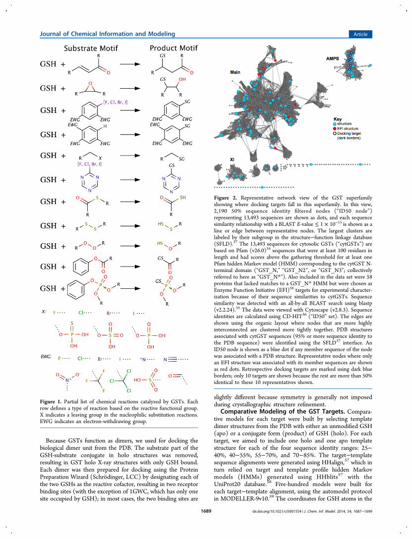
Construction of the Virtual Screening Library. GSTscatalyze a range of different reactions. Here, we considered 11different kinds of GST catalyzed reactions, each with a differentsubstrate motif (Figure 1). For each substrate motif, we foundall matching molecules in the KEGG and MetaCyc compounddatabases. The search was performed with the OECHEMtools29 by matching a SMILES30 string of a database moleculeto the SMARTS30 strings representing the substrate motifs. Wefound 4,149 and 3,259 in KEGG and MetaCyc, respectively.We also added 64 substrates from the literature,31−52 for thetotal of 6,738 unique substrates.Next, each of the substrates was converted to one or more
products, as follows. The conversions corresponding to the 11reactions were carried out using OECHEM’s library generationfunction29 with explicit hydrogens. The reactive functionalgroups in the substrate were identified by comparing thesubstrate SMILES string with the SMARTS string representingthe substrate motif undergoing conversion to a product motif,for each reaction. Each match was then used to convert thesubstrate to a product that was added to the virtual screeninglibrary.Finally, we prepared products for docking. For products with
undefined stereocenters, we first enumerated the stereoisomersusing OECHEM,29 with the maximum number of stereo-isomers retained arbitrarily set to 16 for computationalefficiency. Second, protonation states of each stereoisomerwas enumerated within pH range 6−8, followed by generating aconformation for each protonation state, using Epik53,54 andLigPrep.55 Finally, force field parameters for each product weregenerated using the hetgrp_ffgen utility (Schrodinger, LCC).
Selection of the GST Targets for Docking. A subset ofGST structures was selected from the Protein Data Bank(PDB) for retrospective docking by maximizing the sequenceand functional coverage of the GST superfamily (Figure 2),following three steps: First, all GST structures with acocrystallized GSH-substrate conjugate were extracted fromthe PDB. Second, structures for the same protein were groupedtogether, resulting in 14 groups with unique sets of ligands.Finally, for each group, the structure with the highestresolution, the lowest Rfree,, and a unique ligand was selected,resulting in 14 target holo X-ray structures (SupportingInformation Table S1).
Journal of Chemical Information and Modeling Article
dx.doi.org/10.1021/ci5001554 | J. Chem. Inf. Model. 2014, 54, 1687−16991688
Because GSTs function as dimers, we used for docking thebiological dimer unit from the PDB. The substrate part of theGSH-substrate conjugate in holo structures was removed,resulting in GST holo X-ray structures with only GSH bound.Each dimer was then prepared for docking using the ProteinPreparation Wizard (Schrodinger, LCC) by designating each ofthe two GSHs as the reactive cofactor, resulting in two receptorbinding sites (with the exception of 1GWC, which has only onesite occupied by GSH); in most cases, the two binding sites are
slightly different because symmetry is generally not imposedduring crystallographic structure refinement.
Comparative Modeling of the GST Targets. Compara-tive models for each target were built by selecting templatedimer structures from the PDB with either an unmodified GSH(apo) or a conjugate form (product) of GSH (holo). For eachtarget, we aimed to include one holo and one apo templatestructure for each of the four sequence identity ranges: 25−40%, 40−55%, 55−70%, and 70−85%. The target−templatesequence alignments were generated using HHalign,57 which inturn relied on target and template profile hidden Markovmodels (HMMs) generated using HHblits57 with theUniProt20 database.58 Five-hundred models were built foreach target−template alignment, using the automodel protocolin MODELLER-9v10.59 The coordinates for GSH atoms in the
Figure 1. Partial list of chemical reactions catalyzed by GSTs. Eachrow defines a type of reaction based on the reactive functional group.X indicates a leaving group in the nucleophilic substitution reactions.EWG indicates an electron-withdrawing group.
Figure 2. Representative network view of the GST superfamilyshowing where docking targets fall in this superfamily. In this view,2,190 50% sequence identity filtered nodes (“ID50 node”)representing 13,493 sequences are shown as dots, and each sequencesimilarity relationship with a BLAST E-value ≤ 1 × 10⁻¹³ is shown as aline or edge between representative nodes. The largest clusters arelabeled by their subgroup in the structure−function linkage database(SFLD).37 The 13,493 sequences for cytosolic GSTs (“cytGSTs”) arebased on Pfam (v26.0)34 sequences that were at least 100 residues inlength and had scores above the gathering threshold for at least onePfam hidden Markov model (HMM) corresponding to the cytGST N-terminal domain (“GST_N,” “GST_N2″, or “GST_N3”; collectivelyreferred to here as “GST_N*”). Also included in the data set were 58proteins that lacked matches to a GST_N* HMM but were chosen asEnzyme Function Initiative (EFI)56 targets for experimental character-ization because of their sequence similarities to cytGSTs. Sequencesimilarity was detected with an all-by-all BLAST search using blastp(v2.2.24).35 The data were viewed with Cytoscape (v2.8.3). Sequenceidentities are calculated using CD-HIT36 (“ID50” set). The edges areshown using the organic layout where nodes that are more highlyinterconnected are clustered more tightly together. PDB structuresassociated with cytGST sequences (95% or more sequence identity tothe PDB sequence) were identified using the SFLD37 interface. AnID50 node is shown as a blue dot if any member sequence of the nodewas associated with a PDB structure. Representative nodes where onlyan EFI structure was associated with its member sequences are shownas red dots. Retrospective docking targets are marked using dark blueborders; only 10 targets are shown because the rest are more than 50%identical to these 10 representatives shown.
Journal of Chemical Information and Modeling Article
dx.doi.org/10.1021/ci5001554 | J. Chem. Inf. Model. 2014, 54, 1687−16991689
template structure were then copied to the target models, usingthe BLK function of MODELLER. Models were then assessedby the discrete optimized protein energy (DOPE)60 function ofMODELLER. Finally, the model with the lowest normalizedDOPE score was used for docking.To compare different comparative models and compare the
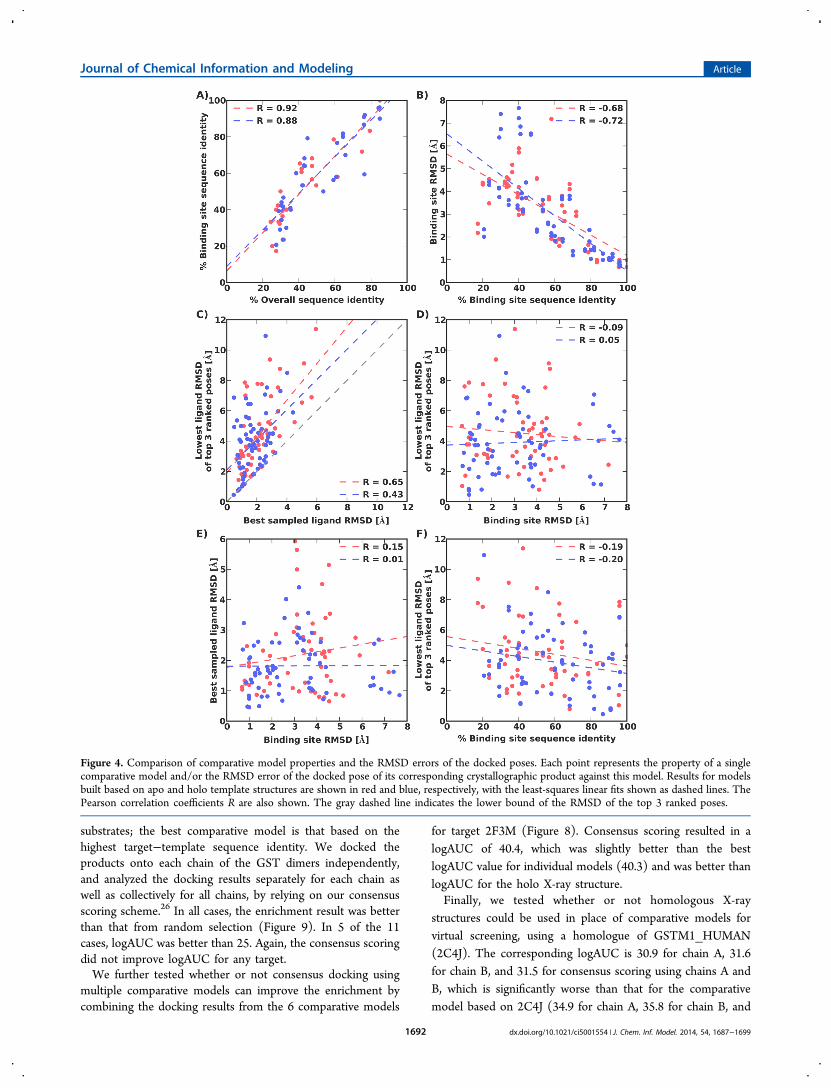
models against X-ray structures, we calculated the binding sitesequence identity of the templates to the X-ray structures, andthe binding site non-hydrogen atom root-mean-squaredeviation (RMSD) between a comparative model and X-raystructure. The binding site was defined to contain atoms in allresidues with at least one atom within 6 Å of any ligand atom inthe X-ray structure. As expected, strong correlations wereobserved between the overall target−template sequenceidentity and the target−template binding site sequence identity,and between the target−template binding site sequence identityand the model’s non-hydrogen atom RMSD error (Figure4A,B).As designed by the choice of the template structures, the
distribution of the non-hydrogen atom RMSD error of thebinding site confirms that there is a range of accuracy across theset of modeled structures. The models with subangstrom non-hydrogen atom RMSD error tend to be those based ontemplates with over 80% sequence identity in the binding siteresidues to the target sequence. Over a third of models had abinding site RMSD error over 4 Å. Thus, homology modelscover a range of sequence identities and a range of accuracy.Protocol for Covalent Docking. Covalent docking of a
potential product to a receptor was started by placing it in thebinding site. More specifically, the coordinates of the GSH partof the potential product were matched to the GSH molecule inthe receptor, and the coordinates of the remaining atoms of theproduct were then built using OMEGA.61 Up to 20 initialconfigurations were generated for each product molecule.For each initial configuration, we then used PLOP’s tether
pred (Academic version 25.6) function62 to rotate the rotatablebonds in the GSH-substrate conjugate. The rotatable bondswere identified using OECHEM. We sampled all of therotatable bonds in the substrate part of the GSH-substrateconjugate as well as the CA−CB and CB−SG bonds in thecysteine residue of GSH. Up to 50 configurations weregenerated by PLOP starting from each initial configuration.These configurations were then scored by calculating the totalpotential energy of the product−receptor complex by PLOP(in the units of kilocalories per mole), which in turn relies onthe OPLS force field with a variable-dielectric generalized Bornmodel.63 The product−receptor distances alone were alsoscored by the atomic statistical potential PoseScore (in arbitraryunits).64 While the PLOP potential energy does not contain theentropic contributions to the binding free energy, thePoseScore term does approximate the contribution of theinterface to the binding free energy.65
Finally, for virtual screening, a substrate was ranked using themedian PLOP energy of its products’ different stereoisomersand protonation states. The PLOP energy of a product in aspecific stereoisomer form and protonation state was themedian PLOP energy of its different configurations.Assessment of Docking and Virtual Screening. When a
native product was docked (Supporting Information Table S2),the docking pose of the product was assessed for accuracybased on its non-hydrogen atom RMSD from the nativeconfiguration, after superposition of the receptor used for
docking on the native structure of the receptor; GSH wasexcluded, except for the cysteine sulfur atom.The accuracy of virtual screening was evaluated by the
enrichment for the known products (Supporting InformationTable S3) among the top scoring potential products. Theenrichment curve was obtained by plotting the percentage ofactual products found (y-axis) within the top ranked subset ofall database compounds (x-axis on logarithmic scale). logAUC,the area under the curve of the enrichment plot, was alsocalculated to indicate the accuracy of enrichment; randomselection has a logAUC of 14.5.
■ RESULTS
We begin by evaluating the docking pose of the native ligandsusing holo and apo structures, followed by evaluating thedocking pose of the native ligands using comparative modelsand homologous structures. Next, enrichment of the knownligands using X-ray structures is benchmarked and analyzed.Finally, we analyze the enrichment of the known ligands usingcomparative models.
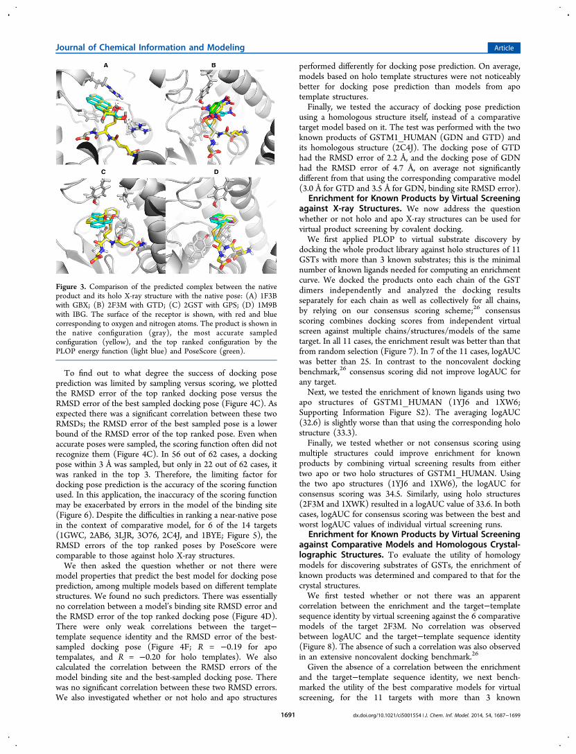
Docking of Products into X-ray Structures. As theeasiest test, we first applied covalent docking by PLOP toreconstruct binding poses of the crystallographic products inthe corresponding holo X-ray structures (Supporting Informa-tion Table S1). In all 14 cases, a docking pose within 3 Å of thenative structure was sampled (within 2 Å for 13 cases), thoughnot necessarily recognized as such by the scoring function(Supporting Information Table S4); the 3 Å threshold on theall-atom RMSD subjectively distinguishes near-native andnonnative poses. However, the top 3 ranked structure waswithin 3 Å to the native structure in 9 out of 14 cases using thePLOP energy function, and in 11 out of 14 cases usingPoseScore (Figure 3).Next, we performed a slightly more difficult, but still easy,
test. For targets with more than one crystallographic product,we docked each product to the nonnative holo X-ray structureof the target (cross-docking; Supporting Information TableS5). In all 12 cases, a docking pose within 3 Å to the nativestructure was sampled (within 2 Å for 11 cases). The top 3ranked complex was within 3 Å to the native structure in 7 outof 12 cases for the PLOP energy function, and in 8 out of 12cases for PoseScore. To test whether combining the twoscoring functions can further improve the result, we consideredan optimal linear combination of the PoseScore and the PLOPpotential energy, using the average all-atom RMSD of the topranked docking poses as the optimization criterion; the optimallinear combination has zero weight for the PLOP potentialenergy.Finally, to test whether or not apo structures can be used for
predicting the native product docking pose, we docked the twoproducts of GSTM1_HUMAN (GDN and GTD) to its twoavailable apo structures (1XW6 and 1YJ6). The results werecomparable to those from cross-docking (Supporting Informa-tion Table S6).
Docking of Native Products into Comparative Modelsand Homologous Crystallographic Structures. To testwhether or not comparative models can be used for dockingpose prediction of native products, a total of 62 homologymodels were generated for the 14 GST targets (SupportingInformation Table S7). We performed native product dockingagainst these models as well as some of their templatestructures.
Journal of Chemical Information and Modeling Article
dx.doi.org/10.1021/ci5001554 | J. Chem. Inf. Model. 2014, 54, 1687−16991690
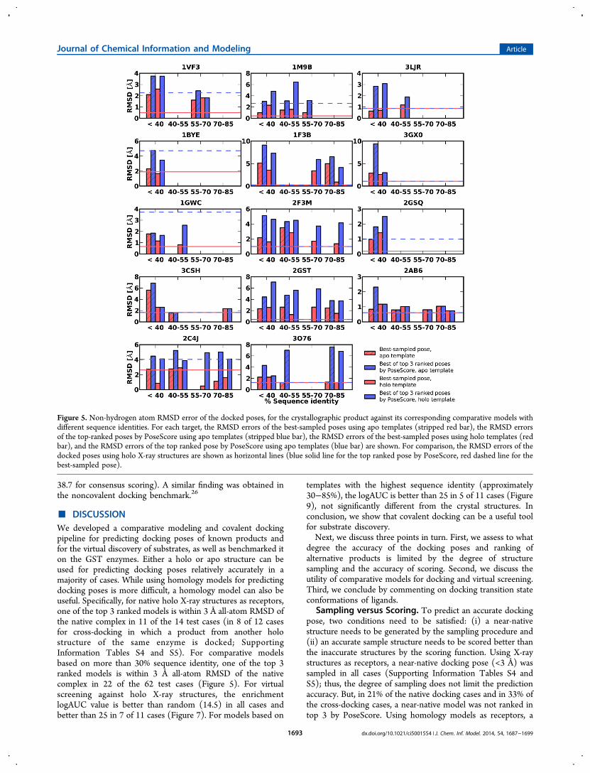
To find out to what degree the success of docking poseprediction was limited by sampling versus scoring, we plottedthe RMSD error of the top ranked docking pose versus theRMSD error of the best sampled docking pose (Figure 4C). Asexpected there was a significant correlation between these twoRMSDs; the RMSD error of the best sampled pose is a lowerbound of the RMSD error of the top ranked pose. Even whenaccurate poses were sampled, the scoring function often did notrecognize them (Figure 4C). In 56 out of 62 cases, a dockingpose within 3 Å was sampled, but only in 22 out of 62 cases, itwas ranked in the top 3. Therefore, the limiting factor fordocking pose prediction is the accuracy of the scoring functionused. In this application, the inaccuracy of the scoring functionmay be exacerbated by errors in the model of the binding site(Figure 6). Despite the difficulties in ranking a near-native posein the context of comparative model, for 6 of the 14 targets(1GWC, 2AB6, 3LJR, 3O76, 2C4J, and 1BYE; Figure 5), theRMSD errors of the top ranked poses by PoseScore werecomparable to those against holo X-ray structures.We then asked the question whether or not there were
model properties that predict the best model for docking poseprediction, among multiple models based on different templatestructures. We found no such predictors. There was essentiallyno correlation between a model’s binding site RMSD error andthe RMSD error of the top ranked docking pose (Figure 4D).There were only weak correlations between the target−template sequence identity and the RMSD error of the best-sampled docking pose (Figure 4F; R = −0.19 for apotempalates, and R = −0.20 for holo templates). We alsocalculated the correlation between the RMSD errors of themodel binding site and the best-sampled docking pose. Therewas no significant correlation between these two RMSD errors.We also investigated whether or not holo and apo structures
performed differently for docking pose prediction. On average,models based on holo template structures were not noticeablybetter for docking pose prediction than models from apotemplate structures.Finally, we tested the accuracy of docking pose prediction
using a homologous structure itself, instead of a comparativetarget model based on it. The test was performed with the twoknown products of GSTM1_HUMAN (GDN and GTD) andits homologous structure (2C4J). The docking pose of GTDhad the RMSD error of 2.2 Å, and the docking pose of GDNhad the RMSD error of 4.7 Å, on average not significantlydifferent from that using the corresponding comparative model(3.0 Å for GTD and 3.5 Å for GDN, binding site RMSD error).
Enrichment for Known Products by Virtual Screeningagainst X-ray Structures. We now address the questionwhether or not holo and apo X-ray structures can be used forvirtual product screening by covalent docking.We first applied PLOP to virtual substrate discovery by
docking the whole product library against holo structures of 11GSTs with more than 3 known substrates; this is the minimalnumber of known ligands needed for computing an enrichmentcurve. We docked the products onto each chain of the GSTdimers independently and analyzed the docking resultsseparately for each chain as well as collectively for all chains,by relying on our consensus scoring scheme;26 consensusscoring combines docking scores from independent virtualscreen against multiple chains/structures/models of the sametarget. In all 11 cases, the enrichment result was better than thatfrom random selection (Figure 7). In 7 of the 11 cases, logAUCwas better than 25. In contrast to the noncovalent dockingbenchmark,26 consensus scoring did not improve logAUC forany target.Next, we tested the enrichment of known ligands using two
apo structures of GSTM1_HUMAN (1YJ6 and 1XW6;Supporting Information Figure S2). The averaging logAUC(32.6) is slightly worse than that using the corresponding holostructure (33.3).Finally, we tested whether or not consensus scoring using
multiple structures could improve enrichment for knownproducts by combining virtual screening results from eithertwo apo or two holo structures of GSTM1_HUMAN. Usingthe two apo structures (1YJ6 and 1XW6), the logAUC forconsensus scoring was 34.5. Similarly, using holo structures(2F3M and 1XWK) resulted in a logAUC value of 33.6. In bothcases, logAUC for consensus scoring was between the best andworst logAUC values of individual virtual screening runs.
Enrichment for Known Products by Virtual Screeningagainst Comparative Models and Homologous Crystal-lographic Structures. To evaluate the utility of homologymodels for discovering substrates of GSTs, the enrichment ofknown products was determined and compared to that for thecrystal structures.We first tested whether or not there was an apparent
correlation between the enrichment and the target−templatesequence identity by virtual screening against the 6 comparativemodels of the target 2F3M. No correlation was observedbetween logAUC and the target−template sequence identity(Figure 8). The absence of such a correlation was also observedin an extensive noncovalent docking benchmark.26
Given the absence of a correlation between the enrichmentand the target−template sequence identity, we next bench-marked the utility of the best comparative models for virtualscreening, for the 11 targets with more than 3 known
Figure 3. Comparison of the predicted complex between the nativeproduct and its holo X-ray structure with the native pose: (A) 1F3Bwith GBX; (B) 2F3M with GTD; (C) 2GST with GPS; (D) 1M9Bwith IBG. The surface of the receptor is shown, with red and bluecorresponding to oxygen and nitrogen atoms. The product is shown inthe native configuration (gray), the most accurate sampledconfiguration (yellow), and the top ranked configuration by thePLOP energy function (light blue) and PoseScore (green).
Journal of Chemical Information and Modeling Article
dx.doi.org/10.1021/ci5001554 | J. Chem. Inf. Model. 2014, 54, 1687−16991691
substrates; the best comparative model is that based on thehighest target−template sequence identity. We docked theproducts onto each chain of the GST dimers independently,and analyzed the docking results separately for each chain aswell as collectively for all chains, by relying on our consensusscoring scheme.26 In all cases, the enrichment result was betterthan that from random selection (Figure 9). In 5 of the 11cases, logAUC was better than 25. Again, the consensus scoringdid not improve logAUC for any target.We further tested whether or not consensus docking using
multiple comparative models can improve the enrichment bycombining the docking results from the 6 comparative models
for target 2F3M (Figure 8). Consensus scoring resulted in alogAUC of 40.4, which was slightly better than the bestlogAUC value for individual models (40.3) and was better thanlogAUC for the holo X-ray structure.Finally, we tested whether or not homologous X-ray
structures could be used in place of comparative models forvirtual screening, using a homologue of GSTM1_HUMAN(2C4J). The corresponding logAUC is 30.9 for chain A, 31.6for chain B, and 31.5 for consensus scoring using chains A andB, which is significantly worse than that for the comparativemodel based on 2C4J (34.9 for chain A, 35.8 for chain B, and
Figure 4. Comparison of comparative model properties and the RMSD errors of the docked poses. Each point represents the property of a singlecomparative model and/or the RMSD error of the docked pose of its corresponding crystallographic product against this model. Results for modelsbuilt based on apo and holo template structures are shown in red and blue, respectively, with the least-squares linear fits shown as dashed lines. ThePearson correlation coefficients R are also shown. The gray dashed line indicates the lower bound of the RMSD of the top 3 ranked poses.
Journal of Chemical Information and Modeling Article
dx.doi.org/10.1021/ci5001554 | J. Chem. Inf. Model. 2014, 54, 1687−16991692
38.7 for consensus scoring). A similar finding was obtained inthe noncovalent docking benchmark.26
■ DISCUSSIONWe developed a comparative modeling and covalent dockingpipeline for predicting docking poses of known products andfor the virtual discovery of substrates, as well as benchmarked iton the GST enzymes. Either a holo or apo structure can beused for predicting docking poses relatively accurately in amajority of cases. While using homology models for predictingdocking poses is more difficult, a homology model can also beuseful. Specifically, for native holo X-ray structures as receptors,one of the top 3 ranked models is within 3 Å all-atom RMSD ofthe native complex in 11 of the 14 test cases (in 8 of 12 casesfor cross-docking in which a product from another holostructure of the same enzyme is docked; SupportingInformation Tables S4 and S5). For comparative modelsbased on more than 30% sequence identity, one of the top 3ranked models is within 3 Å all-atom RMSD of the nativecomplex in 22 of the 62 test cases (Figure 5). For virtualscreening against holo X-ray structures, the enrichmentlogAUC value is better than random (14.5) in all cases andbetter than 25 in 7 of 11 cases (Figure 7). For models based on
templates with the highest sequence identity (approximately30−85%), the logAUC is better than 25 in 5 of 11 cases (Figure9), not significantly different from the crystal structures. Inconclusion, we show that covalent docking can be a useful toolfor substrate discovery.Next, we discuss three points in turn. First, we assess to what
degree the accuracy of the docking poses and ranking ofalternative products is limited by the degree of structuresampling and the accuracy of scoring. Second, we discuss theutility of comparative models for docking and virtual screening.Third, we conclude by commenting on docking transition stateconformations of ligands.
Sampling versus Scoring. To predict an accurate dockingpose, two conditions need to be satisfied: (i) a near-nativestructure needs to be generated by the sampling procedure and(ii) an accurate sample structure needs to be scored better thanthe inaccurate structures by the scoring function. Using X-raystructures as receptors, a near-native docking pose (<3 Å) wassampled in all cases (Supporting Information Tables S4 andS5); thus, the degree of sampling does not limit the predictionaccuracy. But, in 21% of the native docking cases and in 33% ofthe cross-docking cases, a near-native model was not ranked intop 3 by PoseScore. Using homology models as receptors, a
Figure 5. Non-hydrogen atom RMSD error of the docked poses, for the crystallographic product against its corresponding comparative models withdifferent sequence identities. For each target, the RMSD errors of the best-sampled poses using apo templates (stripped red bar), the RMSD errorsof the top-ranked poses by PoseScore using apo templates (stripped blue bar), the RMSD errors of the best-sampled poses using holo templates (redbar), and the RMSD errors of the top ranked pose by PoseScore using apo templates (blue bar) are shown. For comparison, the RMSD errors of thedocked poses using holo X-ray structures are shown as horizontal lines (blue solid line for the top ranked pose by PoseScore, red dashed line for thebest-sampled pose).
Journal of Chemical Information and Modeling Article
dx.doi.org/10.1021/ci5001554 | J. Chem. Inf. Model. 2014, 54, 1687−16991693
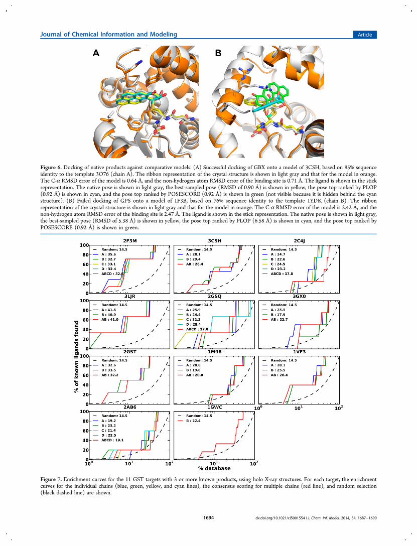
Figure 6. Docking of native products against comparative models. (A) Successful docking of GBX onto a model of 3CSH, based on 85% sequenceidentity to the template 3O76 (chain A). The ribbon representation of the crystal structure is shown in light gray and that for the model in orange.The C-α RMSD error of the model is 0.64 Å, and the non-hydrogen atom RMSD error of the binding site is 0.71 Å. The ligand is shown in the stickrepresentation. The native pose is shown in light gray, the best-sampled pose (RMSD of 0.90 Å) is shown in yellow, the pose top ranked by PLOP(0.92 Å) is shown in cyan, and the pose top ranked by POSESCORE (0.92 Å) is shown in green (not visible because it is hidden behind the cyanstructure). (B) Failed docking of GPS onto a model of 1F3B, based on 76% sequence identity to the template 1YDK (chain B). The ribbonrepresentation of the crystal structure is shown in light gray and that for the model in orange. The C-α RMSD error of the model is 2.42 Å, and thenon-hydrogen atom RMSD error of the binding site is 2.47 Å. The ligand is shown in the stick representation. The native pose is shown in light gray,the best-sampled pose (RMSD of 5.38 Å) is shown in yellow, the pose top ranked by PLOP (6.58 Å) is shown in cyan, and the pose top ranked byPOSESCORE (0.92 Å) is shown in green.
Figure 7. Enrichment curves for the 11 GST targets with 3 or more known products, using holo X-ray structures. For each target, the enrichmentcurves for the individual chains (blue, green, yellow, and cyan lines), the consensus scoring for multiple chains (red line), and random selection(black dashed line) are shown.
Journal of Chemical Information and Modeling Article
dx.doi.org/10.1021/ci5001554 | J. Chem. Inf. Model. 2014, 54, 1687−16991694
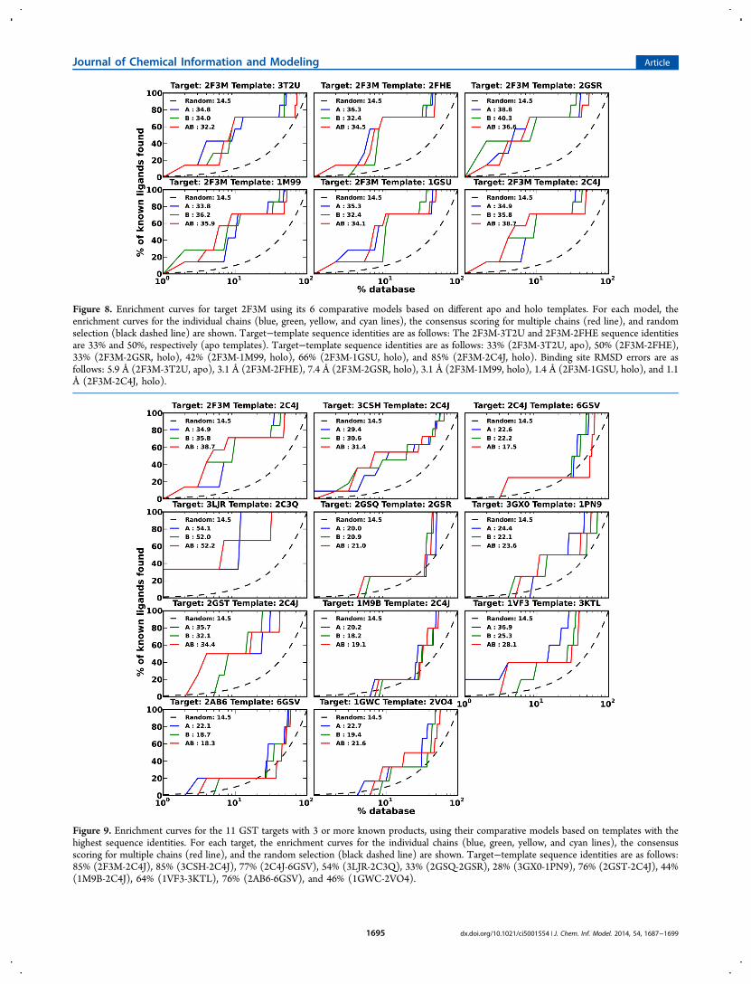
Figure 8. Enrichment curves for target 2F3M using its 6 comparative models based on different apo and holo templates. For each model, theenrichment curves for the individual chains (blue, green, yellow, and cyan lines), the consensus scoring for multiple chains (red line), and randomselection (black dashed line) are shown. Target−template sequence identities are as follows: The 2F3M-3T2U and 2F3M-2FHE sequence identitiesare 33% and 50%, respectively (apo templates). Target−template sequence identities are as follows: 33% (2F3M-3T2U, apo), 50% (2F3M-2FHE),33% (2F3M-2GSR, holo), 42% (2F3M-1M99, holo), 66% (2F3M-1GSU, holo), and 85% (2F3M-2C4J, holo). Binding site RMSD errors are asfollows: 5.9 Å (2F3M-3T2U, apo), 3.1 Å (2F3M-2FHE), 7.4 Å (2F3M-2GSR, holo), 3.1 Å (2F3M-1M99, holo), 1.4 Å (2F3M-1GSU, holo), and 1.1Å (2F3M-2C4J, holo).
Figure 9. Enrichment curves for the 11 GST targets with 3 or more known products, using their comparative models based on templates with thehighest sequence identities. For each target, the enrichment curves for the individual chains (blue, green, yellow, and cyan lines), the consensusscoring for multiple chains (red line), and the random selection (black dashed line) are shown. Target−template sequence identities are as follows:85% (2F3M-2C4J), 85% (3CSH-2C4J), 77% (2C4J-6GSV), 54% (3LJR-2C3Q), 33% (2GSQ-2GSR), 28% (3GX0-1PN9), 76% (2GST-2C4J), 44%(1M9B-2C4J), 64% (1VF3-3KTL), 76% (2AB6-6GSV), and 46% (1GWC-2VO4).
Journal of Chemical Information and Modeling Article
dx.doi.org/10.1021/ci5001554 | J. Chem. Inf. Model. 2014, 54, 1687−16991695
near-native docking pose was sampled in 56 of 62 cases but wasonly ranked in top 3 in 22 of 62 cases (Figure 5). The reason isthat our scoring function is not always accurate enough todifferentiate between accurate and inaccurate docking poses.To rank different substrates, we used the median PLOP
energy of their product−enzyme complexes. We chose themedian instead of the lowest PLOP energy because it resultedin a significantly higher average logAUC (27.3 vs 18.9). A likelyreason is that the energy estimate of a single configuration isnoisier than the median, compensating for the lack ofaccounting for the proper physics of the problem. In thefuture, other functions such as the Boltzmann average of thesampled energies will be explored.Although PoseScore reranking of the docking poses
generated by optimizing the PLOP energy further improvedthe accuracy of the top scoring docked poses, this result doesnot imply that PoseScore is more accurate than the PLOPenergy. It is possible that reranking by PLOP would alsoimprove the accuracy of the poses generated by optimizingPoseScore; for technical reasons, it is not straightforward toactually make this test. Our result indicates merely that the twoscoring functions are different and imperfect and thatPoseScore contains helpful information not present in PLOP,while being silent on whether or not PLOP also containsinformation not present in PoseScore.In contrast to noncovalent docking,26 on average, consensus
scoring did not improve virtual screening accuracy. Acombination of the following two assumptions explains thisfinding: (i) covalent docking is limited more by scoring than bysampling compared to noncovalent docking; and (ii) consensusscoring works best when sampling is a limiting factor indocking. A major difference between noncovalent and covalentdocking is that the latter is constrained by a covalent bond,resulting in an easier sampling problem all other thingsconsidered equal. Using multiple receptor structures to mimicreceptor flexibility was already shown to lead to more accuratesampled docking poses in noncovalent docking.26 Thus, thedisadvantage of a larger number of decoys produced by usingmultiple templates, which increases the burden on the scoringfunction for identifying the most accurate pose, is outweighedby the more accurately sampled poses that are more likely to beranked highly by the scoring function. In contrast, if scoring islimiting and sampling is not, as is the case in covalent docking(cf. our native docking and cross-docking tests), consideringposes from multiple receptor structures is not likely to improvethe results, as observed.”Given the demonstrated limitations of current scoring, the
accuracy of ranking ligand poses and ligands can be improvedby more accurate scoring functions. On the one hand, theability of the PLOP potential energy to rank the native state (orat least a near-native state) may be improved by estimating thefree energy of the native state.66 On the other hand, theaccuracy of the statistical potential could be improved byapplying the Bayesian framework for inferring statisticallyoptimized atomic potentials (SOAP).67 This framework (1)uses multiple data-driven “recovery” functions based onprobability theory, without recourse to questionable statisticalmechanical assumptions about statistical potentials; (2)restrains the relative orientation between two covalent bondsinstead of a simple distance between two atoms, in an effort tocapture orientation-dependent interactions such as hydrogenbonds; (3) performs Bayesian smoothing for estimating theunderlying smooth distributions from noisy observations; (4)
applies Bayesian inference for calculating parameter values thatmaximize the posteriori probability; and (5) benefits fromBayesian model selection based on Bayesian predictivedensities, in an effort to improve the generalizability of thederived statistical potentials.
Comparative Models for Docking and Virtual Screen-ing. Using comparative models for predicting docking posesoften produces inaccurate models (Figures 4 and 5). Onepossible reason is that covalent docking is sensitive to minorchanges in the binding site, especially for residues close to theGSH sulfur atom due to the substrate−receptor covalent bondconstraint (Figure 6). Another possible reason is that the activesites of GST enzymes are often defined in part by the C-terminus, which is often flexible or missing in a templatestructure (Figure 6). Moreover, there was no significantcorrelation between the binding site non-hydrogen atomRMSD and the docking pose RMSD (Figure 4). The lack ofthis correlation may also result from the disproportionateimpact of residues close to the GSH sulfur atom on the dockingpose.Virtual screening using comparative models generated results
comparable to the X-ray structures (average logAUC of 28.2versus 27.3, better than 25 in 5 versus 7 out of 11 cases),consistent with what has been observed when usingcomparative models for virtual screening by noncovalentdocking (average logAUC of 28.7 versus 30.6),26 even thoughcomparative models performed much worse than X-raystructures for docking pose prediction (average non-hydrogenatom RMSD of 3.98 Å versus 1.83 Å). This finding may berationalized by comparatively worse scoring of both true anddecoy products (average PoseScore of −4.2 and 4.1 versus−60.0 and −52.2) when docking against inaccurate bindingsites in comparative models.In prospective applications, if some substrates for the target
of interest are known, virtual screening using comparativemodels can be improved by selecting the most enriching modelfor known substrates.68−70 If no substrates are known for thetarget of interest, it might be advantageous to build and dockinto multiple structurally diverse models and analyze the virtualscreening results by hand.14 An experienced user may be able toidentify an accurate docking result based on the totality of theirknowledge about the proteins of interest.
Reactant, Product, Intermediate, and TransitionStates. Generally, a substrate of an enzyme is required tohave a sufficiently high kcat/Km; a typical threshold is 104 (mol/L)−1 s−1. An enzymatic reaction involves interconversionsbetween pairs of stable states, separated by a transition state;the stable states include the enzyme and substrate withoutinteraction (E + S), an enzyme−substrate complex inter-mediate (ES), an enzyme−product intermediate (EP), and theenzyme and product without interaction (E + P).71 In general,kcat/Km depends on the free energies of all states. Thus, virtualligand screening should in principle consider all states, which isgenerally not the case.When conversion from ES to EP is the rate-limiting step, kcat
is mostly determined by the transition state energy barrier forthis step. Enzymes lower this barrier by creating a binding sitethat is complementary to the transition state of the substrate.Thus, docking a substrate (or a product) in its transition stateconformation may be more appropriate than docking asubstrate (or a product) in its ground state. Although quantummechanics calculations can model the transition state andestimate the activation energy barrier, running these calcu-
Journal of Chemical Information and Modeling Article
dx.doi.org/10.1021/ci5001554 | J. Chem. Inf. Model. 2014, 54, 1687−16991696
lations for the entire virtual screening library is not computa-tionally feasible.72 Instead, we docked here the reaction productstates and achieved relative success. However, the enrichmentresults could possibly be greatly improved if we can dock thetransition state, or a transition state like structure, withoutrecourse to quantum mechanics. For GST substrate discovery,compounds similar to the high-energy intermediates used fordocking to amidohydrolases73 could be constructed, providedsome technical challenges are solved (e.g., building C atomswith 2 partial bonds for a total of 5 bonds). Although enzymespresumably maximize complementarity to the transition state,other states during the reaction must also be compatible withthe potentially flexible binding site of the enzyme. Thus,substrate discovery might also be improved by consideration ofdocking of all ligand states to the corresponding state(s) of theenzyme.
■ CONCLUSIONCan covalent docking accurately predict docking poses ofknown ligands, given experimentally determined, homolo-gous or modeled structures in the GST superfamily? Yes,either a holo or apo structure can be used for predictingdocking poses accurately in a majority of cases, while usinghomology models is less successful. Specifically, for native holoX-ray structures as receptors, one of the top 3 ranked models iswithin 3 Å all-atom RMSD of the native complex in 11 of the14 cases (in 8 of 12 cases for cross-docking). For comparativemodels as receptors, one of the top 3 ranked models is within 3Å all-atom RMSD of the native complex in 22 of the 62 testcases.Can covalent docking accurately predict ligands despite
the catalytically promiscuity of many GST enzymes? Often.What is the difference in the utility of apo, holo,
comparative modeling, and homologous structures forvirtual screening in the GST superfamily? Holo and apostructures, homologous structures, and comparative models canall enrich for known products. For virtual screening againstholo X-ray structures, the enrichment logAUC value is betterthan random in all cases, and better than 25 in 7 of 11 cases(average logAUC 27.3; Figure 7). Using apo X-ray structures of2F3M, the average logAUC (32.6) is slightly worse than thatusing the corresponding holo structure (33.3). For modelsbased on templates with the highest sequence identity(approximately 30−85%), the logAUC is better than 25 in 5of 11 cases (average logAUC 28.2; Figure 9), which iscomparable to that for X-ray structure. Using the homologousstructure of 2F3M, the logAUC (31.3) is significantly worsethan that using comparative models (36.5).Can the virtual screening be improved by consensus
scoring, relying on independent screening against multipleholo, apo, comparative modeling, and homologous struc-tures in the GST superfamily? Rarely. Consensus scoringusing multiple chains from the same structure/model ormultiple structures/models of the same target often does notimprove the virtual screening accuracy. Although consensusscoring improves logAUC in comparison to using a singlereceptor in some cases, it makes it worse in others.If multiple models are calculated on the basis of different
templates, can any of them outperform apo and even holo X-ray structures of the target? If so, can one reliably identifywhich model will do so, or even perform optimally among aset of modeled structures; are there sequence and/orstructural attributes (i.e., the overall target−template
sequence identity, the binding site target−template sequenceidentity, and the predicted accuracy of a model) that reliablypredict the accuracy of ligand docking? For docking poseprediction, there are a few cases where comparative modelsoutperformed the corresponding holo X-ray structure (Figure5). However, we did not find a predictor that could reliablypredict the accuracy of the docked pose. For virtual screening,some models also outperform the corresponding holo X-raystructure (Figures 8 and 9), but we did not find a correlationbetween logAUC and the binding site sequence identity or thebinding site RMSD errors.
■ ASSOCIATED CONTENT*S Supporting InformationTables listing of GST targets used for docking, their crystalligands and known substrates, the detailed results of usingcrystal structures and comparative models for docking poseprediction, and target−template pairs used to generatecomparative model sand figures showing the enrichment curvesfor GSTM1_HUMAN using its apo structures and thecorrelations between the Z-DOPE score and the model’sRMSD. This material is available free of charge via the Internetat http://pubs.acs.org. The scripts, comparative models,benchmark, and the docking library are available at http://salilab.org/GST.
■ AUTHOR INFORMATIONCorresponding Authors*(A.S.) Tel.: +1 415 514 4227. Fax: +1 415 514 4231. E-mail:[email protected].*(R.N.A.) Tel.: +1 615 343 2920. E-mail: [email protected] authors declare no competing financial interest.
■ ACKNOWLEDGMENTSWe are grateful to Dr. Ben Webb for his help with computinginfrastructure. This work was supported by the NIH Grant U54GM093342 to A.S., R.N.A., B.K.S., M.P.J., and P.C.B.
■ REFERENCES(1) Mannervik, B.; Danielson, U. H. Glutathione transferases–structure and catalytic activity. CRC Crit. Rev. Biochem. 1988, 23, 283−337.(2) Armstrong, R. N. Structure, catalytic mechanism, and evolutionof the glutathione transferases. Chem. Res. Toxicol. 1997, 10, 2−18.(3) Armstrong, R. N. Mechanistic imperatives for the evolution ofglutathione transferases. Curr. Opin. Chem. Biol. 1998, 2, 618−623.(4) Hayes, J. D.; Flanagan, J. U.; Jowsey, I. R. Glutathionetransferases. Annu. Rev. Pharmacol. Toxicol. 2005, 45, 51−88.(5) Atkinson, H. J.; Babbitt, P. C. Glutathione transferases arestructural and functional outliers in the thioredoxin fold. Biochemistry2009, 48, 11108−11116.(6) Copley, S. D.; Novak, W. R.; Babbitt, P. C. Divergence offunction in the thioredoxin fold suprafamily: Evidence for evolution ofperoxiredoxins from a thioredoxin-like ancestor. Biochemistry 2004, 43,13981−13995.(7) Abagyan, R.; Totrov, M. High-throughput docking for leadgeneration. Curr. Opin. Chem. Biol. 2001, 5, 375−382.(8) Apostolakis, J.; Pluckthun, A.; Caflisch, A. Docking small ligandsin flexible binding sites. J. Comput. Chem. 1998, 19, 21−37.(9) Cavasotto, C. N.; Orry, A. J. Ligand docking and structure-basedvirtual screening in drug discovery. Curr. Top. Med. Chem. 2007, 7,1006−1014.
Journal of Chemical Information and Modeling Article
dx.doi.org/10.1021/ci5001554 | J. Chem. Inf. Model. 2014, 54, 1687−16991697
(10) Schroder, J.; Klinger, A.; Oellien, F.; Marhofer, R. J.; Duszenko,M.; Selzer, P. M. Docking-Based Virtual Screening of CovalentlyBinding Ligands: An Orthogonal Lead Discovery Approach. J. Med.Chem. 2013, 56, 1478−1490.(11) Chen, Y.; Shoichet, B. K. Molecular docking and ligandspecificity in fragment-based inhibitor discovery. Nat. Chem. Biol.2009, 5, 358−364.(12) de Graaf, C.; Oostenbrink, C.; Keizers, P. H. J.; van der Wijst,T.; Jongejan, A.; Vemleulen, N. P. E. Catalytic site prediction andvirtual screening of cytochrome P450 2D6 substrates by considerationof water and rescoring in automated docking. J. Med. Chem. 2006, 49,2417−2430.(13) Gohda, K.; Teno, N.; Wanaka, K.; Tsuda, Y. Predicting subsiteinteractions of plasmin with substrates and inhibitors throughcomputational docking analysis. J. Enzyme Inhib. Med. Chem. 2012,27, 571−577.(14) Fan, H.; Hitchcock, D. S.; Seidel, R. D., II; Hillerich, B.; Lin, H.;Almo, S. C.; Sali, A.; Shoichet, B. K.; Raushel, F. M. Assignment ofpterin deaminase activity to an enzyme of unknown function guided byhomology modeling and docking. J. Am. Chem. Soc. 2013, 135, 795−803.(15) Wallrapp, F. H.; Pan, J. J.; Ramamoorthy, G.; Almonacid, D. E.;Hillerich, B. S.; Seidel, R.; Patskovsky, Y.; Babbitt, P. C.; Almo, S. C.;Jacobson, M. P.; Poulter, C. D. Prediction of function for thepolyprenyl transferase subgroup in the isoprenoid synthase super-family. Proc. Natl. Acad. Sci. U. S. A. 2013, 110, E1196−E1202.(16) Carlsson, J.; Coleman, R. G.; Setola, V.; Irwin, J. J.; Fan, H.;Schlessinger, A.; Sali, A.; Roth, B. L.; Shoichet, B. K. Ligand discoveryfrom a dopamine D3 receptor homology model and crystal structure.Nat. Chem. Biol. 2011, 7, 769−778.(17) Schlessinger, A.; Geier, E.; Fan, H.; Irwin, J. J.; Shoichet, B. K.;Giacomini, K. M.; Sali, A. Structure-based discovery of prescriptiondrugs that interact with the norepinephrine transporter, NET. Proc.Natl. Acad. Sci. U. S. A. 2011, 108, 15810−15815.(18) Jacobson, M.; Sali, A. Comparative protein structure modelingand its applications to drug discovery. Annu. Rep. Med. Chem. 2004, 39,259−276.(19) Bissantz, C.; Bernard, P.; Hibert, M.; Rognan, D. Protein-basedvirtual screening of chemical databases. II. Are homology models of G-protein coupled receptors suitable targets? Proteins: Struct., Funct.,Genet. 2003, 50, 5−25.(20) Evers, A.; Klebe, G. Ligand-supported homology modeling of G-protein-coupled receptor sites: Models sufficient for successful virtualscreening. Angew. Chem., Int. Ed. 2004, 43, 248−251.(21) Evers, A.; Klebe, G. Successful virtual screening for asubmicromolar antagonist of the neurokinin-1 receptor based on aligand-supported homology model. J. Med. Chem. 2004, 47, 5381−5392.(22) Radestock, S.; Weil, T.; Renner, S. Homology model-basedvirtual screening for GPCR ligands using docking and target-biasedscoring. J. Chem. Inf. Model. 2008, 48, 1104−1117.(23) Kalyanaraman, C.; Imker, H. J.; Federov, A. A.; Federov, E. V.;Glasner, M. E.; Babbitt, P. C.; Almo, S. C.; Gerlt, J. A.; Jacobson, M. P.Discovery of a dipeptide epimerase enzymatic function guided byhomology modeling and virtual screening. Structure (Oxford, U. K.)2008, 16, 1668−1677.(24) Diller, D. J.; Li, R. X. Kinases, homology models, and highthroughput docking. J. Med. Chem. 2003, 46, 4638−4647.(25) Nguyen, T. L.; Gussio, R.; Smith, J. A.; Lannigan, D. A.; Hecht,S. M.; Scudiero, D. A.; Shoemaker, R. H.; Zaharevitz, D. W. Homologymodel of RSK2 N-terminal kinase domain, structure-based identi-fication of novel RSK2 inhibitors, and preliminary commonpharmacophore. Bioorg. Med. Chem. 2006, 14, 6097−6105.(26) Fan, H.; Irwin, J. J.; Webb, B. M.; Klebe, G.; Shoichet, B. K.;Sali, A. Molecular Docking Screens Using Comparative Models ofProteins. J. Chem. Inf. Model. 2009, 49, 2512−2527.(27) Kanehisa, M.; Goto, S.; Sato, Y.; Furumichi, M.; Tanabe, M.KEGG for integration and interpretation of large-scale molecular datasets. Nucleic Acids Res. 2012, 40, D109−114.
(28) Caspi, R.; Altman, T.; Dreher, K.; Fulcher, C. A.; Subhraveti, P.;Keseler, I. M.; Kothari, A.; Krummenacker, M.; Latendresse, M.;Mueller, L. A.; Ong, Q.; Paley, S.; Pujar, A.; Shearer, A. G.; Travers,M.; Weerasinghe, D.; Zhang, P.; Karp, P. D. The MetaCyc database ofmetabolic pathways and enzymes and the BioCyc collection ofpathway/genome databases. Nucleic Acids Res. 2012, 40, D742−D753.(29) OEChem T, version 1.7. 4.3; OpenEye Scientific Software:Santa Fe, NM, USA, 2010.(30) James, C. A.; Weininger, D.; Delany, J. Daylight theory manual;Daylight Chemical Information Systems: Aliso Viejo, CA, USA, 2004.(31) Prade, L.; Huber, R.; Bieseler, B. Structures of herbicides incomplex with their detoxifying enzyme glutathione S-transferaseExplanations for the selectivity of the enzyme in plants. Structure 1998,6, 1445−1452.(32) Cardoso, R. M.; Daniels, D. S.; Bruns, C. M.; Tainer, J. A.Characterization of the electrophile binding site and substrate bindingmode of the 26-kDa glutathione S-transferase from Schistosomajaponicum. Proteins 2003, 51, 137−146.(33) Gu, Y.; Singh, S. V.; Ji, X. Residue R216 and catalytic efficiencyof a murine class alpha glutathione S-transferase toward benzo[a]-pyrene 7(R),8(S)-diol 9(S), 10(R)-epoxide. Biochemistry 2000, 39,12552−12557.(34) Thom, R.; Cummins, I.; Dixon, D. P.; Edwards, R.; Cole, D. J.;Lapthorn, A. J. Structure of a tau class glutathione S-transferase fromwheat active in herbicide detoxification. Biochemistry 2002, 41, 7008−7020.(35) Norrgard, M. A.; Ivarsson, Y.; Tars, K.; Mannervik, B.Alternative mutations of a positively selected residue elicit gain orloss of functionalities in enzyme evolution. Proc. Natl. Acad. Sci. U. S. A.2006, 103, 4876−4881.(36) Federici, L.; Lo Sterzo, C.; Pezzola, S.; Di Matteo, A.; Scaloni,F.; Federici, G.; Caccuri, A. M. Structural basis for the binding of theanticancer compound 6-(7-nitro-2,1,3-benzoxadiazol-4-ylthio)hexanolto human glutathione s-transferases. Cancer Res. 2009, 69, 8025−8034.(37) Patskovsky, Y.; Patskovska, L.; Almo, S. C.; Listowsky, I.Transition state model and mechanism of nucleophilic aromaticsubstitution reactions catalyzed by human glutathione S-transferaseM1a-1a. Biochemistry 2006, 45, 3852−3862.(38) Rowe, J. D.; Patskovsky, Y. V.; Patskovska, L. N.; Novikova, E.;Listowsky, I. Rationale for reclassification of a distinctive subdivision ofmammalian class Mu glutathione S-transferases that are primarilyexpressed in testis. J. Biol. Chem. 1998, 273, 9593−9601.(39) Mannervik, B.; Alin, P.; Guthenberg, C.; Jensson, H.; Tahir, M.K.; Warholm, M.; Jornvall, H. Identification of three classes ofcytosolic glutathione transferase common to several mammalianspecies: Correlation between structural data and enzymatic properties.Proc. Natl. Acad. Sci. U. S. A. 1985, 82, 7202−7206.(40) Ji, X.; von Rosenvinge, E. C.; Johnson, W. W.; Armstrong, R. N.;Gilliland, G. L. Location of a potential transport binding site in a sigmaclass glutathione transferase by x-ray crystallography. Proc. Natl. Acad.Sci. U. S. A. 1996, 93, 8208−8213.(41) Ji, X.; von Rosenvinge, E. C.; Johnson, W. W.; Tomarev, S. I.;Piatigorsky, J.; Armstrong, R. N.; Gilliland, G. L. Three-dimensionalstructure, catalytic properties, and evolution of a sigma classglutathione transferase from squid, a progenitor of the lens S-crystallins of cephalopods. Biochemistry 1995, 34, 5317−5328.(42) Ji, X.; Armstrong, R. N.; Gilliland, G. L. Snapshots along thereaction coordinate of an SNAr reaction catalyzed by glutathionetransferase. Biochemistry 1993, 32, 12949−12954.(43) Ji, X.; Johnson, W. W.; Sesay, M. A.; Dickert, L.; Prasad, S. M.;Ammon, H. L.; Armstrong, R. N.; Gilliland, G. L. Structure andfunction of the xenobiotic substrate binding site of a glutathione S-transferase as revealed by X-ray crystallographic analysis of productcomplexes with the diastereomers of 9-(S-glutathionyl)-10-hydroxy-9,10-dihydrophenanthrene. Biochemistry 1994, 33, 1043−1052.(44) Reinemer, P.; Dirr, H. W.; Ladenstein, R.; Huber, R.; Lo Bello,M.; Federici, G.; Parker, M. W. Three-dimensional structure of class piglutathione S-transferase from human placenta in complex with S-hexylglutathione at 2.8 A resolution. J. Mol. Biol. 1992, 227, 214−226.
Journal of Chemical Information and Modeling Article
dx.doi.org/10.1021/ci5001554 | J. Chem. Inf. Model. 2014, 54, 1687−16991698
(45) Parker, L. J.; Ciccone, S.; Italiano, L. C.; Primavera, A.; Oakley,A. J.; Morton, C. J.; Hancock, N. C.; Bello, M. L.; Parker, M. W. Theanti-cancer drug chlorambucil as a substrate for the humanpolymorphic enzyme glutathione transferase P1−1: Kinetic propertiesand crystallographic characterisation of allelic variants. J. Mol. Biol.2008, 380, 131−144.(46) Ji, X.; Tordova, M.; O’Donnell, R.; Parsons, J. F.; Hayden, J. B.;Gilliland, G. L.; Zimniak, P. Structure and function of the xenobioticsubstrate-binding site and location of a potential non-substrate-bindingsite in a class pi glutathione S-transferase. Biochemistry 1997, 36,9690−9702.(47) Liu, L. F.; Liaw, Y. C.; Tam, M. F. Characterization of chicken-liver glutathione S-transferase (GST) A1−1 and A2−2 isoenzymes andtheir site-directed mutants heterologously expressed in Escherichiacoli: Identification of Lys-15 and Ser-208 on cGSTA1−1 as residuesinteracting with ethacrynic acid. Biochem. J. 1997, 327 (Pt 2), 593−600.(48) Wadington, M. C.; Ladner, J. E.; Stourman, N. V.; Harp, J. M.;Armstrong, R. N. Analysis of the structure and function of YfcG fromEscherichia coli reveals an efficient and unique disulfide bond reductase.Biochemistry 2009, 48, 6559−6561.(49) Kanai, T.; Takahashi, K.; Inoue, H. Three distinct-typeglutathione S-transferases from Escherichia coli important for defenseagainst oxidative stress. J. Biochem. 2006, 140, 703−711.(50) Rossjohn, J.; McKinstry, W. J.; Oakley, A. J.; Verger, D.;Flanagan, J.; Chelvanayagam, G.; Tan, K. L.; Board, P. G.; Parker, M.W. Human theta class glutathione transferase: The crystal structurereveals a sulfate-binding pocket within a buried active site. Structure(Oxford, U. K.) 1998, 6, 309−322.(51) Tan, K. L.; Chelvanayagam, G.; Parker, M. W.; Board, P. G.Mutagenesis of the active site of the human Theta-class glutathionetransferase GSTT2−2: Catalysis with different substrates involvesdifferent residues. Biochem. J. 1996, 319 (Pt 1), 315−321.(52) McManus, G.; Costa, M.; Canals, A.; Coll, M.; Mantle, T. J. Site-directed mutagenesis of mouse glutathione transferase P1−1 unlocksmasked cooperativity, introduces a novel mechanism for ’ping pong’kinetic behaviour, and provides further structural evidence forparticipation of a water molecule in proton abstraction fromglutathione. FEBS J. 2011, 278, 273−281.(53) Greenwood, J. R.; Calkins, D.; Sullivan, A. P.; Shelley, J. C.Towards the comprehensive, rapid, and accurate prediction of thefavorable tautomeric states of drug-like molecules in aqueous solution.J. Comput.-Aid Mol. Des 2010, 24, 591−604.(54) Shelley, J. C.; Cholleti, A.; Frye, L. L.; Greenwood, J. R.; Timlin,M. R.; Uchimaya, M. Epik: A software program for pK(a) predictionand protonation state generation for drug-like molecules. J. Comput.-Aid Mol. Des 2007, 21, 681−691.(55) Ligprep, V. 2.3; Schrodinger: New York, NY, USA, 2009.(56) Gerlt, J. A.; Allen, K. N.; Almo, S. C.; Armstrong, R. N.; Babbitt,P. C.; Cronan, J. E.; Dunaway-Mariano, D.; Imker, H. J.; Jacobson, M.P.; Minor, W.; Poulter, C. D.; Raushel, F. M.; Sali, A.; Shoichet, B. K.;Sweedler, J. V. The Enzyme Function Initiative. Biochemistry 2011, 50,9950−9962.(57) Soding, J. Protein homology detection by HMM-HMMcomparison. Bioinformatics 2005, 21, 951−960.(58) Apweiler, R.; Martin, M. J.; O’Donovan, C.; Magrane, M.; Alam-Faruque, Y.; Alpi, E.; Antunes, R.; Arganiska, J.; Casanova, E. B.; Bely,B.; Bingley, M.; Bonilla, C.; Britto, R.; Bursteinas, B.; Chan, W. M.;Chavali, G.; Cibrian-Uhalte, E.; Da Silva, A.; De Giorgi, M.; Dimmer,E.; Fazzini, F.; Gane, P.; Fedotov, A.; Castro, L. G.; Garmiri, P.;Hatton-Ellis, E.; Hieta, R.; Huntley, R.; Jacobsen, J.; Jones, R.; Legge,D.; Liu, W. D.; Luo, J.; MacDougall, A.; Mutowo, P.; Nightingale, A.;Orchard, S.; Patient, S.; Pichler, K.; Poggioli, D.; Pundir, S.; Pureza, L.;Qi, G. Y.; Rosanoff, S.; Sawford, T.; Sehra, H.; Turner, E.; Volynkin,V.; Wardell, T.; Watkins, X.; Zellner, H.; Corbett, M.; Donnelly, M.;van Rensburg, P.; Goujon, M.; McWilliam, H.; Lopez, R.; Xenarios, I.;Bougueleret, L.; Bridge, A.; Poux, S.; Redaschi, N.; Auchincloss, A.;Axelsen, K.; Bansal, P.; Baratin, D.; Binz, P. A.; Blatter, M. C.;Boeckmann, B.; Bolleman, J.; Boutet, E.; Breuza, L.; de Castro, E.;
Cerutti, L.; Coudert, E.; Cuche, B.; Doche, M.; Dornevil, D.; Duvaud,S.; Estreicher, A.; Famiglietti, L.; Feuermann, M.; Gasteiger, E.;Gehant, S.; Gerritsen, V.; Gos, A.; Gruaz-Gumowski, N.; Hinz, U.;Hulo, C.; James, J.; Jungo, F.; Keller, G.; Lara, V.; Lemercier, P.; Lew,J.; Lieberherr, D.; Martin, X.; Masson, P.; Morgat, A.; Neto, T.;Paesano, S.; Pedruzzi, I.; Pilbout, S.; Pozzato, M.; Pruess, M.; Rivoire,C.; Roechert, B.; Schneider, M.; Sigrist, C.; Sonesson, K.; Staehli, S.;Stutz, A.; Sundaram, S.; Tognolli, M.; Verbregue, L.; Veuthey, A. L.;Zerara, M.; Wu, C. H.; Arighi, C. N.; Arminski, L.; Chen, C. M.; Chen,Y. X.; Huang, H. Z.; Kukreja, A.; Laiho, K.; McGarvey, P.; Natale, D.A.; Natarajan, T. G.; Roberts, N. V.; Suzek, B. E.; Vinayaka, C. R.;Wang, Q. H.; Wang, Y. Q.; Yeh, L. S.; Yerramalla, M. S.; Zhang, J.;Consortium, U. Update on activities at the Universal Protein Resource(UniProt) in 2013. Nucleic Acids Res. 2013, 41, D43−D47.(59) Sali, A.; Blundell, T. L. Comparative protein modelling bysatisfaction of spatial restraints. J. Mol. Biol. 1993, 234, 779−815.(60) Shen, M. Y.; Sali, A. Statistical potential for assessment andprediction of protein structures. Protein Sci. 2006, 15, 2507−2524.(61) Hawkins, P. C. D.; Skillman, A. G.; Warren, G. L.; Ellingson, B.A.; Stahl, M. T. Conformer Generation with OMEGA: Algorithm andValidation Using High Quality Structures from the Protein Databankand Cambridge Structural Database. J. Chem. Inf. Model. 2010, 50,572−584.(62) Jacobson, M. P.; Pincus, D. L.; Rapp, C. S.; Day, T. J.; Honig, B.;Shaw, D. E.; Friesner, R. A. A hierarchical approach to all-atom proteinloop prediction. Proteins 2004, 55, 351−367.(63) Zhu, K.; Shirts, M. R.; Friesner, R. A. Improved methods forside chain and loop predictions via the protein local optimizationprogram: Variable dielectric model for implicitly improving thetreatment of polarization effects. J. Chem. Theory Comput. 2007, 3,2108−2119.(64) Fan, H.; Schneidman-Duhovny, D.; Irwin, J. J.; Dong, G.;Shoichet, B. K.; Sali, A. Statistical potential for modeling and rankingof protein−ligand interactions. J. Chem. Inf. Model. 2011, 51, 3078−3092.(65) Sippl, M. J. Boltzmann’s principle, knowledge-based mean fieldsand protein folding. An approach to the computational determinationof protein structures. J. Comput.-Aided Mol. Des. 1993, 7, 473−501.(66) Chang, C. E.; Chen, W.; Gilson, M. K. Ligand configurationalentropy and protein binding. Proc. Natl. Acad. Sci. U. S. A. 2007, 104,1534−1539.(67) Dong, G. Q.; Fan, H.; Schneidman-Duhovny, D.; Webb, B. M.;Sali, A. Optimized atomic statistical potentials: Assessment of proteininterfaces and loops. Bioinformatics 2013, 29 (24), 3158−3166.(68) Kamat, S. S.; Bagaria, A.; Kumaran, D.; Holmes-Hampton, G. P.;Fan, H.; Sali, A.; Sauder, J. M.; Burley, S. K.; Lindahl, P. A.;Swaminathan, S.; Raushel, F. M. Catalytic mechanism and three-dimensional structure of adenine deaminase. Biochemistry 2011, 50,1917−1927.(69) Kamat, S. S.; Fan, H.; Sauder, J. M.; Burley, S. K.; Shoichet, B.K.; Sali, A.; Raushel, F. M. Enzymatic deamination of the epigeneticbase N-6-methyladenine. J. Am. Chem. Soc. 2011, 133, 2080−2083.(70) Goble, A. M.; Fan, H.; Sali, A.; Raushel, F. M. Discovery of acytokinin deaminase. ACS Chem. Biol. 2011, 6, 1036−1040.(71) Nelson, D. L.; Lehninger, A. L.; Cox, M. M. Lehninger principlesof biochemistry; Macmillan: New York, 2008.(72) Dykstra, C.; Frenking, G.; Kim, K.; Scuseria, G. Theory andApplications of Computational Chemistry: The First Forty Years; Elsevier:Amsterdam, 2011; accessed online.(73) Hermann, J. C.; Ghanem, E.; Li, Y.; Raushel, F. M.; Irwin, J. J.;Shoichet, B. K. Predicting substrates by docking high-energyintermediates to enzyme structures. J. Am. Chem. Soc. 2006, 128,15882−15891.
Journal of Chemical Information and Modeling Article
dx.doi.org/10.1021/ci5001554 | J. Chem. Inf. Model. 2014, 54, 1687−16991699