polymer centre doctoral thesis - digilib.k.utb.cz
TRANSCRIPT
Polymer Centre
Doctoral thesis
Preparation of Antibacterial Packaging Materials
Příprava antibakteriálních obalových materiálů
František Bílek
Zlín 2012
Doctoral study programme:
P 2808 Chemistry and Materials Technology
2808V006 Technology of Macromolecular Compounds
Supervisor: doc. Ing. Marián Lehocký, Ph.D.
Consultant: Ing. Adriana Gregorová, Ph.D.

3
CONTENTS ABSTRACT ............................................................................................................ 4
ABSTRAKT ............................................................................................................ 5
LIST OF PAPERS ................................................................................................... 7
THEORETICAL BACKGROUND ......................................................................... 8
1. Antibacterial packaging materials ........................................................................ 8
1.1 Active packaging ........................................................................................ 8
2. Nosocomial infection ...................................................................................... 10
3. Bacterial surface colonization ........................................................................... 12
4. Protection of material against microbial colonization ............................................ 14
5. Treating of polymer surface by grafting ............................................................. 16
5.1 Synthesis of grafted surfaces ...................................................................... 16
6. Plasma treatment of polymer materials ............................................................... 18
6.1 Dielectric barrier discharge ....................................................................... 20
7. Antibacterial agents ........................................................................................ 23
AIM OF THE WORK ............................................................................................ 28
SUMMARIES OF PAPERS .................................................................................. 29
CONCLUSION ...................................................................................................... 31
ACKNOWLEDGEMENTS ................................................................................... 32
REFERENCES ...................................................................................................... 33
LIST OF ABBREVIATIONS ................................................................................ 40
CURRICULUM VITAE ........................................................................................ 41

4
ABSTRACT
Antibacterial materials can be prepared by several ways. Incorporation of antibacterial agents on the surface of modified synthetic polymer materials appears to be one of the best. Modification of polymer synthetic materials involves changing the chemical composition of top surface layer, while bulk properties, especially mechanical, remain unchanged and the product achieves adequate strength and flexibility. Such approach can be applied in food packaging field or in medical area, such as infection-prevention related to medical instruments, implants and equipment.
Prostheses, artificial replacements, heart valves, urinary catheters, vascular grafts
and other devices introduced into the body are often easy targets for pathogenic organisms. Their attachment and growth arise many complications, costs are increasing, treatment is prolonged and it can lead even to mortality.
In this work, the attention is primarily focused on the treatment of commodity
polymer material – low density polyethylene (LDPE) by multistep physico-chemical method. The polymer surface is activated by air plasma discharge. Then, the surface was functionalized by radical copolymer reactions with monomers forming a polymer brush on the surface, which is able to bind commercially produced antibacterial agents. The surface sample was characterized by contact angle measurement, ATR infrared spectroscopy, scanning electron microscopy and X-ray photoelectron spectroscopy. Antibacterial activity was assessed using agar diffusion test.
This work provides insight into the problems of bacterial adhesion to polymer
surfaces and surface modification methods to achieve antibacterial properties. So prepared materials application can be found in food and packaging industry as well as medical tools in healthcare.
Key words: polyethylene ● plasma ● polymer brush ● antibacterial activity
5
ABSTRAKT
Antibakteriální materiály jsou připravovány několika možnými cestami. Jako nejlepší z nich se jeví nanášení antibakteriálních činidel na povrch modifikovaných syntetických polymerních materiálů. Modifikace polymerních syntetických materiálů spočívá ve změně chemického složení povrchové vrstvy, přičemž celkové vlastnosti substrátu, zejména mechanické, zůstávají nedotčeny a celkový výrobek dosahuje odpovídající pevnosti a pružnosti. Takovýto postup pak nachází využití jak v oblasti balení potravin, tak jako prevence vzniku infekce při používání lékařských nástrojů, protéz a zařízení.
Povrchy protéz, umělých náhrad, chlopní, močových a žilních katétrů a jiných
zařízení zaváděných do těla jsou často snadným cílem patogenních organismů. S jejich rozšířením pak vznikají mnohé komplikace, léčba se prodlužuje, zvětšují se její náklady a může končit i smrtí.
V této práci je pozornost zaměřena na úpravu běžně používaného polymeru –
nízkohustotního polyetylenu (LDPE) více stupňovou fyzikálně-chemickou metodou. Povrch polymeru je aktivován plazmovým výbojem v atmosféře vzduchu. Na tomto povrchu pak proběhla radikálová kopolymerační reakce s monomery tvořícími na povrchu polymerní řetězec („polymer brush“), jež je schopný vázat průmyslově vyráběná antibakteriální činidla. Povrch vzorků byl charakterizován měřením kontaktních úhlů smáčení, infračervenou ATR spektroskopií, elektronovou rastrovací mikroskopií a rentgenovou fotoelektronovou spektroskopií. Antibakteriální aktivita byla hodnocena pomocí tzv. difúzního testu na agaru.
Tato práce přináší náhled do problematiky bakteriální adheze na povrch a způsoby úpravy polymerního substrátu tak, aby dosáhl antibakteriálních vlastností. Aplikace takto připravených materiálů může být nalezena v potravinářství a obalové technice, tak jako v medicíně coby povrch zdravotnických prostředků.
Klíčová slova: polyetylen ● plasma ● polymer brush ● antibakteriální aktivita

6
I dedicate this work to my Lord Jesus Christ.
7
LIST OF PAPERS
Paper I Preparation of active antibacterial LDPE surface through multistep physicochemical approach: I. Allylamine grafting, attachment of antibacterial agent and antibacterial activity assessment František Bílek, Táňa Křížová, Marián Lehocký Colloids and Surfaces B: Biointerfaces 88 (2011) 440. Paper II Anti-bacterial Treatment of Polyethylene by Cold Plasma for Medical Purpose Anton Popelka, Igor Novák, Marián Lehocký, Ivan Chodák, Ján Sedliačik, Milada Gajtanská, Mariana Sedliačiková, Alenka Vesel, Ita Junkar, Angela Kleinová, Milena Špírková and František Bílek Molecules, 17 (2012) 762. Paper III A new route for Chitosan immobilization onto polyethylene surface Anton Popelka, Igor Novák, Marián Lehocký, Ita Junkar, Miran Mozetič, Angela Kleinová, Ivica Janigová, Miroslav Šlouf, František Bílek, Ivan Chodák Carbohydrate Polymers 90 (2012) 1501. OTHER PAPERS • Preparation of active antibacterial LDPE surface through multistep
physicochemical approach II: Graft type effect on antibacterial properties František Bílek, Kateřina Sulovská, Marián Lehocký, Petr Sáha, Petr Humpolíček, Miran Mozetič, Ita Junkar Colloids and Surfaces B: Biointerfaces, IN PRESS. DOI: 10.1016/.colsurfb.2012.08.026
• Polysaccharides Coatings on Medical-Grade PVC: A Probe into Surface Characteristics and the Extent of Bacterial Adhesion Asadinezhad, A. J.,Novák, I., Lehocký, M., Bílek F., Vesel, A., Junkar I., Sáha P., Popelka, A., Molecules 15 (2010), 1007.
8
THEORETICAL BACKGROUND
1. Antibacterial packaging materials
The right packaging should comply with all these features:
protect the product from spoilage, harmful mechanical, climatical, biological and social environmental influences
to be a rational handling unit adapted for weight, shape and constructions requirements of transport, trade and consumers
to be a visual medium of communication among the trade partners
In view of packaging as products protection is especially the critical strength of packaging, differentiated impermeability to water and water vapour, gases, grease and dust resistance, chemicals and microorganisms. Furthermore, resistance against daylight and UV irradiation has adequate damping effects, good closing and cleaning ability and is harmless.
1.1 Active packaging
Active packaging changes the conditions of packaged foods during storage. Thus they are able to extend their shelf life, safety, but also visual and sensory features, such as taste, smell, appearance, structure and nutritional properties. Packaging can be divided into several groups.
• Oxygen absorption
Oxygen absorption is the foremost type of active packaging. There were patented more than 50 types of absorbers since 1989. Their applications can reduce the residual oxygen concentration of the package to less than 0. 01% (based on a simple package without changing the atmosphere). The oxygen causes discolouration, nutrient loss, increase microbial growth and also participates in the formation of harsh tastes. The oxygen uptake may be performed by oxidation of metals or their oxides, oxygen absorbents based polymer or enzymatic oxidation. These compounds are applied in the form of bags, which are inserted into the packaging, attached to the inner wall of the package incorporated into polymer material.
9
• Humidity control
Systems affecting the humidity can be divided into systems absorbing released water (for example the pads in packages of cut meat, poultry or of frozen fish and seafood) and products or systems regulating moisture on the surface of the packaged product (packaging materials with anti-condensation coating).
• Absorption of undesirable flavors and smells
This is a rarely used technique because of its high cost. Enzymes are mainly used forming or removing sensorially active substance immobilized on the packing material.
• Release of antimicrobial agents
For all foods that may be subjected to microbial destruction, must cover a perfect barrier against microorganisms which can penetrate into the food from the external environment vicinity. The container must be airtight and can be an active carrier of antimicrobial substances. Bacterial growth can be counteracted by the removal of oxygen or by number of antibacterial substances which are part of the packaging material. These may be bacteriocins (nisin, pediocin), potassium sorbate or benzoic acid anhydride. The widespread applications include antimicrobial packaging systems releasing ethanol and carbon dioxide [1-4]. The stability of fresh meat can be controlled by using of antimicrobial agents as organic acids, bacteriocides and spice extracts [5, 6]. Garlic oil (sulphur compounds – allicin, diallyl disulfide and diallyl trisulfide) possesses good antibacterial activity [7]. Chitosan (ChT) is also a frequently used antimicrobial agent, since film can be prepared by evaporating from dilute acid solutions [8 - 11]. Chlorhexidine (CH) was used as vegetable preservative [12]. Silver or copper ions (Ag-zeolite is the most common agent), quaternary ammonium salts and natural compounds are generally considered as safe antimicrobial agents [13]. Triclosan-based antimicrobial agents, which are commercially developed (Microban®, Sanitized® and Ultra-Fresh®), have activity against a wide range of food pathogens [14, 15]. Other group of compounds with antimicrobial effects used in food packaging are natural plant extracts [16 - 18].
10
2. Nosocomial infection
Infection diseases are still a critically important issue in a variety of areas, such as medical devices, health care products, water purification systems, hospitals, dental office equipment, food storage and household sanitation [19]. Infections cause the highest threat to patients during the hospitalization time. These infections are called nosocomial (from Latin nosocomium “hospital”) or hospital-acquired infections (HAI). HAI are able to make patient unhealthy and often are reason of mortality. The 2002 there were approximately 1, 7 million patients infected in USA hospitals and almost 100,000 of them died, Germany, Italy and Switzerland show similar results. The situation in developing countries can embrace an own chapter, because there is very high risk of bacterial infection [20]. From 5% to 15% of hospital in-patients and about 20% of patients in an intensive care unit develop an infection during their admission just in western countries [21, 22].
Pathogens can enter hospital environment from other infected patients, hospital staff, visitors or other persons. Transfer of pathogens occurs by several path (vehiculum) shown in table 1.
Table 1: The ways of pathogens transmission in hospital [23]
Direct non-specific vehiculum Indirect specific vehiculum
Air, water, food, linen items and tools, waste, insect.
Operative wounds, infusion solutions, central venous catheters, urinary
catheters, artificial ventilation systems, endoscopes, installation of prostheses
or other foreign objects, stomatological, haemodialigical or
other medical tools.
11
Indeed developing of infection in hospital depends on many factors. The main factors are related to hygiene of hospital staff and patient. Besides, the current patient condition is also important (age, geographical location, sex or level of immunity, use of antibiotics). Pathogens can enter the human body through 3 large (skin, respiration system, alimentary tract) and through 2 small (eye conjunctivas, urogenital tract) epithelium surfaces. Apart from these regular ways, there is always a potential risk of infection after interruption of epithelium surface due to invasive intervention. These nosocomial infections are then called specific. Any foreign object from outside environment (anaesthesia and ventilation tubes, prosthesis, urinary and intravascular catheters or prostheses are the most common) introduced into the body significantly increase risk of infection [23].
12
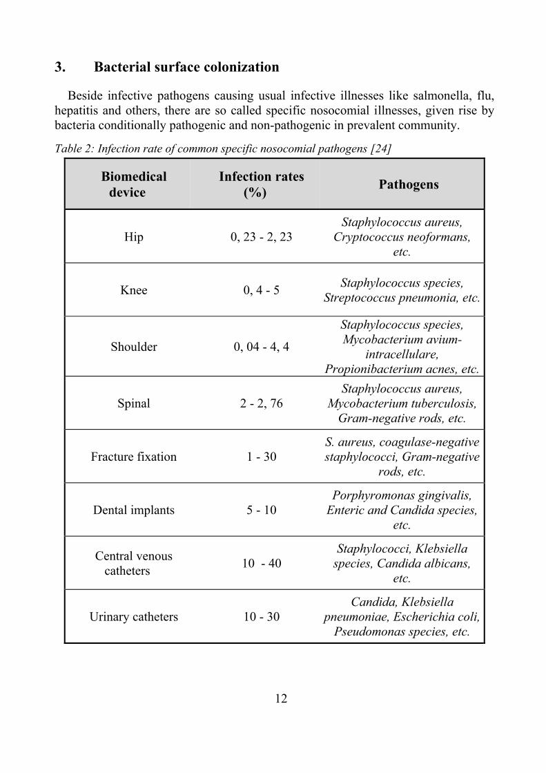
3. Bacterial surface colonization
Beside infective pathogens causing usual infective illnesses like salmonella, flu, hepatitis and others, there are so called specific nosocomial illnesses, given rise by bacteria conditionally pathogenic and non-pathogenic in prevalent community.
Table 2: Infection rate of common specific nosocomial pathogens [24]
Biomedical device
Infection rates (%) Pathogens
Hip 0, 23 - 2, 23 Staphylococcus aureus,
Cryptococcus neoformans, etc.
Knee 0, 4 - 5 Staphylococcus species, Streptococcus pneumonia, etc.
Shoulder 0, 04 - 4, 4
Staphylococcus species, Mycobacterium avium-
intracellulare, Propionibacterium acnes, etc.
Spinal 2 - 2, 76 Staphylococcus aureus,
Mycobacterium tuberculosis, Gram-negative rods, etc.
Fracture fixation 1 - 30 S. aureus, coagulase-negative staphylococci, Gram-negative
rods, etc.
Dental implants 5 - 10 Porphyromonas gingivalis,
Enteric and Candida species, etc.
Central venous catheters 10 - 40
Staphylococci, Klebsiella species, Candida albicans,
etc.
Urinary catheters 10 - 30 Candida, Klebsiella
pneumoniae, Escherichia coli, Pseudomonas species, etc.
13
Bacteria strains Staphylococcus aureus, Staphylococcus epidermidis, Enterobacter aerogenes, Escherichia coli, Klebsiella pneumoniae and fungus Candida albicans are the most prevalent nosocomial pathogens [25]. Pathogens related to particular biomedical device are listed in table 2. Situation in hospital environment is often complicated by bacteria with antibiotic resistance, where methicilin resistant Staphylococcus aureus (MRSA) is the most significant and it is increasingly reported from many countries worldwide [26].
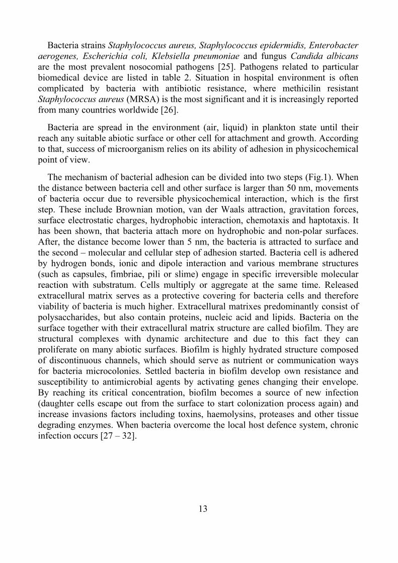
Bacteria are spread in the environment (air, liquid) in plankton state until their reach any suitable abiotic surface or other cell for attachment and growth. According to that, success of microorganism relies on its ability of adhesion in physicochemical point of view.
The mechanism of bacterial adhesion can be divided into two steps (Fig.1). When the distance between bacteria cell and other surface is larger than 50 nm, movements of bacteria occur due to reversible physicochemical interaction, which is the first step. These include Brownian motion, van der Waals attraction, gravitation forces, surface electrostatic charges, hydrophobic interaction, chemotaxis and haptotaxis. It has been shown, that bacteria attach more on hydrophobic and non-polar surfaces. After, the distance become lower than 5 nm, the bacteria is attracted to surface and the second – molecular and cellular step of adhesion started. Bacteria cell is adhered by hydrogen bonds, ionic and dipole interaction and various membrane structures (such as capsules, fimbriae, pili or slime) engage in specific irreversible molecular reaction with substratum. Cells multiply or aggregate at the same time. Released extracellural matrix serves as a protective covering for bacteria cells and therefore viability of bacteria is much higher. Extracellural matrixes predominantly consist of polysaccharides, but also contain proteins, nucleic acid and lipids. Bacteria on the surface together with their extracellural matrix structure are called biofilm. They are structural complexes with dynamic architecture and due to this fact they can proliferate on many abiotic surfaces. Biofilm is highly hydrated structure composed of discontinuous channels, which should serve as nutrient or communication ways for bacteria microcolonies. Settled bacteria in biofilm develop own resistance and susceptibility to antimicrobial agents by activating genes changing their envelope. By reaching its critical concentration, biofilm becomes a source of new infection (daughter cells escape out from the surface to start colonization process again) and increase invasions factors including toxins, haemolysins, proteases and other tissue degrading enzymes. When bacteria overcome the local host defence system, chronic infection occurs [27 – 32].
14
Figure 1: Biofilm formation
4. Protection of material against microbial colonization
Factor influencing bacterial adhesion to the surface can be divided into three main areas:
• Nature of the environment (pH, ionic strength, protein presence, antibiotics or calcium cations, magnesium, iron presence, zinc cations, cobalt, silver, nickel, blood flow, salts, time)
• Type of microorganism (concentration, membrane hydrophobicity, zeta- potential, membrane features - such as pili or flagella, proteins, and lipopolysaccharides, susceptibility to antibiotics, extracellural substances)
• Substratum properties (surface functional groups, roughness, porosity, hydrophobicity).
As mentioned above bacterial attachment on solid substrate surface is highly dependent on material properties, so that their target changing leads to decrease of infection risk. Polymeric materials are frequently used in medical care due to their great flexibility of design, as it is seen in table 3. However, they need to be surface treated to resist bacterial colonization. Only few polymers are resistant against bacterial by themselves (e.g. ChT), but unfortunately they do not provide appropriate mechanical properties.

15
Table 3: Application of polymer in medicine [33, 34]
Synthetic polymer Application
Polyethylene tubing drains, catheters, artificial hip or prosthetic joints
Polyvinyl chloride
blood containers and solution storage bags, surgical packaging, tubes of central venous s
catheter, ventilation tubes, cannulae, cardiovascular implants.
Polypropylene catheters, orthopaedic implants (where more rigidity is required)
Polyethylene terephthalate artificial vascular implants, heart valves
Polyamides catheters, packaging films, artificial tendons, sutures
Polyurethanes artificial skin, mammary prosthesis, vascular catheters, vascular graft, artificial heart
diaphragms and valves, tubing
Polyvinyl alcohol surgical threads
Polystyrene filter wares
Polymethyl methacrylate blood pump and reservoirs, implantable ocular lens, artificial vascular grafts
Polytetrafluoroethylene catheters and artificial vascular grafts
Materials evince antibacterial properties can be prepared by various ways. One method is to incorporate an organic or inorganic biocide to the polymer bulk during the material processing or we can modify monomer to be antibacterial and then to polymerize it. Another method is to change polymer properties to antibacterial after processing. Polymer can be coated by antibacterial layer, grafted by antimicrobial low molecular agents which are slowly realising or covalently bonded [35 - 39]. Other novel way is to incorporate bacteriophage onto polymer surface [40, 41] or change surface morphology towards avoiding prospective attachment ability of the bacteria [42, 43].

16
5. Treating of polymer surface by grafting
Numerous studies have been devoted to the problem of polymer surface modification. Surface is an interface between material bulk and the environment and therefore its characteristics are responsible for interaction behaviour. Conventional polymeric materials have hydrophobic, chemically inert surface due to this fact, problems with adhesion, coating, painting, colouring, lamination, packaging, colloid stabilization, undesirable protein absorption or poor biocompatibility related to certain area of their application are arising. Physical or chemical processes can be used for their modification. Physical processes include surface segregation, electromagnetic waves irradiation of and oxidation with gases; while chemical use wet-treatment, blending, coating and metallization.
Grafting is a physicochemical process. This method is the most favourable one, because of the monomer introduction control, space density or exact location of chains. The chains covalently bonded to the surface avoid its delaminating. By this technique, surface can be tailored by introducing a specific functional group: one end of grafted polymer is attached to surface while the other part of the polymer with appropriate functional groups is capable of consequent surface interaction [44].
Figure 2: Polymer brush formation techniques
5.1 Synthesis of grafted surfaces
Polymer chain interaction with solid state surface could be both reversible and irreversible process. Grafted surface can be obtained generally by physiosorption or covalent attachment (Fig. 2). Physiosorption method is a reversible process. One polymer block interacts strongly with the substrate, while opposite part of the chain interacts weakly forming the graft. Covalently bonded polymers make the polymer
17
more robust and resistant to common chemicals. When pre-synthesized polymer interacts to polymer substrata, it is called "grafting to" approach. However, the "grafting from" approach occurs, when a polymer chain “grows” (polymerizing) on the backbone surface. Nevertheless, it can be synthesized polymer higher density structure by “grafting from” approach because monomers can easily reach reactive sites, but the polymer backbone needs to have a radical initiator which can supply the radical which is necessary for radical polymerization [45].
Polymer surface initiators are essential for polymerization initiation. They can be surface immobilized by several synthetic methods, such as cationic and anionic polymerization, living free radical polymerization and free radical polymerization [46]. The active surface sites can be generated by UV irradiation in presence of photoinitiators or photosensitizer, as well as by ozone oxidation or by various plasma surface treatments.
The graft chains conformation strongly depends on graft density. Firstly, at low densities, a “mushroom” structure is formed with a random-coil dimension [47]. With graft density increase, chains try to stretch away from surface forming polymer brush. Numerous AFM studies or contact angle measurements showed proper information related to design and architecture of so-called polymer brushes. It has been shown, that varying chain length can be done by optimizing the monomer concentration or irradiation time in case of photopolymerization. Grafted chains parameters respond to reaction conditions (mainly pH and ionic strength) and their intermolecular interaction by moderating of their shape and dynamic surface characteristics. Polymer brush properties offer a wide range of applications. Adequately formed brush can eliminate surface electrostatic charge generation, increase of interactive forces at the interface and significantly lower friction coefficient value. Grafted chains are capable of absorbing substances like both small molecules and large biomacromolecules, with application as slowly releasing antibacterial agents [48, 44].

18
6. Plasma treatment of polymer materials
Plasma is a set of charged and neutral particles in different quantum states (electrons, ions, excited states, neutral atoms, molecules), which forms the approximately equal to zero space charge (the so-called quasineutrality). Plasma is also called as the fourth state of matter. Its outward behaviour most resembles the gas from which it differs mainly by the presence of free charge carriers. It is capable of electric and magnetic fields generation and response to such field presence. Plasma is not a unique phenomenon, it is estimated that up to 99% of the total space is found right in this state. It is all about stars, but also a variety of nebulae and galaxies. Plasma can be found less often in earth conditions, because its existence has a large energy demands (high temperature, pressure, radiation, etc.), it will not last long as for example flashes and other discharges.
Figure 3: Example of plasma reactor: radio frequency plasma discharge chamber:
1 – radio frequency source; 2 – reactor vacuum chamber; 3 - radio frequency electrode; 4 – plasma discharge; 5 – grounded electrode; 6 – plasma gas input; 7 – gas output [51]
It is essential to release electrons from atoms or molecules in the gaseous phase or to be subjected of ionization for plasma creation. The ionization occurs when the atom or molecule gains sufficient energy - it can be accepted from secondary sources, but also from mutual collisions with neighboring particles. The generation and maintenance of plasma is source of energy causing ionization. Generally, the gas
19
plasma triggers when electrodes are connected to the appropriate voltage (Fig. 4). Breakthrough energy depends on the gas pressure and discharge gap width. The lowest breakthrough energy comes at low pressures. At atmospheric pressure occurs an arc discharge. Breakthrough energy can be reduced more by using radiofrequency source, laser, microwave, magnetic field, etc. [49, 50]
Plasma is a very chemical reactive environment in which unusual reactions take place. The high density of ionized and excited particles inside the plasma can change the properties of otherwise inert material. Plasma also modifies the surface energy of material, which has a consequent effect on the adhesion strength characteristics of surface coatings and biocompatibility. Ingestion plasma editing biomaterials offers the following benefits:
• reliable treatment which does not affect bulk properties; modifications depth reaches only a few angstroms.
• can be applied on any material substrate: metals, polymers, ceramics, composites, ...
• may influence variety of surface characteristics: chemical, tribology (friction), electrical, optical, biological, ...
• sterilizes treated surfaces
• treatment is environmentally friendly
• selection of the processing carrier gas influences the type of chemical modification
Of course plasma treatments comprise some disadvantages:
• treatment must take place mostly in a vacuum chamber, which increases the total number of discontinual operations and finally leads to the total cost increase
• the processes in plasma are very complex. It is difficult to understand well all the prospective interactions between the plasma and the surface and for example accurately control the functional groups quantity
• launching of industrial production from laboratory experiments is complicated
The species responsible for plasma – surface reaction are electrons, ions and radicals. Which reaction is initiated by these species depends mainly on used reaction gas, its feed rate, as well as the energy level of the plasma and type of treated material. The noble gas plasma (helium, neon, argon) used only its

20
mechanical energy of particles and caused mainly physical changes of surface or forming free radical by splitting off surface hydrogen. This type of radical formation is happening also simultaneously with chemical reactions when no-inert gases are used. Activation of polymer surface by plasma by free radical introduction onto the surface is well described technique, where radicals serve as initiators for further copolymer reaction (“grafting from”) and forming of polymer brush [52]. Processes using oxygen plasma are most common in the modification of polymer surfaces. It is known that oxygen in the plasma reacts with a variety of polymers to form oxygen-functional groups C-O, C = O, O-C = O, etc. Besides pure oxygen is widely used, its compounds, such as CO, CO2, SO2 or H2O incorporate hydroxyl groups into the surface. Gases as N2, NH3, F2, HF and F4S are used for surface wettability alterations. Extraordinary physical properties such as micro-hardness, optical reflection angle and tightness can be reached by reaction of polymer in hydrocarbons (methane, ethane, ethylene, acetylene, benzene). Possible surface application of plasma is shown in table 4 [53].
6.1 Dielectric barrier discharge
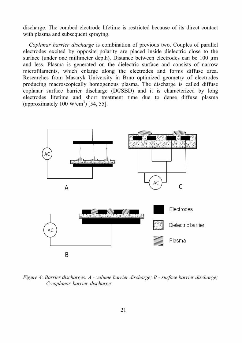
Contemporary trend in plasma technology development is oriented to treatment at atmospheric conditions without expensive vacuum technology. The barrier discharge is generated between electrodes, at least one of which is covered with dielectric layer at atmospheric pressure. Glass, ceramics, corundum or polytetrafluorethylene are mostly used as the dielectric. Dielectric barrier has two basic roles: discharge at high pressure is split into large numbers of micro-discharges homogenously distributed in time and space throughout the discharge area. Second role is the inhibition of transition to arc discharge by limitation of charge transferred by each micro – discharge. Micro – discharges, occurred at atmospheric pressure, last typically nanoseconds. Dielectric application also leads to altering or pulsed voltage supplying. Dielectric barrier discharge can be divided into three basic groups according to configurations: volume, surface and coplanar barrier discharge (Fig. 4).
Volume barrier discharge can be generated, when a dielectric barrier is inserted in the area between electrodes. The most often is one of electrodes covered by dielectric. Volume plasma discharge consists of many micro-discharges perpendicular to electrodes, which enlarges its volume in contact with dielectric and gradually covered whole dielectric surface by rising voltage.
Surface barrier discharge arises between electrodes firmly attached to dielectric barrier. Typically, the first electrode is square shaped and second electrode is ordered in comb structure. Plasma appears in gaps of combed electrode. The most active plasmochemical place is founded on the top of electrodes and on the peak of
21
discharge. The combed electrode lifetime is restricted because of its direct contact with plasma and subsequent spraying.
Coplanar barrier discharge is combination of previous two. Couples of parallel electrodes excited by opposite polarity are placed inside dielectric close to the surface (under one millimeter depth). Distance between electrodes can be 100 µm and less. Plasma is generated on the dielectric surface and consists of narrow microfilaments, which enlarge along the electrodes and forms diffuse area. Researches from Masaryk University in Brno optimized geometry of electrodes producing macroscopically homogenous plasma. The discharge is called diffuse coplanar surface barrier discharge (DCSBD) and it is characterized by long electrodes lifetime and short treatment time due to dense diffuse plasma (approximately 100 W/cm3) [54, 55].
Figure 4: Barrier discharges: A - volume barrier discharge; B - surface barrier discharge;
C-coplanar barrier discharge……...……………………………………………..
22
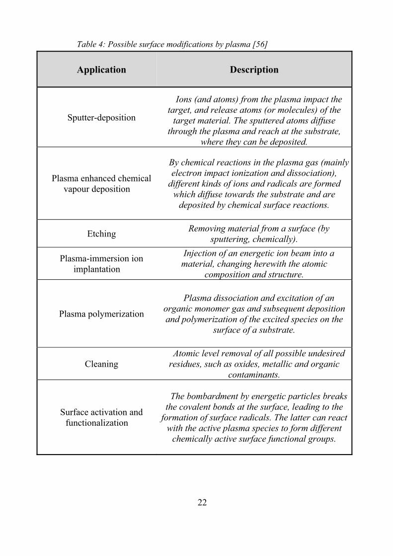
Table 4: Possible surface modifications by plasma [56]
Application Description
Sputter-deposition
Ions (and atoms) from the plasma impact the target, and release atoms (or molecules) of the
target material. The sputtered atoms diffuse through the plasma and reach at the substrate,
where they can be deposited.
Plasma enhanced chemical vapour deposition
By chemical reactions in the plasma gas (mainly electron impact ionization and dissociation),
different kinds of ions and radicals are formed which diffuse towards the substrate and are
deposited by chemical surface reactions.
Etching Removing material from a surface (by sputtering, chemically).
Plasma-immersion ion implantation
Injection of an energetic ion beam into a material, changing herewith the atomic
composition and structure.
Plasma polymerization
Plasma dissociation and excitation of an organic monomer gas and subsequent deposition and polymerization of the excited species on the
surface of a substrate.
Cleaning Atomic level removal of all possible undesired
residues, such as oxides, metallic and organic contaminants.
Surface activation and functionalization
The bombardment by energetic particles breaks the covalent bonds at the surface, leading to the
formation of surface radicals. The latter can react with the active plasma species to form different
chemically active surface functional groups.
23
7. Antibacterial agents
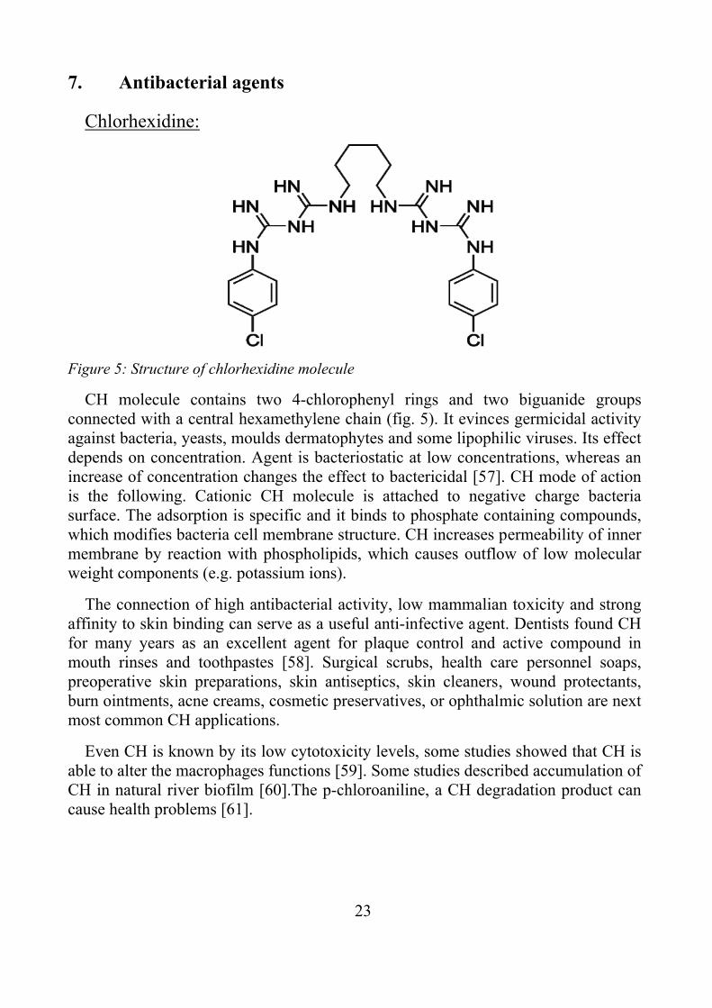
Chlorhexidine:
Figure 5: Structure of chlorhexidine molecule
CH molecule contains two 4-chlorophenyl rings and two biguanide groups connected with a central hexamethylene chain (fig. 5). It evinces germicidal activity against bacteria, yeasts, moulds dermatophytes and some lipophilic viruses. Its effect depends on concentration. Agent is bacteriostatic at low concentrations, whereas an increase of concentration changes the effect to bactericidal [57]. CH mode of action is the following. Cationic CH molecule is attached to negative charge bacteria surface. The adsorption is specific and it binds to phosphate containing compounds, which modifies bacteria cell membrane structure. CH increases permeability of inner membrane by reaction with phospholipids, which causes outflow of low molecular weight components (e.g. potassium ions).
The connection of high antibacterial activity, low mammalian toxicity and strong affinity to skin binding can serve as a useful anti-infective agent. Dentists found CH for many years as an excellent agent for plaque control and active compound in mouth rinses and toothpastes [58]. Surgical scrubs, health care personnel soaps, preoperative skin preparations, skin antiseptics, skin cleaners, wound protectants, burn ointments, acne creams, cosmetic preservatives, or ophthalmic solution are next most common CH applications.
Even CH is known by its low cytotoxicity levels, some studies showed that CH is able to alter the macrophages functions [59]. Some studies described accumulation of CH in natural river biofilm [60].The p-chloroaniline, a CH degradation product can cause health problems [61].
24
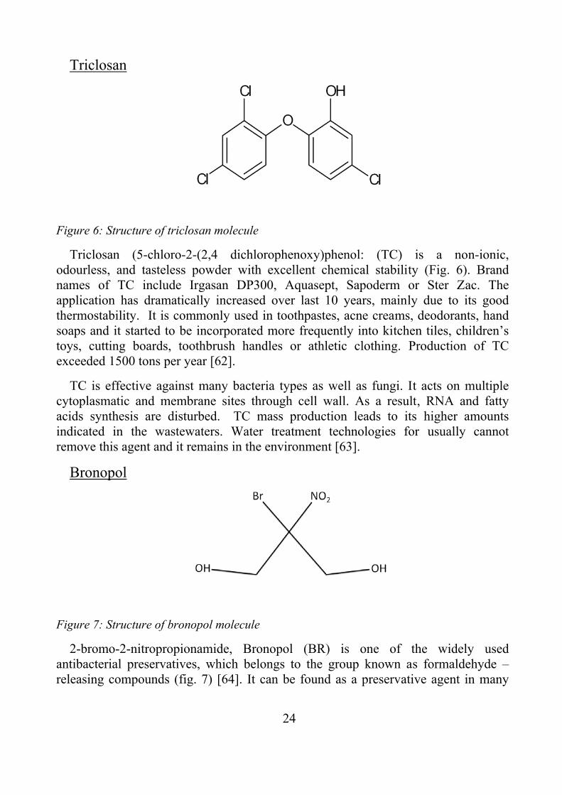
Triclosan
Figure 6: Structure of triclosan molecule
Triclosan (5-chloro-2-(2,4 dichlorophenoxy)phenol: (TC) is a non-ionic, odourless, and tasteless powder with excellent chemical stability (Fig. 6). Brand names of TC include Irgasan DP300, Aquasept, Sapoderm or Ster Zac. The application has dramatically increased over last 10 years, mainly due to its good thermostability. It is commonly used in toothpastes, acne creams, deodorants, hand soaps and it started to be incorporated more frequently into kitchen tiles, children’s toys, cutting boards, toothbrush handles or athletic clothing. Production of TC exceeded 1500 tons per year [62].
TC is effective against many bacteria types as well as fungi. It acts on multiple cytoplasmatic and membrane sites through cell wall. As a result, RNA and fatty acids synthesis are disturbed. TC mass production leads to its higher amounts indicated in the wastewaters. Water treatment technologies for usually cannot remove this agent and it remains in the environment [63].
Bronopol
Figure 7: Structure of bronopol molecule
2-bromo-2-nitropropionamide, Bronopol (BR) is one of the widely used antibacterial preservatives, which belongs to the group known as formaldehyde – releasing compounds (fig. 7) [64]. It can be found as a preservative agent in many

25
areas, mainly in preservation of milk analysis samples and in cosmetics and hygiene products or leather conservation [65 - 67]
BR mechanism of action is supposed to be based on catalytic oxidation of thiol within the cell wall and free radicals generation [68]. Formaldehyde is released during BR decomposition which can cause allergy and irritation [69].
Benzalkonium chloride
Figure 8: Structure of benzalkonium chloride molecule
Benzalkonium chloride (BCH) is a cationic surfactant and an antimicrobial agent commonly used in nasal sprays, ophthalmic preparations, mouth rinses, cosmetic products or infant care products. Chemically, it is a quaternary ammonium compound; a mixture of alkylbenzyldimethylammonium chloride with various even-numbered, straight alkyl chains with 8 to 18 carbons (fig. 8) [70]. It behaves highly lipophilic and it charges positively in an emulsion of host lipids [71].
Mechanism of antibacterial activity depends on N-alkyl chain length. Optimal activity against gram positive bacteria and yeast is achieved with chain lengths of 12–14 alkyls, while the lengths effective against gram negative is 14-16 alkyls and compounds containing less then 4 or more then 18 alkyls in a structure are almost inactive.
BCH influence the cell membranes integrity. Positively charged nitrogen of BCH molecule is attracted to acidic phospholipids of bacterial membranes. Then, hydrophobic BCH chain incorporates into the bacterial hydrophobic membrane core [72].
26
However, number of studies shown appreciable toxic effects on corneal and conjunctive epithelium including irritation, tear film instability or allergic reactions [73].
Chitosan
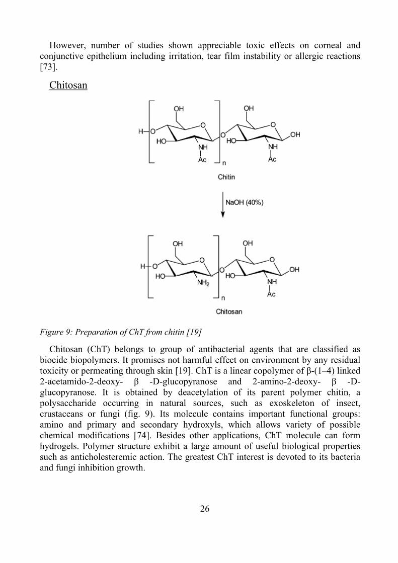
Figure 9: Preparation of ChT from chitin [19]
Chitosan (ChT) belongs to group of antibacterial agents that are classified as biocide biopolymers. It promises not harmful effect on environment by any residual toxicity or permeating through skin [19]. ChT is a linear copolymer of β-(1–4) linked 2-acetamido-2-deoxy- β -D-glucopyranose and 2-amino-2-deoxy- β -D-glucopyranose. It is obtained by deacetylation of its parent polymer chitin, a polysaccharide occurring in natural sources, such as exoskeleton of insect, crustaceans or fungi (fig. 9). Its molecule contains important functional groups: amino and primary and secondary hydroxyls, which allows variety of possible chemical modifications [74]. Besides other applications, ChT molecule can form hydrogels. Polymer structure exhibit a large amount of useful biological properties such as anticholesteremic action. The greatest ChT interest is devoted to its bacteria and fungi inhibition growth.

27
Although action mechanism is still not completely understood, there are six theoretical mechanisms, which are based on: leakage of cell membrane due to interaction of ChT amine group and cell membrane, blocking of cell’s active centres, inhibition of toxin production cell growth and by selective chelating of metals, blocking the entry of nutrients into the cell by fully covering of the cell, penetration of low-molecular weight species into the cell and interference with the synthesis of mRNA and proteins or disturbing the physiological activities in the cell [75].
This compound is relatively non-toxic, biocompatible material [76].
28
AIM OF THE WORK
The primary aim of the presented thesis is the preparation and characterization of an active antibacterial polymer surface by using multistep physicochemical approach including plasma activation and polymer brush formation. It could be used for chemical commodities and polymers for potential application in medicine and food industry.
The main attention is paid to the investigation of antibacterial activity of prepared samples against S. aureus and E. coli strains. The optimization of preparation approach towards achieving the best antibacterial properties is next goal of thesis.
29
SUMMARIES OF PAPERS
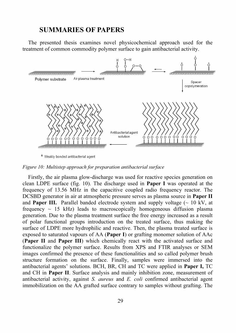
The presented thesis examines novel physicochemical approach used for the treatment of common commodity polymer surface to gain antibacterial activity.
Figure 10: Multistep approach for preparation antibacterial surface
Firstly, the air plasma glow-discharge was used for reactive species generation on clean LDPE surface (fig. 10). The discharge used in Paper I was operated at the frequency of 13.56 MHz in the capacitive coupled radio frequency reactor. The DCSBD generator in air at atmospheric pressure serves as plasma source in Paper II and Paper III. Parallel banded electrode system and supply voltage (~ 10 kV, at frequency ~ 15 kHz) leads to macroscopically homogeneous diffusion plasma generation. Due to the plasma treatment surface the free energy increased as a result of polar functional groups introduction on the treated surface, thus making the surface of LDPE more hydrophilic and reactive. Then, the plasma treated surface is exposed to saturated vapours of AA (Paper I) or grafting monomer solution of AAc (Paper II and Paper III) which chemically react with the activated surface and functionalize the polymer surface. Results from XPS and FTIR analyses or SEM images confirmed the presence of these functionalities and so called polymer brush structure formation on the surface. Finally, samples were immersed into the antibacterial agents’ solutions. BCH, BR, CH and TC were applied in Paper I, TC and CH in Paper II. Surface analysis and mainly inhibition zone, measurement of antibacterial activity, against S. aureus and E. coli confirmed antibacterial agent immobilization on the AA grafted surface contrary to samples without grafting. The
30
highest increase of antibacterial activity was observed by the sample containing TC and grafted by AA. On the other hand, samples covered by BR did not show any antibacterial activity. The multilayer coating was created by several alternating dipping into ChT and pectin solutions and final crosslinking by glutaraldehyde in Paper III. However, samples grafted by AAc observed more antibacterial inhibition zone efficiency than samples coated by multilayer chitosan/pectin system.
Surface wettability was investigated by static contact angle measurement using sessile drop technique. Acid – Bases theory were chose for surface energy evaluation. Generally, significant increase of total surface energy (mainly basic part) was observed after the plasma treatment because characteristics reactive polar functional groups were introduced onto the LDPE surface. The monomer grafting led to the further increase of basic part of surface energy, whereas AA and AAc contain polar amine and carboxylic groups. Surface energies of antibacterial coated samples indicate their presence on the surface and alter according to polarity and amount of attached molecules.
The ATR-FTIR and X-ray photoelectron spectroscopy (XPS) described chemical changes during samples preparation. The results showed that the LDPE samples treatment by the low temperature plasma in air caused oxygen-containing groups’ introduction to the surface. XPS analysis together with FTIR spectroscopy confirmed the grafted layer and antibacterial agent presence on the surface, because of detection their characteristic groups.
The amount of attached antibacterial agent, estimated from characteristic groups´ intensity was generally higher for all samples covered by grafted layer. XPS analysis revealed to be more suitable analysis then FTIR. Because compared to the FTIR technique, XPS depth of analyzed layer is substantially smaller and thus the description of surface characteristics is not distorted by signal of non-modified subsurface layer and surface changes are easily detectable. Surface morphology changes were observed by scanning electron microscopy (SEM) (Paper I and III) and atomic force microscopy (AFM) (Paper II). The plasma effect led to the slightly increase of LDPE surface roughness as a result of functionalization and mechanical ablation processes. Further grafting by AA or AAc created a brush-like pattern appropriate for subsequent modification, which is more noticeable by AFM analysis. Antibacterial agent deposition caused smoothening of SEM images.

31
CONCLUSION
Health damages caused by various infections as a consequence of bacterial growth on polymeric products or deterioration of packed food by bacteria are two main reasons of active antibacterial material improvement. The material protection key point lies in the adhesion controlling as its first step of bacterial surface colonization.
The thesis deals with effective polymer surface treatment, while bulk properties remain unchanged. This advantage enable to use mentioned approach after product processing, so that there are reduced problems related to mixing antibacterial agent into bulk polymer during processing, such as blend homogeneity, antibacterial agent degradation or antibacterial gents impact on bulk properties. Moreover, antibacterial agents’ consumption is significantly reduced, considering their impact on environment.
Our results showed, that it can be prepared antibacterial active polymer surface that resist most effectively E. coli and S. aureus strains. Most effective configuration reveals to be TC attachment to AA or AAc as grafting monomer and activation of surface by diffuse coplanar surface barrier air discharge at atmospheric pressure. This approach can be advantageously integrated for example to packaging foil production line. However, there are still a lot of opened questions before final industrial application. Antibacterial effect’s durability, peel resistance, potential cytotoxicity and in vivo assay need to be tested.
32
ACKNOWLEDGEMENTS
This thesis would not have been finished without the patient help and support of my supervisors: doc. Ing. Marián Lehocký, Ph.D. and Ing. Adriana Gregorová, Ph.D.
I would like to acknowledge the financial, academic and technical support of the Tomas Bata University in Zlín, to the Ministry of Education, Youth and Sport of the Czech Republic (MSM7088352101) and (CZ.1.05/2.1.00/03.0111) and also to the Czech Science Foundation (project 104/09/H080) for financial support.
Special thanks belong to my wife, family, friends, Jorge Lopez-Garcia for correction and other colleagues, who provided me with support and help during my studies.
Thank you!
33
REFERENCES
[1] URBÁNKOVÁ, M., Princip a Význam Bariérových Vlastností Plastových Obalů pro Potravinářské Aplikace, Bachelor thesis of. Tomas Bata University in Zlín, Faculty of Technology, Zlín Supervized by Ing. Sedlařík, V.,2010
[2] KAČEŇÁK, I., Obaly a Obalová Technika, SVŠT Bratislava, 1989. ISBN 80-227-0301-X
[3] ČURDA D., Balení Potravin, SNTL PRAHA ,1982. ISBN 04-832-82 [4] DOBIAS, J., CHUDACKOVA, K., VOLDRICH, M., MAREK, M. Properties
of Polyetylene Films with Incorporated Benzoic Anhydride and/or Ethyl and Propyl Esters of 4- Hydroxybenzoic Acid and Their Suitability for Food Packaging, Food Add Contamin, 2000, vol.17, no. 12, p.1047.
[5] ABOUGROUN, H. A., COUSIN, N. A., JUDGE, M. D., Extended Shelf Life of Unrefrigerated Prerigor Cooked Meat, Meat Sci. 1993, vol. 33, p. 207.
[6] MILLER, A. J., CALL, J. E., WHITING, R. C. Comparison of Organic Acid Salts for Clostridium Botulinum Control in an Uncured turkey Product. J. Food Prot. 1993, vol. 56, p. 958.
[7] NICHOLSON, M. D. The Role of Natural Antimicrobials in Food Packaging Biopreservation, J Plastic Film Sheeting, 1998, vol. 14, no. 3, p. 234.
[8] CHEN, M. C., YEH, G. H. C., CHIANG, B. H., Antimicrobial and Physicochemical Properties of Methylcellulose and Chitosan Films Containing a Preservative. J Food Proc reserve 1996, vol. 20, no. 5, p. 379.
[9] COMA, V., MARTIAL-GROS, A., GARREAU, S., COPINET, A., DESCHAMPS, A. Edible Antimicrobial Film Based on Chitosan Matrix. J. Food Sci. 2002, vol. 67, no. 3, p. 1162.
[10] OUATTARA, B., SIMARD, R. E., PIETTE, G., BEGIN, A., HOLLEY, R. A. Inhibition of Surface Spoilage Bacteria in Processed Meats by Application of Antimicrobial Films Prepared with Chitosan, Int J Food Microbiol 2000, vol. 62, p. 139.
[11] OUATTARA, B., SIMARD, R. E., PIETTE, G., BEGIN, A., HOLLEY, R. A. Diffusion of Acetic and Propionic Acids from Chitosan - based Antimicrobial Packaging Films, J Food Sci 2000, vol. 65, p. 768.
[12] VELÁZQUEZ, L. C., BARBINI, B. B., ESCUDERO, M. E., ESTRADA, C. L., DE GUZMÁN, A. M. S., Evaluation of Chlorine, Benzalkonium Chloride and Lactic Acid as Sanitizers for Reducing Escherichia coli O157:H7 and Yersinia enterocolitica on Fresh Vegetables, Food Control 2009, vol. 20, p. 262.
34
[13] CHUNG, S.K., CHO, S. H., LEE, D. S. Modified Atmosphere Packaging of Fresh Strawberries by Antimicrobial Plastic Films, Korean J Food Sci Technol 1998, vol. 30, p. 1140.
[14] CUTTER, C. N. The Effectiveness of Triclosan Incorporated Plastic against Bacteria on Beef Surfaces, J. Food Prot. 1999, vol. 62, p. 474.
[15] VERMEIREN, L., DEVLIEGHERE, F., DEBVERE, J., Effectiveness of Some Recent Antimicrobial Packaging Concepts, Food Add Contamin 2002, vol. 19, p. 163.
[16] HOFFMAN, K. L., HAN, I. Y., DAWSON, P. L. Antimicrobial Effects of Corn Zein Films Impregnated with Sisin, Lauric Acid and EDTA, J Food Prot 2001, vol. 64, p. 885.
[17] HONG, S. I., PARK, J.D., KIM, D.M. Antimicrobial and Physical Properties of Food Packaging Films Incorporated with Some Natural Compounds, Food Sci Biotechnol 2000, vol. 9, p. 38.
[18] SUPPAKUL, P., MILTZ, J., SONNEVELD, K., BIGGER, S. W. Antimicrobial Properties of Basil and Its Possible Application in Food Packaging, J. Agric. Food Chem. 2003, vol. 51, p. 3197.
[19] KENAWY, E.R., WORLEY, S.D., BROUGHTON, R. The Chemistry and Applications of Antimicrobial Polymers: A State-of-the-Art Review, Biomacromolecules 2007, vol. 8, p. 1359.
[20] BROKOW, G., GABBAY, J. Preventing Pathogens Proliferation and Reducing Potential Sources of Nosocomial Infections with Biocidal Textiles in Developing Countries, The Open Biology Journal 2010, vol. 3, p. 81.
[21] BREATHNACH, A. Nosocomial Infections, The Medicine Publishing Company Ltd 2005, vol. 33, p. 22.
[22] Lim, S. M., Webb, S. A. R. Nosocomial Bacterial Infections in Intensive Care Units. I: Organisms and Mechanisms of Antibiotic Resistance, Anaesthesia 2005, vol. 60, p. 887.
[23] ŠRÁMKOVÁ H. Nozokomiální nákazy 2, Maxdorf , 2001. ISBN 80-85912-25-2
[24] CAO H., LIU, X. Silver Nanoparticles - modified Films versus Biomedical Device-Associated Infections, WIREs Nanomedicine and Nanobiotechnology 2010, vol. 2, p. 670.
[25] ZIEBUHR, W., HENNING, S., ECKART, M., KRÄNZLER, H., BATZILLA, C., KOZITSKAYA, S. Nosocomial infections by Staphylococcus Epidermidis: How a Commensal Bacterium Turns into a Pathogen, International Journal of Antimicrobial Agents 2006, vol. 28, p. 14.

35
[26] TENOVER, F. C., BIDDLE, J. W., LANCASTER, M. V. Increasing Resistance to Vancomycin and Other Glycopeptides in Staphylococcus aureus, Emerging Infection Diseases 2001, vol. 7, p. 327.
[27] ZHANG, L., GOWARDMAN, J., RICKARD, C. M. Impact of Microbial Attachment on Intravascular Catheter-related Infections, International Journal of Antimicrobial Agents 2011, vol. 38, p. 9.
[28] COENYE, T., NELIS, H. J. In vitro and In vivo Model Systems to Study Microbial Biofilm Formation, Journal of Microbiological Methods 2010, vol. 83, p. 89.
[29] ATABEK, A. Investigating Bacterial Outer Membrane Polymers and Bacterial Interaction with Organic Molecules Using Atomic Force Microscopy, Master Thesis. Worcester Polytechnic Institute, supervized by Camesano, A., Dibiasio D., 2006.
[30] SPERANZA, G., GOTTARDI, G., PEDERZOLLI, C., LUNELLI, L., CANTERI, R., PASQUARDINI, L., CARLI, E., LUI, A., MANIGLIO, D., BRUGNARA, M., ANDERLE, M. Role of Chemical Interactions in Bacterial Adhesion to Polymer Surfaces, Biomaterials 2004, vol. 25, p. 2029.
[31] YUEHUEI, H. A., FRIEDMAN A. R. Concise Review of Mechanisms of Bacterial Adhesion to Biomaterial Surfaces, Journal of Biomedical Materials research 1998, vol. 43, p. 338.
[32] BAZAKA, K., JACOB, M. V., CRAWFORD, R. J., IVANOVA, E. P. Plasma-assisted Surface Modification of Organic Biopolymers to Prevent Bacterial Attachment, Acta Biomaterialia 2011, vol. 7, p. 2015.
[33] WONG, J. Y., BRONZINO, J.D. Biomaterials. 1st ed. Boca Raton: CRC Press, 2007. ISBN 978-0-84-93-7888-1
[34] GALYA T. Antibacterial Modifications of Polymers by Using Metallic Compounds, Doctoral thesis, Tomas Bata University in Zlín, Zlín, 2009. 122 p. ISBN 978-80-7318-872-6
[35] VASILEV, K., COOK, J., GRIESSER, H. J. Antibacterial Surfaces for Biomedical Devices, Expert Rev. Med. Devices 2009, vol. 6, p. 553.
[36] THOME, J., HOLLÄNDER, A., JAEGER, W., TRICK, I., OEHR, C. Ultrathin Antibacterial Polyammonium Coatings on Polymer Surfaces, Surface and Coating Technology 2003, vol. 174, p. 584.
[37] ZHANG, W., CHU, P. K., JI, J., ZHANG Y., FU R. K. Y., YAN, Q. Antibacterial Properties of Plasma-modified and Triclosan or Bronopol Coated Polyethylene, Polymer 2006, vol. 47, p. 931.
36
[38] ZHANG, W., CHU, P. K., JI, J., ZHANG Y., LIU, X., FU R. K. Y., HA, P. C. T., YAN, Q. Plasma Surface Modification of Polyvinylchloride for Improvement of Antibacterial Properties, Biomaterials 2006, vol. 27, p. 44.
[39] YE, Y., SONG, Q., MAO, Y. Solventless Hybrid Grafting of Antimicrobial Polymers for Self-sterilizing Surfaces, Journal of Materials Chemistry 2011, vol. 2, p. 13188.
[40] FU, W., FORSTER, T., MAZER, O., CURTIN, J. J., LEHMAN, S. M., DONLAN, R. M. Bacteriophage Cocktail for the Prevention of Biofilm Formation by Pseudomonas aeruginosa on Catheters in an In Vitro Model System, Antimicrobial agents and chemotherapy 2010, vol. 54, p. 397.
[41] DONLAN, R. D. Preventing Biofilms of Clinically Relevant Organisms Using Bacteriophage, Trends in Microbiology 2009, vol. 17, p. 66.
[42] HOU, S., GU, H., SMITH, C., REN, D. Microtopographic Patterns Affect Escherichia coli Biofilm Formation on Poly(dimethylsiloxane) Surfaces, Langmuir 2011, vol. 27, p. 2686.
[43] CARMAN, M., ESTES, T. G., FEINBERG, A. W., SCHUMACHER J. F., WILKERSON, W., WILSON, L. H., CALLOW, M. E., CALLOW, J. A., BRENNAN, A. B. Engineered Antifouling Microtopographies - Correlating Wettability with Cell Attachment, Biofouling 2006, vol. 22, p. 11.
[44] KATO, K., UCHIDAB, E., KANGC, E., UYAMAA, Y., IKADAD Y., Polymer Surface with Graft Chains, Prog. Polym. Sci. 2003, vol. 28, p. 209.
[45] MCBRIDE, M. C., MALCOLM, R. K., WOOLFSON, A. D., SEAN P. GORMAN, S. P. Persistence of Antimicrobial Activity through Sustained Release of Triclosan from Pegylated Silicone Elastomers, Biomaterials 2009, vol. 30, p. 6739.
[46] SENARATNE,W., ANDRUZZI L., OBER, CH. K., Self-Assembled Monolayers and Polymer Brushes in Biotechnology: Current Applications and Future Perspectives, Biomacromolecules 2005, vol. 6, p. 2427.
[47] LIU, G., YAN, L., CHEN, X., ZHANG, G., Study of the kinetics of mushroom-to-brush transition of charged polymer chains, Polymer 2006, vol. 47, p. 3157.
[48] ZHAO, B., BRITTAIN, J. W. Polymer Brushes: Surface-immobilized Macromolecules, Progress in Polymer Science 2000, vol. 25, p.677.
[49] CHAN C. M., KO T.-M., HIRAOKA H. Polymer Surface Modification by Plasmas and Photons, Surface Science Reports 2001, vol. 24, p.1.
37
[50] CHU P.K., CHEN J.Y. WANG L.P., HUANG N. Plasma-surface Modification of Biomaterials, Material science and Engineering 2002, vol. 36, p. 143.
[51] ÚSTAV FYZIKY A MATERIÁLOVÉHO INŽENÝRSTVÍ Radiofrekvenční a mikrovlnné plazma [online] Tomas Bata University in Zlín [cit. 6.9. 2012], available on World Wide Web: http://ufmi.ft.utb.cz/texty/plazmochemie/PCH_07.pdf
[52] BHATTACHARYA, A., MISTRA, B. N. Grafting: a Versatile Means to Modify Polymers Techniques, Factors and Applications, Progress in Polymer Science 2004, vol. 29, p. 767.
[53] DESMET, T., MORENT, R., DE GEYTER, N., LEYS, C., SHNACHT, E., DUBRUEL, P. Nonthermal Plasma Technology as a Versatile Strategy for Polymeric Biomaterials Surface Modification: A Review. Biomacromolecules 2009, vol. 10, p. 2351.
[54] HANUSOVÁ, J. DCSBD ve Směsích Pracovních Plynů, Master thesis, Masaryk University, Faculty of Science, Brno supervised by Sťahel, P, 2011
[55] ČECH, J. Porovnávací Studium Vlivů Parametrů Výboje na Vlastnosti Plazmatu koplanárního bariérového výboje, Master thesis, Masaryk University, Faculty of Science, Brno, supervised by Černák, M., 2006
[56] BOGAERTS, A., NEYTS, E., GIJBELS, R., MULLEN, J. Gas Discharge Plasmas and Their Applications, Spectrochimica Acta Part B 2002, vol. 57, p. 609.
[57] SAAD, S., HEWETT, K., GREENMAN, J. Effect of Mouth - Rinse Formulations on Oral Malodour Processes in Tongue-derived Perfusion Biofilm Model, J. Breath Res. 2012, vol. 27, p. 1.
[58] JONES, G. R. Chlorhexidine: Is It Still The Gold Standard?, Periodontology 2000 1997, vol. 15, p. 55.
[59] LI, Y., KUAN, Y., LEE, S., HUANG, F., CHANG, Y. Chlorhexidine: Cytotoxicity and Genotoxicity of Chlorhexidine on Macrophages, In Vitro, Inc. Environ Toxicol, DOI 10.1002/tox
[60] DYNES, J. J., LAWRENCE, J. R., KORBER, D. R., SWERHONE, G. D. W., LEPPARD, G. G., HITCHCOCK A. P. Quantitative Mapping of Chlorhexidine in Natural River Biofilms, Science of the Total Environment 2006, vol. 369, p. 369.
[61] ZONG, Z., KIRSCH, L. E. Studies on the Instability of Chlorhexidine, Part I: Kinetics and Mechanisms, J PharmSci 2012, vol. 101, no. 7, p. 2417.
38
[62] DANN, A. B., HONTELA, A. Triclosan: Environmental Exposure, Toxicity and Mechanisms of Action, J. Appl. Toxicol. 2011, vol. 31, p. 285.
[63] CARSTEN VON DER OHE, P., SCHMITT-JANSEN, M., SLOBODNIK, J., BRACK, W., Triclosan - the Forgotten Priority Substance?, Environ Sci Pollut Res 2012, vol.19, p.585.
[64] KAJIMURA, K., TAGAMI, T., YAMAMATO, T., IWAGAMI, S. The Release of Formaldehyde upon Decomposition of 2 - bromo - 2 nitropropan – 1, 3 – diol (Bronopol), Journal of health science 2008, vol.54, p.488.
[65] BUTLER, G., STERGIADIS, S. Suitability of Bronopol Preservative Treated Milk for Fatty Acid Determination, Journal of Dairy Research 2011, vol. 78, p. 220.
[66] LASMAR, M.M., LEITE, M.O., FONSECA, L.M., SOUZA, M.R., CERQUEIRA, M.M.O.P., PENNA, C.F.A.M., COUTO, C.N.B., FERREIRA, J.M. Detection of Cheese Whey in Raw Milk Preserved with Bronopol through High Performance Liquid Chromatography, Arq. Bras. Med. Vet. Zootec. 2011, vol. 63, no. 6, p. 1553.
[67] MUTHUSUBRAMANIAN, L., MITRA, R.B. A Cleaner Production Method for the Synthesis of Bronopol - A bactericide that is Useful in Leather Making. Journal of Cleaner Production 2006, vol. 14, p. 536.
[68] SHEPHERD, J. A., WAIGH, R. D., GILBERT, P. Antibacterial Action of 2-Bromo-2-Nitropropane-1,3-Diol (Bronopol), Antimicrobial agents and chemotherapy, 1988, vol. 13, no. 11, p. 1693.
[69] DE GROOT, A., WHITE, I. R., FLYVHOLM, M., LENSEN, G., COENRAADS, P. Formaldehyde-releasers in Cosmetics: Relationship to Formaldehyde Contact Allergy, Contact Dermatitis 2010, vol. 62, p. 18.
[70] GRAF, P. Benzalkoniumchloride as a Preservative in Nasal Solutions: Re-examining the Data, Respiratory medicine 2001, vol. 95, n. 728.
[71] BUFFET-BATAILLON, S., TATTEVIN, P., BONNAURE-MALLET, M., JOLIVET-GOUGEON, A. Emergence of Resistance to Antibacterial Agents: the Role of Quaternary Ammonium Compounds - a Critical Review, International Journal of Antimicrobial Agents 2012, vol. 39, p. 381.
[72] MARPLE, B., ROLAND, P., BENNINGER, M. Safety review of benzalkonium chloride used as a preservative in intranasal solutions: An overview of conflicting data and opinions, Otolaryngology - Head and Neck Surgery 2004, vol. 130, no. 1, p. 131.
39
[73] STEVENS, A. M., KESTELYN, P. A., DE BACQUER, D., KESTELYN, P. G. Benzalkonium Chloride Induces Anterior Chamber Inflammation in Previously Untreated Patients with Ocular Hypertension as Measured by Flare Meter: a Randomized Clinical Trial, Acta Ophthalmol. 2012, vol. 90, p. 221.
[74] XIE, Y., LIU, X., CHEN, Q. Synthesis and Characterization of Water-Soluble Chitosan Derivate and its Antibacterial Activity, Carbohydrate Polymers 2007, vol. 69, p. 142.
[75] ALISHAHI, A., AÏDER, M. Applications of Chitosan in the Seafood Industry and Aquaculture: A Review, Food Bioprocess Technol 2012, vol. 5, p. 817.
[76] DASH, M., CHIELLINI F., OTTENBRITE, R.M., CHIELLINI, E. Chitosan -
A Versatile Semi-synthetic Polymer in Biomedical Applications, Prog. Polym. Sci. 2011, vol. 36, p. 981.
40
LIST OF ABBREVIATIONS
AA polyallylamine
AAc polyacrylic acid
AFM atomic force microscopy
ATR – FTIR attenuated total reflectance - Fourier transform infrared (spectroscopy)
BCH benzalkonium chloride
BR bronopol
DCSBD diffuse coplanar surface barrier discharge
CH chlorhexidine
ChT chitosan
HAI hospital-acquired infections
LDPE low-density polyethylene
mRNA messenger ribonucleic acid
MRSA methicilin resistant Staphylococcus aureus
SEM scanning electron microscopy
TC triclosan
XPS X-ray photoelectron spectroscopy

41
CURRICULUM VITAE
František Bílek *1984 in Uherské Hradiště, Czech Republic
• 2003 - 2006 Bachelor degree, Tomas Bata University in Zlín, Faculty of Technology, study program: Chemistry and Materials Technology.
• 2006 - 2008 Master’s degree, Tomas Bata University in Zlín, Faculty of Technology Tomas Bata University in Zlín, Faculty of Technology, study programme: Chemistry and Materials Technology; study course: Material Engineering
• 2008 - present Doctoral studies at Faculty of Technology, Tomas Bata University in Zlín
Publication activities
• BÍLEK, F.: Preparation and application of bioactive surfaces by physiochemical methods Bachelor thesis, Tomas Bata University in Zlín, Zlín, 2006.
• BÍLEK, F.: Modulation of 3D Intrinsic Microstructure of Molecular Gels for Clinical Applications on Sodium Hyaluronate Basis, Master thesis, Tomas Bata University in Zlín, Zlín, 2008.
• ASADINEZHAD, A. J.,NOVÁK, I., LEHOCKÝ,M., BÍLEK F., VESEL, A., JUNKAR I., SÁHA P., POPELKA, A. Polysaccharides Coatings on Medical-Grade PVC: A Probe into Surface Characteristics and the Extent of Bacterial Adhesion, Molecules 2010, vol.15, p. 1007.
• BÍLEK, F., KŘÍŽOVÁ, T., LEHOCKÝ, M. Polysaccharides Preparation of Active Antibacterial LDPE Surface through Multistep Physicochemical Approach: I. Allylamine Grafting, Attachment of Antibacterial Agent and Antibacterial Activity Assessment, Colloids and Surfaces B: Biointerfaces 2011, vol.88, p. 440.
• POPELKA, A., NOVÁK, I., LEHOCKÝ, M., CHODÁK, I., SEDLIAČIK, J., GAJTANSKA, M., SEDLIAČIKOVÁ, M., VESEL, A., JUNKAR, I., KLEINOVÁ, A., ŠPÍRKOVÁ M., BÍLEK, F. Anti-bacterial Treatment of Polyethylene by Cold Plasma for Medical Purpose, Molecules 2012, vol.17, p. 762.
• POPELKA, A., NOVÁK, I., LEHOCKÝ, M., JUNKAR, I., MOZETIČ, M., KLEINOVÁ, A., JANIGOVÁ, I, ŠLOUF, M., BÍLEK, F., CHODÁK, I., A New Route for Chitosan Immobilization onto Polyethylene Surface, Carbohydrate Polymers 2012, vol. 90, p. 1501.

42
• BÍLEK, F., SULOVSKÁ K., LEHOCKÝ, M., SÁHA P., HUMPOLÍČEK, P., MOZETIČ, M., JUNKAR, I. Preparation of Active Antibacterial LDPE Surface through Multistep Physicochemical Approach II: Graft Type Effect on Antibacterial Properties, Colloids and Surfaces B: Biointerfaces, IN PRESS. DOI: 10.1016/.colsurfb.2012.08.026
Conference contributions
• ASADINEZHAD, A., BÍLEK, F., LEHOCKÝ, M., SEDLAŘÍK, V., SÁHA P., NOVÁK, I., CHODÁK, I., VESEL, A., JUNKAR I. An Investigation on Antibacterial Properties of Medical-grade PVC Modified through a Multistep Physicochemical Approach, Antec 2010 – Society of Plastic Engineers, Orlando (USA) 2010.
• ASADINEZHAD, A., BÍLEK, F., LEHOCKÝ, M., SEDLAŘÍK, V., SÁHA P., NOVÁK, I., CHODÁK, I., VESEL, A., JUNKAR I. Preparation of Anti-inflammatory PVC: Plasma Treatment, Grafting and Antibacterial Coating, Drug Delivery Devices / Medical Plastics 2010 – Hexagon, Copenhagen (Denmark) 2010.
• BÍLEK, F., LEHOCKÝ, M., SEDLAČÍK, ASADINEZHAD, A., NOVÁK, I., POPELKA, A., ŠTEVIAR, M., JUNKAR I. Preparation of Active Antibacterial Polymer Substrate via Plasma Treatment and Grafting of Acrylic Acid, ICAPT, Strunjan (Slovenia) 2011.
• BÍLEK, F., SULOVSKÁ, K., LEHOCKÝ, M., Graft Type Effect on Antibacterial Properties of LDPE, Plastko 2012 (Czech Republic) 2012.


PAPER I

Ppa
Fa
b
a
ARRAA
KAPAPP
1
iadrdethdnbisosbctb
0d
Colloids and Surfaces B: Biointerfaces 88 (2011) 440– 447
Contents lists available at ScienceDirect
Colloids and Surfaces B: Biointerfaces
j our na l ho me p age: www.elsev ier .com/ locate /co lsur fb
reparation of active antibacterial LDPE surface through multistephysicochemical approach: I. Allylamine grafting, attachment of antibacterialgent and antibacterial activity assessment
rantisek Bíleka, Tána Krízováb, Marián Lehockya,∗
Centre of Polymer Systems, Tomas Bata University in Zlín, Nam. T.G.M. 5555, 76001 Zlín, Czech RepublicDepartment of Food Analysis and Chemistry, Tomas Bata University in Zlín, Nam. T.G.M. 275, 76272 Zlín, Czech Republic
r t i c l e i n f o
rticle history:eceived 24 May 2011eceived in revised form 7 July 2011ccepted 7 July 2011vailable online 18 July 2011
a b s t r a c t
Low-density polyethylene (LDPE) samples were treated in air plasma discharge, coated by polyallyaminebrush thought copolymeric grafting surface-from reaction and deposited four common antibacterialagents (benzalkonium chloride, bronopol, chlorhexidine and triclosan) to gain material with activeantibacterial properties. Surface characteristics were evaluated by static contact angle measurement withsurface energy evaluation ATR-FTIR, X-ray Photoelectron Spectroscopy (XPS) and SEM analysis. Inhibi-
eywords:ntibacterial activitylasmallylamineolymer brusholyethylene
tion zone on agar was used as in vitro test of antibacterial properties on two representative gram positiveStaphylococcus aureus (S. aureus) and gram negative Escherichia coli (E. coli) strains. It was confirmed,that after grafting of polyallyamine, more antibacterial agent is immobilized on the surface. The highestincrease of antibacterial activity was observed by the sample containing triclosan. Samples covered bybronopol did not show significant antibacterial activity.
© 2011 Elsevier B.V. All rights reserved.
. Introduction
Despite a great progress in the area of medical science, solv-ng of problems referred to nosocomial infections are maintainedt approximately the same level [1,2]. Nosocomial infections areiseases emerging during hospitalization and at once they are notelated to the problem that the patient is hospitalized with. Suchisorder brings worsening of health condition and patient comfort,xtending the time of healing, additional antibiotic therapy, andherefore its price, the patient also becomes a source of infection foris environment and these complications frequently cause patienteath [3]. Nosocomial infections may be endogenous or exoge-ous origin. Infections of endogenous (internal) origin are causedy microorganism normally occurred in the human body, that
s weaken during immunodepression. For exogenous (external)ource of nosocomial infections lies a factor in the surroundings:ther patients, hospital staff, insects, air, food, but highly potentialource of infection are foreign objects introduced into the patientody as medical devices. These are for example: urinary catheters,
entral vascular cannulae, tubes of respirators or artificial pros-heses [4]. The surface of such devices can be easily colonized byacteria, when hardly removable biofilm is created as a source and∗ Corresponding author. Tel.: +420 608616048.E-mail address: [email protected] (M. Lehocky).
927-7765/$ – see front matter © 2011 Elsevier B.V. All rights reserved.oi:10.1016/j.colsurfb.2011.07.027
centre of further infection [5]. This infection may be partly reducedby compliance with hygienic-epidemiological regime of nursingstaff. Equally important way how to reduce foreign object infec-tion risk on in human body on minimum is to treat its surface togain active antibacterial properties [6].
Materials with antibacterial properties can be prepared byseveral approaches [7–9]. One of them is immobilization of antibac-terial agent on the surface of polymer material [10]. This seems tobe more effective than incorporation of the active species into thepolymer bulk with respect to the relatively short application periodof these devices which is mostly not extending 2 weeks. Surfacemodification of synthetic polymer materials changes the chemicalcomposition of the upper layer [11–13]. The overall substrate prop-erties, especially mechanical, remain unchanged and the substrateachieves adequate strength and flexibility [6,14].
Surface modification can be achieved by several methods, i.e.:mechanical treatment, flame treatment, wet chemical etching bystrong oxidizing acids, corona or plasma treatment [15–17]. Thelast listed method seems to be the most effective as well as envi-ronmental friendly. Plasma treatment of polymer materials in airas a carrier gas is an effective tool to modify the surface via incor-poration of oxide containing groups onto the surface structure, i.e.:
hydroxyl, carbonyl, carboxyl, ether, hydroperoxide, etc. Some ofthe groups are unstable and system leads to the reaction form-ing active radicals [18–20]. These metastables are capable to reactwith suitable monomer creating polymer “brush-like” structure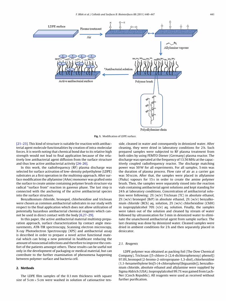
F. Bílek et al. / Colloids and Surfaces B: Biointerfaces 88 (2011) 440– 447 441
tion o
[tfsta
ssftrci
wrpn
rsXirafocb
2
s
Fig. 1. Modifica
21–23]. This kind of structure is suitable for reaction with antibac-erial agent molecule functionalities by creation of intra-molecularorces. It is worth noting that chemical bond due to its relative hightrength would not lead to final application because of the rela-ively low antibacterial agent diffusion from the surface structurend thus low active antibacterial activity [24–26].
In this work, the radiofrequency (RF) plasma discharge waselected for surface activation of low–density polyethylene (LDPE)ubstrates as a first operation in the multistep approach. After sur-ace modification the allylamine (AAm) monomer was grafted ontohe surface to create amine containing polymer brush structure viaadical “surface from” reaction in gaseous phase. The last step isonnected with the anchoring of the active antibacterial speciesnto the surface structure.
Benzalkonium chloride, bronopol, chlorhexidine and triclosanere chosen as common antibacterial substrates in our study with
espect to the final application which does not allow utilization ofotentially hazardous antibacterial chemical reagents which can-ot be used in direct contact with the body [6,27–29].
In this paper, the active antibacterial material multistep prepa-ation approach, surface characterization by contact angle mea-urements, ATR-TIR spectroscopy, Scanning electron microscopy,-ray Photoelectron Spectroscopy (XPS) and antibacterial assay
s described in order to prepare a novel active functional mate-ial which can bring a new potential in healthcare reducing themount of nosocomial infections and therefore to improve the com-ort of the patients amongst others. These results can be useful notnly in the development of packaging or medical material, but canontribute to the further examination of phenomena happeningetween polymer surface and bacteria cell.
. Methods
The LDPE film samples of the 0.1 mm thickness with squareize of 5 cm × 5 cm were washed in solution of cationactive ten-
f LDPE surface.
side, cleaned in water and consequently in deionized water. Aftercleaning, they were dried in laboratory conditions for 2 h. Suchprepared samples were subjected to RF plasma treatment fromboth sides by using FEMTO Diener (Germany) plasma reactor. Thedischarge was operated at the frequency of 13.56 MHz at the capac-itively coupled radiofrequency reactor. The discharge matchingpower was 50 W for all experiments. For all samples, 5 min wasthe duration of plasma process. Flow rate of air as a carrier gaswas 50 sccm. After that, the samples were placed in allylamine(Fluka) vapours for 15 s in order to create the amine polymerbrush. Then, the samples were separately rinsed into the reactionvials containing antibacterial agent solutions and kept standing for24 h at laboratory conditions. Concentration of antibacterial solu-tion were following: 2% (w/v) Triclosan (TC) in absolute ethanol,2% (w/v) bronopol (BrP) in absolute ethanol, 2% (w/v) benzalko-nium chloride (BCh) aq. solution, 2% (w/v) chlorhexidine (ChlH)in isopropylalcohol 70% (v/v) aq. solution. Finally, the sampleswere taken out of the solution and cleaned by stream of waterfollowed by ultrasonication for 5 min in deionized water to elimi-nate the unanchored antibacterial agent from sample surface. Thelast cleaning was done by deionized water. Cleaned samples weredried in ambient conditions for 2 h and then separately placed indesiccator.
2.1. Reagents
LDPE polymer was obtained as packing foil (The Dow ChemicalCompany), Triclosan ([5-chloro-2-(2,4-dichlorophenoxy) phenol])97.0%, bronopol (2-bromo-2-nitropropane-1,3-diol), chlorhexidine(1,1-hexamethylene bis[5-(4-chlorophenyl)biguanide]), benzalko-
nium chloride, absolute ethanol and allylamine were supplied bySigma Aldrich (USA). Isopropylalcohol 99.7% was gained from Lach-Ner (Czech Republic). All reagents were used as received withoutfurther purification.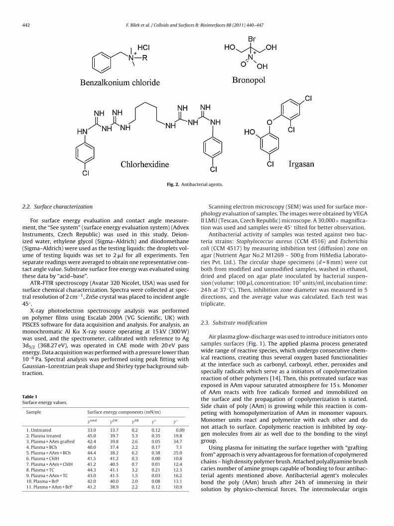
442 F. Bílek et al. / Colloids and Surfaces B: Biointerfaces 88 (2011) 440– 447
bacte
2
mIi(ustt
st4
oPmw3e1Gt
TS
Fig. 2. Anti
.2. Surface characterization
For surface energy evaluation and contact angle measure-ent, the “See system” (surface energy evaluation system) (Advex
nstruments, Czech Republic) was used in this study. Deion-zed water, ethylene glycol (Sigma–Aldrich) and diiodomethaneSigma–Aldrich) were used as the testing liquids: the droplets vol-me of testing liquids was set to 2 �l for all experiments. Teneparate readings were averaged to obtain one representative con-act angle value. Substrate surface free energy was evaluated usinghese data by “acid–base”.
ATR-FTIR spectroscopy (Avatar 320 Nicolet, USA) was used forurface chemical characterization. Spectra were collected at spec-ral resolution of 2 cm−1, ZnSe crystal was placed to incident angle5◦.
X-ray photoelectron spectroscopy analysis was performedn polymer films using Escalab 200A (VG Scientific, UK) withISCES software for data acquisition and analysis. For analysis, anonochromatic Al K� X-ray source operating at 15 kV (300 W)as used, and the spectrometer, calibrated with reference to Ag
d5/2 (368.27 eV), was operated in CAE mode with 20 eV passnergy. Data acquisition was performed with a pressure lower than
0−6 Pa. Spectral analysis was performed using peak fitting withaussian–Lorentzian peak shape and Shirley type background sub-raction.
able 1urface energy values.
Sample Surface energy components (mN/m)
� total �LW �AB �+ �−
1. Untreated 33.9 33.7 0.2 0.12 0.092. Plasma treated 45.0 39.7 5.3 0.35 19.83. Plasma + AAm grafted 42.4 39.8 2.6 0.05 34.74. Plasma + BCh 40.0 37.4 2.2 0.17 7.15. Plasma + AAm + BCh 44.4 38.2 6.2 0.38 25.06. Plasma + ChlH 41.5 41.2 0.3 0.00 10.87. Plasma + AAm + ChlH 41.2 40.5 0.7 0.01 12.48. Plasma + TC 44.3 41.1 3.2 0.21 12.39. Plasma + AAm + TC 43.0 41.5 1.5 0.03 16.210. Plasma + BrP 42.0 40.0 2.0 0.08 13.111. Plasma + AAm + BrP 41.2 38.9 2.2 0.12 10.9
rial agents.
Scanning electron microscopy (SEM) was used for surface mor-phology evaluation of samples. The images were obtained by VEGAII LMU (Tescan, Czech Republic) microscope. A 30,000× magnifica-tion was used and samples were 45◦ tilted for better observation.
Antibacterial activity of samples was tested against two bac-teria strains: Staphylococcus aureus (CCM 4516) and Escherichiacoli (CCM 4517) by measuring inhibition test (diffusion) zone onagar (Nutrient Agar No.2 M1269 – 500 g from HiMedia Laborato-ries Pvt. Ltd.). The circular shape specimens (d = 8 mm) were cutboth from modified and unmodified samples, washed in ethanol,dried and placed on agar plate inoculated by bacterial suspen-sion (volume: 100 �l, concentration: 107 units/ml, incubation time:24 h at 37 ◦C). Then, inhibition zone diameter was measured in 5directions, and the average value was calculated. Each test wastriplicate.
2.3. Substrate modification
Air plasma glow-discharge was used to introduce initiators ontosamples surfaces (Fig. 1). The applied plasma process generatedwide range of reactive species, which undergo consecutive chem-ical reactions, creating thus several oxygen based functionalitiesat the interface such as carbonyl, carboxyl, ether, peroxides andspecially radicals which serve as a initiators of copolymerizationreaction of other polymers [14]. Then, this pretreated surface wasexposed in AAm vapour saturated atmosphere for 15 s. Monomerof AAm reacts with free radicals formed and immobilized onthe surface and the propagation of copolymerization is started.Side chain of poly (AAm) is growing while this reaction is com-peting with homopolymerization of AAm in monomer vapours.Monomer units react and polymerize with each other and donot attach to surface. Copolymeric reaction is inhibited by oxy-gen molecules from air as well due to the bonding to the vinylgroup.
Using plasma for initiating the surface together with “graftingfrom” approach is very advantageous for formation of copolymeredchains – high density polymer brush. Attached polyallyamine brush
caries number of amine groups capable of bonding to four antibac-terial agents mentioned above. Antibacterial agent’s moleculesbond the poly (AAm) brush after 24 h of immersing in theirsolution by physico-chemical forces. The intermolecular origin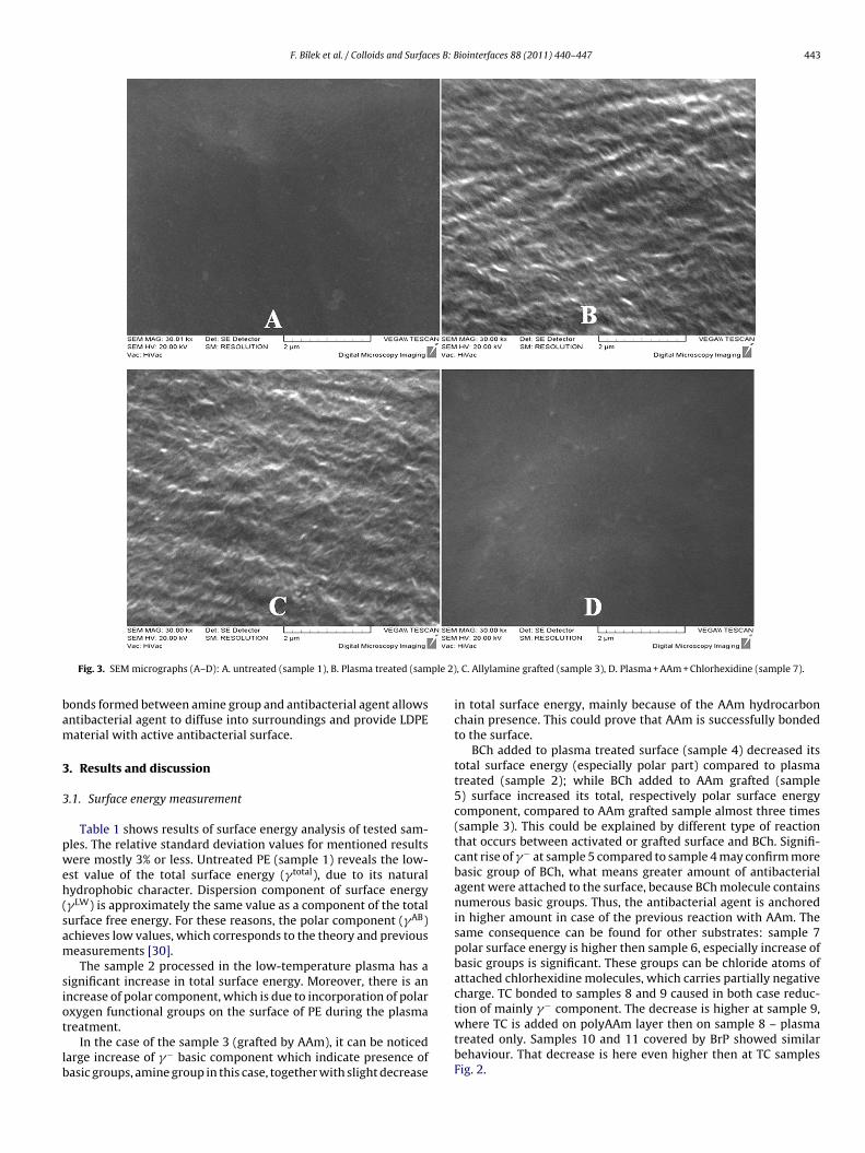
F. Bílek et al. / Colloids and Surfaces B: Biointerfaces 88 (2011) 440– 447 443
ple 2)
bam
3
3
pweh(sam
siot
lb
Fig. 3. SEM micrographs (A–D): A. untreated (sample 1), B. Plasma treated (sam
onds formed between amine group and antibacterial agent allowsntibacterial agent to diffuse into surroundings and provide LDPEaterial with active antibacterial surface.
. Results and discussion
.1. Surface energy measurement
Table 1 shows results of surface energy analysis of tested sam-les. The relative standard deviation values for mentioned resultsere mostly 3% or less. Untreated PE (sample 1) reveals the low-
st value of the total surface energy (� total), due to its naturalydrophobic character. Dispersion component of surface energy�LW) is approximately the same value as a component of the totalurface free energy. For these reasons, the polar component (�AB)chieves low values, which corresponds to the theory and previouseasurements [30].The sample 2 processed in the low-temperature plasma has a
ignificant increase in total surface energy. Moreover, there is anncrease of polar component, which is due to incorporation of polarxygen functional groups on the surface of PE during the plasma
reatment.In the case of the sample 3 (grafted by AAm), it can be noticedarge increase of �− basic component which indicate presence ofasic groups, amine group in this case, together with slight decrease
, C. Allylamine grafted (sample 3), D. Plasma + AAm + Chlorhexidine (sample 7).
in total surface energy, mainly because of the AAm hydrocarbonchain presence. This could prove that AAm is successfully bondedto the surface.
BCh added to plasma treated surface (sample 4) decreased itstotal surface energy (especially polar part) compared to plasmatreated (sample 2); while BCh added to AAm grafted (sample5) surface increased its total, respectively polar surface energycomponent, compared to AAm grafted sample almost three times(sample 3). This could be explained by different type of reactionthat occurs between activated or grafted surface and BCh. Signifi-cant rise of �− at sample 5 compared to sample 4 may confirm morebasic group of BCh, what means greater amount of antibacterialagent were attached to the surface, because BCh molecule containsnumerous basic groups. Thus, the antibacterial agent is anchoredin higher amount in case of the previous reaction with AAm. Thesame consequence can be found for other substrates: sample 7polar surface energy is higher then sample 6, especially increase ofbasic groups is significant. These groups can be chloride atoms ofattached chlorhexidine molecules, which carries partially negativecharge. TC bonded to samples 8 and 9 caused in both case reduc-tion of mainly �− component. The decrease is higher at sample 9,
where TC is added on polyAAm layer then on sample 8 – plasmatreated only. Samples 10 and 11 covered by BrP showed similarbehaviour. That decrease is here even higher then at TC samplesFig. 2.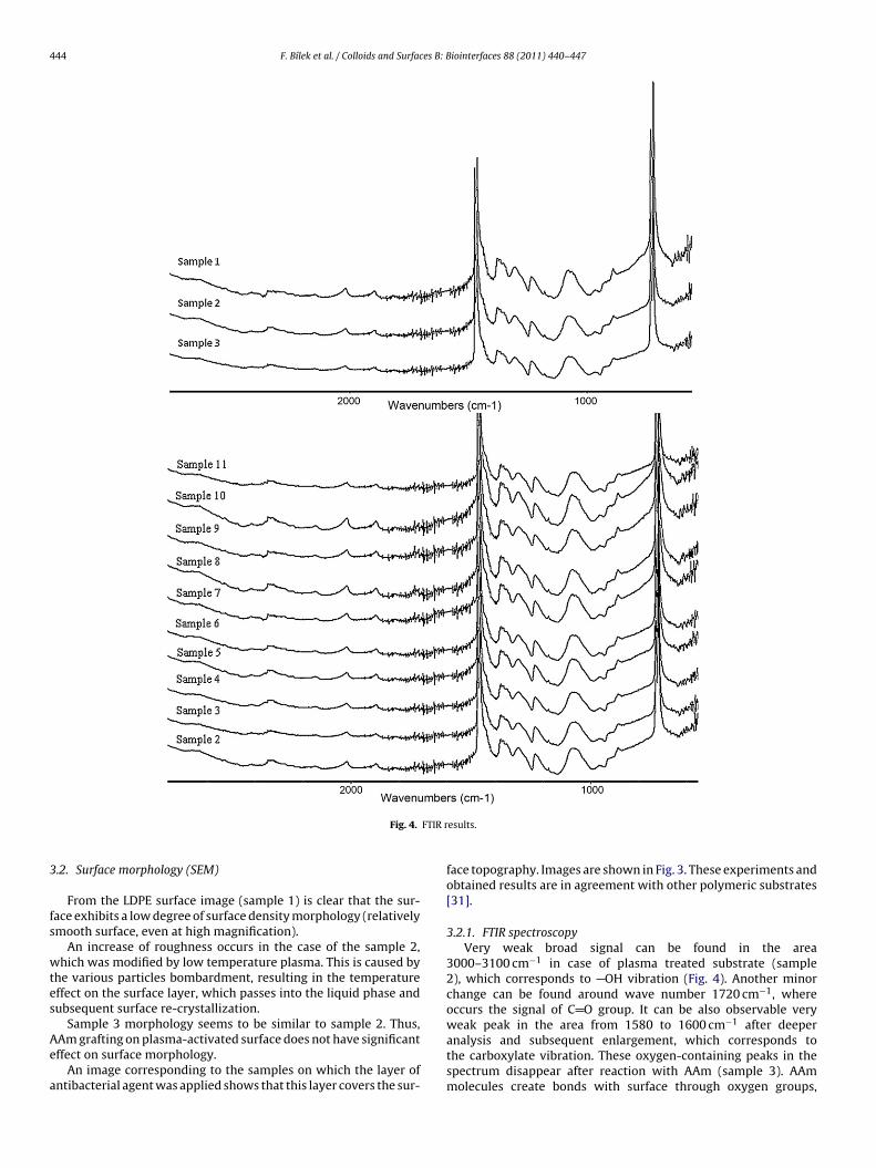
444 F. Bílek et al. / Colloids and Surfaces B: Biointerfaces 88 (2011) 440– 447
FTIR r
3
fs
wtes
Ae
a
Fig. 4.
.2. Surface morphology (SEM)
From the LDPE surface image (sample 1) is clear that the sur-ace exhibits a low degree of surface density morphology (relativelymooth surface, even at high magnification).
An increase of roughness occurs in the case of the sample 2,hich was modified by low temperature plasma. This is caused by
he various particles bombardment, resulting in the temperatureffect on the surface layer, which passes into the liquid phase andubsequent surface re-crystallization.
Sample 3 morphology seems to be similar to sample 2. Thus,
Am grafting on plasma-activated surface does not have significantffect on surface morphology.An image corresponding to the samples on which the layer ofntibacterial agent was applied shows that this layer covers the sur-
esults.
face topography. Images are shown in Fig. 3. These experiments andobtained results are in agreement with other polymeric substrates[31].
3.2.1. FTIR spectroscopyVery weak broad signal can be found in the area
3000–3100 cm−1 in case of plasma treated substrate (sample2), which corresponds to OH vibration (Fig. 4). Another minorchange can be found around wave number 1720 cm−1, whereoccurs the signal of C O group. It can be also observable veryweak peak in the area from 1580 to 1600 cm−1 after deeper
analysis and subsequent enlargement, which corresponds tothe carboxylate vibration. These oxygen-containing peaks in thespectrum disappear after reaction with AAm (sample 3). AAmmolecules create bonds with surface through oxygen groups,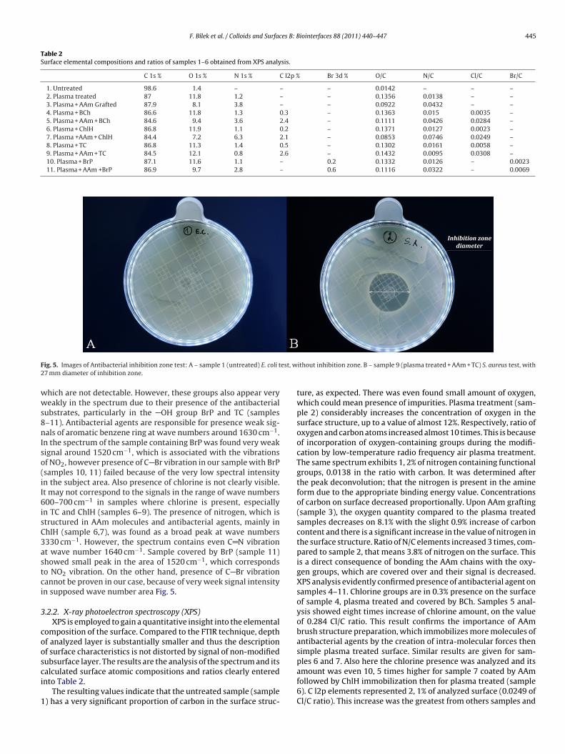
F. Bílek et al. / Colloids and Surfaces B: Biointerfaces 88 (2011) 440– 447 445
Table 2Surface elemental compositions and ratios of samples 1–6 obtained from XPS analysis.
C 1s % O 1s % N 1s % C l2p % Br 3d % O/C N/C Cl/C Br/C
1. Untreated 98.6 1.4 – – – 0.0142 – – –2. Plasma treated 87 11.8 1.2 – – 0.1356 0.0138 – –3. Plasma + AAm Grafted 87.9 8.1 3.8 – – 0.0922 0.0432 – –4. Plasma + BCh 86.6 11.8 1.3 0.3 – 0.1363 0.015 0.0035 –5. Plasma + AAm + BCh 84.6 9.4 3.6 2.4 – 0.1111 0.0426 0.0284 –6. Plasma + ChlH 86.8 11.9 1.1 0.2 – 0.1371 0.0127 0.0023 –7. Plasma +AAm + ChlH 84.4 7.2 6.3 2.1 – 0.0853 0.0746 0.0249 –8. Plasma + TC 86.8 11.3 1.4 0.5 – 0.1302 0.0161 0.0058 –9. Plasma + AAm + TC 84.5 12.1 0.8 2.6 – 0.1432 0.0095 0.0308 –10. Plasma + BrP 87.1 11.6 1.1 – 0.2 0.1332 0.0126 – 0.002311. Plasma + AAm +BrP 86.9 9.7 2.8 – 0.6 0.1116 0.0322 – 0.0069
F est, w2
wws8nIso(iI6isC3astci
3
coosci
1
ig. 5. Images of Antibacterial inhibition zone test: A – sample 1 (untreated) E. coli t7 mm diameter of inhibition zone.
hich are not detectable. However, these groups also appear veryeakly in the spectrum due to their presence of the antibacterial
ubstrates, particularly in the OH group BrP and TC (samples–11). Antibacterial agents are responsible for presence weak sig-als of aromatic benzene ring at wave numbers around 1630 cm−1.
n the spectrum of the sample containing BrP was found very weakignal around 1520 cm−1, which is associated with the vibrationsf NO2, however presence of C Br vibration in our sample with BrPsamples 10, 11) failed because of the very low spectral intensityn the subject area. Also presence of chlorine is not clearly visible.t may not correspond to the signals in the range of wave numbers00–700 cm−1 in samples where chlorine is present, especially
n TC and ChlH (samples 6–9). The presence of nitrogen, which istructured in AAm molecules and antibacterial agents, mainly inhlH (sample 6,7), was found as a broad peak at wave numbers330 cm−1. However, the spectrum contains even C N vibrationt wave number 1640 cm−1. Sample covered by BrP (sample 11)howed small peak in the area of 1520 cm−1, which correspondso NO2 vibration. On the other hand, presence of C Br vibrationannot be proven in our case, because of very week signal intensityn supposed wave number area Fig. 5.
.2.2. X-ray photoelectron spectroscopy (XPS)XPS is employed to gain a quantitative insight into the elemental
omposition of the surface. Compared to the FTIR technique, depthf analyzed layer is substantially smaller and thus the descriptionf surface characteristics is not distorted by signal of non-modifiedubsurface layer. The results are the analysis of the spectrum and its
alculated surface atomic compositions and ratios clearly enterednto Table 2.The resulting values indicate that the untreated sample (sample) has a very significant proportion of carbon in the surface struc-
ithout inhibition zone. B – sample 9 (plasma treated + AAm + TC) S. aureus test, with
ture, as expected. There was even found small amount of oxygen,which could mean presence of impurities. Plasma treatment (sam-ple 2) considerably increases the concentration of oxygen in thesurface structure, up to a value of almost 12%. Respectively, ratio ofoxygen and carbon atoms increased almost 10 times. This is becauseof incorporation of oxygen-containing groups during the modifi-cation by low-temperature radio frequency air plasma treatment.The same spectrum exhibits 1, 2% of nitrogen containing functionalgroups, 0.0138 in the ratio with carbon. It was determined afterthe peak deconvolution; that the nitrogen is present in the amineform due to the appropriate binding energy value. Concentrationsof carbon on surface decreased proportionally. Upon AAm grafting(sample 3), the oxygen quantity compared to the plasma treatedsamples decreases on 8.1% with the slight 0.9% increase of carboncontent and there is a significant increase in the value of nitrogen inthe surface structure. Ratio of N/C elements increased 3 times, com-pared to sample 2, that means 3.8% of nitrogen on the surface. Thisis a direct consequence of bonding the AAm chains with the oxy-gen groups, which are covered over and their signal is decreased.XPS analysis evidently confirmed presence of antibacterial agent onsamples 4–11. Chlorine groups are in 0.3% presence on the surfaceof sample 4, plasma treated and covered by BCh. Samples 5 anal-ysis showed eight times increase of chlorine amount, on the valueof 0.284 Cl/C ratio. This result confirms the importance of AAmbrush structure preparation, which immobilizes more molecules ofantibacterial agents by the creation of intra-molecular forces thensimple plasma treated surface. Similar results are given for sam-ples 6 and 7. Also here the chlorine presence was analyzed and its
amount was even 10, 5 times higher for sample 7 coated by AAmfollowed by ChlH immobilization then for plasma treated (sample6). C l2p elements represented 2, 1% of analyzed surface (0.0249 ofCl/C ratio). This increase was the greatest from others samples and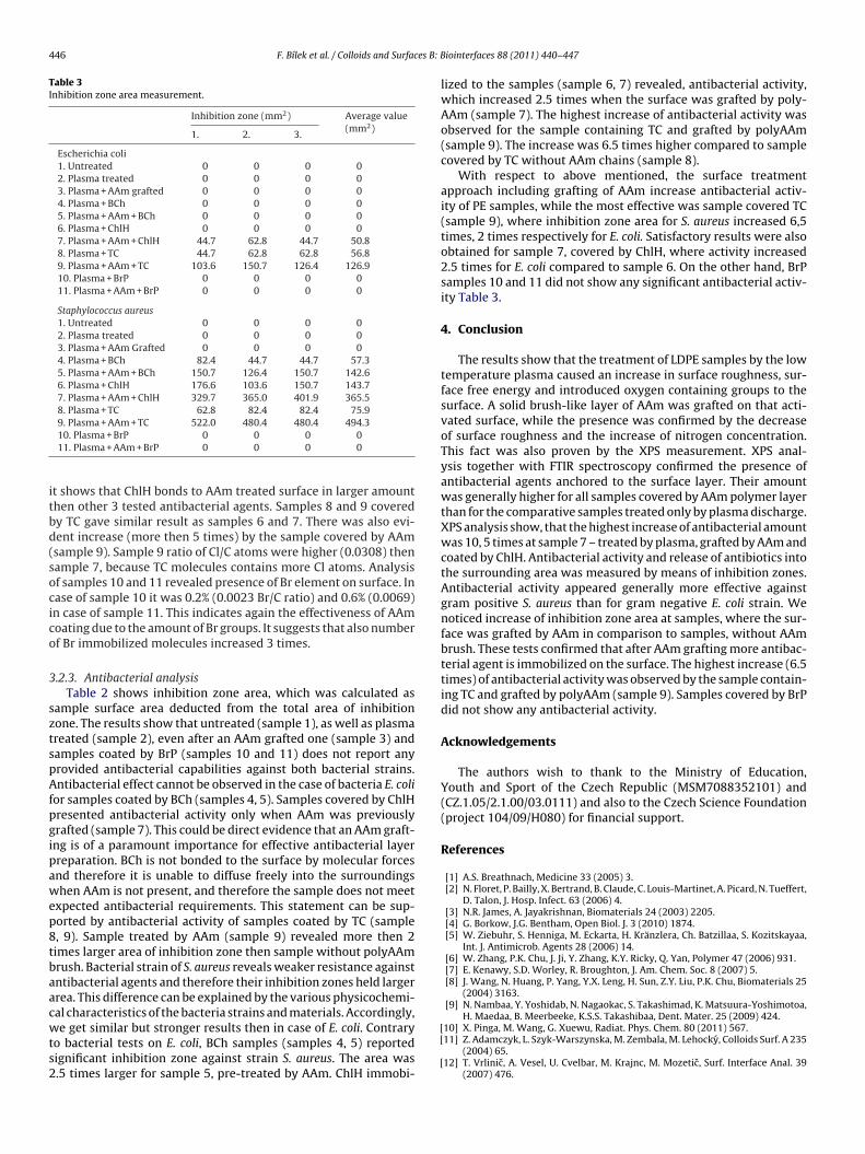
446 F. Bílek et al. / Colloids and Surfaces B: B
Table 3Inhibition zone area measurement.
Inhibition zone (mm2) Average value(mm2)
1. 2. 3.
Escherichia coli1. Untreated 0 0 0 02. Plasma treated 0 0 0 03. Plasma + AAm grafted 0 0 0 04. Plasma + BCh 0 0 0 05. Plasma + AAm + BCh 0 0 0 06. Plasma + ChlH 0 0 0 07. Plasma + AAm + ChlH 44.7 62.8 44.7 50.88. Plasma + TC 44.7 62.8 62.8 56.89. Plasma + AAm + TC 103.6 150.7 126.4 126.910. Plasma + BrP 0 0 0 011. Plasma + AAm + BrP 0 0 0 0
Staphylococcus aureus1. Untreated 0 0 0 02. Plasma treated 0 0 0 03. Plasma + AAm Grafted 0 0 0 04. Plasma + BCh 82.4 44.7 44.7 57.35. Plasma + AAm + BCh 150.7 126.4 150.7 142.66. Plasma + ChlH 176.6 103.6 150.7 143.77. Plasma + AAm + ChlH 329.7 365.0 401.9 365.58. Plasma + TC 62.8 82.4 82.4 75.99. Plasma + AAm + TC 522.0 480.4 480.4 494.3
itbd(socico
3
sztspAfpgipawep8tbaacwts2
10. Plasma + BrP 0 0 0 011. Plasma + AAm + BrP 0 0 0 0
t shows that ChlH bonds to AAm treated surface in larger amounthen other 3 tested antibacterial agents. Samples 8 and 9 coveredy TC gave similar result as samples 6 and 7. There was also evi-ent increase (more then 5 times) by the sample covered by AAmsample 9). Sample 9 ratio of Cl/C atoms were higher (0.0308) thenample 7, because TC molecules contains more Cl atoms. Analysisf samples 10 and 11 revealed presence of Br element on surface. Inase of sample 10 it was 0.2% (0.0023 Br/C ratio) and 0.6% (0.0069)n case of sample 11. This indicates again the effectiveness of AAmoating due to the amount of Br groups. It suggests that also numberf Br immobilized molecules increased 3 times.
.2.3. Antibacterial analysisTable 2 shows inhibition zone area, which was calculated as
ample surface area deducted from the total area of inhibitionone. The results show that untreated (sample 1), as well as plasmareated (sample 2), even after an AAm grafted one (sample 3) andamples coated by BrP (samples 10 and 11) does not report anyrovided antibacterial capabilities against both bacterial strains.ntibacterial effect cannot be observed in the case of bacteria E. coli
or samples coated by BCh (samples 4, 5). Samples covered by ChlHresented antibacterial activity only when AAm was previouslyrafted (sample 7). This could be direct evidence that an AAm graft-ng is of a paramount importance for effective antibacterial layerreparation. BCh is not bonded to the surface by molecular forcesnd therefore it is unable to diffuse freely into the surroundingshen AAm is not present, and therefore the sample does not meet
xpected antibacterial requirements. This statement can be sup-orted by antibacterial activity of samples coated by TC (sample, 9). Sample treated by AAm (sample 9) revealed more then 2imes larger area of inhibition zone then sample without polyAAmrush. Bacterial strain of S. aureus reveals weaker resistance againstntibacterial agents and therefore their inhibition zones held largerrea. This difference can be explained by the various physicochemi-al characteristics of the bacteria strains and materials. Accordingly,
e get similar but stronger results then in case of E. coli. Contraryo bacterial tests on E. coli, BCh samples (samples 4, 5) reportedignificant inhibition zone against strain S. aureus. The area was.5 times larger for sample 5, pre-treated by AAm. ChlH immobi-
[[
[
iointerfaces 88 (2011) 440– 447
lized to the samples (sample 6, 7) revealed, antibacterial activity,which increased 2.5 times when the surface was grafted by poly-AAm (sample 7). The highest increase of antibacterial activity wasobserved for the sample containing TC and grafted by polyAAm(sample 9). The increase was 6.5 times higher compared to samplecovered by TC without AAm chains (sample 8).
With respect to above mentioned, the surface treatmentapproach including grafting of AAm increase antibacterial activ-ity of PE samples, while the most effective was sample covered TC(sample 9), where inhibition zone area for S. aureus increased 6,5times, 2 times respectively for E. coli. Satisfactory results were alsoobtained for sample 7, covered by ChlH, where activity increased2.5 times for E. coli compared to sample 6. On the other hand, BrPsamples 10 and 11 did not show any significant antibacterial activ-ity Table 3.
4. Conclusion
The results show that the treatment of LDPE samples by the lowtemperature plasma caused an increase in surface roughness, sur-face free energy and introduced oxygen containing groups to thesurface. A solid brush-like layer of AAm was grafted on that acti-vated surface, while the presence was confirmed by the decreaseof surface roughness and the increase of nitrogen concentration.This fact was also proven by the XPS measurement. XPS anal-ysis together with FTIR spectroscopy confirmed the presence ofantibacterial agents anchored to the surface layer. Their amountwas generally higher for all samples covered by AAm polymer layerthan for the comparative samples treated only by plasma discharge.XPS analysis show, that the highest increase of antibacterial amountwas 10, 5 times at sample 7 – treated by plasma, grafted by AAm andcoated by ChlH. Antibacterial activity and release of antibiotics intothe surrounding area was measured by means of inhibition zones.Antibacterial activity appeared generally more effective againstgram positive S. aureus than for gram negative E. coli strain. Wenoticed increase of inhibition zone area at samples, where the sur-face was grafted by AAm in comparison to samples, without AAmbrush. These tests confirmed that after AAm grafting more antibac-terial agent is immobilized on the surface. The highest increase (6.5times) of antibacterial activity was observed by the sample contain-ing TC and grafted by polyAAm (sample 9). Samples covered by BrPdid not show any antibacterial activity.
Acknowledgements
The authors wish to thank to the Ministry of Education,Youth and Sport of the Czech Republic (MSM7088352101) and(CZ.1.05/2.1.00/03.0111) and also to the Czech Science Foundation(project 104/09/H080) for financial support.
References
[1] A.S. Breathnach, Medicine 33 (2005) 3.[2] N. Floret, P. Bailly, X. Bertrand, B. Claude, C. Louis-Martinet, A. Picard, N. Tueffert,
D. Talon, J. Hosp. Infect. 63 (2006) 4.[3] N.R. James, A. Jayakrishnan, Biomaterials 24 (2003) 2205.[4] G. Borkow, J.G. Bentham, Open Biol. J. 3 (2010) 1874.[5] W. Ziebuhr, S. Henniga, M. Eckarta, H. Kränzlera, Ch. Batzillaa, S. Kozitskayaa,
Int. J. Antimicrob. Agents 28 (2006) 14.[6] W. Zhang, P.K. Chu, J. Ji, Y. Zhang, K.Y. Ricky, Q. Yan, Polymer 47 (2006) 931.[7] E. Kenawy, S.D. Worley, R. Broughton, J. Am. Chem. Soc. 8 (2007) 5.[8] J. Wang, N. Huang, P. Yang, Y.X. Leng, H. Sun, Z.Y. Liu, P.K. Chu, Biomaterials 25
(2004) 3163.[9] N. Nambaa, Y. Yoshidab, N. Nagaokac, S. Takashimad, K. Matsuura-Yoshimotoa,
H. Maedaa, B. Meerbeeke, K.S.S. Takashibaa, Dent. Mater. 25 (2009) 424.
10] X. Pinga, M. Wang, G. Xuewu, Radiat. Phys. Chem. 80 (2011) 567.11] Z. Adamczyk, L. Szyk-Warszynska, M. Zembala, M. Lehocky, Colloids Surf. A 235(2004) 65.12] T. Vrlinic, A. Vesel, U. Cvelbar, M. Krajnc, M. Mozetic, Surf. Interface Anal. 39
(2007) 476.
es B: B
[[
[
[[[
[[
[
[
[
[
[
[
[
[
F. Bílek et al. / Colloids and Surfac
13] A. Vesel, M. Mozetic, A. Zalar, Surf. Interface Anal. 40 (2008) 661.14] A. Vesel, I. Junkar, U. Cvelbar, J. Kovac, M. Mozetic, Surf. Interface Anal. 40 (2008)
1444.15] M. Lehocky, L. Lapcík, R. Dlabaja, L. Rachunek, J. Stoch, Czech. J. Phys. 54 (2004)
C533.16] M. Lehocky, A. Mrácek, Czech. J. Phys. 56 (2006) B1277.17] M. Mozetic, J. Phys. D-Appl. Phys. 44 (17) (2011) 174028.18] M. Sowe, I. Novák, A. Vesel, I. Junkar, M. Lehocky, P. Sáha, I. Chodák, Int. J. Polym.
Anal. Charact. 14 (2009) 641.19] A. Doliska, M. Kolar, Mater. Technol. 45 (2011) 275.20] U. Maver, T. Maver, A. Znidarsic, Z. Persin, M. Gaberscek, K. Stana-Kleinschek,
Mater. Technol. 45 (2011) 259.21] M. Lehocky, P. St’ahel, M. Koutny, J. Cech, J. Institoris, A. Mrácek, J. Mater.
Process. Technol. 209 (2009) 2871.22] M. Lehocky, P.F.F. Amaral, P. St’ahel, M.A.Z. Coelho, A.M. Barros-Timmons, J.A.P.
Coutinho, Surf. Eng. 24 (2008) 23.
[
[[
iointerfaces 88 (2011) 440– 447 447
23] M. Lehocky, P.F.F. Amaral, P. St’ahel, M.A.Z. Coelho, A.M. Barros-Timmons, J.A.P.Coutinho, J. Chem. Technol. Biotechnol. 82 (2007) 360.
24] A. Asadinezhad, I. Novák, M. Lehocky, V. Sedlarík, A. Vesel, I. Junkar, P. Sáha, I.Chodák, Colloids Surf. B 77 (2010) 246.
25] Asadinezhad, I. Novák, M. Lehocky, V. Sedlarík, A. Vesel, I. Junkar, P. Sáha, I.Chodák, Plasma Processes Polym. 7 (2010) 504.
26] A. Asadinezhad, I. Novák, M. Lehocky, F. Bílek, A. Vesel, I. Junkar, P. Sáha, A.Popelka, Molecules 15 (2010) 1007.
27] Y. Xue, Y. Hieda, Y. Saito, T. Nomura, J. Fujihara, K. Takayama, K. Kimura, H.Takeshita, Toxicol. Lett. 148 (2004) 113.
28] B. Marple, P. Roland, M. Beninnger, Otolaryngol. Head Neck Surg. 130 (2004)
131.29] J. Truin, H.M. van Rijkom, J. Mulder, M.A. vant Hof, Dent. Res. 75 (1996)790.
30] C.S. Ren, K. Wang, Q.Y. Nie, D.Z. Wang, S.H. Guo, Appl. Surf. Sci. 255 (2008) 3421.31] J. Hu, C. Yin, H.Q. Mao, K. Tamada, W. Knoll, Adv. Funct. Mater. 13 (2003) 692.


PAPER II
Molecules 2012, 17, 762-785; doi:10.3390/molecules17010762
molecules ISSN 1420-3049
www.mdpi.com/journal/molecules
Article
Anti-bacterial Treatment of Polyethylene by Cold Plasma for Medical Purposes
Anton Popelka 1, Igor Novák 1,*, Marián Lehocký 2, Ivan Chodák 1, Ján Sedliačik 3,
Milada Gajtanska 3, Mariana Sedliačiková 3, Alenka Vesel 4, Ita Junkar 4, Angela Kleinová 1,
Milena Špírková 5 and František Bílek 6
1 Polymer Institute, Slovak Academy of Sciences, Dúbravská cesta 9, 845 41 Bratislava 45, Slovakia 2 Centre of Polymer Systems, University Institute, Tomas Bata University in Zlín,
Nad Ovčírnou 3685, 760 01 Zlín, Czech Republic 3 Faculty of Wood Sciences and Technology, Technical University in Zvolen,
T.G. Masaryka 2117/24, 960 53 Zvolen, Slovakia 4 Plasma Laboratory, Department of Surface Engineering, Jožef Stefan Institute, Jamova cesta 39,
SI-1000 Ljubljana, Slovenia 5 Institute of Macromolecular Chemistry AS CR, v. v. i, Heyrovsky Sq. 2, 162 06 Prague 6,
Czech Republic 6 Polymer Centre, Faculty of Technology, Tomas Bata University in Zlín, T.G.M Sq. 275,
762 72 Zlín, Czech Republic
* Author to whom correspondence should be addressed: E-Mail: [email protected];
Tel.: +421-903-925-725.
Received: 16 November 2011; in revised form: 9 January 2012 / Accepted: 10 January 2012 /
Published: 13 January 2012
Abstract: Polyethylene (PE) is one of the most widely used polymers in many industrial
applications. Biomedical uses seem to be attractive, with increasing interest. However, PE
it prone to infections and its additional surface treatment is indispensable. An increase in
resistance to infections can be achieved by treating PE surfaces with substances containing
antibacterial groups such as triclosan (5-Chloro-2-(2,4-dichlorophenoxy)phenol) and
chlorhexidine (1,1'-Hexamethylenebis[5-(4-chlorophenyl)biguanide]). This work has
examined the impact of selected antibacterial substances immobilized on low-density
polyethylene (LDPE) via polyacrylic acid (PAA) grafted on LDPE by low-temperature
barrier discharge plasma. This LDPE surface treatment led to inhibition of Escherichia coli
and Staphylococcus aureus adhesion; the first causes intestinal disease, peritonitis, mastitis,
pneumonia, septicemia, the latter is the reason for wound and urinary tract infections.
OPEN ACCESS
Molecules 2012, 17 763
Keywords: polyethylene; grafting; plasma treatment; immobilization; triclosan;
chlorhexidine; acrylic acid
1. Introduction
PE is one of the most common biomedical polymers due to its excellent mechanical properties, but
it suffers from insufficient biocompatibility and bioactivity [1]. PE is widely used in many biomedical
applications including the production of catheters for percutaneous transluminal coronary angioplasty
in medical and pharmaceutical industries [2], but infections resulting from application of this medical
polymer represent the main clinical complication [3]. These infections may cause implant failure,
complex revision processes and implant removal, and all can lead to patient suffering, prolonged
hospitalization and even death in some cases [4]. Biocompatibility depends on many surface
characteristics such as wettability [5], roughness, chemistry, surface charge, density of functional
groups. The presence of hydrophobic and hydrophilic domains [6], charge [7], the functional group
densities, and their conformation [8,9] play ascendant roles in affecting cell behavior [10]. Although
PE has superiority concerning volume properties, its surface free energy has a low value that reflects
its low wettability. This property is related to its hydrophobic and chemically inert surface without
polar functional groups [11]. The solution of the problem consists in PE surface modification.
Low-temperature plasma can be suggested as the appropriate procedure for the hydrophilization of the
surface. Due to the plasma treatment surface the free energy is increased as a result of introduction of
polar functional groups on the treated surface, thus making the surface of PE more hydrophilic [12,13].
Developing plasma techniques belong to an important class of polymer surface modification
techniques where a very thin layer of the polymer surface is treated without any changes in bulk.
Moreover, plasma technology is based on ecological, clean and dry processes suitable for industrial
applications without the use of chemicals. Low-temperature plasma is often used in many applications,
for example in the electronic, aeronautic, automotive, medical [14], biomedical, textile, optical and
paper industries [15]. In this process a polymer is exposed to a plasma reactive species such as ions,
electrons, excited atoms and molecules, which cleave existing chemical bonds and form new reactive
functional groups, which may initiate or participate in grafting, polymerization, or cross-linking
reactions on the surface. Plasma processing can significantly contribute to adhesion improvement by
removing surface contamination and to surface morphology changes through increased roughness due
to etching [16,17].
The Diffuse Coplanar Surface Barrier Discharge (DCSBD) plasma generator [18] appears to be an
effective tool for creating macroscopically homogeneous plasmas, which has many advantages
compared with conventional devices. The most important advantage considering the application of
DCSBD includes performance at atmospheric pressure, which is significant in terms of continuous
industrial technologies. Another advantage is that the plasma does not directly contact the electrodes,
which protects these from wear [19]. DCSBD equipment consists of two parallel banded system of
electrodes (usually 1-mm wide, 50 micron thick, with 0.5 mm spacing between the strips, made of

Molecules 2012, 17 764
Ag-paste) embedded in 96% Al2O3-promotion of natural purity. Such an arrangement of electrodes
leads to a visually almost completely macroscopically homogeneous diffusion plasma [20–22].
Adhesion and surface growth of bacteria, also called biofilm formation, is a widespread
problem [23]. To prevent its formation, anti-infection modification of polymers for medical
applications may be applied. Anti-infective properties of polymers can be achieved by following:
(a) anti-infection agents mixed in the polymer; (b) copolymerization anti-infection agents with
monomer; (c) appropriate surface treatment of medical polymers.
Antibacterial surface modification is controlled by the physical-chemical interactions between
bacteria and polymer surface. This treatment has several advantages, because it does not influence the
bulk properties of the polymer, antibacterial agents are not released from the polymer volume, and the
technique is relative simple and effective. Triclosan [24] and chlorhexidine [25] (Figure 1) shows
straight, steady, broad-spectral antibacterial efficiency and very low clinical toxicity in clinical tests.
This treatment in combination with plasma can affect significantly biochemical and physical properties
of LDPE [3] by following a multistep physicochemical approach [26]. In the first step, formation of
functional groups on the polymer surface is necessary via the plasma species created by a DCSBD
generator [27–29].
Figure 1. (a) triclosan; (b) chlorhexidine; (c) 3D structure of triclosan; (d) 3D structure
of chlorhexidine.
(a) (b)
(c) (d)
In the second step, an end-functionalized polymer brush is formed on polymer surface via radical
graft polymerization of acrylic acid (AA), which is anchored on the plasma treated surface [30]. The
PAA grafted on the LDPE surface represents a new approach for subsequent antibacterial treatment.
Finally, biomolecules are immobilized on this pre-treated surface using EDAC coupling, whereby
carboxyl groups of AA are then activated and ready to provide the immobilization sites [31].
Molecules 2012, 17 765
2. Results and Discussion
2.1. Surface Wettability
Wetting (wettability) can be defined as the degree to which a solid is wetted. When a drop is totally
spread on solid surface and the contact angle approaches 0 deg, then the complete wettability of the
surface is achieved. However, in many cases it is only a partial wettability occurs (or non-wettability).
Contact angle measurements are usually used for estimating the extent to which a solid surface will be
wetted. Wettability can be expressed as relative strength of cohesion (liquid/liquid) and adhesion
(solid/liquid) forces. Weak cohesion with strong adhesion due to the very low contact angle is close to
full wettability. If solid/liquid interactions decrease and liquid/liquid interactions increase, wettability
decreases. If the contact angle of water is 90 deg or more the polymeric surface is hydrophobic. This
relates with poor wettability, low surface energy and weak adhesion of the polymer. On the other side
drop with a small contact angle relates to more hydrophilic surface, that causes better wettability,
adhesion and higher surface energy of investigated material. The contact angles changes of testing
liquid set, graft yield (GY), and surface free energy (γtot) and its components of antibacterial treated
LDPE are shown in Table 1. The graft yield (GY) was calculated by the following equation:
100/[%] 112 WWWGY , where W1 and W2 represent the weight of the samples before and after
surface treatment [32]. The graphic changes of contact angles of testing liquids caused by antibacterial
treatment are shown in Figure 2. The water contact angle (θw) of untreated LDPE (Sample 1) achieves the
highest values from the all samples because it is polymer with hydrophobic and chemical inert surface.
θw significantly decreased after plasma effect of the Sample 2 when different functional groups were
introduced on to the surface formed from plasma species and therefore the treated surface acquired
more polar or hydrophilic character. The highest decrease of the contact angle was observed in case of
surface covered by polyacrylic acid (PAA, Sample 3) which corresponds to its hydrophilic character.
Also triclosan (Sample 4) and chlorhexidine (Sample 5) immobilization led to θw decrease. For
investigation of other physicochemical parameters of the treated surface Lifshitz-Van der Waals/
acid-base (LW/AB) theory was used, which allows to obtain γtot and its components such as non-polar
LW (γLW) and polar AB (γAB) components. LW indicates the total dispersive Lifshitz-Van der Waals
interaction and AB refers to the acid-base or electron-acceptor/electron donor interaction according to
Lewis [33]. LDPE belongs to group of low-energy polymeric materials and therefore γtot of Sample 1
achieves very low values which correspond with difficulties during processing, such as dyeing,
printing and bonding (low adhesion). This can be removed by plasma treatment of LDPE when γtot can
significantly increases as in the case of Sample 2. The largest increase of γtot and γAB was observed for
Sample 3 due to highest polarity in comparison with other samples as a result of polar oxygen group’s
presence. Sample 4 and 5 showed similar increases of surface free energy values, thereby confirming
the increase in wettability.
Molecules 2012, 17 766
Table 1. Surface properties of LDPE treated by multistep process.
Sample θw (°) θe (°) θg (°) θd (°) θf (°) γ−
(mN/m)
γ+
(mN/m)
γAB
(mN/m)
γLW
(mN/m)
γtot
(mN/m)
GY
(%)
1 99.2
(±0.6)
70.9
(±1.2)
85.3
(±0.9)
48.4
(±1.2)
80.7
(±0.9)
1.0 0.1 0.7 34.5 35.2 -
2 77.5
(±1.1)
51.0
(±2.8)
67.1
(±2.8)
36.0
(±1.2)
52.8
(±1.5)
6.6 0.1 1.1 41.4 42.6 0.0
3 66.9
(±0.7)
32.1
(±2.4)
57.2
(±2.7)
32.5
(±1.6)
37.0
(±2.0)
10.4 0.5 4.5 43.7 48.1 0.5
4 75.8
(±1.6)
36.1
(±0.7)
60.4
(±1.0)
30.5
(±1.5)
48.3
(±1.2)
5.0 0.4 2.8 44.0 46.8 1.8
5 76.7
(±0.5)
38.1
(±2.5)
63.2
(±2.72)
30.0
(±1.6)
50.4
(±1.5)
5.2 0.2 2.0 44.4 46.4 2.0
w = deionized water, e = ethylene glycol, g = glycerol, d = diiodomethane, f = formamide;
* Sample 1: untreated LDPE; Sample 2: plasma-treated; Sample 3: AA grafted; Sample 4: triclosan
coated; Sample 5: chlorhexidine coated.
Figure 2. Contact angle vs. surface treatment and vs. testing liquid; 1 - untreated LDPE;
2 - plasma-treated; 3 - AA grafted; 4 - triclosan coated; 5 - chlorhexidine coated.
2.2. Adhesive Properties
The results of peel strength measurements of adhesive joint to poly(acrylate) are shown in Figure 3.
Surface free energy changes are closely related to adhesion between two materials in contact.
Therefore, the increased wettability resulted in an increase of adhesion strength of adhesive joint to
more polar poly(acrylate). However, adhesion depends not only chemical composition and the
chemical nature of the surface, but also on surface morphology (roughness). The rougher is the surface
the higher is the adhesion and vice versa. Thus, adhesion is a complex parameter consisting of several
related chemical and physicochemical properties. Therefore, in the case of Sample 3 even though the
surface energy reaches its highest value the peel strength is less than for Sample 4 and 5. Cross-linking
occurred in Sample 5 (via glutaraldehyde) is another factor that contributes to the increase in the
adhesion strength [34].
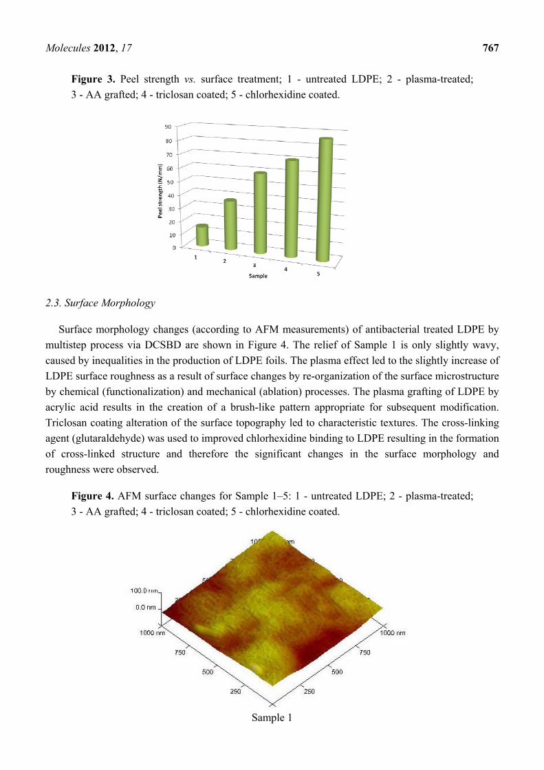
Molecules 2012, 17 767
Figure 3. Peel strength vs. surface treatment; 1 - untreated LDPE; 2 - plasma-treated;
3 - AA grafted; 4 - triclosan coated; 5 - chlorhexidine coated.
2.3. Surface Morphology
Surface morphology changes (according to AFM measurements) of antibacterial treated LDPE by
multistep process via DCSBD are shown in Figure 4. The relief of Sample 1 is only slightly wavy,
caused by inequalities in the production of LDPE foils. The plasma effect led to the slightly increase of
LDPE surface roughness as a result of surface changes by re-organization of the surface microstructure
by chemical (functionalization) and mechanical (ablation) processes. The plasma grafting of LDPE by
acrylic acid results in the creation of a brush-like pattern appropriate for subsequent modification.
Triclosan coating alteration of the surface topography led to characteristic textures. The cross-linking
agent (glutaraldehyde) was used to improved chlorhexidine binding to LDPE resulting in the formation
of cross-linked structure and therefore the significant changes in the surface morphology and
roughness were observed.
Figure 4. AFM surface changes for Sample 1–5: 1 - untreated LDPE; 2 - plasma-treated;
3 - AA grafted; 4 - triclosan coated; 5 - chlorhexidine coated.
Sample 1
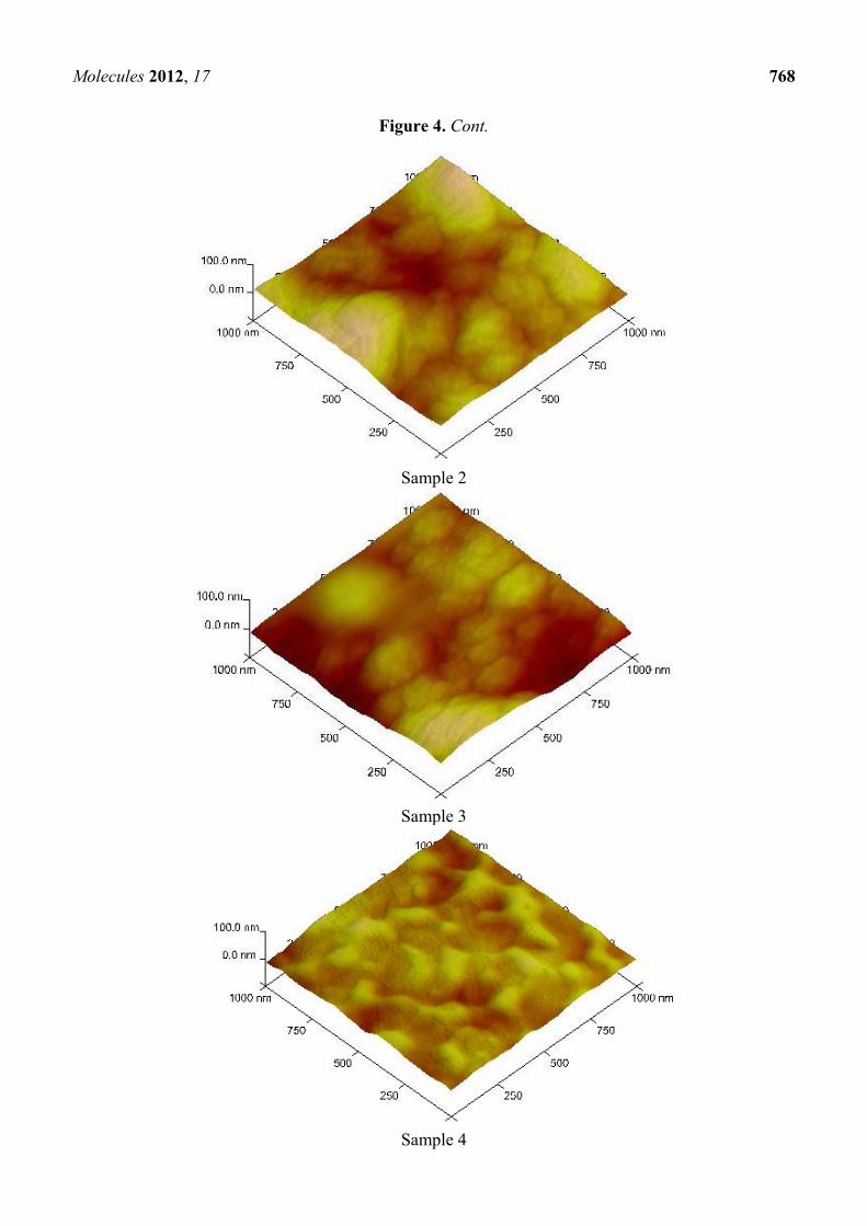
Molecules 2012, 17 768
Figure 4. Cont.
Sample 2
Sample 3
Sample 4
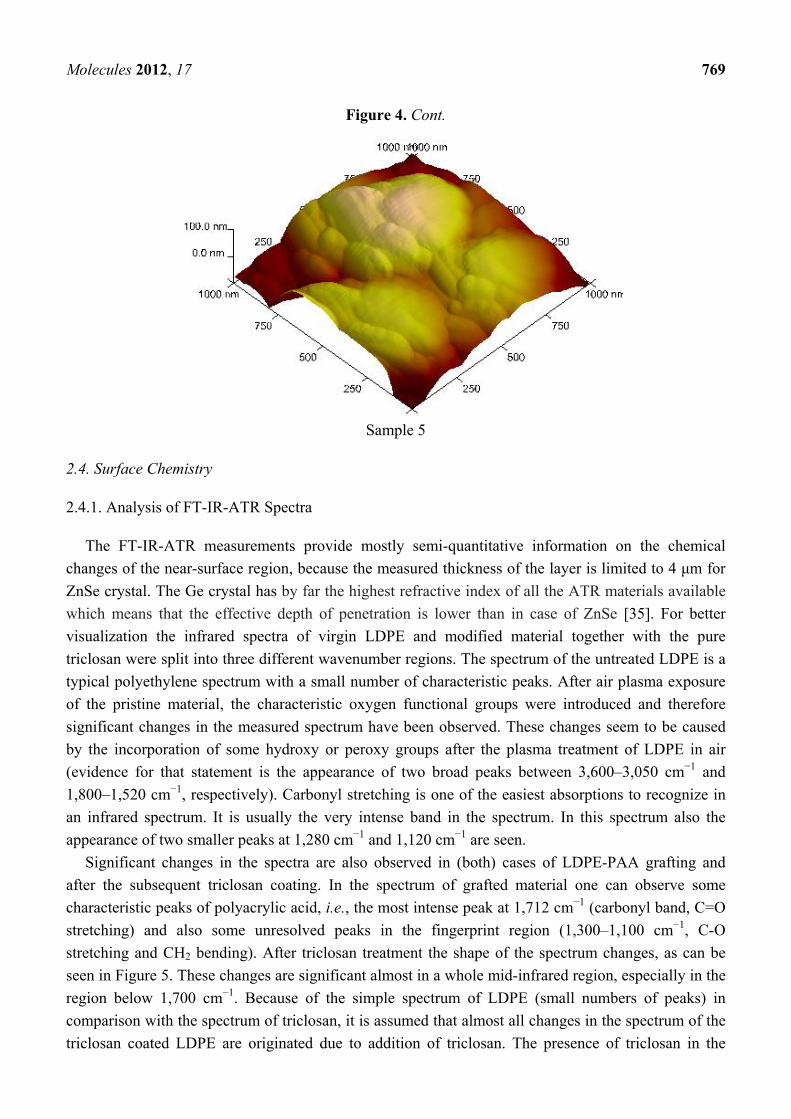
Molecules 2012, 17 769
Figure 4. Cont.
Sample 5
2.4. Surface Chemistry
2.4.1. Analysis of FT-IR-ATR Spectra
The FT-IR-ATR measurements provide mostly semi-quantitative information on the chemical
changes of the near-surface region, because the measured thickness of the layer is limited to 4 μm for
ZnSe crystal. The Ge crystal has by far the highest refractive index of all the ATR materials available
which means that the effective depth of penetration is lower than in case of ZnSe [35]. For better
visualization the infrared spectra of virgin LDPE and modified material together with the pure
triclosan were split into three different wavenumber regions. The spectrum of the untreated LDPE is a
typical polyethylene spectrum with a small number of characteristic peaks. After air plasma exposure
of the pristine material, the characteristic oxygen functional groups were introduced and therefore
significant changes in the measured spectrum have been observed. These changes seem to be caused
by the incorporation of some hydroxy or peroxy groups after the plasma treatment of LDPE in air
(evidence for that statement is the appearance of two broad peaks between 3,600–3,050 cm−1 and
1,800–1,520 cm−1, respectively). Carbonyl stretching is one of the easiest absorptions to recognize in
an infrared spectrum. It is usually the very intense band in the spectrum. In this spectrum also the
appearance of two smaller peaks at 1,280 cm−1 and 1,120 cm−1 are seen.
Significant changes in the spectra are also observed in (both) cases of LDPE-PAA grafting and
after the subsequent triclosan coating. In the spectrum of grafted material one can observe some
characteristic peaks of polyacrylic acid, i.e., the most intense peak at 1,712 cm−1 (carbonyl band, C=O
stretching) and also some unresolved peaks in the fingerprint region (1,300–1,100 cm−1, C-O
stretching and CH2 bending). After triclosan treatment the shape of the spectrum changes, as can be
seen in Figure 5. These changes are significant almost in a whole mid-infrared region, especially in the
region below 1,700 cm−1. Because of the simple spectrum of LDPE (small numbers of peaks) in
comparison with the spectrum of triclosan, it is assumed that almost all changes in the spectrum of the
triclosan coated LDPE are originated due to addition of triclosan. The presence of triclosan in the
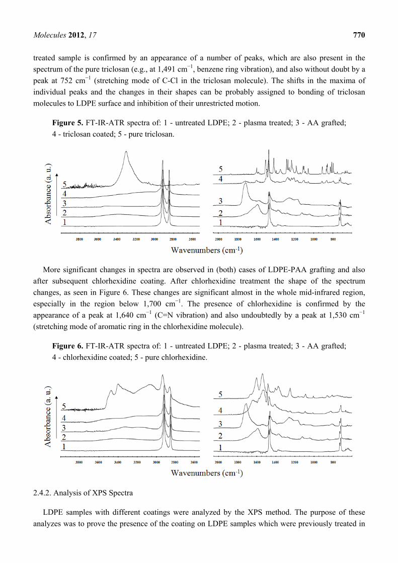
Molecules 2012, 17 770
treated sample is confirmed by an appearance of a number of peaks, which are also present in the
spectrum of the pure triclosan (e.g., at 1,491 cm−1, benzene ring vibration), and also without doubt by a
peak at 752 cm−1 (stretching mode of C-Cl in the triclosan molecule). The shifts in the maxima of
individual peaks and the changes in their shapes can be probably assigned to bonding of triclosan
molecules to LDPE surface and inhibition of their unrestricted motion.
Figure 5. FT-IR-ATR spectra of: 1 - untreated LDPE; 2 - plasma treated; 3 - AA grafted;
4 - triclosan coated; 5 - pure triclosan.
More significant changes in spectra are observed in (both) cases of LDPE-PAA grafting and also
after subsequent chlorhexidine coating. After chlorhexidine treatment the shape of the spectrum
changes, as seen in Figure 6. These changes are significant almost in the whole mid-infrared region,
especially in the region below 1,700 cm−1. The presence of chlorhexidine is confirmed by the
appearance of a peak at 1,640 cm−1 (C=N vibration) and also undoubtedly by a peak at 1,530 cm−1
(stretching mode of aromatic ring in the chlorhexidine molecule).
Figure 6. FT-IR-ATR spectra of: 1 - untreated LDPE; 2 - plasma treated; 3 - AA grafted;
4 - chlorhexidine coated; 5 - pure chlorhexidine.
2.4.2. Analysis of XPS Spectra
LDPE samples with different coatings were analyzed by the XPS method. The purpose of these
analyzes was to prove the presence of the coating on LDPE samples which were previously treated in
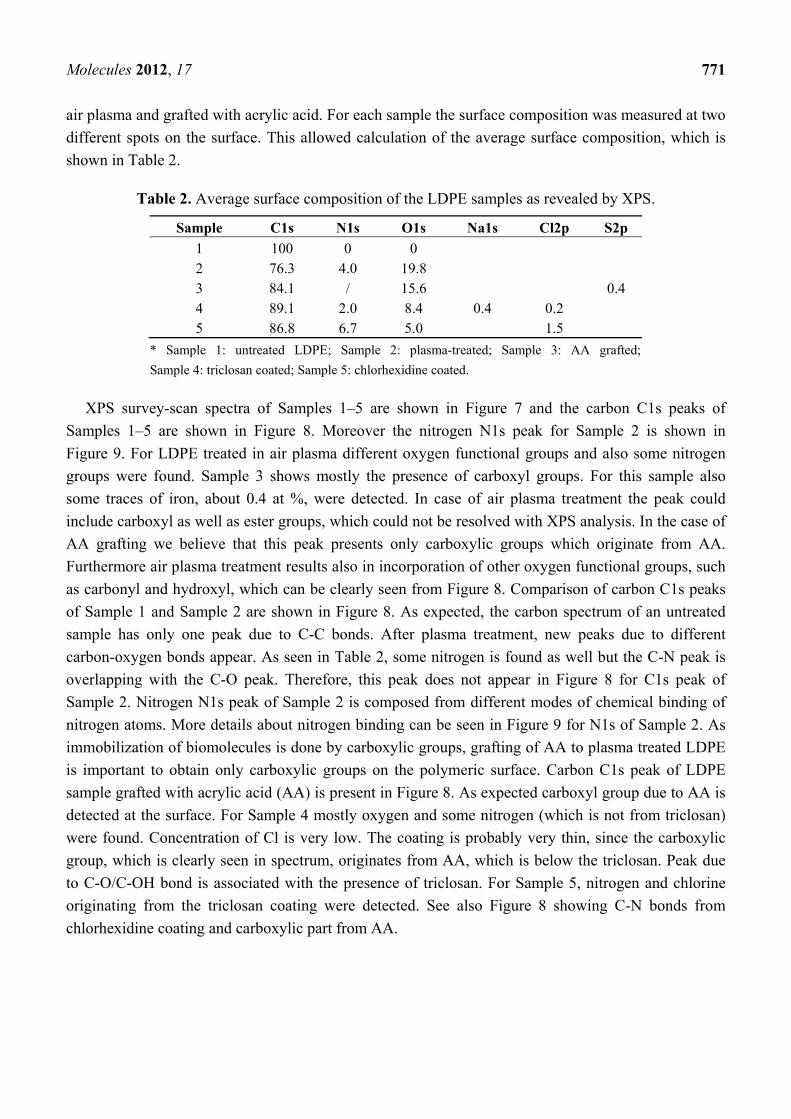
Molecules 2012, 17 771
air plasma and grafted with acrylic acid. For each sample the surface composition was measured at two
different spots on the surface. This allowed calculation of the average surface composition, which is
shown in Table 2.
Table 2. Average surface composition of the LDPE samples as revealed by XPS.
Sample C1s N1s O1s Na1s Cl2p S2p
1 100 0 0 2 76.3 4.0 19.8 3 84.1 / 15.6 0.4 4 89.1 2.0 8.4 0.4 0.2 5 86.8 6.7 5.0 1.5
* Sample 1: untreated LDPE; Sample 2: plasma-treated; Sample 3: AA grafted;
Sample 4: triclosan coated; Sample 5: chlorhexidine coated.
XPS survey-scan spectra of Samples 1–5 are shown in Figure 7 and the carbon C1s peaks of
Samples 1–5 are shown in Figure 8. Moreover the nitrogen N1s peak for Sample 2 is shown in
Figure 9. For LDPE treated in air plasma different oxygen functional groups and also some nitrogen
groups were found. Sample 3 shows mostly the presence of carboxyl groups. For this sample also
some traces of iron, about 0.4 at %, were detected. In case of air plasma treatment the peak could
include carboxyl as well as ester groups, which could not be resolved with XPS analysis. In the case of
AA grafting we believe that this peak presents only carboxylic groups which originate from AA.
Furthermore air plasma treatment results also in incorporation of other oxygen functional groups, such
as carbonyl and hydroxyl, which can be clearly seen from Figure 8. Comparison of carbon C1s peaks
of Sample 1 and Sample 2 are shown in Figure 8. As expected, the carbon spectrum of an untreated
sample has only one peak due to C-C bonds. After plasma treatment, new peaks due to different
carbon-oxygen bonds appear. As seen in Table 2, some nitrogen is found as well but the C-N peak is
overlapping with the C-O peak. Therefore, this peak does not appear in Figure 8 for C1s peak of
Sample 2. Nitrogen N1s peak of Sample 2 is composed from different modes of chemical binding of
nitrogen atoms. More details about nitrogen binding can be seen in Figure 9 for N1s of Sample 2. As
immobilization of biomolecules is done by carboxylic groups, grafting of AA to plasma treated LDPE
is important to obtain only carboxylic groups on the polymeric surface. Carbon C1s peak of LDPE
sample grafted with acrylic acid (AA) is present in Figure 8. As expected carboxyl group due to AA is
detected at the surface. For Sample 4 mostly oxygen and some nitrogen (which is not from triclosan)
were found. Concentration of Cl is very low. The coating is probably very thin, since the carboxylic
group, which is clearly seen in spectrum, originates from AA, which is below the triclosan. Peak due
to C-O/C-OH bond is associated with the presence of triclosan. For Sample 5, nitrogen and chlorine
originating from the triclosan coating were detected. See also Figure 8 showing C-N bonds from
chlorhexidine coating and carboxylic part from AA.
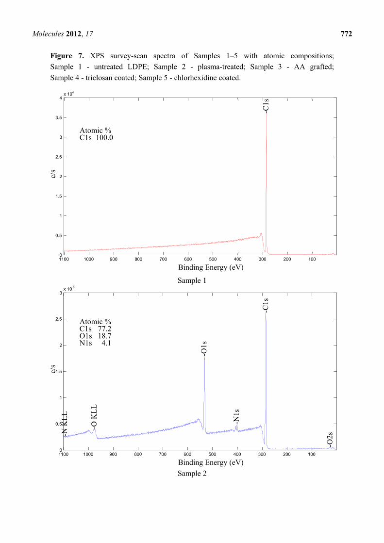
Molecules 2012, 17 772
Figure 7. XPS survey-scan spectra of Samples 1–5 with atomic compositions;
Sample 1 - untreated LDPE; Sample 2 - plasma-treated; Sample 3 - AA grafted;
Sample 4 - triclosan coated; Sample 5 - chlorhexidine coated.
Sample 1
Sample 2
10020030040050060070080090010001100 0
0.5
1
1.5
2
2.5
3x 10 4
Binding Energy (eV)
c/s
Atomic %C1s 77.2 O1s 18.7 N1s 4.1
-C
1s
-N
KL
L
-O
KL
L
-O
1s
-O
2s
-N
1s
1002003004005006007008009001000 1100 0
0.5
1
1.5
2
2.5
3
3.5
4x 10⁴
Binding Energy (eV)
c/s
Atomic % C1s 100.0
-C1s
Molecules 2012, 17 773
Figure 7. Cont.
Sample 3
Sample 4
100 200300400500600700800 90010001100 0
0.1
0.2
0.3
0.4
0.5
0.6
0.7
0.8
0.9
1
Binding Energy (eV)
Atomic %C1s 88.8 O1s 8.5 N1s 2.2 Na1s 0.4Cl2p 0.2
-C1s
-N
KL
L
-N
1s
-O
KL
L
-O
1s -N
a K
LL
-N
a2s
-N
a2p
-N
a1s
-C
l2s
-C
l2p
-O
2s
100200300400500600700800900100011000
0.5
1
1.5
2
2.5
3
3.5 x 10⁴
Binding Energy (eV)
c/s
-C
1s
-F
e2p
Atomic % C1s 82.9 O1s 16.3 Fe2p3 0.4 S2p 0.4
-O
KL
L
-O
2s
-O
1s
-S2s
-S
2p
-Fe3
p
Nor
mal
ized
Int
ensi
ty
Molecules 2012, 17 774
Figure 7. Cont.
Sample 5
Figure 8. Carbon C1s peaks of Sample 1–5; Sample 1 - untreated LDPE;
Sample 2 - plasma-treated; Sample 3 - AA grafted, Sample 4 - triclosan coated;
Sample 5 - chlorhexidine coated.
Sample 1
100 200300400500600700800 9001000 11000
0.5
1
1.5
2
2.5
3
3.5 x 10 4
Binding Energy (eV)
c/s
-C
l2p
Atomic % C1s 85.5 N1s 7.0 O1s 5.8 Cl2p 1.7
-C
1s
-N
KL
L
-N
1s
-O
KL
L
-O
1s
-C
l2s
-O
2s
280282 284 286288290292 294 296 0
100
200
300
400
500
600
700
800
900
1000
Binding Energy (eV)
c/s
C1s

Molecules 2012, 17 775
Figure 8. Cont.
Sample 2
Sample 3
280282 284 286288290292 294 296
0
1000
2000
3000
4000
5000
6000
Binding Energy (eV)
c/s
Pos. Sep. %Area 284.79 0.00 90.69286.53 1.74 2.86 288.76 3.97 6.46
C1s C-C
C-OO=C-O
280282 284 286288290292294 296
0
1000
2000
3000
4000
5000
Binding Energy (eV)
c/s
C1s
Pos. Sep. %Area 284.76 0.00 79.27286.52 1.75 9.20 287.77 3.01 5.25 288.99 4.23 6.28
C-OO=C-O
C-C
C=O
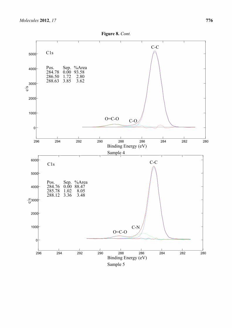
Molecules 2012, 17 776
Figure 8. Cont.
Sample 4
Sample 5
280282 284 286288290292 294 296
0
1000
2000
3000
4000
5000
6000
Binding Energy (eV)
c/s
Pos. Sep. %Area 284.76 0.00 88.47 285.78 1.02 8.05 288.12 3.36 3.48
C-NO=C-O
C1s C-C
280282 284 286288290292 294296
0
1000
2000
3000
4000
5000
Binding Energy (eV)
c/s
Pos. Sep. %Area 284.78 0.00 93.58 286.50 1.72 2.80 288.63 3.85 3.62
C1s C-C
C-OO=C-O
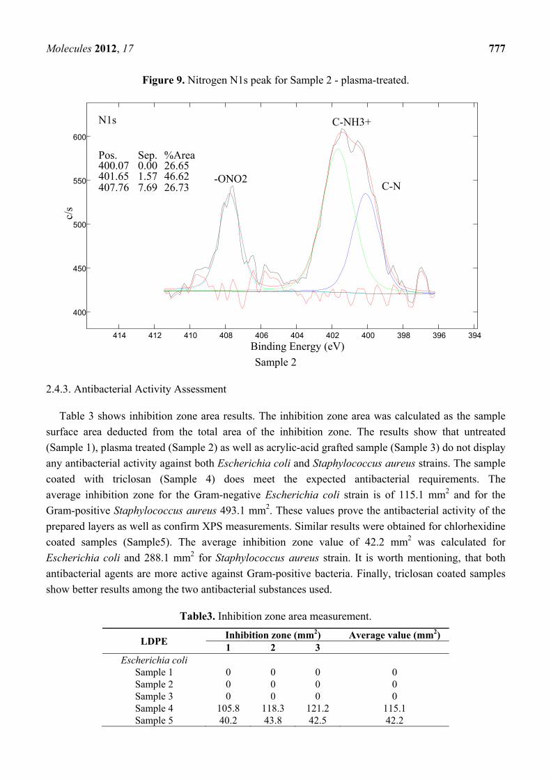
Molecules 2012, 17 777
Figure 9. Nitrogen N1s peak for Sample 2 - plasma-treated.
Sample 2
2.4.3. Antibacterial Activity Assessment
Table 3 shows inhibition zone area results. The inhibition zone area was calculated as the sample
surface area deducted from the total area of the inhibition zone. The results show that untreated
(Sample 1), plasma treated (Sample 2) as well as acrylic-acid grafted sample (Sample 3) do not display
any antibacterial activity against both Escherichia coli and Staphylococcus aureus strains. The sample
coated with triclosan (Sample 4) does meet the expected antibacterial requirements. The
average inhibition zone for the Gram-negative Escherichia coli strain is of 115.1 mm2 and for the
Gram-positive Staphylococcus aureus 493.1 mm2. These values prove the antibacterial activity of the
prepared layers as well as confirm XPS measurements. Similar results were obtained for chlorhexidine
coated samples (Sample5). The average inhibition zone value of 42.2 mm2 was calculated for
Escherichia coli and 288.1 mm2 for Staphylococcus aureus strain. It is worth mentioning, that both
antibacterial agents are more active against Gram-positive bacteria. Finally, triclosan coated samples
show better results among the two antibacterial substances used.
Table3. Inhibition zone area measurement.
LDPE Inhibition zone (mm2) Average value (mm2) 1 2 3
Escherichia coli Sample 1 0 0 0 0 Sample 2 0 0 0 0 Sample 3 0 0 0 0 Sample 4 105.8 118.3 121.2 115.1 Sample 5 40.2 43.8 42.5 42.2
394396 398 400402404406408410 412414
400
450
500
550
600
Binding Energy (eV)
c/s
Pos. Sep. %Area 400.07 0.00 26.65 401.65 1.57 46.62 407.76 7.69 26.73 C-N
-ONO2
N1s C-NH3+
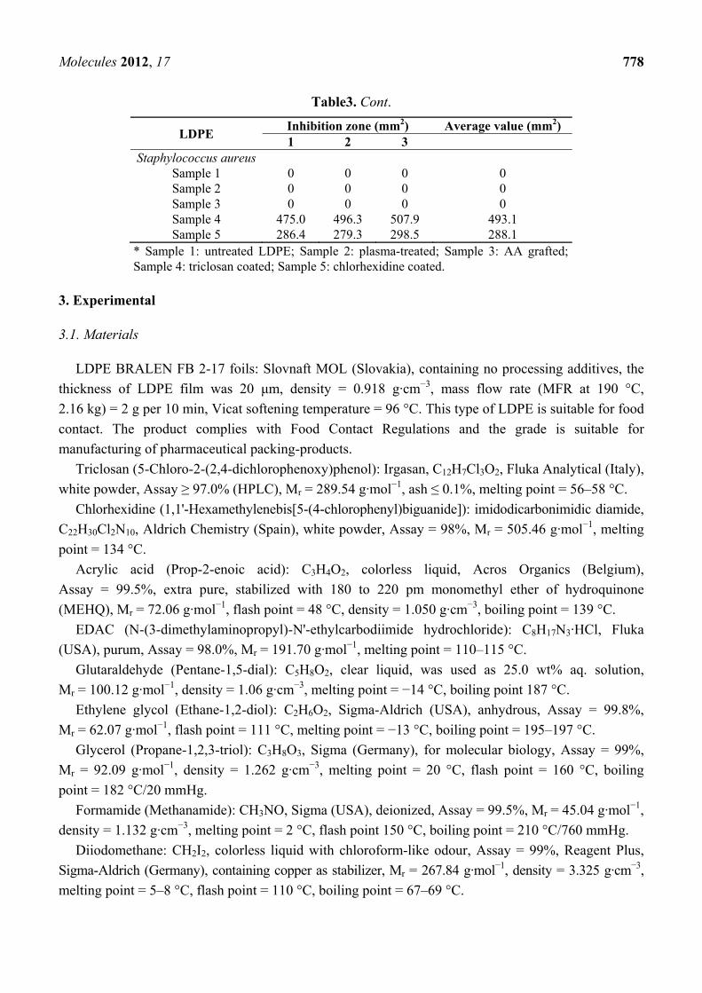
Molecules 2012, 17 778
Table3. Cont.
LDPE Inhibition zone (mm2) Average value (mm2) 1 2 3
Staphylococcus aureus Sample 1 0 0 0 0 Sample 2 0 0 0 0 Sample 3 0 0 0 0 Sample 4 475.0 496.3 507.9 493.1 Sample 5 286.4 279.3 298.5 288.1
* Sample 1: untreated LDPE; Sample 2: plasma-treated; Sample 3: AA grafted; Sample 4: triclosan coated; Sample 5: chlorhexidine coated.
3. Experimental
3.1. Materials
LDPE BRALEN FB 2-17 foils: Slovnaft MOL (Slovakia), containing no processing additives, the
thickness of LDPE film was 20 μm, density = 0.918 g·cm−3, mass flow rate (MFR at 190 °C,
2.16 kg) = 2 g per 10 min, Vicat softening temperature = 96 °C. This type of LDPE is suitable for food
contact. The product complies with Food Contact Regulations and the grade is suitable for
manufacturing of pharmaceutical packing-products.
Triclosan (5-Chloro-2-(2,4-dichlorophenoxy)phenol): Irgasan, C12H7Cl3O2, Fluka Analytical (Italy),
white powder, Assay ≥ 97.0% (HPLC), Mr = 289.54 g·mol−1, ash ≤ 0.1%, melting point = 56–58 °C.
Chlorhexidine (1,1'-Hexamethylenebis[5-(4-chlorophenyl)biguanide]): imidodicarbonimidic diamide,
C22H30Cl2N10, Aldrich Chemistry (Spain), white powder, Assay = 98%, Mr = 505.46 g·mol−1, melting
point = 134 °C.
Acrylic acid (Prop-2-enoic acid): C3H4O2, colorless liquid, Acros Organics (Belgium),
Assay = 99.5%, extra pure, stabilized with 180 to 220 pm monomethyl ether of hydroquinone
(MEHQ), Mr = 72.06 g·mol−1, flash point = 48 °C, density = 1.050 g·cm−3, boiling point = 139 °C.
EDAC (N-(3-dimethylaminopropyl)-N'-ethylcarbodiimide hydrochloride): C8H17N3·HCl, Fluka
(USA), purum, Assay = 98.0%, Mr = 191.70 g·mol−1, melting point = 110–115 °C.
Glutaraldehyde (Pentane-1,5-dial): C5H8O2, clear liquid, was used as 25.0 wt% aq. solution,
Mr = 100.12 g·mol−1, density = 1.06 g·cm−3, melting point = −14 °C, boiling point 187 °C.
Ethylene glycol (Ethane-1,2-diol): C2H6O2, Sigma-Aldrich (USA), anhydrous, Assay = 99.8%,
Mr = 62.07 g·mol−1, flash point = 111 °C, melting point = −13 °C, boiling point = 195–197 °C.
Glycerol (Propane-1,2,3-triol): C3H8O3, Sigma (Germany), for molecular biology, Assay = 99%,
Mr = 92.09 g·mol−1, density = 1.262 g·cm−3, melting point = 20 °C, flash point = 160 °C, boiling
point = 182 °C/20 mmHg.
Formamide (Methanamide): CH3NO, Sigma (USA), deionized, Assay = 99.5%, Mr = 45.04 g·mol−1,
density = 1.132 g·cm−3, melting point = 2 °C, flash point 150 °C, boiling point = 210 °C/760 mmHg.
Diiodomethane: CH2I2, colorless liquid with chloroform-like odour, Assay = 99%, Reagent Plus,
Sigma-Aldrich (Germany), containing copper as stabilizer, Mr = 267.84 g·mol−1, density = 3.325 g·cm−3,
melting point = 5–8 °C, flash point = 110 °C, boiling point = 67–69 °C.
Molecules 2012, 17 779
Dichloromethane: CH2Cl2, mikroCHEM (SVK), Assay = 99.5%, Mr = 84.93 g·mol−1,
density = 1.33 g·cm−3, melting point = −96.7 °C, boiling point = 39.6 °C.
3.2. Plasma Treatment
The LDPE foils were first cleaned with dichloromethane to remove impurities. Then the LDPE foil
activation was carried out under dynamic conditions at atmospheric pressure and room temperature
with the DCSBD equipment developed at Comenius University (Department of Experimental Physics,
Faculty of Mathematics, Physics and Informatics) in Bratislava. The schematic representation with
description of this system is given in Scheme 1. The treatment was performed with the following
settings: power supply = 200 W, plasma treatment time = 15 s, in air atmosphere and all samples were
treated on both sides. DCSBD equipment generates macroscopically homogeneous plasma without
direct contact with the electrodes, which protects the electrodes from wear. Plasma is generated by two
parallel banded system of electrodes (1-mm wide, 50 micron thick, with 0.5 mm spacing between the
strips, made of Ag-paste) embedded in 96% Al2O3-promotion of national purity, while the electrodes
are supplied via high frequency sinusoidal voltage (~15 kHz, Um~10 kV). Such an arrangement of
electrodes and supply voltage leads to visually almost perfectly homogeneous diffusion plasma.
Scheme 1. DCSBD scheme and detail of burning plasma panel.
Top view
3.3. Grafting by PAA
Immediately after plasma treatment the LDPE foil was immersed into 10 volume % aqueous
solution of AA for 24 h at 30 °C in order to initiate of radical graft polymerization of AA onto
activated surface of LDPE foil. This solution contained also 0.1 wt.% sodium metabisulfite as a
relevant reductant to inhibit AA homopolymerization. After AA polymerization PAA brushes were
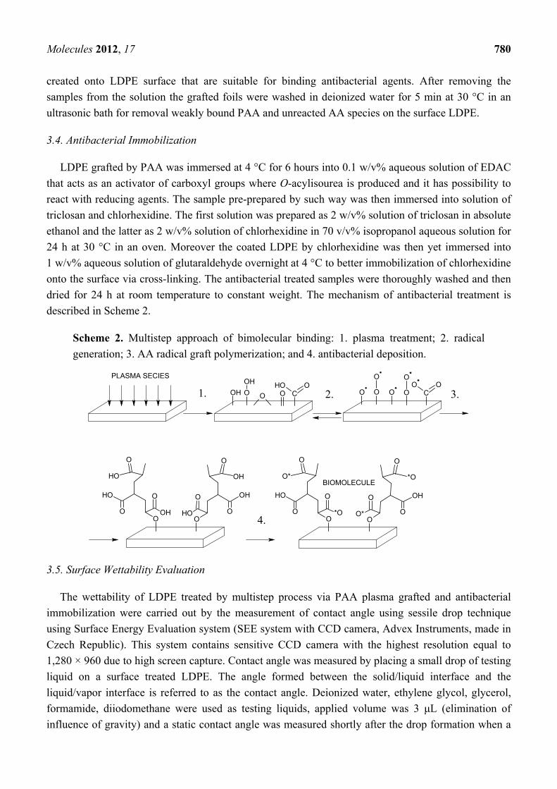
Molecules 2012, 17 780
created onto LDPE surface that are suitable for binding antibacterial agents. After removing the
samples from the solution the grafted foils were washed in deionized water for 5 min at 30 °C in an
ultrasonic bath for removal weakly bound PAA and unreacted AA species on the surface LDPE.
3.4. Antibacterial Immobilization
LDPE grafted by PAA was immersed at 4 °C for 6 hours into 0.1 w/v% aqueous solution of EDAC
that acts as an activator of carboxyl groups where O-acylisourea is produced and it has possibility to
react with reducing agents. The sample pre-prepared by such way was then immersed into solution of
triclosan and chlorhexidine. The first solution was prepared as 2 w/v% solution of triclosan in absolute
ethanol and the latter as 2 w/v% solution of chlorhexidine in 70 v/v% isopropanol aqueous solution for
24 h at 30 °C in an oven. Moreover the coated LDPE by chlorhexidine was then yet immersed into
1 w/v% aqueous solution of glutaraldehyde overnight at 4 °C to better immobilization of chlorhexidine
onto the surface via cross-linking. The antibacterial treated samples were thoroughly washed and then
dried for 24 h at room temperature to constant weight. The mechanism of antibacterial treatment is
described in Scheme 2.
Scheme 2. Multistep approach of bimolecular binding: 1. plasma treatment; 2. radical
generation; 3. AA radical graft polymerization; and 4. antibacterial deposition.
OH O
OH
O O O
O
OO COH O
CO O
O
O
O
OH
O
O
OH
OH
O
O
OH
O
O
OH
OH
O
O
O*
O
O
*O
OH
O
O
*O
O
O
O*
OH
O
BIOMOLECULE
PLASMA SECIES
3.5. Surface Wettability Evaluation
The wettability of LDPE treated by multistep process via PAA plasma grafted and antibacterial
immobilization were carried out by the measurement of contact angle using sessile drop technique
using Surface Energy Evaluation system (SEE system with CCD camera, Advex Instruments, made in
Czech Republic). This system contains sensitive CCD camera with the highest resolution equal to
1,280 × 960 due to high screen capture. Contact angle was measured by placing a small drop of testing
liquid on a surface treated LDPE. The angle formed between the solid/liquid interface and the
liquid/vapor interface is referred to as the contact angle. Deionized water, ethylene glycol, glycerol,
formamide, diiodomethane were used as testing liquids, applied volume was 3 μL (elimination of
influence of gravity) and a static contact angle was measured shortly after the drop formation when a
1. 2. 3.
4.
Molecules 2012, 17 781
thermodynamic equilibrium is reached between the three phases: solid, liquid, and gas. Surface energy
(γtot), its polar acid-base (γAB), dispersive (γLW), electron-acceptor (γ−) and electron-donor (γ+)
components were calculated by Acid-Base regression model using method of least squares.
3.6. Adhesive Properties Assessment
The adhesive properties, namely peel strength (force per unit width) of the adhesive joint of
antibacterial treated LDPE by triclosan and chlorhexidine via DCSBD to poly(2-ethylhexyl acrylate)
deposited onto polypropylene foil (with 15 mm width), were carried out by measurements of 90° peel
test at a 10 mm per minute rate of peel using a 100 N universal INSTRON 4301 dynamometer
(England). Ends of the polymer film were firmly fixed in the jaws of dynamometer so that tension was
evenly distributed across the entire width of the surface.
3.7. Surface Topography Analysis
The surface morphology and local surface heterogeneities of the modified polymer were measured
by AFM. All measurements were performed under ambient conditions using a commercial atomic
force microscope (NanoScopeTM Dimension IIIa, MultiMode Digital Instr., USA) equipped with a
PPP-NCLR tapping-mode probe (NanosensorsTM, Switzerland; spring constant 39 N·m−1, resonance
frequency ≈ 160 kHz). The surface properties of all the films were measured in x and y axis sizes
between 2 to 25 μm on different sites of the films in order to find characteristic and significant surface
features. The AFM analyses were performed in tapping mode for all the images. This technique allows
the obtaining either two- or three-dimensional information of both height and material heterogeneity
contrast with high resolution when recording height and phase shifts simultaneously.
3.8. Surface Chemistry Investigation
3.8.1. XPS
Samples were analyzed with a TFA XPS Physical Electronics XPS instrument. The base pressure in
the chamber was about 6 × 10⁻⁸ Pa. The samples were excited with X-rays over a 400 µm spot area
with a monochromatic Al K1,2 radiation at 1,486.6 eV. The photoelectrons were detected with a
hemispherical analyzer positioned at an angle of 45° with respect to the normal to the sample surface.
Survey-scan spectra were made at a pass energy of 187.85 eV and 0.4 eV energy step. An electron gun
was used for surface neutralization. The concentration of elements was determined by using MultiPak
v7.3.1 software from Physical Electronics, which is supplied by the spectrometer producer.
3.8.2. FT-IR-ATR
Attenuated total reflectance FTIR measurements were performed on a NICOLET 8700 FTIR
spectrometer (Thermo Scientific) through the single bounce ATR accessory equipped with Ge crystal
at an angle of incidence 45°. For each measurement the spectral resolution and the number of scans
were 2 cm−1 and 64, respectively. The quality of spectra depends on good contact between the crystal
Molecules 2012, 17 782
and the sample. This requirement was achieved through the use of a pressure clamp. The acquired
spectra were analyzed using spectroscopic software OMNIC™, v. 8.1.
3.9. In Vitro Antibacterial Test
Bacterial adhesion and biofilm experiments were performed using Gram-positive (S. aureus 3953)
and Gram-negative (E. coli 3954) bacteria. Circular shaped specimens (d ≈ 8 mm) were cut from
pristine and modified LDPE samples. A so called agar diffusion plate (inhibition) test was performed
for antibacterial activity evaluation of tested substrates. The polymer samples were washed in ethanol
and dried under laboratory conditions. The substrates prepared by such a way were placed on agar
plate (Nutrient Agar No. 2 M1269, Hi Media Laboratories Pvt. Ltd.) inoculated by bacterial
suspension. The bacterial suspension volume was 100 µL for all samples. Bacteria concentration was
107 units·mL−1 and incubation time was 24 h at the temperature 37 °C. After that, inhibition zone
diameter was measured in 5 directions and average value was calculated. Each test was repeated
in triplicate.
4. Conclusions
This work was aimed at examining the impact of selected antibacterial agents, namely triclosan and
chlorhexidine bound to the surface of LDPE. DCSBD plasma treatment leads to increased surface free
energy, roughness and surface wettability by introducing characteristic oxygen groups. A DCSBD
plasma generator was used as activator of the LDPE surface for efficient binding of acrylic acid and
for its transformation to polymeric form by radical polymerization. Thus the bound acrylic acid created
polymer brushes on the polymer surface that provided physical forces to bind antibacterial agents in an
effective manner. The presence of triclosan and chlorhexidine was confirmed by different surface
analysis techniques. Moreover the antibacterial effect of such treated LDPE film was proven by
in vitro bacterial tests against E. coli and S. aureus when adhesion of bacteria to polymer was
effective diminished.
Acknowledgements
Financial supports by the Ministry of Education, Youth, and Sports of the Czech Republic (Grant
CZ.1.05/2.1.00/03.0111), the Slovak Academy of Sciences (Grant VEGA 2/0185/10), Technical
University in Zvolen (Grant VEGA 1/0517/09 and VEGA 1/0581/12, KEGA 060-005 TUZVO 4/2010,
and IPA TUZVO 1/2011), Grant Agency of the Academy of Sciences of the Czech Republic (project
No. IAAX08240901), Czech Science Foundation (project 104/09/H080) and the Slovenia Ministry of
Higher Education, Science, and Technology (Program P2-0082-2) are gratefully acknowledged.
References and Notes
1. Zhang, W.; Luo, Y.; Wang, H.; Jiang, J.; Pu, S.; Chu, P.K. Ag and Ag/N2 plasma modification of
polyethylene for the enhancement of antibacterial properties and cell growth/proliferation.
Acta Biomater. 2008, 4, 2028–2036.
Molecules 2012, 17 783
2. Kuzuya, M.; Sawa, T.; Mouri, M.; Kondo, S.I.; Takai, O. Plasma technique for the fabrication of
a durable functional surface on organic polymers. Surf. Coatings Technol. 2003, 169-170,
587–591.
3. Zhang, W.; Chu, P.K.; Ji, J.; Zhang, Y.; Fu, R.K.Y.; Yan, Q. Antibacterial properties of
plasma-modified and triclosan or bronopol coated polyethylene. Polymer 2006, 47, 931–936.
4. Costa, F.; Carvalho, I.F.; Montelaro, R.C.; Gomes, P.; Martins, M.C. Covalent immobilization of
antimicrobial peptides (AMPs) onto biomaterial surfaces. Acta Biomater. 2011, 7, 1431–1440.
5. Goddard, J.M.; Hotchkiss, J.H. Tailored functionalization of low-density polyethylene surfaces.
J. Appl. Polym. Sci. 2008, 108, 2940–2949.
6. Faucheux, N.; Schweiss, R.; Lutzow, K.; Wemer, C.; Groth, T. Self-assembled monolayers with
different terminating groups as model substrates for cell adhesion studies. Biomaterials 2004, 25,
2721–2730.
7. Michael, K.E.; Vernekar, V.N.; Keselowsky, B.G.; Meredith, J.C.; Latour, R.A.; Garcia, A.J.
Adsorption-induced conformational changes in fibronectin due to interactions with well-defined
surface chemistries. Langmuir 2003, 19, 8033–8040.
8. Keselowsky, B.G.; Collard, D.M.; Garcia, A.J.J. Surface chemistry modulates fibronectin
conformation and directs integrin binding and specificity to control cell adhesion. J. Biomed.
Mater. Res. A 2003, 66, 247–259.
9. Luk, Y.Y.; Kato, M.; Mrksich, M. Self-assembled monolayers of alkanethiolates presenting
mannitol groups are inert to protein adsorption and cell attachment. Langmuir 2000, 16, 9604–9608.
10. Vesel, A.; Junkar, I.; Cvelbar, U.; Kovac, J.; Mozetic, M. Surface modification of polyester by
oxygen and nitrogen-plasma treatment. Surf. Interface Anal. 2008, 40, 1444–1453.
11. Drnovská, H.; Lapčík, L., Jr; Buršíková, V.; Zemek, J.; Barros-Timmons, A.M. Surface properties
of polyethylene after low-temperature plasma treatment. Colloid Polym. Sci. 2003, 281,
1025–1033.
12. Novák, I.; Števiar, M.; Chodák, I.; Krupa, I.; Nedelčev, T.; Špírková, M.; Chehimi, M.M.;
Mosnáček, J.; Kleinová, A. Study of adhesion and surface properties of low density polyethylene
pre-treated by cold discharge plasma. Polym. Adv. Technol. 2007, 18, 97–105.
13. Olifirenko, A.S.; Novak, I.; Rozova, E.Y.; Saprykina, N.N.; Mitilineos, A.G.; Elyashevich, G.K.
Hydrophilization of porous polyethylene films by cold plasma of different types. Polym. Sci.
2009, 51, 247–255.
14. Lloyd, G.; Friedman, G.; Jafri, S.; Schultz, G.; Fridman, A.; Harding, K. Gas plasma: Medical
uses and developments in wound care. Plasma Process. Polym. 2010, 7, 194–211.
15. Sanchis, R.; Fenollar, O.; García, D.; Sánchez, L.; Balart, R. Improved adhesion of LDPE films to
polyolefin foams for automotive industry using low-pressure plasma. Int. J. Adh. Adhesives 2008,
28, 445–451.
16. Pappas, D. Status and potential of atmospheric plasma processing of materials. J. Vac. Sci.
Technol. A 2011, 29, 020801:1–020801:17.
17. Yang, L.; Chen, J.; Guo, Y.; Zhan, Z. Surface modification of a biomedical polyethylene
terephthalate (PET) by air plasma. Appl. Surf. Sci. 2009, 255, 4446–4451.

Molecules 2012, 17 784
18. Černák, M.; Černáková, L’.; Hudec, I.; Kováčik, D.; Zahoranová, A. Diffuse coplanar surface
barrier discharge and its applications for in-line processing of low-added-value materials. Eur.
Phys. J. Appl. Phys. 2009, 47, 22806:1–22806:6.
19. Šimor, M.; Ráheľ, J.; Vojtek, P.; Černák, M.; Brablec, A. Atmospheric-pressure diffuse coplanar
surface discharge for surface treatments. Appl. Phys. Lett. 2002, 81, 2716–2718.
20. Černák, M.; Ráheľ, J.; Kováčik, D.; Šimor, M.; Brablec, A.; Slavíček, P. Generation of thin
surface plasma layers for atmospheric-pressure surface treatments. Contrib. Plasma Phys. 2004,
44, 492–495.
21. John, P.I. Plasma Sciences and the Creation of Wealth; Tata McGraw-Hill: New Delhi, India,
2005; p. 80.
22. Šíra, M.; Trunec, D. Surface modification of polyethylene and polypropylene in atmospheric
pressure glow discharge. J. Phys. D Appl. Phys. 2005, 38, 621–627.
23. Sarghini, S.; Paulussen, S.; Terryn, H. DBD Atmospheric Plasma Deposition of Antibacterial
Coatings. In Proceedings of the 11th International Symposium on High Pressure Low
Temperature Plasma Chemistry (Hakone XI), Oléron Island, France, 7–12 September 2008.
24. Newton, A.P.N.; Cadena, S.M.S.C.; Rocha, M.E.M.; Carnieri, E.G.S.; de Oliveira, M.B.M. Effect
of triclosan (TRN) on energy-linked functions of rat liver mitochondria. Toxicol. Lett. 2005, 160,
49–59.
25. Maia Ribeiro, L.G.M.; Hashizume, L.N.; Maltz, M. The effect of different formulations of
chlorhexidine in reducing levels of mutans streptococci in the oral cavity: A systematic review of
the literature. J. Dent. 2007, 35, 359–370.
26. Kenawy, E.R.; Worley, S.D.; Broughton, R. The chemistry and applications of antimicrobial
polymers: A state-of-the-art review. Biomacromolecules 2007, 8, 1359–1384.
27. Desmet, T.; Morent, R.; Geyter, N.D.; Leys, C.; Schacht, E.; Dubruel, P. Nonthermal plasma
technology as a versatile strategy for polymeric biomaterials surface modification: A review.
Biomacromolecules 2009, 10, 2351–2378.
28. Kenawy, E.R.; Worley, S.D.; Broughton, R. The chemistry and applications of antimicrobial
polymers: A state-of-the-art review. Biomacromolecules 2007, 8, 1359–1384.
29. Denes, F.S.; Manolache, S. Macromolecular plasma-chemistry: An emerging field of polymer
science. Prog. Polym. Sci. 2004, 29, 815–885.
30. Zhao, B.; Brittain, W.J. Polymer brushes: Surface-immobilized macromolecules. Prog. Polym.
Sci. 2000, 25, 677–710.
31. Bazaka, K.; Jacob, M.V.; Crawford, R.J.; Ivanova, E.P. Plasma-assisted surface modification of
organic biopolymers to prevent bacterial attachment. Acta Biomater. 2011, 7, 2015–2028.
32. Işiklan, N.; Kurşun, F.; İnal, M. Graft copolymerization of itaconic acid onto sodium alginate
using benzoyl peroxide. Carbohydr. Polym. 2010, 79, 665–672.
33. Buršíková, V.; Sťahel, P.; Navrátil, Z.; Buršík, J.; Janča, J. Surface Energy Evaluation of Plasma
Treated Materials by Contact Angle Measurement; Masaryk University: Brno, Czech Republic,
2004; pp. 7–25.
34. Singh, A.; Silverman, J. Radiation processing of polymers. Radiat. Phys. Chem. 1999, 56, 375.
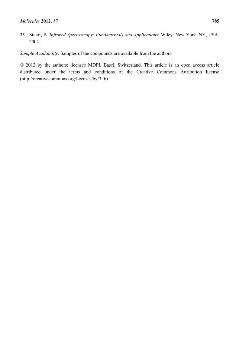
Molecules 2012, 17 785
35. Stuart, B. Infrared Spectroscopy: Fundamentals and Applications; Wiley: New York, NY, USA,
2004.
Sample Availability: Samples of the compounds are available from the authors.
© 2012 by the authors; licensee MDPI, Basel, Switzerland. This article is an open access article
distributed under the terms and conditions of the Creative Commons Attribution license
(http://creativecommons.org/licenses/by/3.0/).


PAPER III

A
AIa
b
c
d
a
ARRAA
KIPCPMG
1
psmuoccMwJpmatpat(
0h
Carbohydrate Polymers 90 (2012) 1501– 1508
Contents lists available at SciVerse ScienceDirect
Carbohydrate Polymers
jo u rn al hom epa ge: www.elsev ier .com/ locate /carbpol
new route for chitosan immobilization onto polyethylene surface
nton Popelkaa, Igor Nováka,∗, Marián Lehockyb, Ita Junkarc, Miran Mozetic c, Angela Kleinováa,vica Janigováa, Miroslav Sloufd, Frantisek Bílekb, Ivan Chodáka
Polymer Institute, Slovak Academy of Sciences, Dúbravská cesta 9, 842 36 Bratislava, SlovakiaCentre of Polymer Systems, University Institute, Tomas Bata University in Zlín, Nam. T.G.M. 5555, 76001 Zlín, Czech RepublicPlasma Laboratory, Department of Surface Engineering, Jozef Stefan Institute, Jamova cesta 39, SI-1000 Ljubljana, SloveniaInstitute of Macromolecular Chemistry, Academy of Sciences of the Czech Republic, Heyrovsky Sq. 2, 162 06 Prague 6, Czech Republic
r t i c l e i n f o
rticle history:eceived 26 April 2012eceived in revised form 3 July 2012ccepted 7 July 2012vailable online 16 July 2012
a b s t r a c t
Low-density polyethylene (LDPE) belongs to commodity polymer materials applied in biomedical appli-cations due to its favorable mechanical and chemical properties. The main disadvantage of LDPE inbiomedical applications is low resistance to bacterial infections. An antibacterial modification of LDPEappears to be a solution to this problem. In this paper, the chitosan and chitosan/pectin multilayer wasimmobilized via polyacrylic acid (PAA) brushes grafted on the LDPE surface. The grafting was initiated bya low-temperature plasma treatment of the LDPE surface. Surface and adhesive properties of the sam-
eywords:mmobilizationlasma treatmenthitosanectinultilayer
ples prepared were investigated by surface analysis techniques. An antibacterial effect was confirmedby inhibition zone measurements of Escherichia coli (E. coli) and Staphylococcus aureus (S. aureus). Thechitosan treatment of LDPE led to the highest and most clear inhibition zones (35 mm2 for E. coli and275 mm2 for S. aureus).
© 2012 Elsevier Ltd. All rights reserved.
rafting. Introduction
Several modification methods are commonly used to modify theolymer surface. One of the most frequent consists in an immer-ion in the strong acid solution. Nevertheless, such wet chemicalethods are technologically complicated and environmentally
nfriendly especially because hazardous chemical substances areften used. Recently, a plasma treatment is a preferred procedureonsidered as a progressive technique for polymer surface modifi-ation without the use of aggressive chemicals (Lloyd et al., 2010).oreover, the plasma treatment enables surface modificationsithout changing the bulk properties of treated material (Vesel,
unkar, Cvelbar, Kovac, & Mozetic, 2008). The low-temperaturelasma belongs to a clean, dry, ecologically method of the surfaceodification and it is often used in various applications, such as in
utomotive, electronic, aeronautic, textile, optical and paper indus-ry (Pelletier et al., 2001). The main effect of the low-temperaturelasma application consists in an increase of a surface free energys a result of the incorporation of polar functional groups to
he treated surface making the surface of LDPE more hydrophilicNovák et al., 2007).∗ Corresponding author. Tel.: +421 903 925 725; fax: +421 2 54775923.E-mail address: [email protected] (I. Novák).
144-8617/$ – see front matter © 2012 Elsevier Ltd. All rights reserved.ttp://dx.doi.org/10.1016/j.carbpol.2012.07.021
Etching (ablation), polymerization, or cross-linking processestake place during the plasma treatment of polymers. Moreover,species created in the plasma discharge, such as electrons, ionsand excited atoms (Vesel, Drenik, Mozetic, & Balat-Pichelin, 2010b)are capable to initiate chemical processes on the polymer surface,leading to a formation of new reactive functional groups (function-alization) (Pappas, 2011; Vesel et al., 2010a; Yang, Chen, Guo, &Zhan, 2009).
The uniform layer and high surface power density of plasmacan be generated by the diffuse coplanar surface barrier discharge(DCSBD) plasma generator. The equipment operates at atmosphericpressure and therefore it is suitable for continual industry appli-cations (Cernák, Cernáková, Hudec, Kovácik, & Zahoranová, 2009).An another advantage of the abovementioned process is the indi-rect contact with the electrodes, what leads to the lower polymersurface contamination as well as longer electrode lifetime (Simor,Ráhel’, Vojtek, Cernák, & Brablec, 2002). DCSBD plasma equipmentconsists of two parallel electrodes embedded in Al2O3. Several pairsof electrodes are supplied by a high frequency sinusoidal voltage(John, 2005). Such arrangement of electrodes leads to the almostmacroscopically homogeneous plasma (Cernák et al., 2004; Síra &Trunec, 2005).
A bacterial surface growth on the polymer surface, also called abiofilm formation is a widespread problem (Hallab, Skipor, & Jacobs,2003). Anti-infective properties of polymers can be reached by thesurface treatment of medical polymer materials. This antibacterial
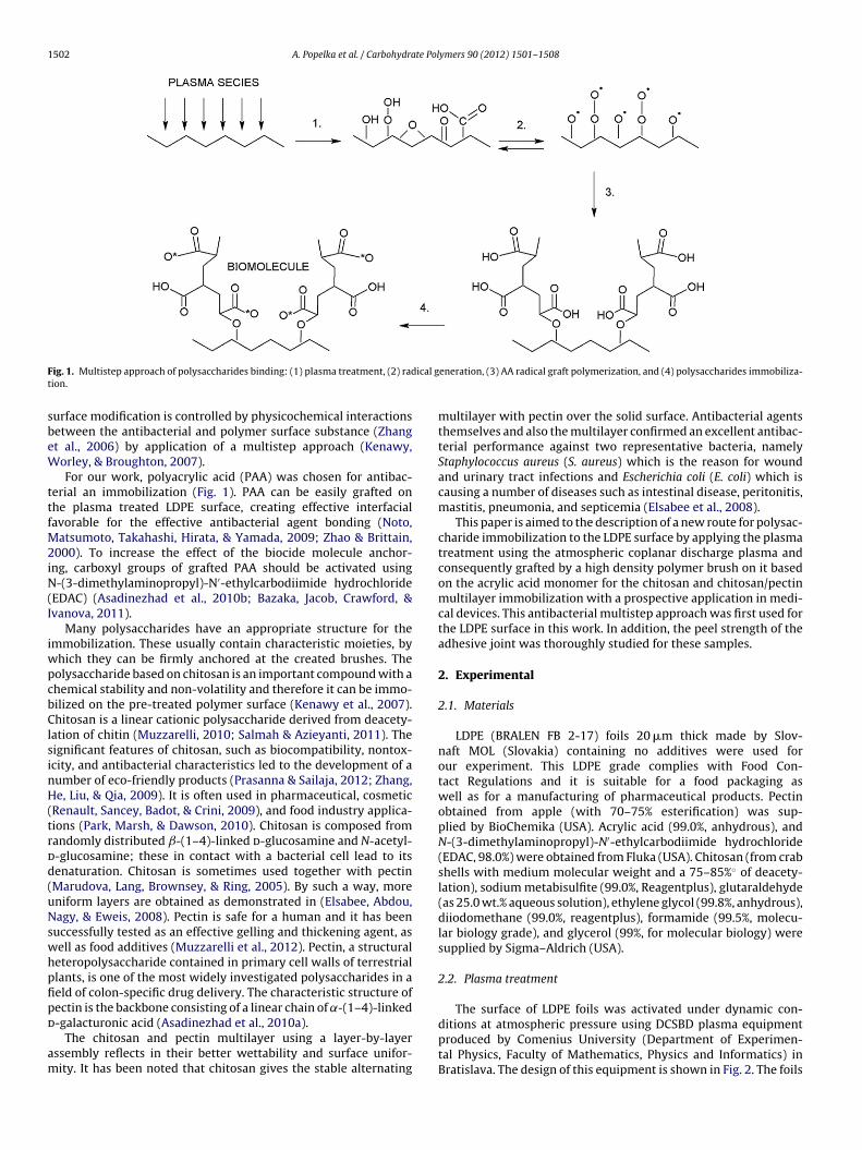
1502 A. Popelka et al. / Carbohydrate Polymers 90 (2012) 1501– 1508
F ical gt
sbeW
ttfM2iN(I
iwpcbClsinH(trdd(uNswhpfipd
am
ig. 1. Multistep approach of polysaccharides binding: (1) plasma treatment, (2) radion.
urface modification is controlled by physicochemical interactionsetween the antibacterial and polymer surface substance (Zhangt al., 2006) by application of a multistep approach (Kenawy,orley, & Broughton, 2007).For our work, polyacrylic acid (PAA) was chosen for antibac-
erial an immobilization (Fig. 1). PAA can be easily grafted onhe plasma treated LDPE surface, creating effective interfacialavorable for the effective antibacterial agent bonding (Noto,
atsumoto, Takahashi, Hirata, & Yamada, 2009; Zhao & Brittain,000). To increase the effect of the biocide molecule anchor-
ng, carboxyl groups of grafted PAA should be activated using-(3-dimethylaminopropyl)-N′-ethylcarbodiimide hydrochloride
EDAC) (Asadinezhad et al., 2010b; Bazaka, Jacob, Crawford, &vanova, 2011).
Many polysaccharides have an appropriate structure for themmobilization. These usually contain characteristic moieties, by
hich they can be firmly anchored at the created brushes. Theolysaccharide based on chitosan is an important compound with ahemical stability and non-volatility and therefore it can be immo-ilized on the pre-treated polymer surface (Kenawy et al., 2007).hitosan is a linear cationic polysaccharide derived from deacety-
ation of chitin (Muzzarelli, 2010; Salmah & Azieyanti, 2011). Theignificant features of chitosan, such as biocompatibility, nontox-city, and antibacterial characteristics led to the development of aumber of eco-friendly products (Prasanna & Sailaja, 2012; Zhang,e, Liu, & Qia, 2009). It is often used in pharmaceutical, cosmetic
Renault, Sancey, Badot, & Crini, 2009), and food industry applica-ions (Park, Marsh, & Dawson, 2010). Chitosan is composed fromandomly distributed ˇ-(1–4)-linked d-glucosamine and N-acetyl--glucosamine; these in contact with a bacterial cell lead to itsenaturation. Chitosan is sometimes used together with pectinMarudova, Lang, Brownsey, & Ring, 2005). By such a way, moreniform layers are obtained as demonstrated in (Elsabee, Abdou,agy, & Eweis, 2008). Pectin is safe for a human and it has been
uccessfully tested as an effective gelling and thickening agent, asell as food additives (Muzzarelli et al., 2012). Pectin, a structuraleteropolysaccharide contained in primary cell walls of terrestriallants, is one of the most widely investigated polysaccharides in aeld of colon-specific drug delivery. The characteristic structure ofectin is the backbone consisting of a linear chain of ˛-(1–4)-linked
-galacturonic acid (Asadinezhad et al., 2010a).The chitosan and pectin multilayer using a layer-by-layerssembly reflects in their better wettability and surface unifor-ity. It has been noted that chitosan gives the stable alternating
eneration, (3) AA radical graft polymerization, and (4) polysaccharides immobiliza-
multilayer with pectin over the solid surface. Antibacterial agentsthemselves and also the multilayer confirmed an excellent antibac-terial performance against two representative bacteria, namelyStaphylococcus aureus (S. aureus) which is the reason for woundand urinary tract infections and Escherichia coli (E. coli) which iscausing a number of diseases such as intestinal disease, peritonitis,mastitis, pneumonia, and septicemia (Elsabee et al., 2008).
This paper is aimed to the description of a new route for polysac-charide immobilization to the LDPE surface by applying the plasmatreatment using the atmospheric coplanar discharge plasma andconsequently grafted by a high density polymer brush on it basedon the acrylic acid monomer for the chitosan and chitosan/pectinmultilayer immobilization with a prospective application in medi-cal devices. This antibacterial multistep approach was first used forthe LDPE surface in this work. In addition, the peel strength of theadhesive joint was thoroughly studied for these samples.
2. Experimental
2.1. Materials
LDPE (BRALEN FB 2-17) foils 20 �m thick made by Slov-naft MOL (Slovakia) containing no additives were used forour experiment. This LDPE grade complies with Food Con-tact Regulations and it is suitable for a food packaging aswell as for a manufacturing of pharmaceutical products. Pectinobtained from apple (with 70–75% esterification) was sup-plied by BioChemika (USA). Acrylic acid (99.0%, anhydrous), andN-(3-dimethylaminopropyl)-N′-ethylcarbodiimide hydrochloride(EDAC, 98.0%) were obtained from Fluka (USA). Chitosan (from crabshells with medium molecular weight and a 75–85%◦ of deacety-lation), sodium metabisulfite (99.0%, Reagentplus), glutaraldehyde(as 25.0 wt.% aqueous solution), ethylene glycol (99.8%, anhydrous),diiodomethane (99.0%, reagentplus), formamide (99.5%, molecu-lar biology grade), and glycerol (99%, for molecular biology) weresupplied by Sigma–Aldrich (USA).
2.2. Plasma treatment
The surface of LDPE foils was activated under dynamic con-
ditions at atmospheric pressure using DCSBD plasma equipmentproduced by Comenius University (Department of Experimen-tal Physics, Faculty of Mathematics, Physics and Informatics) inBratislava. The design of this equipment is shown in Fig. 2. The foils
A. Popelka et al. / Carbohydrate Pol
wwt5pi∼s
2
iocallww
2
ogtrs(3ctts
Fig. 2. Scheme of DCSBD plasma equipment.
ere treated at power density of 1 W/cm2; the plasma treatmentas performed for 15 s in air as a carrier gas. Both foil sides were
reated. Two parallel banded systems of electrodes (1 mm wide,0 �m thick, with 0.5 mm spacing between the strips, made of Ag-aste) generate plasma by an effective way. Strips are embedded
n 96% Al2O3. A high frequency sinusoidal voltage (∼15 kHz at Um
10 kV) was used. Plasma generated by this equipment is macro-copically homogenous leading to the uniform surface treatment.
.3. PAA Grafting
Immediately after the plasma treatment, the samples weremmersed into 10 vol.% aqueous solution of AA for 24 h at 30 ◦C inrder to achieve a radical graft polymerization of AA. The solutionontained 0.1 wt.% of sodium metabisulfite as a relevant reductantgent to inhibit an AA homopolymerization. The AA polymerizationed to the creation of PAA brushes that are suitable for the immobi-ization of antibacterial agents. After the grafting the samples were
ashed in deionized water for 5 min at 30 ◦C in order to removeeakly bounded or unreacted AA.
.4. Chitosan and chitosan/pectin immobilization
PAA grafted LDPE foils were immersed into 0.1% (w/v) aque-us solution of EDAC at 4 ◦C for 6 h, for activation of carboxylroups. The activation reaction of carboxyl groups by EDAC ledo the formation of O-acylisourea with ability to react with someeducing agents (Nakajima & Ikada, 1995). Then the pre-treatedamples with activated carboxyl groups were immersed into 1%w/v) chitosan in 2% (v/v) acetic acid aqueous solution for 24 h at0 ◦C. In another case, the samples were dipped into chitosan and
onsequently pectin solution (2% (v/v) acetic acid aqueous solu-ion/prepared by the same way as described above for chitosan);he dipping was repeated nine times with 20 min duration in eacholution. Finally, the samples prepared by any of the proceduresFig. 3. Peel strength vs. surface treatment of LDPE sample: 1 – untreated, 2 – plasma
ymers 90 (2012) 1501– 1508 1503
described above were immersed in 1% (w/v) glutaraldehyde aque-ous solution overnight at 4 ◦C to achieve the immobilization oftwo polysaccharides via crosslinking processes. The crosslinkingreaction occurred with imine formation resulted in the reaction ofprimary amine with aldehyde (Carey & Sundberg, 2007). The pre-pared samples were then thoroughly washed and dried for 24 h atroom temperature.
2.5. Surface wettability evaluation
Wettability changes of the LDPE surface after the polysaccha-rides immobilization by the multistep process were obtained fromthe contact angle measurements. The surface energy evaluationsystem (SEE system with CCD camera, Advex Instruments, CzechRepublic) was used for experiments and a sessile drop techniquewas performed. A volume of 3 �l for each drop of testing liquidplaced on a sample was used for investigation of a static contactangle. Ten separate readings were averaged to obtain one repre-sentative contact angle value for each liquid. The contact angle isreferred as an angle between the solid/liquid and liquid/vapourinterface. Deionized water, ethylene glycol, glycerol, formamide,and diiodomethane were used as testing liquids. Contact angle ofeach drop was measured after approximately 3 s which is sufficientfor an achievement of a thermodynamic equilibrium between solid,liquid, and gas phases was reached. The testing liquids were usedfor a calculation of total (� tot), polar (�p) and dispersive (�d) com-ponents of the surface free energy. Owens–Wendt–Rable–Kaebleregression model using the method of least squares was used forthe evaluation of � tot, �p, and �d (Salimi, Mirabedini, Atai, Mohseni,& Naimi-Jamal, 2011). The graft yield (GY) was calculated accord-ing to the equation GY (%) = ((W2 − W1)/W1)·100%, where W1 andW2 represent weights of the samples before and after the surfacetreatment, respectively (Is iklan, Kurs un, & Inal, 2010).
2.6. Adhesive properties assessment
An adhesion between two materials was characterized by thepeel strength (force per unit width). The peel test was used for peelstrength measurements of the adhesive joint formed of LPDE foilsand poly(2-ethylhexyl acrylate) as an adhesive agent depositedonto polypropylene foil of 15 mm wide. Measurements were per-formed as 90◦ peel test at a rate of peel 10 mm per minute using
100 N universal INSTRON 4301 dynamometer (UK). The both endsof the LDPE sample and PP with adhesive were firmly fixed intodynamometer jaws to achieve an even tension distribution acrossthe entire width.treated, 3 – PAA grafted, 4 – chitosan coated, and 5 – chitosan/pectin coated.
1504 A. Popelka et al. / Carbohydrate Polymers 90 (2012) 1501– 1508
Table 1Surface properties of LDPE treated by multistep process (� – contact angle; � tot, �d, �p – total surface free energy, its dispersive and polar component, respectively; GY – graftyield).
LDPE sample �w (◦) �e (◦) �g (◦) �d (◦) �f (◦) �p (mN/m) �d (mN/m) � tot (mN/m) GY (%)
Untreated 99.2 (±0.6) 70.9 (±1.2) 85.3 (±0.9) 48.4 (±1.2) 80.7 (±0.9) 0.2 31.5 31.7 –Plasma treated (A) 77.5 (±1.1) 51.0 (±2.8) 67.1 (±2.8) 36.0 (±1.2) 52.8 (±1.5) 1.1 41.4 42.6 0.0A + PAA grafted (B) 66.9 (±0.7) 32.1 (±2.4) 57.2 (±2.7) 32.5 (±1.6) 37.0 (±2.0) 4.5 43.7 48.1 0.5B + chitosan coated 69.2 (±0.8) 36.0 (±2.1) 68.3 (±1.2) 35.9 (±1.9) 33.1 (±2.2) 6.1 38.8 44.9 3.1B + chitosan/pectin coated 59.1 (±1.1) 30.0 (±2.8) 53.40 (±1.3) 37.8 (±2.6) 33.8 (±2.7) 11.9 36.1 48.0 8.2
w: deionized water; e: ethylene glycol; g: glycerol; d: diiodomethane; f: formamide.
Fig. 4. SEM micrographs showing surface morphology for LDPE samples: a – untreated, b – plasma treated, c – PAA grafted, d – chitosan coated, and e – chitosan/pectincoated.

A. Popelka et al. / Carbohydrate Polymers 90 (2012) 1501– 1508 1505
eated
2
zot2awas
2
2
lTboasoeuwt
2
rcLbofhut
2
aa
Fig. 5. FTIR-ATR spectra of LDPE samples: 1 – untreated, 2 – plasma tr
.7. Surface morphology analysis
Scanning electron microscope (SEM) was used for a characteri-ation of the surface morphology and local surface heterogeneitiesf LDPE samples. The surfaces of both untreated and antibacterial-reated LDPE films were observed by the SEM microscope (Quanta00 FEG; FEI, Czech Republic) using secondary electrons detectornd accelerating voltage 30 kV. Before measurement, the samplesere sputter-coated by a thin layer of Pt (∼4 nm). All samples were
nalyzed at several locations (≥3) in order to find characteristic andignificant surface features.
.8. Surface chemistry investigation
.8.1. X-ray photoelectron spectroscopyThe chemical surface composition of LDPE samples was ana-
yzed with the X-ray photoelectron spectroscopy (XPS) instrumentFA XPS Physical Electronics (USA). The pressure in the XPS cham-er was about 6 × 10⁻⁸ Pa. The samples were irradiated with X-raysver a 400 �m spot area with a monochromatic Al K�1,2 radiationt 1486.6 eV. Created photoelectrons were detected with a hemi-pherical analyzer placed at angle of 45◦ with respect to the normalf the sample surface. Each survey-scan spectra was made at a passnergy of 187.85 eV and 0.4 eV energy step. An electron gun wassed for the surface neutralization. The concentration of elementsas determined using MultiPak v7.3.1 software from Physical Elec-
ronics.
.8.2. Infrared spectroscopyFourier transform infrared spectroscopy with attenuated total
eflectance (FTIR–ATR) was used for an investigation of the surfacehemical composition. The spectra were recorded by the FTIR NICO-ET 8700 spectrometer (Thermo Scientific USA) through the singleounce ATR with Ge crystal at 45◦ incident angle. The spectral res-lution and the number of scans were 2 cm−1 and 64, respectivelyor each measurement. The pressure clamp was used to obtain theighest quality of the spectra. The acquired spectra were analyzedsing OMNICTM, v. 8.1 software. Each measurement was triplicateo obtain the average spectra for different spots.
.8.3. Antibacterial activity assessmentThe antibacterial activity of prepared samples was tested
gainst two bacterial strains Staphylococcus aureus (CCM 4516)nd Escherichia coli (CCM 4517) by the inhibition zone method
, 3 – PAA grafted, 4 – chitosan coated, and 5 – chitosan/pectin coated.
(diffusion test) on agar. Nutrient agar No. 2 M1269 – 500 g fromHiMedia Laboratories PII. Ltc. was used for our experiments. Testedsamples were cut in a circular shape (d = 8 mm), washed in ethanol,dried and placed on an agar plate inoculated by the bacterialsuspension (volume = 100 �l, concentration = 107 units/ml). Thesamples were incubated for 24 h at 37 ◦C and diameters of the inhi-bition zone were measured in 5 directions to obtain average valuesfor inhibition zone calculations. The test with each sample wastriplicate.
3. Results and discussion
3.1. Surface wettability
Surface parameters of untreated and treated LDPE calculatedfrom contact angle data for various testing liquid are shown inTable 1. The hydrophobic and chemical inert surface nature ofuntreated LDPE is the reason for high values of contact angle (�)due to low surface wettability. The significant decrease of � wasobserved after the plasma treatment because characteristics reac-tive polar functional groups were introduced onto the LDPE surface.The PAA grafting led to the further decrease of �, whereas PAAcontains polar carboxylic groups. In addition, the chitosan and chi-tosan/pectin multilayer led to the significant decrease of � due tothe presence of characteristic polar functional groups. Accordinglyto the measured contact angle values, low values were calculatedalso for � tot of untreated LPDE associated with its hydrophobicnature. The plasma treatment leads to the increase of LDPE � tot indi-cating the surface polarity increase. Even greater increase of LDPE� tot was recorded for PAA grafted LDPE and for chitosan immobi-lized LDPE. The highest increase of � tot and �p was observed forchitosan/pectin multilayer immobilized on the LDPE surface viaPAA.
3.2. Adhesive properties
The information about adhesion changes of the adhesive jointto more polar polyacrylate were obtained from peel test measure-ments that are shown in Fig. 3. The adhesion can be expressed by aforce per width (peel strength). The peel strength closely relates to
� tot, roughness and chemical nature of investigated materials form-ing an adhesive joint. Therefore, the increase of wettability resultsin the peel strength increase of the adhesive joint to more polarpolyacrylate. On the other side, rougher surface results in higher
1506 A. Popelka et al. / Carbohydrate Polymers 90 (2012) 1501– 1508
F reatedc
asfTsPftoct
3
tm
ig. 6. XPS survey-scan spectra of LDPE samples with atomic composition: a – untoated.
dhesion and vice versa. The adhesion is thus a complex of theeveral related chemical and physicochemical properties. There-ore, the peel strength of untreated LDPE achieves very low values.he plasma treatment resulted in a double increase of the peeltrength caused by the changes in polarity and surface roughness.AA grafting and chitosan/pectin multilayer coating leads to theurther increase in the peel strength compared to values for plasmareated LDPE. The most pronounced increase in the peel strengthf adhesive joint LDPE samples-polyacrylate was observed for thehitosan coating. The chitosan coating led to the most increase ofhe surface roughness.
.3. Surface morphology
Changes in the surface morphology of untreated and antibac-erial treated LDPE by the multistep process obtained from SEM
easurements are shown in Fig. 4. The surface morphology of
, b – plasma treated, c – PAA grafted, d – chitosan coated, and e – chitosan/pectin
untreated LDPE (Fig. 4a) is characterized by a very low sur-face roughness. The plasma treatment of LDPE (Fig. 4b) resultsin a slight increase of the surface roughness as a result of sur-face changes by a combination of functionalization and ablationprocesses. PAA brushes formed on the LDPE surface exhibited acharacteristic texture (Fig. 4c). The domain size increased as thegrafting advanced. The other conclusive factor influencing the sur-face morphology is a grafting mechanism. A certain amount ofgenerated radicals in the sublayer initiates the grafting reaction.The bulged top layer results from the AA monomer polymeriza-tion participating in the chain propagation process. The chitosanimmobilization by glutaraldehyde as a crosslinking agent leads tothe formation of chitosan agglomerates on the continuous layer
of the PAA grafted surface (Fig. 4d). Pectin significantly increasesthe uniformity of the chitosan layer; more uniform surface mor-phology is obtained for the chitosan/pectin multilayer as seenin Fig. 4e.
te Polymers 90 (2012) 1501– 1508 1507
3
3
iifapcFoo1b
mbsoCrttcstse(pipia
mcm
3
adtAtc
fLptptLisbPdcnhca
Fig. 7. Inhibition zone area of LDPE samples for S. aureus and E. coli strains. Each
A. Popelka et al. / Carbohydra
.4. Surface chemistry
.4.1. Analysis of FTIR-ATR spectraFTIR-ATR measurements provide mostly semi-quantitative
nformation about chemical changes in a near-surface region. Thenfrared spectra of LDPE samples were splitted into three regionsor better visualization. The spectrum of untreated LDPE is a char-cteristic spectrum of polyethylene with only few characteristiceaks. After plasma exposure of the untreated material, signifi-ant changes in a measured spectrum are observed. As seen inig. 5, the incorporation of oxygen containing groups was obvi-us, i.e. hydroperoxides (region 3700–3080 cm−1) and/or otherxygen containing products at the surface of the material (region845–1510 cm−1, 1280 cm−1, 1126 cm−1, 1150 cm−1, carboxyl, car-onyl or aldehydic moieties).
Other significant changes in the spectra are observed for LDPEodified by PAA grafting, and also after the subsequent treatment
y chitosan, chitosan/pectin and glutaraldehyde, respectively. Thepectrum of grafted material contains several characteristic peaksf PAA, i.e. the most intense peak at 1712 cm−1 (carbonyl band,
O stretching), and also some unresolved peaks in the fingerprintegion (1300–1100 cm−1, C O stretching and CH2 bending). Afterhe chitosan and glutaraldehyde treatment, the shape of the spec-rum is changing, as can be seen in Fig. 5. Because of the treatmentomplexity, these changes in the spectra can be interpreted withome difficulties – the spectra of chitosan, pectin and also of glu-araldehyde are very similar in the fingerprint region. Despite thistatement, the spectra of samples 4 and 5 indicates also the pres-nce of acrylic acid (carbonyl band, 1712 cm−1), pectin – 1734 cm−1
C O band arising from pectin). The presence of chitosan in sam-les 4 and 5 is confirmed by an appearing of the band at 1653 cm−1
n corresponding spectra (-CNH band arising from chitosan). Theresence of glutaraldehyde (suggesting as a crosslinking agent) is
ndicated in the spectrum at lower wavenumbers (approximatelyt 1100 cm−1 as a contribution to C O absorbance).
The changes in the spectra are significant almost in a wholeid-infrared region, especially in the fingerprint region and they
onfirm the incorporation of chemicals used for the surface treat-ent of LDPE.
.4.2. Analysis of XPS spectraThe LDPE samples with the different treatment were thoroughly
nalyzed by the XPS method. The objective was to get the evi-ence of the presence of the antibacterial substances coating onhe LDPE surface via the plasma treatment in air and grafting withA. The surface composition for each sample was measured at
wo different spots allowing the calculation of an average surfaceomposition.
The XPS survey-scan spectra of samples with the average sur-ace composition are shown in Fig. 6. As expected, the untreatedDPE has a characteristic spectrum composed of 100 at.% of C1seak (Fig. 6a), belonging to C C bonds. Different oxygen func-ional groups and also some nitrogen groups were found in thelasma treated sample in air (Fig. 6b). Carbon C1s peak correspondso C C, O C O, C O, C O groups. The nitrogen N1s peak of theDPE sample treated in air plasma is composed of different chem-cal bonds of nitrogen atoms such as C N, C NH3
+, ONO2. Theample of PAA grafted on LDPE showed mainly the presence of car-oxyl groups (Fig. 6c). The oxygen groups originate mainly fromAA but also other oxygen groups are present, which were createduring and immediately after the plasma treatment. In samplesoated with chitosan or chitosan-pectin, large oxygen and some
itrogen content is detected. The sample coated only with chitosanas higher nitrogen content (Fig. 6d), while the sample coated withhitosan–pectin has higher oxygen content (Fig. 6e). In this samplelso traces of silicon impurities were detected – about 0.4 at.%. Thecolumn represents the inhibition zone area for one experiment out of three.
XPS spectrum indicates, that a number of different moieties (e.g.carbonyl, carboxyl, etc.) containing oxygen is present in treatedLDPE.
3.5. Antibacterial activity
The inhibition zone area was calculated from an average diam-eter of the inhibition zone, whereas the area of the sample wasnot taken into account. Each measurement was triplication (Fig. 7).The untreated, plasma treated, and PAA grafted LDPE sample withchitosan together with glutaraldehyde did not show any antibacte-rial activity against E. coli and S. aureus strains. The chitosan/pectincoated sample showed minor activity only against S. aureus,the inhibition zone being around 70 mm2. Similar results wereobtained for the chitosan/pectin coated sample after crosslinkingby glutaraldehyde. This sample showed activity also against E. coli.However, the antibacterial activity of these samples is not signif-icant. The highest and most clear inhibition zones were given bysamples grafted by PAA and coated by chitosan. The levels in thiscase were on average 35 mm2 for E. coli and 275 mm2 for S. aureus.
The PAA grafted sample did not show any inhibition zone forE. coli, nevertheless the same sample indicated the antibacterialactivity for S. aureus. This could be explained by high sensitivity ofthe PAA brushes and their ability to easily absorb impurities duringmanipulation. As it can be seen from results, the sample graftedby PAA and coated by chitosan only demonstrated active antibac-terial properties against both bacterial strains. Other samples didnot prove the significant antibacterial activity. Chitosan is prob-ably weakly bonded to the PAA surface and it can diffuse easily.On the other hand the LDPE surface treated by the multilayer of
chitosan/pectin or additionally crosslinked by glutaraldehyde pre-vents chitosan molecule to diffuse and form the inhibition zone.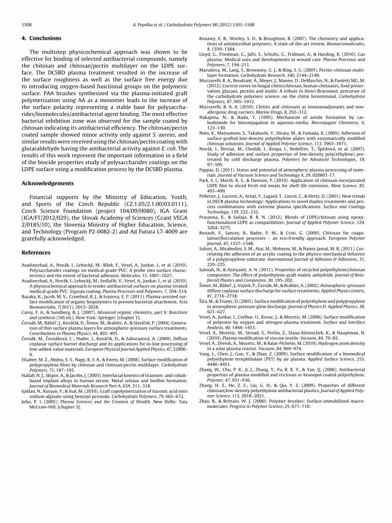
1 te Pol
4
etfttsptrbccsgroL
A
aC(2ag
R
A
A
B
C
C
C
E
H
I
J
508 A. Popelka et al. / Carbohydra
. Conclusions
The multistep physicochemical approach was shown to beffective for binding of selected antibacterial compounds, namelyhe chitosan and chitosan/pectin multilayer on the LDPE sur-ace. The DCSBD plasma treatment resulted in the increase ofhe surface roughness as well as the surface free energy dueo introducing oxygen-based functional groups on the polymericurface. PAA brushes synthesized via the plasma-initiated graftolymerization using AA as a monomer leads to the increase ofhe surface polarity representing a stable base for polysaccha-ides/biomolecules/antibacterial agent binding. The most effectiveacterial inhibition zone was observed for the sample coated byhitosan indicating its antibacterial efficiency. The chitosan/pectinoated sample showed minor activity only against S. aureus, andimilar results were received using the chitosan/pectin coating withlutaraldehyde having the antibacterial activity against E. coli. Theesults of this work represent the important information in a fieldf the biocide properties study of polysaccharides coatings on theDPE surface using a modification process by the DCSBD plasma.
cknowledgements
Financial supports by the Ministry of Education, Youth,nd Sports of the Czech Republic (CZ.1.05/2.1.00/03.0111),zech Science Foundation (project 104/09/H080), IGA GrantIGA/FT/2012/029), the Slovak Academy of Sciences (Grant VEGA/0185/10), the Slovenia Ministry of Higher Education, Science,nd Technology (Program P2-0082-2) and Ad Futura L7-4009 areratefully acknowledged.
eferences
sadinezhad, A., Novák, I., Lehocky, M., Bílek, F., Vesel, A., Junkar, I., et al. (2010).Polysaccharides coatings on medical-grade PVC: A probe into surface charac-teristics and the extent of bacterial adhesion. Molecules, 15, 1007–1027.
sadinezhad, A., Novák, I., Lehocky, M., Sedlarík, V., Vesel, A., Junkar, I., et al. (2010).A physicochemical approach to render antibacterial surfaces on plasma-treatedmedical-grade PVC: Irgasan coating. Plasma Processes and Polymers, 7, 504–514.
azaka, K., Jacob, M. V., Crawford, R. J., & Ivanova, E. P. (2011). Plasma-assisted sur-face modification of organic biopolymers to prevent bacterial attachment. ActaBiomaterialia, 7(2011), 2015–2028.
arey, F. A., & Sundberg, R. J. (2007). Advanced organic chemistry, part b: Reactionsand synthesis (5th ed.). New York: Springer. [chapter 1].
ˇernák, M., Ráhel’, J., Kovácik, D., Simor, M., Brablec, A., & Slavícek, P. (2004). Genera-tion of thin surface plasma layers for atmospheric-pressure surface treatments.Contributions to Plasma Physics, 44, 492–495.
ˇernák, M., Cernáková, L’., Hudec, I., Kovácik, D., & Zahoranová, A. (2009). Diffusecoplanar surface barrier discharge and its applications for in-line processing oflow-added-value materials. European Physical Journal Applied Physics, 47, 22806-6.
lsabee, M. Z., Abdou, E. S., Nagy, K. S. A., & Eweis, M. (2008). Surface modification ofpolypropylene films by chitosan and chitosan/pectin multilayer. CarbohydratePolymers, 71, 187–195.
allab, N. J., Skipor, A., & Jacobs, J. (2003). Interfacial kinetics of titanium- and cobalt-based implant alloys in human serum: Metal release and biofilm formation.
Journal of Biomedical Materials Research Part A, 65A, 311–318.s iklan, N., Kurs un, F., & Inal, M. (2010). Graft copolymerization of itaconic acid ontosodium alginate using benzoyl peroxide. Carbohydrate Polymers, 79, 665–672.
ohn, P. I. (2005). Plasma Sciences and the Creation of Wealth. New Delhi: TataMcGraw-Hill. [chapter 3].
ymers 90 (2012) 1501– 1508
Kenawy, E. R., Worley, S. D., & Broughton, R. (2007). The chemistry and applica-tions of antimicrobial polymers: A state-of-the-art review. Biomacromolecules,8, 1359–1384.
Lloyd, G., Friedman, G., Jafri, S., Schultz, G., Fridman, A., & Harding, K. (2010). Gasplasma: Medical uses and developments in wound care. Plasma Processes andPolymers, 7, 194–211.
Marudova, M., Lang, S., Brownsey, G. J., & Ring, S. G. (2005). Pectin–chitosan multi-layer formation. Carbohydrate Research, 340, 2144–2149.
Muzzarelli, R. A., Boudrant, A., Meyer, J., Manno, D., DeMarchis, N., & Paoletti MG, M.(2012). Current views on fungal chitin/chitosan, human chitinases, food preser-vation, glucans, pectins and inulin: A tribute to Henri Braconnot, precursor ofthe carbohydrate polymers science, on the chitin bicentennial. CarbohydratePolymers, 87, 995–1012.
Muzzarelli, R. A. A. (2010). Chitins and chitosans as immunoadjuvants and non-allergenic drug carriers. Marine Drugs, 8, 292–312.
Nakajima, N., & Ikada, Y. (1995). Mechanism of amide formation by car-bodiimide for bioconjugation in aqueous-media. Bioconjugate Chemistry, 6,123–130.
Noto, K., Matsumoto, S., Takahashi, Y., Hirata, M., & Yamada, K. (2009). Adhesion ofsurface-grafted low-density polyethylene plates with enzymatically modifiedchitosan solutions. Journal of Applied Polymer Science, 113, 3963–3971.
Novák, I., Steviar, M., Chodák, I., Krupa, I., Nedelcev, T., Spírková, et al. (2007).Study of adhesion and surface properties of low-density poly(ethylene) pre-treated by cold discharge plasma. Polymers for Advanced Technologies, 18,97–105.
Pappas, D. (2011). Status and potential of atmospheric plasma processing of mate-rials. Journal of Vacuum Science and Technology A, 29, 020801-17.
Park, S. I., Marsh, K. S., & Dawson, P. (2010). Application of chitosan-incorporatedLDPE film to sliced fresh red meats for shelf life extension. Meat Science, 85,493–499.
Pelletier, J., Lacoste, A., Arnal, Y., Lagard, T., Lincot, C., & Hertz, D. (2001). New trendsin DECR plasma technology: Applications to novel duplex treatments and pro-cess combinations with extreme plasma specifications. Surface and CoatingsTechnology, 139, 222–232.
Prasanna, K., & Sailaja, R. R. N. (2012). Blends of LDPE/chitosan using epoxy-functionalized LDPE as compatibilizer. Journal of Applied Polymer Science, 124,3264–3275.
Renault, F., Sancey, B., Badot, P. M., & Crini, G. (2009). Chitosan for coagu-lation/flocculation processes – an eco-friendly approach. European PolymerJournal, 45, 1337–1348.
Salimi, A., Mirabedini, S. M., Atai, M., Mohseni, M., & Naimi-Jamal, M. R. (2011). Cor-relating the adhesion of an acrylic coating to the physico-mechanical behaviorof a polypropylene substrate. International Journal of Adhesion & Adhesives, 31,220–225.
Salmah, H., & Azieyanti, A. N. (2011). Properties of recycled polyethylene/chitosancomposites: The effect of polyethylene-graft-maleic anhydride. Journal of Rein-forced Plastics and Composites, 30, 195–202.
Simor, M., Ráhel’, J., Vojtek, P., Cernák, M., & Brablec, A. (2002). Atmospheric-pressurediffuse coplanar surface discharge for surface treatments. Applied Physics Letters,81, 2716–2718.
Síra, M., & Trunec, D. (2005). Surface modification of polyethylene and polypropylenein atmospheric pressure glow discharge. Journal of Physics D: Applied Physics, 38,621–627.
Vesel, A., Junkar, I., Cvelbar, U., Kovac, J., & Mozetic, M. (2008). Surface modificationof polyester by oxygen and nitrogen-plasma treatment. Surface and InterfaceAnalysis, 40, 1444–1453.
Vesel, A., Mozetic, M., Strnad, S., Persin, Z., Stana-Kleinschek, K., & Hauptman, N.(2010). Plasma modification of viscose textile. Vacuum, 84, 79–82.
Vesel, A., Drenik, A., Mozetic, M., & Balat-Pichelin, M. (2010). Hydrogen atom densityin a solar plasma reactor. Vacuum, 84, 969–974.
Yang, L., Chen, J., Guo, Y., & Zhan, Z. (2009). Surface modification of a biomedicalpolyethylene terephthalate (PET) by air plasma. Applied Surface Science, 255,4446–4451.
Zhang, W., Chu, P. K., Ji, J., Zhang, Y., Fu, R. K. Y., & Yan, Q. (2006). Antibacterialproperties of plasma-modified and triclosan or bronopol coated polyethylene.Polymer, 47, 931–936.
Zhang, H. Z., He, Z. C., Liu, G. H., & Qia, Y. Z. (2009). Properties of differentchitosan/low-density polyethylene antibacterial plastics. Journal of Applied Poly-mer Science, 113, 2018–2021.
Zhao, B., & Brittain, W. J. (2000). Polymer brushes: Surface-immobilized macro-molecules. Progress in Polymer Science, 25, 677–710.
