original article core decompression combined with ...the osteonecrosis of femoral head (onfh) could...
TRANSCRIPT
Int J Clin Exp Med 2016;9(6):10281-10288www.ijcem.com /ISSN:1940-5901/IJCEM0022220
Original ArticleCore decompression combined with superselective arterial infusion in treating early nontraumatic osteonecrosis of femoral head
Qian Chen, Li Qian, Liang Zhang, Da-Shou Wang, Qi Pan, Qian-Ming Wu, Meng He, Yong Jin, Yan Chen, Rui-Hong Huang, Yue Zhai, Jin-Yu Luo, Dan Liao, Yang Xiao, Yang-Ming Sunwen
Department of Pain Medicine, Orthopedics Hospital of Guizhou Province, Guiyang 550002, China
Received December 18, 2015; Accepted March 5, 2016; Epub June 15, 2016; Published June 30, 2016
Abstract: The aim of this study was to investigate the early- and mid-term therapeutic effects of core decompres-sion (CD) combined with superselective arterial infusion (SAI) in treating the osteonecrosis of femoral head (ONFH). 71 patients with Ficat stage I-III avascular necrosis of femoral head and accepted the CD treatment were randomly divided into: group A: simply performed the CD surgery, group B: CD+SAI. The pain rating index (PRI) and the Harris score were measured before the surgery, as well as 1 and 6 months after the surgery, the bi-hip MRI examination was performed before and 6 months after the surgery. PRI of Ficat stage I-II patients in group B at all postoperative time points were lower than group A, while the Harris scores were higher than group A (P<0.05). The Ficat stage III patients in group A showed no significant changes 1- and 6-month after the PRI surgery, while the postoperative Harris scores were increased first (1 month after the surgery), then fell to the preoperative level; the group B PRI was reduced 1- and 6-month after the surgery, while the Harris scores were increased. The reduction rate of MRI necrotic area of group B 6-month after the surgery was significantly higher than group A (P<0.05). The post-CD SAI could be more effective in relieving the avascular necrosis of femoral head.
Keywords: Osteonecrosis, core decompression, arterial infusion, pain
Introduction
The osteonecrosis of femoral head (ONFH) could be divided into traumatic and nontrau-matic according to the pathogenic factors [1], among which the main pathophysiological char-acteristics of the noninvasive ONFH (long-term application of corticosteroids, alcoholism, radio- therapy, chemotherapy and idiopathic osteone-crosis) were the occurrence of femoral head ischemic events, secondary cartilage fracture and collapse, etc. [2]. Therefore, the treatment of noninvasive ONFH was mainly focused on promoting the angiogenesis, blood flow and structural support towards the necrotic region, slowing the progress of ONFH and improving the patients’ life qualities [3-5].
The CD surgery was currently the most widely used surgical treatment in treating the isch-emic ONFH, with the effective rate as 63.5% (33%-95%) towards the early ONFH patients (Ficat I, II) [6]. While the effects of simple CD
surgery towards the patients with subchondral fracture even collapse were not obvious, the patients still needed to be combined with the structure or cell support to improve the effica-cies [7-9], especially the treatment towards the Ficat stage III patients still remained controver-sial. The uncertain indications and difficulties in selecting the treatment programs (CD+stru- ctural support or total hip replacement) were the main difficulties during the treatment [10].
In view of this, based on the treatment princi-ples of ischemic ONFH, our department select-ed CD combined with superselective arterial infusion to treat the ischemic ONFH patients (Ficat stage I-III) and achieved better clinical efficacies.
Methods
General information
71 non-traumatic ONFH patients (71 hips), treated in our hospital from May 2010 to

Treating early nontraumatic osteonecrosis
10282 Int J Clin Exp Med 2016;9(6):10281-10288
December 2013, were selected, the diagnostic criteria were as follows: joint pain mainly at thigh, groin and buttock, the hip’s mobility was limited, with the history of trauma, corticoste-roid application or alcohol abuse, etc., the X-ray examination exhibited cystic degeneration, hardening or “crescent sign”, MRI showed the necrotic femoral head. Among the patients, 51 cases were in Ficat stage I-II and 20 cases were in stage III. The patients included 44 males and 27 females, aged 20 to 59 years old. Causative factors: 37 cases of corticosteroids applica-tion, 20 cases of excessive alcohol abuse, and 14 cases had no apparent reason. Depending on different treatment programs, the patients were randomly divided into two groups, group A (n=42) was performed CD; group B (n=29) was performed the ipsilateral SAI 7 to 10 days after the CD surgery. All patients were informed about the experiments, and this experiment met the relevant requirements of the Declaration of Helsinki on Ethics. It could be known from Table 1 that: age distribution, gen-der, stage proportion and etiology proportion of the two groups had no significant difference (P>0.05) (non-parametric Chi-Square test and Kruskal-Wallis H test), the two groups were comparable. This study was conducted in accordance with the declaration of Helsinki. This study was conducted with approval from the Ethics Committee of Orthopedics Hospital of Guizhou Province. Written informed consent was obtained from all participants.
Treatment methods
Method of CD surgery: The patient was preop-eratively applied the continuous epidural block,
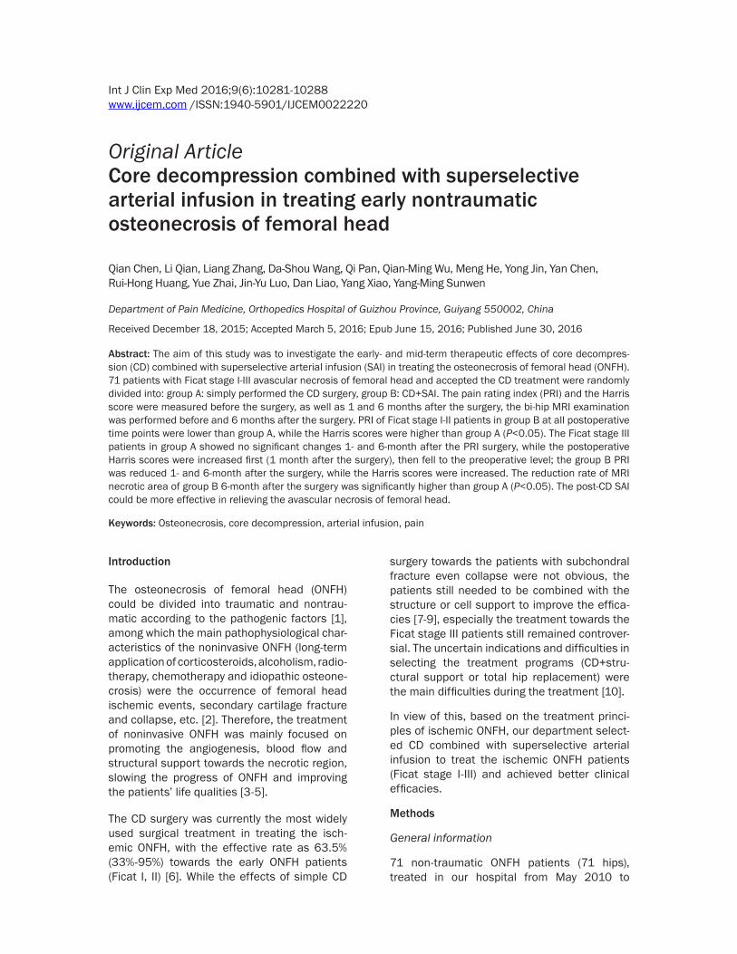
used to break the cortical bone to determine the needling direction and point under the guid-ance of C-arm X-ray machine, which set the center of necrotic area as the needling end point. When puncturing into the necrotic area, it should be careful not to break through the sub-chondral bone, after entered the necrotic area, pull out the needle core, used the cannulated reamer to drill through the necrotic region along the guide needle and removed partial bone tis-sues, then pull out the cannulated reamer; then drilled again with the solid drill a little until the necrotic lesions, then used the bone cement injector to inject the calcium sulfate bone sub-stitute alternatively, with the needle withdrawn during the injection, when the needle was with-drawn to the base of femoral neck, the gel hemostatic sponge was then injected to fill the channel. The second decompression channel was completed with the same method while towards different direction. The sterile elastic bandage was then used to fix and pressure-dress the puncture site Figure 1.
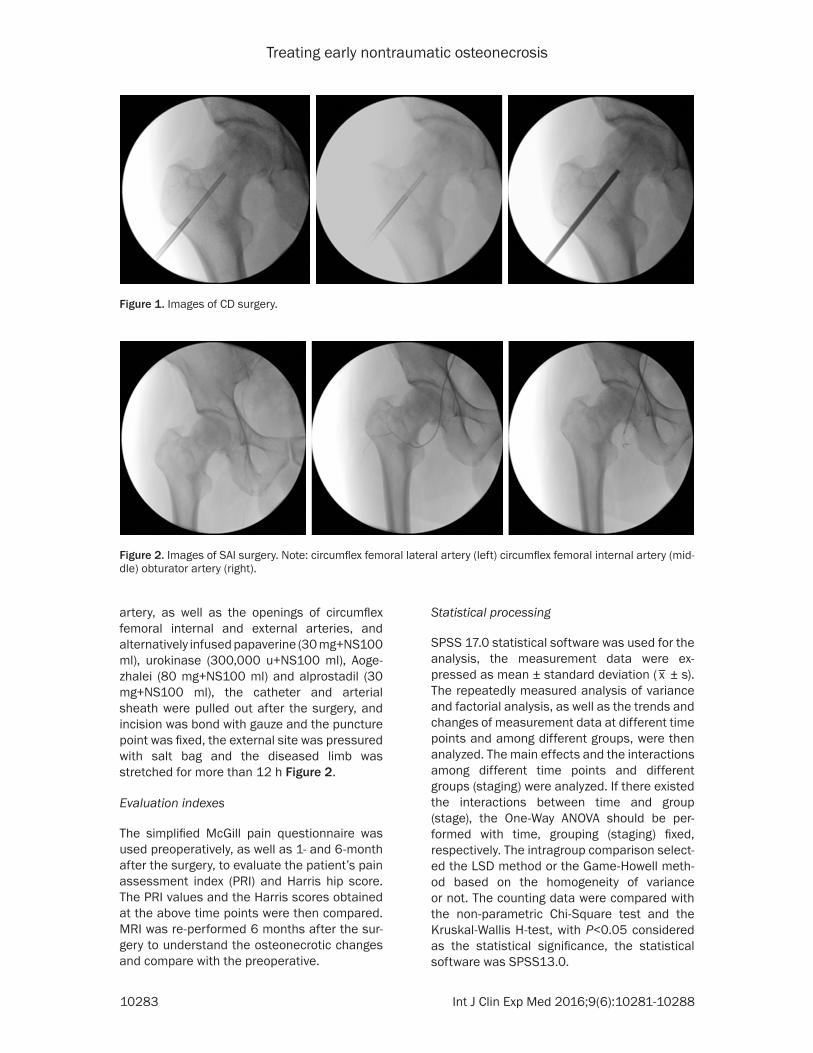
Method of SAI surgery: The patients was placed in the supine position, then under the guidance of C-type X-ray machine, the puncture point was selected at the artery pulse site, which was 1 cm below the midpoint of contralateral groin against the necrotic femoral head, after rou-tinely disinfected and paved the surgical tow-els, the patient was given 3 ml of 2% lidocaine for the topical anesthesia, and performed 0.2 cm incision, the arterial puncture needle was used to puncture the femoral artery, then pulled out the needle core, placed into the soft guide-wire and 5F arterial sheath, the C2 or C3 cath-eter was then introduced into the obturator
Table 1. Clinical characteristics of the 2 groupsClinical features Group A (n=42) Group B (n=29) P Age (years) The median age 38 41 >0.05 Range 18-62 20-59Gender (No, %) Male 26/42 (61.90%) 18/29 (62.03%) >0.05Ficat stage (%) I-II (n=51) 33/42 (78.57%) 18/29 (62.07%) >0.05 III (n=20) 9/42 (21.43%) 11/29 (37.93%)Cause of disease Corticosteroids application 21/42 (50.00%) 16/29 (55.17%) >0.05 Excessive alcohol abuse 13/42 (30.95%) 7/29 (24.14%) Without apparent reason 8/42 (19.05%) 6/29 (20.69%)
and placed in the lateral posi-tion, with the diseased limb placed upwards, the contralat-eral leg straightened and the ipsilateral leg flexed. The patient was performed the sur-face positioning to determine the drilling location, then one puncture point was selected 1 cm below the greater trochan-ter of diseased femur for one 4 mm incision. under the guid-ance of C-arm X-ray machine, one 4.0 mm bone puncturing needle was used to puncture to the bone cortex, then one 3.5 mm solid core drill was
Treating early nontraumatic osteonecrosis
10283 Int J Clin Exp Med 2016;9(6):10281-10288
artery, as well as the openings of circumflex femoral internal and external arteries, and alternatively infused papaverine (30 mg+NS100 ml), urokinase (300,000 u+NS100 ml), Aoge- zhalei (80 mg+NS100 ml) and alprostadil (30 mg+NS100 ml), the catheter and arterial sheath were pulled out after the surgery, and incision was bond with gauze and the puncture point was fixed, the external site was pressured with salt bag and the diseased limb was stretched for more than 12 h Figure 2.
Evaluation indexes
The simplified McGill pain questionnaire was used preoperatively, as well as 1- and 6-month after the surgery, to evaluate the patient’s pain assessment index (PRI) and Harris hip score. The PRI values and the Harris scores obtained at the above time points were then compared. MRI was re-performed 6 months after the sur-gery to understand the osteonecrotic changes and compare with the preoperative.
Statistical processing
SPSS 17.0 statistical software was used for the analysis, the measurement data were ex- pressed as mean ± standard deviation (
_x ± s).
The repeatedly measured analysis of variance and factorial analysis, as well as the trends and changes of measurement data at different time points and among different groups, were then analyzed. The main effects and the interactions among different time points and different groups (staging) were analyzed. If there existed the interactions between time and group (stage), the One-Way ANOVA should be per-formed with time, grouping (staging) fixed, respectively. The intragroup comparison select-ed the LSD method or the Game-Howell meth-od based on the homogeneity of variance or not. The counting data were compared with the non-parametric Chi-Square test and the Kruskal-Wallis H-test, with P<0.05 considered as the statistical significance, the statistical software was SPSS13.0.
Figure 1. Images of CD surgery.
Figure 2. Images of SAI surgery. Note: circumflex femoral lateral artery (left) circumflex femoral internal artery (mid-dle) obturator artery (right).
Treating early nontraumatic osteonecrosis
10284 Int J Clin Exp Med 2016;9(6):10281-10288
Results
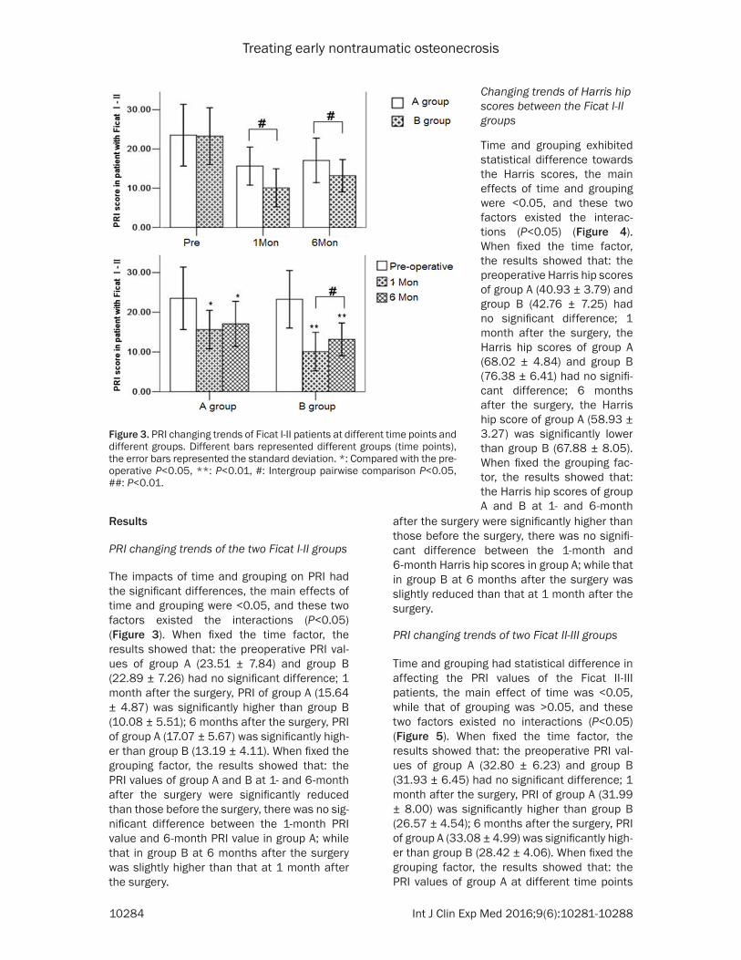
PRI changing trends of the two Ficat I-II groups
The impacts of time and grouping on PRI had the significant differences, the main effects of time and grouping were <0.05, and these two factors existed the interactions (P<0.05) (Figure 3). When fixed the time factor, the results showed that: the preoperative PRI val-ues of group A (23.51 ± 7.84) and group B (22.89 ± 7.26) had no significant difference; 1 month after the surgery, PRI of group A (15.64 ± 4.87) was significantly higher than group B (10.08 ± 5.51); 6 months after the surgery, PRI of group A (17.07 ± 5.67) was significantly high-er than group B (13.19 ± 4.11). When fixed the grouping factor, the results showed that: the PRI values of group A and B at 1- and 6-month after the surgery were significantly reduced than those before the surgery, there was no sig-nificant difference between the 1-month PRI value and 6-month PRI value in group A; while that in group B at 6 months after the surgery was slightly higher than that at 1 month after the surgery.
after the surgery were significantly higher than those before the surgery, there was no signifi-cant difference between the 1-month and 6-month Harris hip scores in group A; while that in group B at 6 months after the surgery was slightly reduced than that at 1 month after the surgery.
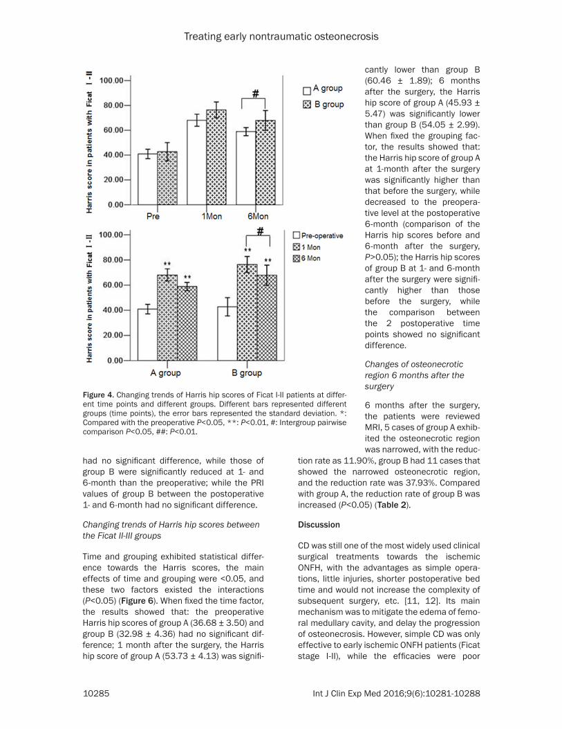
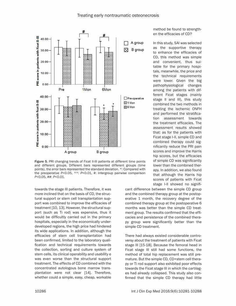
PRI changing trends of two Ficat II-III groups
Time and grouping had statistical difference in affecting the PRI values of the Ficat II-III patients, the main effect of time was <0.05, while that of grouping was >0.05, and these two factors existed no interactions (P<0.05) (Figure 5). When fixed the time factor, the results showed that: the preoperative PRI val-ues of group A (32.80 ± 6.23) and group B (31.93 ± 6.45) had no significant difference; 1 month after the surgery, PRI of group A (31.99 ± 8.00) was significantly higher than group B (26.57 ± 4.54); 6 months after the surgery, PRI of group A (33.08 ± 4.99) was significantly high-er than group B (28.42 ± 4.06). When fixed the grouping factor, the results showed that: the PRI values of group A at different time points
Figure 3. PRI changing trends of Ficat I-II patients at different time points and different groups. Different bars represented different groups (time points), the error bars represented the standard deviation. *: Compared with the pre-operative P<0.05, **: P<0.01, #: Intergroup pairwise comparison P<0.05, ##: P<0.01.
Changing trends of Harris hip scores between the Ficat I-II groups
Time and grouping exhibited statistical difference towards the Harris scores, the main effects of time and grouping were <0.05, and these two factors existed the interac-tions (P<0.05) (Figure 4). When fixed the time factor, the results showed that: the preoperative Harris hip scores of group A (40.93 ± 3.79) and group B (42.76 ± 7.25) had no significant difference; 1 month after the surgery, the Harris hip scores of group A (68.02 ± 4.84) and group B (76.38 ± 6.41) had no signifi-cant difference; 6 months after the surgery, the Harris hip score of group A (58.93 ± 3.27) was significantly lower than group B (67.88 ± 8.05). When fixed the grouping fac-tor, the results showed that: the Harris hip scores of group A and B at 1- and 6-month
Treating early nontraumatic osteonecrosis
10285 Int J Clin Exp Med 2016;9(6):10281-10288
had no significant difference, while those of group B were significantly reduced at 1- and 6-month than the preoperative; while the PRI values of group B between the postoperative 1- and 6-month had no significant difference.
Changing trends of Harris hip scores between the Ficat II-III groups
Time and grouping exhibited statistical differ-ence towards the Harris scores, the main effects of time and grouping were <0.05, and these two factors existed the interactions (P<0.05) (Figure 6). When fixed the time factor, the results showed that: the preoperative Harris hip scores of group A (36.68 ± 3.50) and group B (32.98 ± 4.36) had no significant dif-ference; 1 month after the surgery, the Harris hip score of group A (53.73 ± 4.13) was signifi-
Figure 4. Changing trends of Harris hip scores of Ficat I-II patients at differ-ent time points and different groups. Different bars represented different groups (time points), the error bars represented the standard deviation. *: Compared with the preoperative P<0.05, **: P<0.01, #: Intergroup pairwise comparison P<0.05, ##: P<0.01.
cantly lower than group B (60.46 ± 1.89); 6 months after the surgery, the Harris hip score of group A (45.93 ± 5.47) was significantly lower than group B (54.05 ± 2.99). When fixed the grouping fac-tor, the results showed that: the Harris hip score of group A at 1-month after the surgery was significantly higher than that before the surgery, while decreased to the preopera-tive level at the postoperative 6-month (comparison of the Harris hip scores before and 6-month after the surgery, P>0.05); the Harris hip scores of group B at 1- and 6-month after the surgery were signifi-cantly higher than those before the surgery, while the comparison between the 2 postoperative time points showed no significant difference.
Changes of osteonecrotic region 6 months after the surgery
6 months after the surgery, the patients were reviewed MRI, 5 cases of group A exhib-ited the osteonecrotic region was narrowed, with the reduc-
tion rate as 11.90%, group B had 11 cases that showed the narrowed osteonecrotic region, and the reduction rate was 37.93%. Compared with group A, the reduction rate of group B was increased (P<0.05) (Table 2).
Discussion
CD was still one of the most widely used clinical surgical treatments towards the ischemic ONFH, with the advantages as simple opera-tions, little injuries, shorter postoperative bed time and would not increase the complexity of subsequent surgery, etc. [11, 12]. Its main mechanism was to mitigate the edema of femo-ral medullary cavity, and delay the progression of osteonecrosis. However, simple CD was only effective to early ischemic ONFH patients (Ficat stage I-II), while the efficacies were poor
Treating early nontraumatic osteonecrosis
10286 Int J Clin Exp Med 2016;9(6):10281-10288
towards the stage III patients. Therefore, it was more inclined that on the basis of CD, the struc-tural support or stem cell transplantation sup-port was combined to improve the efficacies of treatment [10, 13]. However, the structural sup-port (such as Ti rod) was expensive, thus it would be difficultly carried out in the primary hospitals, especially in the economically under-developed regions, the high price had hindered its wide applications. In addition, although the efficacies of stem cell transplantation had been confirmed, limited to the laboratory quali-fication and technical requirements towards the collection, sorting and culture system of stem cells, its clinical operability and usability e was even worse than the structural support treatment. The effects of CD combined with the concentrated autologous bone marrow trans-plantation were not clear [14]. Therefore, whether could a simple, easy, cheap, workable
cant difference between the simple CD group and the combined therapy group at the postop-erative 1 month, the recovery degree of the combined therapy group at the postoperative 6 months was better than the simple CD treat-ment group. The results confirmed that the effi-cacies and persistence of the combined thera-py group were significantly better than the simple CD treatment.
There had always existed considerable contro-versy about the treatment of patients with Ficat stage III [15-18]. Because the femoral head in Ficat stage III still had some functions, the method of total hip replacement was still pre-mature. But the simple CD, CD+stem cell thera-py or Ti rod support also exhibited poor effects towards the Ficat stage III in which the cartilag-es had already collapsed. This study also con-firmed that the simple CD therapy had little
Figure 5. PRI changing trends of Ficat II-III patients at different time points and different groups. Different bars represented different groups (time points), the error bars represented the standard deviation. *: Compared with the preoperative P<0.05, **: P<0.01, #: Intergroup pairwise comparison P<0.05, ##: P<0.01.
method be found to strength-en the efficacies of CD?
In this study, SAI was selected as the supportive therapy to enhance the efficacies of CD, this method was simple and convenient, thus sui- table for the primary hospi-tals, meanwhile, the price and the technical requirements were lower. Given the big pathophysiological changes among the patients with dif-ferent Ficat stages (mainly stage II and III), this study combined the two methods in treating the ischemic ONFH and performed the stratifica-tion assessment towards the treatment efficacies. The assessment results showed that: as for the patients with Ficat stage I-II, simple CD and combined therapy could sig-nificantly reduce the PRI pain scores and improve the Harris hip scores, but the efficacies of simple CD was significantly lower than the combined ther-apy. In addition, we also found that although the Harris hip scores of patients with Ficat stage I-II showed no signifi-

Treating early nontraumatic osteonecrosis
10287 Int J Clin Exp Med 2016;9(6):10281-10288
effects towards the patients with Ficat stage III, the PRI scores 1 and 6 months after the sur-gery did not change significantly, although the Harris hip scores were slightly increased 1 month after the surgery, they returned to the preoperative levels 5 months later, similar to previous literature [19]. The patients with CD+SAI exhibited significantly decreased PRI scores 1 and 6 months after the surgery, and the Harris scores were significantly increased. The results confirmed that CD+SAI could effec-tively relieve pain and improve the hip score.
According to the pathophysiological mecha-nisms of the occurrence and development of
treatment. But the exact mechanism still need-ed to be further explored.
This study did not focus on comparing the effi-cacies of CD combined with SAI against other treatment methods (such as osteotomy, hip resurfacing, total hip replacement, Ti rod sup-port, stem cell transplantation, etc.) in treating ONFH, while focused on providing an effective, simple, inexpensive, feasible clinical alternative method. Although the long-term effects still needed to be assessed, the early- and mid-term results showed that the effectiveness and efficacy’s persistence had been demonstrated. Based on the actual situations of our country,
Figure 6. Changing trends of Harris hip scores of Ficat II-III patients at dif-ferent time points and different groups. Different bars represented different groups (time points), the error bars represented the standard deviation. *: Compared with the preoperative P<0.05, **: P<0.01, #: Intergroup pairwise comparison P<0.05, ##: P<0.01.
ischemic ONFH, the principles of treating the ischemic ONFH were to promote the circula-tion in the necrotic areas and delay the occurrence of non-vascular events. In this study, the SAI drugs were selected to dilate the blood vessels, anticoagulate, improve the local circulation and release the pain. The previous studies had confirmed that these drugs were all the commonly used non-surgical treatment drugs towards the ischemic ONFH, but these drugs were mostly administrated intrave-nously, and showed poor effi-cacies towards the patients with already-collapsed carti-lages [20]. However, this study confirmed that by changing the administration route, namely injected the above drugs directly into the femoral artery through the intervention way, not only could the effects of CD towards the patients with Ficat stage I-II be further enhanced, but also effective towards the patients with Ficat stage III. The postopera-tive MRI scanning also con-firmed the efficiency rate towards the osteonecrotic range obtained by the com-bined therapy was significant-ly higher than the simple CD
Table 2. Changes of osteonecrotic regions of the 2 groups 6 months after the surgery (cases)Time point
Cases with narrowed osteonecrotic region
Cases without change or dila-tion in the osteonecrotic region
reduction rate (%)
Group A 5 37 11.90Group B 11 18 37.93a
Note: Compared with group A, aP<0.05.
Treating early nontraumatic osteonecrosis
10288 Int J Clin Exp Med 2016;9(6):10281-10288
as well as the actual distribution ratio of clinical resources, CD combined with SAI was applica-ble to the most patients, could be undertaken in the primary hospitals, and should be popularized.
Disclosure of conflict of interest
None.
Address correspondence to: Qian Chen, Department of Pain Medicine, Orthopedics Hospital of Guizhou Province, 25 South Shachong Road, Guiyang 550002, China. Tel: +86 851 5793204; E-mail: [email protected]
References
[1] Wang XS, Zhuang QY, Weng XS, Lin J, Jin J and Qian WW. Etiological and clinical analysis of osteonecrosis of the femoral head in Chinese patients. Chin Med J (Engl) 2013; 126: 290-295.
[2] Mao Q, Wang W, Xu T, Zhang S, Xiao L, Chen D, Jin H and Tong P. Combination Treatment of Biomechanical Support and Targeted Intra-ar-terial Infusion of Peripheral Blood Stem Cells mobilized by granulocyte-colony stimulating factor for the Osteonecrosis of the Femoral Head: A Randomised Controlled Clinical Trial. J Bone Miner Res 2015; 30: 647-656.
[3] van der Jagt D, Mokete L, Pietrzak J, Zalavras CG and Lieberman JR. Osteonecrosis of the femoral head: evaluation and treatment. J Am Acad Orthop Surg 2015; 23: 69-70.
[4] Tripathy SK, Goyal T and Sen RK. Management of femoral head osteonecrosis: Current con-cepts. Indian J Orthop 2015; 49: 28-45.
[5] Chen CH, Chang JK, Lai KA, Hou SM, Chang CH and Wang GJ. Alendronate in the prevention of collapse of the femoral head in nontraumatic osteonecrosis: A two-year multicenter, pro-spective, randomized, double-blind, placebo-controlled study. Arthritis Rheum 2012; 64: 1572-1578.
[6] Bozic KJ, Zurakowski D and Thornhill TS. Survi-vorship analysis of hips treated with core de-compression for nontraumatic osteonecrosis of the femoral head. J Bone Joint Surg Am 1999; 81: 200-209.
[7] Grayson WL, Bunnell BA, Martin E, Frazier T, Hung BP and Gimble JM. Stromal cells and stem cells in clinical bone regeneration. Nat Rev Endocrinol 2015; 11: 140-150.
[8] Rackwitz L, Eden L, Reppenhagen S, Reichert JC, Jakob F, Walles H, Pullig O, Tuan RS, Rudert M and Nöth U. Stem cell- and growth factor-based regenerative therapies for avascular ne-crosis of the femoral head. Stem Cell Res Ther 2012; 3: 7.
[9] Zhao D, Wang B, Guo L, Yang L and Tian F. Will a vaseularized greater trochanter graft pre-serve the necrotic femoral head? Clin Orthop Relat Res 2010; 468: 1316-1324.
[10] Floerkemeier T, Lutz A, Nackenhorst U, Thorey F, Waizy H, Windhagen H and von Lewinski G. Core decompression and osteonecrosis inter-vention rod in osteonecrosis of the femoral head: clinical outcome and finite element anal-ysis. Int Orthop 2011; 35: 1461-1466.
[11] Wei BF and Ge XH. Treatment of asteoflecrosis of the femoral head with core decompression and bone grafting. Hip Int 2011; 21: 206-210.
[12] Lieberman JR. Core decompression for osteo-necrosis of the hip. Clin Orthop Relat Res 2004; 29-33.
[13] Zeng YR, He S, Feng WJ, Li FL, Li J, Jian LY, Zeng JC and Fan YG. Vascularised greater troe-hanter bone graft, combined free iliac flap and impaction bone grafting for osteonecrosis of the femoral head. Int Orthop 2013; 37: 391-398.
[14] Chotivichit A, Korwutthikulrangsri E, Auewara-kul C and Sarirasririd S. Core decompression and concentrated autologous bone marrow in-jection for treatment of osteonecrosis of the femoral head. J Med Assoc Thai 2012; 95: S14-20.
[15] Baksi DP, Pal AK and Baksi DD. Long-term re-sults of decompression and muscle-pedicle bone grafting for osteonecrosis of the femoral head. Int Orthop 2009; 33: 41-47.
[16] Hungerford DS. Osteonecrosis:avoiding total hip arthroplast. J Arthroplasty 2002; 17: 121-124.
[17] Koc ON and Lazarus HM. Mesenchymal stem cells:heading into the clinic. Bone Marrow Transplant 2001; 27: 235-239.
[18] Yoo MC, Kim KI, Hahn CS and Parvizi J. Long-term followup of vascularized fibular grafting for femoral head necrosis. Clin Orthop Relat Res 2008; 466: 1133-1140.
[19] Pierannunzii L. Endoscopic and arthroscopic in femoral head core decompression. Arthrosc Tech 2012; 1: e225-230.
[20] Maruotti N, Corrado A, Neve A and Cantatore FP. Bisphosphonates: effects on osteoblast. Eur J Clin Pharmacol 2012; 68: 1013-1018.