linh 1t. tran , l. chartier - kek
TRANSCRIPT
Linh T. Tran1, L. Chartier1, D. A. Prokopovich2, D. Bolst1, S. Guatelli1, M. Petasecca1, M. I. Reinhard2, M. L. F.Lerch1, N. Matsufuji3, Benjamin Clasie4,
Chris Beltran5, V. L. Pereverlaylo6, and A. B. Rosenfeld1
1Centre for Medical Radiation Physics, University of Wollongong, NSW 2Australian Nuclear Science and Technology Organization, Australia 3Research Centre for Charge Particle Therapy, National Institute of Radiological Science, Japan 4Massachusetts General Hospital, MGH, Boston, USA 5Mayo Clinics, Rochester, Minnesota, USA 6SPA, BIT, Kiev, Ukraine
Depth
Re
lati
ve
do
se
Tumor
Depth
Rela
tive d
ose
Tumor
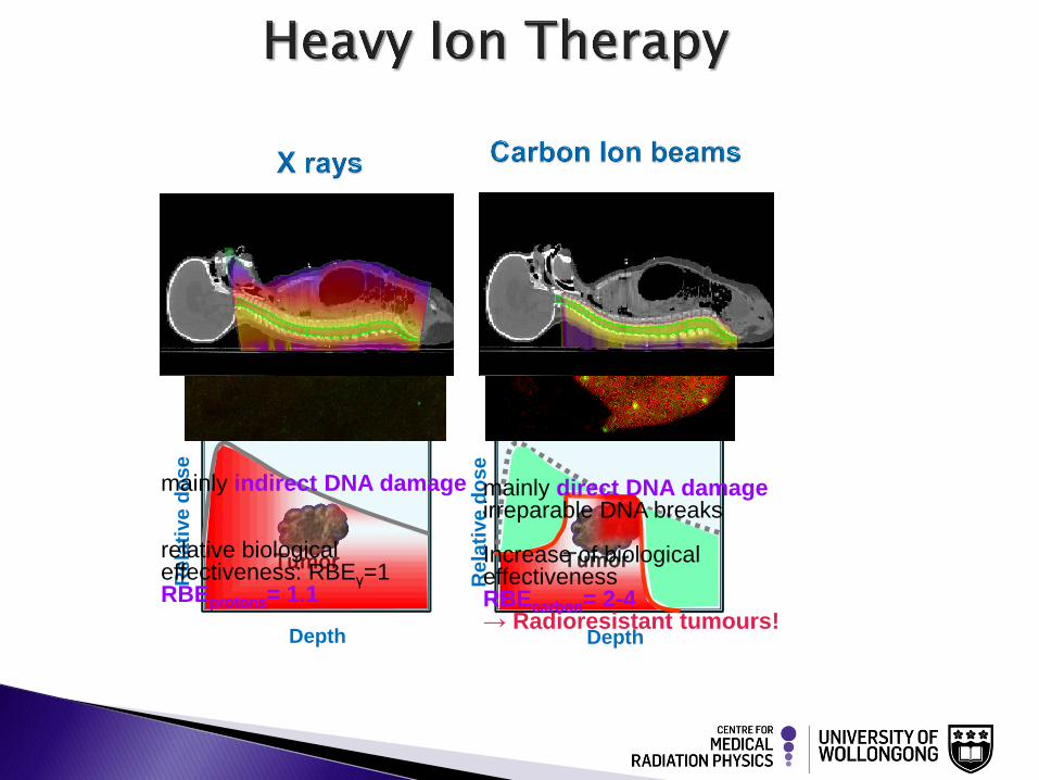
mainly direct DNA damage irreparable DNA breaks Increase of biological effectiveness RBEcarbon= 2-4 → Radioresistant tumours!
densely ionising
mainly indirect DNA damage relative biological effectiveness: RBEγ=1 RBEprotons= 1.1
sparsely ionising
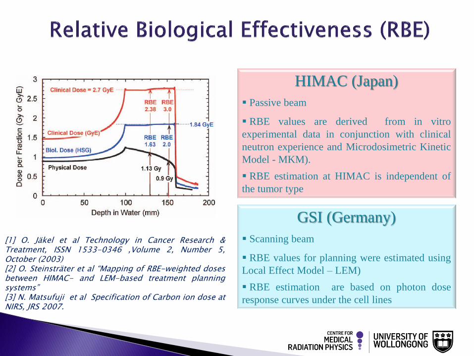
GSI (Germany)
Scanning beam
RBE values for planning were estimated using
Local Effect Model – LEM)
RBE estimation are based on photon dose
response curves under the cell lines
HIMAC (Japan)
Passive beam
RBE values are derived from in vitro
experimental data in conjunction with clinical
neutron experience and Microdosimetric Kinetic
Model - MKM).
RBE estimation at HIMAC is independent of
the tumor type
[1] O. Jäkel et al Technology in Cancer Research & Treatment, ISSN 1533-0346 ,Volume 2, Number 5, October (2003) [2] O. Steinsträter et al “Mapping of RBE-weighted doses between HIMAC- and LEM-based treatment planning systems” [3] N. Matsufuji et al Specification of Carbon ion dose at NIRS, JRS 2007.
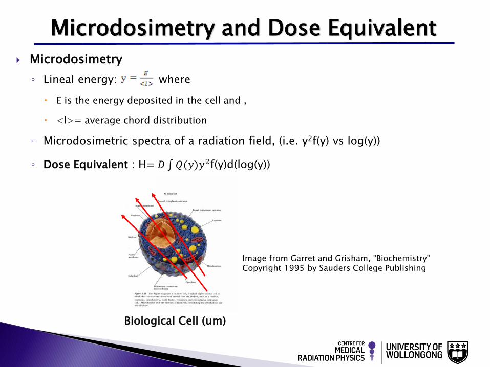
Microdosimetry
◦ Lineal energy: where
E is the energy deposited in the cell and ,
<l>= average chord distribution
◦ Microdosimetric spectra of a radiation field, (i.e. y2f(y) vs log(y))
◦ Dose Equivalent : H= 𝐷 𝑄(𝑦)𝑦2f(y)d(log(y))
Microdosimetry and Dose Equivalent
Biological Cell (um)
Image from Garret and Grisham, "Biochemistry" Copyright 1995 by Sauders College Publishing
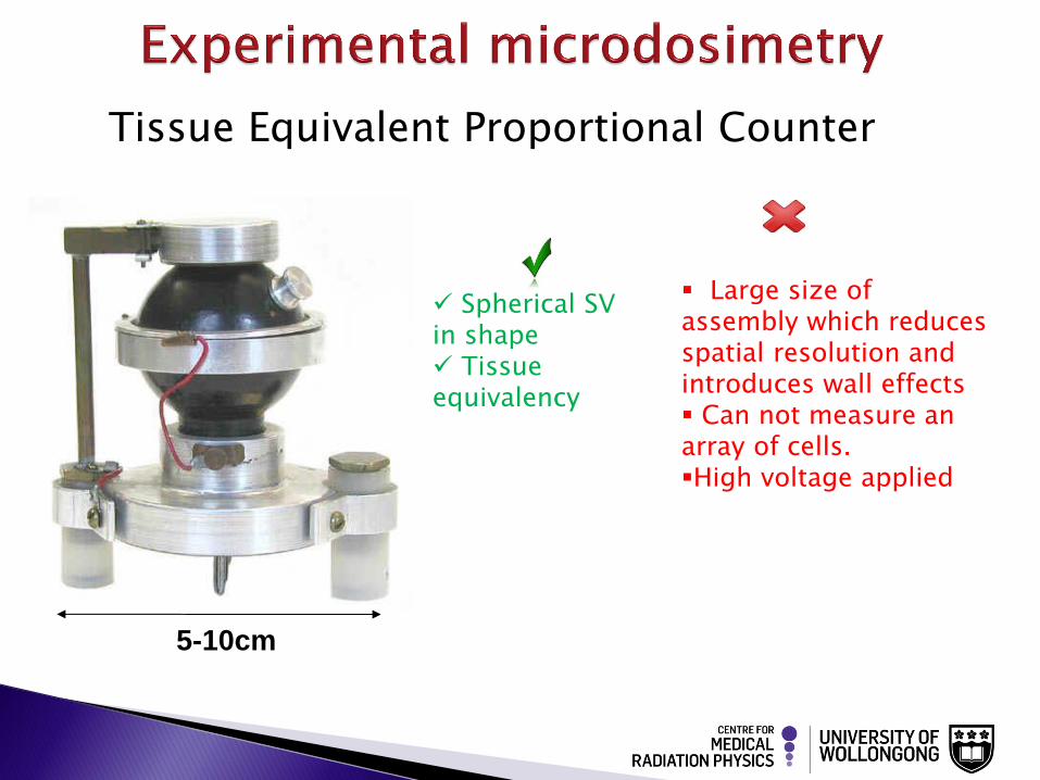
Tissue Equivalent Proportional Counter
Spherical SV in shape Tissue equivalency
5-10cm
Large size of assembly which reduces spatial resolution and introduces wall effects Can not measure an array of cells. High voltage applied
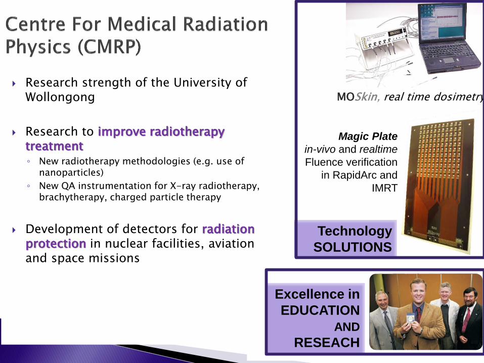
Research strength of the University of Wollongong
Research to improve radiotherapy treatment ◦ New radiotherapy methodologies (e.g. use of
nanoparticles)
◦ New QA instrumentation for X-ray radiotherapy, brachytherapy, charged particle therapy
Development of detectors for radiation protection in nuclear facilities, aviation and space missions
Excellence in
EDUCATION
AND
RESEACH
Technology
SOLUTIONS
MOSkin, real time dosimetry
Magic Plate
in-vivo and realtime
Fluence verification
in RapidArc and
IMRT

Image from www.visitsouthcoast.com.au
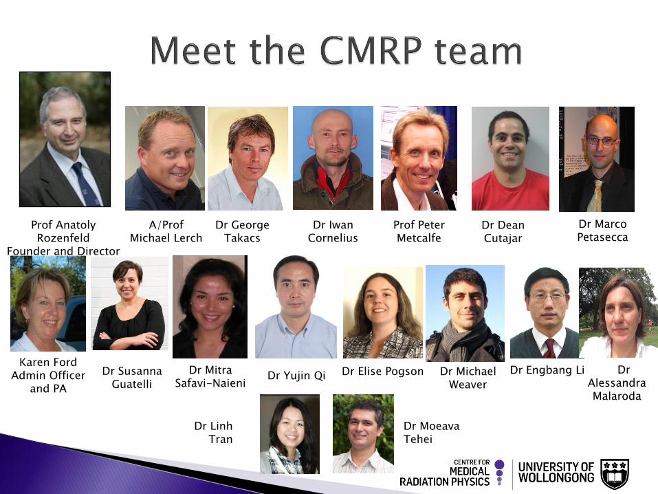
Prof Anatoly Rozenfeld
Founder and Director
Prof Peter Metcalfe
Dr Marco Petasecca
Dr Dean Cutajar
A/Prof Michael Lerch
Dr George Takacs
Karen Ford Admin Officer
and PA
Dr Susanna Guatelli
Dr Mitra Safavi-Naieni
Dr Yujin Qi Dr Elise Pogson Dr Michael Weaver
Dr Iwan Cornelius
Dr Engbang Li Dr Alessandra Malaroda
Dr Linh Tran
Dr Moeava Tehei
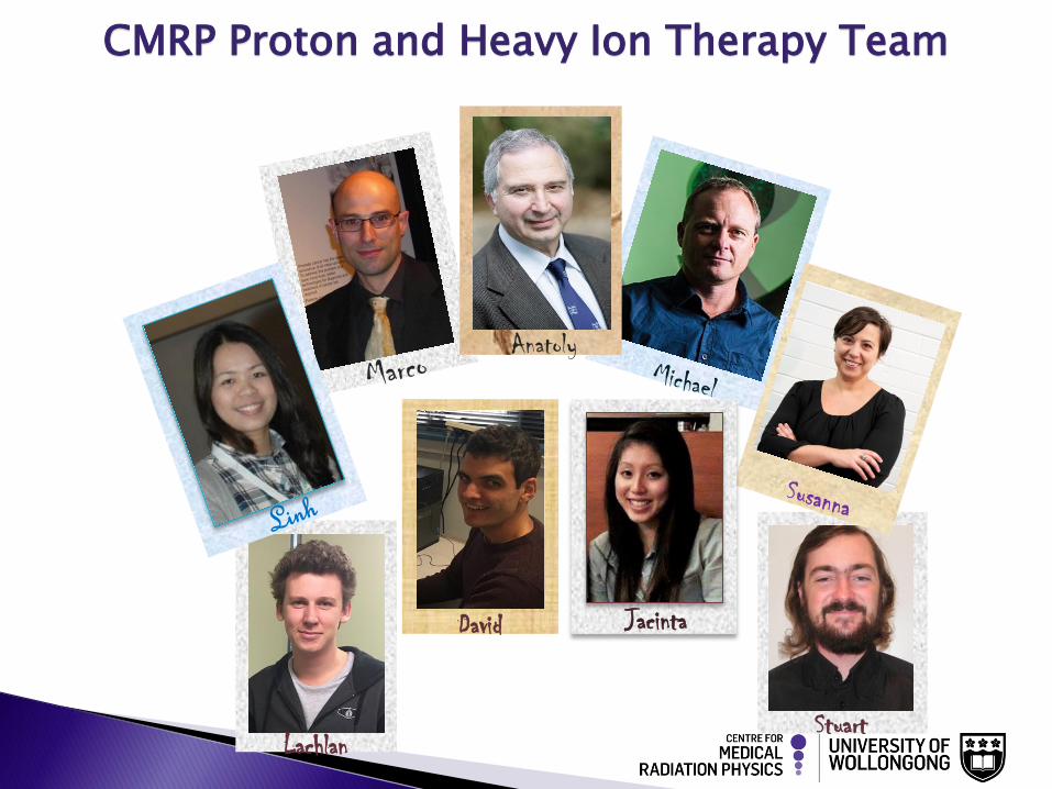
Anatoly
Lachlan
David Jacinta
Stuart
CMRP Proton and Heavy Ion Therapy Team
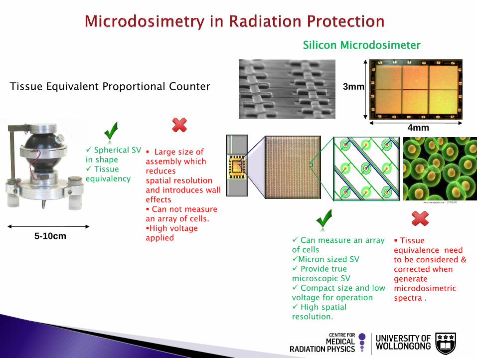
Tissue Equivalent Proportional Counter
Spherical SV in shape Tissue equivalency
Can measure an array of cells Micron sized SV Provide true microscopic SV Compact size and low voltage for operation High spatial resolution.
Silicon Microdosimeter
3mm
4mm
5-10cm
Large size of assembly which reduces spatial resolution and introduces wall effects Can not measure an array of cells. High voltage applied
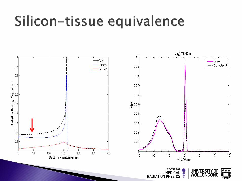
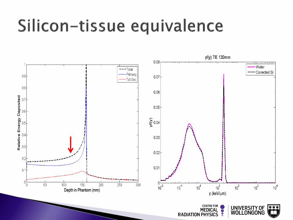
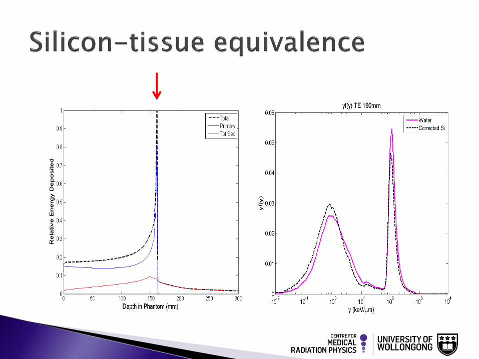
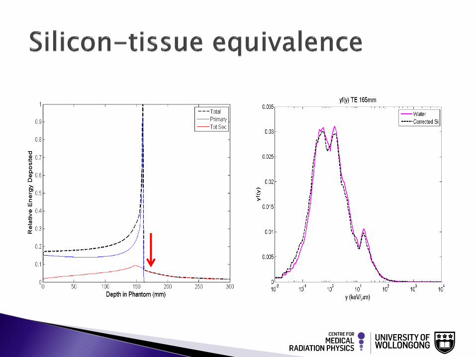
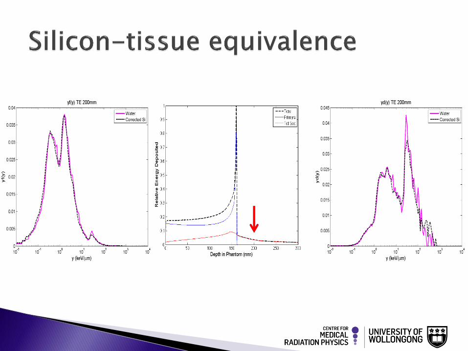
Tissue equivalence need to be considered & corrected when generate microdosimetric spectra .
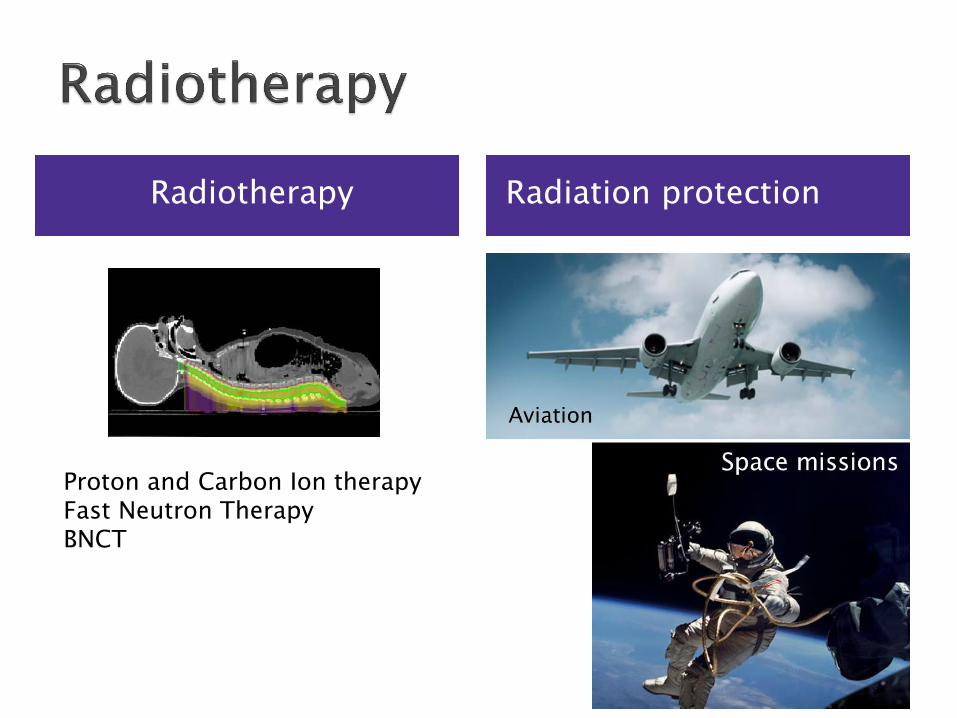
Radiotherapy Radiation protection
Proton and Carbon Ion therapy Fast Neutron Therapy BNCT
Separate varying LET components
Measure dose due to 10B neutron capture and total dose
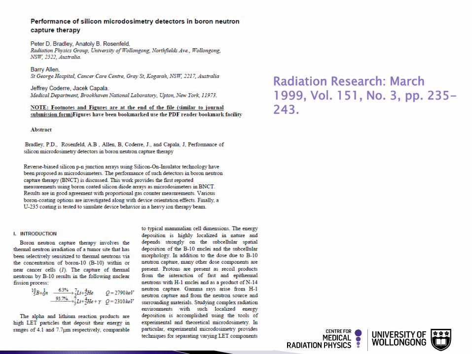
Radiation Research: March 1999, Vol. 151, No. 3, pp. 235-243.
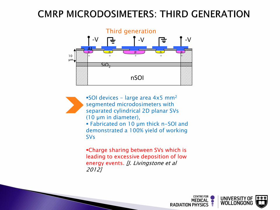
SOI devices - large area 4x5 mm2 segmented microdosimeters with separated cylindrical 2D planar SVs (10 µm in diameter), Fabricated on 10 µm thick n-SOI and demonstrated a 100% yield of working SVs Charge sharing between SVs which is leading to excessive deposition of low energy events. [J. Livingstone et al 2012]
Third generation
n+
n+ 10
µm
p+
SiO2
Al p+
-V -V -V
p+
nSOI
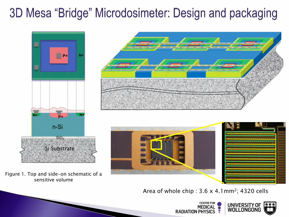
Figure 1. Top and side-on schematic of a sensitive volume
Area of whole chip : 3.6 x 4.1mm2; 4320 cells
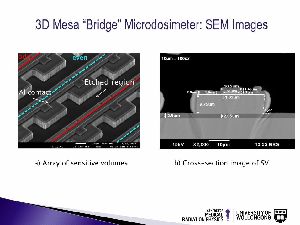
a) Array of sensitive volumes b) Cross-section image of SV
odd even
Al contact
Etched region
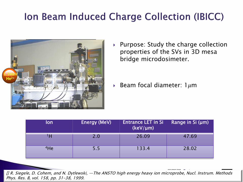
Ion Energy (MeV) Entrance LET in Si (keV/μm)
Range in Si (μm)
1H 2.0 26.09 47.69
4He 5.5 133.4 28.02
[] R. Siegele, D. Cohem, and N. Dytlewski, ―The ANSTO high energy heavy ion microprobe, Nucl. Instrum. Methods Phys. Res. B, vol. 158, pp. 31–38, 1999.
Purpose: Study the charge collection properties of the SVs in 3D mesa bridge microdosimeter.
Beam focal diameter: 1𝜇m
5.5MeV He2+
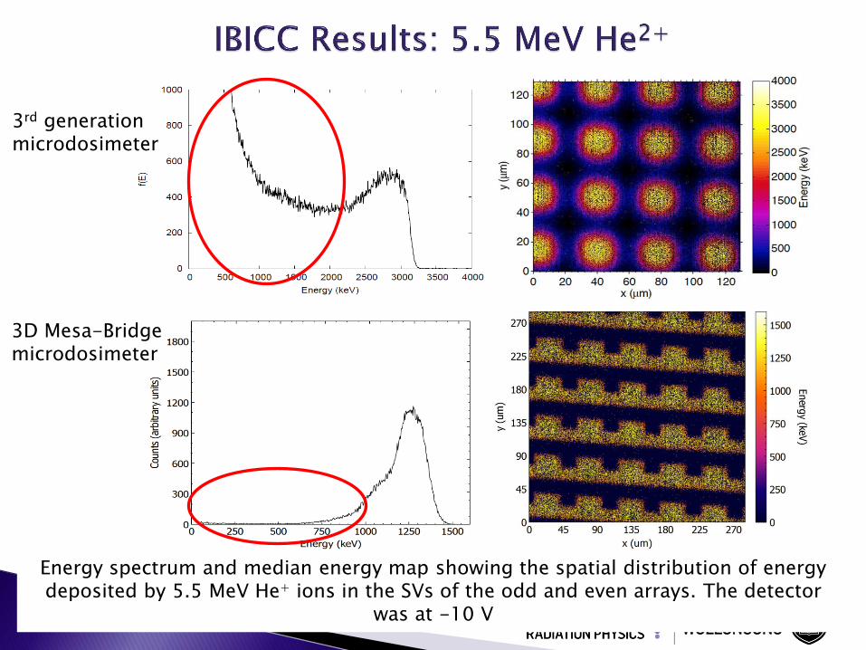
3D Mesa-Bridge microdosimeter
3rd generation microdosimeter
Energy spectrum and median energy map showing the spatial distribution of energy deposited by 5.5 MeV He+ ions in the SVs of the odd and even arrays. The detector
was at -10 V
By Geant4 simulations
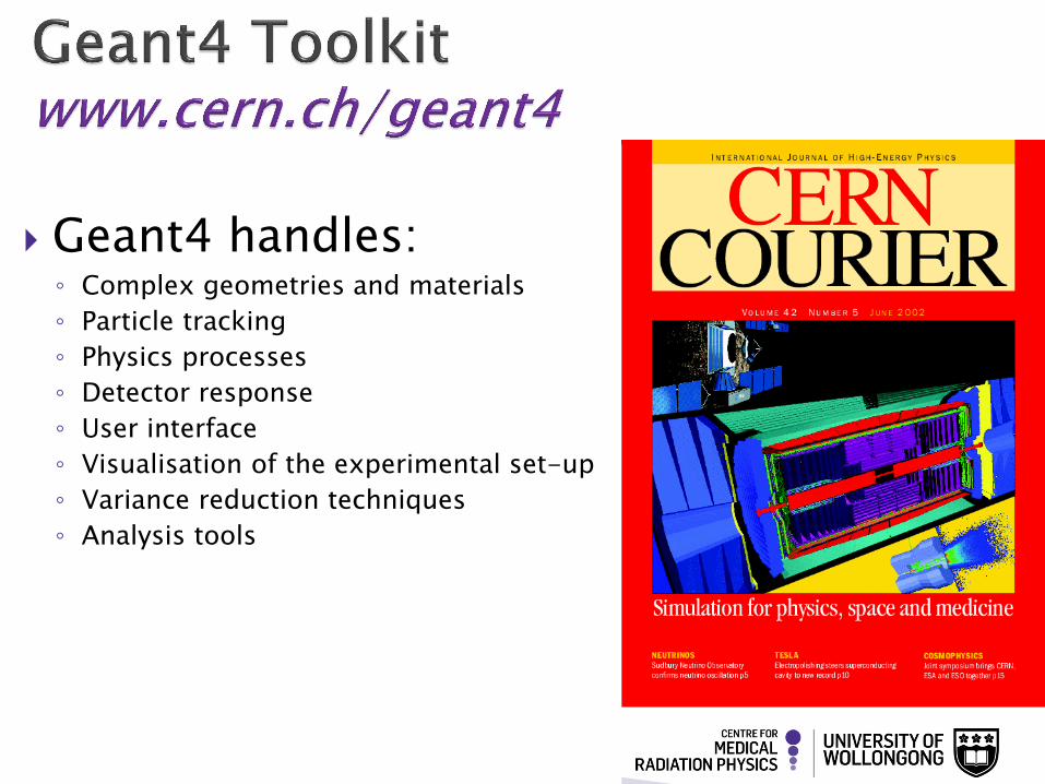
Geant4 handles: ◦ Complex geometries and materials
◦ Particle tracking
◦ Physics processes
◦ Detector response
◦ User interface
◦ Visualisation of the experimental set-up
◦ Variance reduction techniques
◦ Analysis tools
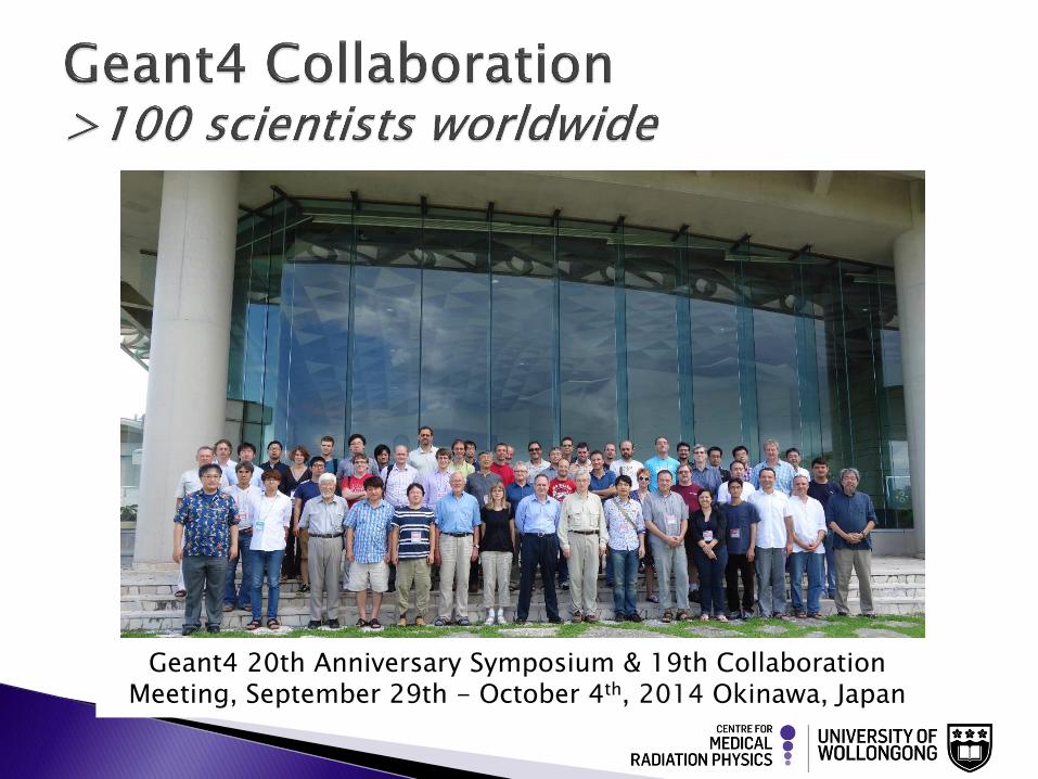
Geant4 20th Anniversary Symposium & 19th Collaboration Meeting, September 29th - October 4th, 2014 Okinawa, Japan
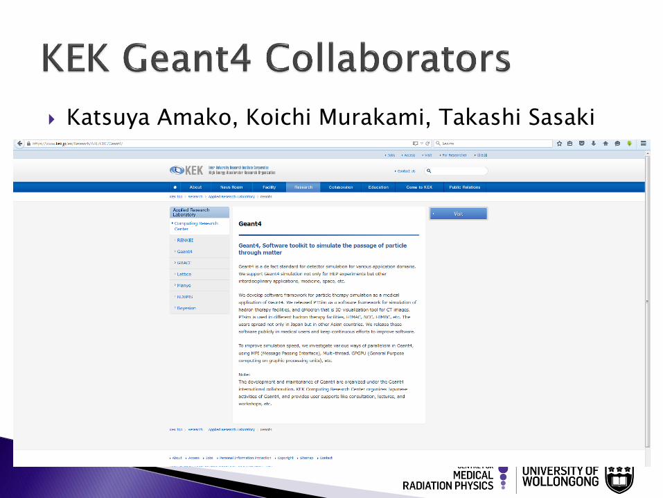
Katsuya Amako, Koichi Murakami, Takashi Sasaki
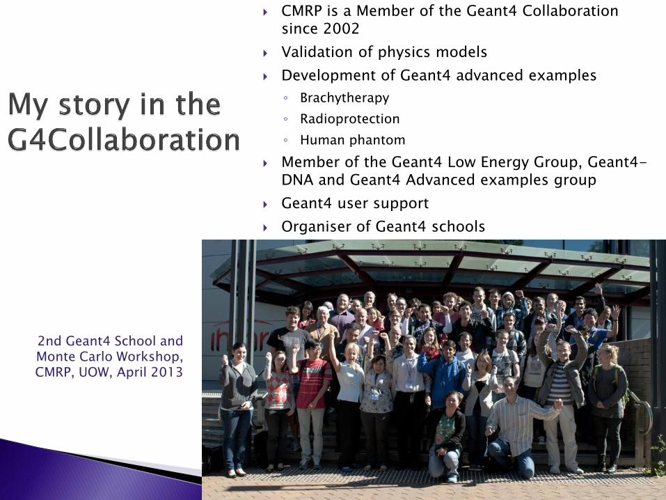
CMRP is a Member of the Geant4 Collaboration since 2002
Validation of physics models
Development of Geant4 advanced examples
◦ Brachytherapy
◦ Radioprotection
◦ Human phantom
Member of the Geant4 Low Energy Group, Geant4-DNA and Geant4 Advanced examples group
Geant4 user support
Organiser of Geant4 schools
2nd Geant4 School and Monte Carlo Workshop, CMRP, UOW, April 2013
25
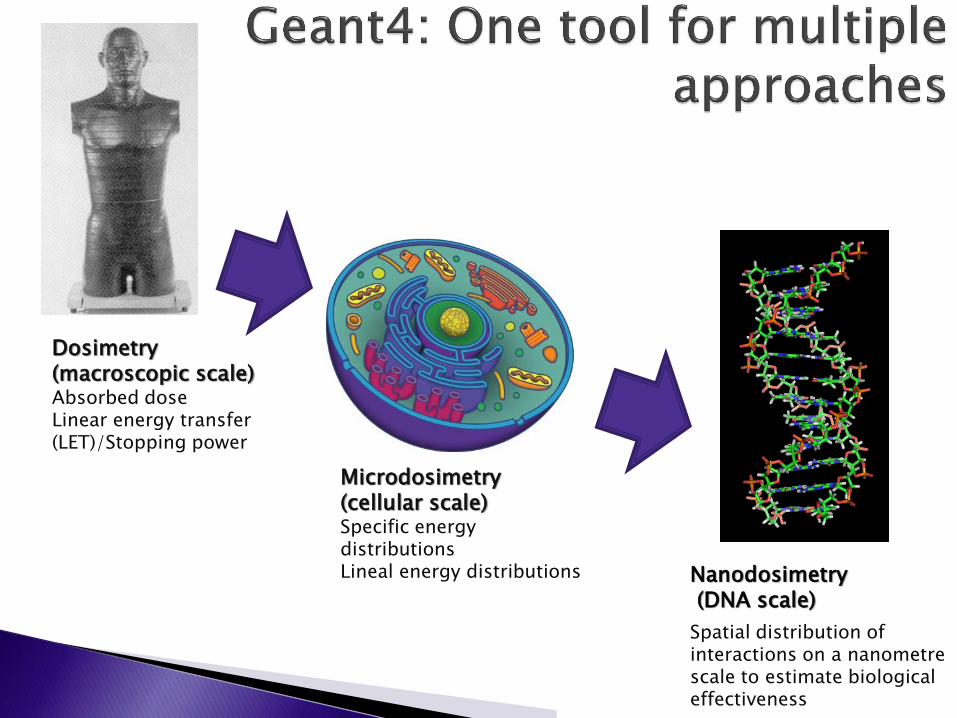
Dosimetry (macroscopic scale) Absorbed dose Linear energy transfer (LET)/Stopping power
Microdosimetry (cellular scale) Specific energy distributions Lineal energy distributions Nanodosimetry
(DNA scale)
Spatial distribution of interactions on a nanometre scale to estimate biological effectiveness
Slides adapted from
Marc Marc Verderi, LLR – Ecole Polytechnique, France
Sebastien Incerti, CENBG, Bordeaux, France
27
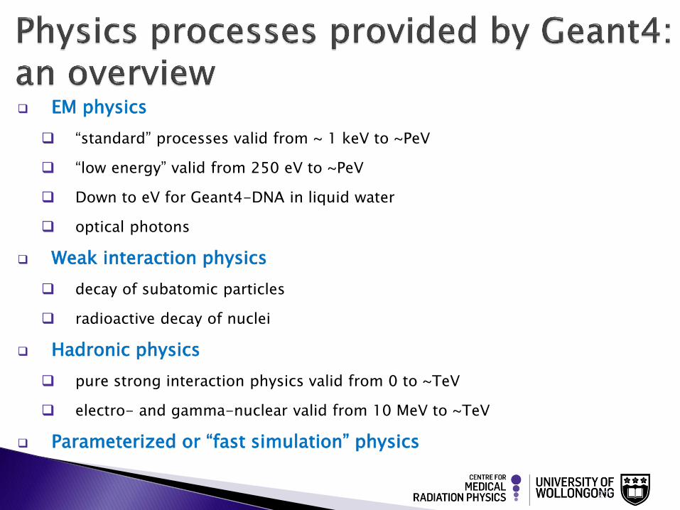
EM physics
“standard” processes valid from ~ 1 keV to ~PeV
“low energy” valid from 250 eV to ~PeV
Down to eV for Geant4-DNA in liquid water
optical photons
Weak interaction physics
decay of subatomic particles
radioactive decay of nuclei
Hadronic physics
pure strong interaction physics valid from 0 to ~TeV
electro- and gamma-nuclear valid from 10 MeV to ~TeV
Parameterized or “fast simulation” physics
28
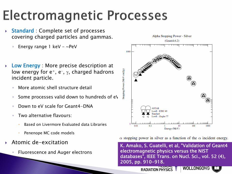
Standard : Complete set of processes covering charged particles and gammas.
◦ Energy range 1 keV - ~PeV
Low Energy : More precise description at
low energy for e+, e-, g, charged hadrons incident particle.
◦ More atomic shell structure detail
◦ Some processes valid down to hundreds of eV
◦ Down to eV scale for Geant4-DNA
◦ Two alternative flavours:
Based on Livermore Evaluated data Libraries
Penenope MC code models
Atomic de-excitation
◦ Fluorescence and Auger electrons
K. Amako, S. Guatelli, et al, "Validation of Geant4 electromagnetic physics versus the NIST databases", IEEE Trans. on Nucl. Sci., vol. 52 (4), 2005, pp. 910-918.
Capture at rest
Fission / evaporation
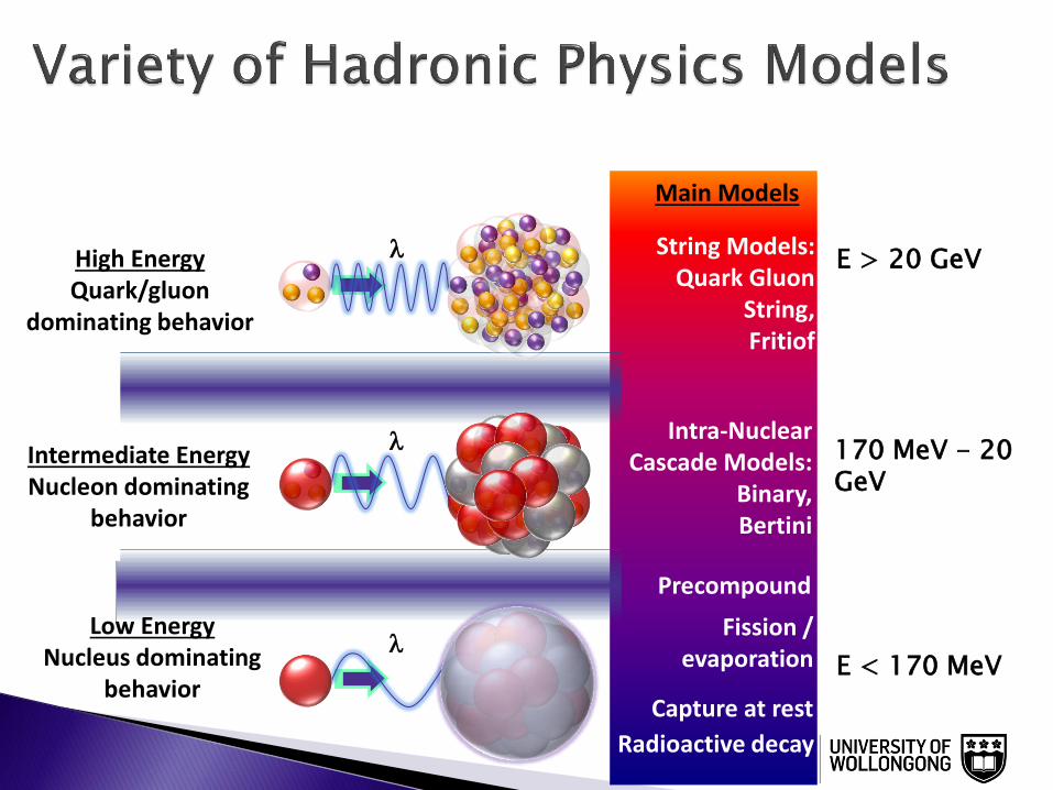
String Models: Quark Gluon
String, Fritiof
Intra-Nuclear Cascade Models:
Binary, Bertini
Low Energy Nucleus dominating
behavior
l
l
l
Main Models
Precompound
Radioactive decay
E > 20 GeV
170 MeV - 20 GeV
E < 170 MeV
Intermediate Energy Nucleon dominating
behavior
High Energy Quark/gluon
dominating behavior
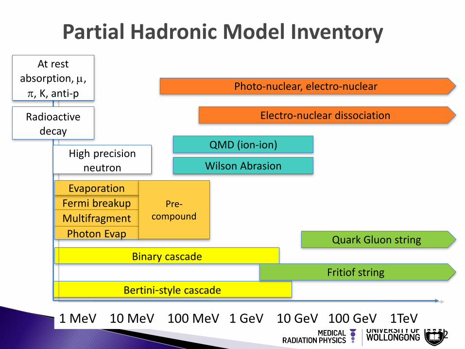
Partial Hadronic Model Inventory
Bertini-style cascade
Binary cascade
1 MeV 10 MeV 100 MeV 1 GeV 10 GeV 100 GeV 1TeV
Fermi breakup
12
At rest absorption, ,
, K, anti-p
High precision neutron
Evaporation
Multifragment
Photon Evap
Pre-compound
Radioactive decay
Fritiof string
Quark Gluon string
Photo-nuclear, electro-nuclear
QMD (ion-ion)
Electro-nuclear dissociation
Wilson Abrasion
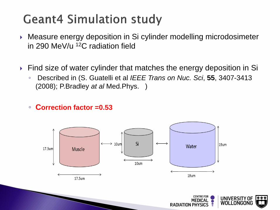
Measure energy deposition in Si cylinder modelling microdosimeter
in 290 MeV/u 12C radiation field
Find size of water cylinder that matches the energy deposition in Si
◦ Described in (S. Guatelli et al IEEE Trans on Nuc. Sci, 55, 3407-3413
(2008); P.Bradley at al Med.Phys. )
◦ Correction factor =0.53
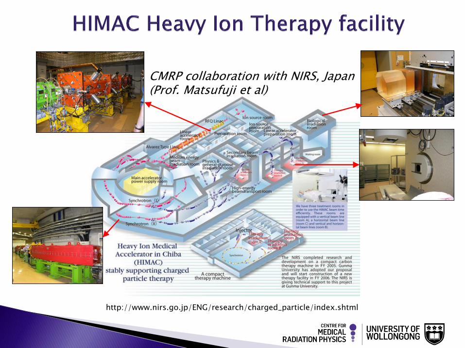
http://www.nirs.go.jp/ENG/research/charged_particle/index.shtml
CMRP collaboration with NIRS, Japan (Prof. Matsufuji et al)
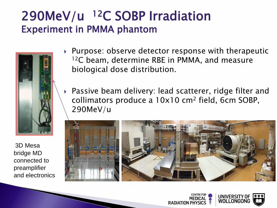
3D Mesa
bridge MD
connected to
preamplifier
and electronics
Purpose: observe detector response with therapeutic 12C beam, determine RBE in PMMA, and measure biological dose distribution.
Passive beam delivery: lead scatterer, ridge filter and collimators produce a 10x10 cm2 field, 6cm SOBP, 290MeV/u
290MeV/u 12C SOBP Irradiation Experiment in PMMA phantom
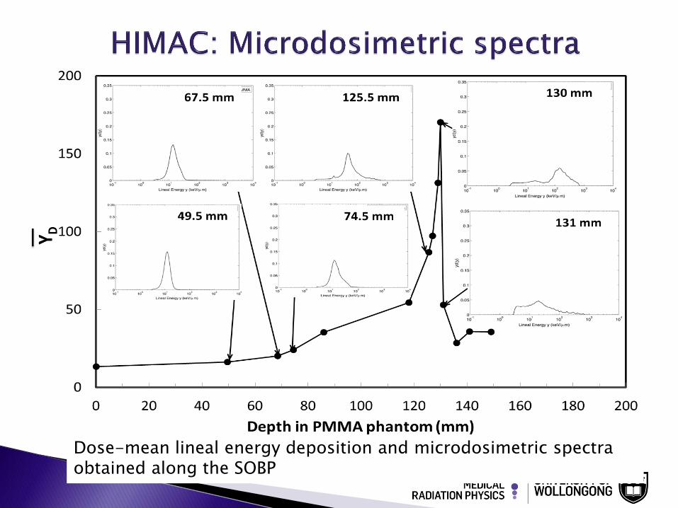
Dose-mean lineal energy deposition and microdosimetric spectra obtained along the SOBP
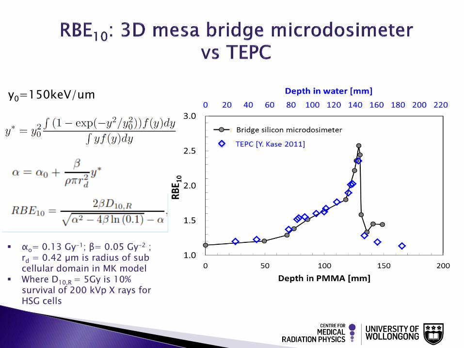
αo= 0.13 Gy-1; β= 0.05 Gy-2 ; rd = 0.42 µm is radius of sub cellular domain in MK model
Where D10,R = 5Gy is 10% survival of 200 kVp X rays for HSG cells
y0=150keV/um
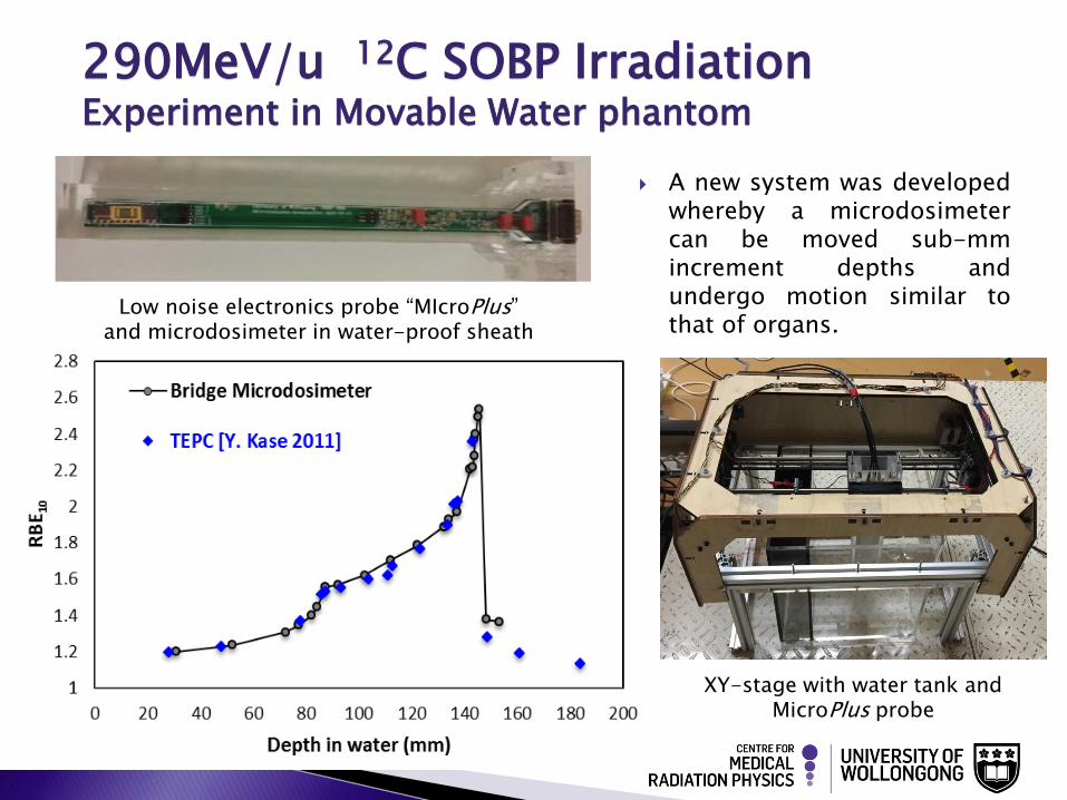
290MeV/u 12C SOBP Irradiation Experiment in Movable Water phantom
A new system was developed whereby a microdosimeter can be moved sub-mm increment depths and undergo motion similar to that of organs.
Low noise electronics probe “MIcroPlus” and microdosimeter in water-proof sheath
XY-stage with water tank and MicroPlus probe
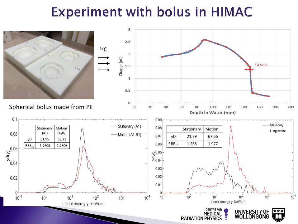
Spherical bolus made from PE
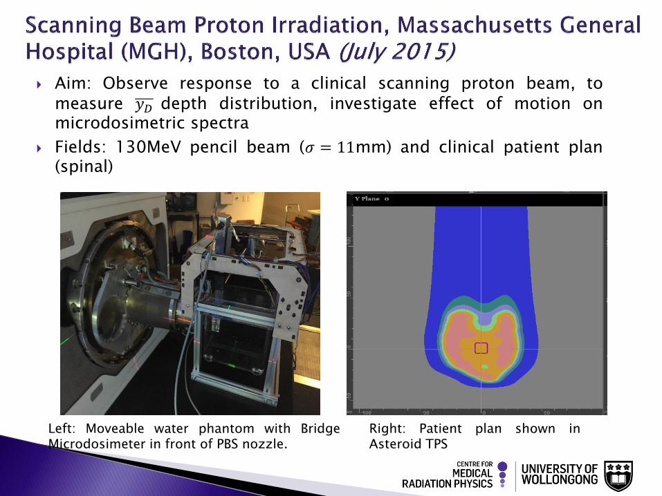
Aim: Observe response to a clinical scanning proton beam, to measure 𝑦𝐷 depth distribution, investigate effect of motion on microdosimetric spectra
Fields: 130MeV pencil beam (𝜎 = 11mm) and clinical patient plan (spinal)
Right: Patient plan shown in Asteroid TPS
Left: Moveable water phantom with Bridge Microdosimeter in front of PBS nozzle.
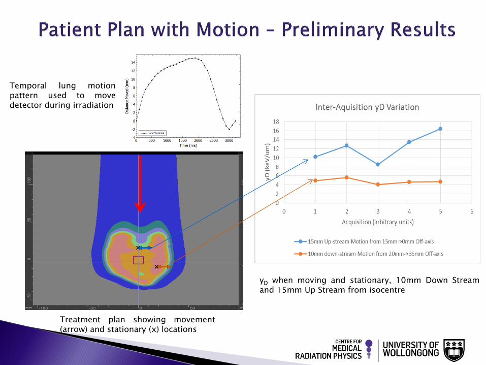
Temporal lung motion pattern used to move detector during irradiation
yD when moving and stationary, 10mm Down Stream and 15mm Up Stream from isocentre
Treatment plan showing movement (arrow) and stationary (x) locations
This work presented the first high spatial resolution RBE10 derivation in 12C ion therapeutic beam line ◦ SOI microdosimeter
◦ The RBE10 values are in good agreement with values obtained using a TEPC, with an exception at the distal part of the SOBP. This is due to TEPC measurements being carried out in water which lacks the C atoms that comprise PMMA.
Significant difference observed between the stationary microdosimetric spectra at distal part of the SOBP and the case where the detector mimicked lung motion. ◦ Microdosimetric spectra and dose mean lineal energy obtained out-of-field in proton
beam scanning allow the determination of neutron dose equivalent and the comparison with passive treatment delivery.

The Centre for Medical Radiation Physics has developed a new microdosimeter probe, with measurement threshold as low as ~ 0.3 keV/µm.
The motion can lead to changes in the microdosimetric spectrum and consequently the RBE.
The microdosimeter has the ability of measuring neutrons dose outside of the treatment field.
Silicon microdosimeters can be used for BNCT
NIRS, Japan
Prof. Naruhiro Matsufuji
MGH, Boston, USA
Dr. Benjamin Clasie
Mayo Clinic, USA
Dr. Chris Beltran
SPA, BIT, Ukraine
Dr. Vladimir Perevertaylo
Dr. Mark Reinhard
Dr. Dale Prokopovich
Dr. Angela Kok
Dr. Marco Povoli