CONSEJO SUPERIOR DE INVESTIGACIONES CIENTÍFICAS
ESTACIÓN EXPERIMENTAL DEL ZAIDÍN
UNIVERSIDAD DE GRANADA
DEPARTAMENTO DE BIOQUÍMICA Y BIOLOGÍA MOLECULAR
TRANSPORTADORES INVOLUCRADOS EN LA
TOLERANCIA A DISOLVENTES ORGÁNICOS EN
Pseudomonas putida DOT-T1E
TESIS DOCTORAL
Vanina García Altamirano
2009

Editor: Editorial de la Universidad de GranadaAutor: Vanina García AltamiranoD.L.: GR. 2624-2009ISBN: 978-84-692-3887-5


TRANSPORTADORES INVOLUCRADOS EN LA TOLERANCIA A
DISOLVENTES ORGÁNICOS EN Pseudomonas putida DOT-T1E
Memoria que presenta la Microbióloga, Vanina GARCÍA ALTAMIRANO, para aspirar al Título de Doctora
Fdo.: Vanina García Altamirano
Vº Bº del Director Vº Bº del Director
Fdo.: Juan Luis Ramos Martín Fdo.: Ana Segura Carnicero Doctor en Biología Doctora en Biología Profesor de investigación del C.S.I.C. Científica titular del C.S.I.C.
Universidad de Granada 2009


Esta Tesis Doctoral ha sido realizada en el Departamento de Protección Ambiental de la
Estación Experimental del Zaidín (C.S.I.C.), Granada.


A la memoria de mi Padre
A mi Madre
A mis Hermanas

“… Y así después de esperar tanto, un día como cualquier otro decidí triunfar…
decidí no esperar a las oportunidades sino yo mismo buscarlas,
decidí ver cada problema como la oportunidad de encontrar una solución,
decidí ver cada desierto como la oportunidad de encontrar un oasis,
decidí ver cada noche como un misterio a resolver,
decidí ver cada día como una nueva oportunidad de ser feliz.
Aquel día descubrí que mi único rival no eran más que mis propias debilidades,
y que en éstas, está la única y mejor forma de superarnos.
Aquel día dejé de temer a perder y empecé a temer a no ganar,
descubrí que no era yo el mejor y que quizás nunca lo fui.
Me dejó de importar quién ganara o perdiera;
ahora me importa simplemente saberme mejor que ayer.
Aprendí que lo difícil no es llegar a la cima,
sino jamás dejar de subir.
Aprendí que el mejor triunfo que puedo tener,
es tener el derecho de llamar a alguien “Amigo”.
Descubrí que el amor es más que un simple estado de enamoramiento,
“el amor es una filosofía de vida”.
Aquel día dejé de ser un reflejo de mis escasos triunfos pasados
y empecé a ser mi propia tenue luz de este presente;
aprendí que de nada sirve ser luz
si no vas a iluminar el camino de los demás.
Aquel día decidí cambiar tantas cosas…
Aquel día aprendí que los sueños son solamente para hacerse realidad.
Desde aquel día ya no duermo para descansar…
ahora simplemente duermo para soñar."
Walt Disney


Agradecimientos
El trabajo presentado en esta memoria es el resultado de varios años de esfuerzo y
dedicación en los que he necesitado una gran ayuda tanto en lo personal como en lo
profesional. Es por eso que quiero dar las gracias a todas aquellas personas que de una u
otra forma han contribuido a la realización de esta Tesis Doctoral, sin la ayuda de las
cuales no hubiese sido posible.
A mis directores, la Dra. Ana Segura Carnicero, y el Dr. Juan Luis Ramos Martín, de
los que estoy muy agradecida por el tiempo y el esfuerzo dedicados a esta Tesis. Al Dr.
Juan Luis Ramos Martín, director del grupo de Degradación de Tóxicos Orgánicos de
la Estación Experimental del Zaidín, por permitirme ser parte de este grupo de
investigación.
Al Dr. Carlos Eduardo Domenech, por darme (sin ser conciente de ello) el incentivo que
necesitaba para entrar en el mundo de la investigación en aquella primera clase de
Bioquímica Microbiana que siempre quedará grabada en mi memoria. Por tener siempre
abierta la puerta de su despacho para “clases de consulta”. Por estar siempre presente,
aún en la distancia. Por sus buenos consejos en la ciencia y en la vida.
A la Dra. Gloria I. Lucchesi, mi “Madre Científica”, por enseñarme a dar los primeros
pasos de mi carrera investigadora, por enseñarme a “estar de pie” frente a la audiencia,
por sus buenos consejos, por su paciencia, por creer en mi.
A la Dra. Estrella Duque y a los integrantes del “Salón Comedor” (Marichu, Jesús
“JTZ”, Patri G.), por ayudarme a familiarizarme con el laboratorio y las técnicas más
utilizadas en este grupo de investigación.
A mi compi y gran AMIGA, Ana H., por su ayuda y sus buenos consejos, por estar ahí
cuando la necesito.
A “mi Anto”, por tener siempre unos minutos libres para compartir mis penas y
alegrías, por sus palabras de aliento, por sus consejos, por alimentar a todo el grupo con
sus deliciosas “recetas de la abuela”, por tener siempre tiempo para resolver mis tragi-
cómicos enfrentamientos con la informática. Por ser uno de mis grandes AMIGOS.
A René, “mi hijo del corazón” y gran AMIGO, por acompañarme y animarme en estos
últimos años de tesis, por tener siempre un abrazo y un consejo en el momento que más
lo necesitaba. Por sus “diabluras” que siempre me arrancan una sonrisa. Por ser “mi
familia” aquí en estas tierras tan lejanas.
A Jean-Manuel, un ser muy especial, por su PACIENCIA que a veces parece infinita,
por su apoyo incondicional, por ayudarme a rotular los 200 tubos de betagal un
domingo por la tarde (eso es amor!), por enseñarme esas “pequeñas grandes cosas” que
tiene la vida, por llenar mis vacíos con la Palabra de Dios.
A mis informáticos favoritos, César y Ale M., por solucionar mis problemas
informáticos y por la amistad que me han brindado fuera del trabajo.
A Ale M., por su amistad y sus consejos, porque su punto de vista me ha guiado muchas
veces en la búsqueda del norte.
A Mónica, Wilson y Antonio C., por la amistad que me brindaron cuando llegué a estas
tierras, por dejarse conquistar por el tango, y por acompañarme a dar mis primeros
“cortes y quebradas” en Granada.
A Dietmar, por enseñarme los misterios del BLAST en mis comienzos con las
secuencias, por su amistad y por animarme a conquistar el pico más alto de Sierra
Nevada y a recorrer la Vega en bicicleta.
A Hortencia, Cristina y Alicia, por brindarme su amistad a pesar del poco tiempo que
hace que nos conocemos. Por estar siempre pendientes. Sepan que son únicas y que
espero seguir cultivando nuestra amistad.
A las niñas del lab 1.3 (Sara, Cecilia, Ana H., Isa, Alicia), quienes me han tenido mucha
paciencia sobretodo en los últimos meses de tesis, gracias por esos buenos momentos
compartidos durante la larga jornada de trabajo, por las “frases célebres” que tanto me

han hecho reír y olvidar el mal resultado del día. Por aguantar mi humor, sobre todo en
los días en los que llevaba “una pinza en el pelo” (ustedes saben de lo que hablo).
A Miguel, Amalia, Jesús M., Jesús “JTZ” y Marichu, por hacer más llevadera la
investigación, por tener siempre una sonrisa, por poner un poco de humor al día a día,
por la amistad que me han brindado en estos años.
A Angustias, Carmen y Sonia, porque el trabajo que realizan supone una gran ayuda
para nuestras tesis.
A todos los que integran el grupo de Degradación de Tóxicos Orgánicos, porque todos
han aportado su granito de arena en este trabajo.
A todo el personal de administración, guardas de seguridad, limpieza, etc.
A mis AMIGAS DEL ALMA (Andru, Lore, Vale A., Ceci A. y Analía “la perdu”). Por
estar siempre presentes aun en la distancia, por hacer que esta amistad crezca cada día y
permanezca inalterable aunque pasen años sin vernos. Por quererme y apoyarme.
A mi Padre, que desde algún lugar privilegiado me sigue cuidando y guiando. A mi
Madre, que siempre me ha apoyado en todos mis proyectos. A mis hermanas, que
siempre me dan ánimos para seguir adelante.
A Dios, a Quien le debo todo lo que tengo y todo lo que soy.


ÍNDICE


Índice
Página
APÉNDICE
Abreviaturas i
Índice de Figuras ii
Índice de Tablas iv
INTRODUCCIÓN 1
I) Importancia de estudiar microorganismos degradadores
y/o resistentes a disolventes orgánicos. 3
II) Biología de Pseudomonas. 5
III) Mecanismos de tolerancia a disolventes orgánicos. 7
III.a) Alteración en la composición de fosfolípidos de la membrana debido
a la exposición a disolventes orgánicos. 8
III.a.i) Cambios en la composición de ácidos grasos. 8
III.a.i.1) Isomerización cis/trans de los ácidos grasos insaturados. 9
III.a.i.2) cambio en la proporción de ácidos grasos saturados e
Insaturados. 10
III.a.i.3) cambio en la proporción de ácidos grasos de cadena larga
y cadena corta. 11
III.a.ii) cambio en los grupos de cabeza de los fosfolípidos. 11
III.a.iii) cambios en la velocidad de síntesis de los ácidos grasos. 12
III.a.iv) alteración de lipopolisacaridos. 13
III.b) Bombas de expulsión de disolventes. 13
III.b.i) La familia “RDN” (RND, Resistance Nodullation cell Division). 15
III.b.ii) La Superfamilia “ABC” (ATP-Binding-Cassette). 21
III.b.iii) La Superfamilia “MFS” (Major Facilitator Superfamily). 24
III.b.iv) La familia “MATE” (Multidrug and toxic compound extrusion). 26
III.b.v) La familia “SMR” (Small Multidrug Resistance). 27
III.c) Otros factores implicados en la resistencia a tolueno. 28
IV) Regulacion de la tolerancia a disolventes orgánicos. 29
OBJETIVOS 33
Índice
RESULTADOS 37
CAPÍTULO 1. Plasmolysis induced by toluene in a cyoB mutant of
Pseudomonas putida. 39
RESUMEN 39
SUMMARY 41
INTRODUCTION 41
RESULTS 42
Transcriptional organization of the cyo cluster of Pseudomonas
putida DOT-T1E 42
Construction and characterization of a CyoB mutant 42
Analysis of well-defined defence mechanisms against solvents in P. putida
DOT-T1E and its isogenic DOT-T1E-CyoB mutant 43
Analysis of the proton-motive force in P. putida DOT-T1E and its isogenic
Cyo mutant 45
Scanning electron microscopy revealed defects at the poles of the cyoB
mutant 45
DISCUSSION 46
MATERIALS AND METHODS 48
REFERENCES 50
CAPÍTULO 2. Functional replacement of the outer membrane protein
TtgC in the TtgABC efflux pump in antibiotic resistance in Pseudomonas
putida DOT-T1E 53
RESUMEN 53
SUMMARY 55
INTRODUCTION 55
RESULTS 57
Replacement of TtgC in the TtgABC efflux pump by other OMPs lead to
functional efflux pumps 57
Replacement of TtgA in the TtgABC efflux pump by other MFPs 58
Chimeric efflux pumps with TtgE as transporter are not able to increase
antibiotic resistance 58
DISCUSSION 58
REFERENCES 60
Índice
CAPÍTULO 3. The ttgGHI solvent efflux pump operon of Pseudomonas
putida DOT-T1E is located on a large self-transmissible plasmid. 65
RESUMEN 65
SUMMARY 67
INTRODUCTION 67
RESULTS 68
Genomic characterization of a P. putida DOT-T1E derivative that is toluene
sensitive 68
Pseudomonas putida DOT-T1E carries a low-copy large plasmid 70
pGRT1 stability in P. putida DOT-T1E 71
pGRT1 is a self-transmissible plasmid that replicates in different
Pseudomonas strains 72
Pseudomonas putida KT2440 and P. aeruginosa PAO1 cells carrying the
pGRT1 plasmid are highly tolerant to toluene 73
DISCUSSION 74
MATERIALS AND METHODS 75
REFERENCES 76
CAPÍTULO 4. New transporters involved in stress tolerance: from proteomic
and transcriptomic data to functional analysis. 79
RESUMEN 79
SUMMARY 81
INTRODUCTION 81
RESULTS 85
Identification of transporters in P. putida DOT-T1E and construction of the
corresponding mutants 85
Solvent tolerance of the five transporters mutants and growth with toluene as
the sole carbon source 86
Resistance to other stresses 87
Growth with different carbon, nitrogen and sulphur sources 87
DISCUSSION 88
MATERIALS AND METHODS 84
REFERENCES 90

Índice
DISCUSIÓN GENERAL 93
CONCLUSIONES 101
BIBLIOGRAFÍA DE LA INTRODUCCIÓN Y DISCUSIÓN 105

APÉNDICE


Apéndice
i
ABREVIATURAS
(En esta sección sólo se comentan las abreviaturas de mayor uso en esta Tesis Doctoral,
las demás están explicadas a lo largo del texto)
ABC familia de casetes de unión a ATP (del inglés, ATP-Binding Cassette)
ATP trifosfato de adenosina (del inglés, Adenosin tri-phosphate)
bcr gen de resistencia a Biciclomicina (del inglés, Bicyclomicin resistance)
CFU Unidades formadoras de colonias
CIM Concentración inhibitoria mínima
COV’s Compuestos Orgánicos Volátiles
Cti Cis – trans Isomerasa
cyoB gen de la Citocromo Ubiquinol Oxidasa
Log Pow Logaritmo del coeficiente de partición en una mezcla de octanol-agua
LPS Lipopolisacáridos
MATE Familia de extrusión de múltiples antibióticos y compuestos tóxicos (del
inglés, multidrug and toxic compound extrusion)
MDR Transportadores de resistencia a múltiples drogas (del inglés, multidrug
resistance)
MFP Proteína de fusión de membrana (del inglés, Membrane Fusion Protein)
MFS Superfamilia de facilitadores mayores (del inglés, Major Facilitator
Superfamily)
OMP Proteína de membrana externa (del inglés, Outer Membrane Protein)
RND Familia de resistencia-nodulación-división celular (del inglés,
Resistance-Nodulation-cell Division)
SBP Proteína de unión al sustrato (del inglés, Substrate binding protein)
SMR Familia de proteínas de bajo peso molecular que confieren resistencia a
múltiples compuestos (del inglés, Small Multidrug Resistance)
STM Segmentos Transmembranas
Ttg Gen de tolerancia a tolueno (del inglés, Toluene tolerance gene)
Apéndice
ii
ÍNDICE DE FIGURAS
Página
INTRODUCCIÓN 1
Figura 1. Estructura de los ácidos grasos insaturados. 10
Figura 2. Esquema del complejo tripartito. 15
Figura 3. Modelo estructural representativo de los miembros de la familia “RND”. 16
Figura 4. Estructura del transportador trimérico AcrB. 17
Figura 5. Estructura cristalina de TolC, AcrA y AcrB. 19
Figura 6. Diagrama de transportadores de resistencia pertenecientes a las
familias ABC, RND y MFS. 21
Figura 7. Transportadores de la familia ABC. 23
Figura 8. Modelo estructural de las bombas de extrusión de la Superfamilia de
los Facilitadores Mayores. 25
Figura 9. Modelo estructural de los transportadores de la familia “SMR”. 27
Figura 10. Grado de supervivencia de las diferentes cepas de Pseudomonas
putida DOT-T1E ante choques de tolueno. 31
RESULTADOS 37
CAPÍTULO 1. Plasmolysis induced by toluene in a cyoB mutant of
Pseudomonas putida. 39
Figure 1. Physical organization of the cyoABCDE cluster and evidence that
the genes form an operon. 42
Figure 2. Survival of P. putida DOT-T1E and the CyoB mutant. 44
Figure 3. Ultrastructure of P. putida DOT-T1E and the CyoB mutant. 46
Figure 4. Ultrastructure of P. putida DOT-T1E and CyoB mutant growing
on LB medium with toluene supplied via the gas phase. 47
Figura 5. SEM of P. putida CyoB mutant. 47
CAPÍTULO 2. Functional replacement of the outer membrane protein TtgC
in the TtgABC efflux pump in antibiotic resistance in Pseudomonas putida
DOT-T1E. 53
Figure 1. PCR amplification of the corresponding Ttg genes was carried out
using oligonucleotides to which the corresponding restriction sites were added. 57

Apéndice
iii
CAPÍTULO 3. The ttgGHI solvent efflux pump operon of Pseudomonas
putida DOT-T1E is located on a large self-transmissible plasmid. 65
Figure 1. Physical organization of the Tn4653 region of pWW0 (A) and
pGRT1 (B). 70
Figure 2. (A) High-molecular-weight plasmid preparations from strains
DOT-T1E and DOT-T1E-100. (B) Plasmid DNA preparation from P. putida
strains MTB5 and MTB6. 71
Fugure 3. Southern blot analysis of the pGRT1 plasmid. 71
Figure 4. Southern hybridization of some solvent-sensitive strains identified
in this study. 72
Figure 5. Analysis of the plasmid present in strain DOT-T1EPS51- VAN5. 72
Figure 6. Colony PCR analysis of the strains used in the mating experiment
between P. putida DOT-T1E PS62 and P. putida KT2440crc. 73
Figure 7. Survival of P. putida DOT-T1E PS62 (A), KT2440crc (B) and
KT2440crc (pGRT1-ttgV::aphA3) (C) after a toluene shock. 73
CAPÍTULO 4. New transporters involved in stress tolerance: from
proteomic and transcriptomic data to functional analysis. 79
Figure 1. Schematic representation of the genes under study and the surrounding
chromosomal area in P. putida DOT-T1E. 85
Figure 2. Survival of P. putida DOT-T1E (A), P. putida DOT-T1E-PS127 (B)
and P. putida DOT-T1E-PS130 (C) after the addition of 0.3% (v/v) toluene. 86
Figure 3. Growth of P. putida DOT-T1E (A) and P. putida DOT-T1E-PS129
(B) on different sulphur sources. 87
Figure 4. Siderophore released by wild-type and transporter mutants. 89

Apéndice
iv
ÍNDICE DE TABLAS
Página
RESULTADOS 37
CAPÍTULO 1. Plasmolysis induced by toluene in a cyoB mutant of
Pseudomonas putida. 39
Table 1. Phospholipid composition of P. putida DOT-T1E and its isogenic
CyoB mutant cells growing in the absence and in the presence of toluene
in the exponential and the stationary phase. 45
Table 2. Expression of the efflux pumps in cells growing in the absence and
in the presence of toluene in the exponential and the stationary phase. 45
CAPÍTULO 2. Functional replacement of the outer membrane protein TtgC
in the TtgABC efflux pump in antibiotic resistance in Pseudomonas putida
DOT-T1E. 53
Table 1. Antibiotic resistance of the strains containing the different hybrid
efflux pumps. 56
Table 2. Percentage of identity between components of different efflux pumps. 59
CAPÍTULO 3. The ttgGHI solvent efflux pump operon of Pseudomonas
putida DOT-T1E is located on a large self-transmissible plasmid. 65
Table 1. Bacterial strains used in this study. 68
Table 2. Pseudomonas putida DOT-T1E ORFs (present on the DNA chip)
which are absent from the DOT-T1E-100 strain. 69
CAPÍTULO 4. New transporters involved in stress tolerance: from proteomic
and transcriptomic data to functional analysis. 79
Table 1. Transporters induced in response to toluene. 82
Table 2. List of strains constructed in this study. 83
Table 3. Generation time (tg) and duration of lag phase of the wild-type and
mutant strains subjected to different stressors. 88

1
ÍNTRODUCCIÓN

2

Introducción
3
I.) Importancia de estudiar microorganismos degradadores y/o resistentes a
disolventes orgánicos
Desde la revolución industrial, la producción y el uso de productos químicos se ha
incrementado enormemente, y como consecuencia de ello, se han producido todo tipo de
residuos tóxicos, muchos de los cuales han sido liberados al medio ambiente. Algunos de
estos productos se utilizan como insecticidas y pesticidas para el control de plagas y maleza
mientras que otros, como los disolventes orgánicos y combustibles, alcanzan la biosfera
como resultado de escapes accidentales durante su producción o almacenamiento (Ramos et
al., 2002; Ramos et al., 1994).
A medida que se ha ido disponiendo de mayores conocimientos sobre el efecto tóxico o
carcinogénico de muchos de estos productos químicos, su liberación al medio ambiente se
ha restringido por la legislación. No obstante, un buen número de contaminantes ya han
alcanzado la biosfera y otros lo harán accidentalmente y por tanto sigue existiendo una
necesidad real de disponer de medios que permitan su eliminación.
El uso de tratamientos biológicos para la eliminación de productos tóxicos es una opción
prometedora. Para la utilización de microorganismos en la eliminación de contaminantes es
necesario disponer de una gran diversidad de bacterias con propiedades degradativas que
puedan ser utilizadas para la mineralización de diferentes compuestos. Además, en algunos
casos la toxicidad de algunos productos químicos puede limitar la aplicación de
microorganismos en la remoción de éstos (Ramos et al., 2002; Segura et al., 1999).
Los Compuestos Orgánicos Volátiles (COVs) son principalmente hidrocarburos
derivados del petróleo, que exhiben en su mayoría una elevada inflamabilidad, toxicidad,
carácter mutagénico y cancerígeno, lo cual los convierte en residuos peligrosos (Directivas
91/689/CE y 96/61/CE). Los vertidos, emisiones y escapes accidentales de COVs suponen
un serio perjuicio para el ecosistema natural. La generación de residuos, efluentes líquidos y
emisiones gaseosas procedentes de actividades industriales en sectores como el
petroquímico, refino de petróleo, pintura, procesado de gomas y plásticos, textil, madera,
acabado de materiales, semi-conductores, entre otros, se encuentran entre los principales
focos emisores de COVs (Cárdenas et al. 2003; Yeom y Daugulis, 2001). Así la cantidad de
COVs liberada a la atmósfera en España en 2005 fue de 1.300.000 Kg./año. Aunque muchas
industrias ya han adaptado sus emisiones a la normativa vigente (Directiva 1999/13/EC,
Real Decreto 117/2003) que persigue la prevención y reducción de los efectos directos e
indirectos de sus emisiones, para poder cumplir con la normativa las empresas tienen que
modernizar sus instalaciones adaptándolas a las nuevas tecnologías que permitan la
Introducción
4
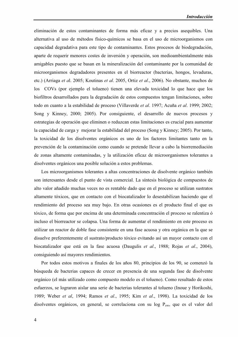
eliminación de estos contaminantes de forma más eficaz y a precios asequibles. Una
alternativa al uso de métodos físico-químicos se basa en el uso de microorganismos con
capacidad degradativa para este tipo de contaminantes. Estos procesos de biodegradación,
aparte de requerir menores costes de inversión y operación, son medioambientalmente más
amigables puesto que se basan en la mineralización del contaminante por la comunidad de
microorganismos degradadores presentes en el biorreactor (bacterias, hongos, levaduras,
etc.) (Arriaga et al. 2005; Koutinas et al. 2005, Ortiz et al., 2006). No obstante, muchos de
los COVs (por ejemplo el tolueno) tienen una elevada toxicidad lo que hace que los
biofiltros desarrollados para la degradación de estos compuestos tengan limitaciones, sobre
todo en cuanto a la estabilidad de proceso (Villaverde et al. 1997; Acuña et al. 1999; 2002;
Song y Kinney, 2000; 2005). Por consiguiente, el desarrollo de nuevos procesos y
estrategias de operación que eliminen o reduzcan estas limitaciones es crucial para aumentar
la capacidad de carga y mejorar la estabilidad del proceso (Song y Kinney; 2005). Por tanto,
la toxicidad de los disolventes orgánicos es uno de los factores limitantes tanto en la
prevención de la contaminación como cuando se pretende llevar a cabo la biorremediación
de zonas altamente contaminadas, y la utilización eficaz de microorganismos tolerantes a
disolventes orgánicos una posible solución a estos problemas.
Los microorganismos tolerantes a altas concentraciones de disolvente orgánico también
son interesantes desde el punto de vista comercial. La síntesis biológica de compuestos de
alto valor añadido muchas veces no es rentable dado que en el proceso se utilizan sustratos
altamente tóxicos, que en contacto con el biocatalizador lo desestabilizan haciendo que el
rendimiento del proceso sea muy bajo. En otras ocasiones es el producto final el que es
tóxico, de forma que por encima de una determinada concentración el proceso se ralentiza ó
incluso el biorreactor se colapsa. Una forma de aumentar el rendimiento en este proceso es
utilizar un reactor de doble fase consistente en una fase acuosa y otra orgánica en la que se
disuelve preferentemente el sustrato/producto tóxico evitando así un mayor contacto con el
biocatalizador que está en la fase acuosa (Daugulis et al., 1988; Rojas et al., 2004),
consiguiendo así mayores rendimientos.
Por todos estos motivos a finales de los años 80, principios de los 90, se comenzó la
búsqueda de bacterias capaces de crecer en presencia de una segunda fase de disolvente
orgánico (el más utilizado como compuesto modelo es el tolueno). Como resultado de estos
esfuerzos, se lograron aislar una serie de bacterias tolerantes al tolueno (Inoue y Horikoshi,
1989; Weber et al, 1994; Ramos et al., 1995; Kim et al., 1998). La toxicidad de los
disolventes orgánicos, en general, se correlaciona con su log Pow, que es el valor del

Introducción
5
logaritmo del coeficiente de partición del disolvente en una mezcla definida de octanol y
agua (log Pow); dicho valor se usa comúnmente como medida de la lipofilicidad de un
disolvente (Rekker y Kort, 1979). Los disolventes orgánicos con un valor de log Pow entre 1
y 4 son extremadamente tóxicos para los microorganismos (Rekker y Kort, 1979), por
ejemplo, benceno (log Pow 2,0), estireno (log Pow 3,6), xileno (log Pow 3,2) y tolueno (log Pow
2,5), porque se acumulan en la membrana de las bacterias modificando la estructura de la
misma. Sin embargo, el efecto tóxico de los disolventes no sólo depende de la toxicidad
inherente del compuesto sino también de la tolerancia intrínseca de las especies bacterianas.
Por ejemplo, se han identificado algunas cepas de E. coli como tolerantes a ciclohexano
(logPow 3,2) (Aono et al., 1991) pero no a tolueno (logPow 2,4). En este contexto, no es de
extrañar que todas las cepas Gram negativas identificadas hasta el momento como altamente
resistentes a disolventes orgánicos pertenezcan al género Pseudomonas. Una de estas cepas
es Pseudomonas putida DOT-T1E, la cepa que se ha utilizado en este estudio de tesis
doctoral, que es capaz de crecer hasta en el 90% (v/v) de tolueno (Ramos et al., 1995). Esta
cepa se aisló de la planta de tratamiento de aguas residuales del puente de los Vados en
Granada (Ramos et al., 1995) y no sólo es tolerante a tolueno, sino que también puede
utilizarlo como fuente de carbono y energía a través de la ruta de la tolueno dioxigenasa
(ruta TOD) (Gibson et al., 1970; Mosqueda et al., 1999).
II.) Biología de Pseudomonas
El género Pseudomonas fue descrito por primera vez por Migula (1894) en un simple
párrafo de dos líneas cuya traducción sería: “Células con órganos polares para su movilidad.
Algunas especies forman esporas, aunque en general es un evento raro (por ejemplo,
Pseudomonas violacea)”. Esta definición tan poco específica fue generalmente aceptada y
algunas bacterias, que habían sido asignadas a otros géneros, fueron rebautizadas en años
posteriores como pertenecientes al género Pseudomonas.
A principios del siglo XX los microbiólogos, en particular la escuela de Delft,
establecieron que las cepas de Pseudomonas eran muy comunes en hábitats naturales,
particularmente en suelo, agua, alimentos y plantas enfermas. La ubicuidad de estos
microorganismos y la capacidad para crecer en medios de cultivo muy simples hicieron que
se considerasen a las bacterias de este género como protagonistas en el proceso de
mineralización de materia orgánica en la naturaleza, un papel que fue claramente
demostrado por den Dooren de Jong (1926).
Introducción
6
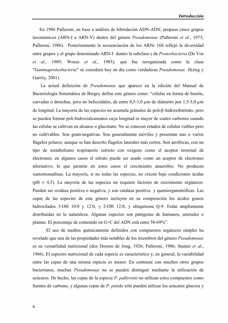
En 1986 Palleroni, en base a análisis de hibridación ADN-ADN, propuso cinco grupos
taxonómicos (ARN-I a ARN-V) dentro del género Pseudomonas (Palleroni et al., 1973;
Palleroni, 1986). Posteriormente la secuenciación de los ARNr 16S reflejó la diversidad
entre grupos y el grupo denominado ARN-I dentro la subclase-γ de Proteobacteria (De Vos
et al., 1989; Woese et al., 1985), que fue reorganizada como la clase
“Gammaproteobacteria” se considera hoy en día como verdaderas Pseudomonas (Krieg y
Garrity, 2001).
La actual definición de Pseudomonas que aparece en la edición del Manual de
Bacteriología Sistemática de Bergey define este género como: “células en forma de bastón,
curvadas o derechas, pero no helicoidales, de entre 0,5-1,0 µm de diámetro por 1,5-5,0 µm
de longitud. La mayoría de las especies no acumula gránulos de poli-β-hidroxibutirato, pero
se pueden formar poli-hidroxialcanoatos cuya longitud es mayor de cuatro carbonos cuando
las células se cultivan en alcanos o gluconato. No se conocen estados de células viables pero
no cultivables. Son gram-negativas. Son generalmente móviles y presentan uno o varios
flagelos polares; aunque se han descrito flagelos laterales más cortos. Son aeróbicas, con un
tipo de metabolismo respiratorio estricto con oxígeno como el aceptor terminal de
electrones; en algunos casos el nitrato puede ser usado como un aceptor de electrones
alternativo, lo que permite en estos casos el crecimiento anaerobio. No producen
xantomonadinas. La mayoría, si no todas las especies, no crecen bajo condiciones ácidas
(pH ≤ 4,5). La mayoría de las especies no requiere factores de crecimiento orgánicos.
Pueden ser oxidasa positiva o negativa, y son catalasa positiva y quimiorganotróficas. Las
cepas de las especies de este género incluyen en su composición los ácidos grasos
hidroxilados 3-OH 10:0 y 12:0, y 2-OH 12:0, y ubiquinona Q-9. Están ampliamente
distribuidas en la naturaleza. Algunas especies son patógenas de humanos, animales o
plantas. El porcentaje de contenido en G+C del ADN está entre 58-69%”.
El uso de medios químicamente definidos con compuestos orgánicos simples ha
revelado que una de las propiedades más notables de los miembros del género Pseudomonas
es su versatilidad nutricional (den Dooren de Jong, 1926; Palleroni, 1986; Stainer et al.,
1966). El espectro nutricional de cada especie es característico y, en general, la variabilidad
entre las cepas de una misma especie es menor. En contraste con muchos otros grupos
bacterianos, muchas Pseudomonas no se pueden distinguir mediante la utilización de
azúcares. De hecho, las cepas de la especie P. palleronii no utilizan estos compuestos como
fuentes de carbono, y algunas cepas de P. putida sólo pueden utilizar los azúcares glucosa y
Introducción
7
sacarosa. Los compuestos orgánicos empleados como fuentes de carbono y energía por
muchas especies de Pseudomonas incluyen hidrocarburos lineales y aromáticos, ácidos
alifáticos, aminas, amidas, aminoácidos, alcoholes y compuestos aromáticos. La capacidad
de utilizar los compuestos aromáticos es particularmente interesante, dadas las distintas rutas
por las cuales Pseudomonas es capaz de metabolizarlos, y su posible utilización en procesos
de descontaminación y biocatálisis.
La gran versatilidad del género no sólo se deriva de la gran cantidad y variedad de
fuentes de carbono que pueden utilizar sino también de la capacidad de explotar diferentes
nichos ecológicos. El género Pseudomonas incluye cepas patógenas oportunistas de
animales y el hombre, como P. aeruginosa, patógenos de plantas como P. syringae, y cepas
que estimulan el crecimiento de plantas actuando como fungicidas, como P. fluorescens y
cepas de suelo como P. putida (Lugtenberg, 1999; Palleroni y Moore, 2004). En los últimos
cinco años se ha secuenciado el genoma de las cepas P. aeruginosa PAO1 (Stover et al.,
2000), P. putida KT2440 (Nelson et al., 2002), Pseudomonas enthomophila (Vodovar et al.,
2006), P. syringae pv. tomatoe DC3000 (http://pseudomonas-syringae.org/) y P.
fluorescens Pf-5 (http://pseudomonas-fluorescens.org/). Estas bacterias presentan en
promedio 5500 genes en sus genomas, la mitad de ellos sin función conocida. Vodovar et al.
han identificado un grupo de casi 2100 genes que constituyen el núcleo del genoma del
género Pseudomonas. Los datos de secuenciación sugieren que el número de genes
“exclusivos” de P. putida KT2440 es mayor que el de genes que tiene en común con otras
Pseudomonas. Estas regiones no compartidas se asocian a la versatilidad y “fitness” de P.
putida.
III.) Mecanismos de tolerancia a disolventes orgánicos
La principal función de la membrana celular de los microorganismos es la de formar una
barrera permeable, regulando el paso de solutos entre la célula y el medio externo. Las
propiedades de la membrana citoplasmática como barrera son de especial importancia para
la transducción de energía de la célula y para el mantenimiento de funciones vitales (Ramos
et al., 2002; Segura et al., 1999, Sikkema et al., 1995). Los disolventes orgánicos se
acumulan preferentemente en las bicapas lipídicas de las membranas, desorganizándolas y
provocando la interrupción de las funciones vitales (pérdida de iones, metabolitos, lípidos y
proteínas; la disipación del gradiente de pH y del potencial eléctrico). Todo esto conlleva a
Introducción
8
la lisis y posterior muerte celular (Sikkema et al., 1995; Ramos et al., 2002; Segura et al.,
1999).
Aunque el metabolismo de los agentes químicos disminuya las concentraciones efectivas
del mismo a las que está sometido el microorganismo, no parece que este sea un mecanismo
de tolerancia en aquellas bacterias expuestas a altas concentraciones del disolvente orgánico.
Por un lado, algunos microorganismos tolerantes a un disolvente orgánico en particular no
pueden metabolizarlo, por ejemplo, P. putida S12 es tolerante a concentraciones
supersaturantes de tolueno pero no utiliza este hidrocarburo como fuente de carbono y por
otro lado, un mutante de Pseudomonas putida DOT-T1E en el que se ha inactivado la
tolueno dioxigenasa, una enzima clave en la ruta de degradación de tolueno en esta cepa, no
es más sensible al disolvente que la cepa silvestre (Mosqueda et al., 1999).
En la tolerancia a disolventes orgánicos, se han descrito una serie de mecanismos
involucrados en la respuesta a esos agentes tóxicos: rigidificación de la membrana celular,
alteración de la composición de los fosfolípidos; alteración de la superficie celular que hace
que las células sean menos permeables; expulsión de los compuestos tóxicos en un proceso
dependiente de energía; y formación de vesículas que remueven los disolventes desde la
superficie celular entre otros. (Ramos et al., 2002; Kadurugamuwa y Beveridge, 1995). A
continuación se explican en detalle algunos de estos mecanismos.
III.a) Alteración en la composición de fosfolípidos de la membrana debido a la
exposición a disolventes orgánicos
Cuando las células bacterianas se exponen a disolventes orgánicos, éstos entran en
contacto con la bicapa lipídica y penetran en ella, provocando un aumento en la fluidez de la
misma. Inmediatamente en la bacteria se desencadenan una serie de respuestas para
disminuir los efectos perjudiciales de este cambio. La fluidez de la membrana es reajustada,
alterando la composición de la bicapa lipídica por medio de mecanismos compensatorios
que se asemejan a algunos de los observados en respuesta a cambios físicos y químicos
impuestos por el medio ambiente (Sikkema et al., 1992; Ramos et al., 2001)
III.a.i) -Cambios en la composición de ácidos grasos
Existen distintos mecanismos que alteran la composición de ácidos grasos esterificados
y por consiguiente la fluidez de la bicapa lipídica bacteriana: la isomerización cis/trans de
Introducción
9
ácidos grasos como una respuesta a muy corto plazo; el cambio en la proporción de ácidos
grasos saturados e insaturados, como una respuesta a más largo plazo y el cambio en la
proporción de ácidos grasos de cadena corta y cadena larga (Ramos et al., 2002).
III.a.i.1. Isomerización cis / trans de los ácidos grasos insaturados
Los ácidos grasos insaturados son sintetizados mayoritariamente en la configuración cis
y por tanto estos son los isómeros mayoritarios en las membranas celulares bacterianas. Sin
embargo, se han descrito en algunas bacterias Gram negativas (del género Pseudomonas y
Vibrio) la presencia de isómeros trans como resultado de una modificación postsintética
catalizada por la enzima cis-trans-isomerasa (Cti).
La conformación estérica de ácidos grasos insaturados trans y de los ácidos grasos saturados
es muy similar, ambos poseen una conformación extendida lo que permite un
empaquetamiento más compacto de las cadenas acilo de los fosfolípidos en la membrana.
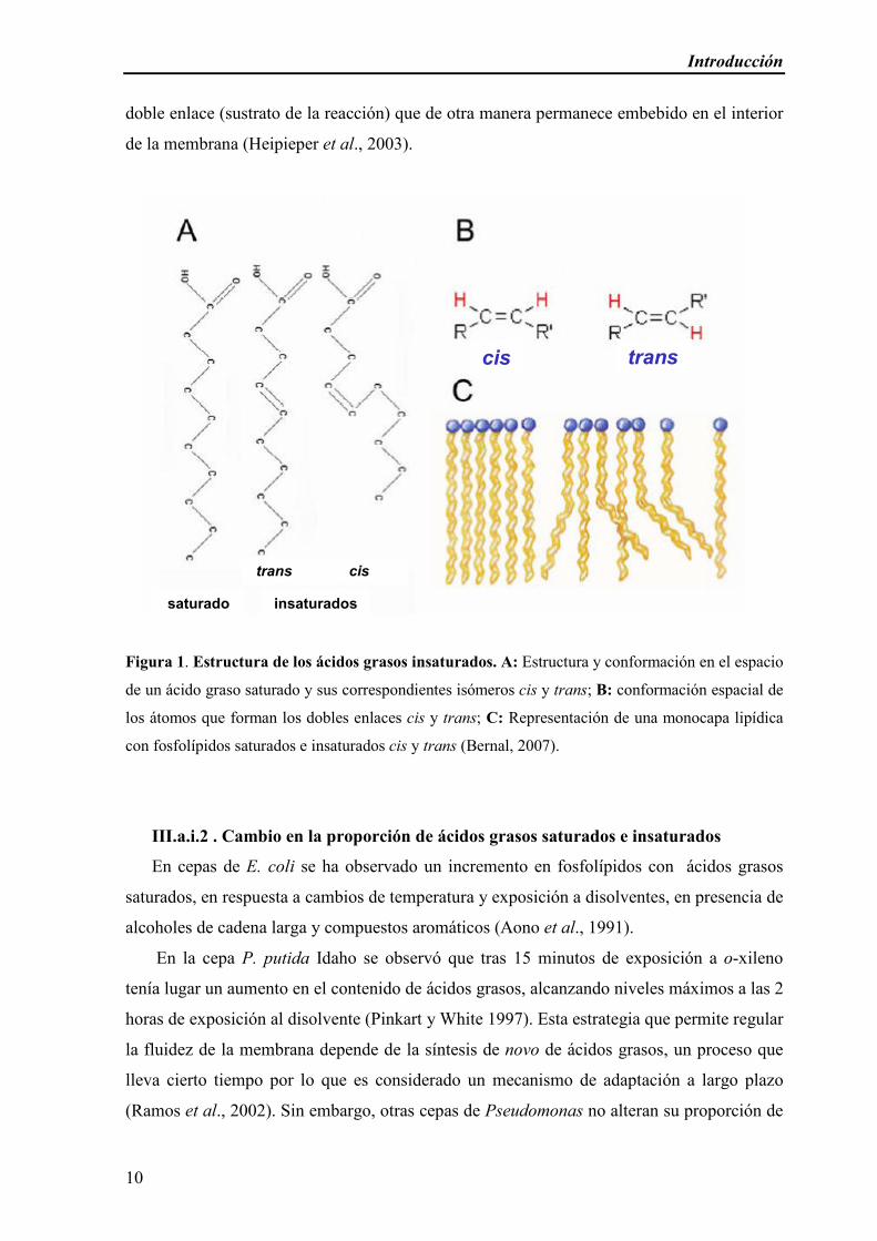
Por el contrario, la configuración cis del doble enlace provoca un ángulo fijo de 30º en la
cadena acílica lo que impide un empaquetamiento compacto con los ácidos grasos,
provocando una mayor fluidez en las membranas donde están presentes (Figura 1).
Precisamente por esta conformación diferente de los ácidos grasos insaturados, la
isomerización cis / trans juega un papel importante en la adaptación de las bacterias frente a
cambios de temperatura o presencia de disolventes orgánicos en el medio. Estos estreses
ambientales provocan un aumento de la fluidez de membrana, que es compensado mediante
la síntesis de isómeros trans que contrarrestan el efecto fluidificante del estrés.
Varias cepas de P. putida son capaces de cambiar rápidamente su proporción cis/trans
en la membrana en respuesta a la exposición a disolventes orgánicos, compuestos
aromáticos o temperatura; este cambio se observa tan sólo cinco minutos después de la
adición del disolvente orgánico en P. putida DOT-T1E (Ramos et al., 1995). Ésta se
considera una respuesta rápida, que no necesita de energía ya que no depende de la síntesis
de novo de ácidos grasos. De esta manera, las células ganan tiempo para un ajuste
metabólico y poner en marcha las respuestas a largo plazo. (Junker y Ramos, 1999; Bernal
et al., 2007a). En P. putida DOT-T1E esta modificación postsintética ha sido bien
caracterizada (Junker y Ramos, 1999). Diversos estudios señalan que el gen cti se expresa
constitutivamente (Heipieper et al., 2003; Bernal et al., 2007a) y sin embargo la actividad
enzimática se induce en presencia del estrés. La hipótesis más plausible hasta el momento es
que tan sólo cuando la membrana tiene una fluidez elevada la enzima Cti puede alcanzar el
Introducción
10
doble enlace (sustrato de la reacción) que de otra manera permanece embebido en el interior
de la membrana (Heipieper et al., 2003).
Figura 1. Estructura de los ácidos grasos insaturados. A: Estructura y conformación en el espacio
de un ácido graso saturado y sus correspondientes isómeros cis y trans; B: conformación espacial de
los átomos que forman los dobles enlaces cis y trans; C: Representación de una monocapa lipídica
con fosfolípidos saturados e insaturados cis y trans (Bernal, 2007).
III.a.i.2 . Cambio en la proporción de ácidos grasos saturados e insaturados
En cepas de E. coli se ha observado un incremento en fosfolípidos con ácidos grasos
saturados, en respuesta a cambios de temperatura y exposición a disolventes, en presencia de
alcoholes de cadena larga y compuestos aromáticos (Aono et al., 1991).
En la cepa P. putida Idaho se observó que tras 15 minutos de exposición a o-xileno
tenía lugar un aumento en el contenido de ácidos grasos, alcanzando niveles máximos a las 2
horas de exposición al disolvente (Pinkart y White 1997). Esta estrategia que permite regular
la fluidez de la membrana depende de la síntesis de novo de ácidos grasos, un proceso que
lleva cierto tiempo por lo que es considerado un mecanismo de adaptación a largo plazo
(Ramos et al., 2002). Sin embargo, otras cepas de Pseudomonas no alteran su proporción de
transcis
saturado insaturados
trans cis
transcis transcis
saturado insaturados
trans cis

Introducción
11
ácidos grasos saturados/insaturados en respuesta a disolventes orgánicos como sucede en la
cepa Pseudomonas putida DOT-T1E, que no varía esta proporción tras su exposición a
distintos disolventes orgánicos (Ramos et al., 1997).
III.a.i.3 Cambios en la proporción de ácidos grasos de cadena larga y cadena corta.
La razón de ácidos grasos de cadena corta/cadena larga también se puede alterar para
regular la fluidez de la membrana (Keweloh y Heipieper, 1996; Heipieper y de Bont, 1994;
Heipieper et al., 1996; Holtwick et al., 1997; Pinkart y White, 1997; Pinkart et al., 1996;
Ramos et al., 1997; Weber y de Bont, 1996). Este es uno de los mecanismos a largo plazo
que ponen en marcha Pseudomonas spp., E. coli y otros microorganismos cuando son
expuestos a concentraciones subletales de disolventes orgánicos, dando como resultado un
incremento en la cantidad total de fosfolípidos y un aumento en la proporción de lípidos
saturados de cadena larga. Todos estos cambios son concomitantes con las alteraciones
producidas en los grupos de cabeza de los fosfolípidos. En suma estos cambios modifican la
fluidez de la membrana celular con el fin de volverla más rígida (Ramos et al, 2002; Segura
et al., 1999).
III.a.ii Cambio en los grupos de cabeza de los fosfolípidos
Los principales tipos de fosfolípidos en las membranas de P. putida son
fosfatidiletanolamina (PE), fosfatidilglicerol (PG), y difosfatidilglicerol o cardiolipina (CL).
Diferentes cepas de Pseudomonas sp. parecen haber desarrollado diferentes estrategias para
responder a disolventes orgánicos mediante el cambio en la composición de los grupos de
cabeza de los fosfolípidos. Se han analizado los cambios en los fosfolípidos de tres cepas: P.
putida S12, P. putida DOT-T1E, y P. putida Idaho. En general, en todas se observó un
aumento de los niveles de CL (Weber, 1994; Ramos et al., 1997) sin embargo se han visto
diferencias significativas entre ellas. Un análisis detallado del recambio de los grupos de
cabeza de los fosfolípidos en ausencia y presencia de o-xileno en la cepa P. putida Idaho
reveló que mientras en ausencia del disolvente la incorporación de 32P era mayor en PG,
seguida de PE y CL, en presencia de 200 ppm de o-xileno, la incorporación de 32P se
producía mayoritariamente en PE, seguida muy de cerca por PG. Experimentos similares
hechos con P. putida DOT-T1E sin embargo indicaron que la mayor parte del 32P se
incorporaba en CL cuando en el medio había tolueno (Ramos et al., 1997). Pinkart y White

Introducción
12
(1997) también demostraron que en la cepa Idaho la isomerización cis/trans de ácidos
grasos insaturados ocurría principalmente en los ácidos esterificados en PE como grupo de
cabeza, sin embargo, en la cepa DOT-T1E no parece que haya una distribución preferente de
ácidos grasos esterificados en los grupos de cabeza (Bernal et al., resultados no
publicados). Aunque el incremento en PE para contrarrestar los efectos de los disolventes no
es muy común entre las bacterias (Weber y de Bont, 1996), el PE tiene una temperatura de
fusión más alta que PG, por tanto, un aumento en PE podría estabilizar la membrana celular.
El aumento en los niveles de CL es algo más común; la CL tiene una temperatura de
transición (10ºC) mayor que el PE, lo cual en teoría podría disminuir la fluidez de la
membrana y estabilizarla en presencia del tóxico orgánico (Weber y de Bont, 1996). Sin
embargo, mutantes de P. putida DOT-T1E en el gen cls (cardiolipina sintasa) que tienen
menor proporción de este grupo de cabeza tienen membranas más rígidas. Este mutante es
más sensible al tolueno y a otras drogas que son transportados por bombas de la familia
RND (ver más abajo), lo que indica que la falta de CL provoca la desorganización de
algunas proteínas de membrana y por tanto la sensibilidad al tolueno estaría provocada más
por este efecto sobre las bombas que sobre la fluidez de la membrana (Bernal et al., 2007b).
III.a.iii.- Cambios en la velocidad de síntesis de los ácidos grasos
El análisis de la biosíntesis de fosfolípidos en la cepa tolerante P. putida Idaho
mostró que el nivel basal de biosíntesis de fosfolípidos era mayor en esta cepa que en cepas
sensibles a disolventes de la misma especie y que la velocidad de biosíntesis aumentaba en
respuesta a la exposición de concentraciones subletales de disolventes orgánicos (Pinkart y
White, 1997). También se ha constatado que mutantes sensibles a tolueno de P. putida
DOT-T1E eran incapaces de incorporar 13CH3-13COOH en ácidos grasos en presencia de
cantidades subletales de tolueno (Segura et al., 2004). Esto sugiere que el tolueno afecta
fuertemente a la síntesis de novo de ácidos grasos. El recambio limitado en el metabolismo
de ácidos grasos en esta cepa mutante contribuye a la pérdida de la integridad de la
membrana en presencia de disolventes, y el consecuente aumento de la sensibilidad a estos
compuestos.

Introducción
13
III.a. iv- Alteración de lipopolisacáridos
Los lipopolisacáridos (LPS) de las bacterias gram-negativas son uno de los
principales componentes de la membrana externa y son considerados como una barrera de
defensa. En varias cepas de P. putida la adición de altas concentraciones de cationes
divalentes (Mg2+ y Ca2+) al medio de cultivo con disolventes orgánicos resultó en un
incremento en la supervivencia (Ramos et al., 1995; Weber y de Bont, 1996). La hipótesis
desarrollada a partir de estos datos fue que los cationes divalentes se unían
electrostáticamente a las moléculas polianiónicas adyacentes de LPS y así reducían la
repulsión de cargas. Los cationes permitirían un empaquetamiento más denso de las
moléculas aniónicas de las membranas. Sin embargo, un mutante en el gen wbpL de P.
putida DOT-T1E, que codifica la enzima que inicia la síntesis de la cadena lateral del
antígeno-O de los LPS, es tan tolerante como la cepa silvestre a disolventes orgánicos
(tolueno, octanol, p-xileno, propilbenceno, y heptano) lo que sugiere que los LPS juegan un
papel menor en la respuesta a disolventes (Junker et al., 2001)
III.b - Bombas de expulsión de disolventes
Probablemente las bombas de expulsión de disolventes orgánicos sean el principal
responsable de la tolerancia, al menos en aquellas cepas que son altamente tolerantes a
disolventes orgánicos. Casi todas las bombas identificadas hasta el momento como
involucradas en los procesos de tolerancia pertenecen a la familia de las RND (Resistencia,
Nodulación, División celular).
Los organismos vivos se comunican con el medio ambiente que les rodea en parte a
través de sistemas de transporte sustrato-específicos. Estos sistemas consisten en proteínas
integrales de membrana que se expanden múltiples veces por la membrana citoplasmática.
Estas proteínas que forman canales específicos para cada sustrato y que están acopladas a
una fuente de energía y/o a proteínas situadas en la membrana externa, pueden conferirle a
la célula la capacidad para bombear un soluto y reconocerlo con una alta afinidad (Saier et
al., 1998). Las proteínas que conforman estos transportadores han sido estudiadas
extensamente desde el punto de vista estructural, funcional y filogenético, y como
consecuencia de ello se han descrito familias de proteínas transportadoras, algunas de las
cuales se ha visto que son ubicuas, mientras que otras están restringidas sólo a uno de los
principales reinos de seres vivos (Saier et al., 1998). Actualmente hay descritas en bacterias
cinco familias de transportadores; MFS (Major Facilitator Superfamily), ABC (ATP-

Introducción
14
Binding-Cassette) y MATE (multidrug and toxic compound extrusion) son mayoritarias y
ancestrales, mientras que las otras dos familias SMR (Small Multidrug Resistance) y RND
(Resistance-Nodulation-cell Division) son minoritarias y con un origen evolutivo más
reciente (Saier et al., 1998). Por lo general, el número de sistemas de transporte está en
proporción al tamaño del genoma del microorganismo en cuestión, excepto en bacterias
adaptadas a sobrevivir en medio ambientes pobres en materia orgánica y ricos en minerales
donde llegan a tener hasta dos veces menos transportadores en relación al tamaño del
genoma (Saier et al., 1998).
Muchos de estos transportadores tienen un papel importante en lo que se denomina
resistencia a multidrogras (MDR, multidrug resistance). Estos sistemas catalizan la
expulsión activa de compuestos biogénicos y xenobióticos no relacionados estructural o
funcionalmente, desde el citoplasma o la membrana interna, hacia el medio externo
(Krulwich et al., 2005; Piddock, 2006; Borges-Walmsley y Walmsley, 2001). Las
evidencias experimentales indican que las características físico-químicas de los compuestos
(carga, hidrofobicidad, o anfipatía), las interacciones de van der Waals que establecen éstos
con los residuos de los sitios activos, y la flexibilidad de estos sitios para acomodar distintas
moléculas, son los factores determinantes de la amplia especificidad de sustrato de estos
sistemas de extrusión (Neyfakh, 2001; Murakami et al., 2002; Yu et al., 2003). Aunque se
puede pensar que estos transportadores de antimicrobianos surgieron recientemente en
respuesta a la quimioterapia antimicrobiana y a compuestos de síntesis humana, el hecho de
que haya un número similar de bombas de extrusión que confieren multirresistencia en
microorganismos patógenos y no patógenos y el hecho de que muchos estén codificados en
el cromosoma, sugiere que juegan un rol fisiológico importante en la expulsión de sustancias
tóxicas que normalmente tienen lugar en la naturaleza (tales como toxinas naturales,
productos finales del metabolismo endógeno, y antibióticos) y que también expulsan nuevas
drogas de fabricación reciente por ser éstas de estructura similar a los respectivos sustratos
naturales (Saier et al., 1998).
Junto con las proteínas transportadoras, se han identificado dos familias de proteínas
accesorias que funcionan conjuntamente con algunos de los transportadores. Estas dos
familias han sido designadas de acuerdo a su localización celular en la envoltura de las
bacterias Gram-negativas y su presunta función, y se conocen con el nombre de familia
OMP (Outer Membrane Protein) y familia MFP (Membrane Fusion Protein). Se cree que el
conjunto de estas tres proteínas (transportador, OMP y MFP) permiten el transporte a través
de ambas membranas de la envoltura de las bacterias Gram-negativas en un simple paso
Introducción
15
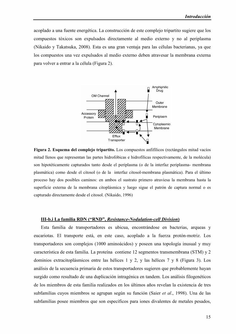
acoplado a una fuente energética. La construcción de este complejo tripartito sugiere que los
compuestos tóxicos son expulsados directamente al medio externo y no al periplasma
(Nikaido y Takatsuka, 2008). Esta es una gran ventaja para las células bacterianas, ya que
los compuestos una vez expulsados al medio externo deben atravesar la membrana externa
para volver a entrar a la célula (Figura 2).
Figura 2. Esquema del complejo tripartito. Los compuestos anfifílicos (rectángulos mitad vacíos
mitad llenos que representan las partes hidrofóbicas e hidrofílicas respectivamente, de la molécula)
son hipotéticamente capturados tanto desde el periplasma (o de la interfaz periplasma- membrana
plasmática) como desde el citosol (o de la interfaz citosol-membrana plasmática). Para el último
proceso hay dos posibles caminos: en ambos el sustrato primero atraviesa la membrana hasta la
superficie externa de la membrana citoplásmica y luego sigue el patrón de captura normal o es
capturado directamente desde el citosol. (Nikaido, 1996)
III-b.i La familia RDN (“RND”, Resistance-Nodulation-cell Division)
Esta familia de transportadores es ubicua, encontrándose en bacterias, arqueas y
eucariotas. El transporte está, en este caso, acoplado a la fuerza protón-motriz. Los
transportadores son complejos (1000 aminoácidos) y poseen una topología inusual y muy
característica de esta familia. La proteína contiene 12 segmentos transmembrana (STM) y 2
dominios extracitoplásmicos entre las hélices 1 y 2, y las hélices 7 y 8 (Figura 3). Los
análisis de la secuencia primaria de estos transportadores sugieren que probablemente hayan
surgido como resultado de una duplicación intragénica en tandem. Los análisis filogenéticos
de los miembros de esta familia realizados en los últimos años revelan la existencia de tres
subfamilias cuyos miembros se agrupan según su función (Saier et al., 1998). Una de las
subfamilias posee miembros que son específicos para iones divalentes de metales pesados,

Introducción
16
otra subfamilia probablemente sea específica para el transporte de lipo-oligosacáridos, y otra
subfamilia esta conformada por proteínas que expulsan múltiples drogas. Miembros de estas
tres subfamilias se han descrito solamente en bacterias gram-negativas. (Marger y Saier,
1993; Saier et al., 1998; Borges-Walmsley y Walmsley, 2001).
La mayoría de los transportadores identificados como relevantes en la tolerancia a
disolventes orgánicos pertenecen a esta familia. En P. putida DOT-T1E, tres bombas de esta
familia, denominadas Ttg (Toluene tolerance genes) participan en la extrusión de
disolventes orgánicos (TtgABC, TtgDEF y TtgGHI) (Ramos et al., 1998; Mosqueda y
Ramos, 2000; Rojas et al., 2001); en P. putida S12 y P. putida GM73 también se han
identificado bombas de esta familia como los principales determinantes de tolerancia a
Figura 3. Modelo estructural representativo de los miembros de la familia “RND”. La figura
muestra los segmentos transmembrana y los dos lazos periplásmicos (Saier et al., 1998)
tolueno (Kieboom et al., 1998a, Kim et al, 1998). En P. aeruginosa y E. coli se ha
relacionado este tipo de bombas con la tolerancia a disolventes (Aono et al., 1998; Fralick,
1996; Fukimori et al., 1998; Kieboom et al., 1998b; Kim et al., 1998; Li et al., 1998; Ma et
al., 1993; Mosqueda y Ramos, 2000; Ramos et al., 1998; Rojas et al., 2001; Zgurskaya y
Nikaido, 1999). En general, estos transportadores juegan un rol importante en la resistencia
a una gran variedad de compuestos tóxicos en bacterias gram-negativas siendo también
capaces de transportar una gran variedad de sustratos que no siempre guardan una gran
similitud a nivel de su estructura, por ejemplo, antibióticos, compuestos flavonoides y
Introducción
17
mutágenos como el bromuro de etidio (Piddock, 2006; Ramos et al., 1998; Mosqueda y
Ramos, 2000; Rojas et al., 2001, Saier et al., 1998).
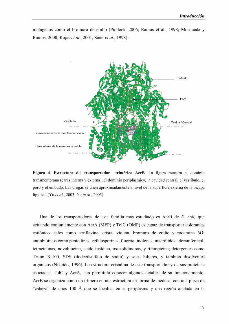
Figura 4. Estructura del transportador trimérico AcrB. La figura muestra el dominio
transmembrana (caras interna y externa), el dominio periplásmico, la cavidad central, el vestíbulo, el
poro y el embudo. Las drogas se unen aproximadamente a nivel de la superficie externa de la bicapa
lipídica. (Yu et al., 2003; Yu et al., 2005).
Una de los transportadores de esta familia más estudiado es AcrB de E. coli, que
actuando conjuntamente con AcrA (MFP) y TolC (OMP) es capaz de transportar colorantes
catiónicos tales como acriflavina, cristal violeta, bromuro de etidio y rodamina 6G;
antiobióticos como penicilinas, cefalosporinas, fluoroquinolonas, macrólidos, cloramfenicol,
tetraciclinas, novobiocina, acido fusídico, oxazolidinonas, y rifampicina; detergentes como
Tritón X-100, SDS (dodecilsulfato de sodio) y sales biliares, y también disolventes
orgánicos (Nikaido, 1996). La estructura cristalina de este transportador y de sus proteínas
asociadas, TolC y AcrA, han permitido conocer algunos detalles de su funcionamiento.
AcrB se organiza como un trímero en una estructura en forma de medusa, con una pieza de
“cabeza” de unos 100 Å que se localiza en el periplasma y una región anclada en la
Cavidad Central
Poro
Embudo
Vestíbulo
Cara externa de la membrana celular
Cara interna de la membrana celular
Introducción
18
membrana interna de unos 50 Å (Figura 4). Tres de los dominios transmembrana de cada
protómero se organizan en una estructura a modo de anillo con un hueco central que cruza la
membrana y se extiende hasta la parte de debajo de la pieza de cabeza. El transportador
posee unas pequeñas aperturas (vestíbulos) entre las subunidades al final del dominio
periplásmico cerca de la superficie externa de la membrana citoplasmática.
Estas aperturas conducen a la gran cavidad central dentro del dominio transmembrana
(Figura 4). Se cree que los compuestos tóxicos difunden a través del vestíbulo hasta llegar a
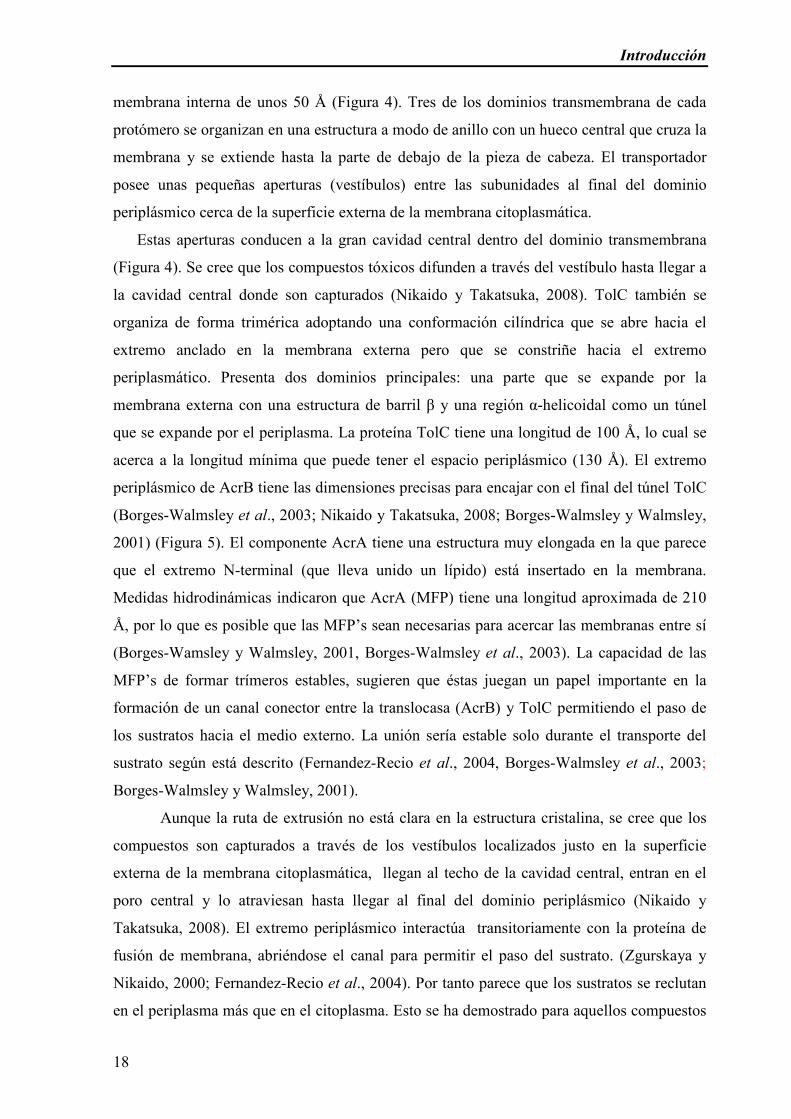
la cavidad central donde son capturados (Nikaido y Takatsuka, 2008). TolC también se
organiza de forma trimérica adoptando una conformación cilíndrica que se abre hacia el
extremo anclado en la membrana externa pero que se constriñe hacia el extremo
periplasmático. Presenta dos dominios principales: una parte que se expande por la
membrana externa con una estructura de barril β y una región α-helicoidal como un túnel
que se expande por el periplasma. La proteína TolC tiene una longitud de 100 Å, lo cual se
acerca a la longitud mínima que puede tener el espacio periplásmico (130 Å). El extremo
periplásmico de AcrB tiene las dimensiones precisas para encajar con el final del túnel TolC
(Borges-Walmsley et al., 2003; Nikaido y Takatsuka, 2008; Borges-Walmsley y Walmsley,
2001) (Figura 5). El componente AcrA tiene una estructura muy elongada en la que parece
que el extremo N-terminal (que lleva unido un lípido) está insertado en la membrana.
Medidas hidrodinámicas indicaron que AcrA (MFP) tiene una longitud aproximada de 210
Å, por lo que es posible que las MFP’s sean necesarias para acercar las membranas entre sí
(Borges-Wamsley y Walmsley, 2001, Borges-Walmsley et al., 2003). La capacidad de las
MFP’s de formar trímeros estables, sugieren que éstas juegan un papel importante en la
formación de un canal conector entre la translocasa (AcrB) y TolC permitiendo el paso de
los sustratos hacia el medio externo. La unión sería estable solo durante el transporte del
sustrato según está descrito (Fernandez-Recio et al., 2004, Borges-Walmsley et al., 2003;
Borges-Walmsley y Walmsley, 2001).
Aunque la ruta de extrusión no está clara en la estructura cristalina, se cree que los
compuestos son capturados a través de los vestíbulos localizados justo en la superficie
externa de la membrana citoplasmática, llegan al techo de la cavidad central, entran en el
poro central y lo atraviesan hasta llegar al final del dominio periplásmico (Nikaido y
Takatsuka, 2008). El extremo periplásmico interactúa transitoriamente con la proteína de
fusión de membrana, abriéndose el canal para permitir el paso del sustrato. (Zgurskaya y
Nikaido, 2000; Fernandez-Recio et al., 2004). Por tanto parece que los sustratos se reclutan
en el periplasma más que en el citoplasma. Esto se ha demostrado para aquellos compuestos
Introducción
19
que no son capaces de atravesar la membrana citoplasmática, como los compuestos
dianiónicos (carbenicilina, ceftriaxona) y los aminoglicósidos, pero que sin embargo son
sustrato de estas bombas (Nikaido, 1996). Si bien parece quedar claro que la captura de
sustratos desde el citoplasma también tiene lugar, no queda tan claro si éste es el modo de
captura que predomina.
Figura 5. Estructura cristalina de TolC, AcrA y AcrB. Organización hipotética de la estructura
tripartita TolC (rojo)- AcrA (azul)- AcrB (verde) basado en la estructura cristalina obtenida por
difracción de rayos X (Nikaido y Takatsuka, 2008).
También se ha visto que las bombas RND actúan en sinergismo con otros
transportadores. Un ejemplo de esto sucede en P. aeruginosa; TetA una bomba de la familia
MFS que expulsa tetraciclina, confiere un nivel de resistencia relativamente bajo (CIM = 32
µg/mL) en ausencia del principal transportador RND, MexA-MexB-OprM; en ausencia de
Introducción
20
TetA la CIM conferida por el transportador RND es de 4 µg/mL. Sin embargo, la CIM
aumenta fuertemente (CIM = 512 µg/mL) cuando ambos transportadores se expresan a
niveles normales y constitutivos. De acuerdo con lo expuesto, parece que estos dos
transportadores actúan de modo sinérgico. La explicación más plausible de este resultado es
que la tetraciclina es bombeada hacia el periplasma por TetA desde donde es capturada por
el complejo tripartito (MexAB-OprM) y expulsada hacia el exterior de la célula (Lee et al,
2000). Esto a su vez enfatiza la importancia del mecanismo de captura desde el periplasma
de las bombas RND (Nikaido y Takatsuka, 2008).
En algún momento se ha sugerido que estos transportadores trabajan
sinergísticamente con la membrana externa. Sin embargo, la inactivación del transportador
más importante de una bacteria (por ejemplo, AcrB en E. coli ó TtgGHI para disolventes en
P. putida DOT-T1E) hace que ésta se vuelva susceptible al compuesto toxico en cuestión, a
pesar de tener la membrana externa intacta (Nikaido y Takatsuka, 2008; Ramos et al., 1998;
Mosqueda y Ramos, 2000; Rojas et al., 2001).
Cada uno de los componentes es imprescindible para la extrusión del compuesto tóxico,
ya que la falta de alguno de ellos hace que el transportador no funcione (Nikaido y
Takatsuka, 2008). Algunos de estos transportadores están codificados junto con sus
correspondientes OMP y MFP (MexAB-OprM, MexEF-OprN, TtgABC, TtgDEF, TtgGHI
entre otros (Poole et al., 1993; Koehler et al., 1997; Ramos et al., 1998; Mosqueda y Ramos,
2000; Rojas et al., 2001) mientras que en otros casos tan sólo se cotranscriben la translocasa
con su correspondiente MFS, utilizando una OMP que puede ser compartida entre varios
transportadores (por ejemplo AcrAB que utiliza TolC, MexXY que se une a OprM;
(Srikumar et al., 1997; Aires et al, 1999; Fralick, 1996; Mine et al., 1999). No está claro
cómo se ensamblan estos complejos y porqué en unos casos las tres proteínas se
cotranscriben y en otros casos no. A diferencia de las proteínas de fusión de membrana
(MFP), las proteínas de membrana externa (OMP) de los sistemas de extrusión descritos en
bacterias Gram-negativas pueden intercambiarse entre diferentes sistemas de transporte y
entre bombas de la misma familia (Yoneyama et al., 1998, Srikumar et al., 1997, Zgurskaya
y Nikaido, 2000). TolC de E. coli (esencial para el funcionamiento del transportador AcrAB
en esta cepa) también colabora en la actividad transportadora de las bombas MexD y MexY
de P. aeruginosa cuando éstas son expresadas en células de E. coli en ausencia de su
correspondiente proteína de membrana externa nativa (Srikumar et al., 1998). Otro ejemplo

Introducción
21
lo constituye la proteína OprM de P. aeruginosa la cual se acopla al menos a dos
transportadores de la familia RND, MexB y MexY (Zgurskaya y Nikaido, 2000). Además,
TolC tiene la capacidad de interactuar con diferentes clases de antiportes de membrana
interna pertenecientes a la misma familia o de familia diferente (RND, ABC y MFS) (Figura
6) (Piddock, 2006; Fernandez-Recio et al., 2004).
Figura 6. Diagrama de transportadores de resistencia pertenecientes a las familias ABC, RND
y MFS, todos ellos formando un complejo tripartito con TolC en bacterias gram-negativas (Piddock,
2006).
III. b. ii. La Superfamilia “ABC” (ATP-Binding-Cassette)
Estos sistemas de transporte pertenecen al grupo de los transportadores activos primarios
según la clasificación de Saier (2000), en el que el transporte está acoplado a la presencia de
una fuente primaria de energía, en este caso, a la hidrólisis de ATP. En este tipo de sistemas,
la proteína transportadora puede ser o no transitoriamente fosforilada, pero el sustrato que se
transporta nunca es fosforilado durante su translocación (Saier, 2000). Esta familia de
transportadores es ubicua, estando presente tanto en bacterias como en organismos
superiores. Esta superfamilia está compuesta por más de 30 familias; cada familia transporta

Introducción
22
una enorme variedad de sustratos, cada uno de los cuales es específico de un transportador.
Estos sustratos incluyen moléculas pequeñas que pueden ser transportadas al interior o
exterior de la célula dependiendo del transportador pero también transportan
macromoléculas tales como proteínas y carbohidratos complejos que son sintetizados en el
citoplasma y secretados a la envoltura celular o al medio externo; igualmente transportan
nutrientes al citoplasma bacteriano y participan además en numerosos procesos como en la
transducción de señales, en la resistencia a drogas y a antibióticos, en los mecanismos de
patogénesis y esporulación, etc. (Higgins, 1992; Wandersman, 1998).
Los transportadores de la familia ABC se caracterizan por presentar una secuencia
conservada, de aproximadamente 215 aminoácidos, que se estructura en un dominio soluble
con actividad ATPasa (Young y Holland, 1999) y que se denomina dominio de unión a ATP
o dominio ABC (ATP-Binding-Cassette). Es importante destacar la diferencia entre
proteínas ABC y transportadores ABC. El término proteína ABC se refiere a proteínas que
contienen un dominio de unión a ATP y/o de hidrólisis del ATP y que participan en muchos
procesos fisiológicos no necesariamente (aunque sí normalmente) relacionados con el
transporte. Por otro lado, un transportador ABC se forma cuando dicha actividad ATPasa se
asocia con un domino hidrofóbico de membrana (Young y Holland, 1999).
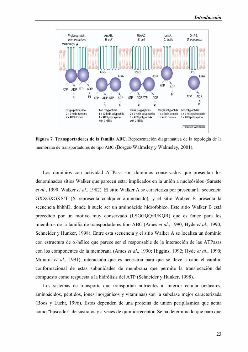
Los transportadores ABC se componen normalmente de cuatro dominios: dos dominios
hidrofóbicos integrales de membrana interna que consisten en 6 STM con estructura de α-
hélice, y dos dominios hidrofílicos que se localizan en la cara citoplásmica de la membrana
y catalizan la hidrólisis del ATP (Higgins, 1992) (Figura 7). En sistemas eucariotas los
módulos se encuentran en una única cadena polipeptídica, mientras que en bacterias
generalmente se conforman en subunidades diferentes (aunque no siempre). Dichos
dominios del transportador ABC pueden estar formados por cuatro polipéptidos diferentes o
pueden ser sintetizados como dos proteínas independientes que presentan un dominio
hidrofóbico que se ensambla en la membrana y un dominio hidrofílico con actividad
ATPasa (Wandersman, 1998). Esto es lo que sucede en la mayoría de los transportadores
ABC de bacterias (dominios transmembrana codificados por uno o dos genes independientes
al gen que codifica la proteína ABC) (Young y Holland, 1999).
Introducción
23
Figura 7. Transportadores de la familia ABC. Representación diagramática de la topología de la
membrana de transportadores de tipo ABC (Borges-Walmsley y Walmsley, 2001).
Los dominios con actividad ATPasa son dominios conservados que presentan los
denominados sitios Walker que parecen estar implicados en la unión a nucleósidos (Saraste
et al., 1990; Walker et al., 1982). El sitio Walker A se caracteriza por presentar la secuencia
GXXGXGKS/T (X representa cualquier aminoácido), y el sitio Walker B presenta la
secuencia hhhhD, donde h suele ser un aminoácido hidrofóbico. Este sitio Walker B está
precedido por un motivo muy conservado (LSGGQQ/R/KQR) que es único para los
miembros de la familia de transportadores tipo ABC (Ames et al., 1990; Hyde et al., 1990;
Schneider y Hunker, 1998). Entre esta secuencia y el sitio Walker A se localiza un dominio
con estructura de α-hélice que parece ser el responsable de la interacción de las ATPasas
con los componentes de la membrana (Ames et al., 1990; Higgins, 1992; Hyde et al., 1990;
Mimura et al., 1991), interacción que es necesaria para que se lleve a cabo el cambio
conformacional de estas subunidades de membrana que permite la translocación del
compuesto como respuesta a la hidrólisis del ATP (Schneider y Hunker, 1998).
Los sistemas de transporte que transportan nutrientes al interior celular (azúcares,
aminoácidos, péptidos, iones inorgánicos y vitaminas) son la subclase mejor caracterizada
(Boos y Lucht, 1996). Estos dependen de una proteína de unión periplásmica que actúa
como “buscador” de sustratos y a veces de quimiorreceptor. Se ha determinado que para que
Introducción
24
se produzca la hidrólisis del ATP es necesaria la interacción de la proteína periplásmica con
los dominios de las subunidades de membrana del transportador ABC presentes en la
superficie de la monocapa externa de la membrana (Ames et al., 1990; Davidson et al.,
1992). En estos sistemas de transporte, el sustrato y el ATP se encuentran situados en sitios
distintos del transportador (en la cara periplásmica y citoplásmica del mismo,
respectivamente) y es la proteína de unión periplásmica la que reconoce el compuesto que se
va a transportar, lo que implica que la proteína ABC no participaría en el reconocimiento
directo de dicha sustancia (Schneider y Hunker, 1998).
Sin embargo, en el caso de los sistemas exportadores tipo ABC, el sustrato y el ATP
están presentes en la cara citoplásmica del transportador y en este caso existen evidencias de
que las ATPasas implicadas en los sistemas de secreción de polipéptidos podrían participar
en el reconocimiento de sus sustratos (Schneider y Hunker, 1998). Los exportadores tipo
ABC de las bacterias gram-negativas están asociados con una proteína de la membrana
externa y con la denominada proteína de fusión que presenta un dominio N-terminal de
anclaje a la membrana interna y un dominio periplásmico (Young y Holland, 1999;
Wandersman, 1998). Estos sistemas están implicados en la secreción de un gran número de
compuestos hidrofílicos, desde moléculas pequeñas (como drogas o antibióticos), hasta
polímeros complejos de carbohidratos (como el LPS, polisacáridos capsulares, etc.).
Los sistemas de transporte tipo ABC son sensibles a una gran variedad de compuestos
que inhiben su actividad ATPasa. Uno de estos compuestos es el vanadato, y se ha
determinado que dicho compuesto inhibe la actividad enzimática del transportador
atrapando el ADP en el sitio catalítico del enzima e impidiendo su liberación de la proteína
(Urbatsch et al., 1995). Se ha sugerido que en los sistemas de transporte tipo ABC la
hidrólisis de ATP se llevaría a cabo de manera cooperativa entre todos sus componentes
(Davidson et al., 1996).
Kim et al. (1998) identificaron en P. putida GM73 un transportador ABC que confería
resistencia a tolueno y lo denominaron Ttg2ABCD, sin embargo en dicho trabajo no
caracterizaron en detalle el sistema de transporte.
II.b.iii. La Superfamilia “MFS” (Major Facilitator Superfamily)
La superfamilia MFS está integrada por transportadores secundarios formados por un
solo polipéptido de aproximadamente 400 aminoácidos que solo son capaces de transportar
pequeños solutos en respuesta a gradientes quimiosmóticos de iones. Esta superfamilia, al

Introducción
25
igual que la superfamilia ABC, es ancestral y está integrada por 17 familias cada una con
una especificidad de sustrato diferente. Un estudio basado en la homología de las proteínas
comprendidas dentro de esta superfamilia ha permitido subdividirla en 5 familias en donde
podemos encontrar uniportes, antiportes y simportes, todos ellos específicos para azúcares,
ácidos orgánicos y otras drogas. La primera familia comprende proteínas de resistencia a
drogas, las cuales catalizan la extrusión activa de antimicrobianos como quinolonas,
tetraciclinas, metilenomicina A, antisépticos y múltiples drogas en células bacterianas y
aminotriazol en levaduras. Dentro de esta familia se distinguen dos tipos porque una posee
12 segmentos transmembrana (STM) y la otra familia posee 14 STM (Figura 8). El análisis
de las secuencias sugiere que las proteínas con 14 STM surgieron de la inserción de un
segmento dentro del gen que codifica para la proteína de 12 STM dando lugar a la aparición
de los segmentos 7 y 8; esto sucedió durante los estadios tempranos en la evolución de estos
dos transportadores, antes de la duplicación de los genes que codifican sus numerosos
miembros (Jin et al., 2001; Saier et al., 1998; Borges-Walmsley y Walmsley, 2001).
Figura 8. Modelo estructural de las bombas de extrusión de la Superfamilia de los
Facilitadores Mayores, con A) 12 STM y B) 14 STM (Saier et al., 1998).
La segunda gran familia de transportadores incluye uniportes que transportan azucares,
azúcar-protón simportes, y un transportador de quinato que está presente tanto en células
eucarióticas como procarióticas. Las proteínas de la tercera familia son transportadores
Introducción
26
específicos para los intermediarios del ciclo de Krebs (citrato y α-cetoglutarato) y
probablemente funcionen como protón-simportes. La cuarta familia incluye un grupo de
antiportes específicos para fosfo-ésteres orgánicos y sorprendentemente un regulador
transcripcional de E. coli (UhpC) (Jin et al., 2001; Marger y Saier, 1993; Schwöppe et al.,
2003). La quinta familia consiste en protón-simportes para algunos oligosacáridos (rafinosa,
sacarosa, lactosa) en E. coli. Las familias tercera, cuarta y quinta están presentes solo en
bacterias.
En la literatura hay una vasta evidencia acerca de la topología de estas proteínas, que
consiste en 6 STM (alfa-hélices) seguidos de un lazo citoplasmático que a su vez le siguen 6
STM adicionales (alfa-hélices), este patrón de 6 + 6 (12 segmentos) ha sido publicado para
uniportes de glucosa en mamíferos, para una permeasa de lactosa de E. coli (LacY) y para
los antiportes de tetraciclina en bacterias. (Saier et al., 1998; Borges-Walmsley y Walmsley,
2001) Para los transportadores con 14 STM el lazo citoplasmático suele ser de menor
tamaño (Borges-Walmsley y Walmsley, 2001). Ninguno de estos transportadores ha sido
implicado en la tolerancia a disolventes orgánicos hasta el momento.
III.b.iv. La familia “MATE” (multidrug and toxic compound extrusion).
Esta familia de transportadores fue descrita primeramente como familia de
transportadores exclusiva de bacterias pero actualmente se sabe que están presentes tanto en
procariotas como en eucariotas, y constituyen una de las familias de transportadores más
conservadas en la naturaleza. Los transportadores bacterianos constan de unos 450
aminoácidos y forman 12 STM. Las proteínas de las levaduras tienen aproximadamente
unos 700 aminoácidos, y probablemente ellos también formen un transportador con 12
STM. Dos de estos transportadores bacterianos descritos, utilizan un mecanismo de
transporte inusual: antiporte Na+/soluto (Moriyama et al., 2008).
Las proteínas NorM de Neisseria gonorrhoeae y YdhE de Escherichia coli pertenecen a
esta familia. NorM de Neisseria gonorrhoeae es una proteína de membrana interna que
consta de 459 aminoácidos, y reconoce como sustratos a compuestos catiónicos tóxicos
como bromuro de etidio, acriflavina, y ciprofloxacina (Su et al., 2008; Long et al., 2008).
YdhE de E coli tiene un 57% de identidad y un 88% de similitud con respecto a NorM de V.
parahaemolyticus, el cual transporta norfloxacina y ciproflofaxina además de bromuro de
etidio, pero a pesar de la gran similitud entre ambas proteínas, YdhE no transporta bromuro

Introducción
27
de etidio. Tanto NorM de V. parahaemolyticus como YdhE de E coli constan de 456
aminoácidos (Morita et al., 1998).
III.b.v La familia “SMR” (Small Multidrug Resistance)
Algunos de los miembros de esta familia catalizan la multirresistencia a drogas. Estos
transportadores homo-oligoméricos son pequeños, estando constituidos por cadenas
polipeptídicas de 100 a 110 aminoácidos que se expanden por la membrana celular
atravesándola 4 veces como α-hélices (Figura 9). Se cree que su estado nativo dentro de la
membrana es como homotrímero. Debido a su naturaleza hidrófoba son solubles en
disolventes orgánicos. Es una familia pequeña, bien conservada, que solo esta presente en
bacterias (Saier et al., 1998)
Figura 9. Modelo estructural de los transportadores de la familia “SMR” (Saier et al., 1998).
Esta familia está integrada por dos subfamilias, una de ellas tiene miembros que
confieren multirresistencia a drogas y cataliza la extrusión de éstas vía antiporte droga/H+ al
igual que los miembros de la superfamilia MFS. Por el contrario, los miembros de la otra
subfamilia parecen no tener ninguna de estas características, además se desconoce la
naturaleza de sus sustratos naturales (Saier et al., 1998; Borges-Walmsley y Walmsley,
2001).
Introducción
28
III.d. Otros factores implicados en la resistencia a tolueno
En bacterias gram-negativas se han descrito otros mecanismos que contribuyen a la
tolerancia a disolventes. Algunos investigadores sugieren que la formación de vesículas en
Pseudomonas sp. IH-20000, una cepa tolerante a tolueno, es una respuesta específica de esta
estirpe cuando se añade tolueno al medio de cultivo con el fin de remover dicho disolvente
(Kobayashi et al., 2000). Las vesículas de Pseudomonas sp. IH-20000, formadas por
fosfolípidos, LPS y una pequeña cantidad de proteínas, contenían 0,17-0,63 mol tolueno/
mol lípidos. También se ha descrito la formación de vesículas en cultivos de P. putida DOT-
T1E cuando crece en presencia de altas concentraciones de tolueno, sin embargo, no ha
evidencias de que en P. putida DOT-T1E la función de estas vesículas sea la de expulsar
tolueno (Ramos et al., 1995).
En nuestro grupo se ha demostrado que mutaciones producidas en genes que
codifican los componentes que exportan las proteínas flagelares (i.e flhB) de P. putida
DOT-T1E, tornaban a esta cepa en inmóvil y sensible a tolueno. También en la cepa P.
putida S12, una mutación en el gen flgK, la hacía sensible a tolueno (Segura et al., 2001;
Kieboom et al., 2001). Estos datos sugieren que algunas proteínas flagelares, aquellas
implicadas en el sistema de transporte de los distintos componentes del flagelo, juegan un
rol indirecto en la tolerancia a tolueno, probablemente permitiendo la exportación a través
de la membrana de alguna proteína que tenga un papel importante en tolerancia.
Para mantener la homeostasis celular frente a una variedad de estreses ambientales,
las bacterias usan factores sigma alternativos para redirigir la RNA polimerasa y expresar
selectivamente algunos genes. En P. putida se han identificado 24 factores sigma, 20 de los
cuales corresponden a la subfamilia de los factores sigma extracitoplasmáticos (ECF)
(Ramos et al., 2001; Duque et al., 2007). En nuestro grupo se descubrió que alrededor del
1% del genoma de P. putida DOT-T1E está bajo la influencia directa o indirecta del factor
sigma RpoT y que el mismo está involucrado en la tolerancia a tolueno, hecho que quedó
demostrado al generar un mutante (DOT-T1E-∆rpoT) sensible a tolueno. Esto se debe a que
RpoT regula la expresión de TtgGHI, uno de los transportadores clave en la expulsión de
tolueno (Duque et al., 2007; Rojas et al., 2001).
Aunque los factores anteriormente descritos son fundamentales en la respuesta al
choque a altas concentraciones de tolueno, las células bacterianas expuestas a la presencia
constante de un disolvente orgánico requieren otras adaptaciones que les permiten crecer en
estas condiciones. Mediante el análisis de proteínas (Segura et al., 2005) que se expresan
diferencialmente en cultivos de P. putida DOT-T1E creciendo en presencia y ausencia de
Introducción
29
tolueno, se han identificado otros factores en respuesta a estas condiciones, Una de las
principales aportaciones de este trabajo es que la presencia de tolueno, aun en
concentraciones mucho más bajas del límite de resistencia de la cepa y pese a que no se
muestra ningún efecto a nivel de crecimiento cuando se comparaba con el crecimiento en
glucosa, es percibida por las células como un estrés y por tanto se inducen una serie de
proteínas relacionadas con el mismo (CspA, GroES entre otras). También que las células
necesitaban más energía en presencia de tolueno y por tanto aceleraban el metabolismo de la
glucosa. Resultados similares se obtuvieron en la cepa P. putida S12 cultivada en
quimiostatos con y sin tolueno (Volkers et al., 2006).
A su vez, ensayos de microarray realizados en nuestro grupo (Domínguez-Cuevas et al.,
2006) y llevados a cabo en P. putida KT2440 (una cepa sensible), para analizar la respuesta
global de esta cepa en presencia de compuestos aromáticos, revelaron que en esta estirpe se
inducen, en presencia de tolueno, toda una batería de genes en respuesta a estrés.
En todos estos experimentos se identificaron como inducidos por tolueno una serie de
transportadores de diversas familias, cuyo papel en la tolerancia a disolventes orgánicos no
ha sido estudiado hasta el momento.
IV. Regulación de la tolerancia a disolventes orgánicos
La complejidad de la respuesta al tolueno queda de manifiesto no sólo en los
experimentos de microarrays y proteómica que se han llevado a cabo en distintos
laboratorios, sino por la gran cantidad de mecanismos de defensa diferentes que participan
en la tolerancia a disolventes orgánicos.
El principal mecanismo de defensa frente a concentraciones supersaturantes de
tolueno en P. putida DOT-T1E son las bombas de extrusión; hasta el momento sólo
mutantes en algunas de estas bombas son totalmente incapaces de sobrevivir al choque con
0,3% (v/v) de tolueno. Otros mutantes en, por ejemplo, componentes de membrana son
capaces de sobrevivir al choque, si bien el porcentaje de células que sobreviven es menor
(Bernal et al., 2007 a; Pini et al., 2009)
La tolerancia a tolueno en P. putida DOT-T1E es un proceso inducible;
aproximadamente 1 de cada 10.000 células sobrevive al choque con 0,3% (v/v) cuando las
células se han cultivado en LB, sin embargo, si el cultivo se ha preinducido con bajas
concentraciones de tolueno (tolueno en la fase gaseosa) casi el 90% de las células es capaz
de sobrevivir al choque (Figura 10-A). Como se ha dicho anteriormente, en P. putida DOT-

Introducción
30
T1E hay identificadas tres bombas implicadas en la tolerancia a tolueno (TtgABC, TtgDEF
y TtgGHI) (Ramos et al., 1998; Mosqueda y Ramos, 2000; Rojas et al., 2001). TtgABC se
expresa constitutivamente a niveles bajos y expulsa tanto disolventes orgánicos como
antibióticos, pero solo es inducida por antibióticos y algunos flavonoides (Duque et al.,
2001; Terán et al., 2003; Terán, 2006). Mutantes en esta cepa no son capaces de sobrevivir
al choque con tolueno a no ser que hayan sido previamente preinducidos (Figura 10-B;
Ramos et al., 1998). Además, esta bomba parece estar ampliamente distribuida en cepas de
P. putida (Segura et al., 2003). TtgR es el regulador transcripcional de esta bomba y
pertenece a la familia de TetR (Duque et al., 2000).
TtgDEF no se expresa bajo condiciones normales de laboratorio pero es inducida por
disolventes. Así, en un mutante en esta cepa tan sólo se observa una disminución de su
capacidad de sobrevivir al choque con tolueno cuando el cultivo ha sido previamente
inducido con disolvente orgánico (Figura 10-C). Esta bomba no está involucrada en
resistencia a antibióticos, solo expulsa disolventes (Mosqueda y Ramos, 2000) y está
presente en todas las cepas que presentan la ruta tod para la degradación de tolueno (Segura
et al., 2003). TtgGHI tiene un nivel de expresión basal relativamente alto y es inducida por
disolventes orgánicos (Rojas et al., 2001). Es quizá la bomba que confiere mayor tolerancia
a disolventes orgánicos lo que se manifiesta en que mutantes en esta cepa son incapaces de
sobrevivir al choque de tolueno independientemente de las condiciones en las que se haya
cultivado la cepa (Figura 10-D). En ensayos llevados a cabo en diferentes cepas de P .putida
sólo se identificaron genes similares a los del operón ttgGHI en las cepas altamente
tolerantes a disolventes orgánicos, como por ejemplo, P. putida MTB6 (Huertas et al.,
2000), y P. putida S12 (Isken y de Bont, 1998). Esta bomba participa también en la
extrusión de disolventes orgánicos y antibióticos, aunque su función en resistencia a
antibióticos está enmascarada por la presencia de la bomba TtgABC (Rojas et al., 2001).
Los reguladores TtgT (Terán et al., 2007) y TtgV (Rojas et al., 2003) son represores de las
bombas TtgDEF y TtgGHI respectivamente. TtgT y TtgV pertenecen a la familia de
reguladores transcripcionales IclR y muestran una regulación cruzada hacia las bombas
TtgDEF y TtgGHI.
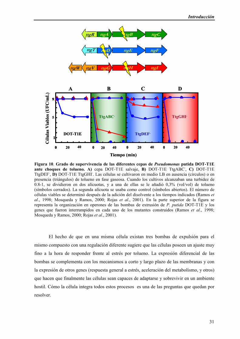
Introducción
31
Figura 10. Grado de supervivencia de las diferentes cepas de Pseudomonas putida DOT-T1E
ante choques de tolueno. A) cepa DOT-T1E salvaje, B) DOT-T1E TtgABC-, C) DOT-T1E TtgDEF-, D) DOT-T1E TtgGHI-. Las células se cultivaron en medio LB en ausencia (círculos) o en presencia (triángulos) de tolueno en fase gaseosa. Cuando los cultivos alcanzaban una turbidez de 0.8-1, se dividieron en dos alícuotas, y a una de ellas se le añadió 0,3% (vol/vol) de tolueno (símbolos cerrados). La segunda alícuota se usaba como control (símbolos abiertos). El número de células viables se determinó después de la adición del disolvente a los tiempos indicados (Ramos et
al., 1998; Mosqueda y Ramos, 2000; Rojas et al., 2001). En la parte superior de la figura se representa la organización en operones de las bombas de extrusión de P. putida DOT-T1E y los genes que fueron interrumpidos en cada uno de los mutantes construidos (Ramos et al., 1998; Mosqueda y Ramos, 2000; Rojas et al., 2001).
El hecho de que en una misma célula existan tres bombas de expulsión para el
mismo compuesto con una regulación diferente sugiere que las células poseen un ajuste muy
fino a la hora de responder frente al estrés por tolueno. La expresión diferencial de las
bombas se complementa con los mecanismos a corto y largo plazo de las membranas y con
la expresión de otros genes (respuesta general a estrés, aceleración del metabolismo, y otros)
que hacen que finalmente las células sean capaces de adaptarse y sobrevivir en un ambiente
hostil. Cómo la célula integra todos estos procesos es una de las preguntas que quedan por
resolver.
ttgA ttgB ttgCttgR
ttgT ttgD ttgE ttgF
ttgV ttgG ttgH ttgIttgW
1
200
9
5
DOT-T1E TtgDEF-
TtgABC- TtgGHI-
40200 40200 40200
ttgA ttgB ttgCttgR
ttgT ttgD ttgE ttgF
ttgV ttgG ttgH ttgIttgW
1
400
9
TtgDEF-
TtgABC- TtgGHI-
0 0 0
A B C D
Tiempo (min)
Cél
ula
s V
iable
s (U
FC
/mL
)
ttgA ttgB ttgCttgR
ttgT ttgD ttgE ttgF
ttgV ttgG ttgH ttgIttgW
1
200
9
5
DOT-T1E TtgDEF-
TtgABC- TtgGHI-
40200 40200 40200
ttgA ttgB ttgCttgR
ttgT ttgD ttgE ttgF
ttgV ttgG ttgH ttgIttgW
1
400
9
TtgDEF-
TtgABC- TtgGHI-
0 0 0
A B C D
Tiempo (min)
Cél
ula
s V
iable
s (U
FC
/mL
)

Introducción
32

33
OBJETIVOS

34
Objetivos
35
El objetivo general de esta Tesis Doctoral es el de profundizar en los mecanismos de
tolerancia a disolventes orgánicos en la cepa Pseudomonas putida DOT-T1E, haciendo
especial énfasis en mecanismos de transporte. Para ello se establecieron cuatro objetivos
específicos:
1.- Identificación y caracterización de nuevos factores de tolerancia a disolventes
orgánicos: participación del sistema cyoB en la tolerancia.
2.- Construcción y análisis del comportamiento de diferentes bombas híbridas (con
distintas combinaciones de los tres elementos que las conforman) en cepas de P. putida.
3.- Profundización del análisis de los genes ttgGHI: Análisis y caracterización de un
mutante espontáneo sensible al tolueno derivado de Pseudomonas putida DOT-T1E (P.
putida DOT-T1E-100).
4.- Implicación de otros sistemas de transporte cuya expresión se ve afectada por la
presencia de tolueno en P. putida KT2440, la cepa sensible a tolueno, y en la tolerancia a
disolventes orgánicos en la cepa resistente a tolueno, P. putida DOT-T1E.

Objetivos
36

37
RESULTADOS

38

Resultados
39
Plasmolysis induced by toluene in a cyoBmutant of Pseudomonas putida
Estrella Duque, Vanina García, Jesús de la Torre, Patricia Godoy, Patricia Bernal and
Juan-Luis Ramos
El operón cyoABCDE de Pseudomonas putida DOT-T1E codifica para una citocromo
oxidasa terminal. Un fragmento de 500 pb del gen “cyoB” se clonó en el vector
pCHESIW Km y se utilizó para generar un mutante knock-out en el gen cyoB in vivo.
El mutante no se vio limitado en la generación de fuerza protón motriz, aunque cuando
se cultivó en medio mínimo con glucosa o citrato, el mutante en CyoB exhibió un ligero
incremento en el tiempo de duplicación con respecto a la cepa salvaje. Este efecto fue
aun más pronunciado cuando se suplementó el medio con tolueno en fase gaseosa. En
consonancia con el efecto negativo del tolueno en el crecimiento, se vio que el mutante
CyoB era hipersensible a los choques de 0,3% (v/v) de tolueno, en contraste con la cepa
silvestre. Este efecto se vio particularmente exacerbado en células que alcanzaban la
fase estacionaria. El incremento en la sensibilidad a los disolventes en el mutante CyoB
no parecía estar relacionado con la incapacidad de las células de reforzar el
empaquetamiento de la membrana o de inducir las bombas de extrusión en respuesta a
los disolventes, sino que la plasmólisis inducida por disolventes puede ser
desencadenada por pliegues de la membrana citoplasmática en los polos de las células
mutantes, y una invaginación de la membrana externa que eventualmente pueden
conducir a la muerte celular.
Environmental Microbiology (2004), 6(10): 1021–1031.

Resultados
40

Environmental Microbiology (2004)
6
(10), 1021–1031 doi:10.1111/j.1462-2920.2004.00621.x
© 2004 Blackwell Publishing Ltd
Blackwell Science, LtdOxford, UKEMIEnvironmental Microbiology 1462-2912Society for Applied Microbiology and Blackwell Publishing Ltd, 20046
1010211031
Original Article
Solvent sensitivity in cyoB mutantE. Duque
et al.
Received 1 December, 2003; accepted 18 February, 2004. *For cor-respondence. E-mail [email protected]; Tel. (+34) 958181608;Fax (+34) 958135740.
Plasmolysis induced by toluene in a
cyoB
mutant of
Pseudomonas putida
Estrella Duque, Vanina García, Jesús de la Torre, Patricia Godoy, Patricia Bernal and Juan-Luis Ramos
*
Consejo Superior de Investigaciones Científicas, Estación Experimental del Zaidín, Department of Plant Biochemistry and Molecular and Cellular Biology, Calle Profesor Albareda number 1, E-18008 Granada, Spain.
Summary
The
cyoABCDE
gene cluster of
Pseudomonas putida
DOT-T1E encodes a terminal cytochrome oxidase. A500-bp ‘
cyoB
’ DNA fragment was cloned in pCHESI-
WWWW
Km and used to generate a cyoB knock-out mutant
in vivo
. The mutant strain was not limited in the gen-eration of proton-motif force, although when grownon minimal medium with glucose or citrate, the CyoBmutant exhibited a slight increase in duplication timewith respect to the wild-type strain. This effect waseven more pronounced when toluene was supplied inthe gas phase. In consonance with the negative effectof toluene on the growth was the finding that theCyoB mutant was hypersensitive to sudden 0.3%(v/v) toluene shocks, in contrast with the wild-typestrain. This effect was particularly exacerbated incells that reached the stationary phase. The increasedsensitivity to solvents of the CyoB mutant did notappear to be related to the inability of the cells tostrengthen the membrane package or to induce theefflux pumps in response to the solvent, but rather tosolvent-induced plasmolysis that may be triggered bywrinkles in the cytoplasmic membrane at the poles ofthe mutant cells, and invagination of the outer mem-branes, which eventually lead to cell death.
Introduction
Aerobic bacteria generate energy through respiratory pro-cesses in which electrons flow through a series of electroncarriers until they reach terminal oxidases that reduceoxygen to water. In
Pseudomonas
sp. two main terminalcytochrome oxidases are known, namely the cytochromeubiquinol
o
oxidase (Cyo) and the cyanide insensitive
oxidase (Cio) (Cunningham and Williams, 1995; Cooper
et al
., 2003). The
cyoABCDE
gene cluster encodes thecytochrome
o
oxidase complex (CyoA,B,C and D pro-teins), and the heme
o
synthase (CyoE) (Hirayama
et al
.,1998; Dinamarca
et al
., 2002). This cytochrome receiveselectrons from the ubiquinone pools (Nakamura
et al
.,1997). The CyoABCD terminal oxidase seems to play amajor role in cells growing in conditions of high oxygentension (Sweet and Peterson, 1978; Cotter
et al
., 1990;1997), and is expressed preferentially in cells in the expo-nential phase although expression in the stationary phasealso takes place but at a lower level (Dinamarca
et al
.,2003). In contrast, at least in
Escherichi coli
the CydABterminal oxidase (CioAB in
P. aeruginosa
) is preferentiallyexpressed in cells growing under low oxygen tension andin the stationary phase (Taylor and Zhulin, 1999). None-theless, in
Escherichia coli
each of these terminal oxi-dases replaces the other one in isogenic mutants deficientin their synthesis.
Mutants in the
cyo
genes in different strains of
Pseudomonas putida
have been isolated and the effectson gene regulation and solvent tolerance have been anal-ysed. Mutants in the
cyoABCDE
cluster in
P. putida
KT2440 (Dinamarca
et al
., 2002; 2003) and
P. putida
strain H (Petruschka
et al
., 2001) escape from catabolitecontrol, in the case of the alkane degradation pathway inthe KT2440 strain, and the phenol degradation pathway,in the H strain. It was hypothesized that the catabolicrepression system could monitor the physiological statusof the cells by sensing the redox state of the elements ofthe electron transport chain, and that this could be usedas a signalling system. However, the authors could notascertain whether the Cyo terminal oxidase plays a director an indirect role in signal transmission. A mutant in thecytochrome ubiquinol oxidase system was also describedin the solvent-tolerant
P. putida
IH2000 strain. This muta-tion lead to increased solvent sensitivity in comparisonwith wild-type strain (Hirayama
et al
., 1998). The reasonfor this increased tolerance was believed to be changesin the membrane hydrophobicity, although the intimatereasons remain unknown (Kobayashi
et al
., 1999).
Pseudomonas putida
DOT-T1E is a solvent-tolerantstrain (Ramos
et al
., 1995) that exhibits physical and bio-chemical barriers to decrease solvent toxicity (Ramos
et al
., 1997; 2002). In response to toluene in the culturemedium, the cell membrane is packaged by transforming

1022
E. Duque
et al.
© 2004 Blackwell Publishing Ltd,
Environmental Microbiology
,
6
, 1021–1031
cis
unsaturated fatty acids into
trans
isomers (Junker andRamos, 1999), and by the increased synthesis of phos-pholipids (Segura
et al
., 2004). In addition to the physicalbarrier, this bacterium, and other solvent-resistancemicroorganisms, uses a series of efflux pumps, known inDOT-T1E as TtgABC, TtgDEF and TtgGHI, that removetoluene from the cell membranes (Isken and de Bont,1996; Fukumori
et al
., 1998; Ramos
et al
., 1998;Mosqueda and Ramos, 2000; Rojas
et al
., 2001). Theenergy required by these efflux pumps for solvent extru-sion is linked to the generation of the electron proton-motive force.
We decided to explore the organization of the
cyoAB-CDE
cluster in the solvent-tolerant
P. putida
DOT-T1Estrain, and to generate a mutant in
cyoB
to analyse itsbehaviour under different growth conditions with regard tosolvent sensitivity. Our results showed that the mutantstrain grew in a wide variety of culture conditions, but thatalthough the cells exhibited normal levels of efflux pumpcomponents, and were able to isomerize unsaturated fattyacids, they were hypersensitive to solvents. This does notseem to be result of the limited generation of proton-motive force, but rather to solvent-induced plasmolysisthat may be triggered by wrinkles in the cytoplasmic mem-brane at the poles of the mutant cells, and invagination ofthe membranes as observed with scanning electronmicroscopy. These defects may account for the cells’extreme sensitivity to organic solvents.
Results
Transcriptional organization of the
cyo
cluster of
Pseudomonas
putida
DOT-T1E
The
cyoB
gene cluster had been previously identified inseveral
P. putida
strains (Kobayashi
et al
., 1999; Petrus-chka
et al
., 2001; Dinamarca
et al
., 2002). Based on thesequence of the
cyo
cluster of
P. putida
KT2440, wedesigned suitable primers for the amplification of the chro-mosomal region of
P. putida
DOT-T1E, which wassequenced. Analysis revealed the presence of five openreading frames (ORF) corresponding to the
cyoABCDE
cluster (Fig. 1A, the sequence deposited at GenBankunder accession number AY466718). Overall identity withthe
P. putida
KT2440
cyoABCDE
cluster was 99.1%, andwhen the translated products were compared it was closeto 100%. Sequence analysis revealed short extragenicDNA sequences between contiguous genes (3–11 nucle-otides) or overlapping ORFs (
cyoC
and
cyoD
), suggestingthat the genes could form an operon. To determinewhether all the genes were part of the same transcript,RT-PCR, with primers based on the 3
¢
and 5
¢
ends ofcontiguous genes was carried out with mRNA isolatedfrom cells in the exponential and the stationary growth
phase (Fig. 1B). Under both conditions, cDNA fragmentsof the expected sizes were found with all five genes.These results suggest that there is a single transcriptionalunit made up of
cyoABCDE
in
P. putida
DOT-T1E.
Construction and characterization of a CyoB mutant
To generate a knock-out mutant in the
cyoB
gene wetransferred the pEDX-1 plasmid into DOT-T1E asdescribed in
Experimental procedures
, and selected Km
R
clones. These appeared at a rate of about 10
-
7
per recip-ient cell, and 10 clones were examined by PCR with theappropriate primers for insertion of the Km
R
cassette withinthe
cyoB
gene. We expected a 1.3 kb amplification band
Fig. 1.
Physical organization of the
cyoABCDE
cluster and evidence that the genes form an operon.A. The sequence of the
cyoABCDE
operon can be accessed from GenBank (accession no. AY466718). The approximate sizes of the products of each ORF are given in kilodaltons below each gene.B. The products resulting from each RT-PCR were separated on agarose gels. Lane 1, molecular weight markers. Lane 2,
cyoAB
amplification (expected size, 150 bp). Lane 3,
cyoBC
amplification (expected size, 210 bp). Lane 4,
cyoCD
amplification (expected size, 300 bp). Lane 5,
cyoDE
amplification (expected size, 290 bp). Control assays without RT yielded no cDNA.

Solvent sensitivity in
cyoB
mutant
1023
© 2004 Blackwell Publishing Ltd,
Environmental Microbiology
,
6
, 1021–1031
in clones with a knock-out gene. Two of the clones yieldedthe expected band. Subsequently, we sequenced themutant clones based on sequencing primers located atthe end of the Km
R
cassette. This allowed us to identifythe position where the gene was disrupted by the Km
R
cassette, which occurred between positions 2132 and2133 in the sequence deposited at GeneBank. To furtherconfirm the nature of the mutation we prepared total DNAfrom one of the clones, digested it with
Ava
I and hybridizedit against the Km
R
genes coding for the
cyoB
gene (notshown). This mutant was called
P. putida
DOT-T1E-CyoB.We determined the growth rate of the wild type and the
CyoB mutant in LB medium and minimal medium withglucose and citrate in the absence and in the presence oftoluene (logP
ow
2.5) supplied in the gas phase. In LB nosignificant difference was found in the absence of toluene(tg 46–49 min) or in the presence of toluene (tg 54–58 min) in the gas phase. In the presence of toluene, boththe wild type and the mutant exhibited a 3 h lag beforegrowth started. The wild-type strain grew at a similar rateon M9 minimal medium with glucose (tg 65–68 min) orcitrate (tg 70–73 min) as the carbon source in theabsence and in the presence of toluene. In contrast,growth of the mutant was slower than that of the wild type.In M9 minimal medium with glucose the growth rate was83 min and 107 min in the absence and in the presenceof toluene, respectively, whereas in M9 minimal mediumbut with citrate the doubling time was 88 and 119 min withand without toluene supplied in the gas phase respec-tively. Again a prolonged lag of about 5 h was found beforethe mutant or the wild type started to grow (not shown).
These results prompted us to test the effect of severalsolvents (supplied at 3 mM)
n
-decane (logP
wo
5.6),
n
-heptane (logP
ow
4.5),
n
-ethylbenzene (logP
wo
3.1),
n
-pro-pylbenzene (logP
ow
3.6),
m
-xylene (logP
ow
3.2), styrene(logP
ow
3.0) and 1-octanol (logP
ow
2.9) on the growth of thestrain in M9 minimal medium with glucose. We found thatoctanol, styrene and toluene inhibited or fully preventedthe growth of the CyoB mutant. In M9 minimal medium,ethyl- and propylbenzene delayed growth and
n
-heptaneand
n
-decane did not affect growth as compared to aculture without solvent. The effect of solvent on growth ofthe CyoB mutant correlated with the log P
ow
of thechemicals.
We then decided to study survival of the wild-type strainand the DOT-T1E-CyoB mutant in the log growth phaseand in the stationary growth phase after a sudden tolueneshock. Both types of cells were grown in LB or inM9 minimal medium with citrate in the absence and in thepresence of toluene supplied at sublethal concentrationsin the gas phase. The behaviour of the wild type grown inLB in the exponential phase was similar to our previousobservation: one out of 10
4
non-induced cells survived thesolvent shock, whereas
>
50% cells survived if preinduced
@ @
@@
with toluene (Fig. 2 and Ramos
et al
., 1998). We thenanalysed the survival of DOT-T1E in the stationary phase.Only one out of 10
6
non-induced cells in the stationaryphase survived the solvent shock, whereas about one outof 10
3
cells survived the solvent shock when preinducedwith toluene. This indicated that cells in the stationarygrowth phase were intrinsically more sensitive to toluenethan cells in the exponential growth phase. The mutantDOT-T1E-CyoB cells were more sensitive to a suddensolvent shock than the wild type regardless of the growthconditions (Fig. 2). Particularly, in the stationary phase thenumber of viable cells after the solvent shock wasnegligible for non-induced cells and very low (one out of104-105) for induced cells (Fig. 2).
We also analysed the behaviour of the wild type andmutant CyoB when grown on M9 minimal medium withcitrate both in the absence and in the presence of toluenein response to a sudden toluene shock. We found thatregardless of the growth conditions, the wild-type strainwas more resistant than the mutant, particularly the CyoBmutant cells in the stationary phase were hypersensitiveto the sudden toluene shock because no cells were recov-ered in non-induced cells, and fewer than one in 107 wererecovered when induced cells were used (not shown).This contrasted with the survival rate of the wild-typestrain which was in the order of 10-5 and 10-3 for non-induced and induced cells respectively.
Analysis of well-defined defence mechanisms against solvents in P. putida DOT-T1E and its isogenic DOT-T1E-CyoB mutant
Several possible mechanisms of organic solvent tolerancein bacteria have been reported (Diefenbach et al., 1992;Heipieper et al., 1992; Sikkema et al., 1994; Weber andde Bont, 1996; Ramos et al., 1997; White et al., 1997).The two main contributors are an increase in membranepackaging, achieved through cis to trans isomerization ofunsaturated fatty acids, and a number of solvent effluxpumps.
As mentioned above, the growth curves of Pseudomo-nas putida DOT-T1E and the P. putida CyoB mutant in LBmedium were similar when grown in the absence and inthe presence of sublethal concentrations of toluene. Wedetermined the composition of the fatty acids in the expo-nential and stationary phase in the wild-type and mutantcells under those growth conditions. The results revealedthat in both the wild-type and the CyoB mutant cells, thepattern of fatty acid composition was similar, with anincrease in the level of trans isomers in the presence oftoluene which was particularly evident in the stationaryphase (Table 1). It also should be noted that because ofthis high level of C16 : 1 trans in the presence of toluene,the level of C17 : cyclo was much lower in these cells

1024 E. Duque et al.
© 2004 Blackwell Publishing Ltd, Environmental Microbiology, 6, 1021–1031
compared with cells grown in the absence of toluene(Table 1). We also determined the composition of thephospholipid head groups in the wild-type and the mutantstrain in the log- and stationary growth phase in cellsgrown in the absence and in the presence of toluene. Thecomposition was similar in the wild-type and the mutantstrain regardless of the growth conditions. The main phos-pholipid head group was phosphatidylethanolamine(70%-76.6% of total), with similar levels of phosphatidylg-lycerol (12.7%-14%) and cardiolipin (10.3%-15.2%).Therefore the increased solvent sensitivity of DOT-T1Eseems not to be related to defects in the pattern of phos-pholipids.
Rojas et al. (2001) demonstrated that up to three effluxpumps were involved in solvent tolerance. To analyse the
level of expression of these pumps in the wild-type and inthe CyoB mutant strain, we transformed wild-type andCyoB mutant cells with plasmids pED14, pMTT andpANA96, which carry a fusion of the ttgA, ttgD and ttgGpromoter regions to a promoterless ‘lacZ gene (resultsare shown in Table 2). The pattern of expression of allthree efflux pumps in the wild type and the mutant wassimilar. In the log phase, ttgA, ttgD and ttgG wereexpressed at a higher level than in the stationary phase,which suggest that the expression of the efflux pumpsmay not be altered in the CyoB mutant. It appears thatthe lower survival of the cells in the stationary phase withrespect to cells in the log phase could be due the dimin-ished ability of the cells in the stationary phase to removesolvents.
Fig. 2. Survival of P. putida DOT-T1E and the CyoB mutant. Cells were grown in 30 ml of LB medium (circle) or LB medium with toluene in the gas phase (triangles) until the cultures reached a turbidity of 0.8 at 660 nm (log phase) or a turbidity of about 3.0 at 660 nm (stationary phase). The cultures were divided in two halves; to one of them 0.3% (v/v) toluene (closed symbols) was added, whereas the other was kept as a control (open symbols). The number of viable cells was determined at the indicated times.A and B. Wild-type in the log and stationary phase respectively.C and D. cyoB mutant in the log and stationary phase respectively.

Solvent sensitivity in cyoB mutant 1025
© 2004 Blackwell Publishing Ltd, Environmental Microbiology, 6, 1021–1031
Analysis of the proton-motive force in P. putida DOT-T1E and its isogenic Cyo mutant
The similar growth rate in the exponential phase of thewild type and the CyoB mutant suggested that, as occursin E. coli, the Cyd terminal oxidase successfully replacesthe Cyo terminal oxidase. However, the increased solventsensitivity of CyoB may be linked to limited generation ofproton-motive force. We used the method described byLlamas et al. (2003) to determine the ability to generateproton-motive force in cells of the wild-type and mutantCyoB strain grown on LB with or without toluene in thegas phase, when cells reached the mid-log phase or theearly stationary phase. The results revealed that the mag-nitude of proton-motive force was similar in both strains,
suggesting that proton-motive force seems not to be thelimiting factor (not shown).
To rule out proton-motive force as the limiting factor, wetested the sensitivity of DOT-T1E CyoB to antibioticswhich are removed through RND pumps, i.e. Tc, Ap, Cb,Pip, Gm, Cm and Nal. The MICs (mg ml-1) of wild-type andCyoB mutant were identical [(ampicillin > 1000; carbeni-cillin > 400; Pip, 100; Tc, 20; Gm, 20; Cm, 324 and Nal,50)], which supports no limitation in the generation ofproton-motive force to the extrusion of drugs.
Scanning electron microscopy revealed defects at the poles of the cyoB mutant
The above findings suggested that the basic solventdefence mechanisms such as efflux pumps, energy gen-eration and fatty acid modifications were operational in theCyoB mutant. Consequently, the increased sensitivity ofthe CyoB mutant cannot be ascribed to defects in specificdefence mechanisms. What, then, makes this strain moresensitive to solvents than the parental one? We previouslyreported that an OprL-null mutant that exhibited alteredcell morphology was hypersensitive to toluene (Ramoset al., 1997). To test the integrity of the outer membranewe transformed the wild type and CyoB mutant with plas-mid pJB3Km. We found that no b-lactamase was releasedto the outer medium, suggesting that outer membraneintegrity is not affected (not shown). On the other hand,De Smet et al., 1978) showed that solvents lead to alter-ations in the cytoplasmic membrane. This led us to exam-ine the wild type and CyoB mutant inner membrane incells in the exponential and stationary phases under dif-ferent growth conditions.
Cells were prepared for transmission electron micros-copy as described in Experimental procedures. We foundthat in the absence of solvent, the wild-type and mutant
Table 1. Phospholipid composition of P. putida DOT-T1E and its isogenic CyoB mutant cells growing in the absence and in the presence of tolu-ene in the exponential and the stationary phase.
StrainMedia
Exponential phase Stationary phase
T1ELB
CyoBLB
T1ELB + toluene
CyoBLB + toluene
T1ELB
CyoBLB
T1ELB + toluene
CyoB LB + toluene
C14 : 0 0.3 0.4 0.5 0.5 0.6 0.4 0 0C15 : 0 0 0.2 0 0 0.5 0.4 0 0C16 : 1 cis 35 36.0 26.3 31.4 12.3 18.5 6.4 5.3C16 : 1trans 1.8 2.6 8.9 8.3 2.2 2.1 24.7 22.4C16 : 0 34.1 35.9 38.6 42.6 52.0 47.8 40.9 45.9C17 : cyclo 1.6 3.3 0.2 0.4 18.0 11.5 4.1 3.7C17 : 0 0 0.2 0 0 0.4 0.4 0 0C18 : cis vacc 26.4 20.7 24.6 16.5 13.0 18.1 8.8 12.8C18 : trans vacc 0 0 0 0 0.4 0 14. 9.0C18 : 0 0.8 0.7 0.8 0.5 0.6 0.7 0.8 0.9C19 : ciclo 0 0 0 0 0 0 0 0cis/trans 34.1 22.2 5.68 5.74 9.65 17.06 0.39 0.57sat/unsat 0.55 0.63 0.66 0.77 1.94 1.29 0.77 0.95
Table 2. Expression of the efflux pumps in cells growing in theabsence and in the presence of toluene in the exponential and thestationary phase.
Toluene PttgA PttgD PttgG
ExponentialWild-type – 120 20 310Wild-type + 140 45 1530
CyoB-mutant – 120 20 305CyoB-mutant + 170 40 1930
StationaryWild-type – 80 1 130Wild-type + 50 5 420
CyoB-mutant – 50 1 120CyoB-mutant + 40 5 480
The strains were transformed with pED14 (PttgA::lacZ),pMTT(PttgD::lacZ) or pANA96 (PttgG::lacZ) and cells were grown in theabsence (–) or in the presence (+) of toluene until they reached themid-exponential phase (i.e. OD660 0.8–1) or the stationary phase (i.e.OD660 3–3.5). b-galactosidase activity was determined in duplicate intwo independent assays. The data are rounded values of the aver-ages with standard deviations in the range of 5% to 17% of the givenvalues.
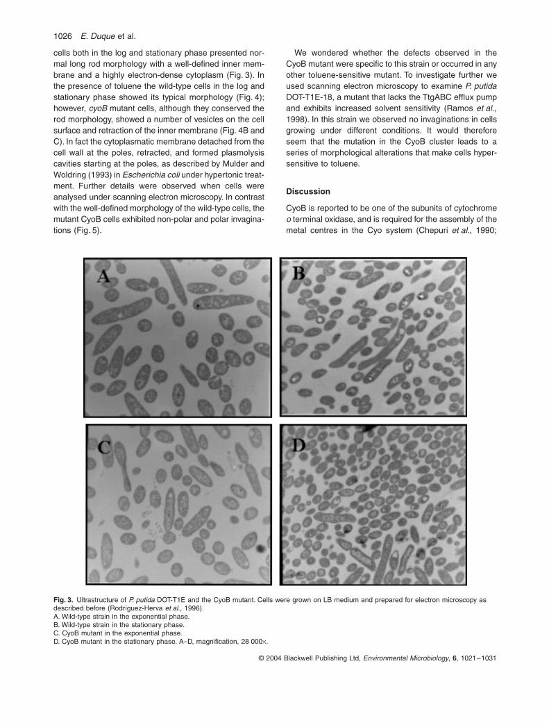
1026 E. Duque et al.
© 2004 Blackwell Publishing Ltd, Environmental Microbiology, 6, 1021–1031
cells both in the log and stationary phase presented nor-mal long rod morphology with a well-defined inner mem-brane and a highly electron-dense cytoplasm (Fig. 3). Inthe presence of toluene the wild-type cells in the log andstationary phase showed its typical morphology (Fig. 4);however, cyoB mutant cells, although they conserved therod morphology, showed a number of vesicles on the cellsurface and retraction of the inner membrane (Fig. 4B andC). In fact the cytoplasmatic membrane detached from thecell wall at the poles, retracted, and formed plasmolysiscavities starting at the poles, as described by Mulder andWoldring (1993) in Escherichia coli under hypertonic treat-ment. Further details were observed when cells wereanalysed under scanning electron microscopy. In contrastwith the well-defined morphology of the wild-type cells, themutant CyoB cells exhibited non-polar and polar invagina-tions (Fig. 5).
We wondered whether the defects observed in theCyoB mutant were specific to this strain or occurred in anyother toluene-sensitive mutant. To investigate further weused scanning electron microscopy to examine P. putidaDOT-T1E-18, a mutant that lacks the TtgABC efflux pumpand exhibits increased solvent sensitivity (Ramos et al.,1998). In this strain we observed no invaginations in cellsgrowing under different conditions. It would thereforeseem that the mutation in the CyoB cluster leads to aseries of morphological alterations that make cells hyper-sensitive to toluene.
Discussion
CyoB is reported to be one of the subunits of cytochromeo terminal oxidase, and is required for the assembly of themetal centres in the Cyo system (Chepuri et al., 1990;
Fig. 3. Ultrastructure of P. putida DOT-T1E and the CyoB mutant. Cells were grown on LB medium and prepared for electron microscopy as described before (Rodríguez-Herva et al., 1996).A. Wild-type strain in the exponential phase.B. Wild-type strain in the stationary phase.C. CyoB mutant in the exponential phase.D. CyoB mutant in the stationary phase. A–D, magnification, 28 000¥.
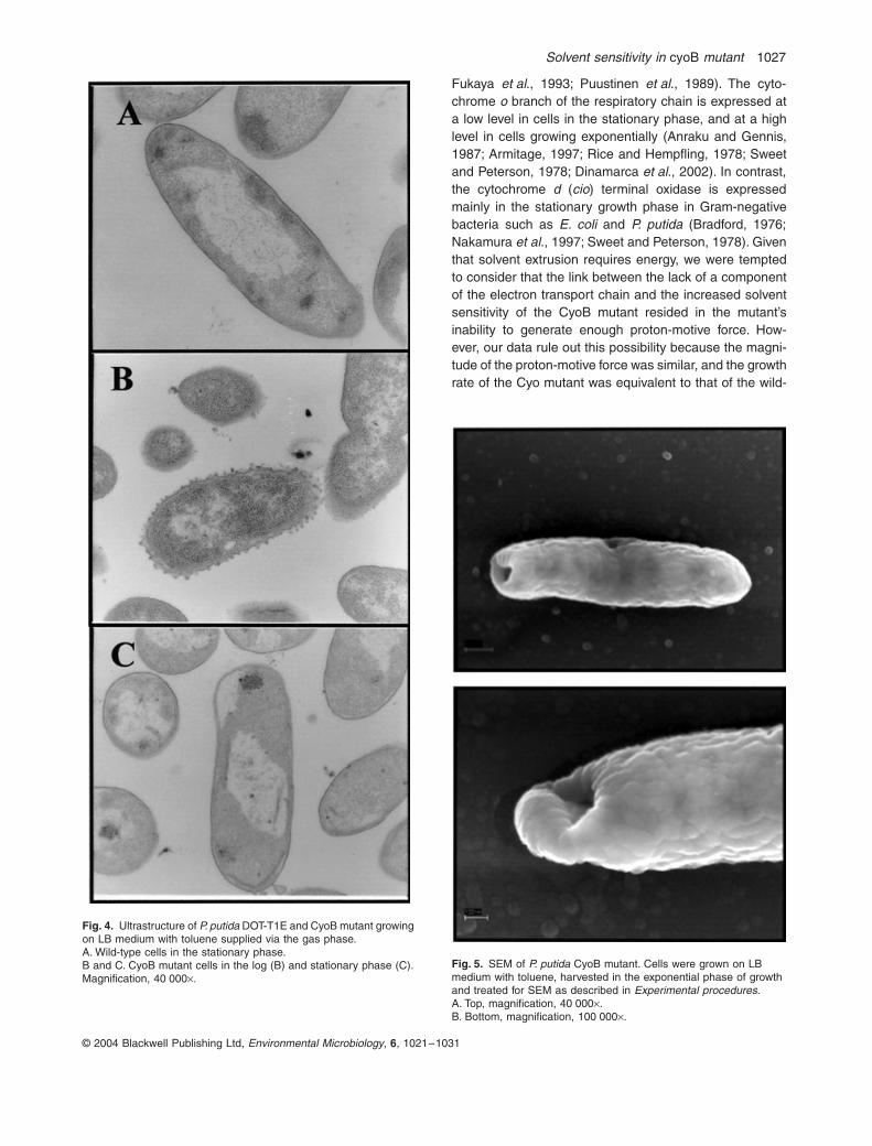
Solvent sensitivity in cyoB mutant 1027
© 2004 Blackwell Publishing Ltd, Environmental Microbiology, 6, 1021–1031
Fukaya et al., 1993; Puustinen et al., 1989). The cyto-chrome o branch of the respiratory chain is expressed ata low level in cells in the stationary phase, and at a highlevel in cells growing exponentially (Anraku and Gennis,1987; Armitage, 1997; Rice and Hempfling, 1978; Sweetand Peterson, 1978; Dinamarca et al., 2002). In contrast,the cytochrome d (cio) terminal oxidase is expressedmainly in the stationary growth phase in Gram-negativebacteria such as E. coli and P. putida (Bradford, 1976;Nakamura et al., 1997; Sweet and Peterson, 1978). Giventhat solvent extrusion requires energy, we were temptedto consider that the link between the lack of a componentof the electron transport chain and the increased solventsensitivity of the CyoB mutant resided in the mutant’sinability to generate enough proton-motive force. How-ever, our data rule out this possibility because the magni-tude of the proton-motive force was similar, and the growthrate of the Cyo mutant was equivalent to that of the wild-
Fig. 4. Ultrastructure of P. putida DOT-T1E and CyoB mutant growing on LB medium with toluene supplied via the gas phase.A. Wild-type cells in the stationary phase.B and C. CyoB mutant cells in the log (B) and stationary phase (C). Magnification, 40 000¥.
Fig. 5. SEM of P. putida CyoB mutant. Cells were grown on LB medium with toluene, harvested in the exponential phase of growth and treated for SEM as described in Experimental procedures.A. Top, magnification, 40 000¥.B. Bottom, magnification, 100 000¥.

1028 E. Duque et al.
© 2004 Blackwell Publishing Ltd, Environmental Microbiology, 6, 1021–1031
type strain in different growth media, and also becausethe RND efflux pumps that extrude antibiotics were func-tional in the Cyo mutant, as revealed by a similar patternof antibiotic resistance in the wild-type and the Cyomutant. It was reported in E. coli that Cyd system wasused instead of Cyo in cyo-deficient mutants. This seemsalso to be the case in P. putida, because the resultspresented here show that inactivation of cytochrome oubiquinol oxidase (Cyo) had no significant effect on thegrowth rate in the logarithmic phase under the growthconditions we tested.
Dinamarca et al. (2002) showed that the cyo genes ofP. putida were expressed preferentially in the log phase.Based on this finding we expected solvent sensitivity inCyoB mutants to be greater in the log phase that in thestationary phase. However, our results with the wild typerevealed that as far as tolerance to highly toxic solventswas concerned, non-induced and induced cells of P.putida DOT-T1E were more sensitive to a solvent shockwhen cells reached the stationary phase (see Fig. 2). Thissituation was exacerbated in the CyoB mutant: no cellswere found to survive a sudden toluene shock in thestationary phase. We found that the expression of theefflux pumps decreased in the stationary phase in thewild-type and Cyo mutant, a finding which correlates withthe increased solvent sensitivity. Whether the reduction inttg expression in the stationary phase is the result of atype of global regulation (Duque et al., 2001; Rojas et al.,2001) or whether it is mostly related to the physiologicalstatus of the cells remains unknown. In this regard, itshould be noted that Dinamarca et al. (2002) and Petrus-chka et al. (2001) also found that Cyo mutants escapefrom catabolite repression, and speculated that the redoxstate of the respiratory chains could influence the expres-sion of the Crc catabolite repression protein (O’Tooleet al., 2000; Dinamarca et al., 2002). It may well happenthat P. putida DOT-T1E cells sense the redox state of themembrane, which is transduced in a signal that influencesthe control of the expression of TtgABC, TtgDEF andTtgGHI in the stationary phase.
The lipid bilayer of the outer membrane is closelyattached to the peptidoglycan through lipoproteinspresent in the inner leaflet, and non-covalent interactionswith other outer membrane proteins (Mulder and Wold-ringh, 1993; Schwarz and Koch, 1995; Korber et al.,1996). The poles, however, are ‘free’ of peptidoglycan andcan present a point of cell weakness. It is also known thatingrowth of the outer membrane can be dissociated fromingrowth of the division septum by mutations, and that thisaffects the attachment of the murein layer to the outermembrane, which in turn favours plasmolysis (Rothfieldand Justice, 1997; Fung et al., 1978). Our EM studieshave revealed that solvent exposure in the CyoB mutantinduces plasmolysis from one of the poles. The lack of the
CyoB mutant itself is unlikely to directly affect solventsensitivity but the lack of CyoB could result an inappropri-ate signal transmission that negatively influences cellarchitecture in the poles. In this context we should notethat Segura et al. (2001; 2004) demonstrated that solventtolerance is compromised in certain flagellarless mutantsin P. putida DOT-T1E, and that this strain has a singleflagellum located in one of the poles of the cell.
Experimental procedures
Bacterial strains, plasmids and culture media
Pseudomonas putida DOT-T1E (RifR, TolR, Tol+) (Ramoset al., 1995) and DOT-T1E-CYOB (this study) were grown at30∞C in LB medium or in M9 minimal medium supplementedwith glucose (0.5% wt/vol) or citrate (10 mM), as the carbonsource. Escherichia coli DH5a (F¢/hsdR17, recA1, gyrA) cellswere grown on LB medium at 37∞C, and used for cloningexperiments.
Plasmids pUC18 and pUC19 (ApR) were used for cloning(Sambrook et al., 1989). Plasmid pRK600 (CmR, mob+, tra+,ColE1 replicon) was used as a helper for the mobilization oftra–, mob+ plasmids (Herrero et al., 1990). Derivatives of thesuicide plasmid pCHESIWKm (KmR, mob+, ColE1 replicon)were used to generate in vivo knock-outs of target genes(Llamas et al., 2003).
The antibiotics used were ampicillin (Ap), 100 mg ml-1;kanamycin (Km), 50 mg ml-1; piperacillin (Pip), 100 mg ml-1;rifampicin (Rif), 20 mg ml-1; streptomycin (Sm), 50–100 mg ml-1, and tetracycline (Tc), 15 mg ml-1.
Solvent shock assay
Cells were grown in 30 ml LB medium or M9 minimal mediumwith 10 mM citrate with or without toluene in the gas phaseovernight. On the following day the cultures were diluted1 : 50 in the same medium and grown under the same con-ditions until the culture reached the mid-exponential growthphase (turbidity about 0.8 at 660 nm) or the early stationaryphase (turbidity about 2.0 at 660 nm). The cultures weredivided in two halves; to one we added 0.3% (v/v) toluene,and the other was kept as a control. The number of viablecells in the log and stationary growth phase was determinedbefore toluene was added, and 10, 30 and 60 min later.
DNA techniques
Plasmid DNA was isolated by the alkaline lysis method withthe QIAprep Spin Plasmid Minipreps Kit. Total DNA wasisolated with a modification of the method of Kado and Liuas described by Ramos-González et al. (1991), except thatthe 30 min incubation step at 55∞C was omitted. DNA diges-tions with restriction enzymes, ligations, and transformationswere performed by standard procedures (Sambrook et al.,1989).

Solvent sensitivity in cyoB mutant 1029
© 2004 Blackwell Publishing Ltd, Environmental Microbiology, 6, 1021–1031
Polymerase chain reactions (PCRs)
The standard PCR mixture (25 ml) contained 10 ng of DNA,200 mM each deoxynucleoside triphosphate, 50 pmol of eachprimer, 2 ml of dimethyl sulphoxide, and 0.25 U of Taq poly-merase. The PCR conditions were as follows: 4 min at 95∞C,and then 35 cycles of 60∞C for 45 s, 72∞C for 30–180 s, and94∞C for 4 s, followed by a final 5 min step at 72∞C.
Generation of a cyoB mutant by insertional inactivation
To generate a —-cyoB:Km P. putida mutant strain, a 500 bpDNA fragment of the cyoB gene was amplified by PCRfrom the P. putida DOT-T1E chromosome using the primers:5¢-GGAATTCTGCACTCGCTCTGCGACTGTTGA-3¢ and 5¢-CGAATTCATGCCACCGATGGAGAAGGTGAT-3¢, which con-tain EcoRI sites at their 5¢-terminal ends. The amplifiedproduct was digested with EcoRI and cloned at the EcoRIsite of plasmid pCHESIWKm (Llamas et al., 2003), yieldingplasmid pEDX-1. This plasmid was designed for markerexchange mutagenesis because it replicates in E. coli butnot in P. putida, and carries a KmR cassette to select forinsertion of the plasmid in the host chromosome. PlasmidpEDX-1 was transferred to P. putida DOT-T1E in triparentalmatings with pRK600 as the donor of transfer functions.Transconjugants incorporating the KmR cassette wereselected on M9 minimal medium with citrate as the solecarbon source, and Km and Ap. These cells were expectedto contain pEDX-1 integrated into the chromosome and togenerate a mutant by insertional inactivation at the cyoBgene. The absence of the wild-type allele and the presenceof the cyoB::Km allele were confirmed by PCR and South-ern blot. One of the clones was retained for further studiesand was called DOT-T1E-CyoB.
Southern hybridization and DNA labelling
DNA fragments were separated in agarose gels and trans-ferred onto nylon membranes by capillary blotting as previ-ously described (Sambrook et al., 1989). Specific probes forhybridization were recovered from agarose gels with an aga-rose gel DNA extraction kit (Boehringer Mannheim). Allprobes were labelled with digoxigenin by Klenow randomprimer extension according to the recommended procedure(Ausubel et al., 1991). Blotted filters were prehybridized,hybridized, washed and immunologically developed accord-ing to the supplier’s instructions. High-stringency conditions[50% (v/v) formamide at 42∞C] were used.
Assay for b-galactosidase
An overnight culture of the appropriate strain was diluted toa final turbidity (A600) of ~ 0.04 in fresh LB medium andcultures were grown at 30∞C. When turbidity reached 0.08the inducer was added, and 4 h later aliquots were taken andb-galactosidase activity measured as described by Miller(1972). At least three independent assays were performed ineach case.
Analysis of phospholipids
Phospholipids were extracted by the method of Bligh andDyer (1959). To measure fatty acids, phospholipids weretrans esterified by dissolving 3–10 mg of phospholipid extractin 350 ml of hexane. Then 40 ml of 2 M KOH dissolved inmethanol was added and mixed vigorously. After the additionof 2 ml of hexane, the upper (hexane) phase was recoveredand, within 1 h, methyl ester derivatives of fatty acids wereidentified by mass spectrometry after gas chromatographicseparation as described previously (Ramos et al., 1997).
To measure the phospholipid headgroups, the chloroformlayer was dried by evaporation under a stream of nitrogen.The lipid residue was dissolved in chloroform and the differ-ent phospholipids were separated on silica-gel 60 G thin layerchromatography plates, using a mixture of chloroform/methanol/acetic acid glacial (65 : 25 : 8) as solvent.
The spots corresponding to each phospholipid were visu-alized by exposing the plates to iodine vapours and scraped.The phospholipids were then quantified by Bartlett’s method(1959).
Determination of Dy
Cells were converted to spheroplasts by treatment withlysozyme and ethylenediaminetetraacetic acid (EDTA). Thecells were grown to an OD660 of ~0.5–0.7 in M9 minimalmedium supplemented with glucose or citrate in the absenceand in the presence of toluene supplied via the gas phase.Cultures (50–100 ml) were centrifuged (10.000 g, 5 min, at4∞C), and the cells were resuspended in 6.3 ml of 0.75 Msucrose and 10 mM Tris-HCl (pH 7.8), and then treated with0.1 mg ml-1 of lysozyme for 2–5 min on ice. Then 13.2 ml of1.5 mM EDTA (pH 7.5) solution was added. The cell suspen-sion was incubated for 15 min at 30∞C and observed undera phase-contrast microscope. More than 95% of the cellsappeared as spheroplasts. The generation of Dy in sphero-plasts of the wild-type strain and the CyoB mutant was mon-itored using the cationic dye 3,3’-diethylthiadicarbocyanineiodide [DiSC2(3)], which translocates into the lipid bilayer ofhyperpolarized membranes, resulting in quenching of its flu-orescence. The reaction mixture (total volume 1 ml) con-tained buffer A [125 mM Hepes, 0.9% (w/v) NaCl, 1 mM KCl,1 mM MgCl2 and 0.4% (wt/vol) glucose], spheroplasts (about106) and 0.08 mM DiSC2(3). The fluorescence emission ofDiSC2(3) was measured at 570 nm with excitation at 555 nmusing an SLM Aminco SPF-500C fluorimeter. To dissipate themembrane potential, valinomycin (2 mM final concentration)was added to the reaction mixture. The membrane potentialwas determined as the difference between the fluorescencequenching of DiSC2(3) when it accumulated in the cytoplas-mic membrane, and the fluorescence obtained when it wasreleased upon the addition of valinomycin.
Microscopy studies
Pseudomonas putida cells grown in LB medium were har-vested in the logarithmic or stationary growth phase andsubjected to microscopic analysis. For transmission electronmicroscopy (TEM), samples were prepared and observed aspreviously described (Rodríguez-Herva et al., 1996). For

1030 E. Duque et al.
© 2004 Blackwell Publishing Ltd, Environmental Microbiology, 6, 1021–1031
scanning electron microscopy (SEM), cells were fixed withglutaraldehyde vapors for 24 h in a humid chamber at 4∞C.Then the cells were rinsed with distilled water, dehydratedwith a graded series of ethanol solutions, suspended in amylacetate, critical-point dried, and coated with gold. Sampleswere examined in Zeiss DSM950 scanning electronmicroscope.
Acknowledgements
Work in the authors’ laboratory was supported by a grantfrom the European Commission (QLK3-CT-2001–00435) anda grant from the Ministry of Science and Technology(BIO2003-00515). We thank Juan Pedro Donaire and K. Ven-ema for help with proton-motive force measurements. We areindebted to Alejandro Dinamarca and Fernando Rojo for con-structions and helpful discussions, Miguel-Angel de Pedro fordirecting our attention to the plasmolysis cavities and BoriSegovia for help in phospholipid headgroup quantification.EM pictures were taken at the Servicios Generales of theUniversity of Granada. We thank Carmen Lorente and KarenShashok for improving the use of English.
References
Anraku, Y., and Gennis, R.B. (1987) The aerobic respiratorychain of Escherichia coli. Trends Biochem Sci 12: 262–266.
Armitage, J.P. (1997) Behavioural responses of bacteria tolight and oxygen. Arch Microbiol 168: 249–261.
Ausubel, F.M., Brent, R., Kingston, R.E., Moore, D.D.,Seidman, J.G., Smith, J.A., and Struhl, K. (eds). (1991)Current Protocols in Molecular Biology. New York, USA:John Wiley & Sons.
Bartlett, G.R. (1959) Phosphorous assay in column chroma-tography. J Biol Chem 234: 466–469.
Bligh, E.G., and Dyer, W.J. (1959) A rapid method of totallipid extraction and purification. Can J Biochem Physiol 37:911–917.
Bradford, M.M. (1976) A rapid and sensitive method for thequantitation of microgram quantities of protein utilizing theprinciple of protein-dye binding. Anal Biochem 72: 248–254.
Chepuri, V., Lemieux, L., Au, D.C.T., and Gennis, R.B. (1990)The sequence of the cyo operon indicates substantialstructural similarities between the cytochrome o ubiquinoloxidase of Escherichia coli and the aa3-type family of cyto-chrome c oxidases. J Biol Chem 265: 11185–11192.
Cooper, M., Tavankar, G.R., and Williams, H.D. (2003) Reg-ulation of expression of the cyanide-insensitive terminaloxidase in Pseudomonas aeruginosa. Microbiology 149:1275–1284.
Cotter, P.A., Chepuri, V., Gennis, R.B., and Gunsalus, R.P.(1990) Cytochrome o (cyoABCDE) and d (cydAB) oxidasegene expression in Escherichia coli is regulated by oxy-gen, pH, and the fnr gene product. J Bacteriol 172: 6333–6338.
Cotter, P.A., Melville, S.B., Albrecht, J.A., and Gunsalus,R.P. (1997) Aerobic regulation of cytochrome d oxidase(cydAB) operon expression in Escherichia coli: roles of Fnr
and ArcA in repression and activation. Mol Microbiol 25:605–615.
Cunningham, L., and Williams, H.D. (1995) Isolation andcharacterization of mutants defective in the cyanide-insensitive respiratory pathway of Pseudomonas aerugi-nosa. J Bacteriol 177: 432–438.
De Smet, M.J., Kingma, J., and Witholt, B. (1978) The effectof toluene on the structure and permeability of the outerand cytoplasmic membranes of Escherichia coli. BiochimBiophys Acta 506: 64–80.
Diefenbach, R., Heipieper, H.J., and Keweloh, H. (1992) Theconversion of cis into trans unsaturated fatty acids inPseudomonas putida P8: evidence for a role in the regu-lation of membrane fluidity. Appl Microbiol Biotechnol 38:382–387.
Dinamarca, A., Ruíz-Manzano, A., and Rojo, F. (2002) Inac-tivation of cytochrome o ubiquinol oxidase relieves cata-bolic repression of the Pseudomonas putida Gpo1 alkanedegration pathway. J Bacteriol 184: 3785–3793.
Dinamarca, A., Aranda-Olmedo, I., Puyet, A., and Rojo, F.(2003) Expression of the Pseudomonas putida OCT plas-mid alkane degradation pathway is modulated by two dif-ferent global control signals: evidence from continuouscultures. J Bacteriol 185: 4772–4778.
Duque, E., Segura, A., Mosqueda, G., and Ramos, J.L. (2001)Global and cognate regulators control the expression ofthe organic solvent efflux pumps TtgABC and TtgDEF ofPseudomonas putida. Mol Microbiol 39: 1100–1106.
Fukaya, M., Tayama, K., Tamaki, T., Ebisuya, H., Okumura,H., Kawamura, Y., et al. (1993) Characterization of a cyto-chrome a1 that functions as a ubiquinol oxidase in Aceto-bacter aceti. J Bacteriol 175: 4307–4314.
Fukumori, F., Hirayama, H., Takami, H., Inoue, A., and Hori-koshi, K. (1998) Isolation and transposon mutagenesis ofa Pseudomonas putida KT2442 toluene-resistant variant:involvement of an efflux system in solvent resistance.Extremophiles 2: 395–400.
Fung, J., MacAllister, T.J., and Rothfield, L.I. (1978) Role ofmurcin lipoprotein in morphogenesis of the bacterial divi-sion septum: phenotypic similarity of lkyD and lpo mutans.J Bacteriol 133: 1467–1471.
Heipieper, H.J., Diefenbach, R., and Keweloh, H. (1992) Con-version of cis unsaturated fatty acids to trans: a possiblemechanism for the protection of phenol-degradingPseudomonas putida P8 from substrate toxicity. Appl Envi-ron Microbiol 58: 1847–1852.
Herrero, M., de Lorenzo, V., and Timmis, K.N. (1990) Trans-poson vectors containing non-antibiotic resistance selec-tion markers for cloning and stable chromosomal insertionof foreign genes in Gram-negative bacteria. J Bacteriol172: 6557–6567.
Hirayama, H., Takami, H., Inoue, A., and Horikoshi, K. (1998)Isolation and characterization of toluene-sensitive mutantsfrom Pseudomonas putida IH-2000. FEMS Microbiol Lett169: 219–225.
Isken, S., and de Bont, J.A.M. (1996) Active efflux of toluenein a solvent-resistant bacterium. J Bacteriol 178: 6056–6058.
Junker, F., and Ramos, J.L. (1999) Involvement of the cis/trans isomerase CtiT1 in solvent resistance in Pseudomo-nas putida DOT-T1E. J Bacteriol 181: 5693–5700.

Solvent sensitivity in cyoB mutant 1031
© 2004 Blackwell Publishing Ltd, Environmental Microbiology, 6, 1021–1031
Kobayashi, H., Takami, H., Hirayama, H., Kobata, K., Usami,R., and Horikoshi, K. (1999) Outer membrane changes ina toluene-sensitive mutant of toluene-tolerant Pseudomo-nas putida IH-2000. J Bacteriol 181: 4493–4498.
Korber, D.R., Choi, A., Wolfaardt, G.M., and Caldwell, D.E.(1996) Bacterial plasmolysis as a physical indicator of via-bility. Appl Environ Microbiol 62: 3939–3947.
Llamas, M.A., Rodríguez-Herva, J.J., Hancock, R.E.W., Bit-ter, W., Tomasen, J., and Ramos, J.L. (2003) Role of thePseudomonas putida tol-oprL gene products in the uptakeof solutes through the cytoplasmic membrane. J Bacteriol185: 4770–4716.
Miller, J.H. (1972) Experiments in Molecular Genetics. ColdSpring Harbor, NY: Cold Spring Harbor Laboratory.
Mosqueda, G., and Ramos, J.L. (2000) A set of genes encod-ing a second toluene efflux system in Pseudomonas putidaDOT-T1E is linked to the tod genes for toluene metabolism.J Bacteriol 182: 937–943.
Mulder, E., and Woldringh, C.L. (1993) Plasmolysis bays inEscherichia coli: are they related to development and posi-tioning of division sites? J Bacteriol 175: 2241–2247.
Nakamura, H., Saiki, K., Mogi, T., and Anraku, Y. (1997)Assignment and functional roles of the cyoABCDE geneproducts required for the Escherichia coli bo-type quinoloxidase. J Biochem 122: 415–421.
O’Toole, G.A., Gibbs, K.A., Hager, P.W., Phibbs, P.V. Jr andKolter, R. (2000) The global carbon metabolism regulatorCrc is a component of a signal transduction pathwayrequired for biofilm development by Pseudomonas aerug-inosa. J Bacteriol 182: 425–431.
Petruschka, L., Burchhardt, G., Müller, C., Weihe, C., andHerrmann, H. (2001) The cyo operon of Pseudomonasputida is involved in catabolic repression of phenol degra-dation. Mol Gen Genomics 266: 199–206.
Puustinen, A., Finel, M., Virkki. M., and Wikstrom, M. (1989)Cytochrome o (bo) is a proton pump in Paracoccus deni-trificans and Escherichia coli. FEBS Lett 249: 163–167.
Ramos, J.L., Duque, E., Huertas, M.J., and Haïdour, A.(1995) Isolation and expansion of the catabolic potential ofa Pseudomonas putida strain able to grow in the presenceof high concentrations of aromatic hydrocarbons. J Bacte-riol 177: 3911–3916.
Ramos, J.L., Duque, E., Rodríguez-Herva, J.J., Godoy, P.,Haïdour, A., Reyes, F., and Fernández-Barrero, A. (1997)Mechanisms for solvent tolerance in bacteria. J Biol Chem272: 3887–3890.
Ramos, J.L., Duque, E., Godoy, P., and Segura, A. (1998)Efflux pumps involved in toluene tolerance in Pseudomo-nas putida DOT-T1E. J Bacteriol 180: 3323–3329.
Ramos, J.L., Duque, E., Gallegos, M.T., Godoy, P., Ramos-González, M.I., Rojas, A., et al. (2002) Mechanisms ofsolvent tolerance in Gram-negative bacteria. Ann RevMicrobiol 56: 743–768.
Ramos-González, M.I., Duque, E., and Ramos, J.L. (1991)
Conjugational transfer of recombinant DNA in cultures andin soils: host range of Pseudomonas putida TOL plasmids.Appl Environ Microbiol 57: 3020–3027.
Rice, C.W., and Hempfling, W.P. (1978) Oxygen-limited con-tinuous culture and respiratory energy conservation inEscherichia coli. J Bacteriol 134: 115–124.
Rodríguez-Herva, J.J., Ramos-González, M.I., and Ramos,J.L. (1996) The Pseudomonas putida peptidoglycan-associated outer membrane lipoprotein is involved in main-tenance of the integrity of the cell envelope. J Bacteriol178: 1699–1706.
Rojas, A., Duque, E., Mosqueda, G., Golden, G., Hurtado,A., Ramos, J.L., and Segura, A. (2001) Three efflux pumpsare required to provide efficient tolerance to toluene inPseudomonas putida DOT-T1E. J Bacteriol 183: 3967–3973.
Rothfield, L.I., and Justice, S.S. (1997) Bacterial cell division:The cycle of the ring. Cell 88: 581–584.
Sambrook, J., Fritsch, E.F., and Maniatis, T. (1989) Molecu-lar Cloning: A Laboratory Manual. 2nd ed. Cold SpringHarbor, NY: Cold Spring Harbor Laboratory.
Schwarz, H., and Koch, A.L. (1995) Phase and electronmicroscopic observations of osmotically induced wrinklingand the role of endocytotic vesicles in the plasmolysis ofthe Gram-negative cell wall. Microbiology 141: 3161–3170.
Segura, A., Duque, E., Hurtado, A., and Ramos, J.L. (2001)Mutations in genes involved in the flagellar export appara-tus of the solvent-tolerant Pseudomonas putida DOT-T1Estrain impair motility and lead to hypersensitibity to tolueneshocks. J Bacteriol 183: 4127–4133.
Segura, A., Hurtado, A., Duque, E., and Ramos, J.L. (2004)Transcriptional phase variation at the flhB gene ofPseudomonas putida DOT-T1E is involved in response toenvironmental changes and supports the participation ofthe flagellar export system in solvent tolerance. J Bacteriol186: 1905–1909.
Sikkema, J., de Bont, J.A.M., and Poolman, B. (1994) Inter-actions of cyclic hydrocarbons with biological membranes.J Biol Chem 269: 8022–8028.
Sweet, W.J., and Peterson, J.A. (1978) Changes in cyto-chrome content and electron transport patterns inPseudomonas putida as a function of growth phase. JBacteriol 133: 217–224.
Taylor, B.L., and Zhulin, I.B. (1999) PAS domains: internalsensors of oxygen, redox potential, and light. Microbiol MolBiol Rev 63: 479–506.
Weber, F.J., and de Bont, J.A.M. (1996) Adaptation mecha-nisms of microorganisms to the toxic effects of organicsolvents on membranes. Biochim Biophys Acta 1286: 225–245.
White, D.G., Goldman, J.D., Demple, B., and Levy, S.B.(1997) Role of the acrAB locus in organic solvent tolerancemediated by expression of marA, soxS, or robA in Escher-ichia coli. J Bacteriol 179: 6122–6126.

Resultados
52

Resultados
53
Functional replacement of the outer membrane protein TtgC in the TtgABC
efflux pump in antibiotic resistance in Pseudomonas putida DOT-T1E
Vanina García, Isabel Jimenez, Juan Luis Ramos, and Ana Segura.
La clave en la resistencia de la cepa altamente tolerante a disolventes orgánicos
Pseudomonas putida DOT-T1E es la presencia de tres bombas de extrusión (TtgABC,
TtgDEF, TtgGHI) que pertenecen a la familia RND la cual exporta compuestos tóxicos
tales como antibióticos y disolventes orgánicos. Los transportadores de la familia RND
normalmente son complejos tripartitos compuestos por un sistema de extrusión que está
localizado en la membrana interna junto con una proteína de membrana externa (OMP)
y una proteína de fusión de membrana (MFP) como proteínas auxiliares.
Para estudiar el intercambio de componentes entre ellas, construimos bombas de
extrusión híbridas. Observamos que a nivel de resistencia un intercambio funcional tuvo
lugar cuando TtgC (OMP) fue sustituida por componentes de otras bombas. Aunque en
nuestros estudios el reemplazo de TtgA (MFP) fue funcional en cuanto a la expulsión de
antibióticos, para confirmar este resultado son necesarios muchos análisis.
Manuscrito en preparación

Resultados
54

55
Manuscrito en preparación
Functional replacement of the outer membrane protein TtgC
in the TtgABC efflux pump in antibiotic resistance in
Pseudomonas putida DOT-T1E Vanina García, Isabel Jimenez, Juan Luis Ramos, and Ana Segura*
Environmental Protection Department, Estación Experimental del Zaidín, Consejo Superior de Investigaciones
Científicas, C/ Profesor Albareda 1, 18008-Granada, Spain
* Corresponding author:
Ana Segura
EEZ-CSIC
C/ Prof. Albareda, 1
E-18008 Granada
Spain
Phone: +34 958 181600
Fax: +34 958 129600
e-mail: [email protected]
SUMMARY
In Pseudomonas putida DOT-T1E three efflux pumps (TtgABC,
TtgDEF, TtgGHI) that belong to the RND family have been
described as able to extrude toxic compounds such as antibiotics
and organics solvents. Transporters of the RND family usually
consist of an efflux system that is made up by one component
that is located at inner membrane, and is often associated with an
auxiliary outer membrane protein (OMP) and a membrane fusion
protein (MFP). In order to study the exchange between
components among these three efflux pumps, we have
constructed hybrid efflux pumps. The results show that
functional replacement took place when the TtgC (OMP) was
substituted by other pump components. Although we found
some indications of functional replacement of the TtgA (MFP)
component, further analysis will be necessary to confirm that
result.
INTRODUCTION
Pseudomonas putida DOT-T1E is an
antibiotic- and solvent-tolerant strain
isolated from a wastewater treatment plant
in Granada (Ramos et al., 1995). The
common basis of the resilience of this strain
is its ability extrude toxic compound by
means of efflux pumps (Ramos et al., 2002;
Daniels and Ramos 2009). Two efflux
pumps of the RND (resistance, nodulation,
cell division) family, named TtgABC
(Ramos et al., 1998; Mosqueda and Ramos,
2000) and TtgGHI (Rojas et al., 2001), have
been shown to be involved in solvent and
antibiotic resistance, while a third efflux
pump, named TtgDEF, has been implicated
in the efflux of solvents but not antibiotics
(Rojas et al., 2006). The TtgGHI efflux
pump is the main determinant of solvent-
tolerance in Pseudomonas putida DOT-T1E,
while its participation in antibiotic efflux is
masked by the presence of the TtgABC
efflux pump. The RND family of efflux
pumps have been extensively studied in
recent years due to their involvement in the
multidrug resistance observed in clinically
interesting strains such as Pseudomonas
aeruginosa (Poole, 2004). Transporters of
the RND family usually consist of a efflux
system that is made up by one component
that is located at inner membrane, and is
often associated with an auxiliary outer
membrane protein (OMP) and a membrane
fusion protein (MFP).
The most studied RND efflux pump is
AcrAB-TolC of E. coli (Osuku et al., 1996).
The genes that make up this pump form an
operon (acrAB), while the OMP (tolC) is
monocistronic. It is also known that TolC
can act as the outer membrane element for
other transport systems, such as the
haemolysis transporter HylD (Dinh et al.,
1994). In Pseudomonas aeruginosa some
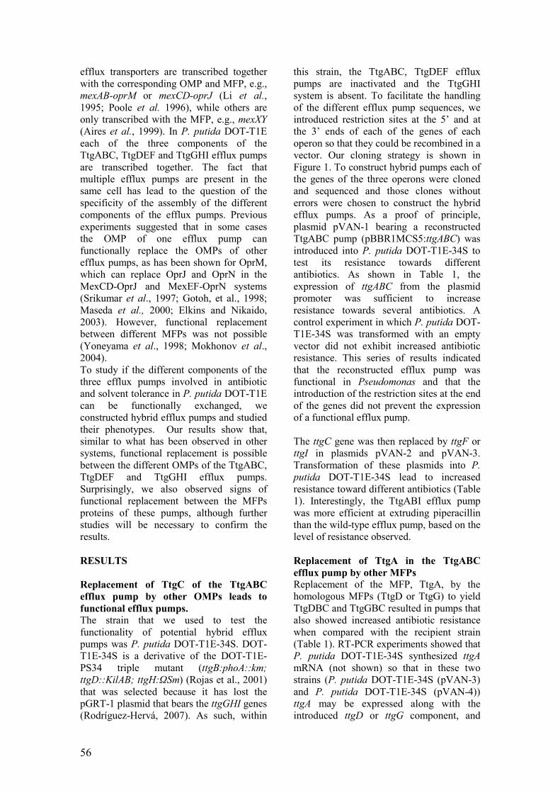
56
efflux transporters are transcribed together
with the corresponding OMP and MFP, e.g.,
mexAB-oprM or mexCD-oprJ (Li et al.,
1995; Poole et al. 1996), while others are
only transcribed with the MFP, e.g., mexXY
(Aires et al., 1999). In P. putida DOT-T1E
each of the three components of the
TtgABC, TtgDEF and TtgGHI efflux pumps
are transcribed together. The fact that
multiple efflux pumps are present in the
same cell has lead to the question of the
specificity of the assembly of the different
components of the efflux pumps. Previous
experiments suggested that in some cases
the OMP of one efflux pump can
functionally replace the OMPs of other
efflux pumps, as has been shown for OprM,
which can replace OprJ and OprN in the
MexCD-OprJ and MexEF-OprN systems
(Srikumar et al., 1997; Gotoh, et al., 1998;
Maseda et al., 2000; Elkins and Nikaido,
2003). However, functional replacement
between different MFPs was not possible
(Yoneyama et al., 1998; Mokhonov et al.,
2004).
To study if the different components of the
three efflux pumps involved in antibiotic
and solvent tolerance in P. putida DOT-T1E
can be functionally exchanged, we
constructed hybrid efflux pumps and studied
their phenotypes. Our results show that,
similar to what has been observed in other
systems, functional replacement is possible
between the different OMPs of the TtgABC,
TtgDEF and TtgGHI efflux pumps.
Surprisingly, we also observed signs of
functional replacement between the MFPs
proteins of these pumps, although further
studies will be necessary to confirm the
results.
RESULTS
Replacement of TtgC of the TtgABC
efflux pump by other OMPs leads to
functional efflux pumps.
The strain that we used to test the
functionality of potential hybrid efflux
pumps was P. putida DOT-T1E-34S. DOT-
T1E-34S is a derivative of the DOT-T1E-
PS34 triple mutant (ttgB:phoA::km;
ttgD::KilAB; ttgH:ΩSm) (Rojas et al., 2001)
that was selected because it has lost the
pGRT-1 plasmid that bears the ttgGHI genes
(Rodríguez-Hervá, 2007). As such, within
this strain, the TtgABC, TtgDEF efflux
pumps are inactivated and the TtgGHI
system is absent. To facilitate the handling
of the different efflux pump sequences, we
introduced restriction sites at the 5’ and at
the 3’ ends of each of the genes of each
operon so that they could be recombined in a
vector. Our cloning strategy is shown in
Figure 1. To construct hybrid pumps each of
the genes of the three operons were cloned
and sequenced and those clones without
errors were chosen to construct the hybrid
efflux pumps. As a proof of principle,
plasmid pVAN-1 bearing a reconstructed
TtgABC pump (pBBR1MCS5:ttgABC) was
introduced into P. putida DOT-T1E-34S to
test its resistance towards different
antibiotics. As shown in Table 1, the
expression of ttgABC from the plasmid
promoter was sufficient to increase
resistance towards several antibiotics. A
control experiment in which P. putida DOT-
T1E-34S was transformed with an empty
vector did not exhibit increased antibiotic
resistance. This series of results indicated
that the reconstructed efflux pump was
functional in Pseudomonas and that the
introduction of the restriction sites at the end
of the genes did not prevent the expression
of a functional efflux pump.
The ttgC gene was then replaced by ttgF or
ttgI in plasmids pVAN-2 and pVAN-3.
Transformation of these plasmids into P.
putida DOT-T1E-34S lead to increased
resistance toward different antibiotics (Table
1). Interestingly, the TtgABI efflux pump
was more efficient at extruding piperacillin
than the wild-type efflux pump, based on the
level of resistance observed.
Replacement of TtgA in the TtgABC
efflux pump by other MFPs
Replacement of the MFP, TtgA, by the
homologous MFPs (TtgD or TtgG) to yield
TtgDBC and TtgGBC resulted in pumps that
also showed increased antibiotic resistance
when compared with the recipient strain
(Table 1). RT-PCR experiments showed that
P. putida DOT-T1E-34S synthesized ttgA
mRNA (not shown) so that in these two
strains (P. putida DOT-T1E-34S (pVAN-3)
and P. putida DOT-T1E-34S (pVAN-4))
ttgA may be expressed along with the
introduced ttgD or ttgG component, and

57
TABLE 1. Antibiotic resistance of the strains containing the different hybrid efflux pumps.
Strain pump Ap Tc Cb Pip
P. putida DOT-T1E 2000 15 2000 35
P. putida DOT-T1E-34S 125 0.23 7.8 1.1
P. putida DOT-T1E-34S (pVAN-1) ttgABC 1000 0.94 250 4.4
P. putida DOT-T1E-34S (pVAN-2) ttgABF 125 0.06 62.5 4.4
P. putida DOT-T1E-34S (pVAN-3) ttgABI 500 0.23 500 35
P. putida DOT-T1E-34S (pVAN-4) ttgDBC 500 0.94 500 4.4
P. putida DOT-T1E-34S (pVAN-5) ttgGBC 500 0.47 500 8.8
P. putida DOT-T1E-34S (pVAN-6) ttgDBF 250 0.47 250 8.8
P. putida DOT-T1E-34S (pVAN-7) ttgDBI 500 0.94 250 8.8
P. putida DOT-T1E-34S (pVAN-8) ttgGBF 250 0.47 125 17.6
P. putida DOT-T1E-34S (pVAN-9) ttgGBI 250 0.23 125 4.4
P. putida DOT-T1E-34S (pVAN-10) ttgAEC 62.5 0.47 7.8 0.56
P. putida DOT-T1E-34S (pVAN-11) ttgAEF 62.5 0.23 7.8 0.56
P. putida DOT-T1E-34S (pVAN-12) ttgAEI 62.5 0.12 7.8 0.56
P. putida DOT-T1E-34S (pVAN-13) ttgDEC 62.5 0.47 7.8 1.1
P. putida DOT-T1E-34S (pVAN-14) ttgDEF 125 0.47 7.8 0.56
P. putida DOT-T1E-34S (pVAN-15) ttgDEI 62.5 0.23 7.8 0.56
therefore the possibility of the formation of
the native TtgABC efflux pump can not be
excluded. However, when both the OMP
(TtgC) and the MFP (TtgA) were replaced
by other pump components not only all of
the combinations were functional regarding
antibiotic extrusion but their antibiotic
resistance was higher than that of the
corresponding efflux pumps with the TtgA
component alone. Differences were
observed between the extrusion ability of
these chimeras, as the TtgDBF and TtgGBF
pumps were more resistant toward
tetracycline than the TtgABF efflux pump.
The strain containing TtgDBF also showed a
4-fold increased resistance toward
carbenicillin in comparison to the TtgABF
containing strain. Similarly, TtgDBI
conferred higher tetracycline resistance than
TtgABI. These results suggest that TtgA can
be functionally exchanged by TtgD and
TtgG.
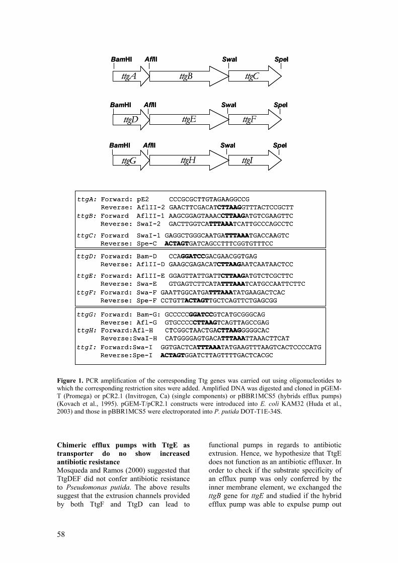
58
Figure 1. PCR amplification of the corresponding Ttg genes was carried out using oligonucleotides to
which the corresponding restriction sites were added. Amplified DNA was digested and cloned in pGEM-
T (Promega) or pCR2.1 (Invitrogen, Ca) (single components) or pBBR1MCS5 (hybrids efflux pumps)
(Kovach et al., 1995). pGEM-T/pCR2.1 constructs were introduced into E. coli KAM32 (Huda et al.,
2003) and those in pBBR1MCS5 were electroporated into P. putida DOT-T1E-34S.
Chimeric efflux pumps with TtgE as
transporter do no show increased
antibiotic resistance
Mosqueda and Ramos (2000) suggested that
TtgDEF did not confer antibiotic resistance
to Pseudomonas putida. The above results
suggest that the extrusion channels provided
by both TtgF and TtgD can lead to
functional pumps in regards to antibiotic
extrusion. Hence, we hypothesize that TtgE
does not function as an antibiotic effluxer. In
order to check if the substrate specificity of
an efflux pump was only conferred by the
inner membrane element, we exchanged the
ttgB gene for ttgE and studied if the hybrid
efflux pump was able to expulse pump out
ttgA: Forward: pE2 CCCGCGCTTGTAGAAGGCCG
Reverse: AflII-2 GAACTTCGACATCTTAAGGTTTACTCCGCTT
ttgB: Forward AflII-1 AAGCGGAGTAAACCTTAAGATGTCGAAGTTC
Reverse: SwaI-2 GACTTGGTCATTTAAATCATTGCCCAGCCTC
ttgC: Forward SwaI-1 GAGGCTGGGCAATGATTTAAATGACCAAGTC
Reverse: Spe-C ACTAGTGATCAGCCTTTCGGTGTTTCC
ttgE: Forward: AflII-E GGAGTTATTGATTCTTAAGATGTCTCGCTTC
Reverse: Swa-E GTGAGTCTTCATATTTAAATCATGCCAATTCTTC
ttgD: Forward: Bam-D CCAGGATCCGACGAACGGTGAG
Reverse: AflII-D GAAGCGAGACATCTTAAGAATCAATAACTCC
ttgF: Forward: Swa-F GAATTGGCATGATTTAAATATGAAGACTCAC
Reverse: Spe-F CCTGTTACTAGTTGCTCAGTTCTGAGCGG
ttgH: Forward:Afl-H CTCGGCTAACTGACTTAAGGGGGCAC
Reverse:SwaI-H CATGGGGAGTGACATTTAAATTAAACTTCAT
ttgI: Forward:Swa-I GGTGACTCATTTAAATATGAAGTTTAAGTCACTCCCCATG
Reverse:Spe-I ACTAGTGGATCTTAGTTTTGACTCACGC
ttgG: Forward: Bam-G: GCCCCCGGATCCGTCATGCGGGCAG
Reverse: Afl-G GTGCCCCCTTAAGTCAGTTAGCCGAG
ttgA: Forward: pE2 CCCGCGCTTGTAGAAGGCCG
Reverse: AflII-2 GAACTTCGACATCTTAAGGTTTACTCCGCTT
ttgB: Forward AflII-1 AAGCGGAGTAAACCTTAAGATGTCGAAGTTC
Reverse: SwaI-2 GACTTGGTCATTTAAATCATTGCCCAGCCTC
ttgC: Forward SwaI-1 GAGGCTGGGCAATGATTTAAATGACCAAGTC
Reverse: Spe-C ACTAGTGATCAGCCTTTCGGTGTTTCC
ttgA: Forward: pE2 CCCGCGCTTGTAGAAGGCCG
Reverse: AflII-2 GAACTTCGACATCTTAAGGTTTACTCCGCTT
ttgB: Forward AflII-1 AAGCGGAGTAAACCTTAAGATGTCGAAGTTC
Reverse: SwaI-2 GACTTGGTCATTTAAATCATTGCCCAGCCTC
ttgC: Forward SwaI-1 GAGGCTGGGCAATGATTTAAATGACCAAGTC
Reverse: Spe-C ACTAGTGATCAGCCTTTCGGTGTTTCC
ttgE: Forward: AflII-E GGAGTTATTGATTCTTAAGATGTCTCGCTTC
Reverse: Swa-E GTGAGTCTTCATATTTAAATCATGCCAATTCTTC
ttgD: Forward: Bam-D CCAGGATCCGACGAACGGTGAG
Reverse: AflII-D GAAGCGAGACATCTTAAGAATCAATAACTCC
ttgF: Forward: Swa-F GAATTGGCATGATTTAAATATGAAGACTCAC
Reverse: Spe-F CCTGTTACTAGTTGCTCAGTTCTGAGCGG
ttgE: Forward: AflII-E GGAGTTATTGATTCTTAAGATGTCTCGCTTC
Reverse: Swa-E GTGAGTCTTCATATTTAAATCATGCCAATTCTTC
ttgD: Forward: Bam-D CCAGGATCCGACGAACGGTGAG
Reverse: AflII-D GAAGCGAGACATCTTAAGAATCAATAACTCC
ttgF: Forward: Swa-F GAATTGGCATGATTTAAATATGAAGACTCAC
Reverse: Spe-F CCTGTTACTAGTTGCTCAGTTCTGAGCGG
ttgH: Forward:Afl-H CTCGGCTAACTGACTTAAGGGGGCAC
Reverse:SwaI-H CATGGGGAGTGACATTTAAATTAAACTTCAT
ttgI: Forward:Swa-I GGTGACTCATTTAAATATGAAGTTTAAGTCACTCCCCATG
Reverse:Spe-I ACTAGTGGATCTTAGTTTTGACTCACGC
ttgG: Forward: Bam-G: GCCCCCGGATCCGTCATGCGGGCAG
Reverse: Afl-G GTGCCCCCTTAAGTCAGTTAGCCGAG
ttgH: Forward:Afl-H CTCGGCTAACTGACTTAAGGGGGCAC
Reverse:SwaI-H CATGGGGAGTGACATTTAAATTAAACTTCAT
ttgI: Forward:Swa-I GGTGACTCATTTAAATATGAAGTTTAAGTCACTCCCCATG
Reverse:Spe-I ACTAGTGGATCTTAGTTTTGACTCACGC
ttgG: Forward: Bam-G: GCCCCCGGATCCGTCATGCGGGCAG
Reverse: Afl-G GTGCCCCCTTAAGTCAGTTAGCCGAG
ttgA ttgB ttgC
ttgD ttgE ttgF
ttgG ttgH ttgI
AflII SwaI SpeIBamHI
AflII SwaI SpeIBamHI
AflII SwaI SpeIBamHI
ttgA ttgB ttgC
ttgD ttgE ttgF
ttgG ttgH ttgI
AflII SwaI SpeIBamHI
AflII SwaI SpeIBamHI
AflII SwaI SpeIBamHI

59
TABLE 2. sequence homology among the Mex and Ttg systems. A) MFP’s B) RND’s, C)
OMP’s components.
A
B
C
antibiotics. As shown in table 1, expression
of wild type and hybrid efflux pumps
containing the TtgE component from
plasmid promoter did not increase the
antibiotic resistance of the recipient strain.
Tetracycline resistance was increased two-
fold when TtgAEC, TtgDEF or TtgDEC was
expressed, however, two-fold changes are
not significant enough as to speculate about
the participation of the OMPs and MFPs in
substrate specificity.
DISCUSSION
Resolution of the 3D structure of TolC,
AcrA, AcrB and MexA and Z (Koronakis et
al., 2000; Avila-Sakar et al., 2001;
Murakami et al., 2002; Yu et al., 2003;
Akama et al., 2004), and mutational analysis
and cross-linking experiments have provided
information about the contacts between the
three different components of the efflux
pumps, however, how they interact with
each other is still unclear.
MFPs are highly asymmetrical proteins that
are anchored in the inner membrane via a
fatty acid moeity. Of the three domains
identified in AcrA and MexA (Akama et al.,
2004), the α-helical hairpin was implicated
as being important to the functional contacts
between MexA and OprM, between AcrA
and TolC, and between HylD and TolC
(Schlor, et al., 1997; Gerken and Misra,
2004; Nehme et al., 2004; Touzé et al.,
AcrA MexA TtgA TtgD TtgG
AcrA 100 56 53 49 50
MexA 100 67 56 58
TtgA 100 58 59
TtgD 100 72
TtgG 100
AcrB MexB TtgB TtgE TtgH
AcrB 100 70 65 58 57
MexB 100 78 64 64
TtgB 100 63 63
TtgE 100 84
TtgH 100
TolC OprM TtgC TtgF TtgI
TolC 100 21 20 20 18
OprM 100 68 60 58
TtgC 100 58 56
TtgF 100 59
TtgI 100

60
2004; Stegmeier et al., 2006; Lobedanz et
al., 2007). Only two (D73
and R104
) out of the
eight AcrA residues reported to be involved
in TolC recognition are conserved between
AcrA, MexA, TtgA, TtgD and TtgG, while
two other residues (A75
, Q112
) were
conserved between AcrA, TtgA and MexA.
The MexA residues involved in OprM
recognition (Nehme et al., 2004; Nehme and
Poole, 2007), A108
and V129
(located at
hairpin domain), and P68
are well conserved
between MexA and the three Ttg efflux
pumps. Conservation of these residues
among the different MFPs of the Ttg
systems supports our observations that
MFPs (i.e. TtgA) may interact with different
OMPs (TtgF and TtgI).
The OMP structure shows that this protein
folds to generate a hollow exit duct with an
α-helical domain projecting across the
periplasm and a β-barrel domain that is
anchored to the outer membrane (Koronakis
et. al, 2000). TolC residues involved in
AcrA recognition are located in the lower α-
barrel of the OMP (Lobedanz et al., 2007).
Although none of the TolC residues that
have been shown to be involved in AcrA
recognition (S124
, Q139
, S142
and S363
) are
conserved among the Ttg proteins, OprM
residues (T198
and F439
), which suppress the
effect of the V129M mutation in MexA, are
conserved in all Ttg OMPs. Conservation of
the residues that establish contacts between
MFPs and OMPs support the notion that, at
least within these four systems (MexAB-
OprM, TtgABC, TtgDEF and TtgGHI), the
subunits may form contacts with different
corresponding components to form hybrid
complexes. This hypothesis is corroborated
by our results, which show the resilient
activity of hybrid efflux pumps such us
TtgDBF, TtgDBI, TtgGBF or TtgGBI in
which the OMP and the MFP have been
exchanged.
In order for efflux pumps to be functionally
active, MFPs and the OMPs must also
establish contacts with their corresponding
transporters. Tamura et al. (2005) identified
contacts between residues Q255
, D256
and
G257
of AcrB with Q142
, R143
, G147
and A150
of TolC. Of these residues, D256
and G257
are
conserved between the five translocases
analyzed in this study while only G147
is
present in all the OMPs. MexB residues
important for contact with OprM (V203
, G581
)
were reported to not be conserved among
different efflux pumps (Middlemiss and
Poole, 2004); however, these two residues
are present in all Ttg efflux proteins
suggesting that it is a common feature within
MexB and Ttg proteins. These
commonalities further support the notion of
functional interchangeability between OMPs
that have been observed in Ttg (this work)
and Mex efflux systems (Srikumar et al.,
1997; Gotoh et al., 1998; Maseda et al.
2000; Elkins and Nikaido, 2003) .The
conservation of three (G220
, A618
and R716
) of
the four MexB residues involved in MexA
binding within the three Ttg components
(the fourth aminoacid, T578
, is conserved in
TtgB and TtgH but not in TtgE) suggests
again the possibility of functional
replacement between the MFPs of members
of the Ttg efflux pumps as observed in this
study. Studies done by Nehme et al., (2004)
and Nehme and Poole (2007) identified
MexA mutations that suppressed the effect
of the T578I or G220S mutations in MexB.
All the MexA residues (V66
, A227
, V259
, A263
,
V278
, and L282
) that restore the phenotype of
the G220S mutants are conserved among the
five efflux pumps, while residues that
restore the phenotype of the T578I mutant
are not well conserved. All these data
indicated that despite the low sequence
homology among the Mex and Ttg systems
(Table 2) the residues that are involved in
contacts between OMPs, MFPs and
translocases in the MexAB-OprM efflux
pump are relatively well conserved among
the TtgABC, TtgDEF and TtgGHI pumps,
and provide a strong case for the
interchangeability of these components.
Whether hybrids efflux pumps can operate
in vivo or not is still an open question and
the answer will partially depend on the
effects of expression of the three efflux
pumps in the cell at the same time. The
expression pattern of the three Ttg pumps is
influenced by different effectors and
mediated by distinct regulators under
laboratory conditions (Duque et al., 2001,
Terán et al., 2003; Mosqueda and Ramos,
2000; Rojas et al., 2001 and Guazzaroni et
al., 2005), however expression of the efflux
pumps in the environment has yet been
studied in detail. Future work, involving In
vitro binding assays using the different
components of the three efflux pumps and
61
inmunolocalization of the components
within the membrane will help further our
understanding of the principles governing
the association between different
components of the RND efflux pumps.
ACKNOWLEDGMENTS
This work was supported by a Consolider-C
grants (BIO2006-05668) from the Spanish
Ministry of Science and Education. We
thank Ben Pakuts for checking the English
in the manuscript and Alicia García for
technical assistance.
REFERENCES
Aires, J.R., Kohler, T., Nikaido, H. and
Plesiat, P. (1999). Involvemente of an active
efflux system in the natural resistance of
Pseudomonas aeruginosa to
aminoglycosides. Antimicrob. Agents
Chemother. 43: 2621-2628.
Akama, H., Matsuura, T., Kashiwagi, S.,
Yoneyama, H., Narita, S., Tsukihara, T.,
Nakagawa, A. and Nakae, T. (2004) Crystal
structure of the membrane fusion protein,
MexA, of the multidrug transporter in
Pseudomonas aeruginosa. J. Biol. Chem.
279:25939-25942.
Amsterdam, D. (1991) Susceptibility testing
of antimicrobials in liquid media. In
Antibiotics in laboratory medicine. Lorian,
V. (ed.) Baltimore, MD, USA: The Williams
& Wilkins, pp72-78.
Avila-Sakar, A.J., Misaghi, S., Wilson-
Kubalek, E.M., Downing, K.H., Zgurskaya,
H., Nikaido, H.and Nogales, E. (2001).
Lipid-layer crystallization and preliminary
three-dimensional structural analysis of
AcrA, the periplasmic component of a
bacterial multidrug efflux pump. J. Struct.
Biol. 136:81-88.
Bernal, P., Muñoz-Rojas J., Hurtado A.,
Ramos J.L., and Segura A. (2007) A
Pseudomonas putida cardiolipin synthesis
mutant exhibits increased sensitivity to
drugs related to transport functionality.
Environ. Microbiol. 9:1135-11
Daniels C., Ramos J.L. (2009) Adaptive
drug resistance mediated by root-nodulation-
cell division efflux pumps. Clin Microbiol
Infect. 15: Suppl 1:32-6.
Dinh, T., Paulsen, I.T. and Saier, H. Jr.
(1994). A family of extracytoplasmic
proteins that allow transport of large
molecules across the outer membranes of
gram-negative bacteria. J. Bacteriol. 176:
3825-3821.
Duque, E., Segura, A., Mosqueda G., and
Ramos J.L. (2001). Global and cognate
regulators control expression of the organic
solvent efflux pump TtgABC and TtgDEF
of Pseudomonas putida. Mol. Microbiol. 39:
1100-1106
Elkins, C. A. and Nikaido, H. (2003)
Chimeric analysis of AcrA function reveals
the importance of its C-Terminal domain in
its interaction with the AcrB multidrug
efflux pump. J. Bacteriol. 185: 5349-5356.
Gerken, H. and Misra, R (2004). Genetic
evidence for functional interactions between
TolC and AcrA proteins of a major
antibiotic efflux pump of Escherichia coli.
Mol. Microbiol. 54: 620-631.
Gotoh, N., Tsujimoto, H., Nomura, A.,
Okamoto, K., Tsuda, M., and Nishino, T.
(1998) Functional replacement of OprJ by
OprM in the MexCD-OprJ multidrug efflux
system of Pseudomonas aeruginosa. FEMS
Microbiol. Letters 165:21-27.
Huda,N.; Lee,E.W.; Chen,J.; Morita,Y.;
Kuroda,T.; Mizushima,T.; Tsuchiya,T.
2003. Molecular cloning and
characterization of an ABC multidrug efflux
pump, VcaM, in non-O1 Vibrio cholerae.
Antimicrob Agents Chemother. 47 (8):
2413-2417.
Junker, F. and Ramos J.L. (1999)
Involvement of the cis/trans isomerase Cti
in solvent resistance of Pseudomonas putida
DOT-T1E. J. Bacteriol. 181:5693-5700.
Koronakis, V., Sharff, A., Koronakis, E.,
Luisi, B. and Hughes, C. (2000). Crystal
structure of the bacterial membrane protein
TolC central to multidrug efflux and protein

62
export. Nature 405:914-919.
Kovach, M.E., Elzer, P.H., Hill, D.S.,
Robertson, G.T., Farris, M.A., Roop IIª, R.
M. and Peterson, K.M. (1995). Four new
derivatives of the broad-host-range cloning
vector pBBR1MCS, carrying different
antibiotic-resistance cassettes. Gene
166:175-176.
Li, X.-Z., Nikaido, H., and Poole, K. (1995)
Role of MexA-MexB-OprM in antibiotic
efflux in Pseudomonas aeruginosa.
Antimicrob. Agents. Chemother. 39:1948-
1953.
Lobedanz, S., Bokma, E., Symmons, M.F.,
Koronakis, E., Hughes, C. and Koronakis,
V. (2007). A periplasmic coiled-coil
interface underlying TolC recruitment and
the assembly of bacterial drug efflux pumps.
Proc. Natl. Acad. Sci. USA 104:4612-4617.
Maseda, H., Yoneyama, H. and Nakae,T.
(2000). Assignment of the substrate
selective subunits of the MexEF-OprN
multidrug efflux pump of Pseudomonas
aeruginosa. Antimicrob. Agents Chemother.
44:658-664.
Middlemiss J.K. and Poole, K. (2004)
Differential impact of MexB mutations on
substrate selectivity of the MexAB-OprM
multidrug efflux pump of Pseudomonas
aeruginosa. J. Bacteriol. 186: 1258-1269.
Mokhonov, V.V., Mokhonova, E.I., Akama,
H., and Nakae, T. (2004). Role of the
membrane fusion protein in the assembly of
resistance-nodulation-cell division multidrug
efflux pump in Pseudomonas aeruginosa.
Biochem. Biophys. Res. Commun. 322: 483-
489.
Mosqueda, G. and Ramos, J.L. (2000). A set
of genes encoding a second toluene efflux
system in Pseudomonas putida DOT-T1E is
linked to the tod genes for toluene
metabolism. J. Bacteriol. 181:937-943.
Murakami, S., Nakashima, R., Yamashita, E
and Yamaguchi, A. (2002). Crystal structure
of bacterial multidrug efflux transporter
AcrB. Nature 419:587-593.
Nehme, D., Li, X.-Z, Elliot, R. and Poole, K.
(2004). Assembly of the MexAB-OprM
multidrug efflux system of Pseudomonas
aeruginosa: identification and
characterization of mutations in mexA
compromising MexA multimerization and
interaction with MexB. J. Bacteriol.
186:2973-2983.
Nehme, D. and Poole, K. (2005). Interaction
of the MexA and MexB components of the
MexAB-OprM multidrug efflux system of
Pseudomonas aeruginosa: identification of
MexA extragenic suppersors of a T578I
mutation in MexB. Antimicrob. Agents
Chemother. 49:4375-4378.
Nehme D. and Poole, K. (2007). Assembly
of the MexAB-OprM multidrug pump of
Pseudomonas aeruginosa: component
interactions defined by the study of pump
mutant suppressors. J. Bacteriol. 189:6118-
6127.
Okusu H, Ma D, Nikaido H. (1996) AcrAB
efflux pump plays a major role in the
antibiotic resistance phenotype of
Escherichia coli multiple-antibiotic-
resistance (Mar) mutants. J Bacteriol.
178:306-8.
Poole, K., Gotoh, N., Tsujimoto, H., Zhao,
Q., Wada, A., Yamasaki, T., Neshat, S.,
Yamagishi, J., Li, X.Z. and Nishino, T.
(1996). Overexpression of the mexC-mexD-
oprJ efflux operon in nfxB-type multidrug-
resistant strains of Pseudomonas
aeruginosa. Mol. Microbiol. 21:713-724.
Ramos, J.L., Duque E., Gallegos M.T.,
Godoy P., Ramos-Gonzalez M.I., Rojas A.,
Teran W., and Segura A. (2002)
Mechanisms of solvent tolerance in gram-
negative bacteria. Ann. Rev. Microbiol.
56:743-768.
Ramos, J.L, Duque E., Godoy P., and
Segura A. (1998) Efflux pumps involved in
toluene tolerance in Pseudomonas putida
DOT-T1E. J. Bacteriol. 180:3323-3329.
Ramos, J.L., Duque, E., Huertas, M.J. and
Haïdour, A. (1995). Isolation and expansion
of the catabolic potencial of a Pseudomonas
putida strain able to grow in the presence of

63
high concentrations of aromatic
hydrocarbons. J. Bacteriol. 177:3911-3916.
Ramos, J.L., Duque E., Rodríguez-Herva
J.J., Godoy P., Haïdour A., Reyes F., and
Fernández-Barrero A. (1997) Mechanisms
for solvent tolerance in bacteria. J. Biol.
Chem. 272:3887-3890.
Rodriguez-Hervá, J.J., García V., Hurtado
A., Segura A., and Ramos J.L. (2007) The
ttgGHI solvent efflux pump operon of
Pseudomonas putida DOT-T1E is located
on a large self-transmissible plasmid.
Environ. Microbiol. 9:1550-1561.
Rojas, A., E. Duque, G. Mosqueda, G.
Golden, A. Hurtado, J.L. Ramos, and A.
Segura (2001). Three efflux pumps are
required to provide efficient tolerance to
toluene in Pseudomonas putida DOT-T1E.
J. Bacteriol. 183:3967-3973.
Schlor S., Schmidt, A., Maier, E., Benz, R.,
Goebel, W. And Gentschev. I. (1997). In
vivo and in vitro studies on interactions
between the components of the hemolysin
(HlyA) secretion machinery of Escherichia
coli. Mol. Gen. Genet. 256: 306-319.
Srikumar R., Li, X.Z. and Poole, K. (1997)
Inner membrane efflux components are
responsible for beta-lactam specificity of
multidrug efflux pumps in Pseudomonas
aeruginosa . J Bacteriol. 179:7875-81.
Stegmeier, J.F., Polleichtner, G., Brandes,
N., Hotz, C. and Andersen, C. (2006).
Importance of the adaptor (membrane
fusion) protein hairpin domain for the
functionality of multidrug efflux pumps.
Biochemistry 45:10303-10312.
Tamura, N, Murakami, S., Oyama, Y.,
Ishiguro, M., and Yamaguchi, A. (2005).
Direct interaction of multidrug efflux
transporter AcrB and outer membrane
channel TolC detected via site-directed
disulfide cross-linking. Biochemistry
44:11115-11121.
Touzé, T., Eswaran, J., Bokma, E,
Koronakis, E., Hughes, C. and Koronakins,
V. (2004). Interactions underlying assembly
of the Escherichia coli AcrAB-TolC
multidrug efflux system. Mol. Microbiol.
53:697-706.
Yoneyama, H., Ocaktan, A., Gotoh, N.,
Nishino, T., and Nakae, T. (1998) Subunit
swapping in the Mex-extrusion pumps in
Pseudomonas aeruginosa. Biochem.
Biophys. Res. Commun. 244:898-902.
Yu, E.W., McDermott, G., Zgurskaya, H.I.,
Nikaido, H., Koshland, D.E. Jr. (2003).
Structural basis of multiple drug-binding
capacity of the AcrB multidrug efflux pump.
Science 300: 976-980.

Resultados
64

Resultados
65
The ttgGHI solvent efflux pump operon of Pseudomonas putida DOT-T1E is
located on a large self-transmissible plasmid
José J. Rodríguez-Herva, Vanina García, Ana Hurtado, Ana Segura and
Juan L. Ramos.
Pseudomonas putida DOT-T1E es una cepa tolerante a disolventes capaz de crecer en
presencia de mas del 1% (v/v) de tolueno en el medio de cultivo. Se ha encontrado que
un grupo de bombas que expulsan múltiples compuestos juegan un rol principal en la
tolerancia de esta bacteria a disolventes orgánicos (Rojas et al., J Bacteriol 183: 3967–
3973). Durante el estudio de los mecanismos subyacentes de tolerancia a disolventes de
DOT-T1E, aislamos un mutante espontáneo sensible a disolventes derivado de esta cepa
el cual había perdido los genes que codificaban para la bomba TtgGHI, el elemento de
extrusión más importante en términos cuantitativos. Comparaciones de genoma por
análisis de microarray entre la cepa mutante y su estirpe parental revelaron que además
del grupo de genes ttgVW-ttgGHI, otro conjunto de genes muy similares a los
encontrados en los elementos transponibles, Tn4653A y ISPpu12, del plasmido TOL
(pWW0) de P. putida mt-2, estaban también ausentes. Análisis más detallados
demostraron que la cepa DOT-T1E albergaba un plasmido de gran tamaño (llamado
pGRT1) que se perdía en el mutante sensible a disolventes. Análisis de mapeo revelaron
que los genes ttgVW-ttgGHI y el transposon similiar a Tn4653A formaban parte del
esqueleto del plasmido pGRT1. El plásmido pGRT1 es muy estable y la frecuencia con
la que se pierde es de 10-8 por célula por generación bajo una variedad de condiciones
de crecimiento que incluyen estreses nutricional y físico. El plásmido pGRT1 es
autotransferible, y su adquisición por las cepas sensibles a tolueno P. putida KT2440 y
Pseudomonas aeruginosa PAO1 incrementa la tolerancia a tolueno del receptor a
niveles similares a los exhibidos por P. putida DOT-T1E. En este trabajo se discute la
importancia y el beneficio potencial del plasmido para el desarrollo de bacterias con la
tolerancia a disolventes incrementada y su impacto potencial para la biorremediación y
las biotransformaciones llevadas a cabo por células enteras.
Environmental Microbiology (2007) Jun; 9 (6): 1550-1561.

Resultados
66
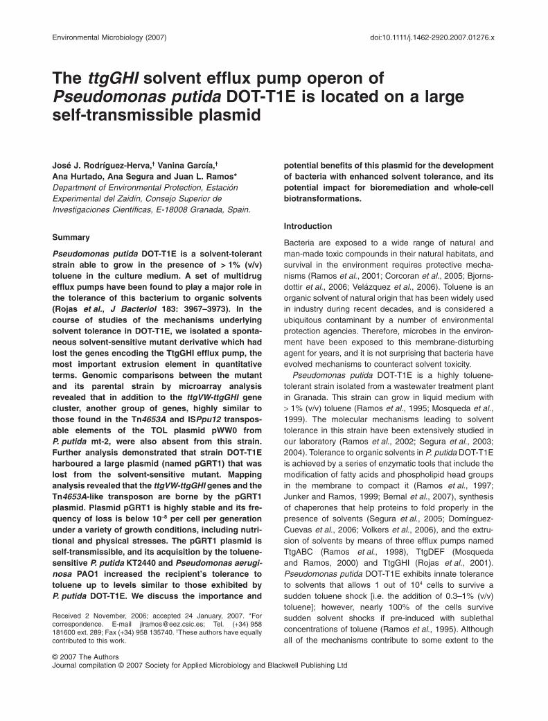
The ttgGHI solvent efflux pump operon ofPseudomonas putida DOT-T1E is located on a largeself-transmissible plasmid
José J. Rodríguez-Herva,† Vanina García,†
Ana Hurtado, Ana Segura and Juan L. Ramos*Department of Environmental Protection, EstaciónExperimental del Zaidín, Consejo Superior deInvestigaciones Científicas, E-18008 Granada, Spain.
Summary
Pseudomonas putida DOT-T1E is a solvent-tolerantstrain able to grow in the presence of > 1% (v/v)toluene in the culture medium. A set of multidrugefflux pumps have been found to play a major role inthe tolerance of this bacterium to organic solvents(Rojas et al., J Bacteriol 183: 3967–3973). In thecourse of studies of the mechanisms underlyingsolvent tolerance in DOT-T1E, we isolated a sponta-neous solvent-sensitive mutant derivative which hadlost the genes encoding the TtgGHI efflux pump, themost important extrusion element in quantitativeterms. Genomic comparisons between the mutantand its parental strain by microarray analysisrevealed that in addition to the ttgVW-ttgGHI genecluster, another group of genes, highly similar tothose found in the Tn4653A and ISPpu12 transpos-able elements of the TOL plasmid pWW0 fromP. putida mt-2, were also absent from this strain.Further analysis demonstrated that strain DOT-T1Eharboured a large plasmid (named pGRT1) that waslost from the solvent-sensitive mutant. Mappinganalysis revealed that the ttgVW-ttgGHI genes and theTn4653A-like transposon are borne by the pGRT1plasmid. Plasmid pGRT1 is highly stable and its fre-quency of loss is below 10-8 per cell per generationunder a variety of growth conditions, including nutri-tional and physical stresses. The pGRT1 plasmid isself-transmissible, and its acquisition by the toluene-sensitive P. putida KT2440 and Pseudomonas aerugi-nosa PAO1 increased the recipient’s tolerance totoluene up to levels similar to those exhibited byP. putida DOT-T1E. We discuss the importance and
potential benefits of this plasmid for the developmentof bacteria with enhanced solvent tolerance, and itspotential impact for bioremediation and whole-cellbiotransformations.
Introduction
Bacteria are exposed to a wide range of natural andman-made toxic compounds in their natural habitats, andsurvival in the environment requires protective mecha-nisms (Ramos et al., 2001; Corcoran et al., 2005; Bjorns-dottir et al., 2006; Velázquez et al., 2006). Toluene is anorganic solvent of natural origin that has been widely usedin industry during recent decades, and is considered aubiquitous contaminant by a number of environmentalprotection agencies. Therefore, microbes in the environ-ment have been exposed to this membrane-disturbingagent for years, and it is not surprising that bacteria haveevolved mechanisms to counteract solvent toxicity.
Pseudomonas putida DOT-T1E is a highly toluene-tolerant strain isolated from a wastewater treatment plantin Granada. This strain can grow in liquid medium with> 1% (v/v) toluene (Ramos et al., 1995; Mosqueda et al.,1999). The molecular mechanisms leading to solventtolerance in this strain have been extensively studied inour laboratory (Ramos et al., 2002; Segura et al., 2003;2004). Tolerance to organic solvents in P. putida DOT-T1Eis achieved by a series of enzymatic tools that include themodification of fatty acids and phospholipid head groupsin the membrane to compact it (Ramos et al., 1997;Junker and Ramos, 1999; Bernal et al., 2007), synthesisof chaperones that help proteins to fold properly in thepresence of solvents (Segura et al., 2005; Domínguez-Cuevas et al., 2006; Volkers et al., 2006), and the extru-sion of solvents by means of three efflux pumps namedTtgABC (Ramos et al., 1998), TtgDEF (Mosquedaand Ramos, 2000) and TtgGHI (Rojas et al., 2001).Pseudomonas putida DOT-T1E exhibits innate toleranceto solvents that allows 1 out of 104 cells to survive asudden toluene shock [i.e. the addition of 0.3–1% (v/v)toluene]; however, nearly 100% of the cells survivesudden solvent shocks if pre-induced with sublethalconcentrations of toluene (Ramos et al., 1995). Althoughall of the mechanisms contribute to some extent to the
Received 2 November, 2006; accepted 24 January, 2007. *Forcorrespondence. E-mail [email protected]; Tel. (+34) 958181600 ext. 289; Fax (+34) 958 135740. †These authors have equallycontributed to this work.
Environmental Microbiology (2007) doi:10.1111/j.1462-2920.2007.01276.x
© 2007 The AuthorsJournal compilation © 2007 Society for Applied Microbiology and Blackwell Publishing Ltd

solvent-tolerant phenotype of this strain, it has beenshown that the main determinants of solvent tolerance arethe efflux pumps.
Two of the efflux pump operons, ttgDEF and ttgGHI, areinduced by sublethal toluene concentrations (Mosquedaet al., 1999; Rojas et al., 2003), whereas the level ofexpression of ttgABC does not change in response tosolvents (Duque et al., 2001; Terán et al., 2006). TheTtgGHI efflux pump plays a pivotal role in innate andinduced tolerance to toluene, as shown by the finding thata mutant in this efflux pump was not able to survive theaddition of 0.3% (v/v) toluene even if bacteria were pre-cultured with sublethal toluene concentrations (Rojaset al., 2001). Furthermore, in a survey of differentP. putida strains, the presence of the ttgGHI-like geneswas detected only in highly solvent-tolerant strains, i.e.P. putida MTB6 (Huertas et al., 2000) and P. putida S12(Isken and de Bont, 1996), whereas the TtgDEF effluxpump was present only in those strains carrying the todpathway for toluene degradation (Phoenix et al., 2003;Segura et al., 2003). TtgABC seems to be widespreadamong the P. putida strains tested, and its main role is theextrusion of antimicrobial agents (Rojas et al., 2001;Terán et al., 2006).
In the course of our research, we found that a P. putidaDOT-T1E derivative clone spontaneously turned solventsensitive (Table 1). We first determined whether thesolvent-sensitive clone had all the three efflux pumps, anddiscovered that the clone had lost the ttgGHI genes, aphenomenon that was subsequently linked to the loss of aplasmid of about 121 kb, dubbed pGRT1, in which thettgGHI operon is located. Plasmid stability is high, andunder different growth and stress conditions its rate of
loss is below 10-8 per cell and per generation. The plasmidis self-transmissible and constitutes a valuable tool forbiotechnological processes.
Results
Genomic characterization of a P. putida DOT-T1Ederivative that is toluene sensitive
Pseudomonas putida strain DOT-T1E-100 is a serendipi-tous toluene-sensitive derivative of P. putida DOT-T1Eisolated from a culture in M9 minimal medium plus toluenein the gas phase that was stored at 4°C for several weeks.This mutant, in contrast to its parental strain, was unableto grow in Luria–Bertani (LB) medium supplemented with0.3% (v/v) toluene. As efflux pumps are key for solventtolerance, we decided to check whether the genes thatencode these pumps were still present in the DOT-T1E-100 strain, using Southern hybridization analysis of totalDNA of the mutant strain and the ttgABC, ttgDEF andttgGHI genes as probes. We found that whereas thettgABC and ttgDEF genes gave positive hybridizationsignals, the ttgGHI operon was no longer present in themutant strain (not shown).
To further confirm this observation and to define moreprecisely the extent of the DNA region hypotheticallydeleted in DOT-T1E-100, we took advantage of the avail-ability of a genome-wide microarray from the closelyrelated strain P. putida KT2440. Total DNA preparationsfrom P. putida DOT-T1E-100 and its parental strain DOT-T1E were fluorescently labelled with Cy3 and Cy5 dyesand analysed by comparative genomic hybridization asdescribed in Experimental procedures. The results, sum-
Table 1. Bacterial strains used in this study.
Pseudomonas putida strains Relevant characteristics References
KT2440 (pWW0) TolS; ATCC 33015 Williams and Murray (1974)KT2440 mt-2 pWW0 cured, TolS Nakazawa (2002)KT2442 KT2440 RifR Franklin et al. (1981)KT2440crc KT2440crc::aacC1, GmR Aranda-Olmedo et al. (2005)DOT-T1E TolR, RifR Ramos et al. (1998)DOT-T1E PS62 DOT-T1E ttgV::aphA3, KmR Rojas et al. (2003)DOT-T1E PS51 DOT-T1E ttgW::WKm, KmR Rojas et al. (2003)MTB5 TolS Huertas et al. (2000)MTB6 TolR Huertas et al. (2000)DOT-T1E-100 Spontaneous TolS derivative of DOT-T1E, RifR This studyVAN1 through to VAN4 Spontaneous TolS derivative of DOT-T1E-PS51, RifR, KmS This studyVAN5 and VAN6 Spontaneous TolS derivative of DOT-T1E-PS51, RifR, KmS
(carbon starvation assay)This study
VAN7 and VAN8 Spontaneous TolS derivative of DOT-T1E-PS51, RifR, KmS
(growth under sublethal tetracycline concentrations)This study
DOT-T1E-109 DOT-T1E fadD::Tn5phoA, TolS, RifR, KmR Segura et al. (2004)DOT-T1E-16 DOT-T1E ttgX::Km, TolS, RifR, KmR E. Duque and J.L. Ramos (unpublished)Pseudomonas aeruginosa PAO1 Prototroph Strain provided as a gift
GmR, KmR, RifR, resistance to gentamicin, kanamycin and rifampicin respectively. KmS stands for sensitivity to kanamycin (50 mg ml-1). TolR and TolS
stand for tolerance and sensitivity, respectively, to 0.3% (v/v) toluene in liquid medium.
2 J. J. Rodríguez-Herva et al.
© 2007 The AuthorsJournal compilation © 2007 Society for Applied Microbiology and Blackwell Publishing Ltd, Environmental Microbiology
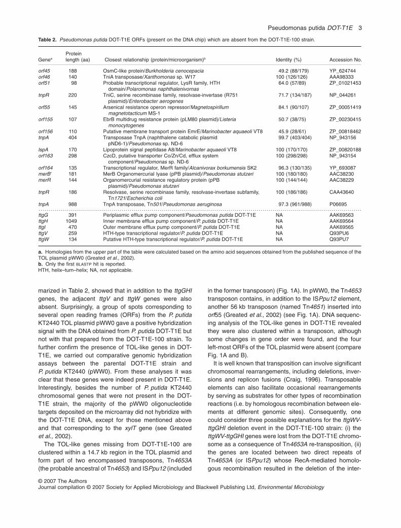
marized in Table 2, showed that in addition to the ttgGHIgenes, the adjacent ttgV and ttgW genes were alsoabsent. Surprisingly, a group of spots corresponding toseveral open reading frames (ORFs) from the P. putidaKT2440 TOL plasmid pWW0 gave a positive hybridizationsignal with the DNA obtained from P. putida DOT-T1E butnot with that prepared from the DOT-T1E-100 strain. Tofurther confirm the presence of TOL-like genes in DOT-T1E, we carried out comparative genomic hybridizationassays between the parental DOT-T1E strain andP. putida KT2440 (pWW0). From these analyses it wasclear that these genes were indeed present in DOT-T1E.Interestingly, besides the number of P. putida KT2440chromosomal genes that were not present in the DOT-T1E strain, the majority of the pWW0 oligonucleotidetargets deposited on the microarray did not hybridize withthe DOT-T1E DNA, except for those mentioned aboveand that corresponding to the xylT gene (see Greatedet al., 2002).
The TOL-like genes missing from DOT-T1E-100 areclustered within a 14.7 kb region in the TOL plasmid andform part of two encompassed transposons, Tn4653A(the probable ancestral of Tn4653) and ISPpu12 (included
in the former transposon) (Fig. 1A). In pWW0, the Tn4653transposon contains, in addition to the ISPpu12 element,another 56 kb transposon (named Tn4651) inserted intoorf55 (Greated et al., 2002) (see Fig. 1A). DNA sequenc-ing analysis of the TOL-like genes in DOT-T1E revealedthey were also clustered within a transposon, althoughsome changes in gene order were found, and the fourleft-most ORFs of the TOL plasmid were absent (compareFig. 1A and B).
It is well known that transposition can involve significantchromosomal rearrangements, including deletions, inver-sions and replicon fusions (Craig, 1996). Transposableelements can also facilitate occasional rearrangementsby serving as substrates for other types of recombinationreactions (i.e. by homologous recombination between ele-ments at different genomic sites). Consequently, onecould consider three possible explanations for the ttgWV-ttgGHI deletion event in the DOT-T1E-100 strain: (i) thettgWV-ttgGHI genes were lost from the DOT-T1E chromo-some as a consequence of Tn4653A re-transposition, (ii)the genes are located between two direct repeats ofTn4653A (or ISPpu12) whose RecA-mediated homolo-gous recombination resulted in the deletion of the inter-
Table 2. Pseudomonas putida DOT-T1E ORFs (present on the DNA chip) which are absent from the DOT-T1E-100 strain.
GeneaProteinlength (aa) Closest relationship (protein/microorganism)b Identity (%) Accession No.
orf45 188 OsmC-like protein/Burkholderia cenocepacia 49.2 (88/179) YP_624744orf46 140 TniA transposase/Xanthomonas sp. W17 100 (126/126) AAA98333orf51 98 Probable transcriptional regulator, LysR family, HTH
domain/Polaromonas naphthalenivornas64.0 (57/89) ZP_01021453
tnpR 220 TniC, serine recombinase family, resolvase-invertase (R751plasmid)/Enterobacter aerogenes
71.7 (134/187) NP_044261
orf55 145 Arsenical resistance operon repressor/Magnetospirillummagnetotacticum MS-1
84.1 (90/107) ZP_00051419
orf155 107 EbrB multidrug resistance protein (pLM80 plasmid)/Listeriamonocytogenes
50.7 (38/75) ZP_00230415
orf156 110 Putative membrane transport protein EmrE/Marinobacter aquaeoli VT8 45.9 (28/61) ZP_00818462tnpA 404 Transposase TnpA (naphthalene catabolic plasmid
pND6-1)/Pseudomonas sp. ND-699.7 (403/404) NP_943156
lspA 170 Lipoprotein signal peptidase A8/Marinobacter aquaeoli VT8 100 (170/170) ZP_00820188orf163 298 CzcD, putative transporter Co/Zn/Cd, efflux system
component/Pseudomonas sp. ND-6100 (298/298) NP_943154
orf164 135 Transcriptional regulator, MerR family/Alcanivorax borkumensis SK2 96.3 (130/135) YP_693087merB 181 MerB Organomercurial lyase (pPB plasmid)/Pseudomonas stutzeri 100 (180/180) AAC38230merR 144 Organomercurial resistance regulatory protein (pPB
plasmid)/Pseudomonas stutzeri100 (144/144) AAC38229
tnpR 186 Resolvase, serine recombinase family, resolvase-invertase subfamily,Tn1721/Escherichia coli
100 (186/186) CAA43640
tnpA 988 TnpA transposase, Tn501/Pseudomonas aeruginosa 97.3 (961/988) P06695. . . . . . . . . . . . . . . . . . . . . . . . . . . . . . . . . . . . . . . . . . . . . . . . . . . . . . . . . . . . . . . . . . . . . . . . . . . . . . . . . . . . . . . . . . . . . . . . . . . . . . . . . . . . . . . . . . . . . . . . . . . . . . . . . . . . . . . . . . . . . . . . . . . . . . . . . . . . . . . .ttgG 391 Periplasmic efflux pump component/Pseudomonas putida DOT-T1E NA AAK69563ttgH 1049 Inner membrane efflux pump component/P. putida DOT-T1E NA AAK69564ttgI 470 Outer membrane efflux pump component/P. putida DOT-T1E NA AAK69565ttgV 259 HTH-type transcriptional regulator/P. putida DOT-T1E NA Q93PU6ttgW 134 Putative HTH-type transcriptional regulator/P. putida DOT-T1E NA Q93PU7
a. Homologies from the upper part of the table were calculated based on the amino acid sequences obtained from the published sequence of theTOL plasmid pWW0 (Greated et al., 2002).b. Only the first BLASTP hit is reported.HTH, helix–turn–helix; NA, not applicable.
Pseudomonas putida DOT-T1E 3
© 2007 The AuthorsJournal compilation © 2007 Society for Applied Microbiology and Blackwell Publishing Ltd, Environmental Microbiology

vening DNA region, or (iii) the ttgWV-ttgGHI genes arelocated on an as-yet-unidentified plasmid (which sharesthe presence of Tn4653A and ISPpu12 with pWW0) thatis lost from DOT-T1E-100 under certain environmentalconditions. The first hypothesis can be ruled out, asTn4653A belongs to the Tn3 family (class II) of transpos-able elements, which transpose via a replicativemechanism. These elements can yield chromosomaldeletions by means of replicative intramolecular transpo-sition, but this always leads to the appearance of asecond transposon copy (Berg and Berg, 1996), whichwas not the case for DOT-T1E-100 according to themicroarray results. Regarding the second hypothesis,homologous recombination between direct DNA repeatswould leave a transposon copy in the chromosome,which, again, did not fit the deduced DOT-T1E-100genotype. We therefore decided to test the last hypoth-esis: that the ttgWV-ttgGHI cluster is located in a largeplasmid which has not previously been identified in DOT-T1E.
Pseudomonas putida DOT-T1E carries a low-copylarge plasmid
The presence of extrachromosomal genetic elements inP. putida DOT-T1E has never been convincingly observed
in DNA preparations of this strain (E. Duque, pers.comm.). To detect the possible presence of a high-molecular-weight plasmid in DOT-T1E, we adapted theplasmid isolation procedure developed by Zhou and col-leagues (1990) for the purification of large plasmids (seeExperimental procedures). Using this protocol, we wereable to detect and purify a high-molecular-weight plasmid(named pGRT1) from the parental P. putida DOT-T1E, butnot from strain DOT-T1E-100 (Fig. 2A). On the basis ofthe electrophoretic mobilities of the ScaI, EcoRV and XhoIfragments of pGRT1 (data not shown), the size of theplasmid was estimated to be about 121 5 kb (notshown).
To further confirm the plasmid coding of the TtgGHIefflux pump and the Tn4653 transposon, we performedSouthern blot analysis of pGRT1 plasmid preparationsdigested with ScaI or XhoI, using the P. putida DOT-T1EttgH gene (Fig. 3A) or the amplified lspA-orf156 DNAregion (Fig. 3B) as probes. Both probes gave positivehybridization signals, demonstrating that the genes men-tioned above were indeed carried on the pGRT1 plasmid.No positive hybridization was observed when total DNAfrom strain DOT-T1E-100 was used as the target (data notshown).
Segura and colleagues (2003) carried out a compara-tive genomic analysis of solvent extrusion pumps in
14,70 kb 8 9 10 11 12 13 147654321
ISPpu12
orf4
1
orf4
3
orf4
4
orf4
5
orf4
6or
f51
tnpR
orf5
5or
f155
orf1
56
tnpA
lspA
orf1
63
orf1
64m
erB
’m
erR
tnpR
tnpA
8 9 10 11 1276543210 kb
orf4
6
orf5
1
tnpR
orf5
5or
f155
orf1
56
tnpA
lspA
orf1
63
orf1
64m
erB
’m
erR
tnpR
tnpA
A
B
ScaI
ScaI
ScaI
ScaI
tnpA
´
Fig. 1. Physical organization of the Tn4653 region of pWW0 (A) and pGRT1 (B). Arrows represent the different genes, their relative sizes andthe directions of transcription. Gene nomenclature and organization are based on the previously annotated sequence of the TOL plasmidpWW0 from P. putida KT2440 (Greated et al., 2002). Stippled arrows correspond to genes putatively involved in transposition functions.Among these, the Tn4653A transposase and resolvase genes are shaded in grey. The 38 bp inverted repeats (IRs) of Tn4653A are shown ingrey boxes, and the 24 bp IRs of the ISPpu12 transposable element are shown in black boxes. The vertical arrow pointing to orf55 in (A)indicates the point at which the Tn4651 transposon is inserted in pWW0. The shaded box indicates the res region upstream of the tnpRresolvase gene of Tn4653, which is defective in pWW0 but not in pGRT1. The pGRT1 DNA sequence has been deposited in GenBank underAccession No. EF094543.
4 J. J. Rodríguez-Herva et al.
© 2007 The AuthorsJournal compilation © 2007 Society for Applied Microbiology and Blackwell Publishing Ltd, Environmental Microbiology
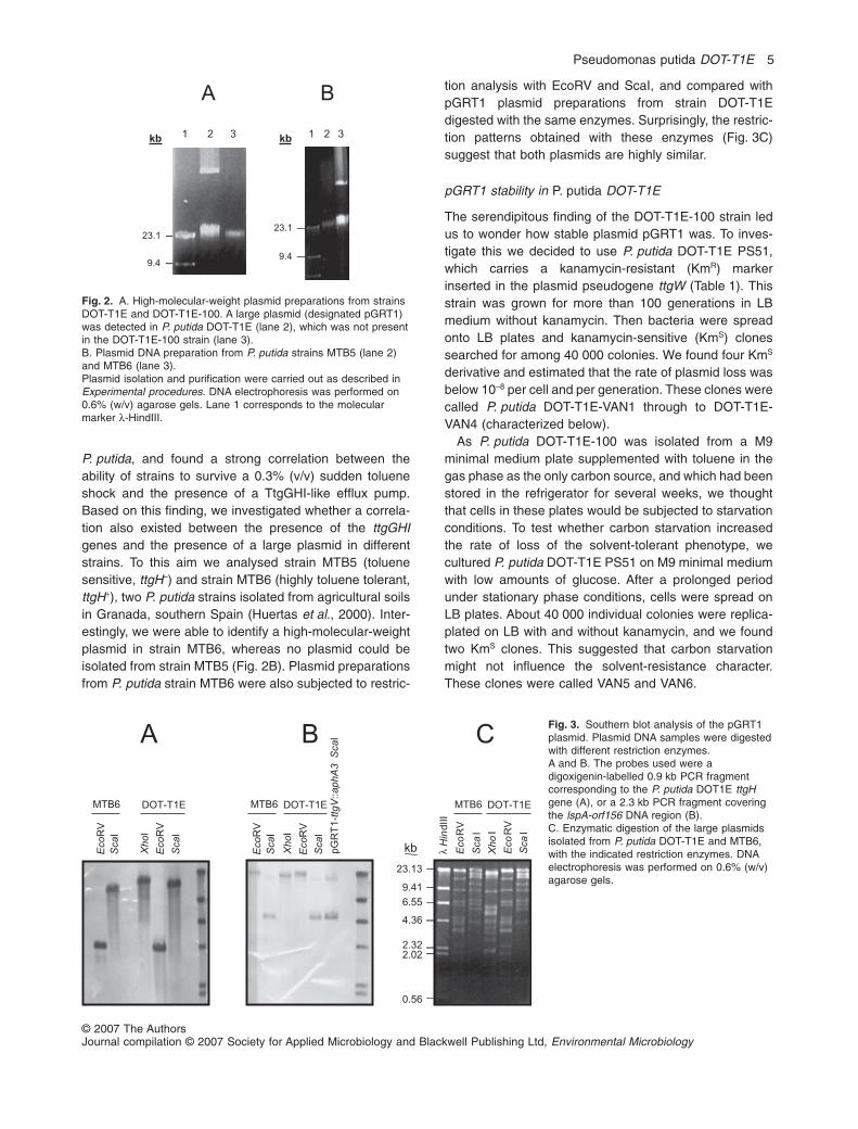
P. putida, and found a strong correlation between theability of strains to survive a 0.3% (v/v) sudden tolueneshock and the presence of a TtgGHI-like efflux pump.Based on this finding, we investigated whether a correla-tion also existed between the presence of the ttgGHIgenes and the presence of a large plasmid in differentstrains. To this aim we analysed strain MTB5 (toluenesensitive, ttgH–) and strain MTB6 (highly toluene tolerant,ttgH+), two P. putida strains isolated from agricultural soilsin Granada, southern Spain (Huertas et al., 2000). Inter-estingly, we were able to identify a high-molecular-weightplasmid in strain MTB6, whereas no plasmid could beisolated from strain MTB5 (Fig. 2B). Plasmid preparationsfrom P. putida strain MTB6 were also subjected to restric-
tion analysis with EcoRV and ScaI, and compared withpGRT1 plasmid preparations from strain DOT-T1Edigested with the same enzymes. Surprisingly, the restric-tion patterns obtained with these enzymes (Fig. 3C)suggest that both plasmids are highly similar.
pGRT1 stability in P. putida DOT-T1E
The serendipitous finding of the DOT-T1E-100 strain ledus to wonder how stable plasmid pGRT1 was. To inves-tigate this we decided to use P. putida DOT-T1E PS51,which carries a kanamycin-resistant (KmR) markerinserted in the plasmid pseudogene ttgW (Table 1). Thisstrain was grown for more than 100 generations in LBmedium without kanamycin. Then bacteria were spreadonto LB plates and kanamycin-sensitive (KmS) clonessearched for among 40 000 colonies. We found four KmS
derivative and estimated that the rate of plasmid loss wasbelow 10-8 per cell and per generation. These clones werecalled P. putida DOT-T1E-VAN1 through to DOT-T1E-VAN4 (characterized below).
As P. putida DOT-T1E-100 was isolated from a M9minimal medium plate supplemented with toluene in thegas phase as the only carbon source, and which had beenstored in the refrigerator for several weeks, we thoughtthat cells in these plates would be subjected to starvationconditions. To test whether carbon starvation increasedthe rate of loss of the solvent-tolerant phenotype, wecultured P. putida DOT-T1E PS51 on M9 minimal mediumwith low amounts of glucose. After a prolonged periodunder stationary phase conditions, cells were spread onLB plates. About 40 000 individual colonies were replica-plated on LB with and without kanamycin, and we foundtwo KmS clones. This suggested that carbon starvationmight not influence the solvent-resistance character.These clones were called VAN5 and VAN6.
A B
kb
23.1
9.4
1 32kb 1 32
23.1
9.4
Fig. 2. A. High-molecular-weight plasmid preparations from strainsDOT-T1E and DOT-T1E-100. A large plasmid (designated pGRT1)was detected in P. putida DOT-T1E (lane 2), which was not presentin the DOT-T1E-100 strain (lane 3).B. Plasmid DNA preparation from P. putida strains MTB5 (lane 2)and MTB6 (lane 3).Plasmid isolation and purification were carried out as described inExperimental procedures. DNA electrophoresis was performed on0.6% (w/v) agarose gels. Lane 1 corresponds to the molecularmarker l-HindIII.
Fig. 3. Southern blot analysis of the pGRT1plasmid. Plasmid DNA samples were digestedwith different restriction enzymes.A and B. The probes used were adigoxigenin-labelled 0.9 kb PCR fragmentcorresponding to the P. putida DOT1E ttgHgene (A), or a 2.3 kb PCR fragment coveringthe lspA-orf156 DNA region (B).C. Enzymatic digestion of the large plasmidsisolated from P. putida DOT-T1E and MTB6,with the indicated restriction enzymes. DNAelectrophoresis was performed on 0.6% (w/v)agarose gels.
Eco
RV
pG
RT
1-t
tgV
::ap
hA3
Sca
IB
Sca
I
Eco
RV
Sca
I
Eco
RV
Sca
I
Xho
I
DOT-T1E
kb
4.36
23.13
9.41
6.55
2.32
λH
indIII
2.02
0.56
MTB6
Eco
RV
Sca
I
Xho
I
MTB6 DOT-T1E
Eco
RV
Sca
I
MTB6
Eco
RV
Sca
I
Xho
I
DOT-T1E
CA
Pseudomonas putida DOT-T1E 5
© 2007 The AuthorsJournal compilation © 2007 Society for Applied Microbiology and Blackwell Publishing Ltd, Environmental Microbiology
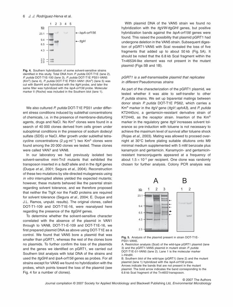
We also cultured P. putida DOT-T1E PS51 under differ-ent stress conditions induced by sublethal concentrationsof chemicals, i.e. in the presence of membrane-disturbingagents, drugs and NaCl. No KmS clones were found in asearch of 40 000 clones derived from cells grown undersuboptimal conditions in the presence of sodium dodecylsulfate (SDS) or NaCl. After growth under sublethal tetra-cycline concentrations (2 mg ml-1) two KmS clones werefound among the 20 000 clones we tested. These cloneswere called VAN7 and VAN8.
In our laboratory we had previously isolated twosolvent-sensitive mini-Tn5 mutants that exhibited thetransposon inserted in a fadD allele and in the ttgX genes(Duque et al., 2001; Segura et al., 2004). Reconstructionof these two mutations by site-directed mutagenesis usingin vitro interrupted alleles yielded the expected mutants;however, these mutants behaved like the parental strainregarding solvent tolerance, and we therefore proposedthat neither the TtgX nor the FadD proteins are requiredfor solvent tolerance (Segura et al., 2004; E. Duque andJ.L. Ramos, unpubl. results). The original clones, calledDOT-T1-109 and DOT-T1E-16, were reanalysed hereregarding the presence of the ttgGHI genes.
To determine whether the solvent-sensitive charactercorrelated with the absence of the plasmid in VAN1through to VAN8, DOT-T1-E-109 and DOT-T1E-16, wefirst prepared plasmid DNA as above using DOT-T1E as acontrol. We found that VAN5 bore a plasmid that wassmaller than pGRT1, whereas the rest of the clones boreno plasmids. To further confirm the loss of the plasmidsand the genes we identified on pGRT1, we carried outSouthern blot analysis with total DNA of the strains andused the ttgGHI and lpsA-orf156 genes as probes. For allstrains except for VAN5 we found no hybridization with theprobes, which points toward the loss of the plasmid (seeFig. 4 for a number of clones).
With plasmid DNA of the VAN5 strain we found nohybridization with the ttgVW-ttgGHI genes, but positivehybridization bands against the lspA-orf156 genes werefound. This raised the possibility that plasmid pGRT1 hadundergone deletion in the VAN5 strain. Subsequent diges-tion of pGRT1-VAN5 with ScaI revealed the loss of fivefragments that added up to about 50 kb (Fig. 5A). Itshould be noted that the 6.8 kb ScaI fragment within theTn4653A-like element was not present in the mutantplasmid (Figs 5B and 1B).
pGRT1 is a self-transmissible plasmid that replicatesin different Pseudomonas strains
As part of the characterization of the pGRT1 plasmid, wetested whether it was able to self-transfer to otherP. putida strains. We set up biparental matings betweendonor strain P. putida DOT-T1E PS62, which carries aKmR marker in the ttgV gene (ttgV::aphA3), and P. putidaKT2440crc, a gentamicin-resistant derivative strain ofKT2440, as the receptor strain. Insertion of the KmR
marker in the regulatory gene ttgV increases solvent tol-erance so pre-induction with toluene is not necessary toachieve the maximum level of survival after toluene shock(Rojas et al., 2003). Mating was allowed to proceed over-night at 30°C before plating suitable dilutions onto M9minimal medium supplemented with 5 mM benzoate pluskanamycin and gentamicin. Kanamycin- and gentamicin-resistant transconjugants appeared at a frequency ofabout 1.5 ¥ 10-5 per recipient. One clone was randomlychosen for further analysis. Colony PCR analysis was
ttgH
lspA-orf156
1 32 4 5
23.1
9.4
6.5
4.3
2.32.0
kb
Fig. 4. Southern hybridization of some solvent-sensitive strainsidentified in this study. Total DNA from P. putida DOT-T1E (lane 2),P. putida DOT-T1E-100 (lane 3), P. putida DOT-T1E PS51-VAN5(KmR) (lane 4), P. putida DOT-T1E PS51-VAN1 (KmR) (lane 5) wascut with BamHI and hybridized with the ttgH probe, and later thesame filter was hybridized with the lspA-orf156 probe. Molecularmarker II (Roche) was included in the Southern blot (lane 1).
1 2 3 1 2
23.1
9.4
6.5
4.3
2.3
A Bkb
Fig. 5. Analysis of the plasmid present in strain DOT-T1E-PS51-VAN5.A. Restriction analysis (ScaI) of the wild-type pGRT1 plasmid (lane3) and the pGRT1-VAN5 plasmid in mutant strain P. putidaDOT-T1E-51-VAN5 (lane 2). Lane 1 is the molecular markerl-HindIII.B. Southern blot of the wild-type (pGRT1) (lane 2) and the mutantplasmid (lane 1) hybridized with the lspA-orf156 probe.Arrows indicate the bands that are not present in the mutantplasmid. The bold arrow indicates the band corresponding to the6.8 kb ScaI fragment of the Tn4653 transposon.
6 J. J. Rodríguez-Herva et al.
© 2007 The AuthorsJournal compilation © 2007 Society for Applied Microbiology and Blackwell Publishing Ltd, Environmental Microbiology
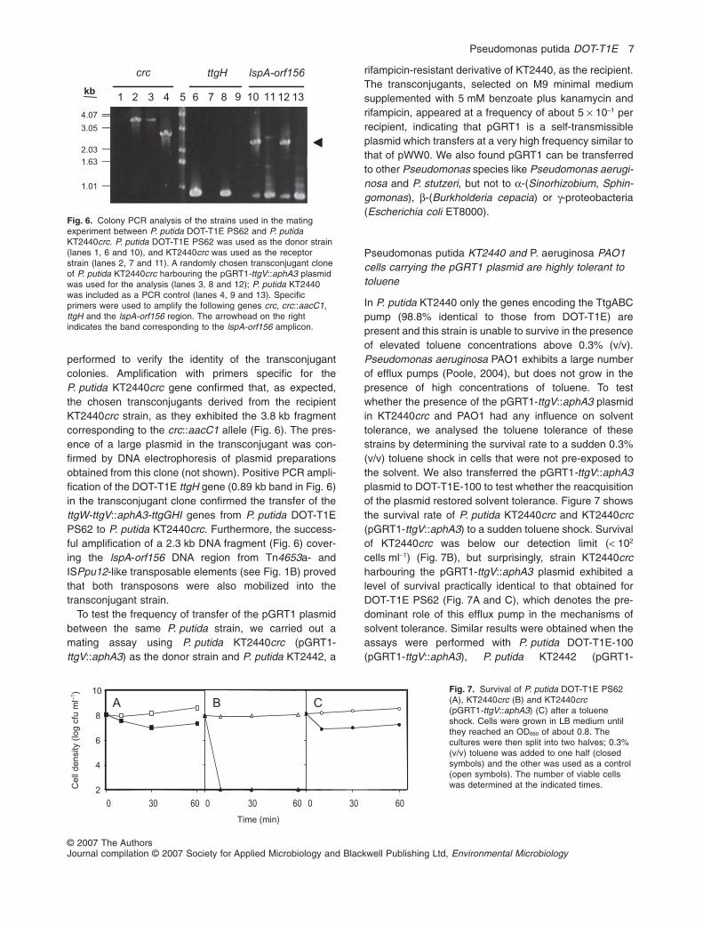
performed to verify the identity of the transconjugantcolonies. Amplification with primers specific for theP. putida KT2440crc gene confirmed that, as expected,the chosen transconjugants derived from the recipientKT2440crc strain, as they exhibited the 3.8 kb fragmentcorresponding to the crc::aacC1 allele (Fig. 6). The pres-ence of a large plasmid in the transconjugant was con-firmed by DNA electrophoresis of plasmid preparationsobtained from this clone (not shown). Positive PCR ampli-fication of the DOT-T1E ttgH gene (0.89 kb band in Fig. 6)in the transconjugant clone confirmed the transfer of thettgW-ttgV::aphA3-ttgGHI genes from P. putida DOT-T1EPS62 to P. putida KT2440crc. Furthermore, the success-ful amplification of a 2.3 kb DNA fragment (Fig. 6) cover-ing the lspA-orf156 DNA region from Tn4653a- andISPpu12-like transposable elements (see Fig. 1B) provedthat both transposons were also mobilized into thetransconjugant strain.
To test the frequency of transfer of the pGRT1 plasmidbetween the same P. putida strain, we carried out amating assay using P. putida KT2440crc (pGRT1-ttgV::aphA3) as the donor strain and P. putida KT2442, a
rifampicin-resistant derivative of KT2440, as the recipient.The transconjugants, selected on M9 minimal mediumsupplemented with 5 mM benzoate plus kanamycin andrifampicin, appeared at a frequency of about 5 ¥ 10-1 perrecipient, indicating that pGRT1 is a self-transmissibleplasmid which transfers at a very high frequency similar tothat of pWW0. We also found pGRT1 can be transferredto other Pseudomonas species like Pseudomonas aerugi-nosa and P. stutzeri, but not to a-(Sinorhizobium, Sphin-gomonas), b-(Burkholderia cepacia) or g-proteobacteria(Escherichia coli ET8000).
Pseudomonas putida KT2440 and P. aeruginosa PAO1cells carrying the pGRT1 plasmid are highly tolerant totoluene
In P. putida KT2440 only the genes encoding the TtgABCpump (98.8% identical to those from DOT-T1E) arepresent and this strain is unable to survive in the presenceof elevated toluene concentrations above 0.3% (v/v).Pseudomonas aeruginosa PAO1 exhibits a large numberof efflux pumps (Poole, 2004), but does not grow in thepresence of high concentrations of toluene. To testwhether the presence of the pGRT1-ttgV::aphA3 plasmidin KT2440crc and PAO1 had any influence on solventtolerance, we analysed the toluene tolerance of thesestrains by determining the survival rate to a sudden 0.3%(v/v) toluene shock in cells that were not pre-exposed tothe solvent. We also transferred the pGRT1-ttgV::aphA3plasmid to DOT-T1E-100 to test whether the reacquisitionof the plasmid restored solvent tolerance. Figure 7 showsthe survival rate of P. putida KT2440crc and KT2440crc(pGRT1-ttgV::aphA3) to a sudden toluene shock. Survivalof KT2440crc was below our detection limit (< 102
cells ml-1) (Fig. 7B), but surprisingly, strain KT2440crcharbouring the pGRT1-ttgV::aphA3 plasmid exhibited alevel of survival practically identical to that obtained forDOT-T1E PS62 (Fig. 7A and C), which denotes the pre-dominant role of this efflux pump in the mechanisms ofsolvent tolerance. Similar results were obtained when theassays were performed with P. putida DOT-T1E-100(pGRT1-ttgV::aphA3), P. putida KT2442 (pGRT1-
kb
1.63
4.07
3.05
2.03
1.01
1 2 3 4 5 6 7 8 9 1110 12 13
crc ttgH lspA-orf156
Fig. 6. Colony PCR analysis of the strains used in the matingexperiment between P. putida DOT-T1E PS62 and P. putidaKT2440crc. P. putida DOT-T1E PS62 was used as the donor strain(lanes 1, 6 and 10), and KT2440crc was used as the receptorstrain (lanes 2, 7 and 11). A randomly chosen transconjugant cloneof P. putida KT2440crc harbouring the pGRT1-ttgV::aphA3 plasmidwas used for the analysis (lanes 3, 8 and 12); P. putida KT2440was included as a PCR control (lanes 4, 9 and 13). Specificprimers were used to amplify the following genes crc, crc::aacC1,ttgH and the lspA-orf156 region. The arrowhead on the rightindicates the band corresponding to the lspA-orf156 amplicon.
Fig. 7. Survival of P. putida DOT-T1E PS62(A), KT2440crc (B) and KT2440crc(pGRT1-ttgV::aphA3) (C) after a tolueneshock. Cells were grown in LB medium untilthey reached an OD660 of about 0.8. Thecultures were then split into two halves; 0.3%(v/v) toluene was added to one half (closedsymbols) and the other was used as a control(open symbols). The number of viable cellswas determined at the indicated times.
2
4
6
8
10
Cell
density
(log
cfu
ml–
1)
Time (min)
A B C
30 30 30000 60 60 60
Pseudomonas putida DOT-T1E 7
© 2007 The AuthorsJournal compilation © 2007 Society for Applied Microbiology and Blackwell Publishing Ltd, Environmental Microbiology
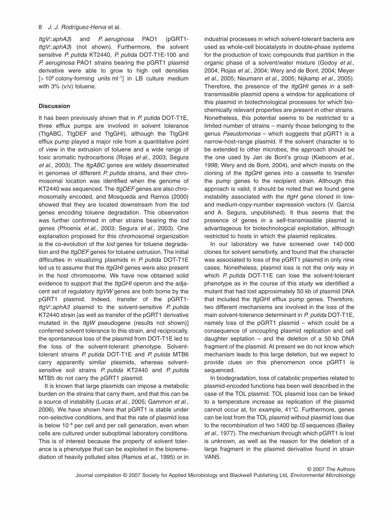
ttgV::aphA3) and P. aeruginosa PAO1 (pGRT1-ttgV::aphA3) (not shown). Furthermore, the solventsensitive P. putida KT2440, P. putida DOT-T1E-100 andP. aeruginosa PAO1 strains bearing the pGRT1 plasmidderivative were able to grow to high cell densities[> 108 colony-forming units ml-1] in LB culture mediumwith 3% (v/v) toluene.
Discussion
It has been previously shown that in P. putida DOT-T1E,three efflux pumps are involved in solvent tolerance(TtgABC, TtgDEF and TtgGHI), although the TtgGHIefflux pump played a major role from a quantitative pointof view in the extrusion of toluene and a wide range oftoxic aromatic hydrocarbons (Rojas et al., 2003; Seguraet al., 2003). The ttgABC genes are widely disseminatedin genomes of different P. putida strains, and their chro-mosomal location was identified when the genome ofKT2440 was sequenced. The ttgDEF genes are also chro-mosomally encoded, and Mosqueda and Ramos (2000)showed that they are located downstream from the todgenes encoding toluene degradation. This observationwas further confirmed in other strains bearing the todgenes (Phoenix et al., 2003; Segura et al., 2003). Oneexplanation proposed for this chromosomal organizationis the co-evolution of the tod genes for toluene degrada-tion and the ttgDEF genes for toluene extrusion. The initialdifficulties in visualizing plasmids in P. putida DOT-T1Eled us to assume that the ttgGHI genes were also presentin the host chromosome. We have now obtained solidevidence to support that the ttgGHI operon and the adja-cent set of regulatory ttgVW genes are both borne by thepGRT1 plasmid. Indeed, transfer of the pGRT1-ttgV::aphA3 plasmid to the solvent-sensitive P. putidaKT2440 strain [as well as transfer of the pGRT1 derivativemutated in the ttgW pseudogene (results not shown)]conferred solvent tolerance to this strain, and reciprocally,the spontaneous loss of the plasmid from DOT-T1E led tothe loss of the solvent-tolerant phenotype. Solvent-tolerant strains P. putida DOT-T1E and P. putida MTB6carry apparently similar plasmids, whereas solvent-sensitive soil strains P. putida KT2440 and P. putidaMTB5 do not carry the pGRT1 plasmid.
It is known that large plasmids can impose a metabolicburden on the strains that carry them, and that this can bea source of instability (Lucas et al., 2005; Gammon et al.,2006). We have shown here that pGRT1 is stable undernon-selective conditions, and that the rate of plasmid lossis below 10-8 per cell and per cell generation, even whencells are cultured under suboptimal laboratory conditions.This is of interest because the property of solvent toler-ance is a phenotype that can be exploited in the bioreme-diation of heavily polluted sites (Ramos et al., 1995) or in
industrial processes in which solvent-tolerant bacteria areused as whole-cell biocatalysts in double-phase systemsfor the production of toxic compounds that partition in theorganic phase of a solvent/water mixture (Godoy et al.,2004; Rojas et al., 2004; Wery and de Bont, 2004; Meyeret al., 2005; Neumann et al., 2005; Nijkamp et al., 2005).Therefore, the presence of the ttgGHI genes in a self-transmissible plasmid opens a window for applications ofthis plasmid in biotechnological processes for which bio-chemically relevant properties are present in other strains.Nonetheless, this potential seems to be restricted to alimited number of strains – mainly those belonging to thegenus Pseudomonas – which suggests that pGRT1 is anarrow-host-range plasmid. If the solvent character is tobe extended to other microbes, the approach should bethe one used by Jan de Bont’s group (Kieboom et al.,1998; Wery and de Bont, 2004), and which insists on thecloning of the ttgGHI genes into a cassette to transferthe pump genes to the recipient strain. Although thisapproach is valid, it should be noted that we found geneinstability associated with the ttgH gene cloned in low-and medium-copy-number expression vectors (V. Garcíaand A. Segura, unpublished). It thus seems that thepresence of genes in a self-transmissible plasmid isadvantageous for biotechnological exploitation, althoughrestricted to hosts in which the plasmid replicates.
In our laboratory we have screened over 140 000clones for solvent sensitivity, and found that the characterwas associated to loss of the pGRT1 plasmid in only ninecases. Nonetheless, plasmid loss is not the only way inwhich P. putida DOT-T1E can lose the solvent-tolerantphenotype as in the course of this study we identified amutant that had lost approximately 50 kb of plasmid DNAthat included the ttgGHI efflux pump genes. Therefore,two different mechanisms are involved in the loss of themain solvent-tolerance determinant in P. putida DOT-T1E,namely loss of the pGRT1 plasmid – which could be aconsequence of uncoupling plasmid replication and celldaughter septation – and the deletion of a 50 kb DNAfragment of the plasmid. At present we do not know whichmechanism leads to this large deletion, but we expect toprovide clues on this phenomenon once pGRT1 issequenced.
In biodegradation, loss of catabolic properties related toplasmid-encoded functions has been well described in thecase of the TOL plasmid. TOL plasmid loss can be linkedto a temperature increase as replication of the plasmidcannot occur at, for example, 41°C. Furthermore, genescan be lost from the TOL plasmid without plasmid loss dueto the recombination of two 1400 bp IS sequences (Baileyet al., 1977). The mechanism through which pGRT1 is lostis unknown, as well as the reason for the deletion of alarge fragment in the plasmid derivative found in strainVAN5.
8 J. J. Rodríguez-Herva et al.
© 2007 The AuthorsJournal compilation © 2007 Society for Applied Microbiology and Blackwell Publishing Ltd, Environmental Microbiology

In summary, our experiments showed that the ttgGHIgenes are borne on a self-transmissible narrow-host-range pGRT1 plasmid. This plasmid is highly stable andits frequency of loss is very low, making it a potentiallysuitable biotechnological tool to enhance solvent toler-ance in other microbes.
Experimental procedures
Bacterial strains, culture media and growth conditions
The P. putida strains used in this study are listed in Table 1.Bacteria were grown routinely at 30°C in liquid LB medium(Sambrook et al., 1989) or in M9 minimal medium supple-mented with benzoate (5 mM) or glucose (10 mM) as the solecarbon source (Abril et al., 1989), on an orbital platformoperating at 200 strokes per min. When required, antibioticswere added at the following final concentrations (mg ml-1):gentamicin, 25; kanamycin, 50; and rifampicin, 20.
Standard DNA techniques
Standard molecular biology techniques were used for DNAmanipulations (Sambrook et al., 1989). Southern blot analy-ses were performed as previously described (Ausubel et al.,2006). DNA probes were labelled by PCR with digoxigenin-11-dUTP (Roche, Cat. No. 11573179910) as recommendedby the manufacturer. The sequences of the primer pairs usedto generate the hybridization probes will be provided onrequest. In cases where two probes were used to hybridizethe same Southern blot, hybridizations were performedsequentially; the filters were boiled in distilled water betweenhybridizations.
For total DNA isolation, 0.5 ml aliquots of overnight cultureswere harvested by centrifugation and re-suspended in 400 mlof buffer A [25 mM Tris-HCl, pH 8.0; 25 mM EDTA; 20% (w/v)sucrose; 100 mg ml-1 RNase A]. Cells were lysed by adding200 ml of a solution containing 2% (w/v) SDS and 0.3 NNaOH. After incubation for 10 min on ice, MgCl2 was added toa final concentration of 10 mM, and the lysate was extractedtwice with phenol–chloroform–isoamyl alcohol (25:24:1) andethanol precipitated with ammonium acetate 0.3 M, pH 4.8.After incubation for 20 min at -20°C, the precipitated DNAwas collected by centrifugation (16 600 g, 15 min, 4°C),washed with 70% (v/v) ethanol, dried and dissolved in 50 ml ofdistilled water.
Mating procedure
Biparental matings between Pseudomonas strains were per-formed as described before (Ramos-González et al., 1991).For the conjugal transfer of pGRT1-ttgV::aphA3 fromP. putida KT2440crc (pGRT1-ttgV::aphA3) to P. aeruginosaPAO1, the receptor strain was grown on solid LB medium at43°C for 2 days prior to mating, to transiently overcome thehost restriction/modification system (Rolfe and Holloway,1966). PAO1 (pGRT1-ttgV::aphA3) transconjugants wereselected by growth on LB plates containing kanamycin(100 mg ml-1), at 42°C.
Plasmid DNA isolation
Isolation of large plasmid DNA was performed according tothe alkaline lysis procedure of Zhou and colleagues (1990)with several modifications. Briefly, 0.5 ml of an overnight bac-terial culture was collected by centrifugation. Most of thesupernatant was decanted and the pellet was thoroughlyre-suspended in the remaining fluid and lysed for 5 min on icewith 300 ml of TENS [10 mM Tris-HCl, pH 8.0; 1 mM EDTA;0.1 N NaOH; 10% (w/v) SDS] and RNase A (100 mg ml-1).Samples were neutralized with 3 M sodium acetate pH 5.2(150 ml), and cell debris and chromosomal DNA were pelletedby centrifugation at 4°C (16 600 g, 10 min). Proteins werethen removed from the resulting supernatant fraction by suc-cessive extractions with phenol–chloroform–isoamyl alcohol(25:24:1) and chloroform–isoamyl alcohol (24:1). PlasmidDNA was ethanol-precipitated (for 30 min at -20°C), concen-trated by centrifugation, washed with 70% (v/v) ethanol andre-suspended in 15 ml of distilled water. Large plasmids wereresolved by electrophoresis on 0.6% (w/v) agarose gels in aTris-acetate buffer system (Sambrook et al., 1989).
Genomic DNA microarrays
The genome-wide DNA chip used in this work (printed byProgenika Biopharma; http://www.progenika.com) has beendescribed in detail previously (Yuste et al., 2006). It consistsof an array of 5539 oligonucleotides (50-mer) spotted in dupli-cate onto g-amino silane-treated slides, and covalently linkedwith UV light and heat. The oligonucleotides represent 5350of the 5421 predicted ORFs annotated in the P. putidaKT2440 genome (Nelson et al., 2002; http://cmr.tigr.org/tigr-scripts/CMR/CmrHomePage.cgi). In addition, 140 of the 148putative ORFs predicted for the TOL plasmid pWW0 are alsoincluded, together with a suite of 49 commonly used reportergenes and antibiotic resistance markers. The chips also havehomogeneity controls consisting of oligonucleotides for therpoD and rpoN genes spotted at 20 different positions, as wellas duplicate negative controls at 203 pre-defined positions.
To prepare fluorescently labelled DNA, 2 mg of total DNAwas primed with 7.5 mg of pd(N)6 random hexamers (Amer-sham, Cat. No. 27-2166-01). Probe synthesis was performedat 37°C for 3 h in a 30 ml reaction volume containing 0.5 mM(each) dATP, dCTP and dGTP; 0.25 mM (each) dTTP andamino allyl-dUTP (aa-dUTP; Sigma, Cat. No. A0410); 1¥Klenow buffer and 50 U Klenow fragment (New EnglandBiolabs, Madrid, Spain). The reaction was stopped by adding3 ml of 0.5 M EDTA, and the DNA was purified by using aQIAquick PCR purification column (Qiagen, Germany) andevaporated in a Speed-Vac. The amino-modified cDNA waslabelled with Cy3 or Cy5 fluorescent dyes (monoreactiveNHS-esters; Amersham Biosciences, Cat No. PA23001 andPA25001 respectively) according to the manufacturer’sinstructions, and purified as described above. Labelling effi-ciency was assessed with a NanoDrop ND1000 spectropho-tometer (NanoDrop Technologies). Equal amounts of Cy3-and Cy5-labelled cDNAs, one of them corresponding to thecontrol and the other to the problem sample, were mixed,dried in a Speed-Vac and reconstituted in 35 ml of hybridiza-tion buffer [5¥ SSC, 25% (v/v) formamide, 0.5% (w/v) SDS,5¥ Denhardt’s solution, 5% (w/v) dextransulfate] pre-heatedto 42°C.
Pseudomonas putida DOT-T1E 9
© 2007 The AuthorsJournal compilation © 2007 Society for Applied Microbiology and Blackwell Publishing Ltd, Environmental Microbiology
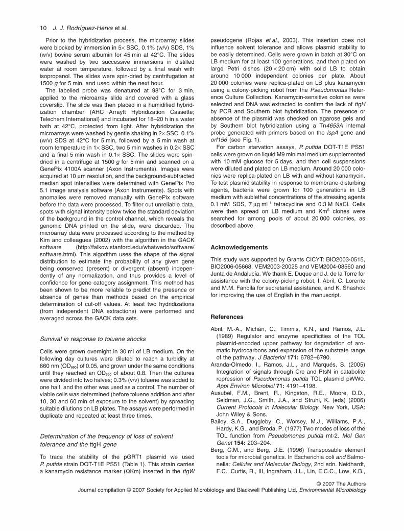
Prior to the hybridization process, the microarray slideswere blocked by immersion in 5¥ SSC, 0.1% (w/v) SDS, 1%(w/v) bovine serum albumin for 45 min at 42°C. The slideswere washed by two successive immersions in distilledwater at room temperature, followed by a final wash withisopropanol. The slides were spin-dried by centrifugation at1500 g for 5 min, and used within the next hour.
The labelled probe was denatured at 98°C for 3 min,applied to the microarray slide and covered with a glasscoverslip. The slide was then placed in a humidified hybrid-ization chamber (AHC ArrayIt Hybridization Cassette;Telechem International) and incubated for 18–20 h in a waterbath at 42°C, protected from light. After hybridization themicroarrays were washed by gentle shaking in 2¥ SSC, 0.1%(w/v) SDS at 42°C for 5 min, followed by a 5 min wash atroom temperature in 1¥ SSC, two 5 min washes in 0.2¥ SSCand a final 5 min wash in 0.1¥ SSC. The slides were spin-dried in a centrifuge at 1500 g for 5 min and scanned on aGenePix 4100A scanner (Axon Instruments). Images wereacquired at 10 mm resolution, and the background-subtractedmedian spot intensities were determined with GenePix Pro5.1 image analysis software (Axon Instruments). Spots withanomalies were removed manually with GenePix softwarebefore the data were processed. To filter out unreliable data,spots with signal intensity below twice the standard deviationof the background in the control channel, which reveals thegenomic DNA printed on the slide, were discarded. Themicroarray data were processed according to the method byKim and colleagues (2002) with the algorithm in the GACKsoftware (http://falkow.stanford.edu/whatwedo/software/software.html). This algorithm uses the shape of the signaldistribution to estimate the probability of any given genebeing conserved (present) or divergent (absent) indepen-dently of any normalization, and thus provides a level ofconfidence for gene category assignment. This method hasbeen shown to be more reliable to predict the presence orabsence of genes than methods based on the empiricaldetermination of cut-off values. At least two hydridizations(from independent DNA extractions) were performed andaveraged across the GACK data sets.
Survival in response to toluene shocks
Cells were grown overnight in 30 ml of LB medium. On thefollowing day cultures were diluted to reach a turbidity at660 nm (OD660) of 0.05, and grown under the same conditionsuntil they reached an OD660 of about 0.8. Then the cultureswere divided into two halves; 0.3% (v/v) toluene was added toone half, and the other was used as a control. The number ofviable cells was determined (before toluene addition and after10, 30 and 60 min of exposure to the solvent) by spreadingsuitable dilutions on LB plates. The assays were performed induplicate and repeated at least three times.
Determination of the frequency of loss of solventtolerance and the ttgH gene
To trace the stability of the pGRT1 plasmid we usedP. putida strain DOT-T1E PS51 (Table 1). This strain carriesa kanamycin resistance marker (WKm) inserted in the ttgW
pseudogene (Rojas et al., 2003). This insertion does notinfluence solvent tolerance and allows plasmid stability tobe easily determined. Cells were grown in batch at 30°C onLB medium for at least 100 generations, and then plated onlarge Petri dishes (20 ¥ 20 cm) with solid LB to obtainaround 10 000 independent colonies per plate. About20 000 colonies were replica-plated on LB plus kanamycinusing a colony-picking robot from the Pseudomonas Refer-ence Culture Collection. Kanamycin-sensitive colonies wereselected and DNA was extracted to confirm the lack of ttgHby PCR and Southern blot hybridization. The presence orabsence of the plasmid was checked on agarose gels andby Southern blot hybridization using a Tn4653A internalprobe generated with primers based on the lspA gene andorf156 (see Fig. 1).
For carbon starvation assays, P. putida DOT-T1E PS51cells were grown on liquid M9 minimal medium supplementedwith 10 mM glucose for 5 days, and then cell suspensionswere diluted and plated on LB medium. Around 20 000 colo-nies were replica-plated on LB with and without kanamycin.To test plasmid stability in response to membrane-disturbingagents, bacteria were grown for 100 generations in LBmedium with sublethal concentrations of the stressing agents0.1 mM SDS, 7 mg ml-1 tetracycline and 0.3 M NaCl. Cellswere then spread on LB medium and KmS clones weresearched for among pools of about 20 000 colonies, asdescribed above.
Acknowledgements
This study was supported by Grants CICYT: BIO2003-0515,BIO2006-05668, VEM2003-20025 and VEM2004-08560 andJunta de Andalucía. We thank E. Duque and J. de la Torre forassistance with the colony-picking robot, I. Abril, C. Lorenteand M.M. Fandila for secretarial assistance, and K. Shashokfor improving the use of English in the manuscript.
References
Abril, M.-A., Michán, C., Timmis, K.N., and Ramos, J.L.(1989) Regulator and enzyme specificities of the TOLplasmid-encoded upper pathway for degradation of aro-matic hydrocarbons and expansion of the substrate rangeof the pathway. J Bacteriol 171: 6782–6790.
Aranda-Olmedo, I., Ramos, J.L., and Marqués, S. (2005)Integration of signals through Crc and PtsN in cataboliterepression of Pseudomonas putida TOL plasmid pWW0.Appl Environ Microbiol 71: 4191–4198.
Ausubel, F.M., Brent, R., Kingston, R.E., Moore, D.D.,Seidman, J.G., Smith, J.A., and Struhl, K. (eds) (2006)Current Protocols in Molecular Biology. New York, USA:John Wiley & Sons.
Bailey, S.A., Duggleby, C., Worsey, M.J., Williams, P.A.,Hardy, K.G., and Broda, P. (1977) Two modes of loss of theTOL function from Pseudomonas putida mt-2. Mol GenGenet 154: 203–204.
Berg, C.M., and Berg, D.E. (1996) Transposable elementtools for microbial genetics. In Escherichia coli and Salmo-nella: Cellular and Molecular Biology, 2nd edn. Neidhardt,F.C., Curtis, R., III, Ingraham, J.L., Lin, E.C.C., Low, K.B.,
10 J. J. Rodríguez-Herva et al.
© 2007 The AuthorsJournal compilation © 2007 Society for Applied Microbiology and Blackwell Publishing Ltd, Environmental Microbiology

Magasanik, B., et al. (eds). Washington, DC, USA: ASMPress, pp. 2588–2612.
Bernal, P., Muñoz-Rojas, J., Hurtado, A., Ramos, J.L., andSegura, A. (2007) A Pseudomonas putida cardiolipin syn-thesis mutant exhibits increased sensitivity to drugs relatedto transport functionality. Environ Microbiol (in press). doi:10.1111/j.1462-2920.2006.01236.x.
Bjornsdottir, K., Breidt, F., Jr, and McFeeters, R.F. (2006)Protective effects of organic acids on survival of Escheri-chia coli 0157:H7 in acidic environments. Appl EnvironMicrobiol 72: 660–664.
Corcoran, B.M., Staurton, C., Fitzgerald, G.F., and Ross,R.P. (2005) Survival of probiotic Lactobacilli in acidic envi-ronments is enhanced in the presence of metabolizablesugars. Appl Environ Microbiol 71: 3060–3067.
Craig, N.L. (1996) Transposition. In Escherichia coli and Sal-monella: Cellular and Molecular Biology, 2nd edn.Neidhardt, F.C., Curtis, R., III, Ingraham, J.L., Lin, E.C.C.,Low, K.B., Magasanik, B., et al. (eds). Washington, DC,USA: ASM Press, pp. 2339–2362.
Domínguez-Cuevas, P., González-Pastor, J.E., Marqués, S.,Ramos, J.L., and de Lorenzo, V. (2006) Transcriptionaltrade-off between metabolic and stress-response pro-grams in Pseudomonas putida KT2440 cells exposed totoluene. J Biol Chem 281: 11981–11991.
Duque, E., Segura, A., Mosqueda, G., and Ramos, J.L. (2001)Global and cognate regulators control the expression ofthe organic solvent efflux pumps TtgABC and TtgDEF ofPseudomonas putida. Mol Microbiol 39: 1100–1106.
Franklin, F.C., Bagdasarian, M., Bagdasarian, M.M., andTimmis, K.N. (1981) Molecular and functional analysis ofthe TOL plasmid pWWO from Pseudomonas putida andcloning of genes for the entire regulated aromatic ring metacleavage pathway. Proc Natl Acad Sci USA 78: 7458–7462.
Gammon, K., Jones, G.W., Hope, S.J., Oliveira, C.M.F.,Regis, L., Silva-Filha, M.H.N.L., et al. (2006) Conjugaltransfer of a toxin-coding megaplasmid from Bacillus tomosquitocidal strains of Bacillus sphaericus. Appl EnvironMicrobiol 76: 1766–1770.
Godoy, P., Ramos-González, M.I., and Ramos, J.L. (2004)Pseudomonas putida mutants in the exbBexbDtonB genecluster are hypersensitive to environmental and chemicalstressors. Environ Microbiol 6: 605–610.
Greated, A., Lambertsen, L., Williams, P.A., and Thomas,C.M. (2002) Complete sequence of the IncP-9 TOL plasmidpWW0 from Pseudomonas putida. Environ Microbiol 4:856–871.
Huertas, M.J., Duque, E., Rosselló-Mora, R., Mosqueda, G.,Godoy, P., Christensen, B., et al. (2000) Tolerance tosudden organic solvent shocks by soil bacteria and char-acterization of Pseudomonas putida strains isolated fromtoluene polluted sites. Environ Sci Technol 34: 3395–3400.
Isken, S., and de Bont, J.A.M. (1996) Active efflux of toluenein a solvent-resistant bacterium. J Bacteriol 178: 6056–6058.
Junker, F., and Ramos, J.L. (1999) Involvement of the cis/trans isomerase CtiT1 in solvent resistance in Pseudomo-nas putida DOT-T1E. J Bacteriol 181: 5693–5700.
Kieboom, J., Dennis, J.J., de Bont, J.A.M., and Zylstra, G.J.(1998) Identification and molecular characterization of an
efflux pumps involved in Pseudomonas putida S12 solventtolerance. J Biol Chem 273: 85–91.
Kim, C.C., Joyce, E.A., Chan, K., and Falkow, S. (2002)Improved analytical methods for microarray-basedgenome-composition analysis. Genome Biol 3: 64.1–65.17.
Lucas, P.M., Wolken, W.A.M., Claisse, O., Lolkema, J.S., andLonvand-Funel, A. (2005) Histamine-producing pathwayencoded an unstable plasmid in Lactobacillus hilgardi 006.Appl Environ Microbiol 71: 1417–1424.
Meyer, D., Witholt, B., and Schmid, A. (2005) Suitability ofrecombinant Escherichia coli and Pseudomonas putidastrains for selective biotransformation of m-nitrotoluene byxylene monooxygenase. Appl Environ Microbiol 71: 6624–6632.
Mosqueda, G., and Ramos, J.L. (2000) A set of genes encod-ing a second toluene efflux system in Pseudomonas putidaDOT-T1E is linked to the tod genes for toluene metabolism.J Bacteriol 181: 937–943.
Mosqueda, G., Ramos-González, M.I., and Ramos, J.L.(1999) Toluene metabolism by the solvent-tolerantPseudomonas putida DOT-T1 strain, and its role in solventimpermeabilization. Gene 232: 69–76.
Nakazawa, T. (2002) Travels of a Pseudomonas, from Japanaround the world. Environ Microbiol 4: 782–786.
Nelson, K.E., Weinel, C., Paulsen, I.T., Dodson, R., Hilbert,H., Martins dos Santos, V.A., et al. (2002) Completegenome sequence and comparative analysis of the meta-bolically versatile Pseudomonas putida KT2440. EnvironMicrobiol 4: 799–808.
Neumann, G., Kabelitz, N., Zehndorf, A., Miltner, A., Lippold,H., Meyer, D., et al. (2005) Prediction of the adaptabilityof Pseudomonas putida DOT-T1E to a second phaseof a solvent for economically sound two-phasebiotransformations. Appl Environ Microbiol 71: 6606–6612.
Nijkamp, K., van Luijk, N., de Bont, J.A.M., and Wery, J.(2005) The solvent-tolerant Pseudomonas putida S12 as ahost for the production of cinnamic acid from glucose. ApplEnviron Microbiol 69: 170–177.
Phoenix, P., Keane, A., Patel, A., Bergeron, H., Ghoshal, S.,and Lau, P.C. (2003) Characterization of a new solvent-responsive gene locus in Pseudomonas putida F1 and itsfunctionalization as a versatile biosensor. Environ Micro-biol 5: 1309–1327.
Poole, K. (2004) Efflux pumps. In Pseudomonas Vol. I,Genomics, Life Style and Molecular Architecture. Ramos,J.L. (ed.). New York, USA: Kluwer Academic Press, pp.635–674
Ramos, J.L., Duque, E., Huertas, M.J., and Haidour, A.(1995) Isolation and expansion of the catabolic potential ofa Pseudomonas putida strain able to grow in the presenceof high concentrations of aromatic hydrocarbons.J Bacteriol 177: 3911–3916.
Ramos, J.L., Duque, E., Rodríguez-Herva, J.J., Godoy, P.,Haïdour, A., Reyes, F., and Fernández-Barrero, A. (1997)Mechanisms for solvent tolerance in bacteria. J Biol Chem272: 3887–3890.
Ramos, J.L., Duque, E., Godoy, P., and Segura, A. (1998)Efflux pumps involved in toluene tolerance in Pseudomo-nas putida DOT-T1E. J Bacteriol 180: 3323–3329.
Pseudomonas putida DOT-T1E 11
© 2007 The AuthorsJournal compilation © 2007 Society for Applied Microbiology and Blackwell Publishing Ltd, Environmental Microbiology
Ramos, J.L., Gallegos, M.T., Marqués, S., Ramos-González,M.I., Espinosa-Urgel, M., and Segura, A. (2001)Responses of gram-negative bacteria to certainenvironmental stressors. Curr Opin Microbiol 4: 166–171.
Ramos, J.L., Duque, E., Gallegos, M.-T., Godoy, P., Ramos-González, M.-I., Rojas, A., et al. (2002) Mechanisms ofsolvent tolerance in gram-negative bacteria. Annu RevMicrobiol 56: 743–768.
Ramos-González, M.-I., Duque, E., and Ramos, J.L. (1991)Conjugational transfer of recombinant DNA in cultures andin soils: host range of Pseudomonas putida TOL plasmids.J Bacteriol 57: 3020–3027.
Rojas, A., Duque, E., Mosqueda, G., Golden, G., Hurtado, A.,Ramos, J.L., and Segura, A. (2001) Three efflux pumps arerequired to provide efficient tolerance to toluene inPseudomonas putida DOT-T1E. J Bacteriol 183: 3967–3973.
Rojas, A., Segura, A., Guazzaroni, M.E., Terán, W., Hurtado,A., Gallegos, M.T., and Ramos, J.L. (2003) In vitro and invivo evidence that TtgV is the local specific regulator of theTtgGHI multidrug and solvent efflux pump of Pseudomo-nas putida. J Bacteriol 185: 4755–4763.
Rojas, A., Duque, E., Schmid, A., Hurtado, A., Ramos, J.L.,and Segura, A. (2004) Biotransformation in double-phasesystems: physiological responses of Pseudomonas putidaDOT-T1E to a double phase made of aliphatic alcohols andbiosynthesis of substituted catechols. Appl Environ Micro-biol 70: 3637–3643.
Rolfe, B., and Holloway, B.W. (1966) Alterations in hostspecificity of bacterial deoxyribonucleic acid after anincrease in growth temperature of Pseudomonasaeruginosa. J Bacteriol 92: 43–48.
Sambrook, J., Fritsch, E.F., and Maniatis, T. (1989) Molecu-lar Cloning: A Laboratory Manual. New York, USA: ColdSpring Harbor Laboratory Press.
Segura, A., Rojas, A., Hurtado, A., Huertas, M.-J., andRamos, J.L. (2003) Comparative genomic analysis ofsolvent extrusion pumps in Pseudomonas strains exhibit-
ing different degrees of solvent tolerance. Extremophiles 7:371–376.
Segura, A., Duque, E., Rojas, A., Godoy, P., Delgado, A.,Hurtado, A., et al. (2004) Fatty acid biosynthesis isinvolved in solvent tolerance in Pseudomonas putida DOT-T1E. Environ Microbiol 6: 416–423.
Segura, A., Godoy, P., van Dillewijn, P., Hurtado, A., Arroyo,N., Santacruz, S., and Ramos, J.L. (2005) Proteomicanalysis reveals the participation of energy- and stress-related proteins in the response of Pseudomonas putidaDOT-T1E to toluene. J Bacteriol 187: 5937–5945.
Terán, W., Krell, T., Ramos, J.L., and Gallegos, M.T. (2006)Effector–repressor interactions, binding of a single effectormolecule to the operator-bound TtgR homodimer mediatesderepression. J Biol Chem 281: 7102–7109.
Velázquez, F., de Lorenzo, V., and Valls, M. (2006) Them-xylene biodegradation capacity of Pseudomonas putidamt2 is submitted to adaptation to abiotic stress: evidencefrom expression profiling of xyl genes. Environ Microbiol 8:591–602.
Volkers, R., de Jong, A.L., Hulst, A.G., van Baar, B.L.M., deBont, J.A.M., and Wery, J. (2006) Chemostat-based pro-teomic analysis of toluene-affected Pseudomonas putidaS12. Environ Microbiol 8: 1674–1679.
Wery, J., and de Bont, J.A.M. (2004) Solvent-tolerance ofPseudomonads: a new degree of freedom in biocatalysis.In Pseudomonas, Vol. 3. Ramos, J.L. (ed.). New York,USA: Kluwer Academic/Plenum Publishers, pp. 609–634.
Williams, P.A., and Murray, K. (1974) Metabolism of ben-zoate and the methylbenzoates by Pseudomonas putida(arvilla) mt-2: evidence for the existence of a TOL plasmid.J Bacteriol 120: 416–423.
Yuste, L., Hervás, A.B., Canosa, I., Tobes, R., Jiménez, J.I.,Nogales, J., et al. (2006) Growth-phase dependent expres-sion of the Pseudomonas putida KT2440 transcriptionalmachinery analyzed with a genome-wide DNA microarray.Environ Microbiol 8: 165–177.
Zhou, C., Yang, Y., and Jong, A.Y. (1990) Mini-prep in tenminutes. Biotechniques 8: 172–173.
12 J. J. Rodríguez-Herva et al.
© 2007 The AuthorsJournal compilation © 2007 Society for Applied Microbiology and Blackwell Publishing Ltd, Environmental Microbiology

Resultados
79
New transporters involved in stress tolerance: from proteomic and
transcriptomic data to functional analysis
Vanina García, Patricia Godoy, Ana Hurtado, Juan-Luis Ramos and Ana Segura
Pseudomonas putida DOT-T1E es una cepa muy tolerante a disolventes. Aunque el
principal mecanismo que confiere tolerancia a disolventes en esta cepa es la bomba de
extrusión TtgGHI, muchas otras proteínas están involucradas en la respuesta a tolueno.
Análisis previos de transcriptómica y proteómica llevados a cabo en nuestro laboratorio
con la cepa P. putida DOT-T1E y la cepa sensible a tolueno, P. putida KT2440,
revelaron que una serie de transportadores fueron inducidos en presencia de tolueno.
Para determinar el rol de esos transportadores preparamos mutantes de los
correspondientes genes en P. putida DOT-T1E con el propósito de analizar sus
fenotipos con respecto a la tolerancia a disolventes, resistencia a estrés y crecimiento en
diferentes fuentes de carbono, nitrógeno y azufre. Con esos datos hemos asignado una
función a tres de los cinco transportadores analizados. Uno de ellos es un transportador
de sulfato/sulfito, mientras que los otros dos transportadores están involucrados en la
resistencia múltiple a drogas y tolerancia a tolueno.
Manuscrito en preparación.

Resultados
80

Manuscrito en preparación
New transporters involved in stress tolerance: from proteomic
and transcriptomic data to functional analysis Vanina García
+, Patricia Godoy
+, Ana Hurtado, Juan-Luis Ramos and Ana Segura*
1Environmental Protection Department, Estación Experimental del Zaidín, Consejo Superior de Investigaciones Científicas.
C/ Profesor Albareda 1, 18008-Granada, Spain
+ These co-authors contribute equally to the work
Running title: New transporters in solvent tolerance
* Corresponding author:
Ana Segura
EEZ-CSIC
C/ Prof. Albareda, 1
E-18008 Granada
Spain
Phone: +34 958 181600
Fax: +34 958 129600
e-mail: [email protected]
ABSTRACT Pseudomonas. putida DOT-T1E is a highly solvent-tolerant
strain. Although the main mechanism that confers the strain
with solvent tolerance is the TtgGHI efflux pump, several
other proteins are involved in the response to toluene.
Previous proteomic and transcriptomic analysis carried out in
our lab with P. putida DOT-T1E, and the solvent-sensitive
strain, P. putida KT2440, revealed a number of transporters
that were induced in the presence of toluene. To determine
the role of these transporters we prepared mutants of the
corresponding genes in P. putida DOT-T1E in order to
analyse their phenotypes regarding solvent tolerance, stress
endurance and growth in different carbon, nitrogen and
sulphur sources. With these data we have assigned a function
to three out of the five transporters under analysis. One of
them is a sulphate/sulphite transporter, while other two
transporters are involved in multidrug resistance and toluene
tolerance.
INTRODUCTION
Pseudomonas putida DOT-T1E is a highly
solvent-tolerant strain that was isolated from a
waste water treatment plant in Granada
(Ramos et al., 1995). Solvent tolerance in this
strain is achieved by a combination of lipid
modifications at the membrane level (Ramos
et al., 1997; Junker and Ramos, 1999; Ramos
et al., 2002; Bernal et al., 2007) and the
extrusion of the toxic compound via efflux
pumps belonging to the RND (Resistance,
Nodulation and cell Division) family. In this
strain, three efflux pumps have been identified
and found to be involved in solvent tolerance,
namely TtgABC (Ramos et al., 1998),
TtgDEF (Mosqueda and Ramos, 2000) and
TtgGHI (Rojas et al., 2001). TtgABC and
TtgGHI have been shown to be able to
extrude not only organic solvents such as
toluene, xylenes or propylbenzene but also
antibiotics and ethidium bromide (Terán et
al., 2005). The three efflux pumps identified
in P. putida DOT-T1E are expressed under
different conditions. The ttgABC operon is
expressed under normal laboratory conditions
and is induced by antibiotics such as
tetracycline or chloramphenicol but is only
slightly induced by organic solvents (Duque
et al., 2001; Terán et al., 2003). The ttgDEF
operon is not expressed under normal
laboratory conditions but its expression is
triggered by the presence of organic solvents
(Mosqueda and Ramos, 2000), and

TABLE 1.
Tra
nsp
ort
ers
ind
uced
in r
esp
on
se t
o t
olu
ene
(Seg
ura
et al.
, 2
00
2;
Do
min
guez
-Cuev
as et
al.
, 2
00
6).
(*)
Pro
tein
s in
duce
d i
n t
he p
rese
nce
of
tolu
ene i
n P
. putida D
OT
-
T1E
; ª
Pro
tein
is
only
vis
ible
in t
he
gel
wh
en t
olu
ene
is p
rese
nt
in t
he
med
ia.
n.s
: no
t se
quence
d.
Description
Fold-
change
Oligonucleotides used for amplification
Fragment size
(bp)
GenBank number
(P. putida DOT-T1E)
Su
gar
AB
C t
ransp
ort
er,
per
ipla
smic
su
gar
-bin
din
g
pro
tein
5.7
5
---
---
n.s
.
Maj
or
faci
lita
tor
sup
erfa
mil
y t
ransp
ort
er
9.5
4
PP
33
49
.1 g
gaa
tcC
AA
TA
CC
CT
GC
AG
TG
AA
CC
C
PP
33
49
.2 g
cggat
ccG
CC
GT
GC
TC
AC
CA
TC
GG
CT
TG
91
3
---
AB
C t
ransp
ort
er,
per
ipla
smic
bin
din
g p
rote
in*
ª
PP
17
26
.1 C
AG
CA
AG
TA
CG
GC
CT
CA
AG
C
PP
17
26
.2 T
GG
GG
TG
CT
TG
GC
GT
AC
TT
G
64
0
FJ8
539
90
RN
D e
fflu
x t
ran
spo
rter
2
.41
PP
15
17
.1 g
gaa
ttcG
GC
CT
GC
AG
GA
CG
AG
AA
AG
PP
15
17
.2 g
cggat
ccG
TG
CT
GA
TC
AG
CC
AA
CT
GC
G
13
90
F
J84
29
59
Mult
idru
g r
esis
tance
tra
nsp
ort
er B
cr/C
flA
fam
ily
4.3
5
PP
35
88
.1 g
gaa
ttcC
GG
TA
TT
CG
TG
AT
CA
TC
GC
T
PP
35
88
.2 c
gggat
ccA
CC
AT
GA
AC
AG
CA
CG
CC
CA
61
0
FJ8
539
90
To
luen
e to
lera
nce
AB
C t
ransp
ort
er
2.3
3
PP
09
60
.5 G
GA
CG
CC
TG
AT
GA
GC
TG
AT
G
PP
09
60
.7 T
CG
TC
TT
CA
CC
GC
CA
AC
GC
12
90
F
J84
29
58
AB
C t
ransp
ort
er,
per
ipla
smic
bin
din
g p
rote
in
2.3
8
PP
02
19
.1 g
gaa
ttcC
TG
GT
AG
CC
AT
AC
CG
AT
AG
G
PP
02
19
.2 c
gggat
ccT
CG
GT
GC
AA
CA
CT
CG
CC
GC
72
8
FJ8
539
91
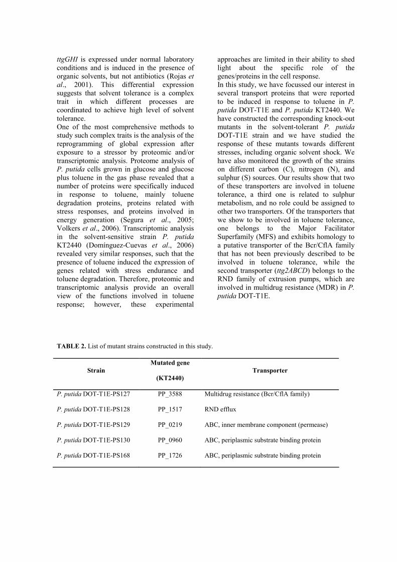
ttgGHI is expressed under normal laboratory
conditions and is induced in the presence of
organic solvents, but not antibiotics (Rojas et
al., 2001). This differential expression
suggests that solvent tolerance is a complex
trait in which different processes are
coordinated to achieve high level of solvent
tolerance.
One of the most comprehensive methods to
study such complex traits is the analysis of the
reprogramming of global expression after
exposure to a stressor by proteomic and/or
transcriptomic analysis. Proteome analysis of
P. putida cells grown in glucose and glucose
plus toluene in the gas phase revealed that a
number of proteins were specifically induced
in response to toluene, mainly toluene
degradation proteins, proteins related with
stress responses, and proteins involved in
energy generation (Segura et al., 2005;
Volkers et al., 2006). Transcriptomic analysis
in the solvent-sensitive strain P. putida
KT2440 (Domínguez-Cuevas et al., 2006)
revealed very similar responses, such that the
presence of toluene induced the expression of
genes related with stress endurance and
toluene degradation. Therefore, proteomic and
transcriptomic analysis provide an overall
view of the functions involved in toluene
response; however, these experimental
approaches are limited in their ability to shed
light about the specific role of the
genes/proteins in the cell response.
In this study, we have focussed our interest in
several transport proteins that were reported
to be induced in response to toluene in P.
putida DOT-T1E and P. putida KT2440. We
have constructed the corresponding knock-out
mutants in the solvent-tolerant P. putida
DOT-T1E strain and we have studied the
response of these mutants towards different
stresses, including organic solvent shock. We
have also monitored the growth of the strains
on different carbon (C), nitrogen (N), and
sulphur (S) sources. Our results show that two
of these transporters are involved in toluene
tolerance, a third one is related to sulphur
metabolism, and no role could be assigned to
other two transporters. Of the transporters that
we show to be involved in toluene tolerance,
one belongs to the Major Facilitator
Superfamily (MFS) and exhibits homology to
a putative transporter of the Bcr/CflA family
that has not been previously described to be
involved in toluene tolerance, while the
second transporter (ttg2ABCD) belongs to the
RND family of extrusion pumps, which are
involved in multidrug resistance (MDR) in P.
putida DOT-T1E.
TABLE 2. List of mutant strains constructed in this study.
Strain
Mutated gene
(KT2440)
Transporter
P. putida DOT-T1E-PS127 PP_3588 Multidrug resistance (Bcr/CflA family)
P. putida DOT-T1E-PS128 PP_1517 RND efflux
P. putida DOT-T1E-PS129 PP_0219 ABC, inner membrane component (permease)
P. putida DOT-T1E-PS130 PP_0960 ABC, periplasmic substrate binding protein
P. putida DOT-T1E-PS168 PP_1726 ABC, periplasmic substrate binding protein
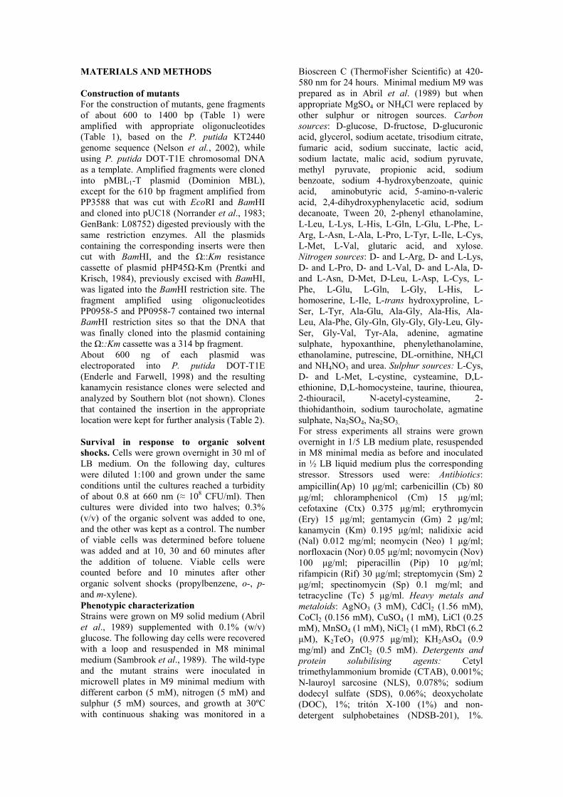
MATERIALS AND METHODS
Construction of mutants
For the construction of mutants, gene fragments
of about 600 to 1400 bp (Table 1) were
amplified with appropriate oligonucleotides
(Table 1), based on the P. putida KT2440
genome sequence (Nelson et al., 2002), while
using P. putida DOT-T1E chromosomal DNA
as a template. Amplified fragments were cloned
into pMBL1-T plasmid (Dominion MBL),
except for the 610 bp fragment amplified from
PP3588 that was cut with EcoRI and BamHI
and cloned into pUC18 (Norrander et al., 1983;
GenBank: L08752) digested previously with the
same restriction enzymes. All the plasmids
containing the corresponding inserts were then
cut with BamHI, and the Ω::Km resistance
cassette of plasmid pHP45Ω-Km (Prentki and
Krisch, 1984), previously excised with BamHI,
was ligated into the BamHI restriction site. The
fragment amplified using oligonucleotides
PP0958-5 and PP0958-7 contained two internal
BamHI restriction sites so that the DNA that
was finally cloned into the plasmid containing
the Ω::Km cassette was a 314 bp fragment.
About 600 ng of each plasmid was
electroporated into P. putida DOT-T1E
(Enderle and Farwell, 1998) and the resulting
kanamycin resistance clones were selected and
analyzed by Southern blot (not shown). Clones
that contained the insertion in the appropriate
location were kept for further analysis (Table 2).
Survival in response to organic solvent
shocks. Cells were grown overnight in 30 ml of
LB medium. On the following day, cultures
were diluted 1:100 and grown under the same
conditions until the cultures reached a turbidity
of about 0.8 at 660 nm (≈ 108 CFU/ml). Then
cultures were divided into two halves; 0.3%
(v/v) of the organic solvent was added to one,
and the other was kept as a control. The number
of viable cells was determined before toluene
was added and at 10, 30 and 60 minutes after
the addition of toluene. Viable cells were
counted before and 10 minutes after other
organic solvent shocks (propylbenzene, o-, p-
and m-xylene).
Phenotypic characterization Strains were grown on M9 solid medium (Abril
et al., 1989) supplemented with 0.1% (w/v)
glucose. The following day cells were recovered
with a loop and resuspended in M8 minimal
medium (Sambrook et al., 1989). The wild-type
and the mutant strains were inoculated in
microwell plates in M9 minimal medium with
different carbon (5 mM), nitrogen (5 mM) and
sulphur (5 mM) sources, and growth at 30ºC
with continuous shaking was monitored in a
Bioscreen C (ThermoFisher Scientific) at 420-
580 nm for 24 hours. Minimal medium M9 was
prepared as in Abril et al. (1989) but when
appropriate MgSO4 or NH4Cl were replaced by
other sulphur or nitrogen sources. Carbon
sources: D-glucose, D-fructose, D-glucuronic
acid, glycerol, sodium acetate, trisodium citrate,
fumaric acid, sodium succinate, lactic acid,
sodium lactate, malic acid, sodium pyruvate,
methyl pyruvate, propionic acid, sodium
benzoate, sodium 4-hydroxybenzoate, quinic
acid, aminobutyric acid, 5-amino-n-valeric
acid, 2,4-dihydroxyphenylacetic acid, sodium
decanoate, Tween 20, 2-phenyl ethanolamine,
L-Leu, L-Lys, L-His, L-Gln, L-Glu, L-Phe, L-
Arg, L-Asn, L-Ala, L-Pro, L-Tyr, L-Ile, L-Cys,
L-Met, L-Val, glutaric acid, and xylose.
Nitrogen sources: D- and L-Arg, D- and L-Lys,
D- and L-Pro, D- and L-Val, D- and L-Ala, D-
and L-Asn, D-Met, D-Leu, L-Asp, L-Cys, L-
Phe, L-Glu, L-Gln, L-Gly, L-His, L-
homoserine, L-Ile, L-trans hydroxyproline, L-
Ser, L-Tyr, Ala-Glu, Ala-Gly, Ala-His, Ala-
Leu, Ala-Phe, Gly-Gln, Gly-Gly, Gly-Leu, Gly-
Ser, Gly-Val, Tyr-Ala, adenine, agmatine
sulphate, hypoxanthine, phenylethanolamine,
ethanolamine, putrescine, DL-ornithine, NH4Cl
and NH4NO3 and urea. Sulphur sources: L-Cys,
D- and L-Met, L-cystine, cysteamine, D,L-
ethionine, D,L-homocysteine, taurine, thiourea,
2-thiouracil, N-acetyl-cysteamine, 2-
thiohidanthoin, sodium taurocholate, agmatine
sulphate, Na2SO4, Na2SO3.
For stress experiments all strains were grown
overnight in 1/5 LB medium plate, resuspended
in M8 minimal media as before and inoculated
in ½ LB liquid medium plus the corresponding
stressor. Stressors used were: Antibiotics:
ampicillin(Ap) 10 µg/ml; carbenicillin (Cb) 80
µg/ml; chloramphenicol (Cm) 15 µg/ml;
cefotaxine (Ctx) 0.375 µg/ml; erythromycin
(Ery) 15 µg/ml; gentamycin (Gm) 2 µg/ml;
kanamycin (Km) 0.195 µg/ml; nalidixic acid
(Nal) 0.012 mg/ml; neomycin (Neo) 1 µg/ml;
norfloxacin (Nor) 0.05 µg/ml; novomycin (Nov)
100 µg/ml; piperacillin (Pip) 10 µg/ml;
rifampicin (Rif) 30 µg/ml; streptomycin (Sm) 2
µg/ml; spectinomycin (Sp) 0.1 mg/ml; and
tetracycline (Tc) 5 µg/ml. Heavy metals and
metaloids: AgNO3 (3 mM), CdCl2 (1.56 mM),
CoCl2 (0.156 mM), CuSO4 (1 mM), LiCl (0.25
mM), MnSO4 (1 mM), NiCl2 (1 mM), RbCl (6.2
µM), K2TeO3 (0.975 µg/ml); KH2AsO4 (0.9
mg/ml) and ZnCl2 (0.5 mM). Detergents and
protein solubilising agents: Cetyl
trimethylammonium bromide (CTAB), 0.001%;
N-lauroyl sarcosine (NLS), 0.078%; sodium
dodecyl sulfate (SDS), 0.06%; deoxycholate
(DOC), 1%; tritón X-100 (1%) and non-
detergent sulphobetaines (NDSB-201), 1%.

Figure 1. Schematic representation of the genes under study and the surrounding chromosomal area in P.
putida DOT-T1E. Mutated genes are indicated as grey arrows and the corresponding gene number in the
P. putida KT2440 genome is given inside the arrow. Numbers between arrows indicated the number of
base pairs of the intergenic regions.; when not indicated the genes overlapped. We refer as permease to
the inner membrane component of the ABC systems. SBP stands for periplasmic substrate binding
protein. Ttg2ABCDE is the nomenclature of these genes in P. putida GM73.
Chelating agents: ethylenediaminetetraacetic
acid (EDTA), 0.125 mM and 2,2'-bipyridyl
(Bip) (0.5 mM). Osmotic stress agents: NaCl
(0.5 M) Oxidative stress: K2Cr2O7, 12.5 µg/ml;
H2O2, 0.004%; NH2OH, 30 µg/ml; methyl
viologen (MV), 100 µM; Tert-butyl
hydroperoxide (TBH), 0.0015%. Others:
Ethidium bromide (EtBr) 0.1 mg/ml; KCN
0.325 mg/ml and KSCN (80 mM) and NaBr
(0.25 M).
At least three independent experiments were
performed for each condition. Growth curves
were drawn using the media values of the three
experiments (standard deviations were always
less than 0.1 units of the O.D. value).
Generation time (tg) was calculated using the
formula tg = log2/r, with r being the slope of
curve when bacteria were growing in
exponential phase. When indicated, stress
experiments were done in 100 ml flasks using
30 ml of LB liquid medium.
RESULTS
Identification of transporters in P. putida
DOT-T1E and construction of the
corresponding mutants
Seven Pseudomonas transporter systems
were reported to be induced by toluene in
previous proteomic and transcriptomic
experiments (Segura et al., 2005;
Domínguez-Cuevas et al., 2006, Table 1).
PP1015, which encodes a sugar ABC
transporter (del Castillo et al., 2008), is
likely induced in the proteomic analysis
because of the higher energy demand of
cultures growing in the presence toluene
than those growing without toluene,
therefore, it was discarded.
We were unable to amplify the predicted
913 bp fragment of the major facilitator
Monoxigenase DszA Sensory box protein SBP ATP - binding Permease
MFP RND transporter TetR family regulator
KpsF / GutQ family protein ATP - binding permease SBP
Ttg2A Ttg2B Ttg2C Ttg2D Ttg2E
regulator , GntR family SBP permease permease ATP - binding
thiol peroxidase MDR, Bcr / CflA family P. putida DOT - T1E - PS - 127
Hypothetical protein P. putida DOT - T1E - PS - 128
P. putida DOT - T1E - PS - 129
P. putida DOT - T1E - PS - 130
P. putida DOT - T1E - PS - 168
Type I phosphodiesterase / Nucleotide pyrophosphatase
254 PP3588
250 6 48 PP1517
28 1 2 PP0219
1 5 8 114 PP1726
327
PP0960 248 9 119

family transporter (PP3349) using P. putida
DOT-T1E chromosomal DNA; furthermore
hybridization of P. putida DOT-T1E
chromosomal DNA with a probe
synthesized by amplification from P. putida
KT2440 DNA failed to reveal any
hybridization band. These results suggested
that a gene homologous to the PP3349 does
not exist in P. putida DOT-T1E.
Five mutants were constructed that
corresponded to knock-outs in P. putida
genes homologous to PP3588, PP1517,
PP0219, PP0960 and PP1726 (Table 2).
The complete sequence of the chromosomic
region surrounding these genes was
determined. Gene organization of these
chromosomal regions in strain P. putida
DOT-T1E (Figure 1) was identical to that
of the P. putida KT2440 and P. putida F1
strains. ORFs analysis revealed that the
deduced protein sequences were around 98-
99% identical to that of KT2440 and F1.
Solvent tolerance of the five transporters
mutants and growth with toluene as the
sole carbon source
Solvent tolerance in P. putida DOT-T1E is
an inducible response (Ramos et al., 1998);
only 1 out of 10,000 cells survived the
sudden addition of 0.3% (v/v) toluene.
However, when cultures were grown in the
presence of sublethal concentrations of
toluene (i.e. toluene in the gas phase)
almost 100% of the cells survived the
toluene shock (Figure 2). This response was
similar in mutants, P. putida DOT-T1E-
PS128 (knocked out mutant in an RND
transporter homologous to PP1517), P.
putida DOT-T1E-PS129 (knocked out
mutant in an ABC permease homologous to
PP0219) , and P. putida DOT-T1E-PS168
(knocked out mutant in periplasmic
substrate binding protein homologous to
PP1726) (not shown). However, P. putida
DOT-T1E-PS127 (knocked out mutant in
Figure 2. Survival of P. putida DOT-T1E (A), P. putida DOT-T1E-PS127 (B) and P. putida DOT-T1E-
PS130 (C) after the addition of 0.3% (v/v) toluene. Circles: non-induced cultures, Squares: induced
cultures. Closed symbols represent the survival after toluene addition, and open symbols represent
controls without toluene.
MFS transporter homologous to PP3588)
and P. putida DOT-T1E-PS130 (knocked
out mutant in the substrate binding protein
of the ABC transporter homologous to
PP0960) mutants did not survive the sudden
addition of 0.3% (v/v) toluene and their
survival rate upon pre-induction with low
toluene concentrations was lower (2 and 5
orders of magnitude, respectively) than in
wild-type (Figure 2).
Solvent tolerance of the wild-type and
mutant strains was also tested with other
organic solvents, such as different xylene
isomers and propylbenzene. No significant
differences were observed between the
wild-type and mutant strains (not shown),
so sensitivity of P. putida DOT-T1E-PS127
and P. putida DOT-T1E-PS130 to organic
solvents seemed to be restricted to toluene.
200 40 600
5
10
Log CFU/ml
A
200 40 600
5
10
Time (min)
B
200 40 600
5
10
C
200 40 600
5
10
Log CFU/ml
A
200 40 600
5
10
Time (min)
B
200 40 600
5
10
C

Figure 3. Growth of P. putida DOT-T1E (A) and P. putida DOT-T1E-PS129 (B) on different sulphur
sources. Thiouracyl (black circles), L-cystine (crosses), L-methionine (diamonds), sodium sulphate
(squares), sodium sulphite (triangles) and agmantine sulphate (white circles).
Resistance to other stresses
As many transporters have been reported to
be involved in multidrug resistance
(Krulwich et al., 2005; Piddock, 2006;
Borges-Walmsley et al., 2003), we decided
to subject the mutants to different stresses
(heavy metals, chelating agents, mutagens,
detergents, antibiotic and oxidative stress).
We have previously determined the lethal
concentration of each stressing agent for P.
putida DOT-T1E (Godoy et al., personal
communication) and therefore performed
the assays using sublethal concentrations of
the stressors. The concentrations used for
the growth curves were chosen such that a
two-fold increase in concentration
completely inhibited the growth of the
wild-type strain.
Results clearly showed that P. putida DOT-
T1E-PS130 was not only sensitive to
toluene but also towards several oxidative
stress agents (Cr2O72-
and TBH), ethidium
bromide, CTAB, some heavy metals (Mn2+
,
Ni+), and antibiotics (Tc, Sm, Cm, Ery and
Gm), suggesting that this is a multidrug
resistance (MDR) transporter (Table 3).
Mutant P. putida DOT-T1E-PS127 showed
a longer lag phase than the wild-type when
grown in the presence of Zn2+
or the
chelating agent bypiridyl and the growth
rate was lower in the presence of the
detergent CTAB as well as the antibiotic
Gm. P. putida DOT-T1E-PS128 was only
affected by the presence of bypiridyl (lag =
10 h). P. putida DOT-T1E-PS129 was
more susceptible than the wild-type to EtBr,
DOC, Mn2+
and Ery (Table 3). P. putida
DOT-T1E-PS168 was more sensitive
toward MV (lag = 17 h versus lag = 11 h of
the wild-type strain) and Gm (tg = 5.4 h).
Growth with different carbon, nitrogen
and sulphur sources
To test if some of the transporters were
required for the growth with certain carbon,
nitrogen or sulphur sources, we decided to
analyze the growth of the wild-type and
mutant strains in minimal medium plus
different carbon, nitrogen or sulphur
sources. We did not find significant
differences in the pattern of nitrogen source
utilization among the wild-type and the
mutant strains. However, under the
conditions tested, P. putida DOT-T1E-
PS129 grew very poorly, if at all, with any
carbon or nitrogen source when the culture
media was supplemented with magnesium
sulphate. The growth of this mutant was
also impaired when cultured with glucose
as the sole carbon source and agmantine
sulphate or sodium sulphite as the sulphur
source. However, this mutant grew well in
minimal media plus glucose when the
sulphur source was L-cystine or 2-tiouracile
(Figure 3). No differences were observed
among the other mutants and the wild-type
strain regarding the utilization of sulphur
sources.
B
0
0,4
0,8
1,2
1,6
0 5 10 15 20
Time (h)
A
0
0,4
0,8
1,2
1,6
0 5 10 15 20
Time (h)
O.D
. 660n
m
O.D
. 660n
m
B
0
0,4
0,8
1,2
1,6
0 5 10 15 20
Time (h)
A
0
0,4
0,8
1,2
1,6
0 5 10 15 20
Time (h)
O.D
. 660n
m
O.D
. 660n
m

Table 3 : Generation time (tg) and duration of lag phase of the wild-type and mutant strains subjected to
different stressors. Shaded numbers indicated the parameters that showed differences between the wild-
type and mutant strains. Only differences in generation time and lag phase duration of 40% and higher are
considered to be relevant and are shaded in the table. (*) These experiments have been carried out in 100
ml flasks
DOT-T1E DOT-T1E-PS127 DOT-T1E-PS129 DOT-T1E-PS130
Stressor tg (h) lag (h) tg (h) lag (h) tg (h) lag(h) tg (h) lag (h)
Cr2O7= 4.2 3 4.3 3 3.8 3 8.3 5
TBH 2.1 5 1.7 5 2.5 5 2.4 11
Bip 2.7 5 2.3 11 4.0 6 2.6 7
EtBr 1.9 5 2.3 5 3.6 5 3 6
CTAB 1.4 4 3.3 13 1.9 4 1.9 7
DOC 1.5 6 1.8 4 2.8 7 1.3 7
Mn2+ 2.3 11 2.0 10 3.8 12 3.8 12
Zn2+ 1.9 5 2.2 8 2.1 6 2.4 6
Ni+ 4.9 5 5.3 5 4.6 5 12.4 5
Sm* 0.8 2 0.8 2 0.9 2 1.4 2
Tc 2.5 6 2.4 6 3.0 6 10.2 14
Cm 2.2 4 1.9 4 2.1 4 3.5 5
Ery 4.9 6 3.3 5 10.4 6 13.9 6
Gm* 2.1 1 3.1 1 2.9 1 16.8 1
DISCUSSION
Cellular membranes are the first
contact point between the cell and the
environment. Transporters are important
proteins for living organisms not only because
permeability to nutrients and metabolites are
basic for maintenance and growth but also
because efflux of toxic compounds is crucial
for survival in the presence of toxins such as
antibiotics and organic solvents. In this study,
five transporters belonging to three different
superfamilies of bacterial transporters
(Resistance Nodulation cell Division [RND]
family, ATP-binding transporters [ABC], and
major facilitator superfamily [MFS]), have
been investigated. These genes were
originally selected because they were induced
in the presence of toluene within the solvent
tolerant P. putida DOT-T1E strain or in the
solvent sensitive P. putida KT2440 strain
(Segura et al., 2005; Domínguez-Cuevas et
al., 2006). However, preliminary analysis of
transcriptomic data of P. putida DOT-T1E
under conditions similar to those used by
Domínguez-Cuevas et al. (2006), showed no
significant transcriptional changes after 15
minutes of exposure to 1 mM of the organic
solvent (Rodríguez-Hervá, personal
communication). This was probably because
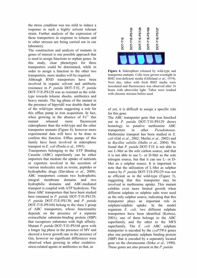
the stress condition was too mild to induce a
response in such a highly solvent tolerant
strain. Further analysis of the expression of
these transporters in response to toluene and
to other stresses are being carried out in our
laboratory.
The construction and analysis of mutants in
genes of interest is one possible approach that
is used to assign functions to orphan genes. In
this study, clear phenotypes for three
transporters could be determined, while in
order to assign a function to the other two
transporters, more studies will be required.
Although RND transporters have been
involved in organic solvent and antibiotic
resistance in P. putida DOT-T1E, P. putida
DOT-T1E-PS128 was as resistant as the wild-
type towards toluene shocks, antibiotics and
heavy metals. The lag phase of the mutant in
the presence of bipyridil was double than that
of the wild-type strain suggesting a role for
this efflux pump in iron acquisition. In fact,
when growing in the absence of Fe3+
this
mutant released more fluorescent
siderophores than the wild-type and the other
transporter mutants (Figure 4); however more
experimental data will have to be done to
confirm this function. Efflux pumps of this
family have been involved in siderophore
transport in E. coli (Poole et al., 1993).
Transporters belonging to the ATP Binding
Cassette (ABC) superfamily can be either
importers that mediate the uptake of nutrients
or exporters involved in the secretion of
various molecules such as toxins, peptides or
hydrophobic drugs (Davidson et al., 2008).
ABC transporters contain two hydrophobic
integral membrane domains and two
hydrophilic domains and ABC-mediated
transport is coupled with ATP hydrolysis. The
three ABC transporters that have been studied
here (mutated in P. putida DOT-T1E-PS129;
P. putida DOT-T1E-PS130; and P. putida
DOT-T1E-PS168) belong to the class 3 group
of ABC transporters, whose functionality
depends on the presence of a separate
extracellular substrate-binding protein (SBP)
that recognizes substrates with high affinity.
Mutant P. putida DOT-T1E-PS168 grew with
a longer lag phase in the presence of MV and
showed a lower growth rate in the presence of
Gm, however no significant differences were
observed when growing in other oxidative-
stress-related agents or antibiotics so that, as
Figure 4. Siderophore released by wild-type and
transporter mutants. Cells were grown overnight in
BM2 iron-deficient media (Gilleland et al., 1974).
Next day, tubes with fresh BM2 media were
inoculated and fluorescence was observed after 16
hours with ultraviolet light. Tubes were washed
with chromic mixture before used.
of yet, it is difficult to assign a specific role
for this gene.
The ABC transporter gene that was knocked
out in P. putida DOT-T1E-PS129 shows
homology to putative methionine ABC
transporters in other Pseudomonas.
Methionine transport has been studied in E.
coli (Gál et al., 2002; Merlin et al., 2002) and
in Bacillus subtilis (Hullo et al., 2004). We
found that P. putida DOT-T1E is not able to
use L-Met as the sole carbon source, and that
it is not able to use L- or D-Met as the sole
nitrogen source, but that it can use L- or D-
Met as a sulphur source. It is important to
note that the utilization of L-Met as sulphur
source by P. putida DOT-T1E-PS129 was not
as efficient as in the wild-type (Figure 3),
suggesting that this transporter may be
involved in methionine uptake. This mutant
exhibits even more limited growth when
different sulphate or sulphite salts were used
as the only sulphur source, indicating that this
transporter plays an important role in
sulphate/sulphite uptake. In the model
organism E. coli, two different sulphate
transporters have been identified (Kertesz,
2001); one of them belongs to the ABC
superfamily and the other to the MFS
superfamily. The E. coli ABC sulphate
transporter is encoded by the cysPTWA genes
plus one periplasmic sulphate binding protein
(SBP) that is encoded by a separately located
gene on the chromosome (Sirko et al., 1990).
These genes are also present in the P. putida
DO
T-T
1E
DO
T-T
1E
-PS
130
DO
T-T
1E
-PS
12
8
DO
T-T
1E
-PS
16
8
DO
T-T
1E
-PS
127
DO
T-T
1E
DO
T-T
1E
-PS
130
DO
T-T
1E
-PS
12
8
DO
T-T
1E
-PS
16
8
DO
T-T
1E
-PS
127

DOT-T1E chromosome. The presence of
several transport systems for sulphate and
other sulphur-related acquisition mechanisms
with overlapping specificities have been
described in different organisms (Kadner and
Watson, 1974; Sirko et al., 1995). We have
found that culture conditions influence the
growth of mutant P. putida DOT-T1E-PS129
when growing with sulphate as the only S
source (not shown).
The last ABC transporter that we looked at,
which was knocked out in the P. putida
mutant DOT-T1E-PS130, was clearly shown
to be involved in multidrug resistance. The
mutant was not only sensitive to toluene but
also to a wide range of antibiotics, several
heavy metals, and oxidative stress agents
(Table 3). The observation that P. putida
DOT-T1E-PS130 is toluene sensitive is in
agreement with previous results by Kim et al.
(1998), which showed that mutations in this
transporter of the solvent-tolerant strain P.
putida GM73 lead to a solvent sensitive
phenotype. However, to our knowledge, this
is the first report to demonstrate that this
transporter also confers multidrug resistance
to the strain.
The gene that has been mutated in the P.
putida strain DOT-T1E-PS127 encodes for a
member of the MFS that has homology within
the Bcr/CflA family of transporters that are
involved in bicyclomycin resistance (Bentley
et al., 1993). This gene has been reported to
confer moderate resistance also toward Tc,
phosphomycin and Km when expressed from
a plasmid in an E. coli acrAB deleted mutant
(Nishino and Yamaguchi, 2001). It has also
been shown to be involved in sulfathiazole
resistance (Vedantam et al., 1998) and in the
efflux of L-Cys (Yamada et al., 2006). We
found that the DOT-T1E-PS127 mutant is
toluene sensitive (Figure 2), that it was more
sensitive than wild-type to CTAB and Gm,
and it showed longer lag phases when
growing in the presence of Bip and Zn2+
. No
differences were found when Tc was added to
the medium, possibly because of the presence
of other efflux systems (i.e. TtgABC [Ramos
et al., 1998]) with overlapping specificity. In
conclusion, while this transporter seemed to
have a less broad transport capacity than that
of Ttg2A, this is the first transporter
belonging to the MFS that has been shown to
be involved in solvent tolerance.
ACKNOWLEDGMENTS
This work was supported by a Consolider-C
grants (BIO2006-05668) and grant (GEN
2006-27750-C5-5E/SYS), both from the
Spanish Ministry of Science and Education.
We thank Ben Pakuts for checking the
English in the manuscript and Alicia García
for technical assistance.
REFERENCES
1. Abril, M.A., Michán C., Timmis K.N.,
and Ramos J.L. (1989) Regulator and enzyme
specificities of the TOL plasmid-encoded
upper pathway for degradation of aromatic
hydrocarbons and expansion of the substrate
range of the pathway. J. Bacteriol. 171: 6782-
6790.
2. Bentley, J., Hyatt, L.S., Ainley, K.,
Parish, J.H., Herbert, R.B. and White, G.R.
(1993). Cloning and sequence analysis of an
Escherichia coli gene conferring bicyclomycin
resistance. Gene 127:117-120.
3. Bernal, P., Muñoz-Rojas J., Hurtado A.,
Ramos J.L., and Segura A. (2007) A
Pseudomonas putida cardiolipin synthesis
mutant exhibits increased sensitivity to drugs
related to transport functionality. Environ.
Microbiol. 9:1135-11
4. Borges-Walmsley M.I., McKeegan K.S.,
Walmsley A.R. (2003) Structure and function
of efflux pumps that confer resistance to
drugs. Biochem J. 376:313-38.
5. Davidson, A.L., Dassa, E., Orelle, C. and
Chen, J. (2008) Structure, function and
evolution of bacterial ATP-binding cassette
systems. Microbiol. Mol. Biol. Rev. 72:317-
364.
6. del Castillo, T., Duque, E. and Ramos,
J.L. (2008) A set of activators and repressors
control peripherals glucose pathways in
Pseudomonas putida to yield a common
central intermediate. J. Bacteriol. 190: 2331-
2339.
7. Domínguez-Cuevas, P., González-Pastor,
J.E., Marqués, S., Ramos, J.L. and de
Lorenzo, V. (2006) Transcriptional tradeoff
between metabolic and stress-response
programs in Pseudomonas putida KT2440
cells exposed to toluene. J. Biol. Chem.
281:11981-11991.
8. Duque, E., Segura, A., Mosqueda G., and
Ramos J.L. (2001). Global and cognate
regulators control expression of the organic

solvent efflux pump TtgABC and TtgDEF of
Pseudomonas putida. Mol. Microbiol. 39:
1100-1106
9. Enderle, P.J., and Farwell M.A. (1998)
Electroporation of freshly plated Escherichia
coli and Pseudomonas aeruginosa cells.
Biotechniques 25:954-958.
10. Gál, J., Szvetnik, A., Schnell, R. And
Kálmán, M. (2002). The metD D-Methionine
transporter locus of Escherichia coli is an
ABC transporter gene cluster. J. Bacteriol.
184:4930-4932.
11. Gilleland, H.E., Jr., Stinnett, J.D. and
Eagon, R.G. (1974) Ultrastructural and
chemical alteration of the cell envelope of
Pseudomonas aeruginosa, associated with
resistance to ethylenediaminetetraacetate
resulting from growth in a Mg2+-deficient
medium. J. Bacteriol. 117:302-311
12. Hullo, M.-F, Auger, S., Dassa, E.,
Danchin, A., Martin-Verstraete, I. (2004). The
metNPQ operon of Bacillus subtilis encodes
an ABC permease transporting methionine
sulfoxide, D- and L-methionine. Res.
Microbiol. 155:80-86
13. Junker, F. and Ramos J.L. (1999)
Involvement of the cis/trans isomerase Cti in
solvent resistance of Pseudomonas putida
DOT-T1E. J. Bacteriol. 181:5693-5700.
14. Kadner R.J. and Watson, W.J. (1974)
Methionine transport in Escherichia coli:
physiological and genetic evidence for two
uptake systems. J. Bacteriol. 119: 401-409.
15. Kertesz, M.A. (2001). Bacterial
transporters for sulfate and organosulfur
compounds. Res. Microbiol. 152: 279-290.
16. Kim, K., Lee, S., Lee, K. And Lim, D.
(1998). Isolation and characterization of
toluene-sensitive mutants from the toluene-
resistant bacterium Pseudomonas putida
GM73. J. Bacteriol. 180: 3692-3696.
17. Krulwich T.A., Lewinson O., Padan E.,
Bibi E. (2005) Do physiological roles foster
persistence of drug/multidrug-efflux
transporters? A case study. Nat Rev
Microbiol. 3:566-72
18. Merling, C., Gardiner, G., Durand, S. and
Masters, M. (2002). The Escherichia coli
metD locus encodes an ABC transporter
which includes Abc (MetN), YaeE (MetI) and
YaeC (MetQ). J. Bacteriol. 184:5513-5517.
19. Mosqueda, G. and Ramos, J.L. (2000). A
set of genes encoding a second toluene efflux
system in Pseudomonas putida DOT-T1E is
linked to the tod genes for toluene
metabolism. J. Bacteriol. 181:937-943.
20. Nelson et al., (2002) Complete genome
sequence and comparative analysis of the
metabolically versatile Pseudomonas putida
KT2440. Environ. Microbiol. 4:799-808.
21. Nishino, K. and Yamaguchi, A. (2001).
Analysis of a complete library of putative
drug transporter genes in Escherichia coli. J.
Bacteriol. 183:5803-5812
22. Norrander, K., T. Kempe and K. Messing.
1983. Construction of improved M13 vectors
using oligodeoxynucleotide-directed
mutagenesis. Gene 26: 101-106.
23. Piddock L.J. (2006) Multidrug-resistance
efflux pumps - not just for resistance. Nat.
Rev. Microbiol. 4:629-36.
24. Poole, K., Heinrichs, D.E., and Neshat, S.
(1993). Cloning and sequence analysis of an
EnvCD homologue in Pseudomonas
aeruginosa: regulation by iron and possible
involvement in the secretion of the
siderophore pyoverdine. Mol. Microbiol.
10:529-544.
25. Prentki, P., and Krisch H.M. (1984) In
vitro insertional mutagenesis with a selectable
DNA fragment. Gene 29:303-313.
26. Ramos, J.L., Duque, E., Huertas, M.J. and
Haïdour, A. (1995). Isolation and expansion
of the catabolic potencial of a Pseudomonas
putida strain able to grow in the presence of
high concentrations of aromatic hydrocarbons.
J. Bacteriol. 177:3911-3916.
27. Ramos, J.L., Duque E., Rodríguez-Herva
J.J., Godoy P., Haïdour A., Reyes F., and
Fernández-Barrero A. (1997) Mechanisms for
solvent tolerance in bacteria. J. Biol. Chem.
272:3887-3890.
28. Ramos, J.L, Duque E., Godoy P., and
Segura A. (1998) Efflux pumps involved in
toluene tolerance in Pseudomonas putida
DOT-T1E. J. Bacteriol. 180:3323-3329.
29. Ramos, J.L., Duque E., Gallegos M.T.,
Godoy P., Ramos-Gonzalez M.I., Rojas A.,
Teran W., and Segura A. (2002) Mechanisms
of solvent tolerance in gram-negative bacteria.
Ann. Rev. Microbiol. 56:743-768.
30. Rojas, A., E. Duque, G. Mosqueda, G.
Golden, A. Hurtado, J.L. Ramos, and A.
Segura (2001). Three efflux pumps are
required to provide efficient tolerance to
toluene in Pseudomonas putida DOT-T1E. J.
Bacteriol. 183:3967-3973.
31. Sambrook, J., E. F. Fritsch y T. Maniatis.
1989. Molecular cloning: a laboratory manual,
Cold Spring Harbor Laboratory Press, New
York.
32. Segura, A., Godoy, P., van Dillewijn, P.,
Hurtado, A., Arroyo, N., Santacruz, S. and
Ramos, J.L. (2005). Proteomic analysis
reveals the participation of energy- and stress-
related proteins in the response of
Pseudomonas putida DOT-T1E to toluene. J.
Bacteriol. 187: 5937-5945.
33. Sirko, A., Hryniewicz, M., Hulanicka, D.
and Bock, A. (1990). Sulfate and thiosulfate
transport in Escherichia coli K-12 –
nucleotide sequence and expression of the
cysTWAM gene cluster. J. Bacteriol.
172:3351-3357.
34. Sirko, A., Zatyka, M., Sadowy, E.,
Hulanicka, D. (1995) Sulfate and thiosulfate
transport in Escherichia coli K-12: evidence
for a functional overlapping of sulphate- and
thiosulfate-binding proteins. J. Bacterio.
177:4134-4136.
35. Spaink, H. P., Okker R. J. H., Wijffelman
C. A., Pees E. and Lugtenberg B. J. J. (1987)
Promoters in the nodulation region of the
Rhizobium leguminosarum Sym plasmid
pRL1JI. Plant Molecular Biology. 9:27-39.
36. Terán, W., Felipe, A., Segura, A., Rojas,
A., Ramos, J.L. and Gallegos, M.T. (2003).
Antibiotic-dependent induction of
Pseudomonas putida DOT-T1E TtgABC
efflux pump is mediated by the drug binding
repressor TtgR Antimicrob. Agents
Chemother. 47:3067-3072.
37. Terán, W. (2005). Regulación de la
Expresión del Transportador TtgABC de
Pseudomonas putida DOT-T1E.
Caracterización bioquímica de su regulador
TtgR. Doctoral Thesis.
38. Vedantam, G., Guay, G.G., Austria, N.E.,
Doktor, S.Z. and Nichols, B.P. (1998).
Characterization of mutations contributing to
sulfathiazole resistance in Escherichia coli.
Antimicrob. Agents Chemother. 42:88-93.
39. Volkers, R.J.M, de Jong, A.L., Hulst,
A.G., van Baar, B.L.M, de Bont, J.A.M. and
Wary, J. Chemostat-based proteomic analysis
of toluene-affected Pseudomonas putida S12.
Environ. Microbiol. 8:1674-1679.
40. Yamada, S., Awano, N., Inubushi, K.,
Maeda, E., Nakamori, S., Nishino, K.,
Yamaguchi, A. and Takagi, H. (2006). Effect
of drug transporter genes on cysteine export
and overproduction in Escherichia coli. Appl.
Environ. Microbiol. 72:4735-4742.

93
DISCUSIÓN

94
Discusión
95
La caracterización de los distintos mecanismos moleculares de la tolerancia a
disolventes orgánicos en Pseudomonas putida DOT T1E ha sido uno de los temas de
estudio en nuestro laboratorio durante los últimos 10 años. El transporte de tolueno vía
bombas de extrusión es un factor fundamental en la tolerancia en esta cepa, siendo la
bomba TtgGHI la principal implicada. Los transportadores que participan en mayor o
menor medida en la tolerancia a disolventes que ya estaban descritos al inicio de esta
tesis (TtgABC, TtgDEF y TtgGHI) pertenecen todos a la familia de transportadores
secundarios RND (Resistencia, Nodulación, División celular).
Uno de los ejes conductores de este trabajo de Tesis Doctoral ha consistido en
una profundización sobre diversos aspectos de estas bombas; por un lado caracterizando
la localización genómica de la bomba TtgGHI y por otro explorando la posibilidad de
intercambios funcionales entre los diferentes componentes de estas bombas.
Ensayos previos a esta Tesis Doctoral realizada en nuestro laboratorio, indicaron
que los genes ttgABC estaban presentes en el cromosoma de todas las Pseudomonas
analizadas y que la bomba ttgDEF aparecía siempre asociada a los genes cromosómicos
tod de degradación de tolueno (Huertas et al., 1998; Segura et al., 2005). Sin embargo,
la presencia de genes ttgGHI se restringía a cepas altamente tolerantes a tolueno, y su
localización, cromosómica ó plasmídica, se desconocía. En este trabajo se han
encontrado evidencias sólidas de la localización del operón ttgGHI en un plásmido de
gran tamaño (≈ 121 ± 5 kb) al que denominamos pGRT1, y que no había sido descrito
previamente. Este plásmido está presente al menos en dos cepas tolerantes a disolventes
orgánicos (DOT-T1E y MTB6). Aunque es bien sabido que el mantenimiento de los
plásmidos de gran tamaño pueden impone una carga energética grande en las cepas
portadoras, y que esto es posiblemente una fuente de inestabilidad (Lucas et al., 2005;
Gammon et al., 2006), en este trabajo se ha demostrado que la pérdida de pGRT1 es un
evento raro, estando la tasa de pérdida del plásmido por debajo de 10-8
por célula y por
generación, tanto en condiciones de cultivo no selectivas, como cuando las células se
cultivaban bajo condiciones desfavorables como la presencia de concentraciones sub-
letales de detergentes (SDS) y sales (NaCl). Además, aunque a lo largo de este estudio
se identificó un mutante espontáneo (P. putida DOT-T1E-VAN5) que había perdido un
fragmento del plásmido pGRT1 de aproximadamente 50 kb que incluía los genes
ttgGHI, este evento es aún menos frecuente que el de la pérdida del plasmido pGRT1
completo. Estos resultados indican que la tolerancia a disolventes orgánicos es una
Discusión
96
propiedad bastante estable y que la posible utilización de esta cepa en procesos
biotecnológicos no va a estar limitada por la inestabilidad del plásmido. Al igual que la
pérdida del plásmido provoca la pérdida de la tolerancia a tolueno en P. putida DOT-
T1E, la transferencia del plásmido mediante conjugación desde P. putida-DOT-T1E a
otras cepas de Pseudomonas sensibles a tolueno, tiene como consecuencia que éstas
adquieran alta tolerancia a disolventes orgánicos. Este hecho es de gran importancia ya
que abre una puerta a la mejora de cepas que, si bien poseen rutas catabólicas para la
degradación y/o mineralización de algún contaminante del tipo de los hidrocarburos
aromáticos, no son lo suficientemente tolerantes al mismo como para ser utilizadas en
procesos de biorremediación de sitios contaminados con altas concentraciones del
tóxico en cuestión. Aunque no se ha explorado en esta Tesis Doctoral, las implicaciones
que pueda tener esta transferencia en la naturaleza es un aspecto importante que deberá
ser estudiado en más detalle. Recientemente se ha llevado a cabo la secuenciación
completa del plásmido pGRT1 y un análisis preliminar de los marcos de lectura abierta
codificados en el plásmido revelan la existencia de un gran número de genes implicados
en la transferencia y movilización del mismo.
Las bombas de la familia RND están constituidas por un componente de
membrana interno (translocasa), la proteína de membrana externa (OMP) y una
proteína de fusión de membrana (MFP). En el caso de E. coli y P. aeuginosa se había
comprobado que las proteínas de membrana externa podían actuar con diferentes
bombas de extrusión (Srikumar et al., 1997; Poole, 2004; Fralick, 1996; Buchanan,
2002; Mine et al., 1999; Yoneyama et al., 1998, Tikhonova et al., 2002). No obstante,
no se tenían datos sobre si los componentes de las tres bombas Ttg de P. putida DOT-
T1E podrían intercambiarse entre sí manteniendo su funcionalidad. Para estudiar esta
posibilidad, analizamos el funcionamiento de la bomba TtgABC sustituyendo el
componente MFP (TtgA) por los otros dos componentes de las otras dos bombas (TtgE
ó TtgG) y el componente OMP (TtgC) por TtgF ó TtgI. La construcción de quimeras
funcionales de ambos transportadores indica que algunos de los elementos de estos
complejos tripartitos pueden ser intercambiables, fundamentalmente los componentes
de membrana externa, hecho que concuerda con lo ya descrito para las bombas Mex de
P. aeruginosa (Srikumar et al., 1997) que también pertenecen a la familia RND.
Además los datos sugieren que también algunos de los componentes MFPs podrían
intercambiarse. En cualquier caso, parece que estas quimeras no son tan eficaces como
la bomba nativa. Estos resultados podrían interpretarse como que las bombas RND

Discusión
97
podrían existir en las cepas silvestres como diferentes combinaciones de bombas
quiméricas. Los distintos componentes de las bombas RND (IMP, OMP, y MFP) se
encontrarían al azar en la membrana y se ensamblarían formando múltiples
combinaciones. No obstante, el hecho de que los tres genes se co-transcriban en un
mismo operón sugiere que ha habido una co-evolución de los mismos dando lugar a
complejos tripartitos específicos. El hecho de que el patrón de expresión de estos genes
también sea diferente para cada uno de los complejos (ttgABC, ttgDEF y ttgGHI)
parece apoyar también la especificidad en el ensamblaje del complejo. Finalmente, el
hecho de que estas bombas quiméricas sean menos eficaces que las nativas también
indican que ó bien son menos estables ó la formación del complejo no permite un
funcionamiento óptimo. Aunque no se han hecho estudios de afinidad entre
componentes ó co-localización de componentes en la membrana, es muy probable que
la combinación más afín y más estable sea la codificada por cada operón (TtgABC,
TtgDEF y TtgGHI) y que las bombas quiméricas, si existen en las cepas silvestres sean
en proporción minoritaria.
El segundo eje conductor de esta tesis se relaciona con la complejidad de la
respuesta a tolueno. Aunque las bombas son el principal determinante de la tolerancia,
distintos tipos de ensayos llevados a cabo en el laboratorio sugerían que en la respuesta
a tolueno actúan otros factores cuya relevancia, en algunos casos, estaba todavía por
determinar. La participación en la respuesta a tolueno de diferentes componentes de
membrana (Junker y Ramos, 1999; Bernal et. al., 2007, Pini et al., 2009), de genes
flagelares (Segura et al., 2001), la inducción de proteínas relacionadas con otros
estreses (Segura et al., 2005; Domínguez-Cuevas et al., 2006), y de algunos
transportadores sin función específica asignada (Segura et al., 2005; Domínguez-
Cuevas et al., 2006), entre otros, nos llevó a realizar un estudio detallado de algunos de
estos genes. Para este fin se construyeron los correspondientes mutantes y se realizaron
los ensayos pertinentes para la caracterización en relación a su papel en tolerancia a
tolueno. En esta Tesis Doctoral, se ha determinado la implicación del complejo
citocromo ubiquinol oxidasa (cyoABCD), y de dos transportadores (Ttg2ABCDE y Bcr)
en la tolerancia a tolueno.
El gen cyoB se estudió inicialmente en relación a la tolerancia a tolueno porque
forma parte de una de las dos oxidasas terminales que reducen oxígeno a agua y que
representa el final del transporte de electrones en procesos respiratorios. No hay que

Discusión
98
olvidar que la fuerza protón-motriz, de la que dependen los transportadores Ttg en P.
putida DOT-T1E, es generada a través de procesos respiratorios. Los genes Ttg2ABC y
el gen Bcr se seleccionaron inicialmente por su inducción en presencia de tolueno en la
cepa sensible P. putida KT2440. Mutantes en estos dos genes son hipersensibles a
tolueno, poniendo de manifiesto que no sólo transportadores de la familia RND, sino
también de la familia ABC (Ttg2ABC) y MFS están implicados en la tolerancia a
tolueno. Los otros tres transportadores inducidos en presencia de tolueno y estudiados
durante esta Tesis Doctoral no participan, al menos de forma relevante, en la tolerancia
a altas concentraciones de tolueno. A través de estos trabajos, se pone de manifiesto
una vez más, que la construcción de mutantes en funciones que podrían estar
implicadas en respuesta a estrés es una de las herramientas genéticas más útiles en el
estudio de estos procesos. Hay que destacar, aunque para los objetivos de esta Tesis
Doctoral no fue relevante, que los análisis funcionales llevados a cabo en los distintos
transportadores han llevado a la asignación inequívoca de una función como
transportador de sulfato/sulfito a uno de los transportadores de la familia ABC
estudiados.
En conclusión, conjuntamente con algunos estudios previos, en los que se puso
de manifiesto que un mutante en la bomba TtgGHI era hipersensible al choque de
tolueno (Rojas et al., 2001), durante esta Tesis Doctoral se ha confirmado el papel
esencial que la bomba TtgGHI, codificada en el plásmido pGRT1, tiene en la tolerancia
a tolueno. También los datos obtenidos en esta Tesis Doctoral confirman la
complejidad de la respuesta a tolueno. La participación de genes presentes tanto en
cepas sensibles como en las tolerantes a tolueno en la respuesta a este disolvente
orgánico tiene una fácil explicación cuando los correspondientes mutantes sólo
presentan una reducción (aunque significativa) de la supervivencia del cultivo tras el
choque con tolueno. Estos serían genes que participarían en la tolerancia como “ayuda”
al mecanismo principal que sería la extrusión de tolueno por la bomba TtgGHI. Así por
ejemplo, la isomerización cis-trans compacta las membranas celulares aumentando su
rigidez y contrarrestando el efecto fluidificante del tolueno, aunque a concentraciones
altas esta compactación de las membranas no sería suficientemente efectiva como para
evitar la toxicidad (Junker y Ramos, 1999). Similar situación se presentaría con otros
mecanismos como la síntesis de cardiolipina (Bernal et al., 2007), la participación de
ácidos grasos ciclopropano (Pini et al., 2009) ó incluso el papel que jugaría cyoB en la
tolerancia. Más difícil de explicar es el hecho de que proteínas presentes en los dos
Discusión
99
tipos de cepas jueguen un papel esencial en la tolerancia. Mutantes en ttgB o los
estudiados durante esta Tesis Doctoral (Ttg2ABCDE y Bcr) son incapaces de
sobrevivir la adición repentina de 0,3% (v/v) de tolueno, pese a que la bomba TtgGHI
siga funcionando y eliminando tolueno. Esto indica que aunque la bomba TtgGHI
confiere el carácter de tolerancia a la cepa, ésta necesita de una batería adicional de
mecanismos para poder sobrevivir a estos choques de tolueno. Algunos de estos
mecanismos son también esenciales (TtgABC, Ttg2ABCDE y Bcr), mientras que otros
(mecanismos relacionados con la modificación de membranas ó CyoB) permiten que
las células respondan al choque con la mayor eficiencia posible.
Los resultados obtenidos en esta Tesis Doctoral por tanto son relevantes tanto
desde el punto de vista biotecnológico, ya que se ha comprobado que el fenotipo de
tolerancia es un carácter estable, y por tanto la cepa puede usarse en reactores aún sin
presión selectiva, como desde el punto de vista de la biología básica ya que se han
identificado nuevos factores implicados en la tolerancia, siendo la primera vez que se
describe la participación de los genes cyoB y bcr en la tolerancia a tolueno. De hecho,
la cepa se ha utilizado como base para la síntesis de productos tóxicos como son los
catecoles (Rojas et al., 2004) y derivados nitroaromáticos (Meyer et al., 2005). Otro de
los potenciales de esta cepa es el del desarrollo de biosensores para la detección de
contaminantes, un área en la que se vislumbran avances al disponer la cepa de un
sensor de aromáticos muy versátil como es el regulador TodS (Lacal et al., 2006).

Discusión
100

101
CONCLUSIONES

102
Conclusiones
103
1- La mutación en el gen cyoB no afecta a la producción de fuerza protón motriz en la
cepa P. putida DOT-T1E, ni produce cambios significativos en el crecimiento de la
misma independientemente de las condiciones de cultivo. La sensibilidad a tolueno en
P. putida está influenciada por la fase de crecimiento del cultivo, así tanto células
inducidas como sin inducir de P. putida DOT-T1E fueron más sensibles al choque de
tolueno en fase estacionaria. Esta situación se exacerbó en el mutante CyoB.
2- Los resultados de Microscopia electrónica (ME) revelaron que la exposición a
disolventes del mutante CyoB induce plasmólisis desde uno de los polos.
3- El intercambio funcional de proteínas de membrana externa (OMP) y de proteínas
de fusión de membranas (MFP) es posible entre las bombas Ttg (RND) de P. putida
DOT-T1E, debido a la conservación de los residuos de las zonas que están en contacto
entre los elementos de estos complejos tripartitos.
4- Las bombas quiméricas construidas entre elementos de transportadores Ttg de P.
putida DOT-T1E fueron menos efectivas que las respectivas bombas nativas.
5- Los operones ttgVW-ttgGHI están localizados en un plásmido de gran tamaño
llamado pGRT1.
6- El plásmido pGRT1 de P. putida DOT-T1E es autotransferible a cepas del género
Pseudomonas, confiriendo a las células receptoras la capacidad de tolerar altas
concentraciones de tolueno.
7- La pérdida del plásmido pGRT1 es un evento raro que ocurre con una frecuencia de
10-8
células por generación y conduce a la pérdida de tolerancia a tolueno en P. putida
DOT-T1E debido a que se pierde el principal determinante de tolerancia a tolueno de
esta cepa (ttgGHI).
8- La pérdida de tolerancia a tolueno también puede tener lugar mediante la deleción
espontánea de un fragmento de 50 kb aproximadamente del plásmido pGRT1, en el que
se encuentra el operón ttgGHI. Este evento es aun más raro que la pérdida del plásmido
completo.

Conclusiones
104
9- El transportador ttg2ABCDE está implicado en la respuesta a altas concentraciones
de tolueno en la cepa P. putida DOT-T1E y participa en la tolerancia de esta cepa a
muchos otros compuestos tóxicos.
10- El transportador Bcr está implicado en la tolerancia a tolueno.

105
BIBLIOGRAFÍA DE INTRODUCCIÓN Y DISCUSIÓN

106
Bibliografía
107
Acuña ME, Pérez F, Auria R, Revah S. 1999. Microbiological and kinetic aspects of a
biofilter for the removal of toluene from waste gases. Biotechnol. Bioeng. 63(2):175–
184.
Acuña, M. E., C. Villanueva, B. Cárdenas, P. Christen y S. Revah. 2002. The effect
of nutrient concentration on biofilm formation on peat and gas phase toluene
biodegradation under biofiltration conditions. Process. Biochem. 38 (1): 7-13.
Aires, J. R., T. Köhler, H. Nikaido, y P. Plesiat. 1999. Involvement of an active efflux
system in the natural resistance of Pseudomonas aeruginosa to aminoglycosides.
Antimicrob. Agents Chemother. 43: 2624-2628.
Ames, G. F., C. S. Mimura y V. Shyamala. 1990. Bacterial periplasmic permeases
belong to a family of transport proteins operating from Escherichia coli to human:
Traffic ATPases. FEMS Microbiol. Rev. 6:429-446.
Arriaga, S. y S. Revah. 2005. Improving hexane removal by enhancing fungal
development in a microbial consortium biofilter. Biotechnol Bioeng. 90(1):107-115.
Arriaga S., R. Muñoz, S. Hernandez, B. Guieysse y S. Revah. 2006. Gaseous hexane
biodegradation by Fusarium solani in two liquid phase packed-bed and stirred tank
bioreactors, Environ Sci Technol 40:2390–2395.
Aono, R., K. Aibe, A. Inoue y K. Horikoshi. 1991. Preparation of organic solvent-
tolerant mutants from Escherichia coli K12. Agric. Biol. Chem. 55: 1935-1938.
Aono, R., N. Tukagoshi y M. Yamamoto. 1998. Involvement of outer membrane
protein TolC, a possible member of the mar-sox regulon, in maintenance and
improvement of organic solvent tolerance of Escherichia coli K-12. J. Bacteriol. 180:
938-944.
Bernal, P. 2007. Implicaciones de los fosfolípidos en respuesta a estreses abióticos en
Pseudomonas putida. Tesis Doctoral. Universidad de Granada, España.

Bibliografía
108
Bernal P., J.Muñoz-Rojas, A. Hurtado, J. L. Ramos y A. Segura. 2007a. A
Pseudomonas putida cardiolipin synthesis mutant exhibits increased sensitivity to drugs
related to transport functionality. Environ Microbiol. 9(5), 1135–1145.
Bernal, P., A. Segura y J. L. Ramos. 2007b. Compesatory role of the cis-
transisomerase and cardiolipin synthase in the membrane fluidity of Pseudomonas
putida DOT-T1E. Environ Microbiol. 9:1658-1664.
Borges-Walmsley, M. I., K. S. McKeegan y A. R. Walmsley. 2003. Structure and
function of efflux pumps that confer resistenace to drugs. Biochem. J. Rev. 376: 313-
338.
Borges-Wamsley, M. I. y A. R. Walmsley. 2001. The structure and function of drug
pumps. Trends in Microbiol. Rev. 9 (2): 71-79.
Boos, W. y J. M. Lucht. 1996. Periplasmic binding protein-dependent ABC
transporters, p. 1175-1209. En F. C. Neidhardt, R. Curtis, III, J. L. Ingraham, E. C. C.
Lin, K. B. Low, B. Magasanik, W. S. Reznikoff, M. Riley, M. Schaechter y H. E.
Umbarger (ed.), Escherichia coli and Salmonella: cellular and molecular biology. ASM
Press, Washington, D. C
Buchanan, S.K. 2002. Type I secretion and multidrug efflux: transport through the
TolC channel-tunnel. Trends Biochem. Sci. 26:3-6.
Cárdenas-González B., Revah-Moiseneev S., Hernández-Jiménez S., Martínez-
Sánchez A. y Gutiérrez-Avedoy, V. 2003. Tratamiento biológico de compuestos
orgánicos volátiles de fuentes fijas. Instituto Nacional de Ecología (INE-Semarnat).
México D.F., pp. 29-30.
Davidson, A. L., H. A. Shuman y H. Nikaido. 1992. Mechanism of maltose transport
in Escherichia coli: transmembrane signaling by periplasmic binding proteins. Proc.
Natl. Acad. Sci. USA 89:2360-2364.
Bibliografía
109
Davidson, A. L., S. S. Laghaeian y D. E. Mannering. 1996. The maltose transport
system of Escherichia coli displays positive cooperativity in ATP hydrolysis. J. Biol.
Chem. 271:4858-4863.
Daugulis, A. J. 1988. Integrated reaction and product recovery in bioreactor systems.
Biotechonl. Prog. 4: 113-122.
den Dooren Jong, L. E. 1926. Bigdrage tot de kennis van het mineralisatieproces.
Nijgh & van Ditmar, Rotterdam.
De Vos, P., A. V. Landschoot, P. Segers, R. Tytgat, M. Gillis, M. Bauwens, R.
Rossau, M. Goor, B. Pot, K. Kerster, P. Lizzaraga, y J. De Ley. 1989. Genotypic
relationships and taxonomic localization of unclassified Pseudomonas and
Pseudomonas-like strains by deoxy-ribonucleic acid: ribosomal ribonucleic acid
hybridizations. Int. J. Syst. Bacteriol. 39: 35-49.
Domínguez-Cuevas P., J. E. González-Pastor, S. Marqués, J. L. Ramos y V. de
Lorenzo. 2006. Transcriptional tradeoff between metabolic and stress-response
programs in Pseudomonas putida KT2440 cells exposed to toluene. Journal of
Biological Chemistry, 281(17):11981-11991.
Duque, E., A. Segura, G. Mosqueda y J. L. Ramos. 2001. Global and cognate
regulators control the expression of the organic solvent efflux pumps TtgABC and
TtgDEF of Pseudomonas putida. Mol Microbiol 39:1100-1106.
Duque, E., J. J. Rodriguez-Herva, J. de la Torre, P. Dominguez-Cuevas, J. Muñoz-
Rojas y J. L. Ramos. 2007. The RpoT regulon of Pseudomonas putida DOT-T1E and
its role in stress endurance against solvents. J. Bacteriol. 189 (1): 207-219.
Fernandez-Recio, J., F. Walas, L. Federici, J. Venkatesh-Pratap, V. N. Bravo, R.
Nuñez-Miguel, K. Mizuguchi y B. Luisi. 2004. A model of a trasnmembrane pump
from Gram-negative bacteria. FEBS Lett. 578: 5-9.

Bibliografía
110
Fralick, J. A. 1996. Evidence that TolC is required for functioning of the Mar/AcrAB
efflux pump of Escherichia coli. J. Bacteriol.178: 5803-5805.
Fukimori, F., H. Hirayama, H. Tamami, A. Inoue y K. Horikoshi. 1998. Isolation
and transposon mutagenesis of a Pseudomonas putida KT2442 toluene-resistant variant:
involvement of an efflux system in solvent resistance. Extremophiles 2: 395-400.
Gibson, D. T., M. Hensley, H. Yoshioka y T. J. Mabry. 1970. Formation of (+)-cis-
2,3-dihydroxy-1-methylcyclohexa-4,6-diene from toluene by Pseudomonas putida.
Biochemistry 9: 1626-1630.
Gammon, K., G.W. Jones, S.J. Hope, C. M. F. Oliveira, L. Regis y M. H. N. L.
Silva-Filha. 2006. Conjugal transfer of a toxin-coding megaplasmid from Bacillus to
mosquitocidal strains of Bacillus sphaericus. Appl. Envirom. Microbiol. 76: 1766-1770.
Heipieper, H. J., G. Menlenbeld, Q. van Oirstchot, y J. A. M. de Bont. 1996. Effect
of environmental factors on the cis/trans ratio of unsaturated fatty acids in
Pseudomonas S12. Appl. Environ. Microbiol. 62: 2773-2777.
Heipieper, H. J., F. Meinhardt y A. Segura. 2003. The cis-trans isomerase of
unsaturated fatty acids in Pseudomonas and Vibrio: biochemistry, molecular biology
and physiological function of a unique stress adaptive mechanism. FEMS Microbiol.
Lett. 229:1-7
Heipieper, H. J. y J. A. M. de Bont. 1994. Adaptation of Pseudomonas putida S12 to
ethanol and toluene at the level of fatty acid composition of membranes. Appl. Environ.
Microbiol. 60: 4440-4444.
Higgins, C. F. 1992. ABC transporters: from microorganisms to man. Annu. Rev. Cell
Biol. 8:67-113
Holtwick, R., F. Meinhardt y H. Keweloh. 1997. Cis-trans isomerization of
unsaturated fatty acids: cloning and sequencing of the cti gene from Pseudomonas
putida P8. Appl. Environ. Microbiol. 63: 4292-4297.

Bibliografía
111
Huertas, M. J., E. Duque, S. Marqués y J. L. Ramos. 1998. Survival in soil of
different toluene-degrading Pseudomonas strains after solvent shock. Appl. Environ.
Microbiol. 64 (1): 38-42.
Huertas, M. J., E. Duque, L. Molina, R. Rossello-Mora, G. Mosqueda, P. Godoy, B.
Christensen, S. Molin y J. L. Ramos. 2000. Tolerance to sudden organic solvent
shocks by soil bacteria and characterization of Pseudomonas putida strains isolated
from toluene polluted sites. Env. Sci. Technol. 34: 3395-3400.
Hyde, S. C., P. Emsley, M. J. Hartshorn, M. M. Mimmack, U. Gileadi, S. R.
Pearce, M. P. Gallagher, D. R. Gill, R. E. Hubbard y C. F. Higgins. 1990. Structural
model of ATP-binding proteins associated with cystic fibrosis, multidrug resistance and
bacterial transport. Nature. 346:362-365.
Inoue, H. y K. Horikoshi. 1989. A Pseudomonas thrives in high concentrations of
toluene. Nature 338: 264-266.
Isken, S. y J. A. M. de Bont. 1996. Active efflux of toluene in a solvent-resistant
bacterium. J. Bacteriol. 178: 6056-6058.
Jin, J., A. A. Guffanti, C. Beck y T. A. Krulwich. 2001. Twelve-transmembrane-
segment (TMS) version (∆TMS VII-VIII) of the 14-TMS Tet-L antibiotic resistance
protein retains monovalent cation transport modes but lacks tetracycline efflux capacity.
J. Bacteriol. 183 (8): 2667-2671.
Junker, F. y J. L. Ramos, 1999. Involvement of the cis/trans isomerase Cti in solvent
resistance of Pseudomonas putida DOT-T1E. J. Bacteriol. 181: 5693-5700.
Junker, F., J. J. Rodríguez-Herva, E. Duque, M. I. Ramos- González, y J. L.
Ramos. 2001. A WbpL mutant of Pseudomonas putida DOT-T1E strains, which lacks
the O-antigenic side chain of lipopolysaccharides, is tolerant to organic solvent shocks.
Extremophiles 5: 93-99.

Bibliografía
112
Kadurugamuwa, J. L. y T. J. Beveridge. 1995. Virulence Factors Are Released from
Pseudomonas aeruginosa in Association with Membrane Vesicles during Normal
Growth and Exposure to Gentamicin: a Novel Mechanism of Enzyme Secretion. J.
Bacteriol. 177(14): 3998-4008.
Keweloh, H. y H. J. Heipieper. 1996. Trans unsaturated fatty acids in bacteria. Lipids
31: 129-137.
Kieboom, J., J. J. Dennis, J. A. M. de Bont y G. J. Zylstra. 1998a. Identification and
molecular characterization of an efflux pump involved in Pseudomonas putida S12
solvent tolerance. J. Biol. Chem. 273: 85-91
Kieboom, J., J. J. Dennis, G. J. Zylstra y J. A. M. de Bont. 1998b. Active efflux of
organic solvents by Pseudomonas putida S12 is induced by solvents. J. Bacteriol. 180:
6769-6772.
Kieboom, J., R. Bruinenberg, I. Keizer-Gunnink y J. A. M. de Bont. 2001.
Transposon mutations in the flagella biosynthetic pathway of the solvent-tolerant
Pseudomonas putida S12 result in a decreased expression of solvent efflux genes.
FEMS Microbiol. Lett. 198: 117-122.
Kim, K., S. Lee, K. Lee y D. Lim. 1998. Isolation and characterization of toluene-
sensitive mutants from the toluene-resistant bacterium Pseudomonas putida GM73. J.
Bacteriol. 180: 3692-3696.
Kobayashi, H., K. Uematsu, H. Hirayama y K. Horikoshi. 2000. Novel toluene
elimination system in a toluene-tolerant microorganism. J. Bacteriol. 182: 6451-6455.
Koehler, T., M. Michea-Hamzehpour, U. Henze, N. Gotoh, L. K. Curty y J. C.
Pechere. 1997. Characterization of MexE-MexF-OprM, a positively regulated
multidrug efflux system of Pseudomonas aeruginosa. Mol. Microbiol. 23: 345-354.
Bibliografía
113
Koronakis V., A. Sharft, E. Koronakis, B. Luisi y C. Hughes. 2000. Crystal structure
of the bacterial membrane protein TolC central to multidrug efflux and protein export.
Nature 405: 914-919.
Koutinas M., Peeva L. G. y A. G. Livingston. 2005. An attempt to compare the
performance of bioscrubbers and biotrickling filters for degradation of ethyl acetate in
gas streams. J. Chem. Tech.Biotech. 80:1252-1260.
Krieg, N. R. y G. M. Garrity. 2001. On using the manual, p. 15-19. In D. R. Boone, R.
W. Castensholz, and G. M. Garrity (ed.), Bergey’s Manual of Systematic Bacteriology,
2nd
Ed., Vol I. Springer Verlag, New York.
Lacal, J., A. Busch, M. E. Guazzaroni, T. Krell y J. L. Ramos. 2006. The TodS–
TodT two-component regulatory system recognizes a wide range of effectors and works
with DNA-bending proteins. Proc. Nat. Acad. Sci. 103(21): 8191-8196.
Li, X. Z., L. Zhang y K. Poole. 1998. Role of the multidrug efflux systems of
Pseudomonas aeruginosa in organic solvent tolerance. J. Bacteriol. 180: 2987-2991.
Lucas, P. M., W. A. M. Wolken, O. Claisse, J. S. Lolkema y A. Lonvand-Funel.
2005. Histamine-producing pathway encoged an instable plasmid in Lactobacillus
hilgardi 006. Appl. Environ. Microbiol. 71: 1417-1424.
Long, F., C. Rouquette-Loughlin, W. M. Shafer y E. W. Yu. 2008. Functional
cloning and characterization of the multidrug efflux pumps NorM from Neisseria
gonorrhoeae and YdhE from Escherichia coli. Antimicrob. Agents Chemother.
52(9):3052-60.
Lugtenberg, B. J. y L. C. Dekkers. 1999. What makes Pseudomonas bacteria
rhizosphere competent? Environ. Microbiol. 1: 9-13.
Ma, D., D. N. Cook, M. Alberti, N. G. Pon, H. Nikaido y J. E. Hearst. 1993.
Molecular cloning and characterization of acrA and acrE genes of Escherichia coli. J.
Bacteriol. 175: 6299-6313.
Bibliografía
114
Marger, M. D. y M. H. Saier. 1993. A major superfamily of transmembrane
facilitators that catalyse uniport, symport and antiport. TIBS. 18: 13-20.
Meyer, D., B. Witholt y A. Schmid. 2005. Suitability of recombinant Escherichia coli
and Pseudomonas putida strains for selective biotransformation of m-nitrotoluene by
xylene monooxygenase. Appl. Environ. Microbiol. 71(11): 6624–6632 .
Migula, W.1894. Ueber ein neues System der Bakterien. Arb. Bakteriol. Inst. Karlsruhe
1: 235-238
Mimura, C. S., S. R. Holbrook y G. F. Ames. 1991. Structural model of the
nucleotide-binding conserved component of periplasmic permeases. Proc. Natl. Acad.
Sci. USA 88:84-88.
Mine, T., Y. Morita, A. Kataoka, T. Mizushima y T. Tsuchiya. 1998. Evidence for
chloramphenicol/H+ antiport in Cmr (MdfA) system of Escherichia coli and properties
of the antiporter. J. Biochem. (Tokyo). 124:187-193.
Mine, T., Y. Morita, A. Kataoka, T. Mizushima y T. Tsuchiya. 1999. Expression in
Escherichia coli of a new multidrug efflux pump, MexXY, from Pseudomonas
aeruginosa. Antimicrob. Agents Chemother. 45:415-417.
Morita, Y., K. Kodama, S. Shiota, T. Mine, A. Kataoka, T. Mizushima y T.
Tsuchiya. 1998. NorM, a putative multidrug efflux protein, of Vibrio parahaemolyticus
and its homolog in Escherichia coli. Antim. Agents Chemother. 42(7): 1778–1782
Moriyama, Y., M. Hiasa, T. Matsumoto y H. Omote. 2008. Multidrug and toxic
compound extrusion (MATE)-type proteins as anchor transporters for the excretion of
metabolic waste products and xenobiotics. Xenobiotica. 38(7-8):1107-1118.
Mosqueda, G, M. I. Ramos-González y J. L. Ramos. 1999. Toluene metabolism by
the solvent-tolerant Pseudomonas putida DOT-T1 strain, and its role in solvent
impermeabilization. Gene 232: 69-76.

Bibliografía
115
Mosqueda, G. y J. L. Ramos. 2000. A set of genes enconding a second toluene efflux
system in Pseudomonas putida DOT-T1E is linked to the tod genes for toluene
metabolism. J. Bacteriol. 182: 937-943.
Murakami, S., R. Nakashima, E. Yamashita y A. Yamaguchi. 2002. Crystal
structure of bacterial multidrug efflux transporter AcrB. Nature. 419: 587-593.
Nelson, K. E., C. Weinel, I. T. Paulsen, R. J. Dodson, H. Hilbert, V. A. Martins dos
Santos, D. E. Fouts, S. R. Gill, M. Pop, M. Holmes, L. Brinkac, M. Beanan, R. T.
DeBoy, S. Daugherty, J. Kolonay, R. Madupu, W. Nelson, O. White, J. Peterson,
H. Khouri, I. Hance, P. Chris Lee, E. Holtzapple, D. Scanlan, K. Tran, A. Moazzez,
T. Utterback, M. Rizzo, K. Lee, D. Kosack, D. Moestl, H. Wedler, J. Lauber, D.
Stjepandic, J. Hoheisel, M. Straetz, S. Heim, C. Kiewitz, J. A. Eisen, K. N. Timmis,
A. Düsterhöft, B. Tümmler y C. M. Fraser. 2002. Complete genome sequence and
comparative analysis of the metabolically versatile Pseudomonas putida KT2440.
Environ Microbiol 4:799-808.
Neyfakh, A. A. 2001. The ostensible paradox of multidrug recognition. J. Mol.
Microbiol. Biotechnol. 3: 151-154.
Nikaido, H. y Y. Takatsuka. 2008. Mechanisms of RND multidrug efflux pumps.
Biochim. Biophys. Acta. In press.
Ortiz, I., A. Velasco y S. Revah. 2006. Effect of toluene as gaseous cosubstrate in
bioremediation of hydrocarbon-polluted soil. J. Hazard Mater. 131(1-3):112-7.
Palleroni, N. J., R. Kunisawa, R. Contopoulou y M. Doudoroff. 1973. Nucleic acid
homologies in the genus Pseudomonas. Int. J. Syst. Bacteriol. 23: 333-339.
Palleroni, N. J. 1986. Taxonomy of Pseudomonas. In “The Bacteria”. Vol. X. Sokatch,
J. R. (ed.). Academic Press. London. pp 3-25.
Palleroni, N. J. y E. Moore. 2004. Taxonomy of Pseudomonas. Chapter 1 in The
Pseudomonads. vol. I.
Bibliografía
116
Piddock,L. J. 2006. Multidrug-resistance efflux pumps - not just for resistance
Nat.Rev.Microbiol. 4: 629-636.
Pinkart, H. C., J. W. Wolfram, R. Rogers y D. C. White. 1996. Cell envelope
changes in solvent-tolerant and solvent-sensitive Pseudomonas putida strains following
exposure to o-xylene. Appl. Environ. Microbiol. 62. 1129-1132.
Pinkart, H.C. y D. C. White. 1997. Phospholipid biosynthesis and solvent tolerance in
Pseudomonas putida strains. J. Bacteriol. 179: 4219-4226.
Pini, C. V., P. Bernal, P. Godoy, J. L. Ramos y A. Segura. 2009. Cyclopropane fatty
acids are involved in organic solvent tolerance but not in acid stress resistance in
Pseudomonas putida DOT-T1E.
Poole, K., K. Krebes, C. McNally y S. Neshat. 1993. Multiple antibiotic resistance in
Pseudomonas aeruginosa: evidence for involvement of an efflux operon. J. Bacteriol.
175: 7363-7372.
Poole, K. 2004. Efflux-mediated multiresistance in Gram-negative bacteria. Clin.
Microbiol. Infect. 10: 12–26.
Ramos, J. L., E. Díaz, D. Dowling, V. de Lorenzo, S. Molin, S. O’Gara, C. Ramos y
K. N. Timmis. 1994. The behavior of bacteria designed for biodegradation.
Biotechnology N.Y. 12: 1349-1358.
Ramos, J. L., E. Duque, M. J. Huertas y A. Haidour. 1995. Isolation and expansion
of the catabolic potential of a Pseudomonas putida strain able to grow in the presence of
high concentrations of aromatic hydrocarbons. J. Bacteriol. 177: 3911-3916.
Ramos, J. L., E. Duque, J. J. Rodríguez-Herva, P. Godoy, A. Haidour, F. Reyes y
A. Fernández-Barrero.1997. Mechanisms for solvent tolerance in bacteria. J. Biol.
Chem. 272: 3887-3890.

Bibliografía
117
Ramos, J. L., E. Duque, P. Godoy y A. Segura. 1998. Efflux pumps involved in
toluene tolerance in Pseudomonas putida DOT-T1E. J. Bacteriol. 180: 3323-3329.
Ramos, J. L., M. T. Gallegos, S. Marqués, M. I. Ramos-González, M. Espinosa-
Urgel y A. Segura. 2001. Responses of gram-negative bacteria to certain environmental
stresses. Curr. Opin. Microbiol. 4: 166-171.
Ramos, J. L., E. Duque, M. T. Gallegos, P. Godoy, M. I. Ramos-González, A.
Rojas, W. Terán y A. Segura. 2002. Mechanisms of solvent tolerance in gram-
negative bacteria. Ann. Rev. Microbiol. 56: 743-768.
Rekker, R. F. y H. M. D. Kort. 1979. Hydrophobic Fragmental Constant - Extension
to A 1000 Data Point Set. Eur. J. Medic. Chem. 14: 479-488
Rojas, A., E. Duque, G. Mosqueda, G. Golden, A. Hurtado, J. L. Ramos y A.
Segura. 2001. Three efflux pumps are required to provide effcient tolerance to toluene
in Pseudomonas putida DOT-T1E. J. Bacteriol. 183: 3967-3973.
Rojas, A., A. Segura, M. E. Guazzaroni, W. Terán, A. Hurtado, M. T. Gallegos y J.
L. Ramos. 2003. In vivo and in vitro evidences that TtgV is the specific regulator of the
TtgGHI multidrug and solvent efflux pump of Pseudomonas putida. J. Bacteriol. 185:
4755-4763.
Rojas, A., E. Duque, A. Schmid, A. Hurtado, J. L. Ramos y A. Segura. 2004.
Biotransformation in double phase systems: physiological responses of Pseudomonas
putida DOT-T1E to a double phase made of aliphatic alcohols and biosynthesis of
subtituted catechols. Appl Environ Microbiol. 70(6): 3637-3643.
Saier, M. H. Jr. 1998. Molecular phylogeny as a basis for the classification of transport
proteins from bacteria, archaea and eukarya. Adv. Microb. Physiol. 40:81-136.
Saier, M. H. Jr. 2000. Vectorial metabolism and the evolution of transport systems. J.
Bacteriol. 182:5029-5035.
Bibliografía
118
Saraste, M., P. R. Sibbald y A. Wittinghofer. 1990. The P-loop a common motif in
ATP- and GTP-binding proteins. Trends Biochem. Sci. 15:430-434.
Schneider, E. y S. Hunke. 1998. ATP-binding-cassette (ABC) transport systems:
functional and structural aspects of the ATP-hydrolyzing subunits/domains. FEMS
Microbiol. Rev. 22:1-20.
Schwöppe, C., H. H. Winkler y E. Neuhaus. 2003. Connection of transport and
sensing by UhpC, the sensor for external glucose-6-phosphate in Escherichia coli. Eur.
J. Biochem. FEBS. 270: 1450-1457.
Segura, A., E. Duque, G. Mosqueda, J. L. Ramos y F. Junker. 1999. Multiple
responses of gram-negative bacteria to organic solvents. Environ. Microbiol. 1: 191-
198.
Segura, A., E. Duque, A. Hurtado y J. L. Ramos. 2001. Mutations in genes involved
in the flagellar export apparatus of the solvent-tolerant Pseudomonas putida DOT-T1E
strain impair motility and lead to hypersensibility to toluene shocks. J. Bacteriol. 183:
4127-4133.
Segura, A., A. Rojas, A. Hurtado, M. J. Huertas y J. L. Ramos. 2003. Comparative
genomic analysis of solvent extrusion pumps in Pseudomonas strains exhibiting
different degrees of solvent tolerance. Extremophiles. 7:371-376.
Segura, A., E. Duque, A. Rojas, P. Godoy, A. Delgado, A. Hurtado, J. E. Cronan,
Jr. y J. L. Ramos. 2004. Fatty acid biosynthesis is involved in solvent tolerance
in Pseudomonas putida DOT-T1E. Environ. Microbiol. 6 (4), 416–423
Segura, A., P. Godoy, P. van Dillewijn, A. Hurtado, N. Arroyo, S. Santacruz y J. L.
Ramos. 2005. Proteomic analysis reveals the participation of energy- and stress-related
proteins in the response of Pseudomonas putida DOT-T1E to toluene. J. Bacteriol.
187:5937-5945.

Bibliografía
119
Sikkema, J., B. Poolman, W. N. Konings y J. A. M. de Bont. 1992. Effects of the
membrane action of tetralin on the functional and structural properties of artificial and
bacterial membranes. J. Bacteriol. 174: 2986-2992.
Sikkema, J., J. A. M. de Bont y B. Poolman. 1995. Mechanisms of membrane
toxicity of hydrocarbons. Microbiol.Rev. 59: 201-222.
Song, J.H. y K. A. Kinney. 2000. Effect of vapor-phase biofilter operation on biomass
accumulation, distribution, and activity: linking biofilm properties to bioreactor
performance. Biotechnology and Bioengineering. 68:508-516.
Song, J.H. y K. A. Kinney. 2005. Microbial response and elimination capacity in
biofilters subjected to high toluene loadings. Applied Microbiology and Biotechnology.
68:554-559.
Srikumar, R., X. Z. Li y K. Poole.1997. Inner membrane efflux components are
responsible for the β-lactam specificity of multidrug efflus pumps in Pseudomonas
aeruginosa.J. Bacteriol. 179: 7875-7881.
Srikumar, R., T. Kon, N. Gotoh y K. Poole. 1998. Expression of Pseudomonas
aeruginosa multidrug efflux pumps MexA-MexB-OprM and MexC-MexD-OprJ in a
multidrug-sensitive Escherichia coli strain .Antimicrob. Agents. Chem. 42: 65-71.
Stainer, R. Y., N. J. Palleroni, and M. Doudoroff. 1966. The aerobic Pseudomonads:
a taxonomic study. J. Gen. Microbiol. 43: 159-271.
Stover, C.K., X. Q. Pham, A. L. Erwin, S. D. Mizoguchi, P. Warrener,M. J.
Hickey, F. S. L. Brinkman, W. O. Hufnagle, D. J. Kowalik, M. Lagrou, R. L.
Garber, L. goltry, E. Tolentino, S. Westbrock-Wadman, Y. Yuan, L. L. Brody, S.
N. Coulter, K. R. Folger, A. Kas, K. Larbic, R. Lim, K. Smith, D. Spencer, G. K. S.
Wong, Z. Wu, I. T. Paulsen, J. Reizer, M. H. Saier, R. E. W. Hanckock, S. Lory y
M. V.Olson. 2000. Complete genoma sequence of Pseudomonas aeruginosa PAO1,
and opportunistic pathogen. Nature 406: 959-964.

Bibliografía
120
Su, C. C., F. Long, G. McDermott, W. M. Shafer y E. W. Yu. 2008. Crystallization
and preliminary X-ray diffraction analysis of the multidrug efflux transporter NorM
from Neisseria gonorrhoeae. Acta Crystallogr. Sect. F. Struct. Biol. Cryst. Commun.
64(4):289-92.
Terán W., A. Felipe, A. Segura, A. Rojas, J. L. Ramos y M. T. Gallegos. 2003.
Antibiotic-dependent induction of Pseudomonas putida DOT-T1E TtgABC efflux
pump is mediated by the drug binding repressor TtgR. Antimicrob. Agents Chemother.
47(10) 3067–3072.
Teran, W., T. Krell,J. L. Ramos y M. T. Gallegos. 2006. Effector-repressor
interactions, binding of a ingle effector molecule to the operator-bound TtgR
homodimer mediates derepression. J. Biol. Chem. 281(11): 7102–7109.
Terán, W., A. Felipe, S. Fillet, M. E. Guazzaroni, T. Krell, R. Ruiz, J. L. Ramos y
M. T. Gallegos. 2007. Complexity in efflux pump control: cross-regulation by the
paralogues TtgV and TtgT. Mol. Microbiol. 66(6), 1416–1428
Tikhonova, E.B., Q. Wang y H.I. Zgurskaya. 2002. Chimeric analysis of the
multicomponent efflux transporters from gram-negative bacteria. J. Bacteriol. 184
(23):6499-6507.
Urbatsch, I. L., B. Sankaran, J. Weber y A. E. Senior. 1995. P-glycoprotein is stably
inhibited by vanadate-induced trapping of nucleotide at a single catalytic site. J. Biol.
Chem. 270:19383-19390
Villaverde, S., R. G. Mirpuri, Z. Lewandowski y W. L. Jones. 1997. Physiological
and chemical gradients in a Pseudomonas putida 54G biofilm degrading toluene in a
flat plate vapor phase bioreactor. Biotech. Bioeng. 56(4):361-371.
Vodovar, N., D. Vallenet, S. Cruveiller, Z. Rouy, V. Barbe, C. Acosta, L. Cattolico,
C. Jubin, A. Lajus, B. Segurens, B. Vacherie, P. Wincker, J. Weissenbach, B.
Lemaitre, C. Mediguez y F. Boccardi. 2006. Complete genome sequence of the

Bibliografía
121
entomopathogenic and metabolically versatile soil bacterium Pseudomonas
entomophila. Nat. Biotech. 24(6): 673-679.
Walker, J. E., M. Saraste, M. J. Runswick y N. J. Gay. 1982. Distantly related
sequences in the α- and β-subunits of ATP synthase, myosin, kinases and other ATP
requiring enzymes and a common nucleotide binding fold. EMBO J. 1:945-951.
Wandersman, C. 1998. Protein and peptide secretion by ABC exporters. Res.
Microbiol. 149:163-170.
Weber, F. J. 1994. Toluene: biological waste-gas treatment, toxicity and microbial
adaptation. PhD thesis. Univ. Wageningen. The Netherlands.
Weber, F. J. y J. A. M. de Bont. 1996. Adaptation mechanisms of microorganisms to
the toxic effects of organic solvents on membranes. Biochim. Biophys. Acta 1286: 225-
245.
Woese, C. R., W. G. Weisburg, C. M. Hahn, B. J. Paster, L. B. Zablen, B. J. Lewis,
T. J. Macke, W. Ludwig y E. Stackebrandt. 1985. The phylogeny of purple bacteria:
the gamma subdivision. Syst. Appl. Microbiol. 6: 25-33.
Yeom, S. H. y A. J. Daugulis. 2001. Development of a novel bioreactor system for
treatment of gaseous benzene. Biotechnol. Bioeng. 72: 156-165
Yoneyama, H., A. Ocaktan, N. Gotoh, T. Nishino y T. Nakae. 1998. Subunit
swapping in the Mex-extrussion pumps in Pseudomonas aeruginosa. Biochem. Biophys.
Res. Commun. 244:898-902.
Young, J. y I. B. Holland. 1999. ABC transporters: bacterial exporters-revisited five
years on. Biochim. Biophys. Acta. 1461:177-200.
Yu, E. W., G. McDermott, H. Zgurskaya, H. Nikaido, and D. E. Koshland Jr. 2003.
Structural basis of multiple drug-binding capacity of the AcrB multidrug efflux pump.
Science 300: 976-980.

Bibliografía
122
Yu, E. W., J. R. Aires, G. McDermott y H. Nikaido. 2005. A periplasmic drug-
binding site of the AcrB multidrug efflux pump: a crystallographic and site-directed
mutagenesis study. J. Bacteriol. 187 (19): 6804–6815.
Zgurskaya, H.I., and H. Nikaido. 1999. Bypassing the periplasm: Reconstitution of
the AcrAB multidrug efflux pump of Escherichia coli. Proc. Natl. Acad. Sci. U.S.A.
96: 7190-7195.
Zgurskaya, H. I. y H. Nikaido. 2000. Multidrug resistance mechanisms: drug efflux
across two membranes. Mol. Microbiol. 37: 219-225


