caracterizaciÓn genÉtica y molecular de mecanismos de
TRANSCRIPT

UNIVERSIDAD AUTÓNOMA DE MADRID
FACULTAD DE CIENCIAS
DEPARTAMENTO DE BIOLOGÍA
CARACTERIZACIÓN GENÉTICA Y
MOLECULAR DE MECANISMOS DE
REPRESIÓN FLORAL EN ARABIDOPSIS
TESIS DOCTORAL
ANA LÁZARO SOMOZA
MADRID, 2011


UNIVERSIDAD AUTÓNOMA DE MADRID
FACULTAD DE CIENCIAS
DEPARTAMENTO DE BIOLOGÍA
CARACTERIZACIÓN GENÉTICA Y
MOLECULAR DE MECANISMOS DE
REPRESIÓN FLORAL EN ARABIDOPSIS
Memoria presentada por Ana Lázaro Somoza para optar al grado de Doctor por la
Facultad de Ciencias de la Universidad Autónoma de Madrid.
Trabajo realizado en el Departamento de Biotecnología del Instituto Nacional de
Investigación y Tecnología Agraria y Alimentaria (INIA) bajo la dirección de los
Doctores José Antonio Jarillo Quiroga y Manuel Piñeiro Galvín.
VºBº DE LOS DIRECTORES VºBº TUTOR LA DOCTORANDA
J.A. JARILLO M. PIÑEIRO RAFAEL RIVILLA A. LÁZARO
TESIS DOCTORAL
ANA LÁZARO SOMOZA
MADRID, 2011


AGRADECIMIENTOS
Han pasado ocho años desde que decidí que me iba a dedicar a la investigación y
en este tiempo he recibido ayuda y ánimo de un montón de gente. Tengo que dar las
gracias a mis directores de Tesis, Jari y Manolo, y a todas las personas que han
pasado por el laboratorio durante estos años, Iván, Eugenio, Leti, Laura, Raquel,
Angelita, David, Pablo, Silvia, Sandra, Alfonso y Nuria. Una buena parte de esta Tesis
se realizó en el Edificio Z del INIA, dónde compartí los primeros años con Bruno,
Pepe, Elenita, Ana del Cueto, Mariano, Rafa, Tamara, Iñaki, Carlitos, Fernando
Novillo, Gemita, Claudia, Carmen Mansilla, Pablo Lunello, Chuchi, Marga, Adela y
Vicky. También tengo mucho que agradecer a Carlos, Conchi, Zamira, Gema, Nacho,
Julieta y Rosa, con los que he convivido todo este tiempo, y a Sara, David Carrasco,
Kata, Mar y Elena Ramírez, que llegaron más tarde al CBGP. Tampoco quiero
olvidarme de otras personas que me han ayudado y enseñado muchas de las técnicas
que he usado en estos años, Maremoto, Isra, César Poza, Lars Hennig, Pablo
González-Melendi; y de todas las cosas que le he pedido “prestadas” a Julio Salinas.
Gracias a todos por echarme una mano siempre que lo he necesitado, por todo el
apoyo en los momentos duros y por aguantarme, que tiene tela.
Y, por supuesto, gracias a David, que es mi refugio, a mi familia y a mi madre en
especial, a mis amigos de hace muchos años, María, Lorenzo, Laura Rol, Sara,
Gabino, Betty, Raúl, Laura Hillán, y a los amigos que comparto con David estos
últimos años. También quiero dar las gracias a la gente que ya no está aunque, de un
modo u otro, también han hecho esto posible, mi padre y mis abuelos.


ÍNDICE
Página
SUMMARY 1
ABREVIATURAS Y SIGLAS 2
INTRODUCCIÓN 3
1. La transición floral 5
2. El control genético de la floración en Arabidopsis thaliana 6
2.1. Variación genética natural 6
2.2. Variación genética inducida 8
2.2.1. Rutas de inducción de la floración 8
Ruta del fotoperiodo 9
Ruta de la vernalización 12
Ruta dependiente de la temperatura ambiental 14
Ruta autónoma 14
Ruta de las giberelinas 15
Ruta dependiente de la edad 15
2.2.2. Integradores florales 16
2.2.3. Represores de la floración 18
3. El control epigenético de la floración en Arabidopsis thaliana 19
3.1. Activación de la expresión de FLC 20
3.2. Represión de la expresión de FLC 22
3.3. Regulación epigenética de otros genes implicados en el control 25
del tiempo de floración
4. El papel de la degradación específica de proteínas en la transición 25
floral en Arabidopsis thaliana
OBJETIVOS 29
RESULTADOS 33
CAPÍTULO 1. 35
EARLY IN SHORT DAYS 1 (ESD1) encodes ACTIN-RELATED PROTEIN 6
(AtARP6), a putative component of chromatin remodelling complexes
that positively regulates FLC accumulation in Arabidopsis
(Martin-Trillo y col., 2006).

CAPÍTULO 2. 59
Mutations in the Arabidopsis SWC6 gene, encoding a component of the
SWR1 chromatin remodelling complex, accelerate flowering time and alter
leaf and flower development (Lazaro y col., 2008).
CAPÍTULO 3. 81
The E3 ubiquitin ligase HOS1 participates in the control of photoperiodic
flowering negatively regulating CONSTANS abundance
(Lazaro y col., 2011 en revisión).
DISCUSIÓN 109
1. Papel de la variante histónica H2A.Z en la regulación del tiempo de 111
floración
2. Función de HOS1 en la regulación fotoperiódica de la floración 120
CONCLUSIONES 125
BIBLIOGRAFÍA 129

1
SUMMARY
The coordination of flowering time with seasonal and developmental cues is
critical to maximize reproductive success in plants. In this work we have characterised
components in two different mechanisms involved in the floral repression in
Arabidopsis.
In one hand, we have isolated two early flowering mutations, esd1/arp6 and
swc6, affecting putative orthologues of components of the yeast Swr1 chromatin
remodelling complex. We found that ESD1/ARP6 and SWC6 are required for
maintaining the expression of the FLC repressor to levels that inhibit flowering.
Genetic and physical interactions between SWC6 and ESD1 have been demonstrated
in this study, suggesting that both proteins act in the same complex. Besides, we
have established that ESD1/ARP6 and SWC6 are required for both histone H3
acetylation and H3K4 trimethylation on FLC chromatin. Altogether, the results
obtained suggest that SWC6 and ESD1 are part of an Arabidopsis SWR1 chromatin
remodelling complex involved in the regulation of diverse aspects of plant
development, including floral repression through the activation of FLC and FLC-like
genes.
On the other hand, we found the early in short days 6 (esd6) mutant in a
screening for mutations that accelerate flowering time in Arabidopsis and showed that
it was affected in the HOS1 locus, which encodes a RING finger-containing protein
that works as an E3 ubiquitin ligase. The esd6/hos1 mutation showed a strong
requirement of a functional CO protein for its early flowering phenotype under long
days. Besides, CO and HOS1 physically interact in vitro and in vivo, and HOS1
regulates CO abundance, particularly during the daylight period. Accordingly, the hos1
mutation causes a shift in the typical long day pattern of the FT transcript, starting to
rise four hours after dawn. In addition, HOS1 interacts synergistically with COP1,
another regulator of CO protein stability, in the control of flowering time. Taken
together, these results indicate that HOS1 is involved in regulating CO abundance
ensuring that CO activation of FT occurs only when the light period reaches a certain
length and preventing precocious flowering in Arabidopsis.

2
ABREVIATURAS Y SIGLAS
ADN Ácido desoxirribonucleico
ARN Ácido ribonucleico
ARNi ARN de interferencia
col. Colaboradores
DC Día corto
DL Día largo
GFP Green fluorescent protein
miR microRNA
pb Pares de bases
ZT Zeitgeber time

INTRODUCCIÓN

INTRODUCCIÓN
5
1. La transición floral
El desarrollo de las plantas es el resultado de la división de grupos de células
pluripotentes denominados meristemos y su posterior diferenciación en los distintos
órganos vegetales (Ma, 1998). La porción aérea de la planta se forma a partir del
meristemo apical, mientras que el meristemo radicular da lugar a las raíces.
Las plantas son organismos sésiles que tienen la capacidad de percibir multitud de
señales ambientales y de adaptar su desarrollo a los cambios que se producen en el
medio que las rodea. La transición de la fase de desarrollo vegetativo a la fase
reproductiva, lo que se conoce como transición floral, es uno de los procesos más
finamente regulado, ya que del momento en que tenga lugar depende el éxito
reproductivo de las plantas (Amasino, 2010). La transición floral implica importantes
cambios en la identidad del meristemo apical. Durante la fase vegetativa el
meristemo da lugar a hojas y meristemos axilares, que a su vez producirán ramas
vegetativas. Sin embargo, una vez que se produce la transición floral, comienza la
formación de flores a partir de los meristemos reproductivos (Coen y Meyerowitz,
1991).
El tiempo de floración está controlado por multitud de factores, unos endógenos y
otros ambientales. Los primeros dependen fundamentalmente del estado de
desarrollo de la planta, mientras que los factores ambientales que regulan la floración
son el fotoperiodo (relación entre los periodos diarios de luz y de oscuridad), la
intensidad y la calidad de la luz que recibe la planta, y la temperatura (Kim y col.,
2009; Michaels, 2009; Amasino, 2010; Imaizumi, 2010). En concreto, la aceleración
del tiempo de floración que se produce como consecuencia de la exposición de las
plantas a periodos prolongados de bajas temperaturas se conoce como vernalización.
Entre las especies sensibles al fotoperiodo se pueden distinguir plantas en las que
la floración se induce por exposición a día corto (DC, el periodo de oscuridad es más
prolongado que el de luz), como el arroz (Oryza sativa), y plantas en las que se
induce por condiciones de día largo (DL, el periodo de luz es más prolongado que el
de oscuridad), como la avena (Avena sativa). En cambio, otras especies vegetales,
como el tomate (Solanum lycopersicum), son insensibles al fotoperiodo, (Jarillo y col.,
2008). De igual modo, hay especies que presentan un requerimiento absoluto de
vernalización para florecer, como la remolacha (Beta vulgaris), mientras que otras
responden a vernalización sin un requerimiento obligado o no responden en absoluto,
como diversas especies de cereales (Kim y col., 2009).

INTRODUCCIÓN
6
2. El control genético de la floración en Arabidopsis thaliana
Arabidopsis thaliana presenta una fase de desarrollo vegetativo en roseta
caracterizada por la formación reiterada de hojas sin elongación de los entrenudos. La
transición entre la fase juvenil y la fase adulta del desarrollo vegetativo determina la
adquisición de competencia del meristemo apical para responder al estímulo floral
(Poethig, 1990), y se ha asociado con cambios en la morfología foliar y en el patrón
de distribución de tricomas en las hojas (Telfer y col., 1997). Cuando se induce la
transición floral, se produce la elongación de los entrenudos de las hojas del tallo
principal (caulinares) y la formación de una inflorescencia. Los meristemos axilares de
las hojas caulinares se desarrollan dando lugar a una inflorescencia lateral o
coflorescencia, mientras que los meristemos florales dan lugar a flores.
Arabidopsis es una planta facultativa de DL, es decir, florece de forma más
temprana y con menor número de hojas en DL que en DC, y además algunas
accesiones son capaces de responder a tratamientos de vernalización (Martínez-
Zapater y col., 1994).
El análisis de la variación natural que existe entre las distintas accesiones de
Arabidopsis y la caracterización de mutantes afectados en el tiempo de floración que
se ha llevado a cabo en los últimos años, ha permitido identificar una serie de genes
que participan en la regulación de este proceso.
2.1 Variación genética natural
Arabidopsis presenta una elevada variación genética para el tiempo de floración
en poblaciones naturales, probablemente como consecuencia de procesos de
adaptación a distintas condiciones ambientales (Koornneef y col., 2004). El análisis
genético de la variación existente entre accesiones ha permitido identificar diversos
loci que son responsables de esta variación fenotípica para el tiempo de floración.
Se ha descrito que alelos funcionales y dominantes de los loci FRIGIDA (FRI) y
FLOWERING LOCUS C (FLC) son los responsables del requerimiento de vernalización
(Johanson y col., 2000; Shindo y col., 2005; Werner y col., 2005). FRI codifica una
proteína específica de plantas que es necesaria para retrasar la floración a través de
la activación de la expresión de FLC, ya que mutaciones de pérdida de función de FLC
suprimen el efecto de FRI sobre el tiempo de floración (Johanson y col., 2000;
Michaels y Amasino, 2001). Datos recientes sugieren que FRI activa la transcripción
de FLC a través de un mecanismo cotranscripcional que implica la interacción de FRI
con componentes del complejo de unión al 5´CAP del mensajero de FLC (Geraldo y
INTRODUCCIÓN
7
col., 2009; Crevillen y Dean, 2011). Por su parte, FLC codifica un factor de
transcripción de la familia MADS que actúa como represor de la floración de forma
cuantitativa (Michaels y Amasino, 1999; Sheldon y col., 1999). Los tratamientos de
vernalización disminuyen la expresión de FLC y hacen a este gen insensible a la
activación por FRI (He y Amasino, 2005). Una vez que la planta ha sido vernalizada,
la represión de FLC se mantiene estable durante el resto del ciclo de vida de la planta,
y sólo se restablecen niveles elevados de expresión de FLC en la siguiente generación
(He y Amasino, 2005). En Arabidopsis hay cinco genes parálogos de FLC,
denominados MADS AFFECTING FLOWERING 1/FLOWERING LOCUS M (MAF1/FLM),
MAF2, MAF3, MAF4 y MAF5. Se ha demostrado que al menos dos de ellos, MAF1/FLM
y MAF2, actúan como represores florales (Ratcliffe y col., 2001; Ratcliffe y col.,
2003), lo que podría explicar por qué plantas con mutaciones nulas para FLC no
eliminan totalmente la respuesta a la vernalización. De acuerdo con esta hipótesis, el
gen MAF1/FLM sufre los mismos cambios epigenéticos que produce la vernalización en
la cromatina de FLC (Sung y col., 2006a).
Las accesiones de ciclo rápido o de primavera pueden aparecer como
consecuencia de la pérdida de una proteína FRI funcional, incapaz de regular
positivamente a FLC, o de la existencia de un alelo débil o inactivo de FLC. Entre las
accesiones empleadas en el laboratorio, Columbia (Col) y Landsberg erecta (Ler)
poseen una mutación en el locus FRI (Grennan, 2006). Además, Ler presenta un alelo
débil de FLC debido a la inserción de un trasposón en su primer intrón (Gazzani y col.,
2003; Michaels y col., 2003). Por su parte, la accesión C24 también presenta un alelo
débil de FLC (Grennan, 2006).
El análisis de variantes naturales también ha permitido identificar genes
implicados en la regulación del tiempo de floración en respuesta a factores como la
temperatura de crecimiento y el fotoperiodo. Entre ellos se encuentran varios loci que
codifican para fotorreceptores, como CHRYPTOCHROME 2/EARLY DAYLENGTH
INSENSITIVE (CRY2/EDI) y PHYTOCHROME C (PHYC) y PHYD (Aukerman y col., 1997;
El-Din El-Assal y col., 2001; Balasubramanian y col., 2006b). Cry2 participa en la
percepción de la luz azul, que induce la floración, mientras que PhyC y PhyD actúan
como receptores de la luz roja, que reprime la transición floral. Además, MAF1/FLM
codifica un factor de transcripción que actúa modulando la inducción de la floración en
respuesta a pequeños aumentos de la temperatura de crecimiento (Balasubramanian
y col., 2006a).
INTRODUCCIÓN
8
2.2 Variación genética inducida
2.2.1 Rutas de inducción de la floración
El análisis genético del tiempo de floración en Arabidopsis se ha basado
clásicamente en el estudio de mutantes de floración tardía, que se clasificaron en
función de su respuesta al fotoperiodo y a la vernalización. Además, las interacciones
genéticas entre los distintos loci permitió postular un modelo genético del control de
la floración en el cual una serie de rutas promotoras convergen en la regulación de la
expresión de los integradores florales (Moon y col., 2003; Turck y col., 2008). De las
seis rutas actualmente aceptadas, tres responden a factores ambientales: la ruta del
fotoperiodo, la ruta de la vernalización y la ruta dependiente de la temperatura
ambiental, mientras que las otras tres responden a factores endógenos: la ruta
dependiente de las giberelinas, la ruta autónoma y la ruta dependiente de la edad
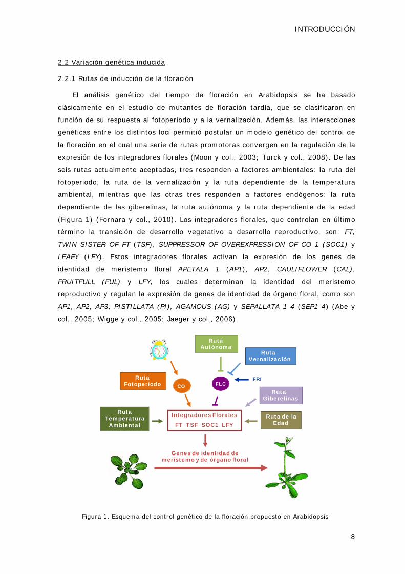
(Figura 1) (Fornara y col., 2010). Los integradores florales, que controlan en último
término la transición de desarrollo vegetativo a desarrollo reproductivo, son: FT,
TWIN SISTER OF FT (TSF), SUPPRESSOR OF OVEREXPRESSION OF CO 1 (SOC1) y
LEAFY (LFY). Estos integradores florales activan la expresión de los genes de
identidad de meristemo floral APETALA 1 (AP1), AP2, CAULIFLOWER (CAL),
FRUITFULL (FUL) y LFY, los cuales determinan la identidad del meristemo
reproductivo y regulan la expresión de genes de identidad de órgano floral, como son
AP1, AP2, AP3, PISTILLATA (PI), AGAMOUS (AG) y SEPALLATA 1-4 (SEP1-4) (Abe y
col., 2005; Wigge y col., 2005; Jaeger y col., 2006).
Ruta Autónoma
Ruta Vernalización
FLCFRIRuta
FotoperiodoRuta
Giberelinas
Integradores FloralesFT TSF SOC1 LFY
Genes de identidad de meristemo y de órgano floral
CO
Ruta Temperatura
AmbientalRuta de la
Edad
Figura 1. Esquema del control genético de la floración propuesto en Arabidopsis

INTRODUCCIÓN
9
Ruta del fotoperiodo
Clásicamente, la ruta del fotoperiodo se estableció a partir de la identificación de
una serie de mutantes de floración tardía que afectaban a los loci CONSTANS (CO),
FHA/CRY2, GIGANTEA (GI), FD y FT (Searle y Coupland, 2004). Estos mutantes
presentan un retraso en el tiempo de floración en DL aunque en DC florecen al mismo
tiempo que plantas de tipo silvestre, además de no mostrar alteraciones en la
respuesta a vernalización.
CRY2, como ya hemos descrito, codifica la apoproteína de un fotoreceptor de luz
azul (Guo y col., 1998) y GI codifica una proteína nuclear que regula, entre otros
genes, la expresión de CO (Fowler y col., 1999; Rubio y Deng, 2007). Por su parte,
CO codifica una proteína nuclear con dos dedos de zinc de tipo B-box y un dominio
CCT (de CO, CO-LIKE y TOC1) que actúa como un activador floral (Putterill y col.,
1995; Robson y col., 2001). CO pertenece a una familia génica formada por 17
miembros en Arabidopsis. Se ha descrito que los genes CO-like 3 (COL3) y COL9
pueden tener un papel como represores florales (Cheng y Wang, 2005; Datta y col.,
2006). Por otro lado, FT codifica una proteína con homología a inhibidores de Raf
quinasas que actúa como un potente inductor floral (Kardailsky y col., 1999;
Kobayashi y col., 1999). FD codifica un factor de transcripción de tipo b-Zip que se
expresa en el meristemo apical (Wigge y col., 2005). Las mutaciones en FD suprimen
el fenotipo de floración temprana que produce la sobreexpresión del integrador floral
FT, lo cual sugirió que FD participa en la inducción de la floración por debajo de FT
(Abe y col., 2005; Wigge y col., 2005).
El mecanismo que poseen las plantas para medir la duración del día está basado
en un sistema circadiano. Dicho sistema utiliza la información lumínica transmitida
por los fotorreceptores para “poner en hora” el mecanismo oscilador central del reloj
circadiano (Jarillo y col., 2008). Este mecanismo oscilador consite en una serie de
bucles de retroalimentación positiva y negativa entre proteínas que se expresan por la
mañana y otras que se expresan al atardecer, y es el encargado de regular el patrón
de expresión de multitud de genes con un periodo de oscilación próximo a las 24
horas (de Montaigu y col., 2010; Imaizumi, 2010).
En Arabidopsis, la capacidad para distinguir la longitud de los periodos de luz y
oscuridad se basa en la coincidencia de un ritmo interno de la planta, representado
por el patrón de expresión de CO, con una señal ambiental como es la luz. Los niveles
de expresión de CO están regulados por el reloj circadiano, de modo que en DL se
observan los niveles máximos al amanecer, el atardecer y durante la noche; en

INTRODUCCIÓN
10
cambio, en DC la expresión de CO se limita al periodo de oscuridad (Figura 2)
(Suarez-Lopez y col., 2001). Los niveles de la proteína CO no están determinados
solamente por el ARN mensajero de CO, sino que también están regulados mediante
su degradación por el proteosoma (Jang y col., 2008; Liu y col., 2008c). Así, en la
oscuridad, la proteína CO no es estable y sólo en DL, cuando la transcripción de CO
coincide con el periodo de luz, la proteína CO se acumula a niveles capaces de activar
la expresión de FT (y de TSF) (Samach y col., 2000; Suarez-Lopez y col., 2001;
Yamaguchi y col., 2005). FT es un integrador floral cuyo nivel de expresión es
máximo en DL durante la fase de coincidencia de la luz con el pico de expresión de
CO, condiciones en las que FT promueve el inicio de la floración. En cambio, en DC la
proteína CO se degrada y el nivel de expresión de FT se mantiene bajo, de modo que
la floración se retrasa (Figura 2).
DIA LARGO DIA CORTO
Figura 2. Representación de los niveles de expresión de los mensajeros de CO y de FT en
condiciones de DL y DC en Arabidopsis (adaptada de de Montaigu y col., 2010).
El patrón de expresión de CO también está regulado por una serie de proteínas
codificadas por genes cuya transcripción, a su vez, está controlada por el reloj
circadiano. Varios miembros de la familia de proteínas CYCLING DOF FACTORs (CDFs)
reprimen la transcripción de CO en la primera parte del día (Imaizumi y col., 2005;
Fornara y col., 2009). Hacia el final de la tarde, la expresión de los genes GI y
FLAVIN-BINDING KELCH REPEAT F-BOX 1 (FKF1) aumenta y se produce la interacción
dependiente de luz azul entre ambas proteínas (Sawa y col., 2007). FKF1 contiene un
dominio F-box, implicado en degradación de proteínas, y un dominio receptor de luz
azul tipo LOV (Demarsy y Fankhauser, 2009). El complejo formado entre FKF1 y GI
promueve la degradación de CDF1 y, así, permite eliminar la represión que ejerce
esta proteína sobre la expresión de CO (Imaizumi y col., 2003; Sawa y col., 2007).
En diversos trabajos se han descrito otros reguladores transcripcionales de CO
como DAY NEUTRAL FLOWERING (DNF), LONG VEGETATIVE PHASE 1 (LOV1), RED
AND FAR-RED INSENSITIVE 2 (RFI2) o SENSITIVE TO FREEZING 6 (SFR6), a los
INTRODUCCIÓN
11
cuales haremos referencia más adelante (Chen y Ni, 2006b; Yoo y col., 2007; Knight
y col., 2008; Morris y col., 2010).
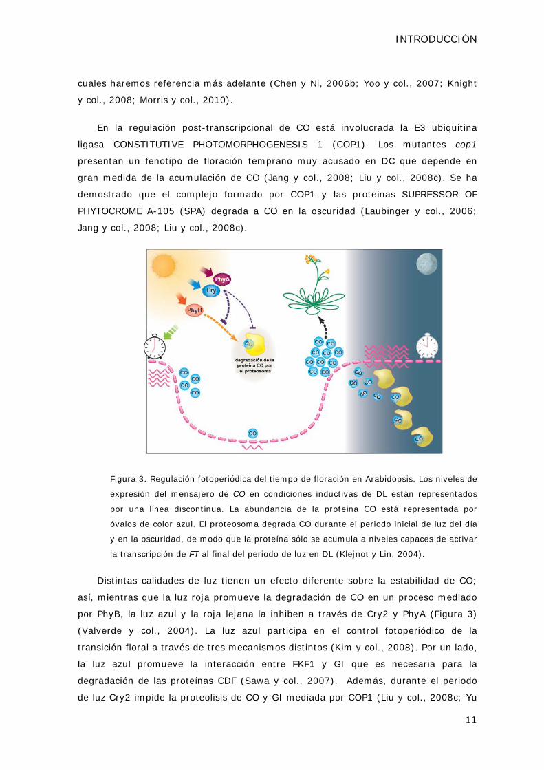
En la regulación post-transcripcional de CO está involucrada la E3 ubiquitina
ligasa CONSTITUTIVE PHOTOMORPHOGENESIS 1 (COP1). Los mutantes cop1
presentan un fenotipo de floración temprano muy acusado en DC que depende en
gran medida de la acumulación de CO (Jang y col., 2008; Liu y col., 2008c). Se ha
demostrado que el complejo formado por COP1 y las proteínas SUPRESSOR OF
PHYTOCROME A-105 (SPA) degrada a CO en la oscuridad (Laubinger y col., 2006;
Jang y col., 2008; Liu y col., 2008c).
Figura 3. Regulación fotoperiódica del tiempo de floración en Arabidopsis. Los niveles de
expresión del mensajero de CO en condiciones inductivas de DL están representados
por una línea discontínua. La abundancia de la proteína CO está representada por
óvalos de color azul. El proteosoma degrada CO durante el periodo inicial de luz del día
y en la oscuridad, de modo que la proteína sólo se acumula a niveles capaces de activar
la transcripción de FT al final del periodo de luz en DL (Klejnot y Lin, 2004).
Distintas calidades de luz tienen un efecto diferente sobre la estabilidad de CO;
así, mientras que la luz roja promueve la degradación de CO en un proceso mediado
por PhyB, la luz azul y la roja lejana la inhiben a través de Cry2 y PhyA (Figura 3)
(Valverde y col., 2004). La luz azul participa en el control fotoperiódico de la
transición floral a través de tres mecanismos distintos (Kim y col., 2008). Por un lado,
la luz azul promueve la interacción entre FKF1 y GI que es necesaria para la
degradación de las proteínas CDF (Sawa y col., 2007). Además, durante el periodo
de luz Cry2 impide la proteolisis de CO y GI mediada por COP1 (Liu y col., 2008c; Yu

INTRODUCCIÓN
12
y col., 2008). Por último, se ha demostrado que Cry2 se une al factor de transcripción
CRYPTOCHROME-INTERACTING BASIC-HELIX-LOOP-HELIX 1 (CIB1) y regula
directamente la transcripción de FT (Liu y col., 2008b). De forma opuesta al papel de
la luz azul, se ha descrito que PhyB participa en la degradación de CO en las primeras
horas del día aunque se desconoce el mecanismo molecular que media esta respuesta
(Valverde y col., 2004; Jang y col., 2008).
Recientemente, se ha avanzado en el conocimiento del mecanismo de activación
de FT por parte de CO. Se han identificado varias regiones aguas arriba del gen FT
que son importantes para la regulación transcripcional de este locus y se ha
propuesto que CO contiene dominios tanto de unión a ADN como de activación
transcripcional. Estos resultados sugieren que CO se une a un elemento en tándem
presente en el promotor de FT y que esta unión es suficiente para la activación
dependiente de CO (Adrian y col., 2010; Tiwari y col., 2010). Además, también se ha
descrito que CO podría actuar como parte de un complejo homólogo al complejo
activador de la transcripción Heme Activator Protein (HAP) de levaduras (Ben-Naim y
col., 2006; Cai y col., 2007). CO contiene dominios homólogos a HAP2 que sugieren
que pueda reemplazar a esta proteína en el complejo HAP y unirse de esta forma al
promotor de FT en Arabidopsis (Wenkel y col., 2006).
En conclusión, es la interacción entre la luz y el reloj circadiano la que regula la
expresión y modula la actividad de CO y, por tanto, la que permite a la planta percibir
la duración del día. En fotoperiodos de DL, la expresión de CO en el tejido vascular de
las hojas induce la expresión del mensajero de FT (An y col., 2004). Se ha
demostrado que la proteína FT es capaz de moverse desde las hojas hasta el
meristemo apical y, por tanto, que actuaría como parte de la señal de larga distancia
denominada “florígeno” que inicia el proceso de floración en respuesta a condiciones
de DL (Corbesier y col., 2007; Turck y col., 2008; Fornara y col., 2010).
Ruta de la vernalización
La vernalización es el proceso mediante el cual la exposición prolongada a bajas
temperaturas hace a las plantas competentes para florecer (Kim y col., 2009). En
Arabidopsis esta ruta regula el tiempo de floración a través de la represión de FLC.
Este mecanismo represor presenta dos características principales: la primera es que
tiene carácter cuantitativo, es decir, los niveles de expresión de FLC disminuyen de
forma gradual a medida que se aumenta el tiempo de exposición a bajas
temperaturas (Sheldon y col., 2000). La segunda es que conlleva un cambio

INTRODUCCIÓN
13
epigenético en el estado de la cromatina de FLC, ya que el estado reprimido de FLC se
mantiene aún cuando la planta es transferida a temperaturas normales de crecimiento
(Turck y Coupland, 2011). Se han identificado tres mutantes denominados
vernalization 1 y 2 (vrn1 y vrn2) y vernalization insensitive 3 (vin3), que muestran un
fenotipo de floración tardío aún cuando se someten a un tratamiento de vernalización
(revisado en Kim y col., 2009).
VRN2 codifica un homólogo de Suppressor of Zeste 12 (Su(z)12), el primer
componente identificado del grupo Polycomb (PcG) de Drosophila (Gendall y col.,
2001). Su(z)12 pertenece al Complejo de Represión Polycomb 2 (PRC2), que está
implicado en procesos de represión génica tanto en animales como en plantas
(Hennig y Derkacheva, 2009). VRN1, en cambio, presenta dominios implicados en
unión a ADN (Levy y col., 2002). Aunque en los mutantes vrn1 y vrn2 la exposición a
bajas temperaturas reprime transcripcionalmente a FLC, este estado no se mantiene
cuando la planta deja de ser vernalizada (Sheldon y col., 2006). Por tanto, VRN1 y
VRN2 no están involucrados en el establecimiento de un estado silenciado de FLC en
respuesta frío, sino en su mantenimiento cuando la planta se expone a la temperatura
normal de crecimiento. Además, se ha demostrado que LIKE HETEROCHROMATIN
PROTEIN 1 (LHP1) o TERMINAL FLOWER 2 (TFL2), homólogo de HETEROCHROMATIN
PROTEIN 1 (HP1) de animales y levaduras, también es necesario para mantener el
estado reprimido de FLC característico de la vernalización (Mylne y col., 2006; Sung y
col., 2006b).
El mutante vin3 no presenta respuesta a la vernalización ni disminución en los
niveles de FLC tras su exposición al frío (Sung y Amasino, 2004). La expresión de
VIN3 se induce en respuesta a bajas temperaturas, de modo que se ha propuesto que
VIN3 participa en el establecimiento de la represión de FLC (Sung y Amasino, 2004).
VIN3 posee un dominio tipo Plant Homeo Domain (PHD), característico de
componentes de complejos de remodelación de cromatina, y pertenece a una familia
de proteínas que tienen la capacidad de dimerizar. Se ha descrito que VIN3-like 1
(VIL1)/VRN5 también participa en la respuesta a vernalización y es capaz de
interaccionar con VIN3 (Sung y col., 2006a; Greb y col., 2007).
Por otro lado, durante la exposición a bajas temperaturas se produce un aumento
de los niveles de tránscritos no codificantes de FLC, denominados COLD INDUCED
LONG ANTISENSE INTRAGENIC RNA (COOLAIR), y COLD ASSISTED INTRONIC
NONCODING RNA (COLDAIR) (Swiezewski y col., 2009; Heo y Sung, 2011). Se ha
demostrado recientemente que COLDAIR participa en el establecimiento de la
INTRODUCCIÓN
14
represión epigenética de FLC reclutando al complejo PRC2 a la región genómica de
este represor floral (Heo y Sung, 2011; Turck y Coupland, 2011).
Ruta dependiente de la temperatura ambiental
Las plantas de Arabidopsis florecen antes cuando se cultivan a una temperatura
ambiental de 23ºC que cuando se cultivan a 16ºC (Blazquez y col., 2003). El gen
SHORT VEGETATIVE PHASE (SVP) juega un papel importante en esta respuesta, ya
que los mutantes svp son insensibles a estas variaciones en la temperatura de
crecimiento (Lee y col., 2007). SVP codifica una proteína tipo MADS box que reprime
la expresión del integrador floral FT y es necesario para retrasar la floración a
temperaturas bajas (16ºC) (Lee y col., 2007). SVP forma parte de un complejo en el
que también se encuentra FLC, y que puede desempeñar un papel central en modular
el inicio de la floración en respuesta a la temperatura ambiental (Li y col., 2008).
Ruta autónoma
Los mutantes de floración tardía que definen esta ruta corresponden a los loci
FCA, FY, FPA, LUMINIDEPENDENS (LD), FLOWERING LOCUS D (FLD), FVE,
FLOWERING LOCUS K (FLK) y RELATIVE OF EARLY FLOWERING 6 (REF6). Estos
mutantes se caracterizan por presentar un retraso en la floración tanto en DL como
en DC, que puede ser revertido cuando se someten a un tratamiento de vernalización
(revisado en Amasino, 2010).
Todos los mutantes de esta ruta presentan niveles altos de expresión del represor
floral FLC (Michaels y Amasino, 1999; Sheldon y col., 1999; Sheldon y col., 2000;
Michaels y Amasino, 2001). La función bioquímica de las proteínas de la ruta
autónoma sugiere que puedan participar, bien en mecanismos de unión y
procesamiento de ARN, o bien en procesos de remodelación de la cromatina de FLC
(Amasino y Michaels, 2010). FCA, FPA y FLK contienen dominios de unión a RNA
(Macknight y col., 1997; Schomburg y col., 2001; Lim y col., 2004; Manzano y col.,
2009) y FY presenta homología con factores de procesamiento de ARNs mensajeros
(Simpson y col., 2003). LD codifica un factor de transcripción con un dominio
homeobox (Lee y col., 1994). Por otro lado, REF6 codifica una proteína tipo Jumonji y
FLD una proteína homóloga a la LYSINE-SPECIFIC DEMETHYLASE 1 (LSD1) humana,
dos clases diferentes de demetilasas de histonas (He y col., 2003; Noh y col., 2004;
Jiang y col., 2007). Por su parte, FVE participa en procesos de deacetilación de
histonas sobre la cromatina de FLC (Ausin y col., 2004). Recientemente se ha descrito
una familia de metiltransferasas de arginina (PRMTs) que se pueden adscribir a esta
INTRODUCCIÓN
15
ruta y que también participan en la represión de FLC (Wang y col., 2007; Niu y col.,
2008; Schmitz y col., 2008).
En resumen, la ruta autónoma que controla el tiempo de floración no parece ser
una ruta lineal sino que comprende una colección de genes implicados en: (i) la
represión de la expresión génica, y (ii) el establecimiento de los niveles basales de
expresión de FLC (Amasino, 2010).
Ruta de las giberelinas
Las giberelinas son hormonas que promueven la floración en Arabidopsis y que,
en DC, resultan imprescindibles para que ocurra la transición de desarrollo vegetativo
a reproductivo (Mutasa-Gottgens y Hedden, 2009). Las mutaciones en los loci
GIBBERELLIC ACID 1-5 (GA1-5), que codifican enzimas de la ruta de biosíntesis de
giberelinas, retrasan la floración, y en concreto en el caso de ga1-3, que carece
completamente de giberelinas, se suprime totalmente la floración en DC (Wilson y
col., 1992). Además, las mutaciones que provocan una activación constitutiva de la
señalización dependiente de giberelinas, como spindly (spy), provocan una
aceleración de la floración (Jacobsen y Olszewski, 1993). Las giberelinas regulan la
transición floral a través de SOC1, aunque se desconoce el mecanismo molecular por
el cual estas hormonas activan la expresión de este integrador floral. También se ha
descrito que las giberelinas activan la expresión de LFY, que presenta elementos de
respuesta a giberelinas en su zona promotora (Blazquez y col., 1998; Lee y Lee,
2010).
Ruta dependiente de la edad
En Arabidopsis, se ha descrito que la familia de factores de transcripción
SQUAMOSA PROMOTER BINDING PROTEIN-LIKE (SPL) regula de forma positiva, tanto
la transición de fase juvenil a adulta, como la transición floral. Las proteínas SPL
participan en el control del tiempo de floración a través de la regulación de la
expresión de los integradores florales SOC1 y LFY (Wang y col., 2009; Yamaguchi y
col., 2009; Poethig, 2010).
Recientemente, se ha desvelado el papel que desempeñan algunos microRNAs
(miRNAs) en la transición floral. La expresión del miR156 mantiene la fase juvenil y
retrasa la floración en Arabidopsis (Wu y Poethig, 2006), mientras que el miR172
muestra un patrón de expresión temporal contrario al del miR156 y aumenta durante
la fase adulta del desarrollo. El miR172 promueve la floración a través de un
mecanismo de represión post-transcripcional de genes AP2-like como TARGET OF EAT

INTRODUCCIÓN
16
1, 2 y 3 (TOE1, 2 y 3), SCHLAFMÜTZE (SMZ) y SCHNARCHZAPFEN (SNZ), que actúan
como represores de FT (Aukerman y Sakai, 2003; Mathieu y col., 2009; Yant y col.,
2009). Además, el miRNA172 participa en la regulación fotoperiódica del tiempo de
floración mediante un aumento de la expresión de FT que es dependiente de GI pero
independiente de la actividad de CO (Jung y col., 2007; Fornara y Coupland, 2009).
Existe un circuito regulador formado por estos miRNAs y las proteínas SPL.
Durante la fase juvenil del desarrollo, las proteínas SPLs están silenciadas por el
miR156 (Wu y Poethig, 2006). Las proteínas SPL, a su vez, son reguladores positivos
de la expresión del miR172. De este modo, a medida que la planta se desarrolla la
expresión del miR156 disminuye y, por tanto, la de las proteinas SPL y la del miR172
aumenta y se promueve la transición floral (Fornara y Coupland, 2009; Wang y col.,
2009; Wu y col., 2009a).
2.2.2 Integradores florales
Todas las rutas comentadas con anterioridad convergen en la regulación de la
expresión de los integradores florales FT, TSF, SOC1 y LFY. De los niveles de
expresión de estos integradores depende el momento exacto en el que se produce la
transición floral (Figura 4) (Lee y Lee, 2010).
Como hemos visto anteriormente, FT y TSF se expresan en el tejido vascular de
las hojas en respuesta a fotoperiodo (Samach y col., 2000; Suarez-Lopez y col.,
2001; Yamaguchi y col., 2005). La proteína FT se mueve a través del floema hasta el
meristemo apical y, junto con el factor de transcripción FD, activa la expresión de
SOC1 y los genes de identidad de meristemo floral, e induce la transición floral bajo
las condiciones ambientales adecuadas (Wigge y col., 2005; Yoo y col., 2005; Jaeger
y col., 2006).
Por su parte, SOC1 es un factor de transcripción tipo MADS box cuya expresión
está regulada por todas las rutas inductoras de la floración, bien de forma directa,
como es el caso de la ruta dependiente de la edad y de la ruta de las giberelinas, o
bien de forma indirecta a través de FT o de FLC (Lee y Lee, 2010). AGAMOUS-LIKE 24
(AGL24) es un factor de transcripción tipo MADS box que actúa como un activador
floral similar a SOC1 (Michaels y col., 2003). El nivel de expresión de AGL24 depende
de las rutas del fotoperiodo, de la vernalización y de la ruta autónoma, lo que sugiere
que este gen pudiera actuar como otro integrador floral (Lee y Lee, 2010). AGL24 y
SOC1 promueven la floración a través de un bucle de activación transcripcional, ya
que cada uno de ellos es capaz de activar la expresión del otro, y ambos activan la
del gen LFY (Lee y col., 2008; Liu y col., 2008a).

INTRODUCCIÓN
17
La ruta autónoma y la ruta de la vernalización promueven la floración a través de
la represión de FLC (Amasino, 2010). FLC se une directamente a los promotores de
SOC1 y FD, así como al primer intrón de FT, y reprime la expresión de FT en la hoja y
de SOC1 y FD en el meristemo apical (Searle y col., 2006). Se ha descrito que el
complejo formado por FLC y SVP sería el encargado de reprimir la transcripción de FT
y SOC1 (Lee y col., 2007; Li y col., 2008). La expresión de SVP está regulada
principalmente por las giberelinas, la ruta autónoma y la ruta dependiente de la
temperatura ambiental (Li y col., 2008).
Ruta VernalizaciónRuta Autónoma
Complejos PAF1 y SWR1
Reloj Circadianoy Fotoperiodo
Transporte floema
Figura 4. Esquema general de la regulación del tiempo de floración en Arabidopsis. Se
representan los genes, proteínas (óvalos) y miRNAs que están implicados en este proceso.
Las flechas indican inducción o estabilización, mientras que las líneas cruzadas con una
barra perpendicular indican represión o degradación. Los componentes que promueven la
floración se representan en color verde y los que la reprimen en color rojo (adaptado de
Amasino, 2010).
Por último, aunque el mutante lfy se describió por sus defectos en la
determinación de la identidad del meristemo floral, se ha demostrado que el locus LFY
participa en la transición floral (Weigel y Nilsson, 1995; Blazquez y col., 1997) y que
podría integrar señales procedentes tanto de la ruta dependiente de la edad como de
las giberelinas (Blazquez y col., 1998; Yamaguchi y col., 2009).

INTRODUCCIÓN
18
Por tanto, el efecto antagonista de CO y FLC en la regulación de los integradores
florales podría proporcionar el mecanismo para coordinar los efectos del fotoperiodo y
la temperatura en el control espacio-temporal de la floración. Los mecanismos de
activación que existen entre los integradores florales y los genes de identidad de
meristemo floral aseguran que, una vez que se ha iniciado la floración, ésta se
mantenga aún en ausencia de los estímulos ambientales que la desencadenaron (Kim
y col., 2009).
2.2.3. Represores de la floración
El papel que desempeñan los represores florales y cómo estos interaccionan con
las rutas de inducción de la floración también contribuye a asegurar que la transición
floral tenga lugar en el momento adecuado (Yant y col., 2009). La identificación de
mutantes de floración temprana en Arabidopsis ha puesto de manifiesto la variedad
de genes y de mecanismos moleculares que participan en la represión de la transición
floral (Sung y col., 2003). Muchos de los mutantes tempranos descritos hasta el
momento presentan un alto grado de alteraciones pleiotrópicas. Esto puede ser
consecuencia de la existencia de distintos procesos generales de regulación génica
que convergen sobre dianas clave de la transición floral (Pouteau y col., 2004), o bien
revelar la posible existencia de reguladores generales que pueden afectar al control
de distintos procesos de desarrollo, además del tiempo de floración (Roux y col.,
2006).
A través del análisis de las interacciones genéticas de estos loci con los
componentes de las rutas inductoras de la floración, los represores se han ido
integrando en el modelo conceptual establecido. Así, se ha descrito una variedad de
mutantes tempranos entre los que vamos a citar algunos ejemplos representativos.
Entre los mutantes relacionados con la percepción y transmisión de las señales
procedentes de la luz, con el funcionamiento del reloj circadiano o con la ruta del
fotoperiodo podemos destacar algunos como lux arrhytmo (lux) o early flowering 4
(elf4), que afectan a componentes del oscilador central del reloj (Doyle y col., 2002;
Hazen y col., 2005) o cop1 y los mutantes de la familia SPA, que afectan a la
estabilidad de CO y que discutiremos en detalle más adelante (Laubinger y col., 2006;
Jang y col., 2008; Liu y col., 2008c).
También se han descrito mutaciones tempranas que afectan a la expresión de
genes responsables de la identidad de meristemo, la identidad de órgano floral, o a la
expresión del integrador floral FT. Es el caso de los factores de transcripción
TEMPRANILLO 1 (TEM1) y TEM2, que reprimen la expresión de FT de forma directa

INTRODUCCIÓN
19
(Castillejo y Pelaz, 2008). Por otro lado, el gen TERMINAL FLOWER 1 (TFL1) codifica
una proteína similar a los inhibidores de Raf quinasas de animales y presenta un alto
grado de similitud con FT, por lo que se ha propuesto que ambos genes puedan
actuar de forma antagonista en la regulación de las señales de floración por debajo de
CO (Kobayashi y col., 1999).
Además, como veremos en el siguiente apartado, se han descrito varias
mutaciones tempranas que afectan a la estructura de la cromatina de diversos genes
implicados en la regulación del tiempo de floración, como son el represor FLC y el
integrador floral FT (Farrona y col., 2008). Igualmente, hay una serie de mutantes
tempranos afectados en reguladores específicos de la expresión de FLC, y se ha
descrito recientemente que estas proteínas forman un complejo denominado FRI-C
(Choi y col., 2011). Al contrario que los mutantes en componentes de complejos de
remodelación de la cromatina, que presentan alteraciones pleiotrópicas del fenotipo,
los mutantes del FRI-C sólo están afectados en el tiempo de floración (Choi y col.,
2011).
Por otro lado, existen mutantes tempranos afectados en otros represores florales
tipo MADS box que ya hemos mencionado como SVP y los homólogos de FLC, MAF1-
MAF5 (Ratcliffe y col., 2001; Ratcliffe y col., 2003; Li y col., 2008). La sobreexpresión
de SVP, MAF1, MAF2, MAF3, MAF4 o MAF5 provoca un retraso en el tiempo de
floración, mientras que las mutaciones en MAF1, MAF2 y SVP presentan un fenotipo
de floración temprana, lo que indica que estos genes actúan como represores florales
(Ratcliffe y col., 2001; Ratcliffe y col., 2003; Scortecci y col., 2003; Li y col., 2008).
3. El control epigenético de la floración en Arabidopsis thaliana
Los procesos de remodelación de la cromatina desempeñan un papel central en el
establecimiento de los patrones de expresión génica que dirigen el desarrollo de las
plantas. Además, la organización de la cromatina proporciona un mecanismo que
asegura la estabilidad de los patrones de expresión a lo largo de las divisiones
mitóticas que tienen lugar en una línea celular (Jarillo y col., 2009). Numerosos
trabajos han puesto de manifiesto que la dinámica estructural de la cromatina es
esencial en la regulación transcripcional de componentes de las rutas que controlan la
transición floral y de los propios integradores florales (Farrona y col., 2008; Crevillen
y Dean, 2011; Choi y col., 2011).
La cromatina de las células eucariotas está formada por unas unidades
estructurales básicas denominadas nucleosomas. Cada nucleosoma está compuesto
por un octámero de histonas (dos dímeros H2A-H2B y un tetrámero H3-H4) y

INTRODUCCIÓN
20
aproximadamente 140 pb de ADN, que se enrollan alrededor de las histonas (Luger y
col., 1997). La cromatina poco condensada o eucromatina contiene la mayor parte de
los genes que se expresan activamente puesto que su conformación permite el acceso
de la maquinaria de transcripción. Las cadenas de nucleosomas se pueden
empaquetar en fibras más compactas, que dan lugar a la heterocromatina, y que
generalmente coinciden con zonas transcripcionalmente inactivas. Las proteínas que
participan en la remodelación de la estructura de la cromatina pertenecen a tres
grandes grupos: (i) complejos remodeladores de la cromatina dependientes de ATP
que participan en el desplazamiento de nucleosomas sobre el ADN, por ejemplo los
complejos SWI/SNF2, (ii) complejos que intercambian histonas por variantes
histónicas y crean regiones genómicas con una estructura y función diferenciada, y
(iii) complejos implicados en la modificación post-transcripcional de histonas y ADN
que afectan al estado de condensación de la cromatina (Altaf y col., 2009).
Las modificaciones covalentes de las histonas como la acetilación o la metilación
conforman el llamado “código de histonas” y suponen un nivel de regulación de la
expresión génica adicional al ejercido por los factores de transcripción (He y Amasino,
2005). La acetilación de histonas y la trimetilación de la histona 3 en la lisina 4 y en la
36 (H3K4me3 y H3K36me3) están asociadas a estados transcripcionalmente activos
(Carrozza y col., 2003; Rando, 2007; Xu y col., 2008). En cambio, la deacetilación de
histonas y la H3K9me3 y H3K27me3 son marcas características de represión de la
expresión génica (Carrozza y col., 2003; He y Amasino, 2005; Ringrose y Paro,
2007).
Diversos estudios han desvelado la importancia que tienen los procesos de
modificación de la estructura de la cromatina en la activación de la expresión del
represor floral FLC durante el desarrollo vegetativo y en su posterior silenciamiento
previo a la floración. Es por ello que la regulación de este locus es un ejemplo
excelente de regulación epigenética de la transcripción en plantas (Deal y Henikoff,
2010; Crevillen y Dean, 2011).
3.1. Activación de la expresión de FLC
En las variedades de invierno de Arabidopsis, el nivel de expresión de FLC se
mantiene elevado en la fase de desarrollo vegetetivo, lo que impide un cambio
prematuro a la fase de desarrollo reproductivo (Kim y col., 2009). Niveles altos de
expresión del represor FLC están asociados a modificaciones activadoras presentes en
la cromatina de este gen. Las accesiones que poseen un alelo de FRI funcional
presentan niveles elevados de trimetilación en H3K4 en la cromatina de FLC (He y

INTRODUCCIÓN
21
Amasino, 2005). Recientemente, se ha descrito la existencia de un complejo en el que
participa FRI (FRI-C) que activa la expresión de FLC a través del reclutamiento de
complejos remodeladores de la cromatina (Choi y col., 2011).
El aislamiento de mutantes de floración temprana en variedades de invierno que
presentan una reducción en la expresión de FLC ha permitido identificar en
Arabidopsis varios de los componentes del complejo PAF1 (RNA Polymerase
Associated Factor 1) descrito inicialmente en Saccharomyces cerevisiae. En levaduras,
el complejo PAF1 se asocia con la ARN polimerasa II y recluta a la metiltransferasa
Set 1 del complejo COMPASS (por Complex Proteins Associated with Set 1). Esta
metiltransferasa incorpora grupos metilo a la H3K4 y así facilita la transcripción de
determinados genes (Ng y col., 2003). Entre los homólogos del complejo PAF1
identificados en Arabidopsis se encuentran VERNALIZATION INDEPENDENCE 4 y 5
(VIP4 y 5), ELF7 y ELF8, y AtCDC73 (Zhang y van Nocker, 2002; Zhang y col., 2003;
He y col., 2004; Park y col., 2010b; Yu y Michaels, 2010). De forma análoga a lo que
ocurre en levaduras, el complejo PAF1 de Arabidopsis recluta proteínas con actividad
metiltransferasa. Dentro de la familia de genes ARABIDOPSIS TRITHORAX (ATX), se
ha demostrado que ATX1 se une directamente a la cromatina de FLC y cataliza la
metilación de residuos de Lys 4 de la histona H3 (Pien y col., 2008). También se ha
identificado en Arabidopsis un homólogo de la metiltransferasa Set 2, que en
levaduras trimetila la H3K36, denominada EARLY FLOWERING IN SHORT DAYS/SET
DOMAIN GROUP 8 (EFS/SDG8) (Kim y col., 2005). Recientemente se ha demostrado
que EFS/SDG8 posee una doble actividad sobre la activación de FLC; por un lado
recluta un complejo activador que contiene a FRI y, por otro, actúa como un enzima
metiltransferasa tanto de H3K4 como de H3K36 en la cromatina de dicho locus
(Figura 5) (Ko y col., 2010). Las mutaciones en los componentes del complejo PAF1
son capaces de suprimir el aumento en los niveles de expresión de FLC que se
producen como consecuencia de la existencia de un locus FRI funcional o mutaciones
en componentes de la ruta autónoma. Este hecho indica que el complejo PAF1 es
necesario para mantener altos los niveles de expresión FLC (He y Amasino, 2005).
La monoubiquitinación de la lisina 123 de la histona H2B (H2Bub1) también es
una marca asociada a la activación de la expresión génica. En levaduras, un complejo
formado por la enzima E2 conjugadora de ubiquitina RAD6 y la E3 ligasa de ubiquitina
BRE1 participa en la monoubiquitinación de la histona H2B de genes específicos
(Wood y col., 2003). En Arabidopsis existen tres homólogos de RAD6: UBIQUITIN
CONJUGATING ENZIME 1, 2 y 3 (AtUBC1, 2 y 3). Se ha descrito que AtUBC1 y 2
participan en la monoubiquitinación de la histona H2B en la cromatina de FLC y que

INTRODUCCIÓN
22
presentan funciones redundantes respecto al control del tiempo de floración puesto
que el doble mutante ubc1 ubc2 florece temprano, mientras que los mutantes simples
no están afectados en el tiempo de floración (Cao y col., 2008; Gu y col., 2009; Xu y
col., 2009). En cambio, se han identificado dos homólogos de BRE1 en Arabidopsis:
HISTONE MONOUBIQUITINATION 1 y 2 (HUB1 y 2) (Cao y col., 2008; Gu y col.,
2009; Xu y col., 2009). Se ha demostrado que la H2Bub1 es un importante pre-
requisito para el aumento de los niveles de H3K4me3 y para la activación
transcripcional de los genes diana en otros organismos (Wood y col., 2003). De
acuerdo con estas observaciones, en Arabidopsis los mutantes hub1 y hub2 presentan
un fenotipo de floración temprana, así como niveles de expresión de FLC reducidos y
bajos niveles de H3K4me3 en la región promotora de este represor floral (Figura 5)
(Cao y col., 2008; Gu y col., 2009).
El intercambio de variantes histónicas en la cromatina de FLC también regula los
niveles de expresión de este regulador negativo de la floración (Deal y col., 2007). En
levaduras, el complejo Swr1 se encarga del reemplazamiento de la histona H2A por la
variante histónica H2A.Z (Krogan y col., 2003; Kobor y col., 2004; Mizuguchi y col.,
2004). El papel que juega la variante histónica H2A.Z en la regulación de la expresión
génica es controvertido, puesto que se encuentra tanto en zonas de heterocromatina
como en zonas transcripcionalmente activas en distintos organismos (Draker y
Cheung, 2009). Se ha descrito que las zonas ricas en nucleosomas que contienen
H2A.Z pueden definir dominios específicos que permitan el acceso de complejos
reguladores de la estructura de la cromatina al ADN (Marques y col., 2010). En
Arabidopsis se han identificado varias proteínas homólogas a los componentes del
complejo Swr1 de levaduras que están implicadas en la activación transcripcional de
FLC. Entre ellas se encuentra PHOTOPERIOD-INDEPENDENT EARLY FLOWERING (PIE),
que es el ortólogo a la subunidad ATPasa del complejo en levaduras (Noh y Amasino,
2003; Deal y col., 2007), y ACTIN RELATED PROTEIN 4 (AtARP4) (Kandasamy y col.,
2005). Además, como se expondrá a lo largo de esta Tesis Doctoral, nuestro
laboratorio ha contribuido a la identificación de los ortólogos de Arabidopsis de dos
nuevas subunidades del complejo SWR1, en concreto las proteínas ESD1/ARP6 y
SWC6, confirmando la existencia de este tipo de complejos en plantas (Figura 5)
(Choi y col., 2005; Deal y col., 2005; Martin-Trillo y col., 2006; Choi y col., 2007;
March-Diaz y col., 2007; Lazaro y col., 2008).
3.2. Represión de la expresión de FLC
Durante la vernalización, residuos específicos presentes en las colas de las
histonas de la cromatina de FLC se desacetilan y se produce una disminución en la
INTRODUCCIÓN
23
metilación de la H3K4 y un aumento en la metilación de la H3K27 y de la H3K9. Todas
estas modificaciones de la cromatina participan en el establecimiento del estado
reprimido de FLC y contribuyen a que esta represión se pueda mantener estable a lo
largo de la vida de la planta (Amasino, 2010). Como se comentó anteriormente, VIN3
se expresa cuando la planta es sometida a bajas temperaturas. En los mutantes vin3
no se observan ninguna de estas modificaciones sobre la cromatina de FLC y, por
tanto, se ha propuesto que participa en el establecimiento del estado reprimido de
FLC (Sung y Amasino, 2004). VRN2, VIN3 y VIL1/VRN5 forman parte de un PRC2
homólogo al de Drosophila (Wood y col., 2006; De Lucia y col., 2008). Tanto en
animales como en plantas, los PRC2 están implicados en la trimetilación de la H3K27
(H3K27me3) a través de la función metil transferasa de histonas de la subunidad
Enhancer of Zeste [E(Z)]. El PRC2 del que forma parte VRN2 contiene dos homólogos
de esta subunidad en Arabidopsis: CLF y SWINGER (SWN) (Wood y col., 2006; De
Lucia y col., 2008). Recientemente, se ha demostrado que uno de los ARN no
codificantes de FLC, COLDAIR, interacciona físicamente con componentes del PRC2 y
dirige a este complejo a la cromatina de FLC (Heo y Sung, 2011). Por otro lado, el
dominio PHD que contienen las proteínas de la familia de VIN3, se une a
modificaciones histónicas específicas, por lo que es probable que la actividad del
complejo PRC2 sobre la cromatina de FLC se vea incrementada por la unión de un
dímero VIN3-VIL1/VRN5 (De Lucia y col., 2008). Cuando la exposición al frío cesa, la
expresión de VIN3 también lo hace, pero los niveles de H3K27me3 en FLC siguen
aumentando y la asociación de VIL1/VRN5 a la cromatina de FLC se generaliza
(Finnegan y Dennis, 2007; De Lucia y col., 2008). Por esta razón, se ha propuesto
que VIL1/VRN5 contribuye a mantener el estado reprimido de FLC (De Lucia y col.,
2008). También la metilación de la H3K9 parece estar involucrada en el
mantenimiento de la represión de FLC una vez que la planta ha vuelto a la
temperatura normal de crecimiento (Bastow y col., 2004; Sung y Amasino, 2004;
Sung y col., 2006b; Greb y col., 2007). En los mutantes vrn1, la metilación de la
H3K27 aumenta durante el tratamiento de frío pero, en cambio, la metilación de la
H3K9 no se incrementa y no se mantiene el estado reprimido de FLC cuando las
plantas se desarrollan a temperatura normal de crecimiento (Levy y col., 2002;
Bastow y col., 2004; Sung y Amasino, 2004). De forma análoga, en el mutante lhp1
tampoco se pueden mantener los niveles de metilación de la H3K9, que sólo
aumentan cuando las plantas se someten al frío (Sung y col., 2006b). En animales, el
mantenimiento de la represión mediada por PRC2 requiere del Complejo de Represión
Polycomb 1 (PRC1), que se une y mantiene la H3K27me3. En plantas no se ha
descrito un PRC1 similar al de animales que esté implicado en el control de la
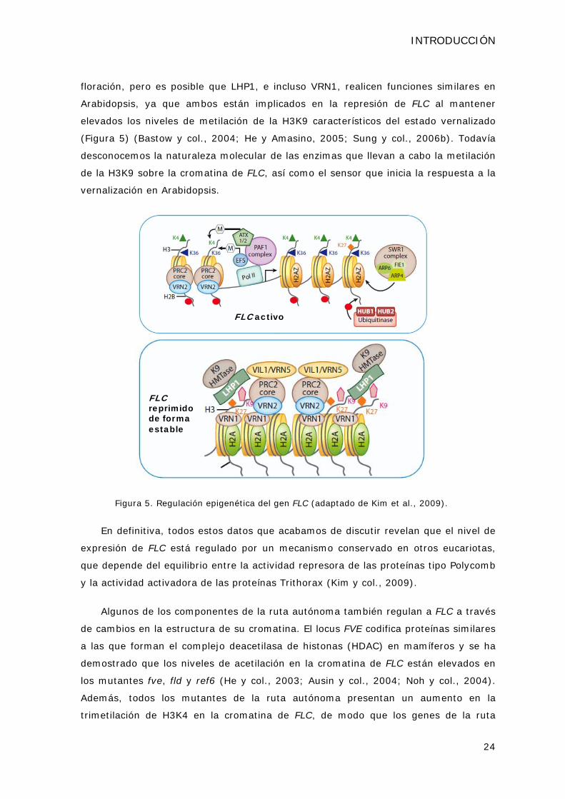
INTRODUCCIÓN
24
floración, pero es posible que LHP1, e incluso VRN1, realicen funciones similares en
Arabidopsis, ya que ambos están implicados en la represión de FLC al mantener
elevados los niveles de metilación de la H3K9 característicos del estado vernalizado
(Figura 5) (Bastow y col., 2004; He y Amasino, 2005; Sung y col., 2006b). Todavía
desconocemos la naturaleza molecular de las enzimas que llevan a cabo la metilación
de la H3K9 sobre la cromatina de FLC, así como el sensor que inicia la respuesta a la
vernalización en Arabidopsis.
FLC activo
FLCreprimido de forma estable
Figura 5. Regulación epigenética del gen FLC (adaptado de Kim et al., 2009).
En definitiva, todos estos datos que acabamos de discutir revelan que el nivel de
expresión de FLC está regulado por un mecanismo conservado en otros eucariotas,
que depende del equilibrio entre la actividad represora de las proteínas tipo Polycomb
y la actividad activadora de las proteínas Trithorax (Kim y col., 2009).
Algunos de los componentes de la ruta autónoma también regulan a FLC a través
de cambios en la estructura de su cromatina. El locus FVE codifica proteínas similares
a las que forman el complejo deacetilasa de histonas (HDAC) en mamíferos y se ha
demostrado que los niveles de acetilación en la cromatina de FLC están elevados en
los mutantes fve, fld y ref6 (He y col., 2003; Ausin y col., 2004; Noh y col., 2004).
Además, todos los mutantes de la ruta autónoma presentan un aumento en la
trimetilación de H3K4 en la cromatina de FLC, de modo que los genes de la ruta

INTRODUCCIÓN
25
autónoma podrían actuar, directa o indirectamente, como represores de este tipo de
modificación (He y Amasino, 2005).
3.3 Regulación epigenética de otros genes implicados en el control del tiempo de floración
FLC no es el único gen implicado en la transición floral que está regulado a nivel
epigenético (Farrona y col., 2008). Las mutaciones en componentes de los complejos
PRC2 como CLF y EMBRYONIC FLOWER 2 (EMF2) no sólo afectan a la expresión de
FLC, sino que también regulan la expresión de FT (Jiang y col., 2008). Además, los
niveles de expresión de otra proteína tipo MADS box, AGL19, que actúa como un
inductor floral, también están regulados por un mecanismo dependiente del PRC2
(Schonrock y col., 2006).
Otros genes que regulan la estructura de la cromatina de FT incluyen a
LHP1/TFL2 y EARLY BOLTING IN SHORT DAYS (EBS). Las mutaciones en ambos loci
presentan un fenotipo de floración temprana y altos niveles de expresión del
integrador floral FT (Kotake y col., 2003; Pineiro y col., 2003; Takada y Goto, 2003).
Por otro lado, AtBRAHMA (AtBRM), una proteína de la familia de complejos
remodeladores de cromatina dependientes de ATP SWI/SNF2, está implicada en la
represión de la transición floral mediante la inhibición de la activación de CO y FT
(Farrona y col., 2004).
4. El papel de la degradación específica de proteínas en la transición floral en Arabidopsis thaliana
En los últimos años se ha realizado un gran avance en el estudio de la implicación
que tiene la ruta de degradación de proteínas dependiente de ubiquitina/proteosoma
26S en la regulación de distintos procesos en organismos eucariotas. En las plantas,
más del 50% de las proteínas totales están sometidas a un recambio semanal. Entre
los procesos regulados por esta ruta de degradación de proteínas en plantas se
incluyen, entre otros, embriogénesis, fotomorfogénesis, floración, señalización
hormonal, resistencia a enfermedades o senescencia (Smalle y Vierstra, 2004).
La función general de esta ruta es conjugar polímeros de ubiquitina a las
proteínas diana en un residuo de Lys y, de este modo, marcarlas para su posterior
degradación por el proteosoma. La ubiquitina es un polipéptido de 76 aminoácidos
que se une covalentemente a las proteínas que va a degradar mediante la acción de
tres enzimas: la activadora de ubiquitina (E1), la conjugadora de ubiquitina (E2) y la
ubiquitina ligasa (E3) (Moon et al., 2004). La enzima E1 forma un enlace tioéster con
el extremo carboxilo terminal de la ubiquitina en una reacción dependiente de ATP y

INTRODUCCIÓN
26
transfiere la ubiquitina así activada a la enzima E2. A su vez, la enzima E2 puede
transferir directamente la ubiquitina a la E3 en el caso del tipo HECT (Homologous
with E6-associated protein C-Terminus), o unirse a la E3 y entonces transferir la
ubiquitina a la proteína diana. En ambos casos la enzima E3 es la que confiere
especificidad por el sustrato. Generalmente este proceso se repite sucesivas veces y
permite la unión de múltiples moléculas de ubiquitina al sustrato (Figura 6). Se ha
demostrado que la poliubiquitinación de las proteínas es necesaria para su
degradación por el proteosoma (Moon y col., 2004).
Figura 6. Ruta de ubiquitinación y degradación de proteínas por el proteosoma (adaptada
de Deshaies y Joazeiro, 2009).
El proteosoma 26S es un complejo proteico formado por un núcleo cilíndrico 20S
con actividad proteasa, flanqueado en cada extremo por una partícula reguladora
19S. La partícula 19S es la encargada de reconocer los sustratos ubiquitinados y de
eliminar la cadena de ubiquitina de la proteína que se va a degradar (Moon y col.,
2004).
En Arabidopsis aproximadamente el 5% del proteoma codifica componentes
relacionados con la ruta ubiquitina/proteosoma 26S. De ellos, unos 1200 genes
podrían codificar posibles E3 ligasas (Santner y Estelle, 2010). Las E3 ubiquitina
ligasas engloban una amplia y diversa familia de proteínas que contienen, o bien un
dominio HECT, o un dominio RING/U-box. Las E3 tipo RING (por Really Interesting
New Gene) se pueden subdividir a su vez en aquellas compuestas por una sola
subunidad RING/U-box, y en las E3 tipo RING formadas por varias subunidades, que
incluyen las de tipo SCF (por SKP1, Cullin y F-box) o los complejos APC (Anaphase
Promoting Complex) (Moon y col., 2004; Stone y Callis, 2007). En las RING E3 que
actúan individualmente la especificidad de sustrato reside en la propia proteína RING.
En cambio, en los complejos E3 la subunidad encargada del reconocimiento del
sustrato es otra, y la proteína con el dominio RING participa en la interacción con la
enzima E2 (Deshaies y Joazeiro, 2009).

INTRODUCCIÓN
27
Dentro de los mecanismos que regulan la transición floral en Arabidopsis, la
degradación específica de proteínas a través de la ruta ubiquitina/26S proteosoma ha
cobrado mayor relevancia en los últimos años. En concreto, se han descrito varias
mutaciones en componentes de E3 ligasas que afectan a la regulación fotoperiódica
del tiempo de floración. Como ya hemos discutido anteriormente, los complejos
COP1-SPA participan en la degradación del promotor floral CO durante el periodo de
oscuridad (Laubinger y col., 2006; Jang y col., 2008; Liu y col., 2008c). COP1 es una
proteína que contiene un dominio RING, un dominio coiled-coil y un dominio WD40, y
que está conservada en plantas superiores y vertebrados. En plantas, COP1 actúa
como una E3 ligasa que degrada tanto fotoreceptores como factores de transcripción
implicados en la transducción de las señales lumínicas (Yi y Deng, 2005). COP1
interacciona con las proteínas SPA1-4 para formar complejos E3 ligasa funcionales
que reprimen la fotomorfogénesis en plántulas cultivadas en oscuridad, así como la
transición floral (Laubinger y col., 2006). Recientemente, se ha demostrado que las
proteínas CULLIN 4 (CUL4) y DAMAGED DNA BINDING PROTEIN 1 (DDB1), que
forman el esqueleto de una variedad de complejos SCF, se unen a los complejos
COP1-SPA y participan en la regulación de la fotomorfogénesis y del tiempo de
floración (Chen y col., 2010). De forma análoga, el mutante temprano red and far red
insensitive 2 (rfi2) también fue descrito por estar afectado en las respuestas de
fotomorfogénesis (Chen y Ni, 2006a). RFI2 codifica una proteína nuclear con un
dominio RING que, al contrario que COP1, afecta a los niveles de expresión de CO
(Chen y Ni, 2006b). Más recientemente, se ha descrito otro represor de CO que actúa
en DC, DAY NEUTRAL FLOWERING (DNF). DNF es una proteína unida a membrana
que también presenta un dominio RING (Morris y col., 2010). Las dianas de RFI y DNF
que participan en el control de la transición floral no se han identificado hasta el
momento. Por otro lado, los receptores de luz azul ZEITLUPE (ZTL), LOV KELCH
PROTEIN 2 (LKP2) y FKF1 son proteínas tipo F box que pertenecen a complejos SCF y
que también están involucradas en el control del tiempo de floración (Nelson y col.,
2000; Somers y col., 2000; Jarillo y col., 2001; Schultz y col., 2001). Como hemos
descrito anteriormente, estas proteínas F box participan en la degradación de CDF1 y
CDF2, que son represores transcripcionales de CO (Imaizumi y col., 2003; Sawa y
col., 2007; Fornara y col., 2009).
También se ha descrito que el represor floral FLC está regulado por la ruta
ubiquitina/26S proteosoma. La proteína SINAT5 es una E3 tipo RING que interacciona
con FLC y participa en su degradación en ensayos de ubiquitinación in vitro (Park y
col., 2007). De forma adicional, se ha descrito que SINAT5 también participa en la

INTRODUCCIÓN
28
regulación del tiempo de floración a través de la degradación de uno de los
componentes del reloj circadiano (Park y col., 2010a).
La relación entre los procesos de degradación específica de proteínas y las rutas
de señalización hormonal en plantas se ha caracterizado más en detalle. Como ya
hemos discutido, en Arabidopsis las giberelinas controlan el tiempo de floración, así
como otros procesos del desarrollo (Mutasa-Gottgens y Hedden, 2009). Las proteínas
DELLA, que regulan negativamente las respuestas a giberelinas, son degradadas por
un complejo SCF específico en respuesta a la presencia de esta hormona (Santner y
Estelle, 2010).
Por otra parte, el genoma de Arabidopsis contiene una familia de 27 enzimas con
actividad proteasa de ubiquitina (UBP) que están involucradas en la deubiquitinación
de proteínas (Liu y col., 2008d). Se ha descrito que las mutaciones en UBP15 y UBP26
presentan un fenotipo de floración temprano entre otros defectos del desarrollo (Liu y
col., 2008d; Schmitz y col., 2009). Además, datos recientes confirman que la enzima
UBP26 participa en la deubiquitinación de la histona H2B sobre la cromatina de FLC
(Schmitz y col., 2009).
De forma análoga a la ubiquitinación, hay otros procesos de señalización de
proteínas mediante la adición de una pequeña molécula entre los que se encuentra la
sumoilación (Wilkinson y Henley, 2010). El mecanismo de conjugación y
deconjugación de una molécula de SUMO (Small Ubiquitin-related Modifier) a una
proteína es similar al que se produce en el caso de la ubiquitina. Al contrario que la
poliubiquitinación, la sumoilación no señaliza a las proteínas para su degradación, sino
que puede afectar a su estabilidad, actividad, localización subcelular, etc. (Miura y
col., 2007a; Miura y Hasegawa, 2010). En Arabidopsis, se ha demostrado que la
enzima E3 ligasa de SUMO SIZ1 está implicada en el control del tiempo de floración y
en la aclimatación de las plantas al frío, entre otros procesos (Miura y col., 2007b; Jin
y Hasegawa, 2008). Por un lado, SIZ1 participa en la regulación de los niveles de FLC
a través de la represión de la actividad del componente de la ruta autónoma FLD (Jin
y Hasegawa, 2008). Por otro, SIZ1 participa en la sumoilación y estabilización de
ICE1, que es un regulador positivo de la respuesta de aclimatación a bajas
temperaturas (Miura y col., 2007b). En cambio, la E3 ligasa de ubiquitina HIGH
EXPRESSION OF OSMOTICALLY RESPONSIVE GENES 1 (HOS1) reprime la respuesta
de las plantas al frío mediante la degradación de ICE1 (Dong y col., 2006).
Curiosamente, la sumoilación mediada a través de SIZ1 reduce la poliubiquitinación
que lleva a cabo HOS1 sobre ICE1 (Miura y col., 2007b).

OBJETIVOS

OBJETIVOS
31
Además de las rutas genéticas promotoras de la floración, otro aspecto central de
la regulación de la transición floral es el papel funcional que desempeñan los
represores florales y cómo estos interaccionan con las rutas de inducción de la
floración para asegurar que este cambio del desarrollo tenga lugar en el momento
más apropiado. En este trabajo, nuestro interés principal ha sido profundizar en el
conocimiento de los mecanismos moleculares que regulan el tiempo de floración y, en
particular, la caracterización de algunos factores que inhiben el inicio de la floración
hasta que la planta se encuentra en las condiciones medioambientales óptimas, o
alcanza el nivel de desarrollo adecuado para florecer. Por ello, en la presente Tesis se
planteó como objetivo general la caracterización genética y molecular de varios
represores del tiempo de floración en Arabidopsis mediante el aislamiento inicial de
mutantes de floración temprana. Dicha caracterización ha conducido a la identificación
de proteínas implicadas en dos procesos clave en el control de la transición floral: los
procesos de remodelación de la estructura de la cromatina y los mecanismos de
degradación específica de proteínas. Como se expone a lo largo de este trabajo,
hemos identificado dos ortólogos en Arabidopsis de los componentes del complejo de
remodelación de cromatina Swr1 de levaduras: las proteínas ESD1/ARP6 y SWC6, que
participan en la regulación de la expresión del represor FLC. Por otro lado, también
hemos abordado la caracterización del papel que juega la E3 ligasa de ubiquitina
HOS1 en el control fotoperódico del tiempo de floración en Arabidopsis a través de la
regulación de la estabilidad de la proteína CO.
Para ello, hemos desarrollado los siguientes objetivos concretos:
1. Caracterización genética y molecular del gen ESD1/ARP6 como represor del
tiempo de floración en Arabidopsis.
2. Análisis funcional del papel de ESD1/ARP6 en los procesos de remodelación de la
estructura de la cromatina de FLC.
3. Caracterización genética del locus SWC6 y análisis de la relación funcional de
SWC6 con ESD1/ARP6.
4. Caracterización genética y molecular del locus ESD6/HOS1 como represor del
tiempo de floración en Arabidopsis.
5. Análisis funcional de ESD6/HOS1 en relación con su papel en la degradación
específica del promotor floral CO.


RESULTADOS


CAPÍTULO 1: EARLY IN SHORT DAYS 1 (ESD1) encodes
ACTIN-RELATED PROTEIN 6 (AtARP6), a putative component
of chromatin remodelling complexes that positively regulates
FLC accumulation in Arabidopsis.

ESD1 encodes ACTIN-RELATED PROTEIN 6 (ARP6)
37
Development 133, 1241-1252 (2006)
EARLY IN SHORT DAYS 1 (ESD1) encodes ACTIN-RELATED PROTEIN 6 (AtARP6), a putative component of chromatin remodelling complexes that positively regulates FLC accumulation in Arabidopsis Mar Martin-Trillo1,*, Ana Lázaro2,*, R. Scott Poethig3, Concepción Gómez-Mena1,†, Manuel A.
Piñeiro2, Jose M. Martinez-Zapater1 and Jose A. Jarillo2,‡ 1Departamento de Genética Molecular de Plantas, Centro Nacional de Biotecnología, C/ Darwin 3, Madrid 28049, Spain. 2Departamento de Biotecnología, Instituto Nacional de Investigación y Tecnología Agraria y Alimentaria, Ctra. de A Coruña, km 7, Madrid 28040, Spain. 3Plant Science Institute, Department of Biology, University of Pennsylvania, PA 19104, USA. *These authors contributed equally to this work †Present address: Department of Cell and Developmental Biology, John Innes Centre, Norwich NR4 7UH, UK ‡Author for correspondence (e-mail: [email protected])
We have characterized Arabidopsis esd1 mutations, which cause early flowering independently of
photoperiod, moderate increase of hypocotyl length, shortened inflorescence internodes, and
altered leaf and flower development. Phenotypic analyses of double mutants with mutations at
different loci of the flowering inductive pathways suggest that esd1 abolishes the FLC-mediated
late flowering phenotype of plants carrying active alleles of FRI and of mutants of the autonomous
pathway. We found that ESD1 is required for the expression of the FLC repressor to levels that
inhibit flowering. However, the effect of esd1 in a flc-3 null genetic background and the
downregulation of other members of the FLC-like/MAF gene family in esd1 mutants suggest that
flowering inhibition mediated by ESD1 occurs through both FLC- and FLC-like gene-dependent
pathways. The ESD1 locus was identified through a map-based cloning approach. ESD1 encodes
ARP6, a homolog of the actin-related protein family that shares moderate sequence homology with
conventional actins. Using chromatin immunoprecipitation (ChIP) experiments, we have
determined that ARP6 is required for both histone acetylation and methylation of the FLC
chromatin in Arabidopsis. KEY WORDS: Flowering time, Floral repression, Chromatin remodelling, Arabidopsis INTRODUCTION
The floral transition is highly regulated in many plant species to modulate flowering time in
response to environmental and endogenous factors, and to ensure reproductive success.
Arabidopsis thaliana is a facultative long-day (LD) species in which winter and summer annual
accessions can be distinguished. In winter annual accessions, flowering time is regulated by the
vernalization, photoperiod and gibberellin (GA) pathways (Boss et al., 2004; Komeda, 2004;
Puterill et al., 2004; Amasino, 2005). Winter annuals require exposure to an extended period of
cold (vernalization) to become flowering competent, thus preventing premature flowering in the fall
(Michaels and Amasino, 2000; Henderson and Dean, 2004). This requirement is mainly conferred
by dominant alleles at the FRIGIDA (FRI) (Johanson et al., 2000) and FLOWERING LOCUS C
(FLC) loci (Michaels and Amasino, 1999; Sheldon et al., 1999), as well as by other FLC-related
genes within the MAF clade (Scortecci et al., 2001; Ratcliffe et al., 2003; Werner et al., 2005).
Active alleles of FRI increase FLC expression to levels that delay flowering (Michaels and
Amasino, 1999; Sheldon et al., 1999). FLC is a MADS box transcription factor that acts to delay
flowering, in part by suppressing the expression of the floral promoters FT and SUPPRESSOR OF
OVEREXPRESSION OF CO 1 (SOC1), which function as integrators of flowering signals
(Kobayashi et al., 1999; Samach et al., 2000). Vernalization promotes flowering by overcoming the

ESD1 encodes ACTIN-RELATED PROTEIN 6 (ARP6)
38
effect of FRI and repressing FLC expression; this repression is stably maintained after plants are
returned to warm growth conditions, allowing plants to flower (Michaels and Amasino, 1999;
Sheldon et al., 1999). The photoperiod pathway promotes flowering in response to LD through the
activation of the floral integrators FT and SOC1. Mutations in photoperiod-pathway genes [e.g.
constans (co), fd, fe, fha/cryptochrome2 (cry2), ft, fwa and gigantea (gi)] delay flowering in LD but
have little effect on flowering time under short days (SD) (Searle and Coupland, 2004). The GA
pathway is required for flowering in non-inductive photoperiods, and mutants with reduced GA
levels are extremely delayed in flowering time under SD (Wilson et al., 1992).
Many summer annual accessions of Arabidopsis lack an active FRI allele (Johanson et al., 2000;
Gazani et al., 2003; Shindo et al., 2005). Under these circumstances, FLC expression is low and
flowering occurs rapidly without vernalization. In these accessions, the reduction of FLC
expression depends on the function of the autonomous pathway (Michaels and Amasino, 2001). In
fact, mutations in autonomous pathway genes [fca, flowering locus d (fld), fpa, fve, fy, flowering
locus k (flk) and luminidependens (ld)] cause a flowering delay under any photoperiod (Boss et al.,
2004) that is associated with higher FLC expression, and can be rescued by vernalization
(Michaels and Amasino, 1999; Sheldon et al., 1999; Michaels and Amasino, 2001).
Thus, transcriptional regulation of the FLC repressor is a central checkpoint in both winter and
summer annual accessions of Arabidopsis. Recently, the involvement of chromatin modification in
FLC regulation has been described (for a review, see He and Amasino, 2005). In non-vernalized
winter annual plants, FLC chromatin is in an active conformation and is enriched in modifications,
such as the acetylation of histones 3 (H3) and 4 (H4), and the trimethylation of lysine 4 of H3 (H3-
K4), which are hallmarks of active genes (He et al., 2003; Ausin et al., 2004; He et al., 2004). Late-
flowering autonomous pathway mutants also have increased levels of H3-K4 trimethylation and
histone acetylation compared with the rapid-flowering parental line (He et al., 2003; Ausin et al.,
2004; He et al., 2004; Kim et al., 2005). Many early flowering mutations suppressing the late
flowering phenotype of FRI-containing lines have identified components that are required to
maintain high levels of FLC expression. This is the case of mutants such as early flowering in short
days (efs), photoperiod independent early flowering 1 (pie1), early flowering 5 (elf5), vernalization
independence3 (vip3) and frigida-like1 (frl-1), and mutants in genes encoding components of the
PAF1 complex (ELF7, VIP4, VIP5 and VIP6/ELF8) (Zhang and Van Nocker, 2002; Noh and
Amasino, 2003; Zhang et al., 2003; Noh et al., 2004; He et al., 2004; Michaels et al., 2004; Oh et
al., 2004; Kim et al., 2005). Most of these mutations also appear to affect flowering in an FLC-
independent manner.
After exposure to an extended winter and the completion of vernalization, the level of
modifications associated with ‘active’ chromatin is reduced, and the histone tails of FLC chromatin
are deacetylated and become enriched in methylation of lysine 9 (K9) and 27 (K27) of H3 (Bastow
et al., 2004; Sung and Amasino, 2004), which are hallmarks of repressed genes (Orlando, 2003).
Mutants that are unable to reduce FLC transcript levels by vernalization or to maintain the
vernalised state have permitted the identification of some of the proteins participating in this
process, such as the chromatin remodelling factors VERNALIZATION INSENSITIVE 3 (VIN3) and
VERNALIZATION 2 (VRN2) (Gendall et al., 2001; Sung and Amasino, 2004), and a plant-specific
DNA-binding protein, VRN1 (Levy et al., 2002).
In summer annual accessions, reduced expression of FLC depends on the autonomous
pathway, and is associated with lower histone acetylation of FLC chromatin as a result of FVE and
FLD function (He et al., 2003; Ausin et al., 2004). Mutations in both genes cause FLC chromatin to

ESD1 encodes ACTIN-RELATED PROTEIN 6 (ARP6)
39
become more acetylated at H3 and H4 concomitantly with an increase in FLC expression (He et
al., 2003; Ausin et al., 2004).
Here, we report the identification of EARLY IN SHORT DAYS1 (ESD1), a gene that is required
for the maintenance of FLC expression. The esd1 mutation causes early flowering through the
reduction of FLC expression, although the mutation also appears to affect flowering through other
FLC-like repressors. Using a map-based approach, we have determined that ESD1 encodes
ARP6, a member of the actin-related protein family that share moderate sequence homology and
basal structure with conventional actins. Recently, ARPs and actins have been discovered in the
nucleus as integral components of several chromatin remodelling and histone acetyltransferase
(HAT) complexes (Schafer and Schroer, 1999; Galarneau et al., 2000; Rando et al., 2000; Shen et
al., 2000; Olave et al., 2002; Blessing et al., 2004). We present evidence that ESD1 is needed to
achieve the levels of both H3 acetylation and H3-K4 methylation required for high FLC expression.
MATERIALS AND METHODS
Genetic stocks and growth conditions
Mutant seed stocks used were in the Landsberg erecta (Ler) and Columbia (Col) genetic backgrounds, and
were obtained from the Arabidopsis Biological Resource Centre (ABRC) of Ohio State University (Columbus,
USA), the Nottingham Arabidopsis Centre (NASC) in UK and personal donations. The monogenic fve-1, fca-
1, co-2 and gi-3 mutants were described by Koornneef et al. (Koornneef et al., 1991); flc-3 was described by
Michaels and Amasino (Michaels and Amasino, 2001) and the Col FRISf2 lines were described by Lee et al.
(Lee et al., 1995). GA-deficient ga1-3 and ga2-1 mutants were described by Koornneef and van der Veen
(Koornneef and van der Veen, 1980) and spy-5 by Jacobsen and Olszewski (Jacobsen and Olszewski, 1993).
The origin of the esd1-1 to esd1-9 alleles is summarized in Table 1. The esd1-10 allele in Col corresponds to
the T-DNA line Wisc Ds-Lox 289_29L8, and was kindly provided by the ABRC. We confirmed that all esd1
mutations were allelic by their failure to complement the early flowering phenotype in F1 plants derived from
crosses between them. Plants were grown in plastic pots containing a mixture of substrate and vermiculite
(3:1). Controlled environmental conditions were provided in growth chambers at 21°C and 80% relative
humidity. Plants were illuminated with cool-white fluorescent lights (approximately 120 E m–2 second–1). LD
conditions consisted of 16 hours of light followed by 8 hours of darkness; SD conditions consisted of 8 hours
of light followed by 16 hours of darkness.
Phenotypic analyses
Total leaf number was scored as the number of main leaves in the rosette (excluding cotyledons) plus the
number of leaves in the inflorescence at the time of opening of the first flower (Koornneef et al., 1991).
Cauline, adult and juvenile leaves were scored independently. Rosette leaves lacking abaxial trichomes were
considered as juvenile leaves (Telfer et al., 1997). Floral organs were analyzed by scanning electron
microscopy as described (Ruiz-Garcia et al., 1997).
Genetic analysis
Double mutants were constructed by crossing the monogenic esd1-2 mutant with lines carrying the mutations
co-2, gi-3, fve-1, fca-1, ga1-3, ga2-1 or spy-5. esd1-3 was crossed with a line carrying the flc-3 mutation in
Col and with Col FRI Sf-2 (Lee and Amasino, 1995). Double mutants were isolated from selfed F2 progeny
that showed the esd1 phenotype and that segregated for the second mutation.
Molecular characterization of the esd1 alleles and map-based cloning
The esd1-2 mutation was initially mapped to chromosome 3 between markers GAPab and nga6, using the
cleaved-amplified polymorphic sequence (CAPS) and the simple sequence length polymorphism (SSLP)
molecular markers indicated in Table S1 in the supplementary material. Additional analysis of 925 esd1-like

ESD1 encodes ACTIN-RELATED PROTEIN 6 (ARP6)
40
F2 plants allowed us to locate ESD1 to a pericentromeric region of 1.4 cM, between the T8N9 and ATA1
markers (see Table S2 in the supplementary material). To fine map the esd1 mutation within the interval
deleted in the esd1-1 and esd1-6 mutant plants, which is located between the 5F21A14 and 1T14A11
markers, we designed specific PCR molecular markers (see Table S3 in the supplementary material) that
were used to amplify the genomic DNA of each esd1 mutant allele, in order to score the presence or absence
of the amplified product. Southern blot hybridizations with genomic DNA were performed to confirm the PCR
results (data not shown).
Plant transformation
Four overlapping binary TAC clones (JAtY74I04, JAtY64M05, JAtY54G02, JAtY49O18) spanning the
minimum deleted region in the esd1 alleles were obtained from the Genomic Arabidopsis Resource Network
(GARNET) and introduced into the esd1-3 allele by Agrobacterium tumefaciens-mediated transformation
using the floral-dip method (Clough and Bent, 1998). The Agrobacterium strain used was C58C1.
Transformant plants were selected on soil by spraying seedlings with BASTA.
Only esd1-3 mutant plants transformed with the JAtY T74I04 TAC clone that spans from position 28823 bp
of T4P3 BAC clone to 78776 bp of T14A11 BAC clone, showed complementation of the early flowering
phenotype. To check whether the integration of the TAC clone was complete in transformed esd1-3 plants,
we used a set of specific molecular markers (see Table S3 in the supplementary material) contained in the
deleted region. We chose markers that amplify PCR products over genomic DNA extracted from wild-type
plants, but not from esd1-3 mutant plants. In this way, we demonstrated that the genomic region of the
JAtYT74I04 TAC clone integrated in the complementing transgenic plants contained only two ORFs predicted
to encode proteins, At3g33520 and At3g33530. The rest of the ORFs present in this region correspond to
pseudogenes and retrotransposon elements.
Expression analysis
Total RNA was isolated using TRIzol (Invitrogen-Gibco), electrophoresed and transferred onto Hybond N+
membranes (Amersham), following described protocols. For the FLC probe, we used a 700 bp EcoRI/SphI
fragment from pFLC lacking the MADS-box domain (Michaels and Amasino, 1999). As loading controls, we
used a 305-bp EcoRI fragment of the cauliflower 18S ribosomal DNA gene. ARP6 transcript levels were
assayed by RT-PCR. cDNA was prepared by reverse transcription of total RNA from Arabidopsis roots,
stems, cauline leaves, floral buds and flowers, according to described procedures (Piñeiro et al., 2003). ARP6
gene-specific primers, 5´-GAGCTTCGACCACTTGTCCCAGAT-3´ and 5´-GCATTACAATAT
ACGACAAATAATGTG-3´, were designed to amplify the C-terminal end of the coding region, including the
last intron and a portion of the 3´ untranslated region. For low abundance mRNAs, such as the MAF, FT and
SOC1 genes, we also performed reverse transcriptase-mediated PCR, according to described procedures
(Scortecci et al., 2001; Piñeiro et al., 2003; Ratcliffe et al., 2003). UBIQUITIN 10 (UBQ10) was used as
control in these experiments.
Histochemical -glucuronidase assays
esd1-2 fca-1 FLC:GUS plants were obtained by crossing esd1-2 with fca-1 plants carrying a 6 kb FLC:GUS
translational fusion construct (Sheldon et al., 2002). GUS activity in fca-1 and esd1-2 fca-1 FLC:GUS plants
was revealed by incubation in 100 mM NaPO4 (pH 7.2), 2.5 mM 5-bromo-4-chloro-3-indolyl-β-D-glucuronide,
0.5 mM K3Fe(CN)6, 0.5 mM K4Fe(CN)6 and 0.25% Triton X-100. Plant tissue was incubated at 37°C for 20
hours. After staining, chlorophyll was cleared from the samples by dehydration through an ethanol series.
ChIP assays and PCR
ChIP assays were carried out as described (Ausin et al., 2004). Chromatin proteins and DNA were cross
linked in 10-day-old Col, esd1-3, FRI, esd1-3FRI, Ler, esd1-2, fca-1, esd1-2 fca-1, fve-1 and esd1-2 fve-1
seedlings by formaldehyde fixation. After chromatin isolation, the H3 acetylated and methylated fractions
were immunoprecipitated using specific antibodies to acetylated K9 and K14, and trimethylated K4, residues

ESD1 encodes ACTIN-RELATED PROTEIN 6 (ARP6)
41
(06-599 and 07-473 from Upstate Biotechnology, respectively). Cross-links were reversed by incubations at
65°C for 2 hours, and DNA was purified with QIAquick spin columns (QIAGEN) and eluted in 40 l of TE (pH
8.0). Semiquantitative PCR was used to amplify six different fragments of the FLC gene (Michaels and
Amasino, 1999) (details and primer sequences are available on request). All PCR reactions and quantification
of the amplified DNA were done as described previously (Ausin et al., 2004). We carried out three
independent experiments and data provided in Fig. 7 are from one representative. UBQ10 served as an
internal control for the ChIP analysis. To calculate the fold enrichment in H3 acetylation or methylation, FLC
was first normalized to UBQ10 in each sample, and, subsequently, these values were normalized against
their respective wild-type controls. RESULTS
esd1 mutants are early flowering and display a pleiotropic vegetative and reproductive
phenotype
Mutants at the ESD1 locus were independently identified in screens for Ler mutations conferring
early flowering under SD (esd1-1 and esd1-2) or for Col mutations that accelerate developmental
phase transitions (esd1-3 to esd1-9) (Table 1). All of the selected alleles produced a similar array
of phenotypes, independently of their genetic background. Plants homozygous for esd1 mutations
were early flowering under both LD and SD photoperiods (Table 1), showing a more extreme
phenotype under SD (Table 1, Fig. 1A,B). The fact that esd1 mutants flower earlier under inductive
photoperiods indicates that the mutations do not abolish the photoperiod responses. Earliness was
associated with a reduction in the length of all developmental phases of the plant (Fig. 1C), based
on leaf shape and leaf trichome distribution (Telfer et al., 1997). This reduction was more dramatic
in the case of adult rosette leaves, which were almost absent from esd1 mutants grown under LD
and highly reduced in esd1 mutant plants under SD (Fig. 1C). This behaviour is similar to that
exhibited by other early flowering mutants such as esd4 and ebs, which also show a major
reduction of the adult vegetative phase (Gomez-Mena et al., 2001; Reeves et al., 2002).
Fig. 1. The flowering phenotype of esd1 mutants. (A) Wild-type Ler and esd1-2 2-week-old plants grown
under LD. (B) Ler and esd1-2 4-week-old plants grown under SD. (C) Histogram comparing the number of
juvenile, adult and cauline leaves in Ler and esd1 mutants. Plants were grown under both LD and SD. (D) Ler
and esd1-2 3.5-week-old plants grown under LD.

ESD1 encodes ACTIN-RELATED PROTEIN 6 (ARP6)
42
Apart from their flowering time phenotype, esd1 mutants also displayed a complex pleiotropic
vegetative and reproductive phenotype. esd1 mutants show a moderate increase in hypocotyl
length but have shortened inflorescence internodes. Furthermore, esd1 leaves are smaller and
more curled than wild-type leaves, and usually have serrated margins (Fig. 1D). esd1 flowers are
smaller than wild-type ones (Fig. 2C) and frequently bear extra perianth organs. This phenotype
was more extreme under SD, where esd1 flowers contained an average of two extra sepals and
two extra petals per flower (Fig. 2B,E,G). Mutant flowers showed a reduced fertility that was
associated with a reduction in the amount of pollen and approximately a 50% reduction in seed set
when compared with wild-type plants. Mutant carpels were much smaller and the filaments of the
stamens of mutant flowers were shorter than those of wild-type plants (Fig. 2E). Concomitantly,
siliques were approximately 50% shorter in esd1 mutants than in wild-type plants (Fig. 2F). Thus,
mutations at the ESD1 locus directly or indirectly alter multiple aspects of plant development.
Fig. 2. The inflorescence phenotype of esd1 mutants.
(A) Apex of 3-week-old Ler plants. (B) Apex of 2.5-week-old esd1 mutant plants, showing open flowers with
extra sepals and petals. (C) Detached flowers showing the increased number of sepals and petals in esd1
mutant lowers. (D,E) Scanning electron micrographs of Ler (D) and esd1 (E) flowers of plants grown under
SD. s, sepal; p, petal. (F) A comparison of silique shape and length in Ler, esd1-1 and esd1-2 plants. (G)
Number of sepals, petals, stamens and carpels in Ler and esd1 mutants. Plants were grown under both LD
and SD. Bars represent the standard error.
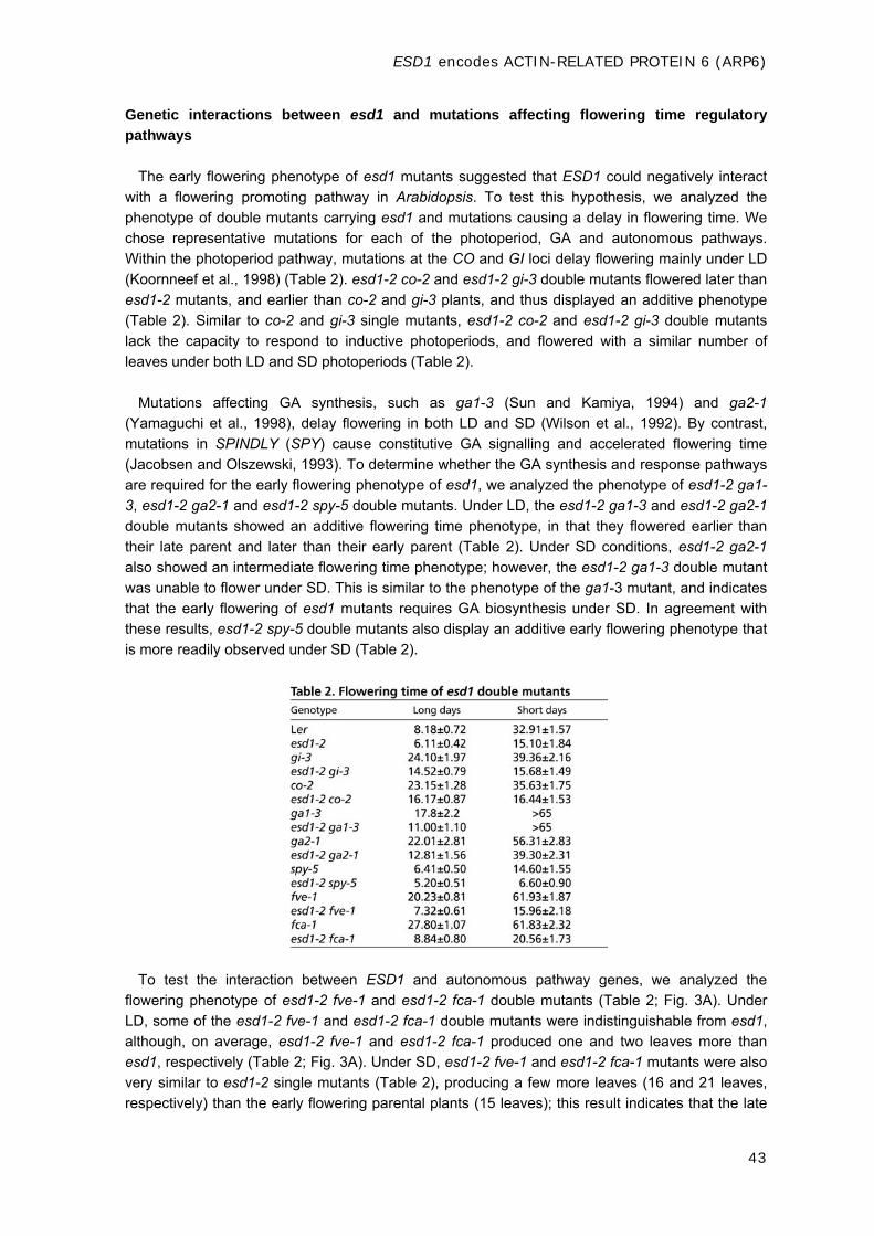
ESD1 encodes ACTIN-RELATED PROTEIN 6 (ARP6)
43
Genetic interactions between esd1 and mutations affecting flowering time regulatory
pathways
The early flowering phenotype of esd1 mutants suggested that ESD1 could negatively interact
with a flowering promoting pathway in Arabidopsis. To test this hypothesis, we analyzed the
phenotype of double mutants carrying esd1 and mutations causing a delay in flowering time. We
chose representative mutations for each of the photoperiod, GA and autonomous pathways.
Within the photoperiod pathway, mutations at the CO and GI loci delay flowering mainly under LD
(Koornneef et al., 1998) (Table 2). esd1-2 co-2 and esd1-2 gi-3 double mutants flowered later than
esd1-2 mutants, and earlier than co-2 and gi-3 plants, and thus displayed an additive phenotype
(Table 2). Similar to co-2 and gi-3 single mutants, esd1-2 co-2 and esd1-2 gi-3 double mutants
lack the capacity to respond to inductive photoperiods, and flowered with a similar number of
leaves under both LD and SD photoperiods (Table 2).
Mutations affecting GA synthesis, such as ga1-3 (Sun and Kamiya, 1994) and ga2-1
(Yamaguchi et al., 1998), delay flowering in both LD and SD (Wilson et al., 1992). By contrast,
mutations in SPINDLY (SPY) cause constitutive GA signalling and accelerated flowering time
(Jacobsen and Olszewski, 1993). To determine whether the GA synthesis and response pathways
are required for the early flowering phenotype of esd1, we analyzed the phenotype of esd1-2 ga1-
3, esd1-2 ga2-1 and esd1-2 spy-5 double mutants. Under LD, the esd1-2 ga1-3 and esd1-2 ga2-1
double mutants showed an additive flowering time phenotype, in that they flowered earlier than
their late parent and later than their early parent (Table 2). Under SD conditions, esd1-2 ga2-1
also showed an intermediate flowering time phenotype; however, the esd1-2 ga1-3 double mutant
was unable to flower under SD. This is similar to the phenotype of the ga1-3 mutant, and indicates
that the early flowering of esd1 mutants requires GA biosynthesis under SD. In agreement with
these results, esd1-2 spy-5 double mutants also display an additive early flowering phenotype that
is more readily observed under SD (Table 2).
To test the interaction between ESD1 and autonomous pathway genes, we analyzed the
flowering phenotype of esd1-2 fve-1 and esd1-2 fca-1 double mutants (Table 2; Fig. 3A). Under
LD, some of the esd1-2 fve-1 and esd1-2 fca-1 double mutants were indistinguishable from esd1,
although, on average, esd1-2 fve-1 and esd1-2 fca-1 produced one and two leaves more than
esd1, respectively (Table 2; Fig. 3A). Under SD, esd1-2 fve-1 and esd1-2 fca-1 mutants were also
very similar to esd1-2 single mutants (Table 2), producing a few more leaves (16 and 21 leaves,
respectively) than the early flowering parental plants (15 leaves); this result indicates that the late
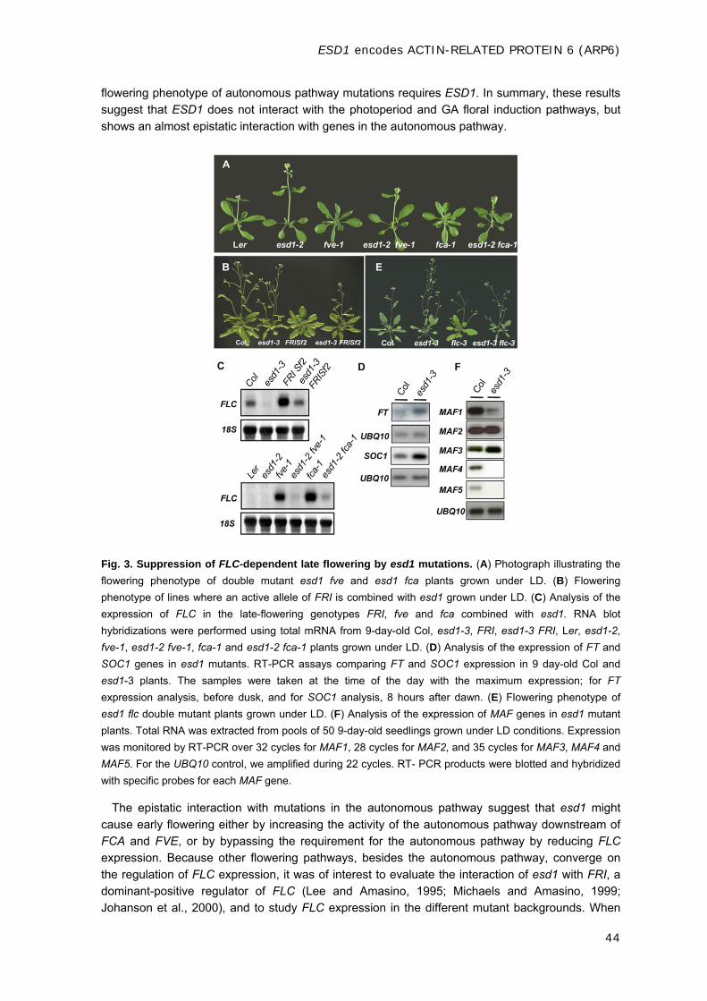
ESD1 encodes ACTIN-RELATED PROTEIN 6 (ARP6)
44
flowering phenotype of autonomous pathway mutations requires ESD1. In summary, these results
suggest that ESD1 does not interact with the photoperiod and GA floral induction pathways, but
shows an almost epistatic interaction with genes in the autonomous pathway.
Fig. 3. Suppression of FLC-dependent late flowering by esd1 mutations. (A) Photograph illustrating the
flowering phenotype of double mutant esd1 fve and esd1 fca plants grown under LD. (B) Flowering
phenotype of lines where an active allele of FRI is combined with esd1 grown under LD. (C) Analysis of the
expression of FLC in the late-flowering genotypes FRI, fve and fca combined with esd1. RNA blot
hybridizations were performed using total mRNA from 9-day-old Col, esd1-3, FRI, esd1-3 FRI, Ler, esd1-2,
fve-1, esd1-2 fve-1, fca-1 and esd1-2 fca-1 plants grown under LD. (D) Analysis of the expression of FT and
SOC1 genes in esd1 mutants. RT-PCR assays comparing FT and SOC1 expression in 9 day-old Col and
esd1-3 plants. The samples were taken at the time of the day with the maximum expression; for FT
expression analysis, before dusk, and for SOC1 analysis, 8 hours after dawn. (E) Flowering phenotype of
esd1 flc double mutant plants grown under LD. (F) Analysis of the expression of MAF genes in esd1 mutant
plants. Total RNA was extracted from pools of 50 9-day-old seedlings grown under LD conditions. Expression
was monitored by RT-PCR over 32 cycles for MAF1, 28 cycles for MAF2, and 35 cycles for MAF3, MAF4 and
MAF5. For the UBQ10 control, we amplified during 22 cycles. RT- PCR products were blotted and hybridized
with specific probes for each MAF gene.
The epistatic interaction with mutations in the autonomous pathway suggest that esd1 might
cause early flowering either by increasing the activity of the autonomous pathway downstream of
FCA and FVE, or by bypassing the requirement for the autonomous pathway by reducing FLC
expression. Because other flowering pathways, besides the autonomous pathway, converge on
the regulation of FLC expression, it was of interest to evaluate the interaction of esd1 with FRI, a
dominant-positive regulator of FLC (Lee and Amasino, 1995; Michaels and Amasino, 1999;
Johanson et al., 2000), and to study FLC expression in the different mutant backgrounds. When

ESD1 encodes ACTIN-RELATED PROTEIN 6 (ARP6)
45
the FRI allele introgressed from the San Feliu-2 ecotype (FRI-Col) (Lee and Amasino, 1995) was
combined with the esd1-3 mutation in a Col genetic background, plants showed an additive
phenotype in which the FRI late-flowering phenotype was only partially suppressed by esd1-3
(Table 3, Fig. 3B).
In order to check if esd1 suppresses the effect of the autonomous pathway mutations and FRI
by reducing FLC mRNA levels, we compared the abundance of the FLC mRNA in wild-type, esd1-
2, fca-1, esd1-2 fca-1, fve-1, esd1-2 fve-1, FRI and esd1-3 FRI seedlings (Fig. 3C). FLC mRNA
was present at higher levels in both fca-1 and fve-1 mutants and in FRI-containing lines than in
wildtype plants, as has been previously shown (Michaels and Amasino, 1999; Sheldon et al.,
1999). FLC transcript levels in FRI and in fca-1 and fve-1 mutants were decreased by the esd1
lesion (Fig. 3C). FLC mRNA levels were also compared between wild-type plants and esd1
mutants. Because FLC is expressed at a low level in Ler wild-type plants, we could not observe a
clear reduction in its expression in the esd1 alleles isolated in Ler background. However, we were
able to detect a reduction in the FLC expression in the esd1 alleles isolated in Col background,
which bears an FLC allele expressed at higher levels (Fig. 3C). In summary, ESD1 is required to
maintain high FLC expression levels, either as promoted by FRI or by mutations that impair the
autonomous pathway, and, consistent with the genetic analysis, esd1 mutations suppress the
increase in FLC expression caused by autonomous pathway mutations more effectively than that
caused by active FRI alleles. In agreement with this scenario, the expression of the floral integrator
genes FT and SOC1, normally repressed by FLC (Moon et al., 2003), was upregulated in the esd1
mutants (Fig. 3D).
Although the effects of esd1 mutations on flowering time are more readily observed in the late-
flowering FRI and autonomous pathway mutant backgrounds, as discussed above, the fact that
esd1 mutants also flower earlier than the rapid-flowering wild-type strains Ler and Col (Fig. 1 and
Table 1) suggests that, in addition to regulating FLC expression, ESD1 plays other roles in the
control of flowering time. To determine the fraction of the esd1 early-flowering phenotype that is
independent of the effect of the esd1 mutation on FLC expression, we analyzed the phenotypic
effect of the esd1-3 mutation in an flc null (flc-3) genetic background (Michaels and Amasino,
1999) under both LD and SD. When combined with flc-3, the esd1 mutation significantly reduces
the number of leaves produced by flc-3 under both photoperiods (Fig. 3E and Table 3), confirming
that esd1 mutations have an FLC-independent effect on flowering time. Indeed, loss of function of
ESD1 also resulted in downregulation of some other members of the FLC/MAF gene family,
particularly MAF1, MAF4 and MAF5 (Fig. 3F). RT-PCR analysis indicated a modest but
reproducible decrease in MAF1 gene expression and a marked silencing of the MAF4 and MAF5
genes, suggesting that these MAF gene family members represent additional regulatory targets of
ESD1.

ESD1 encodes ACTIN-RELATED PROTEIN 6 (ARP6)
46
ESD1 encodes ACTIN-RELATED PROTEIN 6 (ARP6)
To understand the molecular function of ESD1, we decided to identify the gene responsible for
the observed phenotypes in the mutant by a map-based cloning approach. For this, 925 esd1 F2
plants derived from the cross between esd1-2 and Col were screened with the markers shown in
Table S1 in the supplementary material. This allowed us to locate ESD1 south of the T8N9 marker
and north of the ATA1 marker, close to the pericentromeric region of chromosome 3 (see markers
used in Table S2 in the supplementary material). Owing to the severe suppression of
recombination in the vicinity of the centromere and because this chromosomal region is almost
completely sequenced, we designed an alternative strategy to complete the identification of the
ESD1 gene, based on the observation that all of the isolated alleles harbour a deletion in the
pericentromeric region of chromosome 3. We identified the shorter overlapping genomic region
that was deleted in all of the esd1 alleles by using PCR molecular markers to amplify specific
genomic DNA fragments from all of the esd1 alleles, and looked for the presence or absence of an
amplified product (see Table S3 in the supplementary material). In this way, we delimited the
ESD1 locus to a deleted genomic region between the 5F21A14 and 1T14A11 markers. This region
spans three overlapping BAC clones, F21A14, T4P3 and T14A11 (Fig. 4A), and is enriched in
retrotransposon and transposase elements, pseudogenes and highly repeated sequences.
Fig. 4. Identification of ESD1. (A) Map-based cloning of ESD1. The genetic interval, molecular markers and
BAC clones in the ESD1 region are shown. The number of recombinant events between molecular markers is
given in parentheses. The centromere is located between the T15D2 and T25F15 markers
(http://www.arabidopsis.org/info/agicomplete.jsp). GAP indicates the existence of genomic regions of
unknown size, where it was not possible to get overlapping BAC clones. Gray bars correspond to the deleted
region in each esd1 allele. The ESD1 locus was delimited to a deleted overlapping genomic region between
the 5F21A14 and 1T14A11 molecular markers. Mb, megabases. (B) Complementation of the esd1 mutant.

ESD1 encodes ACTIN-RELATED PROTEIN 6 (ARP6)
47
Col, esd1-3 and TN 18.1, a transgenic esd1-3 plant containing the genomic region harbouring open reading
frames At3g33520 y At3g33530, shown at the time of bolting initiation. (C) Flowering phenotype of esd1-10, a
T-DNA insertion allele. Left, Col plant; right, a homozygous plant for the T-DNA insertion within the
At3g33520 gene (Wisc Ds-Lox 289 line), showing an early flowering phenotype. RT-PCR analyses of the
expression of At3g33520 in esd1-10 show no expression of this gene in the T-DNA mutant, indicating that it is
a loss-of-function allele.
Subsequently, different overlapping binary TAC clones spanning the deleted region were
identified and introduced into the esd1-3 allele by Agrobacterium-mediated transformation, in order
to identify those that complemented the mutant phenotype. Only certain transgenic esd1-3 mutant
plants transformed with JAtYT74I04 TAC flowered at a similar time as wild-type plants, and had
lost the pleiotropic phenotype of esd1 mutant plants (Fig. 4B). The analysis of these transgenic
plants showed that TAC clone integration had been incomplete (see Materials and methods), and
that only in those lines that contained the genomic region harbouring open reading frames
At3g33520 y At3g33530 had the mutant phenotype been complemented, suggesting that one of
those ORFs represent ESD1. To determine which one of them corresponded to ESD1, we
searched for T-DNA insertions within the At3g33520 and At3g33530 ORFs and identified the Wisc
Ds-Lox 289_29L8 line for At3g33520 and the SALK_003098 line for At3g33530 (Alonso et al.,
2003). We obtained seeds of these lines and identified plants homozygous for the T-DNA
insertions. Only the plants that harbour a T-DNA insertion in At3g33520 flowered early under both
LD and SD (producing around nine and 29 leaves, respectively; wild-type plants produce 14
leaves in LD and 66 in SD), and showed a pleiotropic phenotype similar to that of esd1 mutants
regarding leaf shape, extra perianth organs and small siliques (Fig. 4C). Reverse-transcription
(RT-PCR) analyses showed no expression of the At3g33520 mRNA in these insertional mutant
plants (Fig. 4C), indicating that the T-DNA insertion causes a loss-of-function allele.
Complementation tests confirmed that this T-DNA mutation was allelic to esd1. Thus, we refer to
the Wisc Ds-Lox 289_29L8 line as the esd1-10 allele, and conclude that the ESD1 locus
corresponds to the At3g33520 gene.
To determine the genomic structure of ESD1, a cDNA was identified and sequenced. The ESD1
gene possesses six exons and five introns, and encodes a protein of 421 amino acids (Fig. 5A).
This protein corresponds to ARP6, a member of the actin-related protein family that shares
moderate sequence homology and basal structure with conventional actins, but it has two peptide
insertions that seemingly provide divergent surface features from actins (Fig. 5B). ARPs are
normally grouped into several classes or subfamilies that are highly conserved in a wide range of
eukaryotes, from yeast to plants and humans (Goodson and Hawse, 2002). Database searches
with the AtARP6 protein sequence identified eight potential ARP proteins in Arabidopsis (ARP2-
ARP9) (McKinney et al., 2002). In particular, AtARP6 is a likely ortholog of a group of less-
characterized ARPs, including ARP6s from yeast, C. elegans, fruit fly and humans (Fig. 5B). RT-
PCR analysis revealed that ARP6 mRNA is detected in most plant organs, with the highest levels
found in roots and floral buds (Fig. 5C). Lower levels were detected in cauline leaves, stems and
flowers. These results indicate that ARP6 is expressed ubiquitously.
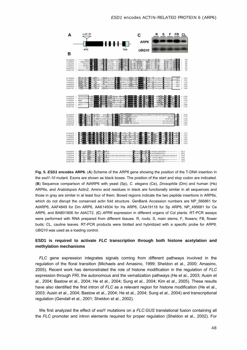
ESD1 encodes ACTIN-RELATED PROTEIN 6 (ARP6)
48
Fig. 5. ESD1 encodes ARP6. (A) Scheme of the ARP6 gene showing the position of the T-DNA insertion in
the esd1-10 mutant. Exons are shown as black boxes. The position of the start and stop codon are indicated.
(B) Sequence comparison of AtARP6 with yeast (Sp), C. elegans (Ce), Drosophila (Dm) and human (Hs)
ARP6s, and Arabidopsis Actin2. Amino acid residues in black are functionally similar in all sequences and
those in gray are similar in at least four of them. Boxed regions indicate the two peptide insertions in ARP6s,
which do not disrupt the conserved actin fold structure. GenBank Accession numbers are NP_566861 for
AtARP6, AAF4849 for Dm ARP6, AAK14934 for Hs ARP6, CAA19116 for Sp ARP6, NP_495681 for Ce
ARP6, and BAB01806 for AtACT2. (C) APR6 expression in different organs of Col plants. RT-PCR assays
were performed with RNA prepared from different tissues. R, roots; S, main stems; F, flowers; FB, flower
buds; CL, cauline leaves. RT-PCR products were blotted and hybridized with a specific probe for ARP6.
UBQ10 was used as a loading control.
ESD1 is required to activate FLC transcription through both histone acetylation and
methylation mechanisms
FLC gene expression integrates signals coming from different pathways involved in the
regulation of the floral transition (Michaels and Amasino, 1999; Sheldon et al., 2000; Amasino,
2005). Recent work has demonstrated the role of histone modification in the regulation of FLC
expression through FRI, the autonomous and the vernalization pathways (He et al., 2003; Ausin et
al., 2004; Bastow et al., 2004; He et al., 2004; Sung et al., 2004; Kim et al., 2005). These results
have also identified the first intron of FLC as a relevant region for histone modification (He et al.,
2003; Ausin et al., 2004; Bastow et al., 2004; He et al., 2004; Sung et al., 2004) and transcriptional
regulation (Gendall et al., 2001; Sheldon et al., 2002).
We first analyzed the effect of esd1 mutations on a FLC:GUS translational fusion containing all
the FLC promoter and intron elements required for proper regulation (Sheldon et al., 2002). For

ESD1 encodes ACTIN-RELATED PROTEIN 6 (ARP6)
49
this purpose, we introduced the FLC:GUS construct into the fca-1 esd1-2 background and
analyzed five independent lines for GUS expression. In contrast to the pattern of GUS expression
in the fca-1 background, all of the fca-1 esd1-2 FLC:GUS lines we examined showed undetectable
FLC:GUS expression in the shoot apical meristem (SAM) and in the root apical meristem (RAM)
(Fig. 6). These results indicate that ARP6 is required for the high level of FLC expression in the
SAM and the RAM.
Fig. 6. Histochemical -glucuronidase assays in fca-1 FLC:GUS and esd1-2 fca-1 FLC:GUS plants. (A-
D) Gus staining is shown in the shoot apical meristem region (A,B) and the root tip (C,D) of representative
fca-1 FLC:GUS (A,C) and esd1-2 fca-1 FLC:GUS (B,D) seedlings grown for 10 days under LD conditions.
Because esd1 suppresses the late-flowering phenotype of autonomous pathway mutants such
as fve and fca, and FVE represses FLC transcription through a histone deacetylation mechanism,
we initially speculated that ARP6 could be required for the acetylation of histones necessary to
activate FLC expression. In fact, as mentioned above, there is considerable evidence implicating
nuclear ARPs in chromatin remodelling complexes (Cairns et al., 1998; Boyer and Peterson, 2000;
Olave et al., 2002).
To determine whether ARP6 promotes histone acetylation of the FLC chromatin, we used
chromatin immunoprecipitation (ChIP) assays (Fig. 7B). High levels of expression of FLC in the fve
mutant were correlated with H3 and H4 hyperacetylation at the FLC locus (He et al., 2003; Ausin
et al., 2004). Chromatin of esd1-2, fve-1 and fve-1 esd1-2 plants was immunoprecipitated by using
antibodies against acetylated H3, and PCR was used to amplify six DNA fragments spanning the
promoter, the first exon and the first intron of FLC from the precipitated chromatin. For five out of
the six probes assayed, FLC amplified sequences were consistently less abundant in DNA from
precipitated chromatin of fve-1esd1-2 double mutants than from chromatin of the fve-1 mutant
plants (Fig. 7B). Thus, in fve-1 esd1-2 plants, FLC chromatin shows a reduction in acetylated H3 in
comparison to the fve-1 mutant, indicating that ARP6 affects the levels of H3 acetylation of FLC.
We concluded that ARP6 is required to activate FLC expression through a mechanism involving
the histone acetylation of FLC chromatin. We extended this assay to other genetic backgrounds
with high levels of FLC expression, such as fca-1 and FRI. For this, we focused our analysis on the
FLC V and FLC IX probes, because they were among those that consistently showed the biggest
effect of esd1 on the histone acetylation of FLC chromatin in a fve background. In agreement with
previous data, we only detected very small changes in acetylated H3 in fca-1 and FRI
backgrounds, when compared with those observed in the fve-1 mutant (He et al., 2003; Ausin et
al., 2004). These differences were suppressed to a certain degree when fca-1 or FRI was
combined with esd1 (Fig. 7C).

ESD1 encodes ACTIN-RELATED PROTEIN 6 (ARP6)
50
Because esd1 mutations reduced FLC expression in the fca and FRI background as shown, we
hypothesized that ARP6 might be required for other chromatin modifications, in addition to histone
acetylation, that are involved in the regulation of FLC expression. To further explore this
hypothesis, we examined if ARP6 has an effect on histone methylation at the FLC locus. It has
been shown recently that H3-K4 hypertrimethylation is associated with actively transcribed FLC
chromatin (He et al., 2004), being elevated in FRI containing winter annuals and autonomous
pathway mutants. Given the fact that esd1 mutations reduce FLC expression in these
backgrounds, we wondered whether ESD1 was required for the elevated trimethylation of H3-K4 in
FLC chromatin. Compared with wild-type plants, the trimethylated H3-K4 levels were elevated in a
FRI-containing line and in autonomous pathway mutants, as reported previously (He et al., 2004;
Kim et al., 2005) (Fig. 7D). Introduction of esd1 into FRI, fca and fve consistently eliminated the
H3-K4 trimethylation increase in FLC chromatin associated with FRI and the autonomous pathway
mutations (Fig. 7D). These data indicate that ESD1 is also required for the hypertrimethylation of
H3-K4 in FLC chromatin.
Fig. 7. Effect of esd1 mutation on histone H3 acetylation and methylation in the FLC genomic region
by ChIP analysis. (A) FLC genomic region analyzed by ChIP. The white box corresponds to the promoter
FLC region, gray boxes to exons and the black box to the first intron. The six FLC fragments analyzed by
semi-quantitative PCR are depicted and numbered. (B) PCR products after 25 cycles of esd1-2, fve-1 and
esd1-2 fve-1 mutants, using as a template DNA purified from chromatin inmunoprecipitated with antibodies
against acetylated H3 (AcH3). UBQ10 was amplified during 22 cycles and used as control for DNA
quantification. Fold enrichment in H3 acetylation of fve-1 over esd1-2 and esd1-2 fve-1 double mutant is
shown. (C) PCR products after 25 cycles of Col, esd1-3, FRI, esd1-3FRI, Ler, esd1-2, fca-1, esd1-2 fca-1,
fve-1, and esd1-2 fve-1 plants, using as a template DNA purified from chromatin inmunoprecipitated with
antibodies against acetylated H3 (AcH3). UBQ10 was amplified during 22 cycles and used as control for DNA
quantification. Fold enrichment in H3 acetylation of mutants over wild-type ecotypes is shown. (D) PCR
products as in C, but using a as template DNA purified from chromatin inmunoprecipitated with antibodies
against trimethylated H3-K4 (MeH3-K4). Fold enrichment in H3-K4 methylation of mutants over wild-type
ecotypes is shown.

ESD1 encodes ACTIN-RELATED PROTEIN 6 (ARP6)
51
DISCUSSION
esd1 mutants were selected from multiple screens for early flowering and accelerated phase
change plants in Arabidopsis. Phenotypical analyses of these mutants revealed a complex
pleiotropic phenotype affecting vegetative and reproductive development, together with a reduction
in flowering time and phase length. The results of our genetic analyses revealed that the early
flowering phenotype of esd1 mutants is almost completely epistatic over the flowering time delay
caused by mutations in the autonomous pathway, and that esd1 partially suppresses the late
flowering phenotype conferred by active FRI alleles (Fig. 3, Tables 2, 3). These epistatic effects
correlate at the molecular level with a decrease in the steady state levels of FLC mRNA in lines
carrying esd1 mutant alleles. Together, these results indicate that ESD1 is required for the
expression of FLC. Thus, mutations of ESD1 behave like mutations at the EFS, ESD4, PIE1,
ELF5, VIP3, ELF7, VIP4, VIP5 and VIP6/ELF8 loci (Soppe et al., 1999; Reeves et al., 2002; Zhang
and Van Nocker, 2002; Noh and Amasino, 2003; Zhang et al., 2003; Noh et al., 2004; He et al.,
2004; Oh et al., 2004; Kim et al., 2005), all of which are also required for high FLC expression and
flowering repression.
Additionally, the residual early flowering phenotype observed in esd1-3 flc-3 double mutants,
especially under SD, indicates an additional role of ESD1 in the repression of flowering time that is
independent of FLC. The most conservative hypothesis is that ESD1 is also required for the
expression of FLC-related repressors, such as some of the MAF genes, which is consistent with
our results showing a decreased expression of MAF1, MAF4 and MAF5, previously shown to play
a role in flowering repression in Arabidopsis under certain environmental conditions (Scortecci et
al., 2001; Ratclife et al., 2003). Finally, until triple and quadruple mutants carrying lesions at FLC,
ESD1 and these MAF genes are analyzed, we cannot discard possible additional effects of ESD1
on flowering time through additional genes. The pleiotropic phenotype of esd1 mutants together
with the broad expression pattern detected for this gene suggest that its function could be required
in other developmental processes apart from flowering time.
Positional identification of the genomic region deleted in esd1 alleles and the complementation
of the esd1 phenotype by a genomic clone containing both At3g33520 and At3g33530 ORFs,
together with the lack of genetic complementation between a T-DNA insertion line in At3g33520
and esd1-3, identified ESD1 as encoding ARP6. Recently, two publications have also described
the characterization of early flowering mutants affected in the ARP6 gene, proposing its role in the
maintenance of FLC expression and repression of flowering in Arabidopsis (Choi et al., 2005; Deal
et al., 2005). ARP6 belongs to the actin-related protein family that shares moderate sequence
homology and basal structure with actins. In Arabidopsis and rice, four divergent ARP classes
(ARP4, ARP5, ARP6 and ARP9) are sequence homologs of ARPs, which are nuclear located in
animals and fungi (McKinney et al., 2002; Kandasamy et al., 2004). Most of the nuclear ARPs are
essential components of large multiprotein chromatin-modifying complexes (Blessing et al., 2004).
The fruit fly ARP6-related protein ARP13E is associated with heterochromatin and may also play a
role in chromatin structure (Frankel and Mooseker, 1996; Kato et al., 2001). ARP13E colocalizes
with heterochromatin protein 1 (HP1) (Frankel et al., 1997), which is also linked to
heterochromatin-mediated gene silencing and chromatin structure (Eissenberg and Elgin, 2000).
Moreover, in nuclei expressing mutant forms of HP1, the localization patterns of HP1 and dARP6
are altered in a parallel fashion (Frankel et al., 1997), implying that dARP6 interacts with HP1
directly or indirectly, and that they play a role in the organization of heterochromatin together.
Mutants with a defect in an Arabidopsis HP1 ortholog, LIKE-HETEROCHROMATIN PROTEIN 1
(LHP1), also show an early flowering phenotype (Gaudin et al., 2001), raising the possibility that
both proteins might be involved in the same chromatin-remodelling complexes in Arabidopsis.

ESD1 encodes ACTIN-RELATED PROTEIN 6 (ARP6)
52
Covalent modification of chromatin histones constitutes a code for maintaining states of gene
activation and repression, and is a major component in the transcriptional regulation of FLC
(Gendall et al., 2001; He et al., 2003; Ausin et al., 2004; Bastow et al., 2004; He et al., 2004; Sung
et al., 2004). High levels of expression of FLC in autonomous pathway mutants are correlated with
H3 and H4 hyperacetylation and trimethylation of H3-K4 at the FLC locus (He et al., 2003; Ausin et
al., 2004; He et al., 2004; Kim et al., 2005). Furthermore, ARP6-like proteins have been found in
other organisms as part of large protein complexes involved in chromatin remodelling (Krogan et
al., 2003; Kobor et al., 2004; Mizuguchi et al., 2004). Because esd1 mutations suppress the late-
flowering phenotype of fve mutants, and FVE represses FLC transcription through a histone
deacetylation mechanism, we initially hypothesized that ESD1 could be required to activate FLC
expression to levels that inhibit flowering, participating in chromatin remodelling complexes
involved in histone acetylation of FLC chromatin. The lack of expression of GUS in esd1 fca plants
expressing the FLC:GUS translational fusion, already suggested that if ESD1 was required for
active expression of FLC, this had to take place at the FLC sequences present in the construct
used (promoter, first exon and first intron) (Sheldon et al., 2002). In fact, the results of ChIP
experiments directed to that chromosomal region of FLC demonstrated that it is hypoacetylated in
the esd1 fve mutant compared with the fve mutant (Fig. 7B). Thus, we conclude that ESD1 is
required for histone acetylation at FLC, probably through its participation in HAT complexes.
However, esd1 mutations also reduce both the late-flowering phenotype and FLC expression in
FRI-containing lines and fca mutants, despite the fact that in these backgrounds the levels of
acetylated H3 of FLC chromatin did not show significant changes in comparison to fve (Fig. 7C).
This raised the possibility that ARP6 would participate in other mechanisms besides histone
acetylation; our results indicate that the hypermethylation of H3-K4 in FLC chromatin is one of
these mechanisms (Fig. 7D). It remains to be determined whether the effect of esd1 on the
expression of other MAF genes takes place through similar mechanisms.
Our observation that ARP6 regulates the activation of FLC expression by promoting both histone
acetylation and methylation is consistent with a role for plant ARPs in chromatin-mediated
transcriptional regulation. ARP4 is also likely to be involved in transcriptional regulation via
chromatin remodelling, as it is a component of the human SWI/SNF and yeast INO80 complexes
that are involved in chromatin remodelling, transcriptional regulation and DNA damage repair
(Zhao et al., 1998; Shen et al., 2003). Other ARP4-containing complexes, such as yeast NuA4 and
human TIP60, are suggested to have roles in chromatin-mediated epigenetic control of
transcription through modifications of core histones (Galarneau et al., 2000; Ikura et al., 2000).
Yeast Arp4 interacts with all four core histones (Harata et al., 1999), and recent findings have
shown that Arp4 and Arp6 are also part of the Swr1 chromatin-remodelling complex, which
catalyzes the exchange of conventional histone H2A for the histone H2A.Z variant in nucleosome
arrays (Krogan et al., 2003; Kobor et al., 2004; Mizuguchi et al., 2004). These histone variants are
involved in the regulation of gene expression and the establishment of a buffer to the spread of
silent heterochromatin (Meneghini et al., 2003). Indeed, a human H2A.Z complex, equivalent to
the yeast Swr1 complex has histone acetyl transferase activity, which might help to understand the
role of ESD1 in histone acetylation (Owen-Hughes and Bruno, 2004). In the same way, the fact
that components of the Swr1 complex were found to interact genetically with the PAF1 complex
might explain the role of ESD1 in the trimethylation of H3-K4 in FLC chromatin (Krogan et al.,
2002; Mueller and Jaehning, 2002; Squazzo et al., 2002; Krogan et al., 2003; Krogan et al., 2004).
Like the yeast PAF1 complex, the PAF1-like complex in Arabidopsis may also recruit an H3-K4
methyl transferase to FLC to regulate its expression (Kim et al., 2005). Indeed, mutations in
Arabidopsis homologs of the components of the PAF1 complex cause a decrease in the
trimethylation of H3-K4 in FLC chromatin, and provoke early flowering and small leaves, similar to

ESD1 encodes ACTIN-RELATED PROTEIN 6 (ARP6)
53
the esd1 mutation (He et al., 2004), raising the possibility that all of these genes are in the same
pathway and regulate similar targets.
In agreement with the pleiotropic phenotype of esd1 mutants, the general pattern of expression
of ESD1/ARP6 suggests that this gene is required in additional vegetative and reproductive
developmental processes in which protein complexes harbouring ARP6 might play a relevant
regulatory role. Given the molecular identity of ESD1, it seems reasonable to propose that loss-of-
function alleles will cause a great effect on transcription, interfering with the expression of genes
controlling various developmental pathways and thereby provoking changes in the morphology of
different organs throughout the development of Arabidopsis. Among them, organ number in the
perianth, which increases in esd1 mutants, is affected in a similar way in pie1 mutants. PIE1
encodes a protein similar to the ATPdependent, chromatin remodelling proteins of the ISWI and
SWI/SNF2 family, and it is a close homolog to the Swr1 ATPase, the core subunit of the yeast
Swr1 complex that harbours Arp6 (Mizuguchi et al., 2004). Loss of function of the PIE1 gene
causes strikingly similar phenotypes to those of the esd1 mutant (Noh and Amasino, 2003), apart
from the development of extra petals. In addition, pie1 mutations also cause early flowering and
suppress FLC-mediated delay of flowering as a result of the presence of FRI or of mutations in
autonomous pathway genes, suggesting that PIE1 and ARP6 may act in the same genetic
pathways and might be part of the same protein complexes. However, in contrast to esd1
mutations, which suppress FLC expression in both SAM and RAM (Fig. 6), the effect of pie1
lesions is restricted to the shoot apex (Noh and Amasino, 2003), suggesting that the root tip
expression of FLC requires ARP6 and probably other root-expressed relatives of PIE1, and that
the level of FLC expression in the shoot apex, but not in the root apex, influences flowering
behaviour.
Recent analyses of knockdown AtARP4 expression in Arabidopsis have also revealed dramatic
pleiotropic phenotypes, both similar to and entirely different from those of esd1/arp6 (Kandasamy
et al., 2005a). For example, silencing of the expression of ARP4 or loss of function of ARP6
caused early flowering; however, silencing of the expression of ARP4 but not ARP6 induced
specific phenotypes, such as the altered organization of plant organs, delayed flower senescence
and high levels of sterility (Kandasamy et al., 2005a), suggesting that both of these proteins may
also be involved in the same and in different chromatin modifying complexes in Arabidopsis.
Another ARP member, AtARP7 is required for normal embryogenesis, plant architecture, root
growth and floral organ abscission (Kandasamy et al., 2005b), and may be also involved in
chromatin-remodelling complexes.
In summary, our results demonstrate that ESD1/ARP6 is required for both FLC and FLC-like
gene expression in the shoot and the root apex, and for the activity of a floral repressor pathway.
The role of ESD1 in FLC regulation is to ensure competence for a high level of expression of this
gene. We propose that ARP6 is required to activate FLC transcription through mechanisms
involving both histone acetylation and methylation. We have determined that FLC, and maybe the
FLC paralogs MAF1, MAF4 and MAF5 are targets of ARP6-containing chromatin-remodelling
complexes, and that some components of the autonomous pathway might affect the activity of
such complexes. Moreover, the pleiotropic phenotype observed for esd1 mutants suggests a
crucial role for the Arabidopsis ARP6 protein in the regulation of several leaf and flower
development stages, probably through chromatin-mediated regulation of gene expression. Further
functional studies, such as the identification of the proteins within ARP6-containing complexes, as
well as the identification of additional genes regulated by these complexes, will help us to
understand the crucial role of ARP6 in Arabidopsis development.
ESD1 encodes ACTIN-RELATED PROTEIN 6 (ARP6)
54
We are grateful to Dr George Coupland for providing us with esd1-1 seeds and Dr Jean Finnegan for fca-1 seeds harbouring the FLC: GUS construct. We would like to thank to Dr Richard Amasino for kindly providing flc-3 and FRICol lines, to the ABRC and NASC for supplying the T-DNAs lines used in this study, to the GARNET for providing us with the binary TAC clones and to GENOSCOPE for the ARP6 cDNA clone. This work was supported by grants BIO2001-00391 to J.M.M.-Z. and BIO2002-00753 to J.A.J. from the Spanish Ministerio de Ciencia y Tecnología. A.L. is funded by a predoctoral fellowship from Direccion General de Investigación, Secretaria General de Política Científica y Tecnológica (Spain). We thank to all members of Jarillo’s lab for helpful discussions. References Alonso, J. M., Stepanova, A. N., Leisse, T. J., Kim, C. J., Chen, H., Shinn, P., Stevenson, D. K.,
Zimmerman, J., Barajas, P., Cheuk, R. et al. (2003). Genome-wide insertional mutagenesis of Arabidopsis thaliana. Science 301, 653-657.
Amasino, R. M. (2005). Vernalization and flowering time. Curr. Opin. Biotechnol. 16,154-158. Ausin, I., Alonso-Blanco, C., Jarillo, J. A., Ruiz-Garcia, L. and Martinez-Zapater, J. M. (2004). Regulation
of flowering time by FVE, a retinoblastoma-associated protein. Nat. Genet. 36,162-166. Bastow, R., Mylne, J. S., Lister, C., Lippman, Z., Martienssen, R. A. and Dean, C. (2004). Vernalization
requires epigenetic silencing of FLC by histone methylation. Nature 427, 164-167. Blessing, C. A., Ugrinova, G. T. and Goodson, H. V. (2004). Actin and ARPs: action in the nucleus. Trends
Cell Biol. 14, 435-442. Boss, P. K., Bastow, R. M., Mylne, J. S. and Dean, C. (2004). Multiple pathways in the decision to flower:
enabling, promoting, and resetting. Plant Cell Suppl. 16, S18-S31. Boyer, L. A. and Peterson, C. L. (2000). Actin-related proteins (ARPs): conformational switches for
chromatin-remodeling machines? BioEssays 22, 666-672. Cairns, B. R., Erdjument-Bromage, H., Tempst, P., Winston, F. and Kornberg, R. D. (1998). Two actin-
related proteins are shared functional components of the chromatin-remodeling complexes RSC and SWI/SNF. Mol. Cell 2, 639-651.
Choi, K., Kim, S., Kim, S. Y., Kim, M., Hyun, Y., Lee, H., Choe, S., Kim, S.-G., Michaels, S. and Lee, I. (2005). SUPPRESSOR OF FRIGIDA3 encodes a nuclear ACTIN-RELATED PROTEIN6 required for floral repression in Arabidopsis. Plant Cell 17, 2647-2660.
Clough, S. J. and Bent, A. F. (1998). Floral dip: a simplified method for Agrobacterium-mediated transformation of Arabidopsis thaliana. Plant J. 16, 735-743.
Coupland, G. (1995). Genetic and environmental control of flowering time in Arabidopsis. Trends Genet. 11, 393-397.
Deal, R. B., Kandasamy, M., McKinney, E. C. and Meagher, R. B. (2005). The nuclear actin-related protein ARP6 is a pleiotropic developmental regulator required for the maintenance of FLOWERING LOCUS C expression and repression of flowering in Arabidopsis. Plant Cell 17, 2633-2646.
Eissenberg, J. C. and Elgin, S. C. (2000). The HP1 protein family: getting a grip on chromatin. Curr. Opin. Genet. Dev. 10, 204-210.
Frankel, S. and Mooseker, M. S. (1996). The actin-related proteins. Curr. Opin. Cell Biol. 8, 30-37. Frankel, S., Siegel, E. A., Craig, C., Elgin, S. C., Mooseker, M. S. and Artavanis-Tsakonas, S. (1997). An
actin-related protein in Drosophila colocalizes with heterochromatin protein 1 in pericentric heterochromatin. J. Cell Sci. 110, 1999-2012.
Galarneau, L., Nourani, A., Boudreault, A. A., Zhang, Y., Heliot, L., Allard, S., Savard, J., Lane, W. S., Stillman, D. J. and Cote, J. (2000). Multiple links between the NuA4 histone acetyltransferase complex and epigenetic control of transcription. Mol. Cell 5, 927-937.
Gaudin, V., Libault, M., Pouteau, S., Juul, T., Zhao, G., Lefebvre, D. and Grandjean, O. (2001). Mutations in LIKE HETEROCHROMATIN PROTEIN 1 affect flowering time and plant architecture in Arabidopsis. Development 128, 4847-4858.
Gazzani, S., Gendall, A. R., Lister, C. and Dean, C. (2003). Analysis of the molecular basis of flowering time variation in Arabidopsis accessions. Plant Physiol. 132, 1107-1114.
Gendall, A. R., Levy, Y. Y., Wilson, A. and Dean, C. (2001). The VERNALIZATION 2 gene mediates the epigenetic regulation of vernalization in Arabidopsis. Cell 107, 525-535.
Gomez-Mena, C., Piñeiro, M., Franco-Zorrilla, J. M., Salinas, J., Coupland, G. and Martinez-Zapater, J. M. (2001). Early bolting in short days: an Arabidopsis mutation that causes early flowering and partially suppresses the floral phenotype of leafy. Plant Cell 13, 1011-1024.
Goodson, H. V. and Hawse, W. F. (2002). Molecular evolution of the actin family. J. Cell Sci. 115, 2619-2622.
Harata, M., Oma, Y., Mizuno, S., Jiang, Y. W., Stillman, D. J. and Wintersberger, U. (1999). The nuclear actin-related protein of Saccharomyces cerevisiae, Act3p/Arp4, interacts with core histones. Mol. Biol. Cell 10, 2595-2605.
He, Y. and Amasino, R. M. (2005). Role of chromatin modification in floweringtime control. Trends Plant Sci. 10, 30-35.
ESD1 encodes ACTIN-RELATED PROTEIN 6 (ARP6)
55
He, Y., Michaels, S. D. and Amasino, R. M. (2003). Regulation of flowering time by histone acetylation in Arabidopsis. Science 302, 1751-1754.
He, Y., Doyle, M. D. and Amasino, R. M. (2004). PAF1 complex-mediated histone methylation of FLOWERING LOCUS C chromatin is required for the vernalization-responsive, winter annual habit in Arabidopsis. Genes Dev. 18, 2774-2784.
Henderson, I. R. and Dean, C. (2004). Control of Arabidopsis flowering: the chill before the bloom. Development 131, 3829-3838.
Ikura, T., Ogryzko, V. V., Grigoriev, M., Groisman, R., Wang, J., Horikoshi, M., Scully, R., Qin, J. and Nakatani, Y. (2000). Involvement of the TIP60 histone acetylase complex in DNA repair and apoptosis. Cell 102, 463-473.
Jacobsen, S. E. and Olszewski, N. E. (1993). Mutations at the SPINDLY locus of Arabidopsis alter gibberellin signal transduction. Plant Cell 5, 887-896.
Johanson, U., West, J., Lister, C., Michaels, S., Amasino, R. and Dean, C. (2000). Molecular analysis of FRIGIDA, a major determinant of natural variation in Arabidopsis flowering time. Science 290, 344-347.
Kandasamy, M. K., Deal, R. B., McKinney, E. C. and Meagher, R. B. (2004). Plant actin-related proteins. Trends Plant Sci 9, 196-202.
Kandasamy, M. K., Deal, R. B., McKinney, E. C. and Meagher, R. B. (2005a). Silencing the nuclear actin-related protein AtARP4 in Arabidopsis has multiple effects on plant development, including early flowering and delayed floral senescence. Plant J. 41, 845-858.
Kandasamy, M. K., McKinney, E. C., Deal, R. B. and Meagher, R. B. (2005b). Arabidopsis ARP7 is an essential actin-related protein required for normal embryogenesis, plant architecture, and floral organ asbscission. Plant Physiol. 138, 2019-2032.
Kato, M., Sasaki, M., Mizuno, S. and Harata, M. (2001). Novel actin-related proteins in vertebrates: similarities of structure and expression pattern to Arp6 localized on Drosophila heterochromatin. Gene 268, 133-140.
Kim, S.-Y., He, Y., Jacob, Y., Noh, Y.-S., Michaels, S. and Amasino, R. (2005). Establishment of the vernalization-responsive winter-annual habit in Arabidopsis requires a putative histone H3 methyl transferase. Plant Cell 17, 3301-3310.
Kobayashi, Y., Kaya, H., Goto, K., Iwabuchi, M. and Araki, T. (1999). A pair of related genes with antagonistic roles in mediating flowering signals. Science 286, 1960-1962.
Kobor, M. S., Venkatasubrahmanyam, S., Meneghini, M. D., Gin, J. W., Jennings, J. L., Link, A. J., Madhani, H. D. and Rine, J. (2004). A protein complex containing the conserved Swi2/Snf2-related ATPase Swr1p deposits histone variant H2A.Z into euchromatin. PLoS Biol. 2, 587-599.
Komeda, Y. (2004). Genetic regulation of time to flower in Arabidopsis thaliana. Annu. Rev. Plant Biol. 55, 521-535.
Koornneef, M. and van der Veen, J. H. (1980). Induction and analysis of gibberellin-sensitive mutants in Arabidopsis thaliana (L.) Heynh. Theor. Appl. Genet. 58, 257-263.
Koornneef, M., Hanhart, C. J. and van der Veen, J. H. (1991). A genetic and physiological analysis of late flowering mutants in Arabidopsis thaliana. Mol. Gen. Genet. 229, 57-66.
Koornneef, M., Alonso-Blanco, C., Peeters, A. J. and Soppe, W. (1998). Genetic control of flowering time in Arabidopsis. Annu. Rev. Plant Physiol. Plant Mol. Biol. 49, 345-370.
Krogan, N. J., Kim, M., Ahn, S. H., Zhong, G., Kobor, M. S., Cagney, G., Emili, A., Shilatifard, A., Buratowsky, S. and Grennblatt, J. F. (2002). RNA pol II elongation factors of Sacharomyces cereviseae: a targeted proteomics approach. Mol. Cell. Biol. 22, 6979-6992.
Krogan, N. J., Keogh, M.-C., Datta, N., Sawa, C., Ryan, O. W., Ding, H., Haw, R. A., Pootoolal, J., Tong, A., Canadien, V. et al. (2003). A Snf2 family ATPase complex required for recruitment of the histone H2A variant Htz1. Mol. Cell 12, 1565-1576.
Krogan, N. J., Baetz, K., Keogh, M.-C., Datta, N., Sawa, C., Kwok, T. C. Y., Thompson, N. J., Davey, M. G., Pootoolal, J., Hughes, T. R. et al. (2004). Regulation of chromosome stability by the histone H2A variant Htz1, the Swr1 chromatin remodeling complex, and the histone acetyltransferase NuA4. Proc. Natl. Acad. Sci. USA 101, 13513-13518.
Lee, I. and Amasino, R. M. (1995). Effect of vernalization, photoperiod, and light quality on the flowering phenotype of Arabidopsis plants containing the FRIGIDA gene. Plant Physiol. 108, 157-162.
Levy, Y. Y., Mesnage, S., Mylne, J. S., Gendall, A. R. and Dean, C. (2002). Multiple roles of Arabidopsis VRN1 in vernalization and flowering time control. Science 297, 243-246.
McKinney, E. C., Kandasamy, M. K. and Meagher, R. B. (2002). Arabidopsis contains ancient classes of differentially expressed actin-related protein genes. Plant Physiol. 128, 997-1007.
Meneghini, M. D., Wu, M. and Madhani, H. D. (2003). Conserved histone variant H2A.Z protects euchromatin from the ectopic spread of silent heterochromatin. Cell 112, 725-736.
Michaels, S. D. and Amasino, R. M. (1999). FLOWERING LOCUS C encodes a novel MADS domain protein that acts as a repressor of flowering. Plant Cell 11, 949-956.
Michaels, S. D. and Amasino, R. M. (2000). Memories of winter: vernalization and the competence to flower. Plant Cell Environ. 23, 1145-1154.
Michaels, S. D. and Amasino, R. M. (2001). Loss of FLOWERING LOCUS C activity eliminates the late-flowering phenotype of FRIGIDA and autonomous pathway mutations but not responsiveness to vernalization. Plant Cell 13, 935-941.
ESD1 encodes ACTIN-RELATED PROTEIN 6 (ARP6)
56
Michaels, S. D., Bezerra, I. C. and Amasino, R. M. (2004). FRIGIDA-related genes are required for the winter-annual habit in Arabidopsis. Proc. Natl. Acad. Sci. USA 101, 3281-3285.
Mizuguchi, G., Shen, X., Landry, J., Wu, W. H., Sen, S. and Wu, C. (2004). ATP-driven exchange of histone H2AZ variant catalyzed by SWR1 chromatin remodeling complex. Science 303, 343-348.
Moon, J., Suh, S. S., Lee, H., Choi, K. R., Hong, C. B., Paek, N. C., Kim, S. G. and Lee, I. (2003). The SOC1 MADS-box gene integrates vernalization and gibberellin signals for flowering in Arabidopsis. Plant J. 35, 613-623.
Mueller, C. L. and Jaehning, J. A. (2002). Ctr9, Rtf1 and Leo1 are components of the Paf1/RNA polymerase II complex. Mol. Cell. Biol. 22, 1971-1980.
Mullins, R. D. and Pollard, T. D. (1999). Structure and function of the Arp2/3 complex. Curr. Opin. Struct. Biol. 9, 244-249.
Neely, K. E. and Workman, J. L. (2002). The complexity of chromatin remodelling and its links to cancer. Biochim. Biophys. Acta 1603, 19-29.
Noh, Y. S. and Amasino, R. M. (2003). PIE1, an ISWI family gene, is required for FLC activation and floral repression in Arabidopsis. Plant Cell 15,1671-1682.
Noh, Y. S., Bizzell, C. M., Noh, B., Schomburg, F. M. and Amasino, R. M. (2004). EARLY FLOWERING 5 acts as a floral repressor in Arabidopsis. Plant J. 38, 664-672.
Oh, S., Zhang, H., Ludwig, P. and van Nocker, S. (2004). A mechanism related to the yeast transcriptional regulator Paf1c is required for expression of the Arabidopsis FLC/MAF MADS box gene family. Plant Cell 16, 2940-2953.
Olave, I. A., Reck-Peterson, S. L. and Crabtree, G. R. (2002). Nuclear actin and actin-related proteins in chromatin remodeling. Annu. Rev. Biochem. 71, 755-781.
Orlando, V. (2003). Polycomb, epigenomes, and control of cell identity. Cell 112, 599-606. Owen-Hughes, T. and Bruno, M. (2004). Breaking the silence. Nature 303, 324-325. Piñeiro, M., Gómez-Mena, C., Schaffer, R., Martínez-Zapater, J. M. and Coupland, G. (2003). EARLY
BOLTING IN SHORT DAYS is related to chromatin remodeling factors and regulates flowering in Arabidopsis by repressing FT. Plant Cell 15, 1552-1562.
Putterill, J., Laurie, R. and Macknight, R. (2004). It’s time to flower: the genetic control of flowering time. BioEssays 26, 363-373.
Rando, O. J., Zhao, K. and Crabtree, G. R. (2000). Searching for a function for nuclear actin. Trends Cell Biol. 10, 92-97.
Ratcliffe, O. J., Kumimoto, R. W., Wong, B. J. and Riechmann, J. L. (2003). Analysis of the Arabidopsis MADS AFFECTING FLOWERING gene family: MAF2 prevents vernalization by short periods of cold. Plant Cell 15, 1159-1169.
Reeves, P. H., Murtas, G., Dash, S. and Coupland, G. (2002). Early in short days 4, a mutation in Arabidopsis that causes early flowering and reduces the mRNA abundance of the floral repressor FLC. Development 129, 5349-5361.
Ruiz-García, L., Madueño, F., Wilkinson, M., Haughn, G., Salinas, J. and Martínez-Zapater, J. M. (1997). Different roles of flowering time genes in the activation of floral initiation genes in Arabidopsis. Plant Cell 9, 1921-1934.
Samach, A., Onouchi, H., Gold, S. E., Ditta, G. S., Schwarz-Sommer, Z., Yanofsky, M. F. and Coupland, G. (2000). Distinct roles of CONSTANS target genes in reproductive development of Arabidopsis. Science 288, 1613-1616.
Schafer, D. A. and Schroer, T. A. (1999). Actin-related proteins. Annu. Rev. Cell Dev. Biol. 15, 341-363. Scortecci, K. C., Michaels, S. D. and Amasino, R. M. (2001). Identification of a MADS-box gene,
FLOWERING LOCUS M, that represses flowering. Plant J. 26, 229-236. Searle, I. and Coupland, G. (2004). Induction of flowering by seasonal changes in photoperiod. EMBO J. 23,
1217-1222. Sheldon, C. C., Burn, J. E., Perez, P. P., Metzger, J., Edwards, J. A., Peacock, W. J. and Dennis, E. S.
(1999). The FLF MADS box gene: a repressor of flowering in Arabidopsis regulated by vernalization and methylation. Plant Cell 11, 445-458.
Sheldon, C. C., Rouse, D. T., Finnegan, E. J., Peacock, W. J. and Dennis, E. S. (2000). The molecular basis of vernalization: the central role of FLOWERING LOCUS C (FLC). Proc. Natl. Acad. Sci. USA 97, 3753-3758.
Sheldon, C. C., Conn, A. B., Dennis, E. S. and Peacock, W. J. (2002). Different regulatory regions are required for the vernalization-induced repression of FLOWERING LOCUS C and for the epigenetic maintenance of repression. Plant Cell 14, 2527-2537.
Shen, X., Mizuguchi, G., Hamiche, A. and Wu, C. (2000). A chromatin remodelling complex involved in transcription and DNA processing. Nature 406, 541-544.
Shen, X., Ranallo, R., Choi, E. and Wu, C. (2003). Involvement of actin-related proteins in ATP-dependent chromatin remodeling. Mol. Cell 12, 147-155.
Shindo, C., Aranzana, M. J., Lister, C., Baxter, C., Nicholls, C., Nordborg, M. and Dean, C. (2005). Role of FRIGIDA and FLOWERING LOCUS C in determining variation in flowering time of Arabidopsis. Plant Physiol. 138, 1163-1173.
Soppe, W. J., Bentsink, L. and Koornneef, M. (1999). The early-flowering mutant efs is involved in the autonomous promotion pathway of Arabidopsis thaliana. Development 126, 4763-4770.

ESD1 encodes ACTIN-RELATED PROTEIN 6 (ARP6)
57
Squazzo, S. L., Costa, P. J., Lindstrom, D. L., Kumer, K. E., Simic, R., Jeennings, J. L., Link, A. J., Arndt, K. M. and Hartzog, G. A. (2002). The Paf1 complex physically and functionally associates with transcription elongation factors in vivo. EMBO J. 21, 1764-1774.
Sun, T. P. and Kamiya, Y. (1994). The Arabidopsis GA1 locus encodes the cyclise ent-kaurene synthetase A of gibberellin biosynthesis. Plant Cell 6, 1509-1518.
Sung, S. and Amasino, R. M. (2004). Vernalization in Arabidopsis thaliana is mediated by the PHD finger protein VIN3. Nature 427, 159-164.
Telfer, A., Bollman, K. M. and Poethig, R. S. (1997). Phase change and the regulation of trichome distribution in Arabidopsis thaliana. Development 124, 645-654.
Werner, J. D., Borevitz, J. O., Warthmann, N., Trainer, G. T., Ecker, J. R., Chory, J. and Weigel, D. (2005). Quantitative trait locus mapping and DNA array hybridization identify an FLM deletion as a cause for natural flowering-time variation. Proc. Natl. Acad. Sci. USA 102, 2460-2465.
Wilson, R. N., Heckman, J. W. and Somerville, C. R. (1992). Gibberellin is required for flowering in Arabidopsis thaliana under short days. Plant Physiol. 100, 403-408.
Yamaguchi, S., Sun, T., Kawaide, H. and Kamiya, Y. (1998). The GA2 locus of Arabidopsis thaliana encodes ent-kaurene synthase of gibberellin biosynthesis. Plant Physiol. 116, 1271-1278.
Zhang, H. and van Nocker, S. (2002). The VERNALIZATION INDEPENDENCE 4 gene encodes a novel regulator of FLOWERING LOCUS C. Plant J. 31, 663-673.
Zhang, H., Ransom, C., Ludwig, P. and van Nocker, S. (2003). Genetic analysis of early flowering mutants in Arabidopsis defines a class of pleiotropic developmental regulator required for expression of the flowering-time switch FLOWERING LOCUS C. Genetics 164, 347-358.
Zhao, K., Wang, W., Rando, O. J., Xue, Y., Swiderek, K., Kuo, A. and Crabtree, G. R. (1998). Rapid and phosphoinositol-dependent binding of the SWI/SNF-like BAF complex to chromatin after T lymphocyte receptor signaling. Cell 95, 625-636.
Supplementary material

ESD1 encodes ACTIN-RELATED PROTEIN 6 (ARP6)
58
CAPÍTULO 2: Mutations in the Arabidopsis SWC6 gene,
encoding a component of the SWR1 chromatin remodelling
complex, accelerate flowering time and alter leaf and flower
development.

Mutations in the AtSWC6 gene accelerate flowering
61
Journal of Experimental Botany, Vol. 59, No. 3, pp. 653-666, 2008
Mutations in the Arabidopsis SWC6 gene, encoding a component of the SWR1 chromatin remodelling complex, accelerate flowering time and alter leaf and flower development Ana Lázaro*, Ángeles Gómez-Zambrano*, Leticia López-González, Manuel Piñeiro and Jose A. Jarillo†
Departamento de Biotecnología, Instituto Nacional de Investigación y Tecnología Agraria y Alimentaria-Centro de Biotecnologia y Genómica de Plantas (INIA-UAU), Ctra. de A Coruña, Km 7,5, Madrid 28040, Spain * These authors contributed equally to this work. † To whom correspondence should be addressed. E-mail: [email protected] Received 31 October 2007; Revised 24 November 2007; Accepted 28 November 2007
Abstract
Mutations affecting the Arabidopsis SWC6 gene encoding a putative orthologue of a component of
the SWR1 chromatin remodelling complex in plants have been characterized. swc6 mutations
cause early flowering, shortened inflorescence internodes, and altered leaf and flower
development. These phenotypic defects resemble those of the photoperiod independent early
flowering 1 (pie1) and early in short days 1 (esd1) mutants, also affected in homologues of the
SWR1 complex subunits. SWC6 is a ubiquitously expressed nuclear HIT-Zn finger-containing
protein, with the highest levels found in pollen. Double mutant analyses suggest that swc6
abolishes the FLC-mediated late-flowering phenotype of plants carrying active alleles of FRI and of
mutants of the autonomous pathway. It was found that SWC6 is required for the expression of the
FLC repressor to levels that inhibit flowering. However, the effect of swc6 in an flc null background
and the down-regulation of other FLC-like/MAF genes in swc6 mutants suggest that flowering
inhibition mediated by SWC6 occurs through both FLC- and FLC-like gene-dependent pathways.
Both genetic and physical interactions between SWC6 and ESD1 have been demonstrated,
suggesting that both proteins act in the same complex. Using chromatin immunoprecipitation, it
has been determined that SWC6, as previously shown for ESD1, is required for both histone H3
acetylation and H3K4 trimethylation of the FLC chromatin. Altogether, these results suggest that
SWC6 and ESD1 are part of an Arabidopsis SWR1 chromatin remodelling complex involved in the
regulation of diverse aspects of plant development, including floral repression through the
activation of FLC and FLC-like genes. Key words: Arabidopsis, chromatin remodelling, floral repression, HIT-Zn finger, phase transition, SWR1 complex. Introduction
To ensure that flowering occurs in optimal conditions, plants integrate both environmental and
endogenous signals before switching to reproductive development. To select the right season for
flowering, plants rely fundamentally on environmental factors such as light and temperature that
suffer predictable changes through the year. Arabidopsis thaliana is a facultative long-day (LD)
species in which winter and summer annual accessions can be distinguished. In winter annual
accessions, flowering time is regulated by the vernalization, photoperiod, and gibberellin (GA)
pathways (Baurle and Dean, 2006; Imaizumi and Kay, 2006; Schmitz and Amasino, 2007). The
photoperiod pathway promotes flowering in response to LD through the activation of the floral
integrators FT and SUPPRESSOR OF OVEREXPRESSION OF CO 1 (SOC1). Indeed, FT protein
has been recently proposed as an essential component of the systemic signal that mediates
photoperiodic induction of flowering (Corbesier et al., 2007). The GA pathway is required for

Mutations in the AtSWC6 gene accelerate flowering
62
flowering in non-inductive photoperiods, and mutants with reduced GA levels are extremely
delayed in flowering time under short days (SD) (Wilson et al., 1992). In addition, winter annuals
require exposure to an extended period of cold (vernalization) to become flowering competent,
thus preventing premature flowering in the autumn (Michaels and Amasino, 2000; Sung and
Amasino, 2006). This requirement is mainly conferred by dominant alleles at the FRIGIDA (FRI)
(Johanson et al., 2000) and FLOWERING LOCUS C (FLC) loci (Michaels and Amasino, 1999;
Sheldon et al., 1999), as well as by other FLC-related genes within the MAF clade (Scortecci et al.,
2001; Ratcliffe et al., 2003; Werner et al., 2005). Active alleles of FRI increase FLC expression to
levels that delay flowering (Michaels and Amasino, 1999; Sheldon et al., 1999). FLC suppresses
the expression of FT and SOC1 genes, which function as integrators of flowering signals;
therefore, FLC confers a flowering response to vernalization in Arabidopsis by repressing both the
generation of flowering-inductive systemic signals and the meristem competence to respond to
such signals (Searle et al., 2006). Vernalization promotes flowering by overcoming the effect of
FRI and repressing FLC expression; this repression is stably maintained after plants are returned
to warm growth conditions, allowing them to flower (Michaels and Amasino, 1999; Sheldon et al.,
1999).
Many summer annual accessions of Arabidopsis lack an active FRI allele (Johanson et al., 2000;
Gazzani et al., 2003; Shindo et al., 2005). Under these circumstances, FLC expression is low and
flowering occurs rapidly without vernalization. In these accessions, the reduction of FLC
expression depends on the function of the autonomous pathway (Michaels and Amasino, 2001). In
fact, mutations in autonomous pathway genes cause a flowering delay under any photoperiod
(Boss et al., 2004) that is associated with higher FLC expression, and can be rescued by
vernalization (Michaels and Amasino, 1999, 2001; Sheldon et al., 1999). Several genes have been
classically ascribed to the autonomous pathway, including either factors involved in the binding
and processing of mRNAs or proteins associated with chromatin remodelling processes (for
reviews see Baurle and Dean, 2006; Schmitz and Amasino, 2007). FVE and FLOWERING LOCUS
D (FLD) belong to the last group. The homologue of FVE in animals is found in nucleosome
Remodelling Factor (NuRF) and histone deacetylase (HDAC) complexes and is likely to act as a
histone chaperone (Ausin et al., 2004). FLD and related proteins, such as swp1, are highly
homologous to human KIAA0601/lysine demethylase 1 (LSD1) (He et al., 2003; Krichevsky et al.,
2007), also present in HDAC complexes (Lee et al., 2006). Consistent with the nature of these
proteins, the increased expression of FLC in fve, fld, and swp1 mutants is correlated with
hyperacetylation of histones H3 and H4 (He et al., 2003; Ausin et al., 2004; Krichevsky et al.,
2007), a modification associated with transcriptionally active chromatin conformations.
During vernalization, histone modifications associated with active genes such as acetylation of
histone H3 and H4 and methylation at H3K4 decrease in FLC chromatin but the level of repressive
markers such as H3K9 and H3K27 trimethylation increase (Sung and Amasino, 2004; Sung et al.,
2006). This vernalization-dependent repressed state of FLC is mitotically stable; upon passing to
the next generation, FLC expression is reset to the active state, suggesting the involvement of a
mechanism conferring cellular memory for remembering winter. VERNALIZATION INSENTIVE 3
(VIN3) appears to be required for histone deacetylation in the FLC region following vernalization
(Sung and Amasino, 2004) and none of the repressive markers associated with vernalization is
present in vin3 mutants. A polycomb group (PcG) complex containing VERNALIZATION 2 (VRN2)
and VIN3 may bring histone deacetylase and histone methyltransferase activities together at FLC
chromatin, providing a coordinated mechanism for the epigenetic modifications associated with the
vernalization-mediated repression of the FLC gene (Wood et al., 2006).

Mutations in the AtSWC6 gene accelerate flowering
63
The establishment of the winter-annual habit of Arabidopsis requires that FLC is expressed at
high levels in the first growing season to block flowering before winter. High levels of acetylation of
histone H3 and H4 and H3K4 methylation contribute to an active chromatin conformation at the
FLC locus during initial stages of development (He et al., 2004; Sung and Amasino, 2004; Kim et
al., 2005; Martin-Trillo et al., 2006; Sung et al., 2006). The isolation of mutants capable of
flowering early in winter-annual backgrounds has led to the identification of genes required to
activate FLC at the beginning of the life cycle and that encode components of putative chromatin
remodelling complexes. Most of these mutants can be classified into two different groups, affecting
putative orthologues of either the SWR1 or the PAF1 complexes. Mutations in genes encoding
proteins related to components of the yeast transcriptional-activating PAF1 complex [early
flowering 7 (elf7), elf8, and vernalization independent 4 (vip4)] (He et al., 2004; Oh et al., 2004)
cause an acceleration of flowering. In yeasts, this complex interacts with SET1 and SET2 histone
methyltransferases involved in methylation of H3K4 and H3K36, respectively (Krogan et al., 2003).
Mutants in the Arabidopsis histone methyltransferase EARLY FLOWERING IN SHORT DAYS
(EFS/SDG8) also flower early and display reduced levels of FLC expression, like PAF1 complex
mutants (Kim et al., 2005; Zhao et al., 2005), suggesting that the PAF1 complex and EFS may act
directly on FLC to maintain high levels of expression. Consistent with this, two different studies
have provided evidence that this protein is required for high levels of either H3K4me3 or
H3K36me2 in the region of FLC (Kim et al., 2005; Zhao et al., 2005).
In the same way, mutations in putative orthologues of the yeast SWR1 complex, including
EARLY IN SHORT DAYS 1/SUPPRESSOR OF FRIGIDA 3/ACTIN RELATED PROTEIN 6
(ESD1/SUF3/ARP6) (Choi et al., 2005; Deal et al., 2005; Martin-Trillo et al., 2006), the SWI/SNF
ATPase PHOTOPERIOD INDEPENDENCE1 (PIE1) (Noh et al., 2003), and SEF (SERRATED
AND EARLY FLOWERING)/AtSWC6 (Choi et al., 2007; March-Diaz et al., 2007) have been
described recently. The SWR1 complex in yeast catalyses the replacement of nucleosomal H2A
with the H2A.Z variant, ensuring full activation of underlying genes. Recently H2A.Z was identified
within FLC and FLC-like chromatin (Deal et al., 2007). Loss of H2A.Z from FLC chromatin in
esd1/suf3/arp6 and pie1 mutants results in reduced FLC expression and premature flowering,
indicating that this histone variant is required for a high level of expression of FLC (Deal et al.,
2007). In addition, H2A.Z interacts with both PIE1 and AtSWC2, and knockdown of the H2A.Z
genes by RNA interference or artificial microRNA caused a phenotype similar to that of
esd1/suf3/arp6 (Choi et al., 2007). These observations support the existence of a SWR1-like
complex in plants that is targeted to different loci including FLC, and show that H2A.Z can
enhance transcriptional activation in plants. The fact that H2A.Z remains associated with chromatin
throughout mitosis suggests that it may serve as an epigenetic memory function by marking active
genes and poising silenced genes for reactivation (Deal et al., 2007).
This work reports the characterization of SWC6, a putative component of the SWR1 complex of
Arabidopsis, required for the maintenance of FLC expression. swc6, like mutations in other
putative components of this complex, causes early flowering mainly through the reduction of FLC
expression, although it also appears to affect flowering through other FLC-like repressors. It is
demonstrated here that SWC6 interacts both genetically and physically with another crucial
subunit of the complex such as ESD1/SUF3/ARP6 and that both proteins are needed to achieve
the levels of both H3 acetylation and H3K4me3 required for high FLC expression. Taken together,
the data indicate that SWC6 and ESD1/SUF3/ARP6 might form a molecular complex in
Arabidopsis related to the SWR1/SRCAP complex identified in other eukaryotes, which regulates
diverse aspects of plant development, including floral repression.
Mutations in the AtSWC6 gene accelerate flowering
64
Materials and methods
Genetic stocks and growth conditions
Mutant seed stocks used were in the Columbia (Col) genetic background, and were obtained from the
Arabidopsis Biological Resource Center (ABRC) of Ohio State University (Columbus, USA) and personal
donations. The fve-3 mutant was described by Ausin et al. (2004); flc-3 was described by Michaels and
Amasino (2001); esd1-10 was described by Martin-Trillo et al. (2006); the Col FRI-Sf2 lines were described
by Lee and Amasino (1995). The origin of the swc6-1 and swc6-2 alleles is described in the text. The same
alleles were identified previously and denoted as sef-2 and sef-1, respectively (March-Diaz et al., 2007).
swc6-1 and swc6-2 mutations were confirmed to be allelic by their failure to complement the early flowering
phenotype in F1 plants derived from crosses between them. Plants were grown in plastic pots containing a
mixture of substrate and vermiculite (3:1). Controlled environmental conditions were provided in growth
chambers at 21 ºC and 80% relative humidity. Plants were illuminated with cool-white fluorescent lights (~120
E m-2 s-1). LD conditions consisted of 16 h of light followed by 8 h of darkness; SD conditions consisted of 8
h of light followed by 16 h of darkness.
Phenotypic analyses
Total leaf number was scored as the number of main leaves in the rosette (excluding cotyledons) plus the
number of leaves in the inflorescence at the time of opening of the first flower; for each experiment the
average flowering time of at least 20 plants ± SE is error is given (Martin-Trillo et al., 2006). Cauline, adult,
and juvenile leaves were scored independently. Rosette leaves lacking abaxial trichomes were considered as
juvenile leaves (Telfer et al., 1997).
Genetic analysis
Double mutants were generated by crossing the monogenic swc6-1 mutant with lines carrying the fve-3
(Ausin et al., 2004), flc-3 (Michaels and Amasino, 2001), and esd1-10 (Martin-Trillo et al., 2006) mutations
and with Col FRI Sf-2 (Lee and Amasino, 1995). Double mutants were isolated from selfed F2 progeny using
molecular markers associated with each mutation.
Molecular characterization of the swc6 alleles
The T-DNA insertion swc6-1 (SAIL_1142_C03) and swc6-2 (SAIL_536_A05) mutant lines were obtained from
NASC. Two specific primers or one specific primer and a T-DNA left border (LBA SAIL) primer were used for
amplification of wild-type or T-DNA insertion alleles, respectively (LBA SAIL, 5’-
TTCATAACCAATCTCGATACAC-3’). T-DNA borders were determined by sequencing PCR products
obtained with T-DNA border primers and gene-specific primers.
Generation of transgenic plants
Transgenic plants expressing SWC6 and ESD1/SUF3/ARP6 fulllength cDNAs under the control of the 35S
cauliflower mosaic virus promoter or expressing a promoter fragment of the SWC6 gene fused to the GUS
gene (353 bp upstream of the ATG) were generated following Agrobacterium tumefaciens-mediated
transformation using the floral-dip method (Clough and Bent, 1998). The Agrobacterium strain used was
C58C1. Transformant plants were selected on GM medium containing appropriate antibiotics. Levels of
overexpressed genes were tested by northern blots using SWC6- and ESD1/SUF3/ARP6-specific probes. As
loading controls, a 305 bp EcoRI fragment of the cauliflower 18S rDNA gene was used.
Histochemical β-glucuronidase assays
GUS activity in pSWC6:GUS plants was revealed by incubation in 100 mM NaPO4 (pH 7.2), 2.5 mM 5-bromo-
4-chloro-3-indolyl-β-D-glucuronide, 0.5 mM K3Fe(CN)6, 0.5 mM K4Fe(CN)6 and 0.25% Triton X-100. Plant
tissue was incubated at 37 ºC overnight. After staining, chlorophyll was cleared from the samples by
dehydration through ethanol.

Mutations in the AtSWC6 gene accelerate flowering
65
Whole-mount anther preparation for microscopy
Anthers were collected and incubated overnight at 4 ºC in coloration buffer, containing equal volumes of
extraction buffer (0.1% Nonidet P40, 10% dimethyl sulphoxide, 5 mM EGTA, pH 7.5, 50 mM PIPES, pH 6.9)
and DAPI solution (1 mg DAPI ml-1 dimethyl sulphoxide).
Yeast two-hybrid analysis
Yeast two-hybrid interaction analyses were conducted in the Y190 strain with the MatchMaker two-hybrid
system (Clontech). pGBT-8 or pGAD plasmids were used for GBD or GAD fusion constructs, respectively.
cDNAs for SWC6 and ESD1/SUF3/ARP6 were obtained by standard PCR techniques and cloned into the
abovementioned vectors using Gateway recombinant technologies (Clontech). Selection was performed on
synthetic complete (SC) minimal medium without His, Leu, and Trp, supplemented with 5–25 mM 3-amino-
1,2,4-triazole (3-AT).
Protein expression, purification, and pull-down assays
The SWC6 expression construct was prepared in the pGEX-6P-3 vector (Amersham Biosciences) and
expressed in Escherichia coli BL21 Rosetta. Standard PCR techniques were used for GST tagging of SWC6.
Proteins were purified on gluthatione 4B Sepharose beads (GE Amersham) and kept on beads as GST–
SWC6 or GST alone. In vitro transcription/translation ESD1/SUF3/ARP6 reactions were performed with the
TNT Quick Coupled Transcription/Translation system (Promega) in the presence of [35S]methionine
(Amersham Biosciences). For pull-down assays, 500 ng of GST or GST–SWC6 bound to beads were
incubated in 200 l of binding buffer (20 mM TRIS-HCl, pH 7.0, 100 mM NaCl, 1 mM EDTA, 10% glycerol,
0.01% Nonidet P-40) with 15 l of the TNT reaction and rinsed with binding buffer supplemented with 500 mM
NaCl. Samples were boiled with Laemmli buffer and analysed by SDS–PAGE.
Expression analysis
Total RNA was isolated using TRIzol (Invitrogen-Gibco). cDNA was prepared by reverse transcription of total
RNA from Arabidopsis roots, stems, rosette and cauline leaves, floral buds, and flowers, according to
described procedures (Martin-Trillo et al., 2006). SWC6 transcript levels were assayed by reverse
transcription (RT)-PCR, with specific primers, 5’-ATGGAGGAAGAGATGTCGAACC-3’ and 5’-
CGAGATCATCATCTTCATCAAGAG-3’, designed to amplify the N-terminal end of the coding region. For the
rest of genes analysed, such as FLC, the MAF family, FT RT–PCR and SOC1 genes, was performed,
according to described procedures (Scortecci et al., 2001; Pin˜eiro et al., 2003; Ratcliffe et al., 2003; Martin-
Trillo et al., 2006). UBIQUITIN 10 (UBQ10) was used as the control in these experiments.
Chromatin immunoprecipitation (ChIP) assays and PCR
ChIP assays were carried out as described (Ausin et al., 2004). Chromatin proteins and DNA were cross-
linked in 10-d-old Col, esd1-10, and swc6-1 seedlings by formaldehyde fixation. After chromatin isolation, the
H3 acetylated and methylated fractions were immunoprecipitated using specific antibodies; one of them
recognizes both acetylated K9 and K14 residues, and the second one recognizes K4me3 residues (06-599
and 07-473 from Upstate Biotechnology, respectively). Cross-links were reversed by incubations at 65 ºC for
2 h, and DNA was purified with QIAquick spin columns (QIAGEN) and eluted in 40 l of TE (pH 8.0).
Semiquantitative PCR was used to amplify two different fragments of the FLC gene as described previously
(Martin-Trillo et al., 2006; details and primer sequences are available on request). All PCR and quantification
of the amplified DNA were done as described previously (Martin-Trillo et al., 2006). Three independent
experiments were carried out. UBQ10 served as an internal control for the ChIP analysis. To calculate the
fold decrease in H3 acetylation or methylation, FLC was first normalized to UBQ10 in each sample, and,
subsequently, these values were normalized against their respective wild-type controls.
Gene sequences described in this article can be found in GenBank under accession numbers NM_123064
(SWC6) and NM_114070 (ESD1/SUF3/ARP6).

Mutations in the AtSWC6 gene accelerate flowering
66
Results swc6 mutants are early flowering and display pleiotropic defects in both vegetative and
reproductive development
Previously, esd1, an Arabidopsis early flowering mutant affected in an orthologue of ACTIN-
RELATED PROTEIN 6 (ARP6), had been identified (Martin-Trillo et al., 2006). The yeast ARP6
protein is a component of the SWR1 complex, which consists of 13 subunits including the ATPase
component SWR1 and SWC6/VSP71 (Kobor et al., 2004; Mizuguchi et al., 2004). A physical
interaction between ARP6 and SWC6 has been proposed in yeast (Wu et al., 2005). A search for
an Arabidopsis protein homologue of the yeast SWC6/VSP71 subunit led to the identification of a
related protein, encoded by the At5g37055 gene. To investigate the role of Arabidopsis SWC6 in
plant development, T-DNA insertion lines were searched in different collections, and two different
lines were identified (Fig. 1A) and designated as swc6-1 and swc6-2. Line SAIL_1142_C03 (swc6-
1) bore an insertion in exon 2 of the At5g37055 locus, in a position corresponding to nucleotide
146 of the coding region. RT-PCR analysis, using primers forward (F) and reverse (R), upstream
of the T-DNA insertion, demonstrated no expression of SWC6 mRNA in homozygous swc6-1
plants (Fig. 1B). In the same way, line SAIL_536_A05 (swc6-2) contained a T-DNA inserted in the
promoter region, upstream of the 5’ UTR of SWC6 mRNA. RT-PCR analysis was unable to detect
SWC6 mRNA in swc6-2 plants (data not shown).
Fig. 1. Isolation of SWC6 loss-of-function mutations. (A) Genomic structure of the SWC6 gene and locations
of T-DNA insertions. Primers F and R used for RT-PCR experiments are indicated. (B) SWC6 expression in
the swc6-1 mutant. RT-PCR analysis showing the level of expression of SWC6 mRNA in Columbia wild-type
and swc6-1 plants grown under LD photoperiods. UBQ10 was used as an internal control.
Because both alleles produced a similar array of phenotypes, swc6-1 was chosen to carry out all
the genetic and phenotypic analyses. Heterozygous plants displayed a wild-type phenotype,
indicating that both swc6-1 and -2 were recessive. Plants homozygous for swc6-1 mutations were
early flowering mainly under non-inductive SD photoperiods (Fig. 2A, B, Table 1). The fact that
swc6-1 mutants flower earlier under inductive photoperiods indicates that this mutation does not
abolish the flowering photoperiodic response. Earliness was associated with a reduction in the
length of all developmental phases of the plant (Fig. 2C), based on leaf shape and leaf trichome
distribution (Telfer et al., 1997). This reduction was more dramatic for adult rosette leaves, which
were highly reduced in swc6 mutant plants under both LD and SD (Fig. 2C). This behaviour is
similar to that exhibited by other early flowering mutants such as esd1, esd4, and ebs, which also
show a major reduction in the adult vegetative phase (Gomez-Mena et al., 2001; Reeves et al.,
2002; Martin-Trillo et al., 2006).
Apart from their flowering-time phenotype, swc6 mutants also displayed complex pleiotropic
alterations of both vegetative and reproductive development. Mutant plants produced more
coflorescence shoots than Col. This was accompanied by a shortening of inflorescence
internodes, resulting in a reduction in inflorescence length and apical dominance (Fig. 2D).
Furthermore, swc6 leaves are smaller and more curled than wild-type leaves, and frequently have
serrated margins (Fig. 2E, F). As shown in Fig. 2G–J, swc6 flowers displayed several
developmental abnormalities, including a reduction in size as compared with wild-type flowers.

Mutations in the AtSWC6 gene accelerate flowering
67
Petals of mutant plants were smaller than wild-type petals and slightly wrinkled (Fig. 2H); mutant
anthers were also smaller than those of the wild type and often presented a heart shape
characteristic of immature anthers (Fig. 2I); indeed the swc6-1 mutant showed a reduced fertility
associated with a reduction in the amount of pollen. In the same way, mutant carpels (Fig. 2J) and
siliques (Fig. 2K) were approximately half the length in swc6 mutants of those in wild type plants.
In addition, swc6 flowers frequently bear extra perianth organs. This phenotype was more extreme
under SD, where swc6 flowers contained 5.8±0.8 sepals and 5.7±0.9 petals per flower (Fig. 2G).
Similar phenotypes have been described in pie1 and esd1/suf3/arp6 mutant plants, suggesting
that these proteins may form part of a complex that regulates multiple aspects of Arabidopsis
development.
Fig. 2. swc6 mutants are early flowering and display pleiotropic alterations in vegetative and reproductive
development. (A) Flowering-time phenotype of swc6 mutants under LD. Wild-type Col and swc6-1 2-week-old
plants are shown. (B) Flowering phenotype of swc6 mutants under non-inductive photoperiods. Col and swc6-
1 4-week-old plants grown under SD. (C) Histogram comparing the number of juvenile, adult, and cauline
leaves in Col and swc6-1 plants grown under both LD and SD photoperiods. (D) Col and swc6 4-week-old
plants grown under LD, showing a reduction in inflorescence length and weak apical dominance in the
mutant. (E) Rosette and cauline leaves of wild-type and swc6-1 plants grown under LD conditions. All leaves,
including cotyledons, are shown in order of production from the first true leaf. (F) Rosette leaves of Col and
swc6-1 plants grown under SD conditions. (G) Detached flowers showing the increased number of petals in
swc6 mutant flowers. (H) Detached petals showing the size reduction and wrinkled appearance of swc6
mutant petals. (I) DAPI staining of wild-type (left) and swc6-1 mutant (right) anthers. (J) Wild-type (left) and
swc6-1 (right) flower gynoecium. (K) Wild-type (left) and swc6-1 (right) siliques.
To complement the mutant swc6 phenotype, an At5g37055 cDNA driven by the 35S promoter
was introduced into swc6-1 mutants. Several transgenic lines of 35S:SWC6 were generated (Fig.
3A). Northern blot analysis showed high accumulation of SWC6 mRNA in the transgenic lines that
complemented all the developmental defects observed in the mutant plants (Fig. 3A, Table 1). It is
significant that overexpression of SWC6 in swc6 or overexpression of ARP6 in esd1 (Fig. 3B) did
not cause an additional delay in flowering time. Consistently, the SWC6 or the ARP6
overexpression lines in the Col genetic background did not show any additional flowering
phenotype.

Mutations in the AtSWC6 gene accelerate flowering
68
Fig. 3. Phenotype of SWC6 and ESD1/SUF3/ARP6 overexpression lines. (A) Col, swc6-1, and 35S::SWC6
swc6-1 transgenic Arabidopsis (line 3-1-6) plants grown for 2 weeks under LD conditions. In the left panel a
northern blot hybridization shows the level of expression of SWC6 in these plants and in the 35S::SWC6
swc6-1 4-1-6 line, a representative that did not show complementation of the swc6 mutant phenotype. (B)
Col, esd1-10, and 35S::ESD1 esd1-10 transgenic Arabidopsis (line 3-2-4) plants grown for 2 weeks under LD
conditions. In the left panel a northern blot hybridization shows the level of expression of ESD1 in these
plants together with the 35S::ESD1 esd1-10 6-1-2 line, which did not show complementation of the esd1
mutant phenotype. 18S risobomal probe was used as a control of loading and integrity of RNAs in the
northern blots.
Table 1. Flowering time of swc6, double mutants with swc6, and 35S::SWC6 transgenic plants
SWC6 encodes a HIT-type zinc-finger protein
To confirm the genomic structure of SWC6, a cDNA of 516 bp was identified and sequenced.
SWC6 is a single gene in Arabidopsis; it possesses four exons and encodes a nuclear HIT-type
zinc-finger protein of 171 amino acids (Choi et al., 2007), whose homologues, SWC6 and ZNHIT1,
are subunits of the yeast SWR1 and mammalian SRCAP (SWI2/SNF2-related CBP activator
protein) complexes, respectively (Mizuguchi et al., 2004; Cai et al., 2005). AtSWC6 is also closely
related to Nicotiana benthamiana CIBP1, identified as a Plum pox virus cylindrical
inclusioninteracting protein, and to a SWC6 rice protein (OsSWC6). All these proteins have seven
cysteines and one histidine highly conserved in a C-terminal region, which are part of a HIT-type
zinc finger domain (Fig. 4A).
Semi-quantitative RT-PCR experiments demonstrated that SWC6 transcript was present at
variable levels in all the tissues tested (Fig. 4B). AtSWC6 expression was more strongly detected

Mutations in the AtSWC6 gene accelerate flowering
69
in roots, flowers, and flower buds (Fig. 4D). Similar expression profiles of SWC6 are obtained from
Genevestigator (http://www.genevestigator.ethz.ch; Zimmermann et al., 2004). In the transgenic
plants expressing a 353 bp transcriptional fusion of the AtSWC6 promoter region with GUS
(AtSWC6p:GUS), GUS expression was detected in actively dividing cells such as root and shoot
apices, lateral root primordia, trichomes, inflorescences, flowers, etc. (Fig. 4D). Interestingly, GUS
expression was particularly high in anthers (Fig. 4D). By contrast, GUS expression was rarely
detected in stems, leaves, seeds, and siliques (data not shown). Analysis of the SWC6 promoter
region fused to GUS shows the presence of various cis-acting elements, including sequences
known to confer anther/pollen-specific gene expression (Fig. 4C).
Fig. 4. SWC6 encodes a nuclear HIT-type zinc-finger protein. Sequence comparisons of AtSWC6 with
tobacco (NtCIBP1), rice (Os), yeast (Sc), Dyctiostelium (Dd), Drosophila (Dm), and human (HsZNHIT1)
SWC6 homologues. Amino acid residues in black are identical and those in grey are functionally similar in all
sequences. Boxed regions indicate the HIT-type zinc-finger domain, with seven cysteines and one histidine
highly conserved. (B) SWC6 expression in different organs of 25-d-old Col plants. RT-PCR assays were
performed with RNA prepared from different tissues. R, Roots; RL, rosette leaves; S, main stem; F, flowers;
CL cauline leaves; FB, flower buds. RT-PCR products were blotted and hybridized with a specific probe for
the SWC6 gene. UBQ10 was used as a loading control. (C) Nucleotide sequence of promoter region and 5’
UTR of the SWC6 gene. ATG at the end of the sequence represents the initiation codon. The first base of the
transcript (+1) is bold and underlined. The GTGA and AGAAA sequences in the regulatory regions, which are
similar to the conserved motifs in the promoter regions of some other anther/pollen-specific genes are
indicated in bold and underlined. A GT-1 motif and a MYB-response element are boxed. (D) Tissue
expression pattern of SWC6. Spatial expression patterns of SWC6 were examined by GUS staining in the
SWC6p:GUS transgenic plants. Histochemical GUS staining was performed in whole (a) 2-, (b) 4-, and (c) 8-
d-old seedlings, in (d) lateral root, (e) trichomes, (f) apical meristem region, (g) anthers, and (h) and(i) flowers.

Mutations in the AtSWC6 gene accelerate flowering
70
The swc6-1 mutation suppresses the late flowering of FRI and autonomous pathway
mutants
The early-flowering phenotype of swc6 mutants suggested that SWC6 could negatively interact
with a flowering promoting pathway or alternatively interact positively with a flowering-repressor
pathway in Arabidopsis. Mutations affecting ESD1, another member of the SWR1 complex,
suppress the late flowering of FRI and autonomous pathway mutants (Martin-Trillo et al., 2006)
and, for that reason, the genetic analysis was focused initially in combinations between swc6 and
these genotypes. To test the possible interaction between SWC6 and autonomous pathway
genes, the flowering phenotype of the swc6
fve-3 double mutant was analysed (Fig. 5A). Under LD, some of the swc6 fve-3 double mutants
were indistinguishable from swc6, although, on average, swc6 fve-3 produced a few more leaves
than swc6 (Table 1); this result indicates that the late-flowering phenotype of fve mutations
requires SWC6.
The quasi-epistatic interaction of swc6 with fve mutations suggests that swc6 might cause early
flowering, either by increasing the activity of the autonomous pathway downstream of FVE or by
bypassing the requirement for the autonomous pathway causing a reduction of FLC expression.
When the swc6 mutation was introduced into FRI-containing Col (Col:FRI SF2, referred to as FRI
below) (Michaels and Amasino, 1999), which displays a very late-flowering phenotype, swc6
partially suppressed FRI-mediated late flowering (Fig. 5B, Table 1). The plants harbouring the
swc6 FRI combination showed much earlier flowering than FRI (Table 1).
In order to test if swc6 suppresses the effect of the autonomous pathway mutations and FRI by
reducing FLC mRNA levels, the abundance of the FLC mRNA in wildtype and swc6 seedlings was
compared. FLC transcript levels were reduced by the swc6 lesion (Fig. 5D), suggesting that SWC6
is required to maintain high FLC expression levels, either as promoted by FRI or by mutations that
impair the autonomous pathway. In agreement with this scenario, the expression of the floral
integrator genes FT and SOC1, normally repressed by FLC (Moon et al., 2003), was up-regulated
in the swc6 mutants under SD conditions where the early-flowering phenotype of the mutant is
more conspicuous (Fig. 5D).
Although the effects of swc6 mutations on flowering time are more readily observed in the late-
flowering FRI and fve mutant backgrounds, as discussed above, the fact that swc6 mutants also
flower earlier than the rapid-flowering wild-type strain Col (Fig. 2A, Table 1) suggests that, in
addition to regulating FLC expression, SWC6 plays other roles in the control of flowering time. To
determine the fraction of the swc6 early-flowering phenotype that is independent of FLC, the
phenotypic effect of the swc6-1 mutation in an flc null (flc-3) background (Michaels and Amasino,
1999) was examined. When combined with flc-3, the swc6 mutation reduces the number of leaves
produced by flc-3 (Fig. 5C, Table 1). In addition, as for ESD1, loss of function of SWC6 also
resulted in down-regulation of some other members of the FLC/MAF gene family, particularly
MAF4 and MAF5 (Fig. 5D), suggesting that these MAF genes represent additional regulatory
targets of SWC6 and confirming that swc6 mutations have an FLC-independent effect on flowering
time.
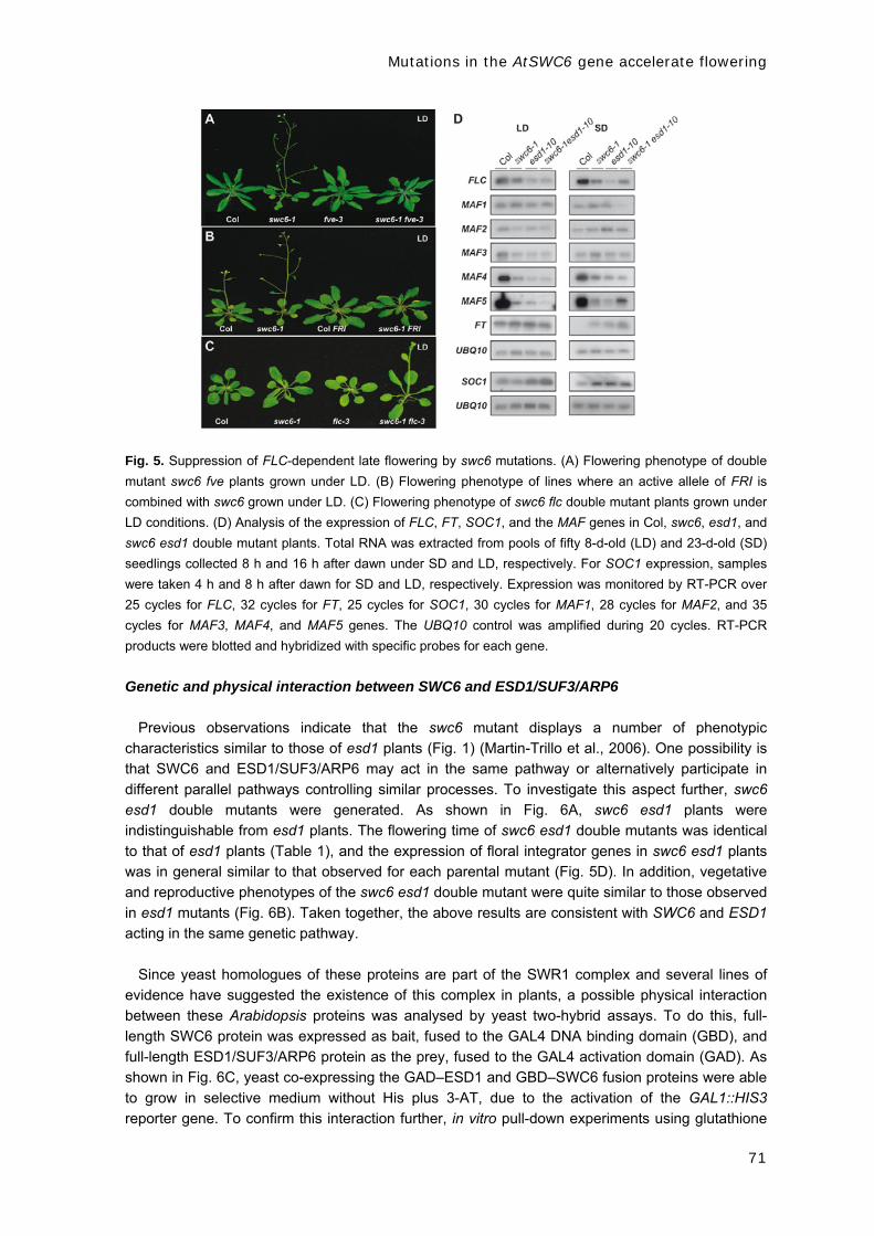
Mutations in the AtSWC6 gene accelerate flowering
71
Fig. 5. Suppression of FLC-dependent late flowering by swc6 mutations. (A) Flowering phenotype of double
mutant swc6 fve plants grown under LD. (B) Flowering phenotype of lines where an active allele of FRI is
combined with swc6 grown under LD. (C) Flowering phenotype of swc6 flc double mutant plants grown under
LD conditions. (D) Analysis of the expression of FLC, FT, SOC1, and the MAF genes in Col, swc6, esd1, and
swc6 esd1 double mutant plants. Total RNA was extracted from pools of fifty 8-d-old (LD) and 23-d-old (SD)
seedlings collected 8 h and 16 h after dawn under SD and LD, respectively. For SOC1 expression, samples
were taken 4 h and 8 h after dawn for SD and LD, respectively. Expression was monitored by RT-PCR over
25 cycles for FLC, 32 cycles for FT, 25 cycles for SOC1, 30 cycles for MAF1, 28 cycles for MAF2, and 35
cycles for MAF3, MAF4, and MAF5 genes. The UBQ10 control was amplified during 20 cycles. RT-PCR
products were blotted and hybridized with specific probes for each gene.
Genetic and physical interaction between SWC6 and ESD1/SUF3/ARP6
Previous observations indicate that the swc6 mutant displays a number of phenotypic
characteristics similar to those of esd1 plants (Fig. 1) (Martin-Trillo et al., 2006). One possibility is
that SWC6 and ESD1/SUF3/ARP6 may act in the same pathway or alternatively participate in
different parallel pathways controlling similar processes. To investigate this aspect further, swc6
esd1 double mutants were generated. As shown in Fig. 6A, swc6 esd1 plants were
indistinguishable from esd1 plants. The flowering time of swc6 esd1 double mutants was identical
to that of esd1 plants (Table 1), and the expression of floral integrator genes in swc6 esd1 plants
was in general similar to that observed for each parental mutant (Fig. 5D). In addition, vegetative
and reproductive phenotypes of the swc6 esd1 double mutant were quite similar to those observed
in esd1 mutants (Fig. 6B). Taken together, the above results are consistent with SWC6 and ESD1
acting in the same genetic pathway.
Since yeast homologues of these proteins are part of the SWR1 complex and several lines of
evidence have suggested the existence of this complex in plants, a possible physical interaction
between these Arabidopsis proteins was analysed by yeast two-hybrid assays. To do this, full-
length SWC6 protein was expressed as bait, fused to the GAL4 DNA binding domain (GBD), and
full-length ESD1/SUF3/ARP6 protein as the prey, fused to the GAL4 activation domain (GAD). As
shown in Fig. 6C, yeast co-expressing the GAD–ESD1 and GBD–SWC6 fusion proteins were able
to grow in selective medium without His plus 3-AT, due to the activation of the GAL1::HIS3
reporter gene. To confirm this interaction further, in vitro pull-down experiments using glutathione

Mutations in the AtSWC6 gene accelerate flowering
72
S-transferase (GST)–SWC6 and in vitro-translated ESD1 protein were performed. As shown in
Fig. 6D, GST–SWC6 was able to interact with ESD1, but not GST alone. Together, the results
show interaction among SWC6 and ESD1, providing further evidence for the existence of a SWR1
complex in Arabidopsis.
Fig. 6. Genetic and physical interaction between SWC6 and ESD1/SUF3/ARP6. (A) Flowering phenotype of
Col, swc6, esd1, and swc6 esd1 double-mutant plants under both LD and SD photoperiods. (B) Pleiotropic
defects in vegetative and reproductive organs displayed by swc6 esd1 mutant plants. Photographs illustrating
leaves, petals, flowers, carpels, DAPI-stained anthers, and siliques of wild type (left), esd1 (middle left), swc6
(middle right), and swc6 esd1 mutants (right). (C) SWC6 and ESD1 interact in yeast. Full-length SWC6 and
ESD1 proteins were fused to the GAL4 DNA binding and activation domain, respectively. Yeast transformed
with these constructs or empty vectors (pGBT8 and pGAD) were grown in nonselective (SC-L,-W) or selective
media (SC-L,-W,-H) with increasing concentrations of 3-AT (5, 10, and 25 mM). Five-fold yeast dilutions were
plated left to right in each panel. (D) Pull-down assay with SWC6 and ESD1 proteins. Bead-bound GST or
GST–SWC6 fusion proteins were incubated with [35S]Met-labelled ESD1 protein. Retained ESD1 protein was
visualized after exposure and autoradiography of the dried gel.
SWC6 is required to activate FLC transcription through both histone acetylation and
methylation mechanisms
FLC gene expression integrates signals coming from different pathways involved in the
regulation of the floral transition (Schmitz and Amasino, 2007). Recent work has demonstrated the
role of histone modification in the regulation of FLC expression through FRI, the autonomous, the
vernalization, and the PAF1 pathways (He et al., 2003, 2004; Ausin et al., 2004; Bastow et al.,

Mutations in the AtSWC6 gene accelerate flowering
73
2004; Sung and Amasino, 2004; He and Amasino, 2005; Kim et al., 2005). These results have
also identified the first intron of FLC as a relevant region for histone modification (He et al., 2003,
2004; Ausin et al., 2004; Bastow et al., 2004; Sung and Amasino, 2004) and transcriptional
regulation (Gendall et al., 2001; Sheldon et al., 2002). Because swc6 suppresses the late-
flowering phenotype of fve autonomous pathway mutants and FVE represses FLC transcription
through a histone deacetylation mechanism, it was speculated that SWC6 could be required for
the acetylation of histones necessary to activate FLC expression. In fact, another putative
component of the SWR1 complex, ESD1/SUF3/ARP6, has been previously reported to be required
for setting this epigenetic marker in FLC chromatin (Martin-Trillo et al., 2006).
To determine whether SWC6 promotes histone acetylation of the FLC chromatin, ChIP assays
were performed (Fig. 7). Chromatin of Col, esd1-10, and swc6-1 plants was immunoprecipitated by
using antibodies against acetylated H3, and PCR was used to amplify two DNA fragments
spanning regions of the promoter and the first intron of FLC, respectively, from the precipitated
chromatin (Fig. 7A). These probes were among those that consistently showed the biggest
differences in previous experiments involving the esd1 mutant (Martin-Trillo et al., 2006). For the
probes assayed, FLC-amplified sequences were consistently more abundant in DNA from
precipitated chromatin of Col than from chromatin of the swc6 and esd1 mutant plants (Fig. 7B),
indicating that both SWC6 and ESD1/SUF3/ARP6 affect the levels of H3 acetylation of FLC.
Therefore, both proteins are required to activate FLC expression through a mechanism involving
the histone acetylation of FLC chromatin.
This assay was extended to explore further if SWC6 also has an effect on histone methylation at
the FLC locus, as does ESD1. It has been shown recently that H3K4 hypertrimethylation is
associated with actively transcribed FLC chromatin (He et al., 2004), and we wondered whether
SWC6 was required for the setting of this epigenetic marker on FLC chromatin. Compared with
wild-type plants, the trimethylated H3K4 levels in the FLC probes assayed were lower in swc6 and
esd1 mutant plants than in Col (Fig. 7C), indicating that SWC6 is also required for the
hypertrimethylation of H3K4 in FLC chromatin. Although in swc6 background a decrease in both
H3 acetylation and H3K4 trimethylation was consistently observed with the probes assayed, this
effect was always less pronounced than that observed in esd1 mutants, suggesting a stronger
involvement of ESD1 in these modifications as compared with SWC6. Discussion
In Arabidopsis, flowering time is regulated by a complex genetic network where the floral
repressor FLC has a pivotal role integrating the autonomous and vernalization pathways and
down-regulating the expression of FT and SOC1 floral integrators (Searle et al., 2006). The
expression level of these integrators is mainly responsible for the correct flowering time (Baurle
and Dean, 2006). Transcriptional regulation of FLC is a central checkpoint in both winter and
summer annual accessions of Arabidopsis. Recently, the regulation of FLC through chromatin
modifications has been intensively demonstrated (reviewed by Reyes, 2006; Sung and Amasino,
2006).

Mutations in the AtSWC6 gene accelerate flowering
74
Fig. 7. Effect of swc6 mutation on histone H3 acetylation and methylation in the FLC genomic region by ChIP
analysis. (A) FLC genomic region analysed by ChIP. The white box corresponds to the promoter FLC region,
grey boxes to exons, and the black box to the first intron. The two FLC fragments analysed by
semiquantitative PCR are depicted and numbered. (B) PCR products after 25 cycles of Col, esd1-10, and
swc6-1 mutant plants using as template DNA purified from chromatin inmunoprecipitated with antibodies
against acetylated H3 (AcH3). UBQ10 was amplified during 22 cycles and used as control for DNA
quantification. Fold decrease in H3 acetylation of swc6 and esd1 mutants over Col is shown. (C) PCR
products after 25 cycles of Col, esd1-10, and swc6-1 mutant plant PCR products as in (B) but using as
template DNA purified from chromatin inmunoprecipitated with antibodies against trimethylated H3K4
(MeH3K4). Fold decrease in H3K4 methylation of mutants over the wild-type ecotype is shown. The data
provided are representative and are from one of three independent experiments.
In this work, Arabidopsis swc6 mutants that are affected in a putative orthologue of the SWR1
chromatin remodelling complex have been characterized. SWC6 is the only Arabidopsis gene
homologue of yeast SWC6/VPS73 (Krogan et al., 2003; Kobor et al., 2004; Wu et al., 2005).
Recently, the function of the ATP-dependent chromatin remodelling complex SWR1 has been
intensively studied in yeast (Wu et al., 2005). The subunits of SWR1 and of the mammalian
homologue SRCAP complexes have been biochemically identified and analysed (Kobor et al.,
2004; Mizuguchi et al., 2004; Cai et al., 2005; Wu et al., 2005), and the evidence for the presence
of a homologous complex in plants has been provided (Choi et al., 2007; Deal et al., 2007). In
addition, how SWR1 homologues affect development in higher eukaryotes remains largely
unknown. Homologues of most SWR1 components are present in Arabidopsis, and thus the
function of this complex is being genetically dissected.
Phenotypical analyses of swc6 mutants revealed a complex array of pleiotropic defects affecting
vegetative and reproductive development, including a reduction in flowering time and phase length
(Fig. 2). swc6 causes early flowering mainly through the reduction in FLC expression (Fig. 5),
suggesting a role for the SWR1 complex in the regulation of flowering time. Genetic analyses have
revealed that the early flowering phenotype of swc6 mutants is almost completely epistatic over
the flowering time delay caused by fve mutation in the autonomous pathway, and that swc6
partially suppresses the late flowering phenotype conferred by active FRI alleles (Fig. 5). These
Mutations in the AtSWC6 gene accelerate flowering
75
genetic interactions correlate at the molecular level with a decrease in the steady-state levels of
FLC mRNA in swc6 mutant alleles (Fig. 5D). Together, these results indicate that SWC6 is
required for the proper expression of FLC. The phenotypic analysis of swc6 mutants indicates that
this protein also controls other developmental processes such as leaf and flower morphology (Fig.
2), suggesting that SWC6 regulates other genes involved in plant development. For instance,
swc6 plants show smaller leaves with serrated margins, which might reflect defects in cell
proliferation along the margins of leaf primordia. In addition, swc6 mutant flowers display extra
floral perianth organs, suggesting a role for the SWR1 complex in the control of floral development.
Mutations in different Arabidopsis homologues of SWR1 components such as PIE1,
ESD1/SUF3/ARP6, and SWC6/SEF cause similar developmental defects (Noh and Amasino,
2003; Choi et al., 2005, 2007; Martin-Trillo et al., 2006; March-Diaz et al., 2007; this study), and
these genes display similar expression patterns, although SWC6 is more highly expressed in
anthers (Fig. 4D). This may be explained by the presence of various cis-acting elements (AGAAA
and GTGA) in the SWC6 promoter region, known to confer anther/pollen-specific gene expression
(Gupta et al., 2007), and is consistent with a role for SWC6 in anther and/or pollen development
(March-Diaz et al., 2007; this study) (Fig. 4C). All three mutants cause suppression of late
flowering in autonomous pathway mutants as well as in FRI-containing lines and other
developmental defects including leaf serration, weak apical dominance (bushy aspect), flowers
with extra petals, and short siliques. They also show earlier flowering than an flc-null mutant,
suggesting that flowering inhibition mediated by these proteins occurs through both FLC- and FLC-
like gene-dependent pathways (Noh and Amasino, 2003; Choi et al., 2005, 2007; Martin-Trillo et
al., 2006). However, some phenotypes are more dramatic in pie1 plants than in swc6/sef and
esd1/suf3/arp6 plants (Noh and Amasino, 2003). pie1 displays a stronger reduction in fertility, a
very notable reduction in primary inflorescence elongation, and smaller and deformed leaves (Noh
and Amasino, 2003; March-Diaz et al., 2007), phenotypes that were not obvious in esd1/suf3/arp6
or in swc6/sef (Noh and Amasino, 2003; Choi et al., 2005, 2007; Deal et al., 2005; Martin-Trillo et
al., 2006; March-Diaz et al., 2007). Moreover, pie1 plants show a stronger down-regulation of FLC
and MAF4 transcript levels than the esd1 and swc6 plants (Deal et al., 2007; March-Diaz et al.,
2007). Furthermore, the MAF5 gene was deregulated in the pie1, esd1/suf3/arp6, and swc6/sef
mutant (Fig. 5D; Martin-Trillo et al., 2006; March-Diaz et al., 2007). Altogether, these observations
suggest that PIE1 might fulfil functions that are at least partially independent from those of SWC6
and ESD1; a tempting possibility is that PIE1 may participate in other chromatin remodelling
complexes besides SWR1.
SWC6 and ARP6 yeast homologues are tightly associated in SWR1C, being necessary for the
interaction with the SWC2 subunit and for nucleosome binding (Wu et al., 2005). According to this,
Arabidopsis SWC6 and ESD1/SUF3/ARP6 have a similar developmental function. The phenotypes
of swc6 and esd1 mutants are quite comparable, and the esd1 swc6 double mutant has the same
phenotype and causes similar alterations in gene expression as any single mutant (Figs 5D, 6B),
indicating that both genes act in the same genetic pathway. Together with the absence of any
obvious developmental phenotype in plants overexpressing either ESD1 or SWC6, the results
described in this study strongly suggest that ESD1 and SWC6 act together as a protein complex.
The protein interaction analyses confirmed that both proteins can physically interact (Fig. 6C, D).
Biochemical characterization of the yeast SWR1 complex indicates that removal of either arp6 or
swc6 results in the reciprocal loss of the other subunit from the complex and also in the loss of two
other proteins, Swc2 and Swc3, suggesting that Arp6, Swc6, Swc2, and Swc3 form a subcomplex
associated with Swr1 (Wu et al., 2005). Similarly, Arabidopsis ARP6 and SWC6, together with
SWC2 and other unidentified factors, may form a subcomplex that associates with PIE1 (Choi et

Mutations in the AtSWC6 gene accelerate flowering
76
al., 2007; March-Diaz et al., 2007). Again, this is consistent with a very similar phenotype of the
swc6 and esd1/suf3/arp6 mutants but a slightly different phenotype of the pie1 mutant.
The SWR1 complex in yeast catalyses the replacement of nucleosomal H2A with the H2A.Z
variant, ensuring full activation of underlying genes (Guillemette et al., 2005; Li et al., 2005;
Raisner et al., 2005; Zhang et al., 2005). Recent studies have shown that two H2A.Z nucleosomes
flank a nucleosome-free region containing the transcription initiation site in promoters of both
active and inactive genes in yeast and that H2A.Z-bearing nucleosomes facilitate transcription
activation through their susceptibility to loss, thereby helping to expose promoter DNA (reviewed in
Raisner and Madhani, 2006). In Arabidopsis, the histone variant H2A.Z has been identified within
FLC, MAF4, and MAF5 chromatin, occupying regions near both the transcription start and
termination sites on the three genes examined (Deal et al., 2007). In addition, H2A.Z interacts with
both PIE1 and AtSWC2, and knockdown of H2A.Z caused a phenotype similar to that of pie1,
esd1/suf3/arp6, and swc6 (Choi et al., 2007). Loss of H2A.Z from FLC chromatin in esd1/suf3/arp6
and pie1 mutants results in reduced FLC expression and premature flowering, indicating that this
histone variant is required for a high level of expression of FLC (Deal et al., 2007). These
observations support the existence of an SWR1-like complex in plants that is targeted to different
loci including FLC, and show that H2A.Z can poise transcriptional activation in plants. Interestingly,
the spatial distribution and the overall levels of H2A.Z on FLC was the same in samples that had a
10-fold higher level of FLC expression (Deal et al., 2007), suggesting that H2AZ by itself does not
activate FLC gene expression and that the replacement of nucleosomal H2A with H2A.Z may form
a variant nucleosome with unique tails that might bind specific regulatory proteins to help promote
FLC gene activation.
High levels of FLC expression are correlated with H3 and H4 hyperacetylation and trimethylation
of H3K4 and H3K36 at the FLC locus (He et al., 2003, 2004; Ausin et al., 2004; Bastow et al.,
2004; Sung and Amasino, 2004; Zhao et al., 2005). Martin-Trillo et al. (2006) have recently
reported that esd1/suf3/arp6 mutants present low levels of histone H3 acetylation and H3K4
methylation in the FLC locus; this work demonstrates a comparable behaviour in the swc6 mutant,
although the effect was consistently more conspicuous for esd1 (Fig. 7). Whether ARP6 and
SWC6, and, consequently, the SWR1 complex are directly involved in setting these epigenetic
markers or whether these alterations are secondary consequences is still unclear. Moreover, it
remains to be determined whether the effect of esd1/suf3/arp6 and swc6 on the expression of
other MAF genes takes place through similar mechanisms.
A human H2A.Z complex equivalent to the yeast SWR1 complex has histone acetyltransferase
activity (Owen-Hughes and Bruno, 2004), and the Swr1 complex shares several subunits with the
NuA4 histone acetyltransferase. Furthermore, mutants of these two complexes share several
phenotypes, suggesting that they may work together, which might help to understand the role of
ESD1/SUF3/ARP6 and SWC6 in histone acetylation. In the same way, the fact that components of
the Swr1 complex were found to interact genetically with the PAF1 complex in yeast might explain
the role of ESD1 and SWC6 in the trimethylation of H3K4 in FLC chromatin (Mueller and Jaehning,
2002; Squazzo et al., 2002; Krogan et al., 2003, 2004). Like the yeast PAF1 complex, the PAF1-
like complex in Arabidopsis may also recruit an H3K4 methyl transferase to FLC to regulate its
expression (Kim et al., 2005). Indeed, mutations in Arabidopsis homologues of the components of
the PAF1 complex cause a decrease in the trimethylation of H3K4 in FLC chromatin, and provoke
early flowering and small leaves, similar to the esd1 and swc6 mutations (He et al., 2004), raising
the possibility that all of these genes are in the same pathway and regulate similar targets.

Mutations in the AtSWC6 gene accelerate flowering
77
We propose that the H2A.Z variant may serve to poise the FLC gene, and maybe other related
genes, in a state competent for activation by other factors, rather than activating transcription
directly (Deal et al., 2007). This may reflect the ability of H2A.Z to facilitate nucleosome
remodelling (Santisteban et al., 2000) and/or to recruit the transcription machinery (Adam et al.,
2001) or other chromatin remodelling complexes to allow high-level transcription under certain
conditions. Thus, in the absence of H2A.Z in swc6, esd1/suf3/arp6, and pie1 mutants, FLC levels
remain low even in the presence of strong activators such as FRI (Noh and Amasino, 2003; Choi
et al., 2005; Deal et al., 2005, 2007; Martin-Trillo et al., 2006; this study), resulting in early
flowering.
Biochemical characterization of the SWR1C homologue and functional studies using
transcriptomic analyses and ChIP-to-chip hybridization will help to identify additional genes
regulated by this complex and to understand the crucial role of the SWR1 complex plant
homologue in chromatin remodelling processes related to leaf and flower development and to the
control of flowering time.
Acknowledgements
We would like to thank Dr Richard Amasino for kindly providing flc-3 and FRI-Col lines, the ABRC and NASC
for supplying the T-DNAs lines used in this study, and GENOSCOPE for the ESD1/SUF3/ARP6 and SWC6
cDNA clones. GST–SWC6 was a gift of Dr Jose Carlos Reyes. This work was supported by grants BIO2005-
00251 to JAJ and BIO2004-00494 to MP from the Spanish Ministerio de Ciencia y Tecnología. AL and AG-Z
were funded by a predoctoral fellowship from Direccion General de Investigacioón, Secretaria General de
Política Científica y Tecnológica (Spain), and LL-G by an INIA predoctoral fellowship. We thank to all
members of Jarillo’s and Piñeiro’s laboratories for helpful discussions, and D Saez for his skilful technical
assistance. References Adam M, Robert F, Larochelle M, Gaudreau L. 2001. H2A.Z is required for global chromatin integrity and
for recruitment of RNA polymerase II under specific conditions. Molecular and Cellular Biology 21, 6270–6279.
Ausin I, Alonso-Blanco C, Jarillo JA, Ruiz-Garcia L, Martinez-Zapater JM. 2004. Regulation of flowering time by FVE, aretinoblastoma-associated protein. Nature Genetics 36, 162–166.
Bastow R, Mylne JS, Lister C, Lippman Z, Martienssen RA, Dean C. 2004. Vernalization requires epigenetic silencing of FLC by histone methylation. Nature 427, 164–167.
Baurle I, Dean C. 2006. The timing of developmental transitions in plants. Cell 125, 655–664. Boss PK, Bastow RM, Mylne JS, Dean C. 2004. Multiple pathways in the decision to flower: enabling,
promoting, and resetting. The Plant Cell 16 (Suppl.), S18–S31. Cai Y, Jin J, Florens L, Swanson SK, Kusch T, Li B, Workman JL, Washburn MP, Conaway RC,
Conaway JW. 2005. The mammalian YL1 protein is a shared subunit of the TRRAP/TIP60 histone acetyltransferase and SRCAP complexes. Journal of Biological Chemistry 280, 13665–13670.
Choi K, Kim S, Kim SY, Kim M, Hyun Y, Lee H, Choe S, Kim SG, Michaels S, Lee I. 2005. SUPPRESSOR OF FRIGIDA3 encodes a nuclear ACTIN-RELATED PROTEIN6 required for floral repression in Arabidopsis. The Plant Cell 17, 2647–2660.
Choi K, Park C, Lee J, Oh M, Noh B, Lee I. 2007. Arabidopsis homologs of components of the SWR1 complex regulate flowering and plant development. Development 134, 1931–1941.
Clough SJ, Bent AF. 1998. Floral dip: a simplified method for Agrobacterium-mediated transformation of Arabidopsis thaliana. The Plant Journal 16, 735–743.
Corbesier L, Vincent C, Jang S, et al. 2007. FT protein movement contributes to long-distance signaling in floral induction of Arabidopsis. Science 316, 1030–1033.
Deal RB, Kandasamy MK, McKinney EC, Meagher RB. 2005. The nuclear actin-related protein ARP6 is a pleiotropic developmental regulator required for the maintenance of FLOWERING LOCUS C expression and repression of flowering in Arabidopsis. The Plant Cell 17, 2633–2646.
Deal RB, Topp CN, McKinney EC, Meagher RB. 2007. Repression of flowering in Arabidopsis requires activation of FLOWERING LOCUS C expression by the histone variant H2A.Z. The Plant Cell 19, 74–83.
Gazzani S, Gendall AR, Lister C, Dean C. 2003. Analysis of the molecular basis of flowering time variation in Arabidopsis accessions. Plant Physiology 132, 1107–1114.

Mutations in the AtSWC6 gene accelerate flowering
78
Gendall AR, Levy YY, Wilson A, Dean C. 2001. The VERNALIZATION 2 gene mediates the epigenetic regulation of vernalization in Arabidopsis. Cell 107, 525–35.
Gomez-Mena C, Pineiro M, Franco-Zorrilla JM, Salinas J, Coupland G, Martinez-Zapater JM. 2001. early bolting in short days: an Arabidopsis mutation that causes early flowering and partially suppresses the floral phenotype of leafy. The Plant Cell 13, 1011–1024.
Guillemette B, Bataille AR, Gevry N, Adam M, Blanchette M, Robert F, Gaudreau L. 2005. Variant histone H2A.Z is globally localized to the promoters of inactive yeast genes and regulates nucleosome positioning. PLoS Biology 3, E384.
Gupta V, Khurana R, Tyagi AK. 2007. Promoters of two antherspecific genes confer organ-specific gene expression in a stagespecific manner in transgenic systems. Plant Cell Reports 26, 1919–1931.
He Y, Amasino RM. 2005. Role of chromatin modification in flowering-time control. Trends in Plant Science 10, 30–35.
He Y, Doyle MR, Amasino RM. 2004. PAF1-complex-mediated histone methylation of FLOWERING LOCUS C chromatin is required for the vernalization-responsive, winter-annual habit in Arabidopsis. Genes and Development 18, 2774–2784.
He Y, Michaels SD, Amasino RM. 2003. Regulation of flowering time by histone acetylation in Arabidopsis. Science 302, 1751–1754.
Imaizumi T, Kay SA. 2006. Photoperiodic control of flowering: not only by coincidence. Trends in Plant Science 11, 550–558.
Johanson U, West J, Lister C, Michaels S, Amasino R, Dean C. 2000. Molecular analysis of FRIGIDA, a major determinant of natural variation in Arabidopsis flowering time. Science 290, 344–347.
Kim SY, He Y, Jacob Y, Noh YS, Michaels S, Amasino R. 2005. Establishment of the vernalization-responsive, winter-annual habit in Arabidopsis requires a putative histone H3 methyl transferase. The Plant Cell 17, 3301–3310.
Kobor MS, Venkatasubrahmanyam S, Meneghini MD, Gin JW, Jennings JL, Link AJ, Madhani HD, Rine J. 2004. A protein complex containing the conserved Swi2/Snf2-related ATPase Swr1p deposits histone variant H2A.Z into euchromatin. PLoS Biology 2, 587–599.
Krichevsky A, Gutgarts H, Kozlovsky SV, Tzfira T, Sutton A, Sternglanz R, Mandel G, Citovsky V. 2007. C2H2 zinc finger-SET histone methyltransferase is a plant-specific chromatin modifier. Developmental Biology 303, 259–269.
Krogan NJ, Baetz K, Keogh MC, et al. 2004. Regulation of chromosome stability by the histone H2A variant Htz1, the Swr1 chromatin remodeling complex, and the histone acetyltransferase NuA4. Proceedings of the National Academy of Sciences, USA 101, 13513–13518.
Krogan NJ, Keogh MC, Datta N, et al. 2003. A Snf2 family ATPase complex required for recruitment of the histone H2A variant Htz1. Molecular Cell 12, 1565–1576.
Lee I, Amasino RM. 1995. Effect of vernalization, photoperiod, and light quality on the flowering phenotype of Arabidopsis plants containing the FRIGIDA gene. Plant Physiology 108, 157–162.
Lee MG, Wynder C, Bochar DA, Hakimi MA, Cooch N, Shiekhattar R. 2006. Functional interplay between histone demethylase and deacetylase enzymes. Molecular and Cellular Biology 26, 6395–6402.
Li B, Pattenden SG, Lee D, Gutierrez J, Chen J, Seidel C, Gerton J, Workman JL. 2005. Preferential occupancy of histone variant H2AZ at inactive promoters influences local histone modifications and chromatin remodeling. Proceedings of the National Academy of Sciences, USA 102, 18385–18390.
March-Diaz R, Garcia-Dominguez M, Florencio FJ, Reyes JC. 2007. SEF, a new protein required for flowering repression in Arabidopsis, interacts with PIE1 and ARP6. Plant Physiology 143, 893–901.
Martin-Trillo M, Lazaro A, Poethig RS, Gomez-Mena C, Pineiro MA, Martinez-Zapater JM, Jarillo JA. 2006. EARLY IN SHORT DAYS 1 (ESD1) encodes ACTIN-RELATED PROTEIN 6 (AtARP6), a putative component of chromatin remodelling complexes that positively regulates FLC accumulation in Arabidopsis. Development 133, 1241–1252.
Michaels SD, Amasino RM. 1999. FLOWERING LOCUS C encodes a novel MADS domain protein that acts as a repressor of flowering. The Plant Cell 11, 949–956.
Michaels SD, Amasino RM. 2000. Memories of winter: vernalization and the competence to flower. Plant, Cell and Environment 23, 1145–1154.
Michaels SD, Amasino RM. 2001. Loss of FLOWERING LOCUS C activity eliminates the late-flowering phenotype of FRIGIDA and autonomous pathway mutations but not responsiveness to vernalization. The Plant Cell 13, 935–941.
Mizuguchi G, Shen X, Landry J, Wu WH, Sen S, Wu C. 2004. ATP-driven exchange of histone H2AZ variant catalyzed by SWR1 chromatin remodeling complex. Science 303, 343–348.
Moon J, Suh SS, Lee H, Choi KR, Hong CB, Paek NC, Kim SG, Lee I. 2003. The SOC1 MADS-box gene integrates vernalization and gibberellin signals for flowering in Arabidopsis. The Plant Journal 35, 613–623.
Mueller CL, Jaehning JA. 2002. Ctr9, Rtf1 and Leo1 are components of the Paf1/RNA polymerase II complex. Molecular and Cellular Biology 22, 1971–1980.
Noh YS, Amasino RM. 2003. PIE1, an ISWI family gene, is required for FLC activation and floral repression in Arabidopsis. The Plant Cell 15, 1671–1682.
Oh S, Zhang H, Ludwig P, van Nocker S. 2004. A mechanism related to the yeast transcriptional regulator Paf1c is required for expression of the Arabidopsis FLC/MAF MADS box gene family. The Plant Cellular 16, 2940–2953.

Mutations in the AtSWC6 gene accelerate flowering
79
Owen-Hughes T, Bruno M. 2004. Breaking the silence. Nature 303, 324–325. Piñeiro M, Gomez-Mena C, Schaffer R, Martinez-Zapater JM, Coupland G. 2003. EARLY BOLTING IN
SHORT DAYS is related to chromatin remodeling factors and regulates flowering in Arabidopsis by repressing FT. The Plant Cell 15, 1552–1562.
Raisner RM, Hartley PD, Meneghini MD, Bao MZ, Liu CL, Schreiber SL, Rando OJ Madhani HD. 2005. Histone variant H2A.Z marks the 5´ ends of both active and inactive genes in euchromatin. Cell 123, 233–248.
Raisner RM, Madhani HD. 2006. Patterning chromatin: form and function for H2A.Z variant nucleosomes. Current Opinion in Genetics and Development 16, 119–124.
Ratcliffe OJ, Kumimoto RW, Wong BJ, Riechmann JL. 2003. Analysis of the Arabidopsis MADS AFFECTING FLOWERING gene family: MAF2 prevents vernalization by short periods of cold. The Plant Cell 15, 1159–1169.
Reeves PH, Murtas G, Dash S, Coupland G. 2002. Early in short days 4, a mutation in Arabidopsis that causes early flowering and reduces the mRNA abundance of the floral repressor FLC. Development 129, 5349–5361.
Reyes JC. 2006. Chromatin modifiers that control plant development. Current Opinion in Plant Biology 9, 21–27.
Santisteban MS, Kalashnikova T, Smith MM. 2000. Histone H2A.Z regulates transcription and is partially redundant with nucleosome remodeling complexes. Cell 103, 411–422.
Schmitz RJ, Amasino RM. 2007. Vernalization: a model for investigating epigenetics and eukaryotic gene regulation in plants. Biochimica et Biophysica Acta 1769, 269–275.
Scortecci KC, Michaels SD, Amasino RM. 2001. Identification of a MADS-box gene, FLOWERING LOCUS M, that represses flowering. The Plant Journal 26, 229–236.
Searle I, He Y, Turck F, Vincent C, Fornara F, Krober S, Amasino RA, Coupland G. 2006. The transcription factor FLC confers a flowering response to vernalization by repressing meristem competence and systemic signaling in Arabidopsis. Genes and Development 20, 898–912.
Sheldon CC, Burn JE, Perez PP, Metzger J, Edwards JA, Peacock WJ, Dennis ES. 1999. The FLF MADS box gene: a repressor of flowering in Arabidopsis regulated by vernalization and methylation. The Plant Cell 11, 445–458.
Sheldon CC, Conn AB, Dennis ES, Peacock WJ. 2002. Different regulatory regions are required for the vernalization-induced repression of FLOWERING LOCUS C and for the epigenetic maintenance of repression. The Plant Cell 14, 2527–2537.
Shindo C, Aranzana MJ, Lister C, Baxter C, Nicholls C, Nordborg M, Dean C. 2005. Role of FRIGIDA and FLOWERING LOCUS C in determining variation in flowering time of Arabidopsis. Plant Physiology 138, 1163–1173.
Squazzo SL, Costa PJ, Lindstrom DL, Kumer KE, Simic R, Jeennings JL, Link AJ, Arndt KM, Hartzog GA. 2002. The Paf1 complex physically and functionally associates with transcription elongation factors in vivo. EMBO Journal 21, 1764–1774.
Sung S, Amasino RM. 2004. Vernalization in Arabidopsis thaliana is mediated by the PHD finger protein VIN3. Nature 427, 159–164.
Sung S, Amasino RM. 2006. Molecular genetic studies of the memory of winter. Journal of Experimental Botany 57, 3369–3377.
Sung S, Schmitz RJ, Amasino RM. 2006. A PHD finger protein involved in both the vernalization and photoperiod pathways in Arabidopsis. Genes and Development 20, 3244–3248.
Telfer A, Bollman KM, Poethig RS. 1997. Phase change and the regulation of trichome distribution in Arabidopsis thaliana. Development 124, 645–654.
Werner JD, Borevitz JO, Warthmann N, Trainer GT, Ecker JR, Chory J, Weigel D. 2005. Quantitative trait locus mapping and DNA array hybridization identify an FLM deletion as a cause for natural flowering-time variation. Proceedings of the National Academy of Sciences, USA 102, 2460–2465.
Wilson RN, Heckman JW, Somerville CR. 1992. Gibberellin is required for flowering in Arabidopsis thaliana under short days. Plant Physiology 100, 403–408.
Wood CC, Robertson M, Tanner G, Peacock WJ, Dennis ES, Helliwell CA. 2006. The Arabidopsis thaliana vernalization response requires a Polycomb-like protein complex that also includes VERNALIZATION INSENSITIVE 3. Proceedings of the National Academy of Sciences, USA 103, 14631–14636.
Wu WH, Alami S, Luk E, Wu CH, Sen S, Mizuguchi G, Wei D, Wu C. 2005. Swc2 is a widely conserved H2AZ-binding module essential for ATP-dependent histone exchange. Nature Structural and Molecular Biology 12, 1064–1071.
Zhang H, Roberts DN, Cairns BR. 2005. Genome-wide dynamics of Htz1, a histone H2A variant that poises repressed/basal promoters for activation through histone loss. Cell 123, 219–231.
Zhao Z, Yu Y, Meyer D, Wu C, Shen WH. 2005. Prevention of early flowering by expression of FLOWERING LOCUS C requires methylation of histone H3 K36. Nature Cell Biology 7, 1256–1260.
Zimmermann P, Hirsch-Hoffmann M, Hennig L, Gruissem W. 2004. GENEVESTIGATOR. Arabidopsis microarray database and analysis toolbox. Plant Physiology 136, 2621–2632.


CAPÍTULO 3: The E3 ubiquitin ligase HOS1 participates in the
control of photoperiodic flowering negatively regulating
CONSTANS abundance.


HOS1 regulates CO abundance
83
Under revision
The E3 ubiquitin ligase HOS1 participates in the control of photoperiodic flowering in Arabidopsis negatively regulating CONSTANS abundance
Ana Lazaro1, Federico Valverde2, Manuel Piñeiro1, Jose A. Jarillo1,3
1 Centro de Biotecnología y Genómica de Plantas (CBGP), INIA-UPM. Instituto Nacional de Investigación y Tecnología
Agraria y Alimentaria, Campus de Montegancedo, 28223 Madrid, Spain. 2 Instituto de Bioquímica Vegetal y Fotosíntesis, Consejo Superior de Investigaciones Científicas y Universidad de Sevilla.
Américo Vespucio 49, 41092 Sevilla, Spain. 3 To whom correspondence should be addressed. e-mail: [email protected]
Telephone: 34-91-3364576; Fax 34-91-7157721
Key words: HOS1, E3 ubiquitin ligase, CONSTANS, flowering time, floral repression, Arabidopsis
We have isolated the early in short days 6 (esd6) mutant in a screening for mutations that
accelerate flowering time in Arabidopsis. esd6 displays early flowering in both long and short day
conditions among other developmental alterations. Fine mapping of the mutation showed that
esd6 was affected in the HIGH EXPRESSION OF OSMOTICALLY RESPONSIVE GENES 1
(HOS1) locus, which encodes a RING finger-containing protein that works as an E3 ubiquitin
ligase. esd6/hos1 mutation causes decreased expression of the FLC gene and shows a strong
requirement of a functional CO protein for its early flowering phenotype under long days. Besides,
CO and HOS1 physically interact in vitro and in vivo, and HOS1 is regulating CO abundance,
particularly during the daylight period. Accordingly, the hos1 mutation causes a shift in the typical
long day pattern of FT transcript, starting to rise four hours after dawn. In addition, HOS1 interacts
synergistically with COP1, another regulator of CO protein stability, in the control of flowering time.
Taken together, these results indicate that HOS1 is involved in regulating CO abundance,
ensuring that CO activation of FT occurs only when the light period reaches a certain length and
avoiding precocious flowering in Arabidopsis.
INTRODUCTION
The integration of complex signals from environmental and endogenous cues is necessary to
enable plants to time the floral transition at the most advantageous moment (Michaels, 2009; de
Montaigu et al., 2010; Imaizumi, 2010). Plants growing at northern latitudes adapt their
developmental program to the varying daylengths and temperatures that occur along the year
(Jackson, 2009). Arabidopsis is a facultative long-day (LD) plant in which flowering time is
controlled by a network of six major pathways: information about daylength, low winter
temperatures and growth temperature are mediated by the photoperiod, the vernalization and the
ambient temperature pathway, respectively. In contrast, the aging, the autonomous and the
gibberellin pathways act more independently of ambient conditions (Fornara et al., 2010). A potent
repressor of flowering, FLOWERING LOCUS C (FLC), integrates signals coming from both the
vernalization and the autonomous pathway (Amasino, 2010). Eventually, the whole network
converges in the regulation of the floral integrators: FLOWERING LOCUS T (FT), TWIN SISTER
OF FT (TSF) and SUPPRESSOR OF OVEREXPRESSION OF CONSTANS 1 (SOC1) (Fornara et
al., 2010).

HOS1 regulates CO abundance
84
The photoperiod pathway comprises several genes, including GIGANTEA (GI), CONSTANS
(CO), and FT (Kobayashi and Weigel, 2007; Turck et al., 2008). Mutations in any of these genes
cause a delay in flowering mainly under LDs, whereas their overexpression accelerates flowering
independently of daylength (Turck et al., 2008). CO is a B-box-type protein that acts in the
vascular tissue of the leaves to activate FT and TSF transcription (An et al., 2004; Jackson, 2009;
Tiwari et al., 2010). CO may induce FT expression by forming a DNA binding complex with
NUCLEAR FACTOR Y (NF-Y)/HEME ACTIVATOR PROTEIN (HAP) proteins (Wenkel et al., 2006;
Kumimoto et al., 2010) and by binding the FT promoter directly at CO-responsive elements (Tiwari
et al., 2010). FT protein, and possibly TSF, are part of the florigen that moves to the shoot apical
meristem to induce flowering in response to LDs (Corbesier et al., 2007; Jaeger and Wigge, 2007;
Jang et al., 2009).
Plants have developed a sophisticated molecular mechanism to measure daylength based on
the coincidence of an internal rhythm, set by the circadian clock, with an external cue, such as
light. The ability to distinguish LDs from short days (SDs) is largely the result of the complex
regulation of CO, both at the transcriptional and post-translational level. Under LDs, CO mRNA
shows two peaks of expression, the first following the expression of GI at the end of a LD, when
plants are still exposed to light; and the second during the night. Under SDs, only the night peak of
CO expression takes place (Suarez-Lopez et al., 2001). The precise timing of CO also requires the
degradation of a family of repressors, the cycling DOF transcription factors (CDFs), by the F-Box
protein FLAVIN-BINDING, KELCH REPEAT, F-BOX 1 (FKF1) in conjunction with GI (Imaizumi et
al., 2005; Sawa et al., 2007; Fornara et al., 2009).
The increased expression of CO in the light under LDs but not SDs is crucial for the
promotion of flowering, because exposure to light is required for stabilization of CO protein
(Valverde et al., 2004; Jang et al., 2008). The high CO transcript levels detected during the dark
phase of both LD and SDs do not correlate with CO protein accumulation because the RING finger
protein CONSTITUTIVE PHOTOMORPHOGENIC 1 (COP1) promotes CO degradation in the dark
(Jang et al., 2008; Liu et al., 2008b). Mutations in COP1, a component of an ubiquitin ligase
complex, cause extreme early flowering under SDs. This early flowering phenotype is largely
dependent on CO activity and correlates with an increase in FT transcription in cop1 mutant (Jang
et al., 2008; Liu et al., 2008b). COP1 and CO interact both in vivo and in vitro, and it has been
proposed that COP1 contributes to daylength perception by reducing the abundance of the CO
protein during the night (Jang et al., 2008; Liu et al., 2008b; Chen et al., 2010). However, in the
morning CO degradation occurs independently of COP1 (Jang et al., 2008). Therefore, it has been
suggested that an unidentified E3 ubiquitin ligase must collaborate in CO degradation during the
early part of the day to ensure that CO induction of FT only takes place in LDs (Jang et al., 2008).
In addition to the duration of the daily light/dark periods, plants also perceive light quality. Blue
and far-red light promote flowering, while red light (RL) delays it (Valverde et al., 2004). Far-red
light can increase CO protein levels independently of transcription (Kim et al., 2008). Blue light
mediates photoperiodic control of the floral initiation at least by three different mechanisms: first, it
promotes the interaction of FKF1 and GI necessary for the CDFs degradation (Sawa et al., 2007);
second, the blue light receptor Cryptochrome 2 (Cry2) prevents GI and CO proteolysis by COP1
(Liu et al., 2008b; Yu et al., 2008); and third, Cry2 modulates FT transcription directly (Liu et al.,
2008a). On the other hand, the red light photoreceptor Phytochrome B (PhyB) has been implicated
in the degradation of CO during the first part of the day (Valverde et al., 2004; Jang et al., 2008).
Screenings devoted to the isolation of early flowering mutants have revealed the existence of
genes that repress the floral transition (Pouteau et al., 2004). Floral repressors are essential to

HOS1 regulates CO abundance
85
safeguard against premature flowering, and knowledge of how these repressors interact with the
floral promotion pathways is just emerging (Pouteau et al., 2004; Roux et al., 2006). Here we
demonstrate that the esd6 early flowering mutant is affected in the HOS1 gene. HOS1 encodes a
protein with E3 ubiquitin ligase activity, previously described as a negative regulator of cold
acclimation responses (Lee et al., 2001; Dong et al., 2006a). The early flowering phenotype of
hos1 is completely suppressed by mutations in CO gene in Landsberg erecta (Ler) background
and notably delayed by co mutations in Columbia (Col) background. In addition, we show that
HOS1 physically interacts with CO and regulates CO protein abundance during the daylight
period, indicating the participation of another RING finger-containing protein, besides COP1, in the
photoperiodic control of flowering time in Arabidopsis. Thus, we propose that HOS1 is required to
modulate precisely the timing of CO accumulation, and that this regulation is essential to maintain
FT levels low during the first part of the day and, subsequently, a correct photoperiodic response
in Arabidopsis.
METHODS
Genetic stocks and growth conditions
Arabidopsis thaliana (L.) mutant seed stocks used were in Ler, Col and C24 genetic backgrounds, and
were obtained from the Arabidopsis Biological Resource Centre (ABRC) of Ohio State University (Columbus,
USA), the Nottingham Arabidopsis Centre (NASC) in the UK, and personal donations. C24 accession and
mutant hos1-1 seeds were kindly donated by Dr. J.K Zhu (Lee et al., 2001). The monogenic mutants used in
this work were described previously: fca-1, ft-1, co-2 and gi-3 (Koornneef, 1991); fve-3 (Ausin et al., 2004);
flc-3 (Michaels and Amasino, 1999); phyB-1 (Reed et al., 1993); fha-1 (Guo et al., 1998); vrn1-2 fca-1 (Levy
et al., 2002); vin3-4 FRI Sf-2 (Sung and Amasino, 2004); fld-1 (He et al., 2003); siz1-2 (Miura et al., 2005);
fkf1-1 (Nelson et al., 2000); cop1-4 (Deng et al., 1991); co-10 (Laubinger et al., 2006); soc1-1 (Samach et al.,
2000); and the Col FRI Sf-2 line was described by Lee and Amasino (Lee and Amasino, 1995).
Plants were grown in plastic pots containing a mixture of substrate and vermiculite (3:1) or in MS
(Murashige and Skoog) medium supplemented with 1% (w/v) sucrose and 0,8% (w/v) agar for in vitro culture.
Controlled environmental conditions were provided in growth chambers at 22°C and 70% relative humidity.
Plants were illuminated with cool-white fluorescent lights (approximately 120 mol m–2 s–1). LD conditions
consisted of 16 h of light followed by 8 h of darkness; SD conditions consisted of 8 h of light followed by 16 h
of darkness.
Phenotypic analysis
Total leaf number was scored as the number of leaves in the rosette (excluding cotyledons) plus the
number of leaves in the inflorescence at the time of opening of the first flower (Koornneef, 1991). Cauline,
adult and juvenile leaves were scored independently. Rosette leaves lacking abaxial trichomes were
considered as juvenile leaves (Telfer et al., 1997). Data are shown as mean ± standard deviation.
Root length was measured at different developmental stages in seedlings grown in MS medium
supplemented with 1% (w/v) sucrose and 1% (w/v) plant agar in Petri dishes placed vertically.
Total chlorophyll content (Ct) was calculated as described by Moran (Moran, 1982).
Map-based cloning of esd6 mutation and molecular characterization of the hos1 alleles
A mapping population was generated from the crossing of the esd6 mutant, in Ler background, and a Col
wild-type plant. The analysis of 550 early flowering plants with several polymorphic molecular markers
(Supplemental Table 1) located the esd6 mutation to the upper arm of chromosome 2, between markers
C005 and T5I7. Mutations hos1-1, in C24 background, hos1-2, in Ler background, and hos1-4, in Col
background, generate premature stop codons in the seventh, fifth and first exon of the HOS1 locus
respectively. The T-DNA insertion mutant hos1-3, isolated in Col background, was obtained from NASC
(SALK_069312).
HOS1 regulates CO abundance
86
Genetic analysis
Double mutants were constructed by crossing the monogenic hos1 mutants with lines carrying the
mutations flc-3, fca-1, fve-3, fld-1, siz1-2, fha-1, gi-3, co-2, co-10, cop1-4, fkf1-1, ft-1, soc1-1, vrn1-2 or vin3-4.
Double mutants were isolated from selfed F2 progeny using molecular markers. A dCAP marker was
designed for the hos1-2 mutation (PCR amplification using 5´-TTTTTACATGGCCGGTTCAGATC-3´ and 5´-
GCAATGTAATGTGAAACTAGGCGA-3´ primers followed by BglII digestion). For the hos1-3 mutation we
used 5´-GGTTTCTGGACCGCATATTTC-3´, 5´- GGCTTCTGACCAGAGAGTGTT-3´ and the SALK LB1
primer. hos1-3 was also crossed with lines carrying the FRI Sf-2 allele (Lee and Amasino, 1995) and the
35S::CO transgene (Simon et al., 1996).
Expression analysis
Total RNA was isolated using TRIzol (Invitrogen-Gibco) and reverse transcriptase-mediated PCR was
performed according to described procedures (Martin-Trillo et al., 2006). For semiquantitative RT-PCR
analysis the HOS1 specific primers, 5´-TTGTCCTCTATTTGCGTTTGT-3´ and 5´-
TCAAATTGGGGAAGAAGTTATG-3´, were designed to amplify the N-terminal part of the HOS1 coding
region. The FLC, CO, FT and SOC1 probes used were described elsewhere (Pineiro et al., 2003; Lazaro et
al., 2008). UBIQUITIN 10 (UBQ10) was used as a loading control in these experiments. Quantitative real-time
PCR (Q-PCR) analyses were performed using FastStart Universal SYBR Green Master (Roche) and
protocols and primers already described for analyzing the expression of CO, FT, SOC1 and β-ACTIN (ACT)
genes (Chiang et al., 2009; Morris et al., 2010).
In vitro pull-down assays
The pMAL and the pMAL-HOS1 constructs were expressed in Escherichia coli BL21 Rosetta strain and
the proteins, Maltose Binding Protein (MBP) or MBP-HOS1, were purified on amylose resin (New England
Biolabs). In vitro transcription/translation CO reactions were performed with the TNT Quick Coupled
Transcription/Translation System (Promega) in the presence of 35S-methionine (Amersham Biosciences). For
pull-down assays, 1 mg of MBP or MBP-HOS1 bound to beads was incubated with 15 μl of the TNT reaction
in 200 μl of binding buffer containing 50mM Hepes (pH 7,4), 1mM EDTA, 150mM NaCl, 10% (v/v) glycerol,
0.1% (v/v) Tween-20, and 0.5mM DTT (Dong et al., 2006a). The mixture was incubated at room temperature
for 1h and then washed five times with washing buffer (50 mM Tris pH 7.5, 150 mM NaCl, 0.2% (v/v) Nonidet
P-40). Samples were boiled in the presence of Laemmli buffer, analyzed by SDS-PAGE and autoradiography.
Bimolecular fluorescence complementation (BiFC) studies
HOS1 and CO complete ORFs were cloned in pYFPN43 and pYFPC43 vectors to produce amino-HOS1
fused to the N terminal part of the Yellow Fluorescent Protein (YFNHOS1), and carboxy-CO fused to the C
terminal part of the YFP (COYFC). These constructs were introduced into Agrobacterium tumefaciens strain
C58C1. 5-week old Nicotiana benthamiana plants were leaf-inoculated with COYFC and YFNHOS1, the
negative control pairs (COYFC and YFN alone, and YFC alone and YFNHOS1) or the positive control (amino
and carboxy parts of AKIN and AKIN10 Sucrose non fermenting (Snf1)-related kinases (SnRK) (Ferrando et
al., 2001), following protocols previously described (Voinnet et al., 2003). Fluorescent interactions were
visualized under a Leica TCS SP2 confocal microscopy set at 550 nm. Images were analysed employing
Leica LCSLite software.
Nuclear protein extraction and immunological experiments
Nuclei were isolated from frozen Arabidopsis seedlings grown in MS plates for two weeks. Plants were
grinded with mortar and pestle in the presence of liquid nitrogen and 30 ml of nuclei isolation buffer containing
50 mM MES-KOH pH 8.0, 1 mM EDTA pH 8.0, 30% (v/v) glycerol, 5% (w/v) sucrose, 50 mM KCl, 10 mM
MgCl2, 10 mM PMSF, 0,1% (v/v) Triton-100 and plant protease inhibitor cocktail (SIGMA). The slurry was
filtered through 100 m mesh and centrifuged sequentially at 6.000 rpm for 20 min; 5.000 rpm for 10 min and
4.000 rpm for 10 min in a Beckman Avanti J-26 XP centrifuge at 4°C employing JA-25.50 rotor, being the

HOS1 regulates CO abundance
87
supernatant discarded in each step and the pellets resuspended in the same nuclei isolation buffer. The final
pellet was resuspended in 1,5 ml of the same buffer omitting the detergent and centrifuged at 2.000 g in a
microfuge at 4ºC. The nuclei pellet was disrupted in the presence of 6M guanidine chlorhydrate with circular
stirring at 4°C, sonicated in a Brandson sonifier set at 10 W force level and centrifuged at 20.000 g 10 min in
a microfuge at 4°C. The supernatant was precipitated with 90% (v/v) ethanol, recentrifuged at the same
speed for 10 min and washed 3 times in 90% (v/v) ethanol. The final pellet was dried and resuspended in
Laemmli loading buffer and loaded into 4-12% (w/v) acrylamide gels. Immunoblots were performed as
described before using CO antibodies (Valverde et al., 2004), and anti-H3 antibodies (AbCAM) as loading
controls. Immuno-chemiluminescence signals were visualized and quantified using a ChemiDoc system (Bio-
Rad).
Luciferase activity assays
A 35S::CO-LUC construct was transformed in Col plants and homozygous lines were established.
Several independent transgenic plants exhibiting early flowering phenotype were selected and one
representative line was crossed with hos1-3 plants. For non-invasive in vivo luciferase (LUC) imaging, 10
day-old Col and hos1-3 seedlings harbouring the 35S::CO-LUC construct were grown in MS plates and
sprayed with 100 M luciferin (Biotium) 3 h after dawn. The imaging system consisted of a PHOTON
COUNTING I-CCD VIDEO CAMERA C2400-32 (Hammamatsu Photonics) mounted in a dark chamber.
Image acquisition and processing were performed with the WASABI software provided by the camera
manufacturer.
Quantification of luciferase activity was assayed on seedlings grown in the same conditions described
above with a MicroBeta TriLux Luminometer (PerkinElmer). Seedlings were grinded in liquid nitrogen and
resuspended in Steadylite Plus Reagent (PerkinElmer). The luciferase activity was measured as a mean of
three independent experiments and expressed as luciferase counts per second (LCPS) in serial dilutions of
fresh tissue in Steadylite Plus Reagent (mg/ml).
Subcellular localization of HOS1.
hos1-3 mutant plants were transformed with a 35S::HOS1-GFP construct and the selected transgenic
plants were grown in MS medium supplemented with 1% (w/v) sucrose and 1% (w/v) plant agar in Petri
dishes placed vertically. 10 day-old transgenic plants grown under continuous light or dark conditions were
analyzed by confocal microscopy (Zeiss LSM 710). DAPI staining of the nuclei was done at a final
concentration of 10 g/ml with 0,1% Tween-20.
RESULTS
esd6 mutant is early flowering and displays pleiotropic defects in both vegetative and
reproductive development
A recessive mutation that accelerated flowering time, named early in short days 6 (esd6), was
identified in a screening of a Ler mutagenized population. Plants homozygous for esd6 were
selected as early flowering under LD conditions, although the esd6 mutation also accelerated
flowering under non inductive SD photoperiods (Figure 1A and B and Table 1). Earliness of esd6
was mainly associated to the production of fewer leaves during the adult vegetative phase (Figure
1D) based on leaf trichome distribution (Telfer et al., 1997).
Besides their flowering phenotype, esd6 mutant plants also displayed complex pleiotropic
alterations of both vegetative and reproductive development. Mutant plants were smaller than wild
type (Figure 1A and B) and showed a reduced leaf size compared to Ler (Figure 1E). Moreover,
esd6 primary root was shorter and produced less secondary roots than the wild type (Figure 1G
and Supplemental Figure 1). In contrast, the stem length was not noticeably affected by the esd6

HOS1 regulates CO abundance
88
mutation (Figure 1C). esd6 flowers also displayed some developmental abnormalities including a
reduced size in comparison to wild-type flowers (Figure 1F and Supplemental Figure 1). Besides,
siliques were approximately 30% shorter in esd6 mutant than in Ler (Figure 1F and Supplemental
Figure 1).
Ler
esd6
E
esd6Ler
A C
Ler esd6esd6Ler
B
H
0,0
0,2
0,4
0,6
0,8
Ct (g/mg)
Ler
esd6
G
F
Ler esd6
D
cauline
adult
juvenile
5
15
25
35
n leaves
LD SD
LDLD SD
Figure 1. Phenotypic characterization of the esd6 mutant.
(A,B) Flowering time phenotype of Ler and esd6 plants grown in LD conditions for 23 days (A) or in SD
conditions for 54 days (B). (C) Phenotype of Ler and esd6 plants grown in LD conditions for 35 days (D)
Histograms comparing the number of juvenile, adult and cauline leaves in Ler and esd6 plants grown under
both LD and SD photoperiods. (E) Rosette and cauline leaves of Ler and esd6 plants grown in LDs. (F)
Detached Ler and esd6 flowers and siliques from plants grown under LD conditions. (G) Root elongation in 11
day-old Ler and esd6 seedlings. (H) Total chlorophyll content (Ct) in Ler and esd6 and phyB-1 mutant
seedlings.
Because esd6 mutant looked paler than Ler (Figure 1H), we decided to measure the total
chlorophyll content (Ct) present in both genotypes. We included phyB-1 as a control in this
experiment, since phyB mutants display a reduced chlorophyll accumulation (Reed et al., 1993).
As expected, both phyB-1 and esd6 mutant showed less Ct than Ler (Figure 1H), indicating an
additional role of ESD6 gene in the control of chlorophyll biosynthesis.
The ESD6 gene encodes HOS1, an E3 ubiquitin ligase
esd6 was identified in a Ler transposon-mutagenized population generated from the genetic
cross between two transgenic Ler plants, one containing the Ds (Dissociation) element, and the
other the transposase gene, capable of mobilizing the Ds element. The selection of esd6 mutant
was carried out in the F2 population where plants with different phenotypes were observed due to
the mobilization of the transposon. We first noticed that the esd6 early flowering phenotype did not
cosegregate with the selection resistance gene. For this reason, we considered that the mutation
was originated due to a second mobilization event of the Ds element that left a fingerprint in the
genome. Consequently, to understand the molecular function of ESD6, we carried out a map-
based cloning approach. ESD6 was initially located in the upper arm of chromosome 2 and further

HOS1 regulates CO abundance
89
linkage analyses allowed us to define a candidate region between C005 and T5I7 molecular
markers, which encompassed eight open reading frames (ORFs) (Figure 2A). Among these loci,
HOS1 (At2g39810) had already been described as a negative regulator of cold acclimation
responses also affecting flowering time (Lee et al., 2001). The sequencing of this transcription unit
in esd6 revealed a single nucleotide deletion in the position 2212 (fifth exon) which generated a
premature stop codon (Figure 2B). To confirm that esd6 was indeed affecting the same locus as
the hos1-1 mutation, we performed an allelism test. The F1 plants derived from the cross between
hos1-1, in C24 background, and esd6 mutant resulted to be early flowering, indicating that both
mutations were allelic (Table 1). esd6 mutant was referred to hereafter as hos1-2. In addition, we
searched for T-DNA insertional alleles within the HOS1 locus and identified the line
SALK_069312, which carried an insertion in the fifth exon of HOS1 gene (Figure 2B). This T-DNA
mutant allele was named hos1-3 and, an additional allele, hos1-4 was obtained later on in our
laboratory during the screening of an EMS-mutagenized population of Col plants. hos1-4 mutation
created a single nucleotide deletion in the position 88 of the HOS1 genomic annotation, which
generated a premature stop codon in the first exon of the gene (Figure 2B). All hos1 alleles
analysed display an early flowering phenotype both in LD and SD photoperiods, but the fact that
they flower earlier under inductive photoperiods indicates that the mutation does not completely
abolish the plant photoperiod response (Figure 2C and Table 1).
B
hos1-1 (C24)hos1-2 (Ler)
TGA4862
ATG1
TGA (3434)TAG (2212)
hos1-3 (Col)
T-DNA (1912)
hos1-4 (Col)
TAG (88)
A
CHROMOSOME 2
T5I7T5I7
T28M21T28M21
T7F6
T16B24
F12L6
T7D17
T3K9
HOS1 (At2g39810)
nga168
(11)
T3K9
(15)
T16B24
(6)
F12L6
(5)
T28M21
(4)
T7D17
(7)
COO5
(1)
BAC CLONE
MARKER T5I7
(1)
LDC
C24 hos1-1 Ler hos1-2 Col hos1-3
SD
Figure 2. Identification of ESD6.
(A) Map-based cloning of ESD6. The genetic interval and the bacterial artificial chromosome (BAC) clones in
the genomic region surrounding ESD6 are shown. The number of recombinant events between molecular
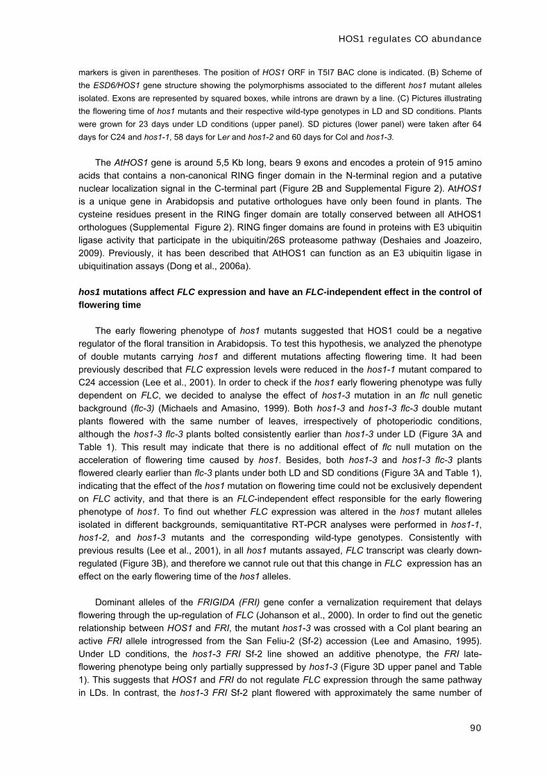
HOS1 regulates CO abundance
90
markers is given in parentheses. The position of HOS1 ORF in T5I7 BAC clone is indicated. (B) Scheme of
the ESD6/HOS1 gene structure showing the polymorphisms associated to the different hos1 mutant alleles
isolated. Exons are represented by squared boxes, while introns are drawn by a line. (C) Pictures illustrating
the flowering time of hos1 mutants and their respective wild-type genotypes in LD and SD conditions. Plants
were grown for 23 days under LD conditions (upper panel). SD pictures (lower panel) were taken after 64
days for C24 and hos1-1, 58 days for Ler and hos1-2 and 60 days for Col and hos1-3.
The AtHOS1 gene is around 5,5 Kb long, bears 9 exons and encodes a protein of 915 amino
acids that contains a non-canonical RING finger domain in the N-terminal region and a putative
nuclear localization signal in the C-terminal part (Figure 2B and Supplemental Figure 2). AtHOS1
is a unique gene in Arabidopsis and putative orthologues have only been found in plants. The
cysteine residues present in the RING finger domain are totally conserved between all AtHOS1
orthologues (Supplemental Figure 2). RING finger domains are found in proteins with E3 ubiquitin
ligase activity that participate in the ubiquitin/26S proteasome pathway (Deshaies and Joazeiro,
2009). Previously, it has been described that AtHOS1 can function as an E3 ubiquitin ligase in
ubiquitination assays (Dong et al., 2006a).
hos1 mutations affect FLC expression and have an FLC-independent effect in the control of
flowering time
The early flowering phenotype of hos1 mutants suggested that HOS1 could be a negative
regulator of the floral transition in Arabidopsis. To test this hypothesis, we analyzed the phenotype
of double mutants carrying hos1 and different mutations affecting flowering time. It had been
previously described that FLC expression levels were reduced in the hos1-1 mutant compared to
C24 accession (Lee et al., 2001). In order to check if the hos1 early flowering phenotype was fully
dependent on FLC, we decided to analyse the effect of hos1-3 mutation in an flc null genetic
background (flc-3) (Michaels and Amasino, 1999). Both hos1-3 and hos1-3 flc-3 double mutant
plants flowered with the same number of leaves, irrespectively of photoperiodic conditions,
although the hos1-3 flc-3 plants bolted consistently earlier than hos1-3 under LD (Figure 3A and
Table 1). This result may indicate that there is no additional effect of flc null mutation on the
acceleration of flowering time caused by hos1. Besides, both hos1-3 and hos1-3 flc-3 plants
flowered clearly earlier than flc-3 plants under both LD and SD conditions (Figure 3A and Table 1),
indicating that the effect of the hos1 mutation on flowering time could not be exclusively dependent
on FLC activity, and that there is an FLC-independent effect responsible for the early flowering
phenotype of hos1. To find out whether FLC expression was altered in the hos1 mutant alleles
isolated in different backgrounds, semiquantitative RT-PCR analyses were performed in hos1-1,
hos1-2, and hos1-3 mutants and the corresponding wild-type genotypes. Consistently with
previous results (Lee et al., 2001), in all hos1 mutants assayed, FLC transcript was clearly down-
regulated (Figure 3B), and therefore we cannot rule out that this change in FLC expression has an
effect on the early flowering time of the hos1 alleles.
Dominant alleles of the FRIGIDA (FRI) gene confer a vernalization requirement that delays
flowering through the up-regulation of FLC (Johanson et al., 2000). In order to find out the genetic
relationship between HOS1 and FRI, the mutant hos1-3 was crossed with a Col plant bearing an
active FRI allele introgressed from the San Feliu-2 (Sf-2) accession (Lee and Amasino, 1995).
Under LD conditions, the hos1-3 FRI Sf-2 line showed an additive phenotype, the FRI late-
flowering phenotype being only partially suppressed by hos1-3 (Figure 3D upper panel and Table
1). This suggests that HOS1 and FRI do not regulate FLC expression through the same pathway
in LDs. In contrast, the hos1-3 FRI Sf-2 plant flowered with approximately the same number of

HOS1 regulates CO abundance
91
leaves as the Col FRI Sf-2 plants under SD conditions, abolishing the effect of the hos1 mutation
(Figure 3D lower panel and Table 1).
A
Col hos1-3 flc-3 hos1-3 flc-3
C
Col hos1-3 fve-3hos1-3 fve-3
UBQ10
FLC
B
Col hos1-3ColFRI
hos1-3 FRI
D
LD
SD
SD
LD
SD
LD
Figure 3. hos1 mutations downregulate FLC expression and have an FLC-independent effect in the
control of flowering time.
(A) Flowering time phenotype of hos1-3 flc-3 double mutant in LD (upper panel) and SD (lower panel)
conditions. (B) Analysis of the expression of FLC in 14 day-old hos1 mutant seedlings and their
corresponding wild-type genotypes. FLC expression was monitored by semiquantitative RT-PCR analysis
over 22 cycles for C24 and hos1-1 and over 28 cycles for Ler, hos1-2, Col and hos1-3. For the UBQ10 control
22 cycles were used. (C) Flowering time phenotype of hos1-3 fve-3 double mutant plants grown in LD (upper
panel) and SD (lower panel) conditions. (D) Flowering time phenotype of hos1-3 plants bearing an active
allele of FRI in LDs (upper panel) and SDs (lower panel).
Because HOS1 locus is involved in cold signal transduction (Lee et al., 2001) and
vernalization regulates FLC expression (Amasino, 2010), we hypothesized that HOS1 could be
controlling FLC transcript levels through the vernalization pathway. To analyze this, we generated
combinations between hos1 and two other mutants impaired in the vernalization response such as
vernalization 1 (vrn1) and vernalization-insensitive 3 (vin3) (Levy et al., 2002; Sung and Amasino,
2004), both in late flowering backgrounds that allowed us to observe the acceleration of flowering
due to the vernalization treatment. The hos1 mutation did not impair the acceleration of flowering
caused by vernalization when combined with the late flowering fca-1 or FRI Sf-2 plants
(Supplemental Table 2). Besides, we found no difference in flowering time for the hos1-2 vrn1-2
fca-1 triple mutant grown after either 1 or 4 weeks of vernalization treatment (Supplemental Table
2). The same result was observed for hos1-3 vin3-4 carrying an active FRI allele, as both 1 and 4
week-vernalized plants flowered with approximately the same number of leaves (Supplemental
Table 2). Thus, we concluded that HOS1 does not regulate FLC expression through the
vernalization pathway.
Considering that the autonomous pathway also converges on the regulation of FLC
expression, we analyzed the flowering phenotype of double mutants combining hos1 and

HOS1 regulates CO abundance
92
mutations in representative autonomous pathway genes, in particular the hos1-3 fve-3, hos1-2 fca-
1 and hos1-3 fld-1 double mutants. Under LD, these double mutant plants showed an additive
phenotype because the late-flowering phenotype of autonomous pathway mutants was only
partially suppressed by hos1 (Figure 3C upper panel and Table 1). In contrast, under SDs,
flowering time of these double mutants was very similar to the one displayed by the autonomous
pathway mutants, as they produced only a few leaves less than fve-3, fca-1 and fld-1 respectively
(Figure 3C lower panel and Table 1).
Altogether, these results suggest that the hos1 mutation cannot accelerate flowering in SD
when combined with genetic backgrounds that have very high FLC expression levels, such as
mutations of the autonomous pathway or active alleles of FRI. In contrast, under LD the repressive
effect of HOS1 on flowering time may be mediated by additional pathways that remain inactive in
SD.
Table 1. Total number of leaves at the time of flowering for the different wild type ecotypes and single, double
and triple mutants described in this work. Data were scored in approximately 30 plants under LD conditions
and 15 plants under SD photoperiods and is represented as mean ± standard deviation.
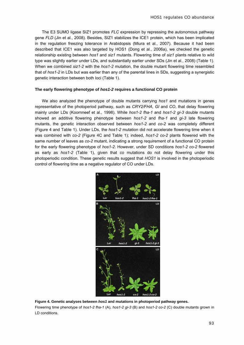
HOS1 regulates CO abundance
93
The E3 SUMO ligase SIZ1 promotes FLC expression by repressing the autonomous pathway
gene FLD (Jin et al., 2008). Besides, SIZ1 stabilizes the ICE1 protein, which has been implicated
in the regulation freezing tolerance in Arabidopsis (Miura et al., 2007). Because it had been
described that ICE1 was also targeted by HOS1 (Dong et al., 2006a), we checked the genetic
relationship existing between hos1 and siz1 mutants. Flowering time of siz1 plants relative to wild
type was slightly earlier under LDs, and substantially earlier under SDs (Jin et al., 2008) (Table 1).
When we combined siz1-2 with the hos1-2 mutation, the double mutant flowering time resembled
that of hos1-2 in LDs but was earlier than any of the parental lines in SDs, suggesting a synergistic
genetic interaction between both loci (Table 1).
The early flowering phenotype of hos1-2 requires a functional CO protein
We also analyzed the phenotype of double mutants carrying hos1 and mutations in genes
representative of the photoperiod pathway, such as CRY2/FHA, GI and CO, that delay flowering
mainly under LDs (Koornneef et al., 1998). While hos1-2 fha-1 and hos1-2 gi-3 double mutants
showed an additive flowering phenotype between hos1-2 and fha-1 and gi-3 late flowering
mutants, the genetic interaction observed between hos1-2 and co-2 was completely different
(Figure 4 and Table 1). Under LDs, the hos1-2 mutation did not accelerate flowering time when it
was combined with co-2 (Figure 4C and Table 1); indeed, hos1-2 co-2 plants flowered with the
same number of leaves as co-2 mutant, indicating a strong requirement of a functional CO protein
for the early flowering phenotype of hos1-2. However, under SD conditions hos1-2 co-2 flowered
as early as hos1-2 (Table 1), given that co mutations do not delay flowering under this
photoperiodic condition. These genetic results suggest that HOS1 is involved in the photoperiodic
control of flowering time as a negative regulator of CO under LDs.
Ler hos1-2 fha-1 hos1-2 fha-1
A
Ler hos1-2 gi-3 hos1-2 gi-3
B
Ler hos1-2 co-2 hos1-2 co-2
C
LD
LD
LD
Figure 4. Genetic analyses between hos1 and mutations in photoperiod pathway genes.
Flowering time phenotype of hos1-2 fha-1 (A), hos1-2 gi-3 (B) and hos1-2 co-2 (C) double mutants grown in
LD conditions.

HOS1 regulates CO abundance
94
FKF1 is an F-Box protein (Imaizumi et al., 2005) that mediates the cyclic degradation of CDF
proteins, which are repressors of CO expression (Imaizumi et al., 2005; Fornara et al., 2009). To
study if there was any genetic interaction between FKF1 and HOS1, the double mutant hos1-3
fkf1-1was analyzed and it showed an additive phenotype between the late flowering time of fkf1-1
and the early flowering phenotype of hos1-3 in LDs (Table 1). This result indicates that HOS1 does
not participate in the FKF1 transcriptional regulation pathway that controls CO expression.
hos1 mutants show an altered pattern of FT expression
FLC acts repressing the expression of the floral integrators FT and SOC1, while the
photoperiod pathway activate FT and SOC1 expression through CO (Yoo et al., 2005; Searle et
al., 2006, Turck et al., 2008). Because hos1 mutations showed downregulation of FLC expression
(Figure 3B) and the co-2 mutation was epistatic to hos1-2 under LDs (Figure 4C), we decided to
check the genetic relationship between HOS1 and the floral integrators FT and SOC1. The hos1-2
ft-1 double mutant showed a similar flowering phenotype to ft-1 under LD conditions, suggesting a
strong requirement of FT by the hos1 early flowering phenotype (Figure 5A and Table 1). In
contrast, hos1-2 soc1-1 double mutant was additive between both parental lines in both LD and
SD conditions (Figure 5A and Table 1). This result is in accordance with the epistasis observed
between co-2 and hos1-2, considering that FT is the main target of CO under LDs (Yoo et al.,
2005). In order to check whether the whole effect of HOS1 on flowering time was through FT and
SOC1, we generated the triple mutant hos1-2 ft-1 soc1-1. Flowering time analysis showed that the
triple mutant was slightly earlier than ft-1soc1-1 (Figure 5A and Table 1). This result indicates that
the early flowering phenotype of hos1 mutation requires functional FT and SOC1 proteins,
although we cannot rule out that HOS1 could regulate other protein(s) involved in the control of
flowering time.
In order to get a deeper insight into the genetic relationship observed between HOS1 and the
photoperiod pathway, we performed a time course expression analysis over a 24h period in Ler
and hos1-2 seedlings both in LD and SD. First of all, we demonstrated that HOS1 transcript did not
show a diurnal oscillation in Ler background (Figure 5B). Subsequently, we analyzed the temporal
expression pattern of CO, FT and SOC1 genes using semiquantitative RT-PCR and quantitative
real-time PCR (Q-PCR) approaches (Fig 5C-E). As previously reported, CO transcript level in Ler
background was high at dawn and dusk, and during the night, remaining low for the rest of the light
period of the day (Suarez-Lopez et al., 2001) (Fig 5C-E). In hos1-2, we observed the same pattern
of CO expression, indicating that hos1 mutation did not affect significantly the levels or the
expression profile of the CO transcript (Figure 5C-E). However, the expression pattern of the floral
integrator FT was clearly altered in the hos1-2 mutant compared to the wild-type both in LD and
SD conditions (Fig 5C-E). FT transcript usually shows a peak of expression at dusk in LDs (around
ZT16), following the evening increase observed in CO mRNA. In the hos1-2 mutant, we observed
a peak of FT expression in the subjective morning, mainly at ZT4, but also at ZT8, when the CO
transcript levels are barely detectable in the mutant (Figure 5C and D). To check whether this
alteration was due to a specific developmental stage of the plant or if it relied on the genetic
background, we analysed FT transcript levels at ZT4 and ZT8 in Col and hos1-3 plants harvested
8, 10, 12 and 15 days after germination (DAG). In every single stage tested, FT expression was
higher in hos1-3 in relation to Col in the first part of the day (Supplemental Figure 3). In SD, we
observed an increased FT expression in the hos1-2 mutant, starting to rise at ZT8 and peaking at
ZT12, which may explain the early flowering phenotype displayed by the mutant under non-
inductive photoperiods (Figure 5C and E). A small but consistent increase in SOC1 expression
was also detected in hos1-2 plants grown in LD photoperiods in comparison to the wild type
(Figure 5C and D). Thus, we conclude that CO transcript levels are not modified substantially by

HOS1 regulates CO abundance
95
the hos1 mutation and that HOS1 is required to repress the expression of FT in the first part of the
day in LD. Considering that HOS1 has an E3 ubiquitin ligase activity, we speculated that it may be
involved in the degradation of protein(s) that regulate FT expression. Both the genetic analysis and
the expression assays suggested that this protein could be CO.
Figure 5. The early flowering phenotype of hos1 depends on FT and SOC1 functional proteins and
hos1 mutation alters the pattern of expression of FT.
(A) Flowering time phenotype of hos1-2 soc1-1, hos1-2 ft-1 and hos1-2 ft-1 soc1-1 triple mutant plants grown
in LD conditions. (B) HOS1 expression pattern over a 24h time course in Ler seedlings grown for 8 days in
LDs and for 16 days in SDs. Samples were harvested every 4h after dawn. HOS1 expression was monitored
by semiquantitative RT-PCR analysis over 20 cycles. (C, D, E) Expression analysis of different flowering time
genes over a 24h time course in Ler and hos1-2 seedlings grown for 8 days in LDs and 16 days in SDs.
Samples were harvested every 4h after dawn (C) Semiquantitative RT-PCR analysis comparing CO (22
cycles), FT (28 cycles) and SOC1 (24 cycles) expression (D) Quantitative real- time PCR (Q-PCR) analysis of
CO, FT and SOC1 expression in LD conditions (E) Same as (D) but Ler and hos1-2 seedlings grown in SD
conditions. Relative expression levels were normalized to β-ACTIN expression.

HOS1 regulates CO abundance
96
HOS1 interacts in vitro and in vivo with CO and regulates its abundance
Given the proposed epistatic interaction between co-2 and hos1-2 mutants and considering
that CO transcript levels were not affected in the hos1-2 mutant, we decided to analyze whether
there was a physical interaction between CO and HOS1. For this purpose, in vitro pull-down
experiments using MBP-HOS1 and in vitro-translated CO protein were performed. As shown in
Figure 6A, MBP-HOS1, but not MBP alone, was able to interact with CO protein. Whether the
interaction between CO and HOS1 also occurred in vivo was tested using the bimolecular
fluorescence complementation (BiFC) technique. For that, the N terminus of YFP was cloned
upstream of HOS1 (YFNHOS1) and the C terminus of YFP was fused C-terminally in-frame to CO
(COYFC). By Agrobacterium tumefaciens coinfiltration, these constructs were transiently
expressed in abaxial epidermal cells of tobacco leaves (Voinnet et al., 2003). Reconstitution of
YFP fluorescence was examined by confocal microscopy two days after transient coexpression of
the protein pairs. Yellow fluorescence in the nucleus was detected for coexpression of COYFC and
YFNHOS1, while no yellow fluorescence was observed when COYFC was coexpressed with YFN
alone, or when YFC alone was coexpressed with YFNHOS1, as negative controls (Figure 6B). As
a positive control, the interaction between amino and carboxy parts of AKIN and AKIN10 SnRKs
proteins (Ferrando et al., 2001), was tested (Figure 6B). CO-HOS1 interaction could be observed
in conspicuous nuclear speckles, which have been often associated with foci of proteasome
degradation, as previously described for the interaction between CO and COP1 (Jang et al.,
2008). These results demonstrate that CO and HOS1 colocalize and physically interact in the
nuclei of plant cells.
It has been reported that HOS1 has auto-ubiquitination ligase activity in vitro and that it can
also mediate the ubiquitination and degradation of ICE1 transcription factor (Dong et al., 2006a).
To further analyze whether HOS1 may also regulate CO degradation in vivo, we transformed a
construct constitutively expressing CO fused to LUC into wild-type Arabidopsis plants. One
representative line, 35S::CO–LUC 6-2, displaying an early flowering phenotype, was introduced
into hos1-3 by genetic crossing. We found that the 35S::CO–LUC 6-2/hos1-3 plants flowered
earlier than either the hos1-3 mutant or the 35S::CO–LUC 6-2 plants, indicating that CO–LUC
construct was fully functional (Supplemental Table 3). Using luciferase fluorescence in vivo
imaging, we found that under LDs the CO protein levels were significantly lower in the wild-type
than in the hos1 mutant background three hours after dawn (ZT3) (Figure 6C), suggesting that the
degradation of CO that occurs in the wild-type is impaired in the hos1 mutant. Quantification of
luciferase activity corroborated that CO protein accumulated to higher levels in the hos1 mutant
than in the wild type plants (Figure 6D). This accumulation of CO protein observed at ZT3
correlates with the early peak of FT expression present in the hos1 mutant (Figure 5C and D and
Supplemental Figure 3). To further assess the role of HOS1 in CO regulation, we performed
western blot assays to detect CO protein in nuclear extracts from wild type and hos1-3 plants
grown under LD photoperiods. In these immunoblots, CO protein was present at lower abundance
in the wild type than in the hos1-3 mutant plants, particularly during the daylight period (Figure 6E).
From these data, we conclude that HOS1 is involved in the photoperiodic regulation of flowering
through the modulation of CO protein levels in vivo.
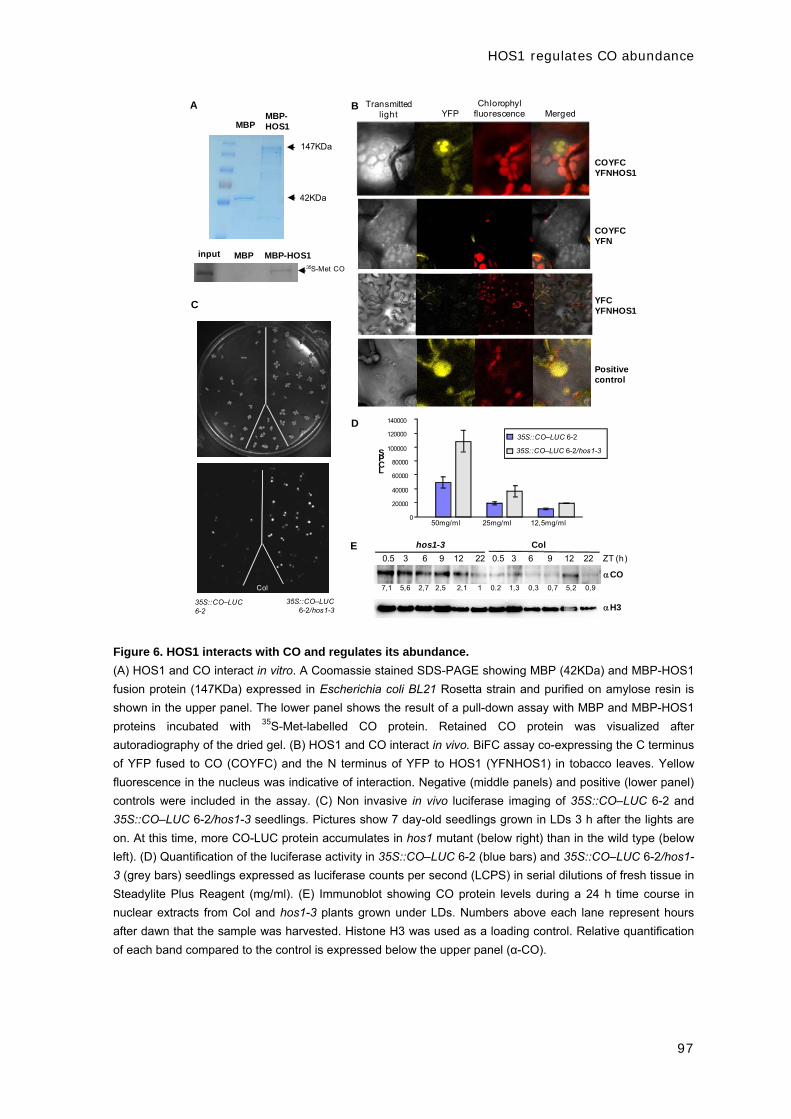
HOS1 regulates CO abundance
97
0
20000
40000
60000
80000
100000
120000
140000
50mg/ml 25mg/ml 12,5mg/ml
LCPS
35S::CO–LUC 6-2
35S::CO–LUC 6-2/hos1-3
D
C
35S::CO–LUC6-2/hos1-3
35S::CO–LUC6-2
E
H3
CO
hos1-3 Col
0.5 3 6 9 12 22 0.5 3 6 9 12 22 ZT (h)
7,1 5,6 2,7 2,5 2,1 1 0.2 1,3 0,3 0,7 5,2 0,9
B
COYFC YFNHOS1
COYFCYFN
Positivecontrol
Transmitted light YFP
Chlorophylfluorescence Merged
YFCYFNHOS1
Col
A
147KDa
MBPMBP-HOS1
42KDa
input MBP MBP-HOS135S-Met CO
Figure 6. HOS1 interacts with CO and regulates its abundance.
(A) HOS1 and CO interact in vitro. A Coomassie stained SDS-PAGE showing MBP (42KDa) and MBP-HOS1
fusion protein (147KDa) expressed in Escherichia coli BL21 Rosetta strain and purified on amylose resin is
shown in the upper panel. The lower panel shows the result of a pull-down assay with MBP and MBP-HOS1
proteins incubated with 35S-Met-labelled CO protein. Retained CO protein was visualized after
autoradiography of the dried gel. (B) HOS1 and CO interact in vivo. BiFC assay co-expressing the C terminus
of YFP fused to CO (COYFC) and the N terminus of YFP to HOS1 (YFNHOS1) in tobacco leaves. Yellow
fluorescence in the nucleus was indicative of interaction. Negative (middle panels) and positive (lower panel)
controls were included in the assay. (C) Non invasive in vivo luciferase imaging of 35S::CO–LUC 6-2 and
35S::CO–LUC 6-2/hos1-3 seedlings. Pictures show 7 day-old seedlings grown in LDs 3 h after the lights are
on. At this time, more CO-LUC protein accumulates in hos1 mutant (below right) than in the wild type (below
left). (D) Quantification of the luciferase activity in 35S::CO–LUC 6-2 (blue bars) and 35S::CO–LUC 6-2/hos1-
3 (grey bars) seedlings expressed as luciferase counts per second (LCPS) in serial dilutions of fresh tissue in
Steadylite Plus Reagent (mg/ml). (E) Immunoblot showing CO protein levels during a 24 h time course in
nuclear extracts from Col and hos1-3 plants grown under LDs. Numbers above each lane represent hours
after dawn that the sample was harvested. Histone H3 was used as a loading control. Relative quantification
of each band compared to the control is expressed below the upper panel (α-CO).
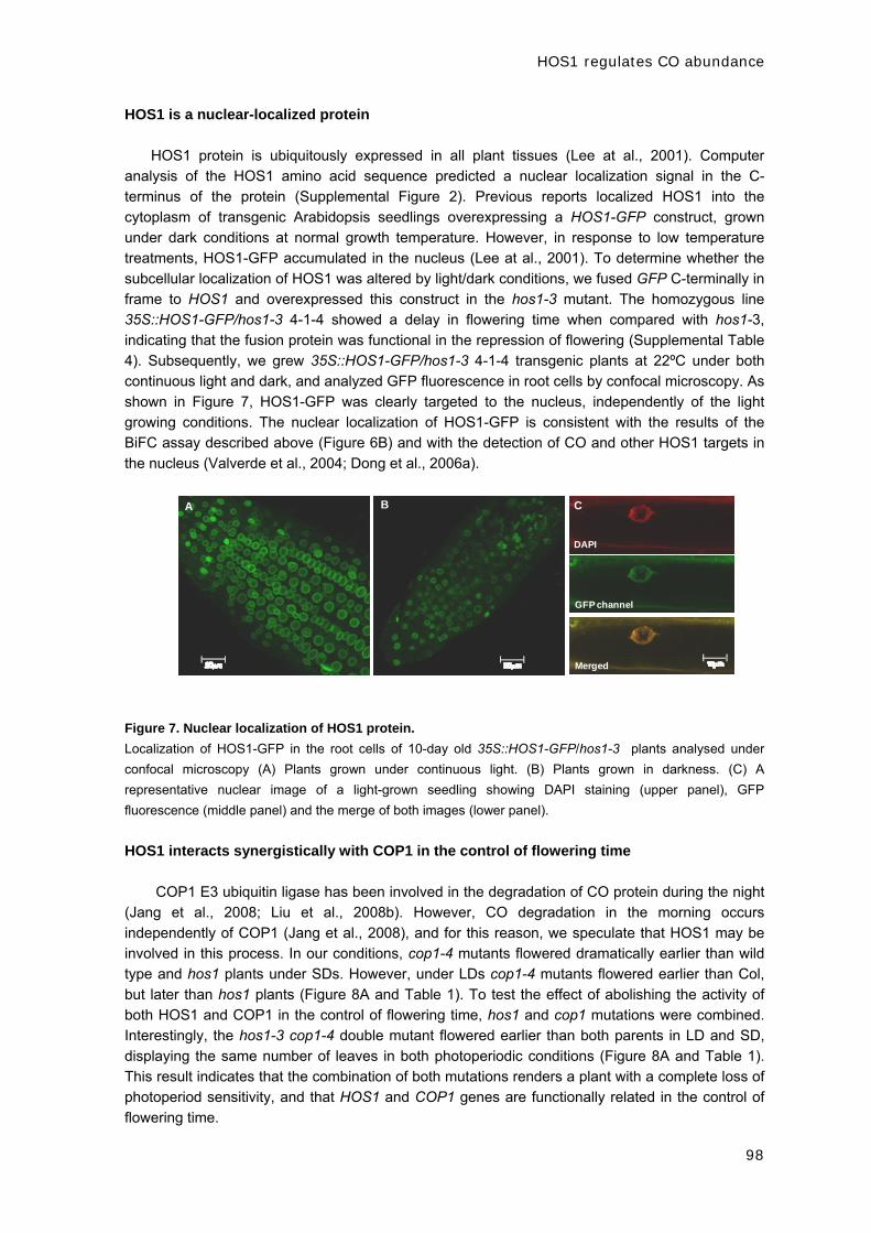
HOS1 regulates CO abundance
98
HOS1 is a nuclear-localized protein
HOS1 protein is ubiquitously expressed in all plant tissues (Lee at al., 2001). Computer
analysis of the HOS1 amino acid sequence predicted a nuclear localization signal in the C-
terminus of the protein (Supplemental Figure 2). Previous reports localized HOS1 into the
cytoplasm of transgenic Arabidopsis seedlings overexpressing a HOS1-GFP construct, grown
under dark conditions at normal growth temperature. However, in response to low temperature
treatments, HOS1-GFP accumulated in the nucleus (Lee at al., 2001). To determine whether the
subcellular localization of HOS1 was altered by light/dark conditions, we fused GFP C-terminally in
frame to HOS1 and overexpressed this construct in the hos1-3 mutant. The homozygous line
35S::HOS1-GFP/hos1-3 4-1-4 showed a delay in flowering time when compared with hos1-3,
indicating that the fusion protein was functional in the repression of flowering (Supplemental Table
4). Subsequently, we grew 35S::HOS1-GFP/hos1-3 4-1-4 transgenic plants at 22ºC under both
continuous light and dark, and analyzed GFP fluorescence in root cells by confocal microscopy. As
shown in Figure 7, HOS1-GFP was clearly targeted to the nucleus, independently of the light
growing conditions. The nuclear localization of HOS1-GFP is consistent with the results of the
BiFC assay described above (Figure 6B) and with the detection of CO and other HOS1 targets in
the nucleus (Valverde et al., 2004; Dong et al., 2006a).
A B C
DAPI
GFP channel
Merged
Figure 7. Nuclear localization of HOS1 protein.
Localization of HOS1-GFP in the root cells of 10-day old 35S::HOS1-GFP/hos1-3 plants analysed under
confocal microscopy (A) Plants grown under continuous light. (B) Plants grown in darkness. (C) A
representative nuclear image of a light-grown seedling showing DAPI staining (upper panel), GFP
fluorescence (middle panel) and the merge of both images (lower panel).
HOS1 interacts synergistically with COP1 in the control of flowering time
COP1 E3 ubiquitin ligase has been involved in the degradation of CO protein during the night
(Jang et al., 2008; Liu et al., 2008b). However, CO degradation in the morning occurs
independently of COP1 (Jang et al., 2008), and for this reason, we speculate that HOS1 may be
involved in this process. In our conditions, cop1-4 mutants flowered dramatically earlier than wild
type and hos1 plants under SDs. However, under LDs cop1-4 mutants flowered earlier than Col,
but later than hos1 plants (Figure 8A and Table 1). To test the effect of abolishing the activity of
both HOS1 and COP1 in the control of flowering time, hos1 and cop1 mutations were combined.
Interestingly, the hos1-3 cop1-4 double mutant flowered earlier than both parents in LD and SD,
displaying the same number of leaves in both photoperiodic conditions (Figure 8A and Table 1).
This result indicates that the combination of both mutations renders a plant with a complete loss of
photoperiod sensitivity, and that HOS1 and COP1 genes are functionally related in the control of
flowering time.

HOS1 regulates CO abundance
99
A
Col hos1-3 cop1-4hos1-3 cop1-4
Col hos1-3Col
35S::CO
C
Col hos1-3 cop1-4hos1-3 cop1-4co-10
hos1-3co-10
hos1-3cop1-4co-10
cop1-4co-10
B
hos1-3 35S::CO
Figure 8. Genetic interaction between HOS1 and COP1 in the control of flowering time.
(A) Flowering time phenotype of hos1-3 cop1-4 double mutant in LD (upper panel) and SD (lower panel)
conditions. (B) Flowering time phenotype of double and triple mutant combinations between hos1-3 co-10
cop1-4 mutants grown in LD conditions. (C) Comparison of flowering time phenotype between LD-grown Col
and hos1-3 plants bearing a 35S::CO transgene.
To further investigate the genetic interaction between HOS1, COP1 and CO genes in
controlling flowering time of Arabidopsis, a hos1 cop1 co triple mutant was generated and its
flowering time was compared with that of hos1 co and cop1 co double mutants (Figure 8B and
Table 1). Under LDs, the hos1-3 co-10 and the cop1-4 co-10 plants flowered with 12 and 10
leaves less than co-10 respectively, indicating that part of the early flowering phenotype of the
hos1-3 and cop1-4 mutants in Col background occurs independently of CO. This result appears to
be in contrast with the epistatic genetic relationship observed between hos1-2 and co-2 alleles in
Ler background (Figure 4C and Table 1), and can be explained because hos1 mutation is
downregulating FLC expression (Figure 3B) and FLC is expressed at higher levels in Col than in
Ler (Michaels and Amasino, 1999). Besides, the hos1 cop1 co triple mutant flowered with 15
leaves more than the hos1 cop1 double mutant under LD conditions, demonstrating that co
mutation notably delays the hos1 cop1 early flowering phenotype in Col background. Interestingly,
the hos1 cop1 co triple mutant formed 6 and 9 leaves less than hos1 co and cop1 co double
mutants respectively (Figure 8B and Table 1), confirming the existence of a synergistic genetic
interaction between hos1 and cop1, even in the absence of CO.
Because HOS1 seems to exert an effect as a negative regulator of CO, we tested whether the
extremely early flowering of 35S::CO plants (Simon et al., 1996) could be further accelerated by
the hos1-3 mutation. To test this hypothesis, the 35S::CO transgene was introduced into wild type
Col and into hos1-3 mutant plants. Although the number of leaves at flowering for both transgenic
plants was very similar, we observed that the hos1-3 35S::CO plants bolted consistently earlier
than the Col 35S::CO (Figure 8C and Table 1), supporting a role for HOS1 in repressing the
promotion of flowering mediated by CO.
DISCUSSION
In many plants, changes in daylength regulate the transition from vegetative growth to
flowering, and plants altered in the daylength-sensing mechanism cannot time flowering properly

HOS1 regulates CO abundance
100
in natural environments (Wilczek et al., 2009). In this work, we have demonstrated through both
genetic and biochemical approaches that HOS1 is involved in the photoperiodic control of
flowering time. The esd6/hos1 mutant was identified through a screening devoted to the isolation
of early flowering mutants in Arabidopsis. The characterization of these mutants allows unveiling
the mechanisms of action of genes involved in the repression of the floral transition and suggests
that a large number of genes participate in this process (Pouteau et al., 2004). Besides precocious
flowering, the hos1 mutant showed pleiotropic alterations of leaf, flower and root development,
similarly to those displayed by other early flowering mutants (Martin-Trillo et al., 2006; del Olmo et
al., 2010).
In Arabidopsis, the flowering response to changes in photoperiod rely on the interaction of
light with the circadian clock-regulated rhythmic expression of CO (Suarez-Lopez et al., 2001).
Besides this transcriptional regulation, a light-dependent regulation of CO protein stability has also
been described (Valverde et al., 2004; Kim et al., 2008). We have demonstrated that HOS1 is
involved in regulating CO protein abundance in vivo (Figure 6), ensuring that CO activation of FT
only occurs at the appropriate times of the day under inductive photoperiods in Arabidopsis. HOS1
has been reported to work as an E3 ubiquitin ligase that mediates the degradation of ICE1
transcription factor (Dong et al., 2006a), and we have demonstrated that HOS1 interacts in vitro
and in vivo with CO (Figure 6). In addition, hos1 mutation altered FT expression pattern in LD,
showing a peak of expression in the subjective morning (Figure 5C and D). Based on these
observations, we speculate that HOS1 could mediate CO degradation during the daylight period
through a mechanism involving ubiquitination, and that the timing of HOS1 activity is crucial to
establish a photoperiodic flowering response (Figure 9). Both the genetic analysis between CO
and HOS1 genes and the expression analyses performed involving CO transcript and CO protein,
support this hypothesis.
Other E3 ubiquitin ligases have been proposed to be involved in the control of flowering time
(Cao et al., 2008; Vega-Sanchez et al., 2008; Park et al., 2010). In particular, DAY NEUTRAL
FLOWERING (DNF) and COP1 have been demonstrated to regulate the precise pattern of CO
expression at the transcriptional and the posttranscriptional level respectively (Jang et al., 2008;
Liu et al., 2008b; Morris et al., 2010). DNF is an important regulator of the rhythm of CO
expression, but it is not acting through the GI/FKF1/CDFs regulatory mechanism (Morris et al.,
2010). Increased CO transcript in the dnf mutant around ZT 4-6 results in an earlier induction of FT
under SD (Morris et al., 2010). On the other hand, CO protein is degraded in the dark by the
SUPPRESSOR OF PHYA-105 1 (SPA1)-COP1 complex (Laubinger et al., 2006; Jang et al., 2008;
Liu et al., 2008b). Besides, it has been recently demonstrated that the Arabidopsis CULLIN4 E3
RING ligase bound to Damaged DNA binding protein 1 (DDB1) interacts with SPA1-COP1
complex to regulate flowering time (Chen et al., 2010). We have demonstrated that HOS1 also
interacts genetically with COP1 in the photoperiodic control of flowering time (Figure 8A).
Interestingly, hos1 cop1 double mutants are completely insensitive to photoperiod, and co
mutations notably delay the early flowering phenotype of the hos1 cop1 double mutant (Figure
8B). This can be interpreted as HOS1 and COP1 being functionally related proteins in the control
of flowering time, regulating CO abundance during the day and in the night, respectively (Figure
9). This is consistent with the observation that the absence of both E3 ubiquitin ligases renders
plants unable to distinguish between LDs and SDs. It has been proposed that a phyB-dependent
mechanism occurring early in the day may promote CO degradation as well, but the E3 ubiquitin
ligase(s) involved in this process remains to be identified (Valverde et al., 2004; Jang et al., 2008).
Our data are consistent with HOS1 playing a crucial role in preventing increased CO protein levels
and FT expression during early hours of the day. Further analyses will be required to establish the
possible participation of HOS1 in the proposed phyB-dependent mechanism of CO proteolysis.

HOS1 regulates CO abundance
101
COmRNA
PhyB
Cry2
HOS1
COP1
CO
CO
CO CO
COCO
CO
COCO
CO
CO
COP1
Figure 9. Model for HOS1 function in the photoperiodic control of flowering time.
The transcription of CO gene depends primarily on the circadian clock (thick black line). In the evening, the
degradation of CDFs by the GI/FKF1 complex allows CO transcript levels to increase, and CO protein
accumulates due to a photoreceptor-mediated repression of COP1. At this time CO can promote FT
expression and induce flowering. During the night, COP1 activity causes rapid degradation of CO protein by
the ubiquitination/26S proteasome system. In the daylight period HOS1 is required to degrade CO. Additional
data will be necessary to establish the possible involvement of HOS1 in the mechanism of CO degradation
mediated by PhyB that has been proposed to operate in the morning.
The ability to respond to photoperiod enables plants to anticipate variations in environmental
conditions that can be predicted to occur periodically each year. In northern latitudes, shortening
daylength in autumn is associated to decreasing cold temperatures while warm temperatures are
typical of longer days. In addition to repress the floral transition, HOS1 was previously described
as a negative regulator of cold signal transduction (Lee et al., 2001). This suggests that HOS1
might function as an integrative link for both responses, allowing plants to discriminate the duration
of the day by regulating CO abundance, and to respond to cold temperatures, by regulating CBF
(C-repeat (CRT)-binding factors) expression through ICE1 degradation (Dong et al., 2006a).
Several evidences point to the existence of overlapping pathways for controlling cold stress and
flowering time responses in Arabidopsis (Yoo et al., 2007; Seo et al., 2009). The characterization
of several mutants altered in cold acclimation responses has uncovered a role of the
corresponding genes in flowering time control. hos9 and sensitive to freezing 6 (sf6) mutants show
a late flowering phenotype, while long vegetative phase 1 (lov1) mutant is early flowering (Zhu et
al., 2004; Yoo et al., 2007; Knight et al., 2008). These three genes regulate the expression of cold-
inducible genes independently of CBFs, and both LOV1 and SFR6 control flowering time through
the photoperiod pathway. Other mutants, such as low expression of osmotically responsive genes
4 (los4) and atnup160, display an early flowering phenotype and altered CBF expression levels
(Gong et al., 2005; Dong et al., 2006b). On the other hand, co and gi photoperiod pathway
mutants show altered tolerance to freezing temperatures (Cao et al., 2005; Yoo et al., 2007), and
fve mutant flowers late and shows elevated expression of FLC and CBF genes (Kim et al., 2004).
It has been proposed very recently that in warm late spring SOC1 downregulates CBFs expression
and promotes flowering, while in cold early spring or fall, induction of FLC expression by the CBFs
delays flowering and confers cold resistance to the plant (Seo et al., 2009). Besides regulating CO
stability, HOS1 controls FLC expression (Figure 3B) (Lee et al., 2001), which is also repressed by

HOS1 regulates CO abundance
102
prolonged exposure to cold temperatures (Amasino, 2010). The positive effect of HOS1 on FLC
expression appears to be independent of the vernalization pathway (Supplemental Table 2) and
awaits to be characterised. Taken together, these results suggest that HOS1, among other genes,
may participate in the photoperiod and temperature signal crosstalk, integrating information
coming from both pathways and facilitating a proper response to changing environmental
conditions. To our knowledge this is the first E3 ubiquitin ligase proposed to integrate both
environmental signals, specifically targeting for degradation key factors involved in the regulation
of each response. Further studies will be necessary for an in-depth understanding of how these
pathways modulate each other´s activity to optimize plant adaptation.
ACKNOWLEDGEMENTS We thank Dr. I. Ausin (University of California, Los Angeles, USA) for isolating the esd6 mutant, Dr. R. Catalá (CIB, CSIC, Madrid, Spain) for providing a Gateway-compatible HOS1 full length cDNA, Dr. J. Salinas (CIB, CSIC, Madrid, Spain) for granting the access to the luciferase imaging system, Dr. P. González-Melendi (CBGP, UPM-INIA, Madrid, Spain) for technical support with confocal microscope and Dr. JC. del Pozo and members of his lab (CBGP, UPM-INIA, Madrid, Spain) for helpful discussions. pYFPN43 and pYFPC43 vectors, and SNF1 protein kinase constructs, used as positive controls in the BiFC assays, were kindly provided by Dr. A. Ferrando (IBMCP, Valencia, Spain). C24 and hos1-1 mutant seeds, as well as a HOS1 expression construct in the pMAL vector, were kindly provided by Dr. J.K Zhu (University of California, Riverside, California, USA). The 35S::CO construct and co-10 cop1-4 seeds were kindly donated by Dr. G. Coupland (Max Planck Institute, Cologne, Germany), while the 35S::CO-LUC plasmid was kindly provided by Dr. H-Q Yang (Shanghai Institute for Biological Sciences, Chinese Academy of Science, Shanghai, China). This work was supported by grants BIO2008-00351 to JAJ, BIO2007-61215 to MP, CSD2007-00057 and BIO2010-15589 to JAJ and MP, and BIO2007-61837 and BIO2010-16027 to FV from the Spanish Ministerio de Ciencia e Innovación.
BIBLIOGRAPHY
Amasino, R. (2010). Seasonal and developmental timing of flowering. Plant J 61, 1001-1013. An, H., Roussot, C., Suarez-Lopez, P., Corbesier, L., Vincent, C., Pineiro, M., Hepworth, S., Mouradov,
A., Justin, S., Turnbull, C., and Coupland, G. (2004). CONSTANS acts in the phloem to regulate a systemic signal that induces photoperiodic flowering of Arabidopsis. Development 131, 3615-3626.
Ausin, I., Alonso-Blanco, C., Jarillo, J.A., Ruiz-Garcia, L., and Martinez-Zapater, J.M. (2004). Regulation of flowering time by FVE, a retinoblastoma-associated protein. Nat Genet 36, 162-166.
Cao, S., Ye, M., and Jiang, S. (2005). Involvement of GIGANTEA gene in the regulation of the cold stress response in Arabidopsis. Plant Cell Rep 24, 683-690.
Cao, Y., Dai, Y., Cui, S., and Ma, L. (2008). Histone H2B monoubiquitination in the chromatin of FLOWERING LOCUS C regulates flowering time in Arabidopsis. Plant Cell 20, 2586-2602.
Corbesier, L., Vincent, C., Jang, S., Fornara, F., Fan, Q., Searle, I., Giakountis, A., Farrona, S., Gissot, L., Turnbull, C., and Coupland, G. (2007). FT protein movement contributes to long-distance signaling in floral induction of Arabidopsis. Science 316, 1030-1033.
Chen, H., Huang, X., Gusmaroli, G., Terzaghi, W., Lau, O.S., Yanagawa, Y., Zhang, Y., Li, J., Lee, J.H., Zhu, D., and Deng, X.W. (2010). Arabidopsis CULLIN4-Damaged DNA Binding Protein 1 Interacts with CONSTITUTIVELY PHOTOMORPHOGENIC1-SUPPRESSOR OF PHYA Complexes to Regulate Photomorphogenesis and Flowering Time. Plant Cell 22, 108-123.
Chiang, G.C., Barua, D., Kramer, E.M., Amasino, R.M., and Donohue, K. (2009). Major flowering time gene, FLOWERING LOCUS C, regulates seed germination in Arabidopsis thaliana. Proc Natl Acad Sci U S A 106, 11661-11666.
de Montaigu, A., Toth, R., and Coupland, G. (2010). Plant development goes like clockwork. Trends Genet 26, 296-306.
del Olmo, I., Lopez-Gonzalez, L., Martin-Trillo, M.M., Martinez-Zapater, J.M., Pineiro, M., and Jarillo, J.A. (2010). EARLY IN SHORT DAYS 7 (ESD7) encodes the catalytic subunit of DNA polymerase epsilon and is required for flowering repression through a mechanism involving epigenetic gene silencing. Plant J 61, 623-636.
Deng, X.W., Caspar, T., and Quail, P.H. (1991). COP1: a regulatory locus involved in lightcontrolled development and gene expression in Arabidopsis. Genes Dev 5, 1172-1182.
Deshaies, R.J., and Joazeiro, C.A. (2009). RING domain E3 ubiquitin ligases. Annu Rev Biochem 78, 399-434.
Dong, C.H., Agarwal, M., Zhang, Y., Xie, Q., and Zhu, J.K. (2006a). The negative regulator of plant cold responses, HOS1, is a RING E3 ligase that mediates the ubiquitination and degradation of ICE1. Proc Natl Acad Sci U S A 103, 8281-8286.

HOS1 regulates CO abundance
103
Dong, C.H., Hu, X., Tang, W., Zheng, X., Kim, Y.S., Lee, B.H., and Zhu, J.K. (2006b). A putative Arabidopsis nucleoporin, AtNUP160, is critical for RNA export and required for plant tolerance to cold stress. Mol Cell Biol 26, 9533-9543.
Ferrando, A., Koncz-Kalman, Z., Farras, R., Tiburcio, A., Schell, J., and Koncz, C. (2001). Detection of in vivo protein interactions between Snf1-related kinase subunits with intron-tagged epitope-labelling in plants cells. Nucleic Acids Res 29, 3685-3693.
Fornara, F., de Montaigu, A., and Coupland, G. (2010). SnapShot: Control of flowering in Arabidopsis. Cell 141, 550, 550 e551-552.
Fornara, F., Panigrahi, K.C., Gissot, L., Sauerbrunn, N., Ruhl, M., Jarillo, J.A., and Coupland, G. (2009). Arabidopsis DOF transcription factors act redundantly to reduce CONSTANS expression and are essential for a photoperiodic flowering response. Dev Cell 17, 75-86.
Gong, Z., Dong, C.H., Lee, H., Zhu, J., Xiong, L., Gong, D., Stevenson, B., and Zhu, J.K. (2005). A DEAD box RNA helicase is essential for mRNA export and important for development and stress responses in Arabidopsis. Plant Cell 17, 256-267.
Guo, H., Yang, H., Mockler, T.C., and Lin, C. (1998). Regulation of flowering time by Arabidopsis photoreceptors. Science 279, 1360-1363.
He, Y., Michaels, S.D., and Amasino, R.M. (2003). Regulation of flowering time by histone acetylation in Arabidopsis. Science 302, 1751-1754.
Imaizumi, T. (2010). Arabidopsis circadian clock and photoperiodism: time to think about location. Curr Opin Plant Biol 13, 83-89.
Imaizumi, T., Schultz, T.F., Harmon, F.G., Ho, L.A., and Kay, S.A. (2005). FKF1 F-box protein mediates cyclic degradation of a repressor of CONSTANS in Arabidopsis. Science 309, 293-297.
Jackson, S.D. (2009). Plant responses to photoperiod. New Phytol 181, 517-531. Jaeger, K.E., andWigge, P.A. (2007). FT protein acts as a long-range signal in Arabidopsis. Curr Biol 17,
1050-1054. Jang, S., Torti, S., and Coupland, G. (2009). Genetic and spatial interactions between FT, TSF and SVP
during the early stages of floral induction in Arabidopsis. Plant J 60, 614-625. Jang, S., Marchal, V., Panigrahi, K.C., Wenkel, S., Soppe, W., Deng, X.W., Valverde, F., and Coupland,
G. (2008). Arabidopsis COP1 shapes the temporal pattern of CO accumulation conferring a photoperiodic flowering response. EMBO J 27, 1277-1288.
Jin, J.B., Jin, Y.H., Lee, J., Miura, K., Yoo, C.Y., Kim, W.Y., Van Oosten, M., Hyun, Y., Somers, D.E., Lee, I., Yun, D.J., Bressan, R.A., and Hasegawa, P.M. (2008). The SUMO E3 ligase, AtSIZ1, regulates flowering by controlling a salicylic acid-mediated floral promotion pathway and through affects on FLC chromatin structure. Plant J 53, 530-540.
Johanson, U., West, J., Lister, C., Michaels, S., Amasino, R., and Dean, C. (2000). Molecular analysis of FRIGIDA, a major determinant of natural variation in Arabidopsis flowering time. Science 290, 344-347.
Kim, H.J., Hyun, Y., Park, J.Y., Park, M.J., Park, M.K., Kim, M.D., Lee, M.H., Moon, J., Lee, I., and Kim, J. (2004). A genetic link between cold responses and flowering time through FVE in Arabidopsis thaliana. Nat Genet 36, 167-171.
Kim, S.Y., Yu, X., and Michaels, S.D. (2008). Regulation of CONSTANS and FLOWERING LOCUS T expression in response to changing light quality. Plant Physiol 148, 269-279.
Knight, H., Thomson, A.J., and McWatters, H.G. (2008). SENSITIVE TO FREEZING 6 integrates cellular and environmental inputs to the plant circadian clock. Plant Physiol 148, 293-303.
Kobayashi, Y., and Weigel, D. (2007). Move on up, it's time for change mobile signals controlling photoperiod-dependent flowering. Genes Dev 21, 2371-2384.
Koornneef, M. (1991). Isolation of higher plant developmental mutants. Symp Soc Exp Biol 45, 1-19. Koornneef, M., Alonso-Blanco, C., Peeters, A.J., and Soppe, W. (1998). Genetic Control of Flowering
Time in Arabidopsis. Annu Rev Plant Physiol Plant Mol Biol 49, 345-370. Kumimoto, R.W., Zhang, Y., Siefers, N., and Holt, B.F., 3rd. (2010). NF-YC3, NF-YC4 and NF-YC9 are
required for CONSTANS-mediated, photoperiod-dependent flowering in Arabidopsis thaliana. Plant J 63, 379-391.
Laubinger, S., Marchal, V., Le Gourrierec, J., Wenkel, S., Adrian, J., Jang, S., Kulajta, C., Braun, H., Coupland, G., and Hoecker, U. (2006). Arabidopsis SPA proteins regulate photoperiodic flowering and interact with the floral inducer CONSTANS to regulate its stability. Development 133, 3213-3222.
Lazaro, A., Gomez-Zambrano, A., Lopez-Gonzalez, L., Pineiro, M., and Jarillo, J.A. (2008). Mutations in the Arabidopsis SWC6 gene, encoding a component of the SWR1 chromatin remodelling complex, accelerate flowering time and alter leaf and flower development. J Exp Bot 59, 653-666.
Lee, H., Xiong, L., Gong, Z., Ishitani, M., Stevenson, B., and Zhu, J.K. (2001). The Arabidopsis HOS1 gene negatively regulates cold signal transduction and encodes a RING finger protein that displays cold-regulated nucleo-cytoplasmic partitioning. Genes Dev 15, 912-924.
Lee, I., and Amasino, R.M. (1995). Effect of Vernalization, Photoperiod, and Light Quality on the Flowering Phenotype of Arabidopsis Plants Containing the FRIGIDA Gene. Plant Physiol 108, 157-162.
Levy, Y.Y., Mesnage, S., Mylne, J.S., Gendall, A.R., and Dean, C. (2002). Multiple roles of Arabidopsis VRN1 in vernalization and flowering time control. Science 297, 243-246.
Liu, H., Yu, X., Li, K., Klejnot, J., Yang, H., Lisiero, D., and Lin, C. (2008a). Photoexcited CRY2 interacts with CIB1 to regulate transcription and floral initiation in Arabidopsis. Science 322, 1535-1539.

HOS1 regulates CO abundance
104
Liu, L.J., Zhang, Y.C., Li, Q.H., Sang, Y., Mao, J., Lian, H.L., Wang, L., and Yang, H.Q. (2008b). COP1-Mediated Ubiquitination of CONSTANS Is Implicated in Cryptochrome Regulation of Flowering in Arabidopsis. Plant Cell 20, 292-306.
Martin-Trillo, M., Lazaro, A., Poethig, R.S., Gomez-Mena, C., Pineiro, M.A., Martinez-Zapater, J.M. and Jarillo, J.A. (2006). EARLY IN SHORT DAYS 1 (ESD1) encodes ACTIN-RELATED PROTEIN 6 (AtARP6), a putative component of chromatin remodelling complexes that positively regulates FLC accumulation in Arabidopsis. Development 133, 1241-1252.
Michaels, S.D. (2009). Flowering time regulation produces much fruit. Curr Opin Plant Biol 12, 75-80. Michaels, S.D., and Amasino, R.M. (1999). FLOWERING LOCUS C encodes a novel MADS domain protein
that acts as a repressor of flowering. Plant Cell 11, 949-956. Miura, K., Jin, J.B., Lee, J., Yoo, C.Y., Stirm, V., Miura, T., Ashworth, E.N., Bressan, R.A., Yun, D.J., and
Hasegawa, P.M. (2007). SIZ1-Mediated Sumoylation of ICE1 Controls CBF3/DREB1A Expression and Freezing Tolerance in Arabidopsis. Plant Cell 19, 1403-1414.
Miura, K., Rus, A., Sharkhuu, A., Yokoi, S., Karthikeyan, A.S., Raghothama, K.G., Baek, D., Koo, Y.D., Jin, J.B., Bressan, R.A., Yun, D.J., and Hasegawa, P.M. (2005). The Arabidopsis SUMO E3 ligase SIZ1 controls phosphate deficiency responses. Proc Natl Acad Sci U S A 102, 7760-7765.
Moran, R. (1982). Formulae for determination of chlorophyllous pigments extracted with n,ndimethylformamide. Plant Physiol 69, 1376-1381.
Morris, K., Thornber, S., Codrai, L., Richardson, C., Craig, A., Sadanandom, A., Thomas, B., and Jackson, S. (2010). DAY NEUTRAL FLOWERING represses CONSTANS to prevent Arabidopsis flowering early in short days. Plant Cell 22, 1118-1128.
Nelson, D.C., Lasswell, J., Rogg, L.E., Cohen, M.A., and Bartel, B. (2000). FKF1, a clock controlled gene that regulates the transition to flowering in Arabidopsis. Cell 101, 331-340.
Park, B.S., Eo, H.J., Jang, I.C., Kang, H.G., Song, J.T., and Seo, H.S. (2010). Ubiquitination of LHY by SINAT5 regulates flowering time and is inhibited by DET1. Biochem Biophys Res Commun 398, 242-246.
Pineiro, M., Gomez-Mena, C., Schaffer, R., Martinez-Zapater, J.M., and Coupland, G. (2003). EARLY BOLTING IN SHORT DAYS is related to chromatin remodeling factors and regulates flowering in Arabidopsis by repressing FT. Plant Cell 15, 1552-1562.
Pouteau, S., Ferret, V., Gaudin, V., Lefebvre, D., Sabar, M., Zhao, G., and Prunus, F. (2004). Extensive phenotypic variation in early flowering mutants of Arabidopsis. Plant Physiol 135, 201-211.
Reed, J.W., Nagpal, P., Poole, D.S., Furuya, M., and Chory, J. (1993). Mutations in the gene for the red/far-red light receptor phytochrome B alter cell elongation and physiological responses throughout Arabidopsis development. Plant Cell 5, 147-157.
Roux, F., Touzet, P., Cuguen, J., and Le Corre, V. (2006). How to be early flowering: an evolutionary perspective. Trends Plant Sci 11, 375-381.
Samach, A., Onouchi, H., Gold, S.E., Ditta, G.S., Schwarz-Sommer, Z., Yanofsky, M.F., and Coupland, G. (2000). Distinct roles of CONSTANS target genes in reproductive development of Arabidopsis. Science 288, 1613-1616.
Sawa, M., Nusinow, D.A., Kay, S.A., and Imaizumi, T. (2007). FKF1 and GIGANTEA Complex Formation is Required for Day-Length Measurement in Arabidopsis. Science 318, 261-265.
Searle, I., He, Y., Turck, F., Vincent, C., Fornara, F., Krober, S., Amasino, R.A., and Coupland, G. (2006). The transcription factor FLC confers a flowering response to vernalization by repressing meristem competence and systemic signaling in Arabidopsis. Genes Dev 20, 898-912.
Seo, E., Lee, H., Jeon, J., Park, H., Kim, J., Noh, Y.S., and Lee, I. (2009). Crosstalk between Cold Response and Flowering in Arabidopsis Is Mediated through the Flowering-Time Gene SOC1 and Its Upstream Negative Regulator FLC. Plant Cell 21, 3185-3197.
Simon, R., Igeno, M.I., and Coupland, G. (1996). Activation of floral meristem identity genes in Arabidopsis. Nature 384, 59-62.
Suarez-Lopez, P., Wheatley, K., Robson, F., Onouchi, H., Valverde, F., and Coupland, G. (2001). CONSTANS mediates between the circadian clock and the control of flowering in Arabidopsis. Nature 410, 1116-1120.
Sung, S., and Amasino, R.M. (2004). Vernalization in Arabidopsis thaliana is mediated by the PHD finger protein VIN3. Nature 427, 159-164.
Telfer, A., Bollman, K.M., and Poethig, R.S. (1997). Phase change and the regulation of trichome distribution in Arabidopsis thaliana. Development 124, 645-654.
Tiwari, S.B., Shen, Y., Chang, H.C., Hou, Y., Harris, A., Ma, S.F., McPartland, M., Hymus, G.J., Adam, L., Marion, C., Belachew, A., Repetti, P.P., Reuber, T.L., and Ratcliffe, O.J. (2010). The flowering time regulator CONSTANS is recruited to the FLOWERING LOCUS T promoter via a unique cis-element. New Phytol 187, 57-66.
Turck, F., Fornara, F., and Coupland, G. (2008). Regulation and identity of florigen: FLOWERING LOCUS T moves center stage. Annu Rev Plant Biol 59, 573-594.
Valverde, F., Mouradov, A., Soppe, W., Ravenscroft, D., Samach, A., and Coupland, G. (2004). Photoreceptor regulation of CONSTANS protein in photoperiodic flowering. Science 303, 1003-1006.
Vega-Sanchez, M.E., Zeng, L., Chen, S., Leung, H., and Wang, G.L. (2008). SPIN1, a K homology domain protein negatively regulated and ubiquitinated by the E3 ubiquitin ligase SPL11, is involved in flowering time control in rice. Plant Cell 20, 1456-1469.
HOS1 regulates CO abundance
105
Voinnet, O., Rivas, S., Mestre, P., and Baulcombe, D. (2003). An enhanced transient expression system in plants based on suppression of gene silencing by the p19 protein of tomato bushy stunt virus. Plant J 33, 949-956.
Wenkel, S., Turck, F., Singer, K., Gissot, L., Le Gourrierec, J., Samach, A., and Coupland, G. (2006). CONSTANS and the CCAAT box binding complex share a functionally important domain and interact to regulate flowering of Arabidopsis. Plant Cell 18, 2971-2984.
Wilczek, A.M., Roe, J.L., Knapp, M.C., Cooper, M.D., Lopez-Gallego, C., Martin, L.J., Muir, C.D., Sim, S., Walker, A., Anderson, J., Egan, J.F., Moyers, B.T., Petipas, R., Giakountis, A., Charbit, E., Coupland, G., Welch, S.M., and Schmitt, J. (2009). Effects of genetic perturbation on seasonal life history plasticity. Science 323, 930-934.
Yoo, S.K., Chung, K.S., Kim, J., Lee, J.H., Hong, S.M., Yoo, S.J., Yoo, S.Y., Lee, J.S., and Ahn, J.H. (2005). CONSTANS activates SUPPRESSOR OF OVEREXPRESSION OF CONSTANS 1 through FLOWERING LOCUS T to promote flowering in Arabidopsis. Plant Physiol 139, 770-778.
Yoo, S.Y., Kim, Y., Kim, S.Y., Lee, J.S., and Ahn, J.H. (2007). Control of flowering time and cold response by a NAC-domain protein in Arabidopsis. PLoS ONE 2, e642.
Yu, J.W., Rubio, V., Lee, N.Y., Bai, S., Lee, S.Y., Kim, S.S., Liu, L., Zhang, Y., Irigoyen, M.L., Sullivan, J.A., Zhang, Y., Lee, I., Xie, Q., Paek, N.C., and Deng, X.W. (2008). COP1 and ELF3 control circadian function and photoperiodic flowering by regulating GI stability. Mol Cell 32, 617-630.
Zhu, J., Shi, H., Lee, B.H., Damsz, B., Cheng, S., Stirm, V., Zhu, J.K., Hasegawa, P.M., and Bressan, R.A. (2004). An Arabidopsis homeodomain transcription factor gene, HOS9, mediates cold tolerance through a CBF-independent pathway. Proc Natl Acad Sci U S A 101, 9873-9878.
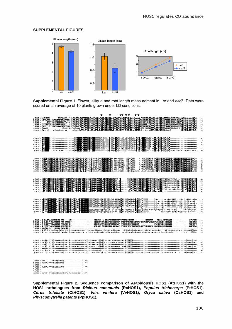
HOS1 regulates CO abundance
106
SUPPLEMENTAL FIGURES
Ler esd6
0,2
0,6
1,0
1,4Silique length (cm)
1
3
5
5 DAG 10DAG 15DAG
Leresd6
Root length (cm)
0
1
2
3
4
5
Ler esd6
Flower length (mm)
Supplemental Figure 1. Flower, silique and root length measurement in Ler and esd6. Data were scored on an average of 10 plants grown under LD conditions.
Supplemental Figure 2. Sequence comparison of Arabidopsis HOS1 (AtHOS1) with the HOS1 orthologues from Ricinus communis (RcHOS1), Populus trichocarpa (PtHOS1), Citrus trifoliate (CtHOS1), Vitis vinifera (VvHOS1), Oryza sativa (OsHOS1) and Physcomytrella patents (PpHOS1).

HOS1 regulates CO abundance
107
Amino acid residues in black are identical, and those in grey are functionally similar in the sequences. The conserved cysteine residues in the RING finger domain are indicated with black triangles. The putative nuclear localization signal present in the C terminal region of HOS1 is underlined. Genbank accession numbers are NP_181511 for AtHOS1, XP_002531460 for RcHOS1, XP_002304293 for PtHOS1, ACY92092 for CtHOS1, XP_002264751 for VvHOS1, AAR07079 for OsHOS1 and XP_001780235 for PpHOS1.
ZT 4 8
FT
UBQ10
4 8 4 8 4 8
8 10 12 15
hos
1-3
Col
hos1
-3
Co
l
ho
s1-3
Co
l
hos
1-3
Col
hos1
-3
Co
l
ho
s1-3
Co
l
hos
1-3
Col
hos1
-3
Co
l
Supplemental Figure 3. FT expression analysis in LD grown Col and hos1-3 seedlings harvested 4 and 8 h after dawn at 8, 10, 12 and 15 DAG.

HOS1 regulates CO abundance
108
SUPPLEMENTAL TABLES

DISCUSIÓN


DISCUSIÓN
111
El momento en el que se produce la transición floral está determinado por el
balance que existe entre señales promotoras y represoras de la floración. La
caracterización de mutantes tempranos en Arabidopsis y el estudio de la interacción
que existe entre los genes afectados en dichos mutantes y las rutas inductoras de la
floración, ha permitido profundizar en el conocimiento de los distintos mecanismos
implicados en la represión floral (Pouteau y col., 2004; Pouteau y col., 2008; Yant y
col., 2009). Entre dichos mecanismos se encuentran aquellos dependientes de la
acción de factores de transcripción, los procesos de modificación y remodelación de
cromatina, los mecanismos de regulación mediados por microRNAs o los circuitos
reguladores que dependen de la degradación selectiva de proteínas. Es frecuente que
estos mutantes de floración temprana presenten alteraciones pleiotrópicas del
fenotipo puesto que los represores florales generalmente afectan a varios procesos
del desarrollo. Como ejemplo, se puede citar el caso de los mutantes de floración
temprana que hemos descrito a lo largo de este trabajo, esd1/arp6, swc6 y hos1
(Choi y col., 2005; Deal y col., 2005; Martin-Trillo y col., 2006; Choi y col., 2007;
March-Diaz y col., 2007; Lazaro y col., 2008; Lazaro y col., 2011 en revisión), u otros
mutantes tempranos con los que hemos trabajado en el laboratorio, como ebs o esd7
(Pineiro y col., 2003; del Olmo y col., 2010).
A lo largo de esta Tesis Doctoral se ha profundizado en el papel que desempeñan
los mecanismos de remodelación de la cromatina, a través del intercambio de la
variante histónica H2A.Z, y la degradación selectiva de proteínas en el control de la
transición floral en Arabidopsis.
1. Papel de la variante histónica H2A.Z en la regulación del tiempo de
floración
Como hemos visto en este trabajo, las propiedades dinámicas de la cromatina
están mediadas por complejos multiproteicos que participan en la incorporación de
marcas epigenéticas con distintas funciones sobre el ADN y las histonas (Loidl, 2004).
Esclarecer los mecanismos moleculares por los que distintas modificaciones
epigenéticas modulan patrones de expresión génica es fundamental para comprender
los procesos de desarrollo y diferenciación en organismos eucarióticos. Durante los
últimos años, diversos estudios han puesto de manifiesto el papel esencial que
desempeña el intercambio de la histona canónica H2A por la variante histónica H2A.Z
en la regulación de la expresión génica y del desarrollo en distintos organismos
(Draker y Cheung, 2009; Svotelis y col., 2009; Marques y col., 2010). Sin embargo,
la base molecular de dicho intercambio y su funcionalidad permanecen en gran
medida desconocidas, tanto en sistemas biológicos animales como vegetales. La

DISCUSIÓN
112
secuencia de H2A.Z está muy conservada en diversos organismos y, al contrario de lo
que ocurre en levaduras, es esencial para la viabilidad de especies como Drosophila
melanogaster o ratón (Clarkson y col., 1999; Faast y col., 2001). La función que
desempeña la variante histónica H2A.Z en la actividad transcripcional de un gen no
está clara, y así, en D. melanogaster se ha descrito que se localiza tanto en regiones
de heterocromatina como de eucromatina (Leach y col., 2000). A pesar de esto, se ha
propuesto que la incorporación de H2A.Z en las zonas promotoras de los genes marca
estos loci para su activación y es necesaria para la adecuada regulación de su
expresión (Draker y Cheung, 2009). En levaduras, los promotores de distintos genes
presentan una región “libre de nucleosomas” 200 pb aguas arriba del codón de inicio
de la traducción que está flanqueada por nucleosomas que contienen H2A.Z (Raisner
y col., 2005; Yuan y col., 2005). Diversos estudios han descrito que este patrón de
H2A.Z está relacionado con la capacidad de inducción de la transcripción de genes
que se encuentran en estado reprimido o que presentan un nivel basal de expresión
(Guillemette y col., 2005; Li y col., 2005; Millar y col., 2006). De este modo, se ha
propuesto que la presencia de H2A.Z prepara la estructura de la cromatina para que
la maquinaria de transcripción pueda activar la expresión génica (Guillemette y col.,
2005). En células de mamífero la disposición de nucleosomas que contienen H2A.Z
afecta de forma más general a la zona de inicio de la transcripción, y también se
observa en promotores de genes inducibles que pierden esta variante histónica
cuando se activan (Zhang y col., 2005; Schones y Zhao, 2008). De hecho, en
humanos, se ha descrito una correlación entre la presencia de H2A.Z en las zonas
promotoras y la marca activadora de la transcripción H3K4me3 (Schones y Zhao,
2008). Por otro lado, también se ha propuesto un papel de la H2A.Z como “barrera” a
la extensión de la heterocromatina a zonas adyacentes de eucromatina (Meneghini y
col., 2003).
La incorporación de la variante histónica H2A.Z a los nucleosomas se produce
mediante un mecanismo independiente de la replicación del ADN (Altaf y col., 2009).
En levaduras, se ha demostrado que el complejo Swr1 (Swr1-C) es el responsable del
intercambio de los dímeros H2A-H2B por H2A.Z-H2B (Krogan y col., 2003; Kobor y
col., 2004; Mizuguchi y col., 2004). Este proceso está catalizado por la subunidad
Swr1, que es miembro de la familia de ATPasas tipo SWI/SNF2. En experimentos de
inmunoprecipitación en levaduras, Swr1 copurifica con 13 proteínas y con el dímero
H2A.Z-H2B (Mizuguchi y col., 2004; Wu y col., 2005). Las subunidades Swr1, Swc2,
Bdf1, Swc3, Arp6, Swc5, Yaf9, Swc6 y Swc7 están codificadas por genes no
esenciales, mientras que Rvb1, Rvb2, Arp4, Swc4 y la actina son indispensables para
la viabilidad celular (Wu y col., 2009b). Algunas subunidades que están presentes en
el complejo Swr1 también forman parte de otros complejos de remodelación de
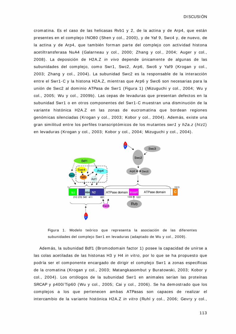
DISCUSIÓN
113
cromatina. Es el caso de las helicasas Rvb1 y 2, de la actina y de Arp4, que están
presentes en el complejo INO80 (Shen y col., 2000), y de Yaf 9, Swc4 y, de nuevo, de
la actina y de Arp4, que también forman parte del complejo con actividad histona
acetiltransferasa NuA4 (Galarneau y col., 2000; Zhang y col., 2004; Auger y col.,
2008). La deposición de H2A.Z in vivo depende únicamente de algunas de las
subunidades del complejo, como Swr1, Swc2, Arp6, Swc6 y Yaf9 (Krogan y col.,
2003; Zhang y col., 2004). La subunidad Swc2 es la responsable de la interacción
entre el Swr1-C y la histona H2A.Z, mientras que Arp6 y Swc6 son necesarias para la
unión de Swc2 al dominio ATPasa de Swr1 (Figura 1) (Mizuguchi y col., 2004; Wu y
col., 2005; Wu y col., 2009b). Las cepas de levaduras que presentan defectos en la
subunidad Swr1 o en otros componentes del Swr1-C muestran una disminución de la
variante histónica H2A.Z en las zonas de eucromatina que bordean regiones
genómicas silenciadas (Krogan y col., 2003; Kobor y col., 2004). Además, existe una
gran similitud entre los perfiles transcriptómicos de los mutantes swr1 y h2a.z (htz1)
en levaduras (Krogan y col., 2003; Kobor y col., 2004; Mizuguchi y col., 2004).
Figura 1. Modelo teórico que representa la asociación de las diferentes
subunidades del complejo Swr1 en levaduras (adaptado de Wu y col., 2009).
Además, la subunidad Bdf1 (Bromodomain factor 1) posee la capacidad de unirse a
las colas acetiladas de las histonas H3 y H4 in vitro, por lo que se ha propuesto que
podría ser el componente encargado de dirigir el complejo Swr1 a zonas específicas
de la cromatina (Krogan y col., 2003; Matangkasombut y Buratowski, 2003; Kobor y
col., 2004). Los ortólogos de la subunidad Swr1 en animales serían las proteínas
SRCAP y p400/Tip60 (Wu y col., 2005; Cai y col., 2006). Se ha demostrado que los
complejos a los que pertenecen ambas ATPasas son capaces de realizar el
intercambio de la variante histónica H2A.Z in vitro (Ruhl y col., 2006; Gevry y col.,

DISCUSIÓN
114
2007). En levaduras se ha sugerido que la deposición de H2A.Z mediada por Swr1-C
necesita la previa acetilación de las histonas de esa región de la cromatina por el
complejo NuA4 y, curiosamente, el complejo Tip60 presente en D. melanogaster y
humanos parece ser una fusión de los complejos Swr1 y NuA4 de levaduras (Doyon y
col., 2004; Auger y col., 2008).
Los resultados presentados en este trabajo han contribuido a la identificación en
Arabidopsis de dos ortólogos de los componentes del complejo Swr1 de levaduras,
ESD1/ARP6 y SWC6. El aislamiento de estas subunidades, junto con la identificación
de PIE1, el homólogo en Arabidopsis de la subunidad catalítica del complejo Swr1 de
levaduras (Noh y Amasino, 2003), ha dado pie a la caracterización de un posible
complejo homólogo a Swr1 en plantas. El genoma de Arabidopsis presenta ortólogos
de la mayor parte de los componentes del Swr1-C. Las células eucariotas poseen al
menos ocho proteínas relacionadas con actina (ARPs) de las cuales, ARP4, 5, 6, 7 y 8
se localizan en el núcleo y están implicadas en mecanismos de remodelación de la
cromatina (Blessing y col., 2004; Kandasamy y col., 2004). La mayoría de los
complejos de remodelación de cromatina que poseen ARPs también presentan una
molécula de actina. Por su parte, SWC6 es una proteína que presenta un dedo de Zinc
tipo HIT (Choi y col., 2007; March-Diaz y col., 2007; Lazaro y col., 2008). Tanto
ESD1/ARP6 como SWC6 son genes únicos en Arabidopsis, homólogos de los que
codifican las subunidades Arp6 y Swc6 de los complejos Swr1 de levaduras y SRCAP
de humanos. Además, se han descrito tres genes de la familia H2A denominados
HTA8, 9 y 11, que codifican para homólogos de H2A.Z en Arabidopsis (Yi y col.,
2006).
Además, la caracterización de ESD1/ARP6 y SWC6 ha permitido desvelar el papel
que tiene el intercambio de variantes histónicas en la regulación de la expresión de
FLC, así como avanzar en el conocimiento de los procesos de remodelación de
cromatina involucrados en la regulación de la transición floral en esta especie modelo.
Los trabajos publicados a lo largo de los últimos años han demostrado la función de
diversas modificaciones histónicas en la regulación de FLC a través de FRI, la ruta
autónoma, la ruta de la vernalización y el complejo PAF1 (He y col., 2003; Ausin y
col., 2004; Bastow y col., 2004; He y col., 2004; Sung y Amasino, 2004; He y
Amasino, 2005; Kim y col., 2005; Choi y col., 2011). Asimismo, se ha observado que
el primer exón y el primer intrón de FLC son las regiones genómicas en las que se
depositan principalmente las marcas epigenéticas que participan en la regulación
transcripcional de este locus (Sheldon y col., 2002).

DISCUSIÓN
115
Las alteraciones pleiotrópicas del fenotipo de los mutantes esd1/arp6 y swc6, así
como el patrón de expresión ubicuo de ambos loci, sugiere que estos genes participan
en diversos procesos del desarrollo vegetativo y reproductivo de las plantas (Choi y
col., 2005; Deal y col., 2005; Martin-Trillo y col., 2006; Choi y col., 2007; March-Diaz
y col., 2007; Lazaro y col., 2008). El fenotipo descrito para las mutaciones en otros
homólogos del complejo SWR1 en Arabidopsis presenta claras similitudes con los
observados en esd1/arp6 y swc6. Las plantas que presentan defectos en PIE1
también muestran un fenotipo de floración temprana, los margenes de las hojas
aserrados, pérdida de la dominancia apical y la presencia de pétalos extra en las
flores (Noh y Amasino, 2003). De la misma forma, las mutaciones pie1, esd1/arp6 y
swc6 son capaces de suprimir el fenotipo de floración tardío causado por los mutantes
de la ruta autónoma o por un alelo funcional del locus FRI, gracias a la disminución de
los niveles de FLC (Noh y Amasino, 2003; Choi y col., 2005; Deal y col., 2005;
Martin-Trillo y col., 2006; Choi y col., 2007; Lazaro y col., 2008). A pesar de las
semejanzas que se observan entre pie1, esd1/arp6 y swc6, algunos de los fenotipos
son más dramáticos en el mutante pie1, por ejemplo la reducción en la elongación de
la inflorescencia primaria es mayor y las hojas son más pequeñas y deformadas (Noh
y Amasino, 2003; March-Diaz y col., 2007). Por otra parte, la reducción de los niveles
del mensajero de AtARP4 por aproximaciones de ARNi también provoca una
aceleración del tiempo de floración en Arabidopsis, además de alteraciones en la
organización de los órganos florales, retraso en el proceso de senescencia y altos
niveles de esterilidad (Kandasamy y col., 2005). Estas diferencias con los mutantes
pie1, esd1/arp6 y swc6 sugieren, como ocurre en levaduras, la participación de
AtARP4 en distintos complejos de remodelación de cromatina en plantas.
Como se ha descrito con anterioridad, Swc6 y Arp6 son dos componentes
íntimamente asociados en el Swr1-C de levaduras (Figura 1) (Wu y col., 2005). De
acuerdo con esto, en Arabidopsis el fenotipo del doble mutante esd1 swc6 es
indistinguible de cualquiera de los dos mutantes simples (Choi y col., 2007; Lazaro y
col., 2008). Además de interaccionar genéticamente, también se ha demostrado que
ESD1/ARP6 y SWC6 interaccionan físicamente en Arabidopsis, así como que ambas
proteínas son capaces de interaccionar con PIE1, lo que sugiere que estas tres
proteínas actúan en el mismo complejo (Choi y col., 2007; March-Diaz y col., 2007;
Lazaro y col., 2008). Por otro lado, se ha demostrado la presencia de H2A.Z en la
zona de inicio y final de la transcripción del gen FLC y de sus homólogos de la familia
MAF, MAF4 y MAF5 (Deal y col., 2007). En los mutantes esd1/arp6 y pie1 se ha
observado una disminución en la acumulación de esta variante histónica en la
cromatina de FLC, que además está relacionada con una reducción de la expresión de
este represor floral y con una aceleración del tiempo de floración (Deal y col., 2007).

DISCUSIÓN
116
Igualmente, se ha descrito que tanto ESD1/ARP6 como SWC6 se unen al promotor de
FLC mediante ensayos de inmunoprecipitación de cromatina (Choi y col., 2007). De
forma adicional, se ha demostrado que H2A.Z interacciona físicamente con PIE1,
AtSWC2 y ESD1/ARP6, y que la reducción de la expresión de los genes que codifican
para H2A.Z provocan un fenotipo similar al que se observa en los mutantes
esd1/arp6, swc6 y pie1 (Choi y col., 2007; Deal y col., 2007; March-Diaz y col.,
2008). También se ha descrito mediante el análisis de perfiles transcriptómicos
globales que existe un 65% de solapamiento entre los genes desregulados en el
mutante pie1 y en el doble mutante para dos de los homólogos de H2A.Z, hta9 hta11
(March-Diaz y col., 2008). Este resultado indica que PIE1 y H2A.Z comparten
funciones en la regulación de la expresión génica, aunque también presentan un
grado de independencia funcional, y de hecho, no es posible descartar que PIE1 tenga
funciones independientes del complejo SWR1 en el control de la expresión génica. En
levaduras, la mutación de Arp6 o Swc6 da como resultado la pérdida de la otra
subunidad en el complejo, así como la pérdida de otras dos proteínas, Swc2 y Swc3,
lo que sugiere que Arp6, Swc6, Swc2 y Swc3 forman un subcomplejo que se asocia a
Swr1 (Wu y col., 2005). De forma similar, parece que en Arabidopsis ESD1/ARP6 y
SWC6, junto con SWC2, pueden formar un subcomplejo que se asocia a PIE1 (Choi y
col., 2007; Deal y col., 2007; March-Diaz y Reyes, 2009).
Todos los datos anteriores sugieren la existencia de un complejo de remodelación
de cromatina tipo SWR1 en Arabidopsis que cataliza el intercambio de la histona H2A
por la variante histónica H2A.Z, y que ESD1/ARP6 y SWC6 forman parte de dicho
complejo. Además, se ha observado que este hipotético complejo SWR1 es necesario
para el correcto desarrollo de Arabidopsis, ya que parece estar implicado en la
regulación de diversos procesos de diferenciación como la transición floral, el
desarrollo de los órganos florales y la morfología y tamaño de la hoja (Choi y col.,
2005; Deal y col., 2005; Martin-Trillo y col., 2006; Choi y col., 2007; March-Diaz y
col., 2007; Lazaro y col., 2008). En concreto, se ha demostrado que el complejo
SWR1 participa en el control del tiempo de floración a través de la activación de la
expresión del represor floral FLC y de los genes MAF4 y MAF5 y, por tanto, parece que
la presencia de H2A.Z puede ser necesaria para activar la expresión de determinados
loci en plantas.
Curiosamente, los niveles de H2A.Z presentes en la cromatina de FLC no están
relacionados con la tasa de transcripción de este locus en distintos tejidos que
presentan una variación de la expresión de FLC de hasta diez veces (Deal y col.,
2007). La distribución espacial de H2A.Z sobre la cromatina de FLC se mantiene
constante en todos los tejidos analizados, de modo que se observa un pico en la zona

DISCUSIÓN
117
de inicio y otro en la de terminación de la transcripción. En cambio, no hay una
correlación entre la expresión de FLC y la cantidad de H2A.Z presente a lo largo de la
región codificante del gen en los distintos tejidos, lo que sugiere que la mera
presencia de esta variante histónica no es capaz de activar la transcripción (Deal y
col., 2007). Además, se ha descrito que durante la vernalización, aunque la expresión
de FLC está reprimida, se produce un aumento de la presencia de H2A.Z en este locus
(Brickner y col., 2007). Ambos resultados sugieren que la función de H2A.Z puede ser
la de “marcar” o disponer a los genes en un estado competente para su activación por
otros factores que permitan la expresión génica cuando las condiciones sean las
adecuadas (Deal y col., 2007). Como se ha descrito en el presente trabajo,
ESD1/ARP6 y SWC6 median la regulación de la expresión de FLC a través de dos
marcas epigenéticas relacionadas con la activación transcripcional: la acetilación de
las Lys 9 y 14 de la H3 y la trimetilación de la Lys 4 de la H3. Las mutaciones en los
loci ESD1/ARP6 y SWC6 provocan una disminución de los niveles de estas dos
modificaciones histónicas activadoras en la zona promotora y en el primer intrón de
FLC. Sin embargo, la posible implicación directa de ESD1/ARP6 y SWC6, y, por tanto
del complejo SWR1, en el establecimiento de estas dos marcas epigenéticas no está
clara. Los complejos Swr1 y NuA4 de levaduras comparten varias subunidades y las
mutaciones en algunos de sus componentes presentan fenotipos similares (Auger y
col., 2008), de modo que es posible especular que podrían trabajar de forma conjunta
en la acetilación de histonas y en el intercambio de H2A.Z en la cromatina. Además,
en D. melanogaster y humanos, se ha descrito que el complejo Tip60 correspondería
a una fusión de los complejos Swr1 y NuA4 de levaduras, puesto que presenta
ortólogos de las subunidades que forman parte de los dos complejos (Doyon y col.,
2004; Auger y col., 2008). Por otro lado, se ha descrito que componentes del
complejo PAF1 de levaduras muestran una interacción genética con subunidades del
Swr1-C (Krogan y col., 2003), lo que sugiere que ambos complejos podrían tener
dianas comunes y podría explicar el papel que juegan ESD1/ARP6 y SWC6 en la
trimetilación de la H3K4 en la cromatina de FLC (Mueller y Jaehning, 2002; Squazzo y
col., 2002; Krogan y col., 2003; Krogan y col., 2004). De forma análoga a lo que
ocurre en levaduras, el complejo PAF1 de Arabidopsis recluta a la metiltransferasa
EFS/SDG8, y así regula los niveles de expresión de FLC (Kim y col., 2005). De hecho,
las mutaciones que afectan a los componentes del complejo PAF1 presentan
alteraciones fenotípicas similares a las de las de los mutantes esd1/arp6 y swc6,
además de una disminución de los niveles de H3K4me3 en la cromatina de FLC (He y
col., 2004), datos que apoyan que los complejos SWR1 y PAF1 puedan actuar de
forma conjunta en la remodelación de la cromatina de determinados genes diana en
plantas.

DISCUSIÓN
118
Estudios previos han establecido el papel de H2A.Z en el reclutamiento de la ARN
polimerasa II (Adam y col., 2001) y la interrelación con complejos de remodelación de
nucleosomas (Santisteban y col., 2000). Como se ha descrito recientemente, FRI y
otras proteínas relacionadas forman un complejo (FRI-C) que actúa como un
activador transcripcional de FLC a través del reclutamiento de complejos
remodeladores de la cromatina (Choi y col., 2011). Entre los factores que recluta el
FRI-C al promotor de FLC se encuentra el complejo SWR1, la metiltransferasa EFS y
un factor general de la transcripción, TAF14 (Choi y col., 2011). El genoma de
Arabidopsis contiene dos proteínas, YAF9 y TAF14, con un dominio YEATS, asociado a
la remodelación de la cromatina y a la transcripción génica (Schulze y col., 2010).
YAF9 es un componente del SWR1-C, homólogo a la subunidad Yaf9 de los complejos
Swr1-C y NuA4 de levaduras (Zhang y col., 2004). Por su parte, la proteína TAF14
forma parte de los factores de transcripción generales TFIID y TFIIF, de los complejos
de remodelación de cromatina INO80 y SWI/SNF2, y del complejo NuA3 de acetilación
de la histona H3 de levaduras y humanos (Kabani y col., 2005). Por otro lado,
EFS/SDG8 cataliza la trimetilación de K4 y K36, que son marcas que promueven el
inicio de la transcripción y la elongación del mensajero (Ko y col., 2010). El reciente
descubrimiento de la relación que existe entre el complejo de FRI, el SWR1-C,
EFS/SDG8 y TAF14 nos da idea de la complicada secuencia de eventos que requiere la
transcripción génica en eucariotas y podría ligar la presencia de H2A.Z y de la marca
H3K4me3 sobre la cromatina de FLC (Choi y col., 2011).
Todavía no se conoce el mecanismo por el cual el complejo Swr1 es reclutado a
determinados genes y no a otros. Una de las hipótesis aceptadas en la actualidad se
basa en la capacidad de la proteína Bdf1 para reconocer patrones de acetilación de
histonas, ya que los mutantes en esta subunidad del complejo presentan una
disminución de la deposición de H2A.Z en las zonas promotoras (Raisner y col., 2005;
Zhang y col., 2005). Sin embargo, en Arabidopsis no se ha identificado un ortólogo de
Bdf1 puesto que en su genoma no se han descrito proteínas que contengan dos
dominios de tipo bromodomain. Por otra parte, se ha propuesto que pueda existir una
señal en el ADN para la incorporación de H2A.Z, ya que en levaduras se ha observado
que el sitio de unión de la proteína tipo Myb Reb1 es suficiente para la formación de
una zona “libre de nucleosomas” y para la incorporación de H2A.Z en los nucleosomas
que flanquean dicha región (Raisner y col., 2005; Raisner y Madhani, 2006). Por otro
lado, se ha propuesto recientemente que la presencia de H2A.Z y la metilación de
ADN son marcas de la cromatina mutuamente antagónicas en Arabidopsis (Zilberman
y col., 2008). Así, mutaciones en la metiltransferasa de ADN MET1, que provocan
tanto pérdida como ganancia de metilación del ADN, originan cambios opuestos en los
niveles de incorporación de H2A.Z, mientras que mutaciones en PIE1 conllevan una

DISCUSIÓN
119
moderada hipermetilación general a lo largo del genoma. Por tanto, parece que la
presencia de H2A.Z previene el silenciamiento génico mediado por la metilación del
ADN (Kobor y Lorincz, 2009).
En Arabidopsis también existe controversia acerca del papel que juega la H2A.Z en
la regulación de la transcripción génica. Las mutaciones en componentes del SWR1-C
liberan la represión sobre ciertos genes de respuesta a estrés biótico y abiótico, como
genes de resistencia a enfermedades, genes inducidos por choque térmico y genes de
respuesta a deficiencia de fosfato, lo que apoyaría un efecto negativo de H2A.Z sobre
la expresión génica (March-Diaz y col., 2008; Kumar y Wigge, 2010; Smith y col.,
2010). En cambio, como ya hemos descrito, la presencia de H2A.Z en el promotor de
FLC está relacionada con el aumento de la expresión de este locus (Deal y col., 2007).
El papel dual que juega H2A.Z en la regulación de la transcripción desde levaduras a
humanos puede depender del contexto de la cromatina en el cual se localiza o de su
disposición, en uno o dos nucleosomas en las zonas promotoras, o en numerosos
nucleosomas contiguos en las zonas teloméricas silenciadas (Guillemette y Gaudreau,
2006). La incorporación de variantes histónicas en ciertos loci confiere a estas zonas
del genoma propiedades estructurales y funcionales específicas que afectan a una
variedad de procesos biológicos en distintos organismos. Como hemos descrito, la
presencia de H2A.Z está relacionada con el control de la expresión de ciertos genes,
pero, además, también participa en la progresión del ciclo celular, la estabilidad del
genoma, la supresión de ARNs antisentido o la adaptación a altas temperaturas en las
plantas (Ahmad y col., 2010). Recientemente, en un ensayo dirigido a la búsqueda de
mutantes que presentaban alteraciones en la respuesta a la temperatura de
crecimiento, se aisló un nuevo alelo del mutante arp6 (Kumar y Wigge, 2010). Se ha
demostrado que a temperaturas bajas de crecimiento H2A.Z reprime la expresión de
una serie de genes, entre los que se encuentra FT, pero que un aumento en la
temperatura ambiental provoca el desplazamiento de esta variante histónica de
dichos genes y la activación de su transcripción. Así, se ha propuesto que la presencia
o ausencia de H2A.Z puede ser la base del mecanismo de medida de los cambios de
la temperatura ambiental en Arabidopsis (Franklin, 2010; Kumar y Wigge, 2010). Al
igual que ocurre en Arabidopsis, la variante H2A.Z se elimina del ADN durante la
activación de multitud de genes en levaduras y en humanos (Santisteban y col.,
2000; Farris y col., 2005; Zhang y col., 2005), de manera que se ha propuesto que
esta variante histónica podría favorecer la transcripción génica al facilitar la
remodelación o la liberación de los nucleosomas de las zonas promotoras (Guillemette
y Gaudreau, 2006).

DISCUSIÓN
120
La caracterización bioquímica de otros ortólogos de los componentes del Swr1-C,
junto con estudios transcriptómicos globales y análisis de distribución genómica
(ChIP-chip o ChiP-seq), permitirán en el futuro identificar nuevos loci regulados por
este complejo en plantas. Los defectos pleiotrópicos de los mutantes descritos hasta
el momento en Arabidopsis indican que los procesos de remodelación de cromatina
mediados por SWR1 afectan a gran variedad de procesos de desarrollo, tanto
vegetativo como reproductivo, en las plantas.
2. Función de HOS1 en la regulación fotoperiódica de la floración
Debido a que los resultados obtenidos en este trabajo acerca del papel de HOS1
como represor floral ya se han discutido en relación a los últimos avances realizados
en el control fotoperíodico del tiempo de floración en Arabidopsis, en este apartado
sólo vamos a recoger aquellos aspectos que no se pudieron desarrollar
exhaustivamente en la discusión del manuscrito presentado en el Capítulo 3 debido a
una limitación de espacio.
Los cambios en el fotoperiodo regulan la transición floral en una gran variedad de
plantas, de modo que aquellas que presentan defectos en los mecanismos de
respuesta a la duración del día no florecen de forma adecuada en la naturaleza
(Wilczek y col., 2009). En el presente trabajo, hemos demostrado mediante el uso
aproximaciones genéticas y moleculares, que el locus HOS1 participa en el control
fotoperiódico del tiempo de floración. El mutante esd6/hos1 se aisló en un programa
de búsqueda de mutantes de floración temprana en Arabidopsis. Además de presentar
una aceleración de la floración, el mutante hos1 muestra alteraciones pleiotrópicas del
fenotipo que afectan al desarrollo de las hojas, las flores y la raíz, lo que sugiere la
implicación de este locus en la regulación del desarrollo de estos órganos (Lazaro y
col., 2011 en revisión).
Como hemos expuesto con anterioridad, CO está regulado tanto a nivel
transcripcional, como a nivel post-traduccional por distintas calidades de luz (Valverde
y col., 2004; Kim y col., 2008). Los análisis genéticos llevados a cabo en este trabajo
con el mutante hos1 demuestran que este locus participa en la ruta del fotoperiodo,
ya que las mutaciones en CO suprimen en gran medida el fenotipo temprano de hos1,
particularmente en fondo genético Ler. Por otro lado, los ensayos de expresión
mostraron que el patrón del mensajero de CO no estaba afectado en el mutante hos1.
En cambio, sí que observamos una alteración en el patrón de expresión de FT en DL
en el mutante hos1, en el que destaca un pico de expresión en las primeras horas del
día. Además, los datos obtenidos en este trabajo indican que la proteína HOS1
interacciona físicamente con CO in vitro e in vivo, y que los niveles de la proteína CO

DISCUSIÓN
121
son más elevados en el mutante hos1 que en plantas silvestres durante el día. Por
otro lado, se ha descrito que HOS1 es una proteína con un dominio RING que tiene
actividad como enzima E3 ligasa de ubiquitina (Dong y col., 2006). En base a todos
estos resultados, proponemos que HOS1 participa en la degradación de CO durante el
periodo de luz del día mediante un mecanismo de ubiquitinación, y que la actividad de
HOS1 en las primeras horas del día es crucial para una correcta regulación
fotoperiódica de la floración (Lazaro y col., 2011 en revisión).
Se han descrito otras ligasas de ubiquitina que participan en el control
fotoperiódico del tiempo de floración, de las cuales sólo COP1 está implicada de forma
directa en la regulación de la estabilidad de la proteína CO (Jang y col., 2008; Liu y
col., 2008c; Morris y col., 2010). La proteína CO se degrada en el periodo de
oscuridad a través de la actividad de los complejos SPA-COP1 (Laubinger y col.,
2006; Jang y col., 2008; Liu y col., 2008c). Además, se ha demostrado recientemente
que la E3 ligasa de tipo CUL4-DDB1 interacciona con los complejos SPA-COP1 en el
control del tiempo de floración (Chen y col., 2010). Queda por determinar si HOS1
actúa como una E3 ligasa de ubiquitina individual o si forma parte de un complejo
multimérico como parece que ocurre con COP1.
Se ha descrito que COP1 presenta mayor actividad durante la oscuridad que en el
periodo de luz (Jang y col., 2008). Se ha propuesto que uno de los mecanismos que
podría explicar esta regulación diferencial es la translocación de la proteína COP1 del
núcleo al citoplasma en presencia de luz, puesto que la mayor parte de sus dianas son
factores de transcripción (von Arnim y Deng, 1994). Sin embargo, resultados
recientes cuestionan la exclusión de COP1 del núcleo en presencia de luz, ya que
demuestran que los niveles de COP1 no varían entre la fracción citoplásmica y la
fracción nuclear de plántulas etioladas o tratadas con luz roja (Jang y col., 2010). Los
datos de localización subcelular de HOS1 presentados en esta Tesis tampoco
coinciden con las observaciones previas que describen la presencia de HOS1-GFP en
el citoplasma cuando las plántulas se cultivan en oscuridad y a temperatura normal de
crecimiento (Lee y col., 2001). En este trabajo, hemos demostrado que en plántulas
de Arabidopsis que sobreexpresan la proteína de fusión de HOS1-GFP se observa un
patrón de localización nuclear tanto en condiciones de luz contínua como en plántulas
etioladas, lo que sugiere que HOS1 se localiza en el núcleo independientemente de las
condiciones lumínicas (Lazaro y col., 2011 en revisión). Esta discrepancia puede
deberse a que la construcción 35S::HOS1-GFP usada previamente por Lee y col. no
contenía la secuencia codificante completa de HOS1, que hemos reanotado en este
trabajo (Lazaro y col., 2011 en revisión). Futuros estudios serán necesarios para
comprender en profundidad cómo la luz es capaz de controlar la actividad de COP1, y

DISCUSIÓN
122
para comprobar si distintas calidades de luz también afectan a la actividad de otras
ligasas de ubiquitina involucradas en el control de la transición floral, como HOS1.
Mientras que la transcripción de CO está regulada por el reloj circadiano, los
niveles de proteína CO son regulados de manera diferencial por la luz. Valverde y
colaboradores describieron cómo la estabilidad de CO se veía afectada por distintas
calidades de luz mediante ensayos de western blot con un anticuerpo específico anti-
CO y estudios de localización subcelular con una fusión de la proteína CO a GFP
(Valverde y col., 2004). Estos experimentos demostraron que las plántulas de
Arabidopsis expuestas a luz roja o mantenidas en oscuridad presentaban unos niveles
de CO muy inferiores a los que se observaban en las plántulas tratadas con luz azul,
luz roja lejana y luz blanca. En plantas sobreexpresoras de CO, esta proteína se
detectaba por western blot a las 8, 12 y 16 horas después del amanecer en
condiciones de DL, mientras que sus niveles eran casi indetectables a ZT4. Además,
se detectaba un aumento transitorio de los niveles de CO a los 30 minutos después
del amanecer tanto en DL como en DC (Valverde y col., 2004). El efecto de los
distintos fotoreceptores sobre la cantidad de CO fue analizado en las plantas que
contenían la construcción 35S::CO. En el mutante phyB se observa un aumento de la
estabilidad de CO durante la mañana; en cambio, en el doble mutante cry1 cry2 y en
el mutante phyA desaparecen los picos de la proteína CO que se observan a primera y
última hora del día en las plantas sobreexpresoras de CO. Estos resultados indican
que al comienzo del día PhyB participa en la degradación de CO, mientras que en la
tarde, los criptocromos y PhyA antagonizan dicha degradación y estabilizan a la
proteína CO, lo que permite que se induzca la expresión de FT en condiciones de DL
(Valverde y col., 2004). Hasta el momento se desconoce el mecanismo que regula el
cambio de actividad de los fotorreceptores entre la mañana y la tarde, puesto que
PhyB es un fitocromo estable en luz y los criptocromos están presentes de forma
constante durante las condiciones fotoperiódicas de DL (El-Din El-Assal y col., 2001;
Mockler y col., 2003). Datos recientes indican que los criptocromos regulan
negativamente la actividad de COP1 en la luz, lo cual estabiliza a CO e induce la
floración en Arabidopsis (Liu y col., 2008c). Además, también se ha demostrado que
COP1 regula a CO a nivel transcripcional a través de GI (Yu y col., 2008). COP1 y
ELF3, una de las proteínas asociadas al reloj circadiano, median la señal transmitida
por Cry2 y participan en la degradación de GI (Yu y col., 2008). Posteriormente, se
describió el papel de COP1 en la degradación de CO en la última parte del día y en la
noche, y se estableció que la degradación de esta proteína en luz roja no estaba
afectada por la mutación cop1 (Jang y col., 2008; Liu y col., 2008c). Estos resultados
apuntan que otra ligasa de ubiquitina podría ser la responsable de la degradación de
CO dependiente de PhyB que tiene lugar en las primeras horas del día (Jang y col.,

DISCUSIÓN
123
2008). Nuestros datos demuestran que HOS1 juega un papel relevante en el ajuste
de los niveles de la proteína CO durante el periodo de luz y que, de este modo,
previene el aumento prematuro de FT en las primeras horas del día, si bien se
necesitan más datos experimentales que permitan establecer la posible participación
de HOS1 en la proteólisis de CO mediada por PhyB.
En plantas se han descrito varios ejemplos de proteínas que, al igual que CO, se
degradan en un proceso dependiente de los fitocromos. Así, PHYTOCHROME-
INTERACTING FACTOR 1 (PIF1), PIF3, PIF4 y PIF5 son factores de transcripción tipo
basic helix-loop-helix que participan en la ruta de señalización de la luz roja y la luz
roja lejana (Ni y col., 1999; Huq y Quail, 2002; Fujimori y col., 2004; Huq y col.,
2004). Se ha observado que los fitocromos, cuando se activan por la luz, sufren una
translocación al núcleo e interaccionan con los PIFs. La unión entre el Phy
fotoactivado y las proteínas PIF1, PIF3 y PIF5 induce la fosforilación de éstas últimas
y su degradación por el proteosoma en menos de cinco minutos (Al-Sady y col.,
2006; Shen y col., 2007; Shen y col., 2008). Recientemente, se ha demostrado que
la señalización de la luz roja mediada por PhyB conlleva un mecanismo de terminación
de la respuesta que implica a COP1. Cuando el PhyB es activado y se transloca al
núcleo, COP1 promueve su degradación mediante un mecanismo en el que participan
los PIFs (Jang y col., 2010). De esta manera, se ha propuesto que PhyB y los PIFs se
regulan mutuamente de forma negativa (Henriques y col., 2009; Jang y col., 2010).
Igualmente, COL3, un homologo de CO que reprime la floración, se ha identificado
como un nuevo interactor de COP1 que participa como regulador positivo de la
respuesta a la luz roja (Datta y col., 2006).
Nuevas aproximaciones experimentales serán necesarias para comprender en
profundidad los mecanismos moleculares que median el papel de HOS1 en la
regulación fotoperiódica de la floración en Arabidopsis, y en particular para abordar el
estudio de la posible implicación de esta E3 ligasa de ubquitina en la degradación de
CO dependiente de luz roja. Nuestra hipótesis de trabajo contempla que PhyB, una
vez activo tras exposición a luz roja, podría translocarse al núcleo y activar a HOS1 en
las primeras horas del día. Un posible mecanismo de activación podría conllevar la
modificación post-traduccional tanto de HOS1 como de sus posibles sustratos,
incluyendo a CO. El esclarecimiento de estos mecanismos arrojará luz sobre el control
de la inducción de la floración en respuesta a fotoperiodo, un proceso complejo cuya
regulación empezamos a comprender gracias a estudios realizados en la especie
modelo Arabidopsis, pero cuyos componentes esenciales están altamente conservados
en otras especies.

DISCUSIÓN
124
Por último, aunque en este trabajo hemos demostrado que HOS1 está implicado en
la degradación de CO, no descartamos que existan otras proteínas que participen en
el control del tiempo de floración y cuya estabilidad pueda estar regulada por esta E3
ligasa. De hecho, HOS1 regula de forma positiva la expresión de FLC (Lee y col.,
2001), que a su vez, está reprimida por la exposición prolongada a bajas
temperaturas (Amasino, 2010). Es posible, por tanto, que HOS1 pueda estar
involucrado en la degradación de un regulador negativo de la expresión de FLC,
aunque los análisis genéticos realizados no han permitido identificar por el momento
cuál puede ser esta diana.

CONCLUSIONES


CONCLUSIONES
127
En el presente trabajo hemos abordado la caracterización genética y molecular de
los loci ESD1/ARP6, SWC6 y ESD6/HOS1 para establecer su papel en el control del
tiempo de floración en Arabidopsis, y hemos profundizado en los mecanismos de
represión floral mediados por la remodelación de la estructura de la cromatina y la
degradación selectiva de proteínas.
1. Los mutantes de floración temprana de Arabidopsis esd1/arp6 y swc6 están
afectados en genes que codifican proteínas ortólogas de las subunidades Arp6 y
Swc6 del complejo de remodelación de cromatina Swr1 de levaduras, implicado
en el intercambio de la variante histónica H2A.Z. La identificación de estas
proteínas apoya la existencia de un complejo SWR1 en Arabidopsis.
2. Además de una aceleración del tiempo de floración, los mutantes esd1/arp6 y
swc6 presentan otras alteraciones de su fenotipo, que afectan al desarrollo de los
órganos florales y a la morfología y tamaño de la hoja, lo que sugiere la posible
participación del complejo SWR1 en la regulación de esos procesos de desarrollo.
De acuerdo con estas observaciones, el patrón de expresión de ESD1/ARP6 y
SWC6 es ubicuo.
3. Las mutaciones esd1/arp6 y swc6 interaccionan de forma epistática con
mutaciones en genes de la ruta autónoma y suprimen parcialmente el fenotipo de
floración tardio conferido por alelos activos de FRI.
4. ESD1/ARP6 y SWC6 son necesarios para la activación del represor floral FLC.
Además, ambos loci desempeñan un papel independiente de FLC en el control del
tiempo de floración, posiblemente mediado por la activación de los genes MAF4 y
MAF5.
5. ESD1/ARP6 y SWC6 se requieren para mantener los niveles de dos marcas
epigenéticas activadoras sobre la cromatina de FLC, la acetilación de la histona
H3 y la trimetilación de la lisina 4 de la histona H3.
6. ESD1/ARP6 y SWC6 interaccionan físicamente, lo que sugiere que forman parte
de un mismo subcomplejo proteico dentro de SWR1-C.
7. El mutante de floración temprana esd6/hos1 está afectado en el locus HOS1, que
codifica una enzima E3 ligasa de ubiquitina identificada previamente por su
participación en la respuesta de aclimatación a bajas temperaturas en
Arabidopsis. Además de participar en el control del tiempo de floración,
ESD6/HOS1 está implicado en la regulación del desarrollo de las hojas, flores y
raíces.
8. ESD6/HOS1 regula de forma positiva la expression de FLC, aunque también tiene
un efecto independiente de FLC en el control del tiempo de floración.

CONCLUSIONES
128
9. HOS1 interacciona genéticamente con CO, si bien la mutación esd6/hos1 no
altera el patrón ni los niveles de expresión de CO a lo largo del día.
10. HOS1 se localiza en el núcleo e interacciona con CO, participando en la
modulación de los niveles de dicha proteína durante las primeras horas del día en
fotoperiodos de DL.
11. HOS1 interacciona genéticamente con COP1, un gen que codifica otra E3 ligasa
de ubiquitina involucrada en la regulación de los niveles de la proteína CO
durante la noche, lo que sugiere que HOS1 y COP1 ajustan de manera precisa la
acumulación de CO a lo largo del ciclo diario.
12. A través de su papel en la modulación de los niveles de CO, HOS1 regula el
patrón de expresión diario de FT durante el periodo de luz, impidiendo su
activación durante las primeras horas del día, y permitiendo así una correcta
regulación fotoperiódica del tiempo de floración en Arabidopsis.

BIBLIOGRAFÍA


BIBLIOGRAFÍA
131
Abe, M., Kobayashi, Y., Yamamoto, S., Daimon, Y., Yamaguchi, A., Ikeda, Y., Ichinoki, H., Notaguchi, M., Goto, K., y Araki, T. (2005). FD, a bZIP protein mediating signals from the floral pathway integrator FT at the shoot apex. Science 309, 1052-1056.
Adam, M., Robert, F., Larochelle, M., y Gaudreau, L. (2001). H2A.Z is required for global chromatin integrity and for recruitment of RNA polymerase II under specific conditions. Mol Cell Biol 21, 6270-6279.
Adrian, J., Farrona, S., Reimer, J.J., Albani, M.C., Coupland, G., y Turck, F. (2010). cis-Regulatory elements and chromatin state coordinately control temporal and spatial expression of FLOWERING LOCUS T in Arabidopsis. Plant Cell 22, 1425-1440.
Ahmad, A., Zhang, Y., y Cao, X.F. (2010). Decoding the epigenetic language of plant development. Mol Plant 3, 719-728.
Al-Sady, B., Ni, W., Kircher, S., Schafer, E., y Quail, P.H. (2006). Photoactivated phytochrome induces rapid PIF3 phosphorylation prior to proteasome-mediated degradation. Mol Cell 23, 439-446.
Altaf, M., Auger, A., Covic, M., y Cote, J. (2009). Connection between histone H2A variants and chromatin remodeling complexes. Biochem Cell Biol 87, 35-50.
Amasino, R. (2010). Seasonal and developmental timing of flowering. Plant J 61, 1001-1013.
Amasino, R.M., y Michaels, S.D. (2010). The timing of flowering. Plant Physiol 154, 516-520.
An, H., Roussot, C., Suarez-Lopez, P., Corbesier, L., Vincent, C., Pineiro, M., Hepworth, S., Mouradov, A., Justin, S., Turnbull, C., y Coupland, G. (2004). CONSTANS acts in the phloem to regulate a systemic signal that induces photoperiodic flowering of Arabidopsis. Development 131, 3615-3626.
Auger, A., Galarneau, L., Altaf, M., Nourani, A., Doyon, Y., Utley, R.T., Cronier, D., Allard, S., y Cote, J. (2008). Eaf1 is the platform for NuA4 molecular assembly that evolutionarily links chromatin acetylation to ATP-dependent exchange of histone H2A variants. Mol Cell Biol 28, 2257-2270.
Aukerman, M.J., y Sakai, H. (2003). Regulation of flowering time and floral organ identity by a MicroRNA and its APETALA2-like target genes. Plant Cell 15, 2730-2741.
Aukerman, M.J., Hirschfeld, M., Wester, L., Weaver, M., Clack, T., Amasino, R.M., y Sharrock, R.A. (1997). A deletion in the PHYD gene of the Arabidopsis Wassilewskija ecotype defines a role for phytochrome D in red/far-red light sensing. Plant Cell 9, 1317-1326.
Ausin, I., Alonso-Blanco, C., Jarillo, J.A., Ruiz-Garcia, L., y Martinez-Zapater, J.M. (2004). Regulation of flowering time by FVE, a retinoblastoma-associated protein. Nat Genet 36, 162-166.
Balasubramanian, S., Sureshkumar, S., Lempe, J., y Weigel, D. (2006a). Potent induction of Arabidopsis thaliana flowering by elevated growth temperature. PLoS Genet 2, e106.
Balasubramanian, S., Sureshkumar, S., Agrawal, M., Michael, T.P., Wessinger, C., Maloof, J.N., Clark, R., Warthmann, N., Chory, J., y Weigel, D. (2006b). The PHYTOCHROME C photoreceptor gene mediates natural variation in flowering and growth responses of Arabidopsis thaliana. Nat Genet 38, 711-715.
Bastow, R., Mylne, J.S., Lister, C., Lippman, Z., Martienssen, R.A., y Dean, C. (2004). Vernalization requires epigenetic silencing of FLC by histone methylation. Nature 427, 164-167.
Ben-Naim, O., Eshed, R., Parnis, A., Teper-Bamnolker, P., Shalit, A., Coupland, G., Samach, A., y Lifschitz, E. (2006). The CCAAT binding factor can mediate interactions between CONSTANS-like proteins and DNA. Plant J 46, 462-476.
Blazquez, M.A., Ahn, J.H., y Weigel, D. (2003). A thermosensory pathway controlling flowering time in Arabidopsis thaliana. Nat Genet 33, 168-171.

BIBLIOGRAFÍA
132
Blazquez, M.A., Soowal, L.N., Lee, I., y Weigel, D. (1997). LEAFY expression and flower initiation in Arabidopsis. Development 124, 3835-3844.
Blazquez, M.A., Green, R., Nilsson, O., Sussman, M.R., y Weigel, D. (1998). Gibberellins promote flowering of arabidopsis by activating the LEAFY promoter. Plant Cell 10, 791-800.
Blessing, C.A., Ugrinova, G.T., y Goodson, H.V. (2004). Actin and ARPs: action in the nucleus. Trends Cell Biol 14, 435-442.
Brickner, D.G., Cajigas, I., Fondufe-Mittendorf, Y., Ahmed, S., Lee, P.C., Widom, J., y Brickner, J.H. (2007). H2A.Z-mediated localization of genes at the nuclear periphery confers epigenetic memory of previous transcriptional state. PLoS Biol 5, e81.
Cai, X., Ballif, J., Endo, S., Davis, E., Liang, M., Chen, D., DeWald, D., Kreps, J., Zhu, T., y Wu, Y. (2007). A putative CCAAT-binding transcription factor is a regulator of flowering timing in Arabidopsis. Plant Physiol 145, 98-105.
Cai, Y., Jin, J., Gottschalk, A.J., Yao, T., Conaway, J.W., y Conaway, R.C. (2006). Purification and assay of the human INO80 and SRCAP chromatin remodeling complexes. Methods 40, 312-317.
Cao, Y., Dai, Y., Cui, S., y Ma, L. (2008). Histone H2B monoubiquitination in the chromatin of FLOWERING LOCUS C regulates flowering time in Arabidopsis. Plant Cell 20, 2586-2602.
Carrozza, M.J., Utley, R.T., Workman, J.L., y Cote, J. (2003). The diverse functions of histone acetyltransferase complexes. Trends Genet 19, 321-329.
Castillejo, C., y Pelaz, S. (2008). The balance between CONSTANS and TEMPRANILLO activities determines FT expression to trigger flowering. Curr Biol 18, 1338-1343.
Clarkson, M.J., Wells, J.R., Gibson, F., Saint, R., y Tremethick, D.J. (1999). Regions of variant histone His2AvD required for Drosophila development. Nature 399, 694-697.
Coen, E.S., y Meyerowitz, E.M. (1991). The war of the whorls: genetic interactions controlling flower development. Nature 353, 31-37.
Corbesier, L., Vincent, C., Jang, S., Fornara, F., Fan, Q., Searle, I., Giakountis, A., Farrona, S., Gissot, L., Turnbull, C., y Coupland, G. (2007). FT protein movement contributes to long-distance signaling in floral induction of Arabidopsis. Science 316, 1030-1033.
Crevillen, P., y Dean, C. (2011). Regulation of the floral repressor gene FLC: the complexity of transcription in a chromatin context. Curr Opin Plant Biol 14, 38-44.
Chen, H., Huang, X., Gusmaroli, G., Terzaghi, W., Lau, O.S., Yanagawa, Y., Zhang, Y., Li, J., Lee, J.H., Zhu, D., y Deng, X.W. (2010). Arabidopsis CULLIN4-Damaged DNA Binding Protein 1 Interacts with CONSTITUTIVELY PHOTOMORPHOGENIC1-SUPPRESSOR OF PHYA Complexes to Regulate Photomorphogenesis and Flowering Time. Plant Cell 22, 108-123.
Chen, M., y Ni, M. (2006a). RED AND FAR-RED INSENSITIVE 2, a RING-domain zinc finger protein, mediates phytochrome-controlled seedling deetiolation responses. Plant Physiol 140, 457-465.
Chen, M., y Ni, M. (2006b). RFI2, a RING-domain zinc finger protein, negatively regulates CONSTANS expression and photoperiodic flowering. Plant J 46, 823-833.
Cheng, X.F., y Wang, Z.Y. (2005). Overexpression of COL9, a CONSTANS-LIKE gene, delays flowering by reducing expression of CO and FT in Arabidopsis thaliana. Plant J 43, 758-768.
Choi, K., Park, C., Lee, J., Oh, M., Noh, B., y Lee, I. (2007). Arabidopsis homologs of components of the SWR1 complex regulate flowering and plant development. Development 134, 1931-1941.
Choi, K., Kim, J., Hwang, H.J., Kim, S., Park, C., Kim, S.Y., y Lee, I. (2011). The FRIGIDA Complex Activates Transcription of FLC, a Strong Flowering Repressor
BIBLIOGRAFÍA
133
in Arabidopsis, by Recruiting Chromatin Modification Factors. Plant Cell 23, 289-303.
Choi, K., Kim, S., Kim, S.Y., Kim, M., Hyun, Y., Lee, H., Choe, S., Kim, S.G., Michaels, S., y Lee, I. (2005). SUPPRESSOR OF FRIGIDA3 encodes a nuclear ACTIN-RELATED PROTEIN6 required for floral repression in Arabidopsis. Plant Cell 17, 2647-2660.
Datta, S., Hettiarachchi, G.H., Deng, X.W., y Holm, M. (2006). Arabidopsis CONSTANS-LIKE3 is a positive regulator of red light signaling and root growth. Plant Cell 18, 70-84.
De Lucia, F., Crevillen, P., Jones, A.M., Greb, T., y Dean, C. (2008). A PHD-polycomb repressive complex 2 triggers the epigenetic silencing of FLC during vernalization. Proc Natl Acad Sci U S A 105, 16831-16836.
de Montaigu, A., Toth, R., y Coupland, G. (2010). Plant development goes like clockwork. Trends Genet 26, 296-306.
Deal, R.B., y Henikoff, S. (2010). Histone variants and modifications in plant gene regulation. Curr Opin Plant Biol, doi:10.1016/j.pbi.2010.1011.1005.
Deal, R.B., Kandasamy, M.K., McKinney, E.C., y Meagher, R.B. (2005). The nuclear actin-related protein ARP6 is a pleiotropic developmental regulator required for the maintenance of FLOWERING LOCUS C expression and repression of flowering in Arabidopsis. Plant Cell 17, 2633-2646.
Deal, R.B., Topp, C.N., McKinney, E.C., y Meagher, R.B. (2007). Repression of flowering in Arabidopsis requires activation of FLOWERING LOCUS C expression by the histone variant H2A.Z. Plant Cell 19, 74-83.
del Olmo, I., Lopez-Gonzalez, L., Martin-Trillo, M.M., Martinez-Zapater, J.M., Pineiro, M., y Jarillo, J.A. (2010). EARLY IN SHORT DAYS 7 (ESD7) encodes the catalytic subunit of DNA polymerase epsilon and is required for flowering repression through a mechanism involving epigenetic gene silencing. Plant J 61, 623-636.
Demarsy, E., y Fankhauser, C. (2009). Higher plants use LOV to perceive blue light. Curr Opin Plant Biol 12, 69-74.
Deshaies, R.J., y Joazeiro, C.A. (2009). RING domain E3 ubiquitin ligases. Annu Rev Biochem 78, 399-434.
Dong, C.H., Agarwal, M., Zhang, Y., Xie, Q., y Zhu, J.K. (2006). The negative regulator of plant cold responses, HOS1, is a RING E3 ligase that mediates the ubiquitination and degradation of ICE1. Proc Natl Acad Sci U S A 103, 8281-8286.
Doyle, M.R., Davis, S.J., Bastow, R.M., McWatters, H.G., Kozma-Bognar, L., Nagy, F., Millar, A.J., y Amasino, R.M. (2002). The ELF4 gene controls circadian rhythms and flowering time in Arabidopsis thaliana. Nature 419, 74-77.
Doyon, Y., Selleck, W., Lane, W.S., Tan, S., y Cote, J. (2004). Structural and functional conservation of the NuA4 histone acetyltransferase complex from yeast to humans. Mol Cell Biol 24, 1884-1896.
Draker, R., y Cheung, P. (2009). Transcriptional and epigenetic functions of histone variant H2A.Z. Biochem Cell Biol 87, 19-25.
El-Din El-Assal, S., Alonso-Blanco, C., Peeters, A.J., Raz, V., y Koornneef, M. (2001). A QTL for flowering time in Arabidopsis reveals a novel allele of CRY2. Nat Genet 29, 435-440.
Faast, R., Thonglairoam, V., Schulz, T.C., Beall, J., Wells, J.R., Taylor, H., Matthaei, K., Rathjen, P.D., Tremethick, D.J., y Lyons, I. (2001). Histone variant H2A.Z is required for early mammalian development. Curr Biol 11, 1183-1187.
Farris, S.D., Rubio, E.D., Moon, J.J., Gombert, W.M., Nelson, B.H., y Krumm, A. (2005). Transcription-induced chromatin remodeling at the c-myc gene involves the local exchange of histone H2A.Z. J Biol Chem 280, 25298-25303.
Farrona, S., Coupland, G., y Turck, F. (2008). The impact of chromatin regulation on the floral transition. Semin Cell Dev Biol 19, 560-573.

BIBLIOGRAFÍA
134
Farrona, S., Hurtado, L., Bowman, J.L., y Reyes, J.C. (2004). The Arabidopsis thaliana SNF2 homolog AtBRM controls shoot development and flowering. Development 131, 4965-4975.
Finnegan, E.J., y Dennis, E.S. (2007). Vernalization-induced trimethylation of histone H3 lysine 27 at FLC is not maintained in mitotically quiescent cells. Curr Biol 17, 1978-1983.
Fornara, F., y Coupland, G. (2009). Plant phase transitions make a SPLash. Cell 138, 625-627.
Fornara, F., de Montaigu, A., y Coupland, G. (2010). SnapShot: Control of flowering in Arabidopsis. Cell 141, 550 e551-552.
Fornara, F., Panigrahi, K.C., Gissot, L., Sauerbrunn, N., Ruhl, M., Jarillo, J.A., y Coupland, G. (2009). Arabidopsis DOF transcription factors act redundantly to reduce CONSTANS expression and are essential for a photoperiodic flowering response. Dev Cell 17, 75-86.
Fowler, S., Lee, K., Onouchi, H., Samach, A., Richardson, K., Morris, B., Coupland, G., y Putterill, J. (1999). GIGANTEA: a circadian clock-controlled gene that regulates photoperiodic flowering in Arabidopsis and encodes a protein with several possible membrane-spanning domains. EMBO J 18, 4679-4688.
Franklin, K.A. (2010). Plant chromatin feels the heat. Cell 140, 26-28. Fujimori, T., Yamashino, T., Kato, T., y Mizuno, T. (2004). Circadian-controlled
basic/helix-loop-helix factor, PIL6, implicated in light-signal transduction in Arabidopsis thaliana. Plant Cell Physiol 45, 1078-1086.
Galarneau, L., Nourani, A., Boudreault, A.A., Zhang, Y., Heliot, L., Allard, S., Savard, J., Lane, W.S., Stillman, D.J., y Cote, J. (2000). Multiple links between the NuA4 histone acetyltransferase complex and epigenetic control of transcription. Mol Cell 5, 927-937.
Gazzani, S., Gendall, A.R., Lister, C., y Dean, C. (2003). Analysis of the molecular basis of flowering time variation in Arabidopsis accessions. Plant Physiol 132, 1107-1114.
Gendall, A.R., Levy, Y.Y., Wilson, A., y Dean, C. (2001). The VERNALIZATION 2 gene mediates the epigenetic regulation of vernalization in Arabidopsis. Cell 107, 525-535.
Geraldo, N., Baurle, I., Kidou, S.I., Hu, X., y Dean, C. (2009). FRIGIDA delays flowering in Arabidopsis via a co-transcriptional mechanism involving direct interaction with the nuclear cap binding complex. Plant Physiol 150, 1611-1618.
Gevry, N., Chan, H.M., Laflamme, L., Livingston, D.M., y Gaudreau, L. (2007). p21 transcription is regulated by differential localization of histone H2A.Z. Genes Dev 21, 1869-1881.
Greb, T., Mylne, J.S., Crevillen, P., Geraldo, N., An, H., Gendall, A.R., y Dean, C. (2007). The PHD finger protein VRN5 functions in the epigenetic silencing of Arabidopsis FLC. Curr Biol 17, 73-78.
Grennan, A.K. (2006). Variations on a theme. Regulation of flowering time in Arabidopsis. Plant Physiol 140, 399-400.
Gu, X., Jiang, D., Wang, Y., Bachmair, A., y He, Y. (2009). Repression of the floral transition via histone H2B monoubiquitination. Plant J 57, 522-533.
Guillemette, B., y Gaudreau, L. (2006). Reuniting the contrasting functions of H2A.Z. Biochem Cell Biol 84, 528-535.
Guillemette, B., Bataille, A.R., Gevry, N., Adam, M., Blanchette, M., Robert, F., y Gaudreau, L. (2005). Variant histone H2A.Z is globally localized to the promoters of inactive yeast genes and regulates nucleosome positioning. PLoS Biol 3, e384.
Guo, H., Yang, H., Mockler, T.C., y Lin, C. (1998). Regulation of flowering time by Arabidopsis photoreceptors. Science 279, 1360-1363.
BIBLIOGRAFÍA
135
Hazen, S.P., Schultz, T.F., Pruneda-Paz, J.L., Borevitz, J.O., Ecker, J.R., y Kay, S.A. (2005). LUX ARRHYTHMO encodes a Myb domain protein essential for circadian rhythms. Proc Natl Acad Sci U S A 102, 10387-10392.
He, Y., y Amasino, R.M. (2005). Role of chromatin modification in flowering-time control. Trends Plant Sci 10, 30-35.
He, Y., Michaels, S.D., y Amasino, R.M. (2003). Regulation of flowering time by histone acetylation in Arabidopsis. Science 302, 1751-1754.
He, Y., Doyle, M.R., y Amasino, R.M. (2004). PAF1-complex-mediated histone methylation of FLOWERING LOCUS C chromatin is required for the vernalization-responsive, winter-annual habit in Arabidopsis. Genes Dev 18, 2774-2784.
Hennig, L., y Derkacheva, M. (2009). Diversity of Polycomb group complexes in plants: same rules, different players? Trends Genet 25, 414-423.
Henriques, R., Jang, I.C., y Chua, N.H. (2009). Regulated proteolysis in light-related signaling pathways. Curr Opin Plant Biol 12, 49-56.
Heo, J.B., y Sung, S. (2011). Vernalization-mediated epigenetic silencing by a long intronic noncoding RNA. Science 331, 76-79.
Huq, E., y Quail, P.H. (2002). PIF4, a phytochrome-interacting bHLH factor, functions as a negative regulator of phytochrome B signaling in Arabidopsis. EMBO J 21, 2441-2450.
Huq, E., Al-Sady, B., Hudson, M., Kim, C., Apel, K., y Quail, P.H. (2004). Phytochrome-interacting factor 1 is a critical bHLH regulator of chlorophyll biosynthesis. Science 305, 1937-1941.
Imaizumi, T. (2010). Arabidopsis circadian clock and photoperiodism: time to think about location. Curr Opin Plant Biol 13, 83-89.
Imaizumi, T., Tran, H.G., Swartz, T.E., Briggs, W.R., y Kay, S.A. (2003). FKF1 is essential for photoperiodic-specific light signalling in Arabidopsis. Nature 426, 302-306.
Imaizumi, T., Schultz, T.F., Harmon, F.G., Ho, L.A., y Kay, S.A. (2005). FKF1 F-box protein mediates cyclic degradation of a repressor of CONSTANS in Arabidopsis. Science 309, 293-297.
Jacobsen, S.E., y Olszewski, N.E. (1993). Mutations at the SPINDLY locus of Arabidopsis alter gibberellin signal transduction. Plant Cell 5, 887-896.
Jaeger, K.E., Graf, A., y Wigge, P.A. (2006). The control of flowering in time and space. J Exp Bot 57, 3415-3418.
Jang, I.C., Henriques, R., Seo, H.S., Nagatani, A., y Chua, N.H. (2010). Arabidopsis PHYTOCHROME INTERACTING FACTOR Proteins Promote Phytochrome B Polyubiquitination by COP1 E3 Ligase in the Nucleus. Plant Cell 22, 2370-2383.
Jang, S., Marchal, V., Panigrahi, K.C., Wenkel, S., Soppe, W., Deng, X.W., Valverde, F., y Coupland, G. (2008). Arabidopsis COP1 shapes the temporal pattern of CO accumulation conferring a photoperiodic flowering response. EMBO J 27, 1277-1288.
Jarillo, J.A., Pineiro, M., Cubas, P., y Martinez-Zapater, J.M. (2009). Chromatin remodeling in plant development. Int J Dev Biol 53, 1581-1596.
Jarillo, J.A., Capel, J., Tang, R.H., Yang, H.Q., Alonso, J.M., Ecker, J.R., y Cashmore, A.R. (2001). An Arabidopsis circadian clock component interacts with both CRY1 and phyB. Nature 410, 487-490.
Jarillo, J.A., Del Olmo, I., Gómez-Zambrano, A., Lázaro, A., López-González, L., Miguel., E., Narro-Diego, L., Sáez, D., y Piñeiro, M. (2008). Photoperiodic control of flowering time. Spanish Journal of Agricultural Research 6, 221-244.
Jiang, D., Wang, Y., y He, Y. (2008). Repression of FLOWERING LOCUS C and FLOWERING LOCUS T by the Arabidopsis Polycomb repressive complex 2 components. PLoS One 3, e3404.
Jiang, D., Yang, W., He, Y., y Amasino, R.M. (2007). Arabidopsis relatives of the human lysine-specific Demethylase1 repress the expression of FWA and

BIBLIOGRAFÍA
136
FLOWERING LOCUS C and thus promote the floral transition. Plant Cell 19, 2975-2987.
Jin, J.B., y Hasegawa, P.M. (2008). Flowering time regulation by the SUMO E3 ligase SIZ1. Plant Signal Behav 3, 891-892.
Johanson, U., West, J., Lister, C., Michaels, S., Amasino, R., y Dean, C. (2000). Molecular analysis of FRIGIDA, a major determinant of natural variation in Arabidopsis flowering time. Science 290, 344-347.
Jung, J.H., Seo, Y.H., Seo, P.J., Reyes, J.L., Yun, J., Chua, N.H., y Park, C.M. (2007). The GIGANTEA-regulated microRNA172 mediates photoperiodic flowering independent of CONSTANS in Arabidopsis. Plant Cell 19, 2736-2748.
Kabani, M., Michot, K., Boschiero, C., y Werner, M. (2005). Anc1 interacts with the catalytic subunits of the general transcription factors TFIID and TFIIF, the chromatin remodeling complexes RSC and INO80, and the histone acetyltransferase complex NuA3. Biochem Biophys Res Commun 332, 398-403.
Kandasamy, M.K., Deal, R.B., McKinney, E.C., y Meagher, R.B. (2004). Plant actin-related proteins. Trends Plant Sci 9, 196-202.
Kandasamy, M.K., Deal, R.B., McKinney, E.C., y Meagher, R.B. (2005). Silencing the nuclear actin-related protein AtARP4 in Arabidopsis has multiple effects on plant development, including early flowering and delayed floral senescence. Plant J 41, 845-858.
Kardailsky, I., Shukla, V.K., Ahn, J.H., Dagenais, N., Christensen, S.K., Nguyen, J.T., Chory, J., Harrison, M.J., y Weigel, D. (1999). Activation tagging of the floral inducer FT. Science 286, 1962-1965.
Kim, D.H., Doyle, M.R., Sung, S., y Amasino, R.M. (2009). Vernalization: winter and the timing of flowering in plants. Annu Rev Cell Dev Biol 25, 277-299.
Kim, S.Y., Yu, X., y Michaels, S.D. (2008). Regulation of CONSTANS and FLOWERING LOCUS T expression in response to changing light quality. Plant Physiol 148, 269-279.
Kim, S.Y., He, Y., Jacob, Y., Noh, Y.S., Michaels, S., y Amasino, R. (2005). Establishment of the vernalization-responsive, winter-annual habit in Arabidopsis requires a putative histone H3 methyl transferase. Plant Cell 17, 3301-3310.
Klejnot, J., y Lin, C. (2004). A CONSTANS experience brought to light. Science 303, 965-966.
Knight, H., Thomson, A.J., y McWatters, H.G. (2008). SENSITIVE TO FREEZING 6 integrates cellular and environmental inputs to the plant circadian clock. Plant Physiol 148, 293-303.
Ko, J.H., Mitina, I., Tamada, Y., Hyun, Y., Choi, Y., Amasino, R.M., Noh, B., y Noh, Y.S. (2010). Growth habit determination by the balance of histone methylation activities in Arabidopsis. EMBO J 29, 3208-3215.
Kobayashi, Y., Kaya, H., Goto, K., Iwabuchi, M., y Araki, T. (1999). A pair of related genes with antagonistic roles in mediating flowering signals. Science 286, 1960-1962.
Kobor, M.S., y Lorincz, M.C. (2009). H2A.Z and DNA methylation: irreconcilable differences. Trends Biochem Sci 34, 158-161.
Kobor, M.S., Venkatasubrahmanyam, S., Meneghini, M.D., Gin, J.W., Jennings, J.L., Link, A.J., Madhani, H.D., y Rine, J. (2004). A protein complex containing the conserved Swi2/Snf2-related ATPase Swr1p deposits histone variant H2A.Z into euchromatin. PLoS Biol 2, E131.
Koornneef, M., Alonso-Blanco, C., y Vreugdenhil, D. (2004). Naturally occurring genetic variation in Arabidopsis thaliana. Annu Rev Plant Biol 55, 141-172.
Kotake, T., Takada, S., Nakahigashi, K., Ohto, M., y Goto, K. (2003). Arabidopsis TERMINAL FLOWER 2 gene encodes a heterochromatin protein 1 homolog and represses both FLOWERING LOCUS T to regulate flowering time and several floral homeotic genes. Plant Cell Physiol 44, 555-564.

BIBLIOGRAFÍA
137
Krogan, N.J., Baetz, K., Keogh, M.C., Datta, N., Sawa, C., Kwok, T.C., Thompson, N.J., Davey, M.G., Pootoolal, J., Hughes, T.R., Emili, A., Buratowski, S., Hieter, P., y Greenblatt, J.F. (2004). Regulation of chromosome stability by the histone H2A variant Htz1, the Swr1 chromatin remodeling complex, and the histone acetyltransferase NuA4. Proc Natl Acad Sci U S A 101, 13513-13518.
Krogan, N.J., Keogh, M.C., Datta, N., Sawa, C., Ryan, O.W., Ding, H., Haw, R.A., Pootoolal, J., Tong, A., Canadien, V., Richards, D.P., Wu, X., Emili, A., Hughes, T.R., Buratowski, S., y Greenblatt, J.F. (2003). A Snf2 family ATPase complex required for recruitment of the histone H2A variant Htz1. Mol Cell 12, 1565-1576.
Kumar, S.V., y Wigge, P.A. (2010). H2A.Z-Containing Nucleosomes Mediate the Thermosensory Response in Arabidopsis. Cell 140, 136-147.
Laubinger, S., Marchal, V., Le Gourrierec, J., Wenkel, S., Adrian, J., Jang, S., Kulajta, C., Braun, H., Coupland, G., y Hoecker, U. (2006). Arabidopsis SPA proteins regulate photoperiodic flowering and interact with the floral inducer CONSTANS to regulate its stability. Development 133, 3213-3222.
Lazaro, A., Valverde, F., Pineiro, M., y Jarillo, J.A. (2011 en revisión). Lazaro, A., Gomez-Zambrano, A., Lopez-Gonzalez, L., Pineiro, M., y Jarillo,
J.A. (2008). Mutations in the Arabidopsis SWC6 gene, encoding a component of the SWR1 chromatin remodelling complex, accelerate flowering time and alter leaf and flower development. J Exp Bot 59, 653-666.
Leach, T.J., Mazzeo, M., Chotkowski, H.L., Madigan, J.P., Wotring, M.G., y Glaser, R.L. (2000). Histone H2A.Z is widely but nonrandomly distributed in chromosomes of Drosophila melanogaster. J Biol Chem 275, 23267-23272.
Lee, H., Xiong, L., Gong, Z., Ishitani, M., Stevenson, B., y Zhu, J.K. (2001). The Arabidopsis HOS1 gene negatively regulates cold signal transduction and encodes a RING finger protein that displays cold-regulated nucleo--cytoplasmic partitioning. Genes Dev 15, 912-924.
Lee, I., Aukerman, M.J., Gore, S.L., Lohman, K.N., Michaels, S.D., Weaver, L.M., John, M.C., Feldmann, K.A., y Amasino, R.M. (1994). Isolation of LUMINIDEPENDENS: a gene involved in the control of flowering time in Arabidopsis. Plant Cell 6, 75-83.
Lee, J., y Lee, I. (2010). Regulation and function of SOC1, a flowering pathway integrator. J Exp Bot 61, 2247-2254.
Lee, J., Oh, M., Park, H., y Lee, I. (2008). SOC1 translocated to the nucleus by interaction with AGL24 directly regulates LEAFY. Plant J 55, 832-843.
Lee, J.H., Yoo, S.J., Park, S.H., Hwang, I., Lee, J.S., y Ahn, J.H. (2007). Role of SVP in the control of flowering time by ambient temperature in Arabidopsis. Genes Dev 21, 397-402.
Levy, Y.Y., Mesnage, S., Mylne, J.S., Gendall, A.R., y Dean, C. (2002). Multiple roles of Arabidopsis VRN1 in vernalization and flowering time control. Science 297, 243-246.
Li, B., Pattenden, S.G., Lee, D., Gutierrez, J., Chen, J., Seidel, C., Gerton, J., y Workman, J.L. (2005). Preferential occupancy of histone variant H2AZ at inactive promoters influences local histone modifications and chromatin remodeling. Proc Natl Acad Sci U S A 102, 18385-18390.
Li, D., Liu, C., Shen, L., Wu, Y., Chen, H., Robertson, M., Helliwell, C.A., Ito, T., Meyerowitz, E., y Yu, H. (2008). A repressor complex governs the integration of flowering signals in Arabidopsis. Dev Cell 15, 110-120.
Lim, M.H., Kim, J., Kim, Y.S., Chung, K.S., Seo, Y.H., Lee, I., Hong, C.B., Kim, H.J., y Park, C.M. (2004). A new Arabidopsis gene, FLK, encodes an RNA binding protein with K homology motifs and regulates flowering time via FLOWERING LOCUS C. Plant Cell 16, 731-740.
Liu, C., Chen, H., Er, H.L., Soo, H.M., Kumar, P.P., Han, J.H., Liou, Y.C., y Yu, H. (2008a). Direct interaction of AGL24 and SOC1 integrates flowering signals in Arabidopsis. Development 135, 1481-1491.

BIBLIOGRAFÍA
138
Liu, H., Yu, X., Li, K., Klejnot, J., Yang, H., Lisiero, D., y Lin, C. (2008b). Photoexcited CRY2 interacts with CIB1 to regulate transcription and floral initiation in Arabidopsis. Science 322, 1535-1539.
Liu, L.J., Zhang, Y.C., Li, Q.H., Sang, Y., Mao, J., Lian, H.L., Wang, L., y Yang, H.Q. (2008c). COP1-Mediated Ubiquitination of CONSTANS Is Implicated in Cryptochrome Regulation of Flowering in Arabidopsis. Plant Cell 20, 292-306.
Liu, Y., Wang, F., Zhang, H., He, H., Ma, L., y Deng, X.W. (2008d). Functional characterization of the Arabidopsis ubiquitin-specific protease gene family reveals specific role and redundancy of individual members in development. Plant J 55, 844-856.
Loidl, P. (2004). A plant dialect of the histone language. Trends Plant Sci 9, 84-90. Luger, K., Mader, A.W., Richmond, R.K., Sargent, D.F., y Richmond, T.J.
(1997). Crystal structure of the nucleosome core particle at 2.8 A resolution. Nature 389, 251-260.
Ma, H. (1998). To be, or not to be, a flower-control of floral meristem identity. Trends Genet 14, 26-32.
Macknight, R., Bancroft, I., Page, T., Lister, C., Schmidt, R., Love, K., Westphal, L., Murphy, G., Sherson, S., Cobbett, C., y Dean, C. (1997). FCA, a gene controlling flowering time in Arabidopsis, encodes a protein containing RNA-binding domains. Cell 89, 737-745.
Manzano, D., Marquardt, S., Jones, A.M., Baurle, I., Liu, F., y Dean, C. (2009). Altered interactions within FY/AtCPSF complexes required for Arabidopsis FCA-mediated chromatin silencing. Proc Natl Acad Sci U S A 106, 8772-8777.
March-Diaz, R., y Reyes, J.C. (2009). The Beauty of Being a Variant H2AZ and the SWR complex in plants. Molecular Plant 2, 565-577.
March-Diaz, R., Garcia-Dominguez, M., Florencio, F.J., y Reyes, J.C. (2007). SEF, a new protein required for flowering repression in Arabidopsis, interacts with PIE1 and ARP6. Plant Physiol 143, 893-901.
March-Diaz, R., Garcia-Dominguez, M., Lozano-Juste, J., Leon, J., Florencio, F.J., y Reyes, J.C. (2008). Histone H2A.Z and homologues of components of the SWR1 complex are required to control immunity in Arabidopsis. Plant J 53, 475-487.
Marques, M., Laflamme, L., Gervais, A.L., y Gaudreau, L. (2010). Reconciling the positive and negative roles of histone H2A.Z in gene transcription. Epigenetics 5, 267-272.
Martin-Trillo, M., Lazaro, A., Poethig, R.S., Gomez-Mena, C., Pineiro, M.A., Martinez-Zapater, J.M., Jarillo, J.A., (Print), -., Article, J., y Research Support, N.-U.S.G.t. (2006). EARLY IN SHORT DAYS 1 (ESD1) encodes ACTIN-RELATED PROTEIN 6 (AtARP6), a putative component of chromatin remodelling complexes that positively regulates FLC accumulation in Arabidopsis. Development 133, 1241-1252.
Martínez-Zapater, J.M., Coupland, G., Dean, C., y Koornneef, M. (1994). The transition to flowering in Arabidopsis. Arabidopsis 403-433. Cold Spring Harbour Laboratory Press. New York.
Matangkasombut, O., y Buratowski, S. (2003). Different sensitivities of bromodomain factors 1 and 2 to histone H4 acetylation. Mol Cell 11, 353-363.
Mathieu, J., Yant, L.J., Murdter, F., Kuttner, F., y Schmid, M. (2009). Repression of flowering by the miR172 target SMZ. PLoS Biol 7, e1000148.
Meneghini, M.D., Wu, M., y Madhani, H.D. (2003). Conserved histone variant H2A.Z protects euchromatin from the ectopic spread of silent heterochromatin. Cell 112, 725-736.
Michaels, S.D. (2009). Flowering time regulation produces much fruit. Curr Opin Plant Biol 12, 75-80.
Michaels, S.D., y Amasino, R.M. (1999). FLOWERING LOCUS C encodes a novel MADS domain protein that acts as a repressor of flowering. Plant Cell 11, 949-956.

BIBLIOGRAFÍA
139
Michaels, S.D., y Amasino, R.M. (2001). Loss of FLOWERING LOCUS C activity eliminates the late-flowering phenotype of FRIGIDA and autonomous pathway mutations but not responsiveness to vernalization. Plant Cell 13, 935-941.
Michaels, S.D., He, Y., Scortecci, K.C., y Amasino, R.M. (2003). Attenuation of FLOWERING LOCUS C activity as a mechanism for the evolution of summer-annual flowering behavior in Arabidopsis. Proc Natl Acad Sci U S A 100, 10102-10107.
Millar, C.B., Xu, F., Zhang, K., y Grunstein, M. (2006). Acetylation of H2AZ Lys 14 is associated with genome-wide gene activity in yeast. Genes Dev 20, 711-722.
Miura, K., y Hasegawa, P.M. (2010). Sumoylation and other ubiquitin-like post-translational modifications in plants. Trends Cell Biol 20, 223-232.
Miura, K., Jin, J.B., y Hasegawa, P.M. (2007a). Sumoylation, a post-translational regulatory process in plants. Curr Opin Plant Biol 10, 495-502.
Miura, K., Jin, J.B., Lee, J., Yoo, C.Y., Stirm, V., Miura, T., Ashworth, E.N., Bressan, R.A., Yun, D.J., y Hasegawa, P.M. (2007b). SIZ1-Mediated Sumoylation of ICE1 Controls CBF3/DREB1A Expression and Freezing Tolerance in Arabidopsis. Plant Cell 19, 1403-1414.
Mizuguchi, G., Shen, X., Landry, J., Wu, W.H., Sen, S., y Wu, C. (2004). ATP-driven exchange of histone H2AZ variant catalyzed by SWR1 chromatin remodeling complex. Science 303, 343-348.
Mockler, T., Yang, H., Yu, X., Parikh, D., Cheng, Y.C., Dolan, S., y Lin, C. (2003). Regulation of photoperiodic flowering by Arabidopsis photoreceptors. Proc Natl Acad Sci U S A 100, 2140-2145.
Moon, J., Parry, G., y Estelle, M. (2004). The ubiquitin-proteasome pathway and plant development. Plant Cell 16, 3181-3195.
Moon, J., Suh, S.S., Lee, H., Choi, K.R., Hong, C.B., Paek, N.C., Kim, S.G., y Lee, I. (2003). The SOC1 MADS-box gene integrates vernalization and gibberellin signals for flowering in Arabidopsis. Plant J 35, 613-623.
Morris, K., Thornber, S., Codrai, L., Richardson, C., Craig, A., Sadanandom, A., Thomas, B., y Jackson, S. (2010). DAY NEUTRAL FLOWERING represses CONSTANS to prevent Arabidopsis flowering early in short days. Plant Cell 22, 1118-1128.
Mueller, C.L., y Jaehning, J.A. (2002). Ctr9, Rtf1, and Leo1 are components of the Paf1/RNA polymerase II complex. Mol Cell Biol 22, 1971-1980.
Mutasa-Gottgens, E., y Hedden, P. (2009). Gibberellin as a factor in floral regulatory networks. J Exp Bot 60, 1979-1989.
Mylne, J.S., Barrett, L., Tessadori, F., Mesnage, S., Johnson, L., Bernatavichute, Y.V., Jacobsen, S.E., Fransz, P., y Dean, C. (2006). LHP1, the Arabidopsis homologue of HETEROCHROMATIN PROTEIN1, is required for epigenetic silencing of FLC. Proc Natl Acad Sci U S A 103, 5012-5017.
Nelson, D.C., Lasswell, J., Rogg, L.E., Cohen, M.A., y Bartel, B. (2000). FKF1, a clock-controlled gene that regulates the transition to flowering in Arabidopsis. Cell 101, 331-340.
Ng, H.H., Robert, F., Young, R.A., y Struhl, K. (2003). Targeted recruitment of Set1 histone methylase by elongating Pol II provides a localized mark and memory of recent transcriptional activity. Mol Cell 11, 709-719.
Ni, M., Tepperman, J.M., y Quail, P.H. (1999). Binding of phytochrome B to its nuclear signalling partner PIF3 is reversibly induced by light. Nature 400, 781-784.
Niu, L., Zhang, Y., Pei, Y., Liu, C., y Cao, X. (2008). Redundant requirement for a pair of PROTEIN ARGININE METHYLTRANSFERASE4 homologs for the proper regulation of Arabidopsis flowering time. Plant Physiol 148, 490-503.
Noh, B., Lee, S.H., Kim, H.J., Yi, G., Shin, E.A., Lee, M., Jung, K.J., Doyle, M.R., Amasino, R.M., y Noh, Y.S. (2004). Divergent roles of a pair of homologous

BIBLIOGRAFÍA
140
jumonji/zinc-finger-class transcription factor proteins in the regulation of Arabidopsis flowering time. Plant Cell 16, 2601-2613.
Noh, Y.S., y Amasino, R.M. (2003). PIE1, an ISWI family gene, is required for FLC activation and floral repression in Arabidopsis. Plant Cell 15, 1671-1682.
Park, B.S., Eo, H.J., Jang, I.C., Kang, H.G., Song, J.T., y Seo, H.S. (2010a). Ubiquitination of LHY by SINAT5 regulates flowering time and is inhibited by DET1. Biochem Biophys Res Commun 398, 242-246.
Park, B.S., Sang, W.G., Yeu, S.Y., Do Choi, Y., Paek, N.C., Kim, M.C., Song, J.T., y Seo, H.S. (2007). Post-translational regulation of FLC is mediated by an E3 ubiquitin ligase activity of SINAT5 in Arabidopsis. Plant Science 173, 269-275.
Park, S., Oh, S., Ek-Ramos, J., y van Nocker, S. (2010b). PLANT HOMOLOGOUS TO PARAFIBROMIN is a component of the PAF1 complex and assists in regulating expression of genes within H3K27ME3-enriched chromatin. Plant Physiol 153, 821-831.
Pien, S., Fleury, D., Mylne, J.S., Crevillen, P., Inze, D., Avramova, Z., Dean, C., y Grossniklaus, U. (2008). ARABIDOPSIS TRITHORAX1 dynamically regulates FLOWERING LOCUS C activation via histone 3 lysine 4 trimethylation. Plant Cell 20, 580-588.
Pineiro, M., Gomez-Mena, C., Schaffer, R., Martinez-Zapater, J.M., y Coupland, G. (2003). EARLY BOLTING IN SHORT DAYS is related to chromatin remodeling factors and regulates flowering in Arabidopsis by repressing FT. Plant Cell 15, 1552-1562.
Poethig, R.S. (1990). Phase change and the regulation of shoot morphogenesis in plants. Science 250, 923-930.
Poethig, R.S. (2010). The past, present, and future of vegetative phase change. Plant Physiol 154, 541-544.
Pouteau, S., Carre, I., Gaudin, V., Ferret, V., Lefebvre, D., y Wilson, M. (2008). Diversification of photoperiodic response patterns in a collection of early-flowering mutants of Arabidopsis. Plant Physiol 148, 1465-1473.
Pouteau, S., Ferret, V., Gaudin, V., Lefebvre, D., Sabar, M., Zhao, G., y Prunus, F. (2004). Extensive phenotypic variation in early flowering mutants of Arabidopsis. Plant Physiol 135, 201-211.
Putterill, J., Robson, F., Lee, K., Simon, R., y Coupland, G. (1995). The CONSTANS gene of Arabidopsis promotes flowering and encodes a protein showing similarities to zinc finger transcription factors. Cell 80, 847-857.
Raisner, R.M., y Madhani, H.D. (2006). Patterning chromatin: form and function for H2A.Z variant nucleosomes. Curr Opin Genet Dev 16, 119-124.
Raisner, R.M., Hartley, P.D., Meneghini, M.D., Bao, M.Z., Liu, C.L., Schreiber, S.L., Rando, O.J., y Madhani, H.D. (2005). Histone variant H2A.Z marks the 5' ends of both active and inactive genes in euchromatin. Cell 123, 233-248.
Rando, O.J. (2007). Global patterns of histone modifications. Curr Opin Genet Dev 17, 94-99.
Ratcliffe, O.J., Nadzan, G.C., Reuber, T.L., y Riechmann, J.L. (2001). Regulation of flowering in Arabidopsis by an FLC homologue. Plant Physiol 126, 122-132.
Ratcliffe, O.J., Kumimoto, R.W., Wong, B.J., y Riechmann, J.L. (2003). Analysis of the Arabidopsis MADS AFFECTING FLOWERING gene family: MAF2 prevents vernalization by short periods of cold. Plant Cell 15, 1159-1169.
Ringrose, L., y Paro, R. (2007). Polycomb/Trithorax response elements and epigenetic memory of cell identity. Development 134, 223-232.
Robson, F., Costa, M.M., Hepworth, S.R., Vizir, I., Pineiro, M., Reeves, P.H., Putterill, J., y Coupland, G. (2001). Functional importance of conserved domains in the flowering-time gene CONSTANS demonstrated by analysis of mutant alleles and transgenic plants. Plant J 28, 619-631.
Roux, F., Touzet, P., Cuguen, J., y Le Corre, V. (2006). How to be early flowering: an evolutionary perspective. Trends Plant Sci 11, 375-381.

BIBLIOGRAFÍA
141
Rubio, V., y Deng, X.W. (2007). Standing on the shoulders of GIGANTEA. Science 318, 206-207.
Ruhl, D.D., Jin, J., Cai, Y., Swanson, S., Florens, L., Washburn, M.P., Conaway, R.C., Conaway, J.W., y Chrivia, J.C. (2006). Purification of a human SRCAP complex that remodels chromatin by incorporating the histone variant H2A.Z into nucleosomes. Biochemistry 45, 5671-5677.
Samach, A., Onouchi, H., Gold, S.E., Ditta, G.S., Schwarz-Sommer, Z., Yanofsky, M.F., y Coupland, G. (2000). Distinct roles of CONSTANS target genes in reproductive development of Arabidopsis. Science 288, 1613-1616.
Santisteban, M.S., Kalashnikova, T., y Smith, M.M. (2000). Histone H2A.Z regulats transcription and is partially redundant with nucleosome remodeling complexes. Cell 103, 411-422.
Santner, A., y Estelle, M. (2010). The ubiquitin-proteasome system regulates plant hormone signaling. Plant J 61, 1029-1040.
Sawa, M., Nusinow, D.A., Kay, S.A., y Imaizumi, T. (2007). FKF1 and GIGANTEA Complex Formation is Required for Day-Length Measurement in Arabidopsis. Science 318, 261-265.
Scortecci, K., Michaels, S.D., y Amasino, R.M. (2003). Genetic interactions between FLM and other flowering-time genes in Arabidopsis thaliana. Plant Mol Biol 52, 915-922.
Schmitz, R.J., Sung, S., y Amasino, R.M. (2008). Histone arginine methylation is required for vernalization-induced epigenetic silencing of FLC in winter-annual Arabidopsis thaliana. Proc Natl Acad Sci U S A 105, 411-416.
Schmitz, R.J., Tamada, Y., Doyle, M.R., Zhang, X., y Amasino, R.M. (2009). Histone H2B deubiquitination is required for transcriptional activation of FLOWERING LOCUS C and for proper control of flowering in Arabidopsis. Plant Physiol 149, 1196-1204.
Schomburg, F.M., Patton, D.A., Meinke, D.W., y Amasino, R.M. (2001). FPA, a gene involved in floral induction in Arabidopsis, encodes a protein containing RNA-recognition motifs. Plant Cell 13, 1427-1436.
Schones, D.E., y Zhao, K. (2008). Genome-wide approaches to studying chromatin modifications. Nat Rev Genet 9, 179-191.
Schonrock, N., Bouveret, R., Leroy, O., Borghi, L., Kohler, C., Gruissem, W., y Hennig, L. (2006). Polycomb-group proteins repress the floral activator AGL19 in the FLC-independent vernalization pathway. Genes Dev 20, 1667-1678.
Schultz, T.F., Kiyosue, T., Yanovsky, M., Wada, M., y Kay, S.A. (2001). A role for LKP2 in the circadian clock of Arabidopsis. Plant Cell 13, 2659-2670.
Schulze, J.M., Wang, A.Y., y Kobor, M.S. (2010). Reading chromatin: Insights from yeast into YEATS domain structure and function. Epigenetics 5, 573-577.
Searle, I., y Coupland, G. (2004). Induction of flowering by seasonal changes in photoperiod. EMBO J 23, 1217-1222.
Searle, I., He, Y., Turck, F., Vincent, C., Fornara, F., Krober, S., Amasino, R.A., y Coupland, G. (2006). The transcription factor FLC confers a flowering response to vernalization by repressing meristem competence and systemic signaling in Arabidopsis. Genes Dev 20, 898-912.
Sheldon, C.C., Conn, A.B., Dennis, E.S., y Peacock, W.J. (2002). Different regulatory regions are required for the vernalization-induced repression of FLOWERING LOCUS C and for the epigenetic maintenance of repression. Plant Cell 14, 2527-2537.
Sheldon, C.C., Finnegan, E.J., Dennis, E.S., y Peacock, W.J. (2006). Quantitative effects of vernalization on FLC and SOC1 expression. Plant J 45, 871-883.
Sheldon, C.C., Rouse, D.T., Finnegan, E.J., Peacock, W.J., y Dennis, E.S. (2000). The molecular basis of vernalization: the central role of FLOWERING LOCUS C (FLC). Proc Natl Acad Sci U S A 97, 3753-3758.
Sheldon, C.C., Burn, J.E., Perez, P.P., Metzger, J., Edwards, J.A., Peacock, W.J., y Dennis, E.S. (1999). The FLF MADS box gene: a repressor of
BIBLIOGRAFÍA
142
flowering in Arabidopsis regulated by vernalization and methylation. Plant Cell 11, 445-458.
Shen, H., Zhu, L., Castillon, A., Majee, M., Downie, B., y Huq, E. (2008). Light-induced phosphorylation and degradation of the negative regulator PHYTOCHROME-INTERACTING FACTOR1 from Arabidopsis depend upon its direct physical interactions with photoactivated phytochromes. Plant Cell 20, 1586-1602.
Shen, X., Mizuguchi, G., Hamiche, A., y Wu, C. (2000). A chromatin remodelling complex involved in transcription and DNA processing. Nature 406, 541-544.
Shen, Y., Khanna, R., Carle, C.M., y Quail, P.H. (2007). Phytochrome induces rapid PIF5 phosphorylation and degradation in response to red-light activation. Plant Physiol 145, 1043-1051.
Shindo, C., Aranzana, M.J., Lister, C., Baxter, C., Nicholls, C., Nordborg, M., y Dean, C. (2005). Role of FRIGIDA and FLOWERING LOCUS C in determining variation in flowering time of Arabidopsis. Plant Physiol 138, 1163-1173.
Simpson, G.G., Dijkwel, P.P., Quesada, V., Henderson, I., y Dean, C. (2003). FY is an RNA 3' end-processing factor that interacts with FCA to control the Arabidopsis floral transition. Cell 113, 777-787.
Smalle, J., y Vierstra, R.D. (2004). The ubiquitin 26S proteasome proteolytic pathway. Annu Rev Plant Biol 55, 555-590.
Smith, A.P., Jain, A., Deal, R.B., Nagarajan, V.K., Poling, M.D., Raghothama, K.G., y Meagher, R.B. (2010). Histone H2A.Z regulates the expression of several classes of phosphate starvation response genes but not as a transcriptional activator. Plant Physiol 152, 217-225.
Somers, D.E., Schultz, T.F., Milnamow, M., y Kay, S.A. (2000). ZEITLUPE encodes a novel clock-associated PAS protein from Arabidopsis. Cell 101, 319-329.
Squazzo, S.L., Costa, P.J., Lindstrom, D.L., Kumer, K.E., Simic, R., Jennings, J.L., Link, A.J., Arndt, K.M., y Hartzog, G.A. (2002). The Paf1 complex physically and functionally associates with transcription elongation factors in vivo. EMBO J 21, 1764-1774.
Stone, S.L., y Callis, J. (2007). Ubiquitin ligases mediate growth and development by promoting protein death. Curr Opin Plant Biol 10, 624-632.
Suarez-Lopez, P., Wheatley, K., Robson, F., Onouchi, H., Valverde, F., y Coupland, G. (2001). CONSTANS mediates between the circadian clock and the control of flowering in Arabidopsis. Nature 410, 1116-1120.
Sung, S., y Amasino, R.M. (2004). Vernalization in Arabidopsis thaliana is mediated by the PHD finger protein VIN3. Nature 427, 159-164.
Sung, S., Schmitz, R.J., y Amasino, R.M. (2006a). A PHD finger protein involved in both the vernalization and photoperiod pathways in Arabidopsis. Genes Dev 20, 3244-3248.
Sung, S., He, Y., Eshoo, T.W., Tamada, Y., Johnson, L., Nakahigashi, K., Goto, K., Jacobsen, S.E., y Amasino, R.M. (2006b). Epigenetic maintenance of the vernalized state in Arabidopsis thaliana requires LIKE HETEROCHROMATIN PROTEIN 1. Nat Genet 38, 706-710.
Sung, Z.R., Chen, L., Moon, Y.H., y Lertpiriyapong, K. (2003). Mechanisms of floral repression in Arabidopsis. Curr Opin Plant Biol 6, 29-35.
Svotelis, A., Gevry, N., y Gaudreau, L. (2009). Regulation of gene expression and cellular proliferation by histone H2A.Z. Biochem Cell Biol 87, 179-188.
Swiezewski, S., Liu, F., Magusin, A., y Dean, C. (2009). Cold-induced silencing by long antisense transcripts of an Arabidopsis Polycomb target. Nature 462, 799-802.
Takada, S., y Goto, K. (2003). TERMINAL FLOWER2, an Arabidopsis Homolog of HETEROCHROMATIN PROTEIN1, Counteracts the Activation of FLOWERING LOCUS T by CONSTANS in the Vascular Tissues of Leaves to Regulate Flowering Time. Plant Cell 15, 2856-2865.

BIBLIOGRAFÍA
143
Telfer, A., Bollman, K.M., y Poethig, R.S. (1997). Phase change and the regulation of trichome distribution in Arabidopsis thaliana. Development 124, 645-654.
Tiwari, S.B., Shen, Y., Chang, H.C., Hou, Y., Harris, A., Ma, S.F., McPartland, M., Hymus, G.J., Adam, L., Marion, C., Belachew, A., Repetti, P.P., Reuber, T.L., y Ratcliffe, O.J. (2010). The flowering time regulator CONSTANS is recruited to the FLOWERING LOCUS T promoter via a unique cis-element. New Phytol 187, 57-66.
Turck, F., y Coupland, G. (2011). When vernalization makes sense. Science 331, 36-37.
Turck, F., Fornara, F., y Coupland, G. (2008). Regulation and identity of florigen: FLOWERING LOCUS T moves center stage. Annu Rev Plant Biol 59, 573-594.
Valverde, F., Mouradov, A., Soppe, W., Ravenscroft, D., Samach, A., y Coupland, G. (2004). Photoreceptor regulation of CONSTANS protein in photoperiodic flowering. Science 303, 1003-1006.
von Arnim, A.G., y Deng, X.W. (1994). Light inactivation of Arabidopsis photomorphogenic repressor COP1 involves a cell-specific regulation of its nucleocytoplasmic partitioning. Cell 79, 1035-1045.
Wang, J.W., Czech, B., y Weigel, D. (2009). miR156-regulated SPL transcription factors define an endogenous flowering pathway in Arabidopsis thaliana. Cell 138, 738-749.
Wang, X., Zhang, Y., Ma, Q., Zhang, Z., Xue, Y., Bao, S., y Chong, K. (2007). SKB1-mediated symmetric dimethylation of histone H4R3 controls flowering time in Arabidopsis. Embo J 26, 1934-1941.
Weigel, D., y Nilsson, O. (1995). A developmental switch sufficient for flower initiation in diverse plants. Nature 377, 495-500.
Wenkel, S., Turck, F., Singer, K., Gissot, L., Le Gourrierec, J., Samach, A., y Coupland, G. (2006). CONSTANS and the CCAAT box binding complex share a functionally important domain and interact to regulate flowering of Arabidopsis. Plant Cell 18, 2971-2984.
Werner, J.D., Borevitz, J.O., Uhlenhaut, N.H., Ecker, J.R., Chory, J., y Weigel, D. (2005). FRIGIDA-independent variation in flowering time of natural Arabidopsis thaliana accessions. Genetics 170, 1197-1207.
Wigge, P.A., Kim, M.C., Jaeger, K.E., Busch, W., Schmid, M., Lohmann, J.U., y Weigel, D. (2005). Integration of spatial and temporal information during floral induction in Arabidopsis. Science 309, 1056-1059.
Wilczek, A.M., Roe, J.L., Knapp, M.C., Cooper, M.D., Lopez-Gallego, C., Martin, L.J., Muir, C.D., Sim, S., Walker, A., Anderson, J., Egan, J.F., Moyers, B.T., Petipas, R., Giakountis, A., Charbit, E., Coupland, G., Welch, S.M., y Schmitt, J. (2009). Effects of genetic perturbation on seasonal life history plasticity. Science 323, 930-934.
Wilkinson, K.A., y Henley, J.M. (2010). Mechanisms, regulation and consequences of protein SUMOylation. Biochem J 428, 133-145.
Wilson, R.N., Heckman, J.W., y Somerville, C.R. (1992). Gibberellin Is Required for Flowering in Arabidopsis thaliana under Short Days. Plant Physiol 100, 403-408.
Wood, A., Schneider, J., Dover, J., Johnston, M., y Shilatifard, A. (2003). The Paf1 complex is essential for histone monoubiquitination by the Rad6-Bre1 complex, which signals for histone methylation by COMPASS and Dot1p. J Biol Chem 278, 34739-34742.
Wood, C.C., Robertson, M., Tanner, G., Peacock, W.J., Dennis, E.S., y Helliwell, C.A. (2006). The Arabidopsis thaliana vernalization response requires a polycomb-like protein complex that also includes VERNALIZATION INSENSITIVE 3. Proc Natl Acad Sci U S A 103, 14631-14636.
Wu, G., y Poethig, R.S. (2006). Temporal regulation of shoot development in Arabidopsis thaliana by miR156 and its target SPL3. Development 133, 3539-3547.

BIBLIOGRAFÍA
144
Wu, G., Park, M.Y., Conway, S.R., Wang, J.W., Weigel, D., y Poethig, R.S. (2009a). The sequential action of miR156 and miR172 regulates developmental timing in Arabidopsis. Cell 138, 750-759.
Wu, W.H., Alami, S., Luk, E., Wu, C.H., Sen, S., Mizuguchi, G., Wei, D., y Wu, C. (2005). Swc2 is a widely conserved H2AZ-binding module essential for ATP-dependent histone exchange. Nat Struct Mol Biol 12, 1064-1071.
Wu, W.H., Wu, C.H., Ladurner, A., Mizuguchi, G., Wei, D., Xiao, H., Luk, E., Ranjan, A., y Wu, C. (2009b). N terminus of Swr1 binds to histone H2AZ and provides a platform for subunit assembly in the chromatin remodeling complex. J Biol Chem 284, 6200-6207.
Xu, L., Zhao, Z., Dong, A., Soubigou-Taconnat, L., Renou, J.P., Steinmetz, A., y Shen, W.H. (2008). Di- and tri- but not monomethylation on histone H3 lysine 36 marks active transcription of genes involved in flowering time regulation and other processes in Arabidopsis thaliana. Mol Cell Biol 28, 1348-1360.
Xu, L., Menard, R., Berr, A., Fuchs, J., Cognat, V., Meyer, D., y Shen, W.H. (2009). The E2 ubiquitin-conjugating enzymes, AtUBC1 and AtUBC2, play redundant roles and are involved in activation of FLC expression and repression of flowering in Arabidopsis thaliana. Plant J 57, 279-288.
Yamaguchi, A., Kobayashi, Y., Goto, K., Abe, M., y Araki, T. (2005). TWIN SISTER OF FT (TSF) acts as a floral pathway integrator redundantly with FT. Plant Cell Physiol 46, 1175-1189.
Yamaguchi, A., Wu, M.F., Yang, L., Wu, G., Poethig, R.S., y Wagner, D. (2009). The microRNA-regulated SBP-Box transcription factor SPL3 is a direct upstream activator of LEAFY, FRUITFULL, and APETALA1. Dev Cell 17, 268-278.
Yant, L., Mathieu, J., y Schmid, M. (2009). Just say no: floral repressors help Arabidopsis bide the time. Curr Opin Plant Biol 12, 580-586.
Yi, C., y Deng, X.W. (2005). COP1 - from plant photomorphogenesis to mammalian tumorigenesis. Trends Cell Biol 15, 618-625.
Yi, H., Sardesai, N., Fujinuma, T., Chan, C.W., Veena, y Gelvin, S.B. (2006). Constitutive expression exposes functional redundancy between the Arabidopsis histone H2A gene HTA1 and other H2A gene family members. Plant Cell 18, 1575-1589.
Yoo, S.K., Chung, K.S., Kim, J., Lee, J.H., Hong, S.M., Yoo, S.J., Yoo, S.Y., Lee, J.S., y Ahn, J.H. (2005). CONSTANS activates SUPPRESSOR OF OVEREXPRESSION OF CONSTANS 1 through FLOWERING LOCUS T to promote flowering in Arabidopsis. Plant Physiol 139, 770-778.
Yoo, S.Y., Kim, Y., Kim, S.Y., Lee, J.S., y Ahn, J.H. (2007). Control of flowering time and cold response by a NAC-domain protein in Arabidopsis. PLoS ONE 2, e642.
Yu, J.W., Rubio, V., Lee, N.Y., Bai, S., Lee, S.Y., Kim, S.S., Liu, L., Zhang, Y., Irigoyen, M.L., Sullivan, J.A., Zhang, Y., Lee, I., Xie, Q., Paek, N.C., y Deng, X.W. (2008). COP1 and ELF3 control circadian function and photoperiodic flowering by regulating GI stability. Mol Cell 32, 617-630.
Yu, X., y Michaels, S.D. (2010). The Arabidopsis Paf1c complex component CDC73 participates in the modification of FLOWERING LOCUS C chromatin. Plant Physiol 153, 1074-1084.
Yuan, G.C., Liu, Y.J., Dion, M.F., Slack, M.D., Wu, L.F., Altschuler, S.J., y Rando, O.J. (2005). Genome-scale identification of nucleosome positions in S. cerevisiae. Science 309, 626-630.
Zhang, H., y van Nocker, S. (2002). The VERNALIZATION INDEPENDENCE 4 gene encodes a novel regulator of FLOWERING LOCUS C. Plant J 31, 663-673.
Zhang, H., Roberts, D.N., y Cairns, B.R. (2005). Genome-wide dynamics of Htz1, a histone H2A variant that poises repressed/basal promoters for activation through histone loss. Cell 123, 219-231.

BIBLIOGRAFÍA
145
Zhang, H., Ransom, C., Ludwig, P., y van Nocker, S. (2003). Genetic analysis of early flowering mutants in Arabidopsis defines a class of pleiotropic developmental regulator required for expression of the flowering-time switch flowering locus C. Genetics 164, 347-358.
Zhang, H., Richardson, D.O., Roberts, D.N., Utley, R., Erdjument-Bromage, H., Tempst, P., Cote, J., y Cairns, B.R. (2004). The Yaf9 component of the SWR1 and NuA4 complexes is required for proper gene expression, histone H4 acetylation, and Htz1 replacement near telomeres. Mol Cell Biol 24, 9424-9436.
Zilberman, D., Coleman-Derr, D., Ballinger, T., y Henikoff, S. (2008). Histone H2A.Z and DNA methylation are mutually antagonistic chromatin marks. Nature 456, 125-129.