awardnumber: $$w81xwh-10-1-0582 ets gene fusions as … › dtic › tr › fulltext › u2 ›...
TRANSCRIPT
Award Number: W81XWH-10-1-0582
TITLE: ETS Gene Fusions as Predictive Biomarkers of Resistance to Radiation Therapy for
Prostate Cancer
PRINCIPAL INVESTIGATOR: Felix Feng, M.D.
CONTRACTING ORGANIZATION: University of Michigan Ann Arbor, MI 48109
REPORT DATE: May 2016
TYPE OF REPORT: Final
PREPARED FOR: U.S. Army Medical Research and Materiel Command Fort Detrick, Maryland 21702-5012
DISTRIBUTION STATEMENT: Approved for Public Release;; Distribution Unlimited
The views, opinions and/or findings contained in this report are those of the author(s) and should not be construed as an official Department of the Army position, policy or decision unless so designated by other documentation.

REPORT DOCUMENTATION PAGE Form Approved
OMB No. 0704-0188 Public reporting burden for this collection of information is estimated to average 1 hour per response, including the time for reviewing instructions, searching existing data sources, gathering and maintaining the data needed, and completing and reviewing this collection of information. Send comments regarding this burden estimate or any other aspect of this collection of information, including suggestions for reducing this burden to Department of Defense, Washington Headquarters Services, Directorate for Information Operations and Reports (0704-0188), 1215 Jefferson Davis Highway, Suite 1204, Arlington, VA 22202-4302. Respondents should be aware that notwithstanding any other provision of law, no person shall be subject to any penalty for failing to comply with a collection of information if it does not display a currently valid OMB control number. PLEASE DO NOT RETURN YOUR FORM TO THE ABOVE ADDRESS. 1. REPORT DATEMay 2016
2. REPORT TYPEFinal (for entire grant period)
3. DATES COVERED15 July 2010 - 14 February 2016
4. TITLE AND SUBTITLE 5a. CONTRACT NUMBER ETS Gene Fusions as Predictive Biomarkers of Resistance to Radiation Therapy for Prostate Cancer 5b. GRANT NUMBER
W81XWH-10-1-0582 5c. PROGRAM ELEMENT NUMBER
6. AUTHOR(S)
Betty Diamond
5d. PROJECT NUMBER
Felix Feng, M.D. 5e. TASK NUMBER
E-Mail: [email protected]
5f. WORK UNIT NUMBER
7. PERFORMING ORGANIZATION NAME(S) AND ADDRESS(ES)
AND ADDRESS(ES)
8. PERFORMING ORGANIZATION REPORTNUMBER
University of Michigan 503 Thompson St Ann Arbor, MI 48109-1340
9. SPONSORING / MONITORING AGENCY NAME(S) AND ADDRESS(ES) 10. SPONSOR/MONITOR’S ACRONYM(S)U.S. Army Medical Research and Materiel Command Fort Detrick, Maryland 21702-5012
11. SPONSOR/MONITOR’S REPORTNUMBER(S)
12. DISTRIBUTION / AVAILABILITY STATEMENTApproved for Public Release;; Distribution Unlimited
13. SUPPLEMENTARY NOTES
14. ABSTRACT
The research goals of this grant proposal are to: 1) investigate the effect of ETS gene fusions on radiation phenotype in preclinical models of prostate cancer, 2) to explore the mechanism of interaction between ERG (the predominant ETS gene fusion product) and the DNA repair protein DNA-PK, and 3) to determine if ETS gene fusion status is a clinical biomarker of radioresistance for prostate cancer. The training goals of this grant proposal included a series of regular meetings with mentors, research seminars, journal clubs, and workshops, all of which are intended to help Dr. Feng develop as a translational scientist. This grant proposal was approved as a five-year award;; this report summarizes accomplishments over the entire period of the grant, from July 15, 2010 to February 28, 2016.
Overall, this grant effort has been very successful. The work accomplished as a result of this grant resulted in seven publications in very high-impact journals, five presentations, and five grants (three from the Prostate Cancer Foundation, one from Celgene, and one from the Fund for Cancer Research). Additionally, Dr. Feng has met the training achievements specified in his original grant.
The research proposed in this training grant represents an important area within the field of prostate cancer research. Because ETS gene fusions are thought to be driver alterations in over half of all prostate cancers, understanding the mechanistic and potential clinical implications of these gene fusions has significant ramifications, particularly in the context of radiation therapy, which represents a primary treatment modality for localized prostate cancer. We have accomplished 17 out of 17 proposed subtasks, as defined in the revised statement of work approved in 2015. Our work has helped define the functional significance of the interaction between ETS gene fusions and DNAPK inhibition, and has established this axis as a potential therapeutic target.

15. SUBJECT TERMS Prostate cancer, ETS gene fusions, ERG, radiation resistance, DNA-PK
16. SECURITY CLASSIFICATION OF: 17. LIMITATIONOF ABSTRACT
18. NUMBEROF PAGES
19a. NAME OF RESPONSIBLE PERSON USAMRMC
a. REPORTU
b. ABSTRACTU
c. THIS PAGEU UU
19b. TELEPHONE NUMBER (include area code)
22

Table of Contents
Page
Introduction…………………………………………………………….………..….. 5
Keywords…………………………………………………………………………….. 5
Body (Achievements and Changes/Problems)……………..…………………. 5
Summary of Key Research Accomplishments………………………………….. 19
Reportable Outcomes and Products…………………………………………….. 19
Participants and Other Collaborating Institutions……………………………... 20
Special Reporting Requirements…………………...……………………………... 20
Impact…………………………………………………………………………………. 20
Conclusion…………………………………………………………………………… 21
References……………………………………………………………………………. 22
Appendices…………………………………………………………………………… 22
Introduction
This report will summarize the accomplishments associated with the Department of Defense Physician Research Training Award (W81XWH-10-1-0582), awarded to Felix Feng, M.D. This award included both research goals and training goals. The research goals of this grant proposal were to: 1) investigate the effect of ETS gene fusions on radiation phenotype in preclinical models of prostate cancer, 2) to explore the mechanism of interaction between ERG (the predominant ETS gene fusion product) and the DNA repair protein DNA-PK, and 3) to determine if ETS gene fusion status is a clinical biomarker of radioresistance for prostate cancer. The training goals of this grant proposal included a series of regular meetings with mentors, research seminars, journal clubs, and workshops, all of which were intended to help Dr. Feng develop as a translational scientist, with the ultimate goals of submitting a NIH-level grant as an independent investigator and developing a translational clinical trial. This grant proposal was approved as a five-year award;; the current annual report summarizes accomplishments over the entire duration of the grant and the final extension of the grant, from July 10, 2010 to February 2016.
Keywords
DNA-dependent protein kinase (DNAPK);; prostate cancer;; radiation therapy;; ETS gene fusions;; biomarker
Body
Research achievements: Tasks and Subtasks As outlined in the original Statement of Work, this grant proposal was comprised of three specific aims, subdivided into 7 tasks, which were further divided into 20 subtasks.
-------------------------------------------------------------------------------------------------------------------------------
In year 1, I accomplished seven subtasks (1A, 1B, 3A, 3B, 4A, 4B, and 4C), resulting in completion of Tasks #1 and #3. Task #1 was to determine the impact of ERG overexpression on in vitro cell survival following radiation (+/- DNAPK inhibition and/or androgen ablation). Specifically, subtask 1A was to develop models of ERG overexpression and subtask 1B was to perform clonogenic survival assays using these models. Figures 1A, 1B, and 1C from the 2011 Annual Report (for this grant) showed the results of these experiments. We stably overexpressed ERG in both PC3 and DU145 prostate cancer cell lines, and then assessed clonogenic survival in these cell lines, following radiation alone or in combination with DNA-PK knockdown or inhibition. Specifically, in Figure 1A, we used siRNA approaches to knockdown DNA-PK;; in Figure 1B, we used the preclinical drug NU7026 to inhibit DNA-PK;; in Figure 1C, we used the preclinical drug NU7441 to inhibit DNA-PK. All of these figures show similar findings: a) overexpression of ERG results in radiation resistance in clonogenic survival assaysb) knockdown or inhibition of DNAPK reverses the radiation resistance conferred by ERG, andpreferentially radiosensitizes ERG-positive cells compared to ERG-negative cells
Task #3 was to evaluate the effect of ERG overexpression/knockdown on the extent and time course of DNA repair following radiation, using standard assays assessing DNA damage or repair, such as assessment of gamma H2A.X foci (subtask 3A) and COMET tail moments (subtask 3B). In particular, using COMET assays (subtask 3B), we discovered that ERG-overexpressing cells repair double-stranded DNA damage more quickly following radiation therapy in both PC3 (Figure 2A from the 2011 Annual Report) and DU145 prostate cancer cells
(Figure 2B from the 2011 Annual Report), but that this DNA repair advantage is reversed with the addition of the DNAPK inhibitor NU7026 to radiation. In fact, the combination of radiation and DNAPK inhibition induced significantly more DNA damage in ERG-positive cells, compared to ERG-negative cells, at longer time points following therapy (Figures 2A and 2B from the 2011 Annual Report). By assessing gamma H2A.X foci (subtask 3A), we were able to confirm the finding that ERG-overexpressing cells have more efficient repair of DNA damage following radiation alone. Task #4 was to determine which regions within the ERG protein are responsible for interacting with DNAPK and which phenotypes are mediated by this interaction. Specifically, subtask 4A was to create FLAG-tagged ERG mutants, consisting of serial 20-30 amino acid deletions in the carboxyl half of ERG, using PCR techniques. Subtask 4B was to perform immunoprecipitation (with a FLAG antibody) using lysates from cells transiently transfected with the mutants from 4A, and to probe these immunoprecipitates for DNAPK. Subtask 4C was to stably overexpress the ERG mutants which do not interact with DNAPK in ERG-negative prostate cell lines. Subtask 4D was to determine which ERG-mediated phenotypes, such as radioresistance (in clonogenic survival assays) or invasion (in Boyden chamber assays), are present in cells overexpressing wildtype ERG versus those overexpressing these mutants (from subtask 4C). Over the first year of this grant, we were able to complete subtasks 4A, 4B, and 4C, as shown in Figure 3 from the 2011 Annual Report. Figure 3A was a schematic showing a series of flag-tagged ERG expression vectors with tiling deletions;; each of these deletions was approximately 100 amino acids in length, and spanned either the N terminus, the pointed (PNT) domain, the middle amino acids, the ETS domain, or the C terminus. Figure 3B from the 2011 Annual Report demonstrated the results from immunoprecipitation experiments performed in HEK293 cells transfected with expression vectors harboring the deletion constructs shown in Figure 3A, and demonstrated that an area within the middle amino acids, the ETS domain, or the C terminus (amino acids 197-479) was responsible for the interaction between ERG and DNAPK. Because we were concerned that the size of the tiling deletions used in Figures 3A and 3B may have affected protein folding, resulting in non-local effects on ERG-DNAPK binding, we proposed to create additional ERG mutants, consisting of smaller amino acid deletions, from amino acids 197-479 (subtask 4A). We first started by creating N-terminal HALO-tagged expression vectors for in vitro purification of these ETS fragments, as shown in the schematic from Figure 3C from the 2011 Annual Report. Using a cell-free in vitro system, we then performed immunoprecipitation-western blot experiments (subtask 4B), which demonstrated that the domain of ERG that interacted with DNAPK was the ETS domain (amino acids 310 to 393) (Figure 3D). We then utilized a series of three smaller HALO-tagged fragments that tiled the ETS domain, which localized the interaction to the final 28 amino acids of the ETS domain (Figures 3C and 3D). We also performed these immunoprecipitation-western blot experiments in the presence or absence of ethidium bromide (EtBr), which disrupts DNA-protein interactions, and demonstrated that the ERG-DNA-PK interaction is a protein-protein interaction instead of a protein-DNA-protein interaction (Figure 3D from the 2011 Annual Report). Because our preliminary data had demonstrated that ERG interacts with both DNAPK and another DNA repair protein, PARP1, we then performed similar immunoprecipitation-western blot experiments using our HALO-tagged ERG fragments and PARP1, in a cell-free in vitro system (Figure 3E from the 2011 Annual Report). This also demonstrated that ERG interacts with PARP1 via the final 28 amino acids of the ETS domain, but that the ERG-PARP1 interaction is dependent upon DNA, as opposed to the ERG-DNAPK interaction (Figure 3D from the 2011 Annual Report). Because the crystal structure of other ETS proteins has previously been demonstrated, we were able to use this information to predict 5 amino acids (from residues 372-376), within the ETS domain, which may be responsible for the ERG-DNAPK interaction. By site-directed mutagenesis of each residue to alanine, we demonstrated that the Y373A mutant was unable to

precipitate DNAPK, suggesting that the ERGDNAPK interaction is mediated by Tyrosine 373 of ERG (Figure 3F from the 2011 Annual Report). Analysis of the ETS1 structure shows that Y373 is adjacent to the arginine residues that fit into the DNA groove and that Y373 is accessible to potential interacting proteins. We have then overexpressed the ERG Y373A mutant into PC3 prostate cancer cells (subtask 4C) in preparation for further experiments to analyze the phenotypes altered with this alteration (subtask 4D).
The figures, with captions, from the 2011 Annual Report are included below:


------------------------------------------------------------------------------------------------------------------------------- In year 2, I performed subtasks 2A, 2B, 6A, 7A, and 7B, resulting in progress in Tasks #2, #6, and #7. Task #2 was to assess for the effect of ERG overexpression on radiation response in mouse xenograft experiments. Specifically, our goal was to assess the effect of radiation +/- DNAPK inhibition on ETS-positive vs ETS-negative prostate cancer xenografts. I submitted an institutional animal use application (UCUCA) which was approved by my institutional University Committee on Use and Care of Animals in March of 2012. This approval represented the completion of subtask 2A. To also prepare for proceeding with the xenograft experiments, I created bioluminescent cells from three tumorigenic cell lines (DU145, PC3, and VCaP) and their corresponding clones of ERG knockdown or overexpression, via transduction of a pLentilox-CMV-Luciferase vector, resulting in completion of subtask 2B. Tasks #6 and #7 comprised Specific Aim 3, which was to evaluate whether ETS gene fusion status is a predictive biomarker of resistance to radiation therapy in prostate cancer patients treated with external beam radiation. Specifically, Task #6 was to determine ETS gene fusion status in prostate cancer specimens from patients treated with radiation, and Task #7 was to determine the association between fusion status and clinical outcomes. In Year 2, I accomplished subtask 6A, which is to obtain IRB approval to assess ETS gene fusion status in prostate cancer specimens. I also obtained IRB approval to review clinical outcomes from these patients (from which these specimens were obtained) (subtask 7A), and compiled their clinical data (subtask 7B). ------------------------------------------------------------------------------------------------------------------------------- In year 3, I was able to complete an additional two subtasks (5A, 5B), resulting in progress in Tasks #5. The goal of Task #5 was to characterize the timing, location, and order of recruitment of the ERG:DNAPK interaction in relation to radiation delivery. Specifically, Task #5 included two subtasks. Subtask #5A was to make fusion constructs of ERG and DNAPK linked to different fluorescent proteins, and Subtask #5B was to overexpress these constructs in the VCaP cell line and perform real-time confocal microscopy to characterize the timing, location, and order of recruitment of the ERG-DNAPK interaction in relation to radiation delivery. In the first half of year 3, we generated fusion constructs of ERG to various fluorescent proteins [green fluorescent protein (GFP), yellow fluorescent protein (YFP), and cyan fluorescent protein (CFP), as well as fusion constructs of DNAPK to these same fluorescent proteins. In the second half of year 3, we overexpressed all these fusion constructs in VCaP prostate cancer cells through stable transfection approaches, with the goal of assessing the ERG-DNAPK interaction in response to radiation. As expected, the DNAPK-GFP fusion protein was expressed diffusely in the nuclear compartment at baseline, but localized to distinct foci upon radiation treatment (Figure 1 from the 2013 Annual Report). The ERG-GFP fusion protein was expressed diffusely in both the cytoplasmic and nuclear compartments at baseline, but did not change in location upon radiation (Figure 1 from the 2013 Annual Report). Even when the level of ERG expression was decreased (via clonal selection of cells with lower levels of expression), the ERG-GFP protein continued to be expressed diffusely throughout the cell. This level of diffuse expression was confirmed via immunofluoresence microscopy approaches using the ERG antibody in both wild-type prostate cancer cells and human prostate cancer samples (data not shown).

Unfortunately, this level of homogenous diffuse expression, as well as its lack of change in localization with radiation treatment, prevented us from characterizing the order of recruitment in the ERG-DNAPK interaction. Fundamentally, ERG appears to be overexpressed at such a high level in ERG-positive prostate cancers, that it was difficult to assess for treatment-induced colocalization of ERG with DNAPK, as we had initially proposed. While this limits our ability to assess recruitment of ERG by DNAPK and vice versa, our completion of subtasks 5A and 5B did allow us to define the location of ERG and DNAPK in prostate cancer cells, as well as changes in this localization upon radiation treatment. Figure 1 from the 2013 Annual Report is shown below:
------------------------------------------------------------------------------------------------------------------------------- In year 4, I made progress in subtask 4D, resulting in completion of Task #4 and subtask 6C, resulting in progress of Task #6. The goal of Task #4 was to determine which ERG domains are responsible for interacting with DNAPK, and which phenotypes are mediated by this interaction. Previously, Subtask 4A (create FLAG-tagged ERG mutants), subtask 4B (use immunoprecipitation to assess for interactions between ERG mutants and DNAPK), and subtask 4C (overexpress these ERG mutants in prostate cells) had been completed. In year 4, we completed subtask 4D, which was to determine which ERG-mediated phenotypes depend on the ERG-DNAPK interaction. Our major findings were a) that both ERG-mediated invasion and ERG-mediated radioresistance depend on the ETS subdomain of ERG and b) the ERG-DNAPK interaction is likely necessary for these phenotypes. As shown in Figure 3 from the 2011 Progress Report (above), we had generated created a series of FLAG-tagged ERG mutants, and had shown that the ETS subdomain, which spans from amino acids 310-393, was necessary for a physical interaction with DNAPK. In Year 4, we stably expressed all of these ERG mutants in RWPE prostate cells, which do not endogeneously overexpress ERG. We then utilized Boyden chamber assays to quantitate the invasive capabilities of RWPE cells harboring these ERG mutants.
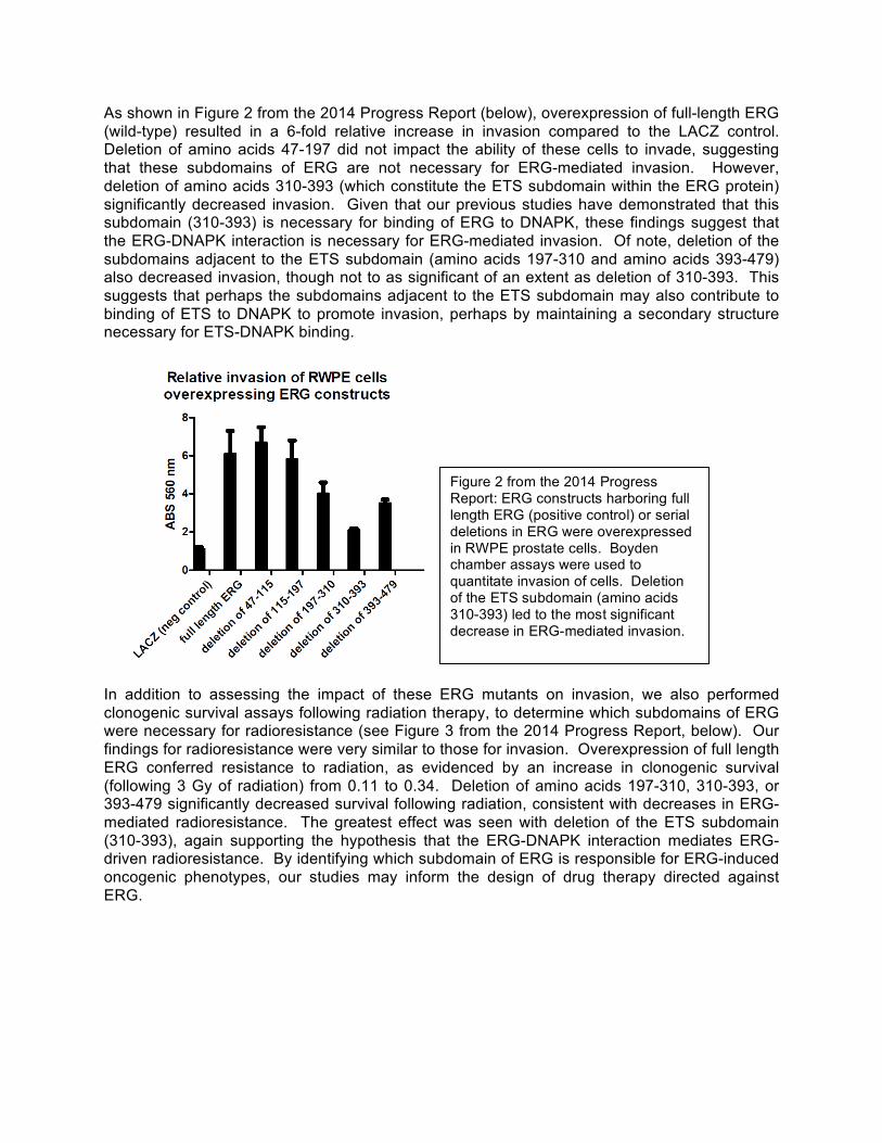
As shown in Figure 2 from the 2014 Progress Report (below), overexpression of full-length ERG (wild-type) resulted in a 6-fold relative increase in invasion compared to the LACZ control. Deletion of amino acids 47-197 did not impact the ability of these cells to invade, suggesting that these subdomains of ERG are not necessary for ERG-mediated invasion. However, deletion of amino acids 310-393 (which constitute the ETS subdomain within the ERG protein) significantly decreased invasion. Given that our previous studies have demonstrated that this subdomain (310-393) is necessary for binding of ERG to DNAPK, these findings suggest that the ERG-DNAPK interaction is necessary for ERG-mediated invasion. Of note, deletion of the subdomains adjacent to the ETS subdomain (amino acids 197-310 and amino acids 393-479) also decreased invasion, though not to as significant of an extent as deletion of 310-393. This suggests that perhaps the subdomains adjacent to the ETS subdomain may also contribute to binding of ETS to DNAPK to promote invasion, perhaps by maintaining a secondary structure necessary for ETS-DNAPK binding.
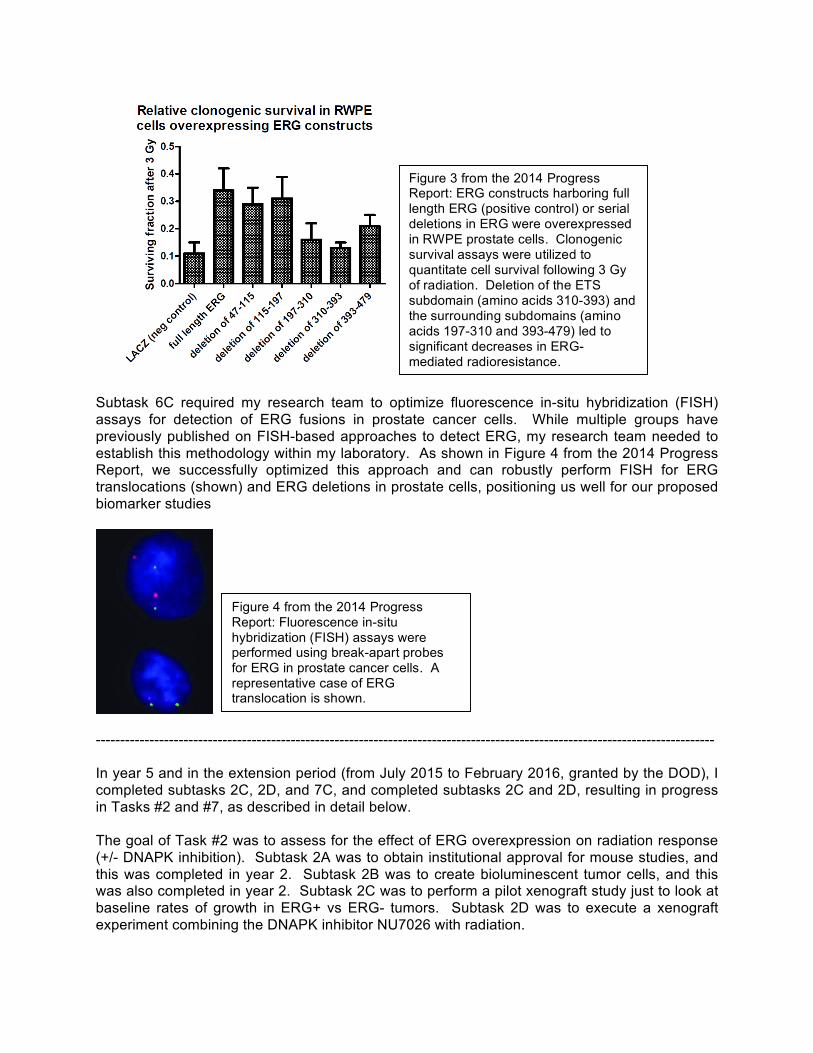
In addition to assessing the impact of these ERG mutants on invasion, we also performed clonogenic survival assays following radiation therapy, to determine which subdomains of ERG were necessary for radioresistance (see Figure 3 from the 2014 Progress Report, below). Our findings for radioresistance were very similar to those for invasion. Overexpression of full length ERG conferred resistance to radiation, as evidenced by an increase in clonogenic survival (following 3 Gy of radiation) from 0.11 to 0.34. Deletion of amino acids 197-310, 310-393, or 393-479 significantly decreased survival following radiation, consistent with decreases in ERG-mediated radioresistance. The greatest effect was seen with deletion of the ETS subdomain (310-393), again supporting the hypothesis that the ERG-DNAPK interaction mediates ERG-driven radioresistance. By identifying which subdomain of ERG is responsible for ERG-induced oncogenic phenotypes, our studies may inform the design of drug therapy directed against ERG.
Figure 2 from the 2014 Progress Report: ERG constructs harboring full length ERG (positive control) or serial deletions in ERG were overexpressed in RWPE prostate cells. Boyden chamber assays were used to quantitate invasion of cells. Deletion of the ETS subdomain (amino acids 310-393) led to the most significant decrease in ERG-mediated invasion.
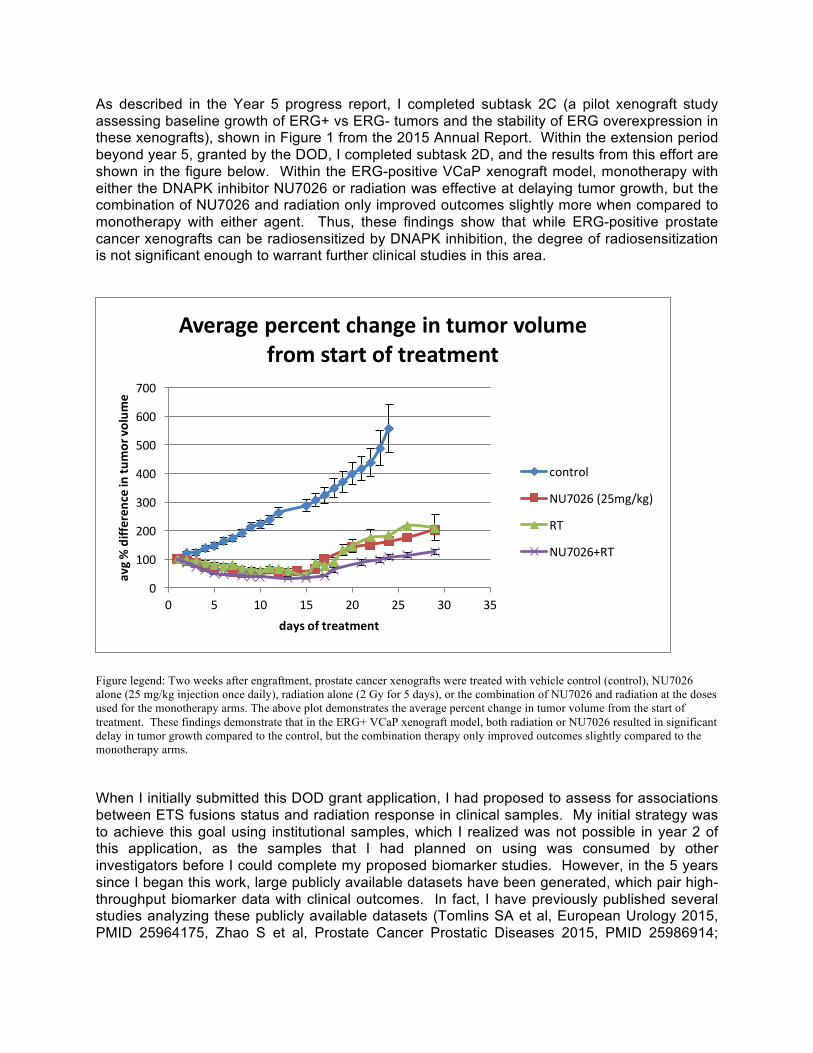
Subtask 6C required my research team to optimize fluorescence in-situ hybridization (FISH) assays for detection of ERG fusions in prostate cancer cells. While multiple groups have previously published on FISH-based approaches to detect ERG, my research team needed to establish this methodology within my laboratory. As shown in Figure 4 from the 2014 Progress Report, we successfully optimized this approach and can robustly perform FISH for ERG translocations (shown) and ERG deletions in prostate cells, positioning us well for our proposed biomarker studies
------------------------------------------------------------------------------------------------------------------------------- In year 5 and in the extension period (from July 2015 to February 2016, granted by the DOD), I completed subtasks 2C, 2D, and 7C, and completed subtasks 2C and 2D, resulting in progress in Tasks #2 and #7, as described in detail below. The goal of Task #2 was to assess for the effect of ERG overexpression on radiation response (+/- DNAPK inhibition). Subtask 2A was to obtain institutional approval for mouse studies, and this was completed in year 2. Subtask 2B was to create bioluminescent tumor cells, and this was also completed in year 2. Subtask 2C was to perform a pilot xenograft study just to look at baseline rates of growth in ERG+ vs ERG- tumors. Subtask 2D was to execute a xenograft experiment combining the DNAPK inhibitor NU7026 with radiation.
Figure 3 from the 2014 Progress Report: ERG constructs harboring full length ERG (positive control) or serial deletions in ERG were overexpressed in RWPE prostate cells. Clonogenic survival assays were utilized to quantitate cell survival following 3 Gy of radiation. Deletion of the ETS subdomain (amino acids 310-393) and the surrounding subdomains (amino acids 197-310 and 393-479) led to significant decreases in ERG-mediated radioresistance.
Figure 4 from the 2014 Progress Report: Fluorescence in-situ hybridization (FISH) assays were performed using break-apart probes for ERG in prostate cancer cells. A representative case of ERG translocation is shown.
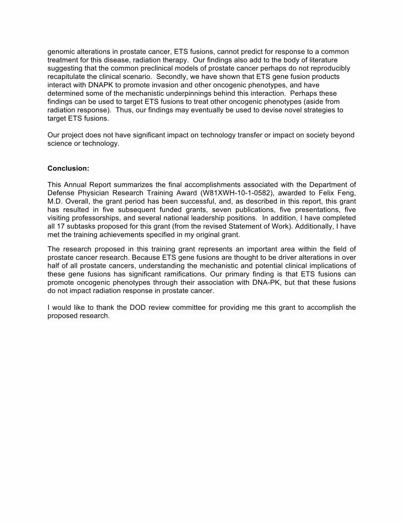
As described in the Year 5 progress report, I completed subtask 2C (a pilot xenograft study assessing baseline growth of ERG+ vs ERG- tumors and the stability of ERG overexpression in these xenografts), shown in Figure 1 from the 2015 Annual Report. Within the extension period beyond year 5, granted by the DOD, I completed subtask 2D, and the results from this effort are shown in the figure below. Within the ERG-positive VCaP xenograft model, monotherapy with either the DNAPK inhibitor NU7026 or radiation was effective at delaying tumor growth, but the combination of NU7026 and radiation only improved outcomes slightly more when compared to monotherapy with either agent. Thus, these findings show that while ERG-positive prostate cancer xenografts can be radiosensitized by DNAPK inhibition, the degree of radiosensitization is not significant enough to warrant further clinical studies in this area.
Figure legend: Two weeks after engraftment, prostate cancer xenografts were treated with vehicle control (control), NU7026 alone (25 mg/kg injection once daily), radiation alone (2 Gy for 5 days), or the combination of NU7026 and radiation at the doses used for the monotherapy arms. The above plot demonstrates the average percent change in tumor volume from the start of treatment. These findings demonstrate that in the ERG+ VCaP xenograft model, both radiation or NU7026 resulted in significant delay in tumor growth compared to the control, but the combination therapy only improved outcomes slightly compared to the monotherapy arms. When I initially submitted this DOD grant application, I had proposed to assess for associations between ETS fusions status and radiation response in clinical samples. My initial strategy was to achieve this goal using institutional samples, which I realized was not possible in year 2 of this application, as the samples that I had planned on using was consumed by other investigators before I could complete my proposed biomarker studies. However, in the 5 years since I began this work, large publicly available datasets have been generated, which pair high-throughput biomarker data with clinical outcomes. In fact, I have previously published several studies analyzing these publicly available datasets (Tomlins SA et al, European Urology 2015, PMID 25964175, Zhao S et al, Prostate Cancer Prostatic Diseases 2015, PMID 25986914;;
0
100
200
300
400
500
600
700
0 5 10 15 20 25 30 35
avg % differen
ce in tu
mor vo
lume
days of treatment
Average percent change in tumor volume from start of treatment
control
NU7026 (25mg/kg)
RT
NU7026+RT

Prensner JR et al, Lancet Oncology 2014, PMID 25456366). Over the last year of the grant, I realized that there was sufficient data in these publicly available datasets to permit an analysis of ERG status and radiation response, as I had initially proposed, and I proceed with such an analysis. This analysis, summarized in Figure 2 below, demonstrated that ETS fusion status was not associated with response to radiation, in multiple cohorts. Because this data is all publicly available (i.e., can be accessed by anyone over the internet) and is completely de-identified, this analysis did not require HRPO review. The available biomarker and clinical data from these de-identified patients is available on the NCBI Gene Expression Omnibus, with accession numbers GSE46691 and GSE62116. Figure 2
Figure 2: Two publicly available cohorts, totaling 225 prostate cancer patients treated with radiation, were analyzed for associations between ETS gene fusion status and metastatic progression (“High” denotes presence of an ERG fusion and “Low” denotes absence of an ERG fusion). ETS gene fusions status did not predict outcomes following radiation therapy, as demonstrated by Kaplan Meier analyses. Because I ended up using publicly available data to complete Task 6 & 7, I submitted a revised statement of work at the end of year 5, which shortened the original 20 subtasks to 17 subtasks (eliminating subtasks 6A and 6B, as well as 7C – this revised statement of work was approved by the DOD). Thus, over the original five-year period and the 7 month extension period of the grant, I completed 17 out of 17 revised subtasks, and accomplished all the major tasks and aims initially proposed in this grant. Thus, I have successfully completed the statement of work proposed (and subsequently revised) in this grant. Research achievements: Milestones In the original Statement of Work, 11 milestones were identified, and targeted over the 5 year course of this grant. As of year 5, I had completed 10 out of 11 milestones (Milestones #1, #2, #4, #5, #6, #7, #8, #9, #10, and #11) during the 5 years of this proposal. These milestones were reduced to 8 milestones with the most recent revision of the statement of work – all 8 milestones have now been completed. Training achievements

In my original grant application, I highlighted a series of training program activities which I hoped would contribute substantially to my scientific development. Over the grant period, as proposed, I have continued to attend a number of basic science seminars, hosted by the Departments Medicine, and Molecular and Cellular Biology, which have broadened by scientific knowledge within my field. I have also regularly attended Gene Fusion and Cancer Biology Research Meetings, run by my mentor Arul Chinnaiyan, as well as the Pathology and Radiation Oncology Research Seminars, run by the two departments with which I am affiliated. Additionally, I have renewed my “Training in the Responsible Conduct of Research” certification, and presented at the national meetings noted above in the milestones section. I have presented my research at national conferences, including the AACR annual meeting, the AACR Prostate Cancer meeting, the ASCO meeting, the Prostate Cancer Foundation annual meeting, and the ASTRO annual meeting. Finally, I have met regularly with my mentors, Drs. Arul Chinnaiyan, Ted Lawrence, and Tom Carey, as planned in my original proposal.
Career achievements The overall goal of my DOD Mentored Physician Research Training Award was to help me develop a career as a physician scientist committed to prostate cancer research. This grant has really helped me accomplish this goal, both directly and indirectly. Because of my need to obtain tissue specimens to fulfill Aim 3 of this grant, I approached the Radiation Therapy Oncology Group (RTOG), and began regularly attending their Genitourinary Cancer Translational Research Committee meetings. Because of my increasing involvement with this group, I was appointed as chair of this committee. As chair of this committee, my role is to help direct RTOG-based prostate cancer research on a national level. This role has resulted in national recognition, as I was asked to present my research from this DOD grant in the 2011 AACR Prostate Cancer conference and the 2012 ASTRO meeting. Similarly, I moderated one of the 3 sessions at the 2013 ASCO GU conference (my session was focused on translational research in prostate cancer), and I chaired and organized a session on localized prostate cancer at the 2015 AACR Annual Meeting. Over the past few years year, I have also served as a grant reviewer for the NIH Cancer Biomarker Study Section (four times), DOD study sections (twice), Prostate Cancer Foundation Young Investigator and Challenge grants (six times), and Prostate Cancer Canada/Movember grants (once). Additionally, my reputation as a translational prostate cancer researcher led to my nomination and subsequent election to the National Cancer Institute Genitourinary Cancer Steering Committee, which reviews national cooperative group clinical trial proposals in prostate cancer. Also, I was named as the Chair of the Biology Scientific Track for ASTRO (American Society of Radiation Oncology), the national organization for radiation oncologists – in this role, I lead and organize the biology scientific sessions for this organization. Because of these successes, my chairman appointed me as Chief of the Division of Translational Genomics in the Department of Radiation Oncology at the University of Michigan. Of note, I recently accepted a new position as Vice Chair, and Director of Translational Research, for the Department of Radiation Oncology at the University of California at San Francisco, and joined the UCSF faculty as an Associate Professor in May 2016.
My DOD-sponsored project has led to the preliminary data necessary for several grants that I have received over the past four years, including a Celgene Translational Award ($500,000 over 2 years) and five separate Prostate Cancer Foundation (PCF) Challenge Award ($1,000,000 split among co-Principal Investigators over 2 years). This includes two PCF Challenge Awards focused on DNA repair (entitled “Interrogation of Aberrant DNA Repair in Sporadic Prostate Cancer” and “Targeting DNA Repair Pathways to Improve Treatment for Advanced Prostate Cancer”), which stem directly from the findings in this DOD grant and seek to translate some of these findings into the clinic. The other three PCF Challenge grants, which include a genomic

sequencing study to identify biomarkers of radioresistance and two additional studies seeking to develop targeted therapies for prostate cancer, build upon skill sets that I have developed in the course of completing the subtasks and training program specified in this DOD grant. In addition, I was funded for five additional prostate cancer-based grants. One of these grants, from the Fund for Cancer Research ($75,000 for 1 year), was based directly on extending the work initiated in Aim 1 of this DOD PCRP grant. Two of the other four grants (I am co-PI of project within a NIH SPORE grant, entitled “Development of Novel BET Bromodomain Inhibitors for the Treatment of Advanced Prostate Cancer” and am a key co-investigator of a U10 grant entitled “Integrated Translational Genoproteomics Center at Washington University”) are NIH grants. The other two grants (Mazzone grant and Medivation/Astellas Investigator Grant) are not directly related to the work included in this DOD PCRP grant, but do focus on different aspects of prostate cancer. In total, during the five years of this DOD PCRP grant, I have received In addition, over the past year, I have received 10 additional grants, from both NIH and Foundation sources. Much of this success has been based upon the data generated and experienced gained from this DOD Grant. In addition, I have had six manuscripts accepted for publication, and a seventh one in submission, based on work from this proposal (detailed in the reportable outcomes section below). In addition to these 7 manuscripts (see references 1-7 below), I have published 97 additional manuscripts over the five years of this grant, including approximately 25 from the last year. I would like to thank the DOD for making all of this possible for me.

Summary of Key Research Accomplishments: The key research accomplishments from this grant proposal include the following:
• ERG overexpression in prostate cancer cell lines confers radiation resistance• This ERG-associated radiation resistance is mediated by increased efficiency of DNA
repair in response to radiation• ERG interacts with the repair protein DNAPK in a DNA-independent manner, at its
tyrosine 373 site• DNAPK knockdown or inhibition preferentially radiosensitizes ERG-positive vs ERG-
negative cells, and can reverse ERG-mediated radiation resistance• ERG is diffusely localized through the prostate cancer cell and does not redistribute
upon genotoxic stress• ERG-mediated invasion depends on the ETS subdomain of the ERG protein• ERG FISH can be optimized in old formalin-fixed paraffin embedded tissue• Determination of baseline levels of growth in ERG+ vs ERG- xenografts• Discovery that, in clinical samples, ERG fusion status does not predict for radiation
response (which differs from data generated from preclinical models in this grant)• Discovery that ERG-positive prostate cancer xenografts can be radiosensitized slightly
with DNAPK inhibition.
Reportable Outcomes and Products: The final year of work from this grant proposal has resulted in the following reportable outcomes and products:
1) A highlight of my work on the DOD CDMRP website(http://cdmrp.army.mil/pcrp/research_highlights/15feng_highlight.shtml)
2) A manuscript, following from Aim 2 of this grant, accepted at Cancer Cell (on which I amco-senior author)1
3) A manuscript, relating to Aim 3 of this grant, accepted at European Urology (on which Iam senior author)2
4) Visiting Professorships at the Cleveland Clinic, UT Southwestern, UCSF, UCLA, andHarvard
These outcomes and products add to the following reportable outcomes and products from the previous years of the grant:
5) A manuscript, reviewing data from Aim 1 this grant, published in Clinical CancerResearch13 (on which I am first author)
6) A funded Challenge grant from the Prostate Cancer Foundation, entitled “Targeting DNARepair Pathways to Improve Treatment for Advanced Prostate Cancer”
7) Publication of work from Task #4 in a Cancer Cell manuscript4, co-published with mymentor and primary collaborator, Dr. Arul Chinnaiyan
8) Two publications on ETS gene fusions in prostate cancer, published in the journal CurrDrug Targets5 and Neoplasia6
9) A publication on DNAPK in prostate cancer, published in the journal Cancer Discovery710) Oral presentation on work from Task #4, at the 2010 American Society of Therapeutic
Radiology and Oncology Annual Meeting811) Poster discussion presenting work from Tasks #1 and #3, at the 2011 American Society
of Clinical Oncology Annual Meeting912) Invited oral presentation on work from Tasks #1 and #3, at the 2011 Prostate Cancer
Foundation Annual Meeting13) A funded Young Investigator Award from the Prostate Cancer Foundation ($225,000
over 3 years), entitled “Cooperativity between TMPRSS2:ERG Gene Fusions and PTEN

Genomic Deletions in the Radiation Resistance of Prostate Cancer”, from January 2011 to January 2014
14) Oral presentation of work from this grant proposal, at the 2012 AACR Prostate CancerConference
15) An invited presentation, in which I reviewed data from this grant, at the Winship CancerCenter (at Emory)
16) A funded Challenge Grant from the Prostate Cancer Foundation ($1,000,000 over 3years, split among 4 co-principal investigators, of which I am one), entitled “Interrogating DNA repair aberrations in advanced prostate cancer”, from 8/2012-7/2015
17) A funded Translational Award from the pharmaceutical company Celgene ($500,000over 2 years, on which I am PI), entitled “CC115 as a therapeutic approach for metastatic Ewing’s sarcoma or prostate cancer”, from 4/2012-4/2014
18) A funded grant from the Fund For Cancer Research, entitled "Investigating ETS GeneFusions as Predictive Biomarkers of Radiation Resistance and Targets for Radiosensitization" ($75,000 over 1 year)
Participants and Other Collaborating Institutions:
Individuals who have worked on this project:
Name: Felix Feng, MD Role: Principal Investigator Changes from previous reporting cycles: None
Name: Sumin Han, PhD Role: Research specialist (technician) Changes from previous reporting cycles: None
Name: Kari Wilder-Romans, MS Role: Research specialist (technician) Changes from previous reporting cycles: None
Collaborating Institutions: None
Special Reporting Requirements:
None
Impact:
The research goals of this grant proposal were to: 1) investigate the effect of ETS gene fusions on radiation phenotype in preclinical models of prostate cancer, 2) to explore the mechanism of interaction between ERG (the predominant ETS gene fusion product) and the DNA repair protein DNA-PK, and 3) to determine if ETS gene fusion status is a clinical biomarker of radioresistance for prostate cancer.
In completing all of these goals, we contributed significant knowledge to the field of prostate cancer. First, we demonstrated that, while ETS fusions confer radiation resistance in preclinical models of prostate cancer, these preclinical findings did not translate clinically, as we found that ETS fusion status was not associated with increased recurrence rates following radiation therapy. These findings are meaningful, in that we have shown that one of the most common
genomic alterations in prostate cancer, ETS fusions, cannot predict for response to a common treatment for this disease, radiation therapy. Our findings also add to the body of literature suggesting that the common preclinical models of prostate cancer perhaps do not reproducibly recapitulate the clinical scenario. Secondly, we have shown that ETS gene fusion products interact with DNAPK to promote invasion and other oncogenic phenotypes, and have determined some of the mechanistic underpinnings behind this interaction. Perhaps these findings can be used to target ETS fusions to treat other oncogenic phenotypes (aside from radiation response). Thus, our findings may eventually be used to devise novel strategies to target ETS fusions.
Our project does not have significant impact on technology transfer or impact on society beyond science or technology.
Conclusion:
This Annual Report summarizes the final accomplishments associated with the Department of Defense Physician Research Training Award (W81XWH-10-1-0582), awarded to Felix Feng, M.D. Overall, the grant period has been successful, and, as described in this report, this grant has resulted in five subsequent funded grants, seven publications, five presentations, five visiting professorships, and several national leadership positions. In addition, I have completed all 17 subtasks proposed for this grant (from the revised Statement of Work). Additionally, I have met the training achievements specified in my original grant.
The research proposed in this training grant represents an important area within the field of prostate cancer research. Because ETS gene fusions are thought to be driver alterations in over half of all prostate cancers, understanding the mechanistic and potential clinical implications of these gene fusions has significant ramifications. Our primary finding is that ETS fusions can promote oncogenic phenotypes through their association with DNA-PK, but that these fusions do not impact radiation response in prostate cancer.
I would like to thank the DOD review committee for providing me this grant to accomplish the proposed research.
References:
1) Goodwin JF, Kothari V, Drake JM, Zhao S, Dylgjeri E, Dean JL, Schiewer MJ, McNair C,Magee MS, Den RB, Zhu Z, Graham NA, Vashisht AA, Wohlschlegel JA, Graeber TG,Davicioni E, Karnes RJ, Tomlins SA, Sharifi N, Witte ON, Feng FY** and Knudsen KE**.DNA-PK mediated transcriptional regulation drives tumor progression and metastasis.Cancer Cell 2015, 28(1): 97-113. **indicates equal senior authorship
2) Tomlins SA, Alshalaifa M, Davicioni E, Erho N, Yousefi K, Zhao S, Haddad Z, Den RB,Dicker AP, Trock BJ, DeMarzo AM, Ross AE, Schaeffer EM, Klein EA, Magi-Galluzzi C,Karnes RJ, Jenkins RB, and Feng FY. Characterization of 1577 primary prostatecancers reveals novel biological and clinicopathologic insights into molecular subtypes.European Urology 2015;; 68(4): 555-67.
3) Feng FY, Brenner JC, Hussain M, Chinnaiyan AM. Molecular Pathways: Targeting ETSGene Fusions in Cancer. Clin Cancer Res. 2014 Sep 1;;20(17):4442-8.
4) Brenner JC, Ateeq B, Li Y, et al: Mechanistic rationale for inhibition of poly(ADP-‐‑ribose)polymerase in ETS gene fusion-‐‑positive prostate cancer. Cancer Cell 19:664-‐‑78, 2011
5) White NM, Feng FY, Maher CA. Recurrent rearrangements in prostate cancer: causesand therapeutic potential. Curr Drug Targets. 14(4):450-9, 2013.
6) Han S, Brenner JC, Sabolch A, Jackson W, Speers C, Wilder-Romans K, Knudsen KE,Lawrence TS, Chinnaiyan AM, Feng FY. Targeted Radiosensitization of ETS Fusion-Positive Prostate Cancer through PARP1 Inhibition. Neoplasia 2013 Oct;; 15(10): 1207-17.
7) Goodwin JF, Schiewer M, Dean JL, Schrecengost RS, Han S, Den RB, Dicker AP, FengFY, and Knudsen KE. A hormone-DNA repair circuit governs the response to genotoxicinsult. Cancer Discov. 2013 Nov;;3(11):1254-71.
8) Feng FY, Han S, Brenner C, et al: PARP inhibition reverses radiation resistanceconferred by ETS fusions in prostate cancer. J Clin Oncol 29:4545, 2011
Appendices:
None