presentación de powerpoint - wpanet.org · los médicos griegos herófilo de calcedonia y...
TRANSCRIPT

HistoriaPapiro egipcio fechado cerca del 1600 a.C.Hipócrates del siglo V a.C.En el siglo IV a.C. Aristóteles aumentó los conocimientos anatómicos sobre los animales.Los médicos griegos Herófilo de Calcedonia y Erasístrato diseccionaron cadáveres humanos y fueron los primeros en determinar muchas funciones, incluidas las del sistema nervioso y los músculos. Reconoce el cerebro como centro del sistema nervioso. Diferencia los nervios motores de los sensoriales y es el primero en conocer que las arterias contienen sangre y no aire
Galeno desarrollo el conocimiento en animales de su anatomía. c. 300? a.C. Los antiguos romanos y los árabes consiguieron algunos pequeños progresosEl renacimiento influyó en la ciencia de la anatomía en la segunda mitad del siglo XVI.
La anatomía moderna se inicia con la publicación en 1543 del trabajo del anatomista belga Andrés VesalioEl primer intento aparentemente de descripción de la esquizofrenia se debe a George Trosse en 1656 el que describe su propia enfermedad, la que se considera puede ser la verdadera descripción en la literatura de la esquizofrenia hasta ese momento publicada. Unas décadas después Thomas Willis también contribuyó en forma sustancial a esta descripción en sus trabajos en la Anatomía del Cerebro en 1683. Algunos autores lo consideran como el primer autor que describió la enfermedad.
La invención en el siglo XVII del microscopio compuesto dio lugar al desarrollo de la anatomía microscópicaPhillipe Pinel en el Hospital Salpetriere de Paris y a John Haslam en el Hospital Bethlem de Londres. Son meritorios sus intentos de relacionar el cuadro clínico con los hallazgos hechos en la autopsia considerando diversos factores posibles en la etiología de la enfermedad.
La anatomía microscópica hizo grandes progresos en el siglo XIX
Rudolf Virchow [Cellular Pathology as Based on Histology] Cajal, Golgi, Alzheimer, Kraepelin, Morel entre otros
hicieron estudios en el sistema nervioso en cerebros autopsiados de pacientes esquizofrénicos. La mayor parte de estos trabajos hasta la primera mitad del siglo XX se realizaron con muestras de la corteza cerebral, especialmente de la región frontal. Estudios de regiones más profundas se deben a los Vogt en Alemania y a Nieto y Escobar en Méjico en la segunda mitad de este siglo.El desarrollo científico-técnico de la segunda mitad del siglo permitió un mejor conocimiento de la anatomía del cerebro en la esquizofrenia gracias a las técnicas de imagen y a la microscopía electrónica. IMAGINOLOGÍA SIGLO XX
Macroscópicos Siglo XVII a.C.
Microscópicos Siglo XIX d.C.
For more than two centuries the brain of schizophrenic patients has been investigated looking for regions of interest (ROI) that would relate these regions to the physiopathology and etiology of the disease.
2

The researches have been directed to:Macroscopic examination of the brain: morphometry, pneumoencephalography, CT, MRI, PET, SPET etc.Microscopic examination of the brain: optic microscopy, electron microscopy, molecular biology, etc.
3
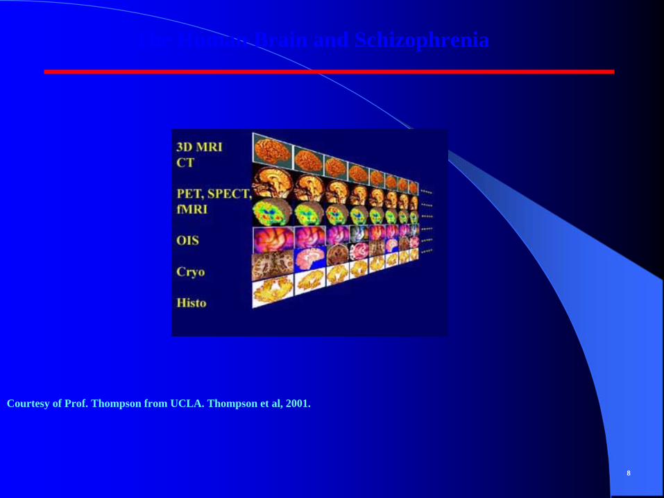
The Human Brain and Schizophrenia
Courtesy of Prof. Thompson from UCLA. Thompson et al, 2001.
8
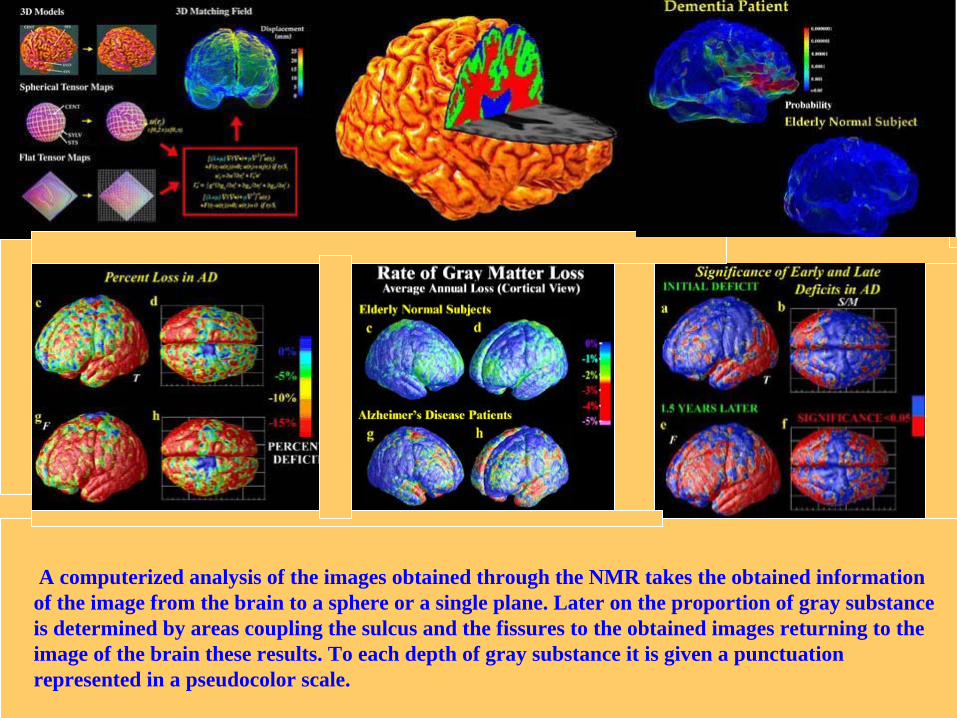
A computerized analysis of the images obtained through the NMR takes the obtained information of the image from the brain to a sphere or a single plane. Later on the proportion of gray substance is determined by areas coupling the sulcus and the fissures to the obtained images returning to the image of the brain these results. To each depth of gray substance it is given a punctuation represented in a pseudocolor scale.

Control left. Schizophrenia right. It is appreciated the enlargement of the ventricles and the reduction of the volume of the amygdala in the schizophrenic patient with widening of the temporal horn (arrow).

Lob. temporal
Courtesy of Prof. Janice Stevens13

The human brain and schizophrenia
These findings have demonstrated the implication of the temporarl lobe in the illness.
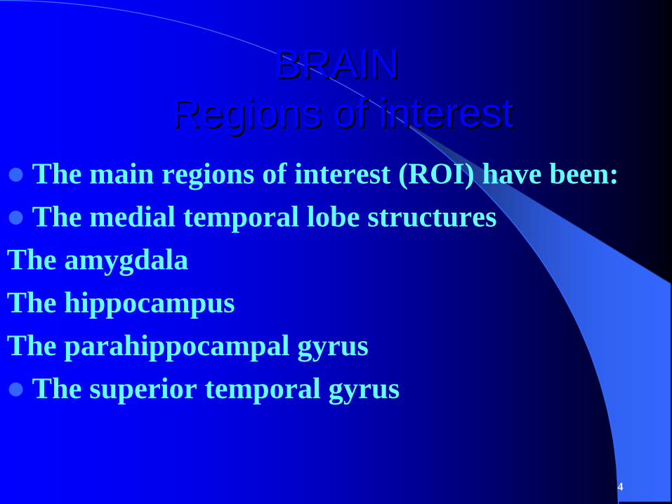
BRAINBRAIN Regions of interestRegions of interest
The main regions of interest (ROI) have been:The medial temporal lobe structures
The amygdalaThe hippocampusThe parahippocampal gyrus
The superior temporal gyrus
4

. Harrison PJ : The neuropathology of schizophrenia. A critical review of the data and their interpretation. Brain 1999; 122 :593–624.
. Nelson MD, Saykin AJ, Flashman LA, Riordan HJ. Hippocampal volume reduction in schizophrenia as assessed by magnetic resonance imaging. A meta-analytic study. Arch Gen Psychiatry 1998; 55:433–440.
. Wright IC, Rabe-Hesketh S, Woodruff PWR, David AS, Murray RM, Bullmore ET (2000): Meta-analysis of regional brain volumes in schizophrenia. Am J Psychiatry 157:16–25.
. Shenton ME, Dickey CC, Frumin M, McCarley RW. A review of MRI findings in schizophrenia. Schizophr Res 2001; 49: 1 –52.
. Joyal CC, Laakso MP, Tiihonen J, Syvalahti E, Vilkman H, Laakso A, et al. Schizophrenia: A Volumetric Magnetic Resonance Imaging Study in First Episope Neuroleptic-Naive Patients. Biological Psychiatry 2003; 54: 1302-4.
. Bogerts B. The neuropathology of schizophrenic diseases: historical aspects and

Such approaches give us a broad sense of how the brain looks anatomically and how it functions physiologically. To understand how genes affect the brain in a way that puts it at risk, we need to examine in a much more finely detailed way the actual cells of the brain. We can do this with living brain tissue to some degree, and we can do this with postmortem brain tissue to a much greater degree
Daniel R. WeinbergerLecture given at the Project on the Decade of the Brain. September 25, 1998, US
Capitol, Washington DC.
5

ELECTRON MICROSCOPE STUDY OF THE AMYGDALA, HIPPOCAMPUS AND PRIMARY AUDITORY CORTEX OF SCHIZOPHRENIC PATIENTS
Up to where we know only a post-mortem study exists in the medical literature in schizophrenic patients where it has been studied by means of these techniques the primary auditory cortex, the amygdala and the hippocampus of the left cerebral hemisphere.
Mesa Castillo, S. Esquizofrenia. Estudio ultraestructural del sistema límbico. Hallazgos en el núcleo amigdalino Rev HospPsiq Habana, 1978:19, 197-225.
6

NEUROPATHOLOGICAL RESEARCH IN SCHIZOPHRENIA
ELECTRON MICROSCOPY
Brains from dead patientsAborted fetuses Experimental animals
Controls
Left temporal lobeCompatible alterations with the viral hypothesis: particles with herpes simplex hominis type Iviral antigen, intranuclear bodies, membrane alterations.
19771977--20052005
7
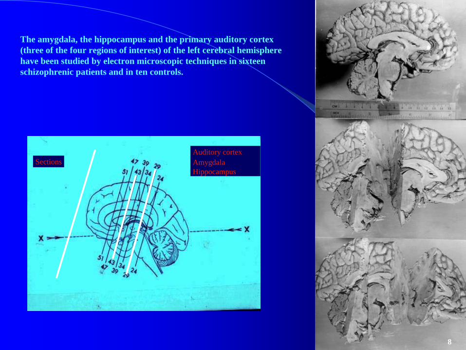
The amygdala, the hippocampus and the primary auditory cortex (three of the four regions of interest) of the left cerebral hemisphere have been studied by electron microscopic techniques in sixteen schizophrenic patients and in ten controls.
24/02/05 30
SectionsAuditory cortexAmygdalaHippocampus
8

Left hippocampus selected for study 9

Left amygdala: third superior and third inferior portion selected for study with optic and electronic microscopy
10
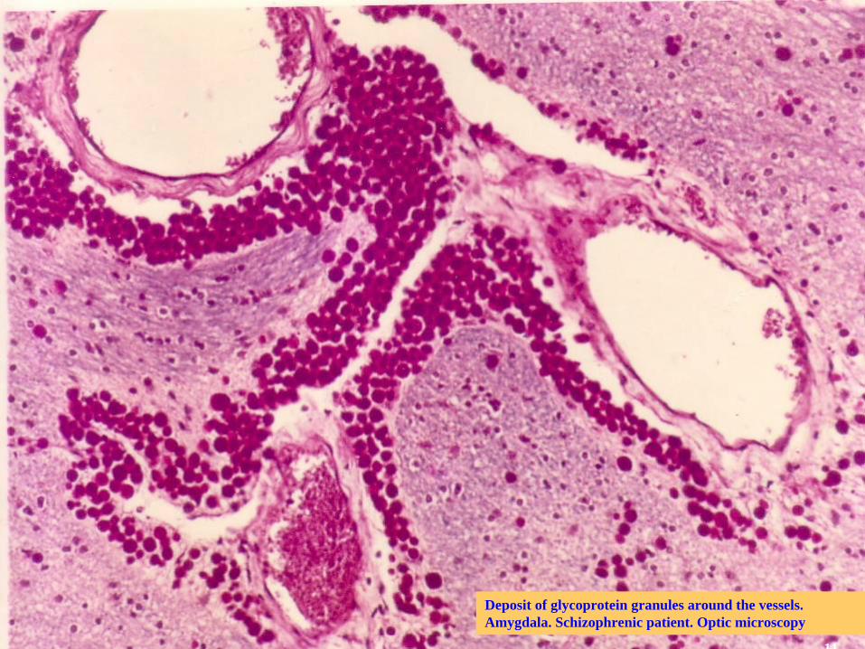
Deposit of glycoprotein granules around the vessels. Amygdala. Schizophrenic patient. Optic microscopy
11

Neuron degenerative changes were observed specially in the amygdaline nucleus. A microglial reaction was present around the degenerated neurons. Arrow.
Optic microscopy
12
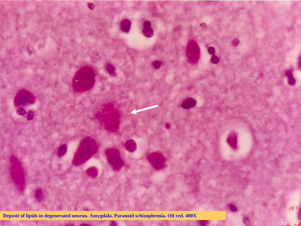
Deposit of lipids in degenerated neuron. Amygdala. Paranoid schizophrenia. Oil red. 400X13

ELECTRON MICROSCOPY
Main findings:
• Particles with viral morphology which react to peroxidase and coloidal gold conjugated with herpes hominis type I antibodies
•Nuclear bodies
•Membrane proliferation
14

Particles with viral morphology: below, herpes simplex virus. Above (arrow) particle observed in schizophrenia.
The image below is from Dennis Kunkel's excellent Microscopy Science and Photography Through a Microscope 15

Intranuclear particle (arrow) with viral morphology. A central core and a capsule are present. Left amygdala. Paranoid schizophrenia.
200 nm
16

Immuno-electron microscopy. Particle (vertical arrow) related to membrane structure (oblique arrow). Digital analysis- right superior inset- shows the morphology of a virus in the antigen-antibody reaction (yellow and green).
100 nm
17

Three particles (arrows) within an axon labeled with antiherpes hominis type I antibody.
Peroxidase technique.
18

19

El análisis digital muestra la estructura interna de la partícula con un núcleo central y varias envolturas alrededor. Esquizofrénico joven. Estudios post-mortem.

20

Intranuclear filamentous inclusion body (IF). Schizophrenia. 21

Nuclear bodies (double head arrows).
22

The possibility that schizophrenia depends on different types of genetic mutations caused by the action of a virus it should be kept in mind

Herpes virus en ganglio trigémino No pasa nadaen una célula normal no mutada PUBERTAD CAMBIOS
HORMONALES
Herpes virus en ganglio trigémino PUBERTAD CAMBIOS Mayor predisposición en una célula mutada HORMONALES replicación del virus
Síntomas psiquiátricos
DISEMINACIÓN DE LA INFECCIÓN A ESTRUCTURAS TEMPORO-LÍMBICAS
HERPES VIRUS +genes= trastornos del neuro-desarrollo

SUMMARY. The boarding of schizophrenia research through images has been characterized by the introduction of new technologies with this purpose. Depending on the larger or smaller degree of the resolution power of the applied technology we can investigate the illness in different dimensions. Some of these researches have not been limited alone for the resolution power of the used technique but for difficulties of different nature that have prevented to achieve the objectives that were looked for. This is the case of post-mortem studies in schizophrenic patients specially when ultramicroscopic techniques has been used. Up to where we know only a work exists in the medical literature where two structures of the limbic system - the amygdaline nuclei and the hippocampus- of schizophrenic patients have been studied by means of this technique starting from 1977. From that date and by means of different image techniques it has been proven the importance of both structures in the physio-pathology of the illness. In this work some of these results were presented and their relationship with the viral hypothesis outlined for the illness.
S. Mesa CastilloPsychiatric Hospital of Havana
23