presentaci n de los trabajos. utilidad de...
TRANSCRIPT

2009
TESIS DOCTORAL UTILIDAD DE LA RESONANCIA MAGNÉTICA
EN PACIENTES CON FIBRILACIÓN
AURICULAR TRIBUTARIOS DE
TRATAMIENTO CON ABLACIÓN
PERCUTÁNEA DE LAS VENAS PULMONARES
ROSARIO JESÚS PEREA PALAZÓN
UNIVERSITAT DE BARCELONA
Línea de investigación de biopatología cardiovascular
Dirigida por el Prof. Josep Brugada Terradellas y por la Dra.
Teresa Mª de Caralt Robira


PRESENTACIÓN DE LOS TRABAJOS 101
1. PRESENTACIÓN DE LOS TRABAJOS

102

PRESENTACIÓN DE LOS TRABAJOS 103
1.1. ESTUDIO 1
Left atrial contractility is preserved after successful circumferential
pulmonary vein ablation in patients with atrial fibrillation.
Perea RJ, Tamborero D, Mont L, Caralt TM, Ortiz JT, Berruezo A,
Matiello M, Sitges M, Vidal B, Sánchez M, Brugada J.
J Cardiovasc Electrophysiol. 2008;19:374-379
1.1.1. OBJETIVOS
Objetivo general
El objetivo general del estudio es valorar y demostrar la utilidad de la RM
en pacientes con FA tributarios de APRF de las VPs.
Objetivo específico
Evaluar los cambios volumétricos y funcionales que se producen a medio
plazo en la AI tras la APRF de la FA y su relación con el éxito del
procedimiento.

104 PRESENTACIÓN DE LOS TRABAJOS
1.1.2. RESULTADOS
De una serie de 90 pacientes consecutivos, se excluyeron 35 debido a
que no se encontraban en ritmo sinusal en el momento del primer y/o
segundo estudio mediante RM. Por tanto, se analizaron finalmente un
total de 55 pacientes. Sus características basales se exponen en la Tabla
3.
Características de los pacientes
Edad (años)
Sexo masculino
Tipo de FA
Paroxística
Persistente
Hipertensión
Enfermedad cardiaca estructural
DD VI (mm)
DS VI (mm)
FE VI (%)
Tiempo de evolución de la FA (años)
52.0±11.3
44 (80.0%)
41 (74.5%)
14 (25.5%)
12 (21.8%)
9 (16.3%)
52.4 ± 4.7
33.1 ± 3.8
60.0 ± 8.8
8.4 ± 8.1
Tabla 3. Características de los pacientes DD VI = diámetro diastólico del ventrículo izquierdo; DS VI = diámetro sistólico del
ventrículo izquierdo; FE VI = Fracción de eyección ventricular izquierda. (Medidas
realizadas mediante ecocardiografía antes de la ablación).

PRESENTACIÓN DE LOS TRABAJOS 105
Treinta y ocho pacientes (69.1%) se mantuvieron libres de arritmia
(grupo I), mientras que los 17 pacientes restantes tuvieron recurrencia de
la arritmia (grupo II) tras 1.2 ± 0.3 procedimientos ablativos y durante una
media de seguimiento de hasta 11.8 ± 7.2 meses (en 15 pacientes
recidivó la FA y dos pacientes tuvieron flutter de nueva aparición). Entre
los pacientes con ablación exitosa, 33 (60%) se mantuvieron sin
tratamiento médico antiarrítmico, tres pacientes recibieron flecaidina para
controlar contracciones auriculares prematuras sintomáticas, y dos
pacientes se mantuvieron libres de arritmia mediante tratamiento ya sea
con flecaidina o con amiodarona, que comenzaron durante el período
blanking (período a partir de la 5ª semana después de la ablación, sin
tratamiento antiarrítmico o con la utilización de un fármaco que era
previamente inefectivo) y no fue interrumpido después por el facultativo de
referencia.
La Tabla 4 muestra los cambios en las medidas de la AI después
de la ACVP en relación al éxito del procedimiento. La Figura 16 muestra
los mismos datos para cada paciente de la serie.

106 PRESENTACIÓN DE LOS TRABAJOS
Tabla 4. Valores de la aurícula izquierda antes y 4-6 meses después del procedimiento
ablativo
*p significativa < 0.05%.
Vmax = volumen máximo de la aurícula izquierda; Vmin = volumen mínimo de
la aurícula izquierda; FE AI = fracción de eyección auricular izquierda;
pre/post ACVP = previo/posterior a la ablación circunferencial de las venas
pulmonares
No recurrencia de la FA
(n =38)
Recurrencia de la FA
(n =17)
Pre
ACVP
Post
ACVP
Disminución
Media
valor p
Pre
ACVP
Post
ACVP
Disminución
media
valor p
Vmax(ml)
98.0±19.9
84.9±17.1
13%
<0.001*
126.2±32.8
103.5±28.1
17%
<0.001*
Vmin(ml)
58.6±16.1
52.2±12.1
10%
0.004*
78.4±22.2
75.8±24.3
4%
0.315
FE AI (%)
40.2±11.5
38.1±9.8
2%
0.268
37.4±10.1
26.9±10.2
11%
<0.001*

PRESENTACIÓN DE LOS TRABAJOS 107
Figura 16. Evolución del volumen telediastólico de la aurícula izquierda (Vmax),
volumen telesistólico de la aurícula izquierda (Vmin) y de la fracción de eyección de la AI
(FE AI), 4-6 meses tras la ablación circunferencial de las venas pulmonares (ACVP) en
cada paciente de la serie. Los datos se muestran dependiendo de si los pacientes
estaban libres de arritmia (paneles de la izquierda) o de si tenían recurrencia de la
arritmia (paneles de la derecha) durante el seguimiento.
Pacientes sin arritmia
post-ablación
Pacientes con recurrencia
post-ablación
Vm
ax (
ml)
Vm
in (
ml)
FE
A
I (%
)
pre post
ACVP ACVP pre post
ACVP ACVP
pre post
ACVP ACVP pre post
ACVP ACVP
pre post
ACVP ACVP pre post
ACVP ACVP

108 PRESENTACIÓN DE LOS TRABAJOS
En primer lugar, se puede observar que el volumen máximo (Vmax)
medio tras la ACVP disminuyó en ambos, tanto en el grupo I como en el
grupo II; además, no se observaron diferencias significativas en el
porcentaje medio de reducción del Vmax entre los dos grupos (13 ± 12%
vs 17 ± 14%, respectivamente, p = 0.217). En segundo lugar, el volumen
mínimo (Vmin) medio sólo disminuyó significativamente en el grupo I.
Consecuentemente, no hubo cambios significativos en la FE AI media
tras la ablación en el grupo I. De hecho, la FE AI permaneció estable o
aumentó en el 68% de pacientes sin recurrencia de la arritmia tras la
ACVP (Figura 17), mientras que en el grupo II se observó una disminución
de la FE AI media.
A 10 pacientes se les tuvo que practicar una segunda ablación, por
recidiva de la FA (en 3 pacientes), o por flutter de nuevo debut (en 7
pacientes). Los cambios en la función contráctil de estos pacientes se
evaluaron en la RM obtenida después del último procedimiento,
correlacionándose con los observados en el resto de casos. La FE AI no
mostró cambios significativos con respecto a las mediciones basales en
los pacientes libres de FA después de la segunda ablación (de 35±11% a
32±7 p=0.05), mientras que la FE AI disminuyó en el resto de pacientes
que presentaron recurrencia de la arritmia a pesar de los dos
procedimientos (de 28±9% a 10±2%, p=0,06).

Figura 17. Variación de la fracción de eyección de la aurícula izquierda (FE AI) tras el
procedimiento ablativo.
Por otra parte, la contractilidad de la OAI no mostró cambios tras la
ablación. La FE de la OAI antes y después de la ACVP fue similar tanto
en el grupo I (41 ± 20% vs 40 ± 20%,
20% vs 32 ± 6%, p
máximo de la OAI (de 8.8 ± 4.4 a 7.6 ± 4.4 ml,
mínimo de la OAI (de 5.2 ± 4.2 a 4.3 ±3.3 ml,
En el grupo II, hubo una tendencia a la reducción del volumen máximo de
la OAI (de 7.1 ± 3.4 a 6.2 ± 3.3 ml,
variaciones significativas en el volumen mínimo de la OAI (de 4.5 ± 2.1 a
0%
10%
20%
30%
40%
50%P
orc
en
taje
de
pa
cie
nte
s
PRESENTACIÓN DE LOS TRABAJOS
Variación de la fracción de eyección de la aurícula izquierda (FE AI) tras el
Por otra parte, la contractilidad de la OAI no mostró cambios tras la
ablación. La FE de la OAI antes y después de la ACVP fue similar tanto
en el grupo I (41 ± 20% vs 40 ± 20%, p = 0.8) como en el grupo II (31 ±
= 0.9). El grupo I presentó una reducción del volumen
máximo de la OAI (de 8.8 ± 4.4 a 7.6 ± 4.4 ml, p < 0.001) y del volumen
mínimo de la OAI (de 5.2 ± 4.2 a 4.3 ±3.3 ml, p <0.001) tras la ablación.
En el grupo II, hubo una tendencia a la reducción del volumen máximo de
e 7.1 ± 3.4 a 6.2 ± 3.3 ml, p = 0.08), y no se observaron
variaciones significativas en el volumen mínimo de la OAI (de 4.5 ± 2.1 a
Variación de la FE AI
Pacientes sin recurrencia
Pacientes con recurrencia
PRESENTACIÓN DE LOS TRABAJOS 109
Variación de la fracción de eyección de la aurícula izquierda (FE AI) tras el
Por otra parte, la contractilidad de la OAI no mostró cambios tras la
ablación. La FE de la OAI antes y después de la ACVP fue similar tanto
= 0.8) como en el grupo II (31 ±
tó una reducción del volumen
< 0.001) y del volumen
<0.001) tras la ablación.
En el grupo II, hubo una tendencia a la reducción del volumen máximo de
= 0.08), y no se observaron
variaciones significativas en el volumen mínimo de la OAI (de 4.5 ± 2.1 a
Pacientes sin recurrencia
Pacientes con recurrencia

110 PRESENTACIÓN DE LOS TRABAJOS
4.1 ± 2.2 ml, p = 0.11) tras las ablación. La OAI presentó un
comportamiento post-ablación similar al resto de la aurícula.
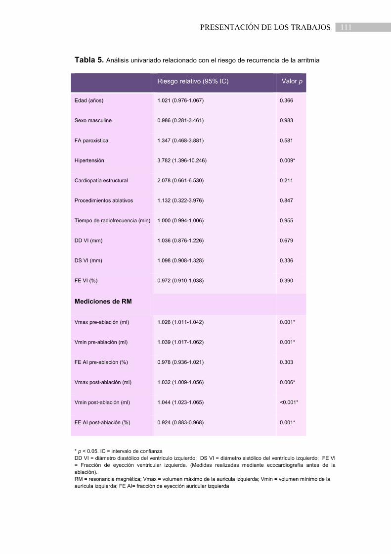
El análisis univariado mostró que los pacientes con recurrencias
tras el procedimiento ablativo tenían mayor proporción de hipertensión
arterial, volúmenes mayores de la AI (medidos antes y después de la
ACVP) y menor FE AI post-ablación (Tabla 5). La única variable
independiente asociada con recurrencia de la arritmia en el modelo
multivariado fue el Vmin medido tras el procedimiento ablativo, con riesgo
relativo de 1.04 (intervalo de confianza del 95%, 1.02-1.06, p < 0.001). El
mejor valor de corte obtenido mediante el análisis de la curva ROC
(receiver operating characteristic) para esta variable (área bajo la curva =
0.85) fue el Vmin ≥ 75 ml (sensibilidad = 89%, especificidad = 35%), que
tenía un riesgo relativo de recurrencia de la arritmia de 11.5 (95% de
intervalo de confianza, 3.8─34.5, p <0.001).
PRESENTACIÓN DE LOS TRABAJOS 111
Tabla 5. Análisis univariado relacionado con el riesgo de recurrencia de la arritmia
Riesgo relativo (95% IC) Valor p
Edad (años) 1.021 (0.976-1.067) 0.366
Sexo masculine 0.986 (0.281-3.461) 0.983
FA paroxística 1.347 (0.468-3.881) 0.581
Hipertensión 3.782 (1.396-10.246) 0.009*
Cardiopatía estructural 2.078 (0.661-6.530) 0.211
Procedimientos ablativos 1.132 (0.322-3.976) 0.847
Tiempo de radiofrecuencia (min) 1.000 (0.994-1.006) 0.955
DD VI (mm) 1.036 (0.876-1.226) 0.679
DS VI (mm) 1.098 (0.908-1.328) 0.336
FE VI (%) 0.972 (0.910-1.038) 0.390
Mediciones de RM
Vmax pre-ablación (ml) 1.026 (1.011-1.042) 0.001*
Vmin pre-ablación (ml) 1.039 (1.017-1.062) 0.001*
FE AI pre-ablación (%) 0.978 (0.936-1.021) 0.303
Vmax post-ablación (ml) 1.032 (1.009-1.056) 0.006*
Vmin post-ablación (ml) 1.044 (1.023-1.065) <0.001*
FE AI post-ablación (%) 0.924 (0.883-0.968) 0.001*
* p < 0.05. IC = intervalo de confianza
DD VI = diámetro diastólico del ventrículo izquierdo; DS VI = diámetro sistólico del ventrículo izquierdo; FE VI
= Fracción de eyección ventricular izquierda. (Medidas realizadas mediante ecocardiografía antes de la
ablación).
RM = resonancia magnética; Vmax = volumen máximo de la auricula izquierda; Vmin = volumen mínimo de la
aurícula izquierda; FE AI= fracción de eyección auricular izquierda

374
Left Atrial Contractility is Preserved After SuccessfulCircumferential Pulmonary Vein Ablation in Patients
with Atrial FibrillationROSARIO J. PEREA, M.D., DAVID TAMBORERO, B. ENG., LLUIS MONT, M.D., PH.D.,
TERESA M. DE CARALT, M.D., PH.D., JOSE T. ORTIZ, M.D., ANTONIO BERRUEZO, M.D.,MARIA MATIELLO, M.D., MARTA SITGES, M.D., PH.D., BARBARA VIDAL, M.D.,
MARCELO SANCHEZ, M.D. and JOSEP BRUGADA, M.D., PH.D.
From the Arrhythmia Section, Thorax Institute, Hospital Clinic, University of Barcelona, Barcelona, Spain
Left Atrial Contractility. Introduction: Circumferential pulmonary vein ablation (CPVA) for atrialfibrillation (AF) consists of creating extensive lesions in the left atrium (LA). The aim of the study was toevaluate changes in LA contractility after ablation and their relationship with procedure outcome.
Methods and Results: A series of 90 consecutive patients underwent cardiac magnetic resonance imaging(MRI) before and 4–6 months after CPVA. Only patients in sinus rhythm during both imaging acquisitionswere included in the study to measure LA end-diastolic (LAmax) and LA end-systolic (LAmin) volumes.Fifty-five patients were finally analyzed (41 men, 52 ± 11 years, 74% paroxysmal AF). During a meanfollow-up of 12 ± 7 months and after 1.2 ± 0.3 ablation procedures, 38 patients (69%) were arrhythmia-free (group I), and the remaining 17 patients had recurrences (group II). There was a significant decreasein mean LAmax volume in both groups, whereas mean LAmin volume only decreased in group I. MeanLA ejection fraction (EF) was preserved after CPVA in group I (40 ± 11% vs 38 ± 10%; P = 0.27) butdecreased in patients with arrhythmia recurrences (37 ± 10% vs 27 ± 10%; P < 0.001). In fact, LA EFremained stable or increased in 68% of patients without arrhythmia recurrences.
Conclusions: LAmax volume reduction following CPVA occurs regardless of the clinical efficacy of theprocedure, whereas mean LAmin volume only decreased in patients without recurrences. LA EF waspreserved or even increased in most patients with successful CPVA. (J Cardiovasc Electrophysiol, Vol. 19,pp. 374-379, April 2008)
atrial fibrillation, catheter ablation, atrial contractility
Although several studies have shown a decrease in leftatrial (LA) size following atrial fibrillation (AF) ablation,1-6
data on the response of atrial contractility are limited. A re-cent study in a small series found an impaired LA ejectionfraction (LA EF) after paroxysmal AF catheter ablation.7
LA EF reflects the relationship between atrial end-diastolicand end-systolic volumes, and has been proposed as a goodmethod for evaluating global atrial contractility.7-10 With re-gard to changes in LA volume after AF ablation, the dataare contradictory. While some authors have reported that LAsize decreases only after successful ablation,1,3,6 others havesuggested that LA size is reduced regardless of the clinicaloutcome of the procedure.2,4,5 However, in the majority ofthese studies, LA end-diastolic (LAmax) and LA end-systolic(LAmin) volumes were not measured separately, and imag-
The study was funded in part by a grant from the Carlos III Health Institute,Madrid, Spain (Fondo Investigacion Sanitaria-PI050081).
The first two authors have contributed equally to this study.
Address for correspondence: Dr. Lluis Mont, Arrhythmia Section, ThoraxInstitute, Hospital Clinic, University of Barcelona, Villarroel 170, 08036Barcelona. Fax: 0034-93-451-3045; E-mail: [email protected]
Manuscript received 13 June 2007; Revised manuscript received 26 October2007; Accepted for publication 31 October 2007.
doi: 10.1111/j.1540-8167.2007.01086.x
ing acquisitions were performed regardless of whether thepatients were in sinus rhythm or arrhythmia.
Current cardiac cine magnetic resonance imaging (MRI)techniques are accurate for depicting the anatomical struc-tures of LA, allowing calculation of LA volumes with un-usual precision.11-15 The purpose of this study was to assessthe effect of circumferential pulmonary vein ablation (CPVA)on LA volumes and LA EF, as well as their relationship withprocedure outcome using cardiac MRI.
Methods
Patients
Cardiac MRI was performed before and 4–6 months af-ter CPVA in a series of 90 consecutive patients with symp-tomatic, ≥2 drugs-refractory AF. No electrical cardioversionwas performed for at least 2 weeks prior to the MRI study.A total of 55 patients were in sinus rhythm at the time ofboth imaging acquisitions and were included in this study.LAmax and LAmin volumes were measured and LA EF wascalculated.
Paroxysmal AF was defined as AF that terminates sponta-neously. Persistent AF was defined as AF lasting more than7 days or requiring electrical cardioversion to be terminated.Patients with continuous AF in whom cardioversion had ei-ther failed or had not been attempted were classified as havingpermanent AF and were excluded from the study.

Perea et al. Left Atrial Contractility 375
Figure 1. In a gradient-echo sequence theboundaries of the left atrium at maximum di-lation (left panel) and at maximum contrac-tion (right panel) are manually drawn foreach slice. Left atrial maximal and minimalvolumes were both calculated by the disc-summation technique (Simpson’s rule).
The study population was divided into two groups: thosearrhythmia-free after the ablation procedure (group I) andthose with recurrences during follow-up (group II) beyond aone-month blanking period (see follow-up section). Patientswere included after written informed consent was obtained.The protocol study was approved by the hospital’s EthicsCommittee.
Magnetic Resonance Imaging
Image acquisition
Cardiac MRI was performed using a 1.5 Tesla scanner(Signa Horizon CV, GE Medical Systems, Milwaukee WI,USA) using a dedicated cardiac phase-array coil. Imageacquisition was gated by surface electrocardiogram duringbreath-hold at end-exhalation. Fast spoiled gradient-echo lo-calizer scans were performed in sagittal and axial views. Asegmented gradient-echo cine sequence (FIESTA) was thencarried out in the axial plane to cover the entire heart. Se-quence parameters were as follows: TE 1.6 ms, TR 3.7 ms,45◦ flip angle, matrix size 256 × 256, field of view 360–440 mm, slice thickness 10 mm without gap, and 125 kHzreceiver bandwidth. The time of acquisition varied accordingto the heart rate. A 3D magnetic resonance angiography inthe axial plane was acquired following intravenous admin-istration of a gadolinium-based contrast bolus at a dose of0.75 mg/kg. The 3D angiographic and volume rendered datasets were registered with the electroanatomic maps to locatethe pulmonary veins (PVs) and their connections to the LAand to assist with the ablation procedure.
Image analysis
LAmax volume was measured on the frame before mi-tral valve opening was identified and the LAmin volume wasquantified on the first frame showing mitral valve closure.The inner contours of the LA were manually traced for allsequential axial cine images with digital markers excludingthe PVs at their ostia and the LA appendage (LAA) (Fig. 1).The actual atrial volumes were calculated by disc summa-tion (Simpson’s rule). LA EF was then calculated as follows:(LAmax − LAmin/LAmax) ×100. Additionally, the contrac-tility of the LAA was evaluated by calculating the LAA EF.For this purpose, LAA diastolic and systolic volumes weremeasured independently from the atrial region.
All data sets were analyzed by two independent radiolo-gists blinded to clinical outcomes. A maximal discordance of5% in the volume estimation was accepted. When a higherdiscordance was observed, measurements were repeated. Fi-nal measurements were recorded as the average of the twoobservations for all parameters.
Ablation Procedure
Conventional transthoracic echocardiography plus trans-esophageal echocardiography were performed prior to theablation procedure in order to discard intracavitary throm-bus. Continuous radiofrequency lesions were delivered af-ter transseptal access as described elsewhere16-18 surround-ing each ipsilateral PV. Ablation lines were also deployedalong LA roof, LA posterior wall, and mitral isthmus by athermocouple-equipped 3.5 mm cooled tip catheter at a tar-get temperature of 50◦C and a maximum output from 35 to40 W (Fig. 2). The endpoint of the ablation procedure wasto reduce the amplitude of the local electrogram inside thesurrounded area to below 0.15 mV. Ablation lines were cre-ated anatomically with the aid of a three-dimensional LA re-construction performed by the CARTO (Biosense Webster)or NaVx (Endocardial Solutions) navigation systems. Aftercircumferential lines were completed, the assessment of theelectrogram abatement was performed in sinus rhythm (aftercardioversion if needed) at several sites within the encircledantrum and inside each PV by the ablation catheter itself.
Follow-up
Patients were followed up as outpatients at 1, 4, and7 months following the ablation procedure, and every sixmonths thereafter if they remained asymptomatic. Routine24- or 48-hour Holter monitoring was performed before eachvisit and patients were also asked to communicate any symp-tom suggestive of AF recurrence between scheduled visits.
Figure 2. Three-dimensional anatomical reconstruction of the left atrium(posteroanterior view) showing ablation scheme. Red dots represent ra-diofrequency delivery sites. LAA = left atrial appendage; PV = pulmonaryvein; LSPV = left superior PV; LIPV = left inferior PV; RSPV = rightsuperior PV; RIPV = right inferior PV .

376 Journal of Cardiovascular Electrophysiology Vol. 19, No. 4, April 2008
TABLE 1
Patient Characteristics
Age (years) 52.0 ± 11.3Male gender 44 (80.0%)Type of AF
Paroxysmal 41 (74.5%)Persistent 14 (25.5%)
Hypertension 12 (21.8%)Structural heart disease 9 (16.3%)LV DD (mm) 52.4 ± 4.7LV SD (mm) 33.1 ± 3.8LV EF (%) 60.0 ± 8.8AF evolution time (years) 8.4 ± 8.1
All patients continued oral anticoagulation to maintain aninternational normalized ratio between 2.0 and 3.0 for a min-imum of 2 months after ablation. All patients received antiar-rhythmic drugs (flecainide in the absence of structural heartdisease or amiodarone otherwise) at least during the first 4weeks after the procedure in order to manage early recur-rences. CPVA was considered successful if no arrhythmiaswere recorded during the follow-up after a 5-week blankingperiod without antiarrhythmic treatment or with the use ofone previously ineffective drug. Minimum follow-up of thisseries was 6 months.
Statistical Analysis
Quantitative data are reported as mean ± SD. MRI mea-sures before and after ablation between subjects were com-pared using a paired Student’s t-test. The relationship be-tween patient variables and the time to recurrence duringfollow-up was evaluated using survival analysis methodol-ogy (Cox regression models). Variables were included in themultivariate analysis using a forward stepwise procedure withcriteria of P < 0.05 for inclusion and P > 0.10 for removalfrom the model. A two-sided P-value ≤0.05 was consideredstatistically significant. The analyses were performed usingthe SPSS 12.0 statistical package (SPSS, Chicago, IL, USA).
Results
From a series of 90 consecutive patients, 35 were excludedbecause they were not in sinus rhythm at the time of the firstand/or second MRI studies. Therefore, a total of 55 patientswere finally analyzed. Their baseline characteristics are sum-marized in Table 1.
Thirty-eight patients (69.1%) were arrhythmia-free (groupI), whereas the remaining 17 patients had arrhythmia recur-rences (group II) after 1.2 ± 0.3 ablation procedures and a
TABLE 2
Left Atrial Values Before and 4–6 Months After Ablation Procedure
No Recurrences (n = 38) Recurrences (n = 17)
Pre CPVA Post CPVA Mean Decrease P-Value Pre CPVA Post CPVA Mean Decrease P-Value
LAmax (mL) 98.0 ± 19.9 84.9 ± 17.1 13% <0.001∗ 126.2 ± 32.8 103.5 ± 28.1 17% <0.001∗LAmin (mL) 58.6 ± 16.1 52.2 ± 12.1 10% 0.004∗ 78.4 ± 22.2 75.8 ± 24.3 4% 0.315LA EF (%) 40.2 ± 11.5 38.1 ± 9.8 2% 0.268 37.4 ± 10.1 26.9 ± 10.2 11% <0.001∗
∗Means P< 0.05.LAmax = left atrial maximal volume; LAmin = left atrial minimal volume; LAEF = left atrial ejection fraction; pre/post CPVA = previous/posterior tocircumferential pulmonary vein ablation.
mean follow up of 11.8 ± 7.2 months (15 patients with AFrelapses and two patients with new-onset LA flutter). Amongpatients with successful ablation, 33 (60%) were without an-tiarrhythmic drug treatment, three patients received flecainideto manage symptomatic premature atrial contractions, andtwo patients were arrhythmia-free under either flecainide oramiodarone treatment that started during blanking period andwas not stopped afterwards by the referring physician.
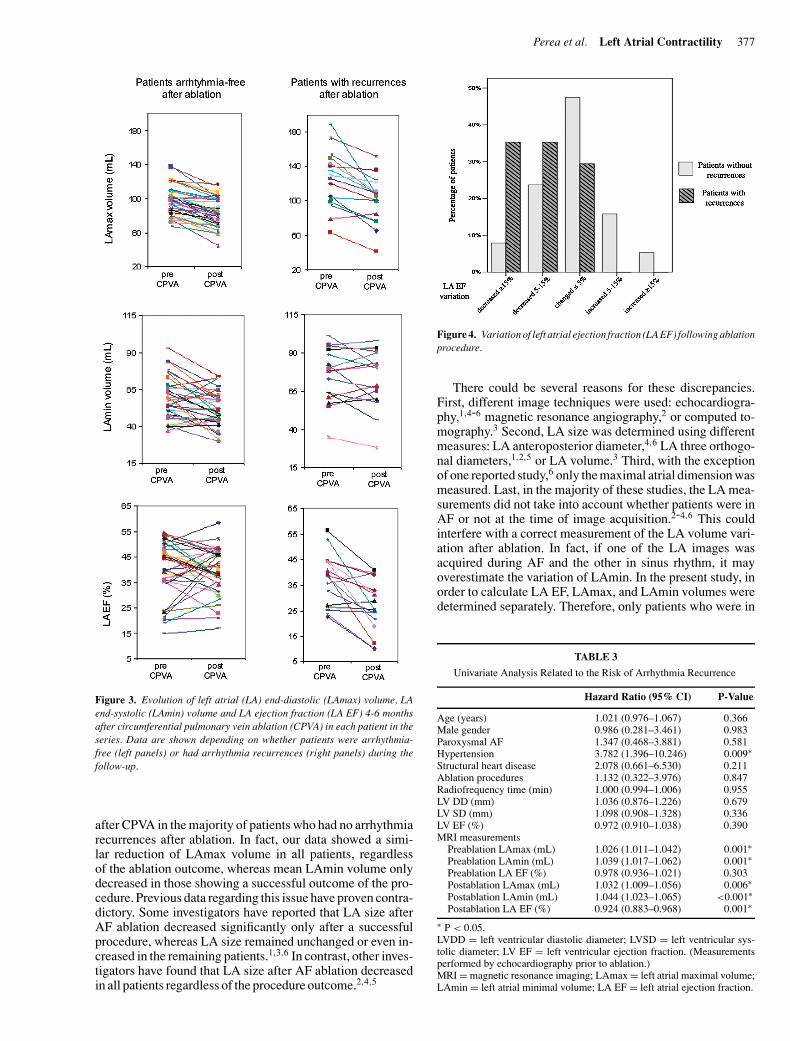
Table 2 shows the changes in LA measurements afterCPVA according to procedure outcome. Figure 3 shows thesame data for each patient in the series. Firstly, it can be ob-served that mean LAmax volume after CPVA decreased inboth group I and group II; furthermore, no differences in themean percentage of LAmax volume reduction were observedbetween the two groups (13±12% vs 17±14%, respectively,P = 0.217). Secondly, mean LAmin volume only decreasedsignificantly in group I. Consequently, there were no signif-icant changes in the mean LA EF after ablation in group I,whereas a decrease in the mean LA EF was seen in groupII. In fact, LA EF remained stable or increased in 68% ofpatients without arrhythmia recurrences after CPVA (Fig. 4).
On the other hand, the contractility of the LAA showed nochange after ablation. EF of the LAA before and after CPVAwas similar in both group I (41 ± 20% vs 40 ± 20%, P =0.8) and group II (31 ± 20% vs 32 ± 6%, P = 0.9). In groupI, there was a reduction in LAA diastolic volume (from 8.8 ±4.4 to 7.6 ± 4.4 mL, P < 0.001) and in LAA systolic volume(from 5.2 ± 4.2 to 4.3 ± 3.3 mL, P < 0.001) after ablation. Ingroup II, there was a trend to LAA diastolic volume reduction(from 7.1 ± 3.4 to 6.2 ± 3.3 mL, P = 0.08), and no significantchanges in LAA systolic volume (from 4.5 ± 2.1 to 4.1 ±2.2 mL, P = 0.11) after ablation.
Univariate analysis showed that patients with recurrencesafter the ablation procedure had a higher proportion of hy-pertension, larger LA volumes (measured before and af-ter CPVA) and lower postablation LA EF (Table 3). Theonly variable independently associated with arrhythmia re-currence in the multivariate model was the LAmin volumemeasured after the ablation procedure, with a hazard ratioof 1.04 (95% confidence interval, 1.02–1.06, P < 0.001).The best cut-off obtained by receiver operating characteristic(ROC) analysis for this variable (area under curve = 0.85)was LAmin volume ≥75 mL (sensitivity = 89%, specificity= 35%), which had a hazard ratio for arrhythmia recurrenceof 11.5 (95% confidence interval, 3.8–34.5, P < 0.001).
Discussion
The main finding of this study is that LA contractilityevaluated by means of LA EF is preserved or even increased
Perea et al. Left Atrial Contractility 377
Figure 3. Evolution of left atrial (LA) end-diastolic (LAmax) volume, LAend-systolic (LAmin) volume and LA ejection fraction (LA EF) 4-6 monthsafter circumferential pulmonary vein ablation (CPVA) in each patient in theseries. Data are shown depending on whether patients were arrhythmia-free (left panels) or had arrhythmia recurrences (right panels) during thefollow-up.
after CPVA in the majority of patients who had no arrhythmiarecurrences after ablation. In fact, our data showed a simi-lar reduction of LAmax volume in all patients, regardlessof the ablation outcome, whereas mean LAmin volume onlydecreased in those showing a successful outcome of the pro-cedure. Previous data regarding this issue have proven contra-dictory. Some investigators have reported that LA size afterAF ablation decreased significantly only after a successfulprocedure, whereas LA size remained unchanged or even in-creased in the remaining patients.1,3,6 In contrast, other inves-tigators have found that LA size after AF ablation decreasedin all patients regardless of the procedure outcome.2,4,5
Figure 4. Variation of left atrial ejection fraction (LA EF) following ablationprocedure.
There could be several reasons for these discrepancies.First, different image techniques were used: echocardiogra-phy,1,4-6 magnetic resonance angiography,2 or computed to-mography.3 Second, LA size was determined using differentmeasures: LA anteroposterior diameter,4,6 LA three orthogo-nal diameters,1,2,5 or LA volume.3 Third, with the exceptionof one reported study,6 only the maximal atrial dimension wasmeasured. Last, in the majority of these studies, the LA mea-surements did not take into account whether patients were inAF or not at the time of image acquisition.2-4,6 This couldinterfere with a correct measurement of the LA volume vari-ation after ablation. In fact, if one of the LA images wasacquired during AF and the other in sinus rhythm, it mayoverestimate the variation of LAmin. In the present study, inorder to calculate LA EF, LAmax, and LAmin volumes weredetermined separately. Therefore, only patients who were in
TABLE 3
Univariate Analysis Related to the Risk of Arrhythmia Recurrence
Hazard Ratio (95% CI) P-Value
Age (years) 1.021 (0.976–1.067) 0.366Male gender 0.986 (0.281–3.461) 0.983Paroxysmal AF 1.347 (0.468–3.881) 0.581Hypertension 3.782 (1.396–10.246) 0.009∗Structural heart disease 2.078 (0.661–6.530) 0.211Ablation procedures 1.132 (0.322–3.976) 0.847Radiofrequency time (min) 1.000 (0.994–1.006) 0.955LV DD (mm) 1.036 (0.876–1.226) 0.679LV SD (mm) 1.098 (0.908–1.328) 0.336LV EF (%) 0.972 (0.910–1.038) 0.390MRI measurements
Preablation LAmax (mL) 1.026 (1.011–1.042) 0.001∗Preablation LAmin (mL) 1.039 (1.017–1.062) 0.001∗Preablation LA EF (%) 0.978 (0.936–1.021) 0.303Postablation LAmax (mL) 1.032 (1.009–1.056) 0.006∗Postablation LAmin (mL) 1.044 (1.023–1.065) <0.001∗Postablation LA EF (%) 0.924 (0.883–0.968) 0.001∗
∗ P < 0.05.LVDD = left ventricular diastolic diameter; LVSD = left ventricular sys-tolic diameter; LV EF = left ventricular ejection fraction. (Measurementsperformed by echocardiography prior to ablation.)MRI = magnetic resonance imaging; LAmax = left atrial maximal volume;LAmin = left atrial minimal volume; LA EF = left atrial ejection fraction.

378 Journal of Cardiovascular Electrophysiology Vol. 19, No. 4, April 2008
sinus rhythm at both assessments were included to ensureoptimal measurements, and no electrical cardioversion wasperformed during the 2 weeks preceding the MRI in orderto avoid “stunning” of the atrial mechanical function.19 Fur-thermore, cardiac-gated cine MRI and the disc-summationtechnique (Simpson’s rule) were used to measure both LAvolumes to avoid geometric assumptions, image plane posi-tioning errors, and inappropriate sampling of the atrial bound-aries.20-22 It is conceivable that all these methodological lim-itations could explain the observed discrepancies in the liter-ature.
To our knowledge, two recent studies have analyzed LAEF after AF ablation, showing contradictory data.7,23 Vermaet al. reported a improvement in atrial function in a seriesof 67 patients evaluated by either echocardiography or cineelectron beam CT, whereas Lemola et al. suggested a de-terioration of atrial contractility in a series of 10 paroxys-mal AF patients evaluated by 3D CT. However, the authorscompared contractility regardless of the success of the pro-cedure. According to our data, LA EF after ablation gener-ally worsened in patients with recurrences. In fact, whereasLAmax volume reduction occurred in almost all patients,mean LAmin volume only decreased in patients without re-currences. Moreover, LAmin volume after CPVA was theonly independent variable related to procedure success in themultivariate analysis. Most patients with a successful abla-tion showed a decrease in both LAmax and LAmin volumesand preserved contractility. Whether the LAmin volume re-duction in these cases occurred immediately after ablationdue to tissue shrinking as a result of the radiofrequency de-livery,24 or if it was subsequent to a mid-term reverse remod-eling secondary to the maintenance of stable sinus rhythm,25
remains inconclusive in the present study. Another hypothe-sis that may be taken into account is that the PVs isolationmay lead to loss of their contractile capability, and this couldcause blood regurgitation from the LA through the PVs dur-ing atrial systole. This fact might result in a decrease in atrialsystolic volume and an improvement in LA ejection fraction.More data are required to elucidate whether the LAmin de-crease was a cause or a consequence of the outcome of theprocedure.
Finally, LAA contractility was not compromised afterCPVA in any patient of this series. Changes in LAA vol-umes were in accordance with those observed in the wholeLA, although to a lesser extent. This could be due to the factthat RF energy is deployed out of the LAA and the effect oftissue shrinkage may be lower. However, there are method-ological limitations in the measurement of volumes in sucha small structure. As a consequence, a broader populationwould be needed to study these subtle differences on detail.
Study Limitations
The impossibility of accurately measuring the AF burdenat each time point25 could partly affect the evaluation of LAcontractility. This is a limitation in this type of study, sinceasymptomatic AF episodes could be misinterpreted unlesscontinuous monitoring of the patient was performed.
In the present study, certain details of the imaging tech-nique should be noted. First, image acquisition was car-ried out at end-exhalation. Imaging during inspiration mayhave produced different results related to changes in dia-stolic relaxation properties and preload conditions. Second,
end-systole and end-diastole were defined using direct visual-ization of mitral valve opening and closure, since electrocar-diogram co-registration is not available for MRI image postprocessing. Finally, PV ostium was defined at the point ofinflection between PV and LA wall. This simplification wasused to facilitate identification and promote interobserver re-producibility. However, these limitations are unlikely to havehad a significant effect on the results of the study since bothbasal and follow-up measurements were performed in thesame manner, and LA variations between subjects may re-flect real changes.
On the other hand, the effect of radiofrequency may bemore significant in those patients who underwent two ab-lations, since changes in LA contractility were evaluated inthe MRI obtained after the second procedure. However, re-sults in these 10 patients were consistent with those observedin the remaining cases. In fact, LA EF showed no significantchanges with respect to the basal measurement in arrhythmia-free patients after the second ablation (from 35 ± 11% to 32± 7%, P = 0.50), whereas LA EF decreased considerablyin the remaining patients suffering arrhythmia recurrences inspite of the two procedures (from 28 ± 9% to 10 ± 2%, P= 0.06). Furthermore, in seven of the 10 second procedures,the amount of radiofrequency delivery was low because onlynew-onset LA flutter was ablated and no extensive lesionswere created.
Finally, it should be considered that the results of this studymay vary depending on the ablation approach. However, theeffect on LA contractility is unlikely to be greater in otherablation procedures, taking into account the extensive lesionscreated in CPVA.
Conclusions
LAmax volume reduction after CPVA occurs regardless ofthe clinical efficacy of the procedure, whereas LAmin volumedecreased only in patients free of arrhythmia recurrences. LAEF was preserved or even increased in the majority of patientsafter successful CPVA.
References
1. Beukema WP, Elvan A, Sie HT, Misier AR, Wellens HJ: Successful ra-diofrequency ablation in patients with previous atrial fibrillation resultsin a significant decrease in left atrial size. Circulation 2005;112:2089-2095.
2. Jayam VK, Dong J, Vasamreddy CR, Lickfett L, Kato R, Dickfeld T,Eldadah Z, Dalal D, Blumke DA, Berger R, Halperin HR, Calkins H:Atrial volume reduction following catheter ablation of atrial fibrilla-tion and relation to reduction in pulmonary vein size: An evaluationusing magnetic resonance angiography. J Interv Card Electrophysiol2005;13:107-114.
3. Lemola K, Sneider M, Desjardins B, Case I, Chugh A, Hall B, Cheung P,Good E, Han J, Tamirisa K, Bogun F, Pelosi F Jr, Kazerooni E, MoradyF, Oral H: Effects of left atrial ablation of atrial fibrillation on size ofthe left atrium and pulmonary veins. Heart Rhythm 2004;1:576-581.
4. Pappone C, Oreto G, Rosanio S, Vicedomini G, Tocchi M, GugliottaF, Salvati A, Dicandia C, Calabro MP, Mazzone P, Ficarra E, DiGioia C, Gulletta S, Nardi S, Santinelli V, Benussi S, Alfieri O: Atrialelectroanatomic remodeling after circumferential radiofrequency pul-monary vein ablation: Efficacy of an anatomic approach in a large cohortof patients with atrial fibrillation. Circulation 2001;104:2539-2544.
5. Reant P, Lafitte S, Jais P, Serri K, Weerasooriya R, Hocini M, PilloisX, Clementy J, Haissaguerre M, Roudaut R: Reverse remodeling ofthe left cardiac chambers after catheter ablation after 1 year in a seriesof patients with isolated atrial fibrillation. Circulation 2005;112:2896-2903.

Perea et al. Left Atrial Contractility 379
6. Tops LF, Bax JJ, Zeppenfeld K, Jongbloed MR, Van Der Wall EE,Schalij MJ: Effect of radiofrequency catheter ablation for atrial fibril-lation on left atrial cavity size. Am J Cardiol 2006;97:1220-1222.
7. Lemola K, Desjardins B, Sneider M, Case I, Chugh A, Good E, Han J,Tamirisa K, Tsemo A, Reich S, Tschopp D, Igic P, Elmouchi D, BogunF, Pelosi F, Kazerooni E, Morady F, Oral H: Effect of left atrial circum-ferential ablation for atrial fibrilation on left atrial transport function.Heart Rhythm 2005;2:923-928.
8. Yamanaka K, Fujita M, Doi K, Tsuneyoshi H, Yamazato A, Ueno K,Zen E, Komeda M: Multislice computed tomography accurately quan-tifies left atrial size and function after the MAZE procedure. Circulation2006;114:I5-I9.
9. Gentlesk PJ, Sauer WH, Gerstenfeld EP, Lin D, Dixit S, Zado E, CallansPCD, Marchlinski FE: Reversal of left ventricular dysfunction followingablation of atrial fibrillation. J Cardiovasc Electrophysiol 2007;18:9-14.
10. Thomas L, Boyd A, Thomas SP, Schiller NB, Ross DL: Atrial structuralremodelling and restoration of atrial contraction after linear ablation foratrial fibrillation. Eur Heart J 2003;24:1942-1951.
11. Kato R, Lickfett L, Meininger G, Dickfeld T, Wu R, Juang G, AngkeowP, LaCorte J, Bluemke D, Berger R, Halperin HR, Calkins H: Pulmonaryvein anatomy in patients undergoing catheter ablation of atrial fibrilla-tion: Lessons learned by use of magnetic resonance imaging. Circulation2003;107:2004-2010.
12. Mogelvang J, Lindvig K, Sondergaard L, Saunamaki K, Henriksen O:Reproducibility of cardiac volume measurements including left ventric-ular mass determined by MRI. Clin Physiol 1993;13:587-597.
13. Pattynama PM, Lamb HJ, Van Der Velde EA, Van Der Wall EE, deRoos A: Left ventricular measurements with cine and spin-echo MRimaging: A study of reproducibility with variance component analysis.Radiology 1993;187:261-268.
14. Shapiro EP, Rogers WJ, Beyar R, Soulen RL, Zerhouni EA, LimaJA, Weiss JL: Determination of left ventricular mass by magnetic res-onance imaging in hearts deformed by acute infarction. Circulation1989;79:706-711.
15. Mohiaddin RH, Hasegawa M: Measurement of atrial volumes bymagnetic-resonance-imaging in healthy-volunteers and in patients withmyocardial-infarction. Eur Heart J 1995;16:106-111.
16. Pappone C, Oreto G, Lamberti F, Vicedomini G, Loricchio ML, ShpunS, Rillo M, Calabro MP, Conversano A, Ben Haim SA, Cappato R,
Chierchia S: Catheter ablation of paroxysmal atrial fibrillation using a3D mapping system. Circulation 1999;100:1203-1208.
17. Pappone C, Rosanio S, Augello G, Gallus G, Vicedomini G, MazzoneP, Gulletta S, Gugliotta F, Pappone A, Santinelli V, Tortoriello V, SalaS, Zangrillo A, Crescenzi G, Benussi S, Alfieri O: Mortality, morbidity,and quality of life after circumferential pulmonary vein ablation foratrial fibrillation: Outcomes from a controlled nonrandomized long-term study. J Am Coll Cardiol 2003;42:185-197.
18. Berruezo A, Tamborero D, Vidal B, Sitges M, Matiello M, Molina I,Mont LL, Brugada J: Preprocedural predictors of recurrences after elec-troanatomical encircling of pulmonary veins. Eur Heart J 2006;27:869-870.
19. Thomas MD, Kalra PR, Jones A, Struthers AD, More RS: Time coursefor recovery of atrial mechanical and endocrine function post DC car-dioversion for persistent atrial fibrillation. Int J Cardiol 2005;102:487-491.
20. Jarvinen V, Kupari M, Hekali P, Poutanen VP: Assessment of left atrialvolumes and phasic function using cine magnetic resonance imaging innormal subjects. Am J Cardiol 1994;73:1135-1138.
21. Jarvinen VM, Kupari MM, Hekali PE, Poutanen VP: Right atrial MRimaging studies of cadaveric atrial casts and comparison with rightand left atrial volumes and function in healthy subjects. Radiology1994;191:137-142.
22. Keller AM, Gopal AS, King DL: Left and right atrial volume by freehandthree-dimensional echocardiography: In vivo validation using magneticresonance imaging. Eur J Echocardiogr 2000;1:55-65.
23. Verma A, Kilicaslan F, Adams JR, Hao S, Beheiry S, Minor S, Ozdu-ran V, Elayi SC, Martin DO, Schweikert RA, Saliba W, Thomas JD,Garcia M, Klein A, Natale A: Extensive ablation during pulmonaryvein antrum isolation has no adverse impact on left atrial function: Anechocardiography and cine computed tomography analysis. J Cardio-vasc Electrophysiol 2006;17:741-746.
24. Victal OA, Teerlink JR, Gaxiola E, Wallace AW, Najar S, Camacho DH,Gutierrez A, Herrera G, Zuniga G, Mercado-Rios F, Ratcliffe MB: Leftventricular volume reduction by radiofrequency heating of chronic my-ocardial infarction in patients with congestive heart failure. Circulation2002;105:1317-1322.
25. Zipes DP: Atrial fibrillation. A tachycardia-induced atrial cardiomy-opathy. Circulation 1997;95:562-564.

112

PRESENTACIÓN DE LOS TRABAJOS 113
ESTUDIO 2
Incidence of pulmonary vein stenosis in patients submitted to atrial
fibrillation ablation: a comparison of the selective segmental ostial
ablation vs the circumferential pulmonary veins ablation.
Tamborero D, Mont L, Nava S, Caralt TM, Molina I, Scalise A, Perea RJ,
Bartholomay E, Berruezo A, Matiello M, Brugada J.
J Interv Card Electrophysiol. 2005;14:21-25.
3.2.1 OBJETIVOS
Objetivo general
El objetivo general de este estudio es demostrar la capacidad de la RM
para el estudio anatómico sistemático de las VPs y de la AI en pacientes
tributarios de APRF de las VPs.
Objetivo específico
Identificar la aparición de estenosis de las VPs como complicación del
procedimiento en el estudio post-ablación y determinar su relación con la
técnica ablativa empleada.

114
3.2.2 RESULTADOS
Se realizó ARM en 73 de 78 pacientes consecutivos sometidos a
APRF de la FA. La ARM no se pudo realizar en 3 pacientes con
claustrofobia, en un paciente con marcapasos y en otro con desfibrilador
automático implantable. Las características demográficas de los pacientes
de encuentran resumidas en la Tabla 6. La ablación se realizó mediante el
procedimiento de ASOS en 32 pacientes; en 41 pacientes la ablación se
realizó mediante el método de ACVP. Durante un período medio de
seguimiento de 14.7 ± 12.2 meses, 23 y 31 pacientes de los grupos de
ASOS y de ACVP se mantuvieron libres de recurrencias (72% vs 76%
libres de arritmias, long rank test p = NS).

PRESENTACIÓN DE LOS TRABAJOS 115
Tabla 6. Características demográficas
Grupo ASOS
Grupo ACVP
P
Pacientes
32
41
FA paroxística
27 (85%)
27 (66%)
0.09
Duración de la FA (meses)
62 ± 71
72 ± 80
NS
Número de FAA fallidos
2.5 ± 0.9
2.6 ± 0.7
NS
Edad (años)
50 ± 12
52 ± 10
NS
Sexo masculino
23 (75%)
33 (80%)
NS
Aurícula izquierda (mm)
37 ± 4
42 ± 5
0.02
FEVI (%)
58 ± 11
53 ±17
NS
Cardiopatía estructural
8 (25%)
11 (27%)
NS
Hipertensión arterial
9 (28%)
12 (29%)
NS
ASOS: Ablación segmentaria ostial selectiva. ACVP: Ablación circunferencial de las
venas pulmonares. FAA: Fármacos antiarrítmicos. FEVI: Fracción de eyección ventricular
izquierda.
Ninguno de los pacientes desarrolló síntomas sugestivos de
estenosis de las VPs. Sin embargo, en 6 pacientes se detectó estenosis
significativa de las VPs en una ARM de rutina, todas ellas en el grupo de
ASOS, ninguna en el grupo de ACVP (18.8% vs 0% de los pacientes

116
evaluados; p = 0.005). En 4 pacientes, la estenosis se localizó en la VPSI,
en 1 paciente en la VPII y en 1 paciente tanto la VPSI como la VPII tenían
estenosis significativa.
En el grupo de ACVP, las líneas de ablación rodeaban todas las VPs
en todos los casos. En el grupo de ASOS se trató una media de 1.8 ± 0.7
VPs por paciente con una media de 10.3 ± 0.8 minutos de aplicación de
radiofrecuencia. Los segmentos ostiales donde se aplicó la
radiofrecuencia para conseguir la desconexión eléctrica de la VP fueron
clasificados, mediante una guía anatómica y fluoroscópica, en 4 divisiones
ostiales: segmento anterior, posterior, superior e inferior. En nuestra serie,
la actividad eléctrica más precoz se registró en el segmento inferior en el
57 y 58% de las VPSIs y VPSDs tratadas, respectivamente (Tabla 7). La
VPSI se aisló en el 93.8% de pacientes, la VPII en el 18.8%, la VPSD en
el 59.4% y la VPID en el 6.3%. Fue necesario un segundo procedimiento
en 3 pacientes del grupo de ASOS debido a recurrencia de la FA, por
tanto el número total de VPs aisladas fue de 33 VPSIs, 6 VPIIs, 21 VPSDs
y 2 VPIDs. En conjunto, un 15.2% de las VPSIs y un 33.3% de las VPIIs
tratadas con ASOS desarrollaron estenosis (Figura 18).

Tabla 7. Segmentos del
radiofrecuencia en el grupo de ASOS
Inferior
(%)
VPSI (n = 30)
56.6
VPSD (n = 19)
57.9
VPII (n = 6)
16.7
VPID (n =2)
0
VPs: venas pulmonares. ASOS: ablación segmentaria ostial selectiva. VPSI: vena
pulmonar superior izquierda. VPSD: vena pulmonar superior derecha. VPII: vena
pulmonar inferior izquierda. VPID: vena pulmonar inferior derecha.
Figura 18. Angio-resonancia magnética. Reconstrucción tridimensional de la aurícula
izquierda y de las venas pulmonaresestenosis post-ablación de la vena pulmonar superior izquierda (flecha); y b) proyección oblicua superoinferior (flecha).
PRESENTACIÓN DE LOS TRABAJOS
Segmentos del ostium de las VPs en que se aplicó
radiofrecuencia en el grupo de ASOS
Inferior
(%)
Posterior Superior Anterior
(%) (%) (%)
56.6
26.6
23.3
26.6
57.9
26.3
21.0
21.0
16.7
33.3
50.0
16.7
0
0
100
100
VPs: venas pulmonares. ASOS: ablación segmentaria ostial selectiva. VPSI: vena
pulmonar superior izquierda. VPSD: vena pulmonar superior derecha. VPII: vena
izquierda. VPID: vena pulmonar inferior derecha.
resonancia magnética. Reconstrucción tridimensional de la aurícula
izquierda y de las venas pulmonares; a) proyección oblicua anteroposterior que muestra ablación de la vena pulmonar superior izquierda (flecha); y b) proyección
oblicua superoinferior que muestra estenosis de la vena pulmonar inferior izquie
a
PRESENTACIÓN DE LOS TRABAJOS 117
de las VPs en que se aplicó la energía de
VPs: venas pulmonares. ASOS: ablación segmentaria ostial selectiva. VPSI: vena
pulmonar superior izquierda. VPSD: vena pulmonar superior derecha. VPII: vena
resonancia magnética. Reconstrucción tridimensional de la aurícula
yección oblicua anteroposterior que muestra ablación de la vena pulmonar superior izquierda (flecha); y b) proyección
estenosis de la vena pulmonar inferior izquierda
b

118
No se encontraron diferencias en las características de los pacientes
o en los detalles del procedimiento entre los pacientes con y sin estenosis
en el grupo de ASOS, y no se pudieron identificar predictores de
estenosis.
En nuestra serie, 16 pacientes (22% del total) tuvo alguna variante
anatómica de las VPs, siendo la más frecuente la presencia de un tronco
común izquierdo (15% de los pacientes) y la presencia de una VPMD (5%
de los pacientes) (Figura 19
Figura 19. Angio-resonancia magnética. Rec
muestran variantes anatómicas de las venas pulmonares; a) Vena pulmonar media derecha (flecha); b) tronco común izquierdo (flecha).
No se encontraron diferencias en las características de los pacientes
o en los detalles del procedimiento entre los pacientes con y sin estenosis
en el grupo de ASOS, y no se pudieron identificar predictores de
16 pacientes (22% del total) tuvo alguna variante
anatómica de las VPs, siendo la más frecuente la presencia de un tronco
común izquierdo (15% de los pacientes) y la presencia de una VPMD (5%
ura 19).
resonancia magnética. Reconstrucciones multiplanares que
muestran variantes anatómicas de las venas pulmonares; a) Vena pulmonar media derecha (flecha); b) tronco común izquierdo (flecha).
a
No se encontraron diferencias en las características de los pacientes
o en los detalles del procedimiento entre los pacientes con y sin estenosis
en el grupo de ASOS, y no se pudieron identificar predictores de
16 pacientes (22% del total) tuvo alguna variante
anatómica de las VPs, siendo la más frecuente la presencia de un tronco
común izquierdo (15% de los pacientes) y la presencia de una VPMD (5%
onstrucciones multiplanares que
muestran variantes anatómicas de las venas pulmonares; a) Vena pulmonar media
b

Journal of Interventional Cardiac Electrophysiology 14, 21–25, 2005C© 2005 Springer Science + Business Media, Inc. Manufactured in The Netherlands.
Incidence of Pulmonary Vein Stenosis in PatientsSubmitted to Atrial Fibrillation Ablation: A Comparisonof the Selective Segmental Ostial Ablation vsthe Circumferential Pulmonary Veins Ablation
David Tamborero, Lluis Mont, Santiago Nava,Teresa M. de Caralt, Irma Molina, Andrea Scalise,Rosario J. Perea, Eduardo Bartholomay,Antonio Berruezo, Maria Matiello,and Josep BrugadaCardiovascular Institute, Institut d’Investigacions BiomediquesAugust Pi i Sunyer (IDIBAPS), Hospital Clınic, Universityof Barcelona
Abstract. Introduction: Pulmonary vein (PV) stenosisis an important complication of the AF ablation andcould be underestimated if their assessment is not sys-tematically done. Selective Segmental Ostial Ablation(SSOA) and Circunferential Pulmonary Veins Ablation(CPVA) have demonstrated efficacy in atrial fibrillation(AF) treatment. In this study the real incidence of PVstenosis in patients (pts) submitted to both SSOA andCPVA was compared.
Methods: Those pts with focal activity and normal leftatrial size were submitted to SSOA, remaining pts weresubmitted to CPVA to treat refractory, symptomatic AF.Contrast enhanced magnetic resonance angiography(MRA) was routinely performed in all patients 4 monthsafter the procedure.
Results: A series of 73 consecutive patients (mean ageof 51 ± 11 years; 75% male) were included. SSOA was per-formed in 32 patients, and the remaining 41 patients un-derwent to CPVA, obtaining similar efficacy rates (72%vs 76% arrythmia free probability at 12 months; log ranktest p = NS). Six patients had a significant PV steno-sis, all in SSOA group none in CPVA group (18.8% vs0%; p = 0.005). All patients were asymptomatic and thestenosis was detected in routine MRA. No predictorsof stenosis has been identified analysing patient pro-cedure characteristics.
Conclusion: PV stenosis is a potential complication ofSSOA not seen in CPVA. The study confirms than MRAis useful for identifying patients with asymptomatic PVstenosis.
Key Words. atrial fibrillation, pulmonary veins steno-sis, catheter ablation
Introduction
Pulmonary vein (PV) radiofrequency (RF) ablationis a curative procedure for patients with atrial fib-rillation (AF). Several strategies have been devel-
oped to achieve the PV isolation with good clini-cal results [1–3]. PV stenosis has been recognizedas one potential complication of the ablation pro-cedure that can be associated with severe respi-ratory symptoms that cause significant morbidity[4–7]. Its incidence is unclear, mainly in asymp-tomatic patients. Several methods have been eval-uated for the proper detection of this complication[8] and Magnetic Resonance Angiography (MRA)has become a useful method for the diagnosis[9,10].
The aim of this study was to analyse the inci-dence of PV stenosis in patients treated with se-lective segmental ostial ablation (SSOA) or withcircumferential pulmonary veins ablation (CPVA)methods.
Methods
PatientsA series of 73 consecutive patients underwentto AF ablation for treatment of drug-refractory,symptomatic AF. All patients were previouslystudied with 24-hours Holter monitoring and
Santiago Nava, A. Berruezo and A. Scalise were supportedin part by a Grant from the Fundacio Clınic per la RecercaBiomedica.
Address for correspondence: Lluıs Mont, MD, PhD,Cardiovascular Institute, Hospital Clınic Universitari deBarcelona, Villarroel 170, Barcelona 08036, Catalonia, Spain.E-mail: [email protected]
Received 25 July 2005; accepted 22 August 2005
21

22 Tamborero et al.
Table 1. Demographic characteristics
SSOA group CPVA group P
Patients 32 41Paroxysmal AF 27 (85%) 27 (66%) 0.09Duration of AF (months) 62 ± 71 72 ± 80 NSNumber of failed AAD 2.5 ± 0.9 2.6 ± 0.7 NSAge (years) 50 ± 12 52 ± 10 NSMale Sex 23 (75%) 33 (80%) NSLeft Atrium (mm) 37 ± 4 42 ± 5 0.02LVEF (%) 58 ± 11 53 ± 17 NSStructural Heart Disease 8 (25%) 11 (27%) NSArterial Hypertension 9 (28%) 12 (29%) NS
SSOA: Selective Segmental ostial ablation. CPVA: Circumferential pul-monary veins ablation. AAD: Antiarrhythmic drugs. LVEF: Left ven-tricular ejection fraction.
transthoracic echocardiography. Informed consentwas obtained in all patients before the procedure.Patients with suspected focal origin AF (identi-fied by structurally normal left atria and >10runs of atrial tachycardia/24 hours) were submit-ted to SSOA in order to isolate pulmonary veins(PV) from left atria. The remaining patients un-derwent to CPVA to modify atrial substrate withextended lesions. Demographic characteristics arepresented in Table 1.
Ablation ProcedureSelective Segmental Ostial Ablation. Af-ter transseptal access, PV disconnection was per-formed as described by Haissaguerre et al. [1].A decapolar Lasso catheter (Biosense-Webster)was used to map the PV potentials and RF en-ergy was applied by a thermocouple-equipped 4mm tip catheter (Biosense-Webster). Ostial le-sions were created where the PV earliest activitywas recorded until PV potentials were eliminatedor dissociated at a target temperature of 50◦C anda maximum output from 40 to 50 W. Only thosePVs with electrical activity were treated. No at-tempts to induce premature beats were done. De-tails of the approach used have been previouslyreported [13].
Circumferential Pulmonary Veins Ab-lation. Non-fluoroscopic navigation system(CARTO; Biosense Webster) was used to delin-eate the left atria and PVs and guide the RFlesions after transseptal access. Ablation lineswere created as described by Pappone et al.[11,12] surrounding ipsilateral PVs at a mini-mum distance of 5 mm from their ostium by athermocouple-equipped 8 mm tip catheter (Nav-istar, Biosense-Webster) at a target temperatureof 55◦C and a maximum output from 50 to 60 W.The end point was to reduce the amplitude of theendocardial potentials inside the encircled areabelow 0.15 mV.
Follow-upPatients were followed in the outpatient clinic at1, 4, 7 months and every 6 months thereafter ifthey remained asymptomatic. Routine 24-hoursHolter monitoring was performed before each con-trol and patients were also asked to communi-cate any symptom suggestive of recurrence be-tween scheduled visits in order to document it. Atransthoracic echocardiogram was also performed4 months after ablation procedure. Acenocumarolwas maintained at least for 3 months after the ab-lation. All patients received antiarrhythmic drugs(flecainide if no structural heart disease was diag-nosed or amiodarone if there was evidence of struc-tural heart disease) during the first month to con-trol early recurrences. Drugs were withdrawn af-terwards if patients remained free of recurrences.
A contrast enhanced MRA (1.5 T. Signa Hori-zon. GE Medical Systems) was routinely per-formed in all the patients submitted to an ablationprocedure. A single radiologist blinded to the typeand result of the procedure and clinical charac-teristics of patients evaluated the test. MRA wasobtained 4 months after the ablation since pro-gression of the stenosis is rare after this period oftime [7,14]. A significant stenosis was consideredwith a diameter lumen reduction of >70%.
Statistical AnalysisContinuous variables are expressed as mean ±SD. Comparisons were made using the Student’sT-test and Chi-square analysis. Recurrence-freewas compared using the Kaplan-Meier survivalcurves with log rank test. Multivariate logisticregression analysis was performed to determineindependent predictors of an event. Results withp < 0.05 were considered statistically significant.
Results
MRA was performed in 73 of 78 consecutive pa-tients submitted to an AF ablation procedure. TheMRA could not be done in 3 patients with claustro-phobia, in a patient with a pacemaker and in an-other with an implantable automatic defibrillator.In 32 patients, the ablation was done with a SSOAapproach; in 41 patients the ablation was donewith the CPVA method. During a mean follow-upof 14.7 ± 12.1 months, 23 and 31 patients of SSOAand CPVA groups respectively were free from re-currences (72% vs 76% arrythmia free; long ranktest p = NS).
None of the patients developed symptoms sug-gestive of PV stenosis. However, in 6 patients asignificant PV stenosis was detected in the rou-tine MRA, all them in the SSOA group, none inthe CPVA group (18.8% vs 0% of the evaluated pa-tients; p = 0.005). In 4 patients, the stenosis was

Incidence of Pulmonary Vein Stenosis in Patients 23
Fig. 1. Two patients with pulmonary vein (PV) stenosis. Left and right images show left superior and left inferior PV stenosisrespectively (arrows).
located in the left superior PV (LSPV), in 1 patientat the left inferior PV (LIPV) and in 1 patient boththe LSPV and the LIPV had a significant stenosis.
In the CPVA group, ablation lines encircled allPV’s in all cases. In SSOA group a mean of 1.8 ± 0.7PVs per patient were treated with a mean of10.3 ± 0.8 minutes of radiofrequency application.Ostial segments where RF was applied to achievethe PV electrical disconnection were classified byanatomic and fluoroscopic guidance among 4 os-tial divisions: anterior, posterior, superior and in-ferior segments. In our series, the earliest electri-cal activity was recorded on the inferior segmentin the 57 and 58% of the treated LSPV and RSPVrespectively (see Table 2). LSPV was isolated inthe 93.8% of patients, LIPV in 18.8%, right su-perior PV (RSPV) in 59.4% and right inferior PV(RIPV) in 6.3%. A second procedure was neces-sary in 3 patients of the SSOA group due to AFrecurrence, therefore the total number of PVs iso-lated were 33 LSPVs, 6 LIPVs, 21 RSPVs and 2RIPVs. Overall, 15.2% of the LSPVs and 33.3%of the LIPVs treated by SSOA developed stenosis.
Table 2. PV ostial segments in which RF energy wasdeployed in SSOA group
Inferior Posterior Superior Anterior(%) (%) (%) (%)
LSPV (n = 30) 56.6 26.6 23.3 26.6RSPV (n = 19) 57.9 26.3 21.0 21.0LIPV (n = 6) 16.7 33.3 50.0 16.7RIPV (n = 2) 0 0 100 100
PV: pulmonary vein. RF: radiofrequency. SSOA: selective segmental os-tial ablation. LSPV: left superior PV. RSPV: right superior PV. LIPV:left inferior PV. RIPV: right inferior PV.
Fig. 2. Example of anatomical variant of the pulmonaryveins where a single ostium of both left and right sided wasobserved.
No differences in patient characteristics or proce-dural details were found between patients withand without PV stenosis in SSOA group, and nopredictors of stenosis could be identified.
In our series 16 patients (22% of the total) hadsome anatomical variant of the PVs, being themost common variant the presence of single os-tium of the left PVs (15% of the patients) and thepresence of 3 right PVs (5% of the patients; seeFig. 2).
Discussion
PV stenosis is a potential complication of AF abla-tion. Correlation between the technique employed

24 Tamborero et al.
for the ablation and the incidence of PV steno-sis has been studied by Saad et al. [7]. The au-thors suggest that the most important point is theaccuracy of the technique for differentiating thereal ostium of the PV. Therefore, different meth-ods like venography, intracardiac ultrasonographyand electroanatomic mapping are used to definemore accurately the junction between the atrialwall and the PV [5–8]. Even with these techniques,in large series, stenosis persists as a complica-tion ranging between 5 to 30% [14]. Some authorsdo not look systematically for PV stenosis in allpatients and only if the patient becomes symp-tomatic, a diagnostic procedure is performed [15];our results show that this method may underesti-mate the real incidence of this complication.
In our series, AF ablation was performed usingtwo different approaches and the incidence of PVstenosis was compared. The absence of stenosisin the CPVA group correlates with the findings ofthe series published by Pappone et al. [3,12,13,16],and although there are reports of PV occlusion us-ing this technique [17], the risk seems lower be-cause of a better definition of the catheter posi-tion in respect of the PV ostium. Furthermore, inCPVA procedures RF is delivered at least 5 mmaway from the defined ostium.
In our experience, all PV stenosis were foundin the SSOA group and this is in part, due to themore difficult differentiation of the PV ostium andbecause of the need to apply energy close to theostium, making it easier to produce a lesion insidethe vein. Moreover, although the total amount ofRF energy was higher in CPVA than in SSOA, amajor concentration of RF lesions was required inSSOA technique in order to achieve the PV iso-lation, delivering the RF energy in a more lim-ited region of the PV ostium. It is of interest thatin the SSOA group the left sided PVs were morestenosed, and this correlates with previous reportsin the literature [14]. A possible explanation maybe that the ablation catheter moves easily insidethe left sided veins with each breathing move-ment, thus delivering the energy inside the PV. Inour approach, SSOA was performed treating onlythose PVs showing electrical activity, being themost commonly treated the LSPV and the RSPV.These results were in accordance with other stud-ies where only arrythmogenic PVs were treated ancould be explained because the muscular sleevesinsertion into PVs was more developed in upperthan in lower veins [1,2].
Recently, a study comparing the efficacy andsafety of both ablation strategies in a series of100 randomized patients has been published [18].Multislice CT was routinely performed 3 monthsafter the ablation procedure, and stenosis of atleast 1 PV was found in 12 and 6% of the SSOA and
CPVA patients respectively. This PV stenosis inci-dence is similar to our results in the SSOA group,however we had not observe any stenosis in theCPVA group. This discrepancy maybe due to thehigher power limit output used in this study, upto 70 W in 8 mm tip or 50 W in cooled 4 mm tipcatheter respectively.
All patients with PV stenosis in our series wereasymptomatic, and were identified because MRAwas sistematically performed. The development ofclinical symptoms is associated with the numberof PVs affected and the degree of narrowing [14].Lung perfusion defects were seen in PV narrow-ing greater than 70% in the left PV [7]. Whenright PV are affected lung scan perfusion defectsappear with somewhat lower degrees of stenosisranging between 50 to 65%; this might be ex-plained because of the lower pressure in the rightPV that may increase the pressure gradient [19].In our series there were no total occlusions, all butone patient had only one vein affected, and rightsided veins were not involved, contributing to theabsence of respiratory symptoms associated withfound PV stenosis.
In this series, a single MRA was performed 4months after the ablation procedure, so later PVstenosis progression could have been missed. How-ever, although PV narrowing have not been ex-actly quantified because a previous MRA was notperformed, all PV stenosis images we obtainedshowed a focalised and high degree narrowing thatmust be catalogued as severe (>70%) in all cases.In the remaining patients, PVs ostium did notshow appreciable alterations, and normal or mildnarrowed (<50%) PVs rarely progress beyond thethird month [7,14]. Therefore, we decided not re-peat MRA beyond the 4th month since the ablationprocedure because of the low probability to observenew findings.
Conclusions
Pulmonary vein stenosis is a potential complica-tion of the selective segmental ostial ablation ofatrial fibrillation. The PV stenosis is seldom ob-served in circumferential pulmonary veins abla-tion approach. The study confirms that magneticresonance angiography is useful for identifying pa-tients with asymptomatic pulmonary vein stenosisand anatomical variants of the left atria.
References
1. Haissaguerre M, Jais P, Shah DC, Garrigue S, TakahashiA, Lavergne T, Hocini M, Peng JT, Roudaut R, ClementyJ. Electrophysiological end point for catheter ablation ofatrial fibrillation initiatied from multiple pulmonary ve-nous foci. Circulation 2000;101:1409–1417.

Incidence of Pulmonary Vein Stenosis in Patients 25
2. Marrouche NF, Dresing T, Cole C, Bash D, Saad E, BalabanK, Pavia SV, Schweikert R, Saliba W, Abdul-Karim A,Pisano E, Fanelli R, Tchou P, Natale A. Circular mappingand ablation of the pulmonary vein for treatment of atrialfibrillation: Impact of different catheter technologies. J AmColl Cardiol 2002;40:464–474.
3. Pappone C, Rosanio S, Oreto G, Tocchi M, Gugliotta F,Vicedomini G, Salvati A, Dicandia C, Mazzone P, SantinelliV, Gulletta S, Chierchia S. Circumferential radiofrequencyablation of pulmonary vein ostia: A new anatomic approachfor curing atrial fibrillation. Circulation 2000;102:2619–2628.
4. Taylor GW, Kay GN, Zheng X, Bishop S, Ideker R et al.Pathological effects of extensive radiofrequency energy ap-plications in the pulmonary veins in dogs. Circulation2000;101:1736–1742.
5. Yu W, Hsu T, Tai C, Tsai CF, Hsieh MH, Lin WS, LinYK, Tsao HM, Ding YA, Chang MS, Chen SA. Acquiredpulmonary vein stenosis after radiofrequency ablation ofparoxysmal atrial fibrillation. J Cardiovasc Electrophysiol2001;12:887–892.
6. Arentz T, Jander N, von Rosenthal J, Blum T, FurmaierR, Gornandt L, Josef Neumann F, Kalusche D. Incidenceof pulmonary vein stenosis 2 years after radiofrequencycatheter ablation of refractory atrial fibrillation. Eur HeartJ 2003;24:963–969.
7. Saad E, Rossillo A, Saad C, Martin DO, Bhargava M,Erciyes D, Bash D, Williams-Andrews M, Beheiry S,Marrouche NF, Adams J, Pisano E, Fanelli R, PotenzaD, Raviele A, Bonso A, Themistoclakis S, Brachmann J,Saliba WI, Schweikert RA, Natale A. Pulmonary veinstenosis alter radiofrequency ablation of atrial fibrilla-tion. Functional characterization, evolution and influ-ence of the ablation strategy. Circulation 2003;108:3102–3107.
8. Scharf C, Sneider M, Case I, Chugh A, Lai SW, Pelosi F Jr,Knight BP, Kazerooni E, Morady F, Oral H. Anatomy of thepulmonary veins in patients with atrial fibrillation and ef-fects of segmental ostial ablation analyzed by computedtomography. J Cardiovasc Electrophysiol 2003;14:150–155.
9. Yang M, Akbari H, Reddy GP, Higgins C et al. Identificationof pulmonary vein stenosis after radiofrequency ablationfor atrial fibrillation using MRI. J Comput Assist Tomogr2001;25:34–35.
10. Kato R, Lickfett L, Meininger G, Dickfeld T, Wu R, Juang G,Angkeow P, LaCorte J, Bluemke D, Berger R, Halperin HR,Calkins H. Pulmonary vein anatomy in patients undergo-ing catheter ablation of atrial fibrillation. Lessons learned
by the use of Magnetic Resonance Imaging. Circulation2003;107:2004–2010.
11. Pappone C, Oreto G, Lamberti F, Vicedomini G, LoricchioML, Shpun S, Rillo M, Calabro MP, Conversano A,Ben-Haim SA, Cappato R, Chierchia S. Catheter ablationof paroxysmal atrial fibrillation using a 3D mapping sys-tem. Circulation 1999;100:1203–1208.
12. Pappone C, Rosanio S, Augello G, Gallus G, Vicedomini G,Mazzone P, Gulletta S, Gugliotta F, Pappone A, SantinelliV, Tortoriello V, Sala S, Zangrillo A, Crescenzi G, BenussiS, Alfieri O. Mortality, morbidity and quality of life aftercircumferential pulmonary vein ablation for atrial fibrilla-tion. J Am Coll Cardiol 2003;42:185–197.
13. Silva RM, Mont L, Berruezo A, Fosch X, Wayar L,Alvarenga N, Chueca E, Brugada J. Radiofrequency ab-lation in the treatment of focal atrial fibrillation usingcircumferential mapping and segmentary disconnection ofpulmonary veins. Rev Esp Cardiol 2003;56:361–367.
14. Saad E, Marrouche N, Saad C, Ha E, Bash D, White RD,Rhodes J, Prieto L, Martin DO, Saliba WI, Schweikert RA,Natale A. Pulmonary vein stenosis after catheter ablationof atrial fibrillation: Emergence of a new clinical syndrome.Ann Intern Med 2003;138:634–638.
15. Mansour M, Ruskin J, Keane D. Efficacy and safety ofsegmental ostial versus circumferential extra-ostial pul-monary vein isolation for atrial fibrillation. J CardiovascElectrophysiol 2004;15:532–537.
16. Pappone C, Santinelli V, Manguso F, Vicedomini G,Gugliotta F, Augello G, Mazzone P, Tortoriello V, LandoniG, Zangrillo A, Lang C, Tomita T, Mesas C, Mastella E,Alfieri O. Pulmonary vein denervation enhances long-termbenefit after circumferential ablation for paroxysmal atrialfibrillation. Circulation 2004;109:327–334.
17. Vasamreddy C, Jayam V, Bluemke DA, Calkins H et al.Pulmonary vein occlusion: An unanticipated complicationof catheter ablation of atrial fibrillation using the anatomiccircumferential approach. Heart Rhythm 2004;1:78–81.
18. Karch MR, Zrenner B, Deisenhofer I, Schreieck J,Ndrepepa G, Dong J, Lamprecht K, Barthel P, Luciani E,Schomig A, Schmitt C. Freedom from atrial tachycardiaafter catheter ablation of atrial fibrillation. A randomizedcomparison between 2 current ablation strategies. Circu-lation 2005;111:2875–2880.
19. Firstenberg M, Greenberg N, Smedira N, Prior DL, ScaliaGM, Thomas JD, Garcia MJ. Doppler echo evaluation ofpulmonary venous-left atrial pressure gradients: Humanand numerial model studies. Am J Physiol 2000;279:H594–H600.