jiménez-ortega, vanesa; cardinali, daniel p.; fernández...
TRANSCRIPT
Jiménez-Ortega, Vanesa; Cardinali, Daniel P.; Fernández-Mateos, María P.; Ríos-
Lugo, María J.; Scacchi, Pablo A.; Esquifino, Ana I.
Effect of cadmium on 24-hour pattern in expression of redox enzyme and
clock genes in rat medial basal hypothalamus
Preprint del documento publicado en Biometals 23(2) 327-337; 2010
Este documento está disponible en la Biblioteca Digital de la Universidad Católica Argentina, repositorio
institucional desarrollado por la Biblioteca Central “San Benito Abad”. Su objetivo es difundir y preservar
la producción intelectual de la institución.
La Biblioteca posee la autorización del autor para su divulgación en línea.
Cómo citar el documento:
Se citará la versión editada:
Jiménez-Ortega V, Cardinali DP, Fernández-Mateos MP, et al. Effect of cadmium on 24-hour pattern inexpression of redox enzyme and clock genes in rat medial basal hypothalamus. Biometals 2010;23(2).doi:10.1007/s10534-010-9292-6

(Publicado en Biometals 23 (2):327–337; 2010)
Effect of cadmium on 24-hour pattern in expression of redox enzyme and clock
genes in rat medial basal hypothalamus
Vanesa Jiménez-Ortega a, Daniel P. Cardinali b *, María P. Fernández-Mateos c,
María J. Ríos-Lugo a, Pablo A. Scacchi b, Ana I. Esquifino a
a Departamento de Bioquímica y Biología Molecular III, Facultad de Medicina,
Universidad Complutense, Madrid, Spain.
b Departamento de Docencia e Investigación, Facultad de Ciencias Médicas,
Pontificia Universidad Católica Argentina, Buenos Aires, Argentina
c Departamento de Biología Celular, Facultad de Medicina, Universidad
Complutense, Madrid, Spain.
* Corresponding Author:
D.P. Cardinali MD PhD,
Director, Departamento de Docencia e Investigación,
Facultad de Ciencias Médicas,
Pontificia Universidad Católica Argentina,
Av. Alicia Moreau de Justo 1500, 4o piso
1107 Buenos Aires, Argentina.
Tel: +54 11 43490200 ext 2310
E-mail: [email protected]; [email protected]
1
Abstract The effect of cadmium (Cd) in the brain has been attributed to an
increase in reactive oxygen species in cells, particularly when high amounts of the metal are given. In this study we examined the effect of a low dose of Cd (7.5 µg/day) on 24-h changes in expression of redox pathway enzyme and circadian genes in rat medial basal hypothalamus (MBH). Rats receiving CdCl2 (5 ppm in drinking water) or tap water for one month were killed at 6 different time intervals throughout a 24 h cycle. MBH mRNA levels were measured by real-time PCR analysis. In CdCl2 treated rats a disruption of 24-h pattern of hypothalamic gene expression of nitric oxide synthase (NOS)-1 and -2, heme oxygenase (HO)-1 and -2, Mn- superoxide dismutase (SOD), catalase, glutathione peroxidase and glutathione reductase was detectable. Mean levels of MBH mRNA for HO-2, Mn-SOD and catalase augmented after Cd intake, whereas those of NOS-2 decreased. After CdCl2 intake rats the 24-h pattern of clock gene expression in MBH seen in controls was significantly suppressed (Bmal1) or changed in phase (Per1, Per2, Cry2) while in the case of Clock significant 24-h variations were induced. The results are compatible with the view that a low amount of Cd given in tap water brought about significant changes in circadian expression of redox enzyme and clock genes in rat MBH.
Keywords: Cadmium; Circadian rhythms; Gene expression; Medial basal hypothalamus; Nitric oxide synthase; Heme oxygenase; Superoxide dismutase; Catalase; Glutathione peroxidase; Glutathione reductase; Clock genes.
2
Introduction Cadmium (Cd) is a nonessential element widely used in industrial
applications and is an important side contaminant of agricultural products (WHO, 1995). Cd in soil and water is taken up by plants and is concentrated and transferred to upper links of the food chain, including humans (WHO, 1995; Satarug et al., 2003). Due to the long biological half-life of Cd (i.e., 10-30 years) its accumulation in the body can increase the risk of toxicity (Sugita and Tsuchiya, 1995). The principal determinants of human Cd exposure are smoking habits, diet and to a certain extent, occupational exposure. According to WHO (Wakabayashi et al., 1987) one cigarette (containing 0.5 – 3 µg Cd per gram of tobacco) can result in up to 3 µg daily Cd absorption via the lungs. Chronic exposure to these low doses of Cd causes neuroendocrine and neurobehavioral disturbances in animals and humans (Viaene et al., 2000; Lafuente et al., 2003, 2004, 2005; Leret et al., 2003).
The effects of Cd in high concentrations have been attributed to an excesive increase in reactive oxygen species (ROS) in cells. Such an effect is particularly evident in kidney and liver and has been also demonstrated in vitro in neurons (Lopez et al., 2006) and glial cells (Yang et al., 2007). ROS play a dual role in biological systems, since they can be either harmful or beneficial to cells (Valko et al., 2006). Beneficial effects of low amounts of ROS involve physiological roles in cellular responses to noxa, as for example in defense against infectious agents and in the function of a number of cellular signalling systems. In contrast, at high concentrations, ROS can be important mediators of damage to cell structures, including lipids and membranes, proteins and nucleic acids (oxidative stress). While some metals, like iron, copper, chromium, vanadium or cobalt undergo redox-cycling reactions, a second group of metals including Cd, mercury and nickel cause toxicity mainly by depleting glutathione and by binding to sulfhydryl groups of proteins (Wright and Baccarelli, 2007). Metal-mediated formation of free radicals causes various modifications to DNA bases and may alter calcium and sulfhydryl homeostasis.
At a high concentration Cd increases oxidative damage in the brain (Lopez et al., 2006; Yang et al., 2007). Less information is available on the mediation by redox mechanisms of the effect of a low concentration of Cd on the rat brain. The objective of the present study was to examine whether Cd intake in low amounts (i.e. 5 ppm CdCl2) modified circadian expression of redox enzyme and clock genes in the medial basal hypothalamus (MBH) of rats. Treatment with these doses may resemble real exposure level in active tobacco smokers, in moderately to heavily polluted areas or under occupational exposure conditions (Brzóska and Moniuszko-Jakoniuk, 2005).
Materials and Methods Animals and experimental design
Male Wistar rats (45 days of age) were maintained under standard conditions with controlled light (12:12 h light/dark schedule; lights on at 0800 h) and temperature (22 ± 2 C). Rats received CdCl2 (5 ppm) or tap water. Since rats at this age drank about 20 mL/day with 90-95% of this total daily water taken up during the dark period, a relatively constant circadian intake of CdCl2 occurred throughout the experiment. After 1 month groups of 6-8 rats were
3
sacrificed by decapitation under conditions of minimal stress at 6 different time intervals (6-8 rats per group), every 4 h throughout a 24-h cycle, starting at 0900 h. At night intervals animals were killed under red dim light.
The brains were rapidly removed and the MBH was dissected out following the landmarks of Szentagothai et al. (1968). Tissues were kept frozen at -70 C until further assayed. The care and use as well as all procedures involving animals were approved by the Institutional Animal Care Committee, Faculty of Medicine, Complutense University, Madrid. The study was in accordance with the guidelines of the Institutional Care and Use Committee of the National Institute on Drug Abuse, National Institutes of Health and the Guide for the Care and Use of Laboratory Animals (Institute of Laboratory Animal Resources, 1996).
Real-time Quantitative Polymerase Chain Reaction (qPCR)
Total RNA extraction was performed using the RNeasy protect mini kit and was analyzed using QuantiTec SYBR green kit (Qiagen, Hielden, Germany). The iScript™ cDNA Synthesis Kit (Bio-Rad Laboratories SA; Madrid) was used to synthesize cDNA from 1 µg of total RNA, according to the manufacturer's protocol. The house keeping gene β-actin was used as a constitutive control for normalization. Reactions were carried out in the presence of 200 nM of specific primers for nitric oxide (NO) synthase-1, NOS-2, heme oxygenase (HO)-1, HO-2, Cu/Zn- superoxide dismutase (SOD), Mn-SOD, catalase, glutathione peroxidase (GPx), glutathione reductase (GSR), Clock, Bmal1, Per1, Per2, Cry1 and Cry2. Primers were designed using Primer3 software (The Whitehead Institute, http://frodo.wi.mit.edu/cgi-bin/primer3/primer3_www.cgi) and are shown in Table 1.
PCR reactions were carried out in an AbiPrism 7300 (Applied Biosystems, Foster City, CA). The real-time qPCR reaction program included a 94 C enzyme activation step for 2 min followed by 40 cycles of 95 C denaturation for 15 sec, 60 C annealing for 30 sec and 72 C extension for 30 sec. Detection of fluorescent product was carried out at the end of the 72 C extension period.
Serial dilutions of cDNA from control MBH were used to perform calibration curves in order to determine amplification efficiencies. For the primers used there were no differences between transcription efficiencies, the amount of initial cDNA in each sample being calculated by the 2-∆∆Ct method (Livak and Schmittgen, 2001). All samples were analyzed in triplicate and in three different measures. Fractional cycle at which the amount of amplified target becomes significant (Ct) was automatically calculated by the PCR device.
To estimate whether CdCl2 treatment or time of day modified the expression of β-actin, PCR with serial dilutions of this housekeeping gene was performed. Ct did not vary significantly as a function of treatment or of time of day, indicating the validity to employ β-actin as a housekeeping gene.
Data analysis
Statistical analysis of results was performed by a factorial analysis of variance (ANOVA). Generally, the ANOVA included assessment of treatment effect (i.e. the occurrence of differences in mean values between Cd-treated and control rats), of time-of-day effects (the occurrence of daily changes) and of the interaction between treatment and time, from which inference about differences in timing and amplitude between the experimental groups could be obtained. Post-hoc Bonferroni´s multiple comparisons tests in a one-way
4
ANOVA were employed to show which time points were significantly different within each experimental group to define the existence of peaks. P-values lower than 0.05 were considered evidence for statistical significance.
Results
As shown in Fig. 1 the 24-h changes in MBH NOS and HO gene expression were changed significantly by CdCl2 administration. Cd affected the 24-h pattern of expression of MBH NOS-1 and NOS-2 genes by inducing a maximum at late scotophase (0500 h, NOS-1) or at midday (1300 h, NOS-2) (F= 7.25 and 14.9 for the interaction “treatment x time” in the factorial ANOVA, p< 0.001, respectively) (Fig. 1, left upper and lower panels). CdCl2 intake also disrupted the 24-h pattern of expression of MBH HO-1 and HO-2 genes by inducing a maximum at early scotophase (0100 h, HO-1) or by augmenting the maximum found at early photophase (0900 h, HO-2) (F= 16.9 and 6.34 for the interaction “treatment x time” in the factorial ANOVA, p< 0.0001, respectively) (Fig. 2, right upper and lower panels). As shown by main factor analysis of the data in the factorial ANOVA, CdCl2 intake decreased by 51 % expression of the NOS-2 gene (F= 26.4, p< 0.0001) and augmented by 74 % that of HO-2 gene (F= 32.1, p< 0.0001).
The effect of Cd on 24-h pattern of expression of SOD and catalase genes is depicted in Fig. 2. In control rats only Cu/Zn SOD mRNA expression exhibited significant 24-h variations while following CdCl2 treatment a peak at early scotophase in expression of Mn-SOD and catalase genes was observed (p< 0.05). As shown by main factor analysis of the data, CdCl2 intake increased significantly MBH mRNA levels for Mn-SOD and catalase genes by 21 and 24 %, respectively (F= 5.61, p< 0.02 and F= 11.6, p< 0.002, factorial ANOVA) (Fig. 2).
After CdCl2 the 24 h pattern of GPx and GSR gene expression became disrupted, with suppression of the maximum seen at midday in the case of GPx (p< 0.01) and of minimum found at late scotophase for GSR (p< 0.05) (Fig. 3). Mean levels of MBH mRNA levels of the antioxidant enzymes GPx and GSR were not affected by CdCl2 intake.
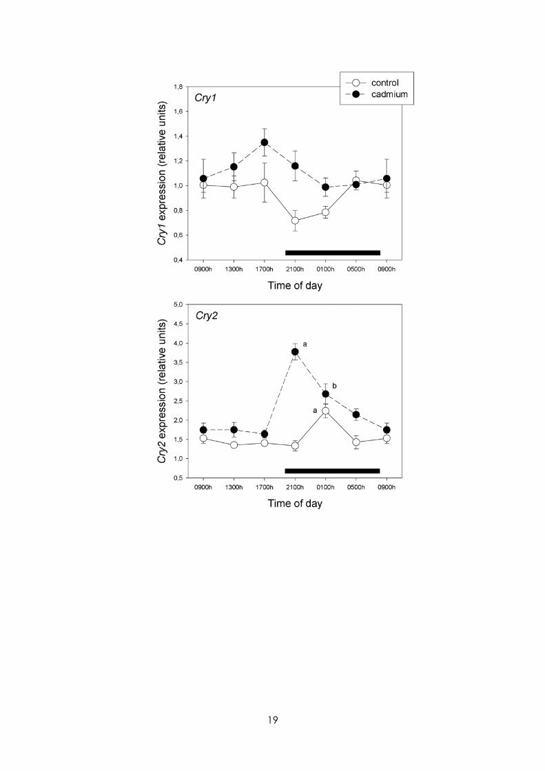
Figures 4 and 5 summarize the effect of CdCl2 on MBH hypothalamic mRNA levels for the circadian genes examined. CdCl2 treatment significantly suppressed Bmal1 circadian rhythmicity and changed the phase of that of Per1, Per2 and Cry2, while in the case of Clock significant 24-h variations were induced (p< 0.01) (Fig. 4). A significant effect of treatment was found in the factorial ANOVA for Clock, Cry1 and Cry2 , CdCl2 augmenting their expression significantly (F= 31.6, 11.7 and 52.7, p<0.001, respectively, main factor analysis, factorial ANOVA).
Discussion
Although metals have multiple effects on biological systems, an understudied effect is their role in programming gene expression. A growing body of evidence suggests that metals may influence epigenetic phenomena which regulate the expression of genes and ultimately their protein products (Wright and Baccarelli, 2007). Foregoing results indicate that the administration of a low amount of CdCl2 in drinking water for a month to young rats disrupted
5
24-h rhythmicity and overall expression of redox enzyme and clock genes in MBH. In particular, a disruption of 24-h pattern of hypothalamic expression of NOS-1 and -2, HO-1 and -2, Mn-SOD, catalase, GPx and GSR genes, and Bmal1, Per1, Per2 and Cry2 was seen in CdCl2-treated rats.
In the brain ROS generation is a continuous and physiological phenomenon and therefore nerve cells possess efficient antioxidant systems that protect them from oxidative damage [for reviews, see (Hardeland et al., 2003; Mancuso et al., 2007; Radak et al., 2007; Reynolds et al., 2007)]. These defense systems are comprised by a wide spectrum of enzymatic (Inal et al., 2001) and non-enzymatic systems (Wang et al., 2006). Circadian variations of brain redox pathway enzymes have been described, including NOS (Tunctan et al., 2002; Ayers et al., 1996; Clemens et al., 2005), HO (Artinian et al., 2001; Rubio et al., 2003), SOD (Diaz-Munoz et al., 1985; Schaper et al., 1986; Martin et al., 2003) and catalase (Sani et al., 2006). In many cases rhythms in enzyme activity and gene expression coincide but in others they are out of phase, e.g. NOS (Ayers et al., 1996; Clemens et al., 2005). The present study indicates that in the MBH a significant 24-h variation in gene expression of NOS-2, HO-1 and HO-2, Cu/Zn-SOD, GPx and GSR is detectable in control rats.
A number of studies examined the toxicity of Cd using a single (acute) dose of the metal in the range of 1-5 mg/kg (Barbier et al., 2004; Majumder et al., 2003). In the present study, and to mimic human exposure, we gave CdCl2 in the drinking water to young rats for 1 month at a low concentration (5 ppm). Under this condition Cd intake (about 7.5 µg/day) was close to that proposed by the World Health Organization as a tolerable limit for humans [1 µg/day for a life span of 60 years (WHO, 1995)]. It should be noted that because Cd absorption in the gastrointestinal tract of rats is lower than in humans, rat models simulating human exposure need to increase exposure doses to be higher than the real daily human intake of Cd (Rogalska et al., 2009).
Two possible interpretations can be entertained to explain the present observations in animals receiving low doses of CdCl2: (i) the changes observed after Cd indicate homeostatic mechanisms related to excessive ROS generation; (ii) the low levels of Cd given fall below the threshold needed to cause oxidative damage in MBH and affect gene programming, probably via a moderate increase of ROS (Wright and Baccarelli, 2007)- Clearly our results are not a consequence of an acute intoxication with Cd since body weight was not modified as we have previously reported using higher doses of the metal (Lafuente and Esquifino, 1999). In a previous study (Poliandri et al., 2006) we found that a Cd intake similar to that used herein did not induce lipid peroxidation in the liver as it was reported using a similar experimental approach with 15 ppm of Cd (Koyu et al., 2006). Indeed, the blood brain barrier effectively prevents Cd uptake into the brain. For example, in a study investigating CdCl2 absorption, distribution, and retention in rats after oral administration, 0.013 % of the dose was taken up by brain tissue (Lazarus et al., 2009).
There is controversy about the role played by NO in Cd toxicity (Misra et al., 1996; Harstad and Klaassen, 2002). The foregoing results indicate that a low Cd dose differentially affects NOS-1 and NOS-2 expression in MBH by disrupting their 24-h pattern and by decreasing the mRNA for the inducible isoform NOS-2. These results are compatible with the view that the toxic effect of a low dose of Cd is independent on excesive NO formation. It must be noted that in a
6
previous publication using the same low dose of CdCl2 at single time points, Cd increased expression of NOS-1 gene at 0900 h, while having no effects on that of NOS-2 in the anterior hypothalamus (Poliandri et al., 2006). Since both the MBH and anterior hypothalamic blocks used to perform the studies are heterogenous, differences in neuronal nuclei composition may explain this regional difference. Further studies including in situ-hybridization of specific protein products could be helpful to answer this query.
HO has an important role in controlling the redox state of the cell by functioning as a rate-limiting enzyme in the heme degradation process (Mancuso et al., 2007). HO-1 is an inducible isoform that is responsive to various stimuli, including oxidative stress. HO-2 is a constitutive isoform not inducible by oxidative stress. In the present study increases in HO-2 expression, but not in HO-1 expression, were observed in MBH of CdCl2-treated rats suggesting that the low levels of the heavy metal given fall below the threshold needed to cause oxidative damage in this brain region. In a previous study using anterior hypothalamic blocks (Poliandri et al., 2006), a similar dose of CdCl2 increased expression of HO-1 gene at 0900 h. Again, in situ-hybridization studies of the specific protein products could be helpful to support regional differences in responses to Cd.
The detoxification of ROS in cells involves the cooperative action of intracellular antioxidant enzymes, among them Cu/Zn-SOD that is cytosolic, Mn-SOD that is mitochondrial, and catalase that is present in peroxisomes (Rodriguez et al., 2004). In addition, GPx and GSR help to maintain adequate levels of reduced glutathione. CdCl2intake increased slightly Mn- SOD and catalase mRNA expression and did not affect that of Cu/Zn-SOD, GPx or GSR, therefore not supporting the notion that this is a compensatory increase caused by the augmented oxidative load.
It is known that the mammalian circadian timing system comprises peripheral oscillators located in almost every cell of the body together with a central rhythm generator located in the hypothalamic suprachiasmatic nucleus (SCN) (Richter et al., 2004; Lincoln, 2006). At the cell level, circadian rhythms are driven by the self-regulatory interaction of a set of genes named clock genes and their protein products. Among these Bmal1, Per1, Per2, Cry1, Cry2, and Clock play a major role. The heterodimer of the proteins Clock:Bmal1 binds E-box elements at the promoter region of Per1, Per2, Cry1 and Cry2, inducing their transcription. Conversely, Per1–2 and Cry1–2 proteins, by interacting with the Clock:Bmal1 heterodimer operate as negative regulators inhibiting their own transcription (Richter et al., 2004; Lincoln, 2006). As shown herein, CdCl2 administration significantly suppressed (Bmal1) or disrupted 24-h pattern of expression (Per1, Per2, Cry2) while in the case of Clock significant 24-h variations were induced. The results indicate that the inherent transcription, translation, and post-translational modifications that give the clock its own natural rhythmicity (Ramsey et al., 2007) is disrupted in rats drinking a low amount of CdCl2 in tap water. Previous studies form this laboratory indicated that chronic exposure of rats to low doses of Cd affected significantly the circadian variation of pituitary hormone release (Lafuente et al., 2003, 2004, 2005). Presumably, the changes in clock gene expression in MBH, a key region in hormone regulation, play a role in the circadian hormone disruption.
There are a number of limitations to the present study. One important is that additionally studies employing Western blotting analysis of enzyme protein
7
levels and measuring enzyme activity are needed to understand CdCl2 effect on redox enzyme gene expression in MBH. Additionally, studies employing Western blotting analysis of clock protein levels are needed to further describe the effect of CdCl2 on circadian gene expression. It should be also important to assess whether the changes in amplitude as well in timing of 24-h rhythm of gene expression discussed herein can be attributed to an effect on the circadian master clock or to a masking effect on some output(s) of the clock. In any event the present results are compatible with the view that a low amount of Cd given in tap water brought about significant changes in circadian expression of redox enzyme and clock genes in rat MBH.
Conflict of interest The authors declare to have no conflict of interests.
Acknowledgements
This work was supported by grants from Fondo de Investigaciones Sanitarias (FIS-PI05-0163), Madrid, Spain, Programa de Creación y Consolidación de Grupos de Investigación, Universidad Complutense-Comunidad de Madrid (CCG08-UCM/SAL-4188) and Agencia Nacional de Promoción Científica y Tecnológica, Argentina (PICT 2007-01045). DPC is a Research Career Awardee and PAS is a postdoctoral fellow from the Argentine Research Council (CONICET).
8
References Artinian LR, Ding JM, Gillette MU (2001) Carbon monoxide and nitric oxide: interacting messengers in muscarinic signaling to the brain's circadian clock. Exp Neurol 171: 293-300.
Ayers NA, Kapas L, Krueger JM (1996) Circadian variation of nitric oxide synthase activity and cytosolic protein levels in rat brain. Brain Res 707: 127-130.
Barbier O, Jacquillet G, Tauc M, Poujeol P, Cougnon M (2004) Acute study of interaction among cadmium, calcium, and zinc transport along the rat nephron in vivo. Am J Physiol Renal Physiol 287: F1067-F1075.
Brzóska MM, Moniuszko-Jakoniuk J (2005) Bone metabolism of male rats chronically exposed to cadmium. Toxicol Appl Pharmacol 207: 195-211.
Clemens S, Sawchuk MA, Hochman S (2005) Reversal of the circadian expression of tyrosine-hydroxylase but not nitric oxide synthase levels in the spinal cord of dopamine D3 receptor knockout mice. Neuroscience 133: 353-357.
Diaz-Munoz M, Hernandez-Munoz R, Suarez J, Chagoya dS, V (1985) Day-night cycle of lipid peroxidation in rat cerebral cortex and their relationship to the glutathione cycle and superoxide dismutase activity. Neuroscience 16: 859-863.
Hardeland R, Coto-Montes A, Poeggeler B (2003) Circadian rhythms, oxidative stress, and antioxidative defense mechanisms. Chronobiol Int 20: 921-962.
Harstad EB, Klaassen CD (2002) iNOS-null mice are not resistant to cadmium chloride-induced hepatotoxicity. Toxicology 175: 83-90.
Inal ME, Kanbak G, Sunal E (2001) Antioxidant enzyme activities and malondialdehyde levels related to aging. Clin Chim Acta 305: 75-80.
Institute of Laboratory Animal Resources CoLSNRC. Guide for the Care and Use of Laboratory Animals. Washington, D.C.: National Academy Press.
Koyu A, Gokcimen A, Ozguner F, Bayram DS, Kocak A (2006) Evaluation of the effects of cadmium on rat liver. Mol Cell Biochem 284: 81-85.
Lafuente A, Esquifino AI (1999) Cadmium effects on hypothalamic activity and pituitary hormone secretion in the male. Toxicol Lett 110: 209-218.
Lafuente A, Gonzalez-Carracedo A, Romero A, Cabaleiro T, Esquifino AI (2005) Toxic effects of cadmium on the regulatory mechanism of dopamine and serotonin on prolactin secretion in adult male rats. Toxicol Lett 155: 87-96.
Lafuente A, Gonzalez-Carracedo A, Romero A, Cano P, Esquifino AI (2004) Cadmium exposure differentially modifies the circadian patterns of norepinephrine at the median eminence and plasma LH, FSH and testosterone levels. Toxicol Lett 146: 175-182.
Lafuente A, Gonzalez-Carracedo A, Romero A, Esquifino AI (2003) Effect of cadmium on 24-h variations in hypothalamic dopamine and serotonin metabolism in adult male rats. Exp Brain Res 149: 200-206.
Lazarus M, Orct T, Jurasoviae J, Blanusa M (2009) The effect of dietary selenium supplementation on cadmium absorption and retention in suckling rats. Biometals in press.
9
Leret ML, Millan JA, Antonio MT (2003) Perinatal exposure to lead and cadmium affects anxiety-like behaviour. Toxicology 186: 125-130.
Lincoln GA. (2006) Decoding the nightly melatonin signal through circadian clockwork. Mol Cell Endocrinol 252: 69-73.
Livak KJ, Schmittgen TD (2001) Analysis of relative gene expression data using real-time quantitative PCR and the 2-∆∆Ct method. Methods 25: 402-408.
Lopez E, Arce C, Oset-Gasque MJ, Canadas S, Gonzalez MP (2006) Cadmium induces reactive oxygen species generation and lipid peroxidation in cortical neurons in culture. Free Radic Biol Med 40: 940-951.
Majumder S, Ghoshal K, Summers D, Bai S, Datta J, Jacob ST (2003) Chromium(VI) down-regulates heavy metal-induced metallothionein gene transcription by modifying transactivation potential of the key transcription factor, metal-responsive transcription factor 1. J Biol Chem 278: 26216-26226.
Mancuso C, Scapagini G, Curro D, Giuffrida Stella AM, De Marco C, Butterfield DA, Calabrese V (2007) Mitochondrial dysfunction, free radical generation and cellular stress response in neurodegenerative disorders. Front Biosci 12: 1107-1123.
Martin V, Sainz RM, Mayo JC, Antolin I, Herrera F, Rodriguez C (2003) Daily rhythm of gene expression in rat superoxide dismutases. Endocr Res 29: 83-95.
Misra RR, Hochadel JF, Smith GT, Cook JC, Waalkes MP, Wink DA (1996) Evidence that nitric oxide enhances cadmium toxicity by displacing the metal from metallothionein. Chem Res Toxicol 9: 326-332.
Poliandri AH, Esquifino AI, Cano P, Jimenez V, Lafuente A, Cardinali DP, Duvilanski BH (2006) In vivo protective effect of melatonin on cadmium-induced changes in redox balance and gene expression in rat hypothalamus and anterior pituitary. J Pineal Res 41(3): 238-246.
Radak Z, Kumagai S, Taylor AW, Naito H, Goto S (2007) Effects of exercise on brain function: role of free radicals. Appl Physiol Nutr Metab 32: 942-946.
Ramsey KM, Marcheva B, Kohsaka A, Bass J (2007) The clockwork of metabolism. Annu Rev Nutr 27: 219-240.
Reynolds A, Laurie C, Mosley RL, Gendelman HE (2007) Oxidative stress and the pathogenesis of neurodegenerative disorders. Int Rev Neurobiol 82: 297-325.
Richter HG, Torres-Farfan C, Rojas-Garcia PP, Campino C, Torrealba F, Seron-Ferre M (2004) The circadian timing system: making sense of day/night gene expression. Biol Res 37: 11-28.
Rodriguez C, Mayo JC, Sainz RM, Antolin I, Herrera F, Martin V, Reiter RJ (2004) Regulation of antioxidant enzymes: a significant role for melatonin. J Pineal Res 36: 1-9.
Rogalska J, Brzóska MM, Roszczenko A, Moniuszko-Jakoniuk J (2009) Enhanced zinc consumption prevents cadmium induced alterations in lipid metabolism in male rats. Chem Biol Interact 177: 142-152.
Rubio MF, Agostino PV, Ferreyra GA, Golombek DA (2003) Circadian heme oxygenase activity in the hamster suprachiasmatic nuclei. Neurosci Lett 353: 9-12.
10
Sani M, Sebai H, Gadacha W, Boughattas NA, Reinberg A, Mossadok BA (2006) Catalase activity and rhythmic patterns in mouse brain, kidney and liver. Comp Biochem Physiol B Biochem Mol Biol 145: 331-337.
Satarug S, Baker JR, Urbenjapol S, Haswell-Elkins M, Reilly PE, Williams DJ, Moore MR (2003) A global perspective on cadmium pollution and toxicity in non-occupationally exposed population. Toxicol Lett 137: 65-83.
Schaper U, Holig K, Weber A, Scheuch DW (1986) [Diurnal variations in superoxide dismutase (EC 1.15.1.1) activity and malondialdehyde concentration in the rat brain and striatum]. Z Med Lab Diagn 27: 50-51.
Sugita M, Tsuchiya K (1995) Estimation of variation among individuals of biological half-time of cadmium calculated from accumulation data. Environ Res 68: 31-37.
Szentagothai B, Flerkó B, Mess B, Halász B. Hypothalamic Control of the Anterior Pituitary, 3rd. edition. Budapest: Akademíai Kiadó.
Tunctan B, Weigl Y, Dotan A, Peleg L, Zengil H, Ashkenazi I, Abacioglu N (2002) Circadian variation of nitric oxide synthase activity in mouse tissue. Chronobiol Int 19: 393-404.
Valko M, Rhodes CJ, Moncol J, Izakovic M, Mazur M (2006) Free radicals, metals and antioxidants in oxidative stress-induced cancer. Chem Biol Interact 160: 1-40.
Viaene MK, Masschelein R, Leenders J, De GM, Swerts LJ, Roels HA (2000) Neurobehavioural effects of occupational exposure to cadmium: a cross sectional epidemiological study. Occup Environ Med 57: 19-27.
Wakabayashi I, Hatake K, Kimura N, Kakishita E, Nagai K (1987) Modulation of vascular tonus by the endothelium in experimental diabetes. Life Sci 40: 643-648.
Wang JY, Wen LL, Huang YN, Chen YT, Ku MC (2006) Dual effects of antioxidants in neurodegeneration: direct neuroprotection against oxidative stress and indirect protection via suppression of glia-mediated inflammation. Curr Pharm Des 12: 3521-3533.
WHO (1995) Inorganic constituents and physical parameters. Guidelines for Drinking Water-Quality: Health Criteria and other Supporting Information,World Health Organization, Geneva.
Wright RO, Baccarelli A (2007) Metals and neurotoxicology. J Nutr 137: 2809-2813.
Yang Z, Yang S, Qian SY, Hong JS, Kadiiska MB, Tennant RW, Waalkes MP, Liu J (2007) Cadmium-induced toxicity in rat primary mid-brain neuroglia cultures: role of oxidative stress from microglia. Toxicol Sci 98: 488-494.
11
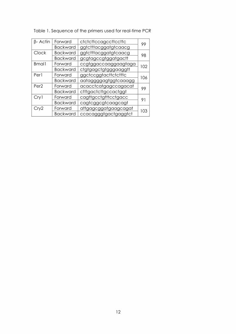
Table 1. Sequence of the primers used for real-time PCR
Forward ctctcttccagccttccttc β- Actin Backward ggtctttacggatgtcaacg 99
Backward ggtctttacggatgtcaacg Clock Backward gcgtagccgtggatgactt 98
Forward ccgtggaccaaggaagtaga Bmal1 Backward ctgtgagctgtgggaaggtt 102
Forward ggctccggtacttctctttc Per1 Backward aataggggagtggtcaaagg 106
Forward acacctcatgagccagacat Per2 Backward ctttgactcttgccactggt 99
Forward cagttgcctgtttcctgacc Cry1 Backward cagtcggcgtcaagcagt 91
Forward attgagcggatgaagcagat Cry2 Backward ccacagggtgactgaggtct 103
12
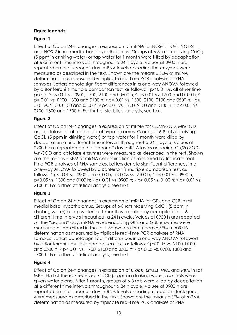
Figure legends Figure 1 Effect of Cd on 24-h changes in expression of mRNA for NOS-1, HO-1, NOS-2 and NOS-2 in rat medial basal hypothalamus. Groups of 6-8 rats receiving CdCl2 (5 ppm in drinking water) or tap water for 1 month were killed by decapitation at 6 different time intervals throughout a 24 h cycle. Values at 0900 h are repeated on the “second” day. mRNA levels encoding the enzymes were measured as described in the text. Shown are the means ± SEM of mRNA determination as measured by triplicate real-time PCR analyses of RNA samples. Letters denote significant differences in a one-way ANOVA followed by a Bonferroni´s multiple comparison test, as follows: a p< 0.01 vs. all other time points; b p< 0.01 vs. 0900, 1700, 2100 and 0500 h; c p< 0.01 vs. 1700 and 0100 h; d p< 0.01 vs. 0900, 1300 and 0100 h; e p< 0.01 vs. 1300, 2100, 0100 and 0500 h; f p< 0.01 vs. 2100, 0100 and 0500 h; g p< 0.01 vs. 1700, 2100 and 0100 h; h p< 0.01 vs. 0900, 1300 and 1700 h. For further statistical analysis, see text.
Figure 2 Effect of Cd on 24-h changes in expression of mRNA for Cu/Zn-SOD, Mn/SOD and catalase in rat medial basal hypothalamus. Groups of 6-8 rats receiving CdCl2 (5 ppm in drinking water) or tap water for 1 month were killed by decapitation at 6 different time intervals throughout a 24 h cycle. Values at 0900 h are repeated on the “second” day. mRNA levels encoding Cu/Zn-SOD, Mn/SOD and catalase enzymes were measured as described in the text. Shown are the means ± SEM of mRNA determination as measured by triplicate real-time PCR analyses of RNA samples. Letters denote significant differences in a one-way ANOVA followed by a Bonferroni´s multiple comparison test, as follows: a p< 0.01 vs. 0900 and 0100 h, p< 0.05 vs. 2100 h; b p< 0.01 vs. 0900 h, p<0.05 vs. 1300 and 0100 h; c p< 0.01 vs. 0900 h; d p< 0.05 vs. 0100 h; e p< 0.01 vs. 2100 h. For further statistical analysis, see text.
Figure 3 Effect of Cd on 24-h changes in expression of mRNA for GPx and GSR in rat medial basal hypothalamus. Groups of 6-8 rats receiving CdCl2 (5 ppm in drinking water) or tap water for 1 month were killed by decapitation at 6 different time intervals throughout a 24 h cycle. Values at 0900 h are repeated on the “second” day. mRNA levels encoding GPx and GSR enzymes were measured as described in the text. Shown are the means ± SEM of mRNA determination as measured by triplicate real-time PCR analyses of RNA samples. Letters denote significant differences in a one-way ANOVA followed by a Bonferroni´s multiple comparison test, as follows: a p< 0.05 vs. 2100, 0100 and 0500 h; b p< 0.01 vs. 1700, 2100 and 0500 h; c p< 0.05 vs. 0900. 1300 and 1700 h. For further statistical analysis, see text.
Figure 4 Effect of Cd on 24-h changes in expression of Clock, Bmal1, Per1 and Per2 in rat MBH. Half of the rats received CdCl2 (5 ppm in drinking water); controls were given water alone. After 1 month, groups of 6-8 rats were killed by decapitation at 6 different time intervals throughout a 24 h cycle. Values at 0900 h are repeated on the “second” day. mRNA levels encoding circadian clock genes were measured as described in the text. Shown are the means ± SEM of mRNA determination as measured by triplicate real-time PCR analyses of RNA
13
samples. Significant time-of-day variations within each experimental group, as detected by a one way ANOVA, were as follows: Clock: cadmium, F= 13.3, p< 0.01. Bmal1: control, F= 3.54, p< 0.02. Per1: control, F= 4.84, p< 0.01; cadmium, F= 7.38, p< 0.01.. Per2: control, F= 6.37, p< 0.01; cadmium, F= 5.09, p< 0.01. Letters denote significant differences in a one-way ANOVA followed by a Bonferroni´s multiple comparison test as follows: a p< 0.01 vs. 0900, 1300 and 0100 h; b p< 0.01 vs. 0900 and 1300 h, p< 0.05 vs. 0100 h; c p< 0.02 vs. 0100 h; d p< 0.05 vs. 0900 h; d p< 0.05 vs. 0900 h; e p< 0.01 vs. 0900, 1300, 1700 and 0100 h; f p< 0.01 vs. 0100 and 2100 h, p< 0.03 vs. 0900 h; g p< 0.01 vs. 1700 h, p< 0.03 vs. 1300 h; h p< 0.01 vs. 0500 h. For further statistical analysis, see text.
Figure 5 Effect of Cd on 24-h changes in expression of Cry1 and Cry2 in rat MBH. Half of the rats received CdCl2 (5 ppm in drinking water); controls were given water alone. After 1 month, groups of 6-8 rats were killed by decapitation at 6 different time intervals throughout a 24 h cycle. Values at 0900 h are repeated on the “second” day. mRNA levels encoding circadian clock genes were measured as described in the text. Shown are the means ± SEM of mRNA determination as measured by triplicate real-time PCR analyses of RNA samples. Significant time-of-day variations within each experimental group, as detected by a one way ANOVA, were as follows: Cry2: control, F= 7.29, p< 0.01; cadmium: F= 18.8, p< 0.01. Letters denote significant differences in a one-way ANOVA followed by a Bonferroni´s multiple comparison test as follows: a p< 0.01 vs. all remaining time intervals; b p< 0.05 vs. 0900, 1300, 1700 and 100 h. For further statistical analysis, see text.
14
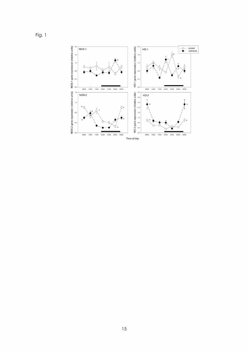
Fig. 1
15
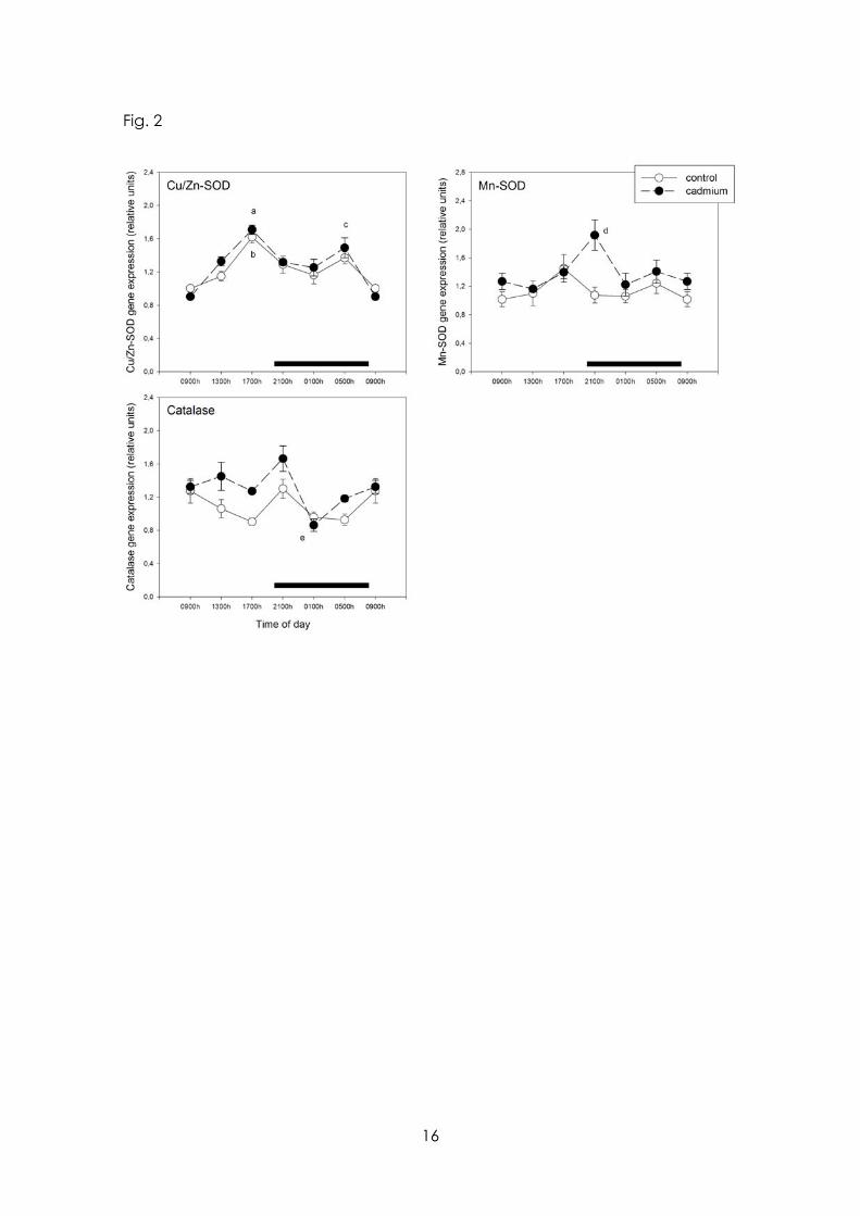
Fig. 2
16
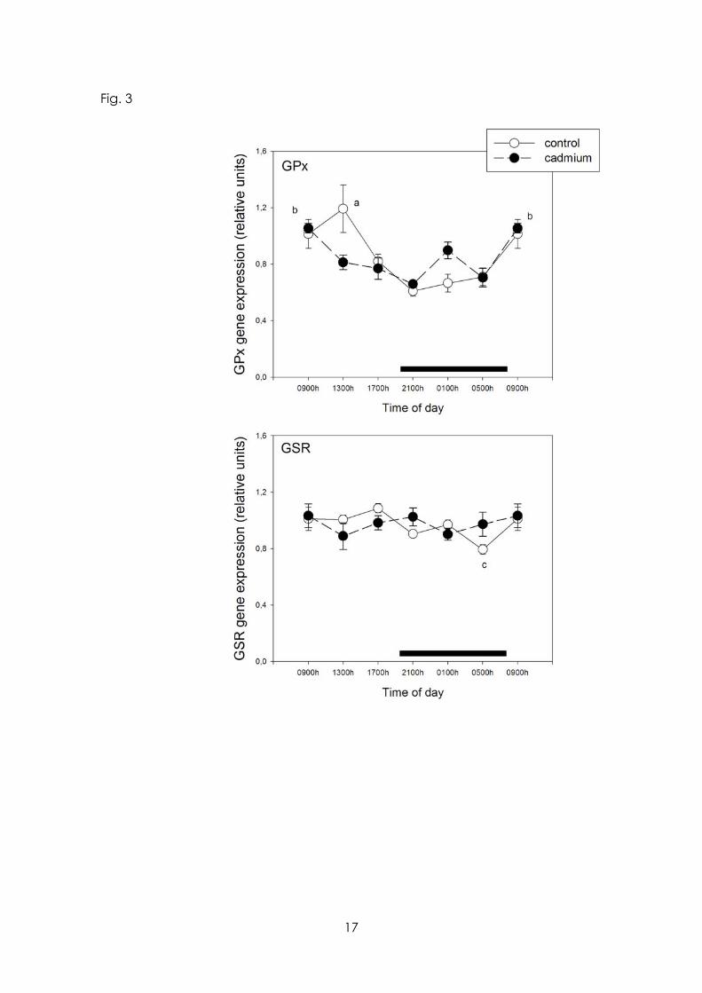
Fig. 3
17
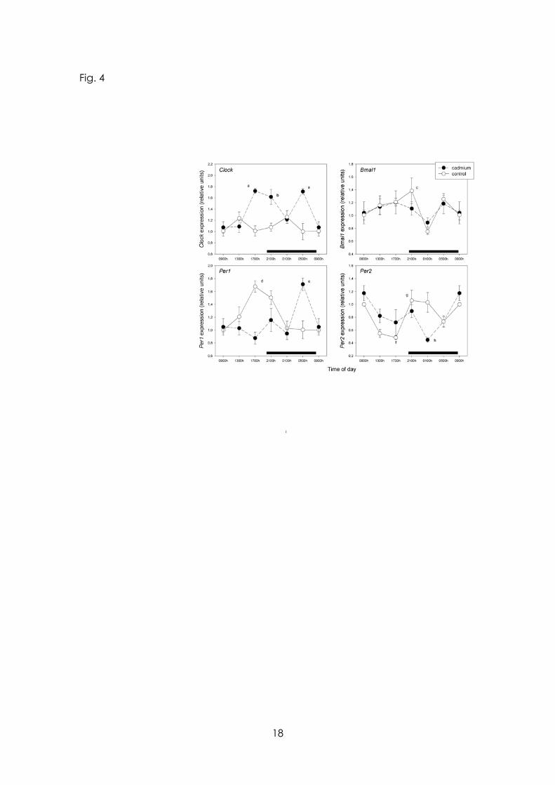
Fig. 4
18
19