informe técnico final - cudi · aplicaciones en nanobiotecnología y nanotecnología utilizando...
TRANSCRIPT

Informe Técnico Final
Proyecto CUDI “Red de estudio de sistemas nanométricos incluidos en materia suave para aplicaciones en Nanobiotecnología y Nanotecnología utilizando técnicas de corte en un Ultramicrotomo Criogénico de última generación”.
En colaboración con la Dra. Patricia Santiago Jacinto, co-responsable del Proyecto se publicó el artículo (revista ISI):
1) Gold nanoparticles conjugated to Benzoylmercaptoacetyltryglicine and L-Cysteine methylester; O. Estévez-Hernández, E.M. Molina-Trinidad, P. Santiago-Jacinto, L. Rendón, E. Reguera;
Journal of Colloid and Interface Science 35 (2010) 161-167. Se adjunta copia de este artículo; Como resultados del mismo Proyecto, se encuentra en fase de arbitraje un segundo artículo, en particular: 2) Gold nanoparticles capped with Lanreotide peptide; E.M. Molina-Trinidad, O. Estévez-
Hernández, L. Rendón, V. Garibay-Febles, E. Reguera; Enviado a: Journal of Materials Science
and Engineering C, se adjunta primera página del manuscrito. 3) Se dispone de datos para escribir otros dos artículos que serán publicados en revistas
internacionales ISI. Otras Acciones Ejecutadas con apoyo del Proyecto.
1) Una estudiante de Doctorado (M. C. Blanca Zamora Reynoso) realizó un adiestramiento avanzado en la Universidad de Southampton, Inglaterra, con apoyo del Proyecto. A partir de los datos allí registrados se publicará un 3er artículo en revista ISI.
2) Un estudiante de Doctorado (M. C. Manuel Ávila Santos) ejecutó un proyecto de medición en el Laboratorio Nacional de Radiación Sincrotrón (LNLS) de Brasil, único de su tipo en el hemisferio sur. A partir de esos datos se publicara un 4to artículo ISI.
3) Con apoyo del Proyecto viajó al país en Profesor de la Universidad de Minnesota (Dr. Andreas Stein), el cual dictó una Conferencia Magistral en CICATA-IPN, Unidad Legaria, sobre la temática del Proyecto, y además participó en otras actividades académicas.

4) Con recursos del Proyecto se apoyó por 2 meses con 6 mil pesos /mes a la Dra. Adela Lemus Santana, recién egresada de la UNAM, y sin vínculo laboral o de posgrado, para que trabajara en la preparación de las muestras a estudiar, y como participante se le financió el boleto de avión para que asistiera a una Escuela de Verano sobre Nanotecnologías realizada en la Universidad de la Habana en Julio 2010.
5) Se apoyo a un estudiante de Doctorado (M. C. Jorge Roque de la Puente) para que asistiera a una Escuela Internacional sobre Nanotecnologías.
6) Se obtuvieron reactivos, material gastable e insumos generales para apoyar las tareas de investigación-desarrollo y formación de recursos humanos relativas al Proyecto.
Dr. Edilso Reguera Responsable Técnico del Proyecto

This article appeared in a journal published by Elsevier. The attachedcopy is furnished to the author for internal non-commercial researchand education use, including for instruction at the authors institution
and sharing with colleagues.
Other uses, including reproduction and distribution, or selling orlicensing copies, or posting to personal, institutional or third party
websites are prohibited.
In most cases authors are permitted to post their version of thearticle (e.g. in Word or Tex form) to their personal website orinstitutional repository. Authors requiring further information
regarding Elsevier’s archiving and manuscript policies areencouraged to visit:
http://www.elsevier.com/copyright

Author's personal copy
Gold nanoparticles conjugated to benzoylmercaptoacetyltriglycineand L-cysteine methylester
O. Estévez-Hernández a,b, E.M. Molina-Trinidad a,c, P. Santiago-Jacinto d, L. Rendón d, E. Reguera a,b,*
a Centro de Investigación en Ciencia Aplicada y Tecnología de Avanzada, IPN, Legaria 694, México DF, Mexicob Instituto de Ciencia y Tecnología de Materiales, Universidad de La Habana, Cubac Facultad de Estudios Superiores Cuautitlán, Universidad Nacional Autónoma de México, Estado de México, Mexicod Instituto de Física, Universidad Nacional Autónoma de México, México DF, Mexico
a r t i c l e i n f o
Article history:Received 22 April 2010Accepted 19 June 2010Available online 25 June 2010
Keywords:Gold nanoparticlesBenzoylmercaptoacetyltriglycineL-cysteine methylesterCapping agents
a b s t r a c t
Benzoyl-protected mercaptoacetyltriglycine, a synthetic precursor used in the preparation of Techne-tium-99 m-mercaptoacetyltriglycine, a radiopharmaceutical for renal tubular function and L-cysteinemethylester, a small, non-zwitterionic amino acid derivative, were used as capping agents of gold nano-particles obtained by borohydride reduction method. The capped gold nanoparticles composites wereprepared from aqueous solutions and characterized by UV–Vis, infrared and Raman spectra and Trans-mission Electron Microscopy images. The presence of the ligands and its different binding mode to theparticles as a consequence of the benzoyl-protection of the thiol group in benzoyl-protected mercapto-acetyltriglycine were evidenced from infrared and Raman spectra. The stability on aging in water solutionof the formed composites is discussed from the obtained UV–Vis spectra.
2010 Elsevier Inc. All rights reserved.
1. Introduction
The applications of surface functionalized metal nanoparticleshave been explored in a wide variety of areas [1]. Colloidal goldnanoparticles (AuNPs) have found technologically uses since an-cient times related to their optical properties, but only in the lastdecades their potential biological applications in labeling, deliver-ing, heating and sensing processes have been demonstrated [2–5].Among noble metal particles, AuNPs have attracted intensive atten-tion related to their easy preparative routes available, their low tox-icity, and the gold surface affinity for the bonding to molecules ofbiological interest [5,6]. Gold and other noble metal nanoparticleshave great potential for applications in biochemical sensing and bio-logical imaging because of their unique optical properties originatedfrom the excitation of local surface plasmon resonances [7,8]. Thesurface plasmon resonance is a coherent oscillation of the surfaceconduction electrons excited by electromagnetic radiation. It is sen-sitive to the local dielectric environment [9]. Typically, local surfaceplasmon resonances devices sense changes in the local environmentthrough a shift for the resonance wavelength. Apart from the envi-ronmental effect, the surface resonance plasmon of nanoparticlesis dramatically affected by their size, shape, and surface modifica-tions [7,10]. The highly confined local electric field enhancement
that accompanies the excitation of the plasmon supports varietyspectroscopic and imaging techniques [11–13].
The synthesis of AuNPs with diameters ranging from a few to sev-eral hundreds of nanometers in aqueous solution as well as in organ-ic solvents is well established [10,14–17]. In typical syntheses, Ausalts are reduced by addition of a reducing agent such as sodium cit-rate or borohydride. In addition, a stabilizing agent (surfactant) isalso required which is either adsorbed or chemically bound to thesurface of AuNPs. The surfactant is typically charged, so that theequally charged NPs repel each other so that they remain stable incolloidal state. Most biological or biomedical applications requirethat the clusters readily dissolve in aqueous media which is favoredif the aggregation is prevented through electrostatic interactions.Biological molecules can be attached to the particles in several ways.If the biological molecules have a functional group which can bind tothe Au surface (like thiols, cyano, amino or specific peptide se-quences), the biological molecules can replace some of the originalstabilizer molecules when they are added directly to the particlessolution. Studies on the interaction of Au with biomolecules is an ac-tive research area where useful information is being obtained[18,19]. Benzoyl-protected mercaptoacetyltriglycine (BzMAG3) is asynthetic ligand used in the preparation of Technetium-99 m-mer-captoacetyltriglycine, a radiopharmaceutical for renal tubular func-tion [20]. From the conjugation of AuNPs to BzMAG3 a useful tool forimaging or diagnostic of renal tubular function could be obtained.Furthermore, it opens up several novel possibilities as the BzMAG3
structure could be derivatized because of its free carboxylic group.Different active groups capable of performing specific functions like
0021-9797/$ - see front matter 2010 Elsevier Inc. All rights reserved.doi:10.1016/j.jcis.2010.06.051
* Corresponding author at: Centro de Investigación en Ciencia Aplicada yTecnología de Avanzada, IPN, Legaria 694, México DF, Mexico. Fax: +52 5553954147.
E-mail address: [email protected] (E. Reguera).
Journal of Colloid and Interface Science 350 (2010) 161–167
Contents lists available at ScienceDirect
Journal of Colloid and Interface Science
www.elsevier .com/locate / jc is
Author's personal copy
catalysis, energy transfer, and sensing, could be attached [1]. Noreports on the conjugation of AuNPs to BzMAG3 were found. In orderto shed light on the nature of BzMAG3 binding to AuNPs surface alsothe conjugation of AuNPs to L-cysteine methylester (CysM) was con-sidered. Both simple molecules could be useful models to study theinteraction of mercapto, amino and amide functions with gold sur-face. L-cysteine methylester is a derivative of L-cysteine, a small ami-no acid that has been used as capping agent for AuNPs [21–25]. Theaim of the present study is to explore the possible conjugation ofAuNPs to BzMAG3 and to shed light on the AuNPs interparticle inter-actions in the presence of BzMAG3 in aqueous media. For a potentialapplication of the formed conjugates for imaging or diagnostic ofrenal tubular function, their stability in aqueous media is a requiredfeature, which was also studied. The formed Au@BzMAG3 and Au@-CysM composites were characterized from X-ray Energy-Disperse(EDS), UV–Vis, infrared (IR) and Raman spectra and from Transmis-sion Electron Microscopy (TEM) images.
2. Experimental
2.1. Materials
The tetrachloroauric (III) acid trihydrate (HAuCl4.3H2O, 99.9%),and the L-cysteine methylester (98%) were purchased from Sig-ma–Aldrich. Sodium borohydride (96%) was obtained from Fluka.All chemicals were employed as received and Milli-Q water wasused. BzMAG3 ligand was synthesized according to the method ofFritzberg et al. [20], m.p. 193–195 C. 1H-NMR (CDCl3/DMSO)3.75–3.88 (6H, d; ACH2ANHACOA), 3.90 (2H, s, ACH2ASA),7.45–7.70 (5H, aromatic) [26].
2.2. Gold nanoparticles preparation
Gold nanoparticles were prepared by sodium borohydridereduction method [27]. In brief, 100 mL aqueous solution 104 Mof HAuCl43H2O (0.0105 mmols) was reduced by 0.01 g of NaBH4
(0.2632 mmols) solved in 5 mL of water at room temperatureresulting in the formation of ruby-red gold colloid. The gold:boro-hydride ratio was 1:25. The sol was left to react 30 min beforedrawing off the capping ligand.
2.3. Au@BzMAG3 composite preparation
0.009 mmols of AuNPs (90 mL) were capped by 103 M aqueoussolution (10 mL, 0.01 mmol) of BzMAG3. The BzMAG3 was added tothe Au colloid solution 30 min after its preparation in order to min-imize the colloid instability effect on the composite features. Thereaction mixture (1.1 M ratio of BzMAG3 ligand to Au colloid)was stirred for 12 h at room temperature, as already reported forcysteine as capping ligand [21]. The capped AuNPs formed wereseparated and washed by repeated centrifugation at 35,000 rpmfor 2 h.
2.4. Au@CysM composite preparation
0.009 mmoles of AuNPs (90 mL) were capped by 103 M aque-ous solution (10 mL, 0.01 mmol) of CysM. The CysM was addedto the Au colloid solution 30 min after its preparation in order tominimize the colloid instability effect on the composite features.The reaction mixture (1.1 M ratio of CysM ligand to Au colloid)was stirred for 12 h at room temperature, as already reported forcysteine as capping ligand [21]. The capped AuNPs formed wereseparated and washed by repeated centrifugation at 35,000 rpmfor 2 h.
2.5. Methods
2.5.1. UV–Vis spectraThe UV–Vis absorption spectra of the solutions were studied
using a Varian Cary 50 Conc UV–Vis spectrophotometer, with10 mm path length quartz cuvettes in the 190–900 nm wave-lengths range.
2.5.2. EDS spectraThe EDS spectra were collected using an INCAx-sight 7582 mod-
el microanalyzer coupled to a scanning electronic microscope(SEM) Jeol JSM-6390LV operated at 20 keV.
2.5.3. IR spectraIR spectra in the range of 4500–400 cm1 were recorded using a
Perkin Elmer Spectrum One FT-IR spectrometer in KBr pressed disks.In the preparation of the KBr pressed disks were used 0.5 mg of thesample and 100 mg of KBr. The number of scans used to record thespectra was 10. Spectra of samples in Nujol mulls using CaF2 win-dows were also run in order to discard a possible reaction betweenthe sample and KBr matrix.
2.5.4. Raman spectraRaman spectra were obtained using an Almega XR Dispersive Ra-
man spectrometer. An Olympus microscope (BX51) and an Olym-pus 50 objective (NA = 0.80) were used for focusing the laser onthe sample, to a spot size of 1 lm, and for collecting the scatteredlight, a charge-coupled device (CCD) detector thermoelectricallycooled to 50 C was used. The Raman spectra were accumulatedover 80 s with a resolution of 4 cm1. The excitation source wasthe 532 nm radiation line from a Nd:YVO4 laser (frequency-dou-bled). The incident laser power was from 0.1 to 100 mW.
2.5.5. TEM imagesStructural and morphological characterization of capped AuNPs
were performed by JEOL JEM 2200 FS Transmission Electron Micro-scope (TEM) equipped with a High-Angle Annular Dark Field(HAADF) detector and operated at 200 kV accelerating voltage.The samples for TEM measurement were prepared by dipping theTEM copper lacey carbon grid (400 meshes) in a dilute dispersionof nanoparticles in water.
3. Results and discussion
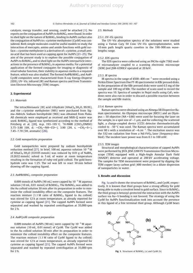
Fig. 1a and b shows the structures of BzMAG3 and CysM, respec-tively. It is known that thiol groups have a strong affinity for goldbeing able to make a covalent bond to gold surface. Since in BzMAG3
the thiol group is benzoyl-protected the interaction with the AuNPssurface via AuAS bonding is not favored. The strategy of using theCysM for AuNPs functionalization took into account the presencein this ligand of a free terminal thiol group. Although CysM bears
Fig. 1. Structures of BzMAG3 (a) and CysM (b).
162 O. Estévez-Hernández et al. / Journal of Colloid and Interface Science 350 (2010) 161–167

Author's personal copy
two potential anchoring groups (SH and NH2) that can covalentlybind Au particles, it is well established the superiority of thiol groupto form covalent bond with gold [28]. Because in CysM the thiol ter-minal group is free, this simple cysteine derivative may be useful tosense comparatively the interaction with Au surface relative to theless accessible benzoylmercapto group in BzMAG3. As already-men-tioned, in the CysM the existence of zwitterion (internal salt) is notpossible because the carboxylic group is derivatized as methylester.This fact hinders the nanoparticles assembling through the zwitter-ions-type electrostatic interactions, which is observed for instancewhen cysteine is used as capping ligand [22].
Freshly prepared AuNPs obtained by sodium borohydridereduction are stable in water and display a characteristic UV–Visabsorption spectrum with a plasmon band in the 522–525 nmspectral range (curves a_1 of Figs. 2a and 3a). On aging, a notice-able 1red shift accompanied of the peak broadening is observed(discussed below). As already-mentioned, the plasmon resonanceabsorption is sensitive to the particles environment. Such effectwas used to sense the AuNPs conjugation to the two consideredcapping ligands. For BzMAG3 a slight red shift, from 525 to
528 nm, together of certain peak broadening and a decrease of itsmaximum absorbance were detected (curve a_2 in Fig. 2a). Suchspectral variations, without appreciable color changes, were inter-preted as resulting from the BzMAG3 binding to the Au surface. Aquite different behavior was observed for the CysM ligand interac-tion with AuNPs. In this case the red shift for the plasmon reso-nance peak was significantly greater, of about 8 nm (from522 nm to 530 nm) and the peak broadening is accompanied of acolor change for the colloidal suspension, from ruby-red to blue(curve a_2 in Fig. 3a).
Such color change in colloidal gold has been ascribed to forma-tion of AuNPs aggregates [29]. The observed differences in behav-ior for the interactions of these two ligands with AuNPs wereinterpreted as related to the nature of the electrostatic interactioninvolved between the capped gold particles; CysM is unchargedand this ligand de-stabilizes the suspension by reducing the elec-trostatic repulsion between the gold particles when it adsorbs. Incontrast BzMAG3 is negatively charged (carboxylic group) insolution at most pH values above four (the measured pH of themixture reaction is about 8–9), and when adsorbed through theamine groups, will retain this negative charge upon adsorption,thus maintaining the electrostatic repulsion between AuNPs. Thestability of BzMAG3 and CysM capped AuNPs in comparison with
Fig. 2. UV–Vis spectra for: (a) AuNPs (a_1) and Au@BzMAG3 (a_2); (b) AuNPs (b_1)and Au@BzMAG3 (b_2) solutions as freshly prepared and after 30 days of aging (b_3and b_4, respectively).
Fig. 3. UV–Vis spectra for: (a) AuNPs (a_1) and Au@CysM (a_2); (b) AuNPs (b_1)and Au@CysM (b_2) as freshly prepared solutions, and after 30 days of aging (b_3and b_4, respectively).
1 For interpretation of color in Figs. 2 and 3, the reader is referred to the webversion of this article.
O. Estévez-Hernández et al. / Journal of Colloid and Interface Science 350 (2010) 161–167 163

Author's personal copy
free Au colloid for a month of aging was also studied. For Au@Bz-MAG3 composite, the wavelength where the plasmon peak maxi-mum is observed remains stable at 528 nm (curves b_2 and b_4of Fig. 2b) and without significant peak broadening. This was inter-preted as formation of a stable composite where the capped nano-particles preserve their colloidal state (no aggregates formation).Such behavior results appropriate for potential applications of thatcomposite for imaging and diagnostic tool. Relative to Au@BzMAG3
the formed Au@CysM composite shows quite different features onaging, and also different to those observed for non-conjugated Aucolloidal suspension. On the Au@CysM aging a gradual red shiftfrom 530 to 544 nm accompanied of peak broadening was ob-served for the plasmon resonance signal (curves b_2 and b_4 ofFig. 3b). This was attributed to a progressive nanoparticles aggre-gation process. It seems the capped nanoparticles interact throughattractive dispersive forces maintaining several nanoparticles to-gether. Such mechanism must be quite different to that reportedfor the aggregation of AuNPs capped with cysteine ligand [22].As already-mentioned, with cysteine the aggregation process takesplace through zwitterion-type electrostatic interactions, mecha-nism that is not possible for CysM.
The above discussed UV–Vis results were supported by qualita-tive information on the chemical composition of the formed com-posite nanoparticles. From the colloidal suspension nanoparticleswere separated from the mother solution by centrifugation, andthen washed several times with distilled water. For the obtainedpowder of dried nanoparticles EDS spectra were recorded. Withoutexception, in all the spectra X-ray peaks corresponding to S and Auwere observed (see Supplementary information), although this factis not a definitive proof that the capping agent is adsorbed and in-tact. Manny thiol systems are known to undergo oxidation andCAS bond scission once adsorbed [30].
Conventional and High Resolution TEM (HRTEM) images wereused to obtain structural information on Au@BzMAG3 and Au@-CysM composites. For Au@BzMAG3 HRTEM images revealed nano-particle sizes with mean diameters in the 2–7 nm range (Fig. 4aand b). The HRTEM images also show that these nanoparticlesare single crystalline, as clearly indicated by atomic lattice fringes(Fig. 4b) and the corresponding interatomic planes distance whichmatch to the 1 1 1 family of planes.
On the other hand, a difference in contrast observed around iso-lated nanoparticles of Au@CysM composite suggests the presenceof CysM linked to the Au surface (Fig. 5a).
For non-aged Au@CysM colloidal solutions (Fig. 5b), the meanparticle diameter was found to be in the 2–9 nm range for non-aged
Au@CysM colloidal solutions (Fig. 5b). HRTEM micrographs showthat small nanoparticles are single crystalline, but the bigger oneshave structural defects (Fig. 5c) as is indicated by the FFT of the par-ticle. The most common family of planes observed corresponds to1 1 1. Nevertheless, even for samples without significant aging,the HRTEM images revealed a marked trend to aggregates formation(Fig. 5c), which is more pronounced when the sample is exposed forsome minutes to the electron beam effect.
The vibrational spectrum of molecular species is affected by itsinteraction with a given surface. From this fact the IR and Ramanspectra can be used as sensors for BzMAG3 and CysM bonding tothe gold particles surface. In addition, the vibrational spectrumcontains information on the functional groups available in the con-sidered molecule and this allows their identification and possibleinteractions through the corresponding absorption band positions(vibration frequencies). From the IR spectrum valuable informationon nature of the bonding interactions that are involved in theAu@BzMAG3 and Au@CysM composites formation was obtained.Usually IR and Raman spectra of the ligands-capping nanoparticlesare compared to those obtained from the free ligands.
The frequency shift and changes in the bands intensity corre-sponding to the ligand functional groups serve as sensor of thosegroups that are participating in the bonding interactions. Fig. 6shows the IR spectra for Au@BzMAG3 and BzMAG3 as free ligand,respectively. In general, both spectra are similar, although theligand shell formation contributes to the broadening and blurringof spectral features. These are typical changes reported in the liter-ature for ligands–surface interactions [31–36]. For BzMAG3 suchchanges were interpreted as evidence of occurrence of a bondinginteraction to the surface of AuNPs. For BzMAG3 backbone theinfrared-active modes are expressed in the amide bands [31]. Thefrequency changes for these bands are evident when the two spec-tra are compared (Table 1, Fig. 6). The NAH stretching bands fall in3300 cm1, for free NH, and at 3083 cm1, for associated NH. Incompounds containing the amide groups (HNCO) these fundamen-tals are known as Amide bands A and A0 [31,32,36,37], respectively.Nevertheless, for BzMAG3 the Amide A0 band could be merged withthe aromatic CH stretch band of the benzoyl group. It can be in-ferred from the recorded spectra (Fig. 6) that the secondary amidefunctions participate in the bonding of the ligand to the surface, asalready found by other authors for peptides [31,36,37]. This is con-cluded from the shift of the centered secondary amide band from3300 cm1 in the free BzMAG3 ligand (spectrum a) to 3437 cm1
in the Au@BzMAG3 composite (spectrum b). Besides the A0 bandat 3083 cm1 is absent in the spectrum of the Au@BzMAG3. The
Fig. 4. HRTEM images of Au@BzMAG3 composite: (a) conventional TEM micrograph showing the 2–7 nm size range of the particles; (b) image of Au@BzMAG3 and thecorresponding FFT that shows 1 1 1 reflection of crystal planes for Au.
164 O. Estévez-Hernández et al. / Journal of Colloid and Interface Science 350 (2010) 161–167

Author's personal copy
broadening observed in this spectral region (3500–2500 cm1) wasattributed to the contribution from the OAH stretching vibrationsof carboxylic groups. The band observed in the free ligand as ashoulder at 1703 cm1 (spectrum a) corresponds to C@O stretchingvibration from carboxylic groups [20,38]. For the Au@BzMAG3
(spectrum b) that band is not observed because of the mentionedspectral broadening on the surface complex formation. On theother hand, the Amide I band have a large contribution of fromC@O stretching motion [36,38] and it appears as a broad intenseband which shifts from 1645 to 1636 cm1 on the conjugate forma-tion. This was interpreted as evidence that carbonyl groups are not
participating in the BzMAG3 bonding to the gold particles surface.The Amide II band that have contributions of both NAH in-planebending and CAN stretching [36,38] centered near 1552 cm1 isnot observed in the Au@BzMAG3 IR spectrum. This suggests a prob-able interaction of the secondary amide groups with the gold sur-face [36].
Infrared-active modes attributed to skeletal motions and out-of-plane vibrations from the substituent groups include CAH stretch-ing modes of the CH2 groups at 2939 (asymmetric), 2867 (symmet-ric) cm1 and OH deformation of the carboxylic group at 1419 cm1
[20,38]. An aromatic CAC stretching at 1208 and CAH out of planedeformation at 664 cm1 were assigned to the aromatic benzenering of the benzoyl group [20,38]. Although these infrared modesare conserved in the Au@BzMAG3 composite formation (spectruma), the majority of the bands in the 1500–500 cm1 region are broad,blurred and reduced in intensity. This indicates that the interactionof the BzMAG3 ligand with gold surface produces a global change inthe ligand electronic structure. The CAS stretching vibration in thefree ligand (BzMAG3, spectrum a) was localized at 612 cm1 [38]but not observed once the Au@BzMAG3 composite is formed (spec-trum b).
Fig. 7 shows the IR spectra of CysM and Au@CysM. The bandsobserved in the spectra (Table 1) were identified as follow: theweek band for CysM (spectrum a) at 3469 cm1 corresponds toNAH stretching mode of the amino group [38,39]. The bands inthe 3250–2750 cm1 region are broad, but it was possible to be as-signed to CAH asymmetric stretching of the methyl group at2954 cm1 and the CAH symmetric stretching of the methylenegroup at 2857 cm1 [38–40]. Fortunately, the SAH stretchingvibration was visible in the IR spectrum of free CysM (spectruma, black arrow) as a very weak band near 2561 cm1 [21,38,40].The most intense band at 1741 cm1 was assigned to the C@Ostretching [38–40]. A band at 1515 cm1 was ascribed to NAHbend [38]. Another infrared-active modes include the CAO, CAN
Fig. 5. (a) Conventional TEM micrograph showing the AuNPs embedded in the CysM layer; (b) HRTEM image of isolated Au@CysM nanoparticles of size in the 2–9 nm range;(c) Au@CysM and the corresponding FFT that shows 1 1 1 reflection of crystal planes for Au.
Fig. 6. IR spectra for BzMAG3 (spectrum a) and Au@BzMAG3 (spectrum b).
Table 1Infrared spectral mode assignments of BzMAG3, Au@BzMAG3, CysM and Au@CysM.
BzMAG3 Au@BzMAG3 Assignment CysM Au@CysM Assignment
3300a 3437 Amide A 3469 3451 NH str3083 Amide A 2954 CH3 Vas
2939 2929 CH2 Vas 2928 CH2 Vas
2867 2866 CH2 Vs 2865 CH3 Vs
1703 C@O str 2857 CH2 Vs
1645 1636 Amide I 2561 SH str1552 1497 Amide II 1741 1733 C@O str1419 1439 OH def 1515 1497 NH def1208 CAC str (aromatic) 1246 CO str1028 1054 CO str 1075 1054 CN str664 CH def 616 619 CS str
a Values in cm1.
O. Estévez-Hernández et al. / Journal of Colloid and Interface Science 350 (2010) 161–167 165

Author's personal copy
and CAS stretching at 1246, 1075 and 616 cm1, respectively [38].The Au@CysM composite formation leads to some spectral changesrelative to the free ligand (CysM), such as broadening, reductionand increase for the intensity of various bands (spectrum b). Theband of the NAH stretching appears broader and it was slightlyshifted to lower frequency (3451 cm1). It indicates that the aminogroup is not involved in the interaction with the AuNPs surface.However, the bands corresponding to stretches of CAH groups(methyl and methylene) were sharper. These two bands were as-signed as asymmetric stretching of the methylene group at2928 cm1 symmetric stretching of the methyl group at2865 cm1. It seems the interaction of the CysM with gold surfacechanges the active modes of vibration of methyl and methylenegroups, probably due to the nearby of the methylene fragment tothe SH group. Similar changes were observed in the IR spectrumof the cysteine capped gold nanoparticles where the bands of theCH2 group of cysteine ligand is absent [21]. The weak band of theSAH stretching mode at 2561 cm1 is not seen for the Au@CysMand this confirms the SAAu bond formation. On the other hand,the band of the C@O stretching appears as a broad band thatslightly shifts to lower energy (1733 cm1). All these spectralchanges support the CysM bonding to the Au surface through theterminal thiol group [21].
Raman spectra were also recorded in order to compare the dif-ferent vibration modes for free BzMAG3 and CysM ligands and inthe Au@BzMAG3 and Au@CysM composites. Signals from motionsof heavy atoms (with a highly polarizable electronic structure) ap-pear with relatively high intensity in the Raman spectra. In conse-quence, vibrations involving motions of the CAS and SAH groupswill be present in Raman spectra with a more prominent intensitythan in IR spectra. These facts have been used specially to investi-gate the mercapto bonds in the conjugated samples. Concerning tothe number of bands and their intensity, the Raman spectra aresimpler. In Fig. 8 the Raman spectra of the BzMAG3 (spectrum a)and Au@BzMAG3 (spectrum b) are shown. It is clear from thesespectra that after conjugation with AuNPs several bands ofBzMAG3 disappear (spectrum b). The doublet representing theAmide A and A0 bands from NAH stretching mode was localizedat 3298 and 3066, respectively in free BzMAG3 (spectrum a). TheNAH stretching vibration for Au@BzMAG3 (spectrum b) was foundat 3248 cm1. This weak band is probably a combination of theAmide bands A and A0 after the interaction with the gold surface.
The band at 2947 cm1 in the BzMAG3 (spectrum a) was assignedto the symmetric stretching of the CH2 groups. This vibration ap-pears in the conjugate as a weak band at 2951 cm1. Other signif-icant bands in the BzMAG3 spectrum are the Amide band I (C@Ostretching mostly) at 1666 cm1, an aromatic CAC stretching fromthe benzoyl group at 1601 and 1218 cm1, a trigonal ring ‘‘breath-ing” from the monosubstituted benzene at 1005 cm1, the CANstretching at 891 cm1 and the CAS stretching at 603 cm1, respec-tively [38].
The two most intense Raman bands for Au@BzMAG3 (Fig. 8, spec-trum b) were observed at 1581 and 1350 cm1 assigned to aromaticCAC stretching from the benzoyl group and Amide III band, a more-complex combination of C@O and CAN stretchings, respectively. TheAmide I band (C@O stretching mostly) observed in the free ligand at1666 cm1 is absent for the conjugated; however, the Amide III bandappears with a relatively high intensity for Au@BzMAG3. Such spec-tral change on the surface complex formation was interpreted asresulting from a symmetry change for BzMAG3 caused by its interac-tion with the surface of AuNPs. This agrees with the above discussedresults from IR spectra.
Raman spectra were also recorded for CysM and Au@CysM(Fig. 9). The spectrum (a) of CysM shows the CAH asymmetricand asymmetric stretching modes of the methyl and methylengroups at 2955 and 2868 cm1, respectively. The band attributedto thiol group SAH was observed at 2561 cm1 [21]. Additionally,C@O stretching mode of the methylester fragment and the CANband were detected at 1741 and 859 cm1, respectively. Two bandsfrom the CAS stretching mode were detected at 675 cm1 and612 cm1 [38]. The Raman spectrum of the Au@CysM-A (b inFig. 9) shows only two intense bands at 2885 and 2850 cm1
attributed to symmetric CAH stretching of the methyl and methy-lene groups, respectively [21,38].
Additionally, a weak band at 1457 cm1 due to the usual super-position of the scissor deformation motion of the methylene groupand the asymmetric bending of the methyl group was observed[38]. The SAH stretch is absent for the conjugated product confirm-ing the SAAu coordination bond formation, a spectral feature al-ready-reported for other molecules containing the SAH whenthey are coordinated to gold surface [21,25]. Significantly, theC@O stretching is no longer seen in the spectrum of Au@CysM,nevertheless, the OAAu interaction is discarded from IR spectra.The base line of the spectrum exhibits a pronounced enhancement
Fig. 7. IR spectra for CysM (spectrum a) and Au@CysM (spectrum b). Fig. 8. Raman spectra for BzMAG3 (spectrum a) and Au@BzMAG3 (spectrum b).
166 O. Estévez-Hernández et al. / Journal of Colloid and Interface Science 350 (2010) 161–167

Author's personal copy
of the Raman intensity. It is known that molecules adsorbed atroughened noble metal surfaces can show the surface-enhance-ment Raman scattering (SERS) effect [11]. This fact is anotherevidence of the effective CysM conjugation to AuNPs.
4. Conclusions
In this contribution, a systematic study to identify the possibleconjugation of AuNPs to BzMAG3 and CysM capping ligands was car-ried out. UV–Vis spectra were used as primary sensor for the Au@Bz-MAG3 and Au@CysM composites formation. For BzMAG3 theobtained conjugate results stable in aqueous solution for at least amonth of aging, a behavior quite different to that observed for Au@-CysM where evidence of aggregation was obtained. The change incolor on aging for the formed Au@CysM composite also suggeststhe appearance of nanoparticles aggregates. These results fromUV–Vis were also suggested by the structural study from TEM andHRTEM images. IR and Raman spectra provided conclusive informa-tion on those functional groups involved in the bonding with thegold particles surface. For BzMAG3 the amide groups are able tointeract with gold because the mercapto group is benzoyl-protected.However, for CysM the free thiol moiety is available to form a coor-dination bond and, in consequence, it results a very effective site tointeract with gold. The results herein discussed are considered aspreliminary for a more advanced study on the potential applicationsof Au@BzMAG3 composite as an imaging and diagnostic tool for re-nal tubular function.
Acknowledgments
This research was supported by the National Council of Scienceand Technology (CONACYT-MÉXICO, Research grant 61541). Theauthors are thankful to Dr. Vicente Garibay-Febles and Instituto
Mexicano del Petróleo for providing us the electron microscopyfacilities used in this study.
Appendix A. Supplementary material
Supplementary data associated with this article can be found, inthe online version, at doi:10.1016/j.jcis.2010.06.051.
References
[1] S. Bhat, U. Maitra, Chem. Mater. 18 (2006) 4224.[2] P. Sharma, S. Brown, G. Walter, S. Santra, B. Moudgil, Adv. Colloid Interface Sci.
123–126 (2006) 471.[3] G.F. Paciotti, D. Weinreich, D. Goia, N. Pavel, R.E. McLauglin, L. Tamarkin, Drug
Delivery 11 (2004) 169.[4] R. Wilson, Chem. Soc. Rev. 37 (2008) 2028.[5] R. Sperling, P. Rivera Gil, F. Zhang, M. Zanella, W.J. Parak, Chem. Soc. Rev. 37
(2008) 1896.[6] E.E. Connor, J. Mwamuka, A. Gole, C.J. Murphy, M.D. Wyatt, Small 1 (2005) 325.[7] K.A. Willets, R.P. Van Duyne, Annu. Rev. Phys. Chem. 58 (2007) 267.[8] Y. Chen, J.A. Preece, R.E. Palmer, Ann. N.Y. Acad. Sci. 1130 (2008) 201.[9] M.M. Miller, A.A. Lazarides, J. Phys. Chem. B 109 (2005) 21556.
[10] J. Kimling, M. Maier, B. Okenve, V. Kotaidis, H. Ballot, A. Plech, J. Phys. Chem. B110 (2006) 15700.
[11] C.D. Keating, K.M. Kovaleski, M.J. Natan, J. Phys. Chem. B 102 (1998) 9404.[12] J. Zhang, Y. Fu, J.R. Lakowicz, J. Phys. Chem. C 111 (2007) 50.[13] T. Li, L. Guo, Z. Wang, Anal. Sci. 24 (2008) 907.[14] T. Jennings, G. Strouse, Bio-applications of Nanoparticles, vol. 620, Springer
Science+Business Media, New York, 2007, p. 34.[15] J. Turkevich, P.C. Stevenson, J. Hillier, Discuss. Faraday Soc. 11 (1951) 55.[16] M. Brust, M. Walker, D. Bethell, D.J. Schiffrin, R. Whyman, J. Chem. Soc., Chem.
Commun. 34 (1994) 801.[17] N.R. Jana, X. Peng, J. Am. Chem. Soc. 125 (2003) 14280.[18] J.R. Siqueira Jr., L. Caseli, F.N. Crespilho, V. Zucolotto, O.N. Oliveira Jr., Biosens.
Bioelectron. 25 (2010) 1254.[19] L. Wang, S. Song, D. Pau, D. Li, C. Fan, Pure Appl. Chem. 82 (2010) 81.[20] A.R. Frtzberg, S. Kasina, D. Eshima, L.D. Johnson, J. Nucl. Med. 27 (1986) 111.[21] S. Aryal, B.K.C. Remant, N. Dharmaraj, N. Bhattarai, C.H. Kim, H.Y. Kim,
Spectrochim. Acta, Part A 63 (2006) 160.[22] A. Mocanu, I. Cernica, G. Tomoaia, L.-D. Bobos, O. Horovitz, M. Tomoaia-Cotisel,
Colloids Surf., A 338 (2009) 93.[23] I. Ojéa-Jiménez, V. Puntes, J. Am. Chem. Soc. 131 (2009) 13320.[24] A. Majzik, R. Patakfalvi, V. Hornok, I. Dékány, Gold Bull. 42 (2009) 113.[25] I. Patean, G. Tomoaia, O. Horovitz, A. Mocanu, M. Tomoaia-Cotisel, J.
Optoelectron. Adv. Mater. 10 (2008) 2289.[26] L. Reyes-Herrera, G. Ferro-Flores, J. Lezama-Carrasco, M.A. Gonzalez-Zavala, F.
Ureña-Nuñez, E. Avila Ramirez, J. Radioanal. Nucl. Chem., Lett. 199 (1995) 507.[27] V. Patil, R.B. Malvankar, M. Sastry, Langmuir 15 (1999) 8197.[28] P.V. Bower, E.A. Louie, J.R. Long, P.S. Stayton, G.P. Drobny, Langmuir 21 (2005)
3002.[29] K.S. Mayya, V. Patil, M. Sastry, Langmuir 13 (1997) 3944.[30] M.H. Schoenfisch, J.E. Pemberton, J. Am. Chem. Soc. 120 (1998) 4502.[31] F. Porta, G. Speranza, Z. Krpetic, V.D. Santo, P. Francescato, G. Scari, Mater. Sci.
Eng. B 140 (2007) 187.[32] L. Burt, C. Gutierrez-Wing, M. Miki-Yoshida, M. Jose-Yacaman, Langmuir 26
(2004) 11778.[33] L. Fabris, S. Antonello, L. Armelao, R.L. Donkers, J. Am. Chem. Soc. 128 (2005)
326.[34] D.V. Leff, L. Brandt, J.R. Heat, Langmuir 12 (1996) 4723.[35] S.Y. Lin, Y.T. Tsai, C.C. Chen, C.M. Lin, C. Chen, J. Phys. Chem. B 108 (2004) 2134.[36] Z.P.P. Surujpaul, C. Gutiérrez-Wing, B. Ocampo-García, F. de M. Ramírez, C.
Arteaga de Murphy, M. Pedraza-López, M.A. Camacho-López, G. Ferro-Flores,Biophys. Chem. 138 (2008) 83.
[37] Z. Krpetic, P. Nativo, F. Porta, M. Brust, Bioconjugate Chem. 20 (2009) 619.[38] D. Lin-Vien, N.B. Colthup, W.G. Fately, J.G. Grasselli, The Handbook of Infrared
and Raman Characteristic Frequencies of Organic Molecules, Academic Press,INC, Sand Diego/CA, 1991. p. 477.
[39] S.L. Dawson, D.A. Tirrell, J. Mol. Recogn. 10 (1997) 18.[40] A. Ihs, B. Liedberg, J. Colloid Interface Sci. 44 (1991) 282.
Fig. 9. Raman spectra for CysM (spectrum a) and Au@CysM (spectrum b).
O. Estévez-Hernández et al. / Journal of Colloid and Interface Science 350 (2010) 161–167 167

1
Gold nanoparticles conjugated to Lanreotide peptide
E. M. Molina-Trinidada,b, O. Estévez-Hernándeza,c, L. Rendónd, V. Garibay-Feblese, E. Regueraa,c*
aCentro de Investigación en Ciencia Aplicada y Tecnología de Avanzada, IPN, Legaria 694, México, DF
bFacultad de Estudios Superiores Cuautitlán,
Universidad Nacional Autónoma de México, Estado de México, México
cInstituto de Ciencia y Tecnología de Materiales (IMRE), Universidad de La Habana, Cuba
dInstituto de Física, Universidad Nacional Autónoma de México, México D.F., México
eInstituto Mexicano del Petróleo, Gustavo A. Madero, México D.F., México
Abstract
Lanreotide, a somatostatin analogue peptide used for peptide receptor mediated therapy in
metastatic neuroendocrine tumors, was used as capping agent of gold nanoparticles (GNPs)
obtained by citrate reduction method. The displacement of the citrate groups from the GNPs
surface by lanreotide (LAN) molecules was evidenced by infrared and Raman spectra. The
nanoparticles system, Au@LAN, was characterized from HRTEM (High-Resolution Transmission
Electron Microscopy) and Z-contrast images, and UV-Vis, EDS spectra. The stability on aging in
water solution of the composite is discussed from the UV-Vis spectra. The affinity constant of
Au@LAN conjugate, calculated from Capillary Zone Electrophoresis data, was found to be 0,52. All
the experimental evidence supports that the gold nanoparticles are effectively capped by the
Lanreotide molecules through relatively strong covalent interactions. This result opens the
possibility of combining the optical properties of gold nanoparticles and of Lanreotide molecule to
form a bifunctional system for potential biomedical applications.
Keywords: Gold nanoparticles; Lanreotide peptide; Capping agents; Bioconjugate
*Corresponding Author: E-mail address: [email protected] (E. Reguera)
*ManuscriptClick here to view linked References

a) Datos del Estudiante:
a-1) Nombre a-2) Especialidad a-4) Descripción de su función dentro del proyecto.
Importe mensual AcumuladoAdela Lemus Santana Posdoctoral 6,000 12,000 Apoyo al proyecto en lo relativo en la
preparacion de muestras12,000
b) Servicios:
b-1) Concepto b-2) Cantidad b-4) Función o aplicación dentro del proyecto
Importe Unitario Importe TotalEdilso Fco Reguera RuizEstafeta Mexicana S.A. de C.V. 1 185.99 185.99 Envió de muestras para
caracterización en el exteriorEstafeta Mexicana S.A. de C.V. 1 184.88 184.88 Envió de muestras para
caracterización en el exterior370.87
d) Viajes realizados:
d-1) Personal que viaja d-2) Días d-4) Motivo del viaje y función para el desarrollo del proyecto.
Importe por P. Importe TotalJorge Roque de la Puente 5 al 9 de julio 5,330.01 5,330.01 Asistir a la Habana, Cuba como
ponente oral a la "Escuela y Taller sobre Nanotecnologìas Mèxico- Cuba".
Adela Lemus Santana 4 al 14 julio 5,770.34 5,770.34 Asistir como profesor a la Escuela Cubano-Mexicano de Nanociencia y Nanotecnología que sesionó a Universidad de la Habana donde impartió la conferencia.
Manuel Avila Santos 20 al 28 de marzo 10,434.30 10,434.30 Visita al Laboratorio Nacional de Luz Síncrotron en Campinas, Brasil.
Sr. Andreas Stein Zhenzhen Stein (Pasajes)
08 de febrero del 2011
8,693.71 8,693.71 Conferencia a temas relacionados con el proyecto
Sr. Andreas Stein Zhenzhen Stein (Hospedaje)
12 de enero del 2011
5,660.80 5,660.80 Conferencia a temas relacionados con el proyecto
35,889.16
e) Compras asociadas al proyecto
e-1) Concepto e-2) Cantidad e-4) Función o aplicación dentro del proyecto
Importe Unitario Importe TotalEdilso Fco Reguera RuizInfra, S.A. de S.V. 1 9,237.04 9,237.04 Material para sintesis y
caracterización de muestras.Infra, S.A. de S.V. 1 25,519.95 25,519.95 Material para sintesis y
caracterización de muestras.Sigma Aldrich, S.A. de C.V. 1 4,027.52 4,027.52 Material para sintesis y
caracterización de muestras.Sigma Aldrich, S.A. de C.V. 1 846.80 846.80 Material para sintesis y
caracterización de muestras.Office Depot 1 426.00 426.00 Material para sintesis y
caracterización de muestras.Alejandro Aguilar Nava 1 1,999.99 1,999.99 Material para sintesis y
caracterización de muestras.Edilso Fco Reguera Ruiz Material para sintesis y
caracterización de muestras.Infra, S.A. de S.V. 1 9,237.08 9,237.08 Material para sintesis y
caracterización de muestras.Infra, S.A. de S.V. 1 4,746.64 4,746.64 Material para sintesis y
caracterización de muestras.Cerrajeria plata 1 65.61 65.61 Material para sintesis y
caracterización de muestras.Digital Print Defensa 1 219.82 219.82 Material para sintesis y
caracterización de muestras.El Crisol S.A. de C.V. 1 2,998.72 2,998.72 Material para sintesis y
caracterización de muestras.Sigma Aldrich, S.A. de C.V. 1 1,873.40 1,873.40 Material para sintesis y
caracterización de muestras.Sigma Aldrich, S.A. de C.V. 1 2,607.68 2,607.68 Material para sintesis y
caracterización de muestras.Sigma Aldrich, S.A. de C.V. 1 1,933.72 1,933.72 Material para sintesis y
caracterización de muestras.65,739.97
d-3) Importe
e-3) Importe
Comprobación erogaciones del presupuesto otorgado para el Proyecto.Anexo 3
a-3) Apoyo otorgado $
b-3) Importe
SEGUNDA ETAPA
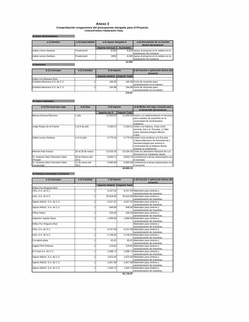
a) Datos del Estudiante:
a-1) Nombre a-2) Especialidad a-4) Descripción de su función dentro del proyecto.
Importe mensual AcumuladoAdela Lemus Santana Posdoctoral 6,000 6,000 Apoyo al proyecto en lo relativo en la
preparacion de muestrasAdela Lemus Santana Posdoctoral 6000 6,000 Apoyo al proyecto en lo relativo en la
preparacion de muestras12,000
b) Servicios:
b-1) Concepto b-2) Cantidad b-4) Función o aplicación dentro del proyecto
Importe Unitario Importe TotalEdilso Fco Reguera RuizEstafeta Mexicana S.A. de C.V. 1 185.99 185.99 Envió de muestras para
caracterización en el exteriorEstafeta Mexicana S.A. de C.V. 1 184.88 184.88 Envió de muestras para
caracterización en el exterior370.87
d) Viajes realizados:
d-1) Personal que viaja d-2) Días d-4) Motivo del viaje y función para el desarrollo del proyecto.
Importe por P. Importe TotalBlanca Zamora Reynoso 1 año 11,000.00 11,000.00 Asistir a un adiestramiento en técnicas
para estudios de superficie en la universidad de Southampton, Inglaterra.
Jorge Roque de la Puente 5 al 9 de julio 5,330.01 5,330.01 Asistir a la Habana, Cuba como ponente oral a la "Escuela y Taller sobre Nanotecnologìas Mèxico- Cuba".
Adela Lemus Santana 4 al 14 julio 5,770.34 5,770.34 Asistir como profesor a la Escuela Cubano-Mexicano de Nanociencia y Nanotecnología que sesionó a Universidad de la Habana donde impartió la conferencia.
Manuel Avila Santos 20 al 28 de marzo 10,434.30 10,434.30 Visita al Laboratorio Nacional de Luz Síncrotron en Campinas, Brasil.
Sr. Andreas Stein Zhenzhen Stein (Pasaje)
08 de febrero del 2011
8,693.71 8,693.71 Conferencia a temas relacionados con el proyecto
Sr. Andreas Stein Zhenzhen Stein (Hospedaje)
12 de enero del 2011
5,660.80 5,660.80 Conferencia a temas relacionados con el proyecto
46,889.16
e) Compras asociadas al proyecto
e-1) Concepto e-2) Cantidad e-4) Función o aplicación dentro del proyecto
Importe Unitario Importe TotalEdilso Fco Reguera RuizInfra, S.A. de S.V. 1 9,237.04 9,237.04 Materiales para sintesis y
caracterización de muestras.Infra, S.A. de S.V. 1 25,519.95 25,519.95 Materiales para sintesis y
caracterización de muestras.Sigma Aldrich, S.A. de C.V. 1 4,027.52 4,027.52 Materiales para sintesis y
caracterización de muestras.Sigma Aldrich, S.A. de C.V. 1 846.80 846.80 Materiales para sintesis y
caracterización de muestras.Office Depot 1 426.00 426.00 Materiales para sintesis y
caracterización de muestras.Alejandro Aguilar Nava 1 1,999.99 1,999.99 Materiales para sintesis y
caracterización de muestras.Edilso Fco Reguera Ruiz Materiales para sintesis y
caracterización de muestras.Infra, S.A. de S.V. 1 9,237.08 9,237.08 Materiales para sintesis y
caracterización de muestras.Infra, S.A. de S.V. 1 4,746.64 4,746.64 Materiales para sintesis y
caracterización de muestras.Cerrajeria plata 1 65.61 65.61 Materiales para sintesis y
caracterización de muestras.Digital Print Defensa 1 219.82 219.82 Materiales para sintesis y
caracterización de muestras.El Crisol S.A. de C.V. 1 2,998.72 2,998.72 Materiales para sintesis y
caracterización de muestras.Sigma Aldrich, S.A. de C.V. 1 1,873.40 1,873.40 Materiales para sintesis y
caracterización de muestras.Sigma Aldrich, S.A. de C.V. 1 2,607.68 2,607.68 Materiales para sintesis y
caracterización de muestras.Sigma Aldrich, S.A. de C.V. 1 1,933.72 1,933.72 Materiales para sintesis y
caracterización de muestras.65,739.97
e-3) Importe
Anexo 3Comprobación erogaciones del presupuesto otorgado para el Proyecto.
a-3) Apoyo otorgado $
b-3) Importe
d-3) Importe
CONCENTRADO FINANCIERO FINAL