
Universidad Miguel Hernández de Elche
El gen MAS2 de Arabidopsis
es esencial, codifica una proteína ortóloga de las
NKAP animales y participa en el silenciamiento del
ADN ribosómico 45S
Ana Belén Sánchez García Elche, 2016


El gen MAS2 de Arabidopsis es esencial,codifica una proteína ortóloga de las
NKAP animales y participa en el silenciamiento del ADN ribosómico 45S
Trabajo realizado por la Licenciada Ana Belén Sánchez García, en la Unidad de Genética
del Instituto de Bioingeniería de la Universidad Miguel Hernández de Elche, para optar al
grado de Doctor.
Elche, 24 de junio de 2016.


MARÍA ROSA PONCE MOLET, Catedrática de Genética de la Universidad Miguel
Hernández de Elche,
HAGO CONSTAR:
Que el presente trabajo ha sido realizado bajo mi dirección y recoge fielmente la labor
realizada por la Licenciada Ana Belén Sánchez García para optar al Grado de Doctor.
Las investigaciones reflejadas en esta Tesis se han desarrollado íntegramente en la
Unidad de Genética del Instituto de Bioingeniería de la Universidad Miguel Hernández de
Elche.
María Rosa Ponce Molet
Elche, 24 de junio de 2016.


Avenida de la Universidad s/n 03202 ELCHE (Alicante)
Telf: 96 591 8817 - Fax: 96 522 2033 e-mail: [email protected]
Universidad Miguel Hernández
A quien corresponda:
Eugenio Vilanova Gisbert, Catedrático de Toxicología y Director del Instituto de
Bioingeniería,
HACE CONSTAR:
Que da su conformidad a la lectura de la Tesis Doctoral presentada por Doña Ana Belén
Sánchez García, titulada “El gen MAS2 de Arabidopsis es esencial, codifica una
proteína ortóloga de las NKAP animales y participa en el silenciamiento del ADN
ribosómico 45S”, que se ha desarrollado dentro del Programa de Doctorado en
Bioingeniería de este Instituto, bajo la dirección de la profesora Dña. María Rosa Ponce
Molet.
Lo que firmo en Elche, a instancias de la interesada y a los efectos oportunos, a
veintitrés de junio de dos mil dieciséis.
Eugenio Vilanova Gisbert
Catedrático de Toxicología
Director del Instituto de Bioingeniería


A mi familia
A Manuel Ginés
A Esperanza del Mar y Miriam


Índices I
ÍNDICE DE MATERIAS
ÍNDICE DE FIGURAS ....................................................................................................... II
ÍNDICE DE TABLAS ....................................................................................................... III
I.- PREFACIO ..................................................................................................................... 1
II.- RESUMEN Y CONCLUSIONES ................................................................................ 3
III.- INTRODUCCIÓN ....................................................................................................... 5 III.1.- Arabidopsis, un sistema modelo para el estudio de la biología vegetal ...................... 5 III.2.- Mecanismos de regulación de la expresión génica en los eucariotas .......................... 6
III.2.1.- Regulación transcripcional .................................................................................. 7 III.2.1.1.- Regulación mediante factores de transcripción ........................................... 7 III.2.1.2.- Regulación mediante marcas epigenéticas .................................................. 7
III.2.1.2.1.- Modificación química de las histonas .................................................. 8 III.2.1.2.2.- Metilación del ADN ............................................................................. 9
III.2.2.- Regulación postranscripcional ............................................................................. 9 III.2.2.1.- Silenciamiento génico mediado por ARN ................................................... 9 III.2.2.2.- Maduración de los ARNm ......................................................................... 13
III.2.2.2.1.- Splicing constitutivo y alternativo ...................................................... 13 III.2.2.2.2.- Funciones de las proteínas SR en el splicing ..................................... 15 III.2.2.2.3.- Funciones de las hnRNP en el splicing .............................................. 18
III.3.- Biogénesis del ribosoma citoplásmico de Arabidopsis ............................................. 21 III.3.1.- Componentes del ribosoma citoplásmico de los eucariotas .............................. 21 III.3.2.- Organización y expresión del ADNr de Arabidopsis ........................................ 21
III.4.- Las proteínas multifuncionales NKAP ...................................................................... 24 III.4.1.- Relación entre las proteínas NKAP y los ácidos nucleicos ............................... 24 III.4.2.- Funciones de la NKAP humana en el procesamiento del ARN ........................ 25
III.5.- Mecanismos de supresión genética ........................................................................... 28 III.6.- Antecedentes y objetivos ........................................................................................... 30
III.6.1.- Aislamiento de alelos ago1 hipomorfos ............................................................ 30 III.6.2.- Búsqueda de supresores del fenotipo morfológico de ago1-52 ......................... 31 III.6.3.- Objetivos ............................................................................................................ 32
IV.- BIBLIOGRAFÍA DE LA INTRODUCCIÓN ......................................................... 35
V.- PUBLICACIONES ...................................................................................................... 45
VI.- ANEXO: COMUNICACIONES A CONGRESOS ................................................ 95
VII.- AGRADECIMIENTOS ......................................................................................... 117
II Índices
ÍNDICE DE FIGURAS
Figura 1.- Mecanismos de represión génica mediante metilación del ADN ...................... 10 Figura 2.- Árbol filogenético de las proteínas AGO de las plantas .................................... 12 Figura 3.- Patrones de splicing alternativo ......................................................................... 14 Figura 4.- Estructura de un intrón de tipo U2 .................................................................... 15 Figura 5.- Mecanismo del splicing de los pre-ARNm por el spliceosoma de tipo
U2 ............................................................................................................................ 16 Figura 6.- Interacciones de las proteínas SR en los exones de tipo U2 durante la
formación del complejo E del spliceosoma ............................................................. 17 Figura 7.- Mecanismos de regulación del splicing mediados por hnRNP ......................... 20 Figura 8.- Localización cromosómica y estructura de los genes que codifican los
ARNr de Arabidopsis .............................................................................................. 23 Figura 9.- Representación esquemática de la proteína AGO1, con indicación de
los efectos de las mutaciones ago1-51 y ago1-52 sobre su secuencia. ................... 31 Figura 10.- Rosetas de las líneas supresoras identificadas tras una mutagénesis
con EMS del mutante ago1-52. ............................................................................... 32

Índices III
ÍNDICE DE TABLAS
Tabla 1.- Genes SynMuv B de Caenorhabditis elegans .................................................... 26 Tabla 2.- Interactores de la NKAP humana de función conocida o predicha ..................... 28


I.- PREFACIO


Prefacio 1
I.- PREFACIO
Este documento se ha elaborado siguiendo la normativa de la Universidad Miguel
Hernández de Elche para la “Presentación de Tesis Doctorales con un conjunto de
publicaciones” y se ha dividido en las partes siguientes:
1.- Un apartado de Resumen y Conclusiones.
2.- Una Introducción, en la que se presenta el tema de la Tesis y los antecedentes y
objetivos del trabajo realizado.
3.- Una Bibliografía de la Introducción.
4.- Un apartado de Publicaciones, que contiene la siguiente (se indica su factor de
impacto [FI] de 2014):
Sánchez-García, A.B., Aguilera, V., Micol-Ponce, R., Jover-Gil, S., y Ponce, M.R. (2015). Arabidopsis MAS2, an essential gene that encodes a homolog of animal NF-kappa B Activating Protein, is involved in 45S rDNA silencing. Plant Cell 27, 1999-2015 (FI: 9,338).
5.- Un Anexo, que incorpora 9 comunicaciones a congresos: 3 nacionales y 6
internacionales.
La introducción de esta Tesis no incluye un apartado de Materiales y Métodos,
que están descritos en el artículo. Este documento incorpora dos bibliografías: la del
artículo y la de la introducción. Todas las citas que se intercalan en la introducción de
esta memoria se corresponden con referencias que aparecen en la bibliografía del mismo
apartado; algunas de dichas citas se repiten en la bibliografía del artículo.
Durante mi periodo predoctoral también he publicado tres artículos que no se
incluyen en esta Tesis:
Ferrández-Ayela, A., Alonso-Peral, M.M., Sánchez-García, A.B., Micol-Ponce, R.,
Pérez-Pérez, J.M., Micol, J.L., y Ponce, M.R. (2013). Arabidopsis TRANSCURVATA1 encodes NUP58, a component of the nucleopore central channel. PLOS ONE 8, e67661 (FI: 3,234).
Ferrández-Ayela, A., Micol-Ponce, R., Sánchez-García, A.B., Alonso-Peral, M.M.,
Micol, J.L., y Ponce, M.R. (2013). Mutation of an Arabidopsis NatB N-alpha-terminal acetylation complex component causes pleiotropic developmental defects. PLOS ONE 8, e80697 (FI: 3,234).

2 Prefacio
Micol-Ponce, R.*, Sánchez-García, A.B.*, Xu, Q., Barrero, J.M., Micol, J.L., y Ponce, M.R. (2015). Arabidopsis INCURVATA2 regulates salicylic acid and abscisic acidsignaling, and oxidative stress responses. Plant and Cell Physiology 56, 2207-2219(FI: 4,931).

II.- RESUMEN Y CONCLUSIONES


Resumen y Conclusiones 3
II.- RESUMEN Y CONCLUSIONES
Una parte importante de la regulación de la expresión de muchos genes
eucarióticos es postranscripcional. Participan en este proceso en Arabidopsis los
complejos del silenciamiento génico inducido por ARN (RISC), cuyo componente principal
es la proteína AGO1 (ARGONAUTE1). La insuficiencia de función del gen AGO1 perturba
numerosos procesos de desarrollo y suele causar esterilidad o letalidad. Para
comprender mejor la actividad y las interacciones de AGO1, se realizó en el laboratorio
de María Rosa Ponce una búsqueda de modificadores del fenotipo morfológico de su
alelo ago1-52, que es hipomorfo, viable y fértil. Se aislaron así 23 mutantes inducidos por
metanosulfonato de etilo, de fenotipo similar al silvestre, y se denominó MAS
(MORPHOLOGY OF AGO1-52 SUPPRESSED) a sus genes supresores. La clonación
posicional de uno de ellos, MAS2, reveló que codifica una proteína homóloga de las
NKAP (NF-kappa B Activating Protein) de los metazoos.
MAS2 es un gen de copia única y esencial: sus alelos insercionales mas2-2 y
mas2-3 son recesivos y letales embrionarios. Hemos identificado 9 alelos portadores de
mutaciones puntuales supresoras, 8 de las cuales (mas2-1 y de mas2-4 a mas2-10) se
agrupan en la región que codifica el dominio conservado SynMuv, que caracteriza a las
NKAP.
mas2-1 suprime el fenotipo de ago1-51 y ago1-52, cuyo splicing está alterado,
pero no el de ago1-25 y ago1-27, que causan sustituciones de aminoácidos. Tanto la
sobreexpresión constitutiva del alelo silvestre MAS2 como la del mutante mas2-1 carecen
de efecto fenotípico en la estirpe silvestre; la sobreexpresión de mas2-1, pero no la de
MAS2, suprime el fenotipo de ago1-52. Estos resultados sugieren que mas2-1 es un alelo
antimorfo del gen MAS2.
El mutante ago1-52 produce dos variantes del ARN mensajero (ARNm) de AGO1,
una mutante y mayoritaria y otra silvestre y casi indetectable. La proporción relativa de la
variante de ARNm silvestre se incrementa en casi todos los dobles mutantes ago1-52
mas2 estudiados. Estos resultados indican que los alelos mas2 portadores de mutaciones
puntuales son supresores informacionales y que la proteína MAS2 participa en el
procesamiento del ARN en general o el de AGO1 en particular.
Hemos construido un microARN artificial que reprime parcialmente a MAS2 (amiR-
MAS2) y causa floración temprana, deforma las hojas y las flores y reduce la fertilidad. La
proporción relativa de la proteína AGO1 silvestre es mucho mayor en los dobles mutantes
ago1-52 mas2 que en el mutante simple ago1-52. En las plantas ago1-52 amiR-MAS2,

4 Resumen y Conclusiones
por el contrario, los niveles de la proteína AGO1 silvestre se reducen. Nuestros
resultados sugieren que MAS2 participa en algún mecanismo de control de la calidad de
los ARNm, favoreciendo la traducción de la variante silvestre o dificultando la de la
mutante.
Hemos obtenido una fusión transcripcional MAS2pro:GUS, con la que hemos
visualizado la expresión de MAS2 en todos los tejidos estudiados, que alcanza valores
máximos en células en división activa, como las de los ápices de la raíz y de las hojas
vegetativas. También hemos obtenido los transgenes 35Spro:MAS2:GFP y
MASpro:MAS2:GFP, que nos han permitido establecer que MAS2 es una proteína nuclear
en células en división muy activa, y perinucleolar en las restantes. Hemos demostrado
mediante hibridación in situ fluorescente que MAS2 se acumula en el ADN ribosómico
(ADNr) 45S de los organizadores nucleolares.
El gen NUC-L1 de Arabidopsis codifica la NUCLEOLINA1, que participa en el
control epigenético de la expresión del ADNr 45S. Tanto mas2-1 como amiR-MAS2
interaccionan genéticamente con un alelo nulo de NUC-L1. Hemos comprobado que
los promotores del ADNr 45S están hipometilados en las plantas amiR-MAS2, una
observación que indica que MAS2 regula negativamente al ADNr 45S.
Hemos realizado una búsqueda de interactores basada en el ensayo del doble
híbrido de la levadura, identificando proteínas presuntamente implicadas en el control del
splicing, en la biogénesis del ribosoma y en la reparación de roturas dobles en el ADN.
Estos interactores de MAS2 son similares a los descritos para la NKAP humana.
Las proteínas SR deben su nombre a su dominio RS, que es rico en serina (S) y
arginina (R); su participación en el splicing está sobradamente demostrada en diferentes
especies de hongos, animales y plantas. La NKAP humana presenta una región
aminoterminal a la que se ha considerado un dominio RS no canónico y se ha encontrado
asociada a diferentes complejos del spliceosoma. Nuestros resultados indican que MAS2,
la ortóloga en Arabidopsis de la NKAP humana, es una proteína multifuncional que
participa en el control del splicing, en la traducción del ARNm y en la regulación
transcripcional y el procesamiento de los genes del ADNr 45S. La letalidad embrionaria
asociada a la insuficiencia de función de MAS2, su localización subcelular, la naturaleza
de sus interactores y los procesos en los que participa revelan una conservación no solo
estructural sino también funcional respecto a la NKAP humana.

III.- INTRODUCCIÓN


Introducción 5
III.- INTRODUCCIÓN
III.1.- Arabidopsis, un sistema modelo para el estudio de la biología vegetal Arabidosis thaliana (en adelante, Arabidopsis) es una dicotiledónea de la familia
de las Brasicáceas, descubierta en el siglo XVI por Johannes Thal. Friedrich Laibach, uno
de los discípulos de Eduard Strasburger, comenzó a estudiarla en 1907 (Bowman, 1994).
Se convirtió en uno de los organismos experimentales de elección preferente en Biología
vegetal a finales de la década de los ochenta (Meyerowitz, 1994).
La comunidad científica de Arabidopsis es muy activa y cooperativa. Comenzó a
vertebrarse en 1964 en torno al boletín Arabidopsis Information Service (AIS) que dejó de
publicarse en 1990. En la actualidad existe un amplísimo repertorio de recursos
informáticos disponibles para quienes estudian esta planta, el más importante de los
cuales es la base de datos TAIR (The Arabidopsis Information Resource; http://www.
arabidopsis.org), que contiene mucha información sobre genes, marcadores moleculares,
polimorfismos, análisis transcriptómicos y epigenómicos y numerosas herramientas
bioinformáticas. Son varios los centros de conservación y distribución de estirpes y clones
de ADN de Arabidopsis, entre los que destacan el norteamericano (ABRC; Arabidopsis
Biological Resource Center; https://abrc.osu.edu/) y el europeo (NASC; Nottingham
Arabidopsis Stock Centre; http://nasc.nott.ac.uk/). La primera conferencia internacional
sobre Arabidopsis (ICAR; International Conference on Arabidopsis Research), se celebró
en Göttingen (Alemania) en 1965 (Provart et al., 2016). Desde 1995 se celebra anual y
cíclicamente en Asia-Oceanía, Norteamérica y Europa.
El genoma haploide de Arabidopsis es uno de los más pequeños de las plantas
superiores: su tamaño, 125 Mb, mucho menor que los de las especies cultivadas, propició
su secuenciación entre 1996 a 2000. Fue el primer genoma vegetal secuenciado
completamente y el tercero de los organismos pluricelulares, solo precedido por los de
Drosophila melanogaster (Adams et al., 2000) y Caenorhabditis elegans (The
Caenorhabditis elegans Sequencing Consortium, 1998). Su anotación más reciente indica
que contiene 27.655 genes que codifican proteínas, 5.051 genes no codificantes, 952
pseudogenes y 35.846 loci de ARN pequeños (Cheng et al., 2016; https://apps.
araport.org/thalemine/dataCategories.do).
Los procedimientos de mutagénesis clásica han resultado muy eficaces para la
disección genética de numerosos procesos biológicos en Arabidopsis. El análisis
funcional de los genes de esta planta se ha visto facilitado por los miles de mutantes
aislados tras mutagénesis química y los centenares de miles de mutantes señalizados,

6 Introducción
obtenidos mediante transformación por infección con la bacteria Agrobacterium
tumefaciens (Azpiroz-Leehan y Feldmann, 1997). La anotación inicial del genoma de
Arabidopsis evidenció que se desconocía la función de la mayor parte de sus genes, que
está siendo comprendida progresivamente mediante la acumulación de grandes
cantidades de datos sobre sus patrones de expresión, la localización subcelular de sus
productos, los fenotipos causados por su sobreexpresión o la ausencia de su función y el
proteoma e interactoma de esta planta.
III.2.- Mecanismos de regulación de la expresión génica en los eucariotas Comprender los mecanismos de la regulación de la expresión génica implica
analizar la capacidad de los ácidos nucleicos para contener información genética, los
diferentes modos en que se ha resuelto el problema de su empaquetamiento en las
células a lo largo de la evolución, los mecanismos por los cuales tal información fluye y es
ejecutada, la inducción o represión de los genes en el momento y lugar adecuados y la
modificación de sus productos proteicos.
Se acepta generalmente la universalidad de la lógica de la regulación de la
expresión génica mediada por factores de transcripción que se unen a sus secuencias
reguladoras diana en el ADN. Este concepto tiene su punto de partida en el abordaje
experimental genético y bioquímico llevado a cabo principalmente por Jacob y Monod a
mediados del siglo XX, que analizaron el operón de la lactosa de la bacteria Escherichia
coli (Jacob y Monod, 1961). El descubrimiento de Jacob y Monod permitió comenzar a
entender la complejidad que subyace en la regulación de la expresión génica tanto en los
procariotas como en los eucariotas.
Los eucariotas son más complejos estructural y funcionalmente y presentan más
mecanismos de regulación de la expresión génica que los procariotas. El núcleo celular
compartimenta la transcripción y la traducción, y genera la necesidad de una etapa
adicional en el flujo de la información genética: la exportación de los ARNm del núcleo al
citoplasma. La regulación de la expresión génica en los eucariotas es transcripcional, se
controla el inicio de la transcripción, tal como ocurre en las bacterias, pero también
postranscripcional y postraduccional, ya que existen mecanismos que operan sobre la
maduración de los transcritos, su longevidad y exportación al citoplasma. A los anteriores
se añaden los que regulan la activación, inactivación, degradación y localización
subcelular de las proteínas.

Introducción 7
III.2.1.- Regulación transcripcional III.2.1.1.- Regulación mediante factores de transcripción
La expresión de los genes eucarióticos y procarióticos está regulada a varios
niveles, el primero de los cuales es la iniciación de la transcripción. La unión de factores
de transcripción generales a una región próxima al lugar en el que se iniciará la
transcripción, que se conoce como promotor basal, tiene como consecuencia final el
ensamblaje de la maquinaria transcripcional. A la decisión de si la transcripción comienza
o no también contribuyen otros factores, que actúan como activadores o represores,
uniéndose a secuencias potenciadoras (enhancers) o silenciadoras, respectivamente.
Estas últimas proteínas pueden actuar en solitario o formando parte de complejos (Struhl,
1999).
Una sola ARN polimerasa (ARN pol) transcribe todos los genes en un procariota;
en los eucariotas existen varias, siendo la ARN pol II la que transcribe los genes que
codifican proteínas, generando un pre-ARNm que debe madurar y ser exportado al
citoplasma para su traducción a proteína. Los factores necesarios para la maduración del
pre-ARNm y la exportación del ARNm al citoplasma son incorporados a la ARN pol II
durante la elongación de la transcripción (Maniatis y Reed, 2002).
III.2.1.2.- Regulación mediante marcas epigenéticas La gran longitud del ADN respecto al volumen del núcleo celular que lo alberga
hace necesaria su compactación. Esta última, sin embargo, complica los procesos de
replicación, reparación y transcripción. Para compactarse, el ADN eucariótico se asocia
con histonas para formar la cromatina junto con otras proteínas y ARN. Las histonas son
proteínas relativamente pequeñas y básicas, con residuos de arginina y lisina cargados
positivamente, que les confieren una gran afinidad por el ADN. Las histonas H1, H2A,
H2B, H3 y H4 forman parte de la cromatina de todos los eucariotas. Dos moléculas de
cada una de las histonas H2A, H2B, H3 y H4 forman un octámero en torno al cual se
enrolla el ADN para formar un nucleosoma, que constituye la unidad básica de
empaquetamiento de la cromatina (Kornberg y Lorch, 1999).
La cromatina se condensa y descondensa a lo largo del ciclo celular. La
heterocromatina es la cromatina interfásica altamente condensada, en la que el acceso
de la maquinaria transcripcional a los promotores de los genes se ve dificultado (Luger y
Hansen, 2005; Li y Reinberg, 2011; Zhou et al., 2011). La actividad de los genes también
depende de la modificación de las histonas y la metilación del ADN. En la compactación y
descompactación de la cromatina intervienen complejos proteicos que modifican las

8 Introducción
histonas, sustituyen unas variantes por otras o desplazan los nucleosomas. Por ejemplo,
la sustitución de las histonas canónicas H2A, H2B y H3 en los nucleosomas de los
mamíferos por sus variantes H2A.Z, H2A.X y H3.3 propicia las interacciones de la
cromatina con complejos remodeladores o con chaperonas de histonas (Klose y Bird,
2006; Kouzarides, 2007; Narlikar, 2010; Talbert y Henikoff, 2010; Bannister y Kouzarides,
2011; Burgess y Zhang, 2013; Das y Tyler, 2013; Harshman et al., 2013; Maze et al.,
2014; Rothbart y Strahl, 2014).
III.2.1.2.1.- Modificación química de las histonas La modificación postraduccional del extremo amino de las histonas juega un papel
clave en la regulación de la estructura y empaquetamiento de la cromatina, y por tanto en
el control de la transcripción. Existen diferentes tipos de modificación, que afectan a
diferentes aminoácidos, siendo las acetilaciones, metilaciones, fosforilaciones y
ubiquitinaciones las que mejor se conocen (Patel y Wang, 2013; Rothbart y Strahl, 2014).
Aunque las primeras modificaciones de las histonas se describieron a mediados del siglo
XX (Allfrey y Mirsky, 1964; Gutierrez y Hnilica, 1967), su significado biológico permaneció
incierto durante tres décadas, hasta que se estableció que las N-acetiltransferasas p55
de Tetrahymena thermophila y Gcn5 (general control nonderepressible 5) de
Saccharomyces cerevisiae y las desacetilasas HDAC1 (histone deacetylase 1) humana y
Rpd3 (reduced potassium dependency 3) de Saccharomyces cerevisiae regulaban la
transcripción (Brownell et al., 1996; Taunton et al., 1996; Struhl, 1998).
Se denomina marcas epigenéticas a las modificaciones químicas de la cromatina
que afectan a las histonas y al ADN pero que no alteran la secuencia primaria de este
último. Aunque se asociaron inicialmente algunas marcas epigenéticas a determinados
estados de actividad o inactividad de los genes (Strahl y Allis, 2000; Turner, 2000), se
está generalizando una visión mucho más compleja del papel de las modificaciones
químicas de las histonas en la regulación génica (Taverna et al., 2007). Se han descrito
diferentes complejos multiproteicos que incorporan, eliminan o reconocen las marcas
epigenéticas de las histonas, que actúan coordinadamente con los complejos metiladores
o desmetiladores del ADN (Musselman et al., 2012). El advenimiento de la secuenciación
masiva ha facilitado la caracterización de las marcas epigenéticas a escala genómica
(Vermeulen et al., 2010; Bartke et al., 2013; Han y Garcia, 2013; Soldi et al., 2013).

Introducción 9
III.2.1.2.2.- Metilación del ADN La metilación del ADN es un proceso que ocurre en los procariotas y los
eucariotas, pero las bases nitrogenedas que se metilan y su función biológica son
distintos. En los procariotas se metilan las adeninas y citosinas, en la mayoría de los
casos como parte de mecanismos de defensa contra ADN exógeno, en los que también
participan las restrictasas (Arber y Dussoix, 1962; revisado en Noyer-Weidner y Trautner,
1993); solo en algunos casos la metilación está asociada al control del inicio de la
replicación y a la reparación del ADN (revisado en Wion y Casadesús, 2006).
En los animales se metilan las citosinas de los dinucleótidos CG, y en las plantas,
en los contextos CG, CHG y CHH (H indica A, C o T) (Cokus et al., 2008; Rothbart y
Strahl, 2014). La metilación de las citosinas tiene dos funciones alternativas en los
eucariotas, según la región metilada: en los promotores se asocia a inactivación génica,
como en la impronta génica, el silenciamiento de retrotransposones o la inactivación del
cromosoma X de las hembras de los mamíferos (Klose y Bird, 2006). La metilación de las
regiones codificantes de los genes, en cambio, se ha asociado a su activación (Zhang et
al., 2006).
La presencia de adeninas metiladas (6mA) en los promotores de los genes de
Chlamydomonas reinhardtii y Drosophila melanogaster se ha correlacionado con el
incremento de su actividad (Fu et al., 2015; Zhang et al., 2015; revisado en Heyn y
Esteller, 2015). La 6mA se ha asociado en Caenorhabditis elegans al mantenimiento
transgeneracional de la herencia epigenética (Greer et al., 2015).
La metilación del ADN reprime la transcripción de dos modos: directamente,
impidiendo la unión de los activadores transcripcionales a sus secuencias reguladoras, e
indirectamente, reclutando proteínas de unión a dinucleótidos CG metilados (MBP;
methyl-CpG-binding proteins) que modifican la cromatina y atraen a correpresores de la
transcripción (Figura1, en la página 10; Klose y Bird, 2006).
III.2.2.- Regulación postranscripcional III.2.2.1.- Silenciamiento génico mediado por ARN
Se denomina silenciamiento génico mediado por ARN al conjunto de los procesos
en que intervienen moléculas de ARN no codificantes que reprimen la expresión génica
de manera específica de secuencia (Brodersen y Voinnet, 2006). Estos mecanismos han
recibido diferentes nombres según el organismo en que se han descrito: cosupresión en
las plantas, extinción (quelling) en los hongos y ciliados, e interferencia de ARN en los
animales (Napoli et al., 1990; van der Krol et al., 1990; Cogoni et al., 1994; Fire et al.,

10 Introducción
MBPCH3CH3
FT
MBPCH3
A B
C
CH3
1998; Kennerdell y Carthew, 1998; Ruiz et al., 1998). Se trata de rutas de regulación muy
conservadas en los eucariotas (Chapman y Carrington, 2007), cuya función es la
protección del genoma frente al ARN viral, y la expresión inadecuada de transgenes,
transposones y otras secuencias endógenas altamente repetitivas como los genes que
codifican los ARN ribosómicos (Earley et al., 2010; Gomes et al., 2013; Pumplin y
Voinnet, 2013; Rajeevkumar et al., 2015).
Figura 1.- Mecanismos de represión génica mediante metilación del ADN. (A) La presencia de nucleótidos metilados en ciertas secuencias reguladoras pueden impedir su unión a factores de transcripción (FT). (B) Las proteínas de unión a dinucleótidos CG metilados (MBP) reconocen directamente el ADN metilado y reclutan moléculas correpresoras que modifican la estructura de la cromatina de su entorno y silencian la transcripción. (C) La metilación de regiones internas de los genes también puede inhibir la elongación de la transcripción, al impedir el avance de la maquinaria transcripcional. Los símbolos ─┤y ↱ representan represión y sitio de inicio de latranscripción, respectivamente. Modificado a partir de Klose y Bird (2006).
Se han descrito diversos tipos de ARN pequeños (20-30 nt) relacionados con
procesos de silenciamiento génico, que reciben el nombre genérico de ARN pequeños
interferentes (ARNpi; siRNA; small interfering RNA). Estos ARNpi se organizan en
complejos riboproteicos denominados RITS (RNA-induced initiation of transcriptional
gene silencing) y RISC (RNA-induced silencing complex), que actúan en procesos de
silenciamiento transcripcional (TGS; transcriptional gene silencing) y postranscripcional

Introducción 11
(PTGS; posttranscriptional gene silencing), respectivamente (Tang, 2005; Jones-Rhoades
et al., 2006; Chapman y Carrington, 2007; Chen, 2009; Rajeevkumar et al., 2015;
revisado en Pumplin y Voinnet, 2013). El agente inductor del silenciamiento en ambos
procesos es un ARN bicatenario (ARNdc), sustrato de una ribonucleasa de tipo III de la
familia Dicer (DCL; ribonuclease III-type Dicer-like), que lo procesa generando los ARNpi
(revisado en Gomes et al., 2013; y en Pumplin y Voinnet, 2013). A pesar de que las rutas
del TGS y el PTGS regulan la expresión génica a diferentes niveles, sus respectivas
maquinarias comparten componentes (Brodersen y Voinnet, 2006; Voinnet, 2008; Xie y
Qi, 2008; Chen, 2009; Hoffer et al., 2011).
La clasificación de los ARNpi se basa en su biogénesis y funciones (Axtell, 2013;
Gomes et al., 2013). Los microARN (miARN) son ARNpi producidos por genes
endógenos, denominados MIRNA, cuya transcripción por la ARN pol II genera un
precursor en el que se forma una horquilla por apareamiento intracatenario, que es
procesada por una ribonucleasa Dicer, rindiendo el miARN maduro, de unos 21 nt de
longitud (Jones-Rhoades et al., 2006). Los miARN silencian a nivel postranscripcional la
expresión de numerosos genes endógenos (Jones-Rhoades et al., 2006).
La transcripción de transposones, repeticiones invertidas y regiones intergénicas o
altamente repetitivas activa la maquinaria del PTGS, induciéndose la síntesis de ARNpi,
de 21-22 o 24 nt (Kasschau et al., 2007). La unión de los ARNpi de 21-22 nt a su ARN
diana atrae a una ARN polimerasa dependiente de ARN (RdRP; RNA-dependent RNA
polymerase) que genera copias adicionales del ARNpi (Lu et al., 2006; Chapman y
Carrington, 2007; Xie y Qi, 2008; Axtell, 2013). Los ARNpi más largos, de 24 nt, parecen
estar implicados en la metilación de novo de las citosinas de sus secuencias
complementarias en el ADN (Matzke et al., 2009; Law y Jacobsen, 2010; Axtell, 2013).
La etapa final de la regulación de la expresión génica mediada por moléculas de
ARN es la asociación de los ARNpi a los complejos RISC y RITS, cuyo elemento más
importante es un miembro de la familia ARGONAUTE (AGO), responsable de la actividad
silenciadora del complejo (revisado en Kawamata y Tomari, 2010). Las proteínas AGO
presentan tres dominios funcionales, denominados PAZ, MID y PIWI, organizados en una
arquitectura bilobulada con su región aminoterminal y el dominio PAZ en uno de los
lóbulos, y MID, PIWI y la región carboxiterminal en el otro (Wang et al., 2009). El dominio
PAZ se une al extremo 3’ de los ARNpi, mientras que los dominios MID y PIWI lo hacen
al 5’ (Wang et al., 2009; Mallory y Vaucheret, 2010; Parker, 2010), posicionando así
correctamente al ARNpi respecto a su ARNm diana. Aunque el dominio PIWI contiene un
sitio catalítico de tipo RNasa H (Wang et al., 2009; Parker, 2010), no todas las proteínas
12 Introducción
Clado I
Clado II
Clado III
AlgasPhyscomitrella patensSelaginella moellendorffiiOryza sativaZea maysArabidopsis thalianaGlycine max
AGO son capaces de cortar a sus ARNm diana (Mallory et al., 2009; Mallory y Vaucheret,
2010).
Las proteínas AGO están muy conservadas y se han identificado tanto en los
procariotas como en los eucariotas (Swarts et al., 2014). Las procarióticas participan en la
defensa contra elementos genéticos móviles y ADN exógeno (Swarts et al., 2014).
También están muy conservadas las AGO eucarióticas, aunque el número de parálogos
es 1 en Schizosaccharomyces pombe, 2 en Neurospora crassa, 5 en Drosophila
melanogaster, 8 en los mamíferos, 10 en Arabidopsis y 27 en Caenorhabditis elegans
(revisado en Höck y Meister, 2008; y en Mallory y Vaucheret, 2010).
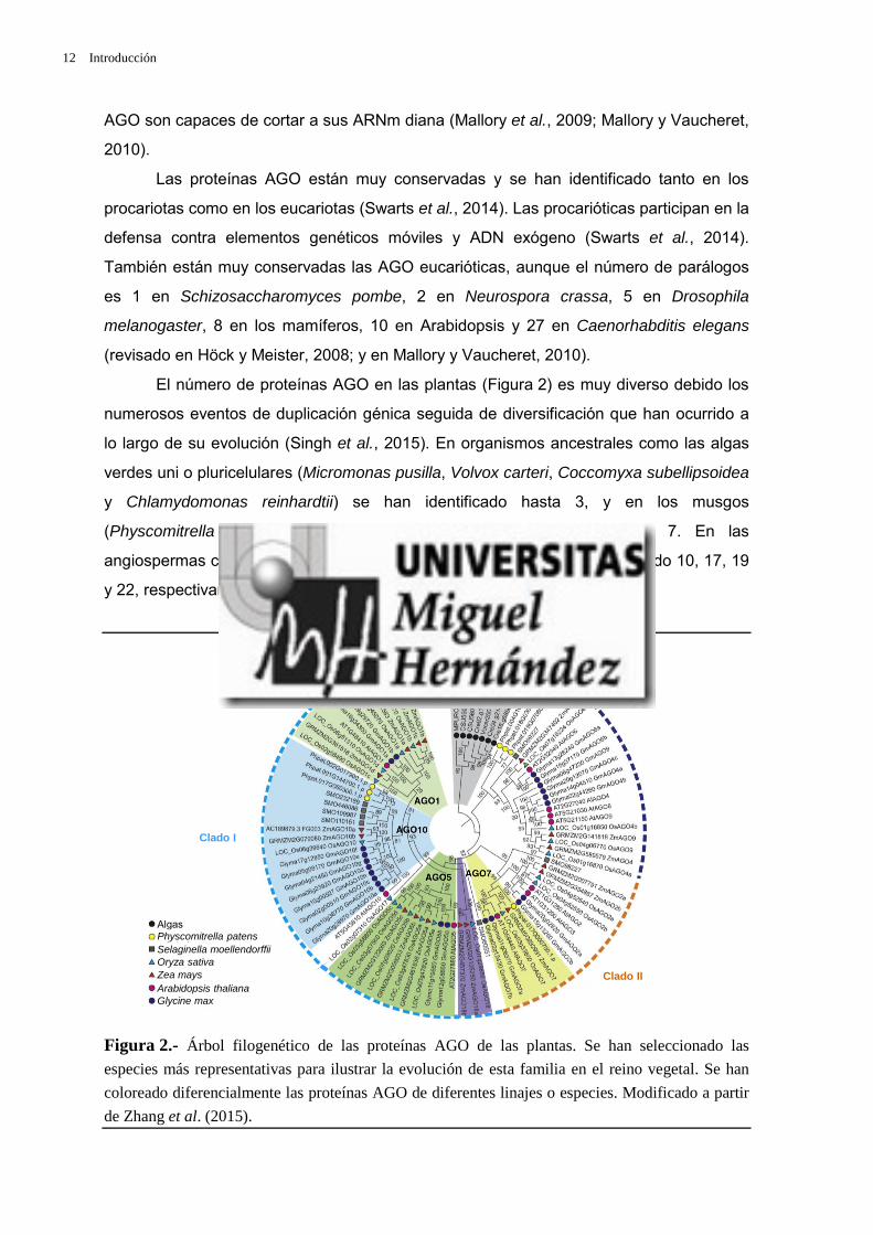
El número de proteínas AGO en las plantas (Figura 2) es muy diverso debido los
numerosos eventos de duplicación génica seguida de diversificación que han ocurrido a
lo largo de su evolución (Singh et al., 2015). En organismos ancestrales como las algas
verdes uni o pluricelulares (Micromonas pusilla, Volvox carteri, Coccomyxa subellipsoidea
y Chlamydomonas reinhardtii) se han identificado hasta 3, y en los musgos
(Physcomitrella patens) y helechos (Selaginella moellendorffii), hasta 7. En las
angiospermas como Arabidopsis, el maíz, el arroz o la soja se han identificado 10, 17, 19
y 22, respectivamente (Zhang et al., 2015).
Figura 2.- Árbol filogenético de las proteínas AGO de las plantas. Se han seleccionado las
especies más representativas para ilustrar la evolución de esta familia en el reino vegetal. Se han
coloreado diferencialmente las proteínas AGO de diferentes linajes o especies. Modificado a partir
de Zhang et al. (2015).

Introducción 13
Las proteínas AGO de las plantas se agrupan en tres clados (Figura 2; revisado
en Vaucheret, 2008; Mallory y Vaucheret, 2010; Zhang et al., 2015). Al clado I de
Arabidopsis pertenecen AGO1, AGO5 y AGO10; al II, AGO2, AGO3 y AGO7; y al III,
AGO4, AGO6, AGO8 y AGO9 (revisado en Vaucheret, 2008).
El gen AGO1 de Arabidopsis es el miembro fundador de la familia AGO, a la que
ha dado nombre. Se identificó en una búsqueda de mutantes afectados en el desarrollo.
Su nombre se debe al aspecto de las hojas vegetativas de sus mutantes de fenotipo más
severo, que recordaron a sus descubridores los tentáculos del cefalópodo Argonauta argo
(Bohmert et al., 1998). La identificación de alelos ago1 adicionales evidenció el papel
fundamental de AGO1 en el silenciamiento mediado por ARN y su unión preferente a
ARNpi de 21 nt con una uridina en su extremo 5’, como la mayoría de los miARN.
Además de jugar un papel central en el PTGS mediado por miARN, AGO1 también es
esencial en la defensa contra los virus (revisado en Vaucheret, 2008; Jover-Gil et al.,
2012; Zhang et al., 2015).
III.2.2.2.- Maduración de los ARNm III.2.2.2.1.- Splicing constitutivo y alternativo
La maduración de los transcritos primarios de los ARNm eucarióticos (pre-ARNm)
comprende varios procesos cuya discusión excede el propósito de esta Tesis. Se
comenta a continuación solo uno de ellos, que elimina los intrones y reúne los exones de
un pre-ARNm: el splicing (no existe una traducción de uso generalmente aceptado de
este vocablo; algunos autores lo traducen como “ayuste”, la unión de dos cuerdas o
trozos de madera por sus extremos).
Se denomina splicing alternativo a la eliminación o retención total o parcial de
exones o intrones de un pre-ARNm, que da lugar a diferentes variantes del ARNm de un
gen, que a su vez rinden proteínas que pueden diferir en su función, incluso ser
antagónicas, o en su localización subcelular (Ellis et al., 2012; Loraine et al., 2013). Los
diferentes patrones de splicing alternativo de un gen suelen ser específicos de tejido y
consisten en la omisión de exones o la retención de intrones (Figura 3, en la página 14;
Black, 2003; Kelemen et al., 2013; Reddy et al., 2013; revisado en Reddy et al., 2013). Un
95% de los genes con varios exones de los animales y un 61% de los de las plantas
sufren splicing alternativo (Syed et al., 2012).
La síntesis de ARNm aberrantes, productos del splicing alternativo, puede activar
diferentes rutas de degradación de ARNm. Una de las mejor estudiadas es la de
decaimiento del ARNm mediado por secuencias sin sentido (NMD; Nonsense-mediated

14 Introducción
Poli(A)
Poli(A)
A
C
B
D
E
G
F
mRNA Decay) que está acoplada a la traducción, degradando en el citoplasma los ARNm
que contienen codones de terminación prematuros (Brogna y Wen, 2009).
Figura 3.- Patrones de splicing alternativo. Los intrones constitutivos, ausentes de todas las
variantes del ARNm maduro, están representados por líneas grises, y los exones, por rectángulos
coloreados. Los exones constitutivos aparecen en verde, y los exones e intrones retenidos
alternativamente, en azul. Los eventos de splicing constitutivo o alternativo están indicados por
líneas negras continuas o discontinuas, respectivamente. (A) Exclusión completa de un exón. (B y
C) La presencia de sitios (B) donantes y (C) aceptores alternativos permite aumentar o disminuir la
longitud de un determinado exón mediante splicing alternativo. (D) Retención de un intrón. (E)
Exclusión mutua de exones: solo uno de los dos representados en azul está presente en cada una de
las dos variantes alternativas del ARNm. (F y G) La presencia de (F) promotores y (G) señales de
poliadenilación [Poli(A)] alternativos también contribuye a generar variantes del ARNm.
Modificado a partir de Black (2003).
El splicing es casi universal en los eucariotas y se lleva a cabo mediante dos
esterificaciones sucesivas que eliminan cada intrón (revisado en Sharp, 2005). El
spliceosoma reconoce cuatro señales: el donante del splicing o sitio 5’, el aceptor o sitio
3’, el sitio de ramificación y la región de polipirimidina; estas dos últimas señales están
dentro del intrón (Figura 4; Hoskins y Moore, 2012; Reddy et al., 2013). La mutación de
alguna de estas secuencias altera el splicing, generándose ARNm defectuosos, que
habitualmente son marcados para su destrucción por los mecanismos de control de la
calidad de los ARNm, que impiden su traducción (revisado en Sharp, 2005).

Introducción 15
Figura 4.- Estructura de un intrón de tipo U2. Se representa esquemáticamente un intrón de tipo
U2 con indicación de sus señales características: los sitios donante, aceptor y de ramificación, y la
región de polipirimidina. Y, R y N indican pirimidina, purina y cualquier base, respectivamente.
Modificado a partir de Will y Lührmann (2011).
El ensamblaje de los componentes del spliceosoma es secuencial (Will y
Lürhmann, 2011; revisado en Meyer et al., 2015). La etapa inicial del procesamiento de
los intrones de tipo U2 es la formación del complejo E, compuesto por la proteína snRNP
U1, que reconoce el sitio donante, y por otros factores, que reconocen el sitio de
ramificación y la región de polipirimidina (Figura 5, en la página 16). El siguiente paso es
la unión de la snRNP U2 al sitio de ramificación dando lugar al complejo A, también
denominado prespliceosoma. La unión de las snRNP U4, U5 y U6 genera el complejo
precatalítico del spliceosoma, denominado complejo B, cuya activación conlleva la salida
de U1 y U4 del complejo. El complejo B catalíticamente activo (B*) lleva a cabo la primera
etapa del splicing, cortando el donante y ligando el extremo 5’ del intrón a la región de
polipirimidina. Estos dos procesos conducen a la segunda etapa del splicing: la formación
del complejo C, que cataliza la ligación de los extremos 5’ y 3’ de dos exones contiguos y
la liberación del intrón.
III.2.2.2.2.- Funciones de las proteínas SR en el splicingPara el correcto procesamiento de los intrones durante el splicing, tanto el
constitutivo como el alternativo, es esencial el reconocimiento de varias señales, como
las de los sitios donante, aceptor y de ramificación y la región de polipirimidina, cuya
presencia es condición necesaria pero no suficiente para inducir la formación y el correcto
posicionamiento del spliceosoma (revisado en Long y Caceres, 2009; Will y Lürhmann,
2011). Este último proceso también requiere otras secuencias intrónicas y exónicas: los
elementos reguladores en cis denominados ESE (exonic splicing enhancer), ESS (exonic
splicing silencer), ISE (intronic splicing enhancer) e ISS (intronic splicing silencer), que
reclutan proteínas de unión a ARN durante el ensamblaje y la actuación del spliceosoma
(revisado en Long y Caceres, 2009 y en Meyer et al., 2015). La mayoría de estas
Exón 1 Exón 2GURAGU YNCURAC Y(n) YAG
Sitio donante delsplicing
Sitio deramificación
Polipirimidinas
Sitio aceptor delsplicing

16 Introducción
proteínas de unión a ARN pertenecen las familias SR y hnRNP (heterogeneous nuclear
ribonucleoproteins).
Las proteínas SR se han descrito en levaduras, insectos, nematodos, mamíferos y
plantas (Busch y Hertel, 2012; Rauch et al., 2014). Presentan al menos un dominio de
unión a ARN de tipo RRM (RNA recognition motif) y otro con repeticiones RS, cuyo
contenido en arginina (R) y serina (S) es superior al 40%. Se denominan proteínas
relacionadas con las SR (SR-like o SR-related) a las que cuentan con un dominio RS
pero carecen del RRM (Busch y Hertel, 2012; Jarvelin et al., 2016). Los dominios RRM
están implicados en interacciones ARN-proteína; y el dominio RS, en interacciones con
otras proteínas SR, con las relacionadas con las SR y con otros componentes de la
maquinaria del splicing. Las proteínas relacionadas con las SR son componentes de la
maquinaria del splicing y también regulan la actividad de las SR (Mueller y Hertel, 2011).
Complejo C
GU A AGPre-ARNm
U1
Complejo E
Complejo A(Prespliceosoma)
U1
U2U5
U6 U4Complejo B(Spliceosomaprecatalítico)
Complejo Bcatalíticamente
activo (B*)U5
U6
U5
U6
Complejopostspliceosoma
ARNmmaduro
U6 U2
U5
intrón
U2
U5U6
U4 U1U5
U1 U2
U6 U2
U2
U2
Figura 5.- Mecanismo del splicing de los pre-ARNm por el spliceosoma de tipo U2. Se simplifican
las etapas del proceso de eliminación canónica de los intrones de tipo U2. Solo se muestran las
snRNP U que participan en el proceso. Los complejos se denotan según la nomenclatura de los
metazoos. Los rectángulos azules y negros representan exones consecutivos, y la línea gris, el
intrón que los une. Modificado a partir de Will y Lührmann (2011).

Introducción 17
Las interacciones mediadas por las proteínas SR son esenciales para el
ensamblaje de los complejos del spliceosoma. Al menos una proteína de la familia SR
reconoce la secuencia ESE de un exón y recluta a diferentes miembros de la maquinaria
del splicing, que se unen al intrón adyacente (Mueller y Hertel, 2011). Para la formación y
estabilización del complejo E se requiere la interacción, en primer lugar, entre el dominio
RRM de una proteína SR y la secuencia ESE, y en segundo lugar, entre el dominio RS y
la snRNP U1 en el sitio donante y la proteína UA2F35 en el aceptor (Figura 6). Las
proteínas SR parecen promover la interacción entre la snRNP U2 y el sitio de
ramificación. Durante la formación del complejo B, las proteínas SR reclutan al trímero
snRNP U4/U6/U5, promoviendo la unión de U6 al sitio donante. La desfosforilación de las
proteínas SR tiene como consecuencia la formación del complejo C activo, que induce la
ligación de los extremos 5’ y 3’ del intrón que va a eliminarse (Will y Lürhmann, 2011;
Howard y Sanford, 2015).
U2Exón 1 Exón 2GU A YYYYYY AG GU
65 35
U2AF
ESERRM
RS
ProteínaSR
U1
Figura 6.- Interacciones de las proteínas SR en los exones de tipo U2 durante la formación del
complejo E del spliceosoma. El dominio RRM de una proteína SR reconoce la secuencia ESE de
un exón, y a continuación el dominio RS interacciona con las proteínas snRNP U1 y la subunidad
35 del complejo UA2F (UA2F35), unidas a los sitios donante y aceptor del splicing,
respectivamente. Modificado a partir de Will y Lührmann (2011).
Se han descrito algunos casos de participación de una proteína SR en la
eliminación de un exón. Por ejemplo, en células humanas en cultivo, la unión de la
proteína SRSF9 a secuencias ISS presentes aguas arriba del extremo 3’ de un exón del
ARN del gen HNRNPA1 promueve su eliminación (Simard y Chabot, 2002); la unión de
SRSF11 a un elemento ESS promueve la eliminación de un exón del ARN del gen Tau
(Wu et al., 2006); y la proteína SRSF10 puede contribuir a la eliminación de exones en
función de su estado de fosforilación (Shi y Manley, 2007).
Algunas proteínas SR participan en otros aspectos del metabolismo del ARN,
como la elongación de la transcripción, la regulación del splicing alternativo, el transporte

18 Introducción
de los ARNm del núcleo al citoplasma, la inducción de la ruta NMD y la traducción
(Howard y Sanford, 2015).
Los organismos con mayor número de proteínas SR son las plantas. El arroz tiene
22, y el maíz, 21 (Barta et al., 2010; Rauch et al., 2014). Las proteínas SR de las plantas
están agrupadas en 6 subfamilias: SR, SC, SCL, RS, RSZ y RS2Z, tres de las cuales
(RS, SCL y RS2Z) son específicas del reino vegetal. También se ha descrito en las
plantas un grupo de proteínas relacionadas con las SR, a las que se ha denominado
SR45 (Rauch et al., 2014). Se ha sugerido que el número de proteínas SR de una
especie dada podría estar relacionado con la complejidad de su splicing (Busch y Hertel,
2012).
Se han descrito 18 proteínas SR en Arabidopsis: SR30, SR34, SR34a y SR34b
son ortólogas de las de la familia ASF/SF2 (SRSF1) de los animales; RSZ21, RSZ22 y
RSZ22a, de las de la familia SRSF7/9G8 de los animales; y SC35 es la ortóloga de su
homónima animal. Las restantes proteínas SR de las plantas son específicas del reino
vegetal (revisado en Meyer et al., 2015). Las de la subfamilia RS2Z (RS2Z32 y RS2Z33)
contienen dos dominios de dedos de cinc entre los RRM y RS, además de una región rica
en residuos de serina y prolina en su región carboxiterminal. La subfamilia RS incluye a
RS31, RS31a, RS40 y RS41; y la SCL a SCL28, SCL30, SCL30a y SCL33. Por último,
SR45 contiene dos dominios RS, uno a cada lado del motivo RRM; es una proteína
similar a las SR, ortóloga de la RNPS1 de los mamíferos, que forma parte de un complejo
implicado en el splicing, denominado EJC (Exon Junction Complex) (revisado en Meyer et
al., 2015).
III.2.2.2.3.- Funciones de las hnRNP en el splicing La familia hnRNP integra proteínas de unión a ARN con importantes funciones en
varias facetas del metabolismo de los ácidos nucleicos, como el reconocimiento de
transcritos de ARN nacientes, la regulación del splicing y la traducción, el mantenimiento
de los telómeros, la remodelación de la cromatina y la reparación del ADN (revisado en
Huelga et al., 2012). Estas proteínas son muy abundantes en el núcleo, aunque también
se han encontrado en el citoplasma, habiéndose descrito más de 20 tipos diferentes de
shRNP humanas (Martinez-Contreras et al., 2007). El análisis filogenético de estas
proteínas revela la presencia de 13 familias cuyos miembros están estrechamente
relacionados, lo que indica que la diversidad de las hnRNP podría deberse a hechos
sucesivos de duplicación génica (Busch y Hertel, 2012).

Introducción 19
La estructura de las hnRNP es modular, con motivos de unión a ARN de los tipos
RRM, KH, caja RGG o dedos de cinc, y al menos un dominio auxiliar, rico en glicina,
prolina o residuos acídicos, implicado en interacciones proteína-proteína. Se conocen 10
tipos de hnRNP, entre las que cabe destacar las de los grupos hnRNP E y K, con
dominios KH; los miembros de los grupos hnRNP F y H, las únicas con especificidad de
secuencia en su unión al ARNm (a regiones poli G); y las del grupo hnRNP I, también
conocidas como PTB (polypyrimidine tract binding proteins) (revisado en Han et al.,
2010).
Las proteínas hnRNP inhiben el splicing, siendo por tanto antagonistas de las SR.
Solo se conoce el mecanismo de acción de algunas hnRNP, que dificultan el ensamblaje
del spliceosoma, bloquean el reclutamiento de las snRNP y/o eliminan exones (Figura 7,
en la página 20). Las hnRNP pueden ocluir una o más de las señales del splicing,
impidiendo su reconocimiento por el spliceosoma. También pueden impedir interacciones
entre componentes del spliceosoma. Por ejemplo, las PTB pueden dificultar la unión de la
proteína U2AF al sitio donante durante la formación del complejo E o favorecer la
interacción entre las snRNP U1 y U2AF (Figura 7C y D, en la página 20). Por último,
varias hnRNP pueden interaccionar entre sí y con el ARN, formando bucles que impiden
el acceso de los componentes del spliceosoma (Figura 7E y F, en la página 20; revisado
en Wachter et al., 2012).
Se ha descrito en las plantas una familia de pequeñas proteínas de unión a ARN
que constituyen una versión simplificada de las hnRNP de los mamíferos, ya que
contienen un solo dominio RRM y una región rica en glicina. Participan en diversos
aspectos del metabolismo del ARN, tal como sus homólogas animales (revisado en
Wachter et al., 2012; Ciuzan et al., 2015; Meyer et al., 2015). POLYPYRIMIDINE TRACT-
BINDING PROTEIN 1 (PTB1) y PTB2 de Arabidopsis son reguladores del splicing
(Simpson et al., 2010; Ruhl et al., 2012). La alteración de sus niveles modifica el patrón
de splicing de los transcritos de PHYTOCHROME INTERACTING FACTOR6 (PIF6),
FLOWERING LOCUS K (FLK) y FLOWERING LOCUS M (FLM); PIF6 participa en el
control de la germinación dependiente del ácido abscísico (ABA) y FLK y FLM en el
control de la floración (Ruhl et al., 2012). También son hnRNP reguladoras del splicing en
Arabidopsis GLYCINE-RICH PROTEIN7 (GRP7) y su paráloga GRP8, que se unen al
pre-ARNm promoviendo el uso de sitios alternativos de splicing, que genera algunos
transcritos alternativos con codones de terminación prematuros, que son reconocidos y
degradados por la maquinaria del NMD (revisado en Wachter et al., 2012). La
transcripción de GRP7 se ha asociado a la respuesta a diferentes tipos de estrés, el

20 Introducción
control del ritmo circadiano y la defensa contra determinados patógenos (Carpenter et al.,
1994; Heintzen et al., 1994; Kim et al., 2008; Schoning et al., 2008; Schmidt et al., 2010;
Nicaise et al., 2013). La sobreexpresión de GRP7 altera los niveles de 44 miARN,
promoviendo la acumulación de ciertos precursores en detrimento de sus formas
maduras, e induce el splicing alternativo de los precursores pri-miR172b y pri-miR162a
(Koster et al., 2014). Estas observaciones indican que GRP7 controla el procesamiento
de los pre-ARNm y los pri-miARN (Meyer et al., 2015).
GU
UA2F
AGA (Y)n ISE GU
hnRNP hnRNP
hnRNP
hnRNP
U2 U1
FS
GU ISE GU
hnRNP hnRNP hnRNP hnRNP
ESE
Zona de silenciamiento
GU (Y)n GUAG
U1 UA2F
hnRNP
GU (Y)n GUAG
U1UA2F
hnRNP
GU AG
hnRNP hnRNP
GUAG
GU AG
hnRNP hnRNP
hnRNP hnRNP
GU
A B
C D
E F
Figura 7.- Mecanismos de regulación del splicing mediados por hnRNP. Los exones e intrones se representan mediante rectángulos y líneas, respectivamente. Los exones alternativos se representan en gris, y los constitutivos, en negro. Se destacan las señales reguladoras ISE y ESE y los complejos U1 y UA2F del spliceosoma. (A) Exclusión de exones por competición por los sitios de unión entre las hnRNP y los componentes del spliceosoma (U1, U2, UA2F) o entre las hnRNP y factores activadores del splicing (FS). (B) Cooperación entre las proteínas hnRNP para la exclusión de exones mediante el establecimiento de una zona de silenciamiento, que impide la asociación del spliceosoma y los FS. (C, D) Antagonismo entre las hnRNP y U1 y UA2F en los procesos de definición de (C) intrones y (D) exones. Las hnRNP impiden el reconocimiento de los exones e intrones dificultando el posicionamiento de los complejos U1 y UA2F en las secuencias de consenso. (E, F) Generación de bucles por interacciones entre hnRNP que (E) previene o (F) induce la inclusión de exones. Modificado a partir de Wachter et al. (2012).

Introducción 21
III.3.- Biogénesis del ribosoma citoplásmico de Arabidopsis III.3.1.- Componentes del ribosoma citoplásmico de los eucariotas
El ribosoma citoplásmico es la maquinaria de la traducción de los ARNm y está
constituido por unas 80 proteínas ribosómicas y cuatro tipos de ARNr. Su síntesis es muy
compleja e implica a tres ARN polimerasas nucleares en todos los eucariotas. La ARN pol
I transcribe el ADNr 45S, rindiendo un ARN policistrónico precursor de los ARNr 28, 26 o
25S (dependiendo de la especie), 18S y 5.8S. La ARN pol II transcribe los genes de las
proteínas ribosómicas y los ARN pequeños nucleolares (ARNpno; small nucleolar RNA)
que participan en el procesamiento del ARNr 45S. La ARN pol III sintetiza el ARNr 5S, así
como los ARN transferentes (ARNt), que no forman parte del ribosoma pero son
esenciales en la traducción. Los genes del ADNr 45S y 5S se encuentran agrupados en
tándem, en un número mucho mayor del aparentemente necesario; de hecho, no todas
sus copias se expresan en un momento o tejido dados (revisado en Layat et al., 2012).
En los eucariotas, el ensamblaje del ribosoma se inicia en el núcleo con la
formación independiente de las subunidades 40S y 60S, que son exportadas al
citoplasma, en donde se completa. La subunidad 40S está formada por el ARNr 18S y
unas 33 proteínas ribosómicas, mientras que la 60S contiene los ARNr 5.8S, 5S y 28S,
26S o 25S, que se ensamblan con otras 47 proteínas ribosómicas (revisado en Weis et
al., 2015).
III.3.2.- Organización y expresión del ADNr de Arabidopsis La transcripción de los genes de los ARN y las proteínas ribosómicas es una
etapa fundamental de la biogénesis del ribosoma, que consume una parte importante de
los recursos de cualquier célula (Grummt, 2003). Las cantidades de estas moléculas
están rigurosamente controladas para garantizar el ensamblaje efectivo de los ribosomas
(Laferte et al., 2006). Las células controlan muy estrictamente la transcripción de los
ARNr, cuyos genes están muy repetidos en todos los genomas eucarióticos y en algunos
procarióticos (Klappenbach et al., 2000; revisado en Layat et al., 2012).
El ARNr 5S de Arabidopsis tiene 120 nt y es el producto de unas 1.000 copias de
ADNr 5S organizadas en tándem y rodeadas de retroelementos. En todos los accesos de
Arabidopsis estudiados se han encontrado grandes agrupaciones de ADNr 5S en la
heterocromatina pericentromérica de los cromosomas 4 y 5. Columbia-0 (Col-0) cuenta
con otros grupos de repeticiones en los cromosomas 3 y 5, aparentemente inactivas
(Figura 8A, en la página 23; Cloix et al., 2000; revisado en Layat et al., 2012). Existen

22 Introducción
variantes de los ADNr 5S con diferentes patrones de expresión espacial y temporal
(Mathieu et al., 2003).
Cada repetición del ADNr 5S tiene unas 500 pb, de las que 380 corresponden a
un espaciador intergénico. Su promotor está dentro de la unidad de transcripción y posee
una estructura bipartita, con secuencias denominadas cajas A y C, separadas por una
región intermedia de secuencia variable y longitud conservada (Figura 8B; Pontvianne et
al., 2010).
El factor de transcripción TFIIIA se une al promotor interno del ADNr 5S y recluta a
los TFIIIB y TFIIIC y a la ARN pol III al sitio de inicio de la transcripción. La concentración
de TFIIIA está regulada durante el desarrollo de Arabidopsis por diferentes mecanismos,
entre ellos su propia traducción y proteolisis, ya que su pre-ARNm sufre splicing
alternativo. Una de las variantes así obtenidas es un ARNm truncado, diana del NMD
(Mathieu et al., 2003). Durante la fase reproductiva de Arabidopsis se incrementa la
transcripción del gen TFIIIA y se reduce la proteolisis de la proteína TFIIIA; como
consecuencia, se acumulan TFIIIA y el ARNr 5S en la semilla. Tras la germinación, el
splicing alternativo de TFIIIA rinde la variante truncada de su ARNm, que se degrada, por
lo que no se produce transcripción de novo del ADNr 5S, cuyos niveles se reducen. Días
después cambia el patrón de splicing rindiendo, de nuevo, un ARNm funcional cuyo
producto activa la transcripción del ADNr 5S (Layat et al., 2012).
Los genes del ADNr 45S se encuentran en constricciones secundarias de los
cromosomas metafásicos, denominadas organizadores nucleolares (NOR; Nucleolar
Organizer Region), cuyo número depende de cada organismo y se sitúan en los brazos
cortos de los cromosomas acrocéntricos, muy próximos a los telómeros. El nucleolo se
organiza en interfase en los NOR, compartimento nuclear en el que se produce gran
parte de la síntesis de ribosomas y se concentra la ARN pol I. El nucleolo se reconstruye
tras cada división celular alrededor de los NOR, que contienen los ADNr 45S.
En Arabidopsis existen dos NOR, en los cromosomas 2 y 4 (Figura 8A en la
página 23; Lam et al., 2005; Layat et al., 2012). Tal como ocurre con las agrupaciones del
ADNr 5S, el número de repeticiones de los ADNr 45S varía entre ecotipos. Col-0 contiene
entre 570 y 750 copias de ADNr 45S por genoma haploide, organizadas en tándem en la
misma orientación en los dos NOR. La unidad de transcripción del ADNr 45S es de unas
10 kb, y contiene dos espaciadores externos (ETS), las regiones que codifican los ARNr
5.8S, 18S y 25S, y dos espaciadores internos (ITS) que separan cada gen estructural. Un
espaciador intergénico, que no se transcribe (IGS) y contiene al promotor, separa dos
copias adyacentes (Figura 8C, en la página 23; y Figura 9 de Sánchez-García et al.,

Introducción 23
2015, en la página 54 de esta Tesis; revisado en Layat et al., 2012). La maduración del
pre-ARN 45S, que rinde los ARNr maduros 5.8S, 18S y 25S, implica procesamientos
endo- y exorribonucleolíticos y dos tipos de modificaciones químicas: metilaciones de las
ribosas y conversiones de uridina en seudouridina (Gruendler et al., 1989; Unfried et al.,
1989; Gruendler et al., 1991).
25S 18S 5.8S 25S 18SSal I -1 Sal I -2 Sal I -3
SP1 SP2 TIS (+1)
GP
5´ ETS
ITS1 ITS2
3´ ETS
repeticionesR1-R5
IGS2
sitio P
1
2
3
4
5
A
BSecuenciaTATA-like GC C Caja A Caja C
Elementointermedio
Sitio determinación de la
ARN pol III
C
Figura 8.- Localización cromosómica y estructura de los genes que codifican los ARNr de
Arabidopsis. (A) Ubicación de los ADNr 5S (destacados en rojo) y 45S (en verde) en los
cromosomas (1-5) de Col-0. Las copias de los genes del ADNr 5S activas están recuadradas en
azul. Las repeticiones centroméricas se destacan en gris. (B) Estructura de una unidad de ADNr 5S.
El símbolo↱ indica la posición del sitio de inicio de la transcripción. (C) Estructura de una unidad
de ADNr 45S. GP: promotor. SP1 y SP2: espaciadores del promotor. SalI: elementos repetitivos
que contienen dianas de restricción SalI. TIS: sitio de iniciación de la transcripción. ETS:
espaciadores externos. ITS1 e ITS2: espaciadores internos. Modificado a partir de (A) Layat et al.
(2012) y (B, C) Pontvianne et al. (2010).
Los ETS del extremo 3’ de la región estructural de estos genes (3’ETS) contienen
cinco secuencias repetidas (R1-R5) polimórficas (Figura 8C), que generan hasta 5
variantes del pre-rDNA 45S, a las que se denomina VAR, cuya presencia difiere entre
accesos de Arabidopsis (Abou-Ellail et al., 2011). La presencia de estos polimorfismos en
genes tan conservados ha permitido establecer que no todas las repeticiones se
expresan. En Col-0 se han descrito hasta la fecha cuatro VAR, con patrones de expresión

24 Introducción
temporal y espacial diferentes. VAR2, VAR3 y VAR4 se expresan en las plantas adultas
mientras que VAR1 solo lo hace en las semillas, tras su germinación. Las repeticiones
silenciadas se organizan en la heterocromatina en la región perinucleolar, mientras que
las activas lo hacen en forma de eucromatina y son fácilmente distinguibles al
microscopio ya que forma lazos visibles al descondensarse y ocupar el nucleolo (Shaw y
Jordan, 1995; Lawrence y Pikaard, 2004; Raska et al., 2004).
Se ha propuesto una relación funcional entre el splicing de los pre-ARNm y la
biogénesis del ribosoma, y la posibilidad de que la regulación de ambos procesos esté
coordinada. Por ejemplo, la helicasa de ARN Prp43p (Pre-mRNA processing 43) de
Saccharomyces cerevisiae interviene tanto en el reciclaje del spliceosoma como en la
biogénesis de los ARNr (Leeds et al., 2006).
III.4.- Las proteínas multifuncionales NKAP III.4.1.- Relación entre las proteínas NKAP y los ácidos nucleicos
Las rutas del factor de crecimiento epidérmico (EGF; Epidermal Growth Factor) y
la proteína transmembrana Notch confluyen en el control de muchos aspectos del
desarrollo animal, actuando en unos casos antagónicamente, y en otros,
cooperativamente (Sundaram, 2005). La vulva de Caenorhabditis elegans se desarrolla a
partir de tres células hipodérmicas precursoras (Sulston y Horvitz, 1977; Felix, 2007),
cuyo destino depende de su activación por la ruta del EGF o su inhibición por LIN-12, una
proteína similar a Notch. Las mutaciones que reducen la señalización de la ruta del EGF
o activan la de Notch producen hermafroditas sin vulva, cuyo fenotipo se denomina Vul
(Vulvaless). Las mutaciones que potencian la ruta del EGF o reprimen la de Notch
producen un fenotipo Multivulva (Muv).
A los dobles mutantes de Caenorhabditis elegans cuyo fenotipo es superaditivo se
les denomina sintéticos o sinérgicos, en función de que sus correspondientes mutantes
simples parentales sean fenotípicamente iguales o distintos del tipo silvestre,
respectivamente (http://www.wormbook.org/). Los genes multivulva sintéticos (SynMuv)
fueron descubiertos en escrutinios de mutantes afectados en el desarrollo de la vulva en
Caenorhabditis elegans (Ferguson y Horvitz, 1985; 1989; Ferguson et al., 1987; Howell y
Rose, 1990). Los genes SynMuv actúan redundantemente para reprimir la diferenciación
de la vulva (Horvitz y Sulston, 1980; Ferguson y Horvitz, 1989).
Los genes SynMuv se clasificaron inicialmente en las categorías A y B, en función
de sus interacciones genéticas. Los mutantes simples synMuv A y synMuv B carecían de
fenotipo Muv, que sí manifestaban los dobles mutantes synMuv AB (Ferguson y Horvitz,

Introducción 25
1989). Muchos de los genes SynMuv B codifican factores implicados en la represión
epigenética de la transcripción, entre ellos componentes de los complejos represores
NuRD (Nucleosome Remodeling Deacetylase) y DREAM (2DP, RB [retinoblastoma], E2F
y MuvB). También se considera SynMuv B al gen HPL1, homólogo del que codifica la
Heterochromatin Protein 1 (HP1) de Drosophila melanogaster (Tabla 1, en la página 26;
Fay y Yochem, 2007). HP1 es una proteína muy conservada entre los eucariotas que
pertenece a una familia cuyos miembros participan en diversos procesos como el
mantenimiento de marcas represoras epigenéticas, la reparación de rupturas en el ADN,
el mantenimiento de los telómeros o el splicing (revisado en Canzio et al., 2014). Dos de
los tres genes SynMuv A que se conocen codifican factores de transcripción con dedos
de cinc y un dominio THAP (zinc-finger-like THanatos-Associated protein) de unión a
ADN, que se encuentra en proteínas con dedos de cinc implicadas en el control de la
proliferación y división celular (Clouaire et al., 2005).
Los alelos mutantes de los genes SynMuv C producen un fenotipo Muv débil, que
se acentúa en sus dobles mutantes con alelos de los genes de las clases A o B (Ceol y
Horvitz, 2004). Pertenecen a esta categoría genes que codifican componentes del
complejo acetilador de histonas Tip60. El complejo Tip60 humano está implicado directa
o indirectamente en numerosos procesos, entre ellos la reparación de ADN y el
mantenimiento de la integridad del genoma (Sun et al., 2010). Todos los genes SynMuv B
y SynMuv C codifican proteínas nucleares implicadas en el control epigenético de la
expresión génica (Tabla 1, en la página 26; Fay y Yochem, 2007).
III.4.2.- Funciones de la NKAP humana en el procesamiento del ARN Se sabe relativamente poco de las NKAP, ello a pesar de que se han descrito en
todos los eucariotas. El ortólogo del gen de la NKAP humano en Caenorhabditis elegans
es let-504 (E01A2.4), un gen esencial (Howell y Rose, 1990) de función desconocida, que
ha sido clasificado como SynMuv B (Poulin et al., 2005; Tabla 1, en la página 26). Existe
una paráloga de la NKAP humana, NKAP-L (NF kappa B activating protein like), cuya
función se ignora (Burgute et al., 2014).
La NKAP humana fue identificada por su capacidad de activar al factor nuclear
kappa B (NF-κB) (Chen et al., 2003), que es exclusivo de los animales y juega un papel
esencial en la respuesta inmune de los vertebrados (Montgomery et al., 2008). La NKAP
humana interacciona en los linfocitos B con una secuencia potenciadora de la
transcripción (enhancer) que forma parte de la región reguladora del gen de las cadenas

26 Introducción
Tabla 1.- Genes SynMuv B de Caenorhabditis elegans Gen Descripción del producto proteico
dpl-1 Miembro de la familia DP que heterodimeriza con factores E2F para el control del ciclo celular.
efl-1 Miembro de la familia E2F de proteínas implicadas en el control del ciclo celular. gei-4 Función desconocida. hda-1 Desacetilasa de histonas.
hpl-2c Heterochromatin protein 1 (HP1). Desregula la respuesta UPR (unfolded protein response) en el intestino1
let-418 Mi-2/CHD3. Helicasa de ADN del complejo NurD. let-504 Ortóloga de la NKAP humana. lex-1 Proteína con bromodominio. Probablemente mantiene los límites entre la
heterocromatina y la eucromatina, junto con TAM-1 (Tandem Array expression Modifier 1)2.
lin-9 Forma parte de complejos DREAM. lin-13 Factor con dedos de zinc. Atenúa la respuesta UPR1 junto con HPL-2
(Heterochromatin protein 1-like). lin15b Regulador negativo de la transición de la fase G1 a la S del ciclo celular3. lin-35 Ortóloga de la proteína retinoblastoma (p107). lin-36 Regulador negativo de la transición de la fase G1 a la S del ciclo celular3. lin-37 Similar a Mip40 de Drosophila melanogaster, componente de complejos DREAM4. lin-52 Función desconocida. Su ortóloga en Drosophila melanogaster es antagonista de las
proteínas de los complejos DREAM5. lin-53 Similar a la proteína RbAP48 (Rb associated protein 48) humana. lin-54 Similar a Mip120 de Drosophila melnogaster. lin-61 Homóloga de la L3MBTL2 humana, un componente del complejo PRC1L4 (Polycomb
repressive complex 1-like 4)6 de proteínas Polycomb. lin-65 Función desconocida. mep-1 Componente del complejo NuRD (Nucleosome Remodeling Deacetylase). met-2 Metiltransferasa de histonas. rpb-11 Subunidad B de la ARN polimerasa II, ortóloga de la subunidad J3 de la ARN
polimerasa II humana (POLR2J3). tam-1 Probablemente mantiene los límites entre la heterocromatina y la eucromatina, junto
con Lex-12. tra-4 Parte de un complejo represor implicado en la determinación del sexo. ubc-9 Enzima conjugadora E2 de ubiquitina. 1Kozlowski et al. (2014); 2Tseng et al.(2007); 3Boxem y van den Heuvel (2002); 4Korenjak et al.(2004); 5Lewis et al. (2012); 6Trojer et al. (2011).
ligeras kappa (κ) de las inmunoglobulinas (Sen y Baltimore, 1986). La NKAP humana
también se une a la desacetilasa de histonas 3 (HDAC3) y forma parte del complejo
correpresor Notch (Pajerowski et al., 2009). HDAC3 se une al factor de transcripción Ying

Introducción 27
Yang 1 (YY1), una proteína del grupo Polycomb (PcG), para reprimir el desarrollo de los
linfocitos B (Pan et al., 2012).
Las proteínas NKAP presentan un dominio muy conservado en su región
carboxiterminal, que inicialmente se denominó DUF926 y más tarde SynMuv. Presenta
una región aminoterminal rica en serina y arginina, a la que se ha considerado un dominio
RS no canónico, con algún parecido al de las proteínas SR (Jarvelin et al., 2016) que
participan en el splicing (apartado III.2.2.2.2.- en la página 15). De hecho, la NKAP
humana se ha encontrado asociada a diferentes complejos del spliceosoma, lo que
sugiere que juega un papel en el splicing (Jurica y Moore, 2002; Bessonov et al., 2008;
2010; Ilagan et al., 2013). Además, la inhibición de la expresión del gen de la NKAP
humana mediante ARN interferentes, en células en cultivo, altera el splicing de los pre-
ARNm de algunos genes (Burgute et al., 2014). Sin embargo, la NKAP humana se une
tanto a los ARNm de genes que contienen intrones como a los que no los contienen.
También se ha encontrado a la NKAP humana unida a diversos tipos de ARN no
codificantes, estructurales y reguladores (Burgute et al., 2014), unión que parece estar
mediada por su dominio RS no canónico (Jarvelin et al., 2016).
Mediante HITS-CLIP (inmunoprecipitación de proteínas asociadas a ARN [CLIP],
seguida de secuenciación masiva de ARN [HITS; High-throughput sequencing]), se han
detectado interacciones entre la NKAP humana y moléculas de ARN nuclear implicadas
en el splicing, ARNr, ARNpno implicados en el procesamiento del pre-ARNr 45S,
conocidos ARN reguladores como XIST y otros ARN largos no codificantes (ARNlnc; long
noncoding RNA) (Burgute et al., 2014). Se ha establecido que la NKAP humana
interacciona con proteínas implicadas en el splicing de los pre-ARNm, con factores que
intervienen en el procesamiento del pre-ARNr 45S y el ensamblaje del ribosoma, con
proteínas implicadas en la reparación de rupturas en las dos cadenas del ADN (DSB;
double strand break) por el mecanismo de unión de extremos no homólogos (NHEJ; non-
homologous end joining), y con factores implicados en el transporte nucleocitoplásmico
de proteínas y ARNm (Tabla 2; Burgute et al., 2014).
III.5.- Mecanismos de supresión genéticaLa obtención de dobles mutantes permite inferir la existencia de relaciones
funcionales entre los genes a estudio. Se llama sinergia a la superaditividad del fenotipo
de un doble mutante respecto a los de sus mutantes simples parentales (Pérez-Pérez et
al., 2009). Se denomina supresión a la atenuación o anulación en un doble mutante del
fenotipo de una de sus líneas parentales.

28 Introducción
La identificación de mutaciones supresoras se realiza mutagenizando una estirpe
mutante (second site mutagenesis). Aunque no puede descartarse la posibilidad de que
se obtengan reversiones y mutaciones supresoras intragénicas, la probabilidad de estos
eventos, especialmente el primero, es muy baja. Debe tenerse en cuenta que en las
búsquedas de supresores no siempre se aíslan genes funcionalmente relacionados con
el portador de la mutación suprimida, ya que los mecanismos de supresión pueden
implicar a procesos generales como el splicing, la degradación, exportación o traducción
de ARNm aberrantes. Se denomina informacionales a estos supresores, que en
ocasiones son específicos de uno o unos pocos alelos del gen suprimido y no informan
necesariamente de la función de este último (revisado en Hodgkin, 2005).
Tabla 2.- Interactores de la NKAP humana de función conocida o predicha
Proteína Funcióna
AKAP17A/SFRS17A
Regulación del splicing alternativo en precursores del ARNm1
ANXA1 Inhibición de la fosfolipasa A2. Actividad antiinflamatoriaCSE1L Transporte nucleocitoplásmico de proteínasDDX21 Reparación de rupturas dobles del ADN por el mecanismo de unión de
extremos no homólogos2
DDX27 Ensamblaje del ribosoma en Saccharomyces cerevisiae3
DDX41 Modificación de la estructura secundaria del ARN, regulación del splicingnuclear y mitocondrial, y ensamblaje de los ribosomas y el spliceosoma
DDX54 Ensamblaje del ribosoma en Saccharomyces cerevisiae4
DDX17/p72 Regulación del splicing alternativo5
FUS Splicing de pre-ARNm y exportación de ARNm maduros al citoplasmaHEATR1 Maduración de los pre-ARNr 45S y síntesis del ribosomahnRNP A2/B1 Asociación con pre-ARNm en el núcleo y control del tráfico de miARN en los
exosomas6
hnRNP L Factor del splicinghnRNP M Control del splicing bajo condiciones de estrés térmico7
hnRNP Q Metabolismo y transporte de los ARNmhnRNPU/SAF-A Control global del splicing alternativo mediante la regulación de la maduración
de la snRNP U28
IFI16 Inhibición de la proliferación celularKPNB1 Transporte nucleocitoplásmico de proteínasLFTD Almacenamiento de hierro en estado soluble y no tóxicoLMNB1 Mantenimiento de la estabilidad del genoma y la estructura de la cromatinaNOP2/SUN2 Reparación de rupturas dobles del ADN por el mecanismo de unión de
extremos no homólogosPDCD11/RRP5 Maduración del pre-ARNr 45S9

Introducción 29
Tabla 2 (continuación).- Interactores de la NKAP humana de función conocida o predicha
Proteína Funcióna
PRKDCReparación de rupturas dobles del ADN por el mecanismo de unión de
extremos no homólogosPRPF3/PRP3 Componente del spliceosoma; se asocia con las snRNP U4 y U6PSMD2 Componente del proteasomaRBM39/CAPER/
CAPER-αInducción de la transcripción mediada por señales hormona esteroidea-
receptor. Interviene en el splicing alternativo10
RNPS1 Activación del splicing constitutivo y alternativo11
SFPQ Factor del splicingSLC3A2/4F2 Regulación de los niveles de calcio intracelular y del transporte de
aminoácidos como la leucinaSNRNP200/U5-200KD
Componente del spliceosoma
TFRC Receptor de la transferrinaTRIM28/KAP1 Reclutamiento de metiltransferasas de histonas a la cromatina12
aSegún lo anotado en el NCBI, excepto en los casos siguientes: 1Jarnaess et al. (2009); 2Zhou et al.
(2010); 3Ripmaster et al. (1992); 4Burger et al. (2000); 5Dardenne et al. (2012); 6Villarroya-Beltri
et al. (2013); 7Gattoni et al. (1996); 8Xiao et al. (2012); 9Venema y Tollervey (1996); 10Huang et
al. (2012); 11Mayeda et al. (1999); 12Schultz et al. (2002).
Existen alelos mutantes de muchos de los genes que codifican diferentes
componentes del spliceosoma, que alteran el splicing. Estos alelos se han usado en
búsquedas de supresores que han permitido identificar nuevos genes cuyos productos
están relacionados con el splicing. Son ejemplo de ello las mutaciones supresoras en
genes que codifican proteínas que reconocen las señales del splicing. Por ejemplo, los
alelos supresores prp8 del gen Prp8, que codifica uno de los componentes del complejo
snRNP U4/U6/U5 del spliceosoma de Saccharomyces cerevisiae, han hecho posible la
caracterización funcional de PRP22 y PRP16, entre otras (Query y Konarska, 2004; Liu et
al., 2007; Hebeisen et al., 2008); las mutaciones supresoras U1-3G y U1-9G restauran el
defecto en el splicing que causa el alelo wDR18 del gen white de Drosophila melanogaster
(Lo et al., 1994); y las alteraciones del splicing producidas por las mutaciones en unc-13 y
unc-73 son suprimidas por alelos de los genes que codifican las proteínas U1 de
Caenorhabditis elegans (Zahler et al., 2004; Dassah et al., 2009).
Un segundo tipo de supresores informacionales está relacionado con el splicing
alternativo. En este caso la mutación supresora induce la inclusión o eliminación de
intrones y/o exones mediante eventos de splicing alternativo. El splicing del pre-ARNm
del gen ABSCISIC ACID INSENSITIVE3 (ABI3) genera dos transcritos alternativos que

30 Introducción
difieren en un intrón, que está presente en la variante ABI3-α, que codifica la proteína
silvestre, y ausente de ABI3-β, que codifica una proteína truncada (Sugliani et al., 2010).
El alelo abi3-5 del gen ABI3 es portador de una deleción de un nucleótido, que altera la
pauta de lectura, apareciendo un codón de terminación prematuro que trunca la proteína
(Sugliani et al., 2010). En una búsqueda de supresores de abi3-5 se identificó el gen SUA
(SUPPRESSOR OF abi3-5), presunto ortólogo de la RBM5 (RNA binding motif protein 5)
humana, que suprime el splicing del intrón alternativo de ABI3 (Sugliani et al., 2010; Jin et
al., 2012). En los dobles mutantes abi3-5 sua-1, la combinación de la deleción abi3-5 y la
eliminación del intrón alternativo de sua-1 restaura la pauta de lectura del ARNm de ABI3,
originando un transcrito ABI3-β que codifica una proteína funcional (Sugliani et al., 2010).
III.6.- Antecedentes y objetivosIII.6.1.- Aislamiento de alelos ago1 hipomorfos
En el laboratorio de José Luis Micol se realizó un escrutinio de mutantes de
morfología foliar alterada tras una mutagénesis con metanosulfonato de etilo (EMS).
Once de estos mutantes fueron agrupados en la clase fenotípica INCURVATA (ICU), por
su limbo hiponástico, recurvado hacia el haz (Berná et al., 1999; Robles y Micol, 2001).
La clonación posicional en el laboratorio de María Rosa Ponce de las mutaciones
recesivas inicialmente denominadas icu9-1 e icu9-2 reveló que eran nuevos alelos del
gen AGO1, por lo que se renombraron como ago1-51 y ago1-52, respectivamente (Jover-
Gil et al., 2005; 2012). Tal como suele ser habitual en las mutaciones inducidas por el
EMS, ago1-51 y ago1-52 son transiciones G→A; las cuales modifican el splicing de
AGO1, alterando la pauta de lectura de su ARNm (Jover-Gil et al., 2012). La mutación
ago1-51 provoca la aparición de dos transcritos aberrantes, de mayor y menor tamaño
que el silvestre. La variante menor codifica una proteína truncada en la que falta todo el
dominio PIWI y parte del PAZ. La variante más larga se traduce a una proteína que
pierde 13 aminoácidos del dominio PAZ (Jover-Gil et al., 2012). Por el contrario, en los
mutantes ago1-52 solo se detectó inicialmente un transcrito que rinde una proteína
truncada, con 55 aminoácidos menos que la silvestre, entre ellos los tres últimos del
dominio PIWI (Figura 9; Jover-Gil et al., 2012). Los mutantes ago1-51 y ago1-52 son
viables y relativamente fértiles y su fenotipo morfológico es claramente distinguible del
silvestre.

Introducción 31
PAZ PIWINH2 COOH
AGO1 479 LPCLQVGNSNRPNYLPMEVCKIVEGQRYSKRLNERQITALLKAGO1-51 splicing product 1 479 LPCLQV-------------CKIVEGQRYSKRLNERQITALLKAGO1-51 splicing product 2 479 LPCLQVGNSNRPNYLPMEVTLKYVIRFY*
AGO1 969 YARCTRSVSIVPPAYYAHLAAFRARFYMEAGO1-52 969 YARCTRSVSIGYCSSPCILCTSSSF*
Figura 9.- Representación esquemática de la proteína AGO1, con indicación de los efectos de las mutaciones ago1-51 y ago1-52 sobre su secuencia. Se destacan en negro los dominios PAZ y PIWI. Se indican las consecuencias de las alteraciones del splicing que sufren los alelos ago1-51 y ago1-52. Los valores numéricos indican posiciones en la secuencia aminoacídica. Tomado de Jover-Gil et al. (2012).
III.6.2.- Búsqueda de supresores del fenotipo morfológico de ago1-52 En el laboratorio de M.R. Ponce se constató que las combinaciones de alelos
hipomorfos o nulos de dos genes cualesquiera de la maquinaria de los miARN
manifiestan fenotipos sinérgicos (Jover-Gil et al., 2005). Esta observación sugirió que la
mutagénesis de ago1-51 o ago1-52 con EMS rendiría dobles mutantes sinérgicos cuya
segunda mutación afectaría a un gen modificador, de función relacionada con AGO1. Se
identificaron 4.189 presuntos dobles mutantes en los que el fenotipo de ago1-52 se
debilitaba o acentuaba, o manifestaron rasgos que la estirpe parental no mostraba (Micol-
Ponce et al., 2014). Solo 302 dobles mutantes completaron su ciclo de vida y resultaron
fértiles, 23 de los cuales manifestaron supresión, en algunos casos completa, del fenotipo
morfológico de ago1-52 (Figura 10, en la página 32). Estas mutaciones supresoras fueron
denominadas mas (morphology of argonaute1-52 suppressed) (Micol-Ponce et al., 2014).
III.6.3.- Objetivos Tal como se ha comentado en el apartado III.2.2.2.2 en la página 15, la
participación de las proteínas SR en el splicing está sobradamente demostrada en
diferentes sistemas biológicos. La clonación posicional del gen MAS2 en el laboratorio de
M.R. Ponce reveló que codifica una proteína homóloga de las NKAP (NF-kappa B
Activating Protein) de los metazoos. La NKAP humana presenta una región
aminoterminal a la que se ha considerado un dominio RS no canónico y se ha encontrado
asociada a diferentes complejos del spliceosoma.
El objetivo inicial de esta tesis fue caracterizar funcionalmente el gen MAS2 de
Arabidopsis. Se planteó para ello (1) la identificación del máximo número posible de

32 Introducción
P 13 1.1P12 3.1
Ler ago1-52 P1 3.1 P1 5.33 P2 11.1
P4 22.2 P5 11.1 P6 65.2
P7 22.1
P7 13.1P6 66.2
P7 24.1 P7 26.1 P7 49.1
P8 4.1 P8 14.1
P7 23.1
P8 25.1 P8 44.1 P9 3.3
P11 1.1 P12 1.1 P12 1.2
Figura 10.- Rosetas de las líneas supresoras identificadas tras una mutagénesis con EMS del mutante ago1-52. Las líneas mutantes se denotan con su número de protocolo, en la forma PN X.Y, en donde PN indica el grupo parental de procedencia, X es el número de la placa de Petri en la que se aisló el mutante M2, e Y es el ordinal que corresponde a cada uno de los mutantes encontrados en una misma placa. Las fotografías fueron tomadas 21 días después de la estratificación. Las barras de escala indican 1 mm. Tomado de Micol-Ponce et al. (2014).
presuntos alelos mas2 puntuales e insercionales, la realización de los correspondientes
ensayos de alelismo y la determinación de su dominancia, recesividad y eventual
letalidad, (2) la obtención de miARN artificiales amiR-MAS2 que produjesen una
insuficiencia genuina pero parcial de la función del gen MAS2, (3) la obtención de
construcciones que permitiesen el rescate fenotípico de los mutantes mas2, la
visualización del patrón de expresión espacial y temporal del gen MAS2 y la
determinación de la localización subcelular de la proteína MAS2, (4) la determinación de
la naturaleza molecular del efecto supresor de las mutaciones mas2 sobre ago1-52 y
otros alelos mutantes de AGO1, (5) la realización de los ensayos genéticos y moleculares
que se requiriesen para definir la función y las interacciones del gen MAS2 y la proteína

Introducción 33
MAS2, que en principio debían incluir al menos la obtención y el estudio de dobles
mutantes y una búsqueda de interactores de MAS2 basada en el ensayo del doble
híbrido de la levadura, y (6) la formulación de una hipótesis sobre la función de MAS2, al
que inicialmente suponíamos relacionado con alguna faceta del metabolismo del ARN.
La constatación de que las mutaciones mas2 solo suprimen el fenotipo de los
alelos de AGO1 cuyo splicing está alterado nos indujo a proponernos un objetivo
sobrevenido: el análisis de los productos del gen AGO1 en los dobles mutantes ago1
mas2, tanto a nivel de transcritos como de proteínas. Los indicios de la relación de MAS2
con los organizadores nucleolares y la regulación de la expresión del ADNr 45S también
nos hicieron proponernos objetivos adicionales a los iniciales: la realización de ensayos
de hibridación in situ fluorescente para establecer la eventual colocalización de MAS2 y
los genes del ADNr 45S y el estudio de la metilación de las regiones reguladoras de estos
últimos.


IV.- BIBLIOGRAFÍA DE LA INTRODUCCIÓN


Bibliografía de la Introducción 35
IV.- BIBLIOGRAFÍA DE LA INTRODUCCIÓN Abou-Ellail, M., Cooke, R., y Saez-Vasquez, J. (2011). Variations in a team: major and minor
variants of Arabidopsis thaliana rDNA genes. Nucleus 2, 294-299. Adams, M.D., Celniker, S.E., Holt, R.A., Evans, C.A., Gocayne, J.D., Amanatides, P.G., Scherer,
S.E., Li, P.W., Hoskins, R.A., Galle, R.F., George, R.A., Lewis, S.E., Richards, S., Ashburner, M., Henderson, S.N., Sutton, G.G., Wortman, J.R., Yandell, M.D., Zhang, Q., Chen, L.X., et al. (2000). The genome sequence of Drosophila melanogaster. Science 287, 2185-2195.
Allfrey, V.G., y Mirsky, A.E. (1964). Structural modifications of histones and their possible role in the regulation of RNA synthesis. Science 144, 559.
Arber, W., y Dussoix, D. (1962). Host specificity of DNA produced by Escherichia coli. I. Host controlled modification of bacteriophage lambda. Journal of Molecular Biology 5, 18-36.
Axtell, M.J. (2013). Classification and comparison of small RNAs from plants. Annual Reviews of Plant Biology 64, 137-159.
Azpiroz-Leehan, R., y Feldmann, K.A. (1997). T-DNA insertion mutagenesis in Arabidopsis: going back and forth. Trends in Genetics 13, 152-156.
Bannister, A.J., y Kouzarides, T. (2011). Regulation of chromatin by histone modifications. Cell Research 21, 381-395.
Barta, A., Kalyna, M., y Reddy, A.S. (2010). Implementing a rational and consistent nomenclature for serine/arginine-rich protein splicing factors (SR proteins) in plants. Plant Cell 22, 2926-2929.
Bartke, T., Borgel, J., y DiMaggio, P.A. (2013). Proteomics in epigenetics: new perspectives for cancer research. Briefings of Functional Genomics 12, 205-218.
Berná, G., Robles, P., y Micol, J.L. (1999). A mutational analysis of leaf morphogenesis in Arabidopsis thaliana. Genetics 152, 729-742.
Bessonov, S., Anokhina, M., Will, C.L., Urlaub, H., y Luhrmann, R. (2008). Isolation of an active step I spliceosome and composition of its RNP core. Nature 452, 846-850.
Bessonov, S., Anokhina, M., Krasauskas, A., Golas, M.M., Sander, B., Will, C.L., Urlaub, H., Stark, H., y Luhrmann, R. (2010). Characterization of purified human Bact spliceosomal complexes reveals compositional and morphological changes during spliceosome activation and first step catalysis. RNA 16, 2384-2403.
Black, D.L. (2003). Mechanisms of alternative pre-messenger RNA splicing. Annual Reviews of Biochemistry 72, 291-336.
Bohmert, K., Camus, I., Bellini, C., Bouchez, D., Caboche, M., y Benning, C. (1998). AGO1 defines a novel locus of Arabidopsis controlling leaf development. EMBO Journal 17, 170-180.
Bowman, J., 1994. Embryogenesis. En Arabidopsis. An Atlas of Morphology and Development. pp. 349-401. Bowman, J. (ed.). Springer-Verlag.
Boxem, M., y van den Heuvel, S. (2002). C. elegans class B synthetic multivulva genes act in G(1) regulation. Current Biology 12, 906-911.
Brodersen, P., y Voinnet, O. (2006). The diversity of RNA silencing pathways in plants. Trends in Genetics 22, 268-280.
Brownell, J.E., Zhou, J., Ranalli, T., Kobayashi, R., Edmondson, D.G., Roth, S.Y., y Allis, C.D. (1996). Tetrahymena histone acetyltransferase A: a homolog to yeast Gcn5p linking histone acetylation to gene activation. Cell 84, 843-851.
Burger, F., Daugeron, M.C., y Linder, P. (2000). Dbp10p, a putative RNA helicase from Saccharomyces cerevisiae, is required for ribosome biogenesis. Nucleic Acids Research 28, 2315-2323.
Burgess, R.J., y Zhang, Z. (2013). Histone chaperones in nucleosome assembly and human disease. Nature Structural and Molecular Biology 20, 14-22.

36 Bibliografía de la Introducción
Burgute, B.D., Peche, V.S., Steckelberg, A.L., Glockner, G., Gassen, B., Gehring, N.H., y Noegel, A.A. (2014). NKAP is a novel RS-related protein that interacts with RNA and RNA binding proteins. Nucleic Acids Research 42, 3177-3193.
Busch, A., y Hertel, K.J. (2012). Evolution of SR protein and hnRNP splicing regulatory factors. Wiley Interdisciplinary Reviews RNA 3, 1-12.
Canzio, D., Larson, A., y Narlikar, G.J. (2014). Mechanisms of functional promiscuity by HP1 proteins. Trends in Cell Biology 24, 377-386.
Carpenter, C.D., Kreps, J.A., y Simon, A.E. (1994). Genes encoding glycine-rich Arabidopsis thaliana proteins with RNA-binding motifs are influenced by cold treatment and an endogenous circadian rhythm. Plant Physiology 104, 1015-1025.
Ceol, C.J., y Horvitz, H.R. (2004). A new class of C. elegans synMuv genes implicates a Tip60/NuA4-like HAT complex as a negative regulator of Ras signaling. Developmental Cell 6, 563-576.
Ciuzan, O., Hancock, J., Pamfil, D., Wilson, I., y Ladomery, M. (2015). The evolutionarily conserved multifunctional glycine-rich RNA-binding proteins play key roles in development and stress adaptation. Physiologia Plantarum 153, 1-11.
Cloix, C., Tutois, S., Mathieu, O., Cuvillier, C., Espagnol, M.C., Picard, G., y Tourmente, S. (2000). Analysis of 5S rDNA arrays in Arabidopsis thaliana: physical mapping and chromosome-specific polymorphisms. Genome Research 10, 679-690.
Clouaire, T., Roussigne, M., Ecochard, V., Mathe, C., Amalric, F., y Girard, J.P. (2005). The THAP domain of THAP1 is a large C2CH module with zinc-dependent sequence-specific DNA-binding activity. Proceedings of the National Academy of Sciences of USA 102, 6907-6912.
Cogoni, C., Romano, N., y Macino, G. (1994). Suppression of gene expression by homologous transgenes. Antonie Van Leeuwenhoek 65, 205-209.
Cokus, S.J., Feng, S., Zhang, X., Chen, Z., Merriman, B., Haudenschild, C.D., Pradhan, S., Nelson, S.F., Pellegrini, M., y Jacobsen, S.E. (2008). Shotgun bisulphite sequencing of the Arabidopsis genome reveals DNA methylation patterning. Nature 452, 215-219.
Chapman, E.J., y Carrington, J.C. (2007). Specialization and evolution of endogenous small RNA pathways. Nature Reviews Genetics 8, 884-896.
Chen, D., Li, Z., Yang, Q., Zhang, J., Zhai, Z., y Shu, H.B. (2003). Identification of a nuclear protein that promotes NF-kappaB activation. Biochemical and Biophysical Research Communications 310, 720-724.
Chen, X. (2009). Small RNAs and their roles in plant development. Annual Reviews of Cell and Developmental Biology 25, 21-44.
Dardenne, E., Pierredon, S., Driouch, K., Gratadou, L., Lacroix-Triki, M., Espinoza, M.P., Zonta, E., Germann, S., Mortada, H., Villemin, J.P., Dutertre, M., Lidereau, R., Vagner, S., y Auboeuf, D. (2012). Splicing switch of an epigenetic regulator by RNA helicases promotes tumor-cell invasiveness. Nature Structural and Molecular Biology 19, 1139-1146.
Das, C., y Tyler, J.K. (2013). Histone exchange and histone modifications during transcription and aging. Biochimica et Biophysica Acta 1819, 332-342.
Dassah, M., Patzek, S., Hunt, V.M., Medina, P.E., y Zahler, A.M. (2009). A genetic screen for suppressors of a mutated 5' splice site identifies factors associated with later steps of spliceosome assembly. Genetics 182, 725-734.
Earley, K.W., Pontvianne, F., Wierzbicki, A.T., Blevins, T., Tucker, S., Costa-Nunes, P., Pontes, O., y Pikaard, C.S. (2010). Mechanisms of HDA6-mediated rRNA gene silencing: suppression of intergenic Pol II transcription and differential effects on maintenance versus siRNA-directed cytosine methylation. Genes and Development 24, 1119-1132.

Bibliografía de la Introducción 37
Ellis, J.D., Barrios-Rodiles, M., Colak, R., Irimia, M., Kim, T., Calarco, J.A., Wang, X., Pan, Q., O'Hanlon, D., Kim, P.M., Wrana, J.L., y Blencowe, B.J. (2012). Tissue-specific alternative splicing remodels protein-protein interaction networks. Molecular Cell 46, 884-892.
Fay, D.S., y Yochem, J. (2007). The SynMuv genes of Caenorhabditis elegans in vulval development and beyond. Developmental Biology 306, 1-9.
Felix, M.A. (2007). Cryptic quantitative evolution of the vulva intercellular signaling network in Caenorhabditis. Current Biology 17, 103-114.
Ferguson, E.L., y Horvitz, H.R. (1985). Identification and characterization of 22 genes that affect the vulval cell lineages of the nematode Caenorhabditis elegans. Genetics 110, 17-72.
Ferguson, E.L., Sternberg, P.W., y Horvitz, H.R. (1987). A genetic pathway for the specification of the vulval cell lineages of Caenorhabditis elegans. Nature 326, 259-267.
Ferguson, E.L., y Horvitz, H.R. (1989). The multivulva phenotype of certain Caenorhabditis elegans mutants results from defects in two functionally redundant pathways. Genetics 123, 109-121.
Fire, A., Xu, S., Montgomery, M.K., Kostas, S.A., Driver, S.E., y Mello, C.C. (1998). Potent and specific genetic interference by double-stranded RNA in Caenorhabditis elegans. Nature 391, 806-811.
Fu, Y., Luo, G.Z., Chen, K., Deng, X., Yu, M., Han, D., Hao, Z., Liu, J., Lu, X., Dore, L.C., Weng, X., Ji, Q., Mets, L., y He, C. (2015). N6-methyldeoxyadenosine marks active transcription start sites in Chlamydomonas. Cell 161, 879-892.
Gattoni, R., Mahe, D., Mahl, P., Fischer, N., Mattei, M.G., Stevenin, J., y Fuchs, J.P. (1996). The human hnRNP-M proteins: structure and relation with early heat shock-induced splicing arrest and chromosome mapping. Nucleic Acids Research 24, 2535-2542.
Gomes, A.Q., Nolasco, S., y Soares, H. (2013). Non-coding RNAs: multi-tasking molecules in the cell. International Journal of Molecular Science 14, 16010-16039.
Greer, E.L., Blanco, M.A., Gu, L., Sendinc, E., Liu, J., Aristizabal-Corrales, D., Hsu, C.H., Aravind, L., He, C., y Shi, Y. (2015). DNA methylation on N6-Adenine in C. elegans. Cell 161, 868-878.
Gruendler, P., Unfried, I., Pointner, R., y Schweizer, D. (1989). Nucleotide sequence of the 25S-18S ribosomal gene spacer from Arabidopsis thaliana. Nucleic Acids Research 17, 6395-6396.
Gruendler, P., Unfried, I., Pascher, K., y Schweizer, D. (1991). rDNA intergenic region from Arabidopsis thaliana. Structural analysis, intraspecific variation and functional implications. Journal of Molecular Biology 221, 1209-1222.
Grummt, I. (2003). Life on a planet of its own: regulation of RNA polymerase I transcription in the nucleolus. Genes and Development 17, 1691-1702.
Gutierrez, R.M., y Hnilica, L.S. (1967). Tissue specificity of histone phosphorylation. Science 157, 1324-1325.
Han, S.P., Tang, Y.H., y Smith, R. (2010). Functional diversity of the hnRNPs: past, present and perspectives. Biochemical Journal 430, 379-392.
Han, Y., y Garcia, B.A. (2013). Combining genomic and proteomic approaches for epigenetics research. Epigenomics 5, 439-452.
Harshman, S.W., Young, N.L., Parthun, M.R., y Freitas, M.A. (2013). H1 histones: current perspectives and challenges. Nucleic Acids Research 41, 9593-9609.
Hebeisen, M., Drysdale, J., y Roy, R. (2008). Suppressors of the cdc-25.1(gf)-associated intestinal hyperplasia reveal important maternal roles for prp-8 and a subset of splicing factors in C. elegans. RNA 14, 2618-2633.
Heintzen, C., Melzer, S., Fischer, R., Kappeler, S., Apel, K., y Staiger, D. (1994). A light- and temperature-entrained circadian clock controls expression of transcripts encoding nuclear proteins with homology to RNA-binding proteins in meristematic tissue. Plant Journal 5, 799-813.

38 Bibliografía de la Introducción
Heyn, H., y Esteller, M. (2015). An adenine code for DNA: a second life for N6-methyladenine. Cell 161, 710-713.
Höck, J., y Meister, G. (2008). The Argonaute protein family. Genome Biology 9, 210. Hodgkin, J. (2005). Genetic suppression. WormBook 1-13. Hoffer, P., Ivashuta, S., Pontes, O., Vitins, A., Pikaard, C., Mroczka, A., Wagner, N., y Voelker, T.
(2011). Posttranscriptional gene silencing in nuclei. Proceedings of the National Academy of Sciences of USA 108, 409-414.
Horvitz, H.R., y Sulston, J.E. (1980). Isolation and genetic characterization of cell-lineage mutants of the nematode Caenorhabditis elegans. Genetics 96, 435-454.
Hoskins, A.A., y Moore, M.J. (2012). The spliceosome: a flexible, reversible macromolecular machine. Trends in Biochemical Sciences 37, 179-188.
Howell, A.M., y Rose, A.M. (1990). Essential genes in the hDf6 region of chromosome I in Caenorhabditis elegans. Genetics 126, 583-592.
Huang, G., Zhou, Z., Wang, H., y Kleinerman, E.S. (2012). CAPER-alpha alternative splicing regulates the expression of vascular endothelial growth factor(1)(6)(5) in Ewing sarcoma cells. Cancer 118, 2106-2116.
Huelga, S.C., Vu, A.Q., Arnold, J.D., Liang, T.Y., Liu, P.P., Yan, B.Y., Donohue, J.P., Shiue, L., Hoon, S., Brenner, S., Ares, M., Jr., y Yeo, G.W. (2012). Integrative genome-wide analysis reveals cooperative regulation of alternative splicing by hnRNP proteins. Cell Reports 1, 167-178.
Ilagan, J.O., Chalkley, R.J., Burlingame, A.L., y Jurica, M.S. (2013). Rearrangements within human spliceosomes captured after exon ligation. RNA 19, 400-412.
Jacob, F., y Monod, J. (1961). Genetic regulatory mechanisms in the synthesis of proteins. Journal of Molecular Biology 3, 318-356.
Jarnaess, E., Stokka, A.J., Kvissel, A.K., Skalhegg, B.S., Torgersen, K.M., Scott, J.D., Carlson, C.R., y Tasken, K. (2009). Splicing factor arginine/serine-rich 17A (SFRS17A) is an A-kinase anchoring protein that targets protein kinase A to splicing factor compartments. Journal of Biological Chemistry 284, 35154-35164.
Jarvelin, A.I., Noerenberg, M., Davis, I., y Castello, A. (2016). The new (dis)order in RNA regulation. Cell Communication Signaling 14, 9.
Jin, W., Niu, Z., Xu, D., y Li, X. (2012). RBM5 promotes exon 4 skipping of AID pre-mRNA by competing with the binding of U2AF65 to the polypyrimidine tract. FEBS Letters 586, 3852-7.
Jones-Rhoades, M.W., Bartel, D.P., y Bartel, B. (2006). MicroRNAs and their regulatory roles in plants. Annual Review in Plant Biology 57, 19-53.
Jover-Gil, S., Candela, H., y Ponce, M.R. (2005). Plant microRNAs and development. International Journal of Developmental Biology 49, 733-744.
Jover-Gil, S., Candela, H., Robles, P., Aguilera, V., Barrero, J.M., Micol, J.L., y Ponce, M.R. (2012). The microRNA pathway genes AGO1, HEN1 and HYL1 participate in leaf proximal-distal, venation and stomatal patterning in Arabidopsis. Plant and Cell Physiology 53, 1322-1333.
Jurica, M.S., y Moore, M.J. (2002). Capturing splicing complexes to study structure and mechanism. Methods 28, 336-345.
Kasschau, K.D., Fahlgren, N., Chapman, E.J., Sullivan, C.M., Cumbie, J.S., Givan, S.A., y Carrington, J.C. (2007). Genome-wide profiling and analysis of Arabidopsis siRNAs. PLoS Biology 5, e57.
Kawamata, T., y Tomari, Y. (2010). Making RISC. Trends in Biochemical Sciences 35, 368-376. Kelemen, O., Convertini, P., Zhang, Z., Wen, Y., Shen, M., Falaleeva, M., y Stamm, S. (2013).
Function of alternative splicing. Gene 514, 1-30. Kennerdell, J.R., y Carthew, R.W. (1998). Use of dsRNA-mediated genetic interference to
demonstrate that frizzled and frizzled 2 act in the wingless pathway. Cell 95, 1017-1026.

Bibliografía de la Introducción 39
Kim, C.Y., Bove, J., y Assmann, S.M. (2008). Overexpression of wound-responsive RNA-binding proteins induces leaf senescence and hypersensitive-like cell death. New Phytologist 180, 57-70.
Klappenbach, J.A., Dunbar, J.M., y Schmidt, T.M. (2000). rRNA operon copy number reflects ecological strategies of bacteria. Applied and Environmental Microbiology 66, 1328-1333.
Klose, R.J., y Bird, A.P. (2006). Genomic DNA methylation: the mark and its mediators. Trends in Biochemical Sciences 31, 89-97.
Korenjak, M., Taylor-Harding, B., Binne, U.K., Satterlee, J.S., Stevaux, O., Aasland, R., White-Cooper, H., Dyson, N., y Brehm, A. (2004). Native E2F/RBF complexes contain Myb-interacting proteins and repress transcription of developmentally controlled E2F target genes. Cell 119, 181-193.
Kornberg, R.D., y Lorch, Y. (1999). Twenty-five years of the nucleosome, fundamental particle of the eukaryote chromosome. Cell 98, 285-294.
Koster, T., Meyer, K., Weinholdt, C., Smith, L.M., Lummer, M., Speth, C., Grosse, I., Weigel, D., y Staiger, D. (2014). Regulation of pri-miRNA processing by the hnRNP-like protein AtGRP7 in Arabidopsis. Nucleic Acids Research 42, 9925-9936.
Kouzarides, T. (2007). SnapShot: Histone-modifying enzymes. Cell 128, 802. Kozlowski, L., Garvis, S., Bedet, C., y Palladino, F. (2014). The Caenorhabditis elegans HP1
family protein HPL-2 maintains ER homeostasis through the UPR and hormesis. Proceedings of National Academy of Sciences of USA 111, 5956-5961.
Laferte, A., Favry, E., Sentenac, A., Riva, M., Carles, C., y Chedin, S. (2006). The transcriptional activity of RNA polymerase I is a key determinant for the level of all ribosome components. Genes and Development 20, 2030-2040.
Lam, S.Y., Horn, S.R., Radford, S.J., Housworth, E.A., Stahl, F.W., y Copenhaver, G.P. (2005). Crossover interference on nucleolus organizing region-bearing chromosomes in Arabidopsis. Genetics 170, 807-812.
Law, J.A., y Jacobsen, S.E. (2010). Establishing, maintaining and modifying DNA methylation patterns in plants and animals. Nature Reviews Genetics 11, 204-220.
Lawrence, R.J., y Pikaard, C.S. (2004). Chromatin turn ons and turn offs of ribosomal RNA genes. Cell Cycle 3, 880-883.
Layat, E., Saez-Vasquez, J., y Tourmente, S. (2012). Regulation of Pol I-transcribed 45S rDNA and Pol III-transcribed 5S rDNA in Arabidopsis. Plant Cell Physiology 53, 267-276.
Leeds, N.B., Small, E.C., Hiley, S.L., Hughes, T.R., y Staley, J.P. (2006). The splicing factor Prp43p, a DEAH box ATPase, functions in ribosome biogenesis. Molecular and Cellular Biology 26, 513-522.
Lewis, P.W., Sahoo, D., Geng, C., Bell, M., Lipsick, J.S., y Botchan, M.R. (2012). Drosophila lin-52 acts in opposition to repressive components of the Myb-MuvB/dREAM complex. Molecular and Cellular Biology 32, 3218-3227.
Li, G., y Reinberg, D. (2011). Chromatin higher-order structures and gene regulation. Current Opinion in Genetics and Development 21, 175-186.
Liu, L., Query, C.C., y Konarska, M.M. (2007). Opposing classes of prp8 alleles modulate the transition between the catalytic steps of pre-mRNA splicing. Nature Structural and Molecular Biology 14, 519-526.
Lo, P.C., Roy, D., y Mount, S.M. (1994). Suppressor U1 snRNAs in Drosophila. Genetics 138, 365-378.
Long, J.C., y Caceres, J.F. (2009). The SR protein family of splicing factors: master regulators of gene expression. Biochemical Journal 417, 15-27.
Loraine, A.E., McCormick, S., Estrada, A., Patel, K., y Qin, P. (2013). RNA-seq of Arabidopsis pollen uncovers novel transcription and alternative splicing. Plant Physiology 162, 1092-1109.
Lu, C., Kulkarni, K., Souret, F.F., MuthuValliappan, R., Tej, S.S., Poethig, R.S., Henderson, I.R., Jacobsen, S.E., Wang, W., Green, P.J., y Meyers, B.C. (2006). MicroRNAs and other small

40 Bibliografía de la Introducción
RNAs enriched in the Arabidopsis RNA-dependent RNA polymerase-2 mutant. Genome Research 16, 1276-1288.
Luger, K., y Hansen, J.C. (2005). Nucleosome and chromatin fiber dynamics. Current Opinion in Structural Biology 15, 188-196.
Mallory, A., y Vaucheret, H. (2010). Form, function, and regulation of ARGONAUTE proteins. Plant Cell 22, 3879-3889.
Mallory, A.C., Hinze, A., Tucker, M.R., Bouché, N., Gasciolli, V., Elmayan, T., Lauressergues, D., Jauvion, V., Vaucheret, H., y Laux, T. (2009). Redundant and specific roles of the ARGONAUTE proteins AGO1 and ZLL in development and small RNA-directed gene silencing. PLoS Genetics 5, e1000646.
Maniatis, T., y Reed, R. (2002). An extensive network of coupling among gene expression machines. Nature 416, 499-506.
Martinez-Contreras, R., Cloutier, P., Shkreta, L., Fisette, J.F., Revil, T., y Chabot, B. (2007). hnRNP proteins and splicing control. Advances in Experimental Medicine and Biology 623, 123-147.
Mathieu, O., Jasencakova, Z., Vaillant, I., Gendrel, A.V., Colot, V., Schubert, I., y Tourmente, S. (2003). Changes in 5S rDNA chromatin organization and transcription during heterochromatin establishment in Arabidopsis. Plant Cell 15, 2929-2939.
Matzke, M., Kanno, T., Daxinger, L., Huettel, B., y Matzke, A.J. (2009). RNA-mediated chromatin-based silencing in plants. Current Opinion in Cell Biology 21, 367-376.
Mayeda, A., Badolato, J., Kobayashi, R., Zhang, M.Q., Gardiner, E.M., y Krainer, A.R. (1999). Purification and characterization of human RNPS1: a general activator of pre-mRNA splicing. EMBO Journal 18, 4560-4570.
Maze, I., Noh, K.M., Soshnev, A.A., y Allis, C.D. (2014). Every amino acid matters: essential contributions of histone variants to mammalian development and disease. Nature Reviews Genetics 15, 259-271.
Meyer, K., Koester, T., y Staiger, D. (2015). Pre-mRNA splicing in plants: in vivo functions of RNA-binding proteins implicated in the splicing process. Biomolecules 5, 1717-1740.
Meyerowitz, E.M. (1994). Pattern formation in plant development: four vignettes. Current Opinion in Genetics and Development 4, 602-608.
Micol-Ponce, R., Aguilera, V., y Ponce, M.R. (2014). A genetic screen for suppressors of a hypomorphic allele of Arabidopsis ARGONAUTE1. Scientific Reports 4, 5533.
Montgomery, T.A., Howell, M.D., Cuperus, J.T., Li, D., Hansen, J.E., Alexander, A.L., Chapman, E.J., Fahlgren, N., Allen, E., y Carrington, J.C. (2008). Specificity of ARGONAUTE7-miR390 interaction and dual functionality in TAS3 trans-acting siRNA formation. Cell 133, 128-141.
Mueller, W.F., y Hertel, K.J., 2011. The role of SR and SR-related proteins in pre-mRNA splicing. En RNA Binding Proteins. Lorkovic, Z. (ed.). Landes Bioscience and Springer Science.
Musselman, C.A., Lalonde, M.E., Côté, J., y Kutateladze, T.G. (2012). Perceiving the epigenetic landscape through histone readers. Nature Structural and Molecular Biology 19, 1218-1227.
Napoli, C., Lemieux, C., y Jorgensen, R. (1990). Introduction of a chimeric chalcone synthase gene into Petunia results in reversible co-suppression of homologous genes in trans. Plant Cell 2, 279-289.
Narlikar, G.J. (2010). A proposal for kinetic proof reading by ISWI family chromatin remodeling motors. Current Opinion in Chemical Biology 14, 660-665.
Nicaise, V., Joe, A., Jeong, B.R., Korneli, C., Boutrot, F., Westedt, I., Staiger, D., Alfano, J.R., y Zipfel, C. (2013). Pseudomonas HopU1 modulates plant immune receptor levels by blocking the interaction of their mRNAs with GRP7. EMBO Journal 32, 701-712.
Noyer-Weidner, M., y Trautner, T.A. (1993). Methylation of DNA in prokaryotes. En DNA methylation: molecular Biology and biological significance. Experientia Supplementum 64, pp. 39-108. Jost, J.P. and Saluz, H.P. (ed). Birkhäuser Verlag Basel/Switzerland.

Bibliografía de la Introducción 41
Pajerowski, A.G., Nguyen, C., Aghajanian, H., Shapiro, M.J., y Shapiro, V.S. (2009). NKAP is a transcriptional repressor of notch signaling and is required for T cell development. Immunity 30, 696-707.
Pan, X., Jones, M., Jiang, J., Zaprazna, K., Yu, D., Pear, W., Maillard, I., y Atchison, M.L. (2012). Increased expression of PcG protein YY1 negatively regulates B cell development while allowing accumulation of myeloid cells and LT-HSC cells. PLoS One 7, e30656.
Parker, J.S. (2010). How to slice: snapshots of Argonaute in action. Silence 1, 3. Patel, D.J., y Wang, Z. (2013). Readout of epigenetic modifications. Annual Review of
Biochemistry 82, 81-118. Pérez-Pérez, J.M., Candela, H., y Micol, J.L. (2009). Understanding synergy in genetic
interactions. Trends in Genetics 25, 368-376. Pontvianne, F., Abou-Ellail, M., Douet, J., Comella, P., Matia, I., Chandrasekhara, C., Debures, A.,
Blevins, T., Cooke, R., Medina, F.J., Tourmente, S., Pikaard, C.S., y Saez-Vasquez, J. (2010). Nucleolin is required for DNA methylation state and the expression of rRNA gene variants in Arabidopsis thaliana. PLoS Genetics 6, e1001225.
Poulin, G., Dong, Y., Fraser, A.G., Hopper, N.A., y Ahringer, J. (2005). Chromatin regulation and sumoylation in the inhibition of Ras-induced vulval development in Caenorhabditis elegans. EMBO Journal 24, 2613-2623.
Provart, N.J., Alonso, J., Assmann, S.M., Bergmann, D., Brady, S.M., Brkljacic, J., Browse, J., Chapple, C., Colot, V., Cutler, S., Dangl, J., Ehrhardt, D., Friesner, J.D., Frommer, W.B., Grotewold, E., Meyerowitz, E., Nemhauser, J., Nordborg, M., Pikaard, C., Shanklin, J., Somerville, C., Stitt, M., Torii, K.U., Waese, J., Wagner, D., y McCourt, P. (2016). 50 years of Arabidopsis research: highlights and future directions. New Phytologist 209, 921-944.
Pumplin, N., y Voinnet, O. (2013). RNA silencing suppression by plant pathogens: defence, counter-defence and counter-counter-defence. Nature Reviews Microbiology 11, 745-760.
Query, C.C., y Konarska, M.M. (2004). Suppression of multiple substrate mutations by spliceosomal prp8 alleles suggests functional correlations with ribosomal ambiguity mutants. Molecular Cell 14, 343-354.
Rajeevkumar, S., Anunanthini, P., y Sathishkumar, R. (2015). Epigenetic silencing in transgenic plants. Frontiers in Plant Science 6, 693.
Raska, I., Koberna, K., Malinsky, J., Fidlerova, H., y Masata, M. (2004). The nucleolus and transcription of ribosomal genes. Biology of the Cell 96, 579-594.
Rauch, H.B., Patrick, T.L., Klusman, K.M., Battistuzzi, F.U., Mei, W., Brendel, V.P., y Lal, S.K. (2014). Discovery and expression analysis of alternative splicing events conserved among plant SR proteins. Molecular Biology and Evolution 31, 605-613.
Reddy, A.S., Marquez, Y., Kalyna, M., y Barta, A. (2013). Complexity of the alternative splicing landscape in plants. Plant Cell 25, 3657-3683.
Ripmaster, T.L., Vaughn, G.P., y Woolford, J.L., Jr. (1992). A putative ATP-dependent RNA helicase involved in Saccharomyces cerevisiae ribosome assembly. Proceeding of National Academy of Sciences of USA 89, 11131-11135.
Robles, P., y Micol, J.L. (2001). Genome-wide linkage analysis of Arabidopsis genes required for leaf development. Molecular Genetics and Genomics 266, 12-19.
Rothbart, S.B., y Strahl, B.D. (2014). Interpreting the language of histone and DNA modifications. Biochimica et Biophysica Acta 1839, 627-643.
Ruhl, C., Stauffer, E., Kahles, A., Wagner, G., Drechsel, G., Ratsch, G., y Wachter, A. (2012). Polypyrimidine tract binding protein homologs from Arabidopsis are key regulators of alternative splicing with implications in fundamental developmental processes. Plant Cell 24, 4360-4375.
Ruiz, F., Vayssié, L., Klotz, C., Sperling, L., y Madeddu, L. (1998). Homology-dependent gene silencing in Paramecium. Molecular Biology of the Cell 9, 931-943.

42 Bibliografía de la Introducción
Schmidt, F., Marnef, A., Cheung, M.K., Wilson, I., Hancock, J., Staiger, D., y Ladomery, M. (2010). A proteomic analysis of oligo(dT)-bound mRNP containing oxidative stress-induced Arabidopsis thaliana RNA-binding proteins AtGRP7 and AtGRP8. Molecular Biology Reports 37, 839-845.
Schoning, J.C., Streitner, C., Meyer, I.M., Gao, Y., y Staiger, D. (2008). Reciprocal regulation of glycine-rich RNA-binding proteins via an interlocked feedback loop coupling alternative splicing to nonsense-mediated decay in Arabidopsis. Nucleic Acids Research 36, 6977-6987.
Schultz, D.C., Ayyanathan, K., Negorev, D., Maul, G.G., y Rauscher, F.J., 3rd (2002). SETDB1: a novel KAP-1-associated histone H3, lysine 9-specific methyltransferase that contributes to HP1-mediated silencing of euchromatic genes by KRAB zinc-finger proteins. Genes and Development 16, 919-932.
Sen, R., y Baltimore, D. (1986). Inducibility of kappa immunoglobulin enhancer-binding protein NF-kappa B by a posttranslational mechanism. Cell 47, 921-928.
Sharp, P.A., y Burge, C.B. (1997). Classification of introns: U2-type or U12-type. Cell 91, 875-879.
Sharp, P.A. (2005). The discovery of split genes and RNA splicing. Trends in Biochemical Sciences 30, 279-281.
Shaw, P.J., y Jordan, E.G. (1995). The nucleolus. Annual Review of Cell and Developmental Biology 11, 93-121.
Shi, Y., y Manley, J.L. (2007). A complex signaling pathway regulates SRp38 phosphorylation and pre-mRNA splicing in response to heat shock. Molecular Cell 28, 79-90.
Simard, M.J., y Chabot, B. (2002). SRp30c is a repressor of 3' splice site utilization. Molecular and Cellular Biology 22, 4001-4010.
Simpson, C.G., Manthri, S., Raczynska, K.D., Kalyna, M., Lewandowska, D., Kusenda, B., Maronova, M., Szweykowska-Kulinska, Z., Jarmolowski, A., Barta, A., y Brown, J.W. (2010). Regulation of plant gene expression by alternative splicing. Biochemical Society Transactions 38, 667-671.
Singh, R.K., Gase, K., Baldwin, I.T., y Pandey, S.P. (2015). Molecular evolution and diversification of the Argonaute family of proteins in plants. BMC Plant Biology 15, 23.
Soldi, M., Cuomo, A., Bremang, M., y Bonaldi, T. (2013). Mass spectrometry-based proteomics for the analysis of chromatin structure and dynamics. International Journal of Molecular Sciences 14, 5402-5431.
Strahl, B.D., y Allis, C.D. (2000). The language of covalent histone modifications. Nature 403, 41-45.
Struhl, K. (1998). Histone acetylation and transcriptional regulatory mechanisms. Genes and Development 12, 599-606.
Struhl, K. (1999). Fundamentally different logic of gene regulation in eukaryotes and prokaryotes. Cell 98, 1-4.
Sugliani, M., Brambilla, V., Clerkx, E.J., Koornneef, M., y Soppe, W.J. (2010). The conserved splicing factor SUA controls alternative splicing of the developmental regulator ABI3 in Arabidopsis. Plant Cell 22, 1936-1946.
Sulston, J.E., y Horvitz, H.R. (1977). Post-embryonic cell lineages of the nematode, Caenorhabditis elegans. Developmental Biology 56, 110-156.
Sun, Y., Jiang, X., y Price, B.D. (2010). Tip60: connecting chromatin to DNA damage signaling. Cell Cycle 9, 930-936.
Sundaram, M.V. (2005). The love-hate relationship between Ras and Notch. Genes and Development 19, 1825-1839.
Swarts, D.C., Makarova, K., Wang, Y., Nakanishi, K., Ketting, R.F., Koonin, E.V., Patel, D.J., y van der Oost, J. (2014). The evolutionary journey of Argonaute proteins. Nature Structural and Molecular Biology 21, 743-753.

Bibliografía de la Introducción 43
Syed, N.H., Kalyna, M., Marquez, Y., Barta, A., y Brown, J.W. (2012). Alternative splicing in plants--coming of age. Trends in Plant Sciene 17, 616-623.
Talbert, P.B., y Henikoff, S. (2010). Histone variants--ancient wrap artists of the epigenome. Nature Reviews Molecular Cell Biology 11, 264-275.
Tang, G. (2005). siRNA and miRNA: an insight into RISCs. Trends in Biochemical Sciences 30, 106-114.
Taunton, J., Hassig, C.A., y Schreiber, S.L. (1996). A mammalian histone deacetylase related to the yeast transcriptional regulator Rpd3p. Science 272, 408-411.
Taverna, S.D., Li, H., Ruthenburg, A.J., Allis, C.D., y Patel, D.J. (2007). How chromatin-binding modules interpret histone modifications: lessons from professional pocket pickers. Nature Structural and Molecular Biology 14, 1025-1040.
The Caenorhabtidis elegans Sequencing Consortium. (1998). Genome sequence of the nematode C. elegans: a platform for investigating biology. Science 282, 2012-2018.
Trojer, P., Cao, A.R., Gao, Z., Li, Y., Zhang, J., Xu, X., Li, G., Losson, R., Erdjument-Bromage, H., Tempst, P., Farnham, P.J., y Reinberg, D. (2011). L3MBTL2 protein acts in concert with PcG protein-mediated monoubiquitination of H2A to establish a repressive chromatin structure. Molecular Cell 42, 438-450.
Tseng, R.J., Armstrong, K.R., Wang, X., y Chamberlin, H.M. (2007). The bromodomain protein LEX-1 acts with TAM-1 to modulate gene expression in C. elegans. Molecular Genetics and Genomics 278, 507-518.
Turner, B.M. (2000). Histone acetylation and an epigenetic code. Bioessays 22, 836-845. Unfried, I., Stocker, U., y Gruendler, P. (1989). Nucleotide sequence of the 18S rRNA gene from
Arabidopsis thaliana Co1-0. Nucleic Acids Research 17, 7513. Van der Krol, A.R., Mur, L.A., Beld, M., Mol, J.N., y Stuitje, A.R. (1990). Flavonoid genes in
petunia: addition of a limited number of gene copies may lead to a suppression of gene expression. Plant Cell 2, 291-299.
Vaucheret, H. (2008). Plant ARGONAUTES. Trends in Plant Science 13, 350-8. Venema, J., y Tollervey, D. (1996). RRP5 is required for formation of both 18S and 5.8S rRNA in
yeast. EMBO Journal 15, 5701-5714. Vermeulen, M., Eberl, H.C., Matarese, F., Marks, H., Denissov, S., Butter, F., Lee, K.K., Olsen,
J.V., Hyman, A.A., Stunnenberg, H.G., y Mann, M. (2010). Quantitative interaction proteomics and genome-wide profiling of epigenetic histone marks and their readers. Cell 142, 967-980.
Villarroya-Beltri, C., Gutierrez-Vazquez, C., Sanchez-Madrid, F., y Mittelbrunn, M. (2013). Analysis of microRNA and protein transfer by exosomes during an immune synapse. Methods in Molecular Biology 1024, 41-51.
Voinnet, O. (2008). Use, tolerance and avoidance of amplified RNA silencing by plants. Trends in Plant Science 13, 317-328.
Wachter, A., Ruhl, C., y Stauffer, E. (2012). The role of polypyrimidine tract-binding proteins and other hnRNP proteins in plant splicing regulation. Frontiers in Plant Science 3, 81.
Wang, Y., Juranek, S., Li, H., Sheng, G., Wardle, G.S., Tuschl, T., y Patel, D.J. (2009). Nucleation, propagation and cleavage of target RNAs in Ago silencing complexes. Nature 461, 754-761.
Weis, B.L., Kovacevic, J., Missbach, S., y Schleiff, E. (2015). Plant-specific features of ribosome biogenesis. Trends in Plant Science 20, 729-740.
Will, C.L., y Lürhmann, R., 2011. En Spliceosome Structure and Function. Cold Spring Harbour Perspectives in Biology 3, pp. a003707. Atkins, J. F., Gesteland, R.F., y Cech, T.R. (ed). Cold Spring Harbour Laboratory Press.
Wion, D., y Casadesús, J. (2006). N6-methyl-adenine: an epigenetic signal for DNA-protein interactions. Nature Reviews Microbiology 4, 183-192.

44 Bibliografía de la Introducción
Wu, J.Y., Kar, A., Kuo, D., Yu, B., y Havlioglu, N. (2006). SRp54 (SFRS11), a regulator for Tau exon 10 alternative splicing identified by an expression cloning strategy. Molecular and Cellullar Biology 26, 6739-6747.
Xiao, R., Tang, P., Yang, B., Huang, J., Zhou, Y., Shao, C., Li, H., Sun, H., Zhang, Y., y Fu, X.D. (2012). Nuclear matrix factor hnRNP U/SAF-A exerts a global control of alternative splicing by regulating U2 snRNP maturation. Molecular Cell 45, 656-668.
Xie, Z., y Qi, X. (2008). Diverse small RNA-directed silencing pathways in plants. Biochimica et Biophysica Acta 1779, 720-724.
Zahler, A.M., Tuttle, J.D., y Chisholm, A.D. (2004). Genetic suppression of intronic +1G mutations by compensatory U1 snRNA changes in Caenorhabditis elegans. Genetics 167, 1689-1696.
Zhang, G., Huang, H., Liu, D., Cheng, Y., Liu, X., Zhang, W., Yin, R., Zhang, D., Zhang, P., Liu, J., Li, C., Liu, B., Luo, Y., Zhu, Y., Zhang, N., He, S., He, C., Wang, H., y Chen, D. (2015). N6-methyladenine DNA modification in Drosophila. Cell 161, 893-906.
Zhang, H., Xia, R., Meyers, B.C., y Walbot, V. (2015). Evolution, functions, and mysteries of plant ARGONAUTE proteins. Current Opinion in Plant Biology 27, 84-90.
Zhang, X., Yazaki, J., Sundaresan, A., Cokus, S., Chan, S.W., Chen, H., Henderson, I.R., Shinn, P., Pellegrini, M., Jacobsen, S.E., y Ecker, J.R. (2006). Genome-wide high-resolution mapping and functional analysis of DNA methylation in Arabidopsis. Cell 126, 1189-1201.
Zhou, R.Z., Akopiants, K., y Povirk, L.F. (2010). Patching and single-strand ligation in nonhomologous DNA end joining despite persistence of a closely opposed 3'-phosphoglycolate-terminated strand break. Radiation Research 174, 274-279.
Zhou, V.W., Goren, A., y Bernstein, B.E. (2011). Charting histone modifications and the functional organization of mammalian genomes. Nature Reviews Genetics 12, 7-18.

V.- PUBLICACIONES


Arabidopsis MAS2, an Essential Gene That Encodes aHomolog of Animal NF-k B Activating Protein, IsInvolved in 45S Ribosomal DNA Silencing
Ana Belén Sánchez-García,1 Verónica Aguilera,1,2 Rosa Micol-Ponce, Sara Jover-Gil, and María Rosa Ponce3
Instituto de Bioingeniería, Universidad Miguel Hernández, 03202 Elche, Alicante, Spain
ORCID IDs: 0000-0002-1315-8400 (A.B.S.-G.); 0000-0003-3885-6624 (V.A.); 0000-0001-9389-2906 (R.M.-P.); 0000-0003-0770-4230 (M.R.P.)
Ribosome biogenesis requires stoichiometric amounts of ribosomal proteins and rRNAs. Synthesis of rRNAs consumes mostof the transcriptional activity of eukaryotic cells, but its regulation remains largely unclear in plants. We conducted a screenfor ethyl methanesulfonate-induced suppressors of Arabidopsis thaliana ago1-52, a hypomorphic allele of AGO1(ARGONAUTE1), a key gene in microRNA pathways. We identified nine extragenic suppressors as alleles of MAS2(MORPHOLOGY OF AGO1-52 SUPPRESSED2). Positional cloning showed that MAS2 encodes the putative ortholog of NKAP(NF-k B activating protein), a conserved eukaryotic protein involved in transcriptional repression and splicing in animals. Themas2 point mutations behave as informational suppressors of ago1 alleles that cause missplicing. MAS2 is a single-copygene whose insertional alleles are embryonic lethal. In yeast two-hybrid assays, MAS2 interacted with splicing and ribosomebiogenesis proteins, and fluorescence in situ hybridization showed that MAS2 colocalizes with the 45S rDNA at the nucleolarorganizer regions (NORs). The artificial microRNA amiR-MAS2 partially repressed MAS2 and caused hypomethylation of 45SrDNA promoters as well as partial NOR decondensation, indicating that MAS2 negatively regulates 45S rDNA expression. Ourresults thus reveal a key player in the regulation of rRNA synthesis in plants.
INTRODUCTION
The regulation of eukaryotic gene expression involves multipleprecisely controlled and coordinated transcriptional and post-transcriptional processes. For example, nuclear rRNA genesare tightly regulated, as transcription of rRNAs involves a vastamount of cell resources, representing ;50% of transcriptionin many eukaryotic cells (reviewed in Grummt, 2003). Assemblyof the ribosome requires stoichiometric amounts of rRNAs andribosomal proteins (Laferté et al., 2006). Arabidopsis thalianacontains ;1000 tandemly arrayed copies of the 5S rRNA geneand 570 to 750 copies of the 45S rRNA gene per haploid ge-nome (Layat et al., 2012). Not all rRNA genes are transcribedat a given cell; active genes are enriched in euchromatin andsilenced genes in heterochromatin (Lawrence et al., 2004).Transcription of the genes in rDNA depends on the action of en-zymes that regulate histone methylation and acetylation, DNAmethylation, and nucleosome positioning (Layat et al., 2012).
In humans, the NF-k B activating protein (NKAP) activatesnuclear factor-k B (Chen et al., 2003) and is a component ofthe Notch corepressor complex (Pajerowski et al., 2009).NOTCH proteins are transmembrane receptors involved inseveral conserved pathways that regulate cellular differentiation,
proliferation, and apoptosis in animals (Bray, 2006). HumanNKAP has also been found bound to different spliceosomalcomplexes (Jurica and Moore, 2002; Bessonov et al., 2008,2010; Ilagan et al., 2013) and to pre-mRNAs and spliced mRNAs(Burgute et al., 2014). A role has been proposed for humanNKAP in regulating constitutive splicing. NKAP interacts withseveral RNA binding proteins, including splicing factors; it bindsto exons in pre-mRNAs as well as to small nuclear RNAs, smallnucleolar RNAs, rRNAs, and long intergenic noncoding RNAs.XIST (X-inactive-specific transcript) regulatory RNA, the majoreffector of X chromosome epigenetic inactivation, has also beenfound bound to NKAP (Burgute et al., 2014).NKAP is a conserved eukaryotic protein that was first dis-
covered in Caenorhabditis elegans and named E01A2.4 or LET-504 (Howell and Rose, 1990). Based on the interactions of itsmutant alleles and their effects on vulvar development, let-504was classified as a class B Synthetic Multivulva (SynMuv) gene(Poulin et al., 2005); genes of this class encode proteins involvedin epigenetic repression of gene expression (Saffer et al., 2011).The NKAP family is defined by the presence of a domain thatwas initially named DUF926 and later SynMuv.One posttranscriptional regulatory mechanism is mediated by
microRNAs, a class of eukaryotic endogenous small RNAs (20 to24 nucleotides) that modulate a wide variety of processes (Jones-Rhoades et al., 2006; Bartel, 2009; Voinnet, 2009). Plant microRNAsplay their regulatory role in the cytoplasm, through their selectiveloading into an ARGONAUTE (AGO) protein, the best known ofwhich is AGO1 (Bartel, 2009; Mallory and Vaucheret, 2010). AGOproteins form the core component of RNA-induced silencing com-plexes (Voinnet, 2009; Rogers and Chen, 2013).Here, we describe a characterization of MORPHOLOGY OF
AGO1-52 SUPPRESSED2 (MAS2), the Arabidopsis putative
1 These authors contributed equally to this work.2 Current address: Centro Nacional de Biotecnología, Consejo Superiorde Investigaciones Científicas, 28049 Madrid, Spain.3 Address correspondence to [email protected] author responsible for distribution of materials integral to the findingspresented in this article in accordance with the policy described in theInstructions for Authors (www.plantcell.org) is: María Rosa Ponce([email protected]).www.plantcell.org/cgi/doi/10.1105/tpc.15.00135
The Plant Cell, Vol. 27: 1999–2015, July 2015, www.plantcell.org ã 2015 American Society of Plant Biologists. All rights reserved.
Publicaciones: Sánchez-García et al. (2015) 45

ortholog of human NKAP. We isolated 11 mas2 alleles, nine ofwhich behave as informational suppressors of two hypomorphicago1 alleles that exhibit missplicing. We found MAS2 to be anessential gene whose protein product colocalizes with andregulates expression of the 45S rDNA.
RESULTS
The mas2-1 Mutation Is an Extragenic Suppressor ofago1-52
We previously isolated and studied ago1-52 (Berná et al., 1999;Jover-Gil et al., 2005; Jover-Gil et al., 2012), a hypomorphic,viable allele of AGO1. To study the regulation, action, and in-teractions of AGO1, we conducted a genetic screen to identifyethyl methanesulfonate (EMS)-induced second-site mutationssuppressing the morphological phenotype of ago1-52; we termedthese extragenic suppressors morphology of argonaute1-52suppressed (mas) mutations (Micol-Ponce et al., 2014). One of thelines isolated was given the P2 11.1 protocol number and carrieda suppressor mutation that we named mas2-1.
The ago1-52 mutant exhibits a pleiotropic phenotype, con-sisting of leaves with no clear boundary between petiole andlamina, partial loss of adaxial-abaxial polarity in leaves and floralorgans, compact inflorescences, moderately low fertility, slowgrowth, reduced plant size and number of vegetative leaves, andlate flowering (Jover-Gil et al., 2012; Micol-Ponce et al., 2014).The ago1-52 mas2-1 plants were closer in phenotype to thewild-type Landsberg erecta (Ler) than to the ago1-52 mutant. Inthese double mutant plants, we observed a clear boundarybetween leaf lamina and petiole (Figures 1A to 1D). Also, theirgrowth rate, height, flowering time, and fertility were in-termediate between those of ago1-52 and Ler, and their generalbody architecture, inflorescences, and fruits looked almost wildtype (Figures 1E to 1I).
To reduce the number of EMS-induced mutations, we back-crossed P2 11.1 twice to Ler. In the progeny of these back-crosses, the mas2-1 mutation did not cause any visiblephenotype on its own in an AGO1/AGO1 background (Figure1B), and its suppressor effect seemed to be dominant.
MAS2 Encodes the Putative Arabidopsis Ortholog ofHuman NKAP
To identify the MAS2 gene, we subjected the ago1-52 mas2-1(P2 11.1) double mutant to iterative linkage analysis (Sup-plemental Table 1), as previously described (Ponce et al., 1999,2006). The results indicated that mas2-1 is a dominant mutationthat maps to the upper telomere of chromosome 4, within a157-kb candidate region encompassing 39 annotated genes(Supplemental Figure 1). A G→A mutation in the coding regionof one of the candidate genes, At4g02720, was present inago1-52 mas2-1 plants but absent from Ler and ago1-52plants (Supplemental Figure 2).
At4g02720 is an intronless, single-copy gene that encodesa protein of 422 amino acids, which contains the SynMuv domaincharacteristic of NKAP family members. The G→A mutation
Figure 1. Suppression of the Phenotype of ago1-52 by mas2-1.
(A) to (D) Rosettes of Ler (A), mas2-1 (B), ago1-52 (C), and ago1-52mas2-1 (D) plants.(E) Adult plants of Ler, mas2-1, ago1-52, and ago1-52 mas2-1.(F) to (I) Inflorescences of Ler (F), mas2-1 (G), ago1-52 (H), and ago1-52mas2-1 (I) plants.Pictures were taken at 21 ([A] to [D]) and 48 (E) DAS. Bars = 1 mm (A) to(D) and (F) to (I) and 1 cm in (E).
2000 The Plant Cell
46 Publicaciones: Sánchez-García et al. (2015)

carried by mas2-1 is predicted to cause an Ala312→Trp sub-stitution within the SynMuv domain (Figure 2). We also se-quenced At4g02720 in the remaining 22 suppressor lines thatwe isolated, finding that nine of them bore additional mas2alleles, which we named mas2-4 to mas2-11. All these alleles,except mas2-11, carry missense mutations mapping to a nar-row (32 bp) region of At4g02720 encoding the SynMuv domain(Figure 2A; Supplemental Figure 2). Such a large allelic seriesfound in a single genetic screen strongly supports the hy-pothesis that At4g02720 (hereafter, MAS2) is the causal genefor the suppression of the phenotype of ago1-52 exhibited byP2 11.1.
All EMS-Induced mas2 Alleles Carry Point MutationsAffecting Conserved Regions of MAS2
To ascertain whether the mas2 mutations affect residues con-served among NKAP family members, we aligned MAS2 withother NKAP proteins. Full-length MAS2 has 32.4% identity tohuman NKAP, 30.0% to Drosophila melanogaster CG6066, and33.5% to C. elegans LET-504. The SynMuv domain, which is109 amino acids in the human NKAP, showed sequence
similarities of 81 to 82%. By selecting putative NKAPs froma single representative of each of the supergroups identified inrecent phylogenies based on massive sequencing (He et al.,2014), we only found conservation in the SynMuv domain: Itsidentity was 43.2% between Arabidopsis and the fungus Mucorcircinelloides and 57.8% between Homo sapiens and the pro-tozoan Eimeria mitis. All mas2 mutations, except mas2-11, af-fected SynMuv domain residues that were highly conservedamong all eukaryotes (Figures 2B and 2C; Supplemental Figure3).We also found very high conservation in a multiple alignment
of several angiosperm NKAPs, to which we added that of themoss Physcomitrella patens: Only four of the 106 amino acids ofthe SynMuv domain differed between Arabidopsis and P. patensor rice (Oryza sativa). We detected a short conserved region, atthe N terminus of MAS2 (residues 42 to 68), to which the mas2-11 mutation maps (Pro64→Leu; Figure 2B; Supplemental Figure4). The latter domain seems to be unique to moss and chloro-phyta, as animals, red algae, and protists lack this domain. Theconservation of this region suggests its relevance for NKAPfunction in plants.
mas2-1 Suppresses the Phenotype of Two ago1 Alleles ThatExhibit Missplicing
To ascertain if suppression of ago1-52 by mas2-1 is allelespecific, we crossed mas2-1 single mutant plants to the ago1-25, ago1-27 (Morel et al., 2002), and ago1-51 (Jover-Gil et al.,2012) single mutants. Both ago1-25 and ago1-27 carry mis-sense mutations, whereas ago1-51 and ago1-52 exhibit mis-splicing of the AGO1 pre-mRNA (Supplemental Figure 5).The ago1-25 mas2-1 and ago1-27 mas2-1 double mutants
were indistinguishable from their ago1-25 and ago1-27 singlemutant F2 siblings (Figures 3A to 3D). By contrast, the ago1-51mas2-1 double mutant exhibited suppression, though to a lesserextent than that seen in ago1-52 mas2-1 plants (Figures 3E to3H). For example, one of the most conspicuous traits of hypo-morphic ago1 mutants, the absence of a well-defined petiole(Jover-Gil et al., 2012), is suppressed in ago1-51 mas2-1 leaves.In addition, ago1-51 mas2-1 plants were slightly taller than theago1-51 plants, but the inflorescences and fruits of the doublemutant were less compact and longer, respectively, than thoseof ago1-51 (Supplemental Figure 6). These results indicate thatmas2-1 suppresses the mutant phenotype of ago1-51 andago1-52, which carry mutations causing missplicing, but notthose of ago1-25 and ago1-27, which carry missense mutations.
The mas2 Mutations Modify the Ratio between SpliceVariants from ago1-51 and ago1-52
The G→A transition carried by ago1-51 and ago1-52(Supplemental Figure 5) causes missplicing. Two abnormalsplice forms are detectable by RT-PCR from ago1-51 mRNA:The short splice form (ago1-51.1) is 3468 nucleotides long andlacks the last 39 nucleotides of the 7th exon and the 7th intron,and the long form (ago1-51.2) is 3578 nucleotides long and in-cludes the entire 7th intron. Translation of the short splice formshould produce an AGO1 protein similar to wild type but lacking
Figure 2. MAS2 Gene Structure and Alleles.
(A) Schematic representation of the MAS2 (At4g02720) gene, showingthe nature and position of the mas2 mutant alleles. White and blackboxes represent untranslated and coding regions, respectively. The re-gion encoding the SynMuv domain is shown in gray. Arrows indicatenucleotide substitutions, and triangles indicate T-DNA insertions.(B) and (C) Predicted effects of themas2mutations on the MAS2 protein.Residues conserved among plants (B) or eukaryotes (C) are shadedblack (identical) and gray (similar). The full-length alignments are shownin Supplemental Figures 3 and 4. The amino acids underlined belong tothe SynMuv domain.
MAS2 Regulates 45S rDNA Expression 2001
Publicaciones: Sánchez-García et al. (2015) 47

13 amino acids of the PAZ domain, but translation of the longsplice form should produce a truncated protein. A single spliceform of AGO1 was detected from ago1-52 RNA; this form is3517 nucleotides long and includes 10 nucleotides of the 20thintron (Jover-Gil et al., 2012).
The mas2-1 mutation suppresses the morphological pheno-type of ago1-51 and ago1-52; therefore, we wondered if mas2-1modifies the number and/or relative amounts of AGO1 splicevariants in these ago1 mutants. We designed PCR primers toamplify the wild type (abbreviated as wAGO1) and mutant (ago1-51.1, ago1-51.2, and ago1-52) splice forms (Supplemental Table2). Using primers designed to amplify all possible splice forms(total AGO1 mRNAs, abbreviated as tAGO1), we could not de-tect the wAGO1 splice form in ago1-51 plants by RT-PCR, aspreviously reported (Jover-Gil et al., 2012) (Supplemental Figure7A). However, using primers that selectively amplify wAGO1, wedid detect this splice form at a very low level in the ago1-51mutant (Supplemental Figure 7A). We confirmed the presence ofthe wAGO1, ago1-51.1, and ago1-51.2 mRNA variants by re-verse transcription and quantitative PCR (RT-qPCR). These re-sults indicated that the wAGO1 splice form represents 0.8 and
2.1% of tAGO1 in ago1-51 and ago1-51 mas2-1 plants, re-spectively (Figure 4A).We also detected wAGO1 in ago1-52 and ago1-52 mas2-1
plants by RT-PCR using specific primers (Supplemental Figure7B and Supplemental Table 2) and found by RT-qPCR thatwAGO1 represents 2.5 and 14.8% of tAGO1 in ago1-52 andago1-52 mas2-1, respectively (Figure 4B).
Figure 3. Genetic Interactions of mas2-1 with ago1-25, ago1-27, ago1-51, and ago1-52.
Rosettes of ago1-25 (A), ago1-25 mas2-1 (B), ago1-27 (C), ago1-27mas2-1 (D), ago1-51 (E), ago1-51 mas2-1 (F), ago1-52 (G), and ago1-52mas2-1 (H) plants. Pictures were taken at 21 DAS. Bars = 1 mm.
Figure 4. Effects of mas2-1 on the Levels of ago1-51 and ago1-52mRNA and Protein Products.
(A) and (B) RT-qPCR analyses using primers to amplify all of the spliceforms of AGO1 (tAGO1) or specifically the wild-type AGO1 mRNA(wAGO1). The percentages of wAGO1 and the mutant splice forms withrespect to tAGO1 were estimated in Ler, mas2-1, ago1-51, and ago1-51mas2-1 plants (A) and in Ler, mas2-1, ago1-52, and ago1-52 mas2-1plants (B).(C) Detection of AGO1 proteins by immunoblot using a primary antibodyagainst AGO1 (a-AGO1). Total proteins from Ler, mas2-1, ago1-52,ago1-52 mas2-1, and ago1-52 amiR-MAS2.1 plants were used. Aster-isks indicate the AGO1 (upper band) and the AGO1-52 (lower band)proteins. a-RbcL was used as the loading control.
2002 The Plant Cell
48 Publicaciones: Sánchez-García et al. (2015)

To study the effects of mas2-4 to mas2-11 on the splicing ofago1-52 in all the ago1-52 mas2 lines isolated in our screen, weused RT-qPCR to measure the ratio between the wAGO1 andago1-52 splice forms, which remained as in ago1-52 mas2-1.The only exception was the P1 5.33 (ago1-52 mas2-5) line, inwhich the ratio was the same as in the ago1-52 mutant(Supplemental Figure 7C).
The mas2 Mutations Modify the Ratio between ProteinVariants Encoded by ago1-51 and ago1-52
The increased wAGO1 level we observed in ago1-51 mas2-1and ago1-52 mas2-1 plants compared with ago1-51 and ago1-52, respectively, seemed to be too modest to explain the strongphenotypic suppression observed. Hence, we performed im-munoblot assays using an a-AGO1 antibody, extracting proteinsfrom plants carrying different ago1 alleles: the null ago1-2(Bohmert et al., 1998) and the hypomorphic ago1-25, ago1-27(Morel et al., 2002), ago1-51, and ago1-52 (Jover-Gil et al., 2012)alleles. As expected, we detected no AGO1 protein in the ago1-2mutant; a single protein of ;130 kD in ago1-25 and ago1-27,likely the AGO1-25 and AGO1-27 proteins, respectively; and;130- and ;60-kD proteins in ago1-51 plants, likely the AGO1-51.1 and AGO1-51.2 mutant proteins, respectively (SupplementalFigure 8). Due to the difference between the wild-type (AGO1) andthe AGO1-51.1 protein, which is only 13 amino acids smaller thanAGO1, we could not discriminate them in immunoblots. Thelargest amount of protein corresponded to the ;130-kD band,while the ;60-kD band was very weak (Supplemental Figure 8),suggesting that the truncated AGO1-51.2 protein is degraded oris less translatable than AGO1-51.1 or the wild-type protein. Inago1-52 plants, two different proteins were also detected: one of;130 kD, the size of the wild-type AGO1 protein, and another of;125 kD, likely the AGO1-52 protein (Figure 4C; SupplementalFigure 8). Both AGO1 and AGO1-52 proteins were present insimilar amounts in ago1-52 (Figure 4C; Supplemental Figure 8),which suggests a greater translatability of the wild-type AGO1splice form compared with the ago1-52 form or a higher stabilityof AGO1 protein.
These results suggest that mas2-1 suppresses the pheno-types of ago1-51 and ago1-52 by increasing the translatability ofwAGO1 mRNA and/or stability of AGO1 protein more thanmodifying the splicing of ago1-51 and ago1-52 mRNAs. Thisassumption is also consistent with our results with the othermas2 suppressor alleles described above, because one of them(mas2-5) does not change the ratio between wAGO1/ago1-52splice forms in ago1-52 mas2-5 plants.
Overexpression of mas2-1, but Not MAS2, Suppressesago1-52 and ago1-51
To further examine the effect of mas2-1 on the phenotypes ofago1-51 and ago1-52, and to compare the effects of the over-expression of the MAS2 and mas2-1 alleles in wild-type AGO1/AGO1 and mutant ago1/ago1 backgrounds, we generated35Spro:MAS2 and 35Spro:mas2-1 constructs, which were trans-ferred into Ler, ago1-51, and ago1-52 plants. The 35Spro:MAS2,35Spro:mas2-1, and Ler plants were indistinguishable (Supplemental
Figures 9A to 9C), and the 35Spro:MAS2 transgene did not sup-press the mutant phenotype of ago1-51 and ago1-52 plants(Supplemental Figures 9E to 9H). By contrast, 35Spro:mas2-1suppressed the mutant phenotype of ago1-52 (SupplementalFigures 9I and 10) and ago1-51. Suppression of ago1-51 bymas2-1 was stronger in the 35Spro:mas2-1 ago1-51 transgenicplants (Supplemental Figures 9F and 11) than in the ago1-51mas2-1 double mutant (Figure 3F). These results reconfirm thatmas2-1 is a true extragenic suppressor of ago1-51 and ago1-52and that it is antimorphic, rather than hypermorphic, as suggestedby the absence of suppression seen in 35Spro:MAS2 ago1 plants.
Insertional mas2 Alleles Are Embryonic Lethal
We also searched for insertional alleles of MAS2 and identifiedthree publicly available lines. The GABI_318G03 line (Sup-plemental Figure 12A) was phenotypically wild type and notstudied further. SAIL_335_C06, which we named mas2-2, is inthe Col-3 background and carries a T-DNA insertion 1074 bpdownstream of the translation start codon of At4g02720, in theregion encoding the SynMuv domain (Figure 2A). We found nomas2-2/mas2-2 homozygotes among 274 T4 genotyped plants.Aborted seeds accounted for 0.28%6 0.97% of seeds (n = 353,n being the number of embryos from fertilized ovules) in thesiliques from six MAS2/MAS2 plants, where no unfertilizedovules were seen. By contrast, the siliques of six MAS2/mas2-2plants showed 29.78% 6 8.38% aborted seeds (n = 339) and3.42% 6 4.68% unfertilized ovules (n = 351, n being the sum ofthe number of unfertilized ovules plus the number of embryosfrom fertilized ovules; Supplemental Figure 13).We named the At4g02720 allele carried by CSHL_GT188551
(in a Ler background) mas2-3 and found the coding region inmas2-3 is interrupted by a T-DNA insertion 233 bp downstreamof the translation start codon (Figure 2A). We genotyped 31CSHL_GT188551 plants, none of which was mas2-3/mas2-3.Aborted seeds were found in dissected siliques from five MAS2/MAS2 (0.75% 6 1.3%; n = 532) and five MAS2/mas2-3 plants(26.33%6 5.19%; n = 433). No unfertilized ovules were found inthe mas2-3/MAS2 plants studied or in their MAS2/MAS2 sib-lings. Taken together, these results suggest that MAS2 is anessential gene whose null alleles are embryonic lethal.
Partial Silencing of MAS2 by amiR-MAS2 Causesa Pleiotropic Phenotype
The recessive embryonic lethality exhibited by the null mas2-2and mas2-3 alleles, together with the likely antimorphic effectsof the mas2 suppressor mutations, prompted us to attemptto obtain viable plants with a partial loss of MAS2 function. Tothis end, we designed and constructed a transgene carrying anartificial microRNA targeting At4g02720 (35Spro:amiR-MAS2),which was transferred into wild-type Col-0, Ler, and En-2 plants,producing 72, 15, and 13 independent T1 transformant lines,respectively. Among these transformants, we identified 10 (Col-0), 3 (Ler), and 6 (En-2) plants with pointed and serrated leaves,to a greater or lesser extent (Supplemental Figure 14; Figure 5B).We analyzed by RT-qPCR the degree of MAS2 silencing inseveral of these amiR-MAS2 lines in the Col-0 background.
MAS2 Regulates 45S rDNA Expression 2003
Publicaciones: Sánchez-García et al. (2015) 49

Homozygous plants of the line that we termed amiR-MAS2.1displayed the strongest mutant phenotype and also exhibiteda 5-fold reduction ofMAS2 mRNA levels compared with the wildtype (Figure 5H); these were chosen for further studies.First- and third-node leaves of amiR-MAS2.1 plants exhibited
a vein pattern with reduced complexity (Supplemental Figures15A to 15D), an increased number of air spaces in the palisademesophyll (Supplemental Figures 15E to 15H), fewer vegetativeleaves (7.82 6 0.56 in amiR-MAS2.1 and 12.87 6 0.61 in Col-0,scored at 27 d after stratification [DAS]; n $ 47), and earlyflowering (bolting was apparent in 51.3% of amiR-MAS2.1plants and 2.1% of Col-0 at 20 DAS; n $ 39). These transgenicplants were also dwarfed, with thinner stems and fewer flowersthan Col-0 (Figures 5A to 5E). We dissected siliques from 10amiR-MAS2.1 homozygous plants and found 18.46%6 16.87%aborted seeds (n = 260) and 17.46% 6 23.05% unfertilizedovules (n = 315; Figure 5G).The pleiotropic phenotype exhibited by amiR-MAS2.1 plants
suggests a role for MAS2 in Arabidopsis development. In par-ticular, amiR-MAS2.1 leaves are reminiscent of the leaves ofmutants carrying loss-of-function alleles of genes encoding ri-bosomal proteins (Van Lijsebettens et al., 1994; Ito et al., 2000;Creff et al., 2010; Horiguchi et al., 2011; Casanova-Sáez et al.,2014).
amiR-MAS2.1 Increases the Severity of the MutantPhenotype of ago1-52
To study the effects of MAS2 partial loss of function on thephenotype of ago1-52, we crossed ago1-52/ago1-52 to amiR-MAS2.1/amiR-MAS2.1 and sowed the F2 progeny on platessupplemented with hygromycin. Only two phenotypic classes ofago1-52/ago1-52 plants were obtained: (1) lethal seedlings thatonly produced cotyledons and a few rod-like leaves (Figure 6C),which we inferred to be ago1-52/ago1-52; amiR-MAS2.1/amiR-MAS2.1; and (2) viable seedlings that developed into plants withnarrow and strongly serrated leaves that exhibited fasciatedstems and aberrant flowers (Figures 6D to 6H) and yielded fewseeds, which we inferred to be ago1-52/ago1-52; amiR-MAS2.1.In the F3 and F4 progeny of the latter plants, a 3:1 (Hygr:Hygs)segregation was observed, confirming that they were homozy-gous for ago1-52 but hemizygous for amiR-MAS2.1. The phe-notype of the ago1-52/ago1-52; amiR-MAS2.1/amiR-MAS2.1lethal seedlings resembled that of plants homozygous for nullalleles of AGO1, whereas the ago1-52/ago1-52; amiR-MAS2.1plants were very similar to those homozygous for the ago1-103mutation, a very strong but viable allele of AGO1 (Fernández-Nohales et al., 2014).We also quantified the percentage of AGO1 mRNAs in the
ago1-52/ago1-52; amiR-MAS2.1/amiR-MAS2.1 lethal seedlings.In these plants, the wAGO1 and ago1-52 splice forms repre-sented 0.7 and 99.3% of tAGO1, respectively (SupplementalFigure 16). The percentage of wAGO1 in ago1-52/ago1-52;amiR-MAS2.1/amiR-MAS2.1 (0.7%) is almost half of that found
Figure 5. Phenotypic Effects of the amiR-MAS2.1 Transgene in theCol-0 Background.
(A) and (B) Rosettes of Col-0 (A) and amiR-MAS2.1 (B) (in the Col-0background) plants.(C) and (D) Inflorescences of Col-0 (C) and amiR-MAS2.1 (D) plants.(E) Adult Col-0 and amiR-MAS2.1 plants.(F) and (G) Dissected siliques from Col-0 (F) and amiR-MAS2.1 (G)plants. Red arrows indicate unfertilized ovules.(H) MAS2 expression levels in Col-0 and amiR-MAS2.1 plants. Threebiological replicas with three technical replicas each were performed.Error bars represent standard deviations.
Pictures were taken at 21 ([A] and [B]) and 48 ([C] to [G]) DAS. Bars =1 mm in (A) to (D), (F), and (G), and 1 cm in (E).
2004 The Plant Cell
50 Publicaciones: Sánchez-García et al. (2015)

in ago1-52 plants (1.2%) and could explain their extremephenotype.
We also detected both the AGO1 and AGO1-52 proteins in theimmunoblot assay of ago1-52 mas2-1 and ago1-52 amiR-MAS2.1homozygous plants. The AGO1 protein was more abundant thanAGO1-52 in ago1-52 mas2-1 plants, whereas ago1-52 amiR-MAS2.1plants displayed an opposite pattern, with the AGO1-52 proteinas the predominant variant (Figure 4C).
MAS2 Interacts in a Yeast Two-Hybrid Assay with ProteinsInvolved in Splicing and Translation
To better understand the function of MAS2, we decided to lookfor its interactors in a yeast two-hybrid (Y2H) assay. Two ArabidopsiscDNA libraries obtained from whole Arabidopsis plants, totaling21 million prey clones, were used in the screening, which wasperformed at PanBioNet (http://www.panbionet.com). The baitcontained the full-length coding region of MAS2. The screenidentified 91 prey clones, representing 14 different genes, and theseclones were confirmed by directed Y2H assays (Supplemental
Table 3). These genes have not been previously studied inArabidopsis, except for At1g20920, but their annotated functionsare related to RNA processing and ribosome-related processes,as indicated by sequence similarity to known proteins.Three of the interactors were related to ribosome biogenesis,
including RPS24B, the second most represented interactor,which is one of the two Arabidopsis S24-type proteins in the40S ribosomal subunit. Two other interactors, the putativeortholog of the Nucleolar protein 53 (NOP53) of Saccharomycescerevisiae and RRNA-processing Protein 7 (RRP7), are nucleolarproteins involved in pre-rRNA processing and ribosome as-sembly in yeast (Granato et al., 2008).The most represented interactor in the Y2H-based screen, in
23 of the 55 clones (Supplemental Table 3), was CAX INTER-ACTING PROTEIN4 (CXIP4), a protein of unknown function thatwas previously identified as interacting with the high-affinityvacuolar calcium antiporter CAX1 (Cheng et al., 2004). CXIP4occurs exclusively in plants and 30 amino acids of its N-terminalregion show 70% similarity to the mammalian splicing factorSREK1-interacting protein 1, as shown in the Aramemnon data-base (http://aramemnon.botanik.uni-koeln.de/index.ep; Schwackeet al., 2003). Other interactors were also involved in pre-mRNAsplicing (Supplemental Table 3), including REGULATOR OF CBFGENE EXPRESSION1/RH42, a DEAD box RNA helicase (Guanet al., 2013).The interaction between MAS2 and the protein encoded by
At4g33690, annotated as an unknown protein, has also beenfound among the more than 6200 Y2H interactions included inthe Arabidopsis interactome map (http://interactome.dfci.harvard.edu/A_thaliana/index.php; Arabidopsis Interactome MappingConsortium, 2011).Our results suggest a role for MAS2 in the regulation of pre-
mRNA alternative splicing and translation. Our results also agreewith those obtained with the mammalian NKAPs, which localizeto nuclear speckles and interact with nuclear ribonuclear pro-teins, splicing factors, several RNA helicases, and other factorsinvolved in the maturation of 45S pre-rRNA and ribosome bio-genesis (Burgute et al., 2014).
MAS2 Expression and MAS2 Nuclear Localization
To determine the spatial expression pattern of MAS2, we gen-erated a MAS2pro:GUS construct in which the MAS2 promoterdrives the expression of the b-glucuronidase gene and trans-ferred this construct into Col-0. Homozygous MAS2pro:GUSplants were selected and used to detect GUS activity, whichwas found in roots, cotyledons, young vegetative and caulineleaves, stems, and flower buds (Supplemental Figure 17). GUSactivity changed depending on the developmental stage, beingmore intense in seedlings and young plants; we did not detectGUS staining in vegetative leaves after bolting (48 DAS) or infully expanded cauline leaves (Supplemental Figures 17F and17H). Additionally, we performed RT-qPCR amplifications usingcDNA from assorted organs of Ler and Col-0 and found thatMAS2 was expressed in all the studied tissues at similar levels(Supplemental Figure 18).To ascertain if MAS2 is nuclear, similar to all animal NKAP
proteins already studied, we generated 35Spro:MAS2:GFP and
Figure 6. Phenotypic Effects of the amiR-MAS2.1 Transgene in theago1-52 Background.
(A) to (C) Rosettes of ago1-52/ago1-52 (A), amiR-MAS2.1/amiR-MAS2.1(B), and ago1-52/ago1-52; amiR-MAS2.1/amiR-MAS2.1 (C) plants.(D) to (H) Rosette (D), stems ([E] and [F]), inflorescence (G), and flower(H) of ago1-52/ago1-52; amiR-MAS2.1 plants.Pictures in (A) to (D) were taken at 21 DAS. Bars = 1 mm.
MAS2 Regulates 45S rDNA Expression 2005
Publicaciones: Sánchez-García et al. (2015) 51

MAS2pro:MAS2:GFP constructs, which we transformed into Col-0 and Ler plants. As expected, we detected GFP signal in thenucleus for both constructs (Figure 7; Supplemental Figures 19Aand 19B). MAS2 subcellular localization depended on the cellcycle: In cells with a high rate of nuclear division, such as thoselocated at the root apex or emerging lateral roots, MAS2:GFPoccurred in a diffuse pattern throughout the nucleus (Figures 7Ato 7C). By contrast, in cells of the root elongation zone, MAS2:GFP was nuclear with an exclusion zone at the nucleolus(Supplemental Figure 20). Furthermore, MAS2:GFP had a punc-tuate distribution (Figures 7D to 7F; Supplemental Figure 20),which became more evident in differentiated cells where it ap-peared only in discrete foci in the nucleus (Figures 7G to 7I).These perinucleolar foci perfectly matched the heterochromaticchromocenters, which were visualized as bright fluorescent dotsby 4’,6-diamidino-2-phenylindole (DAPI) staining. These regionscontain centromeric and other heterochromatic repeats, includingthose corresponding to silent rRNA 45S genes (Costa-Nuneset al., 2010). We obtained identical results with the 35Spro:MAS2:GFP and MAS2pro:MAS2:GFP transgenes (Supplemental Figures19A and 19B).
To ascertain if the subcellular localization of MAS2 is modifiedby the mas2-1 mutation, we generated the MAS2pro:mas2-1:GFP construct, finding the fluorescent emission of MAS2-1:GFPindistinguishable from that of MAS2:GFP (Supplemental Figures19C and 19D). These results indicate that overexpression ofMAS2 and the mas2-1 mutation do not alter the nuclear locali-zation of the MAS2 protein.
MAS2 Colocalizes with the 45S rDNA
In eukaryotes, tandem arrays of the genes encoding the fourclasses of nuclear rRNAs occur in different clusters. In theArabidopsis Col-0 accession, the 5S rRNA genes occur withinthe pericentromeric heterochromatin of chromosomes 3, 4, and5 (Campbell, 1992), and the 45S rRNA genes occur in the nu-cleolar organizer regions (NORs) at the tips of the short arms ofthe acrocentric chromosomes 2 and 4 (NOR2 and NOR4) (Lamet al., 2005; Layat et al., 2012). Transcription of the 5S rRNAgene by RNA polymerase III (Pol III) produces a 5S rRNA tran-script (Cloix et al., 2002), and transcription of the 45S rRNAgene by RNA polymerase I (Pol I) produces a long polycistronic45S pre-rRNA that is processed into the 5.8S, 18S, and 25SrRNA mature forms (Gruendler et al., 1989, 1991; Unfried et al.,1989).The nuclear localization of MAS2 in discrete foci, together with
the Y2H results, prompted us to analyze whether MAS2 colo-calized with the 45S rRNA genes. To answer this question, weperformed a fluorescence in situ hybridization (FISH) assays withplants expressing 35Spro:MAS2:GFP. In the root cells of theseplants, the GFP signal perfectly matched the signal of a 45SrDNA probe (Sanchez Moran et al., 2001), indicating that MAS2colocalizes with 45S rDNA (Figures 7J to 7L).
mas2-1 and amiR-MAS2.1 Genetically Interact with nuc-L1
In Arabidopsis, HISTONE DEACETYLASE6 (HDA6) andNUCLEOLIN-LIKE PROTEIN1 (NUC-L1; also known as PARALLEL1[PARL1]) have been implicated in nuclear organization andtranscriptional regulation of the 45S rDNA (Petricka and Nelson,2007; Layat et al., 2012). The amiR-MAS2.1 plants share somephenotypes, including reticulated and serrated leaves and al-tered leaf vein patterning, with the nuc-L1 and parl1mutants andwith mutants in genes encoding ribosomal proteins (Petrickaand Nelson, 2007; Pontvianne et al., 2007).Based on our interaction, localization, and phenotypic data for
MAS2, we tested for a genetic interaction by crossing mas2-1and amiR-MAS2.1 plants to the parl1-2 nucleolin mutant(Petricka and Nelson, 2007). Althoughmas2-1/- plants show nomorphological phenotype, the mas2-1 parl1-2 double mutantplants displayed a synergistic phenotype, with small rosettesand pointed, serrated, and reticulated leaves resembling thoseof strong loss-of-function alleles of genes encoding ribosomalproteins (Van Lijsebettens et al., 1994; Ito et al., 2000; Creffet al., 2010; Horiguchi et al., 2011; Casanova-Sáez et al., 2014)(Figure 8E). Furthermore, the mas2-1 parl1-2 double mutant hadflowers with abnormal numbers of petals (5 to 6) and/or stamens(4 to 5), traits that we did not observe in either mas2-1 or parl1-2single mutants (Figures 8J to 8L).
Figure 7. Subcellular Localization of the MAS2 Protein.
Confocal laser scanning microscopy of plants homozygous for the35Spro:MAS2:GFP transgene.(A) to (I) Different root zones from root apex ([A] to [C]), root elongationzone ([D] to [F]), and differentiated root cells ([G] to [I]). Fluorescencesignals are DAPI ([A], [D], and [G]), GFP ([B], [E], and [H]), and theiroverlay ([C], [F], and [I]). Fluorescence and transmitted light contrastimages were merged in (A) to (I).(J) to (L) Mitotic prophase nucleus from a flower bud cell showing thefluorescent signals from a hybridized 45S rDNA probe (J), GFP (K), andtheir overlay (L) with DAPI.Bars = 30 μm in (A) to (I) and 50 μm in (J) to (L).
2006 The Plant Cell
52 Publicaciones: Sánchez-García et al. (2015)

After genotyping 84 plants of different F2 and F3 families,we did not find any amiR-MAS2.1/-; parl1-2/parl1-2 plant, andthe amiR-MAS2.1/amiR-MAS2.1; PARL1/parl1-2 and amiR-MAS2.1/amiR-MAS2.1 plants were indistinguishable (Figures8C and 8F). Since combined loss-of-function of MAS2 andPARL1 seemed to be lethal, we dissected siliques of amiR-MAS2.1/amiR-MAS2.1, parl1-2/parl1-2, and amiR-MAS2.1/
amiR-MAS2.1; PARL1/parl1-2 plants (Figures 8M to 8O), inwhich the unfertilized ovules and undeveloped seeds repre-sented 42.9, 31.5, and 33.7% of the total, respectively.Moreover, around 25% of the siliques of amiR-MAS2.1/amiR-MAS2.1; PARL1/parl1-2 plants displayed a disrupted septumbetween the two valves, a trait that we never observed in amiR-MAS2.1/- or parl1-2/parl1-2 plants (Figures 8P to 8R). Theseresults indicate that the combined loss of function of MAS2and PARL1 is synergistic in amiR-MAS2.1; PARL1/parl1-2plants.We also found that mas2-1 hda6-7 leaves were strongly ser-
rated (Figure 8H), suggesting that HDA6 and MAS2 are func-tionally related. The phenotype of mas2-1 hda6-7 leaves isreminiscent of those of double mutants involving alleles of theASYMMETRIC LEAVES1 (AS1) or AS2 genes and axe1-5, an-other loss-of-function allele of HDA6 (Murfett et al., 2001).HDA6, AS1, and AS2 form a complex to repress KNOX genesand regulate leaf development (Luo et al., 2012). The morpho-logical phenotype of hda6-7 was hard to distinguish from thewild type in our growth conditions. The amiR-MAS2.1/amiR-MAS2.1; hda6-7/hda6-7 and amiR-MAS2.1/amiR-MAS2.1 plantswere also indistinguishable (Figure 8I).The phenotype of the mas2-1 parl1-2 double mutant and,
in particular, that of amiR-MAS2.1/amiR-MAS2.1; PARL1/parl1-2 plants suggest that MAS2 and PARL1 are function-ally related. The observation that both the mas2-1 allele andthe partial silencing of MAS2 caused by amiR-MAS2.1 syn-ergistically interact with parl1-2 reinforces the hypothesis thatmas2-1 is an antimorphic rather than a gain-of-function allele ofMAS2.
amiR-MAS2.1 Alters Transcription of 45S rDNA, 45SPre-rRNA Processing, and NOR Condensation
In Arabidopsis, each 45S rDNA unit has an intergenic spacer(IGS), two external transcribed spacers (ETSs), and the rRNAgenes (18S, 5.8S, and 25S), which are separated by two internaltranscribed spacers (ITSs) (Figure 9A). The IGS region separatesadjacent genes and includes three repeat elements containingSalI restriction sites that are separated by two spacer promoters,the promoter region, and the 59ETS. The 39ETS contains fiverepeated sequences (R1-R5) whose polymorphisms generatedifferent variants of 45S rDNA genes (Abou-Ellail et al., 2011).The number of these variants differs among Arabidopsis ac-cessions, with four (VAR1-VAR4) in Col-0 and other related ac-cessions (Abou-Ellail et al., 2011). VAR2, VAR3, and VAR4 areexpressed in Col-0 adult plants, while VAR1 is only expressed ingerminating seeds or in mutants of genes involved in epigeneticregulation of the expression of 45S rDNA, such as nuc-L1and hda6 (Pontvianne et al., 2010). We examined the expres-sion of the 45S rDNA variants in amiR-MAS2.1/amiR-MAS2.1and mas2-1/mas2-1 plants using p3/p4 primers (Figure 9A;Supplemental Table 2), which amplify the 39ETS region of the 45Spre-rRNA and allow detection of all VAR variants (Pontvianneet al., 2010). RT-PCR amplifications showed no differences in thenumber or amount of rRNA variants between amiR-MAS2.1/amiR-MAS2.1 and mas2-1/mas2-1 plants and their respectivebackgrounds, Col-0 and Ler (Figure 9B).
Figure 8. Genetic Interactions of mas2-1 and amiR-MAS2.1 with parl1-2and hda6.
(A) to (I) Rosettes of Ler (A), mas2-1 (B), amiR-MAS2.1 (C), parl1-2 (D),mas2-1 parl1-2 (E), amiR-MAS2.1/amiR-MAS2.1; PARL1/parl1-2 (F),hda6-7 (G), mas2-1 hda6-7 (H), and amiR-MAS2.1 hda6-7 (I) plants.(J) to (L) Flowers of mas2-1 (J), parl1-2 (K), and mas2-1 parl1-2 (L)plants.(M) to (O) Dissected siliques of amiR-MAS2.1 (M), parl1-2 (N), and amiR-MAS2.1/amiR-MAS2.1; PARL1/parl1-2 (O) plants. Red arrows indicateunfertilized ovules.(P) to (R) amiR-MAS2.1/amiR-MAS2.1; PARL1/parl1-2 siliques exhibitinginterrupted septa.Pictures in (A) to (I) were taken at 28 DAS. Bars = 1 mm in (A) to (I) and0.5 mm in (J) to (R).
MAS2 Regulates 45S rDNA Expression 2007
Publicaciones: Sánchez-García et al. (2015) 53

Figure 9. Effects of the amiR-MAS2.1 Transgene on 45S Pre-rRNA Expression and NOR Condensation.
(A) Schematic representation of a 45S rDNA unit, modified from Pontvianne et al. (2010). GP, gene promoter; SP1 and SP2, spacer promoters; TIS,transcription initiation site. SalI, repeat elements containing SalI restriction sites. Upwards arrows with tip rightwards, transcription initiation sites. ITS1and ITS2, internal transcribed spacers. Horizontal arrows indicate the positions of primers (not drawn to scale) used to detect pre-rRNA sequences.(B) RT-PCR analysis of the expression of the VAR1-VAR4 45S rRNA variants in Col-0, amiR-MAS2.1/amiR-MAS2.1, Ler, and mas2-1/mas2-1 plantsusing the p3 and p4 primers (Supplemental Table 2).(C) RT-PCR analysis of 45S pre-rRNA accumulation in Col-0, amiR-MAS2.1/amiR-MAS2-1, parl1-2, hda6-6, and hda6-7 plants using the +/2250-F and+/2250-R primers.(D) RT-qPCR analysis of unprocessed 45S pre-rRNAs in Col-0 and amiR-MAS2.1/amiR-MAS2.1 plants using the U1 and U2 primers.(E) Quantitative PCR analysis of 45S rDNA gene promoter methylation in Col-0 and amiR-MAS2.1/amiR-MAS2.1 plants.(D) and (E) Two biological replicas with three technical replicas each were performed. Error bars represent standard deviations.
2008 The Plant Cell
54 Publicaciones: Sánchez-García et al. (2015)

In the nuc-L1 and hda6 mutants, NORs are decondensed(Probst et al., 2004; Pontvianne et al., 2007) and 45S pre-rRNAaccumulates due to spurious transcription from the IGS regionby Pol I and Pol II (Earley et al., 2010; Pontvianne et al., 2010),which in turn leads to the overproduction of small interferingRNAs (siRNAs) from the IGS region as a consequence of RNA-directed DNA methylation (Earley et al., 2010; Pontvianne et al.,2010). We also analyzed the accumulation of the 45S pre-rRNAtranscripts produced by spurious transcription from the IGSregion and the processing of these transcripts at the first step ofcleavage (P site; Figure 9A). To amplify the 45S pre-rRNA, weused the +/2250-F and +/2250-R primers (Supplemental Table2), which amplify the 2250 to +250 region (Figure 9A; Earleyet al., 2010). The 45S pre-rRNA processing was analyzed withthe U1 + U2 primer pair (Supplemental Table 2), which amplifythe sequences flanking the P site at the 59ETS (Figure 9A;Shi et al., 2005). amiR-MAS2.1/amiR-MAS2.1 and Col-0 plantsaccumulated comparable amounts of 45S pre-rRNA, while inparl1-2 the level was far lower than that of hda6 mutants (Figure9C). Additionally, RT-qPCR analysis revealed that amiR-MAS2.1/amiR-MAS2.1 plants accumulated approximately twice the amountof unprocessed 45S pre-rRNAs compared with Col-0 (Figure 9D).Mature rRNAs from processed 45S pre-rRNA were similar in amiR-MAS2.1 and Col-0 plants (Supplemental Figure 21).
To study the effects of the silencing on NOR condensation,we analyzed NOR status in amiR-MAS2.1/amiR-MAS2.1 plantsby FISH assays, using a 45S rDNA probe (Sanchez Moran et al.,2001). Similar to the phenotype observed in nuc-L1 and hda6mutants, we observed NOR decondensation in amiR-MAS2.1plants compared with Col-0 (Figures 9F to 9K).
amiR-MAS2.1 Affects Cytosine Methylation in the 45SPre-rRNA 59ETS
DNA methylation and histone modification epigenetically regu-late the levels of rRNAs (Lawrence et al., 2004). Loss of NUC-L1and HDA6 function in nuc-L1 and hda6 mutants leads to hy-pomethylation at the 59ETS region affecting mainly the CG and,to a lesser extent, CHG contexts (where H indicates non-Gnucleotides) (Earley et al., 2010; Pontvianne et al., 2010). Todetermine the DNA methylation status of the 59ETS region inamiR-MAS2.1 plants, we digested genomic DNA with themethylation-sensitive endonuclease HpaII. Digested or non-digested DNA was used as template for quantitative PCR am-plification using primers that flank the restriction sites (p2f andp2r) and hybridize downstream of the transcription initiation site(Figure 9A; Harscoët et al., 2010). We found that the ratio be-tween digested and nondigested DNA was strongly reduced inamiR-MAS2.1/amiR-MAS2.1 plants compared with Col-0 (Fig-ure 9E), indicating that the promoter was hypomethylated inamiR-MAS2.1/amiR-MAS2.1 plants, as occurred in the nuc-L1and hda6 mutants.
DISCUSSION
Mutation of the SynMuv Domain of Arabidopsis MAS2Causes Informational Suppression of ago1 Alleles ThatUndergo Missplicing
The existence of SynMuv genes was first shown in C. elegans byscreens for mutants deficient in vulval development (Fergusonand Horvitz, 1985, 1989; Ferguson et al., 1987; Howell andRose, 1990). SynMuv genes act redundantly to prevent in-appropriate vulval development (Horvitz and Sulston, 1980;Ferguson and Horvitz, 1989) and were initially classified intoSynMuv A and B categories, based on their genetic interactions.SynMuv A and B single mutants are phenotypically wild type,but the AB double mutant combinations exhibit a synergistic,multivulva phenotype (Ferguson and Horvitz, 1989). MostSynMuv B genes encode epigenetic repressors, such as LHP1,the putative ortholog of Heterochromatin Protein 1, or are com-ponents of the NuRD (Nucleosome Remodeling Deacetylase) orDREAM (2DP, Retinoblastoma, E2F, and MuvB) repressor com-plexes (Saffer et al., 2011). The NuRD lysine deacetylase com-plex acts in a wide variety of cells (Yildirim et al., 2011), whileDREAM is more specific and represses most cell cycle genes inmammalian quiescent G0 cells (Sadasivam and DeCaprio,2013). Several genes encoding components of the splicingmachinery behave as SynMuv B genes; their role in this processis still unknown (Ceron et al., 2007).Second-site mutagenesis screens allow the identification of
genes functionally related to a given gene of interest; the phe-notype caused by a mutation can be modified, enhanced, orsuppressed by a second-site mutation. One type of extragenicmodifier that can be isolated by second-site mutagenesis isinformational suppressors, mutations affecting any moleculeinvolved in the transmission of information from DNA to protein(Prelich, 1999). Informational suppressors include those modi-fying pre-mRNA splicing, which suppress the splicing defects ofsome alleles of different genes. Some of these suppressors havehelped in the elucidation of the regulation of alternative splicingin C. elegans (Hodgkin, 2005).Here, we describe our characterization of the MAS2 gene,
which we identified from a second-site mutagenesis screen forsuppressors of the morphological phenotype of ago1-52. MAS2is the putative Arabidopsis ortholog of human NKAP and let-504(E01A2.4), a SynMuv B gene of C. elegans (Poulin et al., 2005).MAS2 orthologs exist in almost all eukaryotes, and their se-quence similarity is almost exclusively restricted to the SynMuvdomain. NKAP family members encode nuclear proteins and, inmost organisms except mammals, they exist as single-copygenes. Eight out the nine mas2 suppressor mutations that weisolated affect conserved residues within the SynMuv domain;the remaining mas2 mutation maps to another conserved regionthat seems to be exclusive to plants.
Figure 9. (continued).
(F) to (K) FISH assay. Representative mitotic prophase nuclei from Col-0 ([F] to [H]) and amiR-MAS2.1 ([I] to [K]) cells. Fluorescent emissions are DAPI([F] and [I]), hybridized 45S rDNA probe ([G] and [J]), and their overlay ([H] and [K]). Bars = 2.5 μm.
MAS2 Regulates 45S rDNA Expression 2009
Publicaciones: Sánchez-García et al. (2015) 55

We found that mas2 point alleles function as true extragenicsuppressors of ago1-52, partially correcting AGO1 missplicingin the mas2 ago1-52 double mutants, hence increasing the ratioof AGO1 wild-type and mutant proteins. mas2-1 also partiallysuppresses ago1-51 missplicing. Immunoblot assays using ana-AGO1 antibody showed that the amount of wild-type AGO1protein in ago1-51 and ago1-52 plants increased in the pres-ence of mas2-1 and decreased in the presence of the amiR-MAS2.1 transgene.
Human NKAP interacts with pre-mRNAs and intronlessmRNAs; it also interacts with FUSED IN SARCOMA (Burguteet al., 2014), a multifunctional protein involved in the regulationof alternative splicing and mRNA export from the nucleus to thecytoplasm. Taking together the recent results on human NKAPand our study of Arabidopsis MAS2, it seems plausible thatNKAPs function in the regulation of the translation of aberrant orunspliced mRNAs, by regulating their nuclear export. This hy-pothesis provides an explanation for the observed suppressionof ago1-51 and ago1-52 by the EMS-induced mas2 allelesstudied here, which seems to be due to partial correction ofmissplicing.
MAS2 Is an Essential Gene That Encodesa Multifunctional Protein
Whereas mammalian genes encoding NKAPs have been studiedin some depth, little is known about NKAPs in C. elegans (LET-504) and Drosophila (CG6066). Human NKAP seems to bea multifunctional protein: It is a component of the Notch co-repressor complex, it is required for proper T cell development(Pajerowski et al., 2009), and it plays an unknown role in splicing(Burgute et al., 2014).
Insertional disruption of the coding region of MAS2 in themas2-2 and mas2-3 lines caused embryonic lethality, as ex-pected from the lethal phenotypes described for the lack-of-function mutations of C. elegans, Drosophila, and mammalianNKAP orthologs (Giot et al., 2003; Poulin et al., 2005; Pajerowskiet al., 2009). The generation of transgenic plants carrying anartificial microRNA targeting MAS2 mRNA allowed us to studythe effects of the partial loss of function of MAS2; these plantsdisplayed altered leaf morphology, early flowering, dwarfism,and partial loss of fertility. The embryonic lethality ofmas2-2 andmas2-3 and the pleiotropic phenotype of amiR-MAS2.1 plantsindicate that MAS2 is an essential gene required in multipledevelopmental processes.
MAS2 Is a Perinucleolar Protein That Interacts with Splicingand Ribosome Biogenesis Factors and Negatively Regulates45S rRNA Expression
We found expression of MAS2 was higher in cells un-dergoing active division, which require a continuous pro-vision of proteins. MAS2 localized to the nucleus in a patternthat changed depending on the cell cycle: It was diffusethrough the entire nucleus in actively dividing cells but be-came punctuate in differentiated cells. FISH analysis showedthat MAS2 is a perinucleolar protein that colocalizes with the45S rDNA genes.
The nucleolus is a multifunctional cellular compartment. Inaddition to ribosomal proteins, the Arabidopsis nucleolar pro-teome includes factors involved in mRNA biogenesis, splicingfactors, and components of the exon-junction complex and thenonsense-mediated decay pathway, which participate in thedegradation of aberrant mRNAs (Pendle et al., 2005). Aber-rantly spliced mRNAs have been found to be enriched in theArabidopsis nucleolus (Kim et al., 2009).Perinucleolar localization of MAS2 is in agreement with our
Y2H screen, which detected proteins predicted to function innucleolar processes (Supplemental Table 3); this finding re-inforces the hypothesis of a role for MAS2 in ribosome bio-genesis and splicing. An indirect role for MAS2 in splicing is alsoplausible, as MAS2 interacts with factors directly acting insplicing or in regulation of splicing. MAS2 might also participatein mRNA quality control, preventing the translation of aberrantmRNAs. Further research will be required to analyze thesepossibilities.The reduced methylation at the 45S rDNA promoters and the
accumulation of unprocessed 45S pre-rRNAs observed in amiR-MAS2.1 plants suggest that MAS2 participates in the regulationof 45S rDNA transcription and pre-45S rDNA processing. Thesemolecular phenotypes are shared by the nuc-L1 and hda6 mu-tants; NUC-L1 and HDA6 participate in the silencing, tran-scription, and processing of 45S rDNA genes (Probst et al.,2004; Pontvianne et al., 2007; Earley et al., 2010; Pontvianneet al., 2010).In addition, the similarity of the phenotype of amiR-MAS2.1
plants and the phenotypes of mutants affecting ribosomal pro-teins and nucleolin as well as the synergistic interactions ofmas2 alleles and nuc-L1 strongly suggest that MAS2 plays a keyrole in ribosome biogenesis together with or in parallel to nu-cleolin. The effects of amiR-MAS2.1 on the methylation andaccumulation of unprocessed 45S pre-rRNA were not as strongas those observed in the nuc-L1 and hda6 mutants, probablybecause MAS2 expression is not completely abolished in amiR-MAS2.1 plants.NUC-L2 is a partially redundant paralog of NUC-L1. In con-
trast to the consequences of loss of function of NUC-L1 andMAS2, loss of NUC-L2 function causes hypermethylation ofthe 45S rDNA (Durut et al., 2014). Since our results suggesta functional relationship between MAS2 and NUC-L1, and sinceloss of function of NUC-L2 causes late flowering, a phenotypethat is also opposite to that of amiR-MAS2.1 plants, it should beinteresting to examine whether MAS2 and NUC-L2 play antag-onistic roles in the regulation of the expression of 45S rDNA andperhaps in the control of the transcription of other genes regu-lating flowering.
MAS2 and Human NKAP Likely Share DNA, RNA, andProtein Binding Preferences
Several lines of evidence indicate that MAS2 and human NKAPmight share DNA, RNA, and protein binding preferences. Achromatin immunoprecipitation sequencing analysis revealedthat human NKAP mainly binds to heterochromatic sequences,including those of centromeres (Burgute et al., 2014). Our FISHassays allowed us to visualize the colocalization of MAS2 and
2010 The Plant Cell
56 Publicaciones: Sánchez-García et al. (2015)

the Arabidopsis NORs, which contain 45S rDNA linked to otherheterochromatic regions, including the telomeric ends.
With regard to RNA binding preferences, several noncodingRNAs (ncRNAs), mainly small nucleolar RNAs and rRNAs, im-munoprecipitate with human NKAP (Burgute et al., 2014). Wedid not carry out RNA binding assays, but we found that oursuppressor mas2 mutations modified the splicing and/or trans-lational efficiency of the different AGO1 or ago1 mRNAs presentin the ago1-51 and ago1-52 mutants. Additionally, MAS2 seemsto be involved in the methylation of the promoters of 45S rDNAgenes, which involves siRNAs (Earley et al., 2010). The SynMuvdomain of human NKAP binds to RNA, and it has been sug-gested that the regulatory role of NKAP is mediated by its in-teraction with ncRNAs (Burgute et al., 2014). Due to the highsequence similarity between the SynMuv domains of MAS2 andhuman NKAP, we think it likely that MAS2 binds rRNAs, siRNAs,and other regulatory ncRNAs.
Finally, with regard to protein-protein interactions, humanNKAP interacts with several types of RNA binding proteins,nuclear ribonuclear proteins, splicing factors, and RNA helicasesinvolved in rRNA processing and splicing (Burgute et al., 2014).Our Y2H assays showed that MAS2 interacts with the previouslymentioned classes of proteins (Supplemental Table 3). NKAPhomologs have also been found in corepressor complexes inDrosophila and mammals. Drosophila CG6066 interacts with theortholog of human CBF1-interacting corepressor (CIR) (CG6843)(Hsieh et al., 1999), a Notch corepressor complex component.NKAP associates with CIR and histone HDAC3 in human cellsand its repressor activity depends on the interaction between itsSynMuv domain and HDAC3 (Pajerowski et al., 2009). The Bi-ological General Repository for Interaction Data sets (BioGRID;http://thebiogrid.org/; Stark et al., 2006) includes the inter-actions found in Y2H assays between E01A2.4, the C. elegansNKAP ortholog, and CIR-1 and the ribosomal protein RPL-22(Boxem et al., 2008). In agreement with these results, we foundthat MAS2 physically interacts in a Y2H assay with a proteincontaining a CIR domain (encoded by At2g44200) and with theribosomal protein RPS24B (Supplemental Table 3). Although wehave not detected any physical interaction between MAS2 andany other protein with a known epigenetic role, our resultssuggest that MAS2 functions in both epigenetic regulation andsplicing site selection or regulation of mRNA translation.
In conclusion, our study shows that MAS2, the single NKAPhomolog present in the Arabidopsis genome, is a perinucleolarprotein that colocalizes with 45S rDNA repeats and regulates45S rRNA expression. The functional similarity between humanNKAP and MAS2 seems to be very high. We consider thatour work contributes to the characterization of a conservedfactor that likely plays a key role in the regulation of plant geneexpression.
METHODS
Plant Material and Growth Conditions
Arabidopsis thaliana Ler and Col-0 wild-type accessions were obtainedfrom the Nottingham Arabidopsis Stock Center (NASC) and then prop-agated at our institute for further analysis. Seeds of the SAIL_335_C06
and GABI_318G03 lines and the parl1-2, hda6-6, and hda6-7 mutantswere provided by NASC, and those of the CSHL_GT188551 line wereprovided by R. Martienssen. Seed sterilization and sowing, plant cul-ture, and crosses were performed as previously described (Ponce et al.,1998; Berná et al., 1999). When required, culture media were supple-mented with kanamycin (50 μg$mL21), hygromycin (15 μg$mL21), or Basta(5 mg$mL21).
Positional Cloning and Molecular Characterization of MAS2 and ItsMutant Alleles
Genomic DNA was extracted as described by Ponce et al. (2006). Finemapping of the mas2-1 mutation was performed by iterative linkageanalysis as previously described (Ponce et al., 1999, 2006) using theprimers listed in Supplemental Table 1. The mas2 point mutations wereidentified by sequencing, using the At402720_F1, At402720_R1,At402720_F2, and At402720_R2 primers (Supplemental Table 2). Se-quencing reactions and electrophoresis were performed with ABIPRISM BigDye Terminator Cycle Sequencing kits and on an ABIPRISM 3130xl Genetic Analyzer (Applied Biosystems), respectively.The presence of T-DNA insertions was verified by PCR using theSAIL_LB1 and At402720_R2 primers (SAIL_335_C06; mas2-2) andAt402720_F1, Ds3_1, At402720_F1, and At402720_1R (CSHL_GT188551;mas2-3) primers (Supplemental Table 2). Discrimination between MAS2and mas2-1 alleles was done by PCR amplification with At402720_F2and At402720_R1 primers followed by digestion with BsaHI, as themas2-1mutation (GACGCC→GACACC) eliminates the BsaHI restrictionsite.
RNA Isolation, Reverse Transcription, and RT-qPCR Analysis
Total RNA was isolated from 50 to 100 mg of rosettes with the TRI RNAisolation reagent (Sigma-Aldrich). Since MAS2 lacks introns, prior tocDNA synthesis the RNA was treated with TURBO DNase (Ambion).Reverse transcription and RT-qPCR analyses were performed as de-scribed by Jover-Gil et al. (2012). RT-qPCR amplifications were performedon a Step-One Real-Time PCR System (Applied Biosystems). The OTChousekeeping gene (Quesada et al., 1999) was used as an internal control,as previously described (Cnops et al., 2004).
Construction of Transgenes and Transgenic Lines
To generate constructs for Gateway cloning, we performed PCRamplifications with the Phusion High-Fidelity DNA Polymerase(Finnzymes) and primers containing the attB1 (forward) or attB2 (reverse)sequences at their 59 ends (Supplemental Table 2). Before being clonedinto the pGEM-T Easy221 entry vector, PCR products were purified withthe Illustra GFX PCR DNA and Gel Band Purification Kit (GE HealthcareBio-Sciences). The entry and destination clones were obtained in 2.5-mLreactions using 1 mL of the BP and LR Clonase II kit (Life Technologies),respectively. Chemically competent Escherichia coli DH5a cells weretransformed by the heat shock method; Agrobacterium tumefaciensC58C1 cells carrying the pSOUP helper plasmid were electroporated.
To obtain the 35Spro:MAS2, 35Spro:mas2-1, and 35Spro:MAS2:GFPoverexpression constructs, the full-length coding sequences of MAS2and mas2-1 were PCR amplified from Ler and ago1-52 mas2-1 plants,respectively. The destination vector pMDC32 was used for 35Spro:MAS2and 35Spro:mas2-1, and pMDC85 for 35Spro:MAS2:GFP. A 1.1-kb DNAregion upstream of the translation start codon of theMAS2 genewas usedas promoter in the MAS2pro:GUS and MAS2pro:MAS2:GFP constructs.The fragments were obtained by PCR and subcloned into the pMDC162and pMDC111 destination vectors, respectively. To obtain GFP trans-lational fusions, the coding regions ofMAS2 andmas2-1 lacking their stopcodons were PCR amplified from Ler and ago1-52 mas2-1 plants,
MAS2 Regulates 45S rDNA Expression 2011
Publicaciones: Sánchez-García et al. (2015) 57

respectively. All constructs were sequence verified before being trans-ferred into plants, which was performed by the floral dip method (Cloughand Bent, 1998). Primers used for obtaining these constructs and forsequencing them are described in Supplemental Table 2.
Arabidopsis transgenic lines expressing the amiR-MAS2 artificialmicroRNA (59-UAUACGUUUACCUUGCUGGAC-39) were obtained asdescribed in Jover-Gil et al. (2014).
Plant Gross Morphology and Histology, and Histochemical Assays
Leaf venation diagramswere obtained from first- and third-node leaves asdescribed previously (Candela et al., 1999; Alonso-Peral et al., 2006). Forhistological analyses, first- and third-node leaves were fixed with FAA/Triton (1.85% formaldehyde, 45% ethanol, 5% acetic acid, and 1% TritonX-100) as described by Serrano-Cartagena et al. (2000). After de-hydration, tissues were embedded in Technovit 7100 resin (HeraeusKulzer) following the manufacturer’s instructions. Sections of 7-mmthickness were cut on a Microm International HM350S microtome andstained with 0.1% (w/v) Toluidine blue. GUS assays were performed asdescribed by Robles et al. (2010). In vivo visualization of MAS2 subcellularlocalization was performed in root cells of plants carrying MAS2-GFPfusion proteins. For nuclear staining, plant tissue was submerged in1 mg$mL21 DAPI for 30 s and then rinsed in water. Micrographs weretaken on a Nikon D-Eclipse C1 confocal microscope.
Immunoblot Assay
Proteins were extracted with TRI reagent (Sigma-Aldrich) from 50 to 100mg of material of plants collected at 15 DAS, precipitated with iso-propanol, washed three times with 0.3 M guanidine-HCl (96% ethanol),and resuspended into resuspension buffer (8 M urea, 40 mM Tris-HCl, pH6.8, 0.1 mM EDTA, and 1% SDS). Each protein extract (160 μg) wasloaded in a 7% polyacrylamide gel. SDS-PAGE and electrotransfer tonitrocellulose were performed as previously described (Esteve-Brunaet al., 2013). Two primary antibodies were used: against AGO1 (Agrisera;AS09 527P) at 1:500 dilution and against the large subunit of Rubisco(Agrisera; AS03 037) at 1:500 to 1:2500 dilution.
FISH
Tissue preparation and FISH were performed as previously described(Armstrong and Hultén, 1998; Fransz et al., 1998; Sanchez Moran et al.,2001) with minor modifications. Flower buds were collected into a 50-mLFalcon tube and kept in ice-cold water at 4°C for 64 h to accumulate cellsin mitotic metaphase. After three washes in citrate buffer (pH 4.5), tissuewas incubated with amixture of 2% (w/v) Cellulase Onozuka R-10 and 2%(w/v) Macerozyme R-10 (Duchefa Biochemie) in citrate buffer at 37°C for 2h in a humid hybridization chamber before being macerated with a needle.A biotin-labeled 45S rDNA probe (pTa71; Gerlach and Bedbrook, 1979)and a 1:260 dilution of streptavidin-Cy3 (Roche) antibody in TNB buffer(100 mM Tris-HCl, pH 7.5, 150 mM NaCl, and 0.5% nonfat dry milk asa blocking reagent) were used. Confocal laser scanning microscopyimages were obtained and digitally processed using the EZ-C1 operationsoftware for a Nikon D-Eclipse C1 microscope.
Analysis of the Methylation Status of the 45S rDNA Promoter
Genomic DNA was extracted and digested with the methylation-sensitiveHpaII restriction endonuclease. Cleavage by HpaII of the 59-CCGG-39tetranucleotide is blocked by the presence of 5-MeC at this site (59-CmeCGG-39). Digested and nondigested DNA was used as template forquantitative PCR amplifications using the p2f and p2r primers, which flankthe restriction sites (Supplemental Table 2; Harscoët et al., 2010). The 25S
rRNA gene was amplified with the 25SrRNA_F/R primer set and used asan endogenous control for quantitative PCR experiments. Bars representthe ratio between levels of PCR amplification products representing di-gested and nondigested fragments.
Accession Numbers
Sequence data from this article can be found in The Arabidopsis In-formation Resource (http://www.arabidopsis.org/) under the followingaccession numbers: MAS2 (At4g02720), AGO1 (At1g48410), NUC-L1(At1g48920), and HDA6 (At5g63110).
Supplemental Data
Supplemental Figure 1. Positional cloning of the MAS2 gene.
Supplemental Figure 2. Molecular nature and position of mas2 pointmutations.
Supplemental Figure 3. Sequence conservation among eukaryoticNKAPs.
Supplemental Figure 4. Sequence conservation among plant NKAPs.
Supplemental Figure 5. ago1 alleles used in this work.
Supplemental Figure 6.Morphological phenotype of ago1-51 mas2-1adult plants.
Supplemental Figure 7. Effects of themas2mutations on the levels ofAGO1 splice variants.
Supplemental Figure 8. Visualization of AGO1 protein isoforms inago1 mutants.
Supplemental Figure 9. Phenotypic effects of the overexpression ofMAS2 and mas2-1 on the Ler, ago1-51, and ago1-52 backgrounds atthe rosette stage.
Supplemental Figure 10. Phenotypic effects of the overexpression ofmas2-1 on the ago1-52 background in adult plants.
Supplemental Figure 11. Phenotypic effects of the overexpression ofmas2-1 on the ago1-51 background in adult plants.
Supplemental Figure 12. Analysis of the of GABI_318G03 insertionalline.
Supplemental Figure 13. Seeds and embryos of MAS2/mas2-2plants.
Supplemental Figure 14. Morphological phenotypes caused by theamiR-MAS2 transgene in different genetic backgrounds.
Supplemental Figure 15. Histology of amiR-MAS2.1 leaves.
Supplemental Figure 16. Levels of the AGO1 splice variants in ago1-52 amiR-MAS2.1 plants.
Supplemental Figure 17. Visualization of MAS2pro:GUS activity inwild-type plants.
Supplemental Figure 18. Spatial pattern of expression of MAS2.
Supplemental Figure 19. Subcellular localization of the MAS2:GFPand MAS2-1:GFP proteins.
Supplemental Figure 20. Subcellular localization of the MAS2:GFPprotein in cells of the root elongation zone from homozygous 35Spro:MAS2:GFP plants.
Supplemental Figure 21. Effects of amiR-MAS2.1 on 45S rRNAprocessing.
Supplemental Table 1. Primer sets used for the fine mapping ofMAS2.
2012 The Plant Cell
58 Publicaciones: Sánchez-García et al. (2015)

Supplemental Table 2. Other primer sets used in this work.
Supplemental Table 3. Results of a Y2H-based screen using MAS2as a bait.
Supplemental References.
ACKNOWLEDGMENTS
We thank R. Sarmiento-Mañús, J.M. Serrano, F.M. Lozano, T. Trujillo,and D. Navarro for their excellent technical assistance, J.L. Santos, M.Pradillo, and F.J. Medina for their help and advice, J.L. Micol for usefuldiscussions and comments on the article as well as for the use of hisfacilities, B. Scheres for the pGEM-T Easy221 vector, and J. Simorowskiand R. Martienssen for the CSHL_GT188551 line. This research wassupported by grants from the Ministerio de Economía y Competitividadof Spain (BIO2008-01900 and BIO2014-56889-R) and the GeneralitatValenciana (PROMETEO/2009/112 and PROMETEOII/2014/006) to M.R.P.
AUTHOR CONTRIBUTIONS
M.R.P. conceived and designed research. M.R.P., A.B.S.-G., V.A., R.M.-P.,and S.J.-G. performed research and analyzed data. M.R.P., R.M.-P.,S.J.-G., and A.B.S.-G. wrote the article.
Received February 12, 2015; revised June 2, 2015; accepted June 12,2015; published July 2, 2015.
REFERENCES
Abou-Ellail, M., Cooke, R., and Sáez-Vásquez, J. (2011). Variationsin a team: major and minor variants of Arabidopsis thaliana rDNAgenes. Nucleus 2: 294–299.
Alonso-Peral, M.M., Candela, H., del Pozo, J.C., Martínez-Laborda, A., Ponce, M.R., and Micol, J.L. (2006). The HVE/CAND1 gene is required for the early patterning of leaf venation inArabidopsis. Development 133: 3755–3766.
Arabidopsis Interactome Mapping Consortium (2011). Evidence fornetwork evolution in an Arabidopsis interactome map. Science 333:601–607.
Armstrong, S.J., and Hultén, M.A. (1998). Meiotic segregationanalysis by FISH investigations in sperm and spermatocytes oftranslocation heterozygotes. Eur. J. Hum. Genet. 6: 430–431.
Bartel, D.P. (2009). MicroRNAs: target recognition and regulatoryfunctions. Cell 136: 215–233.
Berná, G., Robles, P., and Micol, J.L. (1999). A mutational analysis ofleaf morphogenesis in Arabidopsis thaliana. Genetics 152: 729–742.
Bessonov, S., Anokhina, M., Will, C.L., Urlaub, H., and Lührmann,R. (2008). Isolation of an active step I spliceosome and compositionof its RNP core. Nature 452: 846–850.
Bessonov, S., Anokhina, M., Krasauskas, A., Golas, M.M., Sander,B., Will, C.L., Urlaub, H., Stark, H., and Lührmann, R. (2010).Characterization of purified human Bact spliceosomal complexesreveals compositional and morphological changes during spliceo-some activation and first step catalysis. RNA 16: 2384–2403.
Bohmert, K., Camus, I., Bellini, C., Bouchez, D., Caboche, M., andBenning, C. (1998). AGO1 defines a novel locus of Arabidopsiscontrolling leaf development. EMBO J. 17: 170–180.
Boxem, M., et al. (2008). A protein domain-based interactome net-work for C. elegans early embryogenesis. Cell 134: 534–545.
Bray, S.J. (2006). Notch signalling: a simple pathway becomescomplex. Nat. Rev. Mol. Cell Biol. 7: 678–689.
Burgute, B.D., Peche, V.S., Steckelberg, A.L., Glöckner, G., Gaßen,B., Gehring, N.H., and Noegel, A.A. (2014). NKAP is a novel RS-related protein that interacts with RNA and RNA binding proteins.Nucleic Acids Res. 42: 3177–3193.
Campbell, D.A. (1992). Bodo caudatus medRNA and 5S rRNA genes:tandem arrangement and phylogenetic analyses. Biochem. Bio-phys. Res. Commun. 182: 1053–1058.
Candela, H., Martínez-Laborda, A., and Micol, J.L. (1999). Venationpattern formation in Arabidopsis thaliana vegetative leaves. Dev.Biol. 205: 205–216.
Casanova-Sáez, R., Candela, H., and Micol, J.L. (2014). Combinedhaploinsufficiency and purifying selection drive retention of RPL36aparalogs in Arabidopsis. Sci. Rep. 4: 4122.
Ceron, J., Rual, J.F., Chandra, A., Dupuy, D., Vidal, M., and van denHeuvel, S. (2007). Large-scale RNAi screens identify novel genesthat interact with the C. elegans retinoblastoma pathway as well assplicing-related components with synMuv B activity. BMC Dev.Biol. 7: 30–45.
Chen, D., Li, Z., Yang, Q., Zhang, J., Zhai, Z., and Shu, H.B. (2003).Identification of a nuclear protein that promotes NF-kappaB acti-vation. Biochem. Biophys. Res. Commun. 310: 720–724.
Cheng, N.H., Liu, J.Z., Nelson, R.S., and Hirschi, K.D. (2004).Characterization of CXIP4, a novel Arabidopsis protein that acti-vates the H+/Ca2+ antiporter, CAX1. FEBS Lett. 559: 99–106.
Cloix, C., Tutois, S., Yukawa, Y., Mathieu, O., Cuvillier, C.,Espagnol, M.C., Picard, G., and Tourmente, S. (2002). Analysisof the 5S RNA pool in Arabidopsis thaliana: RNAs are heteroge-neous and only two of the genomic 5S loci produce mature 5S RNA.Genome Res. 12: 132–144.
Clough, S.J., and Bent, A.F. (1998). Floral dip: a simplified method forAgrobacterium-mediated transformation of Arabidopsis thaliana.Plant J. 16: 735–743.
Cnops, G., Jover-Gil, S., Peters, J.L., Neyt, P., De Block, S.,Robles, P., Ponce, M.R., Gerats, T., Micol, J.L., and VanLijsebettens, M. (2004). The rotunda2 mutants identify a role forthe LEUNIG gene in vegetative leaf morphogenesis. J. Exp. Bot. 55:1529–1539.
Costa-Nunes, P., Pontes, O., Preuss, S.B., and Pikaard, C.S.(2010). Extra views on RNA-dependent DNA methylation andMBD6-dependent heterochromatin formation in nucleolar domi-nance. Nucleus 1: 254–259.
Creff, A., Sormani, R., and Desnos, T. (2010). The two ArabidopsisRPS6 genes, encoding for cytoplasmic ribosomal proteins S6, arefunctionally equivalent. Plant Mol. Biol. 73: 533–546.
Durut, N., et al. (2014). A duplicated NUCLEOLIN gene with antago-nistic activity is required for chromatin organization of silent 45SrDNA in Arabidopsis. Plant Cell 26: 1330–1344.
Earley, K.W., Pontvianne, F., Wierzbicki, A.T., Blevins, T., Tucker,S., Costa-Nunes, P., Pontes, O., and Pikaard, C.S. (2010).Mechanisms of HDA6-mediated rRNA gene silencing: suppressionof intergenic Pol II transcription and differential effects on mainte-nance versus siRNA-directed cytosine methylation. Genes Dev. 24:1119–1132.
Esteve-Bruna, D., Pérez-Pérez, J.M., Ponce, M.R., and Micol, J.L.(2013). incurvata13, a novel allele of AUXIN RESISTANT6, revealsa specific role for auxin and the SCF complex in Arabidopsis em-bryogenesis, vascular specification, and leaf flatness. Plant Physiol.161: 1303–1320.
Ferguson, E.L., and Horvitz, H.R. (1985). Identification and charac-terization of 22 genes that affect the vulval cell lineages of thenematode Caenorhabditis elegans. Genetics 110: 17–72.
MAS2 Regulates 45S rDNA Expression 2013
Publicaciones: Sánchez-García et al. (2015) 59

Ferguson, E.L., and Horvitz, H.R. (1989). The multivulva phenotypeof certain Caenorhabditis elegans mutants results from defects intwo functionally redundant pathways. Genetics 123: 109–121.
Ferguson, E.L., Sternberg, P.W., and Horvitz, H.R. (1987). A geneticpathway for the specification of the vulval cell lineages ofCaenorhabditis elegans. Nature 326: 259–267.
Fernández-Nohales, P., Domenech, M.J., Martínez de Alba, A.E.,Micol, J.L., Ponce, M.R., and Madueño, F. (2014). AGO1 controlsarabidopsis inflorescence architecture possibly by regulating TFL1expression. Ann. Bot. (Lond.) 114: 1471–1481.
Fransz, P., Armstrong, S., Alonso-Blanco, C., Fischer, T.C.,Torres-Ruiz, R.A., and Jones, G. (1998). Cytogenetics for themodel system Arabidopsis thaliana. Plant J. 13: 867–876.
Gerlach, W.L., and Bedbrook, J.R. (1979). Cloning and character-ization of ribosomal RNA genes from wheat and barley. NucleicAcids Res. 7: 1869–1885.
Giot, L., et al. (2003). A protein interaction map of Drosophila mela-nogaster. Science 302: 1727–1736.
Granato, D.C., Machado-Santelli, G.M., and Oliveira, C.C. (2008).Nop53p interacts with 5.8S rRNA co-transcriptionally, and regulatesprocessing of pre-rRNA by the exosome. FEBS J. 275: 4164–4178.
Gruendler, P., Unfried, I., Pointner, R., and Schweizer, D. (1989).Nucleotide sequence of the 25S-18S ribosomal gene spacer fromArabidopsis thaliana. Nucleic Acids Res. 17: 6395–6396.
Gruendler, P., Unfried, I., Pascher, K., and Schweizer, D. (1991).rDNA intergenic region from Arabidopsis thaliana. Structural anal-ysis, intraspecific variation and functional implications. J. Mol. Biol.221: 1209–1222.
Grummt, I. (2003). Life on a planet of its own: regulation of RNA poly-merase I transcription in the nucleolus. Genes Dev. 17: 1691–1702.
Guan, Q., Wu, J., Zhang, Y., Jiang, C., Liu, R., Chai, C., and Zhu, J.(2013). A DEAD box RNA helicase is critical for pre-mRNA splicing,cold-responsive gene regulation, and cold tolerance in Arabidopsis.Plant Cell 25: 342–356.
Harscoët, E., Dubreucq, B., Palauqui, J.C., and Lepiniec, L. (2010).NOF1 encodes an Arabidopsis protein involved in the control ofrRNA expression. PLoS ONE 5: e12829.
He, D., Fiz-Palacios, O., Fu, C.J., Fehling, J., Tsai, C.C., andBaldauf, S.L. (2014). An alternative root for the eukaryote tree oflife. Curr. Biol. 24: 465–470.
Hodgkin, J. (2005). Genetic suppression. In WormBook: The OnlineReview of C. elegans Biology, doi/10.1895/wormbook.1.59.1.
Horiguchi, G., Mollá-Morales, A., Pérez-Pérez, J.M., Kojima, K.,Robles, P., Ponce, M.R., Micol, J.L., and Tsukaya, H. (2011).Differential contributions of ribosomal protein genes to Arabidopsisthaliana leaf development. Plant J. 65: 724–736.
Horvitz, H.R., and Sulston, J.E. (1980). Isolation and genetic char-acterization of cell-lineage mutants of the nematode Caenorhabditiselegans. Genetics 96: 435–454.
Howell, A.M., and Rose, A.M. (1990). Essential genes in the hDf6region of chromosome I in Caenorhabditis elegans. Genetics 126:583–592.
Hsieh, J.J., Zhou, S., Chen, L., Young, D.B., and Hayward, S.D.(1999). CIR, a corepressor linking the DNA binding factor CBF1 tothe histone deacetylase complex. Proc. Natl. Acad. Sci. USA 96:23–28.
Ilagan, J.O., Chalkley, R.J., Burlingame, A.L., and Jurica, M.S.(2013). Rearrangements within human spliceosomes captured afterexon ligation. RNA 19: 400–412.
Ito, T., Kim, G.T., and Shinozaki, K. (2000). Disruption of anArabidopsis cytoplasmic ribosomal protein S13-homologous gene bytransposon-mediated mutagenesis causes aberrant growth and de-velopment. Plant J. 22: 257–264.
Jones-Rhoades, M.W., Bartel, D.P., and Bartel, B. (2006). Micro-RNAS and their regulatory roles in plants. Annu. Rev. Plant Biol. 57:19–53.
Jover-Gil, S., Candela, H., and Ponce, M.R. (2005). Plant microRNAsand development. Int. J. Dev. Biol. 49: 733–744.
Jover-Gil, S., Candela, H., Robles, P., Aguilera, V., Barrero, J.M.,Micol, J.L., and Ponce, M.R. (2012). The microRNA pathway genesAGO1, HEN1 and HYL1 participate in leaf proximal-distal, venationand stomatal patterning in Arabidopsis. Plant Cell Physiol. 53:1322–1333.
Jover-Gil, S., Paz-Ares, J., Micol, J.L., and Ponce, M.R. (2014).Multi-gene silencing in Arabidopsis: a collection of artificial micro-RNAs targeting groups of paralogs encoding transcription factors.Plant J. 80: 149–160.
Jurica, M.S., and Moore, M.J. (2002). Capturing splicing complexesto study structure and mechanism. Methods 28: 336–345.
Kim, S.H., Koroleva, O.A., Lewandowska, D., Pendle, A.F., Clark,G.P., Simpson, C.G., Shaw, P.J., and Brown, J.W. (2009). AberrantmRNA transcripts and the nonsense-mediated decay proteins UPF2and UPF3 are enriched in the Arabidopsis nucleolus. Plant Cell 21:2045–2057.
Laferté, A., Favry, E., Sentenac, A., Riva, M., Carles, C., andChédin, S. (2006). The transcriptional activity of RNA polymerase Iis a key determinant for the level of all ribosome components.Genes Dev. 20: 2030–2040.
Lam, S.Y., Horn, S.R., Radford, S.J., Housworth, E.A., Stahl, F.W.,and Copenhaver, G.P. (2005). Crossover interference on nucleolusorganizing region-bearing chromosomes in Arabidopsis. Genetics170: 807–812.
Lawrence, R.J., Earley, K., Pontes, O., Silva, M., Chen, Z.J., Neves,N., Viegas, W., and Pikaard, C.S. (2004). A concerted DNAmethylation/histone methylation switch regulates rRNA gene dos-age control and nucleolar dominance. Mol. Cell 13: 599–609.
Layat, E., Sáez-Vásquez, J., and Tourmente, S. (2012). Regulationof Pol I-transcribed 45S rDNA and Pol III-transcribed 5S rDNA inArabidopsis. Plant Cell Physiol. 53: 267–276.
Luo, L., Ando, S., Sasabe, M., Machida, C., Kurihara, D.,Higashiyama, T., and Machida, Y. (2012). Arabidopsis ASYM-METRIC LEAVES2 protein required for leaf morphogenesis con-sistently forms speckles during mitosis of tobacco BY-2 cells viasignals in its specific sequence. J. Plant Res. 125: 661–668.
Mallory, A., and Vaucheret, H. (2010). Form, function, and regulationof ARGONAUTE proteins. Plant Cell 22: 3879–3889.
Micol-Ponce, R., Aguilera, V., and Ponce, M.R. (2014). A geneticscreen for suppressors of a hypomorphic allele of ArabidopsisARGONAUTE1. Sci. Rep. 4: 5533.
Morel, J.B., Godon, C., Mourrain, P., Béclin, C., Boutet, S.,Feuerbach, F., Proux, F., and Vaucheret, H. (2002). Fertile hypo-morphic ARGONAUTE (ago1) mutants impaired in post-transcriptionalgene silencing and virus resistance. Plant Cell 14: 629–639.
Murfett, J., Wang, X.J., Hagen, G., and Guilfoyle, T.J. (2001).Identification of Arabidopsis histone deacetylase HDA6 mutantsthat affect transgene expression. Plant Cell 13: 1047–1061.
Pajerowski, A.G., Nguyen, C., Aghajanian, H., Shapiro, M.J., andShapiro, V.S. (2009). NKAP is a transcriptional repressor of notchsignaling and is required for T cell development. Immunity 30: 696–707.
Pendle, A.F., Clark, G.P., Boon, R., Lewandowska, D., Lam, Y.W.,Andersen, J., Mann, M., Lamond, A.I., Brown, J.W., and Shaw,P.J. (2005). Proteomic analysis of the Arabidopsis nucleolus suggestsnovel nucleolar functions. Mol. Biol. Cell 16: 260–269.
Petricka, J.J., and Nelson, T.M. (2007). Arabidopsis nucleolin affectsplant development and patterning. Plant Physiol. 144: 173–186.
2014 The Plant Cell
60 Publicaciones: Sánchez-García et al. (2015)

Ponce, M.R., Quesada, V., and Micol, J.L. (1998). Rapid discrimi-nation of sequences flanking and within T-DNA insertions in theArabidopsis genome. Plant J. 14: 497–501.
Ponce, M.R., Robles, P., and Micol, J.L. (1999). High-throughputgenetic mapping in Arabidopsis thaliana. Mol. Gen. Genet. 261:408–415.
Ponce, M.R., Robles, P., Lozano, F.M., Brotóns, M.A., and Micol,J.L. (2006). Low-resolution mapping of untagged mutations. MethodsMol. Biol. 323: 105–113.
Pontvianne, F., Matía, I., Douet, J., Tourmente, S., Medina, F.J.,Echeverria, M., and Sáez-Vásquez, J. (2007). Characterization ofAtNUC-L1 reveals a central role of nucleolin in nucleolus organi-zation and silencing of AtNUC-L2 gene in Arabidopsis. Mol. Biol.Cell 18: 369–379.
Pontvianne, F., et al. (2010). Nucleolin is required for DNA methyla-tion state and the expression of rRNA gene variants in Arabidopsisthaliana. PLoS Genet. 6: e1001225.
Poulin, G., Dong, Y., Fraser, A.G., Hopper, N.A., and Ahringer, J.(2005). Chromatin regulation and sumoylation in the inhibition ofRas-induced vulval development in Caenorhabditis elegans. EMBOJ. 24: 2613–2623.
Prelich, G. (1999). Suppression mechanisms: themes from variations.Trends Genet. 15: 261–266.
Probst, A.V., Fagard, M., Proux, F., Mourrain, P., Boutet, S., Earley,K., Lawrence, R.J., Pikaard, C.S., Murfett, J., Furner, I.,Vaucheret, H., and Mittelsten Scheid, O. (2004). Arabidopsishistone deacetylase HDA6 is required for maintenance of tran-scriptional gene silencing and determines nuclear organization ofrDNA repeats. Plant Cell 16: 1021–1034.
Quesada, V., Ponce, M.R., and Micol, J.L. (1999). OTC and AUL1,two convergent and overlapping genes in the nuclear genome ofArabidopsis thaliana. FEBS Lett. 461: 101–106.
Rogers, K., and Chen, X. (2013). Biogenesis, turnover, and mode ofaction of plant microRNAs. Plant Cell 25: 2383–2399.
Robles, P., Fleury, D., Candela, H., Cnops, G., Alonso-Peral, M.M.,Anami, S., Falcone, A., Caldana, C., Willmitzer, L., Ponce, M.R.,Van Lijsebettens, M., and Micol, J.L. (2010). The RON1/FRY1/SAL1 gene is required for leaf morphogenesis and venation pat-terning in Arabidopsis. Plant Physiol. 152: 1357–1372.
Sadasivam, S., and DeCaprio, J.A. (2013). The DREAM complex:master coordinator of cell cycle-dependent gene expression. Nat.Rev. Cancer 13: 585–595.
Saffer, A.M., Kim, D.H., van Oudenaarden, A., and Horvitz, H.R.(2011). The Caenorhabditis elegans synthetic multivulva genesprevent ras pathway activation by tightly repressing global ectopicexpression of lin-3 EGF. PLoS Genet. 7: e1002418.
Sanchez Moran, E., Armstrong, S.J., Santos, J.L., Franklin, F.C.,and Jones, G.H. (2001). Chiasma formation in Arabidopsis thalianaaccession Wassilewskija and in two meiotic mutants. ChromosomeRes. 9: 121–128.
Schwacke, R., Schneider, A., van der Graaff, E., Fischer, K.,Catoni, E., Desimone, M., Frommer, W.B., Flügge, U.I., andKunze, R. (2003). ARAMEMNON, a novel database for Arabidopsisintegral membrane proteins. Plant Physiol. 131: 16–26.
Serrano-Cartagena, J., Candela, H., Robles, P., Ponce, M.R.,Pérez-Pérez, J.M., Piqueras, P., and Micol, J.L. (2000). Geneticanalysis of incurvatamutants reveals three independent genetic operationsat work in Arabidopsis leaf morphogenesis. Genetics 156: 1363–1377.
Shi, D.Q., Liu, J., Xiang, Y.H., Ye, D., Sundaresan, V., and Yang,W.C. (2005). SLOW WALKER1, essential for gametogenesis inArabidopsis, encodes a WD40 protein involved in 18S ribosomal RNAbiogenesis. Plant Cell 17: 2340–2354.
Stark, C., Breitkreutz, B.J., Reguly, T., Boucher, L., Breitkreutz, A.,and Tyers, M. (2006). BioGRID: a general repository for interactiondatasets. Nucleic Acids Res. 34: D535–D539.
Unfried, I., Stocker, U., and Gruendler, P. (1989). Nucleotide se-quence of the 18S rRNA gene from Arabidopsis thaliana Col-0.Nucleic Acids Res. 17: 7513.
Van Lijsebettens, M., Vanderhaeghen, R., De Block, M., Bauw, G.,Villarroel, R., and Van Montagu, M. (1994). An S18 ribosomalprotein gene copy at the Arabidopsis PFL locus affects plant developmentby its specific expression in meristems. EMBO J. 13: 3378–3388.
Voinnet, O. (2009). Origin, biogenesis, and activity of plant micro-RNAs. Cell 136: 669–687.
Yildirim, O., Li, R., Hung, J.H., Chen, P.B., Dong, X., Ee, L.S., Weng,Z., Rando, O.J., and Fazzio, T.G. (2011). Mbd3/NURD complexregulates expression of 5-hydroxymethylcytosine marked genes inembryonic stem cells. Cell 147: 1498–1510.
MAS2 Regulates 45S rDNA Expression 2015
Publicaciones: Sánchez-García et al. (2015) 61


Arabidopsis MAS2, an essential gene that encodes
a homolog of animal NF-kappa B Activating Protein,
is involved in 45S ribosomal DNA silencing
Ana Belén Sánchez-García, Verónica Aguilera,
Rosa Micol-Ponce, Sara Jover-Gil and María Rosa Ponce
Instituto de Bioingeniería, Universidad Miguel Hernández,
Campus de Elche, 03202 Elche, Alicante, Spain.
Supplemental figures, tables and references
Publicaciones: Sánchez-García et al. (2015) 63

Chromosome 4
nga1139AthF28J12nga1111
6.0(20)
10.1(41)
16.3 Mb(55)
1.35(3)
6.0(20)
0.14(3)
2.8(5)
1.35(7)
0.14(30)
0.48(26)
0.65(14)
1.2(1)
0.93(6)
157 kb
nga1111CER460616 nga12CER466746
MAS2
Supplemental Figure 1. Positional cloning of the MAS2 gene.The molecular markers used for linkage analysis, their map positions (in Mb) and thenumber of informative recombinants identified (in parentheses) are indicated. F2 mappingpopulations were obtained from two P2 11.1 x Col-0 crosses. A total of 319 F2 plants,exhibiting either the Ago1-52 suppressed or Ago1-52 phenotypes, were separatelysubjected to linkage analysis. Linkage to the Ler alleles of the markers tested was detectedat the proximity to the AGO1 locus (at chromosome 1), among plants from both phenotypicclasses. Unequivocal linkage to the Col-0 alleles of the markers under study was only seenat the top of chromosome 4 in plants of the Ago1-52 class, as expected for the wild-typeallele (MAS2) of a dominant mutation (mas2-1; Micol-Ponce et al., 2014). The genotypes ofthe Ago1-52 suppressed and Ago1-52 phenotypic classes were then inferred to be ago1-52/ago1-52; mas2-1/- and ago1-52/ago1-52; MAS2/MAS2, respectively.
Supplemental Data. Sánchez-García et al. Plant Cell (2015) 10.1105/tpc.15.00135
64 Publicaciones: Sánchez-García et al. (2015)

Supplemental Figure 2. Molecular nature and position of mas2 point mutations.Multiple alignment of the sequences of the regions of the MAS2 gene where the mas2 pointmutations map. Numbers (162 and 898) indicate nucleotide positions, counting from the start codon.The mutated nucleotides are shown in red. Both protocol numbers (PX Y.Z) and allele names areindicated for each suppressor line. The mas2-6 allele was isolated twice (mas2-6a and mas2-6b); itwas carried by two different suppressor lines (P6 65.2 and P6 66.2) obtained from seeds of the sameparental group (P6).
Ler (MAS2) 162 GCCAAGTTCTGCTTTTGGGAGAACACACCGAGTCCACCTAGAGATCAGAACGTTCAGCAP7 13.l (mas2-11) GCCAAGTTCTGCTTTTGGGAGAACACACTGAGTCCACCTAGAGATCAGTATGTTCAGCA
Ler (MAS2) 898 AGTTATGGTGGTGCTTTAAGACCCGGTGAAGGAGACGCCATAGCACAGTATGTTCAGCAP2 11.1 (mas2-1) AGTTATGGTGGTGCTTTAAGACCCGGTGAAGGAGACACCATAGCACAGTATGTTCAGCAPl 3.1 (mas2-4) AGTTATGGTGGTGCTTTAAGACCCGGTGAAGGAGACGTCATAGCACAGTATGTTCAGCAPl 5.33 (mas2-5) AGTTATGGTGGTGCTTTAAGACCCAGTGAAGGAGACGCCATAGCACAGTATGTTCAGCAP6 65.2 (mas2-6a) AGTTATGGTGGTGCTTTAAGACCCGGTGAAGAAGACGCCATAGCACAGTATGTTCAGCAP6 66.2 (mas2-6b) AGTTATGGTGGTGCTTTAAGACCCGGTGAAGAAGACGCCATAGCACAGTATGTTCAGCAP8 44.1 (mas2-7) AGTTATGGTGGTGCTTTAAGACCCGATGAAGGAGACGCCATAGCACAGTATGTTCAGCAPll 1.1 (mas2-8) AGTTATAGTGGTGCTTTAAGACCCGGTGAAGGAGACGCCATAGCACAGTATGTTCAGCGP12 1.l (mas2-9) AGTTATGGTGGTGCTTTAAGACCCGATGAAGGAGACGCCATAGCACAGTATGTTCAGCAPl3 1.l (mas2-10) AGTTATGGTGGTGCTTTAAGACCCGGTGAAGAAGACGCCATAGCACAGTATGTTCAGCA
Supplemental Data. Sánchez-García et al. Plant Cell (2015) 10.1105/tpc.15.00135
Publicaciones: Sánchez-García et al. (2015) 65

Mucor circinelloides 1 ------------------------------MASRSRSPSPYYRRRSPSSPRGRS------Phytophthora parasitica 1 ------------------MKKSCYRHGSHWNTSMGRSSSPRRRYRSPSPRRSRSRSRTSYDictyostelium fasciculatum 1 MGDRDNNNNRDRDRDRDRDIERDRDRDYRDNNNRDRDRDRDYRDKERDRGGDRDRDYRDNArabidopsis thaliana 1 --------------------MTMNSLPRRFGKNHGYLDRDYRNGRRSGSDSDEELKGLSHReticulomyxa filosa 1 --------MSARDHPSAVEKMHSHNNSRSQSQNHYKDRSEHTENRQSRESEPKEEPRTYSHomo sapiens 1 -------------MAPVSGSRSPDREASGSGGRRRSSSKSPKPSKSARSPRGRRSRSHSCEimeria mitis 1 ---------------------MTEEEIKKQKEWRERIQRAERGGTAAETSAAAAAPAAAAconsensus . . . . . . .
Mucor circinelloides 25 -GSSRRRSPSPRRRSPS----PRR-RDDNRKRYRDRSNERYSRHSPNGYHRQQRSTDIDGPhytophthora parasitica 43 RDSRRRRSRSNNRRSPSRSRSPRRGRPRSSSYHRDHEHEVMGRYGPSSGPSGFKGGGGRGDictyostelium fasciculatum 61 RDNNRDRNNNNNNRDRDYRDNNRDRNNNNNRDNYNRDNRRDNNSSSSSSYDRNRRDNGNGArabidopsis thaliana 41 EEYRRQKRLKMRKSAKFCFWENTPSPPRDQNEDSDENADEIQDKNGGERDDNSKGKERKGReticulomyxa filosa 53 KNGSNNGIIDEAMRENLQQIAHLREELIKSANGGNGWTTSYPQQSTYSEEELMKRAQQRRHomo sapiens 48 SRSGDRNGLTHQLGGLSQGSRNQSYRSRSRSRSRERPSAPRGIPFASASSSVYYGSYSRPEimeria mitis 40 AAAANRKKKNKKRASSSSSSSSSSSSSDSSSSDSDSDSSTAAKPNHQQQQQQQQQQQQRAconsensus ... .. . .. . ..
Mucor circinelloides 79 ------------------------ITIRPEESYTDFRTRVRNESTKTIWA----------Phytophthora parasitica 103 RGR--------------------YSANDQTDDFFEERKRQRDSIDFSIWAGI--------Dictyostelium fasciculatum 121 NGYGNGNGMKDNGVKTYEDTYSTTTTTSTTTNTSRGRSRSRSPVPRDTYSKSSYQKYGSSArabidopsis thaliana 101 KSD----------------------SESESDGLRSRKRKSKSSRSKRRRKRS--------Reticulomyxa filosa 113 QMKCPVKIWIQNESSDEDENEIVENLNTKGIELFDEERKKHEKERAEHLHRFEKLQKKTYHomo sapiens 108 YGS----------------------DKPWPSLLDKEREESLRQKRLSERERIG-------Eimeria mitis 100 PGMG-------------------------GVQLETAIEANYKETE---------------consensus . . . . . .
Mucor circinelloides 105 ---------------------PSP---------ERARSMTPE-DRPRK---SKKSKRYDTPhytophthora parasitica 135 ---------------------PSPPPMQKKVKKEKKREHTPSPSRSRSRSFSRSPSRSRSDictyostelium fasciculatum 181 GGSGGGGGRHHRGGGGGQAGDQSQSFVEYRKNLRENADVSPIWERSPTPPREKKRKDKDQArabidopsis thaliana 131 -------------------------------YDSDSESEGSESDSEEEDRRRRRKSSSKRReticulomyxa filosa 173 YSKSIETKDDDKKMRNSEADKSISSSRSQFRSRSRSRSSSPARVKSNTKSKSRSRSRSRSHomo sapiens 139 --------------------------------ELGAPEVWGLSPKNPEPDSDEHTPVEDEEimeria mitis 120 ----------------------------------KRRKQTENKIQKKIHKNKLKGEKMQQconsensus . . .. . .. .
Mucor circinelloides 131 DSASDSSSSEEEEDEDRHRRHRR----SRRSSKHKSSGSSSRHKSSRHKSSRHSSSRHKSPhytophthora parasitica 174 RSLSDVSRSDSKEERRRRRKSKKKAKSSRRSHKRKSSKKSKKHKKE-SKSRRKRRRRSYSDictyostelium fasciculatum 241 DEKRSSSSSKKSSSSSKRDRSRSRSPRKSRRDRDRSSSSQEEHDSDYESRKRKKKEKKQQArabidopsis thaliana 160 KKSRSSRSFRKKRSHRRKTKYSDSDESSDEDSKAEISASSSGEEEDTKSKSKRRKKSSDSReticulomyxa filosa 233 RTRLRSKSKSKSRSRSNSRSTSSSSSSQSSSASSANGDSASEADDKKKRDHKKRSKKNESHomo sapiens 167 EPKKSTTSASTSEEEKKKKSSRSKERSKKRRKKKSSKRKHKKYSEDSDSDSDSETDSSDEEimeria mitis 146 QEYHKFIASSKFRGGRLGYVFKKGEQGKKKRKRASSSSSSSSSSSSSSEDEKETVKPAAAconsensus . .... . ....... . .. . .. ... . ... .. .
Mucor circinelloides 187 SSHRHSKKKRRSSSSSSASSSEEE---STTGRKPALE----------VDQSQLDQVQDLWPhytophthora parasitica 233 DSESDDSRSGSESDASVASEREKERKISVDTGDSMSE----------LDENEKREAAKFKDictyostelium fasciculatum 301 RKKDKKDKKERRKKKRKSSSSLDD---SDSGSDSDSD----------MSDSETDPVGKINArabidopsis thaliana 220 SSKRSKGEKTKSGSDSDGTEEDSKMQVDETVKNTELE----------LDEEELKKFKEMIReticulomyxa filosa 293 ANEKHSKKKRQKKDRSRSKDQNRITDNSVTSEDCKPDSNSDISNHKQKDEPKVNKVQTETHomo sapiens 227 DNKRRAKKAKKKEKKKKHRSKKYKKKRSKKSRKESSD------------SSSKESQEEFLEimeria mitis 206 AAAAVSPGAAAGAAAAEAAATAAAAAAAKAAARPNVA----------VLEAAAAEIQHKTconsensus ... .. . . .. . . . ... .
Mucor circinelloides 234 VE---------------------------------------------------------- Phytophthora parasitica 283 EA----------------------------------------------------------Dictyostelium fasciculatum 348 LN---------------------------------------------------------- Arabidopsis thaliana 270 EL----------------------------------------------------------Reticulomyxa filosa 353 VTNSVTIKVEEGVVNEQQPSASIANNEKITNGATHSKMSNTMNQKDKEEETEFVWVESAK Homo sapiens 275 EN----------------------------------------------------------Eimeria mitis 256 EE----------------------------------------------------------consensus .
Supplemental Data. Sánchez-García et al. Plant Cell (2015) 10.1105/tpc.15.00135
66 Publicaciones: Sánchez-García et al. (2015)

Mucor circinelloides 236 --------------KQVDLP--DDLAPVGPVPLAENEDNDER---AYGSQLLPGEGSAMAPhytophthora parasitica 285 --------------VQGSHKEEEDDEEIGPKPLVAADETSAASSMNYGKALLPGEGAAIADictyostelium fasciculatum 350 --------------DKSLWEEKTQENVSFQTQSVGPKPLPQVLVGSYGGALMPGEGEAIAArabidopsis thaliana 272 --------------KKKSSAVDEEEEEGDVGPMPLPKAEGHIS---YGGALRPGEGDAIAReticulomyxa filosa 413 HSANGDSDSDSDGDKHGSNRNHDDNGAVDTGPQPAEELVTKMDRRDYGGALLPGEGDAIAHomo sapiens 277 --------------PWKDRTKAEEPSDLIGPEAPKTLTSQDDKPLNYGHALLPGEGAAMAEimeria mitis 258 -----------------KEESSSEDEMMGPRPMEVTNKLAQKN-VDYGFALRPGEGEAIAconsensus ... . .. ** .*.****.*.*
Mucor circinelloides 277 AYVKEGKRIPRRGEIGLSGDQIAEFEKAGYVMSGSRHQRMNAVRLRKENQVISAEEKRLVPhytophthora parasitica 331 QFVQKNMRIPRRGEVGWNGEEIENLENLGYVMSGSRHKRMNAVRIRKENQVYTAEEKRALDictyostelium fasciculatum 396 QFVQQNKRIPRRGEVGLTSDEITVFEDSGYVMSGSRHRRMNAIRLRKEGQVYSAEEKRALArabidopsis thaliana 315 QYVQQGKRIPRRGEVGLNAEEIQKFEDLGYVMSGSRHQRMNAIRIRKENQVYSAEDKRALReticulomyxa filosa 473 NFVQRGMRIPRRGEVGLTSEEIEAFESLGFVMSGSRHKRMNAIRIRKENQIYSAEEKRALHomo sapiens 323 EYVKAGKRIPRRGEIGLTSEEIASFECSGYVMSGSRHRRMEAVRLRKENQIYSADEKRALEimeria mitis 300 QFVQEGKRIPRRGEVGLTAEEISNFENLGYVMSGSRHRRMNAIRIRKENQVYSAEEQRALconsensus ..*. ..*******.*.....* .* .*.*******.**.*.*.***.*...*...*..
Mucor circinelloides 337 LQHAQEQKIKRENEIISGFREILSDKFKKEDE----------------Phytophthora parasitica 391 ALINFEEKQQRENAIMNDFKEMLTARLTKKHGSRLVEDMESAAKD---Dictyostelium fasciculatum 456 AMLNKEEKAKRENRLLSDFRSLVNSQIQDADK----------------Arabidopsis thaliana 375 AMFNYEEKAKREAKVMSDLQRLVQRHMGEEVGPNHDPFGAGKTEEDDDReticulomyxa filosa 533 AMVNFEEKLNREKKLMAAYQKMLNNKKKMSQSGIV-------------Homo sapiens 383 ASFNQEERRKRENKILASFREMVYRKTKGKDDK---------------Eimeria mitis 360 AMLNYQEKAAREAQLLYDLRELIKKQNESLFAEEIEDKEKKK------consensus ........ .**.... ...... .
Supplemental Figure 3. Sequence conservation among eukaryotic NKAPs.Sequence alignment of full-length putative MAS2 orthologs from Mucor circinelloides (representing Fungi), Phytophthora parasitica (Stramenopiles), Dictyostelium fasciculatum (Amoebozoa), Arabidopsis thaliana (Viridiplantae), Reticulomyxa filosa (Rhizaria), Homo sapiens (Holozoa), andEimeria mitis (Alveolata). We found NKAP homologs in all of the eukaryotic supergroups except Excavata. Bacteria do not have NKAP homologs. Identical or similar residues shared by at least 50% of proteins are shaded black or gray, respectively. The order in which the sequences appear is determined by the guide tree (Higgins et al., 1992). Numbers indicate residue positions. The SynMuv domain (as described at NCBI) is indicated with a continuous black line under the consensus sequence shown at the bottom of the alignment. Amino acid substitutions caused by the mas2mutations are shown in red. Protein sequences were obtained using the BLASTP (Basic Local Alignment Search Tool) at NCBI (Altschul et al., 1990). This alignment and that of Figure S4 were obtained using ClustalW2 (Larkin et al., 2007) and shaded with Boxshade 3.21 (http://www.ch.embnet.org/software/BOX_form.html).
Supplemental Data. Sánchez-García et al. Plant Cell (2015) 10.1105/tpc.15.00135
Publicaciones: Sánchez-García et al. (2015) 67

Oryza sativa 1 MGRTEG-------RLASVVHLPGRSRVSASPSPRRRRSPSRSPSPRRNRRRDRSPSPYRS Phoenix dactylifera 1 MGRDEARKPKS--RLASTIAFTGRSR--ASPSPLSDADGSDDPSYNRHRRSRRSPRSETP Populus trichocarpa 1 MGRPLSTVEIPDRRYIPDRQNSRYTPESDASPDHRRRRDRGSPSYQSYDDRHREIDRGRR Vitis vinifera 1 MGRPSSTVEIPDKRHRIE--NGGHSPDSDSPRDHRRRR---SPDYGRYDP---DSERRHR Glycine max 1 MGRPSSTIEIPHERHRHN---GRRTPDSDISDDYRRRR---SPSYDSYDR----HTERHR Arabidopsis thaliana ------------------------------------------------------------ Nicotiana sylvestris 1 MGRLASVVEISDERHRSY----RNGDGGRYSPQRRRYRSRSRSPIRNRNRSRSRSLSGSR Physcomytrella patens 1 -------------------------------------------------------MQMDR consensus
Oryza sativa 54 RRDRSPSPY---------RDRRRQWSPYHRDRGRDVEREWARDRDHPAPRRGGGGAGAGA Phoenix dactylifera 57 PR-RSLTPD---------RPRRR---------GRGDGRRWSPYHDRAS--RNGKPPSLGD Populus trichocarpa 61 RRSSSPDYSRSPRRRTTRRSPSPRQ-------RQTLDHPLPKKFGKG-RSFLDR---NGR Vitis vinifera 53 RRSDSPDYP------NPRRSPRSDA-------RRVGSG---DRKNRS-ATFLDRDYKNGR Glycine max 51 RCSESPEHR----------SPRHTN-------GTHNDNTLPKKFGRRNGAYLDRDRGDWQ Arabidopsis thalian 1 ----------------------------------MTMNSLPRRFGKN-HGYLDRDYRNGR Nicotiana sylvestris 57 SRSRSPAYSPYRGQQPNGRNNRRSYSPDRLYRRPSPTNEPPRRFGRAKQPYLDRDYRNGK Physcomytrella patens 6 GRERGRDGY-------------------------VHSHLRSVAVPVRDSNNYRHREEHDR consensus
Oryza sativa 105 WSASDDDDDEQLKGLTYFEYRRVKREKLRKSMKRCIWNITPSPPRREGE-----DEDYGY Phoenix dactylifera 96 GDSSSDDED--LKGLSYFEYRRLKRQKLRKKLKNCIWRVTPSPPRGERDPSEPLDGPNGA Populus trichocarpa 110 ESDEESDEELK--GLSFEERRRLKRQKMRKSMSYCIWNVSPSPPRGDNEE-----EFEEK Vitis vinifera 96 YSESESDEELK--GLSFEEYRRLKRQKLRRMLRNCIWNVTPSPPRNANEE-----EFEPE Glycine max 94 RSDSESDEELK--GLNYEEFRRLKRQKMRKSLKHCIWNVTPSPPRRDNEDP----EDYSK Arabidopsis thaliana 26 RSGSDSDEELK--GLSHEEYRRQKRLKMRKSAKFCFWENTPSPPRDQNED------SDEN Nicotiana sylvestris 117 IADSDSDDELKAKGLNFEEYRRLKRQKLRKQLKNCIWNCTPSPPRNENEP------EYDE Physcomytrella patens 41 LEDSDSDSELR-RTQDPSEYRRLKREKMRRQFKNCIWNVTPSPPRHRGGE---------- consensus * . * ** ** *.*. * * .*****
Oryza sativa 160 SDEEEEEEEKKESPKKVASS--DKSEEEDSKGSSESDSGESDSLSDSSKSDDTRRKKKGR Phoenix dactylifera 154 TDLPAEEDDEEAEEKKGGKDRIGSSESDDSDGSASTNLASERSDSDS----ESPPRKRGR Populus trichocarpa 163 ADKIEEKHGEDEKKSDDSSGK--EKEKGN--NGKGRMESENSGSSESESDDSREKRRRGL Vitis vinifera 149 PEEFTEKYVVEEDKRENNDLK--KEIKG---SAKSELERSERSESESESE---------- Glycine max 148 PDEISDRDDGAGKIDKEIKAK--AKAKSES-ESDSDSESEKSVSSESDGSRSRKKKRKSS Arabidopsis thaliana 78 ADEIQDKNGGE---RDDNSKG--KERKGKS-DSESESDGLRSRKRKSKSSRSKRRRKRSY Nicotiana sylvestris 171 PDEIVEKSVDEKVKKDAESKKDLKFSKSESEASDSDSEASDSESSEDLESRKSKKRKKSR Physcomytrella patens 90 PEQESESDSDVDRKRRHKKMK--HSRRVEESEEDSDVDRKRRHKSKKRSSRSHKRREAS- consensus . .
Oryza sativa 218 KGSHRSSKRSRHRRRHHSSDTEGDDNSKAEE--DSEGSYDSEDSMDRRKKKRSRRHKKSK Phoenix dactylifera 210 GRRSRKGRSGRRRRHLDSSEDDSEDASYEDSSTESESSYNSPSEDDVRKQRKRRSRRSGH Populus trichocarpa 219 S-SKRRRRRNSDSESD-VSESEPDSEEDRKRRKKSRKSISRRRNSSKSIGRRRKSTRKKS Vitis vinifera 194 ----------SDSESE-DSRSRRKRKGSRSRRRTGKSSYS----DSESDGDDSEKDDASE Glycine max 205 AGSSRRSYSESESESESESESESESESEEEERRRRKSRRNRRKRSNRSRKKRRYSDSDER Arabidopsis thaliana 132 D---------SDSESE---GSESDSEEEDRRRRRKSSSKRKKSRSSRSFRKKR-SHRRKT Nicotiana sylvestris 231 S-RRRSRRSRRESESESEEESSNDSEEEERRRRRKKSRRK----QSSRRRDKRGKKKKSK Physcomytrella patens 147 ----------DDSQSDDKVSRRSRRVKSSKRRRSDDSSEE---DSDVERKERARTKRGSS consensus
Oryza sativa 276 RRGRSSRRKKRKSNDTASEGSSEEEAVAAASGSSPSPLRDSKKKSRSSRRKRSKQSDSED Phoenix dactylifera 270 KKSRSTRSNRRKRTRRSSRATSSDESEEESD-SSPRP----KKSSRSKTSKRPKYESS-- Populus trichocarpa 277 KYSDSDYSGSEESDDDRVKSKKRSRSSGSQSKGSKKK-RTTETEGENLDSNEG---SGSE Vitis vinifera 239 NESDSEVSGSDESD--RPRSQKQGQALSTKSKRSKKK-SRSETYSESSYSDKS---SDSE Glycine max 265 EDSESDDSDSSGRKKKKRSSSSKSRSKRIRRSRKRTN-RSPETEGEGSDLEEE---NGSG Arabidopsis thaliana 179 KYSDSDES-SDEDSKAEISASSSGEEEDTKSKSKRRK-KSSDSSSKRSKGEKTKSGSDSD Nicotiana sylvestris 286 RKSYSDESESSESKSQSQESDSDDSDASKKQKRALKS-KKKKDDEDSDIPEKS---LELD Physcomytrella patens 194 RRSSERRSKESRKKLRKLEDSSEETEEEVDLKLGLMS-KEGLKPSGPPHSDGAAESSSGG consensus
Supplemental Data. Sánchez-García et al. Plant Cell (2015) 10.1105/tpc.15.00135
68 Publicaciones: Sánchez-García et al. (2015)

Oryza sativa 336 QAPSDADLGVKEIDETNEPEIDPEAIK-FKEMLEAQKKA---ALENEMP---VGPMPLPRPhoenix dactylifera 323 ---SDLDADARQDDEK-KPEIDPEALK-FKEMLESHKKP---ALDNEPF---VGPVPLPRPopulus trichocarpa 333 K-NEVN--DDDDADKA---ETDVEALM-FKEMIEAQKKP---ALDNEPE---VGPMPLPRVitis vinifera 293 V-DAKSKMKAEDVMIN---EVNAEALI-FKEMIESQKKP---ALDNEPT---VGPMPLPRGlycine max 321 SGDAKSKTTVDEVMKT---EINAEAIK-LKELFESQKKP---ALDNEPA---VGPMPLPRArabidopsis thaliana 237 GTEEDSKMQVDETVKNTELELDEEELKKFKEMIELKKKSS--AVDEEEEEGDVGPMPLPKNicotiana sylvestris 342 G-EAGINEKADELTID---EVDSELLE-FKELIEARKKS---NLDNEPQ---VGPMPLPRPhyscomytrella patens 253 SDSDEADVEEIGEEVANQNEVDEEAFK-FKELLEAQRKAAAGGLENEPM---IGPAPAPRconsensus * * **. * .* .. * .** * *.
Oryza sativa 389 AEGHISYGGALRPGEGDAIAQYVQQGKRIPRRGEVGLSAEEIQKFEDLGYVMSGSRHQRMPhoenix dactylifera 372 AEGHISYGGALRPGEGDAIAQYVQQGKRIPRRGEVGLSAEEIQKFESLGYVMSGSRHQRMPopulus trichocarpa 380 AEGHISYGGALRPGEGDAIAQYVQQGKRIPRRGEVGLSADEIQKFENLGYVMSGSRHQRMVitis vinifera 342 AEGHISYGGALRPGEGDAIAQYVQQGKRIPRRGEVGLSAEEIQKFEGLGYVMSGSRHQRMGlycine max 371 AEGHISYGGALRPGEGDAIAQYVQQGKRIPRRGEVGLSAEEIQKFESLGYVMSGSRHQRMArabidopsis thaliana 295 AEGHISYGGALRPGEGDAIAQYVQQGKRIPRRGEVGLNAEEIQKFEDLGYVMSGSRHQRMNicotiana sylvestris 391 AEGHISYGGALRPGEGDAIAQYVQQGKRIPRRGEVGLSADEISKFEGLGYVMSGSRHQRMPhyscomytrella patens 309 AEGHISYGGALRPGEGDAIAQYVQQGKRIPRRGEVGLSADEISTFEDLGYVMSGSRHQRMconsensus ************************************* *.** ** *************
Oryza sativa 449 NAIRIRKENQVYSAEDKRALAMFNYEEKSKREHKVMADLQRLVQRTIGNDVGPSHDPFATPhoenix dactylifera 432 NAIRIRKENQVYSAEDKRALAMFNYEEKSKREAKVMVDLQRLVQKHIGQDMGPTHDPFAPPopulus trichocarpa 440 NAIRIRKENQVYSAEDKRALAMFNYEEKAKREHKVMADLQRLVQRHIGEDVGPSHDPFAAVitis vinifera 402 NAIRIRKENQVYSAEDKRALAMFNYEEKAKREHKVMADLQRLVQRHIGQDVGPTHDPFAVGlycine max 431 NAIRIRKENQVYSAEDKRALAMFNYEEKAKREHKVMADLQRLVQRHIGQDVGPTHDPFAAArabidopsis thaliana 355 NAIRIRKENQVYSAEDKRALAMFNYEEKAKREAKVMSDLQRLVQRHMGEEVGPNHDPFGANicotiana sylvestris 451 NAIRIRKENQVYSAEDKRALAMFNYEEKAKREQKVMADLQRLVQRHIGQDTGPSHDPFGGPhyscomytrella patens 369 NAIRIRKENQVYSAEDKRALAMFNYEEKAKREHKVMSDLQRLVQRHIGQEVAPNHDPFGTconsensus **************************** *** *** *******. .* . .* ****.
Oryza sativa 509 ADG----- Phoenix dactylifera 492 KASDAADS Populus trichocarpa 500 KASDDADA Vitis vinifera 462 KGAEGTDT Glycine max 491 KASDGADA Arabidopsis thaliana 415 GKTEEDDD Nicotiana sylvestris 511 KSEEGADA Physcomytrella patens 429 TKADEVPS consensus
Supplemental Figure 4. Sequence conservation among plant NKAPs. Sequence alignment of full-length putative MAS2 orthologs from different angiosperm lineages, as classified in Myburg et al. (2014), and mosses: Oryza sativa (representing Poales), Phoenix dactylifera (Arecales), Populus trichocarpa (Malpighiales), Vitis vinifera (Vitales), Glycine max(Fabids), Arabidopsis thaliana (Malvids), Nicotiana sylvestris (Asterids), and Physcomitrella patens (Bryophyte). Identical or similar residues across all of sequences are shaded in black and gray, respectively. Numbers indicates residues positions in each protein. The SynMuv domain, the amino acid changes and the consensus sequences are marked as in Figure S3.
Publicaciones: Sánchez-García et al. (2015) 69
Supplemental Data. Sánchez-García et al. Plant Cell (2015) 10.1105/tpc.15.00135

Supplemental Figure 5. ago1 alleles used in this work.Schematic representation of the AGO1 gene, with the position and molecular nature of themutations used in this work. Introns and exons are represented as black lines and boxes,respectively. The regions encoding the PAZ and PIWI domains of the AGO1 protein are shadedgrey. The triangle indicates the T-DNA insertion carried by the ago1-2 allele. The ago1-51 mutationaffects the PAZ domain of AGO1, and ago1-25 and ago1-52 affect the PIWI domain; ago1-27 doesnot affect any known domain.
At1g48410 (AGO1)
ago1-51(G A)
ago1-52(G A)
ago1-25(G A)
ago1-27(C T)
ago1-2
200 bp
PAZ PIWI
Supplemental Data. Sánchez-García et al. Plant Cell (2015) 10.1105/tpc.15.00135
70 Publicaciones: Sánchez-García et al. (2015)

Supplemental Figure 6. Morphological phenotype of ago1-51mas2-1 adult plants.(A) Adult ago1-51 and ago1-51 mas2-1 plants. (B, C)Inflorescences of (B) ago1-51, and (C) ago1-51 mas2-1 plants.(D) Siliques of ago1-51 and ago1-51 mas2-1 plants. Pictureswere taken at 75 das. Scale bars: (A) 1 cm, and (B-D) 1 mm.
A B
C
D
ago1-51 ago1-51mas2-1
ago1-51
ago1-51 mas2-1
ago1
-51
ago1
-51
mas
2-1
Supplemental Data. Sánchez-García et al. Plant Cell (2015) 10.1105/tpc.15.00135
Publicaciones: Sánchez-García et al. (2015) 71

Supplemental Figure 7. Effects of the mas2 mutations on the levels of AGO1 splice variants.(A, B) RT-PCR analysis of RNA extracted from Ler, mas2-1, ago1-51, ago1-52, ago1-51 mas2-1 and ago1-52 mas2-1 plants using primers (Table S2) designed to amplifiy (A) tAGO1 orwAGO1 mRNAs, and (B) tAGO1, wAGO1 or ago1-52 mRNAs. Asterisk indicates the wAGO1cDNA. (C) RT-qPCR analysis of the modification of the relative levels of wAGO1 and ago1-52splice forms in the eight suppressor lines identified as described in Micol-Ponce et al. (2014),other than P2 11.1 (ago1-52 mas2-1, whose analysis is shown in Figure 4). Error bars representstandard deviations. RNA was extracted from plants collected at 15 das. Two biological replicaswith three technical replicas each were performed.
0
0,5
1
1,5
2
2,5
3
3,5
4
Ler ago1-52 P15.33_1
P665.2_1
P666.2_1
P713.1_1
P844.1_1
P111.1_1
P121.1_1
P131.1_1
Rel
ativ
e ex
pres
sion
tAGO1mRNA AGO1mRNA ago1-52mRNA
1.5
1.0
0.5
0.0
2.5
3.5
3.0
4.0ago1-52tAGO1 wAGO1
2.0
A B
tAG
O1
wA
GO
1
tAG
O1
wA
GO
1 ag
o1-5
2C
*
Supplemental Data. Sánchez-García et al. Plant Cell (2015) 10.1105/tpc.15.00135
72 Publicaciones: Sánchez-García et al. (2015)

AGO1-51.2
AGO1/AGO1-51.1AGO1-52
-AGO1
-RbcL
Supplemental Figure 8. Visualization of AGO1 protein isoforms in ago1 mutants. Detection of AGO1 proteins by immunoblot using a primary antibody against AGO1
-AGO1) in protein extracts from ago1-25 and ago1-27 (in the Col-0 geneticbackground), ago1-2 (Ws-2), and ago1-51 and ago1-52 (Ler). Arrows indicate bandsmatching the expected size for the AGO1-51.1, AGO1-51.2, and AGO1-52 isoforms.The Rubisco -RbcL) large subunit was used as a loading control. Proteins wereextracted from plants collected at 15 das.
Supplemental Data. Sánchez-García et al. Plant Cell (2015) 10.1105/tpc.15.00135
Publicaciones: Sánchez-García et al. (2015) 73

Supplemental Figure 9. Phenotypic effects of the overexpression of MAS2 and mas2-1 on theLer, ago1-51 and ago1-52 backgrounds at the rosette stage.Rosettes of (A) Ler, (B, C) 35Spro:MAS2 and 35Spro:mas2-1, both in the Ler background, (D) ago1-51, (E) ago1-51 35Spro:MAS2, (F) ago1-51 35Spro:mas2-1, (G) ago1-52, (H) ago1-52 35Spro:MAS2,and (I) ago1-52 35Spro:mas2-1 plants. Pictures were taken at 21 das. Scale bars: 1 mm.
A B C
ELer
D
ago1-52ago1-52
35Spro:MAS2ago1-52
35Spro:mas2-1
G H Iago1-51
ago1-5135Spro:MAS2
ago1-5135Spro:mas2-1
F35Spro:MAS2 35Spro:mas2-1
Supplemental Data. Sánchez-García et al. Plant Cell (2015) 10.1105/tpc.15.00135
74 Publicaciones: Sánchez-García et al. (2015)

Supplemental Figure 10. Phenotypic effects of the overexpression of mas2-1 onthe ago1-52 background in adult plants.(A) Adult 35Spro:mas2-1, ago1-52 and 35Spro:mas2-1 ago1-52 plants. (B-G)Inflorescences and flowers of (B, E) 35Spro:mas2-1, (C, F) ago1-52, and (D, G)35Spro:mas2-1 ago1-52 plants. (H) ago1-52, 35Spro:mas2-1 and 35Spro:mas2-1ago1-52 siliques. Pictures were taken at 48 das. Scale bars: (A) 1 cm, and (B-H) 1mm.
A B
C
D G
F
E
35Spro:mas2-1
35Spro:mas2-1ago1-52
ago1-52
35S
pro:m
as2-
1
35S
pro:m
as2-
1 ag
o1-5
2
ago1
-52
H
Supplemental Data. Sánchez-García et al. Plant Cell (2015) 10.1105/tpc.15.00135
Publicaciones: Sánchez-García et al. (2015) 75

ago1-51
A B
C
D
E
Supplemental Figure 11. Phenotypic effects of the overexpression ofmas2-1 on the ago1-51 background in adult plants.(A) Adult 35Spro:mas2-1, ago1-51 and 35Spro:mas2-1 ago1-51 plants. (B-D) Inflorescences of (B) 35Spro:mas2-1, (C) ago1-51, and (D)35Spro:mas2-1 ago1-51 plants. (E) From left to right: ago1-51,35Spro:mas2-1 ago1-51 and 35Spro:mas2-1 siliques. Pictures were takenat 48 das. Scale bars: (A) 1 cm, and (B-E) 1 mm.
35Spro:mas2-1ago1-51
35Spro:mas2-1
Supplemental Data. Sánchez-García et al. Plant Cell (2015) 10.1105/tpc.15.00135
76 Publicaciones: Sánchez-García et al. (2015)

…caataagtaaaaaggcgcagatactgtatatgaatcaaagccctaatttatcagacagctgcttagattttgttttcttcattgtcgtcgtcggagtgaattagggattcgagaATGACGATGAACTCGCTTCCGAGGAGATTTGGGAAGAATCATGGCT…
A
B
0
0,5
1
1,5
2
2,5
Supplemental Figure 12. Analysis of the of GABI_318G03 insertional line.(A) The T-DNA insertion carried by the GABI_318G03 line in the putative promoter region ofMAS2 is represented by a triangle. Plants homozygous for this insertion were viable andphenotypically wild type. The T-DNA insertion is located 103 bp upstream of the start codonof MAS2, probably affecting its promoter region because its annotated leader is 53-nt long.Its position was verified by amplification with the MAS2_F2 + RB-pAC161, and LB-pAC161+ At4g02720_FR1 primer pairs (Table S2), followed of sequencing of the correspondingPCR products. The promoter region and the 5´UTR of MAS2 are shown in black and redlowercase letters, respectively. The coding region is shown in black capital letters and itsstart codon is highlighted in bold. (B) RT-qPCR analysis of MAS2 expression in Col-0 andhomozygous GABI_318G03 plants, showing that the levels of MAS2 transcripts were notreduced in this line. Error bars represent standard deviations. Three biological replicas withthree technical replicas each were performed.
GABI_318G03
2.5
2.0
1.5
1.0
0.5
0.0Col-0 GABI_318G03
MA
S2
rela
tive
expr
essi
on
Supplemental Data. Sánchez-García et al. Plant Cell (2015) 10.1105/tpc.15.00135
Publicaciones: Sánchez-García et al. (2015) 77

Supplemental Figure 13. Seeds and embryos of MAS2/mas2-2 plants.(A, B) Dissected siliques of (A) MAS2/mas2-2, and (B) Col-0 plants. (C) Mature seeds ofMAS2/mas2-2 plants. Arrows indicate wrinkled and abnormal seeds. (D-F) Confocalmicrographs of (D) unfertilized or aborted ovules and (E, F) abnormal embryos arrested in the(E) globular and (F) linear-cotyledon stages found in the siliques of MAS2/mas2-2 plants.Scale bars: (A-C) 1 mm, and (D-F) 200 μm.
A
C FED
B
MAS2/mas2-2 MAS2/MAS2
Supplemental Data. Sánchez-García et al. Plant Cell (2015) 10.1105/tpc.15.00135
78 Publicaciones: Sánchez-García et al. (2015)

A B C
D E FLerCol-0 En-2
amiR-MAS2.1 amiR-MAS2.2 amiR-MAS2.3
Supplemental Figure 14. Morphological phenotypes caused by the amiR-MAS2 transgene in different genetic backgrounds.Rosettes of (A) Col-0, (B) En-2, (C) Ler, (D) amiR-MAS2.1 (in a Col-0background), (E) amiR-MAS2.2 (En-2), and (F) amiR-MAS2.3 (Ler) plants.Pictures were taken at 28 das. Scale bars: 1 mm.
Supplemental Data. Sánchez-García et al. Plant Cell (2015) 10.1105/tpc.15.00135
Publicaciones: Sánchez-García et al. (2015) 79

A B C D
* ** * * ** *
E F
G H
1st nodeCol-0
1st nodeamiR-MAS2.1
3rd nodeCol-0
3rd nodeamiR-MAS2.1
Supplemental Figure 15. Histology of amiR-MAS2.1 leaves.(A-D) Leaf venation pattern of amiR-MAS2.1 plants. The leaf margin is shown in orange. Diagrams weredrawn on micrographs taken from cleared (A, B) first- and (C, D) third-node leaves of (A, C) Col-0 and (B,D) amiR-MAS2.1 plants. (E-H) Transverse sections of the (E, G) central and (F-H) marginal regions ofthird-node leaf lamina of (E, F) Col-0 and (G, H) amiR-MAS2.1 plants. Asterisks highlight enlargedintercellular air spaces. Micrographs of central transects were taken midway between the primary veinand leaf margin. Plant material was collected at 21 das. Scale bars: (A-D) 1 mm, and (E-H) 50 μm.
Supplemental Data. Sánchez-García et al. Plant Cell (2015) 10.1105/tpc.15.00135
80 Publicaciones: Sánchez-García et al. (2015)

Supplemental Figure 16. Levels of the AGO1 splice variants in ago1-52 amiR-MAS2.1plants.RT-qPCR analysis of Col-0, amiR.MAS2.1/amiR-MAS2.1, ago1-52/ago1-52; amiR-MAS2.1/amiR-MAS2.1, ago1-52/ago1-52, and Ler plants using specific primers to amplifycDNA from tAGO1 (black bars), wAGO1 (grey bars) and ago1-52 (white bars) mRNAs. Errorbars represent standard deviations. RNA was extracted from plants collected at 15 das. Twobiological replicas with three technical replicas each were performed.
0
0,02
0,04
0,06
0,08
0,1
0,12
0,14
0,16
0,18
0,2
0,22
0,24
0,26
Col-0 amiR-MAS2 ago1-52 amiR-MAS2.1
ago1-52 Ler
tAGO1 mRNAAGO1 mRNAago1-52 mRNA
1.6
1.4
1.2
1.0
0.8
0.04
0.02
0.00
2.2
2.0
1.8
2.4
2.6
Col-0 amiR-MAS2.1 ago1-52amiR-MAS2.1
ago1-52 Ler
ago1-52wAGO1tAGO1
Rel
ativ
eex
pres
sion
Supplemental Data. Sánchez-García et al. Plant Cell (2015) 10.1105/tpc.15.00135
Publicaciones: Sánchez-García et al. (2015) 81

A B C D
E F H I
Supplemental Figure 17. Visualization of MAS2pro:GUS activity in wild-type plants.GUS staining of MAS2pro:GUS transgenic plants in (A-C) rosettes collected (A) 14, (B) 21 and (C) 28 das,(D) a cotyledon, (E) roots, (F) a mature vegetative leaf (48 das), (G) an expanding cauline leaf, and (H) ashoot with a fully expanded cauline leaf and flower buds that are magnified in I. Scale bars: 1 mm.
14 das 21 das 28 das
G
48 das
Supplemental Data. Sánchez-García et al. Plant Cell (2015) 10.1105/tpc.15.00135
82 Publicaciones: Sánchez-García et al. (2015)

Supplemental Figure 18. Spatial pattern ofexpression of MAS2.RT-qPCR analysis of MAS2 expression in differenttissues. Total RNA was isolated from the tissuesindicated and used as a template. Error barsrepresent standard deviations. Two biologicalreplicas with three technical replicas each wereperformed.
0
0,5
1
1,5
2
MA
S2
rela
tive
expr
essi
on
0.0
1.0
0.5
2.0
1.5
Supplemental Data. Sánchez-García et al. Plant Cell (2015) 10.1105/tpc.15.00135
Publicaciones: Sánchez-García et al. (2015) 83

A B
C D
MA
S2 p
ro:M
AS
2:G
FPM
AS
2 pro
:mas
2-1:
GFP
Supplemental Figure 19. Subcellular localization of the MAS2:GFPand MAS2-1:GFP proteins.Confocal laser scanning microscopy of roots from plants carrying the(A, B) MAS2pro:MAS2:GFP, and (C, D) MAS2pro:mas2-1:GFPtransgenes. The green signal corresponds to the fluorescent GFPemission in the root apex and root elongation zone (magnified in B,D). Scale bars: 50 μm.
Supplemental Data. Sánchez-García et al. Plant Cell (2015) 10.1105/tpc.15.00135
84 Publicaciones: Sánchez-García et al. (2015)

A
C
B
D
E F
Supplemental Figure 20. Subcellular localization of the MAS2:GFPprotein in cells of the root elongation zone from homozygous35Spro:MAS2:GFP plants.Fluorescence signals are shown from (A, B) DAPI, (C, D) GFP, and(E, F) their overlay. A, C and E images are magnified in B, D and F,respectively. Arrows indicate nucleoli. Scale bars: (A, C, E) 50 μm,and (B, D, F) 10 μm.
DA
PI
GFP
Mer
ge
Supplemental Data. Sánchez-García et al. Plant Cell (2015) 10.1105/tpc.15.00135
Publicaciones: Sánchez-García et al. (2015) 85

0
0,5
1
1,5
2
2,5
1 2 3
rRN
Are
lativ
e le
vels
Col-0 amiR-MAS2
5.8S rRNA 18S rRNA 25S rRNA
2.5
1.5
1.0
0.5
0.0
2.0
Col-0 amiR-MAS2.1
Supplemental Figure 21. Effects of amiR-MAS2.1 on 45S rRNAprocessing.Relative expression of mature rRNA products from the 45S rRNAprocessing in Col-0 and homozygous amiR-MAS2.1 plants. rRNAlevels were analyzed by RT-qPCR using the 5.8SrRNA_F +5.8SrRNA_R, 18SrRNA_F + 18SrRNA_R and 25SrRNA_F +25SrRNA_R primer pairs (Table S2). Relative expression levels of5.8S, 18S and 25S rRNAs in amiR-MAS2.1 plants were compared tothe expression levels in Col-0 plants. Error bars represent standarddeviations. Three biological replicas with three technical replicas eachwere performed.
Supplemental Data. Sánchez-García et al. Plant Cell (2015) 10.1105/tpc.15.00135
86 Publicaciones: Sánchez-García et al. (2015)

Supp
lem
enta
l Tab
le 1
. Prim
er s
ets
used
for t
he fi
ne m
appi
ng o
f MA
S2
Mar
ker
Locu
s O
ligon
ucle
otid
e se
quen
ce (5
´→3´
) P
CR
pro
duct
siz
e (b
p)
Nam
e Ty
pe
Forw
ard
prim
er
Rev
erse
prim
er
Ler
Col
-0
CE
R46
6746
In
/Del
A
T4G
0033
0-A
T4G
0033
5 C
GG
CG
TTC
AG
TTA
TCC
ATT
CC
G
AA
CC
AA
AA
ATT
GG
AA
AC
TAG
TCG
34
9 36
9 C
ER
4518
53
In/D
el
AT4
G01
090-
AT4
G01
100
GTA
GA
TGTC
GG
AC
CA
CA
GTT
G
TCC
CTA
ATT
GA
TAA
AA
GG
GC
CC
21
8 24
6 C
ER
4486
72
In/D
el
AT4
G01
540
AA
CC
CA
GC
AA
GG
ATT
TGC
CTA
G
AA
ATT
CA
GC
AC
AA
TAG
CC
TAC
TC
277
289
CE
R45
7983
In
/Del
A
T4G
0210
0-A
T4G
0211
0 C
CG
TCA
AG
CG
AC
TAG
TCTA
TT
AC
AA
ATA
TCG
AG
AA
AC
TAA
AG
CC
25
1 22
9 C
ER
4580
10
In/D
el
AT4
G02
710-
AT4
G02
715
CA
TGA
TAA
CA
CA
GG
GC
GTA
ATT
G
AC
GA
AA
GTA
CC
CTT
GTA
TATG
17
9 21
2 C
ER
4606
16
In/D
el
AT4
G03
060-
AT4
G03
063
GA
GA
ATT
GA
AG
AC
CA
AA
CG
CC
A
GC
GA
GG
CC
GA
TGA
TTG
TAC
34
2 38
6 ng
a12
SS
LP
AT4
G05
520
AA
TGTT
GTC
CTC
CC
CTC
CTC
C
CTT
GTA
GA
TCTT
CTG
ATG
C*
252
262
nga1
111
SS
LP
AT4
G09
640
GG
GTT
CG
GTT
AC
AA
TCG
TGT
AG
TTC
CA
GA
TTG
AG
CTT
TGA
GC
* 15
7 15
2 A
thF2
8J12
.3
SS
LP
AT4
G18
500
GC
TCC
GC
CG
TTG
GA
TTC
TG
GC
CTT
TATC
AA
TGG
ATG
AG
GA
A*
257
265
nga1
139
SS
LP
AT4
G34
390
CTA
GG
CTC
GG
GTG
AG
TCA
C
TTTT
TCC
TTG
TGTT
GC
ATT
CC
* 29
5 30
6 *L
abel
led
with
HE
X (4
, 7, 2
’, 4’
, 5’,
7’-H
exac
hlor
o-6-
carb
oxyf
luor
esce
in).
Publicaciones: Sánchez-García et al. (2015) 87S
uppl
emen
tal D
ata.
Sán
chez
-Gar
cía
et a
l. P
lant
Cel
l (20
15) 1
0.11
05/tp
c.15
.001
35

Supp
lem
enta
l Tab
le 2
. Oth
er p
rimer
set
s us
ed in
this
wor
k
Pur
pose
O
ligon
ucle
otid
e na
me(
s)
Olig
onuc
leot
ide
sequ
ence
(5’→
3’)
Forw
ard
prim
er (F
) R
ever
se p
rimer
(R)
MA
S2
sequ
enci
ng
At4
g027
20_F
1/R
1 C
AA
AG
CC
CTA
ATT
TATC
AG
AC
AG
TC
AA
GTT
CTA
GC
TCA
GTG
TTC
TT
At4
g027
20_F
2/R
2 A
GG
CG
GA
AG
AA
ATC
TTC
GG
ATT
G
CA
AG
TAG
CA
AA
CA
GC
AG
TATC
T-
DN
A in
serti
on
Sai
l_LB
1G
CC
TTTT
CA
GA
AA
TGG
ATA
AA
TAG
CC
TTG
CTT
CC
ve
rific
atio
n S
alk_
LBb1
G
CG
TGG
AC
CG
CTT
GC
TGC
AA
CT
Ds3
_1
AC
CC
GA
CC
GG
ATC
GTA
TCG
GT
LB-p
AC
161
ATA
TTG
AC
CA
TCA
TAC
TCA
TTG
C
RB
-pA
C16
1 G
TGG
ATT
TGA
TGTG
ATA
TCTC
C
Dou
ble
mut
ant
At4
g027
20_F
2/R
2 ge
noty
ping
A
GO
1_F1
/R1
GG
CTA
ATA
TGA
GTC
TTC
TCTG
C
CC
ATC
CC
TGTG
CA
GA
ATA
AC
Cc
AG
O1_
F2/R
2 TC
TGC
CA
CC
CTA
CA
GA
GTT
TG
GTC
ATA
AA
GA
TAG
ATA
GA
GG
GTG
c A
GO
1_F3
/R3
ATC
GA
CA
GC
CTT
CA
TAG
AG
GC
c C
TTG
CA
TAC
CTG
TGC
AG
CA
AC
A
GO
1_F4
/R4
TTTA
CTG
CA
GA
TGG
AC
TTC
AA
TC
CA
AC
TCA
GC
AG
TAG
AA
CA
TGA
C
HD
A6_
F/R
TC
GA
TCG
TTA
GTT
TAA
CA
TTC
AG
A
TCA
GC
GTC
CTG
TCTG
TTTA
A
AtN
UC
-L1;
PA
RL1
_F/R
A
GTT
GC
TGTC
AC
CA
AG
AA
G
TGG
CC
TAC
CA
TGG
AA
TTC
A
Ove
rexp
ress
ion
MA
S2_
F1/R
1 G
GG
GA
CA
AG
TTTG
TAC
AA
AA
AA
GC
AG
GC
TAT
GA
CG
ATG
AA
CTC
GC
TTC
CG
Aa
GG
GG
AC
CA
CTT
TGTA
CA
AG
AA
AG
CTG
GG
TC
AA
TCA
TCA
TCTT
CTT
CA
GTC
TTTC
b pM
DC
32_F
1/R
1 TT
CA
TTTG
GA
GA
GG
AC
CTC
G
GA
AA
TTC
GA
GC
TCC
AC
CG
CG
G
US
ass
ays
MA
S2_
F2/R
2 G
GG
GA
CA
AG
TTTG
TAC
AA
AA
AA
GC
AG
GC
CA
T TA
TCG
TCC
ATG
GC
TGA
CA
Ca
GG
GG
AC
CA
CTT
TGTA
CA
AG
AA
AG
CTG
GG
TT
CTC
GA
ATC
CC
TAA
TTC
AC
TCC
b pM
DC
164_
F1/R
1 A
AG
AC
TGTA
AC
CA
CG
CG
TCTG
TT
GA
CTG
CC
TCTT
CG
CTG
TAC
S
ubce
llula
r lo
caliz
atio
n M
AS
2_F3
/R3
GG
GG
AC
AA
GTT
TGTA
CA
AA
AA
AG
CA
GG
CC
AT
TATC
GTC
CA
TGG
CTG
AC
AC
a G
GG
GA
CC
AC
TTTG
TAC
AA
GA
AA
GC
TGG
GA
T C
ATC
ATC
TTC
TTC
AG
TCTT
TCC
b M
AS
2_F4
/R3
GG
GG
AC
AA
GTT
TGTA
CA
AA
AA
AG
CA
GG
CTA
T G
AC
GA
TGA
AC
TCG
CTT
CC
GA
a pM
DC
111R
TA
TGTT
GC
ATC
AC
CTT
CA
CC
CT
pMD
C85
R
ATA
ATG
ATC
AG
CG
AG
TTG
CA
C
rDN
A v
aria
nt
p3/p
4 G
AC
AG
AC
TTG
TCC
AA
AA
CG
CC
CA
CC
dC
TGG
TCG
AG
GA
ATC
CTG
GA
CG
ATT
d
88 Publicaciones: Sánchez-García et al. (2015)S
uppl
emen
tal D
ata.
Sán
chez
-Gar
cía
et a
l. P
lant
Cel
l (20
15) 1
0.11
05/tp
c.15
.001
35

Supp
lem
enta
l Tab
le 2
. Oth
er p
rimer
set
s us
ed in
this
wor
k
Pur
pose
O
ligon
ucle
otid
e na
me(
s)
Olig
onuc
leot
ide
sequ
ence
(5’→
3’)
Forw
ard
prim
er (F
) R
ever
se p
rimer
(R)
expr
essi
on
45S
pre
-rR
NA
ac
cum
ulat
ion
+/-2
50-F
and
+/-2
50-R
C
AA
GC
AA
GC
CC
ATT
CTC
CTC
eC
AA
CTA
GA
CC
ATG
AA
AA
TCC
e
45S
pre
-rR
NA
pr
oces
sing
U
1/U
2 C
GTA
AC
GA
AG
ATG
TTC
TTG
GC
f A
TGC
GTC
CC
TTC
CA
TAA
GTC
f
rDN
A m
ethy
latio
n p2
f/r
GC
ATG
CA
AA
AA
GA
ATT
TTC
AAg
CTG
GA
AA
AA
GG
CA
AC
AA
AA
CC
g R
T-P
CR
A
GO
1_F3
/R5
GA
GG
CTC
GTA
TAC
TGC
CTC
C
RT-
qPC
R
At4
g027
20_F
2/R
1 A
GO
1_F5
/R5
CTC
CG
GA
AG
TTA
CA
TCA
AG
GG
A
AG
ATT
GA
TTC
TGA
AC
TCC
TTG
G
AG
O1_
F6/R
6 G
GC
ATG
ATA
AA
GG
AG
TTG
CTC
AT
CTG
AC
TCC
ATC
CC
TGTA
GA
AG
A
AG
O1_
F4/R
7 TA
ATA
TGC
AG
GG
GG
AA
CA
ATT
A
GO
1_F7
/R5
AC
TAC
CA
TGC
TTG
CA
AG
TATG
C
AG
O1_
F3/R
3 A
GO
1_F4
/R8
GA
TATT
GTA
GTT
CC
CC
CTG
C
5.8S
rRN
A_F
/R
ATC
TCG
GC
TCTC
GC
ATC
GA
TG
CC
CTC
GG
CC
AG
AA
GG
CTT
GG
GG
18
SrR
NA
_F/R
G
GTG
GTG
GTG
CA
TGG
CC
GTT
CTT
G
AC
GG
GC
GG
TGTG
TAC
AA
AG
GG
C
25S
rRN
A_F
/R
GA
AC
TTTG
AA
GG
CC
GA
AG
AG
h G
AC
AG
GG
AC
AG
TGG
GA
ATC
Th O
TC_F
/R
TGA
AG
GG
AC
AA
AG
GTT
GTG
TATG
TTi
CG
CA
GA
CA
AA
GTG
GA
ATG
GAi
a-b Th
ese
olig
onuc
leot
ides
incl
ude
at th
eir 5
´ end
s a at
tB1 an
d b at
tB2
sequ
ence
s (s
how
n in
ital
ics)
.c-i S
eque
nces
of t
hese
olig
onuc
leot
ides
are
as
desc
ribed
in c Jo
ver-
Gil
et a
l. (2
012)
; d Pon
tvia
nne
et a
l. (2
010)
; e E
arle
y et
al.
(201
0);
f Shi
et a
l. (2
005)
; g H
arsc
oet
et a
l. (2
010)
; h R
en e
t al
. (2
011)
; an
d iC
nops
et a
l. (2
004)
.
Publicaciones: Sánchez-García et al. (2015) 89
Sup
plem
enta
l Dat
a. S
ánch
ez-G
arcí
a et
al.
Pla
nt C
ell (
2015
) 10.
1105
/tpc.
15.0
0135

Supp
lem
enta
l Tab
le 3
. Res
ults
of t
he Y
2H s
cree
n us
ing
MA
S2
as a
bai
t
Pre
y id
entif
ier
Des
crip
tion:
The
act
ivat
ion
dom
ain
(AD
) is
fuse
d 1
in fr
ame
to th
e 17
8th
aa o
f Ara
bido
psis
thal
iana
CXI
P4
(CA
X IN
TER
AC
TIN
G P
RO
TEIN
4);
nucl
eic
acid
bin
ding
/ zi
nc io
n bi
ndin
g (C
XIP
4) (N
M_1
2845
0)
2, 1
7 in
fram
e to
the
44th
aa
of A
rabi
dops
is th
alia
na C
XIP
4 (C
AX
INTE
RA
CTI
NG
PR
OTE
IN 4
); nu
clei
c ac
id b
indi
ng /
zinc
ion
bind
ing
(CXI
P4)
(NM
_128
450)
3-
10to
Ara
bido
psis
thal
iana
CXI
P4
(CA
X IN
TER
AC
TIN
G P
RO
TEIN
4);
nucl
eic
acid
bin
ding
/ zi
nc io
n bi
ndin
g (C
XIP
4) (N
M_1
2845
0)
11to
the
sequ
ence
enc
odin
g A
rabi
dops
is th
alia
na C
XIP
4 (C
AX
INTE
RA
CTI
NG
PR
OTE
IN 4
); nu
clei
c ac
id b
indi
ng /
zinc
ion
bind
ing
(CXI
P4)
whi
ch is
follo
wed
by
Ara
bido
psis
thal
iana
ATP
PC
2 (P
HO
SP
HO
EN
OLP
YR
UV
ATE
CA
RB
OXY
LAS
E 2
); ca
taly
tic/
phos
phoe
nolp
yruv
ate
carb
oxyl
ase
(ATP
PC
2) e
ncod
ing
sequ
ence
12
, 21
in fr
ame
to th
e 11
0th
aa o
f Ara
bido
psis
thal
iana
CXI
P4
(CA
X IN
TER
AC
TIN
G P
RO
TEIN
4);
nucl
eic
acid
bin
ding
/ zi
nc io
n bi
ndin
g (C
XIP
4) (N
M_1
2845
0)
13
in fr
ame
to th
e 21
st a
a of
Ara
bido
psis
thal
iana
CXI
P4
(CA
X IN
TER
AC
TIN
G P
RO
TEIN
4);
nucl
eic
acid
bin
ding
/ zi
nc io
n bi
ndin
g (C
XIP
4) (N
M_1
2845
0)
14
in fr
ame
to th
e 53
rd a
a of
Ara
bido
psis
thal
iana
CXI
P4
(CA
X IN
TER
AC
TIN
G P
RO
TEIN
4);
nucl
eic
acid
bin
ding
/ zi
nc io
n bi
ndin
g (C
XIP
4) (N
M_1
2845
0)
15
to A
rabi
dops
is th
alia
na C
XIP
4 (C
AX
INTE
RA
CTI
NG
PR
OTE
IN 4
); nu
clei
c ac
id b
indi
ng /
zinc
ion
bind
ing
(CXI
P4)
(NM
_128
450)
. 16
to
5’ U
TR (u
ntra
nsla
ted
regi
on) o
f Ara
bido
psis
thal
iana
CXI
P4
(CA
X IN
TER
AC
TIN
G P
RO
TEIN
4);
nucl
eic
acid
bin
ding
/ zi
nc io
n bi
ndin
g (C
XIP
4) m
RN
A a
t -6
nt (N
M_1
2845
0)
18-2
0to
Ara
bido
psis
thal
iana
CXI
P4
(CA
X IN
TER
AC
TIN
G P
RO
TEIN
4);
nucl
eic
acid
bin
ding
/ zi
nc io
n bi
ndin
g (C
XIP
4) (N
M_1
2845
0)
22-2
3to
Ara
bido
psis
thal
iana
CXI
P4
(CA
X IN
TER
AC
TIN
G P
RO
TEIN
4);
nucl
eic
acid
bin
ding
/ zi
nc io
n bi
ndin
g (C
XIP
4) (N
M_1
2845
0)
24in
fram
e to
the
617t
h aa
of A
rabi
dops
is th
alia
na S
WA
P (S
uppr
esso
r-of
-Whi
te-A
Pric
ot)/s
urp
dom
ain-
cont
aini
ng p
rote
in (A
T5G
5510
0)
(NM
_180
863)
25
, 29-
31, 3
3,
34, 3
6 in
fram
e to
the
20th
aa
of A
rabi
dops
is th
alia
na 4
0S ri
boso
mal
pro
tein
S24
(RP
S24
B) (
AT5
G28
060)
(NM
_122
689)
26, 4
2 to
5’ U
TR o
f Ara
bido
psis
thal
iana
DE
AD
box
RN
A h
elic
ase,
put
ativ
e (A
T1G
2092
0) m
RN
A a
t -10
0 nt
(NM
_101
945)
27
in
fram
e to
the
734t
h aa
of A
rabi
dops
is th
alia
na k
inas
e in
tera
ctin
g fa
mily
pro
tein
(AT4
G02
710)
(NM
_116
505)
28
, 41
to 5
’ UTR
of A
rabi
dops
is th
alia
na u
nkno
wn
prot
ein
(AT2
G44
200)
mR
NA
at -
40 n
t (N
M_1
2998
4)
32, 3
7 in
fram
e to
the
4th
aa o
f Ara
bido
psis
thal
iana
end
o/ex
cinu
clea
se a
min
o te
rmin
al d
omai
n-co
ntai
ning
pro
tein
(AT2
G30
350)
(N
M_1
2858
8)
90 Publicaciones: Sánchez-García et al. (2015)S
uppl
emen
tal D
ata.
Sán
chez
-Gar
cía
et a
l. P
lant
Cel
l (20
15) 1
0.11
05/tp
c.15
.001
35

Supp
lem
enta
l Tab
le 3
. Res
ults
of t
he Y
2H s
cree
n us
ing
MA
S2
as a
bai
t
Pre
y id
entif
ier
Des
crip
tion:
The
act
ivat
ion
dom
ain
(AD
) is
fuse
d 35
in
fram
e to
the
6th
aa o
f Ara
bido
psis
thal
iana
NF-
YC
10 (N
UC
LEA
R F
AC
TOR
Y, S
UB
UN
IT C
10);
trans
crip
tion
fact
or (N
F-Y
C10
) (N
M_1
0067
2)
38
to 5
’ UTR
of A
rabi
dops
is th
alia
na p
hoto
tropi
c-re
spon
sive
NP
H3
fam
ily p
rote
in (A
T5G
4780
0) m
RN
A a
t -65
nt (
NM
_124
154)
39
in
fram
e to
the
456t
h aa
of A
rabi
dops
is th
alia
na S
WA
P (S
uppr
esso
r-of
-Whi
te-A
Pric
ot)/s
urp
dom
ain-
cont
aini
ng p
rote
in (A
T5G
5510
0)
(NM
_124
892)
40
, 44
in fr
ame
to th
e 45
th a
a of
Ara
bido
psis
thal
iana
unk
now
n pr
otei
n (A
T4G
3369
0) (N
M_1
1952
7)
43
to A
rabi
dops
is th
alia
na D
EA
D b
ox R
NA
hel
icas
e, p
utat
ive
(AT1
G20
920)
(NM
_101
945)
45
in
fram
e to
the
3rd
aa o
f Ara
bido
psis
thal
iana
PP
CK
1 (P
HO
SP
HO
EN
OLP
YR
UV
ATE
CA
RB
OXY
LAS
E K
INA
SE
); ki
nase
/ pro
tein
se
rine/
thre
onin
e ki
nase
(PP
CK
1) (N
M_1
0073
8)
46
in fr
ame
to th
e 14
3rd
aa o
f Ara
bido
psis
thal
iana
CXI
P4
(CA
X IN
TER
AC
TIN
G P
RO
TEIN
4);
nucl
eic
acid
bin
ding
/ zi
nc io
n bi
ndin
g (C
XIP
4) (N
M_1
2845
0)
47, 4
8, 5
0 in
fram
e to
the
44th
aa
of A
rabi
dops
is th
alia
na C
XIP
4 (C
AX
INTE
RA
CTI
NG
PR
OTE
IN 4
); nu
clei
c ac
id b
indi
ng /
zinc
ion
bind
ing
(CXI
P4)
(NM
_128
450)
49
in
fram
e to
the
110t
h aa
of A
rabi
dops
is th
alia
na C
XIP
4 (C
AX
INTE
RA
CTI
NG
PR
OTE
IN 4
); nu
clei
c ac
id b
indi
ng /
zinc
ion
bind
ing
(CXI
P4)
(NM
_128
450)
51
-54
to A
rabi
dops
is th
alia
na C
XIP
4 (C
AX
INTE
RA
CTI
NG
PR
OTE
IN 4
); nu
clei
c ac
id b
indi
ng /
zinc
ion
bind
ing
(CXI
P4)
(NM
_128
450)
55
in fr
ame
to th
e 61
7th
aa o
f Ara
bido
psis
thal
iana
SW
AP
(Sup
pres
sor-
of-W
hite
-AP
ricot
)/sur
p do
mai
n-co
ntai
ning
pro
tein
(AT5
G55
100)
(N
M_1
8086
3)
56, 5
7 to
5’ U
TR (u
ntra
nsla
ted
regi
on) o
f Ara
bido
psis
thal
iana
DE
AD
box
RN
A h
elic
ase,
put
ativ
e (A
T1G
2092
0) m
RN
A a
t -10
0 nt
(N
M_1
0194
5)
58, 5
9 in
fram
e to
the
20th
aa
of A
rabi
dops
is th
alia
na 4
0S ri
boso
mal
pro
tein
S24
(RP
S24
B) (
AT5
G28
060)
(NM
_122
689)
60
-65
to A
rabi
dops
is th
alia
na 4
0S ri
boso
mal
pro
tein
S24
(RP
S24
B) (
AT5
G28
060)
(NM
_122
689)
66
, 67
in fr
ame
to th
e 4t
h aa
of A
rabi
dops
is th
alia
na e
ndo/
exci
nucl
ease
am
ino
term
inal
dom
ain-
cont
aini
ng p
rote
in (A
T2G
3035
0)
(NM
_128
588)
68
-71
to A
rabi
dops
is th
alia
na e
ndo/
exci
nucl
ease
am
ino
term
inal
dom
ain-
cont
aini
ng p
rote
in (A
T2G
3035
0) (N
M_1
2858
8)
72to
5’ U
TR o
f Ara
bido
psis
thal
iana
unk
now
n pr
otei
n (A
T2G
4043
0) m
RN
A a
t -6
nt (N
M_1
2960
3)
73in
fram
e to
the
163r
d aa
of A
rabi
dops
is th
alia
na m
yosi
n he
avy
chai
n-re
late
d (A
T2G
4625
0) (N
M_1
3018
8)
74, 7
5to
Ara
bido
psis
thal
iana
myo
sin
heav
y ch
ain-
rela
ted
(AT2
G46
250)
(NM
_130
188)
Publicaciones: Sánchez-García et al. (2015) 91
Sup
plem
enta
l Dat
a. S
ánch
ez-G
arcí
a et
al.
Pla
nt C
ell (
2015
) 10.
1105
/tpc.
15.0
0135

Supp
lem
enta
l Tab
le 3
. Res
ults
of t
he Y
2H s
cree
n us
ing
MA
S2
as a
bai
t
Pre
y id
entif
ier
Des
crip
tion:
The
act
ivat
ion
dom
ain
(AD
) is
fuse
d 76
, 77
to 5
’ UTR
of A
rabi
dops
is th
alia
na u
nkno
wn
prot
ein
(AT2
G44
200)
mR
NA
at -
40 n
t (N
M_1
2998
4)
78
to 5
’ UTR
of A
rabi
dops
is th
alia
na u
nkno
wn
prot
ein
(AT5
G38
720)
mR
NA
at -
67nt
(NM
_123
233)
79
, 80
in fr
ame
to th
e 20
6th
aa o
f Ara
bido
psis
thal
iana
CXI
P4
(CA
X IN
TER
AC
TIN
G P
RO
TEIN
4);
nucl
eic
acid
bin
ding
/ zi
nc io
n bi
ndin
g (C
XIP
4) (N
M_1
2845
0)
81, 8
2 ou
t of f
ram
e to
the
154t
h aa
of A
rabi
dops
is th
alia
na C
XIP
4 (C
AX
INTE
RA
CTI
NG
PR
OTE
IN 4
); nu
clei
c ac
id b
indi
ng /
zinc
ion
bind
ing
(CXI
P4)
(NM
_128
450)
83
-87
to A
rabi
dops
is th
alia
na C
XIP
4 (C
AX
INTE
RA
CTI
NG
PR
OTE
IN 4
); nu
clei
c ac
id b
indi
ng /
zinc
ion
bind
ing
(CXI
P4)
(NM
_128
450)
88
in fr
ame
to th
e 7t
h aa
of A
rabi
dops
is th
alia
na P
PC
K1
(PH
OS
PH
OE
NO
LPY
RU
VA
TE C
AR
BO
XYLA
SE
KIN
AS
E);
kina
se/p
rote
in
serin
e/th
reon
ine
kina
se (P
PC
K1)
(NM
_100
738)
89
, 90
to A
rabi
dops
is th
alia
na P
PC
K1
(PH
OS
PH
OE
NO
LPY
RU
VA
TE C
AR
BO
XY
LAS
E K
INA
SE
); ki
nase
/ pro
tein
ser
ine/
thre
onin
e ki
nase
(P
PC
K1)
(NM
_100
738)
91
to
the
456t
h aa
of A
rabi
dops
is th
alia
na S
WA
P (S
uppr
esso
r-of
-Whi
te-A
Pric
ot)/s
urp
dom
ain-
cont
aini
ng p
rote
in (A
T5G
5510
0)
(NM
_180
863)
92 Publicaciones: Sánchez-García et al. (2015)
Sup
plem
enta
l Dat
a. S
ánch
ez-G
arcí
a et
al.
Pla
nt C
ell (
2015
) 10.
1105
/tpc.
15.0
0135

SUPPLEMENTAL REFERENCES Altschul, S.F., Gish, W., Miller, W., Myers, E.W., and Lipman, D.J. (1990). Basic
local alignment search tool. J. Mol. Biol. 215, 403-410. Larkin, M.A., Blackshields, G., Brown, N.P., Chenna, R., McGettigan, P.A.,
McWilliam, H., Valentin, F., Wallace, I.M., Wilm, A., Lopez, R., Thompson, J.D., Gibson, T.J., and Higgins, D.G. (2007). Clustal W and Clustal X version 2.0. Bioinformatics 23, 2947-2948.
Myburg, A.A., Grattapaglia, D., Tuskan, G.A., Hellsten, U., Hayes, R.D., Grimwood, J., Jenkins, J., Lindquist, E., Tice, H., Bauer, D., Goodstein, D.M., Dubchak, I., Poliakov, A., Mizrachi, E., Kullan, A.R., Hussey, S.G., Pinard, D., van der Merwe, K., Singh, P., van Jaarsveld, I., Silva-Junior, O.B., Togawa, R.C., Pappas, M.R., Faria, D.A., Sansaloni, C.P., Petroli, C.D., Yang, X., Ranjan, P., Tschaplinski, T.J., Ye, C.Y., Li, T., Sterck, L., Vanneste, K., Murat, F., Soler, M., Clemente, H.S., Saidi, N., Cassan-Wang, H., Dunand, C., Hefer, C.A., Bornberg-Bauer, E., Kersting, A.R., Vining, K., Amarasinghe, V., Ranik, M., Naithani, S., Elser, J., Boyd, A.E., Liston, A., Spatafora, J.W., Dharmwardhana, P., Raja, R., Sullivan, C., Romanel, E., Alves-Ferreira, M., Kulheim, C., Foley, W., Carocha, V., Paiva, J., Kudrna, D., Brommonschenkel, S.H., Pasquali, G., Byrne, M., Rigault, P., Tibbits, J., Spokevicius, A., Jones, R.C., Steane, D.A., Vaillancourt, R.E., Potts, B.M., Joubert, F., Barry, K., Pappas, G.J., Strauss, S.H., Jaiswal, P.,Grima-Pettenati, J., Salse, J., Van de Peer, Y., Rokhsar, D.S., and Schmutz, J.(2014). The genome of Eucalyptus grandis. Nature 510, 356-362.
Publicaciones: Sánchez-García et al. (2015) 93
Supplemental Data. Sánchez-García et al. Plant Cell (2015) 10.1105/tpc.15.00135


VI.- ANEXO: COMUNICACIONES A CONGRESOS


Comunicaciones a Congresos 95
Análisis genético y molecular de los genes MAS de Arabidopsis thaliana
Sánchez-García A.B., Jover-Gil, S., Aguilera V., Quinto P., Micol-Ponce R., Micol J.L., y
Ponce, M.R.
Instituto de Bioingeniería, Universidad Miguel Hernández, Campus de Elche, 03202
Elche, Alicante
En Arabidopsis thaliana, el principal componente de los complejos del
silenciamiento génico inducido por ARN (RISC) es la proteína AGO1 (ARGONAUTE1),
cuya insuficiencia de función perturba numerosos procesos de desarrollo y suele causar
esterilidad o letalidad. Hemos aislado el mutante ago1-52, que es viable, relativamente
fértil y portador de un alelo hipomorfo del gen AGO1. Con el objetivo de identificar nuevos
genes implicados en la ruta del silenciamiento génico mediado por los microARN, hemos
mutagenizado semillas M1 de ago1-52 con metanosulfonato de etilo. Hemos sometido a
escrutinio 36.810 semillas M2, identificando 17 líneas en las que el fenotipo morfológico
de ago1-52 se suprime total o parcialmente. Hemos cartografiado hasta el momento 5
mutaciones supresoras y clonado tres de ellas, que hemos denominado mas (morphology
of argonaute1-52 suppressed). Estamos obteniendo construcciones para la
sobreexpresión de los alelos mutantes y silvestres de los genes MAS, así como para
visualizar sus patrones de expresión espacial y temporal, y para determinar la
localización subcelular de las proteínas MAS. También estamos intentando desentrañar
los mecanismos moleculares por los cuales mas1-1, mas2-1 y mas3-1 suprimen el
fenotipo morfológico de ago1-52.
2010
X Reunión de Biología Molecular de Plantas Valencia
Póster presentado por A.B. Sánchez-García

96 Comunicaciones a Congresos
Tabla 1.- Presuntos dobles mutantes identificados tras la mutagénesis de ago1-52 con EMS
Análisis genético y molecular de los genes MAS de Arabidopsis thaliana
A.B. Sánchez-García, S. Jover-Gil, V. Aguilera, P. Quinto,R. Micol-Ponce, J.L. Micol y M.R. Ponce
Instituto de Bioingeniería, Universidad Miguel Hernández, Campus de Elche, 03202 Elche, [email protected] genetica.umh.es
Instituto de Bioingeniería
Universidad Miguel Hernández
Figura 1.- Supresión del fenotipo de ago1-52 en cincode las líneas aisladas en nuestro escrutinio. Rosetasde (A) la estirpe silvestre Ler, (D) el mutante ago1-52 ylos dobles mutantes (G) ago1-52 mas1-1, (J) ago1-52mas2-1, (M) ago1-52 mas4-1, (P) ago1-52 mas3-1 y(S) ago1-52 mas5-1. Detalles de las inflorescencias de(B) Ler, (E) ago1-52, (H) ago1-52 mas1-1, (K) ago1-52mas2-1, (N) ago1-52 mas4-1, (Q) ago1-52 mas3-1 y(T) ago1-52 mas5-1. Porte de (C) Ler, (F) ago1-52, (I)ago1-52 mas1-1, (L) ago1-52 mas2-1, (O) ago1-52mas4-1, (R) ago1-52 mas3-1 y (U) ago1-52 mas5-1.Las fotografías se tomaron a los (A, D, G, J, M, P y S)21, (B, C, E y F) 37, (H, I) 54, (K, L) 41, (N, O, T y U)45 y (Q, R) 48 días después de la estratificación (dde).Barras de escala: (A, B, D, E, G, H, J, K, M, N, P, Q, Sy T) 1 mm y (C, F, I, L, O, R y U) 1 cm.
A B DD
B
IG
H
C
E
F
J
K
L
RP
Q
OM
N
S
T
U
MATERIALES Y MÉTODOSLos cultivos se realizaron a 20±1°C y 60-70% de humedad relativa, bajo iluminación continua
(5,000 lux)5.
AGRADECIMIENTOSEl trabajo en el laboratorio de M.R.P. ha sido financiado por el Ministerio de Ciencia e Innovación
(proyecto BIO2008-01900) y por la Generalitat Valenciana (proyectos PROMETEO/2009/112 yACOMP/2009/049). A.B.S.G. disfruta de una beca concedida por la Generalitat Valenciana.
BIBLIOGRAFÍA1.- Bohmert, K., Camus, I., Bellini, C., Bouchez, D., Caboche, M., y Benning, C. (1998). EMBO J. 17, 170-180.2.- Baumbenger, N., y Baulcombe, D.C. (2005). Proc. Natl. Acad. Sci. USA 102, 11928-11933.3.- Qi, Y., Denli, M., y Hannon, G.J. (2005). Mol. Cell 19, 421-428.4.- Jover-Gil, S., Candela, H., y Ponce, M.R. (2005). Int. J. Dev. Biol. 49, 733-744.5.- Ponce, M.R., Quesada, V., y Micol, J.L. (1998). Plant J. 14, 497-501.6.- Ponce, M.R., Robles, P., Lozano, F.M., Brotóns, M.A., y Micol, J.L. (2006). Methods Mol. Biol. 323, 105-113.
M2 M3
Letal Estériles FértilesTotal 3.366 521 302 92Fenotipo Ago1-52 extremo 23 67 42 11Fenotipo Ago1-52 débil 29 91 93 21Sinergia 3.130 3 0 0Otros fenotipos mutantes 184 360 167 60
Figura 3.- Rosetas de (A) el mutante mas2-1, (B) una planta Ler portadora del transgén 35S:mas2-1, (D) elmutante ago1-52, (E) una planta ago1-52 portadora del transgén 35S:MAS2, (G) el mutante hen1-13, (H) unaplanta hen1-13 portadora del transgén 35S:MAS2, (I) la estirpe silvestre Col-0 y (J) una planta Col-0 portadoradel transgén 35S:MAS2. (C) Detalles de las hojas de una planta transgénica que expresa un amiARNdiseñado contra MAS2. (F) Silicuas inmaduras de plantas MAS2/mas2-2 (obsérvese la letalidad embrionaria)y (K) MAS2/MAS2. Las fotos se tomaron (A, B, D, E, G, H, I, J) 15, (F, K) 28 y (C) 45 dde. Barras de escala:1 mm.
I
K
DA
C F
G
B HE J
Ler
Ler
Ler ago1-52 ago1-52
ago1-52
ago1-52 mas1-1
ago1-52 mas1-1
ago1-52 mas1-1
ago1-52 mas2-1
ago1-52 mas2-1
ago1-52 mas2-1
ago1-52 mas4-1
ago1-52 mas4-1
ago1-52 mas4-1
ago1-52 mas3-1
ago1-52 mas3-1
ago1-52 mas3-1
ago1-52 mas5-1
ago1-52mas5-1
ago1-52mas5-1
mas2-1 ago1-52 hen1-13 Col-0
35S:mas2-1mas2-1
35S:MAS2ago1-52
35S:MAS2hen1-13
35S:MAS2Col-0
amiR-MAS2 MAS2/mas2-2 MAS2/MAS2
Figura 2.- Mapa físico de 4 de los 5 cromosomas de Arabidopsis thaliana en el que se indican las posiciones de los siete genessupresores que estamos analizando. La cartografía se realizó según se describe en la referencia 6. Todos los números indican Mb,excepto los que aparecen sobre los cromosomas. Se ha omitido deliberadamente el cromosoma 3.
2
nga1145
nga168
SNP8895
nga1126
19,70
11,67
7,85
0,68
16,30
MAS1
4
nga1111
nga1139
AthF28J12
19,76
6,09
10,31
16,44
MAS2
MAS3
5
AthCTR1
AthPHYC
K8K14
nga151
nga76
MNF13
MCL19
MUA2 MNB8
30,60
10,42
14,01
4,67
0,98
23,29
18,72
26,92
16,30
21,51
MAS4
AthZFPG
1
11,56
16,23
20,63
24,70
27,35
nga59
nga128
JV18/19
SNP10026
F1l21
SNP10490
nga111
33,77
0,01
5,16
8,73
MAS5
MAS7
MAS6
En Arabidopsis thaliana, el principal componente de los complejos de silenciamientogénico mediado por los microARN (RISC) es la proteína AGO1 (ARGONAUTE1), cuyainsuficiencia de función perturba numerosos procesos de desarrollo y suele causaresterilidad o letalidad1-3. Con el objetivo de identificar nuevos genes implicados en elsilenciamiento génico mediado por los miARN, hemos mutagenizado con EMS semillas M1de ago1-52, un mutante aislado anteriormente en nuestro laboratorio4.
Tras el escrutinio de 36.810 semillas M2, identificamos 4.189 presuntos doblesmutantes (Tabla 1), muchos de los cuales fueron estériles y mostraron un fenotipoAgo1-52 extremo. Sólo 302 presuntos dobles mutantes M2 fueron fértiles y viables, y 21de ellos mostraron una supresión parcial del fenotipo Ago1-52. Entre estos 21 mutantes, 4presentaron una supresión muy débil y fueron descartados. Las 17 líneas restantesmostraron un fenotipo muy similar al de la estirpe silvestre Ler. El porte y otros rasgosmorfológicos del fenotipo Ago1-52 se normalizaron en la mayoría de estas líneas (Fig. 1).
Hemos cartografiado hasta el momento siete de estas mutaciones supresoras, a lasque hemos llamado mas (morphology of argonaute1-52 suppressed), y hemos clonadotres de ellas (Fig. 2). La clonación posicional de la mutación mas2-1 nos ha permitidodeterminar que MAS2 es un gen de copia única, que codifica una proteína de funcióndesconocida y que está conservada en animales y plantas. Nuestros resultados sugierenque mas2-1 es un alelo de ganancia de función, que contiene una mutación puntual queno causa un fenotipo visible en las plantas mas2-1/mas2-1;AGO1/AGO1. Hemosidentificado una línea portadora de una inserción de ADN-T en el gen MAS2 y la hemosllamado mas2-2. MAS2 parece ser un gen esencial, ya que los embriones mas2-2/mas2-2sufren letalidad (Fig. 3).
La expresión constitutiva de MAS2 no causa efectos fenotípicos en un fondo silvestrepero es capaz de suprimir el fenotipo mutante de las plantas ago1-52 y hen1-13 (Fig. 3).Estas observaciones confirman que mas2-1 es un alelo hipermorfo, capaz de suprimir losfenotipos de la insuficiencia de función de los genes AGO1 y HEN1, que participan en lasrutas de los miARN y los ARN cortos interfentes (siARN). La sobreexpresión de mas2-1 enun fondo silvestre no tiene efectos fenotípicos.
Otra aproximación llevada a cabo para determinar la función de MAS2 ha sido lageneración de plantas transgénicas portadoras de una construcción que contenía unmiARN artificial (amiARN) diseñado contra el ARNm de MAS2. La expresión de esteamiARN en plantas transgénicas causa un fenotipo de hojas aserradas, que muestran unasenescencia prematura (Fig. 3), lo que sugiere que MAS2 podría estar implicado en lasenescencia o la muerte celular en Arabidopsis.

Comunicaciones a Congresos 97
The Arabidopsis MAS genes
Sánchez-García A.B., Jover-Gil, S., Aguilera V., Quinto P., Micol-Ponce R., Micol J.L.,
and Ponce, M.R.
Instituto de Bioingeniería, Universidad Miguel Hernández, Campus de Elche, 03202
Elche, Alicante
In Arabidopsis thaliana, the core component of the miRNA mediated silencing
complex (RISC) is encoded by the ARGONAUTE1 (AGO1) gene, whose ago1 loss-of-
function mutant alleles disturb many developmental processes and often cause lethality or
sterility. With a view to identify novel genes involved in miRNA-guided gene silencing, we
mutagenized with EMS M1 seeds of the viable and fertile ago1-52 line, which had been
isolated in our laboratory. After screening 36,810 M2 seeds, we identified 4,189 putative
double mutants, most of which were sterile and displayed a mutant phenotype stronger
than that of ago1-52. Only 302 M2 putative double mutants were viable and fertile, 21 of
which displayed partial suppression of the Ago1-52 phenotype. Four of them exhibited
only weak suppression and were discarded. Some of the remaining 17 were not easily
distinguishable from the Ler wild type. Plant body architecture and other morphological
traits of the Ago1-52 phenotype became normalized in most of these lines carrying
suppressor mutations.
We have mapped so far seven of these suppressor mutations, which we named
mas (morphology of argonaute1-52 suppressed), and positionally cloned three of them.
Positional cloning of the mas2-1 mutation allowed us to determine that MAS2 is a single-
copy gene encoding a protein of unknown function, conserved among plants and animals.
mas2-1 seems to be a gain-of-function allele and carries a point mutation that causes no
visible phenotype in mas2-1/mas2-1;AGO1/AGO1 plants. We identified a line carrying a
T-DNA insertion in the MAS2 gene, which we named mas2-2. MAS2 seems to be an
essential gene as suggested by lethality of mas2-2/mas2-2 embryos.
Constitutive expression of MAS2 had no phenotypic effects in a wild-type
background but suppressed the mutant phenotype in ago1-52 and hen1-13 plants. This
observation confirms that gain of function of MAS2 is able to suppress the phenotypes of
loss-of-function mutations affecting the AGO1 and HEN1 genes, which participate in the
miRNA and short interfering RNA (siRNA) pathways. We have also obtained transgenic

98 Comunicaciones a Congresos
plants overexpressing mas2-1 in a wild-type background, which show a wild-type
phenotype.
Another approach taken to determine the function of MAS2 was the generation of
transgenic plants carrying a construct for the expression of an artificial miRNA (amiRNA)
designed to target MAS2 mRNA. These transgenic plants rendered a phenotype of
serrated leaves that exhibited premature senescence, suggesting that MAS2 might be
involved in senescence or cell death pathways in Arabidopsis.
2010
Plant Biology 2010 (Joint Annual Meeting of the American Society of Plant Biologists and the Canadian Society of Plant Physiologists)
Montreal, Canadá Póster presentado por M.R. Ponce

Comunicaciones a Congresos 99
Table 1.- Putative double mutants isolated in our screening
The Arabidopsis MAS genesA.B. Sánchez-García, S. Jover-Gil, V. Aguilera, P. Quinto,
R. Micol-Ponce, J.L. Micol and M.R. PonceInstituto de Bioingeniería, Universidad Miguel Hernández, Campus de Elche, 03202 Elche, Alicante, Spain.
[email protected] genetica.umh.es
In Arabidopsis thaliana, the core component of the miRNA mediated silencingcomplex (RISC) is encoded by the ARGONAUTE1 (AGO1) gene, whose ago1 loss-of-function mutant alleles disturb many developmental processes and often cause lethalityor sterility1-3. With a view to identify novel genes involved in miRNA-guided genesilencing, we mutagenized with EMS M1 seeds of the viable and fertile ago1-52 line,which had been isolated in our laboratory4. After screening 36,810 M2 seeds, weidentified 4,189 putative double mutants (Table 1), most of which were sterile anddisplayed a mutant phenotype stronger than that of ago1-52. Only 302 M2 putativedouble mutants were viable and fertile, 21 of which displayed partial suppression of theAgo1-52 phenotype. Four of them exhibited only weak suppression and were discarded.Some of the remaining 17 were not easily distinguishable from the Ler wild type. Plantbody architecture and other morphological traits of the Ago1-52 phenotype becamenormalized in most of these lines carrying suppressor mutations (Fig. 1).
We have mapped so far seven of these suppressor mutations, which we namedmas (morphology of argonaute1-52 suppressed), and positionally cloned three of them(Fig. 2). Positional cloning of the mas2-1 mutation allowed us to determine that MAS2 isa single-copy gene encoding a protein of unknown function, conserved among plantsand animals. mas2-1 seems to be a gain-of-function allele and carries a point mutationthat causes no visible phenotype in mas2-1/mas2-1;AGO1/AGO1 plants. We identified aline carrying a T-DNA insertion in the MAS2 gene, which we named mas2-2. MAS2seems to be an essential gene as suggested by lethality of mas2-2/mas2-2 embryos(Fig. 3).
Constitutive expression of MAS2 had no phenotypic effects in a wild-typebackground but suppressed the mutant phenotype in ago1-52 and hen1-13 plants(Fig. 3). This observation confirms the gain of function of MAS2 is able to suppress thephenotypes of loss-of-function mutations affecting the AGO1 and HEN1 genes, whichparticipate in the miRNA and short interfering RNA (siRNA) pathways. We have alsoobtained transgenic plants overexpressing mas2-1 in a wild-type background, whichshow a wild-type phenotype.
Another approach taken to determine the function of MAS2 was the generation oftransgenic plants carrying a construct for the expression of an artificial miRNA (amiRNA)designed to target MAS2 mRNA. These transgenic plants rendered a phenotype ofserrated leaves that exhibited premature senescence (Fig. 3), suggesting that MAS2might be involved in senescence or cell death pathways in Arabidopsis.
Instituto de Bioingeniería
Universidad Miguel Hernández
Figure 1.- Partial suppression of the phenotype ofago1-52 in five of the lines isolated in our screen.Rosettes are shown from (A) the Ler wild type, (D) theago1-52 mutant and (G) ago1-52 mas1-1, (J) ago1-52mas2-1, (M) ago1-52 mas4-1, (P) ago1-52 mas3-1and (S) ago1-52 mas5-1 doble mutants. Details of theinflorescences of (B) Ler, (E) ago1-52, (H) ago1-52mas1-1, (K) ago1-52 mas2-1, (N) ago1-52 mas4-1,(Q) ago1-52 mas3-1 and (T) ago1-52 mas5-1. Plantbody architecture in (C) Ler, (F) ago1-52, (I) ago1-52mas1-1, (L) ago1-52 mas2-1, (O) ago1-52 mas4-1, (R)ago1-52 mas3-1 and (U) ago1-52 mas5-1. Pictureswere taken (A, D, G, J, M, P and S) 21, (B, C, E and F)37, (H, I) 54, (K, L) 41, (N, O, T and U) 45 and (Q, R)48 days after stratification. Scale bars: (A, B, D, E, G,H, J, K, M, N, P, Q, S and T) 1 mm, and (C, F, I, L, O,R and U) 1 cm.
A B DD
B
IG
H
C
E
F
J
K
L
RP
Q
OM
N
S
T
U
Figure 2.- Arabidopsis thaliana physical map with indication of the approximate positions of seven of the MAS genes whosecharacterization is in progress. Low-resolution mapping was performed as described in reference 6. All figures indicate Mb, the onlyexceptions being chromosome numbers. Chromosome 3 has been deliberately left out of the figure for simplicity.
2
nga1145
nga168
SNP8895
nga1126
19.70
11.67
7.85
0.68
16.30
MAS1
4
nga1111
nga1139
AthF28J12
19.76
6.09
10.31
16.44
MAS2
MAS3
5
AthCTR1
AthPHYC
K8K14
nga151
nga76
MNF13
MCL19
MUA2 MNB8
30.60
10.42
14.01
4.67
0.98
23.29
18.72
26.92
16.30
21.51
MAS4
AthZFPG
1
11.56
16.23
20.63
24.70
27.35
nga59
nga128
JV18/19
SNP10026
F1l21
SNP10490
nga111
33.77
0.01
5.16
8.73
MAS5
MAS7
MAS6
METHODSPlants were grown at 20±1°C and 60-70% relative humidity, under constant fluorescent light
(5,000 lux)5.
ACKNOWLEDGMENTSResearch in the laboratory of M.R.P. is supported by grants from the Ministerio de Ciencia e
Innovación of Spain (BIO2008-01900) and the Generalitat Valenciana (PROMETEO/2009/112 andACOMP/2009/049). A.B.S.G. holds a fellowship from the Generalitat Valenciana.
REFERENCES1.- Bohmert, K., Camus, I., Bellini, C., Bouchez, D., Caboche, M., and Benning, C. (1998). EMBO J. 17, 170-180.2.- Baumbenger, N., and Baulcombe, D.C. (2005). Proc. Natl. Acad. Sci. USA 102, 11928-11933.3.- Qi, Y., Denli, M., and Hannon, G.J. (2005). Mol. Cell 19, 421-428.4.- Jover-Gil, S., Candela, H., and Ponce, M.R. (2005). Int. J. Dev. Biol. 49, 733-744.5.- Ponce, M.R., Quesada, V., and Micol, J.L. (1998). Plant J. 14, 497-501.6.- Ponce, M.R., Robles, P., Lozano, F.M., Brotóns, M.A., and Micol, J.L. (2006). Methods Mol. Biol. 323, 105-113.
M2 M3
Lethal Esterile FertileTotal 3,366 521 302 92Extremed Ago1-52 phenotype 23 67 42 11Partially supressed Ago1-52 phenotype 29 91 93 21Synergistic 3,130 3 0 0Other mutant phenotypes 184 360 167 60
Figure 3.- Rosettes of (A) the mas2-1 mutant, (B) a Ler plant carrying a 35S:mas2-1 transgene, (D) the ago1-52 mutant, (E) an ago1-52 plant carrying a 35S:MAS2 transgene, (G) the hen1-13 mutant, (H) a hen1-13 plantcarrying a 35S:MAS2 transgen, (I) the wild type Col-0 and (J) a Col-0 plant carrying a 35S:MAS2 trangene. (C)Details of the leaves of a transgenic plant expressing an amiRNA designed against MAS2 in a Col-0background. (F) Seeds of MAS2/mas2-2 and (K) MAS2/MAS2 plants. Pictures were taken at (A, B, D, E, G, H,I, J) 15, (F, K) 28 and (C) 45 days after stratification. Scale bars: 1 mm.
I
K
DA
C F
G
B HE J
Ler
Ler
Ler ago1-52 ago1-52
ago1-52
ago1-52 mas1-1
ago1-52 mas1-1
ago1-52 mas1-1
ago1-52 mas2-1
ago1-52 mas2-1
ago1-52 mas2-1
ago1-52 mas4-1
ago1-52 mas4-1
ago1-52 mas4-1
ago1-52 mas3-1
ago1-52 mas3-1
ago1-52 mas3-1
ago1-52 mas5-1
ago1-52mas5-1
ago1-52mas5-1
mas2-1 ago1-52 hen1-13 Col-0
35S:mas2-1mas2-1
35S:MAS2ago1-52
35S:MAS2hen1-13
35S:MAS2Col-0
amiR-MAS2 MAS2/mas2-2 MAS2/MAS2


Comunicaciones a Congresos 101
Genetic and molecular analysis of the Arabidopsis MAS genes
Sánchez-García, A.B., Jover-Gil, S., Aguilera, V., Quinto, P., Micol-Ponce, R., Micol, J.L.,
and Ponce, M.R.
Instituto de Bioingeniería, Universidad Miguel Hernández, Campus de Elche, 03202
Elche, Alicante, Spain
The Arabidopsis ARGONAUTE1 (AGO1) protein is the core component of the
RNA-induced silencing complex (RISC) that mediates the regulation of gene expression
by microRNAs (miRNAs). The ago1 loss-of-function alleles of the AGO1 gene alter many
developmental processes and often cause lethality or sterility. With a view to identify novel
genes involved in miRNA-guided gene silencing, we mutagenized with EMS M1 seeds of
the viable and fertile ago1-52 mutant, which had been isolated in our laboratory. We
screened 36,810 M2 seeds and identified 17 lines in which the phenotype caused by
ago1-52 is from partially to almost completely suppressed. We have already mapped five
of the suppressor mutations, which we named mas (morphology of argonaute1-52
suppressed), and have positionally cloned three of them. We will present our results on
the genetic and molecular characterization of the MAS genes.
2010
XVII Congress of the Federation of European Societies of Plant Biology Valencia
Póster presentado por A.B. Sánchez-García

102 Comunicaciones a Congresos
Table 1.- Putative double mutants isolated in our screening
Genetic and molecular analysis ofthe Arabidopsis MAS genes
A.B. Sánchez-García, S. Jover-Gil, V. Aguilera, P. Quinto,R. Micol-Ponce, J.L. Micol and M.R. Ponce
Instituto de Bioingeniería, Universidad Miguel Hernández, Campus de Elche, 03202 Elche, Alicante, [email protected] genetica.umh.es
In Arabidopsis thaliana, the core component of the miRNA mediated silencingcomplex (RISC) is encoded by the ARGONAUTE1 (AGO1) gene, whose ago1 loss-of-function mutant alleles disturb many developmental processes and often cause lethalityor sterility1-3. With a view to identify novel genes involved in miRNA-guided genesilencing, we mutagenized with EMS M1 seeds of the viable and fertile ago1-52 line,which had been isolated in our laboratory4. After screening 36,810 M2 seeds, weidentified 4,189 putative double mutants (Table 1), most of which were sterile anddisplayed a mutant phenotype stronger than that of ago1-52. Only 302 M2 putativedouble mutants were viable and fertile, 21 of which displayed partial suppression of theAgo1-52 phenotype. Four of them exhibited only weak suppression and were discarded.Some of the remaining 17 were not easily distinguishable from the Ler wild type. Plantbody architecture and other morphological traits of the Ago1-52 phenotype becamenormalized in most of these lines carrying suppressor mutations (Fig. 1).
We have mapped so far seven of these suppressor mutations, which we namedmas (morphology of argonaute1-52 suppressed), and positionally cloned three of them(Fig. 2). Positional cloning of the mas2-1 mutation allowed us to determine that MAS2 isa single-copy gene encoding a protein of unknown function, conserved among plantsand animals. mas2-1 seems to be a gain-of-function allele and carries a point mutationthat causes no visible phenotype in mas2-1/mas2-1;AGO1/AGO1 plants. We identified aline carrying a T-DNA insertion in the MAS2 gene, which we named mas2-2. MAS2seems to be an essential gene as suggested by lethality of mas2-2/mas2-2 embryos(Fig. 3).
Constitutive expression of MAS2 had no phenotypic effects in a wild-typebackground but suppressed the mutant phenotype in ago1-52 and hen1-13 plants(Fig. 3). This observation confirms the gain of function of MAS2 is able to suppress thephenotypes of loss-of-function mutations affecting the AGO1 and HEN1 genes, whichparticipate in the miRNA and short interfering RNA (siRNA) pathways. We have alsoobtained transgenic plants overexpressing mas2-1 in a wild-type background, whichshow a wild-type phenotype.
Another approach taken to determine the function of MAS2 was the generation oftransgenic plants carrying a construct for the expression of an artificial miRNA (amiRNA)designed to target MAS2 mRNA. These transgenic plants rendered a phenotype ofserrated leaves that exhibited premature senescence (Fig. 3), suggesting that MAS2might be involved in senescence or cell death pathways in Arabidopsis.
Instituto de Bioingeniería
Universidad Miguel Hernández
Figure 1.- Partial suppression of the phenotype ofago1-52 in five of the lines isolated in our screen.Rosettes are shown from (A) the Ler wild type, (D) theago1-52 mutant and (G) ago1-52 mas1-1, (J) ago1-52mas2-1, (M) ago1-52 mas4-1, (P) ago1-52 mas3-1and (S) ago1-52 mas5-1 doble mutants. Details of theinflorescences of (B) Ler, (E) ago1-52, (H) ago1-52mas1-1, (K) ago1-52 mas2-1, (N) ago1-52 mas4-1,(Q) ago1-52 mas3-1 and (T) ago1-52 mas5-1. Plantbody architecture in (C) Ler, (F) ago1-52, (I) ago1-52mas1-1, (L) ago1-52 mas2-1, (O) ago1-52 mas4-1, (R)ago1-52 mas3-1 and (U) ago1-52 mas5-1. Pictureswere taken (A, D, G, J, M, P and S) 21, (B, C, E and F)37, (H, I) 54, (K, L) 41, (N, O, T and U) 45 and (Q, R)48 days after stratification. Scale bars: (A, B, D, E, G,H, J, K, M, N, P, Q, S and T) 1 mm, and (C, F, I, L, O,R and U) 1 cm.
A B DD
B
IG
H
C
E
F
J
K
L
RP
Q
OM
N
S
T
U
Figure 2.- Arabidopsis thaliana physical map with indication of the approximate positions of seven of the MAS genes whosecharacterization is in progress. Low-resolution mapping was performed as described in reference 6. All figures indicate Mb, the onlyexceptions being chromosome numbers. Chromosome 3 has been deliberately left out of the figure for simplicity.
2
nga1145
nga168
SNP8895
nga1126
19.70
11.67
7.85
0.68
16.30
MAS1
4
nga1111
nga1139
AthF28J12
19.76
6.09
10.31
16.44
MAS2
MAS3
5
AthCTR1
AthPHYC
K8K14
nga151
nga76
MNF13
MCL19
MUA2 MNB8
30.60
10.42
14.01
4.67
0.98
23.29
18.72
26.92
16.30
21.51
MAS4
AthZFPG
1
11.56
16.23
20.63
24.70
27.35
nga59
nga128
JV18/19
SNP10026
F1l21
SNP10490
nga111
33.77
0.01
5.16
8.73
MAS5
MAS7
MAS6
METHODSPlants were grown at 20±1°C and 60-70% relative humidity, under constant fluorescent light
(5,000 lux)5.
ACKNOWLEDGMENTSResearch in the laboratory of M.R.P. is supported by grants from the Ministerio de Ciencia e
Innovación of Spain (BIO2008-01900) and the Generalitat Valenciana (PROMETEO/2009/112 andACOMP/2009/049). A.B.S.G. holds a fellowship from the Generalitat Valenciana.
REFERENCES1.- Bohmert, K., Camus, I., Bellini, C., Bouchez, D., Caboche, M., and Benning, C. (1998). EMBO J. 17, 170-180.2.- Baumbenger, N., and Baulcombe, D.C. (2005). Proc. Natl. Acad. Sci. USA 102, 11928-11933.3.- Qi, Y., Denli, M., and Hannon, G.J. (2005). Mol. Cell 19, 421-428.4.- Jover-Gil, S., Candela, H., and Ponce, M.R. (2005). Int. J. Dev. Biol. 49, 733-744.5.- Ponce, M.R., Quesada, V., and Micol, J.L. (1998). Plant J. 14, 497-501.6.- Ponce, M.R., Robles, P., Lozano, F.M., Brotóns, M.A., and Micol, J.L. (2006). Methods Mol. Biol. 323, 105-113.
M2 M3
Lethal Esterile FertileTotal 3,366 521 302 92Extremed Ago1-52 phenotype 23 67 42 11Partially supressed Ago1-52 phenotype 29 91 93 21Synergistic 3,130 3 0 0Other mutant phenotypes 184 360 167 60
Figure 3.- Rosettes of (A) the mas2-1 mutant, (B) a Ler plant carrying a 35S:mas2-1 transgene, (D) the ago1-52 mutant, (E) an ago1-52 plant carrying a 35S:MAS2 transgene, (G) the hen1-13 mutant, (H) a hen1-13 plantcarrying a 35S:MAS2 transgen, (I) the wild type Col-0 and (J) a Col-0 plant carrying a 35S:MAS2 trangene. (C)Details of the leaves of a transgenic plant expressing an amiRNA designed against MAS2 in a Col-0background. (F) Seeds of MAS2/mas2-2 and (K) MAS2/MAS2 plants. Pictures were taken at (A, B, D, E, G, H,I, J) 15, (F, K) 28 and (C) 45 days after stratification. Scale bars: 1 mm.
I
K
DA
C F
G
B HE J
Ler
Ler
Ler ago1-52 ago1-52
ago1-52
ago1-52 mas1-1
ago1-52 mas1-1
ago1-52 mas1-1
ago1-52 mas2-1
ago1-52 mas2-1
ago1-52 mas2-1
ago1-52 mas4-1
ago1-52 mas4-1
ago1-52 mas4-1
ago1-52 mas3-1
ago1-52 mas3-1
ago1-52 mas3-1
ago1-52 mas5-1
ago1-52mas5-1
ago1-52mas5-1
mas2-1 ago1-52 hen1-13 Col-0
35S:mas2-1mas2-1
35S:MAS2ago1-52
35S:MAS2hen1-13
35S:MAS2Col-0
amiR-MAS2 MAS2/mas2-2 MAS2/MAS2

Comunicaciones a Congresos 103
Genetic and molecular analysis of Arabidopsis thaliana MAS2 gene
Sánchez-García, A.B., Jover-Gil, S., Aguilera, V., Quinto, P., Micol-Ponce, R., Kahveci, Z.,
Olechwier, A.M., Micol, J.L., and Ponce, M.R.
Instituto de Bioingeniería, Universidad Miguel Hernández, Campus de Elche, 03202
Elche, Alicante, Spain
In eukaryotes, small non-coding RNAs mediate transcriptional and post-
transcriptional gene silencing by binding to ARGONAUTE (AGO) proteins to form RNA-
Induced Silencing Complexes (RISCs). AGO1 is the main component of the miRNA
pathway in Arabidopsis. We had previously isolated ago1-52, a hypomorphic allele of
AGO1. We isolated suppressor mutations that were named mas (morphology of
argonaute1- 52 suppressed) after an EMS second-site mutagenesis of ago1-52 plants.
Positional cloning of the MAS2 gene revealed that it is a single copy Arabidopsis gene
that encodes a protein of unknown function and conserved among plants and animals.
The mas2-1 point mutation causes an alanine-to-threonine substitution at a highly
conserved position of the MAS2 protein and has no visible phenotype by
its own. A T-DNA insertion that disrupts MAS2 (mas2-2) causes embryonic lethality. We
are obtaining transgenic plants expressing an artificial microRNA targeting MAS2.
Overexpression of the wild-type allele of MAS2 in ago1-52 background suppressed the
mutant phenotype of ago1-52. We have also obtained constructs that showed the
ubiquitous expression of MAS2 and the nuclear localization of its protein product, as
already known for its animal’s orthologs.
2011
Plant Growth Biology and Modeling workshop Elche, Alicante
Póster presentado por A.B. Sánchez-García

104 Comunicaciones a Congresos
Genetic and molecular analysis of the MAS2
gene of Arabidopsis thalianaSánchez-García, A.B., Jover-Gil, S., Aguilera, V., Micol-Ponce, R., Kahveci, Z.,
Olechwier, A.M., Micol, J.L., and Ponce, M.R.Instituto de Bioingeniería
Universidad Miguel Hernández
Figure 1.- Some phenotypic traits of the P2 11.1 (ago1-52 mas2-1) line. (A-C) Rosettes of (A) Ler, (B)ago1-52 and (C) P2 11.1. (D) From left to right, adult plants of Ler, ago1-52 and P2 11.1. (E-G)Inflorescences of (E) Ler, (F) ago1-52 and (G) P2 11.1. Pictures were taken (A-C) 21 and (D-G) 43days after stratification (das). Scale bars: (A-C, E-G) 1 mm and (D) 1 cm.
mas2-2100 bp
ATG TGA
mas2-1(G A)
(Ala Thr)
ADN-T
nga1139AthF28J12nga1111
5.1 9.2 15.4 Mb
nga1111CER460616 nga12
1.3 5.10.1 5
1.3(7)
0.1(30)
0.5(26)
0.6(3)
1.1(1)
0.9(6)
T10P11 T5J8 T4I9
A
B
DC F
E
A
B G
H I J
K L M N
Figure 3.- (A, B) Rosettes of (A) Ler and (B) mas2-1. (C) From left to right, plants of Ler and mas2-1 collected at 45das. (D, E) Inflorescences of (D) Ler and (E) mas2-1. (F, G) Siliques of (F) Ler and (G) mas2-1. (H-J) Seeds of (H)mas2-1, (I) mas2-2 and (J) Ler. (K) Unfertilized ovule in a MAS2/mas2-2 plant. (L-N) Embryos of MAS2/mas2-2 plantsin (L) globular, (M) globular-heart and (N) mature stages. Scale bars: (A, B, and D-J) 1 mm, (C) 1 cm and (K-N) 200µm.
Figure 5.- Expression analysis of the MAS2 gene. (A-I) Detection of β-glucuronidase activity in T2plants carrying a MAS2pro:GUS construct. (A, D) Rosettes, (B, E) roots and (C, F) cotyledons of (A-C)15 and (D-F) of plants collected at 28 das. (G) Leaf, (H) shoot and (I) inflorescence of a 48-das plant.(J-L) Subcellular localization of the MAS2 protein. (J) Detection of the fluorescent emission of DAPI(4',6-diamidino-2-phenylindole), (K) GFP and (L) their overlay in roots of transgenic plants expressinga MAS2-GFP construct. Scale bars: (A-I) 1 mm and (J-L) 50 µm.
In eukaryotes, small non-coding RNAs mediate transcriptional andpost-transcriptional gene silencing by binding to ARGONAUTE (AGO) proteins to formRNA-Induced Silencing Complexes (RISCs). AGO1 is the main component of themicroRNA pathway in Arabidopsis, which also participates in other small interferingRNA-mediated pathways.
Aiming to identify novel genes functionally related to AGO1, we performed ascreen for suppressors of the phenotype of ago1-52, a hypomorphic allele of AGO1previously isolated in our laboratory1. We have named mas (morphology ofargonaute1-52 suppressed) the suppressor mutations isolated after an EMS second-sitemutagenesis of ago1-52 plants, MAS2 to the mutated gene in the P2 11.1 line (Fig. 1)and mas2-1 to its mutant allele.
Positional cloning of the MAS2 gene (Fig. 2A) revealed that it is a single copyArabidopsis gene that encodes a protein of unknown function and conserved amongplants and animals. The mas2-1 point mutation causes an alanine-to-threoninesubstitution at a highly conserved position of the MAS2 protein (Fig. 2B) and has analmost wild-type phenotype, consisting of wider apexes of the fruits and more roundishleaves than those of the wild type (Fig. 3A-G).
We identified a T-DNA insertion that disrupts MAS2, which was named mas2-2(Fig. 2B). This insertional allele causes embryonic lethality (Fig. 3H-N), which suggeststhat MAS2 is essential during embryogenesis.
We obtained transgenic plants expressing an artificial microRNA targeting MAS2,which allow us to study different degrees of loss of function of the MAS2 genethroughout the plant life cycle (Fig. 4).
We also obtained constructs that showed the ubiquitous expression of MAS2(Fig. 5A-I) and the nuclear localization of the MAS2 protein (Fig. 5J-L), as alreadyknown for its animal´s orthologs.
A
B
C
D E
F
G
METHODSPlants were grown as described in ref. 3.
ACKNOWLEDGEMENTSResearch in the laboratory of M.R.P. is supported by grants from the Ministerio de Ciencia e Innovación of
Spain (BIO2008-01900) and the Generalitat Valenciana (PROMETEO/2009/112 and ACOMP/2009/049).
A B C
D E F
Figura 4.- Transgenic T2 plants carrying a35S:amiR-MAS2 construct. Individuals wereclassified according to the severity of theirphenotype in (A) wild type, (B) mutant and(C) mutant with a extreme phenotype. (D-F)Detail of (D, E) leaves of (D) a wild-type and(E) a mutant plant, and (F) a dissected fruitof a mutant displaying the same phenotypeas in B. Red arrows indicate seeds witharrested embryo development andunfertilized ovules. Pictures were taken(A-E) 21 and (F) 54 das. Scale bars: 1 mm.
[email protected] [email protected] http://genetica.umh.esInstituto de Bioingeniería, Universidad Miguel Hernández, Campus de Elche, 03202 Elche, Alicante, Spain
Figure 2.- Positional cloning of theMAS2 gene. (A) Red bars representchromosome 2 segments, and bluebars indicate BAC clones that containthe candidate region for the MAS2gene. The number of informativerecombinants for each marker isshown between brackets. Lowresolution mapping indicated thatMAS2 is flanked by the CER458010and CER460616 markers. (B) MAS2gene sequencing in double mutantsrevealed the presence of a GAtransition that causes an aminoacidchange (alanine-to-threonine) in ahighly conserved region of the MAS2protein. Low resolution mapping wasperformed as described in Ponce etal. (2006).
REFERENCES1.- Jover-Gil, S., Candela, H., and Ponce, M.R. (2005). Int. J. Dev. Biol. 49, 733-744.2.- Ponce, M.R., Robles, P., Lozano, F.M., Brotóns, M.A., and Micol, J.L. (2006). Methods Mol. Biol. 323, 105-113.3.- Ponce, M.R., Quesada, V., and Micol, J.L. (1998). Plant J. 14, 497-501.
A B C
D E F
G H I
J K L

Comunicaciones a Congresos 105
Análisis genético y molecular del gen MAS2 de Arabidopsis thaliana
Sánchez-García AB, Jover-Gil S, Aguilera V, Micol-Ponce R, Kahveci Z, Micol JL, y
Ponce MR
Instituto de Bioingeniería, Universidad Miguel Hernández, Campus de Elche, 03202
Elche, Alicante.
El silenciamiento génico postranscripcional mediado por ARN pequeños no
codificantes tiene lugar en los eucariotas en complejos denominados RISC (RNA-Induced
Silencing Complexes), cuyo componente principal es un miembro de la familia
ARGONAUTE (AGO). En Arabidopsis existen 10 proteínas AGO, siendo AGO1 la más
implicada en la regulación génica mediada por los microARN (miARN). AGO1 también
participa en rutas mediadas por otros ARN pequeños interferentes (siARN).
Pretendemos identificar nuevos genes implicados en las rutas de silenciamiento
génico en las que participa AGO1. Hemos llevado a cabo con este fin una búsqueda de
supresores del fenotipo morfológico de ago1-52, una estirpe portadora de un alelo
hipomorfo de AGO1 anteriormente aislada en nuestro laboratorio. Hemos denominando
mas (morphology of argonaute1-52 suppressed) a las mutaciones supresoras que hemos
identificado tras la mutagénesis de ago1-52 con EMS.
La clonación posicional de mas2-1 nos ha permitido determinar que MAS2 es un
gen de copia única en Arabidopsis, que codifica una proteína de función desconocida,
presente tanto en los animales como en las plantas. La mutación puntual mas2-1 causa
un cambio de alanina por treonina en una posición muy conservada de la proteína MAS2.
Hemos obtenido una inserción de ADN-T que interrumpe el único exón de MAS2,
a la que hemos denominado mas2-2. Su letalidad recesiva indica que MAS2 es un gen
esencial durante la embriogénesis. Hemos generado un microARN artificial para silenciar
MAS2, con el objetivo de estudiar los fenotipos de su insuficiencia parcial de función a lo
largo de todo el ciclo de la planta.
Hemos sobreexpresado el alelo silvestre del gen MAS2 en los mutantes ago1-52 y
hen1-13. Este último es portador de un alelo posiblemente nulo del gen HUA

106 Comunicaciones a Congresos
ENHANCER1 (HEN1), que codifica una metilasa que estabiliza los miARN y siARN. En
ambos casos se suprimieron los fenotipos mutantes. Hemos obtenido además
construcciones que nos han permitido establecer que el gen MAS2 se expresa
ubicuamente y que su producto proteico es nuclear, tal como ocurre con sus ortólogos
animales.
2011
XXXVIII Congreso de la Sociedad Española de Genética Murcia
Póster presentado por A.B. Sánchez-García Premio al mejor póster del congreso

Comunicaciones a Congresos 107
Análisis genético y molecular del gen MAS2 de Arabidopsis thaliana
Sánchez-García, A.B., Jover-Gil, S., Aguilera, V., Micol-Ponce, R., Kahveci, Z., Micol, J.L., y Ponce, M.R.
Instituto de Bioingeniería
Universidad Miguel Hernández
BIBLIOGRAFÍA1.- Jover-Gil, S., Candela, H., y Ponce, M.R. (2005). Int. J. Dev. Biol. 49, 733-744.2.- Ponce, M.R., Robles, P., Lozano, F.M., Brotóns, M.A., y Micol, J.L. (2006). Methods Mol. Biol. 323, 105-113.
AGRADECIMIENTOSLa investigación en el laboratorio de M.R.P. ha sido financiada por el Ministerio de Ciencia e Innovación
(BIO2008-01900) y por la Generalitat Valenciana (PROMETEO/2009/112 y ACOMP/2009/049). A.B.S.G. disfruta de uncontrato predoctoral de la Generalitat Valenciana.
Figura 1.- Algunos rasgos fenotípicos de la línea P2 11.1 (ago1-52 mas2-1). (A-C) Rosetas de (A) Ler, (B) ago1-52 y(C) P2 11.1. (D) De izquierda a derecha, plantas adultas de Ler, ago1-52 y P2 11.1. (E-G) Inflorescencias terminalesde (E) Ler, (F) ago1-52 y (G) P2 11.1. Las fotografías fueron tomadas (A-C) 21 y (D-G) 43 días después de laestratificación (dde). Las barras de escala indican (A-C, E-G) 1 mm y (D) 1 cm.
mas2-2100 pb
ATG TGA
mas2-1(G A)
(Ala Thr)
ADN-T
nga1139AthF28J12nga1111
5,1 9,2 15,4 Mb
nga1111CER460616 nga12
1,3 5,10,1 5
1,3(7)
0,1(30)
0,5(26)
0,6(3)
1,1(1)
0,9(6)
T10P11 T5J8 T4I9
A
B
Figura 2.- Clonación posicional del genMAS2. (A) Las barras rojas representansegmentos del cromosoma 2, y lasazules, los clones BAC que incluyen elintervalo candidato a contener el genMAS2. Se indica entre paréntesis elnúmero de individuos recombinantesinformativos obtenidos para cadamarcador. La cartografía de bajaresolución indicó que MAS2 se localizaentre los marcadores CER458010 yCER460616. (B) La secuenciación delgen MAS2 en los individuos doblesmutantes reveló la presencia de unatransición GA que se traduce en uncambio de aminoácido (alanina portreonina) en una región muy conservadade la proteína MAS2. La cartografía serealizó tal como se detalla en Ponce etal. (2006).
DC F
E
A
B G
H I J
K L M N
Figura 3.- Rasgos fenotípicos de los mutantes mas2-1. (A, B) Rosetas de (A) Ler y (B) mas2-1. (C) De izquierda a derecha, plantasadultas de Ler y mas2-1. (D, E) Inflorescencias de (D) Ler y (E) mas2-1. (F, G) Silicuas de (F) Ler y (G) mas2-1. (H-J) Semillas de (H)mas2-1, (I) mas2-2 y (J) Ler. (K) Óvulo sin fecundar. (L-N) Embriones resultantes de la autofecundación de plantas MAS2/mas2-2 enestadio (L) globular, (M) globular-corazón y (N) maduro. Las barras de escala indican (A, B y D-J) 1 mm, (C) 1 cm y (K-N) 200 µm.
Figura 4.- Análisis de la expresión de MAS2. (A-I) Detección de la actividad β-glucuronidasa en plantas T2 portadorasde la construcción MAS2pro:GUS. (A, D) Rosetas, (B, E) raíces y (C, F) cotiledones de plantas recolectadas (A-C) 15 y(D-F) 28 dde. (G) Hoja vegetativa del tercer nudo, (H) tallo e (I) inflorescencia apical del tallo de 48 dde. (J-L)Localización subcelular de la proteína MAS2. (J) Detección de la emisión de fluorescencia del DAPI (4',6-diamidino-2-phenylindole), (K) GFP y (L) DAPI y GFP solapadas con la emisión de luz transmitida en raíces de plantas T3portadoras de la construcción MAS2-GFP en homocigosis. Las barras de escala indican (A-I) 1 mm y (J-L) 50 µm.
A B C
D E F
G H I
J K L
A B C
D E F
Figura 5.- Plantas transgénicas T2 portadoras dela construcción 35S:amiR-MAS2. Los individuosse clasificaron según la severidad de su fenotipoen (A) Silvestre, (B) Mutante y (C) Mutanteextremo. (D-F) Detalle de (D, E) hojas deindividuos con fenotipo (D) Silvestre y (E)Mutante, y (F) de un fruto de un individuoMutante, como en B. Las flechas rojas señalansemillas en las que el desarrollo del embrión seinterrumpió y óvulos sin fecundar. Las barras deescala indican 1 mm. Las fotografías se tomaron21 (A-E) y 54 (F) dde.
El silenciamiento génico postranscripcional mediado por pequeños ARN nocodificantes tiene lugar en los eucariotas en complejos denominados RISC(RNA-Induced Silencing Complexes), cuyo componente principal es un miembrode la familia ARGONAUTE (AGO). En Arabidopsis existen 10 proteínas AGO,siendo AGO1 la más implicada en la regulación génica mediada por los microARN(miARN). AGO1 también participa en rutas mediadas por otros pequeños ARNinterferentes.
Pretendemos identificar nuevos genes relacionados funcionalmente conAGO1. Hemos llevado a cabo con este fin una búsqueda de supresores delfenotipo morfológico de ago1-52, una estirpe portadora de un alelo hipomorfo deAGO1 anteriormente aislada en nuestro laboratorio1. Hemos denominado mas(morphology of argonaute1-52 suppressed) a las mutaciones supresoras quehemos identificado tras la mutagénesis de ago1-52 con EMS, MAS2 al genmutado en la línea P2 11.1 (Fig. 1) y mas2-1 a su alelo mutante.
La clonación posicional de mas2-1 (Fig. 2A) nos ha permitido determinar queMAS2 es un gen de copia única en Arabidopsis, que codifica una proteína defunción desconocida, presente tanto en los animales como en las plantas. Lamutación puntual mas2-1 causa un cambio de alanina por treonina en unaposición muy conservada de la proteína (Fig. 2B). Sin embargo, las plantasmas2-1 son prácticamente silvestres (Fig. 3A-G).
Hemos obtenido una inserción de ADN-T que interrumpe el único exón deMAS2, a la que hemos denominado mas2-2 (Fig. 2B). La letalidad recesiva quemuestra este alelo (Fig. 3H-N) indica que MAS2 es un gen esencial durante laembriogénesis.
También hemos generado diferentes construcciones que nos han permitidoestablecer que el gen MAS2 se expresa ubicuamente (Fig. 4A-I) y que su productoproteico es nuclear (Fig. 4J-L), tal como ocurre con sus ortólogos animales.
La generación de un microARN artificial para silenciar MAS2 nos estápermitiendo estudiar los efectos de la insuficiencia parcial de función de este gena lo largo de todo el ciclo de vida de la planta (Fig. 5).
A
B
C
D E
F
G
Instituto de Bioingeniería, Universidad Miguel Hernández, Campus de Elche, 03202 Elche, [email protected] [email protected] http://genetica.umh.es


Comunicaciones a Congresos 109
Análisis genético y molecular del gen MAS2 de Arabidopsis thaliana
Sánchez-García A.B., Jover-Gil S., Micol-Ponce R., Aguilera V., Micol J.L., y Ponce M.R.
Instituto de Bioingeniería, Universidad Miguel Hernández, Campus de Elche, 03202
Elche, Alicante.
La insuficiencia de función de la proteína ARGONAUTE1 (AGO1), un elemento
clave de la ruta de los microARN, perturba numerosos procesos de desarrollo y suele
causar esterilidad o letalidad. Con el fin de identificar nuevos genes implicados en las
rutas de silenciamiento génico en las que participa AGO1, hemos llevado a cabo una
búsqueda de supresores del fenotipo morfológico de ago1-52, un alelo hipomorfo de
AGO1, a los que hemos denominando mas (morphology of argonaute1-52 suppressed).
La mutación mas2-1 suprime totalmente el fenotipo de ago1-52, pero carece de
fenotipo por sí misma. Su clonación posicional nos ha permitido establecer que MAS2 es
un gen de copia única en Arabidopsis, que codifica una proteína de función desconocida,
presente tanto en los animales como en las plantas. mas2-1 manifiesta un efecto
supresor dominante, y causa un cambio de alanina por treonina en una posición muy
conservada de la proteína MAS2. Hemos identificado entre nuestros supresores 10 alelos
adicionales de MAS2, 9 de los cuales son portadores de cambios puntuales en una
región muy conservada de MAS2, de solo 13 pb, todos los cuales causan presuntamente
sustituciones de aminoácidos.
La letalidad embrionaria de mas2-2, un alelo insercional de MAS2, indica que este
gen es esencial durante la embriogénesis. Hemos generado un microARN artificial para
silenciar MAS2, con el objetivo de estudiar los efectos de su insuficiencia de función
parcial. Hemos obtenido transgenes que nos han permitido establecer que el gen MAS2
se expresa en todos los tejidos y órganos estudiados, que su expresión constitutiva
suprime el fenotipo de ago1-52, y que su producto proteico es nuclear, tal como ocurre
con sus ortólogos animales. Hemos demostrado mediante hibridación in situ la
colocalización de MAS2 y los ADNr 45S, en los organizadores nucleolares.
2012 XI Reunión de Biología Molecular de Plantas
Segovia Póster presentado por A.B. Sánchez-García

110 Comunicaciones a Congresos
Análisis genético y molecular del gen MAS2 de
Arabidopsis thalianaSánchez-García, A.B., Jover-Gil, S., Micol-Ponce, R., Aguilera, V., Micol, J.L., y
Ponce, M.R.Instituto de Bioingeniería
Universidad Miguel Hernández
Figura 1.- Supresión del fenotipo de ago1-52 por mas2-1. (A-D) Rosetas de (A) Ler, (B) mas2-1, (C) ago1-52 mas2-1 y(D) ago1-52. (E) De izquierda a derecha, plantas adultas de Ler, mas2-1, ago1-52 mas2-1 y ago1-52. (F-I) Inflorescenciasterminales de (F) Ler, (G) mas2-1, (H) mas2-1 ago1-52 y (I) ago1-52. Las fotografías fueron tomadas (A-D) 15 y (E) 43días después de la estratificación (dde). Las barras de escala indican (A-D, F-I) 1 mm y (E) 1 cm.
Figura 3.- Algunos rasgos fenotípicos del mutante mas2-2 y la línea transgénica amiR-MAS2. (A, B)Silicuas inmaduras de (A) mas2-2 y (B) Col-0. (C) Óvulo sin fecundar. (D-F) Embriones resultantes de laautofecundación de plantas MAS2/mas2-2 en estadio (D) globular, (E) globular-corazón y (F) maduro. (G)Roseta de una planta transgénica T3 amiR-MAS2, fotografiada 21 dde. (H) Hoja vegetativa del tercer nudo y(I) silicua inmadura de la planta representada en G. Las flechas rojas señalan semillas abortivas y óvulossin fecundar. Las barras de escala indican (A, B e I) 1 cm, (C-F) 200 µm y (G, H) 1 mm.
La insuficiencia de función de la proteína ARGONAUTE1 (AGO1), unelemento clave de la ruta de los microARN, perturba numerosos procesos dedesarrollo y suele causar esterilidad o letalidad. Con el fin de identificar nuevosgenes implicados en las rutas de silenciamiento génico en las que participa AGO1,hemos llevado a cabo una búsqueda de supresores del fenotipo morfológico deago1-52, un alelo hipomorfo de AGO1 aislado anteriormente en nuestrolaboratorio1, a los que hemos denominando mas (morphology of argonaute1-52suppressed).
La mutación mas2-1 suprime en gran medida el fenotipo de ago1-52 (Fig. 1).Su clonación posicional (Fig. 2A) nos ha permitido establecer que MAS2 es ungen de copia única en Arabidopsis y que codifica una proteína de funcióndesconocida, presente tanto en los animales como en las plantas. mas2-1manifiesta un efecto supresor dominante, y causa un cambio de alanina portreonina en una posición muy conservada de la proteína MAS2 (Fig. 2B). Hemosidentificado entre nuestras 23 líneas supresoras 10 alelos adicionales de MAS2, 9de los cuales son portadores de cambios puntuales en una región muy estrecha,de solo 13 pb, y que causan sustituciones de aminoácidos muy conservadosentre la proteína MAS2 y sus ortólogas.
La letalidad embrionaria de mas2-2, un alelo insercional de MAS2 (Fig. 2B),revela su papel esencial durante la embriogénesis (Fig. 3A-F). Hemos generadoun microARN artificial para silenciar MAS2 (amiR-MAS2) (Fig. 3G-I), con elobjetivo de estudiar los efectos de su insuficiencia de función parcial. Hemosobtenido transgenes que nos han permitido establecer que el gen MAS2 seexpresa en todos los tejidos y órganos estudiados (Fig. 4A-I), que su expresiónconstitutiva suprime el fenotipo de ago1-52, y que su producto proteico es nuclear(Fig. 5A-D), tal como ocurre con sus ortólogos animales. Hemos demostradomediante hibridación in situ la colocalización de MAS2 y los ADNr 45S, en losorganizadores nucleolares (Fig. 5A-D).
Instituto de Bioingeniería, Universidad Miguel Hernández, Campus de Elche, 03202 Elche, [email protected] [email protected] http://genetica.umh.es
A
B
C
D
E F
G
H
I
AGRADECIMIENTOSLa investigación en el laboratorio de M.R.P. ha sido financiada por el Ministerio de Ciencia e Innovación
(BIO2008-01900) y la Generalitat Valenciana (PROMETEO/2009/112). A.B.S.G. disfruta de un contrato predoctoral dela Generalitat Valenciana.
Figura 2.- Clonación posicional del genMAS2. (A) Las barras rojas representandiferentes segmentos del cromosoma 2, ylas azules, los clones BAC que incluyen elintervalo candidato a contener el genMAS2. Se indica entre paréntesis elnúmero de recombinantes informativosobtenidos para cada marcador. (B)Estructura del gen MAS2 y naturaleza desus alelos mutantes mas2-1 y mas2-2. Laregión codificante del gen estárepresentada por un rectángulo negro. Seindican en blanco las regiones 5’ y 3’ notraducidas. La cartografía se realizó talcomo se detalla en Ponce et al. (2006)2.
A B C
D E F
G H I
Figura 4.- Análisis de la expresión de MAS2. (A-I) Detección de la actividad β-glucuronidasa en plantas T2portadoras de la construcción MAS2pro:GUS. (A, D) Rosetas, (B, E) raíces y (C, F) cotiledones de plantasrecolectadas (A-C) 15 y (D-F) 28 dde. (G) Hoja vegetativa del tercer nudo, (H) tallo e (I) inflorescencia apicaldel tallo, recolectados 48 dde. Las barras de escala indican 1 mm.
Figura 5.- Localización subcelular de la proteína MAS2 e hibridación in situ fluorescente (FISH). Micrografíasconfocales de (A) la raíz y (B-D) un núcleo en profase mitótica de una planta en la que se expresa eltransgén 35S:MAS2:GFP. (A) Superposición de la emisión fluorescente de la GFP (verde) y del DAPI(4',6-diamidino-2-fenilindol) (azul) con la imagen de la raíz en campo claro. (B-D) Emisión fluorescente de (B)la sonda pTa71 (detección de los ADNr 45S, Sánchez-Morán et al., 2001)4 (rojo), la GFP (verde) y el DAPI(azul) solapadas, (C) la sonda pTa71 y (D) la GFP. Las barras de escala indican 50 µm.
BIBLIOGRAFÍA1.- Jover-Gil, S., Candela, H., y Ponce, M.R. (2005). Int. J. Dev. Biol. 49, 733-744.2.- Ponce, M.R., Robles, P., Lozano, F.M., Brotóns, M.A., y Micol, J.L. (2006). Methods Mol. Biol. 323, 105-113.3.- Ponce, M.R., Quesada, V., y Micol, J.L. (1998). Plant J. 14, 497-501.4.- Sánchez-Morán, E., Armstrong, S.J., Santos, J.L., Franklin, F.C.H., y Jones, G.H. (2001). Chromosome Research
9, 121-128.
MÉTODOSLas plantas fueron cultivadas tal como se describe en la referencia 3, a 20±1ºC y a 60-70% de humedad
relativa e iluminación continua (5.000 lx). El medio de cultivo para la selección de plantas transgénicas fuesuplementado con 25 µg/ml de higromicina.
A B C Dnga1139AthF28J12nga1111
5,1 9,2 15,4 Mb
nga1111CER460616 nga12
1,3 5,10,1 5
1,3(7)
0,1(30)
0,5(26)
0,6(3)
1,1(1)
0,9(6)
T10P11T5J8
T4I9
mas2-2
100 pb
ATG TGA
mas2-1(G A)
(Ala Thr)
A
B
C
BA
D E
G H
F
I
Ler
mas2-1
ago1-52 mas2-1
ago1-52
Ler mas2-1
ago1-52 mas2-1
ago1-52
Ler
mas2-1
ago1-52 mas2-1
ago1-52
mas2-2 Col-0
amiR-MAS2amiR-MAS2

Comunicaciones a Congresos 111
Genetic and molecular analysis of the Arabidopsis MAS2 gene
Sánchez-García, A.B., Aguilera, V.1, Micol-Ponce, R., Jover-Gil, S., Candela-Noguera, V.,
Micol, J.L., and Ponce, M.R.
Instituto de Bioingeniería, Universidad Miguel Hernández, Campus de Elche, 03202
Elche, Alicante, Spain.
1Current address: Departamento de Genética Molecular de Plantas, Centro Nacional de
Biotecnología, Consejo Superior de Investigaciones Científicas, 28049 Madrid, Spain.
In eukaryotes, small non-coding RNAs mediate transcriptional and post-
transcriptional gene silencing by binding to ARGONAUTE (AGO) proteins to form RNA-
Induced Silencing Complexes (RISCs). In order to study the action and interactions of
AGO1 —a key component of the miRNA pathway in Arabidopsis— we first isolated ago1
hypomorphic alleles of the AGO1 gene. A second-site mutagenesis screen for
suppressors of ago1-52 allowed us to isolate a number of mas (morphology of
argonaute1-52 suppressed) mutants. We identified in this way 11 alleles of the gene that
we termed MAS2, which was positionally cloned and found to encode a protein of
unknown function conserved among plants and animals. A total of 9 of these alleles cause
amino acid substitutions mapping within a 13-bp highly conserved region and have no
visible phenotype by their own. A T-DNA insertion that disrupts MAS2 (mas2-2), however,
causes embryonic lethality. In addition, we obtained transgenic plants expressing an
artificial microRNA targeting MAS2. The pleiotropic phenotype of these transgenic plants
indicates essential roles for MAS2 in many developmental stages. We have also shown
that MAS2 is broadly expressed and the nuclear localization of its protein product, as
already known for its animal orthologs. In situ hybridization (FISH) allowed us to
demonstrate that MAS2 co-localizes with 45S rDNA.
2013
Society for Experimental Biology (SEB) Annual Meeting Valencia
Comunicación oral presentada por A.B. Sánchez-García

112 Comunicaciones a Congresos
Genetic and molecular analysis of the Arabidopsis MAS2 gene
Sánchez-García, A.B., Aguilera, V., Micol-Ponce, R., Jover-Gil, S., Candela-Noguera, V., Micol, J.L., and Ponce, M.R.
Instituto de Bioingeniería
Universidad Miguel Hernández
Figure 1.- (A-W) ago1-52 mas double mutants, (X) the ago1-52 single mutant, and (Y) the Ler wild type.Plants shown in A-C, F-H, Q, S, T and W carry mas2 alleles. Pictures were taken 21 days after stratification(das). Scale bars: 1 mm.
Figure 5.- Expression pattern of the MAS2 gene and subcellular localization of the MAS2 protein. (A-D)Detection of β-glucuronidase activity in T2 plants carrying a MAS2pro:GUS transgene: (A) Cotyledon, (B)rosette, (C) roots and (D) inflorescence. (E-K) MAS2 subcellular localization. (E) Detection of thefluorescent emission of DAPI (4',6-diamidino-2-phenylindole), (F) GFP and (G) their overlay in roots oftransgenic plants expressing a 2x35Spro:MAS2:GFP transgene. (H-K) Fluorescent in situ hibridization (FISH).(H, I) Overlay of DAPI, MAS2:GFP and a 45S rDNA probe (pTa71)4, (H) several nuclei and (I) magnificationof a mitotic prophase nucleus. (J-K) Fluorescent emission of (J) pTa71 probe and (K) MAS2-GFP shown in I.Scale bars: (A-D) 1 mm, (E-G, I-K) 50 µm, and (H) 25 µm.
In eukaryotes, small non-coding RNAs mediate transcriptional and post-transcriptional gene silencing by binding to ARGONAUTE (AGO) proteins to formRNA-Induced Silencing Complexes (RISCs). In order to study the action andinteractions of AGO1 —a key component of the miRNA pathway in Arabidopsis— wefirst isolated ago1 hypomorphic alleles of the AGO1 gene1,2. A second-sitemutagenesis screen for suppressors of ago1-52 allowed us to isolate 23 mas(morphology of argonaute1-52 suppressed) mutants (Fig. 1). We identified in this way10 alleles of the gene that we termed MAS2, which was positionally cloned (Fig. 2)and found to encode a protein of unknown function conserved among plants andanimals. A total of 9 of these alleles contain a mutation which causes amino acidsubstitutions in a highly conserved region of the MAS2 protein. A T-DNA insertionalallele of MAS2 (mas2-2) causes embryonic lethality (Fig. 2B and Fig. 3).
In addition, we obtained transgenic plants expressing an artificial microRNAtargeting MAS2 (2x35Spro:amiR-MAS2)5. The pleiotropic phenotype of thesetransgenic plants indicate essential roles for MAS2 in several developmentalprocesses (Fig. 4). We have also shown that MAS2 is broadly expressed and thenuclear localization of its protein product, as already known for its animal orthologs(Fig. 5A-G). Fluorescent in situ hybridization (FISH) allowed us to demonstrate thatMAS2 co-localizes with 45S rDNA (Fig. 5H-K).
ACKNOWLEDGEMENTSResearch in the laboratory of M.R.P. is supported by grants from the Ministerio de Economía y Competitividad of Spain (BIO2008-
01900) and the Generalitat Valenciana (PROMETEO/2009/112).
Figure 4.- Phenotypes caused by the 2x35Spro:amiR-MAS2 transgene. (A, B) Rosettes of (A) Col-0 and (B) amiR-MAS2. (C,D) Inflorescences of (C) Col-0 and (D) amiR-MAS2. (E) Col-0 and amiR-MAS2 adult plants. (F, G) Seeds of (F) Col-0 and (G)amiR-MAS2. Red arrows indicate aborted ovules and embryos arrested in early stages of development. (H) Expression ofMAS2 in Col-0 and amiR-MAS2 plants. Pictures were taken (A, B) 21 and (E) 48 das. Scale bars: (A-D, F, G) 1 mm and(E) 1 cm.
[email protected] [email protected] http://genetica.umh.esInstituto de Bioingeniería, Universidad Miguel Hernández, Campus de Elche, 03202 Elche, Alicante, Spain.
REFERENCES1.- Jover-Gil, S., Candela, H., and Ponce, M.R. (2005). Int. J. Dev. Biol. 49, 733-744.2.- Jover-Gil, S., Candela, H., Robles, P., Aguilera, V., Barrero, J.M., Micol, J.L., and Ponce, M.R. (2012). Plant Cell Physiol. 53,1322-1333.3.- Ponce, M.R., Robles, P., Lozano, F.M., Brotóns, M.A., and Micol, J.L. (2006). Methods Mol. Biol. 323, 105-113.4.- Sánchez-Morán, E., Armstrong, S.J., Santos, J.L., Franklin, F.C.H., y Jones, G.H. (2001). Chromosome Res. 9, 121-128.5.- Jover-Gil, S., Paz-Ares, J., Micol, J.L., and Ponce, M.R. (2013). Submitted.
Figure 2.- Positional cloning of the MAS2 gene. (A) Red bars represent chromosome 2 segments, and blue bars indicateBAC clones that contain the candidate region for the MAS2 gene. The number of informative recombinants for eachmarker is shown between brackets. (B) MAS2 gene structure with indication of the nature and position of the masmutations. The red box indicates the region that encodes the single domain of the MAS protein, which is highly conservedin plants and animals. Low resolution mapping was performed as previosuly described3.
A EDCB
F JIHG
K ONML
P TSRQ
U YXWV
A
B
Figure 3.- (A, B) Seeds of dissected siliques from (A) Col-0 and (B) MAS2/mas2-2 plants. (C) Aborted ovulein a MAS2/mas2-2 plant. (D-F) Embryos from MAS2/mas2-2 siliques. (D, E) Abnormal embryos arrested inthe (D) globular and (E) globular-heart stages. (F) Phenotypically wild type mature embryo. Scale bars: (A,B) 1 mm and (C-F) 200 µm.
A B
C ED F
A DCB
H KJI
E GF
nga1139AthF28J12nga1111
5.1 9.2 15.4 Mb
nga1111CER460616 nga12
1.3 5.10.1 5
1.3(7)
0.1(30)
0.5(26)
0.6(3)
1.1(1)
0.9(6)
T10P11T5J8
T4I9
mas2-2
100 bp
ATG TGA
mas2-10mas2-1mas2-3
mas2-6mas2-7
mas2-4mas2-5
mas2-8mas2-9
0
0,2
0,4
0,6
0,8
1
1,2
1,4
Col-0 amiR-MAS2
MAS
2re
lativ
eex
pres
sion
A
B
C
D
E F
G
H
0.2
0.6
0.8
1.2
1.4
0.4
1
MAS
2re
lativ
eex
pres
sion
Col-0 amiR-MAS20
Col-0
amiR-MAS2

Comunicaciones a Congresos 113
MAS2, the Arabidopsis ortholog of human NKAP, regulates 45S rDNA transcription Sánchez-García, A.B., Micol-Ponce, R., Jover-Gil, S., Nikolaeva-Koleva, M,. and Ponce, M.R. Instituto de Bioingeniería, Universidad Miguel Hernández, Campus de Elche, 03202
Elche, Alicante, Spain
We performed a second-site mutagenesis screen for extragenic suppressors of the
morphological phenotype of ago1-52 in Arabidopsis. A total of 23 mas (morphology of
argonaute1-52 suppressed) lines were isolated, which exhibited suppression at a different
extent. We identified in this way 10 alleles of the gene that we termed MAS2, which was
positionally cloned and found to encode the human NKAP ortholog, a highly conserved
eukaryotic protein involved in transcriptional repression. Nine of these mas2 alleles cause
amino acid substitutions, map within a 13-bp highly conserved region and cause no visible
phenotype on their own. Two T-DNA insertions that disrupt MAS2 (mas2-2 and mas2-3),
however, cause embryonic lethality. We also obtained transgenic plants expressing an
artificial microRNA targeting MAS2 (amiR-MAS2); the pleiotropic phenotype of these
plants indicate essential roles for MAS2 in many developmental stages. We have also
shown that MAS2 is broadly expressed and the nuclear localization of its protein product,
as already known for its animal orthologs. We demonstrated by in situ hybridization (FISH)
that MAS2 co-localizes with 45S rDNA in wild-type plants. The molecular and genetic
analysis of amiR-MAS2 plants allowed us to uncover a role for MAS2 in the control of 45S
rDNA transcription.
2014
XII Reunión Biología Molecular de Plantas Cartagena
Póster presentado por M.R. Ponce

114 Comunicaciones a Congresos
MAS2, the Arabidopsis ortholog of human NKAP, regulates 45S rDNA transcription
Ana Belén Sánchez-García, Rosa Micol-Ponce, Sara Jover-Gil, Magdalena Nikolaeva-Koleva, and María Rosa Ponce
Instituto de Bioingeniería
Universidad Miguel Hernández
Figure 6.- Rosettes of (A) Ler, (B) mas2-1, (C) amiR-MAS2.1, (D) parl1-2 (carrying a mutant allele of AtNUC-L1), (E) mas2-1 parl1-2, (F) amiR-MAS2;PARL1/parl1-2, (G) hda6-7, (H) mas2-1 hda6-7 and (I) amiR-MAS2hda6-7. (J) Dissected siliques of amiR-MAS2;PARL1/parl1-2 plants. No amiR-MAS2;parl1-2/parl1-2 plantswere identified after genotyping 84 plants of different F2 and F3 families. (K) Quantification of DNAmethylation levels of the 45S rDNA gene promoter in Col-0 and amiR-MAS2.1 plants. The DNA methylationstatus was analyzed by qPCR, using genomic DNA that was digested or not with the methylation-sensitiveHpaII endonuclease, which digests only non-methylated CCGG sequences. Bars represent the ratiobetween digested and undigested amplification levels. Scale bars: (A-I) 1 mm and (J) 0.5 mm.
Figure 5.- Subcellular localization of the MAS2 protein. (A-C) Detection of the fluorescent emission of (A)DAPI (4',6-diamidino-2-phenylindole), (B) GFP and (C) their overlay in roots of transgenic plants expressinga 2x35Spro:MAS2:GFP transgene. (D-G) Fluorescent in situ hibridization (FISH): (D, E) Overlay of DAPI,MAS2:GFP and a 45S rDNA probe (pTa71), (D) several nuclei and (E) magnification of a mitotic prophasenucleus. (F-G) Fluorescent emission of (F) pTa71 probe and (G) MAS2-GFP. Scale bars: (A-C, E-G) 50 µm, and(D) 25 µm.
We performed a second-site mutagenesis screen for extragenic suppressors ofthe morphological phenotype of ago1-52 in Arabidopsis1. A total of 23 mas(morphology of argonaute1-52 suppressed) lines were isolated, which exhibitedsuppression at a different extent2. We identified in this way 10 alleles of the gene thatwe termed MAS2 (Fig. 1), which was positionally cloned and found to encode thehuman NKAP ortholog, a highly conserved eukaryotic protein involved intranscriptional repression. Nine of these mas2 alleles cause amino acid substitutions,map within a 32-bp highly conserved region and cause no visible phenotype on theirown (Fig. 2). Two T-DNA insertions that disrupt MAS2 (mas2-2 and mas2-3), however,cause embryonic lethality (Fig. 2 and 3).
We also obtained transgenic plants expressing an artificial microRNA targetingMAS2 (amiR-MAS2)4; the pleiotropic phenotype of these plants indicate essentialroles for MAS2 in many developmental stages (Fig. 4). We have also shown that MAS2is a nuclear protein, as already known for its animal orthologs (Fig. 5A-C).
We probed that MAS2 co-localizes with 45S rDNA (Fig. 5D-G). Further, mas2-1and amiR-MAS2.1 plants interacted with mutant alleles of the NUCLEOLIN-LIKEPROTEIN1 (AtNUC-L1) and HISTONE DEACETYLASE6 (HDA6) genes, which are involvedin transcriptional regulation of 45S rDNA (Figure 6A-J). We found that amiR-MAS2.1plants are deficient in cytosine methylation at the 45S rDNA promoter (Figure 6K).
ACKNOWLEDGEMENTSResearch in the laboratory of M.R.P. is supported by grants from the Ministerio de Economía y Competitividad of Spain (BIO2008-01900) and the Generalitat Valenciana (PROMETEO/2009/112).
Figure 4.- Phenotypes caused by the 2x35Spro:amiR-MAS2 transgene. (A, B) Rosettes of (A) Col-0 and (B) amiR-MAS2. (C,D) Terminal region of the inflorescences of (C) Col-0 and (D) amiR-MAS2. (E) Col-0 and amiR-MAS2 adult plants. (F, G)Dissected siliques of (F) Col-0 and (G) amiR-MAS2. Red arrows indicate aborted ovules and embryos arrested in early stagesof development. (H) Expression of MAS2 in Col-0 and amiR-MAS2 plants. Pictures were taken (A, B) 21 and (E) 48 das. Scalebars: (A-D, F, G) 1 mm and (E) 1 cm.
[email protected] http://genetics.umh.esInstituto de Bioingeniería, Universidad Miguel Hernández, Campus de Elche, 03202 Elche, Alicante, Spain.
REFERENCES1.- Jover-Gil, S., Candela, H., Robles, P., Aguilera, V., Barrero, J.M., Micol, J.L., and Ponce, M.R. (2012). The microRNA pathway genes AGO1, HEN1 and HYL1
participate in leaf proximal-distal, venation and stomatal patterning in Arabidopsis. Plant Cell Physiol. 53,1322-1333.2.- Micol-Ponce, R., Aguilera, V., and Ponce, M.R. (2014). A genetic screen for suppressors of a hypomorphic allele of Arabidopsis ARGONAUTE1. Submitted.3.- Ponce, M.R., Robles, P., Lozano, F.M., Brotóns, M.A., and Micol, J.L. (2006). Low-resolution mapping of untagged mutations. Methods Mol. Biol. 323, 105-113.4.- Jover-Gil, S., Paz-Ares, J., Micol, J.L., and Ponce, M.R. (2014). Multi-gene silencing in Arabidopsis: a collection of artificial microRNAs targeting groups of
paralogs encoding transcription factors. Submitted.
Figure 2.- MAS2 gene structure with indication of the nature and position of the mas mutations. The redbox indicates the region that encodes the single domain of the MAS2 protein, which is highly conserved inplants and animals. Low resolution mapping was performed as previosuly described3.
Figure 3.- (A, B) Seeds of dissected siliques from (A) Col-0 and (B) MAS2/mas2-2 plants. (C) Aborted ovulein a MAS2/mas2-2 plant. (D-F) Embryos from MAS2/mas2-2 siliques. (D, E) Abnormal embryos arrested inthe (D) globular and (E) globular-heart stages. (F) Phenotypically wild type mature embryo. Scale bars: (A,B) 1 mm and (C-F) 200 µm.
A B
C ED F
D GFE
A CB
0
0,2
0,4
0,6
0,8
1
1,2
1,4
Col-0 amiR-MAS2
MAS
2re
lativ
eex
pres
sion
A
B
C
D
E F
G
H
0.2
0.6
0.8
1.2
1.4
0.4
1
MAS
2re
lativ
eex
pres
sion
Col-0 amiR-MAS20
Col-0
amiR-MAS2
Figure 1.- Supression of the Ago1-52 phenotype in ago1-52 mas2-1 plants. (A-D) Rosettes of (A) Ler, (B) mas2-1, (C)ago1-52 mas2-1 and (D) ago1-52. (E) Adult Ler, mas2-1, ago1-52 mas2-1 and ago1-52 plants. (F-I) Terminal region of theinflorescences of (F) Ler, (G) mas2-1, (H) mas2-1 ago1-52 and (I) ago1-52. Pictures were taken (A-D) 15 and (E) 43 days afterstratification (das). Scale bars: (A-D, F-I) 1 mm and (E) 1 cm.
A
B
C
D
E F
G
H
I
Ler
mas2-1
ago1-52 mas2-1
ago1-52
Ler mas2-1
ago1-52 mas2-1
ago1-52
Ler
mas2-1
ago1-52 mas2-1
ago1-52
0,0
0,2
0,4
0,6
Col-0 amiR-MAS2
Dig
este
d vs
non
-dig
este
dRQ
leve
ls
0.6
0.2
0.4
0
A CB D
FE G
I
H
Col-0 amiR-MAS2
J K
Col-0
amiR-MAS2
Col-0
amiR-MAS2
Col-0
amiR-MAS2
Col-0
Ler mas2-1 amiR-MAS2 parl1-2
mas2-1 parl1-2amiR-MAS2;
PARL1/parl1-2 hda6-7 mas2-1 hda6-7
amiR-MAS2 hda6-7
Col-0 MAS2/mas2-2
MAS2/mas2-2 MAS2/mas2-2 MAS2/mas2-2 MAS2/mas2-2

Comunicaciones a Congresos 115
MAS2, the Arabidopsis ortholog of human NKAP, regulates 45S rDNA transcription Sánchez-García, A.B., Micol-Ponce, R., Jover-Gil, S., and Ponce, M.R. Instituto de Bioingeniería, Universidad Miguel Hernández, Campus de Elche, 03202
Elche, Alicante, Spain
We conducted a screen for suppressors of Arabidopsis thaliana ago1-52, a
hypomorphic allele of AGO1 (ARGONAUTE1), a key gene in microRNA pathways. We
identified nine extragenic suppressors as alleles of MORPHOLOGY OF AGO1-52
SUPPRESSED 2 (MAS2). Positional cloning showed that MAS2 encodes the putative
ortholog of NKAP (NF-kappa B activating protein), a conserved eukaryotic protein
involved in transcriptional repression and splicing in animals. The mas2 mutations behave
as informational suppressors of ago1 alleles that cause missplicing. MAS2 is a single-
copy gene whose insertional alleles are embryonic lethal. The artificial microRNA amiR-
MAS2 partially repressed MAS2 and synergistically interacted with a loss-of-function allele
of AtNuc-L1, which encodes NUCLEOLIN1, which affects epigenetic control of 45S rDNA
expression. 45S rDNA promoters were hypomethylated in amiR-MAS2 plants, indicating
that MAS2 negatively regulates 45S rDNA expression. In yeast two-hybrid assays, MAS2
interacted with splicing and ribosome biogenesis proteins, and fluorescence in situ
hybridization showed that MAS2 co-localized with 45S rDNA at the Nucleolar Organizer
Regions. Our results thus reveal a key player in the regulation of rRNA synthesis in plants.
2015
26th International Conference on Arabidopsis Research París, Francia
Póster presentado por M.R. Ponce

116 Comunicaciones a Congresos
MAS2, the Arabidopsis ortholog of human NKAP, regulates 45S rDNA transcription
Ana Belén Sánchez-García, Rosa Micol-Ponce, Sara Jover-Gil, and María Rosa Ponce
Figure 6.- Rosettes of (A) parl1-2 (carrying a mutant allele of NUC-L1), (B) mas2-1 parl1-2, and (C) amiR-MAS2;PARL1/parl1-2. (D) Dissected siliques of amiR-MAS2.1;PARL1/parl1-2 plants. No amiR-MAS2.1;parl1-2/parl1-2 plants were identified after genotyping 84 plants of different F2 and F3 families. (E-J) FISH assay:Representative mitotic prophase nuclei from (E-G) Col-0, and (H-J) amiR-MAS2.1 cells. Fluorescentemissions are: (E, H) DAPI, (F, I) hybridized 45S rDNA probe, and (G, J) their overlay. (K) Quantification ofDNA methylation levels of the 45S rDNA gene promoter in Col-0 and amiR-MAS2.1 plants. The DNAmethylation status was analyzed by qPCR, using genomic DNA that was digested or not with the HpaIIendonuclease, which digests only non-methylated CCGG sequences. Bars represent the ratio betweendigested and undigested amplification levels. Scale bars: (A-C) 1 mm, (D) 0.5 mm and (E-J) 2.5 μm.
Figure 4.- Subcellular localization of the MAS2 protein. (A-C) Detection of the fluorescent emission of (A)DAPI (4',6-diamidino-2-phenylindole), (B) GFP and (C) their overlay in roots of transgenic plants expressinga 2x35Spro:MAS2:GFP transgene. (D-G) Fluorescent in situ hibridization (FISH): (D, E) Overlay of DAPI,MAS2:GFP and a 45S rDNA probe (pTa71), (D) several nuclei and (E) magnification of a mitotic prophasenucleus. (F-G) Fluorescent emission of (F) pTa71 probe and (G) MAS2-GFP. Scale bars: (A-C, E-G) 50 µm, and(D) 25 µm.
Synthesis of rRNAs consumes most of the transcriptional activity of eukaryoticcells, but its regulation remains largely unclear in plants. We conducted a screenfor EMS-induced suppressors of ago1-521,2, a hypomorphic allele of AGO1(ARGONAUTE1), a key gene in microRNA pathways (Fig. 1). We identified nineextragenic suppressors as alleles of MAS2 (MORPHOLOGY OF AGO1-52SUPPRESSED2; Fig. 2). MAS2 encodes the putative ortholog of NKAP (NF-kappa Bactivating protein), which is involved in transcriptional repression and splicing inanimals. The mas2 point mutations behave as informational suppressors of ago1alleles that cause missplicing. MAS2 is a single-copy gene whose insertionalalleles are embryonic lethal (Fig. 3). In yeast two-hybrid assays, MAS2 interactedwith splicing and ribosome biogenesis proteins, and fluorescence in situhybridization showed that MAS2 co-localizes with the 45S rDNA at the NORs(Fig. 4). The artificial microRNA amiR-MAS2.1 partially repressed MAS2 (Fig. 5),synergistically interacted with Atnuc-L1 mutations, and caused hypomethylationof 45S rDNA promoters as well as partial NOR decondensation (Fig. 6), indicatingthat MAS2 negatively regulates 45S rDNA expression. Our results thus reveal akey player in the regulation of rRNA synthesis in plants.
ACKNOWLEDGEMENTSResearch in the laboratory of M.R.P. is supported by grants from the Ministerio de Economía y Competitividad of Spain (BIO2008-01900 and BIO2014-
56889-R) and the Generalitat Valenciana (PROMETEO/2009/112 and PROMETEOII/2014/006 ).
Figure 5.- Phenotypic effects of the amiR-MAS2.1 transgene in the Col-0 background. (A, B) Rosettes of (A) Col-0 and (B)amiR-MAS2.1 (in the Col-0 background) plants. (C, D) Inflorescences of (C) Col-0 and (D) amiR-MAS2.1 plants. (E) Adult Col-0 and amiR-MAS2.1 plants. (F, G) Dissected siliques from (F) Col-0 and (G) amiR-MAS2.1 plants. Red arrows indicateunfertilized ovules and aborted seeds. (H) MAS2 expression levels in Col-0 and amiR-MAS2.1 plants. Pictures were taken at21 (A, B) and 48 (C-G) das. Scale bars: (A-D, F, G) 1 mm and (E) 1 cm.
[email protected] http://genetics.umh.esInstituto de Bioingeniería, Universidad Miguel Hernández, Campus de Elche, 03202 Elche, Alicante, Spain.
REFERENCES1.- Jover-Gil, S., Candela, H., Robles, P., Aguilera, V., Barrero, J.M., Micol, J.L., and Ponce, M.R. (2012). The microRNA pathway genes AGO1, HEN1 and HYL1
participate in leaf proximal-distal, venation and stomatal patterning in Arabidopsis. Plant Cell Physiol. 53,1322-1333.2.- Micol-Ponce, R., Aguilera, V., and Ponce, M.R. (2014). A genetic screen for suppressors of a hypomorphic allele of Arabidopsis ARGONAUTE1. Scientific
Reports 4, 5533.3.- Jover-Gil, S., Paz-Ares, J., Micol, J.L., and Ponce, M.R. (2014). Multi-gene silencing in Arabidopsis: a collection of artificial microRNAs targeting groups of
paralogs encoding transcription factors. Plant J. 80, 149-160.
Figure 2.- Schematic representation of the MAS2 (At4g02720) gene, showing the position of the mas2mutant alleles. Open and black boxes represent untranslated and coding regions, respectively. The regionencoding the SynMuv domain, which is highly conserved in eukaryotes, is shown in red. Triangles indicateT-DNA insertions. Low resolution mapping was performed as previously described2.
Figure 3.- (A, B) Seeds of dissected siliques from (A) Col-0 and (B) MAS2/mas2-2 plants. (C) Aborted ovulein a MAS2/mas2-2 plant. (D-F) Embryos from MAS2/mas2-2 siliques: (D, E) Abnormal embryos arrested inthe (D) globular and (E) globular-heart stages; (F) Phenotypically wild type mature embryo. Scale bars: (A,B) 1 mm and (C-F) 200 µm.
A B
C ED F
D GFE
A CB
0
0,2
0,4
0,6
0,8
1
1,2
1,4
Col-0 amiR-MAS2
MAS
2re
lativ
eex
pres
sion
A
B
C
D
E F
G
H
0.2
0.6
0.8
1.2
1.4
0.4
1.0
MAS
2re
lativ
eex
pres
sion
Col-0 amiR-MAS2.10.1
Col-0
amiR-MAS2.1
Figure 1.- Supression of the phenotype of ago1-52 by mas2-1. (A-D) Rosettes of (A) Ler, (B) mas2-1, (C)ago1-52 mas2-1 and (D) ago1-52. (E) Adult Ler, mas2-1, ago1-52 mas2-1 and ago1-52 plants. (F-I) Terminal region of theinflorescences of (F) Ler, (G) mas2-1, (H) mas2-1 ago1-52 and (I) ago1-52. Pictures were taken (A-D) 15 and (E) 43 days afterstratification (das). Scale bars: (A-D, F-I) 1 mm and (E) 1 cm.
A
B
C
D
E F
G
ago1-52 mas2-1
A CB
F
Col-0
amiR-MAS2.1
Col-0
amiR-MAS2.1
Col-0
amiR-MAS2.1
Col-0
parl1-2 mas2-1 parl1-2amiR-MAS2.1;PARL1/parl1-2
Col-0 MAS2/mas2-2
MAS2/mas2-2 MAS2/mas2-2 MAS2/mas2-2 MAS2/mas2-2
D
E GF
H JI
Col-0 Col-0 Col-0
amiR-MAS2.1 amiR-MAS2.1 amiR-MAS2.1
0,0
0,2
0,4
0,6
Rela
tive
dige
sted
/non
-di
gest
edDN
A le
vels
K
A
B
C
D
E
ago1-52
ago1-52 mas2-1
mas2-1
Ler
ago1-52
ago1-52 mas2-1
mas2-1Ler
ago1-52
ago1-52 mas2-1
mas2-1
Ler
F
G
H
I
Figure 1.- Suppression of the phenotype of ago1-52 by mas2-1. (A-D) Rosettes of (A) Ler, (B) mas2-1, (C) ago1-52, and (D) ago1-52 mas2-1 plants. (E) adult plants of Ler, mas2-1, ago1-52, and ago1-52 mas2-1. (F-I) Inflorescences of (F) Ler, (G) mas2-1, (H) ago1-52, and (I) ago1-52 mas2-1 plants. Pictures were taken at (A-D) 21 and (E) 48 days after stratification (das). Scale bars: (A-D and F-I) 1 mm, and (E) 1 cm.

VII.- AGRADECIMIENTOS


Agradecimientos 117
VI.- AGRADECIMIENTOS
La realización de esta Tesis ha sido posible gracias a la financiación por proyectos
del Ministerio de Economía y Competitividad (BIO2008-01900; investigadora principal:
María Rosa Ponce) y la Generalitat Valenciana PROMETEO/2009/112 (investigador
principal: José Luis Micol).
A mi directora de Tesis, María Rosa Ponce, por haberme transmitido su pasión
por la ciencia, haberme hecho crecer como científica y como persona, y por depositar su
confianza en mí para la realización de esta Tesis. Gracias por tu apoyo en los momentos
en dónde no era capaz de “ver el final del túnel”.
A José Luis Micol, por haberme dado la oportunidad de iniciarme en el mundo de
la Genética, por sus valiosos y buenos consejos y sus comentarios y sugerencias sobre
esta memoria de Tesis.
A Verónica Aguilera, Sara Jover-Gil y Rosa Micol-Ponce, por su contribución
sustancial al artículo que se presenta en esta Tesis, del que son coautoras.
A los profesores Pedro Robles, Víctor Quesada, José Manuel Pérez y Héctor
Candela por los buenos momentos y risas compartidas.
A Sara y Raquel por haberme ayudado y aconsejado siempre cuando tenía que
hacer una técnica “nueva” con la que no estaba familiarizada y, por supuesto, por su
valiosa amistad.
A todos los técnicos de los laboratorios de María Rosa Ponce y José Luis Micol
por su labor técnica: José Manuel, Paqui, Tania, Bea, Leila, Diana, Aurelia, José María y
María José.
A José Manuel Serrano por su excelente labor y ayuda en el laboratorio. Gracias
por tu amistad y por los innumerables momentos compartidos dentro y fuera del
laboratorio.
A Tania, por contagiarme su alegría y felicidad en las largas horas de trabajo
haciendo que las horas pareciesen minutos.
A todos los que se marcharon para continuar creciendo como científicos y con los
que he compartido muchos buenos momentos: Rafa, Silvia, Almudena Mollá, Almudena
Ferrández, David Esteve y Rubén.
A Rebeca y Rubén porque se convirtieron en mis “almas gemelas” y me
demostraron que no todo en la vida es trabajo y seriedad. Gracias por ser como sois y
por hacer que sonría cada vez que me acuerdo de vosotros.

118 Agradecimientos
A Almudena Ferrández por las tardes y noches de piscina, playa, pelis, atracones
nocturnos, risas, llantos, secretos (…y así hasta el infinito). ¡Gracias, gracias y mil
gracias!
A todos los que continúan en el laboratorio y me “pisan los talones” con sus Tesis:
David Wilson, Tamara, Edu, Rosa y Amani. Porque cada uno de vosotros ha dejado
huella en mi vida, en mayor o menor grado. En especial Edu, por haberse convertido en
una de mis personas de confianza.
A mis multiculturales alumnos Zehra, Zeynep, Agnieszka, Wissem, Vicente y
Magdalena porque con sus millones de preguntas han hecho que me esfuerce al máximo
por saber transmitir mis conocimientos de forma correcta en diversos idiomas.
A mis compañeros de Máster, Fernando y Arantxa, con los que he compartido
muchas horas de clase y prácticas y momentos muy surrealistas.
A Zeynep porque me ha demostrado que la pasión por la ciencia es capaz de
romper diferencias religiosas, culturales y lingüísticas. Gracias por enseñarme un poquito
de turco y por demostrarme tu amistad a pesar de estar separadas por más de 3500 km
de distancia.
A Joan, Sergio, María Salud, Marián y Aurora por hacer que me sienta valorada a
nivel personal.
A mi compañero de piso David, por esos momentos “masterchef” que tanto me
divertían y esas conversaciones interminables a las tantas de la noche.
A mis “superchicas”, Miriam y Esperanza del Mar. A pesar de sufrir las
consecuencias de la esclerosis (ELA) o las secuelas de un ictus, continuamente lucháis
por aquello en lo que creéis consiguiendo todo lo que os proponéis. Para mí sois un
ejemplo a seguir. Como se suele decir: “de mayor quiero ser como vosotras”.
A Patri y Bea, mis “chicas del insti”. Porque desde que nos conocimos hace más
de 10 años, congeniamos a la perfección formando un trio muy particular: la morena, la
castaña y la pelirroja. Porque siempre habéis entendido todas las cosas que os contado,
aunque no compartieseis mis ideas locas.
A todas mis “chicas del cole”: Sandra, Rosi, Saray, Susana, Noelia y Laura.
Porque nuestra amistad de más de 27 años posiblemente sea una de las más largas del
mundo. Pese a seguir diferentes caminos en la vida, a la hora de la verdad siempre
encontramos la forma de estar juntas.
A mis compañeros de Protección Civil: Pedro, Pilar, Nerea, David, Jorge, Antoni,
Kike, Susi y Josema y Cristian. Por ser un grupo excelente, por haberme recibido con
tanto cariño, por darme la oportunidad de demostrar quién soy sin juzgarme a la primera
de cambio y porque… ¡todos para uno y uno para todos!

Agradecimientos 119
A mi “pequeño saltamontes” Dani. Por su apoyo, consejos y buenos momentos
vividos. Eres una de esas pocas personas que, aunque no las veas ni hables con ellas en
mucho tiempo, siempre están cuando las necesitas.
A mi familia y familia política por aguantar mis cambios de humor y salidas de tono
en los momentos de estrés. Porque me han demostrado su amor incondicional. Y en
especial a mis sobrinos Erica, Aitana y Pedro, porque cuando me llaman “tía” hacen que
se me disparen las endorfinas.
A mis hermanos, Verónica y Samuel, por ser la fuente de mi inspiración y por todo
el amor y cariño que me dan. En especial a “mi pequeño Samuel”, por esos abrazos y “te
quiero” que tanto me reconfortan.
A Manuel Ginés, porque como dice una canción que me gusta mucho “somos
paralelas cruzadas por casualidad”. Porque desde que te conocí, mi forma de ser y de
pensar se ha vuelto más optimista. Porque siempre estás ahí cuando te necesito e
intentas ayudarme aunque no tengas ni idea de lo que te hablo. Eres “la sal de mi vida”.
¡Gracias!
A mis mascotas. En especial a Perro por demostrarme su total inteligencia y
porque por muy mal que me haya ido el día, siempre me recibe con alegría al llegar a
casa.


