
UNIVERSIDAD MIGUEL HERNÁNDEZ
Caracterización del receptor y de los mecanismos de señalización implicados en el efecto no genómico del 17beta-estradiol
en el islote de Langerhans de ratón
Memoria de Tesis Doctoral
Ana Belén Ropero Lara San Juan, Julio 2001

INSTITUTO DE BIOINGENIERÍA
DEPARTAMENTO DE FISIOLOGÍA UNIVERSIDAD MIGUEL HERNÁNDEZ
Programa de Doctorado de Bioingeniería
“Caracterización del receptor y de los mecanismos de señalización implicados en el efecto no genómico del 17beta-estradiol en el
islote de Langerhans de ratón”
Memoria presentada por Ana Belén Ropero Lara
para optar al grado de Doctor
Directores: Dr. Bernat Soria Escoms Dr. Ángel Nadal Navajas
Ana Belén Ropero Lara San Juan, Julio 2001
OH
HO

Índice
1
ÍNDICE
Índice
2
ÍNDICE 1
INTRODUCCIÓN 4
I.- ESTRÓGENOS 5
I.A.- Función de los estrógenos 7
Efectos generales en la mujer 7
Ciclo menstrual 7
Gestación 8
I.B.- Receptores de estrógenos intracelulares clásicos 9
I.C.- Efectos rápidos de los estrógenos por vías alternativas 11
Efectos rápidos de los estrógenos 14
Vías de señalización intracelular activadas por estrógenos 15
Sistema nervioso 17
Sistema cardiovascular 18
I.D.- Receptor de estrógenos de membrana 19
El receptor clásico en la membrana plasmática 21
Receptor de membrana no clásico 22
II.- EL ISLOTE DE LANGERHANS 24
II.A.- La célula beta pancreática 25
Secreción de insulina inducida por glucosa 25
El canal de potasio dependiente de ATP (KATP) 27
El calcio intracelular en la célula beta 27
Vías de señalización intracelular activadas por glucosa 29
II.B.- La célula alfa pancreática 31
III.- ESTRADIOL Y PÁNCREAS 34
OBJETIVOS 36
RESULTADOS 38
I.- Los efectos no genómicos del 17beta-estradiol en las células beta
pancreáticas de ratón están mediados por la proteína quinasa
dependiente de GMPc 39
Índice
3
II.- Los efectos no genómicos de los estrógenos son a través de la
unión con un receptor en la membrana plasmática no relacionado
con ER ni ER
Un receptor de estrógenos de membrana no clásico produce
efectos rápidos opuestos en el páncreas endocrino 43
DISCUSIÓN 45
El Islote de Langerhans y el 17-estradiol 46
Vía de señalización utilizada por el estradiol en el islote de Langerhans 47
Receptor de estrógenos de membrana no clásico 50
Genómico – Receptor intracelular versus
No Genómico – Receptor de membrana 55
CONCLUSIONES 57
REFERENCIAS 60
ANEXOS 82
Anexo I 83
Anexo II 95
Anexo III 102

Introducción
4
INTRODUCCIÓN

Introducción
5
I.- ESTRÓGENOS
La Real Academia de la Lengua Española (http://www.rae.es) y la
‘Encyclopedaedia Britannica’ (http://www.britannica.com) definen ESTRÓGENO como
“sustancia que produce el estro o celo de los mamíferos”. Knauer ya demostró en 1900 que
los trasplantes ováricos evitaban la atrofia del útero en las conejas ovariectomizadas. Doce
años más tarde Adler (1912) obtuvo de los ovarios una sustancia que producía el estro en la
conejilla de Indias. La experimentación animal permitió a Doisy (premio Nobel de
Fisiología y Medicina en 1943) y Allen aislar en 1923 un potente estrógeno del líquido
folicular del ovario de la cerda. En 1927 Aschheim y Zondek demostraron que la orina de
las embarazadas era rica en sustancias estrogénicas y antes de los dos años, Doisy (1929) y
Butenandt (1929) anunciaron, casi al mismo tiempo, la cristalización de una sustancia
estrogénica que más adelante recibió el nombre de estrona. Al año siguiente, Browne aisló
del tejido placentario el estriol. Sin embargo, no fue hasta 1936 cuando el grupo de
MacCorquodale cristalizó el estradiol, el más potente de los tres estrógenos {Pritchard &
MacDonald 1981}.
En 1936 el ‘Council on Pharmacy and Chemistry of the American Medical
Association’ adoptó el término estrógeno en sentido colectivo para designar todas las
sustancias capaces de provocar las modificaciones típicas del estro: hipertrofia del útero,
cornificación de la vagina e inducción de la conducta de acoplamiento de animales
inmaduros o adultos ooforectomizados {Pritchard & MacDonald 1981}.
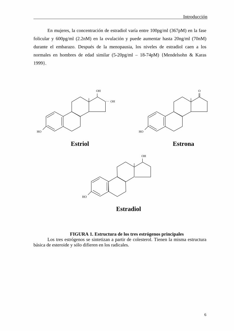
Se ha demostrado la existencia de más de 20 estrógenos en la orina y el plasma de
mujeres embarazadas. Los tres principales son estriol, estrona y estradiol cuya estructura se
presenta en la Figura 1. La fuente mayoritaria de estrógenos en la mujer es el ovario y
durante la gestación la encargada es la unidad fetoplacentaria. También hay una parte de la
biosíntesis que es extraglandular y en todos los casos los estrógenos se obtienen a partir del
colesterol. La producción extraglandular, fundamentalmente en la glándula suprarrenal,
constituye el mecanismo principal para la formación de estrógenos en las niñas
prepuberales, en las mujeres posmenopáusicas y en el hombre adulto joven. En el hombre
normal, una pequeña cantidad de estradiol es secretada directamente por los testículos.
{Pritchard & MacDonald 1981; Fuchs & Klopper 1982; Hadley 1997; Debuse 1998}.
Introducción
6
En mujeres, la concentración de estradiol varía entre 100pg/ml (367pM) en la fase
folicular y 600pg/ml (2.2nM) en la ovulación y puede aumentar hasta 20ng/ml (70nM)
durante el embarazo. Después de la menopausia, los niveles de estradiol caen a los
normales en hombres de edad similar (5-20pg/ml – 18-74pM) {Mendelsohn & Karas
1999}.
FIGURA 1. Estructura de los tres estrógenos principales Los tres estrógenos se sintetizan a partir de colesterol. Tienen la misma estructura
básica de esteroide y sólo difieren en los radicales.
OH
HO
O
HO
OH
HO
OH
Estriol Estrona
Estradiol

Introducción
7
I.A.- FUNCIÓN DE LOS ESTRÓGENOS
EFECTOS GENERALES EN LA MUJER
Los estrógenos producidos durante la maduración puberal en la mujer son los
responsables del desarrollo de los caracteres sexuales femeninos: crecimiento de las
mamas, las trompas de Falopio, el folículo ovárico y engrosamiento del epitelio vaginal.
Estimulan la actividad del epitelio cervical, de manera que el moco permite con más
facilidad la penetración de los espermatozoides. Afectan a la distribución de la grasa y
causan retención de agua y sal, aumento de peso, inducen la secreción de las glándulas
sebáceas, estimulan el crecimiento del vello púbico y axilar, mantienen la libido y el
comportamientamiento sexual. También estimulan el crecimiento lineal del hueso y el
cierre de la epífisis en los animales inmaduros, incluyendo el hombre {Pritchard &
MacDonald 1981; Hadley 1997}.
Los estrógenos sintetizados en la glándula suprarrenal y en los testículos también
participan en el desarrollo sexual masculino. Esto se ha puesto de manifiesto al estudiar
ratones macho carentes (knockout) de RE y . Estos animales presentan un desarrollo
normal del aparato reproductor, pero son estériles {Couse et al. 1999} y además ven
reducido su comportamiento agresivo y sexual {Ogawa et al. 2000}.
CICLO MENSTRUAL
El ciclo menstrual es el proceso por el cual madura un folículo ovárico, libera un
óvulo y si no se produce la fecundación, es expulsado por la vagina junto al endometrio
que se había desarrollado para albergar el huevo fecundado.
Durante la fase folicular, primeros 14 días del ciclo, el folículo se desarrolla
pasando por distintas etapas hasta que libera el óvulo (ovulación), que viaja al útero en
donde puede ser fecundado. Si lo es, se implanta en el endometrio desarrollado durante
todo ese tiempo y da lugar a un feto. Si por el contrario no es fecundado, el cuerpo lúteo
formado con los restos del folículo tras la expulsión del óvulo, degenera. Ésta es la llamada
fase lútea.
El folículo se desarrolla gracias a la secreción de las hormonas LH (hormona
luteinizante) y FSH (hormona estimuladora de los folículos) por la hipófisis. Éstas
producen un aumento en la síntesis de estrógenos por el folículo, que por un mecanismo de
retroalimentación positiva, estimulan la hipófisis para producir más LH y FSH. La
Introducción
8
ovulación coincide con el pico en la secreción de ambas. A continuación, ya en la fase
lútea, la secreción de estrógenos disminuye y la LH es la responsable de transformar el
folículo roto en el cuerpo lúteo. Éste secreta estrógenos y progesterona, que inhiben la
liberación de GnRH (hormona liberadora de gonadotropina) por el hipotálamo, lo que
regula negativamente la secreción de FSH y LH. La progesterona producida por el cuerpo
lúteo prepara el tejido endometrial para la implantación del óvulo fecundado. Si por el
contrario el óvulo no es fecundado se produce el menstruo, en el cual éste es expulsado
junto con el tejido endometrial desarrollado. Tras la menstruación vuelven a disminuir los
valores de estrógenos y progesterona, lo que hace que la secreción de FSH y LH por la
hipófisis vuelva a aumentar y se incie así un nuevo ciclo {Pritchard & MacDonald 1981;
Hadley 1997; Debuse 1998}
GESTACIÓN
Si el óvulo es fecundado se implanta en el endometrio y la placenta en desarrollo
empieza a producir gonadotropina coriónica. Esta hormona, cuya acción es similar a la de
la LH, induce el crecimiento del cuerpo lúteo impidiendo su degeneración. Éste sigue
secretando progesterona y estrógeno hasta que la placenta se hace cargo completamente de
su producción, momento en el que el cuerpo lúteo degenera. La continua secreción de
progesterona y estrógeno durante todo el embarazo hace que la FSH y la LH hipofisarias
no vuelvan a ser secretadas hasta después del parto. Las dos hormonas gonadales
secretadas por el cuerpo lúteo o la placenta inician el crecimiento de los tejidos mamarios
preparándolos para la lactancia {Fuchs & Klopper 1982; Hadley 1997}.
Los niveles de los tres estrógenos principales aumentan en la sangre materna
durante la gestación y descienden bruscamente tras la expulsión del feto y de la placenta.
El estradiol asciende desde una media de 7.1ng/ml (26nM) en la semana 22 hasta 23ng/ml
(84nM) en la semana 41 {Fuchs & Klopper 1982}.
Introducción
9
I.B.- RECEPTORES DE ESTRÓGENOS
INTRACELULARES CLÁSICOS
La mayor parte de los efectos de los estrógenos descritos anteriormente están
mediados por los receptores de estrógenos intracelulares (RE). Estos pertenecen a una
superfamilia de receptores intracelulares con estructura y funcionamiento similares, que
incluye el receptor de glucocorticoides, mineralocorticoides, progesterona, vitamina D,
hormona tiroidea, andrógenos, ácido retinoico y ácido 9-cis retionoico. Todos ellos son
factores de transcripción, funcionalmente activos tras unirse su ligando y que producen
efectos genómicos a largo plazo {Evans 1988; Tsai & O'Malley 1994; Kuiper &
Gustafsson 1997}.
Aunque ya se había descrito la presencia de proteínas intracelulares capaces de unir
estrógenos durante la década anterior, no fue hasta 1986 cuando se clonó el primer receptor
de estrógenos a partir de la línea celular de cáncer de mama humano MCF-7 {Walter et al.
1985; Greene et al. 1986; Green et al. 1986}. Más tarde llegaron los de rata {Koike et al.
1987}, pollo {Krust et al. 1986} y ratón {White et al. 1987}. Durante muchos años se
creyó en la existencia de un solo receptor de estrógenos. Sin embargo, algunos compuestos
relacionados con los estrógenos se comportaban como agonistas o antagonistas
dependiendo del tejido {Katzenellenbogen et al. 1996}, así que a partir de 1996 se fueron
clonando sucesivamente los RE de rata {Kuiper et al. 1996}, humano {Mosselman et al.
1996} y ratón {Tremblay et al. 1997}. Numerosos estudios se han realizado para saber la
diferencia funcional entre los dos receptores {Paech et al. 1997}, la afinidad por sus
ligandos {Kuiper et al. 1997; Kuiper et al. 1998}, el comportamiento diferencial de un
mismo agonista o antagonista {Barkhem et al. 1998} y la expresión en diversos tejidos
{Muramatsu & Inoue 2000}.
El RE humano está formado por 595 aminoácidos, con un peso molecular de
66.2kDa {Greene et al. 1986}, mientras que el RE por 530 aminoácidos y 53.2 kDa
{Mosselman et al. 1996}. Se han encontrado varios ARNm {McEwen & Alves 1999} por
la utilización de diferentes promotores y por procesamiento alternativo de los exones de
RE y RE humanos {Grandien et al. 1993; Flouriot et al. 1998; Moore et al. 1998;
Ogawa et al. 1998}, pollo {Griffin et al. 1998} y rata {Hirata et al. 1996; Chu & Fuller
1997; Petersen et al. 1998; Maruyama et al. 1998; Hanstein et al. 1999}. Además se ha
detectado la presencia de un receptor de estrógenos de 112 kDa en el córtex de cerebro de

Introducción
10
ratón {Asaithambi et al. 1997} y un RE gamma en teleostos, cuyo ARNm tiene un tamaño
de 4.6kb y probablemente haya sido generado por duplicación de ER. Éste último es
abundante en ovario y testículos, mientras que está ausente o poco expresado en hígado,
músculo y cerebro {Hawkins et al. 2000}.
FIGURA 2. Representación esquemática de la distribución en dominios de los RE y RE humanos
En el esquema se presenta la distribución en dominios de los receptores de estrógenos y humanos, así como la localización de las regiones más importantes para su funcionamiento. También se indican los aminoácidos (aa) a partir de los cuales empieza y termina cada uno de los dominios. AF indica función de transactivación; ADN, zona de interacción con el ADN y HORMONA, el dominio en donde interacciona el estrógeno. Tomado de Dechering et al.2000.
En general, los receptores de hormonas esteroideas presentan una distribución en 6
dominios (A-F) con distinta función (Figura 2). Los extremos amino y carboxi terminales,
A/B y E/F respectivamente, poseen función de transactivación. El dominio E es largo y en
él se encuentra el sitio de unión del ligando, así como regiones implicadas en la
dimerización del receptor y de localización nuclear. En el C hay dos dedos de zinc
responsables de la unión al ADN y otras zonas de dimerización. En el dominio D se sitúan
las señales de localización nuclear y de transactivación {Beato 1989; Tsai & O'Malley
1994; Dechering et al. 2000}. La homología entre los dos receptores de estrógenos es del
47% en humanos {Enmark et al. 1997} y varía dependiendo del dominio. Los menos
A D C B F E NH2 COOH
1 38 180 302 263 553 595 aa
A/B C D E F NH2 COOH
148 1 267 530 aa 214 500
AF ADN HORMONA
RE
RE
Introducción
11
conservados son el A/B, F y D, mientras que el dominio de unión a ADN presenta un 96%
de homología y el de unión al ligando del 60%. En otras especies la homología es muy
similar {Dechering et al. 2000}.
Los receptores de esteroides, ya estén localizados en el citosol o en el núcleo en su
forma inactiva, se traslocan al núcleo cuando son ativados por la unión de sus ligandos. El
RE inactivo es parte de un complejo que incluye chaperonas, Hsp90 (heat shock protein) y
Hsp56, que se disocian tras la unión del estrógeno {Pratt & Toft 1997}. Una vez liberado
del complejo, el receptor forma homo o heterodímeros que interaccionan con una zona
específica del ADN. Esta secuencia consiste en un palíndrome invertido de 15 pares de
bases, que se encuentra en la regiones reguladoras de algunos genes. Al situarse sobre ese
región específica del ADN, el complejo RE-estrógeno regula la maquinaria de
transcripción, bien a través de una interacción directa o bien por medio de otros factores de
transcripción {Couse & Korach 1999; Falkenstein et al. 2000}.
I.C.-EFECTOS RÁPIDOS DE LOS ESTRÓGENOS
POR VÍAS ALTERNATIVAS
Los efectos de los estrógenos han estado clásicamente mediados por mecanismos
genómicos a través de los receptores intracelulares descritos anteriormente. Estos
mecanismos son lentos y son necesarios al menos 20-30 min para detectarse un efecto
celular. En el momento de la publicación en 1975 de la primera respuesta rápida a
estrógenos en células endometriales {Pietras & Szego 1975}, los términos RE intracelular
y mecanismo genómico eran inseparables. Por lo tanto, los efectos rápidos encontrados por
Pietras & Szego abrieron un nuevo campo de actuación de estas hormonas hidrofóbicas
(Figura 3). De inmediato se sospechó que la transcripción génica podría no estar
participando en estos nuevos efectos, por la rapidez con la que se producían, y por tanto
también se cuestionó la implicación de los receptores de estrógenos intracelulares. De
modo que surgió la idea de la existencia de un receptor en la membrana plasmática que
estuviera mediando lo que casi con seguridad se trataba de efectos no genómicos. La
hipótesis de un receptor en la membrana fue corroborada dos años más tarde por los
mismos autores cuando detectaron sitios de unión para estrógenos en la membrana
plasmática de las células endometriales {Pietras & Szego 1977}.
Introducción
12
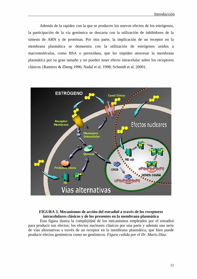
Además de la rapidez con la que se producen los nuevos efectos de los estrógenos,
la participación de la vía genómica se descarta con la utilización de inhibidores de la
síntesis de ARN y de proteínas. Por otra parte, la implicación de un receptor en la
membrana plasmática se demuestra con la utilización de estrógenos unidos a
macromoléculas, como BSA o peroxidasa, que les impiden atravesar la membrana
plasmática por su gran tamaño y no pueden tener efecto intracelular sobre los receptores
clásicos {Ramirez & Zheng 1996; Nadal et al. 1998; Schmidt et al. 2000}.
FIGURA 3. Mecanismos de acción del estradiol a través de los receptores intracelulares clásicos y de los presentes en la membrana plasmática
Esta figura ilustra la complejidad de los mecanismos empleados por el estradiol para producir sus efectos: los efectos nucleares clásicos por una parte y además una serie de vías alternativas a través de un receptor en la membrana plasmática, que bien puede producir efectos genómicos como no genómicos. Figura cedida por el Dr. Mario Díaz.

Introducción
13
A partir de las primeras evidencias de un nuevo mecanismo de acción de los
estrógenos, se dividieron sus efectos en dos. Por un lado los producidos por interacción con
los receptores intracelulares, con el resultado de la regulación de la transcripción génica y
por el otro, los efectos rápidos, no genómicos, mediados por posibles receptores en la
membrana plasmática.
Al contrario de lo que en un principió se creyó, la línea que separa ambos tipos de
efectos no es tan nítida, sino que se está viendo entrecruzamiento entre los dos tipos de
efectos observados, los mecanismos por los que se producen y los receptores de estrógenos
implicados (Figura 3) {Nadal et al. 2001}. No es el objetivo de esta Introducción tratar en
detalle estas consideraciones, sino que serán tema de debate en la Discusión de esta tesis.
Con el propósito de no dispersar los contenidos que se tratarán en esta Introducción y en la
posterior Discusión, se centrará la atención en lo que se ha venido llamando efectos
rápidos y no genómicos de los estrógenos. En el caso en el que sea probable que la
interacción de los estrógenos con receptores en la membrana plasmática, o que
determinados efectos detectados en un corto espacio de tiempo puedan dar lugar a señales
genómicas, se discutirán las razones que puedan existir a favor de esa posibilidad.
En vistas de la enorme cantidad de efectos rápidos detectados no sólo con
estrógenos, sino también con progesterona, vitamina D, testosterona y aldosterona
{Wehling 1997; Alonso & Lopez-Coviella 1998; Schmidt et al. 2000; Falkenstein et al.
2000}, ha sido necesaria establecer una clasificación de los mismos. Ésta se presentó en el
“First International Meeting on Rapid Responses to Steroid Hormones” que tuvo lugar en
Mannheim, Alemania, en 1998 {Norman & Wehling 1999; Falkenstein et al. 2000}. Así,
tal y como muestra la Figura 4, los efectos rápidos de los esteroides se dividen en directos
(A) e indirectos (B), según el esteroide sea el único agonista o si necesita de otro para
generar este tipo de respuesta. A su vez en no específicos (AI, BI) y específicos (AII, BII).
Los efectos específicos se dividen en mediados por receptores clásicos (AIIa, BIIa) y no
clásicos (AIIb, BIIb).

Introducción
14
FIGURA 4. Clasificación de los efectos rápidos de los esteroides
Clasificación establecida en el “First International Meeting on Rapid Responses to Steroid Hormones” que tuvo lugar en Mannheim, Alemania, en 1998. Tomado de Falkenstein et al. 2000.
EFECTOS RÁPIDOS DE LOS ESTRÓGENOS
El 17-estradiol es un modulador de amplio espectro produciendo efectos rápidos,
fundamentalmente en los sistemas nervioso {Alonso & Lopez-Coviella 1998} y
cardiovascular {Farhat et al. 1996b}, razón por la cual se tratarán aparte. Pero afecta
también a otros tipos celulares y sobretodo a las corrientes iónicas, como la del receptor
P2X7 {Cario-Toumaniantz et al. 1998}, canales de cloro {Hardy & Valverde 1994;
Condliffe et al. 2001} y canales de potasio en endotelio {Rusko et al. 1995} y epitelio de
colon {McNamara et al. 2000}. Además cierra el KATP en células pancreáticas {Nadal et
al. 1998}.
En cuanto al calcio intracelular, el 17-estradiol lo aumenta, tanto procedente del
exterior como de los reservorios intracelulares, en numerosos tipos celulares: neuronas
embrionarias de rata {Beyer & Raab 1998}, linfocitos T {Benten et al. 1998}, enterocitos
A. Efecto Directo B. Efecto Indirecto
I. No Específico
II. Específico
b. Receptor nuclear no clásico
a. Receptor nuclear clásico
Introducción
15
{Picotto et al. 1996}, oocitos {Tesarik & Mendoza 1995}, hueso {Lieberherr et al. 1993;
Fiorelli et al. 1996}, células de la granulosa {Morley et al. 1992}, adenocarcinoma de
endometrio {Dopp et al. 1999}, hepatocitos {Sanchez-Bueno et al. 1991}, espermatozoides
humanos {Luconi et al. 1999}, islote de Langerhans {Nadal et al. 1998} y útero {Batra
1986}.
Otros efectos son la regulación del transporte de glucosa en útero {Welch & Gorski
1999} y la disminución de la síntesis de 11-cetotestosterona inducida por gonadotropina en
testículos {Loomis & Thomas 2000}. También modula la comunicación a través de
uniones en hendidura (gap junctions) en miocitos {Verrecchia & Herve 1997} y
osteoblastos {Massas et al. 1998}. Por su parte, los catecolestrógenos afectan la
contracción uterina {Goyache et al. 1995} y la secreción de insulina {Etchegoyen et al.
1998}
En las células MCF-7, tanto estradiol como E-BSA inhiben la apoptosis inducida
por UV y por taxol a través de la activación de c-Jun quinasa y ERK (MAPK). Si bien la
exposición al estrógeno es de varias horas, el hecho de que el E-BSA reproduzca los
resultados, indica que podría ser a través de un receptor en la membrana plasmática. Sin
embargo, en este caso la implicación de c-Jun quinasa y ERK sugieren un mecanismo
genómico por el cual el estradiol inhibe la apoptosis, probablemente a través de un
receptor de membrana {Razandi et al. 2000a}.
VÍAS DE SEÑALIZACIÓN INTRACELULAR
ACTIVADAS POR ESTRÓGENOS
Una evidencia más del nuevo mecanismo de acción de los estrógenos es la
implicación de las mismas vías de señalización intracelular que las hormonas hidrofílicas
activan a través de receptores de membrana. Entre ellas figuran las del NO, AMPc, GMPc,
PKC y proteína G {Wehling 1997; Falkenstein et al. 2000}.
Se ha visto que las proteínas G están implicadas en algunos efectos del 17-
estradiol, ya sea con evidencias directas {Ogata et al. 1996} o indirectas por un aumento en
los niveles de AMPc o IP3, segundos mensajeros producidos por enzimas activadas por
proteínas G, adenilato ciclasa y fosfolipasa C (PLC) respectivamente. Por una parte, el
estradiol produce un aumento rápido de AMPc en útero {Szego & Davis 1967; Aronica et
al. 1994}, células MCF-7 {Aronica et al. 1994}, intestinales {Picotto et al. 1996}, células
de la granulosa {Sirotkin & Nitray 1993} y hueso {Fiorelli et al. 1996}; aunque no siempre

Introducción
16
debido a la activación de una proteína G {Farhat et al. 1996a}. En cuanto a la PLC, se ve
implicada en los movimientos de calcio intracelular producidos por estradiol en enterocitos
de rata junto al AMPc {Picotto et al. 1999}, y las isoformas PLC-1 y 2 modulan el
calcio en osteoblastos {Lieberherr et al. 1993; Le Mellay et al. 1997}. Por su parte, los
fosfoinosítidos intervienen en el aumento de calcio en células de granulosa {Morley et al.
1992} y los niveles de IP3 son elevados por estradiol en epitelio vaginal {Singh & Gupta
1997} y en la línea de hepatoma HEPG2, aumento que en este último caso lleva asociada
la participación de PKC {Marino et al. 1998}. Además, en MCF-7, el estradiol activa
rápidamente la PIP2-PLC {Graber et al. 1993} y la PKC está implicada en el efecto
estrogénico en condroicitos {Sylvia et al. 1998} y en la regulación de la secreción de Cl- en
epitelio de colon {Doolan et al. 2000; Condliffe et al. 2001}.
En algunos casos, las vías de PKA y PKC participan en el mismo efecto {Kelly et
al. 1999} e incluso se sugiere que la interacción directa del estradiol con la PKC es la
responsable de la activación de la adenilato ciclasa, enzima que sintetiza AMPc, en epitelio
de colon {Doolan et al. 2000}. IP3 y AMPc se ven aumentados por la interacción del
estradiol con los receptores de estrógenos RE y RE localizados en la membrana
plasmática de células CHO y que parecen acoplados a proteínas G {Razandi et al. 1999}.
Otra vía de señalización que juega un papel importante en algunos efectos rápidos
de los estrógenos es la del NO y GMPc. El NO activa la guanilato ciclasa soluble, enzima
que sintetiza GMPc {Carvajal et al. 2000}. Un aumento de NO se ha visto en granulocitos
{Stefano et al. 2000} y junto al GMPc son fundamentales en los efectos vasodilatadores
del estrógeno {Mendelsohn & Karas 1999}. Por otro lado, el estradiol activa la guanilato
ciclasa de membrana en PC12, efecto que podría ser directo, puesto que el estrógeno
interacciona con una guanilato ciclasa de membrana formada por sus dominios catalítico y
quinasa {Chen et al. 1998}.
La vía de las MAPKs es activada de forma rápida por estradiol en células MCF-7
{Migliaccio et al. 1996}, hueso {Endoh et al. 1997} y carcinoma de colon {Di Domenico
et al. 1996}. En las MCF-7, la movilización de calcio intracelular determina la activación
de MAPK {Improta-Brears et al. 1999}, mientras que en otros casos está implicada una
proteína G {Filardo et al. 2000} o la vía de c-src, shc y p21ras {Migliaccio et al. 1996;
Migliaccio et al. 2000}. Entre los efectos generados por el estradiol a través de las MAPKs
se encuentra la modulación del crecimiento celular {Di Domenico et al. 1996; Morey et al.
Introducción
17
1997}, el mantenimiento de la forma y función de las células endoteliales {Razandi et al.
2000b}, la inhibición de la apoptosis {Razandi et al. 2000a} o la activación de la
transcripción de c-fos {Watters et al. 1997}. Sin embargo, en el músculo liso vascular, el
estradiol y la progesterona inhiben la proliferación celular por un bloqueo rápido de
MAPK {Morey et al. 1997}.
La vía de señalización de las MAPKs es una de las responsables de transmitir
señales de crecimiento celular y lo hace a través de su función quinasa que fosforila, entre
otras, a proteínas reguladoras de la expresión génica {Alberts et al. 1994}. Por lo tanto,
aunque se tenga una activación rápida y no genómica de MAPK a través de un receptor de
membrana para estradiol, los efectos celulares finales nombrados anteriormente
probablemente sean debidos a la activación de la transcripción génica.
SISTEMA NERVIOSO
Mediante estudios clínicos se ha demostrado el papel de los estrógenos en diversas
funciones del cerebro y su protección contra la enfermedad de Alzheimer, el estrés y el
riesgo de demencia {Garcia-Segura et al. 2001; Wise et al. 2001}. Lo que no se sabe hasta
el momento es cuáles de esos y otros efectos están mediados por acciones rápidas de los
estrógenos, aunque lo cierto es que el estradiol ejerce una gran variedad de efectos rápidos
a lo largo y ancho del sistema nervioso {Ramirez & Zheng 1996; Moss et al. 1997; Zakon
1998}.
El 17-estradiol modula el potencial de reposo y la actividad eléctrica de neuronas
del área preóptica medial {Kelly et al. 1977}, de la amígdala medial {Nabekura et al. 1986;
Minami et al. 1990}, neuronas GnRH {Lagrange et al. 1995}, neuronas piramidales CA1
de hipocampo {Wong & Moss 1991}, hipotalámicas {Kelly et al. 1980}, dopaminérgicas
{Chiodo & Caggiula 1980} y células de la pituitaria {Dufy et al. 1979}. Regula la
liberación de numerosos neurotransmisores y hormonas, como la dopamina y la
noradrenalina {Roosen-Runge et al. 1984; Ramirez & Zheng 1996; Disshon & Dluzen
1997; Xiao & Becker 1998; Kim et al. 2000}, prolactina {Zyzek et al. 1981}, LHRH
{Drouva et al. 1983}, GnRH y LH {Ramirez & Zheng 1996; Prevot et al. 1999}, oxitocina
y vasopresina {Wang et al. 1995}, adrenalina, glutamato, GABA y -endorfina {Roosen-
Runge et al. 1984}. Por otro lado, el estradiol afecta a la respuesta neuronal a -opioides
{Lagrange et al. 1994}, acetilcolina {Uki et al. 1999}, kainato {Gu & Moss 1996}, GABA
Introducción
18
{Lagrange et al. 1996} y serotonina {Wetzel et al. 1998}. En córtex, modula la actividad
de la calcio-ATPasa {Zylinska et al. 1999}.
En el sistema nervioso, la vía de señalización más generalmente implicada en el
efecto del estradiol es la del AMPc-PKA por medio de proteína G. Receptores de
estrógenos acoplados a proteínas G participan en el hipocampo, tanto en la disminución de
la corriente de calcio {Mermelstein et al. 1996}, como en la modulación de las corrientes
inducidas por kainato en neuronas CA1 {Gu & Moss 1996}. En este último caso es
necesario también un efecto interno del estradiol actuando sobre el AMPc {Gu & Moss
1998}. La implicación de una proteína G se ha puesto de manifiesto también en hipotálamo
{Caldwell et al. 1999} y la PKA es la responsable de la regulación del efecto de -opioides
por estradiol en neuronas hipotalámicas {Lagrange et al. 1997}.
Sin embargo, también hay otras vías responsables de algunos de los efectos rápidos
del estradiol en el sistema nervioso, como es la del NO y la guanilato ciclasa soluble, que
participan en la secreción de GnRH {Prevot et al. 1999}. Las MAPKs son activadas por
estradiol en neuroblastoma {Watters et al. 1997} y córtex {Singer et al. 1999} en donde
estimula el crecimiento celular y protege las neuronas corticales de la toxicidad del
glutamato {Singh et al. 1999} y las neuronas de hipocampo de la toxicidad del kainato o
del NMDA {Bi et al. 2000}.
SISTEMA CARDIOVASCULAR
Uno de los grandes beneficios que proporcionan los estrógenos a las mujeres es la
protección cardiovascular. La incidencia de este tipo de enfermedades es menor en las
mujeres premenopáusicas que en los hombres, aunque esta diferencia se ve claramente
disminuida tras la menopausia, protección que se recupera con la terapia hormonal con
estrógenos {Farhat et al. 1996c}.
Al menos parte de ese beneficio es gracias al efecto vasodilatador de los estrógenos
a través de mecanismos no genómicos {Farhat et al. 1996c; Mendelsohn & Karas 1999}.
Esta vasodilatación puede ser dependiente o independiente del endotelio. La independiente
de endotelio está mediada por la disminución de la corriente de calcio de tipo L por
estradiol {Zhang et al. 1994; Kitazawa et al. 1997; Tanabe et al. 1999} o por un aumento
de NO en células de músculo liso. El NO genera GMPc a través de la guanilato ciclasa
soluble {Mugge et al. 1993}, lo que activa la PKG, responsable de la apertura del canal de
potasio de alta conductancia activado por calcio {White et al. 1995; Darkow et al. 1997}.

Introducción
19
El aumento de la corriente de potasio hiperpolariza la membrana plasmática, disminuye la
entrada de calcio y por tanto produce la relajación del vaso sanguíneo {Nelson & Quayle
1995}. También se ha visto interacción directa del estradiol con el canal de potasio, lo que
aumenta la corriente que pasa a su través, con el mismo efecto sobre la contracción
{Valverde et al. 1999}. Tampoco se puede descartar la participación adicional del AMPc,
puesto que éste aumenta en músculo liso tras exposición a estradiol {Mugge et al. 1993;
Farhat et al. 1996a; Buitrago et al. 2000}.
La vasodilatación dependiente de endotelio está mediada por la liberación de NO de
las células endoteliales {Mendelsohn & Karas 1999}, que actuaría sobre las células del
músculo liso. El estradiol aumenta el NO {Lantin-Hermoso et al. 1997; Caulin-Glaser et al.
1997; Kim et al. 1999} por la activación de la óxido nítrico sintasa endotelial (eNOS),
probablemente por su fosforilación por (PI3)-kinase-Akt {Haynes et al. 2000}. Tras la
exposición a estradiol y de una forma dependiente de un aumento de calcio intracelular
{Moini et al. 1997}, eNOS se trasloca desde la membrana a una zona próxima al núcleo
{Goetz et al. 1999}. El NO producido aumenta GMPc {Russell et al. 2000a}, lo que
activaría la PKG, responsable de la apertura del canal de potasio dependiente de calcio y
por tanto produciría la relajación de las células del músculo liso. También se implica la vía
de MAPK en el aumento de la liberación de NO de las células endoteliales {Russell et al.
2000b}.
I.D.- RECEPTOR DE ESTRÓGENOS DE MEMBRANA
Gran parte de los efectos descritos anteriormente están mediados por la acción del
estradiol sobre la membrana plasmática, según se ha demostrado con la utilización de
estradiol unido a macromoléculas. Los compuestos que resultan, estradiol-BSA (E-BSA) y
estradiol-peroxidasa, no pueden atravesar la membrana plasmática por su gran tamaño, de
modo que tanto los efectos como el marcaje que se obtienen son producto de la interacción
del estradiol con un sitio de unión en la membrana {Ramirez 1992; Nadal et al. 1998;
Schmidt et al. 2000}. El compuesto más empleado ha sido E-BSA, aunque su utilización
no está exenta de controversia, puesto que el E-BSA comercial contiene cantidades
considerables de estradiol libre. Además, activa ERK1 y ERK2 en células de
neuroblastoma, efecto no reproducido con estradiol libre y no se une a los RE y
Introducción
20
presentes en el extracto de citosol de células de insecto que los sobrexpresan {Stevis et al.
1999}. Por esta razón se ha utilizado estradiol unido a peroxidasa como una alternativa
para ensayos de interacción y de funcionalidad del receptor de membrana {Nadal et al.
2000}.
Los primeros sitios de unión para estrógenos en la membrana plasmática se
detectaron en células de endometrio {Pietras & Szego 1977} y en espermatozoides
humanos {Hernandez-Perez et al. 1979; Cheng et al. 1981}. Sucesivamente se han ido
viendo en membrana plasmática sináptica de cerebro de rata {Towle & Sze 1983} y
pituitaria {Bression et al. 1986}. E-BSA se ha utilizado en células de cáncer humano
{Berthois et al. 1986}, hueso {Brubaker & Gay 1994; Fiorelli et al. 1996}, neuronas
neostriales {Mermelstein et al. 1996}, cerebro de rata {Ramirez & Zheng 1996}, la línea
tumoral de pituitaria GH3/B6, {Pappas et al. 1995} y células endoteliales {Kim et al.
1999}. También se ha empleado microscopía electrónica para poner de manifiesto la
interacción E-BSA con la membrana plasmática de células de hepatoma {Moats &
Ramirez 2000}. En células pancreáticas se ha sustituido este compuesto por estradiol-
peroxidasa {Nadal et al. 2000} y un derivado biotinilado de estradiol ha servido para ver
marcaje en membrana en células MCF-7 {Germain et al. 1993}.
Por otro lado, se ha visto unión de 2OH-estradiol a membranas de pituitaria
{Schaeffer et al. 1980; Bression et al. 1986}, hipotálamo {Etchegoyen et al. 1986} y
carcinoma de mama {Vandewalle et al. 1988}.
A pesar de los numerosos ejemplos de interacción de los estrógenos con un receptor
en la membrana plasmática, no hay una conclusión clara de la naturaleza de dicho receptor.
Si bien en algunos tipos celulares parecen ser los propios receptores de estrógenos
intracelulares que se expresan en la membrana plasmática, en otros casos se trata de otro
receptor que no guarda ninguna relación con los clásicos.
Para estudiar la naturaleza del receptor de estrógenos de membrana, se cuenta con
los anticuerpos dirigidos contra los RE clásicos y , que permiten determinar la
presencia o no de estos receptores en la membrana plasmática. Una fuente importante de
evidencias a este respecto son los animales carentes (knockout) de RE y RE. A pesar de
que ya se han obtenido ratones que carecen de ambos receptores {Couse et al. 1999;
Ogawa et al. 2000}, aún no se han obtenido datos de los efectos de membrana del estradiol
en esos animales. Sin duda esos estudios serán decisivos para clarificar cuáles de esos

Introducción
21
efectos del estrógeno están mediados por RE o RE y cuáles por otros receptores de
membrana.
EL RECEPTOR CLÁSICO EN LA MEMBRANA PLASMÁTICA
La sobrexpresión de los RE y en células CHO ha permitido ver un 2% de
localización en la membrana plasmática. Estas proteínas de membrana tienen el mismo
peso molecular y provienen del mismo ARNm que los RE intracelulares. Esto demuestra
una cierta capacidad de los receptores de estrógenos intracelulares para expresarse en la
membrana plasmática, sin modificaciones aparentes en su estructura ni la presencia de
dominios adicionales. A través de estos receptores de membrana clásicos, el estradiol
activa dos tipos de proteínas G, Gq y Gs {Razandi et al. 1999}.
Con anticuerpos dirigidos contra diferentes dominios del RE se ha visto marcaje
en membrana en una población de células de la línea tumoral de pituitaria GH3/B6
{Pappas et al. 1995; Norfleet et al. 1999} y en neuronas de hipocampo {Clarke et al.
2000}. Uno de esos anticuerpos reproduce la estimulación de la liberación de prolactina
generada por estradiol en las células GH3/B6 {Norfleet et al. 2000}. Por otro lado, la unión
de E-BSA a la membrana de células de músculo liso es competida por el anticuerpo que
reconoce el dominio de unión del ligando al RE{Morey et al. 1997}.
El bloqueo de los efectos rápidos del estradiol con el antiestrógeno específico para
RE y , ICI 182,780, proporciona evidencias de la implicación de al menos uno de ellos
en dichos efectos del estrógeno {Wakeling et al. 1991}. Esto sucede en el aumento del
calcio intracelular producido por estradiol en células de adenocarcinoma de endometrio
{Dopp et al. 1999}, la activación de la vía de NO en endotelio {Goetz et al. 1999; Kim et
al. 1999; Chen et al. 1999; Chambliss et al. 2000; Haynes et al. 2000} y la protección
frente a la apoptosis en MCF-7 {Razandi et al. 2000a}. ICI también bloquea la activación
de la vía de señalización de MAPK por estradiol en MCF-7 {Migliaccio et al. 1996;
Improta-Brears et al. 1999}, carcinoma de colon {Di Domenico et al. 1996} y células
endoteliales {Russell et al. 2000b}.
Cuando se sobrexpresa el RE en células endoteliales, se obtiene un aumento de la
activación de eNOS por estradiol mucho mayor que el que se da en las células con niveles
de expresión normales. En ambos casos, la activación de eNOS por estradiol es inhibida
por el antiestrógeno ICI 182,780 {Chen et al. 1999}.
Introducción
22
Por otro lado, los anticuerpos dirigidos contra el RE en un intento por aislar el
receptor de estrógenos de membrana, han permitido obtener algunas bandas de diferentes
pesos moleculares. Con un anticuerpo dirigido contra el dominio de unión del estradiol se
ha detectado una banda de 29kDa en espermatozoides humanos, cuyo peso molecular es
muy inferior al del RE intracelular {Luconi et al. 1999}. Con otros anticuerpos también se
ha detectado la misma banda en útero, junto a otras de distintos pesos moleculares {Monje
& Boland 1999}. Además, el estradiol interacciona con un receptor de progesterona de
membrana de 29kDa presente en cerebelo {Bukusoglu & Krieger 1994}. También en
distintos extractos de cerebro se han obtenido varias bandas con E-BSA, en donde una de
ellas corresponde a una subunidad de F0F1-ATPasa/ATPsintasa mitocondrial, que da
sensibilidad a la oligomicina {Ramirez & Zheng 1996; Zheng & Ramirez 1999}.
La obtención de las bandas mencionadas anteriormente a partir de extractos de
membranas con los anticuerpos anti-RE, favorece la hipótesis de la presencia de los
receptores intracelulares en la membrana plasmática. El hecho de que las proteínas posean
distinto peso molecular que el RE, sugiere la idea de que pueda tratarse de receptores que
han sufrido modificaciones, aunque éstas les han dejado intactas las estructuras
reconocidas por los respectivos anticuerpos utilizados.
RECEPTOR DE MEMBRANA NO CLÁSICO
La presencia del RE ó en la membrana plasmática de algunos tipos celulares no
da cuenta de todos los efectos de los estrógenos. Estos se siguen produciendo en células de
cáncer de mama que naturalmente no expresan ninguno de los dos receptores clásicos
{Filardo et al. 2000} y en hipocampo de ratones carentes (knockout) del REEn este
caso, la participación del RE es descartada por la falta de inhibición del efecto del
estradiol por ICI 182,780 {Gu et al. 1999}. Tampoco hay diferencia en la activación de
ERK por estradiol en córtex cerebral de ratones sin RE con respecto a los normales,
aunque en este caso no está clara la implicación de un receptor de membrana {Singh et al.
2000}. Por otro lado, en macrófagos que no expresan el RE, IC-21, anticuerpos anti-RE
no marcan la membrana plasmática {Benten et al. 2001} y la relajación producida por
estradiol en aorta de ratones carentes del RE no es menor que en los animales normales
{Nilsson et al. 2000}.
Por otra parte, el antiestrógeno ICI 182,780 no inhibe el efecto del estradiol en
neuronas dopaminérgicas {Beyer & Karolczak 2000}, tampoco la relajación de arterias
Introducción
23
pequeñas coronarias {Shaw et al. 2000}, ni el aumento de NO en granulocitos {Stefano et
al. 2000}. También es inefectivo en la activación de la vía de MAPK en neuroblastoma
{Watters et al. 1997} e hipotálamo {Kuroki et al. 2000}. En el caso de neuronas corticales,
tanto un mecanismo dependiente como uno independiente de RE y RE se han descrito
{Singer et al. 1999; Singh et al. 1999}.
Con respecto a la naturaleza de ese receptor de membrana distinto de los
intracelulares, cabe dos posibilidades. Por un lado, podría ser un receptor cuyos ligando
sean conocidos y que además interaccione con estradiol. Y por otra, podría tratarse de un
receptor nuevo completamente desconocido. En cualquiera de los dos casos, identificarlo
no es una tarea sencilla, aunque se tenga descartada completamente la participación de los
receptores intracelulares.
Entre otras proteínas con las que se ha visto unión del 17-estradiol se encuentra la
guanilato ciclasa de membrana: el estrógeno interacciona con una proteína que consiste en
sus dominios intracelulares quinasa y catalítico {Chen et al. 1998}. Además el estrógeno
también se une a la gliocoproteína P cuando ésta es expresada en células NIH 3T3
{Edelmann et al. 1999}.
Por otro lado existe la modulación alostérica de receptores de neurotransmisores
por esteroides. Aunque no se tienen datos de una modulación alostérica del receptor del
ácido gamma-aminoburítico (GABA) por estradiol, los catecolestrógenos disminuyen su
unión a membranas hipotalámicas {Etchegoyen et al. 1986} y otros esteroides inhiben la
interacción del t-butilbiciclo-fosforotionato al complejo del receptor GABA {Majewska et
al. 1986}. El sulfato de pregnenolona, pero no el estradiol, aumenta la probabilidad de
apertura del canal de NMDA en parches aislados {Wong & Moss 1994} y la progesterona
compite por el receptor muscarínico M2 {Wilkinson et al. 1995}. Sin embargo, el estradiol
sí interacciona con la proteína transportadora de serotonina (SERT) de forma alostérica,
aunque en un lugar inaccesible desde el dominio extracelular {Chang & Chang 1999}.
Otros receptores con los que también interacciona el estradiol son el receptor de serotonina
tipo 3 {Wetzel et al. 1998} y los receptores de opioides {Schwarz & Pohl 1994}.

Introducción
24
II.- EL ISLOTE DE LANGERHANS
El islote de Langerhans es una estructura formada por varios miles de células, muy
inervada e irrigada por capilares sanguíneos, que se encuentra localizada en el páncreas
endocrino. Su función principal es la regulación de la homeostasis de glucosa en sangre.
Para ello cuenta con las hormonas secretadas por los cuatro tipos celulares que lo
constituyen {Orci & Unger 1975; Ashcroft & Rorsman 1989} (Figura 5).
FIGURA 5. Islote de Langerhans de ratón
Esta imagen muestra un islote de Langerhans de ratón en el que se identifican tres de los tipos celulares presentes en el islote, por medio de técnicas de inmunocitoquímica. Para ello se han empleado anticuerpos dirigidos contra las hormonas secretadas por las células , y , cada uno marcado con un color distinto. Así se tiene que el glucagón de las células está teñido en azul; la insulina de las células se tiene en verde y la somatostatina sintetizada por las células está en rojo. Tomado de: R.L. Sorenson y T.C. Brelje. Departament of Cell Biology and Neuroanatomy. University of Minnesota. (http://www.novo.dk/hri).
Introducción
25
* Las células alfa () son las encargadas de la secreción de glucagón y representan
el 15-20% de la población celular del islote. Se localizan principalmente en la periferia del
islote.
* Las células beta () forman la población mayoritaria, 65-90%, y son las
responsables de la secreción de insulina.
* Las células delta () son las productoras de somatostatina y constituyen entre un 3
y un 10% del total celular del islote. Este tipo celular, al igual que las células , se
distribuye preferentemente en la periferia del islote.
* Las células PP, que contienen el polipéptido pancreático P constituyen el 1%
celular restante.
I.A.- LA CÉLULA BETA PANCREÁTICA
La célula pancreática es la población celular mayoritaria dentro del islote de
Langerhans. La fisiología y el acoplamiento estímulo-secreción de este tipo celular ha sido
muy estudiado a lo largo de los años, lo que ha hecho que el mecanismo por el que se
regula la secreción de insulina sea bien conocido.
Las células secretan insulina en respuesta a niveles elevados de glucosa en
sangre. Esa secreción es modulada por concentraciones variables de glucosa extracelular
por medio de un proceso en el que participan el metabolismo de la glucosa, varios tipos de
canales iónicos, la actividad eléctrica de la membrana plasmática y la maquinaria exocítica.
Además, otras vías de señalización intracelular pueden intervenir en el mecanismo de
liberación de insulina por glucosa en la célula .
SECRECIÓN DE INSULINA INDUCIDA POR GLUCOSA
La glucosa que llega a los islotes a través de los capilares sanguíneos que los irrigan
alcanza el citosol de las células gracias al transportador específico GLUT-2. La glucosa
es entonces fosforilada por la hexoquinasa IV y queda así atrapada en el interior celular. Su
metabolismo produce un aumento intracelular del conciente ATP/ADP y de los diadenosín
polifosfatos {Ripoll et al. 1996}, lo que cierra los canales de potasio dependientes de ATP
(KATP). Estos canales son los responsables de mantener el potencial de reposo de la célula
{Ashcroft et al. 1984; Misler et al. 1986; Dunne & Petersen 1986}. El número de KATP
Introducción
26
cerrados cuando la concentración de glucosa es superior a 5-7mM es suficiente para
alcanzar el umbral de despolarización de la membrana plasmática necesario para generar
actividad eléctrica {Dean & Matthews 1968}. Esta actividad eléctrica sigue un patrón
oscilatorio con superposición de potenciales de acción sobre las mesetas de las
oscilaciones, con una primera oscilación más duradera que el resto {Santos et al. 1991}.
KLKKLK
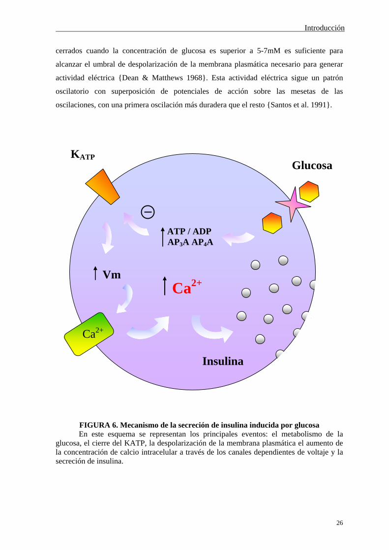
FIGURA 6. Mecanismo de la secreción de insulina inducida por glucosa En este esquema se representan los principales eventos: el metabolismo de la
glucosa, el cierre del KATP, la despolarización de la membrana plasmática el aumento de la concentración de calcio intracelular a través de los canales dependientes de voltaje y la secreción de insulina.
KATP
ATP / ADP AP3A AP4A
Vm Ca2+
Glucosa
Insulina
Ca2+
Introducción
27
La despolarización de la membrana abre los canales de calcio dependientes de voltaje de
la membrana plasmática de las células . Esto produce un aumento de la concentración de
calcio intracelular ([Ca2+]i) que desencadena la exocitosis a través de la activación de
proteínas quinasas que interaccionan con componentes de la maquinaria
microtubular/exocítica. Durante este proceso se libera el contenido de insulina de las
vesículas al medio extracelular {Aizawa et al. 1998} y la hormona alcanza el torrente
sanguíneo, donde actúa sobre los tejidos diana como comunicador de la presencia de
glucosa en la sangre (Figura 6) {Ashcroft & Rorsman 1989}.
EL CANAL DE POTASIO DEPENDIENTE DE ATP (KATP)
El canal de potasio dependiente de ATP (KATP) está presente en las células y
pancreáticas y en otros varios tejidos. Está formado por dos tipos de subunidades, SUR y
Kir, de las que hay varias isoformas. Su modulación varía dependiendo de las subunidades
que lo componen en cada tipo celular {Nelson & Quayle 1995; Yokoshiki et al. 1998;
Aguilar-Bryan et al. 1998; Ashcroft & Gribble 1999}.
El ATP y las sulfonilureas tolbutamida y glibenclamida son inhibidores del KATP,
mientras que el ADP, GDP, UDP y el diazóxido lo activan {Yokoshiki et al. 1998}. El
KATP es regulado por proteína G, PKA y PKC, aunque queda por determinar si es por
medio de una modulación directa por fosforilación o si se trata de un mecanismo indirecto
{Nelson & Quayle 1995}. En el caso de las dos subunidades presentes en la célula
pancreática, SUR1 y Kir6.2, la PKA fosforila directamente ambas subunidades y además
se sabe en qué aminoácidos {Beguin et al. 1999; Lin et al. 2000}.
En la célula del islote de Langerhans el KATP es el responsable de mantener el
potencial de reposo, debido a que la suya es la principal corriente iónica que atraviesa la
membrana plasmática en presencia de una concentración no estimulatoria de glucosa. El
cierre de este canal por distintos estímulos produce la disminución de la corriente de salida
de potasio y por tanto la despolarización de la membrana plasmática {Henquin 1980} y el
resto de eventos que ya han sido descritos anteriormente.
EL CALCIO INTRACELULAR EN LA CÉLULA BETA
El calcio es un mensajero intracelular ampliamente estudiado en la mayoría de los
tipos celulares y que participa en multitud de procesos. Su importancia como principal

Introducción
28
mediador en la secreción de insulina inducida por glucosa está bien establecido {Wollheim
& Sharp 1981; Prentki & Matschinsky 1987}.
La medida de calcio intracelular utilizando sondas fluorescentes sensibles a calcio
ha permitido estudiar la respuesta del islote de Langerhans a glucosa. Con esta técnica se
ha visto que al pasar de 3mM a 11mM glucosa, los islotes de Langerhans de ratón
generalmente responden con una primera elevación del calcio intracelular en el que éste
permanece elevado durante 2-3min, para luego oscilar sobre un nivel basal mayor que el
que había con 3mM {Valdeolmillos et al. 1989}. La frecuencia de las oscilaciones está
comprendida entre 2 y 5 min-1 (Figura 7). A medida que la concentración de glucosa
aumenta también lo hace la duración de las oscilaciones, hasta que se tiene un aumento
sostenido del calcio intracelular cuando la concentración de glucosa es superior a 22mM.
3mM G
11mM G
100%
F
/F0
2 min
FIGURA 7. Oscilaciones de calcio intracelular en cuatro células individuales dentro del islote
En la figura se presenta la respuesta de calcio intracelular de cuatro células localizadas dentro del mismo islote, al pasar de 3mM a 11mM glucosa. Los islotes han sido cargados con la sonda fluorescente Fluo-3 y la señal de calcio se ha obtenido con microscopía confocal. Tomada y modificada con permiso de Nadal et al. 1999.
Introducción
29
Las oscilaciones de calcio en el islote de Langerhans se producen de forma paralela
a la actividad eléctrica oscilatoria de las células dentro del islote, en respuesta a alta
glucosa, hecho que se ha puesto de manifiesto al registrar ambos parámetros
simultáneamente {Santos et al. 1991}. Como resultado, la secreción de insulina es pulsátil
y paralela a las oscilaciones de calcio intracelular {Rosario et al. 1986; Barbosa et al. 1996;
Barbosa et al. 1998}.
El calcio que aumenta en el citosol de las células en respuesta a glucosa proviene
del exterior celular, mientras que no hay participación del calcio de los reservorios
intracelulares. Esto se ha puesto de manifiesto con experimentos en los que se ha utilizado
o bien un medio extracelular carente de calcio o bien bloqueantes de los canales de calcio
de la membrana plasmática. En ambos casos no se tiene aumento de calcio intracelular en
respuesta a alta concentración de glucosa {Valdeolmillos et al. 1989; Gilon & Henquin
1992}, ni tampoco secreción de insulina {Wollheim & Sharp 1981; Prentki & Matschinsky
1987}.
La razón fundamental por la que ha sido tan estudiado el proceso de acoplamiento
estímulo-secreción en la célula pancreática ha sido porque, además de representar la
población celular mayoritaria dentro del islote, los movimientos de calcio intracelular
registrados en el islote de Langerhans son reflejo de lo que sucede en las células . Esto es
así porque a pesar de la heterogeneidad existente entre las células individuales de un
mismo islote {Salomon & Meda 1986; Kiekens et al. 1992; Pipeleers 1992}, una vez se
encuentran localizadas dentro de él, están acopladas y actúan como un sincitio en términos
de actividad eléctrica y de calcio intracelular (Figura 7) {Santos et al. 1991; Valdeolmillos
et al. 1993; Nadal et al. 1999}. Esto hace que se sume la señal de calcio de todas las células
y pueda ser detectada al registrar la señal de todo el islote. Este acoplamiento hace
además que la secreción de insulina producida por islotes de Langerhans sea mayor que la
de las células aisladas {Pipeleers et al. 1982; Halban et al. 1982}.
VÍAS DE SEÑALIZACIÓN INTRACELULAR ACTIVADAS
POR GLUCOSA
Además de la modulación del KATP que desencadena actividad eléctrica y aumento
del calcio intracelular por la glucosa, ésta también activa vías de señalización intracelular
que participan en la secreción de insulina a otros niveles.

Introducción
30
El papel del AMPc y la PKA en la secreción de insulina por glucosa no está clara.
Si bien se produce un modesto aumento de AMPc en respuesta a glucosa en ratones {Grill
& Cerasi 1973; Charles et al. 1975; Sharp 1979; Thams et al. 1988}, éste parece no estar
implicado en la secreción en rata {Persaud et al. 1990}. Sin embargo, no hay duda de que
la activación de esa vía potencia la secreción de insulina {Christie & Ashcroft 1984}, a
través de dos mecanismos: por un aumento en el calcio intracelular {Yaekura et al. 1996}
y por un efecto directo sobre la maquinaria secretora {Ammala et al. 1993; Gillis & Misler
1993}. Algunos secretagogos como GLP-1 (péptido similar al glucagón 1) {Gefel et al.
1990}, GIP (polipéptido inhibitorio gástrico) {Dachicourt et al. 1996; Ding & Gromada
1997} y PACAP {Klinteberg et al. 1996} actúan vía AMPc.
La glucosa induce secreción de insulina en islotes de Langerhans de rata por medio
del GMPc. Además de que la glucosa aumenta los niveles de GMPc, la inhibición de la
guanilato ciclasa soluble que lo sintetiza bloquea la secreción de insulina por glucosa
{Laychock et al. 1991; Jones et al. 1992; Green et al. 1993}. Aunque no se sabe de una
forma precisa sobre qué punto del proceso actúa el GMPc, se ha visto que aumenta tanto la
utilización de glucosa {Laychock 1987} como el calcio intracelular en los islotes {Lee &
Laychock 1997}. Sin embargo, algunos estudios ponen en duda la contribución positiva
del GMPc en la secreción de insulina {Vara & Tamarit-Rodriguez 1991}.
El papel del NO en la secreción de insulina inducida por glucosa no está claro.
Aunque por un lado se le implica en el inicio de la secreción de insulina {Spinas et al.
1998}, por el otro, compuestos que liberan NO dentro de las células producen una
disminución de la secreción de insulina {Cunningham et al. 1994}. Ese efecto del NO está
mediado por la apertura del KATP y la inhibición de la actividad eléctrica {Tsuura et al.
1994; Krippeit-Drews et al. 1995}.
Por otra parte, la glucosa induce la traslocación de PKC a la membrana plasmática
y se le atribuye cierta participación en la secreción de insulina en islotes de rata {Ganesan
et al. 1990; Ganesan et al. 1992; Newgard & McGarry 1995}. Los agonistas muscarínicos,
forbol ésteres y agonistas colinérgicos actúan vía PKC {MacDonald & Fahien 1988; Jones
et al. 1991}.
El papel de las tirosina quinasas en la secreción de insulina inducida por glucosa es
muy controvertido {Jones & Persaud 1998} y la vía de las MAPKs es activada por glucosa
en rata aunque no estimula la liberación de insulina {Persaud et al. 1996; Burns et al.
1997}. Tampoco la PI3-quinasa participa en ese proceso {Straub & Sharp 1996}, aunque sí
la quinasa dependiente de calcio/calmodulina II {Wenham et al. 1994}.

Introducción
31
II.B.- LA CÉLULA ALFA PANCREÁTICA
Así como el acoplamiento estímulo-secreción en la célula pancreática ha sido
muy estudiado, dicho proceso en la célula es bastante menos conocido. Esto se debe
fundamentalmente a que representa tan sólo entre un 15 y un 20% de la población total del
islote y a que las células no están acopladas y por tanto no se comportan de forma
sincrónica {Nadal et al. 1999}.
La célula pancreática es la responsable de secretar glucagón en presencia de bajas
concentraciones de glucosa y, al igual que en el caso de la insulina, esa liberación ocurre
de forma pulsátil {Jaspan et al. 1986}. En las células la glucosa bloquea la secreción de
glucagón a través de un proceso que depende del metabolismo del azúcar {Ostenson 1980;
Dunbar & Walsh 1982; Opara et al. 1988}. Sin embargo otros compuestos activan la
secreción de glucacón, tales como GIP {Ding et al. 1997}, L-arginina {Johansson et al.
1987}, carbacol {Berts et al. 1997} y la estimulación colinérgica {Brunicardi et al. 1990} y
adrenérgica {Oliver et al. 1976; Filipponi et al. 1986}.
Las células presentan actividad eléctrica y potenciales de acción de sodio y calcio
en ausencia de glucosa {Wesslen et al. 1987; Rorsman & Hellman 1988}, lo que produce
un patrón oscilatorio del calcio intracelular. Estas oscilaciones se producen tanto en las
células en cultivo {Berts et al. 1995} como dentro del islote de Langerhans y son inhibidas
al aumentar la concentración de glucosa {Berts et al. 1996; Nadal et al. 1999}.
En células de rata seleccionadas por fluorescencia (FACS) se ha detectado una
corriente de potasio dependiente de ATP que es inhibida por tolbutamida, activada por
diazóxido, pero que sin embargo es poco modulada por glucosa. Estos resultados son
confirmados con hibridación in situ, en donde se ha detectado la expresión de las
subunidades del KATP presente en la célula pancreática {Bokvist et al. 1999}. Sin
embargo, el marcaje con una sulfonilurea fluorescente que reconoce el KATP, muestra que
las células de ratón poseen menor densidad de este canal en su membrana plasmática que
las células {Quesada et al. 1999}.
El modelo iónico presentado por Göpel et al. para la regulación de la secreción de
glucagón en ratón incluye el KATP, los canales de calcio de tipo L y T, canales de sodio y la
corriente A de potasio. En ausencia de glucosa, el modelo establece una ventana de
potencial de membrana, entre –60mV y –20mV, en la que se producirían potenciales de
acción, oscilaciones de calcio intracelular y por tanto se liberaría glucagón. El metabolismo

Introducción
32
de la glucosa provocaría el cierre del KATP, lo que despolarizaría la membrana plasmática
por encima de esa ventana de potencial y los canales responsables de los potenciales de
acción estarían inactivos. En consecuencia, estos no se producirían y tampoco el aumento
de la concentración de calcio intracelular, ni la secreción de glucagón {Gopel et al. 2000}.
Sin embargo, la tolbutamida, un inhibidor del KATP, no afecta las oscilaciones de
calcio intracelular de las células de ratón {Berts et al. 1996; Quesada et al. 1999}, lo que
es difícilmente explicable con el modelo anterior. De modo que son necesarios más
estudios para establecer la naturaleza de la corriente de potasio dependiente de ATP
presente en la célula y su participación en el mecanismo de acoplamiento estímulo-
secreción del glucagón.
Las vías de señalización intracelular por las que los distintos compuestos modulan
la secreción de glucagón es conocida en algunos casos. El GIP estimula la liberación de la
hormona a través de AMPc {Ding et al. 1997} y la somatostatina inhibe la actividad
eléctrica de las células y la secreción de glucagón por medio de una proteína G
{Gromada et al. 2001}. Esta misma vía es utilizada por ANP (péptido natriurético atrial)
para inhibir el aumento del calcio intracelular en células y con los mismos efectos
negativos sobre la secreción. A diferencia de lo que ocurre en la célula , el papel del 8Br-
GMPc en el proceso de secreción de la célula es inhibitorio {Verspohl & Bernemann
1996}.
Las células dentro del islote de Langerhans no están acopladas entre sí ni
tampoco con otros tipos celulares. Esta falta de acoplamiento se ha visto en términos
electrofisiológicos {Gopel et al. 1999} y de calcio intracelular {Nadal et al. 1999}. Sin
embargo, Meda et al. mostraron en 1982 el intercambio de un compuesto fluorescente
entre células , y cultivadas en monocapa {Meda et al. 1982}.
El estudio del proceso de acoplamiento estímulo-secreción en la célula ha sufrido
un importante empuje en los últimos años. Nadal, Quesada y Soria (1999) emplearon
microscopía confocal para registrar las variaciones en la concentración de calcio
intracelular en una sección óptica del islote de Langerhans. Tomando la señal de células
individuales dentro del islote pudieron clasificar las células dependiendo del rango de
concentración de glucosa en el que presentaban oscilaciones de calcio. De esta forma se
Introducción
33
obtuvieron varias poblaciones celulares que con técnicas de inmunocitoquímica
identificaron cada una con un tipo celular del islote de Langerhans , y {Nadal et al.
1999; Quesada et al. 1999}. Por otro lado, Gopel et al. en el mismo año pusieron a punto la
aplicación de la técnica de patch-clamp a células localizadas dentro del propio islote
{Gopel et al. 1999}. Hasta el momento, los trabajos realizados habían estudiado las células
en cultivo, mientras que las nuevas técnicas desarrolladas permiten conocer el
comportamiento de la célula dentro de su contexto fisiológico como es el islote de
Langerhans.
Introducción
34
III.- ESTRADIOL Y PÁNCREAS
Uno de los primeros trabajos en los que se estudió el efecto de los estrógenos sobre
la fisiología del páncreas data de 1976. Ya en ese año, experimentos de perfusión in situ,
mostraron que 0.05g/ml (183nM) de estradiol produce un aumento en las dos fases en las
que se divide la secreción de insulina inducida por 8mM glucosa, tanto en ratas hembra
normales {SutterDub 1976} como en castradas {Sutter-Dub 1979}. El tratamiento de ratas
hembras castradas durante varios días con estradiol produce un aumento de la secreción de
insulina de sus islotes en presencia de alta glucosa, sin variación en la secreción basal de
insulina {Faure & Sutter-Dub 1979}, efecto que se reproduce cuando se realiza perfusión
in situ {Sutter-Dub 1979}. Tras ese tipo de tratamiento prolongado, los niveles plasmáticos
de insulina se ven aumentados ya desde el tercer día {Sutter-Dub et al. 1978; Faure et al.
1987}, aumento que también se da en ratas normales {Faure et al. 1983}. Sin embargo,
16h después del tratamiento con estradiol se produce una disminución de la secreción de
insulina inducida por glucosa medida en islotes {Haouari et al. 1986}, aunque no sucede
así cuando son los islotes los que se mantienen en cultivo en presencia de estradiol, puesto
que en estos la secreción de insulina aumenta {Sorenson et al. 1993}. Con la utilización de
microscopía electrónica, se ha podido observar que al cabo de varios días de tratamiento
con estradiol de ratas hembras ovarectomizadas, se produce un cambio en el número de
gránulos de varios tipos en las células , que parece indicar un aumento de su capacidad
secretora inmediata por el estrógeno {Aerts et al. 1980}.
Por otro lado, el estradiol impide el desarrollo de diabetes en modelos de
pancreatectomía y en animales que han sufrido tratamientos que los predisponen a padecer
diabetes {Watanabe 1990; Efrat 1991}. Además, el estrógeno aumenta la insulina en
plasma en aquellos animales que además de pancreatectomía han sufrido ovarectomía
{Zhu et al. 1998}. El tratamiento con estradiol de mujeres postmenopáusicas con NIDDM
muestra que el estrógeno mejora, entre otras cosas, la sensibilidad a la insulina {Brussaard
et al. 1997} y aumenta los niveles sanguíneos de glucosa {Andersson et al. 1997}.
También en mujeres normales, el estradiol mejora la resistencia a insulina y aumenta la
secreción de insulina {Stevenson et al. 1994}.
También en nuestro laboratorio se ha estudiado el efecto insulinotrópico del 17-
estradiol en los islotes de Langerhans pancreáticos. Concentraciones fisiológicas de esta
hormona, 100pM-10nM, modifican aquellos eventos dentro de la célula pancreática que
Introducción
35
conducen a un aumento de la secreción de insulina del 30% en presencia de 8.3mM
glucosa. El estradiol por sí mismo cierra el KATP, y en sinergismo con glucosa, aumenta la
actividad eléctrica de la célula y la frecuencia de las oscilaciones de calcio intracelular.
Sin embargo, con concentraciones no estimulatorias de glucosa, el estradiol no tiene
ningún efecto sobre el calcio intracelular. El aumento de la frecuencia de las oscilaciones
de calcio es dependiente de la dosis de estrógeno, no se bloquea al inhibir la síntesis de
ARN y proteínas y es específico, puesto que otros esteroides, testosterona y estriol no
tienen efecto cuando se utilizan a la misma concentración. Estos efectos están mediados
por la interacción con un receptor en la membrana plasmática, que se ha podido visualizar
con la utilización de estradiol-peroxidasa {Nadal et al. 1998}.
Estos resultados confirman los previamente publicados en rata, en donde
concentraciones de 1 a 50nM estradiol aumentan la secreción de insulina en presencia de
niveles estimulatorios de glucosa. Sin embargo, la disminuye a concentraciones superiores
a 100nM {Faure & Sutter-Dub 1979; Etchegoyen et al. 1998}.
Pero el estradiol no sólo afecta a la funcionalidad de las células pancreáticas, sino
también la de las . Esto se ha puesto de manifiesto al tratar con estradiol ratas hembras
durante 14 días y ver que se reestablecen los niveles de glucagón en plasma que se habían
visto aumentados por la ovarectomización de los animales {Faure et al. 1983}. Además, en
tan sólo unas horas (4-8h), el estradiol administrado in vivo ya disminuye la secreción de
glucagón estimulada por arginina {Faure et al. 1988}.

Objetivos
36
OBJETIVOS
Objetivos
37
Como se ha comentado al final de la Introducción, previamente a la realización de
esta tesis, en nuestro laboratorio se había descrito el efecto insulinotrópico del 17-
estradiol en el islote de Langerhans pancreático de ratón. Se vio que el estradiol, a
concentraciones fisiológicas, potencia en un 30% la secreción de insulina producida por
8mM glucosa. En ese trabajo se fueron estudiando los distintos eventos implicados en el
acoplamiento estímulo-secreción en la célula : actividad del canal de potasio dependiente
de ATP (KATP), actividad eléctrica de la membrana plasmática y concentración de calcio
intracelular. En todos los casos, se vio que el estradiol producía el efecto que conducía a un
aumento de la secreción de insulina: cierre del KATP, aumento de la actividad eléctrica y de
la concentración de calcio intracelular. Además, este efecto del estrógeno era rápido y
estaba mediado por un mecanismo no genómico, puesto que no se vio bloqueado por la
utilización de inhibidores de la síntesis de proteínas y de ARN. Utilizando estradiol unido a
peroxidasa, se demostró un lugar de unión para estradiol en la membrana plasmática de las
células que podría estar mediando esos efectos rápidos del estradiol {Nadal et al. 1998}.
De modo que esta tesis doctoral es concebida como la continuación de dicho
trabajo, en la que se procede a una mayor caracterización del efecto del estradiol en el
islote de Langerhans de ratón, a través de los siguientes objetivos:
I.- Identificación del mecanismo de señalización intracelular implicado en el efecto
del 17-estradiol en la célula pancreática. Estudio del posible papel de dos de los
segundos mensajeros más utilizados por el estradiol para ejercer efectos rápidos en otros
sistemas celulares: AMPc y GMPc.
II.- Estudio del efecto de concentraciones fisiológicas del 17-estradiol en la célula
pancreática. Puesto que la secreción de glucagón viene controlada por un patrón
oscilatorio del calcio intracelular, es en éste aspecto de la fisiología de la célula en donde
se estudiará el efecto del estradiol.
III.- Caracterización del receptor de estrógenos presente en la membrana plasmática
de las células y pancreáticas. Así mismo, se estudiará si estos receptores son los
responsables de los efectos del estradiol en ambos tipos celulares.

Resultados
38
RESULTADOS
Resultados
39
I. LOS EFECTOS NO GENÓMICOS DEL 17BETA-
ESTRADIOL EN LAS CÉLULAS BETA PANCREÁTICAS
DE RATÓN ESTÁN MÉDIADOS POR LA PROTEÍNA
QUINASA DEPENDIENTE DE GMPc
ANEXO I
[“Non-genomic actions of 17beta-oestradiol in mouse pancreatic beta-cells
are mediated by a cGMP-dependent protein kinase”. A.B. Ropero, E. Fuentes,
J.M. Rovira, C. Ripoll, B. Soria and A. Nadal. J Physiol. 1999 Dec 1; 521 Pt
2: 397-407.]
En un trabajo anterior realizado en el laboratorio se había demostrado el efecto
insulinotrópico del 17-estradiol en el islote de Langerhans y que ese efecto estaba
mediado por un receptor en la membrana plasmática de las células {Nadal et al. 1998}.
El primer objetivo de esta tesis fue realizar una caracterización completa de la
respuesta de los islotes de Langerhans a 17-estradiol en términos de la concentración de
calcio intracelular. Para ello se utilizó una sonda fluorescente sensible a calcio, Indo-1, con
la que se cargaron los islotes de Langerhans y se registró la señal de fluorescencia de todo
el islote. Una vez establecida la respuesta de calcio a 8mM glucosa, 1nM 17-estradiol
tuvo un efecto rápido en el 83% de los islotes, con un aumento general del nivel basal de
calcio. Se obtuvieron cuatro tipos de respuesta (Fig. 1): se generaron oscilaciones de calcio
intracelular con estradiol en aquellos islotes que no las presentaban con glucosa (19%, Fig.
1A); los que sí oscilaban vieron aumentada su frencuencia en un 32% de los casos (Fig.
1B) o aumentada la duración de las mismas (Fig. 1C), el 17%; en un 15% de los islotes se
obtuvo un patrón irregular de calcio intracelular. El aumento de la frecuencia de las
oscilaciones en los dos primeros grupos fue del 89% (Figura 4A) y del 64% en el tiempo
de duración en aquellos islotes con este tipo de respuesta a estradiol (Figura 4B). El efecto
del estradiol no fue reversible durante el experimento.
A continuación se utilizaron análogos de dos de los segundos mensajeros más
utilizados por estradiol en otros sistemas celulares, GMPc y AMPc. En ambos casos,
10M de los derivados permeables de ambos nucleótidos cíclicos, 8Br-GMPc (Figura 2) y
Resultados
40
8Br-AMPc (Figura 3), reprodujeron los resultados obtenidos con estradiol en calcio con
una estadística muy similar (Figura 4). Aunque no mostrados en las figuras, resultados
similares se obtuvieron con 1M forskolina (FK). Estos resultados abrieron la posibilidad
de que fuera tanto GMPc como AMPc los responsables del efecto del estradiol.
La medida de los niveles de AMPc y GMPc en islotes de Langerhans por
radioinmunoanálisis (RIA) mostró un aumento del GMPc en presencia de 10nM estradiol
(Fig. 5B). No hubo cambio en los niveles de AMPc tras 5 ó 15min con estradiol, ni
tampoco por un aumento de la glucosa (Fig. 5A). El aumento del GMPc por estradiol fue
del 3715% en presencia de 8mM glucosa, nivel muy similar al producido por 11mM
glucosa.
A continuación se midió la actividad del KATP en respuesta a 8Br-GMPc utilizando
la técnica de patch-clamp, en la configuración de célula entera (cell-attached). Holz y
colaboradores ya habían demostrado que el KATP era cerrado por análogos de AMPc {Holz
et al. 1993}. Al igual que se había visto para 100pM estradiol (Nadal et al. 1998, Fig. 6A y
C), en ausencia de glucosa, 10M 8Br-GMPc cerró el KATP de forma rápida (Fig. 6B y C).
La disminución de la probabilidad de apertura del canal fue de un 65% con respecto al
control (Fig. 6D). Se descartó la modulación del canal por una interacción directa cuando
se demostró que 8Br-GMPc no tenía efecto en parche aislado (inside-out) (Fig. 7).
Puesto que el GMPc puede verse aumentado por dos guanilato ciclasas, una soluble
y otra de membrana, se utilizó un inhibidor de la guanilato ciclasa soluble, LY 83583, para
ver cuál de las dos estaba implicada. Se descartó la soluble puesto que LY no impedía el
efecto inhibitorio del estradiol sobre el KATP (Fig. 6D).
Para evaluar la participación de la proteína quinasa dependiente de GMPc, PKG, en
el efecto del estradiol, se utilizó un inhibidor competitivo de esta enzima, Rp-8-pCPT-
cGMPS. Éste impidió el efecto bloqueante del estradiol en el KATP, mientras que un
inhibidor de PKA fue ineficaz (Fig. 8).
Todos estos resultados nos permiten concluir que el efecto del 17-estradiol sobre
la célula pancreática está mediado por la vía de señalización del GMPc y PKG.

Resultados
41
II. EFECTOS NO GENÓMICOS DE LOS ESTRÓGENOS A
TRAVÉS DE LA UNIÓN CON UN RECEPTOR EN LA
MEMBRANA PLASMÁTICA NO RELACIONADO CON ER
NI ER
ANEXO II
[“Nongenomic actions of estrogens and xenoestrogens by binding at a plasma
membrane receptor unrelated to estrogen receptor and estrogen receptor ”.
A. Nadal, A.B. Ropero, O. Laribi, M. Maillet, E. Fuentes and B. Soria. Proc
Natl Acad Sci U S A. 2000 Oct 10; 97(21):11603-8.]
Una vez que se supo el segundo mensajero implicado en la vía de señalización
intracelular utlizada por 17-estradiol en la célula pancreática, se procedió a estudiar el
receptor de estrógenos presente en la membrana plasmática responsable de esos efectos.
Para comprobar la funcionalidad del sitio de unión de estradiol en la membrana
plasmática que se había visto anteriormente {Nadal et al. 1998}, se utilizó estradiol unido a
peroxidasa (E-HRP) en experimentos de calcio intracelular. Por su gran tamaño, este
compuesto no puede atravesar la membrana plasmática, por lo que se tiene certeza de que
actúa a través de la membrana. E-HRP reprodujo el efecto del 17-estradiol en células
dentro del islote, con un aumento de la frecuencia de las oscilaciones de calcio debidas a
8mM glucosa dependiente de la dosis y en el mismo rango de concentraciones que el
estradiol (Fig. 1A y C).
En ensayos realizados paralelamente por Ouahiba Laribi, no se vio marcaje en la
membrana plasmática de células pancreáticas con ninguno de los anticuerpos dirigidos
contra diferentes dominios de los receptores de estrógenos intracelulares, RE y RE (Fig.
4). Una vez descartada la participación de alguno de ellos, se procedió a identificar el
receptor responsable de los efectos del estradiol y su perfil farmacológico.
El E-HRP ya había sido utilizado para marcar el sitio de unión de estradiol en la
membrana plasmática de células pancreáticas en cultivo, fijadas pero no permeabilizadas.
El marcaje se puede visualizar utilizando diaminobencidina (DAB) y urea, que reaccionan
con la peroxidasa y forman un precipitado oscuro que absorbe la luz incidente sobre la

Resultados
42
muestra (Fig. 2A). Este precipitado aparece negro o marrón dependiendo de si el DAB
lleva o no metal para aumentar la sensibilidad del marcaje. Cuando se utiliza estradiol
como competidor, o cualquier otro compuesto que se une al mismo sitio en la membrana,
las células presentan menor precipitado y por tanto absorben menos la luz (Fig. 2B). La luz
absorbida se obtiene restando la intensidad de luz que atraviesa el fondo del cubre y la
media que atraviesa la célula a lo largo de una línea que la cruza (Fig. 2C). El programa
que dirige el miscroscopio confocal permite medir esa intensidad de luz y los resultados se
presentan en términos de porcentaje de luz absorbida con respecto al control. Los
parámetros del microscopio se mantienen de forma que la intensidad de luz que atraviesa el
fondo del cubre sea constante a lo largo de los experimentos. Este tipo de medida se realiza
en células individuales cultivadas, fijadas pero no permeabilizadas. En la Figura 5 del
Anexo 2 y en las 6 y 7 del Anexo 3, el marcaje con peroxidasa ha sido restado de todos los
resultados.
Con esta metodología se vio que la competición con estradiol era dependiente de la
dosis (Fig. 2D). Además dopamina (Dpn), noradrenalina (NE) y adrenalina (Epr) también
se unían al mismo sitio, al igual que 2OH-estradiol (2-OHE) (Fig. 5A). Para descartar
ninguno de los receptores clásicos por los que actúan estas catecolaminas, se utilizaron
ligandos de los receptores 1, 2 y adrenérgicos, D1 dopaminérgicos, imidazolinas y
sigma. Ninguno de ellos compitió por el sitio de unión del estradiol (Fig. 5B y C).
Para ver la funcionalidad de ese receptor de estrógenos de membrana, se realizó
medida de calcio intracelular con noradrenalina y estradiol. En presencia de glucosa y
100nM noradrenalina, 100nM estradiol ya no tuvo ningún efecto sobre el calcio (Fig. 5D),
probablemente por saturación de sus sitios de unión. Sin embargo, la noradrenalina siguió
haciendo efecto en el caso inverso (Fig. 5E), de acuerdo con un efecto sobre la célula a
través de receptores adrenérgicos, lo que descartó además un papel de estos en el efecto del
estradiol. De forma similar sucedió con la dopamina (resultado no mostrado).
Con todos estos resultados, podemos decir que el receptor de estrógenos de
membrana responsable del efecto del estradiol en las células pancreáticas no es ningunos
de los receptores intracelulares clásicos, sino que se trata de un receptor que además
interaccióna con otras catecolaminas y con catecolestrógenos.
Resultados
43
III. UN RECEPTOR DE ESTRÓGENOS DE MEMBRANA NO
CLÁSICO PRODUCE EFECTOS RÁPIDOS OPUESTOS EN
EL PÁNCREAS ENDOCRINO
ANEXO III
[“A nonclassical estrogen membrane receptor triggers rapid differential
actions in the endocrine pancreas”. A.B. Ropero, B. Soria, A. Nadal. Enviado
a publicación.]
Un vez estudiado el receptor y la vía de señalización implicados en el efecto del
17-estradiol sobre la célula pancreática, nos preguntamos si en otros tipos celulares del
islote de Langerhans los estrógenos también tenían efecto. De modo que se procedió a
estudiar la célula , secretora de glucagón.
En esta ocasión, para la medida de la concentración de calcio intracelular se utilizó
la sonda fluorescente sensible a calcio Fluo-3 y un microscopio confocal. Con este tipo de
microscopía, se registra la señal de fluorescencia tan sólo de una sección óptica del islote y
el programa informático que controla el sistema permite un posterior análisis, en el que se
pueden tomar las variaciones en la fluorescencia de células individuales dentro del islote.
Esto permite estudiar la respuesta de los distintos tipos celulares del islote cuando se
encuentran en su entorno fisiológico. En la Figura 1A se muestra la imagen de un islote de
Langerhans cargado con Fluo-3 tomada con un microscopio confocal, al que se le ha
aplicado una escala de seudocolor en el que el azul representa el menor nivel de calcio y el
rojo el máximo. Debido a la baja difusibilidad de la sonda, sólo son accesibles a ella las
células situadas en la periferia del islote.
Las células y dentro del islote se identificaron por su patrón oscilatorio de
calcio intracelular a baja y alta concentración de glucosa respectivamente {Nadal et al.
1999}. En la Figura 1B se tiene el patrón oscilatorio de calcio que presenta una célula
dentro del islote en presencia de 8mM glucosa y el aumento de la frecuencia de las
oscilaciones con 1nM de 17-estradiol. De la misma forma, se midieron los movimientos
de calcio intracelular en células dentro del islote, identificadas por su patrón oscilatorio
en respuesta a 0.5mM glucosa (Fig. 1A). Esas oscilaciones se vieron inhibidas en un 51%
de las células por 1nM 17-estradiol, con parada de las oscilaciones al cabo de 10.10.9
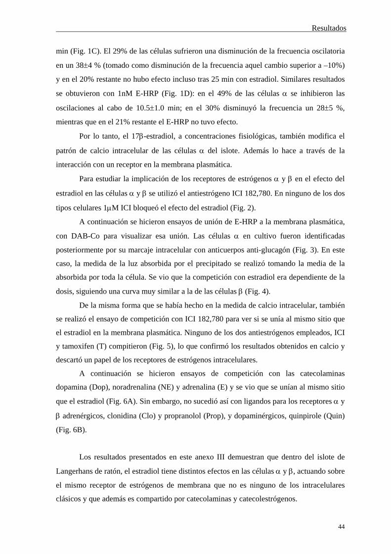
Resultados
44
min (Fig. 1C). El 29% de las células sufrieron una disminución de la frecuencia oscilatoria
en un 384 % (tomado como disminución de la frecuencia aquel cambio superior a –10%)
y en el 20% restante no hubo efecto incluso tras 25 min con estradiol. Similares resultados
se obtuvieron con 1nM E-HRP (Fig. 1D): en el 49% de las células se inhibieron las
oscilaciones al cabo de 10.51.0 min; en el 30% disminuyó la frecuencia un 285 %,
mientras que en el 21% restante el E-HRP no tuvo efecto.
Por lo tanto, el 17-estradiol, a concentraciones fisiológicas, también modifica el
patrón de calcio intracelular de las células del islote. Además lo hace a través de la
interacción con un receptor en la membrana plasmática.
Para estudiar la implicación de los receptores de estrógenos y en el efecto del
estradiol en las células y se utilizó el antiestrógeno ICI 182,780. En ninguno de los dos
tipos celulares 1M ICI bloqueó el efecto del estradiol (Fig. 2).
A continuación se hicieron ensayos de unión de E-HRP a la membrana plasmática,
con DAB-Co para visualizar esa unión. Las células en cultivo fueron identificadas
posteriormente por su marcaje intracelular con anticuerpos anti-glucagón (Fig. 3). En este
caso, la medida de la luz absorbida por el precipitado se realizó tomando la media de la
absorbida por toda la célula. Se vio que la competición con estradiol era dependiente de la
dosis, siguiendo una curva muy similar a la de las células (Fig. 4).
De la misma forma que se había hecho en la medida de calcio intracelular, también
se realizó el ensayo de competición con ICI 182,780 para ver si se unía al mismo sitio que
el estradiol en la membrana plasmática. Ninguno de los dos antiestrógenos empleados, ICI
y tamoxifen (T) compitieron (Fig. 5), lo que confirmó los resultados obtenidos en calcio y
descartó un papel de los receptores de estrógenos intracelulares.
A continuación se hicieron ensayos de competición con las catecolaminas
dopamina (Dop), noradrenalina (NE) y adrenalina (E) y se vio que se unían al mismo sitio
que el estradiol (Fig. 6A). Sin embargo, no sucedió así con ligandos para los receptores y
adrenérgicos, clonidina (Clo) y propranolol (Prop), y dopaminérgicos, quinpirole (Quin)
(Fig. 6B).
Los resultados presentados en este anexo III demuestran que dentro del islote de
Langerhans de ratón, el estradiol tiene distintos efectos en las células y , actuando sobre
el mismo receptor de estrógenos de membrana que no es ninguno de los intracelulares
clásicos y que además es compartido por catecolaminas y catecolestrógenos.

Discusión
45
DISCUSIÓN
Discusión
46
EL ISLOTE DE LANGERHANS Y EL 17-ESTRADIOL
El efecto del 17-estradiol en la fisiología del islote de Langerhans no es nuevo,
sino que ya trabajos fechados durante la década de los setenta y la de los ochenta,
realizados con ratas, demostraron un efecto insulinotrópico del estradiol tanto in vivo como
in situ {SutterDub 1976; Sutter-Dub et al. 1978; Faure & Sutter-Dub 1979; Sutter-Dub
1979; Faure et al. 1983; Faure et al. 1987}. Más adelante se fue corroborando el papel del
estradiol en la prevención o mejora de la diabetes, ya fuera en modelos animales
{Watanabe 1990; Efrat 1991} o en mujeres postmenopáusicas {Stevenson et al. 1994;
Brussaard et al. 1997; Andersson et al. 1997}. De la misma forma, también se vio que el
estradiol disminuía los niveles de glucagón in vivo {Faure et al. 1983; Faure et al. 1988}.
Lo que sí ha permanecido desconocido hasta el momento ha sido el mecanismo por
el cual el estradiol lleva a cabo esos efectos en las células y pancreáticas, encargadas
de la secreción de glucagón e insulina respectivamente. En un trabajo de Nadal et al. de
1998, se demostró que concentraciones fisiológicas de 17-estradiol producían un aumento
rápido de la secreción de insulina de islotes de ratón, de un 30% con respecto a la liberada
por 8mM glucosa. Ese efecto estaba mediado por la interacción del estrógeno con un
receptor en la membrana plasmática de las células pancreáticas {Nadal et al. 1998}. En
esta tesis se presentan evidencias de que ese mismo receptor es el implicado en la
inhibición de las oscilaciones de calcio intracelular por estradiol en la célula pancreática,
con lo que se espera que disminuya la secreción de glucagón, puesto que ésta depende del
calcio intracelular {Leclercq-Meyer et al. 1979}.
La obvia contradicción que surge a partir del efecto del estradiol en dos tipos
celulares del mismo tejido, aumento de calcio intracelular en células y disminución en
células , no es tal si tenemos en cuenta el papel que la insulina y el glucagón juegan en la
homeostasis de glucosa en sangre. El aumento de la secreción de insulina y la disminución
de la de glucagón están ambas dirigidas a disminuir la concentración de glucosa
plasmática. De modo que el estradiol estaría utilizando dos vías aparentemente opuestas,
en dos tipos celulares, para llevar a cabo un mismo efecto fisiológico.

Discusión
47
VÍA DE SEÑALIZACIÓN UTILIZADA POR EL ESTRADIOL
EN EL ISLOTE DE LANGERHANS
La interacción del estradiol con su receptor en la membrana plasmática de la célula
pancreática produce un aumento en la concentración de GMPc. Éste activa la PKG que a
través de una fosforilación cierra el KATP y despolariza la membrana plasmática. Los
canales de calcio dependientes de voltaje de la membrana se abren y dan lugar a un patrón
oscilatorio del calcio intracelular. El resultado fisiológico es el aumento de la secreción de
insulina inducida por glucosa. En Figura 1 se presenta el modelo de la acción del 17-
estradiol en la célula b pancreática de ratón.
La vía GMPc-PKG está implicada en la secreción de insulina por glucosa
{Laychock et al. 1991; Green et al. 1993} aunque no se sabe de qué forma interviene en el
proceso. Al ser utilizada también por el estradiol, éste potencia así una de las vías por las
que la glucosa actúa como secretagogo insulinotrópico en el islote de Langerhans. Se
demuestra aquí por primera vez una modulación negativa del KATP por PKG mediada por
estradiol. Se desconoce si la diana de la PKG es el propio canal o si el efecto es indirecto
por medio de otras proteínas. Éste podría ser también el mecanismo por el cual el GMPc
producido por la glucosa regula la secreción de insulina. Por lo tanto, son necesarios más
experimentos que establezcan si la PKG fosforila el KATP o en su caso, qué proteína es la
diana de la quinasa. Además queda por confirmar la participación de PKG en el cierre del
canal por glucosa.
Las enzimas encargadas de sintetizar GMPc a partir de GTP son las guanilato
ciclasas que existen en dos formas, solubles en el citosol o insertadas en la membrana
plasmática. Las primeras son activadas por NO y CO y las segundas por péptido
natriuréticos, ANP, BNP y CNP {Garbers & Lowe 1994; Carvajal et al. 2000}. En
mamíferos también hay dos tipos de PKG en función de su localización. La PKG I es
soluble y tiene dos isoformas procedentes de procesamiento alternativo de un mismo
ARNm y la PKG II está asociada a la cara intracelular de la membrana plasmática por
miristoilación {Lohmann et al. 1997}.

Discusión
48
FIGURA 1. Modelo del efecto rápido del 17-estradiol en la célula
del islote de Langerhans pancreático de ratón En la figura se resume el mecanismo por el cual el 17-estradiol modifica la
secreción de insulina en la célula pancreática. Se incluye el GMPc como segundo mensajero que modifica el canal de potasio dependiente de ATP, el potencial de membrana, el calcio intracelular y por tanto la secreción de insulina. Figura cedida por el Dr. Mario Díaz.
En el islote de Langerhans se tiene presencia de ambos tipos de GC, puesto que un
inhibidor de la GCs bloquea el aumento de GMPc por glucosa {Laychock et al. 1991} y el
ANP (GCm) potencia los movimientos de calcio intracelular en islotes {Lee & Laychock
1997}. La utilización de un inhibidor de la GCs no bloquea el efecto del estradiol sobre el
KATP en células y compuestos dadores de NO, activador de la GCs, abren el KATP
{Tsuura et al. 1994} y disminuyen la secreción de insulina {Cunningham et al. 1994}.
Vm
Ca2+
GTP
GC
GMP cíclico
PKG
Ca2+
Vesículas Insulina
K+
17-Estradiol

Discusión
49
Además, el NO es el mecanismo por el que la interleuquina 1, liberada por los
macrófagos, produce muerte de las células pancreáticas, causa de la diabetes mellitus
tipo I {Spinas 1999}. De modo que, aunque podría darse un modo dual de acción del NO
dependiendo del tiempo de exposición a él, éste no parece estar implicado en el efecto del
estradiol en la célula pancreática.
Por otra parte se ha visto activación de la GCm por estradiol en células PC12 y la
interacción directa del estradiol con una GCm consistente en los dominios intracelulares
catalítico y quinasa {Chen et al. 1998}. La interacción del estradiol con esos dominios no
puede estar mediando el efecto del estrógeno sobre la célula pancreática, puesto que E-
HRP lo mimetiza sin poder atravesar la membrana plasmática. Por lo tanto, son necesarios
más experimentos en el islote de Langerhans para establecer cuál de las dos GC es la
implicada en el efecto del estradiol en la célula . De la misma forma con la PKG, si se
trata de la soluble o la de membrana la isoforma activada por el aumento de GMPc por
estradiol. Tampoco hay que descartar la participación de las fosfodiesterasas específicas en
el aumento de los niveles de GMPc en el islote de Langerhans.
En cuanto a la vía de señalización del AMPc y la PKA utilizada por estradiol en
otros tipos celulares {Falkenstein et al. 2000}, no participa en el caso de la célula
pancreática, puesto que no se tiene aumento de la concentración de AMPc en respuesta al
estrógeno y además, un inhibidor de la PKA no impide su efecto sobre el KATP. Al
contrario de lo que sucede con el canal de potasio de alta conductancia, Maxi-K {Valverde
et al. 1999}, en la célula no hay interacción directa del estradiol con el KATP, puesto que
no tiene efecto cuando se mide la actividad del canal con la técnica de patch-clamp en la
configuración de parche aislado.
Si bien el mecanismo de secreción de glucagón en la célula apenas es conocido,
sí se sabe que está mediado por movimientos de calcio intracelular {Johansson et al.
1987}. Por un lado, la presencia del KATP y su papel en el acoplamiento estímulo-secreción
en la célula es un tema muy controvertido {Nadal et al. 1999; Gopel et al. 2000}. Por
otra parte, el GMPc disminuye la secreción de glucagón y el calcio intracelular {Verspohl
& Bernemann 1996 y Anexo III de esta tesis}, de modo que el estradiol podría utilizar la
misma vía de señalización que en la célula . De esta forma, se tendría que un mismo
segundo mensajero, el GMPc, produce dos efectos opuestos en dos tipos celulares de un
mismo tejido.
Discusión
50
RECEPTOR DE ESTRÓGENOS DE MEMBRANA NO
CLÁSICO
La gran controversia que existe en el campo del receptor de estrógenos de
membrana se centra principalmente en dos posibilidades que se plantean con respecto a su
naturaleza: que se trate de los mismos receptores intracelulares, localizados en la
membrana, o que por el contrario sea un receptor que no guarde ninguna relación
estructural con ellos. Ambas hipótesis no son excluyentes, dada la enorme variedad de
tipos celulares y de efectos del estradiol. Sin embargo, existen algunas cuestiones a tener
en cuenta a la hora de apoyar cada uno de los modelos.
La utilización de antagonistas de los receptores de estrógenos intracelulares es un
arma importante para distinguir entre la participación de estos o de otro tipo de receptores
en los efectos del estradiol. Actualmente el ICI 182,780 es el antiestrógeno específico de
los receptores clásicos que más se utiliza {Wakeling et al. 1991}. A este respecto y tal y
como se citaron en la Introducción, hay numerosos ejemplos que muestran tanto un
bloqueo como una total ineficacia del ICI 182,780 para inhibir los efectos rápidos del
estradiol. En esta tesis se muestra que 1M de este antiestrógeno no impide el efecto de
1nM 17-estradiol en el calcio intracelular, ni en la célula , ni en la . Además, en
ensayos de competición por el sitio de unión del estradiol con ICI, se ve que éste no se une
al receptor de estrógenos presente en la membrana plasmática de ninguno de los dos tipos
celulares.
Por otro lado, los anticuerpos desarrollados contra los receptores intracelulares
permiten visualizar la presencia o no de estos en la membrana plasmática. Además, por
reconocer distintos dominios del receptor, también dan información acerca de cuáles de
ellos están presentes en el de membrana. Con anticuerpos dirigidos contra diferentes
dominios de RE, se ha visto marcaje en la membrana plasmática de una línea celular de
pituitaria, GH3/B6, y en hipotálamo {Pappas et al. 1995; Clarke et al. 2000}. Además, el
anticuerpo que reconoce el dominio de unión del ligando al RE bloquea la unión de E-
BSA a la membrana de células de músculo liso {Morey et al. 1997}. Sin embargo no
sucede así con la célula pancreática, en donde cinco anticuerpos dirigidos contra RE y
dos contra RE no marcan la membrana plasmática de células no permeabilizadas,
mientras que ambos están presentes en el citosol. Ninguno de ellos compite con el receptor
de estrógenos de membrana en ensayos de unión con E-HRP.
Discusión
51
Por otra parte, se ha visto un 2-3% de expresión de RE y RE en la membrana
plasmática de células CHO transfectadas con el ADNc de ambos receptores. Además estos
están acoplados a proteínas G, Gq y Gs, que al unirse estradiol aumentan los niveles de IP3
y AMPc {Razandi et al. 1999}. Sin embargo este tipo de experimentos no está exento de
controversia.
Para que una proteína se localice en la membrana plasmática es necesario que
disponga de un dominio transmembrana a través del cual se inserte en ella. Es poco
probable que una proteína con tales regiones hidrofóbicas pueda presentarse en forma
soluble en un entorno hidrofílico como es el citosol. Por lo tanto, este dominio debe estar
presente exclusivamente en la forma de membrana y podría incluirse en la proteína
citosólica por procesamiento alternativo. En el caso anterior de expresión de RE y en
CHO, este procesamiento parece improbable, puesto que la transfección se hace con los
ADNc de ambos receptores. Las librerías de ADNc se originan a partir de los ARNm ya
procesados de las células, es decir, sólo contienen exones, puesto que los intrones han sido
ya eliminados. Por lo tanto, no existe la posibilidad de inserción de exones presentes en la
estructura del gen pero eliminados del ARNm para formar la proteína citosólica y que sin
embargo se incluyan en la forma de membrana. De modo que la proteína queda sujeta a
poder insertarse en la membrana plasmática por modificiaciones postraduccionales que la
unan a una molécula lipídica. Al examinar la estructura del RE, los propios autores,
Razandi et al., indican que no hay sitios de palmitoilación y sólo unos pocos candidatos de
miristoilación {Razandi et al. 1999}. Por otro lado, se ha visto que una subpoblación de
RE es glicosilada por N-acetilglucosamina {Jiang & Hart 1997}. Sin embargo todas estas
posibilidades se ven anuladas por el hecho de que ambas proteínas, la citosólica y la de
membrana, tienen el mismo peso molecular y proceden de un mismo tránscrito {Razandi et
al. 1999}.
Otros trabajos apoyan la existencia de un tipo de receptor de estrógenos en la
membrana plasmática que no está relacionado con los intracelulares clásicos. Éste es el
caso del receptor por el que el estradiol modifica las corrientes producidas por kainato en
neuronas de hipocampo. Ese efecto del estrógeno sigue presente en ratones carentes
(knockout) de RE. Aquí, la implicación del RE es descartada por la utilización del
antiestrógeno ICI 182,780, que no es capaz de bloquear el efecto del estradiol {Gu et al.
1999}. Por otro lado, hay activación de ERK a través de la membrana en células carentes
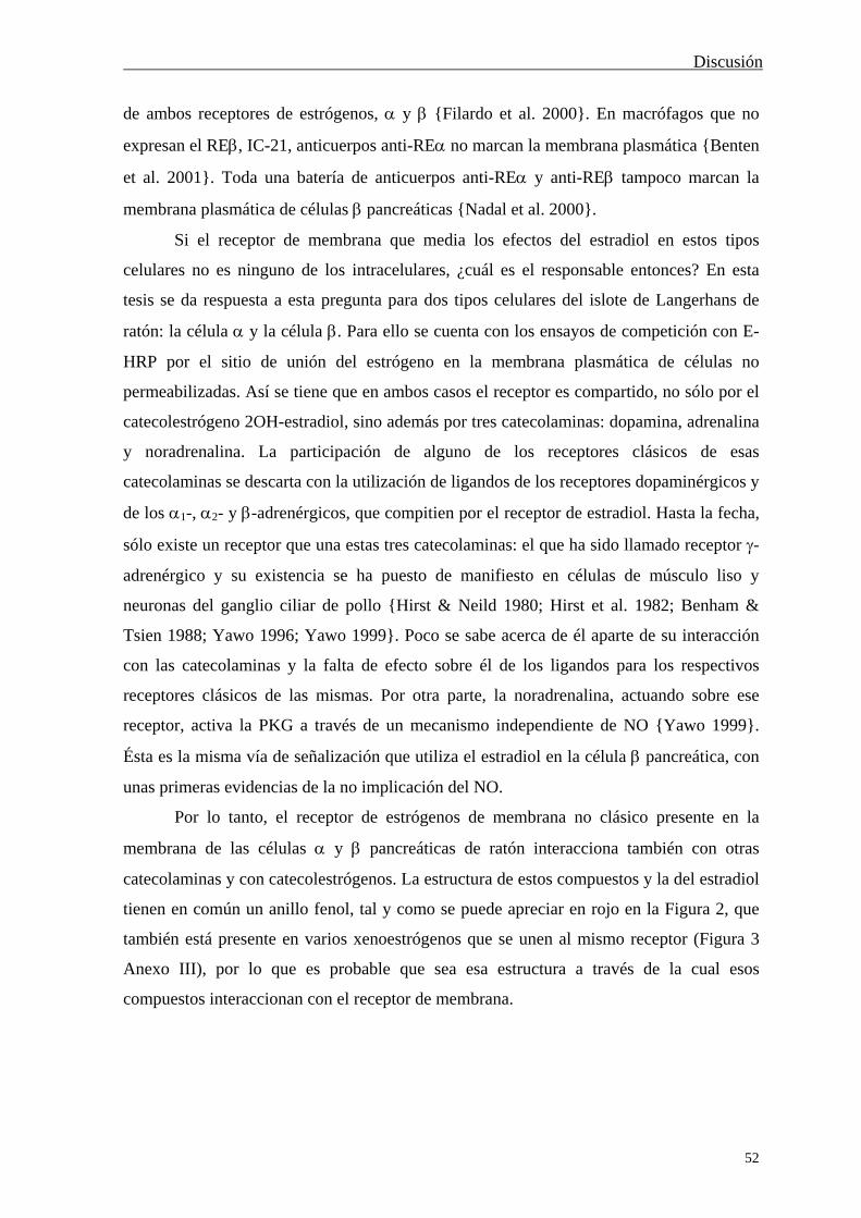
Discusión
52
de ambos receptores de estrógenos, y {Filardo et al. 2000}. En macrófagos que no
expresan el RE, IC-21, anticuerpos anti-RE no marcan la membrana plasmática {Benten
et al. 2001}. Toda una batería de anticuerpos anti-RE y anti-RE tampoco marcan la
membrana plasmática de células pancreáticas {Nadal et al. 2000}.
Si el receptor de membrana que media los efectos del estradiol en estos tipos
celulares no es ninguno de los intracelulares, ¿cuál es el responsable entonces? En esta
tesis se da respuesta a esta pregunta para dos tipos celulares del islote de Langerhans de
ratón: la célula y la célula . Para ello se cuenta con los ensayos de competición con E-
HRP por el sitio de unión del estrógeno en la membrana plasmática de células no
permeabilizadas. Así se tiene que en ambos casos el receptor es compartido, no sólo por el
catecolestrógeno 2OH-estradiol, sino además por tres catecolaminas: dopamina, adrenalina
y noradrenalina. La participación de alguno de los receptores clásicos de esas
catecolaminas se descarta con la utilización de ligandos de los receptores dopaminérgicos y
de los 1-, 2- y -adrenérgicos, que compitien por el receptor de estradiol. Hasta la fecha,
sólo existe un receptor que una estas tres catecolaminas: el que ha sido llamado receptor -
adrenérgico y su existencia se ha puesto de manifiesto en células de músculo liso y
neuronas del ganglio ciliar de pollo {Hirst & Neild 1980; Hirst et al. 1982; Benham &
Tsien 1988; Yawo 1996; Yawo 1999}. Poco se sabe acerca de él aparte de su interacción
con las catecolaminas y la falta de efecto sobre él de los ligandos para los respectivos
receptores clásicos de las mismas. Por otra parte, la noradrenalina, actuando sobre ese
receptor, activa la PKG a través de un mecanismo independiente de NO {Yawo 1999}.
Ésta es la misma vía de señalización que utiliza el estradiol en la célula pancreática, con
unas primeras evidencias de la no implicación del NO.
Por lo tanto, el receptor de estrógenos de membrana no clásico presente en la
membrana de las células y pancreáticas de ratón interacciona también con otras
catecolaminas y con catecolestrógenos. La estructura de estos compuestos y la del estradiol
tienen en común un anillo fenol, tal y como se puede apreciar en rojo en la Figura 2, que
también está presente en varios xenoestrógenos que se unen al mismo receptor (Figura 3
Anexo III), por lo que es probable que sea esa estructura a través de la cual esos
compuestos interaccionan con el receptor de membrana.
Discusión
53
FIGURA 5. Estructura del Estradiol y las catecolaminas Dopamina, Adrenalina y Noradrenalina
En la figura se presenta la estructura de cuatro de los compuestos que interaccionan con el receptor de estrógenos de membrana no clásico: estradiol, dopamina, adrenalina y noradrenalina. La característica común en todos ellos es el grupo fenol, mostrado en la figura en color rojo, estructura candidata a ser la responsable de la interacción con el receptor.
Los intentos de aislar el receptor de estrógenos de la membrana plasmática han
dado por el momento una banda de 29kDa que se ha detectado en cerebelo {Bukusoglu &
Krieger 1994}, espermatozoides humanos {Luconi et al. 1999} y útero {Monje & Boland
1999}. Además se han visto otras bandas en cerebro {Zheng & Ramirez 1997} y útero
{Monje & Boland 1999}. En el caso de los espermatozoides se utilizó un anticuerpo
dirigido contra el dominio de unión a estradiol del RE, H222 {Luconi et al. 1999}. Sin
embargo, en útero fueron varios anticuerpos dirigidos contra diferentes dominios de RE
Esto demuestra que aunque esas bandas sean de un peso molecular inferior al de los
receptores clásicos, aún conservan las regiones reconocidas por todos esos anticuerpos y
además con una estructura similar.
OH
HO Estradiol
HO
HO CH2 CH2 NH2
Dopamina
CH3
HO
HO CH CH2 NH
OH
Adrenalina HO
HO CH CH2 NH2
OH
Noradrenalina

Discusión
54
Por otra parte, el receptor de estrógenos de membrana es compartido con
progesterona en cerebelo {Bukusoglu & Krieger 1994}. La secuencia completa del ADNc
de una proteína de unión a progesterona en membrana se ha obtenido de células de
músculo liso vascular porcino {Falkenstein et al. 1996; Falkenstein et al. 1999} y humano
{Gerdes et al. 1998}. La proteína porcina que se predice tiene 194 aminoácidos y un peso
molecular de 21.67kDa. Falta por saber si esos receptores de progesterona de membrana
son compartidos por estradiol.
Además de identificar el receptor de estrógenos que se expresa en la membrana
plasmática celular, también han de centrarse esfuerzos en demostrar su funcionalidad
mediando los efectos fisiológicos del estradiol. En el caso de la línea celular de pituitaria,
GH3/B6, un anticuerpo dirigido contra RE reproduce la liberación de prolactina por
estradiol y la activación de eNOS en endotelio se ve aumentada por sobrexpresión de RE
e inhibida por ICI 182,780 {Chen et al. 1999}. En las neuronas de hipocampo de rata en
donde se ha visto marcaje de la membrana con anticuerpos anti-RE {Clarke et al. 2000},
no se ha comprobado su funcionalidad y en el caso del ratón, no es ése el receptor
responsable del efecto del estradiol en hipocampo, puesto que sigue produciéndose en
animales carentes (knockout) de ER {Gu et al. 1999}. Por otro lado, en esta tesis se
demuestra que el receptor de estrógenos de membrana descrito en células y del islote
de Langerhans es el responsable del efecto del estradiol, puesto que éste es reproducido por
la misma concentración de E-HRP. Además, en presencia de noradrenalina, el estradiol no
modifica el patrón oscilatorio de calcio de las células , en coherencia con un bloqueo del
receptor por la noradrenalina. A la inversa, en presencia de estradiol, la noradrenalina sigue
ejerciendo el mismo efecto, de acuerdo con su papel a través de los receptores -
adrenérgicos presentes en la célula pancreática. De forma similar sucede con la
dopamina (resultados no mostrados).
Indudablemente, los animales carentes de RE y de RE (doble knockout) serán
una fuente importante de evidencias a favor o en contra de la presencia de estos receptores
en la membrana plasmática. Aunque estos ratones ya se han obtenido {Couse et al. 1999;
Ogawa et al. 2000}, por el momento no se han publicado resultados de los efectos del
estradiol a través de la membrana plasmática.

Discusión
55
GENÓMICO -- RECEPTOR INTRACELULAR versus
NO GENÓMICO -- RECEPTOR DE MEMBRANA
Tras los numerosos estudios que se han venido realizando desde el publicado en
1975 por Pietras & Szego, se ha dado solidez a la existencia de un nuevo mecanismo de
acción en el efecto de los estrógenos. Por lo tanto hemos de rendirnos ante la evidencia de
que estos no sólo actúan desde dentro en un proceso lento y de larga duración, sino que
además son capaces de actuar como las hormonas hidrofílicas, a través de un receptor en la
membrana plasmática, que produce efectos rápidos en los que poco o nada tiene que ver la
transcripción génica.
Pero en contra de lo que podría pensarse en un principio en cuanto a la clara
distinción entre efectos genómicos mediados por receptores intracelulares y los no
genómicos, a través de receptores de membrana, se está viendo que dicha distinción es algo
caprichosa. Según se ha podido observar, la activación rápida de MAPK por estradiol
produce efectos fisiológicos que no se detectan en un corto espacio de tiempo, sino que se
requieren varias horas, aunque se produzcan a través de una interacción del estrógeno con
un receptor en la membrana plasmática. Entre estos ejemplos figura la estimulación del
crecimiento celular {Di Domenico et al. 1996; Morey et al. 1997} y la inhibición de la
apoptosis {Razandi et al. 2000a}. Puesto que tanto el crecimiento celular como la
apoptosis requieren activación de la transcripción génica y que la vía de las MAPKs puede
dar lugar a procesos genómicos {Alberts et al. 1994}, es probable que el estradiol, a través
de la activación rápida de una vía de señalización intracelular produzca efectos finales
genómicos. De hecho, el E-BSA activa la transcripción de un gen formado por el promotor
de c-fos de una forma independiente de los RE intracelulares, en la línea de neuroblastoma
SK-N-SH, lo que además indica que esos efectos genómicos a través de señalización
intracelular podrían estar mediados por un receptor de estrógenos en la membrana
plasmática{Watters et al. 1997}.
Por otra parte, es bien conocido que el AMPc activa la PKA y que una de sus
dianas es la proteína de unión a CRE, CREB. Esta proteína interacciona con CRE (cyclic
AMP response element), que es una zona del ADN presente en la zona reguladora de
algunos genes. Cuando CREB es fosforilada estimula la transcripción génica {Alberts et al.
1994}, por lo que la activación de la vía de señalización del AMPc puede producir efectos
no genómicos en minutos y genómicos a largo plazo.
Discusión
56
Tampoco hay que descartar el hecho de que los mismos receptores de estrógenos
intracelulares pudieran participar en la activación de vías de señalización intracelular con
resultados no genómicos, previa o paralelamente a su papel transcripcional.
Por lo tanto, los términos RE intracelulares, RE de membrana, genómico, no
genómico, a largo plazo y rápido, se pueden entrecruzar de diversas formas en cada tipo
celular con el fin de producir la amplia variedad de efectos fisiológicos de los estrógenos.
Todo esto hace necesario un nuevo punto de vista con el que abordar los efectos de los
estrógenos y sus mecanismos. Para ello es imprescindible desarrollar nuevas herramientas
con las que poder distinguir claramente la participación de los receptores de membrana o
los intracelulares en cada uno de esos efectos, así como los mecanismos empleados en cada
caso.

Conclusiones
57
CONCLUSIONES
Conclusiones
58
1.- Una concentración fisiológica de 17-estradiol produce un aumento de la
concentración de calcio intracelular en el islote de Langerhans de ratón, que se manifiesta
como un aumento en la frecuencia de las oscilaciones mostradas por el islote en presencia
de 8mM glucosa o bien como un aumento en la duración de dichas oscilaciones.
2.- El 17-estradiol produce un aumento en los niveles de GMPc en el islote de
Langerhans en presencia de 8mM glucosa. Este aumento es similar al producido por 11mM
glucosa.
3.- El GMPc modula negativamente el canal de potasio dependiente de ATP de una
forma similar a como lo hace el 17-estradiol. Este efecto no se debe a una interacción
directa del nucleótido cíclico con el canal, sino a la activación de vías intracelulares de
señalización.
4.- El canal de potasio dependiente de ATP es modulado negativamente por una
fosforilación dependiente de PKG, activada por estradiol.
5.- La vía de señalización del AMPc y la PKA no participan en el efecto del 17-
estradiol en las células del islote de Langerhans de ratón.
6.- Concentraciones fisiológicas de 17-estradiol suprime las oscilaciones de calcio
intracelular que presentan las células pancreáticas en ausencia de glucosa.
7.- El receptor de estrógenos presente en la membrana plasmática de las células y
es el responsable del efecto del estradiol en la concentración de calcio intracelular en
ambos tipos celulares.
8.- El receptor de estrógenos de membrana descrito en las células y del islote de
Langerhans de ratón ejerce dos efectos opuestos en ambos tipos celulares, que sin
embargo, conducen a un mismo efecto fisiológico: la disminución de la concentración de
glucosa en sangre.

Conclusiones
59
9.- El receptor de estrógenos de membrana presente en las células y
pancreáticas no es ninguno de los receptores de estrógenos intracelulares, RE o RE.
10.- Este receptor de estrógenos de membrana no clásico es compartido por
catecolestrógenos y por las catecolaminas dopamina, adrenalina y noradrenalina. No es
ninguno de los receptores clásicos adrenérgicos, ni dopaminérgicos, sino que tiene el perfil
farmacológico del llamado receptor -adrenérgico.

Referencias
60
REFERENCIAS
Referencias
61
Aerts L, van Assche FA, Faure A, Sutter-Dub MT (1980) Effects of treatment with progesterone and oestradiol-17 beta on the endocrine pancreas in ovariectomized rats: ultrastructural variations in the B cells. J Endocrinol 84: 317-20.
Aguilar-Bryan L, Clement JP 4th, Gonzalez G, Kunjilwar K, Babenko A, Bryan J (1998) Toward understanding the assembly and structure of KATP channels. Physiol Rev 78: 227-45.
Aizawa T, Komatsu M, Asanuma N, Sato Y, Sharp GW (1998) Glucose action 'beyond ionic events' in the pancreatic beta cell. Trends Pharmacol Sci 19: 496-9.
Alberts B, Bray D, Lewis J, Raff M, Roberts K, Watson JD (1994) Molecular biology of the cell. 3ª edición. Garland Publishing, Inc.: New York.
Alonso R, Lopez-Coviella I (1998) Gonadal steroids and neuronal function. Neurochem Res 23: 675-88.
Ammala C, Ashcroft FM, Rorsman P (1993) Calcium-independent potentiation of insulin release by cyclic AMP in single beta-cells. Nature 363: 356-8.
Andersson B, Mattsson LA, Hahn L, Marin P, Lapidus L, Holm G, Bengtsson BA, Bjorntorp P (1997) Estrogen replacement therapy decreases hyperandrogenicity and improves glucose homeostasis and plasma lipids in postmenopausal women with noninsulin-dependent diabetes mellitus. J Clin Endocrinol Metab 82: 638-43.
Aronica SM, Kraus WL, Katzenellenbogen BS (1994) Estrogen action via the cAMP signaling pathway: stimulation of adenylate cyclase and cAMP-regulated gene transcription. Proc Natl Acad Sci U S A 91: 8517-21.
Asaithambi A, Mukherjee S, Thakur MK (1997) Expression of 112-kDa estrogen receptor in mouse brain cortex and its autoregulation with age. Biochem Biophys Res Commun 231: 683-5.
Ashcroft FM, Gribble FM (1999) ATP-sensitive K+ channels and insulin secretion: their role in health and disease. Diabetologia 42: 903-19.
Ashcroft FM, Harrison DE, Ashcroft SJ (1984) Glucose induces closure of single potassium channels in isolated rat pancreatic beta-cells. Nature 312: 446-8.
Ashcroft FM, Rorsman P (1989) Electrophysiology of the pancreatic beta-cell. Prog Biophys Mol Biol 54: 87-143.
Barbosa RM, Silva AM, Tome AR, Stamford JA, Santos RM, Rosario LM (1998) Control of pulsatile 5-HT/insulin secretion from single mouse pancreatic islets by intracellular calcium dynamics. J Physiol (Lond) 510 ( Pt 1): 135-43.
Barbosa RM, Silva AM, Tome AR, Stamford JA, Santos RM, Rosario LM (1996) Real time electrochemical detection of 5-HT/insulin secretion from single pancreatic islets: effect of glucose and K+ depolarization. Biochem Biophys Res Commun 228: 100-4.
Barkhem T, Carlsson B, Nilsson Y, Enmark E, Gustafsson J, Nilsson S (1998) Differential response of estrogen receptor alpha and estrogen receptor beta to partial estrogen

Referencias
62
agonists/antagonists. Mol Pharmacol 54: 105-12.
Batra S (1986) Effect of estrogen and progesterone treatment on calcium uptake by the myometrium and smooth muscle of the lower urinary tract. Eur J Pharmacol 127: 37-42.
Beato M (1989) Gene regulation by steroid hormones. Cell 56: 335-44.
Beguin P, Nagashima K, Nishimura M, Gonoi T, Seino S (1999) PKA-mediated phosphorylation of the human K(ATP) channel: separate roles of Kir6.2 and SUR1 subunit phosphorylation. EMBO J 18: 4722-32.
Benham CD, Tsien RW (1988) Noradrenaline modulation of calcium channels in single smooth muscle cells from rabbit ear artery. J Physiol (Lond) 404: 767-84.
Benten WP, Lieberherr M, Giese G, Wunderlich F (1998) Estradiol binding to cell surface raises cytosolic free calcium in T cells. FEBS Lett 422: 349-53.
Benten WP, Stephan C, Lieberherr M, Wunderlich F (2001) Estradiol signaling via sequestrable surface receptors. Endocrinology 142: 1669-77.
Berthois Y, Pourreau-Schneider N, Gandilhon P, Mittre H, Tubiana N, Martin PM (1986) Estradiol membrane binding sites on human breast cancer cell lines. Use of a fluorescent estradiol conjugate to demonstrate plasma membrane binding systems. J Steroid Biochem 25: 963-72.
Berts A, Ball A, Gylfe E, Hellman B (1996) Suppression of Ca2+ oscillations in glucagon-producing alpha 2-cells by insulin/glucose and amino acids. Biochim Biophys Acta 1310: 212-6.
Berts A, Gylfe E, Hellman B (1995) Ca2+ oscillations in pancreatic islet cells secreting glucagon and somatostatin. Biochem Biophys Res Commun 208: 644-9.
Berts A, Gylfe E, Hellman B (1997) Cytoplasmic Ca2+ in glucagon-producing pancreatic alpha-cells exposed to carbachol and agents affecting Na+ fluxes. Endocrine 6: 79-83.
Beyer C, Karolczak M (2000) Estrogenic stimulation of neurite growth in midbrain dopaminergic neurons depends on cAMP/protein kinase A signalling. J Neurosci Res 59: 107-16.
Beyer C, Raab H (1998) Nongenomic effects of oestrogen: embryonic mouse midbrain neurones respond with a rapid release of calcium from intracellular stores. Eur J Neurosci 10: 255-62.
Bi R, Broutman G, Foy MR, Thompson RF, Baudry M (2000) The tyrosine kinase and mitogen-activated protein kinase pathways mediate multiple effects of estrogen in hippocampus. Proc Natl Acad Sci U S A 97: 3602-7.
Bokvist K, Olsen HL, Hoy M, Gotfredsen CF, Holmes WF, Buschard K, Rorsman P, Gromada J (1999) Characterisation of sulphonylurea and ATP-regulated K+ channels in rat pancreatic A-cells. Pflugers Arch 438: 428-36.
Bression D, Michard M, Le Dafniet M, Pagesy P, Peillon F (1986) Evidence for a specific
Referencias
63
estradiol binding site on rat pituitary membranes. Endocrinology 119: 1048-51.
Brubaker KD, Gay CV (1994) Specific binding of estrogen to osteoclast surfaces. Biochem Biophys Res Commun 200: 899-907.
Brunicardi FC, Druck P, Seymour NE, Sun YS, Elahi D, Andersen DK (1990) Selective neurohormonal interactions in islet cell secretion in the isolated perfused human pancreas. J Surg Res 48: 273-8.
Brussaard HE, Gevers Leuven JA, Frolich M, Kluft C, Krans HM (1997) Short-term oestrogen replacement therapy improves insulin resistance, lipids and fibrinolysis in postmenopausal women with NIDDM. Diabetologia 40: 843-9.
Buitrago C, Massheimer V, de Boland AR (2000) Acute modulation of Ca2+ influx on rat heart by 17beta-estradiol. Cell Signal 12: 47-52.
Bukusoglu C, Krieger NR (1994) Photoaffinity labeling with progesterone-11 alpha-hemisuccinate- (2-. J Neurochem 63: 1434-8.
Burns CJ, Howell SL, Jones PM, Persaud SJ (1997) Glucose-stimulated insulin secretion from rat islets of Langerhans is independent of mitogen-activated protein kinase activation. Biochem Biophys Res Commun 239: 447-50.
Caldwell JD, Walker CH, Rivkina A, Pedersen CA, Mason GA (1999) Radioligand assays for oestradiol and progesterone conjugated to protein reveal evidence for a common membrane binding site in the medial preoptic area-anterior hypothalamus and differential modulation by cholera toxin and GTPgammaS. J Neuroendocrinol 11: 409-17.
Cario-Toumaniantz C, Loirand G, Ferrier L, Pacaud P (1998) Non-genomic inhibition of human P2X7 purinoceptor by 17beta-oestradiol. J Physiol 508 ( Pt 3): 659-66.
Carvajal JA, Germain AM, Huidobro-Toro JP, Weiner CP (2000) Molecular mechanism of cGMP-mediated smooth muscle relaxation. J Cell Physiol 184: 409-20.
Caulin-Glaser T, Garcia-Cardena G, Sarrel P, Sessa WC, Bender JR (1997) 17 beta-estradiol regulation of human endothelial cell basal nitric oxide release, independent of cytosolic Ca2+ mobilization. Circ Res 81: 885-92.
Chambliss KL, Yuhanna IS, Mineo C, Liu P, German Z, Sherman TS, Mendelsohn ME, Anderson RG, Shaul PW (2000) Estrogen receptor alpha and endothelial nitric oxide synthase are organized into a functional signaling module in caveolae. Circ Res 87: E44-52.
Chang AS, Chang SM (1999) Nongenomic steroidal modulation of high-affinity serotonin transport. Biochim Biophys Acta 1417: 157-66.
Charles MA, Lawecki J, Pictet R, Grodsky GM (1975) Insulin secretion. Interrelationships of glucose, cyclic adenosine 3:5- monophosphate, and calcium. J Biol Chem 250: 6134-40.
Chen Z, Yuhanna IS, Galcheva-Gargova Z, Karas RH, Mendelsohn ME, Shaul PW (1999) Estrogen receptor alpha mediates the nongenomic activation of endothelial nitric oxide synthase by estrogen. J Clin Invest 103: 401-6.
Referencias
64
Chen ZJ, Yu L, Chang CH (1998) Stimulation of membrane-bound guanylate cyclase activity by 17-beta estradiol. Biochem Biophys Res Commun 252: 639-42.
Cheng CY, Rose RJ, Boettcher B (1981) The binding of oestradiol-17beta to human spermatozoa - an electron microscope autoradiographic study. Int J Androl 4: 304-13.
Chiodo LA, Caggiula AR (1980) Alterations in basal firing rate and autoreceptor sensitivity of dopamine neurons in the substantia nigra following acute and extended exposure to estrogen. Eur J Pharmacol 67: 165-6.
Christie MR, Ashcroft SJ (1984) Cyclic AMP-dependent protein phosphorylation and insulin secretion in intact islets of Langerhans. Biochem J 218: 87-99.
Chu S, Fuller PJ (1997) Identification of a splice variant of the rat estrogen receptor beta gene. Mol Cell Endocrinol 132: 195-9.
Clarke CH, Norfleet AM, Clarke MS, Watson CS, Cunningham KA, Thomas ML (2000) Perimembrane localization of the estrogen receptor alpha protein in neuronal processes of cultured hippocampal neurons. Neuroendocrinology 71: 34-42.
Condliffe SB, Doolan CM, Harvey BJ (2001) 17b-Oestradiol acutely regulates Cl- secretion in rat distal colonic epithelium. J Physiol 530: 47-54.
Couse JF, Hewitt SC, Bunch DO, Sar M, Walker VR, Davis BJ, Korach KS (1999) Postnatal sex reversal of the ovaries in mice lacking estrogen receptors alpha and beta. Science 286: 2328-31.
Couse JF, Korach KS (1999) Estrogen receptor null mice: what have we learned and where will they lead us? Endocr Rev 20: 358-417.
Cunningham JM, Mabley JG, Delaney CA, Green IC (1994) The effect of nitric oxide donors on insulin secretion, cyclic GMP and cyclic AMP in rat islets of Langerhans and the insulin-secreting cell lines HIT-T15 and RINm5F. Mol Cell Endocrinol 102: 23-9.
Dachicourt N, Serradas P, Giroix MH, Gangnerau MN, Portha B (1996) Decreased glucose-induced cAMP and insulin release in islets of diabetic rats: reversal by IBMX, glucagon, GIP. Am J Physiol 271: E725-32.
Darkow DJ, Lu L, White RE (1997) Estrogen relaxation of coronary artery smooth muscle is mediated by nitric oxide and cGMP. Am J Physiol 272: H2765-73.
Dean PM, Matthews EK (1968) Electrical activity in pancreatic islet cells. Nature 219: 389-90.
Debuse M (1998) Lo esencial en Sistema Endocrino y aparato reproductor. 1ª edición. Harcourt Brace: Madrid.
Dechering K, Boersma C, Mosselman S (2000) Estrogen receptors alpha and beta: two receptors of a kind? Curr Med Chem 7: 561-76.
Di Domenico M, Castoria G, Bilancio A, Migliaccio A, Auricchio F (1996) Estradiol activation of human colon carcinoma-derived Caco-2 cell growth. Cancer Res 56: 4516-
Referencias
65
21.
Ding WG, Gromada J (1997) Protein kinase A-dependent stimulation of exocytosis in mouse pancreatic beta-cells by glucose-dependent insulinotropic polypeptide. Diabetes 46: 615-21.
Ding WG, Renstrom E, Rorsman P, Buschard K, Gromada J (1997) Glucagon-like peptide I and glucose-dependent insulinotropic polypeptide stimulate Ca2+-induced secretion in rat alpha-cells by a protein kinase A-mediated mechanism. Diabetes 46: 792-800.
Disshon KA, Dluzen DE (1997) Estrogen as a neuromodulator of MPTP-induced neurotoxicity: effects upon striatal dopamine release. Brain Res 764: 9-16.
Doolan CM, Condliffe SB, Harvey BJ (2000) Rapid non-genomic activation of cytosolic cyclic AMP-dependent protein kinase activity and [Ca(2+)]i by 17beta-oestradiol in female rat distal colon. Br J Pharmacol 129: 1375-86.
Dopp E, Vollmer G, Hahnel C, Grevesmuhl Y, Schiffmann D (1999) Modulation of the intracellular calcium level in mammalian cells caused by 17beta-estradiol, different phytoestrogens and the anti-estrogen ICI 182780. J Steroid Biochem Mol Biol 68: 57-64.
Drouva SV, Laplante E, Kordon C (1983) Effects of ovarian steroids on in vitro release of LHRH from mediobasal hypothalamus. Neuroendocrinology 37: 336-41.
Dufy B, Vincent JD, Fleury H, Du Pasquier P, Gourdji D, Tixier-Vidal A (1979) Membrane effects of thyrotropin-releasing hormone and estrogen shown by intracellular recording from pituitary cells. Science 204: 509-11.
Dunbar JC, Walsh MF (1982) Glucagon and insulin secretion by dispersed islet cells: possible paracrine relationships. Horm Res 16: 257-67.
Dunne MJ, Petersen OH (1986) Intracellular ADP activates K+ channels that are inhibited by ATP in an insulin-secreting cell line. FEBS Lett 208: 59-62.
Edelmann HM, Duchek P, Rosenthal FE, Foger N, Glackin C, Kane SE, Kuchler K (1999) Cmdr1, a chicken P-glycoprotein, confers multidrug resistance and interacts with estradiol. Biol Chem 380: 231-41.
Efrat S (1991) Sexual dimorphism of pancreatic beta-cell degeneration in transgenic mice expressing an insulin-ras hybrid gene. Endocrinology 128: 897-901.
Endoh H, Sasaki H, Maruyama K, Takeyama K, Waga I, Shimizu T, Kato S, Kawashima H (1997) Rapid activation of MAP kinase by estrogen in the bone cell line. Biochem Biophys Res Commun 235: 99-102.
Enmark E, Pelto-Huikko M, Grandien K, Lagercrantz S, Lagercrantz J, Fried G, Nordenskjold M, Gustafsson JA (1997) Human estrogen receptor beta-gene structure, chromosomal localization, and expression pattern. J Clin Endocrinol Metab 82: 4258-65.
Etchegoyen GS, Borelli MI, Rossi JP, Gagliardino JJ (1998) Effect of 2-hydroxyoestradiol on insulin secretion in normal rat pancreatic islets. Diabetes Metab 24: 428-33.

Referencias
66
Etchegoyen GS, Cardinali DP, Perez AE, Tamayo J, Perez-Palacios G (1986) Binding and effects of catecholestrogens on adenylate cyclase activity, and adrenoceptors, benzodiazepine and GABA receptors in guinea-pig hypothalamic membranes. Eur J Pharmacol 129: 1-10.
Evans RM (1988) The steroid and thyroid hormone receptor superfamily. Science 240: 889-95.
Falkenstein E, Heck M, Gerdes D, Grube D, Christ M, Weigel M, Buddhikot M, Meizel S, Wehling M (1999) Specific progesterone binding to a membrane protein and related nongenomic effects on Ca2+-fluxes in sperm. Endocrinology 140: 5999-6002.
Falkenstein E, Meyer C, Eisen C, Scriba PC, Wehling M (1996) Full-length cDNA sequence of a progesterone membrane-binding protein from porcine vascular smooth muscle cells. Biochem Biophys Res Commun 229: 86-9.
Falkenstein E, Tillmann HC, Christ M, Feuring M, Wehling M (2000) Multiple actions of steroid hormones-A focus on rapid, nongenomic effects. Pharmacol Rev 52: 513-56.
Farhat MY, Abi-Younes S, Dingaan B, Vargas R, Ramwell PW (1996a) Estradiol increases cyclic adenosine monophosphate in rat pulmonary vascular smooth muscle cells by a nongenomic mechanism. J Pharmacol Exp Ther 276: 652-7.
Farhat MY, Abi-Younes S, Ramwell PW (1996b) Non-genomic effects of estrogen and the vessel wall. Biochem Pharmacol 51: 571-6.
Farhat MY, Lavigne MC, Ramwell PW (1996c) The vascular protective effects of estrogen. FASEB J 10: 615-24.
Faure A, Haouari M, Sutter BC (1987) Oestradiol and insulin secretion in the rat: when does oestradiol start stimulating the insulin release? Horm Res 27: 225-30.
Faure A, Haouari M, Sutter BC (1988) Short term and direct influence of oestradiol on glucagon secretion stimulated by arginine. Diabete Metab 14: 452-4.
Faure A, Sutter-Dub MT (1979) Insulin secretion from isolated pancreatic islets in the female rat. Short and long term oestradiol influence. J Physiol (Paris) 75: 289-95.
Faure A, Sutter-Dub MT, Sutter BC, Assan R (1983) Ovarian-adrenal interactions in regulation of endocrine pancreatic function in the rat. Diabetologia 24: 122-7.
Filardo EJ, Quinn JA, Bland KI, Frackelton AR Jr (2000) Estrogen-induced activation of Erk-1 and Erk-2 requires the G protein- coupled receptor homolog, GPR30, and occurs via trans-activation of the epidermal growth factor receptor through release of HB-EGF. Mol Endocrinol 14: 1649-60.
Filipponi P, Gregorio F, Ferrandina C, Nicoletti I, Mannarelli C, Pippi R, Santeusanio F (1986) Alpha-adrenergic system in the modulation of pancreatic A and B cell function in normal rats. Diabetes Res Clin Pract 2: 325-36.
Fiorelli G, Gori F, Frediani U, Franceschelli F, Tanini A, Tosti-Guerra C, Benvenuti S, Gennari L, Becherini L, Brandi ML (1996) Membrane binding sites and non-genomic
Referencias
67
effects of estrogen in cultured human pre-osteoclastic cells. J Steroid Biochem Mol Biol 59: 233-40.
Flouriot G, Griffin C, Kenealy M, Sonntag-Buck V, Gannon F (1998) Differentially expressed messenger RNA isoforms of the human estrogen receptor-alpha gene are generated by alternative splicing and promoter usage. Mol Endocrinol 12: 1939-54.
Fuchs F, Klopper A. (1982) Endocrinología de la Gestación. 2ª edición. Salvat: Barcelona.
Ganesan S, Calle R, Zawalich K, Greenawalt K, Zawalich W, Shulman GI, Rasmussen H (1992) Immunocytochemical localization of alpha-protein kinase C in rat pancreatic beta-cells during glucose-induced insulin secretion. J Cell Biol 119: 313-24.
Ganesan S, Calle R, Zawalich K, Smallwood JI, Zawalich WS, Rasmussen H (1990) Glucose-induced translocation of protein kinase C in rat pancreatic islets. Proc Natl Acad Sci U S A 87: 9893-7.
Garbers DL, Lowe DG (1994) Guanylyl cyclase receptors. J Biol Chem 269: 30741-4.
Garcia-Segura LM, Azcoitia I, DonCarlos LL (2001) Neuroprotection by estradiol. Prog Neurobiol 63: 29-60.
Gefel D, Hendrick GK, Mojsov S, Habener J, Weir GC (1990) Glucagon-like peptide-I analogs: effects on insulin secretion and adenosine 3',5'-monophosphate formation. Endocrinology 126: 2164-8.
Gerdes D, Wehling M, Leube B, Falkenstein E (1998) Cloning and tissue expression of two putative steroid membrane receptors. Biol Chem 379: 907-11.
Germain PS, Metezeau P, Tiefenauer LX, Kiefer H, Ratinaud MH, Habrioux G (1993) Use of a biotinyl-estradiol derivative to demonstrate estradiol- membrane binding sites on adherent human breast cancer MCF-7 cells. Anticancer Res 13: 2347-53.
Gillis KD, Misler S (1993) Enhancers of cytosolic cAMP augment depolarization-induced exocytosis from pancreatic B-cells: evidence for effects distal to Ca2+ entry. Pflugers Arch 424: 195-7.
Gilon P, Henquin JC (1992) Influence of membrane potential changes on cytoplasmic Ca2+ concentration in an electrically excitable cell, the insulin-secreting pancreatic B-cell. J Biol Chem 267: 20713-20.
Goetz RM, Thatte HS, Prabhakar P, Cho MR, Michel T, Golan DE (1999) Estradiol induces the calcium-dependent translocation of endothelial nitric oxide synthase. Proc Natl Acad Sci U S A 96: 2788-93.
Gopel S, Kanno T, Barg S, Galvanovskis J, Rorsman P (1999) Voltage-gated and resting membrane currents recorded from B-cells in intact mouse pancreatic islets. J Physiol 521 Pt 3: 717-28.
Gopel SO, Kanno T, Barg S, Weng X, Gromada J, Rorsman P (2000) Regulation of glucagon release in mouse -cells by KATP channels and inactivation of TTX-sensitive Na+ channels. J Physiol 528: 509-520.

Referencias
68
Goyache FM, Gutierrez M, Hidalgo A, Cantabrana B (1995) Non-genomic effects of catecholestrogens in the in vitro rat uterine contraction. Gen Pharmacol 26: 219-23.
Graber R, Sumida C, Vallette G, Nunez EA (1993) Rapid and long-term effects of 17 beta-estradiol on PIP2-phospholipase C-specific activity of MCF-7 cells. Cell Signal 5: 181-6.
Grandien KF, Berkenstam A, Nilsson S, Gustafsson JA (1993) Localization of DNase I hypersensitive sites in the human oestrogen receptor gene correlates with the transcriptional activity of two differentially used promoters. J Mol Endocrinol 10: 269-77.
Green IC, Delaney CA, Cunningham JM, Karmiris V, Southern C (1993) Interleukin-1 beta effects on cyclic GMP and cyclic AMP in cultured rat islets of Langerhans-arginine-dependence and relationship to insulin secretion. Diabetologia 36: 9-16.
Green S, Walter P, Kumar V, Krust A, Bornert JM, Argos P, Chambon P (1986) Human oestrogen receptor cDNA: sequence, expression and homology to v- erb-A. Nature 320: 134-9.
Greene GL, Gilna P, Waterfield M, Baker A, Hort Y, Shine J (1986) Sequence and expression of human estrogen receptor complementary DNA. Science 231: 1150-4.
Griffin C, Flouriot G, Sonntag-Buck V, Nestor P, Gannon F (1998) Identification of novel chicken estrogen receptor-alpha messenger ribonucleic acid isoforms generated by alternative splicing and promoter usage. Endocrinology 139: 4614-25.
Grill V, Cerasi E (1973) Activation by glucose of adenyl cyclase in pancreatic islets of the rat. FEBS Lett 33: 311-4.
Gromada J, Hoy M, Olsen HL, Gotfredsen CF, Buschard K, Rorsman P, Bokvist K (2001) Gi2 proteins couple somatostatin receptors to low-conductance K+ channels in rat pancreatic alpha-cells. Pflugers Arch 442: 19-26.
Gu Q, Korach KS, Moss RL (1999) Rapid action of 17beta-estradiol on kainate-induced currents in hippocampal neurons lacking intracellular estrogen receptors. Endocrinology 140: 660-6.
Gu Q, Moss RL (1996) 17 beta-Estradiol potentiates kainate-induced currents via activation of the cAMP cascade. J Neurosci 16: 3620-9.
Gu Q, Moss RL (1998) Novel mechanism for non-genomic action of 17 beta-oestradiol on kainate- induced currents in isolated rat CA1 hippocampal neurones. J Physiol (Lond) 506 ( Pt 3): 745-54.
Hadley ME (1997) Endocrinología. 1ª edición. Prentice Hall: Madrid.
Halban PA, Wollheim CB, Blondel B, Meda P, Niesor EN, Mintz DH (1982) The possible importance of contact between pancreatic islet cells for the control of insulin release. Endocrinology 111: 86-94.
Hanstein B, Liu H, Yancisin MC, Brown M (1999) Functional analysis of a novel estrogen receptor-beta isoform. Mol Endocrinol 13: 129-37.

Referencias
69
Haouari M, Faure A, Sutter BC (1986) Oestradiol inhibits the insulin secretion on the first day of treatment in the ovariectomized rat. Horm Metab Res 18: 143-4.
Hardy SP, Valverde MA (1994) Novel plasma membrane action of estrogen and antiestrogens revealed by their regulation of a large conductance chloride channel. FASEB J 8: 760-5.
Hawkins MB, Thornton JW, Crews D, Skipper JK, Dotte A, Thomas P (2000) Identification of a third distinct estrogen receptor and reclassification of estrogen receptors in teleosts. Proc Natl Acad Sci U S A 97: 10751-6.
Haynes MP, Sinha D, Russell KS, Collinge M, Fulton D, Morales-Ruiz M, Sessa WC, Bender JR (2000) Membrane estrogen receptor engagement activates endothelial nitric oxide synthase via the PI3-kinase-Akt pathway in human endothelial cells. Circ Res 87: 677-82.
Henquin JC (1980) Tolbutamide stimulation and inhibition of insulin release: studies of the underlying ionic mechanisms in isolated rat islets. Diabetologia 18: 151-60.
Hernandez-Perez O, Ballesteros LM, Rosado A (1979) Binding of 17-beta-estradiol to the outer surface and nucleus of human spermatozoa. Arch Androl 3: 23-9.
Hirata S, Koh T, Yamada-Mouri N, Kato J (1996) The novel untranslated first exon "exon 0N" of the rat estrogen receptor gene. Biochem Biophys Res Commun 225: 849-54.
Hirst GD, Neild TO (1980) Evidence for two populations of excitatory receptors for noradrenaline on arteriolar smooth muscle. Nature 283: 767-8.
Hirst GD, Neild TO, Silverberg GD (1982) Noradrenaline receptors on the rat basilar artery. J Physiol (Lond) 328: 351-60.
http://www.novo.dk/hri
http://www.britannica.com
http://www.rae.es
Improta-Brears T, Whorton AR, Codazzi F, York JD, Meyer T, McDonnell DP (1999) Estrogen-induced activation of mitogen-activated protein kinase requires mobilization of intracellular calcium. Proc Natl Acad Sci U S A 96: 4686-91.
Jaspan JB, Lever E, Polonsky KS, Van Cauter E (1986) In vivo pulsatility of pancreatic islet peptides. Am J Physiol 251: E215-26.
Jiang MS, Hart GW (1997) A subpopulation of estrogen receptors are modified by O-linked N- acetylglucosamine. J Biol Chem 272: 2421-8.
Johansson H, Gylfe E, Hellman B (1987) The actions of arginine and glucose on glucagon secretion are mediated by opposite effects on cytoplasmic Ca2+. Biochem Biophys Res Commun 147: 309-14.
Jones PM, Persaud SJ (1998) Protein kinases, protein phosphorylation, and the regulation

Referencias
70
of insulin secretion from pancreatic beta-cells. Endocr Rev 19: 429-61.
Jones PM, Persaud SJ, Bjaaland T, Pearson JD, Howell SL (1992) Nitric oxide is not involved in the initiation of insulin secretion from rat islets of Langerhans. Diabetologia 35: 1020-7.
Jones PM, Persaud SJ, Howell SL (1991) Protein kinase C and the regulation of insulin secretion from pancreatic B cells. J Mol Endocrinol 6: 121-7.
Katzenellenbogen JA, O'Malley BW, Katzenellenbogen BS (1996) Tripartite steroid hormone receptor pharmacology: interaction with multiple effector sites as a basis for the cell- and promoter-specific action of these hormones. Mol Endocrinol 10: 119-31.
Kelly MJ, Kuhnt U, Wuttke W (1980) Hyperpolarization of hypothalamic parvocellular neurons by 17 beta- estradiol and their identification through intracellular staining with procion yellow. Exp Brain Res 40: 440-7.
Kelly MJ, Lagrange AH, Wagner EJ, Ronnekleiv OK (1999) Rapid effects of estrogen to modulate G protein-coupled receptors via activation of protein kinase A and protein kinase C pathways. Steroids 64: 64-75.
Kelly MJ, Moss RL, Dudley CA (1977) The effects of microelectrophoretically applied estrogen, cortisol and acetylcholine on medial preoptic-septal unit activity throughout the estrous cycle of the female rat. Exp Brain Res 30: 53-64.
Kiekens R, In 't Veld P, Mahler T, Schuit F, Van De Winkel M, Pipeleers D (1992) Differences in glucose recognition by individual rat pancreatic B cells are associated with intercellular differences in glucose-induced biosynthetic activity. J Clin Invest 89: 117-25.
Kim HP, Lee JY, Jeong JK, Bae SW, Lee HK, Jo I (1999) Nongenomic stimulation of nitric oxide release by estrogen is mediated by estrogen receptor alpha localized in caveolae. Biochem Biophys Res Commun 263: 257-62.
Kim YJ, Hur EM, Park TJ, Kim KT (2000) Nongenomic inhibition of catecholamine secretion by 17beta-estradiol in PC12 cells. J Neurochem 74: 2490-6
Kitazawa T, Hamada E, Kitazawa K, Gaznabi AK (1997) Non-genomic mechanism of 17 beta-oestradiol-induced inhibition of contraction in mammalian vascular smooth muscle. J Physiol 499 ( Pt 2): 497-511.
Klinteberg KA, Karlsson S, Ahren B (1996) Signaling mechanisms underlying the insulinotropic effect of pituitary adenylate cyclase-activating polypeptide in HIT-T15 cells. Endocrinology 137: 2791-8.
Koike S, Sakai M, Muramatsu M (1987) Molecular cloning and characterization of rat estrogen receptor cDNA. Nucleic Acids Res 15: 2499-513.
Krippeit-Drews P, Kroncke KD, Welker S, Zempel G, Roenfeldt M, Ammon HP, Lang F, Drews G (1995) The effects of nitric oxide on the membrane potential and ionic currents of mouse pancreatic B cells. Endocrinology 136: 5363-9.
Krust A, Green S, Argos P, Kumar V, Walter P, Bornert JM, Chambon P (1986) The
Referencias
71
chicken oestrogen receptor sequence: homology with v-erbA and the human oestrogen and glucocorticoid receptors. EMBO J 5: 891-7.
Kuiper GG, Enmark E, Pelto-Huikko M, Nilsson S, Gustafsson JA (1996) Cloning of a novel receptor expressed in rat prostate and ovary. Proc Natl Acad Sci U S A 93: 5925-30.
Kuiper GG, Carlsson B, Grandien K, Enmark E, Haggblad J, Nilsson S, Gustafsson JA (1997) Comparison of the ligand binding specificity and transcript tissue distribution of estrogen receptors alpha and beta. Endocrinology 138: 863-70.
Kuiper GG, Lemmen JG, Carlsson B, Corton JC, Safe SH, van der Saag PT, van der Burg B, Gustafsson JA (1998) Interaction of estrogenic chemicals and phytoestrogens with estrogen receptor beta. Endocrinology 139: 4252-63.
Kuiper GG, Gustafsson JA (1997) The novel estrogen receptor-beta subtype: potential role in the cell- and promoter-specific actions of estrogens and anti-estrogens. FEBS Lett 410: 87-90.
Kuroki Y, Fukushima K, Kanda Y, Mizuno K, Watanabe Y (2000) Putative membrane-bound estrogen receptors possibly stimulate mitogen- activated protein kinase in the rat hippocampus. Eur J Pharmacol 400: 205-9.
Lagrange AH, Ronnekleiv OK, Kelly MJ (1995) Estradiol-17 beta and mu-opioid peptides rapidly hyperpolarize GnRH neurons: a cellular mechanism of negative feedback? Endocrinology 136: 2341-4.
Lagrange AH, Ronnekleiv OK, Kelly MJ (1997) Modulation of G protein-coupled receptors by an estrogen receptor that activates protein kinase A. Mol Pharmacol 51: 605-12.
Lagrange AH, Ronnekleiv OK, Kelly MJ (1994) The potency of mu-opioid hyperpolarization of hypothalamic arcuate neurons is rapidly attenuated by 17 beta-estradiol. J Neurosci 14: 6196-204.
Lagrange AH, Wagner EJ, Ronnekleiv OK, Kelly MJ (1996) Estrogen rapidly attenuates a GABAB response in hypothalamic neurons. Neuroendocrinology 64: 114-23.
Lantin-Hermoso RL, Rosenfeld CR, Yuhanna IS, German Z, Chen Z, Shaul PW (1997) Estrogen acutely stimulates nitric oxide synthase activity in fetal pulmonary artery endothelium. Am J Physiol 273: L119-26.
Laychock SG (1987) Effects of guanosine 3',5'-monophosphate on glucose utilization in isolated islets of Langerhans. Endocrinology 120: 517-24.
Laychock SG, Modica ME, Cavanaugh CT (1991) L-arginine stimulates cyclic guanosine 3',5'-monophosphate formation in rat islets of Langerhans and RINm5F insulinoma cells: evidence for L- arginine:nitric oxide synthase. Endocrinology 129: 3043-52.
Le Mellay V, Grosse B, Lieberherr M (1997) Phospholipase C beta and membrane action of calcitriol and estradiol. J Biol Chem 272: 11902-7.
Leclercq-Meyer V, Marchand J, Malaisse WJ (1979) Calcium dependency of glucagon

Referencias
72
release: its modulation by nutritional factors. Am J Physiol 236: E98-104.
Lee B, Laychock SG (1997) Atrial natriuretic peptide and cyclic nucleotides affect glucose- induced Ca2+ responses in single pancreatic islet beta-cells: correlation with (Ca[2+] + Mg2+)-ATPase activity. Diabetes 46: 1312-8.
Lieberherr M, Grosse B, Kachkache M, Balsan S (1993) Cell signaling and estrogens in female rat osteoblasts: a possible involvement of unconventional nonnuclear receptors. J Bone Miner Res 8: 1365-76.
Lin YF, Jan YN, Jan LY (2000) Regulation of ATP-sensitive potassium channel function by protein kinase A-mediated phosphorylation in transfected HEK293 cells. EMBO J 19: 942-55.
Lohmann SM, Vaandrager AB, Smolenski A, Walter U, De Jonge HR (1997) Distinct and specific functions of cGMP-dependent protein kinases. Trends Biochem Sci 22: 307-12.
Loomis AK, Thomas P (2000) Effects of estrogens and xenoestrogens on androgen production by Atlantic croaker testes in vitro: evidence for a nongenomic action mediated by an estrogen membrane receptor. Biol Reprod 62: 995-1004.
Luconi M, Muratori M, Forti G, Baldi E (1999) Identification and characterization of a novel functional estrogen receptor on human sperm membrane that interferes with progesterone effects. J Clin Endocrinol Metab 84: 1670-8.
MacDonald MJ, Fahien LA (1988) Glyceraldehyde phosphate and methyl esters of succinic acid. Two "new" potent insulin secretagogues. Diabetes 37: 997-9.
Majewska MD, Harrison NL, Schwartz RD, Barker JL, Paul SM (1986) Steroid hormone metabolites are barbiturate-like modulators of the GABA receptor. Science 232: 1004-7.
Marino M, Pallottini V, Trentalance A (1998) Estrogens cause rapid activation of IP3-PKC-alpha signal transduction pathway in HEPG2 cells. Biochem Biophys Res Commun 245: 254-8.
Maruyama K, Endoh H, Sasaki-Iwaoka H, Kanou H, Shimaya E, Hashimoto S, Kato S, Kawashima H (1998) A novel isoform of rat estrogen receptor beta with 18 amino acid insertion in the ligand binding domain as a putative dominant negative regular of estrogen action. Biochem Biophys Res Commun 246: 142-7.
Massas R, Korenstein R, Benayahu D (1998) Estrogen modulation of osteoblastic cell-to-cell communication. J Cell Biochem 69: 282-90.
McEwen BS, Alves SE (1999) Estrogen actions in the central nervous system. Endocr Rev 20: 279-307.
McNamara B, Winter DC, Cuffe J, Taylor C, O'Sullivan GC, Harvey BJ (2000) Rapid activation of basolateral potassium transport in human colon by oestradiol. Br J Pharmacol 131: 1373-8.
Meda P, Kohen E, Kohen C, Rabinovitch A, Orci L (1982) Direct communication of homologous and heterologous endocrine islet cells in culture. J Cell Biol 92: 221-6.

Referencias
73
Mendelsohn ME, Karas RH (1999) The protective effects of estrogen on the cardiovascular system. N Engl J Med 340: 1801-11.
Mermelstein PG, Becker JB, Surmeier DJ (1996) Estradiol reduces calcium currents in rat neostriatal neurons via a membrane receptor. J Neurosci 16: 595-604.
Migliaccio A, Castoria G, Di Domenico M, de Falco A, Bilancio A, Lombardi M, Barone MV, Ametrano D, Zannini MS, Abbondanza C, Auricchio F (2000) Steroid-induced androgen receptor-oestradiol receptor beta-Src complex triggers prostate cancer cell proliferation. EMBO J 19: 5406-17.
Migliaccio A, Di Domenico M, Castoria G, de Falco A, Bontempo P, Nola E, Auricchio F (1996) Tyrosine kinase/p21ras/MAP-kinase pathway activation by estradiol- receptor complex in MCF-7 cells. EMBO J 15: 1292-300.
Minami T, Oomura Y, Nabekura J, Fukuda A (1990) 17 beta-estradiol depolarization of hypothalamic neurons is mediated by cyclic AMP. Brain Res 519: 301-7.
Misler S, Falke LC, Gillis K, McDaniel ML (1986) A metabolite-regulated potassium channel in rat pancreatic B cells. Proc Natl Acad Sci U S A 83: 7119-23.
Moats RK, Ramirez VD (2000) Electron microscopic visualization of membrane-mediated uptake and translocation of estrogen-BSA:colloidal gold by hep G2 cells. J Endocrinol 166: 631-47.
Moini H, Bilsel S, Bekdemir T, Emerk K (1997) 17 beta-Estradiol increases intracellular free calcium concentrations of human vascular endothelial cells and modulates its responses to acetylcholine. Endothelium 5: 11-9.
Monje P, Boland R (1999) Characterization of membrane estrogen binding proteins from rabbit uterus. Mol Cell Endocrinol 147: 75-84.
Moore JT, McKee DD, Slentz-Kesler K, Moore LB, Jones SA, Horne EL, Su JL, Kliewer SA, Lehmann JM, Willson TM (1998) Cloning and characterization of human estrogen receptor beta isoforms. Biochem Biophys Res Commun 247: 75-8.
Morey AK, Pedram A, Razandi M, Prins BA, Hu RM, Biesiada E, Levin ER (1997) Estrogen and progesterone inhibit vascular smooth muscle proliferation. Endocrinology 138: 3330-9.
Morley P, Whitfield JF, Vanderhyden BC, Tsang BK, Schwartz JL (1992) A new, nongenomic estrogen action: the rapid release of intracellular calcium. Endocrinology 131: 1305-12.
Moss RL, Gu Q, Wong M (1997) Estrogen: nontranscriptional signaling pathway. Recent Prog Horm Res 52: 33-68; discussion 68-9.
Mosselman S, Polman J, Dijkema R (1996) ER beta: identification and characterization of a novel human estrogen receptor. FEBS Lett 392: 49-53.
Mugge A, Riedel M, Barton M, Kuhn M, Lichtlen PR (1993) Endothelium independent relaxation of human coronary arteries by 17 beta-oestradiol in vitro. Cardiovasc Res 27:
Referencias
74
1939-42.
Muramatsu M, Inoue S (2000) Estrogen receptors: how do they control reproductive and nonreproductive functions? Biochem Biophys Res Commun 270: 1-10.
Nabekura J, Oomura Y, Minami T, Mizuno Y, Fukuda A (1986) Mechanism of the rapid effect of 17 beta-estradiol on medial amygdala neurons. Science 233: 226-8.
Nadal, A., Diaz, M., and Valverde, M. A. (2001) The estrogen trinity: membrane, cytosolic and nuclear effects. News Physiol Sci. In press.
Nadal A, Quesada I, Soria B (1999) Homologous and heterologous asynchronicity between identified alpha-, beta- and delta-cells within intact islets of Langerhans in the mouse. J Physiol (Lond) 517 ( Pt 1): 85-93.
Nadal A, Ropero AB, Laribi O, Maillet M, Fuentes E, Soria B (2000) Nongenomic actions of estrogens and xenoestrogens by binding at a plasma membrane receptor unrelated to estrogen receptor alpha and estrogen receptor beta. Proc Natl Acad Sci U S A 97: 11603-8.
Nadal A, Rovira JM, Laribi O, Leon-quinto T, Andreu E, Ripoll C, Soria B (1998) Rapid insulinotropic effect of 17beta-estradiol via a plasma membrane receptor. FASEB J 12: 1341-8.
Nelson MT, Quayle JM (1995) Physiological roles and properties of potassium channels in arterial smooth muscle. Am J Physiol 268: C799-822.
Newgard CB, McGarry JD (1995) Metabolic coupling factors in pancreatic beta-cell signal transduction. Annu Rev Biochem 64: 689-719.
Nilsson BO, Ekblad E, Heine T, Gustafsson J (2000) Increased magnitude of relaxation to oestrogen in aorta from oestrogen receptor beta knock-out mice. J Endocrinol 166: R5-9.
Norfleet AM, Clarke CH, Gametchu B, Watson CS (2000) Antibodies to the estrogen receptor-alpha modulate rapid prolactin release from rat pituitary tumor cells through plasma membrane estrogen receptors. FASEB J 14: 157-65.
Norfleet AM, Thomas ML, Gametchu B, Watson CS (1999) Estrogen receptor-alpha detected on the plasma membrane of aldehyde- fixed GH3/B6/F10 rat pituitary tumor cells by enzyme-linked immunocytochemistry. Endocrinology 140: 3805-14.
Norman AW, Wehling M (1999) Overview of the First International Meeting on Rapid Responses to Steroid Hormones. Steroids 64: 3-4.
Ogata R, Inoue Y, Nakano H, Ito Y, Kitamura K (1996) Oestradiol-induced relaxation of rabbit basilar artery by inhibition of voltage-dependent Ca channels through GTP-binding protein. Br J Pharmacol 117: 351-9.
Ogawa S, Inoue S, Watanabe T, Orimo A, Hosoi T, Ouchi Y, Muramatsu M (1998) Molecular cloning and characterization of human estrogen receptor betacx: a potential inhibitor ofestrogen action in human. Nucleic Acids Res 26: 3505-12.
Ogawa S, Chester AE, Hewitt SC, Walker VR, Gustafsson JA, Smithies O, Korach KS,
Referencias
75
Pfaff DW (2000) From the cover: abolition of male sexual behaviors in mice lacking estrogen receptors alpha and beta (alpha beta ERKO). Proc Natl Acad Sci U S A 97: 14737-41.
Oliver JR, Williams VL, Wright PH (1976) Studies on glucagon secretion using isolated islets of Langerhans of the rat. Diabetologia 12: 301-6.
Opara EC, Atwater I, Go VL (1988) Characterization and control of pulsatile secretion of insulin and glucagon. Pancreas 3: 484-7.
Orci L, Unger RH (1975) Functional subdivision of islets of Langerhans and possible role of D cells. Lancet 2: 1243-4.
Ostenson CG (1980) Alloxan reversibly impairs glucagon release and glucose oxidation by pancreatic A2-cells. Biochem J 188: 201-6.
Paech K, Webb P, Kuiper GG, Nilsson S, Gustafsson J, Kushner PJ, Scanlan TS (1997) Differential ligand activation of estrogen receptors ERalpha and ERbeta at AP1 sites. Science 277: 1508-10.
Pappas TC, Gametchu B, Watson CS (1995) Membrane estrogen receptors identified by multiple antibody labeling and impeded-ligand binding. FASEB J 9: 404-10.
Persaud SJ, Jones PM, Howell SL (1990) Glucose-stimulated insulin secretion is not dependent on activation of protein kinase A. Biochem Biophys Res Commun 173: 833-9.
Persaud SJ, Wheeler-Jones CP, Jones PM (1996) The mitogen-activated protein kinase pathway in rat islets of Langerhans: studies on the regulation of insulin secretion. Biochem J 313 ( Pt 1): 119-24.
Petersen DN, Tkalcevic GT, Koza-Taylor PH, Turi TG, Brown TA (1998) Identification of estrogen receptor beta2, a functional variant of estrogen receptor beta expressed in normal rat tissues. Endocrinology 139: 1082-92.
Picotto G, Massheimer V, Boland R (1996) Acute stimulation of intestinal cell calcium influx induced by 17 beta- estradiol via the cAMP messenger system. Mol Cell Endocrinol 119: 129-34.
Picotto G, Vazquez G, Boland R (1999) 17beta-oestradiol increases intracellular Ca2+ concentration in rat enterocytes. Potential role of phospholipase C-dependent store-operated Ca2+ influx. Biochem J 339 ( Pt 1): 71-7.
Pietras RJ, Szego CM (1975) Endometrial cell calcium and oestrogen action. Nature 253: 357-9.
Pietras RJ, Szego CM (1977) Specific binding sites for oestrogen at the outer surfaces of isolated endometrial cells. Nature 265: 69-72.
Pipeleers D, in't Veld PI, Maes E, Van De Winkel M (1982) Glucose-induced insulin release depends on functional cooperation between islet cells. Proc Natl Acad Sci U S A 79: 7322-5.
Referencias
76
Pipeleers DG (1992) Heterogeneity in pancreatic beta-cell population. Diabetes 41: 777-81.
Pratt WB, Toft DO (1997) Steroid receptor interactions with heat shock protein and immunophilin chaperones. Endocr Rev 18: 306-60.
Prentki M, Matschinsky FM (1987) Ca2+, cAMP, and phospholipid-derived messengers in coupling mechanisms of insulin secretion. Physiol Rev 67: 1185-248.
Prevot V, Croix D, Rialas CM, Poulain P, Fricchione GL, Stefano GB, Beauvillain JC (1999) Estradiol coupling to endothelial nitric oxide stimulates gonadotropin- releasing hormone release from rat median eminence via a membrane receptor. Endocrinology 140: 652-9.
Pritchard JA, MacDonald PC (1981) Obstetricia. 2ª edición. Salvat: Barcelona.
Quesada I, Nadal A, Soria B (1999) Different effects of tolbutamide and diazoxide in alpha, beta-, and delta-cells within intact islets of Langerhans. Diabetes 48: 2390-7.
Ramirez VD (1992) Characterization of membrane action of steroids. Neuroprotocols Comp. Methods Neurosci. 1: 35-41.
Ramirez VD, Zheng J (1996) Membrane sex-steroid receptors in the brain. Front Neuroendocrinol 17: 402-39.
Razandi M, Pedram A, Greene GL, Levin ER (1999) Cell membrane and nuclear estrogen receptors (ERs) originate from a single transcript: studies of ERalpha and ERbeta expressed in Chinese hamster ovary cells. Mol Endocrinol 13: 307-19.
Razandi M, Pedram A, Levin ER (2000a) Plasma membrane estrogen receptors signal to antiapoptosis in breast cancer. Mol Endocrinol 14: 1434-47.
Razandi M, Pedram A, Levin ER (2000b) Estrogen signals to the preservation of endothelial cell form and function. J Biol Chem 275: 38540-6.
Ripoll C, Martin F, Manuel Rovira J, Pintor J, Miras-Portugal MT, Soria B (1996) Diadenosine polyphosphates. A novel class of glucose-induced intracellular messengers in the pancreatic beta-cell. Diabetes 45: 1431-4.
Roosen-Runge G, Epler M, Duker E, Fuchs E, Siegel RA, Demling J, Wuttke W (1984) In vivo release of neurotransmitters in the medial basal hypothalamus of the monkey. Exp Brain Res 54: 575-8.
Rorsman P, Hellman B (1988) Voltage-activated currents in guinea pig pancreatic alpha 2 cells. Evidence for Ca2+-dependent action potentials. J Gen Physiol 91: 223-42.
Rosario LM, Atwater I, Scott AM (1986) Pulsatile insulin release and electrical activity from single ob/ob mouse islets of Langerhans. Adv Exp Med Biol 211: 413-25.
Rusko J, Li L, van Breemen C (1995) 17-beta-Estradiol stimulation of endothelial K+ channels. Biochem Biophys Res Commun 214: 367-72.

Referencias
77
Russell KS, Haynes MP, Caulin-Glaser T, Rosneck J, Sessa WC, Bender JR (2000a) Estrogen stimulates heat shock protein 90 binding to endothelial nitric oxide synthase in human vascular endothelial cells. Effects on calcium sensitivity and NO release. J Biol Chem 275: 5026-30.
Russell KS, Haynes MP, Sinha D, Clerisme E, Bender JR (2000b) Human vascular endothelial cells contain membrane binding sites for estradiol, which mediate rapid intracellular signaling. Proc Natl Acad Sci U S A 97: 5930-5.
Salomon D, Meda P (1986) Heterogeneity and contact-dependent regulation of hormone secretion by individual B cells. Exp Cell Res 162: 507-20.
Sanchez-Bueno A, Sancho MJ, Cobbold PH (1991) Progesterone and oestradiol increase cytosolic Ca2+ in single rat hepatocytes. Biochem J 280 ( Pt 1): 273-6.
Santos RM, Rosario LM, Nadal A, Garcia-Sancho J, Soria B, Valdeolmillos M (1991) Widespread synchronous [Ca2+]i oscillations due to bursting electrical activity in single pancreatic islets. Pflugers Arch 418: 417-22.
Schaeffer JM, Stevens S, Smith RG, Hsueh AJ (1980) Binding of 2-hydroxyestradiol to rat anterior pituitary cell membranes. J Biol Chem 255: 9838-43.
Schmidt BM, Gerdes D, Feuring M, Falkenstein E, Christ M, Wehling M (2000) Rapid, nongenomic steroid actions: A new age? Front Neuroendocrinol 21: 57-94.
Schwarz S, Pohl P (1994) Steroids and opioid receptors. J Steroid Biochem Mol Biol 48: 391-402.
Sharp GW (1979) The adenylate cyclase-cyclic AMP system in islets of Langerhans and its role in the control of insulin release. Diabetologia 16: 287-96.
Shaw L, Taggart MJ, Austin C (2000) Mechanisms of 17 beta-oestradiol induced vasodilatation in isolated pressurized rat small arteries. Br J Pharmacol 129: 555-65.
Singer CA, Figueroa-Masot XA, Batchelor RH, Dorsa DM (1999) The mitogen-activated protein kinase pathway mediates estrogen neuroprotection after glutamate toxicity in primary cortical neurons. J Neurosci 19: 2455-63.
Singh M, Setalo G, Guan X, Warren M, Toran-Allerand CD (1999) Estrogen-induced activation of mitogen-activated protein kinase in cerebral cortical explants: convergence of estrogen and neurotrophin signaling pathways. J Neurosci 19: 1179-88.
Singh M, Setalo G Jr, Guan X, Frail DE, Toran-Allerand CD (2000) Estrogen-induced activation of the mitogen-activated protein kinase cascade in the cerebral cortex of estrogen receptor-alpha knock-out mice. J Neurosci 20: 1694-700.
Singh S, Gupta PD (1997) Induction of phosphoinositide-mediated signal transduction pathway by 17 beta-oestradiol in rat vaginal epithelial cells. J Mol Endocrinol 19: 249-57.
Sirotkin AV, Nitray J (1993) Steroid hormones regulate cAMP and cGMP production by porcine granulosa cells in vitro. J Steroid Biochem Mol Biol 46: 573-7.

Referencias
78
Sorenson RL, Brelje TC, Roth C (1993) Effects of steroid and lactogenic hormones on islets of Langerhans: a new hypothesis for the role of pregnancy steroids in the adaptation of islets to pregnancy. Endocrinology 133: 2227-34.
Spinas GA (1999) The dual role of nitric oxide in islet -cells. News in Physiological Sciences 14: 49-51.
Spinas GA, Laffranchi R, Francoys I, David I, Richter C, Reinecke M (1998) The early phase of glucose-stimulated insulin secretion requires nitric oxide. Diabetologia 41: 292-9.
Stefano GB, Cadet P, Breton C, Goumon Y, Prevot V, Dessaint JP, Beauvillain JC, Roumier AS, Welters I, Salzet M (2000) Estradiol-stimulated nitric oxide release in human granulocytes is dependent on intracellular calcium transients: evidence of a cell surface estrogen receptor. Blood 95: 3951-8.
Stevenson JC, Crook D, Godsland IF, Collins P, Whitehead MI (1994) Hormone replacement therapy and the cardiovascular system. Nonlipid effects. Drugs 47 Suppl 2: 35-41.
Stevis PE, Deecher DC, Suhadolnik L, Mallis LM, Frail DE (1999) Differential effects of estradiol and estradiol-BSA conjugates. Endocrinology 140: 5455-8.
Straub SG, Sharp GW (1996) Glucose-dependent insulinotropic polypeptide stimulates insulin secretion via increased cyclic AMP and [Ca2+]i and a wortmannin- sensitive signalling pathway. Biochem Biophys Res Commun 224: 369-74.
Sutter-Dub MT (1979) Effects of pregnancy and progesterone and/or oestradiol on the insulin secretion and pancreatic insulin content in the perfused rat pancreas. Diabete Metab 5: 47-56.
Sutter-Dub MT, Faure A, Aerts L, Van Assche FA (1978) Effects of progesterone and 17-beta-oestradiol treatments on the pancreatic B cell castrated female rats. Biochemical variations. J Physiol (Paris) 74: 725-30.
SutterDub MT (1976) Preliminary report: effects of female sex hormones on insulin secretion by the perfused rat pancreas. J Physiol (Paris) 72: 795-800.
Sylvia VL, Hughes T, Dean DD, Boyan BD, Schwartz Z (1998) 17beta-estradiol regulation of protein kinase C activity in chondrocytes is sex-dependent and involves nongenomic mechanisms. J Cell Physiol 176: 435-44.
Szego CM, Davis JS (1967) Adenosine 3',5'-monophosphate in rat uterus: acute elevation by estrogen. Proc Natl Acad Sci U S A 58: 1711-8.
Tanabe S, Hata T, Hiraoka M (1999) Effects of estrogen on action potential and membrane currents in guinea pig ventricular myocytes. Am J Physiol 277: H826-33.
Tesarik J, Mendoza C (1995) Nongenomic effects of 17 beta-estradiol on maturing human oocytes: relationship to oocyte developmental potential. J Clin Endocrinol Metab 80: 1438-43.
Thams P, Capito K, Hedeskov CJ (1988) Stimulation by glucose of cyclic AMP
Referencias
79
accumulation in mouse pancreatic islets is mediated by protein kinase C. Biochem J 253: 229-34.
Towle AC, Sze PY (1983) Steroid binding to synaptic plasma membrane: differential binding of glucocorticoids and gonadal steroids. J Steroid Biochem 18: 135-43.
Tremblay GB, Tremblay A, Copeland NG, Gilbert DJ, Jenkins NA, Labrie F, Giguere V (1997) Cloning, chromosomal localization, and functional analysis of the murine estrogen receptor beta. Mol Endocrinol 11: 353-65.
Tsai MJ, O'Malley BW (1994) Molecular mechanisms of action of steroid/thyroid receptor superfamily members. Annu Rev Biochem 63: 451-86.
Tsuura Y, Ishida H, Hayashi S, Sakamoto K, Horie M, Seino Y (1994) Nitric oxide opens ATP-sensitive K+ channels through suppression of phosphofructokinase activity and inhibits glucose-induced insulin release in pancreatic beta cells. J Gen Physiol 104: 1079-98.
Uki M, Nabekura J, Akaike N (1999) Suppression of the nicotinic acetylcholine response in rat superior cervical ganglionic neurons by steroids. J Neurochem 72: 808-14.
Valdeolmillos M, Nadal A, Soria B, Garcia-Sancho J (1993) Fluorescence digital image analysis of glucose-induced [Ca2+]i oscillations in mouse pancreatic islets of Langerhans. Diabetes 42: 1210-4.
Valdeolmillos M, Santos RM, Contreras D, Soria B, Rosario LM (1989) Glucose-induced oscillations of intracellular Ca2+ concentration resembling bursting electrical activity in single mouse islets of Langerhans. FEBS Lett 259: 19-23.
Valverde MA, Rojas P, Amigo J, Cosmelli D, Orio P, Bahamonde MI, Mann GE, Vergara C, Latorre R (1999) Acute activation of Maxi-K channels (hSlo) by estradiol binding to the beta subunit. Science 285: 1929-31.
Vandewalle B, Hornez L, Lefebvre J (1988) Characterization of catecholestrogen membrane binding sites in estrogen receptor positive and negative human breast cancer cell-lines. J Recept Res 8: 699-712.
Vara E, Tamarit-Rodriguez J (1991) Does cyclic guanosine monophosphate mediate noradrenaline-induced inhibition of islet insulin secretion stimulated by glucose and palmitate? Biochem J 278 ( Pt 1): 243-8.
Verrecchia F, Herve JC (1997) Reversible inhibition of gap junctional communication elicited by several classes of lipophilic compounds in cultured rat cardiomyocytes. Can J Cardiol 13: 1093-100.
Verspohl EJ, Bernemann IK (1996) Atrial natriuretic peptide (ANP)-induced inhibition of glucagon secretion: mechanism of action in isolated rat pancreatic islets. Peptides 17: 1023-9.
Wakeling AE, Dukes M, Bowler J (1991) A potent specific pure antiestrogen with clinical potential. Cancer Res 51: 3867-73.
Referencias
80
Walter P, Green S, Greene G, Krust A, Bornert JM, Jeltsch JM, Staub A, Jensen E, Scrace G, Waterfield M, et al (1985) Cloning of the human estrogen receptor cDNA. Proc Natl Acad Sci U S A 82: 7889-93.
Wang H, Ward AR, Morris JF (1995) Oestradiol acutely stimulates exocytosis of oxytocin and vasopressin from dendrites and somata of hypothalamic magnocellular neurons. Neuroscience 68: 1179-88.
Watanabe Y (1990) [Effects of estrogen on pancreatic endocrine function after major resection of a fibrotic pancreas caused by ligation of the pancreatic ducts in dogs]. Nippon Geka Gakkai Zasshi 91: 864-74.
Watters JJ, Campbell JS, Cunningham MJ, Krebs EG, Dorsa DM (1997) Rapid membrane effects of steroids in neuroblastoma cells: effects of estrogen on mitogen activated protein kinase signalling cascade and c- fos immediate early gene transcription. Endocrinology 138: 4030-3.
Wehling M (1997) Specific, nongenomic actions of steroid hormones. Annu Rev Physiol 59: 365-93.
Welch RD, Gorski J (1999) Regulation of glucose transporters by estradiol in the immature rat uterus. Endocrinology 140: 3602-8.
Wenham RM, Landt M, Easom RA (1994) Glucose activates the multifunctional Ca2+/calmodulin-dependent protein kinase II in isolated rat pancreatic islets. J Biol Chem 269: 4947-52.
Wesslen N, Pipeleers D, Van de Winkel M, Rorsman P, Hellman B (1987) Glucose stimulates the entry of Ca2+ into the insulin-producing beta cells but not into the glucagon-producing alpha 2 cells. Acta Physiol Scand 131: 230-4.
Wetzel CH, Hermann B, Behl C, Pestel E, Rammes G, Zieglgansberger W, Holsboer F, Rupprecht R (1998) Functional antagonism of gonadal steroids at the 5-hydroxytryptamine type 3 receptor. Mol Endocrinol 12: 1441-51.
White R, Lees JA, Needham M, Ham J, Parker M (1987) Structural organization and expression of the mouse estrogen receptor. Mol Endocrinol 1: 735-44.
White RE, Darkow DJ, Lang JL (1995) Estrogen relaxes coronary arteries by opening BKCa channels through a cGMP-dependent mechanism. Circ Res 77: 936-42.
Wilkinson M, Siauw M, Horackova M (1995) Modulation of cardiac M2 muscarinic receptor binding by progesterone- related steroids. J Mol Cell Cardiol 27: 1831-9.
Wise PM, Dubal DB, Wilson ME, Rau SW, Liu Y (2001) Estrogens: Trophic and Protective Factors in the Adult Brain. Front Neuroendocrinol 22: 33-66.
Wollheim CB, Sharp GW (1981) Regulation of insulin release by calcium. Physiol Rev 61: 914-73.
Wong M, Moss RL (1991) Electrophysiological evidence for a rapid membrane action of the gonadal steroid, 17 beta-estradiol, on CA1 pyramidal neurons of the rat hippocampus.
Referencias
81
Brain Res 543: 148-52.
Wong M, Moss RL (1994) Patch-clamp analysis of direct steroidal modulation of glutamate receptor-channels. J Neuroendocrinol 6: 347-55.
Xiao L, Becker JB (1998) Effects of estrogen agonists on amphetamine-stimulated striatal dopamine release. Synapse 29: 379-91.
Yaekura K, Kakei M, Yada T (1996) cAMP-signaling pathway acts in selective synergism with glucose or tolbutamide to increase cytosolic Ca2+ in rat pancreatic beta-cells. Diabetes 45: 295-301.
Yawo H (1999) Involvement of cGMP-dependent protein kinase in adrenergic potentiation of transmitter release from the calyx-type presynaptic terminal. J Neurosci 19: 5293-300.
Yawo H (1996) Noradrenaline modulates transmitter release by enhancing the Ca2+ sensitivity of exocytosis in the chick ciliary presynaptic terminal. J Physiol (Lond) 493 ( Pt 2): 385-91.
Yokoshiki H, Sunagawa M, Seki T, Sperelakis N (1998) ATP-sensitive K+ channels in pancreatic, cardiac, and vascular smooth muscle cells. Am J Physiol 274: C25-37.
Zakon HH (1998) The effects of steroid hormones on electrical activity of excitable cells. Trends Neurosci 21: 202-7.
Zhang F, Ram JL, Standley PR, Sowers JR (1994) 17 beta-Estradiol attenuates voltage-dependent Ca2+ currents in A7r5 vascular smooth muscle cell line. Am J Physiol 266: C975-80.
Zheng J, Ramirez VD (1997) Demonstration of membrane estrogen binding proteins in rat brain by ligand blotting using a 17beta-estradiol-[125I]bovine serum albumin conjugate. J Steroid Biochem Mol Biol 62: 327-36.
Zheng J, Ramirez VD (1999) Purification and identification of an estrogen binding protein from rat brain: oligomycin sensitivity-conferring protein (OSCP), a subunit of mitochondrial F0F1-ATP synthase/ATPase. J Steroid Biochem Mol Biol 68: 65-75.
Zhu M, Mizuno A, Kuwajima M, Ogino T, Murakami T, Noma Y, Sano T, Shima K (1998) Ovarian hormone-induced beta-cell hypertrophy contributes to the homeostatic control of beta-cell mass in OLETF female rat, a model of Type II diabetes. Diabetologia 41: 799-805.
Zylinska L, Gromadzinska E, Lachowicz L (1999) Short-time effects of neuroactive steroids on rat cortical Ca2+-ATPase activity. Biochim Biophys Acta 1437: 257-64.
Zyzek E, Dufy-Barbe L, Dufy B, Vincent JD (1981) Short-term effect of estrogen on release of prolactin by pituitary cells in culture. Biochem Biophys Res Commun 102: 1151-7.

Anexos
82
ANEXOS

Anexos
83
ANEXO I
Oestrogens are lipophilic hormones that classically activatenuclear receptors triggering different genomic effects. Inaddition to this established mechanism of action, evidencehas grown in favour of the non-genomic action of oestrogenvia a putative membrane receptor. Non-genomic effects ofoestrogen are characterised by their rapid time course: froma few seconds to 2—10 min after oestrogen application.These fast responses are not blocked by cycloheximide oractinomycin-D, which are inhibitors of protein synthesisand DNA transcription, respectively. Non-genomic actionsare reproduced by oestrogens bound to macromolecules,such as albumin or peroxidase, which are not able to crossthe plasma membrane. These criteria, amongst others, havebeen used to identify the non-transcriptional action of
oestrogens in both excitable and non-excitable cells (Wehling,1997; Moss et al. 1997; Alonso & L�opez-Coviella, 1998). Forinstance, non-genomic modulation of calcium signals bysteroid hormones has been described in sperm (Blackmore etal. 1991), granulosa cells (Morley et al. 1992), male osteoblasts(Lieberherr & Grosse, 1994), human vascular endothelialcells (Moini et al. 1997), breast cancer and HeLa cells(Impronta-Brears et al. 1999), whereas modulation of ionchannels has been described in vascular smooth muscle(Zhang et al. 1994; Kitazawa et al. 1997; Ruehlmann et al.1998), hippocampal neurones (Mermelstain et al. 1996) andfibroblasts (Hardy & Valverde, 1994), amongst others.
The regulatory effects described in the above paragraph arelikely to be induced via a plasma membrane oestrogen
Journal of Physiology (1999), 521.2, pp.397—407 397
Non-genomic actions of 17â_oestradiol in mouse pancreatic
â_cells are mediated by a cGMP-dependent protein kinase
Ana B. Ropero, Esther Fuentes*, Juan M. Rovira, Cristina Ripoll, Bernat Soriaand Angel Nadal
Institute of Bioengineering and Department of Physiology and *Division of Nutrition,Miguel Hern�andez University, San Juan Campus, Alicante, Spain
(Received 30 June 1999; accepted after revision 4 October 1999)
1. Intracellular calcium concentration ([Ca¥]é) was measured in mouse whole islets ofLangerhans using the calcium-sensitive fluorescent dye Indo_1.
2. Application of physiological concentrations of 17â_oestradiol in the presence of astimulatory glucose concentration (8 mÒ) potentiated the [Ca¥]é signal in 83% of isletstested. Potentiation was manifested as either an increase in the frequency or duration of[Ca¥]é oscillations.
3. The effects caused by 17â_oestradiol were mimicked by the cyclic nucleotide analogues8_bromoguanosine-3',5'-cyclic monophosphate (8-Br-cGMP) and 8-bromoadenosine-3',5'-cyclic monophosphate (8-Br-cAMP).
4. Direct measurements of both cyclic nucleotides demonstrated that nanomolar concentrationsof 17â_oestradiol in the presence of 8 mÒ glucose increased cGMP levels, yet cAMP levelswere unchanged. The increment in cGMP was similar to that induced by 11 mÒ glucose.
5. Patch-clamp recording in intact cells showed that 8-Br-cGMP reproduced the inhibitoryaction of 17â_oestradiol on ATP-sensitive K¤ (KATP) channel activity. This was not amembrane-bound effect since it could not be observed in excised patches.
6. The action of 17â_oestradiol on KATP channel activity was not modified by the specificinhibitor of soluble guanylate cyclase (sGC) LY 83583. This result indicates a likelyinvolvement of a membrane guanylate cyclase (mGC).
7. The rapid decrease in KATP channel activity elicited by 17â_oestradiol was greatly reducedusing Rp-8-pCPT-cGMPS, a specific blocker of cGMP-dependent protein kinase (PKG).Conversely, Rp-cAMPS, which inhibits cAMP-dependent protein kinase (PKA), had littleeffect.
8. The results presented here indicate that rapid, non-genomic effects of 17â_oestradiol afterinteraction with its binding site at the plasma membrane of pancreatic â_cells is a cGMP-dependent phosphorylation process.
9791
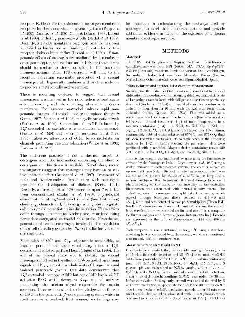
receptor. Evidence for the existence of oestrogen membranereceptors has been described in several systems (Pappas etal. 1995; Ramirez et al. 1996; Monje & Boland, 1999; Luconiet al. 1999), including pancreatic â_cells (Nadal et al. 1998).Recently, a 29 kDa membrane oestrogen receptor has beenidentified in human sperm. Binding of oestradiol to thisreceptor elicits calcium influx (Luconi et al. 1999). If non-genomic effects of oestrogen are mediated by a membraneoestrogen receptor, the mechanism underlying these effectsshould be similar to those operating in lipid-insolublehormone actions. Thus, 17â_oestradiol will bind to thereceptor, activating enzymatic production of a secondmessenger, which generally combines with another moleculeto produce a metabolically active complex.
There is mounting evidence to suggest that secondmessengers are involved in the rapid action of oestrogensafter interacting with their binding sites at the plasmamembrane. For instance, oestradiol induces rapid non-genomic changes of inositol 1,4,5-trisphosphate (Singh &Gupta, 1997; Marino et al. 1998) and cyclic nucleotide levels(Farhat et al. 1996). Generation of cAMP induced by17â_oestradiol in excitable cells modulates ion channels(Picotto et al. 1996) and ionotropic receptors (Gu & Moss,1996). Likewise, alterations of cGMP levels modulate ionchannels promoting vascular relaxation (White et al. 1995;Darkow et al. 1997).
The endocrine pancreas is not a classical target foroestrogens and little information concerning the effect ofoestrogens on this tissue is available. Nonetheless, recentinvestigations suggest that oestrogens may have an in vivoinsulinotropic effect (Brussaard et al. 1997). Treatment ofmale and ovariectomized female mice with oestrogensprevents the development of diabetes (Efrat, 1991).Recently, a direct effect of 17â_oestradiol upon â_cells hasbeen demonstrated (Nadal et al. 1998). Physiologicalconcentrations of 17â-oestradiol rapidly (less that 2 min)close KATP channels and, in synergy with glucose, regulatecalcium signals, potentiating insulin secretion. These effectsoccur through a membrane binding site, visualised usingperoxidase-conjugated oestradiol as a probe. Nevertheless,generation of second messengers involved in the regulationof a â_cell signalling system by 17â_oestradiol has yet to bedemonstrated.
Modulation of Ca¥ and KATP channels is responsible, atleast in part, for the acute vasodilatory effect of 17â-oestradiol in isolated perfused heart (Hugel et al. 1999). Theaim of the present study was to identify the secondmessengers involved in the effect of 17â_oestradiol on calciumsignals and KATP activity in whole islets of Langerhans andisolated pancreatic â_cells. Our data demonstrate that17â_oestradiol increases cGMP but not cAMP levels. cGMPactivates PKG which decreases KATP channel activity,modulating the calcium signal responsible for insulinsecretion. These results extend our knowledge about the roleof PKG in the pancreatic â_cell signalling system, which initself remains unresolved. Furthermore, our findings may
be important in understanding the pathways used byoestrogens to exert their membrane actions and provideadditional evidence in favour of the existence of a plasmamembrane oestrogen receptor.
METHODS
Materials
LY 83583 (6-(phenylamino)-5,8-quinolinedione, 6-anilino-5,8-quinolinedione) was from RBI (Natick, MA, USA). Rp-8-pCPT-cGMPS (TEA salt) was from Alexis Corporation Ltd (L�aufelfingen,Switzerland). Indo_1 AM was from Molecular Probes (Leiden,Netherlands). Other materials were from Sigma (Madrid, Spain).
Islets isolation and intracellular calcium measurement
Swiss albino OF1 male mice (8—10 weeks old) were killed by cervicaldislocation in accordance with national guidelines. Pancreatic isletsof Langerhans were isolated with collagenase digestion as previouslydescribed (Nadal et al. 1994) and loaded at room temperature withIndo_1 by incubation for 90 min with the AM ester form (6 ìÒ,Molecular Probes, Eugene, OR, USA). This was added as aconcentrated stock solution in dimethyl sulfoxide (final concentration0·1% vÏv). Loaded islets were kept at room temperature in amedium containing (mÒ): 115 NaCl, 25 NaHCO×, 5 KCl, 1·1MgClµ, 1·2 NaHµPOÚ, 2·5 CaClµ and 2·5 Hepes; plus 1% albumin,continuously bubbled with a mixture of 95% Oµ and 5% COµ; finalpH 7·35. Individual islets were left to settle down in the recordingchamber for 1—2 min before starting the perifusion. Islets wereperifused with a modified Ringer solution containing (mÒ): 120NaCl, 5 KCl, 25 NaHCO×, 1·1 MgClµ and 2·5 CaCl2; final pH 7·35.
Intracellular calcium was monitored by measuring the fluorescenceemitted by the fluorophore Indo_1 (Grynkiewicz et al. 1985) using adouble emission microfluorescence system. The epifluorescence setup was built on a Nikon-Diaphot inverted microscope. Indo_1 wasexcited at 350 ± 5 nm by means of a 75 W xenon lamp and anarrow band-pass filter. To prevent ultraviolet damage to cells andphotobleaching of the indicator, the intensity of the excitationillumination was attenuated with neutral density filters. TheIndo_1 emission fluorescence was split into two beams, whichpassed through band-pass filters centred at 410 ± 5 and480 ± 5 nm and was detected by two photomultipliers (Thorn EMI9924B). Fluorescence emission at 410 and 480 nm and the ratio ofboth wavelengths were recorded on-line and stored in a computerfor further analysis with Axotape (Axon Instruments Inc.). Recordsare expressed as the ratio of fluorescence at 410 and 480 nm(F410ÏF480).
Bath temperature was maintained at 35 ± 1 °C using a stainless-steel ring heater controlled by a thermostat, which was monitoredcontinuously with a microthermistor.
Measurement of cAMP and cGMP
Once islets were isolated, they were divided among tubes in groupsof 15 islets for cAMP detection and 28—45 islets to measure cGMP.Islets were preincubated for 1 h at 37°C, in a medium containing(mÒ): 120 NaCl, 5 KCl, 25 NaHCO×, 1·1 MgClµ, 2·5 CaCl2 and 3glucose; pH was maintained at 7·35 by gassing with a mixture of95% Oµ and 5% COµ. In the particular case of cGMP detection,1 mÒ 3-isobutyl-1-methylxanthine (IBMX) was added for 30 minbefore stimulation. Subsequently, stimuli were added followed by 5or 15 min incubation as appropiate for cAMP and 30 min for cGMP.Due to low levels of cGMP, incubation periods under 30 min gaveundetectable changes when stimulated with 11 mÒ glucose, whichwas used as a positive control (Laychock et al. 1991). DMSO was
A. B. Ropero and others J. Physiol. 521.2398

used as a vehicle for 17â_oestradiol; final concentration of DMSOwas 10¦Ç% and the same concentration was added to allexperimental samples in order to avoid any possible artefact. At theend of the incubation period, islets were rapidly put on ice to stopthe reaction. Tubes were centrifuged for 5 min at 1000 r.p.m. andthe supernatant was discarded. The pellet containing the islets wastreated with methanol before sonication. Samples were centrifugedat 6000 r.p.m. for 10 min; the pellet was frozen for the Bradfordassay in order to detect the protein content. The supernatant wascollected and after evaporation of methanol was frozen. Later,aliquots were assayed for cAMP and cGMP using radioimmuno-assay (RIA) kits (Amersham Pharmacia Biotech, Barcelona, Spain).The acetylation method was chosen in the case of cGMP. In theseexperiments 10 nÒ oestradiol was used in order to obtain amaximal increase in cyclic nucleotides. Although this concentrationis not considered pharmacological, it is slightly higher thanphysiological (100 pÒ—1 nÒ, used throughout the paper).
Results are expressed as means ± s.e.m. Data were analysed forstatistical differences using one-way analysis of variance. Thenumber of experiments and P values are detailed in figure legends.
Cell isolation and culture
Islets from adult (8—10 week old) Swiss albino male mice (OF1),killed by cervical dislocation, were isolated, dispersed into singlecells and cultured as previously described (Valdeolmillos et al. 1992).Briefly, islets were isolated by collagenase digestion as describedabove, separated by centrifugation and hand picked under a stereo-
microscope. Once isolated, islets were disaggregated into single cellsin a low calcium medium. Cells were then centrifuged, resuspendedin culture medium RPMI 1640 supplemented with 10% fetal calfserum, 100 units ml¢ penicillin, 0·1 mg ml¢ streptomycin and11 mÒ glucose, and plated on glass coverslips. Cells were kept at37°C, in a humidified atmosphere of 95% Oµ and 5% COµ, andused within 1—5 days of culture.
Patch-clamp recordings
Single-channel currents were recorded from cell-attached and inside-out membrane patches using standard patch-clamp recordingprocedures (Hamill et al. 1981). Patch pipettes were pulled fromClark Electromedical glass capillaries (Reading, UK) using a two-stage puller (Mecanex BB-CH, Geneva, Switzerland), withresistances of 8—12 MÙ when filled with the pipette solution. Incell-attached patches the pipette solution contained (mÒ): 140 KCl,1 MgClµ, 10 Hepes and 1 EGTA (pH 7·2); and the bath solutioncontained (mÒ): 5 KCl, 135 NaCl, 10 Hepes, 2·5 CaClµ and 1·1MgClµ (pH 7·4). In inside-out experiments, the bath and pipettesolutions were exchanged, and bath solution was applied through aRSC_100 rapid solution changer (Biologic, Claix, France). KATP
channel unitary currents were recorded using an Axopatch 200amplifier (Axon Instruments Inc.) and stored on a tape recorder(DAT, DTR_1202, Biologic; Claix, France) for subsequent analysiswith an in-house programme. Experiments were replayed throughan 8-pole Bessel filter (Frequency Devices). The pipette potentialwas held at 0 mV throughout recording. KATP channel activity wasquantified by digitising 60 s sections of the current record, filtered
17â_oestradiol modulates KATP channels via cGMPJ. Physiol. 521.2 399
Figure 1. Effects of 17â_oestradiol on [Ca¥]é signals in whole
islets of Langerhans
[Ca¥]é responses of 3 different islets of Langerhans in the presenceof 8 mÒ glucose to 1 nÒ 17â_oestradiol applied for the periodindicated by the horizontal bar. The Ca¥-dependent fluorescence ofIndo_1 is expressed as the ratio F410ÏF480 (see Methods). Fourdistinct kinds of responses were recorded. A, generation of [Ca¥]éoscillations on a plateau (19%, 10 of 53 islets tested). Increment inthe level of F410ÏF480 in the presence of 17â_oestradiol (plateau)with respect to the basal level (Ä(F410ÏF480)) is 0·33 ± 0·09. B, anincrease in the frequency of [Ca¥]é oscillations on a plateau level(32%, 17 of 53 islets). The height of the plateau was determined asthe difference between the fluorescence levels at the valleys of theoscillations in the presence and absence of 17â_oestradiol. In thiscase Ä(F410ÏF480) = 0·3 ± 0·1. C, lengthening of [Ca¥]é oscillationsafter a [Ca¥]é transient (17%, 9 of 53 islets). The plateau was verysmall in these records (Ä(F410ÏF480) = 0·07 ± 0·05). Besides thesethree oscillatory patterns an irregular oscillatory response wasinduced in 15% (8 of 53 islets used), and in an additional 17% (9 of53 islets) 17â_oestradiol was without effect.

at 1 kHz, sampled at 10 kHz by a Digidata 1200, and calculatingthe mean NPï during the sweep. A 70% threshold method was usedto detect events. Data sampling was started 1 min before (control)and 7 min after (test) application of test substances in cell-attachedexperiments, and 1—30 s in excised patches. Channel activity wasdefined as the product of N, the number of functional channels, andPï, the open-state probability. Pï was determined by dividing thetotal time channels spent in the open state by the total sample time.In experiments used to determine PKG implication, due to the highnumber of active channels, instead of NPï, an integral analysis ofthe current was used to measure channel activity. Unless otherwiseindicated, data are expressed as means ± s.e.m. For statisticalanalysis a one-way ANOVA was used. Experiments wereperformed at room temperature (20—24°C).
RESULTS
Basic characteristics of calcium signals in response to
17â_oestradiol
Application of 17â_oestradiol to the islets of Langerhans inthe presence of a stimulatory concentration of glucose (8 mÒ),induced four different types of [Ca¥]é response (see Fig. 1).Once a steady [Ca¥]é signal in response to 8 mÒ glucose wasestablished, application of 1 nÒ 17â_oestradiol induced anoscillatory pattern of [Ca¥]é on a plateau level in islets thatdid not show glucose-induced [Ca¥]é oscillations (Fig. 1A), anincrease in the frequency of [Ca¥]é oscillations (Fig. 1B), or
A. B. Ropero and others J. Physiol. 521.2400
Figure 2. cGMP reproduces 17â_oestradiol effects on
[Ca¥]é signals
Different [Ca¥]é responses to 10 ìÒ 8-Br-cGMP (8Br_cGMP)in 3 different islets exposed to 8 mÒ glucose. A, generation of[Ca¥]é oscillations in non-oscillatory islets compared withbasal (25%, 3 of 12 islets used). B, increase in the frequencyof [Ca¥]é oscillations to a plateau level (33%, 4 of 12 islets).C, lengthening of [Ca¥]é oscillations after a [Ca¥]é transient(42%, 5 of 12 islets).
Figure 3. cAMP reproduces 17â_oestradiol actions on
[Ca¥]é signals
Different [Ca¥]é responses to 10 ìÒ 8-Br-cAMP in3 different islets exposed to 8 mÒ glucose. A, generation of[Ca¥]é oscillations in islets which did not display [Ca¥]éoscillations in response to 8 mÒ glucose (19%, 3 of 16 isletsused). B, increase in the frequency of [Ca¥]é oscillations(44%, 7 of 16 islets). C, lengthening of [Ca¥]é oscillationsafter a [Ca¥]é transient (31%, 5 of 16 islets). Six per cent,1 of 16 islets tested, showed an irregular [Ca¥]é patternproduced by 8-Br-cAMP.
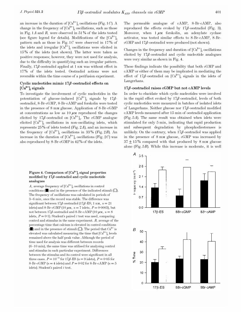
an increase in the duration of [Ca¥]é oscillations (Fig. 1C). Achange in the frequency of [Ca¥]é oscillations, such as thosein Fig. 1A and B, were observed in 51% of the islets tested(see figure legend for details). Modifications of the [Ca¥]épattern such as those in Fig. 1C were observed in 17% ofthe islets and irregular [Ca¥]é oscillations were elicited in15% of the islets (not shown). The latter were taken aspositive responses; however, they were not used for analysis,due to the difficulty in quantifying such an irregular pattern.Finally, 17â_oestradiol applied at 1 nÒ was without effect in17% of the islets tested. Oestradiol actions were notreversible within the time course of a perifusion experiment.
Cyclic nucleotides mimic 17â_oestradiol effects on
[Ca¥]é signals
To investigate the involvement of cyclic nucleotides in thepotentiation of glucose-induced [Ca¥]é signals by 17â-oestradiol, 8-Br-cGMP, 8-Br-cAMP and forskolin were testedin the presence of 8 mÒ glucose. Application of 8-Br-cGMPat concentrations as low as 10 ìÒ reproduced the changeselicited by 17â_oestradiol on [Ca¥]é. The cGMP analogueelicited [Ca¥]é oscillations in non-oscillating islets, whichrepresents 25% of islets tested (Fig. 2A), and an increase inthe frequency of [Ca¥]é oscillations in 33% (Fig. 2B). Anincrease in the duration of [Ca¥]é oscillations (Fig. 2C) wasalso reproduced by 8_Br-cGMP in 42% of the islets.
The permeable analogue of cAMP, 8-Br-cAMP, alsoreproduced the effects evoked by 17â_oestradiol (Fig. 3).Moreover, when 1 ìÒ forskolin, an adenylate cyclaseactivator, was tested similar effects to 8-Br-cAMP, 8_Br-cGMP and 17â_oestradiol were produced (not shown).
Changes in the frequency and duration of [Ca¥]é oscillationselicited by 17â_oestradiol and cyclic nucleotide analogueswere very similar as shown in Fig. 4.
These findings indicate the possibility that both cGMP andcAMP or either of them may be implicated in mediating theeffect of 17â_oestradiol on [Ca¥]é signals in the islets ofLangerhans.
17â_oestradiol raises cGMP but not cAMP levels
In order to elucidate which cyclic nucleotides were involvedin the rapid effect evoked by 17â_oestradiol, levels of bothcyclic nucleotides were measured in batches of isolated isletsof Langerhans. Neither glucose nor 17â_oestradiol modifiedcAMP levels measured after 15 min of oestradiol application(Fig. 5A). The same result was obtained when islets werestimulated for only 5 min, indicating that rapid productionand subsequent degradation by phosphodiesterases isunlikely. On the contrary, when 17â_oestradiol was appliedin the presence of 8 mÒ glucose, cGMP was increased by37 ± 15% compared with that produced by 8 mÒ glucosealone (Fig. 5B). While this increase is moderate, it is well
17â_oestradiol modulates KATP channels via cGMPJ. Physiol. 521.2 401
Figure 4. Comparison of [Ca¥]é signal properties
modified by 17â_oestradiol and cyclic nucleotide
analogues
A, average frequency of [Ca¥]é oscillations in controlconditions (4) and in the presence of the indicated stimuli (5).The frequency of oscillations was calculated in periods of3—6 min, once the record was stable. The difference wassignificant between 17â_oestradiol (17â-ES; 1 nÒ, n = 21islets) and 8-Br-cGMP (10 ìÒ, n = 7 islets, P = 0·0003), butnot between 17â_oestradiol and 8-Br-cAMP (10 ìÒ, n = 8islets, P = 0·1). Student’s paired t test was used, comparingcontrol and stimulus in the same experiment. B, average of thepercentage time that calcium is elevated in control conditions(4) and in the presence of stimuli (5). The period that Ca¥ iselevated was calculated measuring the time that [Ca¥]é levelsremained above the half-peak value. Although the period oftime used for analysis was different between records(6—10 min), the same time was utilised for analysing controland stimulus in each particular experiment. Differencesbetween the stimulus and its control were significant in allthree cases. P = 10¦Ç for 17â_ES (n = 9 islets), P = 0·05 for8_Br-cGMP (n = 4 islets) and P = 0·02 for 8-Br-cAMP (n = 5islets). Student’s paired t test.
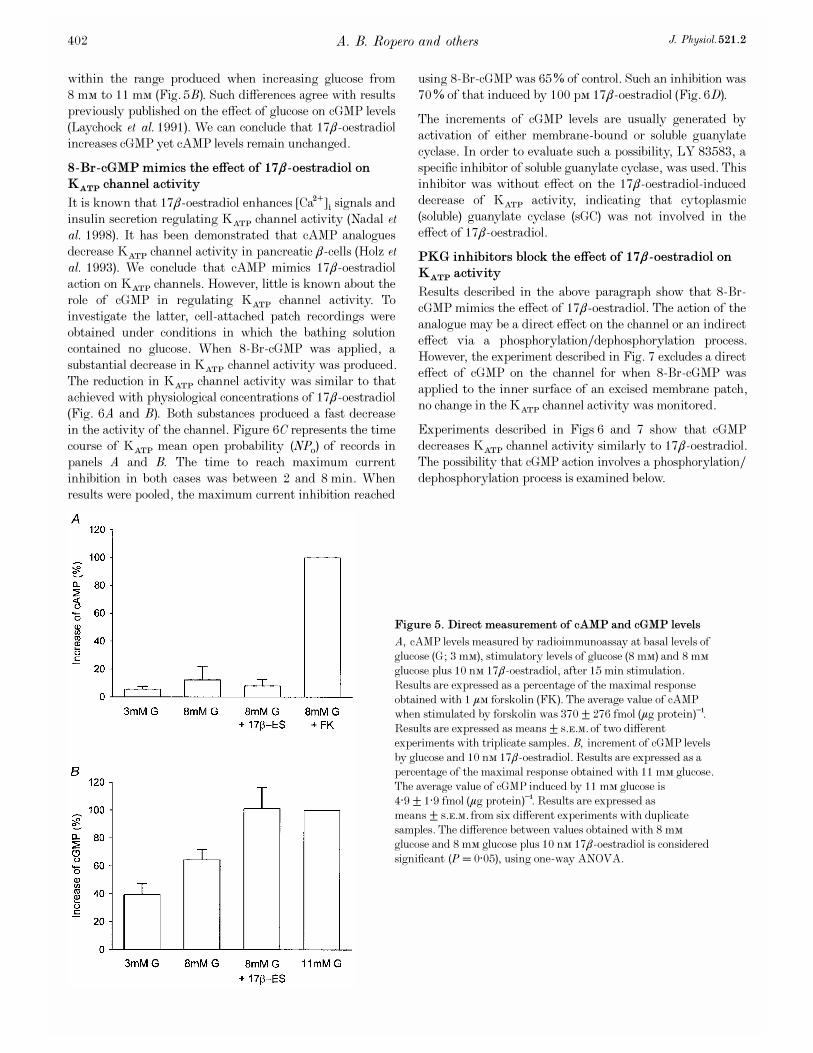
within the range produced when increasing glucose from8 mÒ to 11 mÒ (Fig. 5B). Such differences agree with resultspreviously published on the effect of glucose on cGMP levels(Laychock et al. 1991). We can conclude that 17â_oestradiolincreases cGMP yet cAMP levels remain unchanged.
8-Br-cGMP mimics the effect of 17â_oestradiol on
KATP channel activity
It is known that 17â_oestradiol enhances [Ca¥]é signals andinsulin secretion regulating KATP channel activity (Nadal etal. 1998). It has been demonstrated that cAMP analoguesdecrease KATP channel activity in pancreatic â_cells (Holz etal. 1993). We conclude that cAMP mimics 17â_oestradiolaction on KATP channels. However, little is known about therole of cGMP in regulating KATP channel activity. Toinvestigate the latter, cell-attached patch recordings wereobtained under conditions in which the bathing solutioncontained no glucose. When 8-Br-cGMP was applied, asubstantial decrease in KATP channel activity was produced.The reduction in KATP channel activity was similar to thatachieved with physiological concentrations of 17â_oestradiol(Fig. 6A and B). Both substances produced a fast decreasein the activity of the channel. Figure 6C represents the timecourse of KATP mean open probability (NPï) of records inpanels A and B. The time to reach maximum currentinhibition in both cases was between 2 and 8 min. Whenresults were pooled, the maximum current inhibition reached
using 8-Br-cGMP was 65% of control. Such an inhibition was70% of that induced by 100 pÒ 17â_oestradiol (Fig. 6D).
The increments of cGMP levels are usually generated byactivation of either membrane-bound or soluble guanylatecyclase. In order to evaluate such a possibility, LY 83583, aspecific inhibitor of soluble guanylate cyclase, was used. Thisinhibitor was without effect on the 17â_oestradiol-induceddecrease of KATP activity, indicating that cytoplasmic(soluble) guanylate cyclase (sGC) was not involved in theeffect of 17â_oestradiol.
PKG inhibitors block the effect of 17â_oestradiol on
KATP activity
Results described in the above paragraph show that 8_Br-cGMP mimics the effect of 17â_oestradiol. The action of theanalogue may be a direct effect on the channel or an indirecteffect via a phosphorylationÏdephosphorylation process.However, the experiment described in Fig. 7 excludes a directeffect of cGMP on the channel for when 8-Br-cGMP wasapplied to the inner surface of an excised membrane patch,no change in the KATP channel activity was monitored.
Experiments described in Figs 6 and 7 show that cGMPdecreases KATP channel activity similarly to 17â_oestradiol.The possibility that cGMP action involves a phosphorylation/dephosphorylation process is examined below.
A. B. Ropero and others J. Physiol. 521.2402
Figure 5. Direct measurement of cAMP and cGMP levels
A, cAMP levels measured by radioimmunoassay at basal levels ofglucose (G; 3 mÒ), stimulatory levels of glucose (8 mÒ) and 8 mÒglucose plus 10 nÒ 17â_oestradiol, after 15 min stimulation.Results are expressed as a percentage of the maximal responseobtained with 1 ìÒ forskolin (FK). The average value of cAMPwhen stimulated by forskolin was 370 ± 276 fmol (ìg protein)¢.Results are expressed as means ± s.e.m. of two differentexperiments with triplicate samples. B, increment of cGMP levelsby glucose and 10 nÒ 17â_oestradiol. Results are expressed as apercentage of the maximal response obtained with 11 mÒ glucose.The average value of cGMP induced by 11 mÒ glucose is4·9 ± 1·9 fmol (ìg protein)¢. Results are expressed asmeans ± s.e.m. from six different experiments with duplicatesamples. The difference between values obtained with 8 mÒglucose and 8 mÒ glucose plus 10 nÒ 17â_oestradiol is consideredsignificant (P = 0·05), using one-way ANOVA.
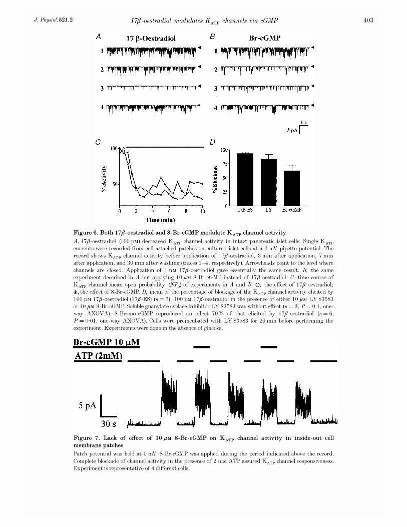
17â_oestradiol modulates KATP channels via cGMPJ. Physiol. 521.2 403
Figure 6. Both 17â_oestradiol and 8-Br-cGMP modulate KATP channel activity
A, 17â_oestradiol (100 pÒ) decreased KATP channel activity in intact pancreatic islet cells. Single KATP
currents were recorded from cell-attached patches on cultured islet cells at a 0 mV pipette potential. Therecord shows KATP channel activity before application of 17â_oestradiol, 3 min after application, 7 minafter application, and 30 min after washing (traces 1—4, respectively). Arrowheads point to the level wherechannels are closed. Application of 1 nÒ 17â_oestradiol gave essentially the same result. B, the sameexperiment described in A but applying 10 ìÒ 8-Br-cGMP instead of 17â_oestradiol. C, time course ofKATP channel mean open probability (NPï) of experiments in A and B. 1, the effect of 17â_oestradiol;*, the effect of 8-Br-cGMP. D, mean of the percentage of blockage of the KATP channel activity elicited by100 pÒ 17â_oestradiol (17â_ES) (n = 7), 100 pÒ 17â_oestradiol in the presence of either 10 ìÒ LY 83583or 10 ìÒ 8-Br-cGMP. Soluble guanylate cyclase inhibitor LY 83583 was without effect (n = 3, P = 0·1, one-way ANOVA). 8_Bromo-cGMP reproduced an effect 70% of that elicited by 17â_oestradiol (n = 6,P = 0·01, one-way ANOVA). Cells were preincubated with LY 83583 for 20 min before performing theexperiment. Experiments were done in the absence of glucose.
Figure 7. Lack of effect of 10 ìÒ 8-Br-cGMP on KATP channel activity in inside-out cell
membrane patches
Patch potential was held at 0 mV. 8-Br-cGMP was applied during the period indicated above the record.Complete blockade of channel activity in the presence of 2 mÒ ATP assured KATP channel responsiveness.Experiment is representative of 4 different cells.
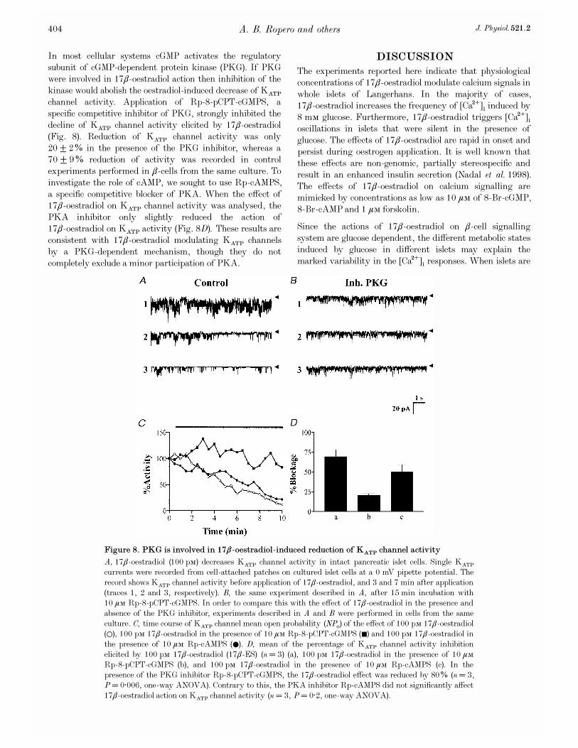
In most cellular systems cGMP activates the regulatorysubunit of cGMP-dependent protein kinase (PKG). If PKGwere involved in 17â_oestradiol action then inhibition of thekinase would abolish the oestradiol-induced decrease of KATP
channel activity. Application of Rp-8-pCPT-cGMPS, aspecific competitive inhibitor of PKG, strongly inhibited thedecline of KATP channel activity elicited by 17â-oestradiol(Fig. 8). Reduction of KATP channel activity was only20 ± 2% in the presence of the PKG inhibitor, whereas a70 ± 9% reduction of activity was recorded in controlexperiments performed in â_cells from the same culture. Toinvestigate the role of cAMP, we sought to use Rp-cAMPS,a specific competitive blocker of PKA. When the effect of17â_oestradiol on KATP channel activity was analysed, thePKA inhibitor only slightly reduced the action of17â_oestradiol on KATP activity (Fig. 8D). These results areconsistent with 17â_oestradiol modulating KATP channelsby a PKG-dependent mechanism, though they do notcompletely exclude a minor participation of PKA.
DISCUSSION
The experiments reported here indicate that physiologicalconcentrations of 17â_oestradiol modulate calcium signals inwhole islets of Langerhans. In the majority of cases,17â_oestradiol increases the frequency of [Ca¥]é induced by8 mÒ glucose. Furthermore, 17â_oestradiol triggers [Ca¥]éoscillations in islets that were silent in the presence ofglucose. The effects of 17â_oestradiol are rapid in onset andpersist during oestrogen application. It is well known thatthese effects are non-genomic, partially stereospecific andresult in an enhanced insulin secretion (Nadal et al. 1998).The effects of 17â_oestradiol on calcium signalling aremimicked by concentrations as low as 10 ìÒ of 8-Br-cGMP,8-Br-cAMP and 1 ìÒ forskolin.
Since the actions of 17â_oestradiol on â_cell signallingsystem are glucose dependent, the different metabolic statesinduced by glucose in different islets may explain themarked variability in the [Ca¥]é responses. When islets are
A. B. Ropero and others J. Physiol. 521.2404
Figure 8. PKG is involved in 17â_oestradiol-induced reduction of KATP channel activity
A, 17â_oestradiol (100 pÒ) decreases KATP channel activity in intact pancreatic islet cells. Single KATP
currents were recorded from cell-attached patches on cultured islet cells at a 0 mV pipette potential. Therecord shows KATP channel activity before application of 17â_oestradiol, and 3 and 7 min after application(traces 1, 2 and 3, respectively). B, the same experiment described in A, after 15 min incubation with10 ìÒ Rp_8-pCPT-cGMPS. In order to compare this with the effect of 17â_oestradiol in the presence andabsence of the PKG inhibitor, experiments described in A and B were performed in cells from the sameculture. C, time course of KATP channel mean open probability (NPï) of the effect of 100 pÒ 17â_oestradiol(1), 100 pÒ 17â_oestradiol in the presence of 10 ìÒ Rp_8-pCPT-cGMPS (þ) and 100 pÒ 17â_oestradiol inthe presence of 10 ìÒ Rp-cAMPS (0). D, mean of the percentage of KATP channel activity inhibitionelicited by 100 pÒ 17â_oestradiol (17â_ES) (n = 3) (a), 100 pÒ 17â_oestradiol in the presence of 10 ìÒRp_8-pCPT-cGMPS (b), and 100 pÒ 17â_oestradiol in the presence of 10 ìÒ Rp-cAMPS (c). In thepresence of the PKG inhibitor Rp_8-pCPT-cGMPS, the 17â_oestradiol effect was reduced by 80% (n = 3,P = 0·006, one-way ANOVA). Contrary to this, the PKA inhibitor Rp-cAMPS did not significantly affect17â_oestradiol action on KATP channel activity (n = 3, P = 0·2, one-way ANOVA).

exposed to nanomolar concentrations of 17â_oestradiol,direct measurement of cyclic nucleotide levels in isolatedislets shows an increment in cGMP, whereas cAMPconcentrations are unchanged. The increment in cGMPlevels is modest, but is nonetheless equivalent to thatproduced when stimulating with 11 mÒ glucose (Laychock etal. 1991, and present paper). It has already beendemonstrated that 17â_oestradiol decreases KATP channelactivity in a rapid manner: initiation of 17â_oestradiol effectoccurs between 30 s and 2 min after oestrogen application(Nadal et al. 1998). This time course of action is similar tothat found in other studies (Hardy & Valverde, 1994; Gu &Moss, 1996). The decline in channel activity is mimicked by8-Br-cGMP, when measured in the cell-attached mode.Remarkably, the concentration of 8-Br-cGMP needed toproduce an effect, both on [Ca¥]é and KATP activity, is atleast an order of magnitude lower than in most of thestudies published. Since the action of cGMP seems to bevery potent, a small increase in cGMP levels such as thatdetected in Fig. 5B, would be enough to account for17â_oestradiol actions on the â_cell signalling system.
The modulation of KATP channel activity by cGMP discussedabove could be accomplished via two different pathways:direct interaction of cGMP with the channel or activation ofa cGMP-dependent protein kinase responsible for KATP
channel phosphorylation. The former possibility isinvalidated by the experiment shown in Fig. 7, since 8_Br-cGMP was without effect when applied to inside-out patches.By contrast, the oestrogen-induced decrease in channelactivity is greatly reduced by a specific blocker of PKG(Rp_8-pCPT-cGMPS). Implication of PKA is minor sinceRp-cAMPS, a blocker of PKA, has a very small effect.
Both PKA and PKG exist in pancreatic â_cells. The formeris involved in a multitude of signalling processes, whereasthe role of the latter is still unclear (see Jones & Persaud,1998, for a review). Implication of PKG in 17â_oestradiolaction is therefore a new finding for pancreatic â_cellsignalling. Since PKG activity influences apoptotic celldeath in â_cells (Loweth et al. 1997), 17â_oestradiol mayalso influence â_cell survival, which may be of therapeuticimportance.
In summary, the modulation of calcium signals by 17â-oestradiol is due to a blockage of KATP channels, inducingdepolarisation of the membrane and an enhanced calciuminflux. Modulation of KATP channels appears to be mediatedby an increase in cGMP levels, which will activate a cGMP-dependent protein kinase causing KATP channelphosphorylation. A minor participation of cAMP in thisprocess cannot be completely ruled out, although aninvolvement of cGMP is clear.
The actions of 17â_oestradiol resemble those described forother modulators such as glucagon-like peptide (GLP)_1 viacAMP (Holz et al. 1993) and atrial natriuretic peptide viacyclic nucleotides (Lee & Laychock, 1997).
Although a direct binding of 17â_oestradiol on maxi-K¤channels has been described in vascular smooth muscle(Valverde et al. 1999), most non-genomic actions of oestrogen,such as those described in â_cells (Nadal et al. 1998, andpresent paper), are likely to be mediated by a plasmamembrane receptor. The molecular characteristics of themembrane oestrogen receptor remain little known. A recentstudy has identified a novel functional membrane oestrogenreceptor in human sperm (Luconi et al. 1999). Specificbinding sites for oestradiol at the plasma membrane havebeen demonstrated in several types of cells (Pappas et al.1995; Ramirez et al. 1996; Nadal et al. 1998; Monje & Boland,1999). Binding of 17â_oestradiol to a hypothetical oestrogenmembrane receptor has been shown to alter cyclic nucleotidelevels. Increases in cAMP elicited by 17â-oestradiol areusual in excitable cells. For instance, 17â-oestradiolpotentiates kainate-induced currents via cAMP (Gu & Moss,1996), independently of intracellular oestrogen receptors(Gu et al. 1999).
cGMP is also involved in rapid oestrogen actions, principallyin endothelial cells. In this report we found that changes incGMP levels are more important than increases in cAMP.An increment in cGMP levels could be mediated viaactivation of a sGC activated by NO. Rapid, non-genomicstimulation of NO by 17â_oestradiol has been described inendothelial cells (Prevot et al. 1999). Although sGC waspresent in â_cells, it seems unlikely that it could play animportant role in 17â_oestradiol generation of cGMP. Thelast conclusion is based on the experiment described inFig. 6D. An inhibitor of soluble guanylate cyclase (LY 83583)did not modify the effect of 17â_oestradiol on KATP channelactivity in cell-attached patches. On the contrary, LY 83583is known to inhibit NO induced smooth muscle relaxation(Kawada et al. 1994). We can further rule out mediation ofNO in the response of â_cells to 17â_oestradiol, because NOactivates KATP channels evoking a decrease of insulinsecretion (Tsuura et al. 1994). Thus, the effect of NO isopposite to that of 17â_oestradiol on KATP channel andinsulin secretion (Nadal et al. 1998).
The most likely pathway for explaining cGMPaugmentation is via a membrane guanylate cyclase (mGC),activated after interaction of 17â_oestradiol with itsbinding site at the membrane. We cannot exclude a directeffect of 17â-oestradiol on mGC, since biochemicalexperiments have illustrated that 17â_oestradiol candirectly activate membrane guanylate cyclase by anunknown mechanism in PC12 cells (Chen et al. 1998).However, this is unlikely since effects of 17â_oestradiol on[Ca¥]é and KATP channels have a typical profile of areceptor-mediated process (Nadal et al. 1998).
In conclusion, the present work demonstrates thatphysiological levels of 17â_oestradiol decrease KATP channelactivity, modulating calcium signals. This modulation is acGMP-dependent phosphorylation process and is likely to
17â_oestradiol modulates KATP channels via cGMPJ. Physiol. 521.2 405
occur after binding of 17â_oestradiol to a plasma membranereceptor, activating a membrane guanylate cyclase.
Existence of a second messenger involved in the rapid, non-genomic effect of 17â_oestradiol is important evidence forthe presence of a plasma membrane oestrogen receptor.
Alonso, R. & L�opez-Coviella, I. (1998). Gonadal steroids andneuronal function. Neurochemistry Research 23, 679—692.
Blackmore, P. F., Neulen, J., Lattanzio, F. & Beebe, S. J. (1991).Cell surface-binding sites for progesterone mediate calcium uptakein human sperm. Journal of Biological Chemistry 266,18655—18659.
Brussaard, H. E., Gevers Leuven, J. A., Fr�olich, M., Kluft, C. &Krans, H. M. J. (1997). Short-term oestrogen replacement therapyimproves insulin resistance, lipids and fibrinolysis inpostmenopausal women with NIDDM. Diabetologia 40, 843—849.
Chen, Z.-J., Yu, L. & Chang, C.-H. (1998). Stimulation of membrane-bound guanylate cyclase activity by 17â_estradiol. Biochemical andBiophysical Research Communications 252, 639—642.
Darkow, D. J., Lu, L. & White, R. E. (1997) Estrogen relaxation ofcoronary artery smooth muscle is mediated by nitric oxide andcGMP. American Journal of Physiology 272, H2765—2773.
Efrat, S. (1991). Sexual dimorphism of pancreatic beta-celldegeneration in transgenic mice expressing an insulin-ras hybridgene. Endocrinology 128, 897—901.
Farhat, M. Y., Abi-Younes, S. & Ramwell, P. W. (1996). Non-genomic effects of oestrogen and the vessel wall. BiochemicalPharmacology 51, 571—576.
Grynkiewicz, G. M., Poenie, M. & Tsien, R. Y. (1985). A newgeneration of Ca¥ indicators with greatly improved fluorescenceproperties. Journal of Biological Chemistry 260, 3440—3450.
Gu, Q., Korach, K. S. & Moss, R. L. (1999). Rapid action of17â_estradiol on kainate-induced currents in hippocampal neuronslacking intracellular estrogen receptors. Endocrinology 140,660—666.
Gu, Q. & Moss, R. L. (1996) 17â_estradiol potentiates kainate-induced currents via activation of the cAMP cascade. Journal ofNeuroscience 16, 3620—3629.
Hamill, O. P., Marty, A., Neher, E., Sakmann, B. & Sigworth,F. J. (1981). Improved patch-clamp techniques for high-resolutioncurrent recording from cell and cell-free membrane patches. Pfl�ugersArchiv 391, 85—100.
Hardy, S. P. & Valverde, M. A. (1994). Novel plasma membraneaction estrogen and antiestrogens revealed by their regulation of alarge conductance chloride channel. FASEB Journal 8, 760—765.
Holz, G. G. IV, K�uhtreiber, W. H. & Habener, J. F. (1993).Pancreatic â_cells are rendered glucose-competent by theinsulinotropic hormone glucagon-like peptide_1(7—37). Nature 361,362—365.
Hugel, S., Neubauer, S., Lie, S. Z., Ernst, R., Horn, M., Schmidt,H. H., Allolio, B. & Reincke, M. (1999). Multiple mechanisms areinvolved in the acute vasodilatory effect of 17â_estradiol in theisolated perfused rat heart. Journal of CardiovascularPharmacology 33, 852—858.
Impronta-Brears, T., Whorton, A. R., Codazzi, F., York, J. D.,Meyer, T. & McDonnell, D. P. (1999). Estrogen-induced activationof mitogen-activated protein kinase requires mobilization of intra-cellular calcium. Proceedings of the National Academy of Sciences ofthe USA 96, 4686—4691.
Jones, P. & Persaud, S. (1998). Protein kinases, proteinphosphorylation, and the regulation of insulin secretion frompancreatic â_cells. Endocrine Reviews 19, 429—461.
Kawada, T., Ishibashi, T., Sasage, H., Kato, K. & Imai, S. (1994).Modification by LY 83583 and methylene blue of relaxation inducedby nitric oxide, glyceryl trinitrate, sodium nitroprusside andatriopeptin in aortae of the rat, guinea-pig and rabbit. GeneralPharmacology 25, 1361—1371.
Kitazawa, T., Hamada, E., Kitazawa, K. & Gaznabi, A. K. M.(1997). Nongenomic mechanism of 17-â oestradiol-inducedinhibition of contraction in mammalian vascular smooth muscle.Journal of Physiology 499, 497—511.
Laychock, S. G., Modica, M. E. & Cavanaugh, C. T. (1991). L_argininestimulates cyclic guanosine 3',5'-monophosphate formation in ratislets of Langerhans and RINm5F insulinoma cells: evidence forL_arginine: nitric oxide synthase. Endocrinology 129, 3043—3052.
Lee, B. & Laychock, S. G. (1997). Atrial natriuretic peptide andcyclic nucleotides affect glucose-induced Ca¥ responses in singlepancreatic islet beta-cells: correlation with (Ca¥-Mg¥)-ATPaseactivity. Diabetes 46, 1312—1318.
Lieberherr, M. & Grosse, B. (1994). Androgens increase intra-cellular calcium concentration and inositol 1,4,5-triphosphate anddiacylglicerol formation via pertussis toxin-sensitive G-protein.Journal of Biological Chemistry 269, 7217—7223.
Loweth, A. C., Williams, G. T., Scarpello, J. H. B. & Morgan, N. G.(1997). Evidence for the involvement of cGMP and protein kinase Gin nitric oxide-induced apoptosis in the pancreatic â cell lineHIT_T15. FEBS Letters 400, 285—288.
Luconi, M., Muratoni, M., Forti, G. & Baldi, E. (1999).Identification of a novel functional oestrogen receptor on humansperm membrane that interferes with progesterone effects. Journalof Clinical Endocrinology and Metabolism 84, 1670—1678.
Marino, M., Pallottini, V. & Trentalance, A. (1998). Estrogenscause rapid activation of IP×-PKC-alpha signal transductionpathway in HEPG2 cells. Biochemical and Biophysical ResearchCommunications 245, 254—258.
Mermelstain, P. G., Becker, J. B. & Surmeier, D. J. (1996).Estradiol reduces calcium currents in neostriatal neurons via amembrane receptor. Journal of Neuroscience 16, 595—604.
Moini, H., Bilsel, S., Bekdemir, T. & Emerk K. (1997).17â_Estradiol increases intracellular free calcium concentrations ofhuman vascular endothelial cells and modulates its responses toacetylcholine. Endothelium 5, 11—19.
Monje, P. & Boland, R. (1999). Characterisation of membraneestrogen binding proteins from rabbit uterus. Molecular CellEndocrinology 147, 75—84.
Morley, P., Whithfield, J. F., Vanderhyden, B. C., Tsang, B. K.& Schwartz, J. L. (1992). A new, nongenomic estrogen action: therapid release of intracellular calcium. Endocrinology 131,1505—1512.
Moss, R. L., Gu, Q. & Wong, M. (1997). Estrogen: non-transcriptionalsignaling pathway. Recent Progress in Hormone Research 52, 33—69.
Nadal, A., Rovira, J. M., Laribi, O., Le�on-Quinto, T., Andreu, E.,Ripoll, C. & Soria, B. (1998). Rapid insulinotropic effect of17â_estradiol via a plasma membrane receptor. FASEB Journal 12,1341—1348.
Nadal, A., Valdeolmillos, M. & Soria, B. (1994). Metabolicregulation of intracellular calcium concentration in mousepancreatic islets of Langerhans. American Journal of Physiology267, E769—774.
Pappas, T. C., Gametchu, B. & Watson, C. S. (1995). Membraneestrogen receptors identified by multiple antibody labelling andimpeded-ligand binding. FASEB Journal 9, 404—410.
A. B. Ropero and others J. Physiol. 521.2406
Picotto, G., Massheimer, V. & Boland, R. (1996). Acutestimulation of intestinal cell calcium influx induced by 17â_estradiolvia the cAMP messenger system. Molecular Cell Endocrinology 119,129—134.
Prevot, V., Croix, D., Rialas, C. M., Poulain, P., Fricchione, G. L.,Stefano, G. B. & Beauvillain, J. C. (1999). Estradiol coupling toendothelial nitric oxide stimulates gonadotropin-releasing hormonerelease from rat median eminence via a membrane receptor.Endocrinology 140, 652—659.
Ramirez, V. D., Zheng, J. & Siddique, K. M. (1996). Membranereceptors for estrogen, progesterone, and testosterone in the ratbrain: fantasy or reality. Cellular and Molecular Neurobiology 16,175—198.
Ruehlmann, D. O., Steinert, J. R., Valverde, M. A., Jacob, R. &Mann, G. E. (1998). Environmental estrogenic pollutants induceacute vascular relaxation by inhibiting L-type Ca¥ channels insmooth muscle cells. FASEB Journal 12, 613—619.
Singh, S. & Gupta, P. D. (1997). Induction of phosphoinositide-mediated signal transduction pathway by 17beta-oestradiol in ratvaginal epithelial cells. Journal of Molecular Endocrinology 19,249—257.
Tsuura, Y., Ishida, H., Hayashi, S., Sakamoto, K., Horie, M. &Seino, Y. (1994). Nitric oxide opens ATP-dependent K¤ channelsthrough suppression of phosphofructokinase activity and inhibitsglucose-induced insulin release in pancreatic â cells. Journal ofGeneral Physiology 104, 1079—1099.
Valdeolmillos, M., Nadal, A., Contreras, D. & Soria, B. (1992).The relationship between glucose-induced KATP channel closure andthe rise in [Ca¥]é in single mouse pancreatic â_cells. Journal ofPhysiology 455, 173—186.
Valverde, M. A., Rojas, P., Amigo, J., Cosmelli, D., Orio, P.,Bahamonde, M. I., Mann, G. E., Vergara, C. & Latorre, R.(1999). Acute activation of maxi-K channels (hSlo) by estradiolbinding to the â subunit. Science 285, 1929—1931.
Wehling, M. (1997). Specific non-genomic actions of steroidhormones. Annual Review of Physiology 59, 365—393.
White, R. E., Darkow, D. J. & Lang, J. L. (1995). Estrogen relaxescoronary arteries by opening BKCa channels through a cGMP-dependent mechanism. Circulation Research 77, 936—942.
Zhang, F., Ram, J. L., Standley, P. R. & Sowers, J. R. (1994). 17-âestradiol attenuates voltage-dependent Ca¥ currents in A7r5vascular smooth muscle cell line. American Journal of Physiology266, C975—980.
Acknowledgements
The authors thank Dr Miguel A. Valverde for introducing us to non-genomic effects of oestrogens, Dr Ruth Shepherd for carefullyreading the manuscript and E. P�erez Garc� úa, R. Garc� úa Velasco andA. P�erez Vegara for technical assistance. This work was supportedby the European Comission, Ministerio de Educaci�on y CulturaPM-98-0105, Fondos FEDER. J.M.R. is a recipient of a researchstudentship from Generalitat Valenciana and A.B.R. from theMinisterio de Educaci�on y Cultura.
Corresponding author
A. Nadal: Institute of Bioengineering and Department ofPhysiology, School of Medicine, Miguel Hern�andez University, SanJuan Campus, Carretera Alicante-Valencia Km 87. 03550, Alicante,Spain.
Email: [email protected]
17â_oestradiol modulates KATP channels via cGMPJ. Physiol. 521.2 407

Anexos
95
ANEXO II
Nongenomic actions of estrogens and xenoestrogensby binding at a plasma membrane receptor unrelatedto estrogen receptor a and estrogen receptor bAngel Nadal*†, Ana B. Ropero*, Ouahiba Laribi*, Marjorie Maillet*, Esther Fuentes*‡, and Bernat Soria*
*Institute of Bioengineering, Department of Physiology, and ‡Department of Applied Biology, Miguel Hernandez University, San Juan Campus, Alicante03550, Spain
Edited by Ramon Latorre, Center for Scientific Studies of Santiago, Valdivia, Chile, and approved August 18, 2000 (received for review June 22, 2000)
The molecular mechanism used by environmental chemicals toexert their hormone-like actions is still only partially resolved.Although it generally is accepted that xenoestrogens act at thegenomic level by binding to intracellular estrogen receptors, wehave shown here that they trigger nongenomic effects in pancre-atic b cells. Both xenoestrogens and the circulating hormone,17b-estradiol, bind with high affinity to a common membranebinding site unrelated to the intracellular estrogen receptors ERaand ERb. This binding site is shared by dopamine, epinephrine, andnorepinephrine and has the pharmacological profile of the g-adrenergic receptor. This study provides an outline of the mem-brane receptor involved in rapid xenoestrogen actions.
pancreatic b cells u confocal microscopy u intracellular calcium u cellsignaling
Xenoestrogens are compounds that present estrogenic effectsbut whose chemical structure does not necessarily resemble
that of steroid hormones. They are found in fresh water and arelikely responsible for the feminization of male fish in severalrivers of the United Kingdom and the decreased reproductionsuccess of alligators and turtles in Lake Apopka in Florida (1, 2).These manmade chemicals enter the body by ingestion oradsorption and mimic the genomic actions of estrogens (3) viaintracellular estrogen receptors (4–6).
Less attention has been paid to nongenomic actions of xe-noestrogens. Several studies have described that xenoestrogensmimic 17b-estradiol when they act on Ca21 and K1 channels insmooth muscle cells (7) as well as when they trigger prolactinrelease from rat pituitary tumor cells (8). Yet, a direct linkbetween an estrogen membrane binding site (9) and xenoestro-gen actions is still a matter of debate. Moreover, despite the greatnumber of studies about nongenomic actions of estrogens, a clearoutline of the membrane receptor involved is still elusive.
We have chosen three widely used xenoestrogens for ourstudy: bisphenol-A (BPA), which is found in the content ofcanned food, dental sealants, and composites; diethylstilbestrol(DES), a synthetic estrogen used between the 1940s and the1970s to prevent miscarriages; and the well-known pesticide1,1,1-trichloro-2-[o-chlorophenyl]-2-[p-chlorophenyl]ethane(o.p9-DDT) (3, 10–12). We have focused our research on theeffect of these agents on the signaling system of pancreatic bcells, which are fundamental for the endocrine pancreas func-tion. b cells, which are situated within the islet of Langerhans,are responsible for insulin secretion after an increase of bloodglucose. A dysfunction of this particular type of cells causes thewidespread pathology diabetes mellitus. An estrogen binding siteat the plasma membrane of pancreatic b cells has been described(13). Once 17b-estradiol binds to this site, cGMP increases,activating protein kinase G, which phosphorylates ATP-dependent potassium channels (KATP), depolarizing the plasmamembrane, and enhancing intracellular Ca21 concentration([Ca21]i) signals. As a consequence, insulin secretion is increased(13, 14).
Here, we show that acute nongenomic actions induced by17b-estradiol and xenoestrogens in pancreatic b cells occur afterthese substances are bound at a common membrane binding site.This is unrelated to the cytosolicynuclear estrogen receptorsERa and ERb and has the pharmacological profile of theso-called ‘‘g-adrenergic receptor.’’
MethodsMeasuring Intracellular Calcium in b Cells Within Intact Islets ofLangerhans. Swiss albino OF1 male mice were killed by cervicaldislocation. Pancreatic islets of Langerhans were isolated frommice by using collagenase as described (15). After isolation isletswere loaded by incubation in 5 mM Fluo-3 AM (MolecularProbes) for at least 1 h at room temperature before imagingintracellular calcium. Calcium records in individual cells wereobtained by imaging intracellular calcium under a Zeiss LSM 510confocal microscope with a Zeiss 340 oil immersion lens,numerical aperture 1.3. Images were collected at 2-s intervals,and fluorescence signals from individual cells were measured asa function of time by using the Zeiss LSM 510 software package.Experiments were performed at 34°C. Results were plotted byusing commercially available software (SIGMAPLOT, Jandel Sci-entific, San Rafael, CA) as the change in fluorescence intensity(DF) expressed as a percentage of the basal f luorescence inten-sity (F0) observed in the absence of stimulus. b cells within theislets were identified by their [Ca21]i pattern in 3 mM and 8 mMglucose (16, 17).
Assay for Estradiol-Peroxidase Binding. Islets were dispersed intosingle cells and cultured as described (18). Cells cultured onpolylysine-coated coverslips for 24 h were fixed in 4% (wtyvol)paraformaldehyde for 30 s and exposed overnight to 4.5 mgymlestradiol-peroxidase (Sigma) at 4°C. Cells then were washed, andestradiol-peroxidase conjugate [E-horseradish peroxidase(HRP)] binding was developed by using 0.5 mgyml 3,39-diaminobenzidine (DAB) tetrahydrochloride in the presence of0.3 mgyml urea hydrogen peroxide in 0.05 M Tris buffer and 0.15M NaCl for 30 min (Sigma FAST DAB tablet set). In experi-ments performed for Fig. 5, 0.2 mgyml Cl2Co was added (SigmaFAST DAB tetrahydrochloride with metal enhancer tablet set)
This paper was submitted directly (Track II) to the PNAS office.
Abbreviations: ERa, estrogen receptor a; ERb, estrogen receptor b; DES, diethylstilbestrol;BPA, bisphenol-A; o.p9-DDT, 1,1,1-trichloro-2-[o-chlorophenyl]-2-[p-chlorophenyl]ethane;E-HRP, estradiol-horseradish peroxidase; DAB, 3,39-diaminobenzidine; NE, norepineph-rine; [Ca21]i, intracellular Ca21 concentration.
†To whom reprint requests should be addressed at: Institute of Bioengineering andDepartment of Physiology, School of Medicine, Miguel Hernandez University, San JuanCampus, Carretera Alicante-Valencia Km 87, 03550, Alicante, Spain. E-mail: [email protected].
The publication costs of this article were defrayed in part by page charge payment. Thisarticle must therefore be hereby marked “advertisement” in accordance with 18 U.S.C.§1734 solely to indicate this fact.
PNAS u October 10, 2000 u vol. 97 u no. 21 u 11603–11608
PHYS
IOLO
GY

to produce the complex Co-DAB, which gives a higher contrastcompared with DAB polymers generated in the absence of metaladditives. To test that the plasma membrane was not perme-abilized, cells were incubated with 2.5 mgyml dextran-conjugated tetramethylrhodamine (40,000 Da) and immediatelyvisualized by using a Zeiss LSM 510 confocal microscope asdescribed (13).
Because 65–80% of the islet cells are b cells, 15% are a cells,which can be easily identified by their particular morphology(17), we conclude that between 80% and 95% of the cells we usedshould be b cells. Nonetheless, we may overestimate the numberof b cells by 5–20%.
Binding Studies Using E-HRP. DAB-based primary reaction prod-ucts of peroxidase are yellow-brown (DAB) or dark blue (Co-DAB complex); these compounds are highly absorptive for laserlight at 488 nm. The absorbed light was used as an indication ofthe amount of E-HRP bound to the membrane, therefore it ispossible to numerically assess binding of E-HRP by analyzing thelight absorbed by the polymer formed by DAB or Co-DAB.Nonetheless, quantifying the amount of precipitate simply bymeasuring the light absorbed may be an unreliable procedure ifdifferences of absorbed light are small. This method is of interestfor comparative purposes, and it works extremely well forconditions in which the difference in the amount of precipitateis large. For that reason detailed concentration-response profileshave been omitted. Absorbed light was measured by takingtransmission photographs with a Zeiss LSM 510 laser scanningconfocal microscope, and the intensity of light along a linethrough the cell was quantified (see Fig. 2) by using the Zeiss LSM510 software. Background light and microscope settings wereconstant throughout different experiments. To obtain an appro-priate staining background, HRP was not conjugated to estra-diol. When incubation and developmental procedure with HRPwere performed in identical condition of experiments, a back-ground absorbed light of 29 6 7% (n 5 150 cells from threedifferent coverslips) was obtained (see Fig. 2D). This back-ground light has not been subtracted.
Immunocytochemistry. Anti-ERa mAbs H222 and H226 and theaffinity-purified peptide antibody ER21 were a kind gift ofGeoffrey Greene, University of Chicago (19–22). Anti-ERarabbit polyclonal antibodies G-20 and MC-20 and anti-ERb goatpolyclonal antibodies L-20 and Y-19 were from Santa CruzBiotechnology. Cells cultured on polylysine-coated coverslips for24 h were fixed in 4% (wtyvol) paraformaldehyde for 30 s andthen incubated 30 min with 1% albumin supplemented with 2%fetal goat serum or normal donkey serum. After rinsing withPBS, cells were incubated with the corresponding antibodies(1:100 dilution) for 2 h at room temperature. After washing,FITC-conjugated secondary antibodies anti-mouseyanti-rabbityanti-goat (1:100 dilution) were applied for 1 h at roomtemperature to visualize staining. To visualize intracellularlabeling, cells were permeabilized after fixation with 1% TritonX-100 for 2 min; the rest of the protocol was followed as above.Confocal scanning laser microscopy was performed with a ZeissLSM 510 microscope. The specificity of the labeling techniqueswas proven by the absence of labeling when either the first or thesecond antibodies were omitted.
Impeded Ligand Binding Assay. Once cells were fixed as describedabove, they were incubated with antibodies G20, H222, Y19, andL20 for 2 h at 4°C. After that, an E-HRP binding assay wasperformed.
ResultsResponses of b Cell [Ca21]i to Xenoestrogens. To investigate the roleof xenoestrogens in pancreatic b cells, [Ca21]i recordings were
obtained from b cells within intact islets of Langerhans frommale mice, using laser scanning confocal microscopy. When 1nM of BPA and DES was applied in the presence of 8 mMglucose, an increase in the frequency of glucose-induced [Ca21]ioscillations was observed (Fig. 1A). Both BPA and DES mim-icked the effect of 17b-estradiol on [Ca21]i signaling within asimilar concentration range (Fig. 1B). In contrast, the effect ofo.p9-DDT was milder, and alterations of the [Ca21]i signals werenot triggered until 100 nM o.p9-DDT was applied (Fig. 1B).These xenoestrogens, as well as 17b-estradiol (13), were withouteffect in the absence of glucose. These acute effects should benongenomic because they were triggered in less than 3 min andwere reproduced by the membrane-impermeable E-HRP (Fig. 1A and C). The latter will be used as a probe to study theproperties of the estradiol binding site (see below). The poten-tiation of [Ca21]i signals by xenoestrogens is expected to producean enhanced insulin secretion as demonstrated previously for17b-estradiol (13).
A Common Membrane Binding Site for Xenoestrogens and 17b-Estradiol. The mimicry found between BPA and DES with17b-estradiol led us to examine whether xenoestrogens act byinteracting with the same membrane binding site as the endog-
Fig. 1. Xenoestrogens mimic 17b-estradiol in modulating [Ca21]i. (A) Changein fluorescence intensity of Fluo-3 in single pancreatic b cells within intact isletsof Langerhans. Islets were perfused with 8 mM glucose, and 1 nM BPA (Sigma),DES (Sigma), o.p9-DDT (Chem Service, Chester, Pa), and E-HRP (Sigma) wereapplied during the bar. The effects were not reversible after 20 min. Recordsare representative of at least five different experiments. (B) Change in thefrequency of [Ca21]i oscillations as a function of the concentration of 17b-estradiol (17b-ES), BPA, DES, and o.p9-DDT. Each point is the mean of at leastfive different experiments. The analysis of the frequency was done during 2-to 4-min periods, always taken when a steady state was reached, usually 4–8min after stimuli application. Error bars shown are mean 6 SEM. (C) Changein the frequency of [Ca21]i oscillations as a function of the concentration of17b-estradiol and E-HPR. Each point is the mean of at least three differentexperiments. The analysis of the frequency was done as described above.
11604 u www.pnas.org Nadal et al.

enous hormone. We have used a binding assay method based onbinding of estradiol conjugated with HRP (E-HRP) as a specificprobe to detect estradiol binding sites (13). The DAB-basedprimary reaction product of peroxidase (DAB or Co-DABcomplex) was used as an indication of the amount of E-HRPbound to the estrogen binding site at the membrane of isolatednonpermeabilized cells. This primary reaction product is highlyabsorptive for laser light at 488 nm and can be easily visualizedwith high contrast by using transmission laser scanning micros-copy (23). Fig. 2A shows two cells stained with E-HRP anddeveloped by using DAB. The binding of E-HRP was blocked bycompetition with a 300-fold excess of 17b-estradiol as shown inFig. 2B. A comparative numerical assessment of absorptionintensities in single cells can be made by measuring the absorbedlight in each desired condition (Fig. 2C). The quantity of E-HRPbound is expressed as the percentage of absorbed light with
respect to the control condition. The lower the percentage ofabsorbed light the higher the competition for an E-HRP bindingsite. Fig. 2D shows how E-HRP binding was blocked by com-petition with 17b-estradiol in a dose-dependent manner. Byusing the described methodology, we were able to demonstratethat BPA, DES, and 17b-estradiol all compete for the sameE-HRP binding site (Fig. 3). The pesticide o.p9-DDT, which wasweaker in inducing [Ca21]i signals, presented low competition.However, we should note that 100 nM o.p9-DDT modified thefrequency of [Ca21]i. This finding may indicate the presence ofan alternative pathway used for other xenoestrogens with adifferent chemical structure than BPA and DES. Estriol, whichdid not present estrogenic activity on the [Ca21]i signal (13), wasused as a negative control with no effect on E-HRP binding(Fig. 3).
The Membrane Estrogen Receptor Is Unrelated to ERa and ERb. Theresults presented up to now highly suggest that a commonsignal-generating receptor exists for 17b-estradiol and xe-noestrogens on the cell surface of pancreatic b cells. Severalreports have described the existence of proteins in the plasmamembrane that cross-react with antibodies raised against cyto-solic classical estrogen receptor (24, 25). These estrogen bindingmembrane proteins are functional in producing prolactin releasefrom pituitary tumor cells (26).
We have studied the possible structural similarity between themembrane estrogen binding site found in b cells and theintracellular estrogen receptors ERa and ERb. Immunocyto-chemistry was performed, using five different antibodies raisedagainst ERa and two anti-ERb antibodies (Fig. 4E). It has beenpreviously described that all of the used anti-ERa antibodiesstain the plasma membrane of GH3yB6 rat pituitary tumor cellline and compete for an estradiol binding site (8, 24). Fig. 4Ashows the staining of the cytoplasm with the H222 antibody,raised against the ligand binding domain of ERa, yet the plasmamembrane was completely unstained (Fig. 4B). The same resultwas obtained with all of the anti-ERa antibodies described inFig. 4E. To complete our study, we used two antibodies raisedagainst the N terminus and the C terminus of ERb. Neither onestained the membrane, yet both labeled the cytoplasm (Fig. 4 Cand D). When the impeded ligand binding assay, using theantibodies G20 and H222 for ERa and Y19 and L20 for ERb,was performed, none of them competed with E-HRP (Fig. 4F).
We conclude that the membrane estrogen receptor presents adifferent structure to that of ERa and ERb.
The Plasma Membrane Estrogen Receptor Is a CatecholaminergicReceptor. There is evidence that indicates that 17b-estradiol andthe catecholestrogen 2-hydroxyestradiol may act by binding atthe catecholamine receptors. For instance, competition of thesetwo estrogens for dopamine and norepinephrine (NE) receptor
Fig. 2. E-HRP binding assay. (A) Binding of E-HRP (100 nM) to nonperme-abilized pancreatic islet cells in primary culture visualized by transmission(LSM) laser scanning. (B) Binding of E-HRP is blocked by using 300-fold excess17b-estradiol. (C) Change in the intensity of light versus length along yellowlines in A. Intensity was measured on an arbitrary gray scale from 0 (blackest)to 255 (whitest). The black line corresponds to the control (cell in A) and thered line to the estradiol-blocked cell (cell in B). The horizontal black lines arethe mean of the light intensity values within the cell, which are used as ameasurement of the absorbed intensity of light in each condition. (D) Com-petition of E-HRP binding by increasing concentrations of 17b-estradiol. Thequantity of E-HRP bound is expressed as the percentage of absorbed light withrespect to the control condition. The lower the percentage of absorbed lightthe higher the competition for an E-HRP binding site. The control containedthe same concentration of DMSO used for experiments. The results are ex-pressed as the mean 6 SEM of at least three different experiments withduplicate or triplicate samples. Bkg is the background absorbed light obtainedby using HPR instead of E-HPR (see Methods). At least 50–100 single cells werecounted per coverslip.
Fig. 3. Competition of E-HRP binding by estriol, 17b-estradiol, DES, BPA, ando.p9-DDT, all used at 30 mM. The results are expressed as the mean 6 SEM ofat least four different experiments with duplicate or triplicate samples.
Nadal et al. PNAS u October 10, 2000 u vol. 97 u no. 21 u 11605
PHYS
IOLO
GY

binding has been described in membranes prepared from thecerebral cortex of the rat (27). It has been suggested that thea2-adrenergic receptor mediates the effect of 2-hydroxyestradiolon insulin secretion (28).
To study the possible implication of a catecholamine receptorin the actions described in this paper, we performed a compe-tition of 2-hidroxyestradiol, dopamine, NE, and epinephrine forE-HRP binding. Remarkably, the binding of E-HRP was blockedequally as well by the three catecholamines as by the catecho-lestrogen 2-hidroxyestradiol (Fig. 5A).
To establish the pharmacological profile of the membraneestrogen binding site described here and to compare it with thatof adrenergic and dopaminergic receptors, the experiment inFig. 5B was performed. The binding of E-HPR was unaffectedby prazosin at concentrations higher than the one required to
completely occupy a1-adrenergic receptors (29). Similar resultswere obtained with the a2-adrenergic receptor ligands, yohim-bine and clonidine, with propranolol, a b-adrenergic receptorligand, and with the D2 dopamine receptor antagonist quinpirole(Fig. 5B). Binding of E-HRP seems to be specifically blocked bycompounds with catechol rings and those with the A ring of17b-estradiol. Other lipophilic substances containing neithercatechol nor A rings, such as sigma and imidazoline binding siteligands, had no effect (Fig. 5C).
The functional interaction between 17b-estradiol and cat-echolamines was studied by measuring the effect of 17b-estradiolon NE and dopamine-induced changes in [Ca21]i. The applica-tion of 100 nM NE led to a modification of the [Ca21]i pattern,mainly because of the activation of the a2-adrenergic receptor(30). When 17b-estradiol was applied in the presence of NE, it
Fig. 4. (A) Anti-ERa antibody H222 stains the cytoplasm of permeabilized b cells in primary culture. (B) Absence of staining at the plasma membrane ofnonpermeabilized b cells with anti-ERa antibody H222. These cells were from the same culture as the cells in A. (C) Anti-ERb antibody Y19 stains the cytoplasmof permeabilized b cells in primary culture. (D) Lack of staining of Y19 antibody at the membrane of nonpermeabilized cells. These cells were from the sameculture as those in C. Fluorescence pictures are on the right and their corresponding transmission pictures are on the left. Results are representative of threedifferent duplicated experiments. (E) Epitopes of the intracellular ERa (upper map of the peptide) and ERb (lower map of the peptide) by available anti-ERa andanti-ERb antibodies. Epitopes are located by the antibodies named above. (F) Impeded binding ligand assay. E-HRP binding was not blocked either by anti-ERa
antibodies H222 and G20 or by anti-ERb antibodies Y19 and L20. Data are from three duplicated experiments, expressed as mean 6 SEM, *, P , 10-5 Student’st test. (Scale bars are 10 mm.)
11606 u www.pnas.org Nadal et al.

had no effect on the oscillatory pattern (Fig. 5D). When NE wasapplied in the presence of a high concentration of 17b-estradiol,which should saturate all estradiol binding sites, the NE responsewas unchanged (Fig. 5E).
Essentially the same results were obtained when dopaminewas used instead of NE. Accordingly, NE and dopamine appearto antagonize the nongenomic action of 17b-estradiol on the[Ca21]i signal, likely by binding at the same receptor. The factthat high concentrations of 17b-estradiol (100 nM and 10 mM)had no effect on the NE response indicates that this receptorshould be different from the classic a and b adrenergic receptor.
DiscussionThis report demonstrates that the circulating hormone 17b-estradiol and the xenoestrogens BPA and DES enhance thefrequency of [Ca21]i oscillations in pancreatic b cells withinintact islets of Langerhans. The effect of these agents, althoughmodest, is well within the range of other insulin secretionmodulators such as vasoactive intestinal polipeptide (31), gastrinreleasing peptide (32), and glucagon-like peptide-1 (33). Be-cause insulin secretion mirrors the [Ca21]i pattern (34, 35) a riseof insulin secretion would be expected, as previously describedfor 17b-estradiol (13). This nongenomic effect is triggered byremarkably low concentrations of BPA and DES, within thenanomolar range. The strong action of BPA is remarkable,because it has been shown to behave as a weaker estrogen, about1,000- to 2,000-fold less potent than 17b-estradiol when inter-acting with the nuclear estrogen receptor (36, 37). This findingindicates an action via a receptor different to the nuclearestrogen receptor.
It has been suggested that estrogen receptors at the plasmamembrane may be involved in the rapid nongenomic effects ofestrogens in several types of cells (38, 39), including pancreaticb cells (13, 14) although their characterization remains unre-solved. Several presumptions about the molecular structure ofthe estrogen receptor involved in acute actions of estrogensand xenoestrogens have been suggested. Evidence has beenpublished showing that the membrane estrogen receptor has asimilar structure to ERa (24, 25, 40). This receptor would beimplicated in triggering nongenomic actions of xenoestrogens(8). In contrast, evidence against that hypothesis also has beenreported (28, 41–43). We have demonstrated here the exis-tence of a membrane estrogen receptor involved in the non-genomic actions of estrogens and xenoestrogens, unrelated toboth ERa and ERb.
The latter is also the case with a recently characterizedputative progesterone membrane binding protein, which showsno significant identity to the classic intracellular receptor (44,45). Likewise, testosterone acts via a plasma receptor, whichdiffers from the intracellular androgen receptor (46). These datamay indicate the existence of a membrane steroid receptorsuperfamily, which is different from that of intracellular steroidreceptors.
This membrane estrogen receptor not only binds 17b-estradiol, BPA, and DES but also the neurotransmitters epi-nephrine, NE, and dopamine. An excitatory receptor equallyactivated by dopamine, epinephrine, and NE has been describedin the vascular (47–49) and nervous systems (50, 51). Thisreceptor is pharmacologically different from the a or b adren-ergic receptor. It is affected neither by the a nor the b antagonist,and it has been defined as the g-adrenergic receptor (47–49).These properties match those described for the membraneestrogen binding site in the present work.
Moreover, the pathway involved in g-adrenergic receptoraction is a nitric oxide-independent guanylyl cyclase, whichincreases cGMP, activating protein kinase G (51). This pathwayexactly imitates the one used by 17b-estradiol for modulating[Ca21]i signals and KATP channels in pancreatic b cells (14). Thefindings show that the so-called g-adrenergic receptor behaves asa membrane estrogen receptor.
Our results demonstrate a functional membrane receptoractivated by the endogenous hormone 17b-estradiol as well as by
Fig. 5. General properties of the plasma membrane estrogen receptor. (A)Competition of E-HRP binding by the catecholamines dopamine (Dpn), NE, andepinephrine (E) and the catecholestrogen 2-hydroxyestradiol. Thesecatechol-derived chemicals competed for the E-HRP binding as much as 17b-estradiol itself or even more as was the case of dopamine and NE. All of thesechemicals were from Sigma. (B) Competition of E-HRP binding by the a1-adrenergic ligand prazosin (Prz), a2-adrenergic ligands yohimbine (Yh) andclonidine (Cln), b-adrenergic ligand propranolol (Pro), and the dopamine D2
receptor ligand quinpirole (Quin). Chemicals were from Tocris Neuramin, Bristol,U.K. (C) Absence of effect on the E-HRP binding of the s-receptor ligands1,3-Di(2-tolyl) guanidine (DTG), BD1047, and BD1063 and the imidazoline bind-ing site ligands KU14R, Idazoxan, and 2-BFI. All of the substances were used at 30mM concentration, but E-HRP was used at 100 nM. DMSO was used as a vehicle forliposoluble chemicals and had no effect on the E-HRP binding at the concentra-tions used. Results are representative of at least two different triplicate experi-ments. Chemicals were from Tocris. (D) Change in fluorescence intensity of Fluo-3in single pancreatic b cells within intact islets of Langerhans. Islets were perfusedwith 8 mM glucose, and 100 nM NE was applied as specified by the bar. Once astable [Ca21]i pattern was established, 100 nM 17b-estradiol was added, produc-ing no change in the frequency of the [Ca21]i oscillations (0.7 6 0.2 min-1 NE, 0.9 60.3 NE 1 17b- estradiol, n 5 3 islets). When dopamine was used instead of NE,essentially the same results were obtained (0.8 6 0.1 min-1 dopamine; 0.6 6 0.2dopamine 1 17b-estradiol, n 5 8 islets). (E) Change in fluorescence intensity ofFluo-3 in single pancreatic b cells within intact islets of Langerhans. Islets wereperfusedwith8mMglucose,and100nM17b-estradiolwasapplied.Onceastable[Ca21]i pattern was established, 100 nM NE was applied. The change in thefrequency of oscillations was from 2.2 6 0.2 min-1 in the presence of estradiol to0.67 6 0.04 min-1 with NE (n 5 3 islets). The same result was essentially obtainedwhen using 10 mM 17b-estradiol. In control experiments the frequency was 1.3 60.1 min-1 with 8 mM glucose only and decreased to 0.57 6 0.11 with NE (n 57 islets).
Nadal et al. PNAS u October 10, 2000 u vol. 97 u no. 21 u 11607
PHYS
IOLO
GY
the xenoestrogens BPA and DES, providing knowledge aboutthe molecular mechanism involved in the nongenomic action ofxenoestrogens.
The data define the pharmacological properties of this type ofmembrane estrogen receptor and unveil a level of cross-talkbetween estrogens and biogenic catecholamines. The latter mayconstitute an important link between the endocrine and thenervous systems.
We thank E. Perez Garcia and A. Perez Vegara for technical assistance andDrs. Pedro Verdugo, Ivan Quesada, and Cristina Ripoll for their help inseveral aspects of the project. This work was supported by Grants fromMinisterio de Educacion y Cultura (Grants PM98y0105 and PM99y0142);European Union-Comision Interministerial de Ciencia y Tecnologia(IFD97–1065-103–02), and Fundacio Marato TV3 (99–1210). A.B.R. is arecipient of a research scholarship from the Ministerio de Educacion yCultura, O.L. is a recipient of a scholarship from the Ministerio de AsuntosExteriores, and M.M. was an ERASMUSySOCRATES program student.
1. Guillette, L. J., Gross, T. S., Masson, G. R., Matter, J. M., Percival, H. F. &Woodward A. R. (1994) Environ. Health Perspect. 102, 680–688.
2. Guillette, L. J., Pickford, D. B., Crain, D. A., Rooney, A. A. & Percival, H. F.(1996) Gen. Comp. Endocrinol. 101, 32–42.
3. Sonnenschein C. & Soto, A. M. (1998) J. Steroid Biochem. Mol. Biol. 65,143–149.
4. Mueller, G. & Kim, U. H. (1978) Endocrinology 102, 1429–1435.5. McLachlan, J. A. (1993) Environ. Health Perspect. 101, 386–387.6. Soto, A. M., Chung, K. L. & Sonnenschein, C. (1994) Environ. Health Perspect.
102, 380–383.7. Ruehlmann, D. O., Steinert, J. R., Valverde, M. A., Jacob, R. & Mann, G. E.
(1998) FASEB J. 12, 613–619.8. Watson, C. S., Campbell, C. H. & Gametchu, B. (1999) Exp. Physiol. 84,
1013–1022.9. Pietras, R. J. & Szego, C. M. (1977) Nature (London) 265, 69–72.
10. Brotons, J. A., Olea-Serrano, M. F., Villalobos, M. & Olea, N. (1994) Environ.Health Perspect. 103, 608–612.
11. Steinmetz, R., Mitcher, N. A., Grant, A., Allen, D. L., Bigsby, R. M. &Ben-Jonathan, N. (1998) Endocrinology 139, 2741–2747.
12. Das, S. & Thomas, P. (1999) Endocrinology. 140, 1953–1956.13. Nadal, A., Rovira, J. M., Laribi, O., Leon-Quinto, T., Andreu, E., Ripoll, C.
& Soria, B. (1998) FASEB J. 12, 1341–1348.14. Ropero, A. B., Fuentes, E., Rovira, J. M., Ripoll, C., Soria, B. & Nadal, A.
(1999) J. Physiol. (London) 521, 397–407.15. Nadal, A., Valdeolmillos, M. & Soria, B. (1994) Am. J. Physiol. 267, E769–E774.16. Nadal, A., Quesada, I. & Soria, B. (1999) J. Physiol. (London) 517, 85–93.17. Quesada, I., Nadal, A. & Soria, B. (1999) Diabetes 48, 2390–2397.18. Valdeolmillos, M., Nadal, A., Contreras, D. & Soria, B. (1992) J. Physiol.
(London) 455, 173–186.19. Greene, G. L., Sobel, N. B., King, W. J. & Jensen, E. V. (1984) J. Steroid.
Biochem. 20, 51–56.20. Kumar, V., Green, G. L., Staub, A. & Chambon, P. (1986) EMBO J. 5,
2231–2236.21. Blaustein, J. D. (1993) Endocrinology 132, 1218–1224.22. Hess, R. A., Gist, D. H., Bunick, D., Lubahn, D. B., Farrell, A., Bahr, J., Cooke,
P. S. & Greene, G. L. (1997) J. Androl. 18, 602–611.23. Halbhuber, K. J., Krier, R. & Konig, K. (1998) Cell. Mol. Biol. 44, 807–826.24. Pappas, T. C., Gametchu, B. & Watson, C. S. (1995) FASEB J. 9, 404–410.25. Razandi, M., Pedram, A., Greene, G. L. & Levin E. R. (1999) Mol. Endocrinol.
13, 307–319.
26. Norfleet, A. M., Clarke, C. H., Gametchu, B. & Watson, C. S. (2000) FASEBJ. 14, 157–165.
27. Paden, C. M., McEwen, B. S., Fishman, J., Snider, L. & DeGroff, V. (1982)J. Neurochem. 39, 512–520.
28. Etchegoyen, G. S., Borelli, M. I., Rossi, J. P. F. C. & Gagliardino, J. J. (1998)Diabetes Metab. 24, 428–433.
29. Davey, M. J. (1980) Med. J. Aust. 2, 4–8.30. Chan, S. L. (1993) Clin. Sci., 85, 671–677.31. Jensen, S. L., Fahrenkrug, J., Holst, J. J., Vang Nielsen, O. & Schaffalizky de
Muckadell, O. B. (1978) Am. J. Physiol. 235, E387–E391.32. Wahl, M. A., Plehn, R. J., Landsbeck, E. A., Verspohl, E. J. & Ammon, H. P. T.
(1991) Endocrinology 128, 3247–3252.33. Holz, G. G., Kuhtreiber, W. H. & Habener, J. F. (1993) Nature (London) 361, 362–365.34. Martin, F., Sanchez-Andres, J. V. & Soria, B. (1995) Diabetes 44, 300–305.35. Barbosa, R. M., Silva, A. M., Tome, A. R., Stamford, J. A., Santos, R. M. &
Rosario, L. M. (1996) Biochem. Biophys. Res. Commun. 228, 100–104.36. Krishnan, A. V., Stathis, P., Permuth, S. F., Tokes, L. & Feldman, D. (1993)
Endocrinology 132, 2279–2286.37. Kuiper, G. G. M. J., Carlsson, B., Grandien, K., Enmark, E., Haggblad, J.,
Nilsson, S. & Gustafsson, J. (1997) Endocrinology. 138, 863–870.38. Wehling, M. (1997) Annu. Rev. Physiol. 59, 365–393.39. Alonso, R. & Lopez-Coviella, I. (1998) Neurochem. Res. 23, 679–692.40. Chen, Z., Yuhanna, I. S., Galcheva-Gargova, Z., Karas, R. H., Mendelsohn,
M. E. & Shaul, P. W. (1999) J. Clin. Invest. 103, 401–406.41. Zheng, J. B. & Ramirez, V. D. (1999) J. Steroids Biochem. Mol. Biol. 68, 65–75.42. Gu, Q., Korach, K. S. & Moss, R. L. (1999) Endocrinology 140, 660–666.43. Valverde, M. A., Rojas, P., Amigo, J., Cosmelli, D., Orio, P., Bahamonde, M. I.,
Mann, G. E., Vergara, C. & Latorre, R. (1999) Science 285, 1929–1931.44. Gerdes, D., Wehling, M., Leube, B. & Falkenstein, E. (1998) Biol. Chem. 379,
907–911.45. Falkenstein, E., Heck, M., Gerdes, D., Grube, D., Christ, M., Weigel, M., Buddhikot,
M., Meizel, S. & Wehling, M. (1999) Endocrinology 140, 5999–6002.46. Benten, W. P. M., Lieberherr, M., Stamm, O., Wrehlke, C., Guo, Z. &
Wunderlich, F. (1999) Mol. Biol. Cell 10, 3113–3123.47. Hirst, G. D. S. & Neild, T. O. (1980) Nature (London) 283, 767–768.48. Hirst, G. D. S., Neild, T. O. & Silverberg, G. D. (1982) J. Physiol. (London) 328,
351–360.49. Benham, C. D. & Tsien, R. W. (1988) J. Physiol. (London) 404, 767–784.50. Yawo, H. (1996) J. Physiol. (London) 493, 385–391.51. Yawo, H. (1999) J. Neurosci. 19, 5293–5300.
11608 u www.pnas.org Nadal et al.

Anexos
102
ANEXO III

A Nonclassical Estrogen Membrane ReceptorTriggers Rapid Differential Actions in theEndocrine Pancreas
ANA B. ROPERO, BERNAT SORIA, AND ANGEL NADAL
Institute of Bioengineering, Miguel Hernandez University, Campus of San Juan, Alicante 03550,Spain
Glucose homeostasis in blood is mainly maintainedby insulin released from �-cells and glucagon re-leased from �-cells, both integrated within thepancreatic islet of Langerhans. The secretory pro-cesses in both types of cells are triggered by a risein intracellular calcium concentration ([Ca2�]i). Inthis study, rapid effects of the natural hormone E2on [Ca2�]i were studied in both types of cells withinintact islets using laser scanning confocal micros-copy. �- And �-cells showed opposite [Ca2�]i re-sponses when stimulated with physiological con-centrations of 17�-E2. Although the estrogenproduced an increase in the frequency of glucose-induced [Ca2�]i oscillations in insulin-releasing�-cells, it prevented the low glucose-induced[Ca2�]i oscillations in glucagon-releasing �-cells.The effects of 17�-E2 on �-cells were mimicked bythe cGMP permeable analog 8bromo-cGMP andblocked by the cGMP-dependent protein kinase
(PKG) inhibitor KT5823. Evidence indicated thatthese were membrane actions mediated by a non-classical ER. Both effects were rapid in onset andwere reproduced by 17�-E2 linked to horseradishperoxidase, a cell-impermeable molecule. Further-more, these actions were not blocked by the spe-cific ER blocker ICI 182,780. Competition studiesperformed with 17�-E2 linked to horseradish per-oxidase binding in �-cells supported the idea thatthe membrane receptor involved is neither ER� norER�. Additionally, the binding site was shared bythe neurotransmitters epinephrine, norepineph-rine, and dopamine and had the same pharmaco-logical profile as the receptor previously describedfor �-cells. Therefore, rapid estrogen actions inislet cells are initiated by a nonclassical estrogenmembrane receptor. (Molecular Endocrinology 16:497–505, 2002)
PANCREATIC ISLETS OF Langerhans are com-posed of a heterogeneous population of cells:
insulin-secreting �-cells (65–90%), glucagon-releas-ing �-cells (15–20%), somatostatin-producing �-cells(3–10%), and pancreatic polypeptide-producing cells(1%). Interaction between the different cell typeswithin the islet is vital for an adequate control of insulinrelease. A diminished glucose-induced insulin secre-tion results in non-insulin-dependent diabetes melli-tus, responsible for 75% of the diabetic syndromes.
The hormones, insulin and glucagon, are released ina [Ca2�]i-dependent manner (1). �-Cells respond to anincrease of extracellular glucose by increasing insulinsecretion. The stimulus-secretion coupling process in-volves glucose metabolism, which provokes the clo-sure of ATP-dependent potassium channels (KATP),responsible for the resting membrane potential. As aresult, the plasma membrane depolarizes, opening thevoltage-dependent calcium channels and increasing[Ca2�]i (2–4). Glucose-induced Ca2� signals in �-cellswithin the islet of Langerhans are organized in a syn-chronous and homogeneous [Ca2�]i oscillatory pat-
tern. This is a consequence of the bursting pattern ofelectrical activity characteristic of pancreatic �-cells(5–7). As a result, pulsatile insulin secretion is triggered(8–10).
As opposed to �-cells, �-cells release glucagon atlow glucose values (11, 12). These cells contain aspecific set of ion channels, responsible for their elec-trical activity (13, 14). Consequently, [Ca2�]i oscillatesat low glucose concentrations, both in cultured cells(15) and in intact islets (7). Due to the calcium influx,the exocytotic machinery is initiated and glucagon isreleased (16). When extracellular glucose concentra-tion increases to the level needed for insulin release,[Ca2�]i oscillations and glucagon release decrease(7, 12).
Both insulin and glucagon secretion are modified bythe presence of 17�-E2 (17–19), but its mechanism ofaction is only partially known in �-cells and is unknownin �-cells. An estrogen membrane receptor has beenvisualized in pancreatic �-cells, responsible for a rapidinsulinotropic effect of 17�-E2 (19). The estrogenbinds to its membrane receptor and triggers cyclicGMP (cGMP) synthesis, which activates protein kinaseG (PKG). The KATP channels close in a PKG-depen-dent manner, and therefore the plasma membranedepolarizes, enhancing [Ca2�]i signals (20). As a con-sequence, insulin secretion is increased (19).
Abbreviations: 8Br-cGMP, 8-Bromo-cGMP; Co-DAB, 3,3�-diaminobenzidine in the presence of CoCl2; E-HRP, E2 conju-gated to horseradish peroxidase; KATP, ATP-dependent potas-sium channel; PKG, protein kinase G.
0888-8809/02/$15.00/0 Molecular Endocrinology 16(3):497–505Printed in U.S.A. Copyright © 2002 by The Endocrine Society
497
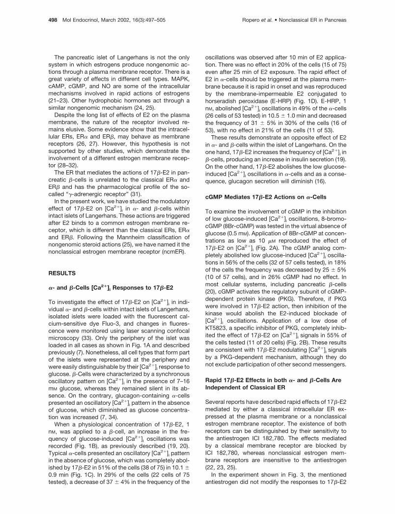
The pancreatic islet of Langerhans is not the onlysystem in which estrogens produce nongenomic ac-tions through a plasma membrane receptor. There is agreat variety of effects in different cell types. MAPK,cAMP, cGMP, and NO are some of the intracellularmechanisms involved in rapid actions of estrogens(21–23). Other hydrophobic hormones act through asimilar nongenomic mechanism (24, 25).
Despite the long list of effects of E2 on the plasmamembrane, the nature of the receptor involved re-mains elusive. Some evidence show that the intracel-lular ERs, ER� and ER�, may behave as membranereceptors (26, 27). However, this hypothesis is notsupported by other studies, which demonstrate theinvolvement of a different estrogen membrane recep-tor (28–32).
The ER that mediates the actions of 17�-E2 in pan-creatic �-cells is unrelated to the classical ER� andER� and has the pharmacological profile of the so-called “�-adrenergic receptor” (31).
In the present work, we have studied the modulatoryeffect of 17�-E2 on [Ca2�]i in �- and �-cells withinintact islets of Langerhans. These actions are triggeredafter E2 binds to a common estrogen membrane re-ceptor, which is different than the classical ERs, ER�and ER�. Following the Mannheim classification ofnongenomic steroid actions (25), we have named it thenonclassical estrogen membrane receptor (ncmER).
RESULTS
�- and �-Cells [Ca2�]i Responses to 17�-E2
To investigate the effect of 17�-E2 on [Ca2�]i in indi-vidual �- and �-cells within intact islets of Langerhans,isolated islets were loaded with the fluorescent cal-cium-sensitive dye Fluo-3, and changes in fluores-cence were monitored using laser scanning confocalmicroscopy (33). Only the periphery of the islet wasloaded in all cases as shown in Fig. 1A and describedpreviously (7). Nonetheless, all cell types that form partof the islets were represented at the periphery andwere easily distinguishable by their [Ca2�]i response toglucose. �-Cells were characterized by a synchronousoscillatory pattern on [Ca2�]i in the presence of 7–16mM glucose, whereas they remained silent in its ab-sence. On the contrary, glucagon-containing �-cellspresented an oscillatory [Ca2�]i pattern in the absenceof glucose, which diminished as glucose concentra-tion was increased (7, 34).
When a physiological concentration of 17�-E2, 1nM, was applied to a �-cell, an increase in the fre-quency of glucose-induced [Ca2�]i oscillations wasrecorded (Fig. 1B), as previously described (19, 20).Typical �-cells presented an oscillatory [Ca2�]i patternin the absence of glucose, which was completely abol-ished by 17�-E2 in 51% of the cells (38 of 75) in 10.1 �0.9 min (Fig. 1C). In 29% of the cells (22 cells of 75tested), a decrease of 37 � 4% in the frequency of the
oscillations was observed after 10 min of E2 applica-tion. There was no effect in 20% of the cells (15 of 75)even after 25 min of E2 exposure. The rapid effect ofE2 in �-cells should be triggered at the plasma mem-brane because it is rapid in onset and was reproducedby the membrane-impermeable E2 conjugated tohorseradish peroxidase (E-HRP) (Fig. 1D). E-HRP, 1nM, abolished [Ca2�]i oscillations in 49% of the �-cells(26 cells of 53 tested) in 10.5 � 1.0 min and decreasedthe frequency of 31 � 5% in 30% of the cells (16 of53), with no effect in 21% of the cells (11 of 53).
These results demonstrate an opposite effect of E2in �- and �-cells within the islet of Langerhans. On theone hand, 17�-E2 increases the frequency of [Ca2�]i in�-cells, producing an increase in insulin secretion (19).On the other hand, 17�-E2 abolishes the low glucose-induced [Ca2�]i oscillations in �-cells and as a conse-quence, glucagon secretion will diminish (16).
cGMP Mediates 17�-E2 Actions on �-Cells
To examine the involvement of cGMP in the inhibitionof low glucose-induced [Ca2�]i oscillations, 8-bromo-cGMP (8Br-cGMP) was tested in the virtual absence ofglucose (0.5 mM). Application of 8Br-cGMP at concen-trations as low as 10 �M reproduced the effect of17�-E2 on [Ca2�]i (Fig. 2A). The cGMP analog com-pletely abolished low glucose-induced [Ca2�]i oscilla-tions in 56% of the cells (32 of 57 cells tested), in 18%of the cells the frequency was decreased by 25 � 5%(10 of 57 cells), and in 26% cGMP had no effect. Inmost cellular systems, including pancreatic �-cells(20), cGMP activates the regulatory subunit of cGMP-dependent protein kinase (PKG). Therefore, if PKGwere involved in 17�-E2 action, then inhibition of thekinase would abolish the E2-induced blockade of[Ca2�]i oscillations. Application of a low dose ofKT5823, a specific inhibitor of PKG, completely inhib-ited the effect of 17�-E2 on [Ca2�]i signals in 55% ofthe cells tested (11 of 20 cells) (Fig. 2B). These resultsare consistent with 17�-E2 modulating [Ca2�]i signalsby a PKG-dependent mechanism, although they donot exclude participation of other second messengers.
Rapid 17�-E2 Effects in both �- and �-Cells AreIndependent of Classical ER
Several reports have described rapid effects of 17�-E2mediated by either a classical intracellular ER ex-pressed at the plasma membrane or a nonclassicalestrogen membrane receptor. The existence of bothreceptors can be distinguished by their sensitivity tothe antiestrogen ICI 182,780. The effects mediatedby a classical membrane receptor are blocked byICI 182,780, whereas nonclassical estrogen mem-brane receptors are insensitive to the antiestrogen(22, 23, 25).
In the experiment shown in Fig. 3, the mentionedantiestrogen did not modify the responses to 17�-E2
498 Mol Endocrinol, March 2002, 16(3):497–505 Ropero et al. • Nonclassical ER in Pancreas

in either �- or �-cells. For that reason, we concludethat rapid [Ca2�]i regulation triggered by 17�-E2 inboth �- and �-cells should be mediated by a nonclas-sical ER.
Binding Studies Using E-HRP
We have used a binding assay method based on theinteraction of E-HRP as a specific probe to detect E2binding sites at the plasma membrane of nonperme-abilized cells (31). This assay has been combined withimmunocytochemistry to specifically study the char-acteristics of the E2 binding site in �-cells. Figure 4Ashows a cell stained with E-HRP and developed usingCo-DAB (3,3�-diaminobenzidine in the presence ofCoCl2). On completion of this assay, the cells werepermeabilized (see Materials and Methods) to identify
Fig. 1. Effects of 1 nM 17�-E2 on [Ca2�]i Oscillations in �-and �-Cells Within the Islet of Langerhans
A, Image of a Fluo-3-loaded islet exposed to 0.5 mM
glucose. Blue corresponds to low and red to high fluores-cence intensity. The scale bar at the bottom right represents20 �m. Location of five �-cells that oscillate at 0.5 mM glu-cose are labeled with numbers 1 to 5. B–D, Records offluorescence vs. time from cells within the islet of Langer-hans. The Ca2�-dependent fluorescence of Fluo-3 is ex-pressed as %�F/F0 (see Materials and Methods). E2 or E-HRP was applied for the period indicated by the horizontalbar. B, Increase of the frequency of [Ca2�]i oscillations in a�-cell induced by 1 nM 17�-E2 in the presence of 8 mM
glucose. C, Inhibition of [Ca2�]i oscillations elicited by 0.5 mM
glucose in an �-cell by E2 (51%, 38 of 75 cells). The meantime for the abolishment of the oscillatory pattern was 10.1 �0.9 min. D, 1 nM E-HRP completely abolished [Ca2�]i oscil-lations in an �-cell (49%, 26 of 53 cells) in 10.5 � 1.0 min.Note the high grade of mimetism between 17�-E2 and E-HRPeffects. Data are expressed as mean � SEM.
Fig. 2. Involvement of cGMP and PKG in 17�-E2-InducedAbolishment of [Ca2�]i Oscillations in �-Cells
A, Application of 10 �M 8Br-cGMP completely abolishes[Ca2�]i oscillations induced by 0.5 mM glucose in �-cells in56% of the cells tested. B, 1 �M KT5823, an inhibitor of PKG,fully blocked the effect of 1 nM 17�-E2 on [Ca2�]i oscillationsinduced by 0.5 mM glucose in 55% of the �-cells tested.
Ropero et al. • Nonclassical ER in Pancreas Mol Endocrinol, March 2002, 16(3):497–505 499
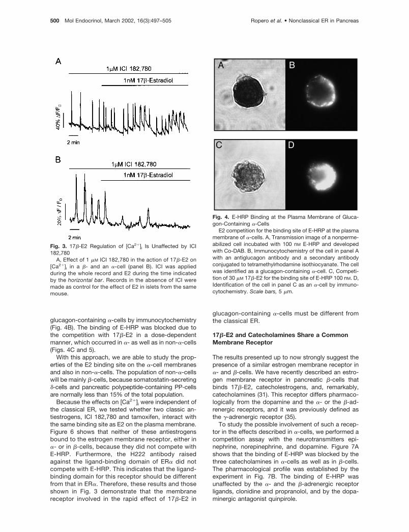
glucagon-containing �-cells by immunocytochemistry(Fig. 4B). The binding of E-HRP was blocked due tothe competition with 17�-E2 in a dose-dependentmanner, which occurred in �- as well as in non-�-cells(Figs. 4C and 5).
With this approach, we are able to study the prop-erties of the E2 binding site on the �-cell membranesand also in non-�-cells. The population of non-�-cellswill be mainly �-cells, because somatostatin-secreting�-cells and pancreatic polypeptide-containing PP-cellsare normally less than 15% of the total population.
Because the effects on [Ca2�]i were independent ofthe classical ER, we tested whether two classic an-tiestrogens, ICI 182,780 and tamoxifen, interact withthe same binding site as E2 on the plasma membrane.Figure 6 shows that neither of these antiestrogensbound to the estrogen membrane receptor, either in�- or in �-cells, because they did not compete withE-HRP. Furthermore, the H222 antibody raisedagainst the ligand-binding domain of ER� did notcompete with E-HRP. This indicates that the ligand-binding domain for this receptor should be differentfrom that in ER�. Therefore, these results and thoseshown in Fig. 3 demonstrate that the membranereceptor involved in the rapid effect of 17�-E2 in
glucagon-containing �-cells must be different fromthe classical ER.
17�-E2 and Catecholamines Share a CommonMembrane Receptor
The results presented up to now strongly suggest thepresence of a similar estrogen membrane receptor in�- and �-cells. We have recently described an estro-gen membrane receptor in pancreatic �-cells thatbinds 17�-E2, catecholestrogens, and, remarkably,catecholamines (31). This receptor differs pharmaco-logically from the dopamine and the �- or the �-ad-renergic receptors, and it was previously defined asthe �-adrenergic receptor (35).
To study the possible involvement of such a recep-tor in the effects described in �-cells, we performed acompetition assay with the neurotransmitters epi-nephrine, norepinephrine, and dopamine. Figure 7Ashows that the binding of E-HRP was blocked by thethree catecholamines in �-cells as well as in �-cells.The pharmacological profile was established by theexperiment in Fig. 7B. The binding of E-HRP wasunaffected by the �- and the �-adrenergic receptorligands, clonidine and propranolol, and by the dopa-minergic antagonist quinpirole.
Fig. 3. 17�-E2 Regulation of [Ca2�]i Is Unaffected by ICI182,780
A, Effect of 1 �M ICI 182,780 in the action of 17�-E2 on[Ca2�]i in a �- and an �-cell (panel B). ICI was appliedduring the whole record and E2 during the time indicatedby the horizontal bar. Records in the absence of ICI weremade as control for the effect of E2 in islets from the samemouse.
Fig. 4. E-HRP Binding at the Plasma Membrane of Gluca-gon-Containing �-Cells
E2 competition for the binding site of E-HRP at the plasmamembrane of �-cells. A, Transmission image of a nonperme-abilized cell incubated with 100 nM E-HRP and developedwith Co-DAB. B, Immunocytochemistry of the cell in panel Awith an antiglucagon antibody and a secondary antibodyconjugated to tetramethylrhodamine isothiocyanate. The cellwas identified as a glucagon-containing �-cell. C, Competi-tion of 30 �M 17�-E2 for the binding site of E-HRP 100 nM. D,Identification of the cell in panel C as an �-cell by immuno-cytochemistry. Scale bars, 5 �m.
500 Mol Endocrinol, March 2002, 16(3):497–505 Ropero et al. • Nonclassical ER in Pancreas

DISCUSSION
This report demonstrates that physiological concen-trations of the gonadal hormone 17�-E2 inhibits the[Ca2�]i oscillatory pattern induced by low glucoseconcentrations in pancreatic �-cells. Because gluca-gon secretion depends on an increase in [Ca2�]i (1), adiminished glucagon secretion is to be expected.Such an effect is in agreement with previous resultsthat described an inhibitory role of 17�-E2 in �-cellsusing long-term exposure. After E2 exposure for aperiod of 4–8 h, a dose-dependent inhibitory effect onthe arginine-stimulated glucagon secretion was man-ifested (18).
An increase in plasma levels of 17�-E2 is achievedin proestrus and during pregnancy. In pregnant rats,plasma insulin is increased in response to increasedlevels of sex steroids (17, 36, 37). Moreover, 17�-E2 atconcentrations comparable to that of pregnancy en-hances insulin secretion in perfused rat pancreas (38).E2 reverses the effects of menopause on glucose andinsulin metabolism, resulting in an increase of pancre-atic insulin secretion and a decrease in insulin resis-tance (39, 40).
Because glucagon inhibits insulin secretion, a de-crease of glucagon release may contribute, in a para-crine manner, to the rapid insulinotropic effect mani-fested by 17�-E2 via a plasma membrane receptor(19).
The mechanism of action of estrogens in �-cells isstill greatly unknown; indeed this is the first reportdescribing a rapid effect of 17�-E2 in this particulartype of cell. The results presented in this work dem-
onstrate that the receptor responsible is located at theplasma membrane. E-HRP, which is membrane imper-meable, mimics the effect of 17�-E2 at the same con-centration. The existence of a membrane receptor isconfirmed by visualization using E-HRP and by trans-mission laser scanning microscopy in unequivocallyidentified �-cells using immunostaining.
Basis for the Opposite Effect of 17�-E2 in �- and�-Cells
The rapid effects of 17�-E2 and E-HRP on [Ca2�]i aremimicked by 8Br-cGMP and blocked in more than50% of the cases with the PKG inhibitor KT5823.These experiments indicate the involvement of cGMPin the 17�-E2 response. We have recently demon-strated in �-cells (20) that physiological levels of 17�-E2, as those used here, decrease KATP channel activityvia a cGMP-dependent phosphorylation process. Thisproduces a depolarization of the membrane and thesubsequent opening of voltage-gated calcium chan-nels, causing a potentiation of glucose-induced[Ca2�]i oscillations. A similar mechanism may operatein pancreatic �-cells, where the existence of KATP
channels has recently been described (14). The KATP
channel has the same function in both �- and �-cells,and depolarization of the membrane as a result ofglucose metabolism is observed in both types of cells.The opposite effect of glucose is explained as a con-sequence of the different set of ion channels pre-sented by �- and �-cells (14). The model predicts that
Fig. 5. Dose-Dependent Competition of E-HRP Binding by17�-E2
Incubation of fixed nonpermeabilized islet cells in culturewith 10 nM E-HRP and increased concentrations of 17�-E2.�-Cells were identified as glucagon-containing cells, and theresult was plotted separately (f) from the rest of the cells (�).As shown, there are no differences between the percentagesof absorbed light in �-cells and non-�-cells (mostly �-cells,see Results) in the same conditions. The results are ex-pressed as the mean � SEM of three different experimentswith duplicate samples.
Fig. 6. No Competition with Antiestrogens for the E2 Bind-ing Site at the Plasma Membrane of �-Cells
Binding assays with 100 nM E-HRP and competition with17�-E2 (E2), the antiestrogens ICI 182,780 (ICI) and tamox-ifen (T), and the antibody H222 against the ligand bindingdomain of ER� (H222). All of them were applied at 30 �M andH222 at 1:100 dilution. Neither ICI182,780 nor tamoxifencompeted for E-HRP binding, whereas E2 decreased lightabsorption up to 80%. Neither antiestrogen affected either �-(f) or �-cell (�) binding of E-HRP. Background binding ob-tained with 100 nM peroxidase has been subtracted. Dimeth-ylsulfoxide was used as a vehicle for insoluble reactives,and it was present both in control and peroxidase samples.Data are from two duplicate experiments, expressed asmean � SEM.
Ropero et al. • Nonclassical ER in Pancreas Mol Endocrinol, March 2002, 16(3):497–505 501
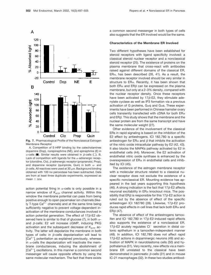
action potential firing in �-cells is only possible in anarrow window of KATP channel activity. Within thiswindow the membrane potential can pass from beingpositive enough to open pacemaker ion channels (like-ly T-type Ca2� channels) and at the same time beingsufficiently negative to prevent voltage-dependent in-activation of the membrane conductances involved inaction potential generation. The effect of 17�-E2 ob-served here is similar to that of glucose (7), in both �-and �-cells: E2 will increase cGMP, inducing PKGactivation and the subsequent decrease of KATP ac-tivity. The latter will depolarize the membrane in bothtypes of cells: in �-cells depolarization will lead to[Ca2�]i potentiation as explained previously, whereasin �-cells the depolarization will inactivate the mem-brane conductances, inducing the abolishment of[Ca2�]i oscillations. In this manner a common secondmessenger will cause opposite effects by using thesame molecular mechanism. The fact that there exists
a common second messenger in both types of cellsalso suggests that the ER involved would be the same.
Characteristics of the Membrane ER Involved
Two different hypotheses have been established forsteroid receptors with ligand specificity involved: aclassical steroid nuclear receptor and a nonclassicalsteroid receptor (25). The existence of proteins on theplasma membrane that cross-react with antibodiesraised against different domains of the classical ER,ER�, has been described (26, 41). As a result, themembrane receptor involved should be very similar instructure to ER�. Recently, it has been shown thatboth ER� and ER� can be expressed on the plasmamembrane, but only at a 2–3% density, compared withthe nuclear receptor density. Once these receptorshave been activated by 17�-E2, they stimulate ade-nylate cyclase as well as IP3 formation via a previousactivation of G proteins, G�q and G�s. These exper-iments have been performed in Chinese hamster ovarycells transiently transfected with cDNA for both ER�and ER�. This study shows that the membrane and thenuclear protein are from the same transcript and havethe same molecular weight (27).
Other evidence of the involvement of the classicalERs in rapid signaling is based on the inhibition of theE2 effect by antiestrogens. ICI 182,780 is a specificantiestrogen for ER� and � that inhibits the activationof the nitric oxide intracellular pathway by E2 (42, 43).It also blocks the MAPKs pathway activated by E2 inendothelial cells (44). Moreover, the E2 activation ofendothelial nitric oxide synthase is enhanced by theoverexpression of ER� in endothelial cells and inhib-ited by ICI (45).
The existence of the estrogen membrane receptorwith a molecular structure related to a classical nu-clear receptor does not exclude the existence of aspecific nonclassical ER. Mounting evidence has ap-peared in the last years supporting this hypothesis(46). A strong indication is the fact that 17�-E2 affectsneuronal excitability in ER� knockout mice. The pos-sibility that ER� is responsible for this 17�-E2 action isruled out by the absence of effect of the specificantiestrogen ICI 182780 (28). Likewise, 17�-E2 pro-duces rapid effects in cell lines that lack both ER� andER� (47).
The absence of effect of the antiestrogens tamox-ifen and ICI 182,780 in 17�-E2-induced rapid effectsalso supports the existence of a nonclassical ER.17�-E2 acutely regulates Cl� secretion in distal co-lonic epithelium in a tamoxifen-independent manner(48). In addition, ICI 182,780 does not affect rapid17�-E2 actions in dopaminergic neurons (49) and ac-tivation of MAPK in neuroblastoma cells (50) and hy-pothalamus (51). Very recently, new effects via a mem-brane ER unrelated to the classical have beendemonstrated in pancreatic �-cells (31) and in mouseIC-21 macrophages (32). In these two studies antibod-
Fig. 7. Pharmacological Profile of the Nonclassical EstrogenMembrane Receptor
A, Competition of E-HRP binding by the catecholaminesdopamine (Dop), norepinephrine (NE), and epinephrine (E) in�-cells (f). Similar results were obtained in �-cells (�). B,Lack of competition with ligands for the �-adrenergic recep-tor (clonidine, Clo), �-adrenergic receptor (propranolol, Prop),and dopamine receptor (quinpirole, Quin) in both �- and�-cells. All reactives were used at 30 �M. Background bindingobtained with 100 nM peroxidase has been subtracted. Dataare from at least three duplicate experiments, expressed asmean � SEM.
502 Mol Endocrinol, March 2002, 16(3):497–505 Ropero et al. • Nonclassical ER in Pancreas

ies against classical nuclear ER failed to stain theplasma membrane.
In pancreatic �-cells, we have demonstrated thatneither of the two classical nuclear ERs are present atthe membrane, and therefore they could not be re-sponsible for the rapid actions of 17�-E2 (31). In ad-dition, we demonstrate in the present paper that reg-ulation of [Ca2�]i by 17�-E2 is not modified by theantiestrogen ICI 182,780 in either �- or �-cells. More-over, no competition for E-HRP binding is manifestedby the antiestrogens tamoxifen and ICI 182,780, aswell as the antibody H222 against the ligand-bindingdomain of ER�. Involvement of ER� at the membraneis also unlikely based on the pharmacological profile ofthe receptor described here, although no antibodyagainst the ligand binding domain of ER� is availableto perform a competition study to fully rule out thispossibility.
The binding studies presented in this paper demon-strate that the membrane receptor acting in �-cellshas the same pharmacological profile as that de-scribed in �-cells (31), i.e. it has a binding site sharedby the hormone 17�-E2 and the neurotransmitters,dopamine, epinephrine, and norepinephrine. It is,nonetheless, neither a classical adrenergic receptornor a dopaminergic receptor, because their classicalligands are without effect on E-HRP binding. Thispharmacological profile is the same manifested by the�-adrenergic receptor, which is characterized by itsequal activation with the neurotransmitters, epineph-rine, norepinephrine and dopamine. It is affected nei-ther by the �- nor the �-adrenergic receptor antagonistand produces activation of protein kinase G via a nitricoxide-independent guanylyl cyclase (35, 52–55).
The presence of a membrane receptor both in �-and �-cells unrelated to the classical ER� and ER� isstrong evidence in favor of a new class of estrogenmembrane receptor.
MATERIALS AND METHODS
Materials
ICI 182,780 was a kind gift of Astra USA, Inc.-Zeneca Phar-maceuticals (Bristol, UK). Fluo-3 AM was obtained from Mo-lecular Probes, Inc. (Leiden, The Netherlands). Propranolol,Clonidine, and Quinpirole were obtained from TocrisNeuramin (Bristol, UK). KT 5823 was from Alexis (Laufelfin-gen, Switzerland). Other substances were obtained fromSigma (Madrid, Spain).
Measuring [Ca2�]i in � and �-Cells Within Intact Isletsof Langerhans
Swiss albino OF1 male mice (8–10 wk old) were killed bycervical dislocation according to national guidelines. Pancre-atic islets of Langerhans were isolated by collagenase diges-tion as previously described (56) and loaded with 5 �M Fluo-3AM for at least 1 h at room temperature. Loaded islets werekept in a medium containing (mM): 115 NaCl, 25 NaHCO3, 5KCl, 1.1 MgCl2, 1.2 NaH2PO4, 2.5 CaCl2, and 2.5 HEPES;
plus 1% albumin and 5 mM D-glucose, continuously gassedwith a mixture of 95% O2 and 5% CO2, pH 7.35. Islets wereperfused with a modified Ringer solution containing (mM): 120NaCl, 5 KCl, 25 NaHCO3, 1.1 MgCl2, and 2.5 CaCl2, pH 7.35,when gassed with 95% O2 and 5% CO2.
Calcium records in individual cells were obtained by im-aging [Ca2�]i under a LSM 510 confocal microscope (CarlZeiss, Jena, Germany) using a Carl Zeiss �40 oil immersionlens, numerical aperture 1.3. Images were collected at 2-secintervals, and fluorescent signals from individual cells weremeasured in function of time using the Carl Zeiss LSM soft-ware package. Experiments were performed at 34 C. Resultswere plotted using a commercially available software (Sig-maplot, Jandel Scientific, San Rafael, CA) where the changein fluorescence (�F) is expressed as a percentage of thebasal fluorescence (F0) observed in absence of stimulus. �And �-cells within the islets were identified by their [Ca2�]ioscillatory pattern in 0.5 mM and 8 mM glucose, respectively(7, 34).
Cell Isolation and Culture
Islets isolated as above were dispersed into single cells andcultured as previously described (4). Briefly, islets were dis-aggregated into single cells with trypsin. Cells were thencentrifuged, resuspended in cultured medium RPMI 1640supplemented with 10% FCS, 200 U ml�1 penicillin, 0.2 mgml�1 streptomycin, 2 mM L-glutamine and 11 mM glucose,and plated on poly-L-lysine-coated glass coverslips. Cellswere kept at 37 C in a humidified atmosphere of 95% O2 and5% CO2 for 24 h.
E2-Peroxidase Binding Assay
After 24 h in culture, the cells were washed and fixed in 4%(wt/vol) fresh paraformaldehyde for 30 sec, which does notpermeabilize the cells (19), and exposed overnight to 4.5�g/ml E-HRP at 4 C (0.45 �g/ml for Fig. 4). The competitionfor the E2 binding site was tested using various reactiveswhile adding E-HRP simultaneously. Cells were then washedand E-HRP binding was developed using 3,3�-diaminoben-zidine tetrahydrochloride in the presence of urea hydrogenperoxide and CoCl2 for 30 min (Sigma FAST 3, 3�-diamino-benzidine tetrahydrochloride with metal enhancer tablet set,Co-DAB). After the development with Co-DAB, the cells werewashed three times with PBS.
Immunocytochemistry
To identify �-cells, immunocytochemistry was performed us-ing an antiglucagon antibody. After the staining with E-HRPand Co-DAB, the cells were permeabilized with 1% Triton for2 min. They were then incubated with the primary antibody(1:500 dilution) for 2 h, after which the secondary antibodyantimouse IgG-tetramethylrhodamine isothiocyanate (1:125dilution) was added for 1 h, both at room temperature. Therewas no labeling with the secondary antibody alone.
Binding Studies Using E-HRP and Transmission LaserScanning Microscopy
The reaction between HRP and Co-DAB produces a darkprecipitate that absorbs light, permitting us to visualize itusing transmission laser scanning microscopy (57). The useof a reactive that competes for the E2 binding site will de-crease the amount of precipitate. To identify those reactivesthat bind to the same site as 17�-E2, the light absorbed bythe precipitate was measured, as previously reported (31).Nonetheless, we should note that quantifying the amount ofprecipitate simply by measuring the light absorbed may be an
Ropero et al. • Nonclassical ER in Pancreas Mol Endocrinol, March 2002, 16(3):497–505 503

unreliable procedure if changes in the amount of absorbedlight are small. This method is of interest for comparativepurpose, and it works extremely well for conditions in whichthe difference in the amount of precipitate is large. For thatreason, detailed concentration-response profiles have beenomitted. Transmission photographs were taken with a CarlZeiss LSM 510 laser scanning confocal microscope, at 543nm. In this work, the light intensity was taken from the wholearea of the cell using Carl Zeiss LSM 510 software, ratherthan along a line as we have previously reported (31). Asimilar area was chosen to measure the background light.The background light and the microscope settings were con-stant throughout the different experiments. The quantity ofE-HRP bound is expressed as the percentage of absorbedlight with respect to the control condition. The lower thepercentage of absorbed light, the higher the competition foran E-HRP binding site. To obtain an appropriate stainingbackground, incubation and developmental procedure withperoxidase were performed in identical conditions. Thisbackground light has been subtracted in Figs. 6 and 7 forclarity.
Acknowledgments
The authors thank Drs. Esther Fuentes and Cristina Ripollfor their help in several aspects of the project and E. PerezGarcıa, A. Perez Vegara, and N. Illera (Miguel HernandezUniversity, Alicante, Spain) for technical assistance. H222antibody was a kind gift of Professor G. Greene (University ofChicago, Chicago, IL).
Received May 23, 2001. Accepted November 7, 2001.Address all correspondence and requests for reprints to:
Dr. Angel Nadal, Institute of Bioengineering and Departmentof Physiology, School of Medicine, Miguel Hernandez Uni-versity, San Juan Campus Carretera Alicante-Valencia Km87. 03550, Alicante, Spain. E-mail: [email protected].
This work was supported by Grants from European Union-Comision Interministerial de Ciencia y Tecnologıa (IFD97-1064-103-02) and Fundacion Navarro Trıpodi. A.B.R. is arecipient of a research scholarship from de Ministerio deEducacion, Cultura y Deporte.
REFERENCES
1. Johansson H, Gylfe E, Hellman B 1987 The actions ofarginine and glucose on glucagon secretion are mediatedby opposite effects on cytoplasmic Ca2�. Biochem Bio-phys Res Commun 147:309–314
2. Prentki M, Matschinsky FM 1987 Ca2�, cAMP, and phos-pholipid-derived messengers in coupling mechanisms ofinsulin secretion. Physiol Rev 67:1185–1248
3. Ashcroft FM, Rorsman P 1989 Electrophysiology of thepancreatic �-cell. Prog Biophys Mol Biol 54:87–143
4. Valdeolmillos M, Nadal A, Contreras D, Soria B 1992 Therelationship between glucose-induced K�
ATP channelclosure and the rise in [Ca2�]i in single mouse pancreatic�-cells. J Physiol (Lond) 455:173–186
5. Santos RM, Rosario LM, Nadal A, Garcia-Sancho J, SoriaB, Valdeolmillos M 1991 Widespread synchronous[Ca2�]i oscillations due to bursting electrical activity insingle pancreatic islets. Pflugers Arch 418:417–422
6. Valdeolmillos M, Nadal A, Soria B, Garcia-Sancho J 1993Fluorescence digital image analysis of glucose-induced[Ca2�]i oscillations in mouse pancreatic islets of Langer-hans. Diabetes 42:1210–1214
7. Nadal A, Quesada I, Soria B 1999 Homologous and het-erologous asynchronicity between identified �-, �- and
�-cells within intact islets of Langerhans in the mouse.J Physiol (Lond) 517:85–93
8. Rosario LM, Atwater I, Scott AM 1986 Pulsatile insulinrelease and electrical activity from single ob/ob mouseislets of Langerhans. Adv Exp Med Biol 211:413–425
9. Martin F, Reig JA, Soria B 1995 Secretagogue-induced[Ca2�]i changes in single rat pancreatic islets and corre-lation with simultaneously measured insulin release. JMol Endocrinol 15:177–185
10. Barbosa RM, Silva AM, Tome AR, Stamford JA, SantosRM, Rosario LM 1998 Control of pulsatile 5-HT/insulinsecretion from single mouse pancreatic islets by intra-cellular calcium dynamics. J Physiol (Lond) 510:135–143
11. Dunbar JC, Walsh MF 1982 Glucagon and insulin secre-tion by dispersed islet cells: possible paracrine relation-ships. Horm Res 16:257–267
12. Opara EC, Atwater I, Go VL 1988 Characterization andcontrol of pulsatile secretion of insulin and glucagon.Pancreas 3:484–487
13. Rorsman P, Hellman B 1988 Voltage-activated currentsin guinea pig pancreatic � 2 cells. Evidence for Ca2�-dependent action potentials. J Gen Physiol 91:223–242
14. Gopel SO, Kanno T, Barg S, Weng X, Gromada J, Rors-man P 2000 Regulation of glucagon release in mouse-cells by KATP channels and inactivation of TTX-sensitiveNa� channels. J Physiol 528:509–520
15. Berts A, Ball A, Gylfe E, Hellman B 1996 Suppression ofCa2� oscillations in glucagon-producing �2-cells by in-sulin/glucose and amino acids. Biochim Biophys Acta1310:212–216
16. Gromada J, Bokvist K, Ding WG, Barg S, Buschard K,Renstrom E, Rorsman P 1997 Adrenaline stimulates glu-cagon secretion in pancreatic A-cells by increasing theCa2� current and the number of granules close to theL-type Ca2� channels. J Gen Physiol 110:217–228
17. SutterDub MT 1976 Preliminary report: effects of femalesex hormones on insulin secretion by the perfused ratpancreas. J Physiol (Paris) 72:795–800
18. Faure A, Haouari M, Sutter BC 1988 Short term anddirect influence of oestradiol on glucagon secretion stim-ulated by arginine. Diabet Metab 14:452–454
19. Nadal A, Rovira JM, Laribi O, Leon-quinto T, Andreu E,Ripoll C, Soria B 1998 Rapid insulinotropic effect of17�-estradiol via a plasma membrane receptor. FASEB J12:1341–1348
20. Ropero AB, Fuentes E, Rovira JM, Ripoll C, Soria B,Nadal A 1999 Non-genomic actions of 17�-oestradiol inmouse pancreatic �-cells are mediated by a cGMP-de-pendent protein kinase. J Physiol (Lond) 521:397–407
21. Kelly MJ, Wagner EJ 1999 Estrogen modulation of G-protein-coupled receptors. Trends Endocrinol Metab 10:369–374
22. Kelly MJ, Levin ER 2001 Rapid actions of plasma mem-brane estrogen receptors. Trends Endocrinol Metab 12:152–156
23. Nadal A, Diaz M, Valverde MA 2001 The estrogen trinity:membrane, cytosolic and nuclear effects. News PhysiolSci 16:251–255
24. Wehling M 1997 Specific, nongenomic actions of steroidhormones. Annu Rev Physiol 59:365–393
25. Falkenstein E, Tillmann HC, Christ M, Feuring M, WehlingM 2000 Multiple actions of steroid hormones—a focus onrapid, nongenomic effects. Pharmacol Rev 52:513–556
26. Watson CS, Campbell CH, Gametchu B 1999 Membraneoestrogen receptors on rat pituitary tumour cells: im-muno-identification and responses to oestradiol and xe-noestrogens. Exp Physiol 84:1013–1022
27. Razandi M, Pedram A, Greene GL, Levin ER 1999 Cellmembrane and nuclear estrogen receptors (ERs) origi-nate from a single transcript: studies of ER� and ER�expressed in Chinese hamster ovary cells. Mol Endocri-nol 13:307–319
504 Mol Endocrinol, March 2002, 16(3):497–505 Ropero et al. • Nonclassical ER in Pancreas
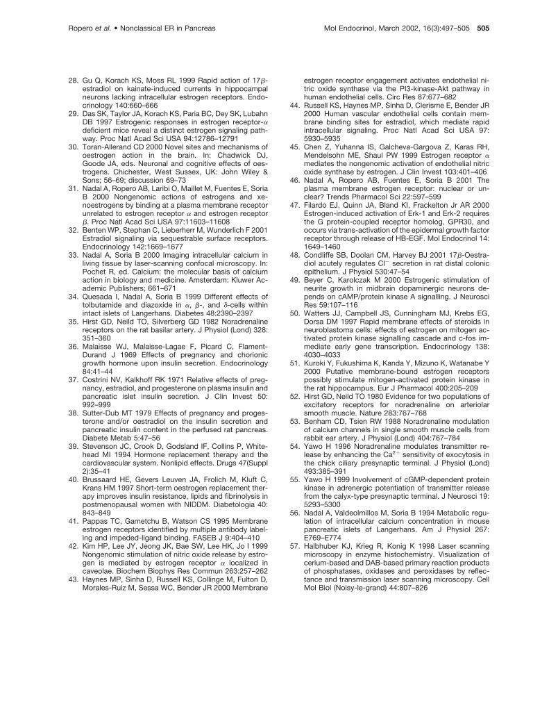
28. Gu Q, Korach KS, Moss RL 1999 Rapid action of 17�-estradiol on kainate-induced currents in hippocampalneurons lacking intracellular estrogen receptors. Endo-crinology 140:660–666
29. Das SK, Taylor JA, Korach KS, Paria BC, Dey SK, LubahnDB 1997 Estrogenic responses in estrogen receptor-�deficient mice reveal a distinct estrogen signaling path-way. Proc Natl Acad Sci USA 94:12786–12791
30. Toran-Allerand CD 2000 Novel sites and mechanisms ofoestrogen action in the brain. In: Chadwick DJ,Goode JA, eds. Neuronal and cognitive effects of oes-trogens. Chichester, West Sussex, UK: John Wiley &Sons; 56–69; discussion 69–73
31. Nadal A, Ropero AB, Laribi O, Maillet M, Fuentes E, SoriaB 2000 Nongenomic actions of estrogens and xe-noestrogens by binding at a plasma membrane receptorunrelated to estrogen receptor � and estrogen receptor�. Proc Natl Acad Sci USA 97:11603–11608
32. Benten WP, Stephan C, Lieberherr M, Wunderlich F 2001Estradiol signaling via sequestrable surface receptors.Endocrinology 142:1669–1677
33. Nadal A, Soria B 2000 Imaging intracellular calcium inliving tissue by laser-scanning confocal microscopy. In:Pochet R, ed. Calcium: the molecular basis of calciumaction in biology and medicine. Amsterdam: Kluwer Ac-ademic Publishers; 661–671
34. Quesada I, Nadal A, Soria B 1999 Different effects oftolbutamide and diazoxide in �, �-, and �-cells withinintact islets of Langerhans. Diabetes 48:2390–2397
35. Hirst GD, Neild TO, Silverberg GD 1982 Noradrenalinereceptors on the rat basilar artery. J Physiol (Lond) 328:351–360
36. Malaisse WJ, Malaisse-Lagae F, Picard C, Flament-Durand J 1969 Effects of pregnancy and chorionicgrowth hormone upon insulin secretion. Endocrinology84:41–44
37. Costrini NV, Kalkhoff RK 1971 Relative effects of preg-nancy, estradiol, and progesterone on plasma insulin andpancreatic islet insulin secretion. J Clin Invest 50:992–999
38. Sutter-Dub MT 1979 Effects of pregnancy and proges-terone and/or oestradiol on the insulin secretion andpancreatic insulin content in the perfused rat pancreas.Diabete Metab 5:47–56
39. Stevenson JC, Crook D, Godsland IF, Collins P, White-head MI 1994 Hormone replacement therapy and thecardiovascular system. Nonlipid effects. Drugs 47(Suppl2):35–41
40. Brussaard HE, Gevers Leuven JA, Frolich M, Kluft C,Krans HM 1997 Short-term oestrogen replacement ther-apy improves insulin resistance, lipids and fibrinolysis inpostmenopausal women with NIDDM. Diabetologia 40:843–849
41. Pappas TC, Gametchu B, Watson CS 1995 Membraneestrogen receptors identified by multiple antibody label-ing and impeded-ligand binding. FASEB J 9:404–410
42. Kim HP, Lee JY, Jeong JK, Bae SW, Lee HK, Jo I 1999Nongenomic stimulation of nitric oxide release by estro-gen is mediated by estrogen receptor � localized incaveolae. Biochem Biophys Res Commun 263:257–262
43. Haynes MP, Sinha D, Russell KS, Collinge M, Fulton D,Morales-Ruiz M, Sessa WC, Bender JR 2000 Membrane
estrogen receptor engagement activates endothelial ni-tric oxide synthase via the PI3-kinase-Akt pathway inhuman endothelial cells. Circ Res 87:677–682
44. Russell KS, Haynes MP, Sinha D, Clerisme E, Bender JR2000 Human vascular endothelial cells contain mem-brane binding sites for estradiol, which mediate rapidintracellular signaling. Proc Natl Acad Sci USA 97:5930–5935
45. Chen Z, Yuhanna IS, Galcheva-Gargova Z, Karas RH,Mendelsohn ME, Shaul PW 1999 Estrogen receptor �mediates the nongenomic activation of endothelial nitricoxide synthase by estrogen. J Clin Invest 103:401–406
46. Nadal A, Ropero AB, Fuentes E, Soria B 2001 Theplasma membrane estrogen receptor: nuclear or un-clear? Trends Pharmacol Sci 22:597–599
47. Filardo EJ, Quinn JA, Bland KI, Frackelton Jr AR 2000Estrogen-induced activation of Erk-1 and Erk-2 requiresthe G protein-coupled receptor homolog, GPR30, andoccurs via trans-activation of the epidermal growth factorreceptor through release of HB-EGF. Mol Endocrinol 14:1649–1460
48. Condliffe SB, Doolan CM, Harvey BJ 2001 17�-Oestra-diol acutely regulates Cl� secretion in rat distal colonicepithelium. J Physiol 530:47–54
49. Beyer C, Karolczak M 2000 Estrogenic stimulation ofneurite growth in midbrain dopaminergic neurons de-pends on cAMP/protein kinase A signalling. J NeurosciRes 59:107–116
50. Watters JJ, Campbell JS, Cunningham MJ, Krebs EG,Dorsa DM 1997 Rapid membrane effects of steroids inneuroblastoma cells: effects of estrogen on mitogen ac-tivated protein kinase signalling cascade and c-fos im-mediate early gene transcription. Endocrinology 138:4030–4033
51. Kuroki Y, Fukushima K, Kanda Y, Mizuno K, Watanabe Y2000 Putative membrane-bound estrogen receptorspossibly stimulate mitogen-activated protein kinase inthe rat hippocampus. Eur J Pharmacol 400:205–209
52. Hirst GD, Neild TO 1980 Evidence for two populations ofexcitatory receptors for noradrenaline on arteriolarsmooth muscle. Nature 283:767–768
53. Benham CD, Tsien RW 1988 Noradrenaline modulationof calcium channels in single smooth muscle cells fromrabbit ear artery. J Physiol (Lond) 404:767–784
54. Yawo H 1996 Noradrenaline modulates transmitter re-lease by enhancing the Ca2� sensitivity of exocytosis inthe chick ciliary presynaptic terminal. J Physiol (Lond)493:385–391
55. Yawo H 1999 Involvement of cGMP-dependent proteinkinase in adrenergic potentiation of transmitter releasefrom the calyx-type presynaptic terminal. J Neurosci 19:5293–5300
56. Nadal A, Valdeolmillos M, Soria B 1994 Metabolic regu-lation of intracellular calcium concentration in mousepancreatic islets of Langerhans. Am J Physiol 267:E769–E774
57. Halbhuber KJ, Krieg R, Konig K 1998 Laser scanningmicroscopy in enzyme histochemistry. Visualization ofcerium-based and DAB-based primary reaction productsof phosphatases, oxidases and peroxidases by reflec-tance and transmission laser scanning microscopy. CellMol Biol (Noisy-le-grand) 44:807–826
Ropero et al. • Nonclassical ER in Pancreas Mol Endocrinol, March 2002, 16(3):497–505 505


