APROVECHAMIENTO DE SUBPRODUCTOS
DE CULTIVOS DE HIGO CHUMBO (Opuntia
ficus-indica L.) Y AGUACATE (Persea americana)
Tesis doctoral presentada por:
Bruno Melgar Castañeda
Dirigida por:
Esperanza M. García Castelló
Antonio D. Rodríguez López
Valencia Febrero 2019

La Dra. Dña. Esperanza M. García Castelló, Investigadora del Instituto
Universitario de Ingeniería de Alimentos para el Desarrollo y el Dr. D.
Antonio D. Rodríguez López, Investigador del Instituto Universitario
de Seguridad Industrial, Radiofísica y Medioambiental.
CERTIFICAN:
Que el trabajo que presenta Bruno Melgar Castañeda para optar al
grado de doctor por la Universidad Politècnica de València, con el título
“Aprovechamiento de subproductos de cultivos de higo chumbo
(opuntia ficus-indica l.) y aguacate (persea americana)”, ha sido
realizado bajo nuestra dirección en el Instituto Universitario de
Ingeniería de Alimentos para el Desarrollo de la Universidad
Politècnica de València.
Y para que conste a los efectos oportunos, firman este certificado en
Valencia a 21 de Febrero de 2019.
______________________ ______________________
Esperanza M. Antonio D.
García Castelló Rodríguez López


PRICKLY PEAR (Opuntia spp) AND AVOCADO
(Persea americana) FRUITS
CHARACTERIZATION AND BY-PRODUCTS
EXPLOITATION


What is not started today
is never finished tomorrow.
J.W. Von Goethe


Agradecimientos
Agradecimientos
Siempre he sido persona de pocas palabras, ya que durante todo este
tiempo he pensado que solo las cosas importantes merecen ser dichas.
Afortunadamente, el realizar esta tesis me ha abierto los ojos y
comienzo a observar las cosas desde otra perspectiva.
Este trabajo no es la lucha de una sola persona, sino todo lo
contrario, sin el respaldo que he tenido siempre, las cosas hoy serian
muy diferentes y lo minimo que puedo hacer es expresar mis
gratificaciones a todas y cada una de esas personas tan especiales.
Me gustaría comenzar agradeciendo a mis padres, porque aunque
suene trillado, sin ellos no estaría donde estoy ahora, se que no es nada
fácil tener un hijo tan lejos y verlo raramente, pero siempre han estado
ahí en todo momento.
A mi hermano, a mi tía Inez y a mi abuela Norma, ya que han sido
compañeros de camino, me han proporcionado tanto cariño y amor, y
han estado muy presentes incondicionalmente en todo momento.
Tambien agradezco a Maite y su familia, por haber sido mi familia
en esta travesia europea, llenando los huequitos que faltan cuando
tienes a tus seres queridos tan lejos.
Profesionalmente a mis tutores, principal apoyo y modelos a seguir,
Esperanza y Antonio, al igual que a mis asesoras en Portugal Isabel y
Lillian. A todos aquellos que colaboraron a hacer esto posible, los
técnicos de laboratorio, Virginia, Carolina, Mario y especialmente a
Milagro por su basta ayuda cromatografica. A los compañeros de
laboratorio que siempre estuvieron para ayudar con las técnicas
instrumentales, acompañando en la comida o simplemente sacando
unas sonrisas, David, Vicky, Cé, Inês, Eliana, Irene y tantos otros que
pudimos coincidir en estos tiempos.
No podían faltar los amigos de la Full family, que siempre
estuvieron presentes para morir en wods y para una que otra cerveza,
Gio, Facu, Hugo, Itahisa, Rolo, Juan Ma, el jabalí y el resto del
Agradecimentos
zoológico que es tan extenso que si tuviera que nombrarlos, seguro sería
mas difícil que escribir la tésis.
Y al resto de personas que de alguna forma han hecho de todo este
tiempo, un periodo maravilloso lleno de diversión, compañeros de piso,
de viajes, y a los erasmus, que me recordaron muchas de las buenas
cosas que había olvidado, sobre todo a Andreea por todos los buenos
momentos compartidos y a Alexandra por demostrame que en la
diversidad y grandeza de este mundo, siempre hay oportunidades de
encontrarse.
Y como no podía ser de otra forma, estos agradecimentos los escribo
en uno de tantos viajes realízados gracias al apoyo CONACyT.
Muchísimas gracias a todos.


Resumen
I
Resumen
La industria agro-alimentaria se encuentra en constante evolución,
adaptándose a las demandas de los consumidores. Un claro ejemplo es
el de la industria frutícola, que ha partido de los productos
mínimamente procesados, hacia procesos mucho más complejos como
las destilaciones, requiriendo mayores cantidades de materias primas.
Estos procesos conllevan a la generación de millones de toneladas de
residuos frutícolas al año, produciendo en muchos de los casos grandes
problemas con impactos medioambientales. No obstante, la riqueza
organoléptica y de compuestos nutraceúticos en estos residuos hacen
posible su revalorización, haciendo de manera sostenible un cambio
sustancial en el impacto negativo medioambiental y generando nuevos
productos con repercusiones económicas.
En este contexto, la presente tesis doctoral explora las características
nutricionales, sensoriales y nutraceúticas de dos especies frutícolas
(Persea americana y Opuntia spp.) y sus subproductos, con la finalidad
de generar una caracterización integral y un aprovechamiento de los
residuos generados por los cultivos de ambos frutos en las industrias
alimentarias (pieles y semillas), así como de especies de Opuntia no
utilizadas comercialmente. Esta exploración ha sido posible usando
diferentes técnicas analítica, bioquímicas y microbiológicas para llevar
a cabo la detección de las posibles alternativas de uso.
Como resultado a las potenciales propiedades de los frutos de
aguacates y de los higos chumbos, se decidió realizar una
caracterización nutricional de los principales macronutrientes, y debido
a la poca información encontrada en Opuntia spp., se realizó la
caracterización lipídica, de los azúcares y ácidos orgánicos de los
frutos. Posteriormente se evaluaron las propiedades nutraceúticas, por
un lado se evaluaron espectrofotométricamente los compuestos
bioactivos de ambos subproductos frutícolas mediante distintas
técnicas como Folin-Ciocalteu para la determinación de polifenoles
totales, Al3Cl3 para determinación de flavonoides totales, y poder
reductor, ABTS, DPPH, TBARS y β-caroteno para la determinación de
capacidad antioxidante hidrofílica e lipofílica de los extractos, además,
en Opuntia spp. se analizó el contenido de los colorantes betaxantinas
y betacianinas.

Resumen
II
Por otro lado, mediante el uso de técnicas cromatográficas HPLC-
DAD-ESI-MS/MS, se caracterizaron los perfiles polifenólicos (ácidos
fenólicos y flavonoides) de ambos subproductos frutícolas, más los
perfiles betalainicos de las muestras de higos chumbos. Por último,
mediante técnicas microbiológicas y ensayos citotoxicológicos, se
comprobó el poder antimicrobiano, antitumoral y hepatotóxico, con
ayuda de 16 cepas bacterianas, y fúngicas, 4 líneas celulares tumorales
y 1 línea celular sana obtenida a partir de hígado de cerdo, denominada
PLP2.
Finalmente, se ha realizado un ensayo de optimización multivariable
de extracción de los compuestos bioactivos de Opuntia spp. mediante
extracciones asistidas por microondas y ultrasonidos, haciendo uso de
un diseño experimental estadístico empleando las herramientas de
metodología de respuesta de superficie acopladas a un diseño central
compuesto 2^4 con 7 puntos centrales, para la obtención de
modelizaciones graficas de los parámetros de respuesta, que ayudan a
identificar las condiciones óptimas de los factores de extracción
empleados.
En definitiva, estos análisis suponen la obtención de información
relevante sobre la extracción de compuestos bioactivos con
aplicaciones nutraceúticas y colorantes, que pueden ser añadidos a una
amplia gama de productos de las industrias farmacéuticas, cosméticas
y alimentarias, para reforzar o mejorar las características y
funcionalidades de estos.

Resum
III
Resum
La indústria agroalimentària es troba en constant evolució, adaptant-se
a les demandes dels consumidors. Un clar exemple és el de la indústria
fructícola, que ha partit dels productes mínimament processats, cap a
processos molt més complexos com les destil∙lacions, requerint majors
quantitats de matèries primeres. Aquests processos comporten la
generació de milions de tones de residus fructícoles a l'any, produint en
molts dels casos grans problemes amb impacte mediambientals. No
obstant això, la riquesa nutracèutica i organolèptica d'aquests residus
fan possible la seua revaloració, fent de manera sostenible un canvi
substancial en l'impacte negatiu mediambiental i generant nous
productes amb repercussions econòmiques.
En aquest context, la present tesi doctoral explora les
característiques nutricionals, sensorials i nutracèutiques de dues
espècies fructícoles (Persea americana i Opuntia spp.) i els seus
subproductes, amb la finalitat generar una caracterització integral i un
aprofitament dels residus generats pels cultius de tots dos fruits en les
indústries alimentàries (pells i llavors), així com de les espècies
d'Opuntia no utilitzades comercialment. Aquesta exploració ha estat
possible usant diferents tècniques analítiques, bioquímiques i
microbiològiques per a dur a terme la detecció de les possibles
alternatives d'ús.
A causa de les potencials propietats dels fruits d'alvocats i de les
figues paleres, es va decidir realitzar una caracterització nutricional dels
principals macronutrients, i a causa de la poca informació trobada en
Opuntia spp., es va realitzar la caracterització lipídica, dels sucres i
àcids orgànics dels fruits. Posteriorment es van avaluar les propietats
nutracèutiques, d'una banda es van avaluar espectrofotomètricament els
compostos bioactius de tots dos fruits mitjançant diferents tècniques
com Folin-Ciocalteu per a la determinació de polifenols totals, Al3Cl3
per a la determinació de flavonoides totals, i poder reductor, ABTS,
DPPH, TBARS i β-caroteno per a la determinació de capacitat
antioxidant hidrofílica i lipofílica dels extractes. A més, en Opuntia
spp. es va analitzar el contingut dels colorants betaxantines i
betacianines. D'altra banda, mitjançant l'ús de tècniques
cromatogràfiques HPLC-DAD-ESI-MS / MS, es van caracteritzar els
perfils polifenòlics (àcids fenòlics i flavonoides) de tots dos fruits, més
els perfils betalainics de les mostres de figues de moro. Finalment,
mitjançant tècniques microbiològiques i assajos citotoxicològics, es va

Resum
IV
comprovar el poder antimicrobià, antitumoral i hepatotòxic, amb ajuda
de 8 ceps bacterians, 8 ceps fúngics, 4 línies cel·lulars tumorals i 1 línia
cel·lular sana obtinguda a partir de fetge de porc, anomenada PLP2 .
Finalment, s'ha realitzat un assaig d'optimització multivariable
d'extracció dels compostos bioactius d'Opuntia spp. mitjançant
extraccions assistides per microones i ultrasons, fent ús d'un disseny
experimental estadístic emprant les eines de metodologia de resposta
de superfície acoplades a un disseny central compost 2 ^ 4 amb 7 punts
centrals, per a l'obtenció de modelitzacions gràfiques dels paràmetres
de resposta, que ajuden a identificar les condicions òptimes dels factors
d'extracció empleats.
En definitiva, aquestes anàlisis suposen l'obtenció d'informació
rellevant sobre l'obtenció de compostos bioactius amb aplicacions
nutracèutiques i colorants, que poden ser afegits a una àmplia gamma
de productes de les indústries farmacèutiques, cosmètiques i
alimentàries, per reforçar o millorar les característiques i funcionalitats
d'aquests.

Abstract
V
Abstract
Agro-alimentary industries are constantly evolving and adapting to
their customer needs, for instance, in the fruit industry, the transition
between minimum processed products to more complex process such
as distillation has been occurring and increasing, thus, demanding
higher volumes of raw materials. All this process and operations lead
to millions of tons of fruit by-products generated every year, creating
deep environmental issues with high impact most of the times.
Nevertheless, revalorization of the by-products it possible due to the
nutraceutical and organoleptic characteristics, which brings potential
benefits in both, environmental protection and positive economic
repercussion in a sustainable manner.
According to the previously mentioned background, this doctoral
thesis focus on the nutrimental, organoleptic and nutraceutical
characteristics of two different fruits (Persea americana and Opuntia
spp.) and their by-products, aiming to develop an integral
characterization and revalorization of the by-product produced in the
food industries (seeds and peels) from both cultivars. Likewise, the
exploitation of non-commercial Opuntia species. The whole
exploration was possible due to the use of analytic, biochemical and
microbiological techniques, which allowed us to elucidate possible
alternatives of usage.
As a result of avocado and prickly pears potential functional
properties, a nutritional characterization from the principal
macronutrients was performed, additionally, due to the lack of
information on Opuntia spp., lipid, sugar and organic acids profiling
were also analysed on the edible fruits. Afterwards, nutraceutical
properties were assessed, on one hand, bioactive compounds from both
fruits by-products were spectrophotometric analysed through different
procedures such as, total phenolic profile by Folin-Ciocalteu, total
flavonoid content through Al3Cl3 methodology and hydrophilic and
lipophilic antioxidant capacity of the extracts via reducing power,
ABTS, DPPH, TBARS and β-carotene bleaching assays, besides,
betaxhantins and betacyanins pigments quantification from Opuntia
spp. was also performed.

Abstract
VI
In the other hand, polyphenolic (phenolic acids and flavonoids)
profiling in both fruit by-products was performed throughout
chromatographic techniques such as HPLC-DAD-ESI-MS/MS, in the
same manner, betalainic profile was also analysed on prickly pears.
Lastly, applying microbiological techniques and cytotoxic assays,
hepatotoxicity, antimicrobial and antitumor activity was tested using a
freshly porcine liver extracted cell line (namely PLP2), 16 bacterial and
fungus strains and 4 carcinogenic cell lines respectively.
Finally, multivariable ultrasound and microwave-assisted
extractions of Opuntia spp. bioactive compounds were optimized,
performing an experimental design using statistical tools such as
response surface methodology (RSM) coupled to 2^4 central composite
design (CCD) with 7 star points, in order to obtain graph modelling of
the response parameters, which help to identify the optimal conditions
of the extraction factors used.
Summarizing, relevant information on the extraction of bioactive
compounds with nutraceutical and colouring applications was achieved
through specific analysis, thus, the previously mention phytochemical
molecules can then be added to a wide range of products within the
pharmaceutical, cosmetic and food industries, to strengthen or improve
the characteristics and functionalities of the final products.


ÍNDICES


Índice
XI
Índice de contenidos
Índice de tablas............................................................................ XX
Íindice de figuras ....................................................................... XXII
Índice de ecuaciones .................................................................. XXV
Capítulo 1 Introducción .................................................... 1
I .1 Justificación ................................................................... 1
I.2 Aguacate (Persea americana Mill) ..................................... 4
I.2.1 Origen, estructuras e hibridaciones ................................... 4
I.2.2 Fisiología, formación del fruto y maduración .................... 5
I.2.3 Temporada de cultivo ........................................................ 7
I.2.4 Producción anual .............................................................. 7
I.3 Higos chumbos (Opuntia spp.) .......................................... 9
I.3.1. Orígenes ........................................................................... 9
I.3.2. Taxonomía ..................................................................... 10
I.3.3. Variedades cultivadas ..................................................... 11
I.3.4. Descripción morfológica................................................. 12
I.3.5. Producción y cultivos de Opuntia spp ............................. 14
I.4 Aprovechamiento de los subproductos frutícolas ............. 16
I.4.1. Perspectiva general ........................................................ 16
I.4.2. Identificación de oportunidades ......................................17
I.4.3. Biocompuestos activos en subproductos ........................ 19

Índice
XII
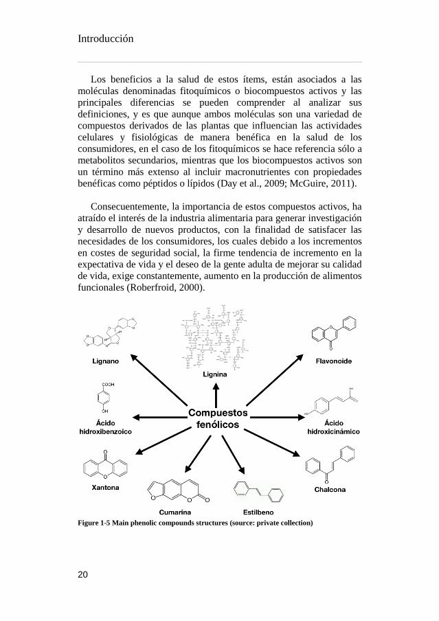
I.4.4. Compuestos fenólicos .................................................... 21
I.4.5. Colorantes naturales ...................................................... 22
I.5. Contribución de la tesis doctoral .................................... 26
I.6. Estructura de la tesis doctoral ....................................... 28
I.6. Bibliografia ................................................................. 30
Capítulo 2 Objetivos y plan de trabajo ............................. 53
II.1 Objetivo General .......................................................... 55
II.2 Objetivos Especificos .................................................... 56
II.3 Plan de Trabajo. ........................................................... 57
Capítulo 3 Metodología experimental ............................. 64
III.1 Materias primas .......................................................... 66
III.1.1 Aguacate (Persea americana Mill) .................................. 66
III.1.2 Higos chumbos (Opuntia Spp). ...................................... 66
III.2 Preparación de la muestra ............................................ 67
III.2.1 Aguacates ..................................................................... 67
III.2.2 Higos chumbos.............................................................. 68
III.2.3 Tratamientos térmicos. ................................................. 69
III.3 Parametros morfologicos ............................................. 70
III.3.1 Dimensiones y peso ....................................................... 70
III.3.2 Color .............................................................................. 70
III.4 Caracterización nutricional .......................................... 71
III.4.1 Proteina..........................................................................71
Índice
XIII
III.4.2 Grasa ..............................................................................71
III.4.3 Humedad .......................................................................71
III.4.4 Cenizas ...........................................................................71
III.4.5 Carbohidratos ............................................................... 72
III.4.6 Energia .......................................................................... 72
III.5 Compuestos hidrofílicos ............................................... 73
III.5.1 Azúcares solubles .......................................................... 73
III.5.2 Ácidos orgánicos ........................................................... 73
III.6 Compuestos lipofílicos ................................................. 75
III.6.1 Ácidos grasos ................................................................ 75
III.6.2 Tocoferoles ................................................................... 76
III.7 Preparacion de extractos ............................................. 77
III.7.1 Extracción por agitación ................................................. 77
III.7.2 Extracción por Ultrasonidos ............................................ 77
III.7.3 Extracción por microondas ............................................ 78
III.7.4 Procesos post-extracción............................................... 79
III.8 Determinaciónes preliminares de compuestos funcionales ........................................................................................ 80
III.8.1 Polifenoles por el metodo de Folin-Ciocalteu ................ 80
III.8.2 Flavonoides por el metodo de tricloro aluminio ............. 81
III.8.3 Betalainas por metodo espectrofotometrico ................. 82
III.9 Evaluación de propiedades bioactivas ........................... 84
Índice
XIV
III.9.1 Actividad antibacteriana ............................................... 84
III.9.2 Actividad antifungica .................................................... 85
III.9.3 Ensayos capacidad antitumoral ..................................... 86
III.9.4 Ensayos de evaluación hepatotoxica ............................. 87
III.9.5 Actividad antioxidante por el método de DPPH ............ 88
III.9.6 Actividad antioxidante por el metodo del poder reductor ............................................................................................... 89
III.9.7 Actividad antioxidante por el método de TBARS ........... 90
III.9.8 Actividad antioxidante por el método de β-caroteno .... 91
III.9.9 Actividad antioxidante por el método de ABTS ............. 91
III.10 Identificación y cuantificación de biocompuestos ......... 93
III.10.1 Polifenoles y Flavonoides ............................................. 94
III.10.2 Betalainas .................................................................... 95
III.11 Determinación y modelización de isotermas de sorción 96
III.12 References ................................................................ 98
Capítulo 4 Caracterización de los frutos y subproductos de Persea americana ......................................................... 122
IV.1 Antecedentes ........................................................... 124
IV.1.1 Características de los frutos de Persea americana ........ 124
IV.1.2 Efecto del secado sobre los compuestos fenólicos ...... 126
IV.2 Caracterización de los compuestos bioactivos de los subproductos de Persea americana mill: Una rica fuente de antioxidantes inherentes. ................................................ 129
1 Abstract .............................................................................. 131

Índice
XV
IV.2.1 Introduction ........................................................... 132
IV.2.2 Material and Methods ............................................. 134
IV.2.2.1 Samples preparation ................................................ 134
IV.2.2.2 Extraction procedure ............................................... 134
IV.2.2.3. Phenolic compounds ............................................... 134
IV.2.2.4. Bioactive properties evaluation ............................... 135
IV.2.2.4.1 Antioxidant activity assays .......................................... 135
IV.2.2.4.2. Antimicrobial activity assays ...................................... 135
IV.2.2.5. Statistical analysis .................................................... 137
IV.2.3. Results and discussion ........................................... 138
IV.2.3.1. Phenolic profile of P. americana by-products ........... 138
IV.2.3.2. Antioxidant capacity of P. americana by-products .. 140
IV.2.3.3. Antimicrobial activity of P. americana by-products . 145
IV.2.4. Conclusion ............................................................ 148
Acknowledgements .............................................................. 148
References ............................................................................ 149
Capítulo 5 Caracterización de los frutos de las especies Opuntia spp ................................................................. 175
V.1 Antecedentes ............................................................. 177
V.1.1 Metodología rápida de detección de betalaínas ............ 177
V.2 Caracterización extensiva de tres variedades de frutos de Opuntia spp. para la innovación de ingredientes alimentarios. ...................................................................................... 180

Índice
XVI
Abstract ................................................................................ 183
V.2.1. Introduction .......................................................... 184
V.2.2. Material and Methods............................................. 186
V.2.2.1. Sample preparation ................................................. 186
V.2.2.2. Morphological parameters ....................................... 186
V.2.2.3. Chemical characterisation ........................................ 186
V.2.2.3.1 Proximal nutritional composition ........................... 186
V.2.2.3.2. Hydrophilic compounds ......................................... 187
V.2.2.3.3. Lipophilic compounds ........................................... 188
V.2.2.4. Antimicrobial effect of fruit pulp .............................. 189
V.2.2.5. Statistical analysis.................................................... 190
V.2.3. Results and discussion ............................................ 191
V.2.3.1. Morphological parameters and nutritional composition ............................................................................................. 191
V.2.3.2. Hydrophilic and lipophilic compounds ..................... 194
V.2.3.3. Antimicrobial properties .......................................... 201
V.2.4. Conclusions ........................................................... 205
Acknowledgements .............................................................. 205
References ............................................................................ 206
Capítulo 6 Caracterización de los subproductos de Opuntia spp. ............................................................................. 232
VI.1 Antecedentes ........................................................... 234
VI.1.1 Efecto del secado en los compuestos fenólicos de las pieles de Opuntia .................................................................. 235

Índice
XVII
VI.1.2 Modelización de isotermas de sorción de las pieles de Opuntia ..................................................................................237
VI.2 Valorización de los subproductos de Opuntia spp.: Determinación de los perfiles fenolicos, betalainicos y sus propiedades bioactivas. ................................................... 239
1 Abstract ............................................................................. 241
VI.2.1. Introduction ......................................................... 242
VI.2.2. Material and Methods ............................................ 244
VI.2.2.1. Samples preparation ............................................... 244
VI.2.2.2. Extraction procedure .............................................. 244
VI.2.2.3. Phenolic compounds ............................................... 245
VI.2.2.4. Betalain compounds ............................................... 245
VI.2.2.5. Bioactive properties evaluation ............................... 246
VI.2.2.5.1. Antioxidant activity assays ........................................ 246
VI.2.2.5.2. Antimicrobial activity assays ...................................... 247
VI.2.2.5.3. Cytotoxicity assays..................................................... 247
VI.2.2.6. Statistical analysis .................................................. 248
VI.2.3. Results and discussion ........................................... 249
VI.2.3.1. Phenolic and betalain composition .......................... 249
VI.2.3.2. Antioxidant activity ................................................. 251
VI.2.3.3. Antibacterial and antifungal activity ........................ 256
VI.2.3.4. Cytotoxic activity .................................................... 259
VI.2.4. Conclusion ............................................................ 260
Índice
XVIII
Aknowledgement ................................................................. 260
References ............................................................................ 261
Capítulo 7 Optimización de los extractos de las pieles de Opuntia spp. ................................................................ 287
Abstract ................................................................................ 292
VII.1.1. Introduction ........................................................ 293
VII.1.2. Material and Methods ........................................... 295
VII.1.2.1. Samples preparation .............................................. 295
VII.1.2.2. Experimental design .............................................. 295
VII.1.2.3. Extraction procedure .............................................. 296
VII.1.2.3.1. Ultrasound-assisted extraction (UAE) ....................... 296
VII.1.2.3.2. Microwave-assisted extraction (MAE) ....................... 297
VII.1.2.4. Colorimetric determination and extraction yield .... 297
VII.1.2.5. Antioxidant activity evaluation ............................... 298
VII.1.2.6. LC-DAD/MS-MS characterization of extracts ......... 298
VII.1.2.6.1. Phenolic profiling ..................................................... 298
VII.1.2.6.2. Betalainic profiling ................................................... 299
VII.1.2.7. Statistical analysis .................................................. 299
VII.1.3. Results and discussion ...........................................301
VII.1.3.1. Tentative identification of bioactive compounds. ... 301
VII.1.3..2. Model fitting and technologies used. .................... 305
VII.1.3.3. Effects of independent variables on betacyanins. ... 308
VII.1.3.3.1 Ultrasound-assisted extraction. .................................. 308

Índice
XIX
VII.1.3.3.2. Microwave-assisted extraction .................................. 309
VII.1.3.4. Effects of independent variables on phenolic acids and flavonoids. ............................................................................ 312
VII.1.3.4.1. Ultrasound-assisted extraction .................................. 312
VII.1.3.4.2. Microwave-assisted extraction .................................. 313
VII.1.3.5. Effects of independent variables on antioxidant activity. ................................................................................. 314
VII.1.3.5.1. Ultrasound-assisted extraction and microwave-assisted extraction ................................................................................... 314
VII.1.3.6. Effects of independent variables on yield and colour. ............................................................................................. 314
VII.1.3.6.1. Extraction yield on UAE and MAE .............................. 314
VII.1.3.7. Colour effect on ultrasonic and microwave extractions .............................................................................................. 315
VII.1.3.8. Comparison of UAE and MAE RSM.......................... 315
VII.1.4. Conclusion ........................................................... 318
Capítulo 8 Conclusiones ................................................ 338
Capítulo 9 Anexos ........................................................ 347
Anexo 1 ................................................................................. 349
Anexo 2 ................................................................................ 352
Anexo 3 ................................................................................. 354

Índice de tablas
XX
Índice de tablas
Table 1-1 Publication list presented within this work ___________________ 27
Table 3-1 GC temperature ramp for fatty acids _______________________ 75
Table 3-2 Common correlation models of sorption Isotherms ____________ 97
Table 4-1 Nutritional and morphological characteristics of avocado fractions ___________________________________________________________ 126
Table 4-2 Tentative identification and quantification of phenolic compounds in the P. americana by-products ___________________________________ 141
Table 4-3 Antioxidant activity of P. americana by-products ____________ 144
Table 4-4 Antibacterial and antifungal activity of P. americana by-products ___________________________________________________________ 147
Table 5-1 Morphological parameters of Opuntia spp. _________________ 191
Table 5-2 Nutritional value and hydrophilic compounds of the studied Opuntia spp. ________________________________________________________ 192
Table 5-3 Chemical composition in lipophilic compounds, tocopherols and fatty acids of Opuntia spp. ______________________________________ 194
Table 5-4 Identification and quantification of betalains detected in Opuntia spp pulp ____________________________________________________ 199
Table 5-5 Antibacterial and antifungal activity of Opuntia samples ______ 202
Table 6-1 Identification and quantification of phenolic compounds in Opuntia peels _______________________________________________________ 252
Table 6-2 Tentative identification and quantification of betalains detected in Opuntia peels ________________________________________________ 254
Table 6-3 Antioxidant capacity of Opuntia peels ____________________ 256
Table 6-4 Antibacterial and antifungal activity of Opuntia peels ________ 258
Table 6-5 Cytotoxic activity of Opuntia peel ________________________ 259
Table 7-1 Tentative identification bioactive compounds in the Opuntia engelmannii cv Valencia peels. ___________________________________ 302

Índice
XXI
Table 7-2 Central Composite design and experimental data for 4-level-4-factor response surface analysis _______________________________________ 306
Table 7-3 Statistical analysis (ANOVA) of the central composite design ___ 310
Índice de figuras
XXII
Íindice de figuras
Figure 1-1 The different parts of the avocado fruit, left avocado var. Fuerte and right var Hass ______________________________________________ 6
Figure 1-2 Production quantity of avocados by country (years 2015-2016) ___ 8
Figure 1-3 Opuntia spp worldwide distribution of crops _________________ 10
Figure 1-4 The different parts of Opuntia plant and fruit _______________ 13
Figure 1-5 Main phenolic compounds structures ______________________ 20
Figure 1-6 Main betalains structures, left OFI var Sangigna, right OFI var Gialla _______________________________________________________ 24
Figure 1-7 Doctoral thesis estructural scheme ________________________ 29
Figure -2-1 Approach of experiments in chronological order _____________ 58
Figure -2-2 Article 1 experimental work _____________________________ 59
Figure -2-3 Article 2 experimental work _____________________________ 60
Figure -2-4 Article 3 experimental work _____________________________ 61
Figure -2-5 Article 4 experimental work _____________________________ 62
Figure 3-1 Persea americana fruit fractions __________________________ 67
Figure 3-2 Opuntia spp fruits fractions) _____________________________ 68
Figure 3-3 Conventional extraction ________________________________ 77
Figure 3-4 Ultrasonic extraction system _____________________________ 78
Figure 3-5 Lyophilized powders ___________________________________ 79
Figure 3-6 Re-suspended samples) _________________________________ 80
Figure 3-7 Folin-Ciocalteu samples _________________________________ 81
Figure 3-8 ALCL3 colorimetric assay ________________________________ 82
Figure 3-9 SPE extraction of betalains ______________________________ 83

Índice
XXIII
Figure 3-10 SRB bond cell treated with Opuntia samples _______________ 87
Figure 3-11 DPPH colourimetric assay ______________________________ 88
Figure 3-12 Power reduction method chemical reactions _______________ 89
Figure 3-13 Power reduction plate reaction __________________________ 90
Figure 3-14 UPLC-DAD-Esi/MS system employed _____________________ 93
Figure 3-15 Elution gradient of solvent B in phenolics determination ______ 94
Figure 3-16 Elution gradient of solvent B in betalainic determination ______ 95
Figure 4-4-1 Avocado fruit fraction percentages _____________________ 125
Figure 4-4-2 Effect of drying and storage on phenolic compounds of avocado peels) ______________________________________________________ 127
Figure 4-4-3 Effect of drying and storage on phenolic compounds of avocado kernels _____________________________________________________ 128
Figure 4-4-4 Phenolic profile of P. americana (A) kernel recorded at 280 nm, (B) and (C) peels recorded at 280 nm and 370 nm, respectively __________ 138
Figure 5-1 Screening of betalains in OFI var Gialla ___________________ 178
Figure 5-2 Screening of betalains in OFI var Sanguigna _______________ 178
Figure 5-3 Effect of pH on betalain extraction _______________________ 179
Figure 5-4 HPLC chromatograms of (A) OG betaxanthin profile recorded at 484 nm and (B) OE betacyanin profile recorded at 535 nm _____________ 198
Figure 6-1 Fraction percentage of Opuntia fruits _____________________ 234
Figure 6-2 Effect of drying and storage in OFI var Sanguigna __________ 236
Figure 6-3 Effect of drying temperatures on flavonoids ________________ 236
Figure 6-4 Sorption isotherms of OFI Sanguigna at 20 °C ______________ 237

Índice de figuras
XXIV
Figure 6-5 HPLC chromatogram of OG phenolic profile recorded at 370 nm (A), betaxanthins profile of OS recorded at 484 nm (B) and betacyanins profile of OE recorded at 535 nm (C). ______________________________________ 250
Figure 7-1 HPLC chromatogram of Opuntia engelmannii cv. Valencia ____ 304
Figure 7-2 Principal effect graphs of UAE and MAE ___________________ 316
Figure 8-1 Desorption isotherms of OFI Sanguigna at 20 °C)_____________393
Figure 8-1 Adsorption isotherms of OFI Sanguigna at 20 °C _____________394
Figure 8-1 Adsorption isotherms regressions of OFI Sanguigna at 20 °C____395
Figure 8-1 Desorption isotherms regressions of OFI Sanguigna at 20 °C____ 396
Índice de ecuaciones
XXV
Índice de ecuaciones
Equation 3-1 Moisture determination .......................................................... 71
Equation 3-2 Total carbohydrates by difference ........................................... 72
Equation 3-3 Total energy ........................................................................... 72
Equation 3-4 Spectrophotometric betalainic content ...................................82
Equation 3-5 Inhibition percentage of DPPH ................................................88
Equation 3-6 Inhibition percentage of b-carotene ........................................ 91
Equation 3-7 Inhibition percentage of ABTS .................................................92
Equation 3-8 Moisture equilibrium of the sorption isotherms ....................... 96
Equation 7-1 Second-order polynomial regression coefficients .................... 296
Equation 7-2 Second-order polynomial coefficients for betalainic content in the UAE extractions. ...................................................................................... 308
Equation 7-3 Second-order polynomial coefficients for betalainic content in the MAE extractions. ..................................................................................... 312
Equation 7-3 Second-order polynomial coefficients for phenolic acids and flavonoids content in the UAE extractions ................................................. 313


Capítulo 1 INTRODUCCIÓN


Introducción
1
I.1 JUSTIFICACIÓN
Los estudios en materia de “Ingredientes alimentarios naturales” y
“Sostenibilidad” han crecido concomitantemente, ambos inclusive han
co-evolucionado para formar un interés particular, relacionándoles de
manera positiva y fructífera. Esta simbiosis, se debe principalmente al
acelerado aumento de la conciencia social, que ha permitido a la misma
sociedad percibir la explotación de fuentes naturales, así como el uso
desmedido de aditivos alimentarios sintéticos con un gran impacto
negativo en la salud del consumidor. Esto ha conseguido que el enfoque
central de interés, se base en la vida saludable y la armonía con el medio
ambiente, como lo muestra el aumento de tendencias y oportunidades
de mercado alimentario en el estudio realizado por Augustin and
Sanguansri (2015).
El crecimiento poblacional, la reducción de zonas de cultivo, así
como la utilización de ciertos alimentos con finalidades distintas a la
alimentación, como la producción de bioenergéticos, han hecho
replantearnos las metodologías agrícolas, el procesado de los
alimentos, y nos ha abierto camino en la exploración de los
subproductos alimentarios. En consecuencia, la subutilización de los
flujos de materia orgánica de los distintos subproductos, conlleva a
serias amenazas medioambientales y a la economía del sector
agricultura y de la industria alimentaria (Chan et al., 2018), y es por
esta razón, que proponemos el estudio de algunos subproductos
frutícolas.
Las frutas son componentes importantes dentro de la dieta humana
y son consumidas frescas y/o procesadas. Las frutas que crecen en
estado salvaje, fueron consumidas por la raza humana desde etapas muy
tempranas de la historia, y con la adopción del sedentarismo, podemos
disfrutar de la selección de distintos genotipos y variedades de frutas.
Además de sus cualidades organolépticas, las frutas, aportan nutrición
básica, ya que estas contienen carbohidratos, ácidos orgánicos,
vitaminas y minerales.
Además de la importancia estructural y organoléptica de las
moléculas mencionadas anteriormente, dentro del sistema celular de las
plantas, se sintetizan un sinnúmero de arreglos químicos moleculares,
de donde podemos destacar la generación de metabolitos secundarios,

Introducción
2
los cuales cumplen distintos propósitos fisiológicos, como la atracción
a través del color y de los compuestos aromáticos, y la protección en
contra de patógenos y condiciones medio ambientales (Jimenez-Garcia
et al., 2018).
De acuerdo a los tecnólogos alimentarios, la comida se come
primero a través de los ojos (Attokaran, 2011). La atracción visual, es
responsabilidad de un grupo de moléculas características del tipo y
variedad de los frutos. Las antocianinas se encuentran en un gran
número de frutas y verduras (moras, cerezas, fresas, uvas, manzanas,
col lombarda, maíz y zanahoria morados, etc) aportando colores rojizos
y purpuras. Tonalidades amarillas pueden ser aportadas por
carotenoides, riboflavina y curcumina, por nombrar algunos. El
licopeno aporta el color rojo al tomate, las clorofilas el verde de una
amplia gama de alimentos, algas y bacterias (Wrolstad and Culver,
2012). Una de las familias de colorantes que abordaremos en este
documento son las betalainas, que cubren un rango de colores entre el
purpura y el amarillo, y que además de aporte del color, contribuyen
también con propiedades antioxidantes (Kanner et al., 2001).
Los metabolitos secundarios, además de aporte de color, cumplen
distintas funciones más complejas como moléculas de señalización,
aromas y protección celular. Alcaloides, terpenos, esteres volátiles,
aminoácidos no proteicos y muchos más compuestos orgánicos con
diversas estructuras, engloban una pequeña parte de éstos compuestos
(Bhatia et al., 2015). Últimamente, los compuestos fenólicos y
flavonoicos han recibido un creciente interés en sus estructuras, pero
sobre todo en sus funciones antioxidantes, debido al rol que juegan en
prevención de enfermedades (Maria et al., 2014), aunque la gran
mayoría de estos trabajos se enfoca en la parte comestible de los frutos,
dejando un importante espacio de estudio para los subproductos y sus
posibles propiedades funcionales.
En esta trabajo, se trata principalmente del estudio de algunos
subproductos frutícolas (aguacate e higo chumbo), con la finalidad de
identificar posibles ingredientes naturales que sean factibles de
implementar en las industrias alimentarias, farmacéuticas y/o
cosméticas, con énfasis en los colorantes naturales y algunos de los
compuestos antioxidantes. Asímismo, las técnicas desarrolladas a lo
Introducción
3
largo del trabajo, han sido pensadas para poder ser extrapoladas a
distintos tipos de subproductos de la industria, que pudieran llegar a
producir particular interés en los sectores mencionados anteriormente.

Introducción
4
I.2 AGUACATE (PERSEA AMERICANA MILL)
I.2.1 Origen, estructuras e hibridaciones
El aguacate, es una planta dicotiledónea miembro de la familia
Lauraceae del orden de los Laurales, una larga familia pantropical de
alrededor de 50 géneros y unas 2500 a 3000 especies en su mayoría
árboles y arbustos (Rohwer, 1993), y fue clasificada como Persea
gratissima por Gaertner, y como Persea americana Mill. por Miller.
Este cultivo es originario de Centro América y fue domesticada por los
pueblos indígenas de esa zona, hace unos 7000 años, según evidencia
arqueológica encontrada en Tehuacán, Puebla (México), se cree que
apareció hace alrededor de unos 12000 años (Yahia and Woolf, 2011).
A pesar de que las hojas del árbol del aguacate tienen muy corta
longevidad (<12 meses), se caracteriza por su rápido crecimiento,
llegando a crecer hasta 20 metros, con raíces superficiales pero con
profundidad de hasta 15 m, aunque cuenta con un pobre sistema de
conducción de agua, este árbol es capaz de producir muchas flores,
aunque solo un bajo porcentaje de ellas producen frutos (< 0,1%). Esto
puede deberse principalmente a tres factores climáticos importantes:
(1) la presencia de escarcha durante el invierno; (2) temperaturas
medias muy bajas ; y (3) temperaturas muy altas durante la formación
de fruto (Carr, 2013).
El nombre botánico del aguacate es Persea america Mill., de las
cuales reconocemos 3 razas (conocidas hoy como variedades botánicas
o subespecies); Persea americana var. drymifolia (ecotipo Mexicano),
Persea americana var. guatemalensis (ecotipo Guatemalteco) y Persea
americana var. americana (ecotipo Antillano). Las principales
diferencias entre estas razas son dos características fisiológicas y
organolépticas: por un lado, (1) la tolerancia al frio, siendo el ecotipo
mexicano el que presenta mayor resistencia, mientras que el antillano
es el de menor resistencia de los tres (Carr, 2013), por otro lado, (2) el
contenido de ácidos grasos en las pulpas, en donde la raza antillana
presenta muy bajo contenido (< 8%) comparado con la raza mexicana
(hasta 30%), haciendo de la raza guatemalteca las deseada en cuanto a
términos de ácido grasos, por su característico balance de ellos y su
sabor con notas de nuez (Cowan and Wolstenholme, 2003).

Introducción
5
El cruce interracial se ha generado como resultado de la hibridación
entre razas en los cultivares de mayor importancia económica, tal es el
caso de la variedad Fuerte (raza mexicana y guatemalteca), la cual se
originó en México (Atlixco, Puebla) y por muchos años fue la de mayor
producción comercial hasta que la variedad Hass (creada y
seleccionada en California) la substituyó, en la mayoría de sembradíos
con clima mediterráneo (Knight and Campbell, 1999).
I.2.2 Fisiología, formación del fruto y maduración
Los aguacates son una fruta drupa climatérica subtropical,
generalmente de forma oblonga y de piel verde, aunque la variedad
comercial predominante Hass, tiene piel púrpura-negra en su estado
óptimo de madurez. El nombre proviene de náhuatl “ahuacatl”, que
significa testículos, esto se refiere al mismo por su forma característica,
y era considerada por los aztecas como un afrodisíaco (Hurtado-
Fernández et al., 2018a).
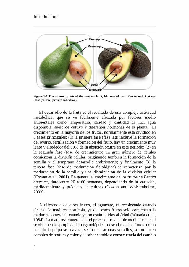
Botánicamente Persea americana es una baya que consiste en una
semilla larga central y del pericarpio, que es la suma de la parte
comestible (mesocarpio), de la piel (exocarpio) y de la capa interior que
rodea a la semilla (endocarpio). La piel varía en texturas y color, la
textura puede ir de flexible a lignínica, suave a rígida y el color puede
ser verde-amarillento, purpura-rojizo, purpura o negro. La pulpa suele
ser amarillo-verdosa o amarillo brillante, y suele tener textura similar a
la de la mantequilla, pero variedades de menor calidad presenta mayor
fibrosidad. La semilla del fruto es grande y normalmente supone del 10
al 25% de peso total del fruto.
Introducción
6
Figure 1-1 The different parts of the avocado fruit, left avocado var. Fuerte and right var
Hass (source: private collection)
El desarrollo de la fruta es el resultado de una compleja actividad
metabólica, que se ve fácilmente afectada por factores medio
ambientales como temperatura, calidad y cantidad de luz, agua
disponible, suelo de cultivo y diferentes hormonas de la planta. El
crecimiento en la mayoría de los frutos, normalmente está dividido en
3 fases principales: (1) la primera fase (fase lag) incluye la formación
del ovario, fertilización y formación del fruto, hay un crecimiento muy
lento y alrededor del 90% de la absición ocurre en este periodo; (2) en
la segunda fase (fase de crecimiento) un gran número de células
comienzan la división celular, originando también la formación de la
semilla y el temprano desarrollo embrionario; y finalmente (3) la
tercera fase (fase de maduración fisiológica) se caracteriza por la
maduración de la semilla y una disminución de la división celular
(Cowan et al., 2001). En general el crecimiento de los frutos de Persea
america, dura entre 20 y 60 semanas, dependiendo de la variedad,
medioambiente y prácticas de cultivo (Cowan and Wolstenholme,
2003).
A diferencia de otros frutos, el aguacate, es recolectado cuando
alcanza la madurez hortícola, ya que estos frutos solo comienzan la
madurez comercial, cuando ya no están unidos al árbol (Watada et al.,
1984). La madurez comercial es el proceso irreversible mediante el cual
se obtienen las propiedades organolépticas deseadas de los frutos, como
cuando la pulpa se suaviza, se forman aromas volátiles, se producen
cambios de textura y color y el sabor cambia a consecuencia del cambio

Introducción
7
en la composición de azúcares y ácidos orgánicos (Hiwasa-Tanase and
Ezure, 2014).
I.2.3 Temporada de cultivo
El árbol del aguacate es muy longevo y con un periodo productivo muy
grande, en árboles injertados, la primera cosecha se realiza al quinto
año de vida del árbol, antes de llegar a esta edad, el árbol es considerado
como joven, y a lo largo de ese año se obtienen en promedio unos 50
frutos, los siguientes 3 años, la producción sube a 150, 300 y 800 frutos
en promedio en los años 6, 7 y 8, respectivamente, y a partir de esta
etapa se le considera un árbol adulto y totalmente productivo. En
general los árboles son plantados con una distancia entre ellos que va
de 7 a 12 metros, y de esta manera se obtiene una hectárea destinada a
la plantación de aguacate de 115 a 180 arboles (SAGARPA, 2011).
Este fruto es muy productivo ya que provee aguacates durante todo
el año, aunque se pueden separar dos periodos clasificados como de
producción máxima y mínima. La máxima que dura 4 meses entre
octubre y enero, y la mínima que comprende el resto de los 8 meses
entre febrero y septiembre. Cabe destacar que las condiciones medio
ambientales siempre tienen un rol muy importante en estos tiempo, pero
generalmente el tiempo de madurez hortícola (entre el florecimiento y
la cosecha) es de 10 a 18 meses para raza guatemalteca, 6 a 8 meses
para la raza antillana y de 5 a 7 meses para la raza mexicana (Crane et
al., 2016).
I.2.4 Producción anual
Aunque el origen del cultivo esta en América, hoy en día, el aguacate
es un cultivo de nivel internacional, y aunque es evidente que la mayor
explotación de este cultivo esta aún en el continente americano (73,2%
de la producción mundial), los demás continentes suman sus
porcentajes para la producción mundial, África (12,3%), Asia (11,3%),
Europa (1,7%) y Oceanía (1,6%). Mientras que los países con mayor
producción por continente son: México, Kenia, Indonesia, España y
Australia, y a nivel mundial, los líderes en producción son México (1,7
millones de toneladas), seguido por República Dominicana (0,5
Introducción
8
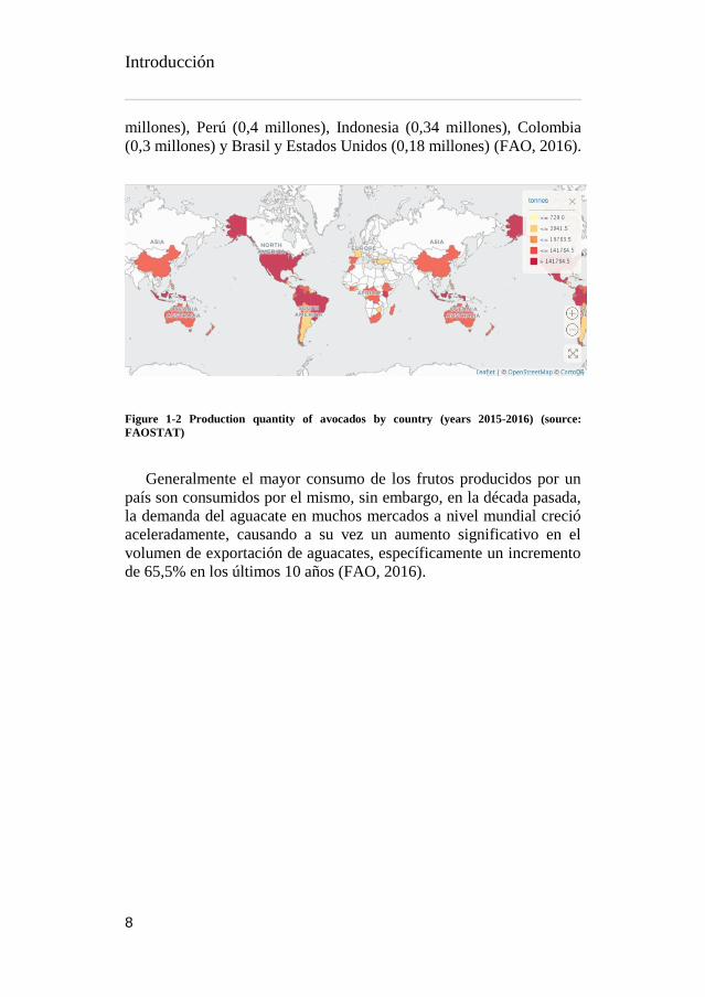
millones), Perú (0,4 millones), Indonesia (0,34 millones), Colombia
(0,3 millones) y Brasil y Estados Unidos (0,18 millones) (FAO, 2016).
Figure 1-2 Production quantity of avocados by country (years 2015-2016) (source:
FAOSTAT)
Generalmente el mayor consumo de los frutos producidos por un
país son consumidos por el mismo, sin embargo, en la década pasada,
la demanda del aguacate en muchos mercados a nivel mundial creció
aceleradamente, causando a su vez un aumento significativo en el
volumen de exportación de aguacates, específicamente un incremento
de 65,5% en los últimos 10 años (FAO, 2016).
Introducción
9
I.3 HIGOS CHUMBOS (OPUNTIA SPP.)
I.3.1. Orígenes
El interés por los cactus y sus frutos se remonta a miles de años. Su
historia y posible origen se remonta a las antiguas civilizaciones
mesoamericanas, concretamente, con el imperio Azteca, en el país que
hoy es conocido como México. Existen evidencias arqueológicas en las
zonas semiáridas de Mesoamérica, que permiten afirmar que las
poblaciones indígenas asentadas en esas zonas iniciaron el cultivo de
modo formal (Pimienta, 1990).
Actualmente, los cactus de Opuntia spp. y sus frutos forman parte
del panorama natural y de los sistemas de agricultura de un gran número
de regiones en el mundo. Esta expansión territorial probablemente
comenzó con los muestrarios de plantas llevadas a España por parte de
Cristóbal Colón (Donkin, 1977) y debido al gran impacto comercial de
sus puertos con el resto de Europa, África, Asia y Oceanía, esta
distribución fue posible.
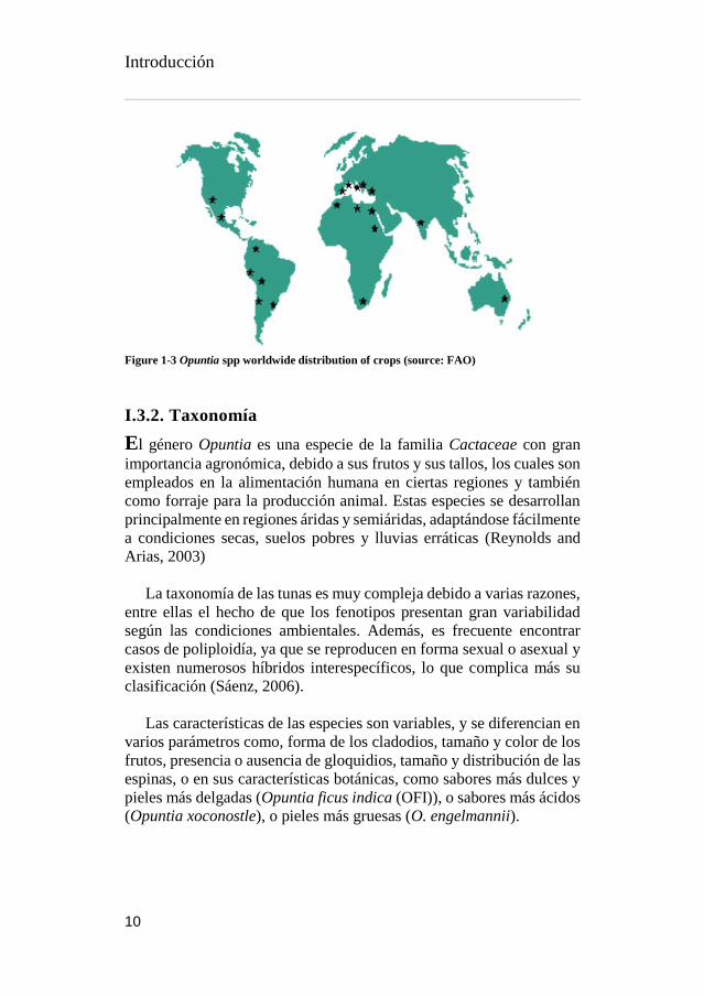
Actualmente estos cultivos se pueden encontrar tanto en forma de
explotación comercial o de manera salvaje en países de América como
México, Estados Unidos de Norte América, Chile, Perú, Bolivia y
Argentina, en África en Argelia, Etiopia, Marruecos, Sudáfrica y
Túnez, en países Asiáticos como Jordania, Siria, Líbano, Israel y
Yemen, se pueden también encontrar en Australia y en Europa en
España, Italia y Portugal. Actualmente el país de mayor producción
mundial comercial es México, seguido de Italia y Estados Unidos
(Ochoa and Barbera, 2017).
Introducción
10
Figure 1-3 Opuntia spp worldwide distribution of crops (source: FAO)
I.3.2. Taxonomía
El género Opuntia es una especie de la familia Cactaceae con gran
importancia agronómica, debido a sus frutos y sus tallos, los cuales son
empleados en la alimentación humana en ciertas regiones y también
como forraje para la producción animal. Estas especies se desarrollan
principalmente en regiones áridas y semiáridas, adaptándose fácilmente
a condiciones secas, suelos pobres y lluvias erráticas (Reynolds and
Arias, 2003)
La taxonomía de las tunas es muy compleja debido a varias razones,
entre ellas el hecho de que los fenotipos presentan gran variabilidad
según las condiciones ambientales. Además, es frecuente encontrar
casos de poliploidía, ya que se reproducen en forma sexual o asexual y
existen numerosos híbridos interespecíficos, lo que complica más su
clasificación (Sáenz, 2006).
Las características de las especies son variables, y se diferencian en
varios parámetros como, forma de los cladodios, tamaño y color de los
frutos, presencia o ausencia de gloquidios, tamaño y distribución de las
espinas, o en sus características botánicas, como sabores más dulces y
pieles más delgadas (Opuntia ficus indica (OFI)), o sabores más ácidos
(Opuntia xoconostle), o pieles más gruesas (O. engelmannii).

Introducción
11
La nomenclatura científica y taxonomía del fruto es la siguiente:
Reino: Plantae
División: Magnoliophyta
Clase: Magnoliopsida
Orden: Caryophyllales
Familia: Cactaceae
Tribu: Opuntiae
Género: Opuntia
Especie: Opuntia ficus-indica Mill.
Las cactus y sus frutos reciben distintos nombres según el país en
que se encuentren. El nombre autóctono de los frutos es “nochtli”, del
náhuatl que significa fruta pera. En México la planta recibe el nombre
de nopal mientras que sus frutos se les conoce como “tunas”, en España
se renombró al nopal con el nombre de chumbera y a la fruta como higo
de Indias (hoy “higo chumbo”), en Italia se le conoce como “fico
d’India”, en Francia se le llama “figue de Barbarie”; en EE.UU. y
Sudáfrica “prickly pear”, (nombre que está evolucionando a cactus
pear, a fin de eliminar el término algo peyorativo de “prickly” que
significa “espinoso”); en Israel se conoce como “sabras”, que significa
espinoso por fuera, pero dulce por dentro. En Brasil, la llaman “palma
forrageira”, ya que se cultiva principalmente para la producción de
forraje (Sáenz, 2006).
I.3.3. Variedades cultivadas
Actualmente casi 300 especies del género Opuntia son reconocidas,
tan solo en México Bravo-Hollis (1978) reporto 104 distintas
variedades y especies. La variedad comercial de los frutos proviene de
Opuntia ficus-indica (OFI) y sus distintas variedades, aunque existen
muchas especies salvajes de igual interés, como O. robusta, O.
streptacantha, O. megacantha, O. xoconostle y O. engelmannii.
Las variedades de O. ficus-indica se diferencian de todos los otros
miembros del género por presentar distintas combinación de las
siguientes características: los cladodios usualmente son elípticos, de
gran tamaño y carecen total o casi totalmente de espinas, presentan
frutos grandes, dulces y carnosos. Las variedades se diferencian
principalmente en cuatro grupos por el color de la cáscara y la pulpa
Introducción
12
del fruto: cáscara verde amarilla y pulpa blanca, cáscara amarilla
anaranjada y pulpa naranja, cáscara verde-roja y pulpa roja; y cáscara
y pulpa púrpura.
Los nombres comunes de las variedades más populares varían según
el país. Así, en México se conocen las variedades Reina, Rojo Pelona,
Esmeralda; mientras que en Italia las mismas variedades reciben los
nombre de Giallia, Rosso y Bianca y en España las llaman Verdales,
Morados, Sanguinos y Blancos. En el mercado internacional las
variedades más apetecidas son las de color (distinto al típico color
verde), sobre todo las rojas, amarillas, rosadas y púrpuras,
atribuyéndose al color atractivo de la fruta y a su bajo contenido de
azúcares. Dos variedades muy demandadas son Rosso y Giallia
(Álvarez, 2007).
I.3.4. Descripción morfológica
Los cactus del género Opuntia están conformados por numerosos tallos
denominados cladodios (conocidos vulgarmente como paletas o
pencas); por frutos, los cuales dependiendo de la latitud, condiciones
de cultivo y especies, brotan una o dos veces por año; y por sus flores,
que dan inicio a la maduración de los frutos. Los cactus tienen una alta
resistencia a las condiciones climatológicas adversas en gran parte
debido a sus raíces, las cuales se extienden horizontalmente por varios
metros, alcanzando profundidades de alrededor de 80 cm (Sudzuki et
al., 1993).
La chumbera o nopal, es un vegetal arborescente de entre 1 y 5
metros, formado por múltiples cladodios unidos unos a otros, los
cuales, en su parte más baja forman un tronco leñoso de entre 20 y 50
centímetros de diámetro, el resto de cladodios en la planta forman
estructuras elípticas, oblongas u ovoides que alcanzan longitudes de 18-
25 cm, similares a una raqueta de tenis, la coloración de estos de verde
pálido a obscuro y dependiendo de la variedad cuenta o no con espinas.
Las espinas de los cladodios se encuentran presentes en las numerosas
aréolas o yemas y pueden ser de dos tipos: unas pequeñas, agrupadas
en gran número denominadas gloquidios, y otras grandes que son hojas
modificadas. Las aréolas presentes en toda la superficie de los
cladodios tienen la capacidad de desarrollar nuevos cladodios, flores y
frutos dependiendo de las condiciones medioambientales. Los nuevos
cladodios formados, se posicionan de tal forma con la finalidad de

Introducción
13
optimizar el aprovechamiento máximo de la luminosidad, razón por la
cual, los cladodios que crecen en verano, tienen distinta orientación que
los desarrollados en invierno (Sáenz, 2006; Sudzuki et al., 1993).
Figure 1-4 The different parts of Opuntia plant and fruit (source: private collection)
Las flores que brotan de las aréolas, son hermafroditas, solitarias y
sésiles, de coloraciones amarillo, naranja, rojo, rosa, blanco, entre otros,
y suelen tener una longitud de aproximadamente 7 cm, son de antesis
diurna y pueden haber más de 10 flores por cladodio, casi siempre en
la parte apical del margen del cladodio. Estas florecen entre los 35-45
días desde su aparición en las aréolas y brotan en los cladodios después
de los seis meses, necesitando una temperatura mínima de 15 ºC para
su optimo desarrollo. Aunque en la mayor parte de latitudes de siembra
estas flores sólo aparecen una vez por año, bajo ciertas condiciones, se
presenta una segunda floración (Alvarez, 2007; Reyes-Agüero et al.,
2005).
Los frutos de Opuntia son una falsa baya con ovario ínfero, uniloculado
y carnoso, donde la pulpa corresponde al lóculo desarrollado y la piel
corresponde a la envoltura del ovario, es necesario tener en cuenta que
son frutos no climatéricos, por lo que es fundamental cosecharlos con
madurez de consumo. Como mencionamos anteriormente, las
coloraciones de los frutos tanto pieles como frutos son variables
respecto a las variedades, con amplia gama de colores que van desde el

Introducción
14
verde, pasando por amarillo y naranja, hasta llegar al rojo-violeta. La
epidermis de los frutos comparte sus características con las de los
cladodios, presentando aréolas distribuidas en toda la superficie, que
cuenta también con gloquidios y espinas, aunque la presencia o
ausencia espinas difiere también con las variedades. Otras
características variables con respecto a las variedades son: el tamaño y
la forma de los frutos (los hay ovoides, redondos, elípticos y oblongos,
con los extremos aplanados, cóncavos o convexos). En la pulpa se
encuentran también semillas abortivas de 4-4,5 mm de longitud, las
cuales son también comestibles, en general, el periodo desde floración
hasta la madurez del fruto, se extiende alrededor de los 100 días, aunque
esto suele variar dependiendo de las condiciones medioambientales
(Alvarez, 2007; Pimienta, 1990; Sáenz, 2006).
I.3.5. Producción y cultivos de Opuntia spp
Existe poca información a nivel mundial disponible con respecto a las
zonas de cultivo de frutas de Opuntia con fines comerciales, y es que
aunque este es cultivo presente en los 5 continente, no existen
estadísticas disponibles de países productores como es el caso de :
Argelia, Brasil, Colombia, Egipto, España, Grecia, Jordania,
Marruecos, Perú, Túnez y Turquía (Inglese et al., 2004).
La principal especie cultivada a nivel mundial para la
comercialización de sus frutos es Opuntia ficus-indica (OFI), aunque
en México otras especies como O. streptacantha O. lindhemeiri, O.
amyclaea, O. megacantha y O. robusta son cultivadas con el mismo
fin (Pimiento Barrios and Munoz-Urias, 1999). De hecho, este país es
el mayor productor a nivel mundial con el 90 % del total de la superficie
de cultivo de higos chumbos a nivel mundial.
Italia mantiene 92% de sus cultivos en Sicilia, con
aproximadamente 3800 hectáreas (ha) de cultivo de tunas, con un
rendimiento de frutas de entre 13-17 toneladas (Tn) por hectárea
aproximadamente. Timpanaro et al. (2015) reportaron entre los años
2008 y 2011, una producción anual de 75000 Tn de frutos por año. Por
otra parte, Flores-Valdez et al. (1995) señalan que en el año 1995 se
obtuvo una producción anual de 354000 Tn de higos chumbos en
México, siendo este el país, con mayor superficie de siembra para este
fruto y consecuentemente el mayor productor de tunas a nivel mundial,
aunque comparado con otros países, el rendimiento mencionado en este

Introducción
15
análisis es casi un 50 % inferior que el observado en Italia o en otros
países como Chile. En México se estiman unas 50000 ha destinadas al
cultivo de los frutos del cactus, lo que representa un rendimiento
aproximado de 7 toneladas de fruto por hectárea cultivada.
Entre otros países importantes en el cultivo del higo chumbo
destacan: Sudáfrica con una superficie de 1500 ha cultivadas; Chile con
unas 1000 ha de chumberas con un rendimiento promedio de 5-20
Tn/ha; Israel y Estados Unidos reportan 300 y 200 hectáreas de
chumberas cultivadas, respectivamente. Situados en este punto, es
importante destacar que los países con mayor cantidad de toneladas
exportadas anualmente son Italia, abarcando el mercado Europeo; y
México, que debido al Tratado de Libre Comercio de América del
Norte (TLCAN), tiene cubiertas la exportaciones de Estados Unidos y
Canadá (Flores-Valdez et al., 1995).
Introducción
16
I.4 APROVECHAMIENTO DE LOS SUBPRODUCTOS
FRUTÍCOLAS
I.4.1. Perspectiva general
Por mucho tiempo, el desecho de subproductos no era una causa de
general interés, sobre todo cuando los productores responsables de
estos flujos de desecho incrementaban la producción de alimentos sin
un particular interés en el aprovechamiento de los residuos o el impacto
medio ambiental que estos producían. Actualmente, la elevada
demanda por alimentos procesados en los mercados de consumo, han
disparado las alarmas para la identificación de direcciones concretas
que minimicen la demanda de energía, reducción de costos, así como
la reducción en perdida de alimentos y en generación de desechos.
Por otro lado, la creciente preocupación por los signos de
degradación de los recursos naturales (descenso en biodiversidad,
campos de cultivo y agua) ha generado un aumento en la
concienciación social con miras en satisfacer demandas globales
futuras. Es un hecho, que en los próximos años habrá un incremento no
sólo de la población mundial, sino de la zonas urbanas, lo que
conllevará a la constante generación de nuevas industrias de procesado
de alimentos (FAO et al., 2015).
Es por eso que la combinación de ambos planteamientos de aumento
en producción de desechos industriales y la preocupación por el medio
ambiente, han dado pauta a nuevos planteamientos industriales. En los
últimos años la industria alimentaria ha pasado de prestar interés sólo
en temas económicos y de mejora en la producción, para enfocarse
también en el impacto medioambiental negativo que generan los flujos
de desecho, adaptándose a los nuevos temas de interés social (Unilever,
2010).
La sostenibilidad es un nuevo eje de interés global, que brevemente
puede ser definido de acuerdo al informe de Brundtland (1987) como
la posibilidad de satisfacer necesidades y aspiraciones del presente, sin
comprometer la habilidad de las generaciones futuras de disfrutar de las
mismas. En este sentido, existen múltiples directrices que se están
adoptando dentro de la sociedad y los cuales dependen directamente del
consumidor, como el consumo de alimentación local y reducción de
desperdicio de alimentos. Aunque en esta índole, las industrias
agroalimentarias tiene mucho mayor peso, algunos ejemplos

Introducción
17
importantes son la aplicación de tecnologías de mitigación de
producción de gases invernadero, flujos de desecho y uso responsable
del agua.
Además de mejoras en las cadenas de producción para disminuir la
generación de desechos, actualmente se ha dado mayor importancia al
estudio de estos, con la finalidad de convertir el desecho en un
subproducto, recordando que el término subproducto se le otorga al
residuo de un proceso al que se le puede sacar una segunda utilidad.
Atendiendo a esta alternativa de doble utilidad, las tres dimensiones de
la sostenibilidad de procesos (medioambiente, social y económico) se
cumplen de una manera excepcional, disminuyendo los desechos,
demostrando integridad y responsabilidad con los intereses sociales y
generando aumento en rentabilidad.
I.4.2. Identificación de oportunidades
Muchos de los procesos agroalimentarios generan exorbitantes
cantidades de subproductos que hoy en día son consideradas fuentes
potenciales de moléculas nutritivas, energéticas o con otras capacidades
comerciales. Gradualmente los productores industriales son
conscientes de las nuevas ofertas comerciales que se pueden generar, y
de la mano de investigadores a nivel mundial, se buscan las mejores
posibilidades de aprovechamiento de los subproductos y de las fibras
vegetales no comestibles.
Análisis como los elaborados en esta tesis doctoral, se hacen
necesarios al intentar dilucidar aplicaciones de los subproductos: 1)
ensayos de carácter químico enfocados en los macronutrientes son parte
importante en la caracterización, y como ejemplo de ello, es la
búsqueda industrial por alternativas de obtención de carbohidratos más
económicos, 2) ensayos microbiológicos, con la finalidad de extraer
moléculas con propiedades antimicrobianas, 3) ensayos inmunológicos
donde se prueben capacidades antioxidantes, anticarcinogénicas, entre
otras, de los extractos obtenidos, 4) ensayos organolépticos con la
finalidad de potenciar posibles alternativas que mejoren las
características sensoriales de algunos productos, 5) ensayos
enzimáticos con los que se puedan detectar posibles catalizadores de
diversas reacciones industriales que mejoren los resultados de procesos
específicos, 6) ensayos físico-químicos que permitan detectar
polímeros que podrían ser empleados en industrias textiles o de

Introducción
18
bioplásticos. Y estos son sólo parte del abanico de propuestas para el
uso de los subproductos.
Algunos investigadores, han dado incluso algunas pautas como
estrategias de recuperación, como es el caso de Galanakis (2015), quien
propone un proceso de recuperación universal de 5 niveles,
brevemente, el primer nivel se centra en las características
macroscópicas (agua, solidos, aceites); el segundo nivel en las
características de las microestructuras; el tercer nivel se encarga de
determinar los grupos que pueden ser extraídos (azúcares, proteínas,
fibras, fenoles totales, entre otros) mediante técnicas rápidas de
cribado; el cuarto nivel estudia más en detalle las macro y
micromoléculas; y el último nivel caracteriza la carga microbiana y
enzimática.
Comprendiendo la magnitud de la industria alimentaria y la gran
cantidad de subproductos de origen animal y vegetal, es importante
destacar que en la industria frutícola existe un gran número de
posibilidades a estudiar, como es el caso de los subproductos generados
por la industria procesadora frutos tropicales como los aguacate o los
higos chumbos.
Aunque los frutos de la chumbera suelen ser consumidos
mayoritariamente en fresco como un producto mínimamente
procesado, existen compañías agroalimentarias (Ciao bella®, Piping
Rock®, Divine essence®, entre otros) que utilizan estos frutos como
materia prima para la elaboración de diversos productos como
mermeladas, destilados, dulces, helados, bebidas no alcohólicas entre
otros, los cuales utilizan en ocasiones las pulpas con semillas o
simplemente las pulpas para sus elaboraciones, generando una corriente
de subproductos que en este caso particular se centra en las pieles y
ocasionalmente las semillas. Dependiendo de la variedad utilizada se
obtienen entre 25 y 30 % del peso total del fruto como subproducto
(Melgar et al., 2017a).
En el caso del aguacate, este fruto cuenta con una industrialización
aún mayor, debido a que es un producto más conocido a nivel mundial
y con una comercialización en constante expansión, penetrando en el
mercado agroalimentario y cosmético. Añadiendo a las grandes
cantidades de aguacates procesados, los porcentajes de subproductos
acumulados (alrededor del 14 % y 16% del peso total en piel y semilla

Introducción
19
respectivamente) (Calderón-Oliver et al., 2016), observamos una buena
oportunidad de aprovechamiento y estudio.
I.4.3. Biocompuestos activos en subproductos
Los subproductos frutícolas son tejidos vegetales, los cuales son una
fuente importante de macro y micronutrientes como carbohidratos,
proteínas, fibra dietética, vitaminas y minerales, y además, contienen
múltiples metabolitos secundarios (ácidos fenólicos, flavonoides,
tioles, terpenos, lignanos, betalaínas, carotenoides) que cada vez son
más reconocidas por sus posibles beneficios para la salud humana y a
los cuales podemos acuñar con los nombres: “fitoquímicos” y
“biocompuestos activos”.
Para poder seguir adelante en este trabajo, se hace indispensable el
definir la terminología que será usada a lo largo de él. Es común utilizar
nombres como “alimentos funcionales” o “nutracéuticos” y atribuir
ciertas características benéficas en la salud a “fitoquímicos” o
“biocompuestos activos”, y si bien son términos interrelacionados, cada
uno tiene su propia definición.
Los “alimentos funcionales” pueden ser definidos como productos
de la dieta, que además de proveer nutrientes y energía, modulan
benéficamente una o más funciones específicas en el cuerpo humano,
al mejorar ciertas respuestas fisiológicas y/o reducir el riesgo de
enfermedades (Nicoletti, 2012). Dentro de este amplio término
podemos incluir frutas, vegetales, granos y cereales, alimentos y
bebidas fortificadas y algunos suplementos dietéticos.
Aunque un “nutracéutico” cumple también las funciones de mejora de
salud, este es un término que es definido según DeFelice (2002) como
cualquier substancia que está en los alimentos, o parte de ellos,
suplemento dietético o alimento médico, que provee beneficios a la
salud, incluyendo prevención y tratamiento de enfermedades. Este
nombre se hace mucho más claro al revisar su etimología proveniente
de las palabras “nutrición” y “farmacéutico”. En contraste a los
alimentos funcionales, los nutracéuticos son artículos derivados de los
alimentos usados en forma de píldoras, capsulas, ungüentos, polvos y
líquidos.
Introducción
20
Los beneficios a la salud de estos ítems, están asociados a las
moléculas denominadas fitoquímicos o biocompuestos activos y las
principales diferencias se pueden comprender al analizar sus
definiciones, y es que aunque ambos moléculas son una variedad de
compuestos derivados de las plantas que influencian las actividades
celulares y fisiológicas de manera benéfica en la salud de los
consumidores, en el caso de los fitoquímicos se hace referencia sólo a
metabolitos secundarios, mientras que los biocompuestos activos son
un término más extenso al incluir macronutrientes con propiedades
benéficas como péptidos o lípidos (Day et al., 2009; McGuire, 2011).
Consecuentemente, la importancia de estos compuestos activos, ha
atraído el interés de la industria alimentaria para generar investigación
y desarrollo de nuevos productos, con la finalidad de satisfacer las
necesidades de los consumidores, los cuales debido a los incrementos
en costes de seguridad social, la firme tendencia de incremento en la
expectativa de vida y el deseo de la gente adulta de mejorar su calidad
de vida, exige constantemente, aumento en la producción de alimentos
funcionales (Roberfroid, 2000).
Figure 1-5 Main phenolic compounds structures (source: private collection)

Introducción
21
I.4.4. Compuestos fenólicos
En general, los subproductos de frutos están compuestos
mayoritariamente por agua (80-90%), seguido por carbohidratos y
pequeñas cantidades de lípidos y proteínas (Mirabella et al., 2014). Sin
embargo, como mencionamos anteriormente, estos residuos también
contienen cantidades significativas de compuestos biológicamente
activos como los polifenoles, que pueden ser recuperados y usados en
productos para añadir valor agregado.
Incluso a día de hoy no existe un consistente término de uso
universal que denote a los polifenoles vegetales, ya que algunos les
llaman “fenoles vegetales”, mientras que otros usan el término
“polifenoles”, pero, de acuerdo a Quideau et al. (2011), el uso del
término “polifenoles” todavía es preferido principalmente para
comunicaciones comerciales.
En cualquier caso, los compuestos fenólicos son un grupo
heterogéneo de sustancias que se encuentran ampliamente en todo el
reino vegetal, son una clase ubicua de metabolitos secundarios no
volátiles de las plantas, y se caracterizan por poseer uno o más grupos
hidroxilos unidos a un anillo aromático. Estas estructuras van desde las
formas más simples como en el caso del catecol (1,2-
dihidroxibenceno), hasta la complejidad de las procianidinas
oligoméricas que son estructuras poli-hidroxiladas (Lea, 2008).
Actualmente en la bibliografía científica se describen más de 5000
moléculas polifenólicas de forma regular, de las cuales nueve clases
diferentes de estos compuestos han sido definidas y son conocidas
como: ácidos hidroxibenzoicos, ácidos hidroxicinámicos, xantonas,
chalconas, flavonoides, ligninas, lignanos, cumarinas y estilbenos
(Figura 1-5). Dentro de estas nueve clases, los flavonoides son los
compuestos polifenólicos más comunes y abundantemente esparcidos
en la dieta de fuente vegetal humana. Este gran grupo a su vez se divide
en 6 subclases: flavonoles, flavonas, flavanonas, flavan-3-oles,
isoflavonas y antocianinas (Marais et al., 2006).
Los polifenoles son sintetizados a trevés de la ruta del ácido
shikímico, durante el crecimiento normal de los tejidos vegetales, así
como en respuesta a ciertas condiciones de estrés (radiación UV,
infecciones, heridas, etc.) (Hogervorst-Cvejić et al., 2017). Estos
metabolitos secundarios son conocidos por su especificidad en

Introducción
22
funciones y juegan un rol muy importante en la resistencia y respuesta
a infecciones por microorganismos patógenos, participan en
importantes mecanismos de regulación de las plantas, contribuyen a
aspectos organolépticos de los tejidos vegetales, tales como coloración
o sabores específicos, además de estar envueltos en mecanismos de
reparación de reacciones oxidativas (Moreno and Peinado, 2012).
Los compuestos fenólicos son considerados importantes
biomoléculas debido a sus potenciales beneficios en la salud, de hecho,
son la fuente más abundante de compuestos antioxidantes presentes en
la dieta humana (Kuhnle, 2012). Actualmente, se han llevado a cabo
múltiples estudios altamente específicos para confirmar y determinar
los mecanismos de acción a partir de los cuales, los polifenoles
confieren sus múltiples propiedades: antioxidantes, analgésicas,
antitumorales, anti-inflamatorias, ansiolíticas, cardioprotectoras,
antimicrobianas, entre otras.
Aunque el número de propiedades atribuidas a estos compuestos es
muy extensa, de todos los efectos biológicos mencionados
anteriormente, el potencial antioxidante de los compuestos fenólicos es
sin lugar a duda el mas estudiado, tanto en extractos de matríces
vegetales, cómo en moléculas aisladas. En estos análisis las propuestas
más iterativas de mecanismos de acción de la actividad antioxidante,
son atribuidas a inhibidores del estrés oxidativo, captación de radicales
libres, reducción de intermediarios oxidativos e inhibidores del estrés
oxidativo (Ferreira et al., 2017).
I.4.5. Colorantes naturales
Otra alternativa industrialmente interesante al uso de los subproductos
la encontramos en las propiedades organolépticas que estos pueden
conferir, como es el caso de los colorantes, ya que los consumidores
asocian el color de los productos como un indicador de un buen
procesado, calidad e incluso de seguridad. Esto no es ninguna sorpresa,
ya que todos como consumidores, antes de probar e incluso oler
cualquier producto, el primer sentido que entra en contacto con nuestra
compra, es la vista.
El color es la representación de un efecto combinado entre los
aspectos químicos y las características físicas. De esta manera, la
percepción del color es un proceso en el que se involucran los

Introducción
23
fenómenos físicos como transmisión, refracción, absorción y
dispersión, entre otros, y las señales químicas posteriores que
involucran las transformaciones neuronales que el cerebro interpreta
como color. Este análisis subjetivo del color requiere tres elementos
indispensables: luz, objeto y observador.
Las sustancias colorantes se han utilizado para mejorar el atractivo
de los alimentos y otros productos desde inicios de la civilización
humana, como es el caso de los papiros egipcios que datan del año 1500
a.C., donde se indica que los colorantes naturales como cúrcuma, el
azafrán y el pimentón se agregaban a los alimentos para hacerlos más
atractivos (Arlt, 2010). Históricamente, los condimentos fueron las
primeros utilizados como colorantes, seguidos por pigmentos
inorgánicos (silicatos, carbonatos, diferentes metales), los cuales
resultaron inductores de potenciales daños a la salud y como resultado,
sólo algunos de ellos poseen usos limitados como es el caso del dióxido
de titanio.
Posteriormente, la industria sustituyó los colorantes inorgánicos,
introduciendo colorantes sintéticos. En las primeras legislaciones,
alrededor de 80 de este tipo de compuestos eran permitidos, aunque
después de varios estudios esta lista fue reducida a 15, y actualmente,
sólo 4 de ellos son ampliamente aceptados a nivel mundial (pigmentos
azóicos, tintes xanteno, colorantes de triarilmetano y colorante añil),
mientras que el resto poseen ciertas restricciones geográficas debido a
los distintos órganos de protección de consumidores y su uso está
estrictamente controlado (Delgado-Vargas and Paredes-Lopez, 2003).
Múltiples legislaciones como la ley de pureza de alimentos y
medicinas de 1906, la ley federal de alimentos, medicamentos y
cosméticos de 1938 y la enmienda de color aditivo de 1960 de la FDA
de los Estados Unidos, han sido las regulaciones que han dado forma al
uso de colorantes como aditivos en la alimentación. Hoy en día todos
los aditivos colorantes deben estar aprobados en listas positivas de uso
provenientes de organizaciones mundiales como el Codex Alimentarius
o el Comité experto en aditivos alimentarios (JECFA), o regulaciones
independientes de los países o regiones de uso (EFSA, FDA, JFCRF).
Actualmente, los aditivos colorantes exentos de certificaciones son
aquellos derivados de plantas o minerales (Barrows et al., 2003), y si a
esto sumamos la reducción de riesgos para la salud que conlleva la

Introducción
24
ingesta de ciertos colorantes sintéticos, nos encontramos con una
excelente alternativa de utilización de los subproductos de tejidos
vegetales como colorantes aditivos naturales.
Dentro de los colorantes naturales extraídos a partir de tejidos
vegetales encontramos una gran diversidad de grupos moleculares que
proporcionan un amplio rango de colores que pueden ser utilizados.
Algunos de los ejemplos más destacados son: las antocianinas,
carotenoides, clorofilas, oleorresinas tumericas, y betalaínas, entre
otros. En esta trabajo nos centramos en las últimas debido a que son los
pigmentos presentes en los frutos del genero Opuntia.
Las betalaínas son industrialmente importantes para la coloración de
alimentos. Estas moléculas son metabolitos secundario nitrogenados
hidrosolubles. Estas estructuras se encuentran depositadas en las
vacuolas celulares de plantas pertenecientes al orden Caryophyllales en
las familias Caryophyllaceae y Molluginaceae y en algunas familias
del género Basidiomycetes. Esta familia de pigmentos se dividen a su
vez en dos grandes familias, las betacianinas las cuales presentan
coloraciones rojo-violeta y betaxantinas que poseen tonalidades
amarillo-naranja, aunque sus pigmentos llegan a ser confundidos con
los de las antocianinas, ambas estructuras betalaínas y antocianinas son
excluyentes y no se ha encontrado un solo informe sobre aparición
simultánea en algún tejido vegetal (Jain and Gould, 2015).
Figure 1-6 Main betalains structures, left OFI var Sangigna, right OFI var Gialla (source:
private collection)
Introducción
25
Las distintas betacianinas reportadas en la bibliografía, son
glucósidos o acilglicósidos de la betanidina, mientras que las
betaxantinas son productos de condensación del ácido betalamico con
aminas o aminoácidos. Algunos datos importantes dentro de estas
familias son que la betacianina mayormente extendida en la naturaleza
es la betanina (betanidin-5-O-β-glucósido) (Esatbeyoglu et al., 2015a),
mientras que la indicaxantina, fue la primer betaxantina aislada e
identificada en Opuntia ficus-indica (OFI) (Piattelli et al., 1964).
Debido a que las betalaínas poseen un alto coeficiente de extinción
molar, su poder colorante es competitivo de sus alternativas sintéticas
(Henry, 1996), además, estas moléculas tienen la capacidad de retener
su color sobre un amplio rango de pH (3-7), lo cual las convierte en una
potente alternativa al uso de antocianinas en productos alimenticios con
pH bajos (Stintzing et al., 2000). Por último, las betalaínas han sido
probadas como captadores de radicales libres, lo que les confiere
también, propiedades antioxidantes (Pedreño and Escribano, 2000),
que en conjunto con todo lo anterior, les convierte en un prominente
promesa de valorización de los subproductos de Opuntia spp.
Introducción
26
I.5. CONTRIBUCIÓN DE LA TESIS DOCTORAL
Entre las principales contribuciones de esta Tesis Doctoral, pueden
destacarse las siguientes:
La caracterización integral de los frutos de Opuntia spp.
y Persea americana, particularmente, sus propiedades
nutricionales, organolépticas y funcionales.
Extracción, identificación y cuantificación de las
principales biomoléculas activas presentes en los frutos y
subproductos de las especies antes mencionadas.
Identificación y cuantificación de las moléculas
colorantes extraídas de los subproductos de Opuntia spp.
Optimización de los procesos de extracción de las
moléculas con propiedades funcionales obtenidas a partir
de los subproductos de Opuntia spp.
Además, y puesto que esta tesis doctoral se presenta como un
compendio de publicaciones científicas, la Tabla 1-1 muestra la lista de
artículos en revistas científicas a los que este trabajo ha dado lugar.

Introducción
27
Table 1-1 Publication list presented within this work
Publicación
número
Título de la
Publicación
Journal de
publicación
Año de
publicación
1 By-product recovery
of Opuntia spp.
peels: Betalainic and
phenolic profiles and
bioactive properties
Industrial
Crops and
Products
2017
2 Extensive profiling
of three varieties of
Opuntia spp. fruit
for innovative food
ingredients
Food Research
International
2017
3 Bioactive
characterization of
Persea americana
mill. by-products: A
rich source of
inherent antioxidants
Industrial
Crops and
Products
2018
4 Extraction recovery
optimization through
UAE and MAE of
Opuntia peels
biocompounds:
Avant-garde use of
the byproducts
En proceso de
publicación

Introducción
28
I.6. ESTRUCTURA DE LA TESIS DOCTORAL
El presente trabajo se ha dividió en 9 capítulos, de los cuales en el
primero (Capítulo 1 – Introducción), se presenta el panorama actual y
la revisión bibliográfica llevada a cabo con respecto a las especies
frutícolas Opuntia spp. y Persea americana, el aprovechamiento de los
subproductos y sugerencias de posibles alternativas de uso, y
finalmente, se mencionan las contribuciones y la estructura de la
presente tesis doctoral.
En el segundo capítulo (Capítulo 2 – Objetivos y plan de trabajo) se
definen los objetivos principales de este trabajo, tanto en términos
generales y posteriormente se enlistan los objetivos específicos, así
como el orden cronológico a seguir en el plan de trabajo, el cual muestra
una figura (Figura 2-1) donde se sintetiza de forma general el trabajo,
seguido mediante 4 figuras (Figura 2-2, 2-3, 2-4 y 2-5) donde se
resumen los capítulos experimentales.
Consecutivamente, el tercer capítulo (Capítulo 3 – Metodología
experimental) es el encargado de describir en profundidad todos los
métodos empleados en los ensayos realizado para la obtención de
resultados de esta tesis doctoral, dichas metodologías intentan seguir el
orden cronológico de ensayos, aunque estas no fueron necesariamente
obligatorias en todos los diseños experimentales realizados.
Posteriormente, en los siguientes cuatro capítulos, se exponen los
resultados correspondientes a los ensayos realizados y que conforman
el eje central de este trabajo.
En el cuarto capítulo (Capítulo 4 – Caracterización de los frutos y
subproductos de Persea americana) se desarrollan los resultados
obtenidos en los frutos del aguacate y sus subproductos; en el capítulo
quinto (Capitulo 5 – Caracterización de los frutos de las especies
Opuntia spp.) se recogen los resultados correspondientes a las
caracterización de los frutos con la intención de proveer mayor robustez
a la bibliografía de estos frutos y para poder diferenciar ciertas
características entre los frutos y los subproductos, y en el sexto capítulo
(Capítulo 6 – Caracterización de los subproductos de Opuntia spp.) se
exhibe información con respecto a los análisis realizados para la
identificación de posibles outputs, para la valorización de los
subproductos de dichos cultivo frutícola, y para ello se emplean
técnicas, físicas, químicas, ópticas, analíticas, microbiológicas e
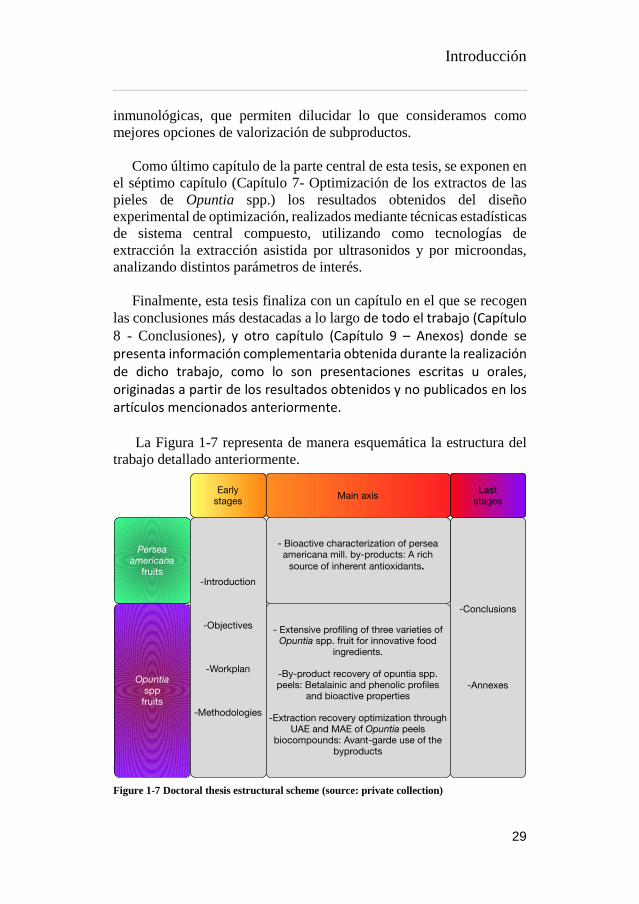
Introducción
29
inmunológicas, que permiten dilucidar lo que consideramos como
mejores opciones de valorización de subproductos.
Como último capítulo de la parte central de esta tesis, se exponen en
el séptimo capítulo (Capítulo 7- Optimización de los extractos de las
pieles de Opuntia spp.) los resultados obtenidos del diseño
experimental de optimización, realizados mediante técnicas estadísticas
de sistema central compuesto, utilizando como tecnologías de
extracción la extracción asistida por ultrasonidos y por microondas,
analizando distintos parámetros de interés.
Finalmente, esta tesis finaliza con un capítulo en el que se recogen
las conclusiones más destacadas a lo largo de todo el trabajo (Capítulo 8 - Conclusiones), y otro capítulo (Capítulo 9 – Anexos) donde se presenta información complementaria obtenida durante la realización de dicho trabajo, como lo son presentaciones escritas u orales, originadas a partir de los resultados obtenidos y no publicados en los artículos mencionados anteriormente. La Figura 1-7 representa de manera esquemática la estructura del
trabajo detallado anteriormente.
Figure 1-7 Doctoral thesis estructural scheme (source: private collection)

Introducción
30
I.6. BIBLIOGRAFIA
Ahn, J.H., Jang, Y.-S., Lee, S.Y., 2016. Production of succinic acid by
metabolically engineered microorganisms. Curr. Opin.
Biotechnol. 42, 54–66.
https://doi.org/10.1016/j.copbio.2016.02.034
Al-Farsi, M.A., Lee, C.Y., 2008. Optimization of phenolics and dietary
fibre extraction from date seeds. Food Chem. 108, 977–985.
https://doi.org/10.1016/j.foodchem.2007.12.009
Albano, C., Negro, C., Tommasi, N., Gerardi, C., Mita, G., Miceli, A.,
De Bellis, L., Blando, F., 2015. Betalains, Phenols and
Antioxidant Capacity in Cactus Pear [Opuntia ficus-indica (L.)
Mill.] Fruits from Apulia (South Italy) Genotypes. Antioxidants
4, 269–280. https://doi.org/10.3390/antiox4020269
Albuquerque, T.G., Santos, F., Sanches-Silva, A., Beatriz Oliveira, M.,
Bento, A.C., Costa, H.S., 2016. Nutritional and phytochemical
composition of Annona cherimola Mill. fruits and by-products:
Potential health benefits. Food Chem. 193, 187–195.
https://doi.org/10.1016/j.foodchem.2014.06.044
Allai, L., Druart, X., Öztürk, M., BenMoula, A., Nasser, B., El Amiri,
B., 2016. Protective effects of Opuntia ficus-indica extract on ram
sperm quality, lipid peroxidation and DNA fragmentation during
liquid storage. Anim. Reprod. Sci. 175, 1–9.
https://doi.org/10.1016/j.anireprosci.2016.09.013
Almeida, M.M.B., de Sousa, P.H.M., Arriaga, A.M.C., do Prado, G.M.,
Magalhães, C.E. de C., Maia, G.A., de Lemos, T.L.G., 2011.
Bioactive compounds and antioxidant activity of fresh exotic
fruits from northeastern Brazil. Food Res. Int. 44, 2155–2159.
https://doi.org/10.1016/j.foodres.2011.03.051
Alvarez, B., 2007. Análisis de factibilidad del cultivo de la tuna en la
localidad de Icaño , Departamento La Paz, Dirección provincial
de programación del desarrollo, Ministerio de Producción y
Desarrollo. Gobierno de la Provincia de Catamarca. Catamarca,
Argentina.
Alzate T, L.M., González, D., Hincapié, S., Cardona S, B.L., Londoño-
Londoño, J., Jiménez-Cartagena, C., 2016. The profile of
bioactive substances in ten vegetable and fruit by-products from
a food supply chain in Colombia. Sustain. Prod. Consum.
https://doi.org/10.1016/j.spc.2016.07.005
Ammar, I., Ben Salem, M., Harrabi, B., Mzid, M., Bardaa, S., Sahnoun,
Z., Attia, H., Ennouri, M., 2018. Anti-inflammatory activity and
phenolic composition of prickly pear (Opuntia ficus-indica)

Introducción
31
flowers. Ind. Crops Prod. 112, 313–319.
https://doi.org/10.1016/j.indcrop.2017.12.028
Angulo-Bejarano, P.I., Martínez-Cruz, O., Paredes-López, O., 2014.
Phytochemical Content, Nutraceutical Potential and
Biotechnological Applications of an Ancient Mexican Plant:
Nopal (Opuntia ficus-indica). Curr. Nutr. Food Sci. 10, 196–217.
https://doi.org/10.2174/157340131003140828121015
Arlt, U., 2010. NATCOL | Foods Colours Legislation.
Aruwa, C.E., Amoo, S.O., Kudanga, T., 2018. Opuntia (Cactaceae)
plant compounds, biological activities and prospects – A
comprehensive review. Food Res. Int. 112, 328–344.
https://doi.org/10.1016/j.foodres.2018.06.047
Association of Official Analytical Chemists, 2016. Official Methods of
Analysis of AOAC International, 20th ed. The Association,
Gaithersburg, MD, USA.
Attokaran, M., 2011. Natural Food Flavors and Colorants Natural Food
Flavors and Colorants, 1st ed. Blackwell Publishing LTD, Iowa.
Augustin, M.A., Sanguansri, L., 2015. Challenges and Solutions to
Incorporation of Nutraceuticals in Foods. Annu. Rev. Food Sci.
Technol. 6, 463–477. https://doi.org/10.1146/annurev-food-
022814-015507
Ayala-Zavala, J.F., Vega-Vega, V., Rosas-Domínguez, C., Palafox-
Carlos, H., Villa-Rodriguez, J.A., Siddiqui, M.W., Dávila-Aviña,
J.E., González-Aguilar, G.A., 2011. Agro-industrial potential of
exotic fruit byproducts as a source of food additives. Food Res.
Int. 44, 1866–1874.
https://doi.org/10.1016/j.foodres.2011.02.021
Azeredo, H.M.C., 2009. Betalains: Properties, sources, applications,
and stability - A review. Int. J. Food Sci. Technol. 44, 2365–2376.
https://doi.org/10.1111/j.1365-2621.2007.01668.x
Balouiri, M., Sadiki, M., Ibnsouda, S.K., 2016. Methods for in vitro
evaluating antimicrobial activity: A review. J. Pharm. Anal. 6,
71–79. https://doi.org/10.1016/j.jpha.2015.11.005
Barros, L., Calhelha, R.C., Queiroz, M.J.R.P., Santos-Buelga, C.,
Santos, E.A., Regis, W.C.B., Ferreira, I.C.F.R., 2015. The
powerful in vitro bioactivity of Euterpe oleracea Mart. seeds and
related phenolic compounds. Ind. Crops Prod. 76, 318–322.
https://doi.org/10.1016/j.indcrop.2015.05.086
Barros, L., Pereira, C., Ferreira, I.C.F.R., 2013. Optimized Analysis of
Organic Acids in Edible Mushrooms from Portugal by Ultra Fast
Liquid Chromatography and Photodiode Array Detection. Food

Introducción
32
Anal. Methods 6. https://doi.org/10.1007/s12161-012-9443-1
Barrows, J.N., Lipman, A.L., Bailey, C.J., Cianci, S., 2003. Color
Additives: FDA’s Regulatory Process and Historical
Perspectives. Food Saf. Mag.
https://www.fda.gov/forindustry/coloradditives/regulatoryproces
shistoricalperspectives/ (accessed 9.3.18).
Baydar, N.G., Özkan, G., Sağdiç, O., 2004. Total phenolic contents and
antibacterial activities of grape (Vitis vinifera L.) extracts. Food
Control 15, 335–339. https://doi.org/10.1016/S0956-
7135(03)00083-5
Belščak-Cvitanović, A., Durgo, K., Huđek, A., Bačun-Družina, V.,
Komes, D., 2018. Metabolism and Health Effects of Polyphenols,
in: Galanakis, C.M. (Ed.), Polyphenols: Properties, Recovery, and
Applications. Woodhead Publishing, pp. 1–44.
https://doi.org/https://doi.org/10.1016/B978-0-12-813572-
3.00001-4
Belwal, T., Dhyani, P., Bhatt, I.D., Rawal, R.S., Pande, V., 2016.
Optimization extraction conditions for improving phenolic
content and antioxidant activity in Berberis asiatica fruits using
response surface methodology (RSM). Food Chem. 207, 115–
124. https://doi.org/10.1016/j.foodchem.2016.03.081
Bessada, S.M.F., Barreira, J.C.M., Barros, L., Ferreira, I.C.F.R.,
Oliveira, M.B.P.P., 2016. Phenolic profile and antioxidant
activity of Coleostephus myconis (L.) Rchb.f.: An underexploited
and highly disseminated species. Ind. Crops Prod. 89, 45–51.
https://doi.org/10.1016/j.indcrop.2016.04.065
Betancourt, C., Cejudo-Bastante, M.J., Heredia, F.J., Hurtado, N.,
2017. Pigment composition and antioxidant capacity of
betacyanins and betaxanthins fractions of Opuntia dillenii (Ker
Gawl) Haw cactus fruit. Food Res. Int. 101, 173–179.
https://doi.org/10.1016/j.foodres.2017.09.007
Bhatia, S., Bera, T., Dahiya, R., Bera, T., Bhatia, S., Bera, T., 2015.
Classical and Nonclassical Techniques for Secondary Metabolite
Production in Plant Cell Culture, in: Modern Applications of Plant
Biotechnology in Pharmaceutical Sciences. Elsevier, pp. 231–
291. https://doi.org/10.1016/B978-0-12-802221-4.00007-8
Brand-Williams, W., Cuvelier, M.E., Berset, C., 1995. Use of a free
radical method to evaluate antioxidant activity. LWT - Food Sci.
Technol. 28, 25–30. https://doi.org/10.1016/S0023-
6438(95)80008-5
Bravo-Hollis, H., 1978. Las cactaceas de Mexico. Universidad

Introducción
33
Nacional Autonoma de Mexico, Mexio.
https://doi.org/10.15713/ins.mmj.3
Brundtland, G.H., 1987. Our common future.
Cai, Y., Sun, M., Corke, H., 2003. Antioxidant activity of betalains
from plants of the Amaranthaceae. J. Agric. Food Chem. 51,
2288–2294. https://doi.org/10.1021/jf030045u
Calderón-Oliver, M., Escalona-Buendía, H.B., Medina-Campos, O.N.,
Pedraza-Chaverri, J., Pedroza-Islas, R., Ponce-Alquicira, E.,
2016. Optimization of the antioxidant and antimicrobial response
of the combined effect of nisin and avocado byproducts. LWT -
Food Sci. Technol. 65, 46–52.
https://doi.org/10.1016/j.lwt.2015.07.048
Cardoso-Ugarte, G.A., Sosa-Morales, M.E., Ballard, T., Liceaga, A.,
San Martín-González, M.F., 2014. Microwave-assisted extraction
of betalains from red beet (Beta vulgaris). LWT - Food Sci.
Technol. 59, 276–282. https://doi.org/10.1016/j.lwt.2014.05.025
Carocho, M., Ferreira, I.C.F.R., 2013. A review on antioxidants,
prooxidants and related controversy: Natural and synthetic
compounds, screening and analysis methodologies and future
perspectives. Food Chem. Toxicol. 51, 15–25.
https://doi.org/10.1016/j.fct.2012.09.021
Carr, M., 2013. The water relations and irrigation requirements of
avocado (Persea americana Mill.): a review. Exp. Agric. 49, 256–
278. https://doi.org/10.1017/S0014479712001317
Cassano, A., Conidi, C., Timpone, R., D’Avella, M., Drioli, E., 2007.
A membrane-based process for the clarification and the
concentration of the cactus pear juice. J. Food Eng. 80, 914–921.
https://doi.org/10.1016/j.jfoodeng.2006.08.005
Castellanos-Santiago, E., Yahia, E.M., 2008. Identification and
quantification of betalains from the fruits of 10 Mexican prickly
pear cultivars by high-performance liquid chromatography and
electrospray ionization mass spectrometry. J. Agric. Food Chem.
56, 5758–5764. https://doi.org/10.1021/jf800362t
Castellar, R., Obón, J.M., Alacid, M., Fernández-López, J.A., 2003.
Color Properties and Stability of Betacyanins from Opuntia
Fruits. J. Agric. Food Chem. 51, 2772–2776.
https://doi.org/10.1021/jf021045h
Cejudo Bastante, M.J., Chaalal, M., Louaileche, H., Parrado, J.,
Heredia, F.J., 2014. Betalain Profile, Phenolic Content, and Color
Characterization of Different Parts and Varieties of Opuntia ficus-
indica. J. Agric. Food Chem. 62, 8491–8499.

Introducción
34
https://doi.org/10.1021/jf502465g
Chahdoura, H., Barreira, J.C.M., Barros, L., Santos-Buelga, C.,
Ferreira, I.C.F.R., Achour, L., 2014. Phytochemical
characterization and antioxidant activity of Opuntia microdasys
(Lehm.) Pfeiff flowers in different stages of maturity. J. Funct.
Foods 9, 27–37. https://doi.org/10.1016/j.jff.2014.04.011
Chahdoura, H., Barreira, J.C.M., Fernández-Ruiz, V., Morales, P.,
Calhelha, R.C., Flamini, G., Soković, M., Ferreira, I.C.F.R.,
Achour, L., 2016. Bioactivity, proximate, mineral and volatile
profiles along the flowering stages of Opuntia microdasys
(Lehm.): defining potential applications. Food Funct. 7, 1458–67.
https://doi.org/10.1039/c5fo01536g
Chan, L.G., Cohen, J.L., Maria, J., Nobrega, L., Bell, D.M., 2018.
Conversion of Agricultural Streams and Food-Processing By-
Products to Value-Added Compounds using Filamentous Fungi.
Annu. Rev. Food Sci. Technol. 9, 1–21.
https://doi.org/10.1146/annurev-food-030117-012626
Chougui, N., Djerroud, N., Naraoui, F., Hadjal, S., Aliane, K., Zeroual,
B., Larbat, R., 2015. Physicochemical properties and storage
stability of margarine containing Opuntia ficus-indica peel extract
as antioxidant. Food Chem. 173, 382–90.
https://doi.org/10.1016/j.foodchem.2014.10.025
Chougui, N., Tamendjari, A., Hamidj, W., Hallal, S., Barras, A.,
Richard, T., Larbat, R., 2013. Oil composition and
characterisation of phenolic compounds of Opuntia ficus-indica
seeds. Food Chem. 139, 796–803.
https://doi.org/10.1016/j.foodchem.2013.01.054
Clifford, M.N., Johnston, K.L., Knight, S., Kuhnert, N., 2003.
Hierarchical Scheme for LC-MSn Identification of Chlorogenic
Acids. J. Agric. Food Chem. 51, 2900–2911.
https://doi.org/10.1021/jf026187q
Clifford, M.N., Knight, S., Kuhnert, N., 2005. Discriminating between
the six isomers of dicaffeoylquinic acid by LC-MSn. J. Agric.
Food Chem. 53, 3821–3832. https://doi.org/10.1021/jf050046h
Cowan, A.K., Cripps, R.F., Richings, E.W., Taylor, N.J., 2001. Fruit
size: Towards an understanding of the metabolic control of fruit
growth using avocado as a model system. Physiol. Plant. 111,
127–136. https://doi.org/10.1034/j.1399-3054.2001.1110201.x
Cowan, A.K., Wolstenholme, B.N., 2003. AVOCADOS, in:
Encyclopedia of Food Sciences and Nutrition. Elsevier, pp. 348–
353. https://doi.org/10.1016/B0-12-227055-X/00073-0

Introducción
35
Crane, J.H., Balerdi, C.F., Maguire, I., 2016. Avocado Growing in the
Florida Home Landscape.
Da-Costa-Rocha, I., Bonnlaender, B., Sievers, H., Pischel, I., Heinrich,
M., 2014. Hibiscus sabdariffa L. – A phytochemical and
pharmacological review. Food Chem. 165, 424–443.
https://doi.org/10.1016/j.foodchem.2014.05.002
da Silva, R.P.F.F., Rocha-Santos, T.A.P., Duarte, A.C., 2016.
Supercritical fluid extraction of bioactive compounds. TrAC -
Trends Anal. Chem. 76, 40–51.
https://doi.org/10.1016/j.trac.2015.11.013
Dabas, D., Shegog, R.M., Ziegler, G.R., Lambert, J.D., 2013. Avocado
(Persea americana) seed as a source of bioactive phytochemicals.
Curr. Pharm. Des. 19, 6133–40.
https://doi.org/10.2174/1381612811319340007
Day, L., Seymour, R.B., Pitts, K.F., Konczak, I., Lundin, L., 2009.
Incorporation of functional ingredients into foods. Trends Food
Sci. Technol. 20, 388–395.
https://doi.org/10.1016/J.TIFS.2008.05.002
DeFelice, S.L., 2002. FIM Rationale And Proposed Guidelines For The
Nutraceutical Research & Education Act - NREA. New York.
Delgado-Vargas, F., Paredes-Lopez, O., 2003. Natural Colorants for
Food and Nutraceutical Uses, Trends in Food Science &
Technology. https://doi.org/10.1016/S0924-2244(03)00076-1
Destro dos Santos, C., Külzer Scherer, R., Schilling Cassini, A.,
Ferreira Marczak, L.D., Tessaro, I.C., 2016. Clarification of red
beet stalks extract by microfiltration combined with
ultrafiltration. J. Food Eng. 185, 35–41.
https://doi.org/10.1016/j.jfoodeng.2016.03.031
Devalaraja, S., Jain, S., Yadav, H., 2011. Exotic fruits as therapeutic
complements for diabetes, obesity and metabolic syndrome. Food
Res. Int. 44, 1856–1865.
https://doi.org/10.1016/j.foodres.2011.04.008
Devasagayam, T.P.A., Boloor, K.K., Ramasarma, T., 2003. Methods
for estimating lipid peroxidation: An analysis of merits and
demerits. Indian J. Biochem. Biophys. 40, 300–308.
Dias, M.I., Sousa, M.J., Alves, R.C., Ferreira, I.C.F.R., 2016. Exploring
plant tissue culture to improve the production of phenolic
compounds: A review. Ind. Crops Prod. 82, 9–22.
https://doi.org/10.1016/j.indcrop.2015.12.016
Donkin, R.A., 1977. Spanish Red: An Ethnogeographical Study of
Cochineal and the Opuntia Cactus. Trans. Am. Philos. Soc. 67, 1–
Introducción
36
84. https://doi.org/10.2307/1006195
Esatbeyoglu, T., Wagner, A.E., Schini-Kerth, V.B., Rimbach, G.,
2015a. Betanin a food colorant with biological activity. Mol. Nutr.
Food Res. 59, 36–47. https://doi.org/10.1002/mnfr.201400484
Espinel-Ingroff, A., 2001. Comparison of the E-test with the NCCLS
M38-P method for antifungal susceptibility testing of common
and emerging pathogenic filamentous fungi. J. Clin. Microbiol.
39, 1360–1367. https://doi.org/10.1128/JCM.39.4.1360-
1367.2001
Esquivel, P., 2016. Handbook on Natural Pigments in Food and
Beverages Industrial Applications for Improving Food Colorl, in:
Carle, R., Schweiggert, R.M. (Eds.). Woodhead Publishing, San
Pedro, Costa Rica, pp. 81–99.
FAO, 2016. FAOSTAT URL
http://www.fao.org/faostat/en/?#data/QC/visualize (accessed
5.3.18).
FAO, 2004. AVOCADO: Post-Harvest Operation. Rome.
FAO, IFAD, WFP, 2015. The State of Food Insecurity in the World.
Meeting the 2015 international hunger targets: taking stock of
uneven progress. Rome.
Farag, M.A., Maamoun, A.A., Ehrlich, A., Fahmy, S., Wesjohann,
L.A., 2017. Assessment of sensory metabolites distribution in 3
cactus Opuntia ficus-indica fruit cultivars using UV
fingerprinting and GC/MS profiling techniques. LWT - Food Sci.
Technol. 80, 145–154. https://doi.org/10.1016/j.lwt.2017.02.014
Fathordoobady, F., Mirhosseini, H., Selamat, J., Manap, M.Y.A., 2016.
Effect of solvent type and ratio on betacyanins and antioxidant
activity of extracts from Hylocereus polyrhizus flesh and peel by
supercritical fluid extraction and solvent extraction. Food Chem.
202, 70–80. https://doi.org/10.1016/j.foodchem.2016.01.121
FDA, 2017. SCOGS (Select Committee on GRAS Substances)]. URL
http://www.accessdata.fda.gov/scripts/fdcc/?set=SCOGS
(accessed 3.14.17).
FDA, 2007. Guidance for Industry and Other Stakeholders
Toxicological Principles for the Safety Assessment of Food
Ingredients, Human Studies.
Fernández-López, J. a., Almela, L., 2001. Application of high-
performance liquid chromatography to the characterization of the
betalain pigments in prickly pear fruits. J. Chromatogr. A 913,
415–420. https://doi.org/10.1016/S0021-9673(00)01224-3
Fernandez-Rojas, B., Ortiz-Moreno, A., Hernandez-Navarro, D., 2010.

Introducción
37
Phenolic Content and its Antioxidant Activity of Persimmon
(Dyospiros kaki L.) Cultivated in Mexico. J. Biotechnol. 150.
https://doi.org/10.1016/j.jbiotec.2010.09.289
Ferreira, I.C.F.R., Martins, N., Barros, L., 2017. Phenolic Compounds
and Its Bioavailability: In Vitro Bioactive Compounds or Health
Promoters? Adv. Food Nutr. Res. 82, 1–44.
https://doi.org/10.1016/bs.afnr.2016.12.004
Ferreres, F., Grosso, C., Gil-Izquierdo, A., Valentão, P., Mota, A.T.,
Andrade, P.B., 2017. Optimization of the recovery of high-value
compounds from pitaya fruit by-products using microwave-
assisted extraction. Food Chem. 230, 463–474.
https://doi.org/10.1016/j.foodchem.2017.03.061
Flores-Valdez, C., Luna-Esquivel, J., Ramirez-Moreno, P., 1995.
Manejo postcosecha de la tuna para el mercado nacional e
internacional y Procesamiento y transformación industrial.
Chapingo, Estado de México.
Galanakis, C.M., 2015. Chapter 3 – The universal recovery strategy,
Food Waste Recovery. Elsevier Inc.
https://doi.org/10.1016/B978-0-12-800351-0.00003-1
Galanakis, C.M., 2012. Recovery of high added-value components
from food wastes: Conventional, emerging technologies and
commercialized applications. Trends Food Sci. Technol. 26, 68–
87. https://doi.org/10.1016/j.tifs.2012.03.003
Gandía-herrero, F., Cabanes, J., Escribano, J., García-carmona, F.,
Jimenez-atienzar, M., Jiménez-atiénzar, M., 2013. Encapsulation
of the most antioxidant betalains in edible matrices as powders of
different colors Encapsulation of the most antioxidant betalains in
edible matrices as powders of different colors.
https://doi.org/10.1021/jf400337g
Gandía-Herrero, F., Escribano, J., García-Carmona, F., 2010. Structural
implications on color, fluorescence, and antiradical activity in
betalains. Planta 232, 449–460. https://doi.org/10.1007/s00425-
010-1191-0
Garcia-Castello, E.M., Rodriguez-Lopez, A.D., Mayor, L., Ballesteros,
R., Conidi, C., Cassano, A., 2015. Optimization of conventional
and ultrasound assisted extraction of flavonoids from grapefruit
(Citrus paradisi L.) solid wastes. LWT - Food Sci. Technol. 64,
1114–1122. https://doi.org/10.1016/j.lwt.2015.07.024
García-Cruz, L., Dueñas, M., Santos-Buelgas, C., Valle-Guadarrama,
S., Salinas-Moreno, Y., 2017. Betalains and phenolic compounds
profiling and antioxidant capacity of pitaya (Stenocereus spp.)
Introducción
38
fruit from two species (S. Pruinosus and S. stellatus). Food Chem.
234, 111–118. https://doi.org/10.1016/j.foodchem.2017.04.174
Gebhardt, D., 2018. The Economics of Natural Color Pigments]. URL
https://sensientfoodcolors.com/en-us/research-
development/economics-natural-color-pigments/
Ghasemzadeh, A., Ghasemzadeh, N., 2011. Flavonoids and phenolic
acids: Role and biochemical activity in plants and human. J. Med.
Plants Res. 5, 6697–6703. https://doi.org/10.5897/JMPR11.363
Gomes-Correa, R.C., de Souza, A.H.P., Calhelha, R.C., Barros, L.,
Glamoclija, J., Sokovic, M., Peralta, R.M., Bracht, A., Ferreira,
I.C.F.R., 2015. Bioactive formulations prepared from fruiting
bodies and submerged culture mycelia of the Brazilian edible
mushroom Pleurotus ostreatoroseus Singer. Food Funct. 6, 2155–
2164. https://doi.org/10.1039/C5FO00465A
Gong, M., Bassi, A., 2016. Carotenoids from microalgae: A review of
recent developments. Biotechnol. Adv. 34, 1396–1412.
https://doi.org/10.1016/j.biotechadv.2016.10.005
Griffith, M.P., 2004. The origins of an important cactus crop, Opuntia
ficus-indica (Cactaceae): New molecular evidence. Am. J. Bot.
91, 1915–1921. https://doi.org/10.3732/ajb.91.11.1915
Guimarães, R., Barros, L., Dueñas, M., Calhelha, R.C., Carvalho,
A.M., Santos-Buelga, C., Queiroz, M.J.R.P., Ferreira, I.C.F.R.,
2013. Nutrients, phytochemicals and bioactivity of wild Roman
chamomile: A comparison between the herb and its preparations.
Food Chem. 136, 718–725.
https://doi.org/10.1016/j.foodchem.2012.08.025
Guldiken, B., Toydemir, G., Nur Memis, K., Okur, S., Boyacioglu, D.,
Capanoglu, E., 2016. Home-processed red beetroot (Beta vulgaris
L.) products: Changes in antioxidant properties and
bioaccessibility. Int. J. Mol. Sci. 17.
https://doi.org/10.3390/ijms17060858
GVR, 2016. Natural Antioxidants Market Analysis By Product
(Vitamin C, Vitamin E, Polyphenols, Carotenoids) And Segment
Forecasts To 2022. URL
https://www.grandviewresearch.com/industry-analysis/natural-
antioxidants-market
Heleno, S.A., Barros, L., Sousa, M.J., Martins, A., Ferreira, I.C.F.R.,
2010. Tocopherols composition of Portuguese wild mushrooms
with antioxidant capacity. Food Chem. 119, 1443–1450.
https://doi.org/10.1016/j.foodchem.2009.09.025
Henry, B.S., 1996. Natural food colours, in: Natural Food Colorants.

Introducción
39
Springer US, Boston, MA, pp. 40–79.
https://doi.org/10.1007/978-1-4615-2155-6_2
Herbach, K.M., Stintzing, F.C., Carle, R., 2006. Betalain stability and
degradation - Structural and chromatic aspects. J. Food Sci. 71,
41–50. https://doi.org/10.1111/j.1750-3841.2006.00022.x
Herbach, K.M., Stintzing, F.C., Carle, R., 2005. Identification of heat-
induced degradation products from purified betanin, phyllocactin
and hylocerenin by high-performance liquid chromatography/
electrospray ionization mass spectrometry. Rapid Commun. Mass
Spectrom. 19, 2603–2616. https://doi.org/10.1002/rcm.2103
Hiwasa-Tanase, K., Ezure, H., 2014. Climateric and non-climateric
ripening, in: Nath, P., Bouzayen, M., Mattoo, A.K., Pech, J.C.
(Eds.), Fruit Ripening: Physiology, Signalling and Genomics.
CABI International, pp. 151–177.
https://doi.org/10.1017/CBO9781107415324.004
Hogervorst-Cvejić, J., Atanacković-Krstonošić, M., Bursać, M., Miljić,
U., 2017. Polyphenols, in: Nutraceutical and Functional Food
Components. Academic Press, pp. 203–258.
https://doi.org/10.1016/B978-0-12-805257-0.00007-7
Hollman, P.C., Bijsman, M.N., van Gameren, Y., Cnossen, E.P., de
Vries, J.H., Katan, M.B., 1999. The sugar moiety is a major
determinant of the absorption of dietary flavonoid glycosides in
man. Free Radic. Res. 31, 569–573.
https://doi.org/10.1080/10715769900301141
Hurtado-Fernández, E., Fernandez-Gutierrez, A., Carrasco-Pancorbo,
A., 2018a. Avocado fruit, in: Rodrigues, S., de Oliveira, E., Sousa,
E. (Eds.), Exotic Fruits. Academic Press, London, pp. 37–46.
Hurtado-Fernández, E., Pacchiarotta, T., Longueira-Suárez, E.,
Mayboroda, O.A., Fernández-Gutiérrez, A., Carrasco-Pancorbo,
A., 2013. Evaluation of gas chromatography-atmospheric
pressure chemical ionization-mass spectrometry as an alternative
to gas chromatography-electron ionization-mass spectrometry:
Avocado fruit as example. J. Chromatogr. A 1313, 228–244.
https://doi.org/10.1016/j.chroma.2013.08.084
Hurtado-Fernández, E., Pacchiarotta, T., Mayboroda, O.A., Fernández-
Gutiérrez, A., Carrasco-Pancorbo, A., 2014. Quantitative
characterization of important metabolites of avocado fruit by gas
chromatography coupled to different detectors (APCI-TOF MS
and FID). Food Res. Int. 62, 801–811.
https://doi.org/10.1016/j.foodres.2014.04.038
Hussain, Z., Thu, H.E., Amjad, M.W., Hussain, F., Ahmed, T.A., Khan,
Introducción
40
S., 2017. Exploring recent developments to improve antioxidant,
anti-inflammatory and antimicrobial efficacy of curcumin: A
review of new trends and future perspectives. Mater. Sci. Eng. C.
https://doi.org/10.1016/j.msec.2017.03.226
IFIC, FDA, 2010. Overview of Food Ingredients, Additives and Colors.
URL
https://www.fda.gov/food/ingredientspackaginglabeling/foodadd
itivesingredients/ucm094211.htm
Inglese, P., Giugliuzza, G., Liguori, G., 2004. Cactus pear fruit
production: from knowledge to development, in: Esparza-Frausto,
G., Valdez-Cepeda, R.D., Méndez- Gallego, S.J. (Eds.), El
Nopal.Tópicos de Actualidad. Universidad Autónoma de
Chapingo. Colegio de Postgraduados, México, pp. 89–108.
IUPAC, IUPAC-IUB, 1976. Nomenclature of cyclitols. Biochem. J.
153, 23–31. https://doi.org/10.1351/pac197437010283
Jain, G., Gould, K.S., 2015. Are betalain pigments the functional
homologues of anthocyanins in plants? Environ. Exp. Bot. 119,
48–53. https://doi.org/10.1016/J.ENVEXPBOT.2015.06.002
Jain, T., Grover, K., Kaur, G., 2016. Effect of processing on nutrients
and fatty acid composition of garden cress (Lepidium sativum)
seeds. Food Chem. 213, 806–812.
https://doi.org/10.1016/j.foodchem.2016.07.034
Jimenez-Garcia, S.N., Vazquez-Cruz, M.A., Garcia-Mier, L.,
Contreras-Medina, L.M., Guevara-González, R.G., Garcia-Trejo,
J.F., Feregrino-Perez, A.A., 2018. Phytochemical and
Pharmacological Properties of Secondary Metabolites in Berries,
in: Therapeutic Foods. Elsevier, pp. 397–427.
https://doi.org/10.1016/B978-0-12-811517-6.00013-1
Kaderides, K., Goula, A.M., 2017. Development and characterization
of a new encapsulating agent from orange juice by-products. Food
Res. Int. 0–1. https://doi.org/10.1016/j.foodres.2017.07.057
Kanner, J., Harel, S., Granit, R., 2001. Betalains - A new class of
dietary cationized antioxidants. J. Agric. Food Chem. 49, 5178–
5185. https://doi.org/10.1021/jf010456f
Karamac, M., Estrella, I., Herna, T., Dykes, G. a., Kosińska, A.,
Karamać, M., Estrella, I., Hernández, T., Bartolomé, B., Dykes,
G. a., 2012. Phenolic compound profiles and antioxidant capacity
of Persea americana Mill. peels and seeds of two varieties. J.
Agric. Food Chem. 60, 4613–4619.
https://doi.org/10.1021/jf300090p
Katsube, T., Tsurunaga, Y., Sugiyama, M., Furuno, T., Yamasaki, Y.,

Introducción
41
2009. Effect of air-drying temperature on antioxidant capacity and
stability of polyphenolic compounds in mulberry (Morus alba L.)
leaves. Food Chem. 113, 964–969.
https://doi.org/10.1016/j.foodchem.2008.08.041
Kaur Kala, H., Mehta, R., Tandey, R., Sen, K.K., Mandal, V., 2016.
Ten years of research on phenolics (2005–2015): A status report.
Pacific Sci. Rev. A Nat. Sci. Eng.
https://doi.org/10.1016/j.psra.2016.07.002
Knight, R.J., Campbell, C.W., 1999. Ecological adaptation and the
evolution of modern avocado cultivars. Rev. Chapingo Ser.
Hortic. 49–54.
Kosińska, A., Karamać, M., Estrella, I., Hernández, T., Bartolomé, B.,
Dykes, G.A., 2012. Phenolic compound profiles and antioxidant
capacity of Persea americana Mill. peels and seeds of two
varieties. J. Agric. Food Chem. 60, 4613–4619.
https://doi.org/10.1021/jf300090p
Kris-Etherton, P.M., Hecker, K.D., Bonanome, A., Coval, S.M.,
Binkoski, A.E., Hilpert, K.F., Griel, A.E., Etherton, T.D., 2002.
Bioactive compounds in foods: their role in the prevention of
cardiovascular disease and cancer. Am. J. Med. 113, 71–88.
https://doi.org/10.1016/S0002-9343(01)00995-0
Kuhnle, G.G., 2012. Nutritional biomarkers for objective dietary
assessment. J. Sci. Food Agric. 92, 1145–1149.
https://doi.org/10.1002/jsfa.5631
Kuti, J.O., 2004. Antioxidant compounds from four Opuntia cactus
pear fruit varieties. Food Chem. 85, 527–533.
https://doi.org/10.1016/S0308-8146(03)00184-5
Kyriacou, M.C., Emmanouilidou, M.G., Soteriou, G.A., 2016.
Asynchronous ripening behavior of cactus pear (Opuntia ficus-
indica) cultivars with respect to physicochemical and
physiological attributes. Food Chem. 211, 598–607.
https://doi.org/10.1016/j.foodchem.2016.05.113
Laqui-Vilca, C., Aguilar-Tuesta, S., Mamani-Navarro, W., Montaño-
Bustamante, J., Condezo-Hoyos, L., 2018. Ultrasound-assisted
optimal extraction and thermal stability of betalains from colored
quinoa (Chenopodium quinoa Willd) hulls. Ind. Crops Prod. 111,
606–614. https://doi.org/10.1016/j.indcrop.2017.11.034
Lea, A., 2008. Analysis of polyphenol antioxidants in fortified foods
and supplements. Food Fortif. Suppl. 175–194.
https://doi.org/10.1533/9781845694265.2.175
Leardi, R., 2013. Experimental Design, 1st ed, Data Handling in
Introducción
42
Science and Technology. Copyright © 2013 Elsevier B.V. All
rights reserved. https://doi.org/10.1016/B978-0-444-59528-
7.00002-8
Li, J., Chen, Z., Di, D., 2012. Preparative separation and purification of
Rebaudioside A from Stevia rebaudiana Bertoni crude extracts by
mixed bed of macroporous adsorption resins. Food Chem. 132,
268–276. https://doi.org/10.1016/j.foodchem.2011.10.077
M. Librán, C., Mayor, L., M. Garcia-Castello, E., Vidal-Brotons, D.,
2013. Polyphenol extraction from grape wastes: Solvent and pH
effect. Agric. Sci. 04, 56–62.
https://doi.org/10.4236/as.2013.49B010
Madrau, M. a., Piscopo, A., Sanguinetti, A.M., Del Caro, A., Poiana,
M., Romeo, F. V., Piga, A., 2009. Effect of drying temperature on
polyphenolic content and antioxidant activity of apricots. Eur.
Food Res. Technol. 228, 441–448.
https://doi.org/10.1007/s00217-008-0951-6
Madunić, J., Madunić, I.V., Gajski, G., Popić, J., Garaj-Vrhovac, V.,
2018. Apigenin: A dietary flavonoid with diverse anticancer
properties. Cancer Lett. 413, 11–22.
https://doi.org/10.1016/j.canlet.2017.10.041
Manach, C., Scalbert, A., Morand, C., Rémésy, C., Jiménez, L., 2004.
Bioavailability, Polyphenols: Food sources and. Am. J. Clin.
Nutr. 79, 727–747. https://doi.org/10.1038/nature05488
Mandal, S.C., Mandal, V., Das, A.K., 2015. Classification of Extraction
Methods, in: Essentials of Botanical Extraction. Academic Press,
pp. 83–136. https://doi.org/10.1016/B978-0-12-802325-9.00006-
9
Marais, J.P.J., Deavours, B., Dixon, R.A., Ferreira, D., 2006. The
science of flavonoids, The Science of Flavonoids. Springer, Ohio.
https://doi.org/10.1007/978-0-387-28822-2
Marco, G.J., 1968. A Rapid Method for Evaluation of Antioxidants. J.
Am. oil Chem. Soc. 45, 594–598.
https://doi.org/10.1007/BF02668958
Maria, J., Andrade, D.M., Fasolo, D., 2014. Polyphenol Antioxidants
from Natural Sources and Contribution to Health Promotion,
Polyphenols in Human Health and Disease. Elsevier Inc.
https://doi.org/10.1016/B978-0-12-398456-2.00020-7
Martins, N., Roriz, C.L., Morales, P., Barros, L., Ferreira, I.C.F.R.,
2016. Food colorants: Challenges, opportunities and current
desires of agro-industries to ensure consumer expectations and
regulatory practices. Trends Food Sci. Technol. 52, 1–15.

Introducción
43
https://doi.org/10.1016/j.tifs.2016.03.009
Mata, A., Ferreira, J.P., Semedo, C., Serra, T., Duarte, C.M.M., Bronze,
M.R., 2016. Contribution to the characterization of Opuntia spp.
juices by LC–DAD–ESI-MS/MS. Food Chem. 210, 558–565.
https://doi.org/10.1016/j.foodchem.2016.04.033
Mayor, L., Calvo, A., Moreira, R., Fito, P., Garcia-Castello, E. M.,
2013. Water sorption isotherms of globe artichoke leaves. Agric.
Sci. 04, 63–69. https://doi.org/10.4236/as.2013.49B011
McGuire, S., 2011. U.S. Department of Agriculture and U.S.
Department of Health and Human Services, Dietary Guidelines
for Americans, 2010. 7th Edition, Washington, DC: U.S.
Government Printing Office, January 2011. Adv. Nutr. 2, 293–
294. https://doi.org/10.3945/an.111.000430
Medina-Meza, I.G., Barbosa-Cánovas, G. V., 2015. Assisted extraction
of bioactive compounds from plum and grape peels by ultrasonics
and pulsed electric fields. J. Food Eng. 166, 268–275.
https://doi.org/10.1016/j.jfoodeng.2015.06.012
Melgar, B., Dias, M.I., Ciric, A., Sokovic, M., Garcia-Castello, E.M.,
Rodriguez-Lopez, A.D., Barros, L., Ferreira, I.C.R.F., 2017a. By-
product recovery of Opuntia spp. peels: Betalainic and phenolic
profiles and bioactive properties.
https://doi.org/10.1016/j.indcrop.2017.06.011
Melgar, B., Pereira, E., Oliveira, M.B.P.P., Garcia-Castello, E.M.,
Rodriguez-Lopez, A.D., Sokovic, M., Barros, L., Ferreira,
I.C.F.R., 2017b. Extensive profiling of three varieties of Opuntia
spp. fruit for innovative food ingredients. Food Res. Int. 101, 259–
265. https://doi.org/10.1016/j.foodres.2017.09.024
Mena, P., Tassotti, M., Andreu, L., Nuncio-Jáuregui, N., Legua, P., Del
Rio, D., Hernández, F., 2018. Phytochemical characterization of
different prickly pear (Opuntia ficus-indica (L.) Mill.) cultivars
and botanical parts: UHPLC-ESI-MSn metabolomics profiles and
their chemometric analysis. Food Res. Int. 108, 301–308.
https://doi.org/10.1016/j.foodres.2018.03.062
Mirabella, N., Castellani, V., Sala, S., 2014. Current options for the
valorization of food manufacturing waste: a review. J. Clean.
Prod. 65, 28–41.
https://doi.org/10.1016/J.JCLEPRO.2013.10.051
Mishra, A., Sharma, A.K., Kumar, S., Saxena, A.K., Pandey, A.K.,
2013. Bauhinia variegata leaf extracts exhibit considerable
antibacterial, antioxidant, and anticancer activities. Biomed Res.
Int. 2013. https://doi.org/10.1155/2013/915436

Introducción
44
Mishra, A.K., Mishra, A., Kehri, H.K., Sharma, B., Pandey, A.K.,
2009. Inhibitory activity of Indian spice plant Cinnamomum
zeylanicum extracts against Alternaria solani and Curvularia
lunata, the pathogenic dematiaceous moulds. Ann. Clin.
Microbiol. Antimicrob. 8, 9. https://doi.org/10.1186/1476-0711-
8-9
Monks, A., Scudiero, D., Skehan, P., Shoemaker, R., Paull, K., Vistica,
D., Hose, C., Langley, J., Cronise, P., Vaigro-wolff, A., Gray-
goodrich, M., Campbell, H., Boyd, M., 1991. Feasibility of a
High-Flux Anticancer Drug Screen Using a Diverse Panel of
Cultured. J. Natl. Cancer Inst. 83, 757–766.
Morales, P., Barros, L., Ramírez-Moreno, E., Santos-Buelga, C.,
Ferreira, I.C.F.R., 2015. Xoconostle fruit (Opuntia matudae
Scheinvar cv. Rosa) by-products as potential functional
ingredients. Food Chem. 185, 289–97.
https://doi.org/10.1016/j.foodchem.2015.04.012
Morales, P., Barros, L., Ramírez-Moreno, E., Santos-Buelga, C.,
Ferreira, I.C.F.R., 2014. Exploring xoconostle by-products as
sources of bioactive compounds. Food Res. Int. 65, 437–444.
https://doi.org/10.1016/j.foodres.2014.05.067
Morales, P., Ramírez-Moreno, E., Sanchez-Mata, M.D.C., Carvalho,
A.M., Ferreira, I.C.F.R., 2012. Nutritional and antioxidant
properties of pulp and seeds of two xoconostle cultivars (Opuntia
joconostle F.A.C. Weber ex Diguet and Opuntia matudae
Scheinvar) of high consumption in Mexico. Food Res. Int. 46.
https://doi.org/10.1016/j.foodres.2011.12.031
Moreno, J., Peinado, R., 2012. Polyphenols, in: Moreno, J., Peinado, R.
(Eds.), Enological Chemistry. Academic Press, pp. 53–76.
https://doi.org/10.1016/B978-0-12-388438-1.00005-4
Naik, S.N., Goud, V. V., Rout, P.K., Dalai, A.K., 2010. Production of
first and second generation biofuels: A comprehensive review.
Renew. Sustain. Energy Rev. 14, 578–597.
https://doi.org/10.1016/j.rser.2009.10.003
Nicoletti, M., 2012. Nutraceuticals and botanicals: overview and
perspectives. Int. J. Food Sci. Nutr. 63, 2–6.
https://doi.org/10.3109/09637486.2011.628012
NIH, 2017. Vitamin E — Fact sheet for Consumer. URL
https://ods.od.nih.gov/factsheets/VitaminE-Consumer/ (accessed
3.14.17).
Nobel, P.S., 1998. Los incomparables agaves y cactos. Trillas, México.
Novoa, A., Le Roux, J.J., Robertson, M.P., Wilson, J.R.U., Richardson,

Introducción
45
D.M., 2015. Introduced and invasive cactus species: A global
review. AoB Plants 7. https://doi.org/10.1093/aobpla/plu078
Ochoa, M. judith, Barbera, G., 2017. History and economic and agro-
ecological importance, in: Crop Ecology, Cultivation and Uses of
Catus Pears. FAO, ICARDA, pp. 1–11.
Osborn, S., 2015. 12 – Labelling relating to natural ingredients and
additives, in: Advances in Food and Beverage Labelling. pp. 207–
221. https://doi.org/10.1533/9781782420934.3.207
Paciulli, M., Medina-Meza, I.G., Chiavaro, E., Barbosa-Cánovas, G.V.,
2016. Impact of thermal and high pressure processing on quality
parameters of beetroot (Beta vulgaris L.). LWT - Food Sci.
Technol. 68, 98–104. https://doi.org/10.1016/j.lwt.2015.12.029
Patel, D., Shukla, S., Gupta, S., 2007. Apigenin and cancer
chemoprevention: Progress, potential and promise (Review). Int.
J. Oncol. 30, 233–245. https://doi.org/10.3892/ijo.30.1.233
Pedreño, M.A., Escribano, J., 2000. Studying the oxidation and the
antiradical activity of betalain from beetroot. J. Biol. Educ. 35,
49–51. https://doi.org/10.1080/00219266.2000.9655736
Peláez-Cid, a. a., Velázquez-Ugalde, I., Herrera-González, a. M.,
García-Serrano, J., 2013. Textile dyes removal from aqueous
solution using Opuntia ficus-indica fruit waste as adsorbent and
its characterization. J. Environ. Manage. 130, 90–97.
https://doi.org/10.1016/j.jenvman.2013.08.059
Pereira, C., Barros, L., Carvalho, A.M., Ferreira, I.C.F.R., 2011.
Nutritional composition and bioactive properties of commonly
consumed wild greens: Potential sources for new trends in modern
diets. Food Res. Int. 44, 2634–2640.
https://doi.org/10.1016/j.foodres.2011.05.012
Pereira, E., Barros, L., Calhelha, R.C., Duenas, M., Carvalho, A.M.,
Santos-Buelga, C., Ferreira, I.C.F.R., 2014. Bioactivity and
phytochemical characterization of Arenaria montana L. Food
Funct. 5, 1848–1855. https://doi.org/10.1039/c4fo00210e
Pereira, E., Barros, L., Martins, A., Ferreira, I.C.F.R., 2012. Towards
chemical and nutritional inventory of Portuguese wild edible
mushrooms in different habitats. Food Chem. 130, 394–403.
https://doi.org/10.1016/j.foodchem.2011.07.057
Piattelli, M., Minale, L., Prota, G., 1964. Isolation, structure and
absolute configuration of indicaxanthin. Tetrahedron 20, 2325–
2329. https://doi.org/10.1016/S0040-4020(01)97621-5
Pimienta, E., 1990. El nopal tunero. Libros de tiempos de ciencia ;5.
Pimiento Barrios, E., Munoz-Urias, A., 1999. Domesticacion de

Introducción
46
nopales tuneros (Opuntis spp.) y descripcion de las principales
variedades cultivadas. FAO, Rome (Italy).
Pinela, J., Prieto, M.A., Barreiro, M.F., Carvalho, A.M., Oliveira,
M.B.P.P., Vázquez, J.A., Ferreira, I.C.F.R., 2016. Optimization
of microwave-assisted extraction of hydrophilic and lipophilic
antioxidants from a surplus tomato crop by response surface
methodology. Food Bioprod. Process. 98, 283–298.
https://doi.org/10.1016/j.fbp.2016.02.002
Primorac, T., Požar, M., Sokolić, F., Zoranić, L., Urbic, T., 2018. A
simple two dimensional model of methanol. J. Mol. Liq. 262, 46–
57. https://doi.org/10.1016/j.molliq.2018.04.055
Quideau, S., Deffieux, D., Douat-Casassus, C., Pouysegu, L., 2011.
Plant polyphenols: chemical properties, biological activities, and
synthesis. Angew. Chem. Int. Ed. Engl. 50, 586–621.
https://doi.org/10.1002/anie.201000044
Rached, W., Calhelha, R.C., Fernandes, Â., Carvalho, A.M.,
Bennaceur, M., Marouf, A., Barros, L., Santos-Buelga, C.,
Ferreira, I.C.F.R., 2016. Phytochemical characterization and
bioactive properties of Osyris quadripartita Salzm. ex Decne.
leaves from Algeria. RSC Adv. 6, 72768–72776.
https://doi.org/10.1039/C6RA11787B
Ramos-Jerz, M.D.R., Villanueva, S., Jerz, G., Winterhalter, P., Deters,
A.M., 2013. Persea americana Mill. Seed: Fractionation,
Characterization, and Effects on Human Keratinocytes and
Fibroblasts. Evidence-based Complement. Altern. Med. eCAM
2013, 391247. https://doi.org/10.1155/2013/391247
Rao, G., 2010. Optimization of ultrasound-assisted extraction of
cyanidin 3-rutinoside from litchi (Lichi chinensis Sonn.) fruit
pericarp. Anal. Methods 2, 1166.
https://doi.org/10.1039/c0ay00203h
Ravichandran, K., Saw, N.M.M.T., Mohdaly, A.A.A., Gabr, A.M.M.,
Kastell, A., Riedel, H., Cai, Z., Knorr, D., Smetanska, I., 2013.
Impact of processing of red beet on betalain content and
antioxidant activity. Food Res. Int. 50, 670–675.
https://doi.org/10.1016/j.foodres.2011.07.002
Raymond Chia, T.W., Dykes, G. a, 2010. Antimicrobial activity of
crude epicarp and seed extracts from mature avocado fruit (Persea
americana) of three cultivars. Pharm. Biol. 48, 753–756.
https://doi.org/10.3109/13880200903273922
Reis, F.S., Barreira, J.C.M., Calhelha, R.C., van Griensven, L.J.I.D.,
Ćirić, A., Glamočlija, J., Soković, M., Ferreira, I.C.F.R., 2014.

Introducción
47
Chemical characterization of the medicinal mushroom Phellinus
linteus (Berkeley & Curtis) Teng and contribution of different
fractions to its bioactivity. LWT - Food Sci. Technol. 58, 478–
485. https://doi.org/10.1016/j.lwt.2014.04.013
Reyes-Agüero, J.A., Aguirre, J.R., Hernández, H., 2005. Systematyc
notes and a Detailed description of Opuntia ficus-indica (L) Mill.
(CACTACEAE). Agrociencia 39, 395–408.
Reynolds, S.G., Arias, E., 2003. Opuntia Spp. -A Strategic fodder and
efficient tool for combat desertification in the Wana Region,
Cactus (Opuntia spp.) as forage. FAO Plant Production and
Protection Paper 169.
Roberfroid, M.B., 2000. Concepts and strategy of functional food
science: the European perspective. Am. J. Clin. Nutr. 71, 1660S–
4S; discussion 1674S–5S.
https://doi.org/10.1093/ajcn/71.6.1660S
Rodrigo, D., Martínez-l, A., 2015. Antimicrobial activity of cauli fl
ower (Brassica oleracea var . Botrytis) by-product against
Listeria monocytogenes 50, 435–440.
https://doi.org/10.1016/j.foodcont.2014.09.031
Rodríguez-Carpena, J.G., Morcuende, D., Estévez, M., 2011. Avocado
by-products as inhibitors of color deterioration and lipid and
protein oxidation in raw porcine patties subjected to chilled
storage. Meat Sci. 89, 166–173.
https://doi.org/10.1016/j.meatsci.2011.04.013
Rohwer, J.G., 1993. Lauraceae, in: The Families and Genera of
Vascular Plants Free Preview, Volume II, Flowering Plants ·
Dicotyledons. Springer, Berlin, pp. 366–391.
https://doi.org/10.1007/978-3-662-02899-5
Roriz, C.L., Barros, L., Prieto, M.A., Morales, P., Ferreira, I.C.F.R.,
2017. Floral parts of Gomphrena globosa L. as a novel alternative
source of betacyanins: Optimization of the extraction using
response surface methodology. Food Chem. 229, 223–234.
https://doi.org/10.1016/j.foodchem.2017.02.073
Saavedra, J., Córdova, A., Navarro, R., Díaz-Calderón, P., Fuentealba,
C., Astudillo-Castro, C., Toledo, L., Enrione, J., Galvez, L., 2017.
Industrial avocado waste: Functional compounds preservation by
convective drying process. J. Food Eng. 198, 81–90.
https://doi.org/10.1016/j.jfoodeng.2016.11.018
Sáenz, C., 2006. Utilización agroindustrial del nopal. Boletín Serv.
Agrícolas La Fao 162, 165.
SAGARPA, 2011. Monografía de cultivos. Aguacate. México city.

Introducción
48
Sansano, S., Rivas, A., Pina-Pérez, M.C., Martinez, A., Rodrigo, D.,
2017. Stevia rebaudiana Bertoni effect on the hemolytic potential
of Listeria monocytogenes, International Journal of Food
Microbiology. https://doi.org/10.1016/j.ijfoodmicro.2017.03.006
SARAVACOS, G.D., 2014. Mass Transfer Properties of Foods 346–
399. https://doi.org/10.1201/9781420028805-11
Sawicki, T., Wiczkowski, W., 2018. The effects of boiling and
fermentation on betalain profiles and antioxidant capacities of red
beetroot products. Food Chem. 259, 292–303.
https://doi.org/10.1016/j.foodchem.2018.03.143
Shahidi, F., Ambigaipalan, P., 2015. Phenolics and polyphenolics in
foods, beverages and spices: Antioxidant activity and health
effects - A review. J. Funct. Foods.
https://doi.org/10.1016/j.jff.2015.06.018
Sharma, K., Mahato, N., Cho, M.H., Lee, Y.R., 2017. Converting citrus
wastes into value-added products: Economic and environmently
friendly approaches. Nutrition 34, 29–46.
https://doi.org/10.1016/j.nut.2016.09.006
Šiler, B., Živković, S., Banjanac, T., Cvetković, J., Nestorović
Živković, J., Ćirić, A., Soković, M., Mišić, D., 2014. Centauries
as underestimated food additives: Antioxidant and antimicrobial
potential. Food Chem. 147, 367–376.
https://doi.org/10.1016/j.foodchem.2013.10.007
Silva, P., Ferreira, S., Nunes, F.M., 2016. Elderberry (Sambucus nigra
L.) by-products a source of anthocyanins and antioxidant
polyphenols. Ind. Crops Prod.
https://doi.org/10.1016/j.indcrop.2016.10.018
Singleton, V.L., Rossi, J.A., 1965. Colorimetry of Total Phenolics with
Phosphomolybdic-Phosphotungstic Acid Reagents. Am. J. Enol.
Vitic. 16, 144–158. https://doi.org/10.12691/ijebb-2-1-5
Song, F.L., Gan, R.Y., Zhang, Y., Xiao, Q., Kuang, L., Li, H. Bin, 2010.
Total phenolic contents and antioxidant capacities of selected
chinese medicinal plants. Int. J. Mol. Sci. 11, 2362–2372.
https://doi.org/10.3390/ijms11062362
Spórna-Kucab, A., Ignatova, S., Garrard, I., Wybraniec, S., 2013.
Versatile solvent systems for the separation of betalains from
processed Beta vulgaris L. juice using counter-current
chromatography. J. Chromatogr. B Anal. Technol. Biomed. Life
Sci. 941, 54–61. https://doi.org/10.1016/j.jchromb.2013.10.001
Stintzing, F., Schieber, A., Carle, R., 2000. Cactus pear-a promising
component to functional food. Obs. Kartof. 85, 40–47.

Introducción
49
Stintzing, F.C., Herbach, K.M., Mosshammer, M.R., Carle, R., Yi, W.,
Sellappan, S., Akoh, C.C., Bunch, R., Felker, P., 2005. Color,
betalain pattern, and antioxidant properties of cactus pear
(Opuntia spp.) clones. J. Agric. Food Chem. 53, 442–451.
https://doi.org/10.1021/jf048751y
Stintzing, F.C., Schieber, A., Carle, R., 2002. Betacyanins in fruits from
red-purple pitaya, Hylocereus polyrhizus (Weber) Britton &
Rose. Food Chem. 77, 101–106. https://doi.org/10.1016/S0308-
8146(01)00374-0
Strack, D., Vogt, T., Schliemann, W., 2003. Recent advances in
betalain research. Phytochemistry 62, 247–269.
https://doi.org/10.1016/S0031-9422(02)00564-2
Sudzuki, F., Muñoz, C., Berger, H., 1993. El cultivo de la tuna (cactus
pear)., 1 ed. ed. Departamento de Reproducción Agrícola.
Universidad de Chile, Santiago., Santiago, Chile.
Svenson, J., Smallfield, B.M., Joyce, N.I., Sansom, C.E., Perry, N.B.,
2008. Betalains in red and yellow varieties of the andean tuber
crop ulluco (Ullucus tuberosus). J. Agric. Food Chem. 56, 7730–
7737. https://doi.org/10.1021/jf8012053
Sýs, M., Švecová, B., Švancara, I., Metelka, R., 2017. Determination
of vitamin E in margarines and edible oils using square wave
anodic stripping voltammetry with a glassy carbon paste
electrode. Food Chem. 229, 621–627.
https://doi.org/10.1016/j.foodchem.2017.02.068
Szot, D., Skopińska, A., Wybraniec, S., 2015. Decomposition of 17-
decarboxy-betanin in selected aqueous-organic solutions induced
by Cu ( II ) cations. PhD Interdiscip. J. Politech. Gdansk Univ. 1,
217–223.
Takshak, S., Agrawal, S.B., 2015. Defence strategies adopted by the
medicinal plant Coleus forskohlii against supplemental
ultraviolet-B radiation: Augmentation of secondary metabolites
and antioxidants. Plant Physiol. Biochem. 97, 124–138.
https://doi.org/10.1016/j.plaphy.2015.09.018
Thirugnanasambandham, K., Sivakumar, V., 2017. Microwave assisted
extraction process of betalain from dragon fruit and its antioxidant
activities. J. Saudi Soc. Agric. Sci. 16, 41–48.
https://doi.org/10.1016/j.jssas.2015.02.001
Timilsena, Y.P., Wang, B., Adhikari, R., Adhikari, B., 2017. Advances
in microencapsulation of polyunsaturated fatty acids (PUFAs)-
rich plant oils using complex coacervation: A review. Food
Hydrocoll. 69, 369–381.

Introducción
50
https://doi.org/10.1016/j.foodhyd.2017.03.007
Timpanaro, G., Urso, A., Spampinato, D., Foti, V., 2015. Cactus pear
market in Italy: Competitiveness and perspectives. Acta Hortic.
1067.
Torres, M.D., Chenlo, F., Moreira, R., 2018. Structural features and
water sorption isotherms of carrageenans: A prediction model for
hybrid carrageenans. Carbohydr. Polym. 180, 72–80.
https://doi.org/10.1016/J.CARBPOL.2017.10.010
Tretter, L., Patocs, A., Chinopoulos, C., 2016. Succinate, an
intermediate in metabolism, signal transduction, ROS, hypoxia,
and tumorigenesis. Biochim. Biophys. Acta - Bioenerg. 1857,
1086–1101. https://doi.org/10.1016/j.bbabio.2016.03.012
Tsuchiya, H., Iinuma, M., 2000. Reduction of membrane fluidity by
antibacterial sophoraflavanone G isolated from Sophora exigua.
Phytomedicine 7, 161–165. https://doi.org/10.1016/S0944-
7113(00)80089-6
UN DESA, 2015. World Population Prospects: The 2015 Revision.
New York.
Unilever, 2010. The Unilever Sustainable Living Plan.
Vieira, V., Barros, L., Martins, A., Ferreira, I., 2016a. Nutritional and
Biochemical Profiling of Leucopaxillus candidus (Bres.) Singer
Wild Mushroom. Molecules 21, 99.
https://doi.org/10.3390/molecules21010099
Vieira, V., Fernandes, Â., Barros, L., Glamočlija, J., Ćirić, A.,
Stojković, D., Martins, A., Soković, M., Ferreira, I.C.F.R., 2016b.
Wild Morchella conica Pers. from different origins: A
comparative study of nutritional and bioactive properties. J. Sci.
Food Agric. 96, 90–98. https://doi.org/10.1002/jsfa.7063
Vinatoru, M., 2001. An overview of the ultrasonically assisted
extraction of bioactive principles from herbs. Ultrason.
Sonochem. 8, 303–313. https://doi.org/10.1016/S1350-
4177(01)00071-2
Wang, W., Bostic, T.R., Gu, L., 2010. Antioxidant capacities,
procyanidins and pigments in avocados of different strains and
cultivars. Food Chem. 122, 1193–1198.
https://doi.org/10.1016/j.foodchem.2010.03.114
Watada, A.E., Herner, R.C., Kader, A.A., Romani, R.J., Staby, G.L.,
1984. Terminology for the description of developmental stages of
horticultural crops. HortScience.
Weinstein, M.P., 2012. Methods for Dilution Antimicrobial
Susceptibility Tests for Bacteria That Grow Aerobically ;

Introducción
51
Approved Standard — Ninth Edition, Methods for Dilution
Antimicrobial Susceptibility Tests for Bacteria That Grow
Aerobically; Approved Standar- Ninth Edition.
https://doi.org/10.4103/0976-237X.91790
WHO, 2014. GLOBAL STATUS REPORT on noncommunicable
diseases 2014. "Attaining the nine global noncommunicable
diseases targets; a shared responsibility"
Wrolstad, R.E., Culver, C.A., 2012. Alternatives to those artificial
FD&C food colorants. Annu. Rev. Food Sci. Technol. 3, 59–77.
https://doi.org/10.1146/annurev-food-022811-101118
Wybraniec, S., Starzak, K., Szneler, E., Pietrzkowski, Z., 2016.
Separation of chlorinated diastereomers of decarboxy-
betacyanins in myeloperoxidase catalyzed chlorinated Beta
vulgaris L. extract. J. Chromatogr. B Anal. Technol. Biomed. Life
Sci. 1036–1037, 20–32.
https://doi.org/10.1016/j.jchromb.2016.09.040
Yahia, E.M., Woolf, A.B., 2011. Avocado (Persea americana Mill), in:
Postharvest Biology and Technology of Tropical and Subtropical
Fruits: Volume 2: Açai to Citrus. Woodhead Publishing, pp. 125–
185. https://doi.org/10.1533/9780857092762.125
Yamashita, C., Chung, M.M.S., dos Santos, C., Mayer, C.R.M.,
Moraes, I.C.F., Branco, I.G., 2017. Microencapsulation of an
anthocyanin-rich blackberry (Rubus spp.) by-product extract by
freeze-drying. LWT - Food Sci. Technol. 84, 256–262.
https://doi.org/10.1016/j.lwt.2017.05.063
Yeddes, N., Chérif, J., Guyot, S., Sotin, H., Ayadi, M., 2013.
Comparative Study of Antioxidant Power, Polyphenols,
Flavonoids and Betacyanins of the Peel and Pulp of Three
Tunisian Opuntia Forms. Antioxidants 2, 37–51.
https://doi.org/10.3390/antiox2020037
Zhishen, J., Mengcheng, T., Jianming, W., 1999. The determination of
flavonoid contents in mulberry and their scavenging effects on
superoxide radicals. Food Chem. https://doi.org/10.1016/S0308-
8146(98)00102-2


Capítulo 2 OBJETIVOS Y PLAN DE
TRABAJO


Objetivos y plan de trabajo
55
II.1 OBJETIVO GENERAL
El objetivo fundamental de esta tesis fue Identificar y evaluar
el potencial bioactivo de los compuestos funcionales obtenidos
apartir de los subproductos de la industria frutícola,
específicamente de las especies Persea americana y Opuntia spp. y
que pueden tener grandes aplicaciones distintas industrias como la
alimentaria, farmacéutica, cosmética, entre otras.

Objetivos y plan de trabajo
56
II.2 OBJETIVOS ESPECIFICOS
Caracterizar desde el punto de vista nutricional las pulpas
de los higos chumbos de cultivo y salvaje (Opuntia ficus-
indica y Opuntia engelmannii, respectivamente) con la
finalidad de poder identificar posibles compuestos
funcionales.
Profundizar en el contenido de los compuestos bioactivos,
de los subproductos de Opuntia spp y Persea america
Mill. Identificar y cuantificar los compuestos presentes en
las pieles y semillas mediante técnicas cromatográficas.
Estudiar los efectos antioxidantes, antimicrobianos y
citotóxicos de los compuestos bioactivos extraidos de las
distintas fracciones de subproductos estudiadas de los
frutos de aguacate e higo chumbo.
Optimizar el proceso de extracción de los compuestos
funcionales de interés de las pieles del higo chumbo
(Opunita spp.), con el objetivo de maximizar los
rendimientos sin dejar de lado sus propiedades
bioactivas.
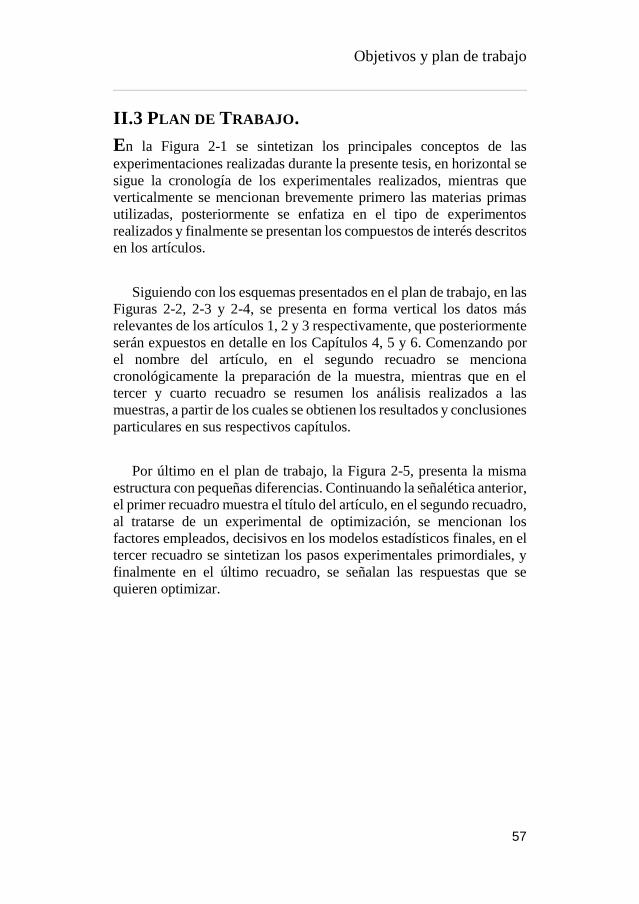
Objetivos y plan de trabajo
57
II.3 PLAN DE TRABAJO.
En la Figura 2-1 se sintetizan los principales conceptos de las
experimentaciones realizadas durante la presente tesis, en horizontal se
sigue la cronología de los experimentales realizados, mientras que
verticalmente se mencionan brevemente primero las materias primas
utilizadas, posteriormente se enfatiza en el tipo de experimentos
realizados y finalmente se presentan los compuestos de interés descritos
en los artículos.
Siguiendo con los esquemas presentados en el plan de trabajo, en las
Figuras 2-2, 2-3 y 2-4, se presenta en forma vertical los datos más
relevantes de los artículos 1, 2 y 3 respectivamente, que posteriormente
serán expuestos en detalle en los Capítulos 4, 5 y 6. Comenzando por
el nombre del artículo, en el segundo recuadro se menciona
cronológicamente la preparación de la muestra, mientras que en el
tercer y cuarto recuadro se resumen los análisis realizados a las
muestras, a partir de los cuales se obtienen los resultados y conclusiones
particulares en sus respectivos capítulos.
Por último en el plan de trabajo, la Figura 2-5, presenta la misma
estructura con pequeñas diferencias. Continuando la señalética anterior,
el primer recuadro muestra el título del artículo, en el segundo recuadro,
al tratarse de un experimental de optimización, se mencionan los
factores empleados, decisivos en los modelos estadísticos finales, en el
tercer recuadro se sintetizan los pasos experimentales primordiales, y
finalmente en el último recuadro, se señalan las respuestas que se
quieren optimizar.

Objetivos y plan de trabajo
58
Para el desarrollo del trabajo se propuso el siguiente orden
cronológico de acuerdo a las experimentaciones realizadas.
Figure -2-1 Approach of experiments in chronological order (source: private collection)

Objetivos y plan de trabajo
59
Figure -2-2 Article 1 experimental work (source: private collection)

Objetivos y plan de trabajo
60
Figure -2-3 Article 2 experimental work (source: private collection)

Objetivos y plan de trabajo
61
Figure -2-4 Article 3 experimental work (source: private collection)
Objetivos y plan de trabajo
62
Figure -2-5 Article 4 experimental work (source: private collection)


Capítulo 3 METODOLOGÍA
EXPERIMENTAL

Material y métodos
66
III.1 MATERIAS PRIMAS
Para la realización de los estudios desarrollados se emplearon dos
especies frutícolas diferente.
III.1.1 Aguacate (Persea americana Mill)
Por un lado se estudiaron los residuos (piel y semilla) del aguacate
(Persea americana Mill. var. Hass), el cual fue adquirido en estado de
madurez comercial. Se utilizaron al menos 25 aguacates de la misma
variedad y lote de venta, de tamaño y color homogéneo, exentos de
alteraciones fisiológicas perceptibles al ojo. Las muestras se
almacenaron a 4 °C hasta su análisis.
III.1.2 Higos chumbos (Opuntia Spp).
En las determinaciones de la especie Opuntia se utilizaron diferente
variedades, 2 comerciales (Opuntia ficus-indica var. Gialla y Opuntia
ficus-indica var. Sanguigna), adquiridas en su estado de maduración
comercial, y 2 variedades salvajes (Opuntia engelmannii y Opuntia
engelmannii cv. Valencia), ambas fueron recolectadas en los meses de
Septiembre, cuando su maduración hortícola era optima, la primer
variedad se recolectó en las periferias de Bragança, Portugal, mientras
que la segunda variedad fue recolectada en la ciudad de Valencia,
España. Siempre se seleccionaron los frutos de tamaño y color
homogéneo, excentos de alteraciones fisiológicas perceptibles al ojo.
Normalmente se partía de un pool de mas de 30 unidades, de las cuales
se seleccionaban aleatoriamente al menos 10 unidades y como minimo
para cada una de las determinaciones, se empleaban al menos 3
unidades. Las muestras se almacenaron a 4 °C hasta su preparación para
las determinaciones.

Material y métodos
67
III.2 PREPARACIÓN DE LA MUESTRA
Previamente al uso de los frutos para las determinaciones analíticas,
estos recibieron distintos tratamientos.
III.2.1 Aguacates
Los frutos fueron lavados para eliminar cualquier tipo de impureza,
secados al aire libre y cortados por la mitad. La semilla fue separada,
brevemente lavada y secada al aire libre, mientras que las pieles fueron
separadas de las pulpas mediante la ayuda de una espátula y
porteriormente lavadas para eliminar los residuos de las pulpas y
secadas al aire libre.
Figure 3-1 Persea americana fruit fractions (source: private collection)

Material y métodos
68
III.2.2 Higos chumbos
A diferencia de los frutos de aguacate, los higos chumbos fueron
cuidadosamente lavados bajo un chorro de agua con la ayuda de una
esponja para remover los gloquidios, y posteriormente secados al aire
libre. La separación de las pieles fue hecha realizando 2 cortes
verticales en los extremos del fruto y un posterior corte vertical, por
donde se realizó la separación. La separación de las semillas (cuando
fue necesario) se realizó mediante el uso de una licuadora compacta
Moulinex Vitae JU200045, obteniendo por una lado la pulpa y zumo y
por otro lado las semillas, que posteriormente son lavadas y secadas al
aire libre.
Figure 3-2 Opuntia spp fruits fractions (source: private collection)

Material y métodos
69
III.2.3 Tratamientos térmicos.
Las muestras, dependiendo del diseño experimental, fueron usadas
frescas, secas o liofilizadas, debido a que estas son las condiciones
típicas utilizadas que se citan comunmente en la bibliografía cientifica.
El secado se realizó siempre en dos etapas, la primera, un secado de
las distintas fracciones por 24 horas en una estufa de secado por
convección modelo Conterm 2000210 (JP Selecta S.A., España) a 60°
C, transcurrido el tiempo necesario, las muestras se secan con una
estufa a vacio modelo Vaciotem-Tv (JP Selecta S.A., España) a 60° C
y un vacío máximo de 0,01 mm Hg por 72 horas o hasta que se obtuvo
peso constante.
Por otro lado, para conservar mejor la integridad de las muestras
biologícas, se utilizó la liofilización como tratamiento de conservación.
Las fracciones de los frutos fueron previamente congeladas a -32° C
utilizando un congelador convencional durante 24 horas, para
posteriormente ser deshidratadas en un liofilizador modelo Telstar
Lioalfa-6 (Azbil corp., Japón) con cámara cerrada a vacío por 72 horas
a -50° C.
Una vez secas las muestras, se disminuyó el tamaño de particula
mediante el uso de una picadora Moulinex A320R1 y un posteriror
cribado mediante una malla fina. Las muestras fueron después
envasadas a vacío hasta su análisis

Material y métodos
70
III.3 PARAMETROS MORFOLOGICOS
III.3.1 Dimensiones y peso
Para la caracterización, los frutos enteros y sus distintas fracciones
fueron pesadas en una balanza analítica Secura 224-1S (Secura Lab
instruments GmbH, Alemania) y sus dimensiones (alto y ancho) fueron
medidas mediante el uso de un calibrador Vernier.
III.3.2 Color
Las coordenadas de color fueron determinadas en dos formas distintas,
las muestras sólidas fueron medidas mediante el uso de un colorímetro
Konica Minolta CR-400 (Konica Minolta Sensing Inc, Japón) con el
uso del iluminante C y una apertura del diafragma de 8 mm, midiendo
por triplicado en tres puntos distintos elegidos al azar. Las muestras
líquidas fueron analizadas mediante el espectrofotómetro Minolta CM-
3600d (Konica Minolta Sensing Inc, Japón) usando el modo
reflactancia con la lente LAV, el componente especular excluido y una
difusión de iluminación con el angulo 65/10°, midiendo el triplicado de
las muestras por tlipicado cada una de ellas. En ambos se utilizó el
promedio de cada muestra y se utilizó el software Spectra magic
(Version 3.6, CyberChrome Inc, Japón) para reportar las coordenadas
cartesianas CIE Lab o las coordenadas cilíndricas CIE L*C*h*.

Material y métodos
71
III.4 CARACTERIZACIÓN NUTRICIONAL
III.4.1 Proteina
La determinación proteica se realizó por el método de Kjeldahl de
acuerdo a lo indicado en AOAC 991.29 (Association of Official
Analytical Chemists, 2016), Utilizando un digestor y destilador
automático modelo Pro Nitro A (JP Selecta S.A., España), los valores
obtenidos fueron ajustados con el factor de conversión proteica (N x
6,25)
III.4.2 Grasa
El ensayo de grasa total fue hecho por el método de Soxhlet de acuerdo
a lo indicado en AOAC 963.15, La extracción fue llevada a cabo con
éter de petróleo a 120 °C por 7 horas.
III.4.3 Humedad
La determinación de humedad de las muestras se realizó mediante el
método gravimétrico indicado en AOAC 930.04, en el cual se
emplearon estufas de vacio Vaciotem-Tv (JP Selecta S.A., España), a
una temperatura de 60 °C con la finalidad de evitar la caramelización
de los azúcares presentes, expresando el porcentaje de humedad de
acuerdo a la siguiente formula.
𝐻𝑢𝑚𝑒𝑑𝑎𝑑 = (𝑃𝑒𝑠𝑜 𝑓𝑟𝑒𝑠𝑐𝑜 − 𝑃𝑒𝑠𝑜 𝑠𝑒𝑐𝑜
𝑃𝑒𝑠𝑜 𝑆𝑒𝑐𝑜) × 100
Equation 3-1 Moisture determination
III.4.4 Cenizas
Para expresar el porcentaje de cenizas en las muestras, se ha empleado
el método gravimétrico 940.26 de la AOAC, removiendo primero la
humedad para después incinerar la muestra en un horno mufla Select-
horn-TFT (JP Selecta S.A., España), a 525 °C durante 5 horas.
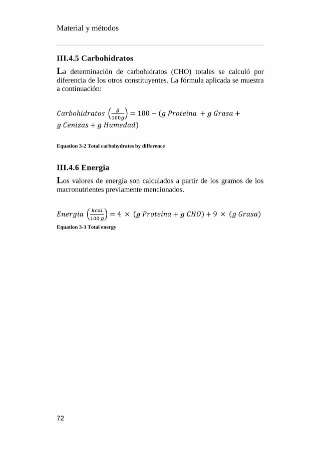
Material y métodos
72
III.4.5 Carbohidratos
La determinación de carbohidratos (CHO) totales se calculó por
diferencia de los otros constituyentes. La fórmula aplicada se muestra
a continuación:
𝐶𝑎𝑟𝑏𝑜ℎ𝑖𝑑𝑟𝑎𝑡𝑜𝑠 (𝑔
100𝑔) = 100 − (𝑔 𝑃𝑟𝑜𝑡𝑒𝑖𝑛𝑎 + 𝑔 𝐺𝑟𝑎𝑠𝑎 +
𝑔 𝐶𝑒𝑛𝑖𝑧𝑎𝑠 + 𝑔 𝐻𝑢𝑚𝑒𝑑𝑎𝑑)
Equation 3-2 Total carbohydrates by difference
III.4.6 Energia
Los valores de energía son calculados a partir de los gramos de los
macronutrientes previamente mencionados.
𝐸𝑛𝑒𝑟𝑔𝑖𝑎 (𝑘𝑐𝑎𝑙
100 𝑔) = 4 × (𝑔 𝑃𝑟𝑜𝑡𝑒𝑖𝑛𝑎 + 𝑔 𝐶𝐻𝑂) + 9 × (𝑔 𝐺𝑟𝑎𝑠𝑎)
Equation 3-3 Total energy

Material y métodos
73
III.5 COMPUESTOS HIDROFÍLICOS
III.5.1 Azúcares solubles
Los azúcares (fructosa, glucosa y sacarosa) fueron determinados a
partir de las muestras desengrasadas en Soxhlet mediante el método
previamente descrito por Pereira et al. (2011). Brevemente, las
muestras (1 g) son adiciondas con el patrón interno melezitosa (1 mL
[25 mg/mL]), extraídas con etanol 80% v/v a 80 °C por 1 h 30 min,
centrifugadas y evaporadas, para posteriormente ser lavadas 3 veces
con éter dietílico; la fase acuosa es recolectada, evaporada,
reconstituida en agua (5 mL) y filtrada a través de un filtro de nylon de
0,2 μm.
El extracto es posteriormente analizado mediante HPLC acoplado a
un detector de índice de refracción (IR) modelo Smartline system 1000
(Knauer, Alemania). La separación se llevó a cabo mediante una
columna Eurosphere 100-5 NH2 (4,6 x 250 mm, 5 μm) (Knauer,
Alemania), y como fase móvil se utilizó una mezcla de
acetonitrilo:agua (70:30 v/v). Los resultados fueron identificados y
comparados con sus respectivos patrones y procesados mediante el
software Clarity 2.4 (DataApex, República Checa).
III.5.2 Ácidos orgánicos
Los ácidos organicos detectados en las muestras (oxálico, quínico,
málico, ascórbico, cítrico, succínico y fumárico), se identificaron
mediante la metodología descrita por (Barros et al., 2013) utilizando
UFLC acoplado a un detector de diodos (DAD) con un equipo
Shimadzu 20A series (Shimadzu coorporation, Japon), usando una
columna SphereClone C18 en fase reversa (Phenomenex, USA) con
tamaño de particula de 5 μm y 4,6 x 250 mm, termostatada a 35 °C y
con elución isocrática de ácido sulfúrico 3,6 mM con un flujo de 0,8
mL/min. La detección de los compuestos se realizó a 215 nm, excepto
para el ácido ascórbico a 245 nm, la identificación y cuantificación se
llevó a cabo mediante comparación con patrones usando el software
Shimadzu LC solutions (Shimadzu coorporation, Japón).
La extracción de la muestra se realizó usando acído metafosfórico
(25 mL [4,5 % p/v]) a 24 °C con agitación magnética (150 rpm) y

Material y métodos
74
protegido de la luz. A continuación la muestra se filtró a través de papel
Whatman No. 4 y posteriormente con un filtro de nylon de 0,2 μm.

Material y métodos
75
III.6 COMPUESTOS LIPOFÍLICOS
III.6.1 Ácidos grasos
Las muestras extraidas en Soxhlet se derivatizan redisolviendo en una
solución de metanol:acído sulfúrico:tolueno (2:1:1 v/v/v), agitandose
en un vortex y otra vez extrayéndose con agitación (160 rpm) durante
12 h a 50 °C. Posteriormente, se adicionó agua destilada (3 mL) y se
agitó con vortex, después se agregó éter etílico (3 mL) y vortex
nuevamente, se esperó a la separación de fases y mediante una pipeta
pasteur se extrajo la fase lipofílica y se añadió a un vial con sulfato de
sodio anhídrido para capturar humedad. Por último se filtra con un filtro
de nylon de 0,2 μm.
La detección y cuantificación de ácidos grasos, se realizó mediante
Cromatografia de gases en el equipo Dani 1000 con detector de
ionización de flama (GC-FID) (Dani Instruments, Switzerland),
usando una columna capilar (Macherey-Nagel GmbH, Alemania) (30
m x 0,32 mm DI x 0,25 μm df ) dentro de un horno programado con las
siguientes rampas de temperatura de acuerdo a lo descrito previamente
por Pereira et al. (2012):
Table 3-1 GC temperature ramp for fatty acids
Time (min) Temperature (°C)
0 50
2 50
4,5 125
11,5 160
12,5 180
19 200
20 220
35 220
El gas acarreador empleado fue hidrógeno a un flujo de 4 mL/min
(0,61 bar), y Split injection (1:40) fue realizada a 250 °C. La

Material y métodos
76
identificación de los ésteres metílicos de ácidos grasos (FAME) se llevó
a cabo comparándolos con los tiempos de retención relativos de los
patrones empleados, usando el software CSW 1.7 (DataApex,
República Checa) y se expresaron en porcentaje relativo.
III.6.2 Tocoferoles
La extracción de estos compuestos se realizó colocando en un tubo
falcon la muestra (500 mg) junto a hidroxitolueno butilado (BHT) (250
μL [10 mg/mL en Hexano]), tocol (400 μL [50 μg/mL]) y metanol (4
mL) y se agitó en el vortex (1 min). A continuación y por duplicado se
adicionó hexano (4 mL), vortex (1 min), y solución concentrada de
NaCl (2 mL), Vortex (1 min). Las extracciones se centrifugaron (5 min,
4000g, 10 °C) en una centrifuga 5804R, (Eppendorf, Alemania) y se
recogieron los sobrenadantes en un vial protegidos de la luz.
Sucesivamente se deshidrataron los sobrenadantes agregandó sulfato de
sódio anhidrido (1 g) y se llevaron a sequedad en una corriente de
nitrógeno. Por último, el concentrado se redisuelve en hexano (2 mL) y
se filtró con un filtro de nylon de 0,2 μm.
La determinación cromatográfica se realizó con el método descrito
por (Heleno et al., 2010), empleando un cromatógrafo de liquidos
Smartline system 1000 (Knauer, Alemania) acoplado a un detector de
flourescencia FP-2020 (Jasco, Japón) programado para excitación a
290 nm y emisión a 330 nm. La separación se llevó a cabo con con una
columna YMC Polyamide II (Waters, Japón) (250 x 4,6 mm x 5 μm)
en fase normal termostatada a 30 °C. La fase móvil utilizada consistió
en n-hexano y etil acetato (70:30 v/v) en isocrático con un flujo de 1
mL/min y un volumen de inyección de 20 μL. Los compuestos fueron
identificados de acuerdo a la comparación de estándares usando la señal
de respuesta de flourescencia mediante el software Clarity 2.4
(DataApex, República Checa).

Material y métodos
77
III.7 PREPARACION DE EXTRACTOS
A lo largo del desarrollo de este trabajo, se probaron distintas
condiciones y tratamientos de extracción modificando diferentes
parámetros, empleando muestras frescas, secas o liofilizadas. A
continuación se mencionan sólo los procesos de extracción empleados
en las publicaciones que figuran en esta tesis.
III.7.1 Extracción por agitación
La muestra (1 g) se mezcló con 25 mL de solvente orgánico (variable
e indicado en cada capítulo) y se extrajo mediante agitación magnetica
(24 °C y 200 rpm) por 1 h, los extractos posteriormente se filtraron a
través de un papel Whatman No. 4. Los sobrenadantes se recolectaron
y los tejidos vegetales se volvieron a re-extraer como se mencionó
anteriormente. Subsecuentemente, los extractos obtenidos se
mezclaron y conservaron en refrigeración (4 °C) hasta su posterior uso.
Figure 3-3 Conventional extraction (source: private collection)
III.7.2 Extracción por Ultrasonidos
Para esta extracción se desarrollo un sistema el cual consiste en un
baño de ultrasonidos ATM40-·LCD (Labbox, España) con sistema de
control de tiempo y fecuencia (40 ± 2 kHz) conectado a un baño de
enfriamento Frigiterm TFT-30 (JP Selecta S.A., España) unido a un

Material y métodos
78
termostato de calefacción Tectron bio (JP Selecta S.A., España), para
controlar la temperatura de forma precisa, y la recirculación de agua
entre la unidad térmica y el baño ultrasónico se realizó con una bomba
peristáltica Pumpdrive 5006 (Heidolph, Alemania). Al mismo tiempo
se efectuo la agitación de las muestras a través de un agitador de cabezal
RZR 2021 (Heidolph, Alemania) a 200 rpm. (Las condiciones
especificas se mencionan en los capítulos correspondientes).
Figure 3-4 Ultrasonic extraction system (source: private collection)
Para las extracciones se pesaron las muestras en matraces Erlen
Meyer y se les adicionaron los volúmenes necesarios de solventes, estos
se introdujeron en el baño ultrasónico y se comienzó la agitación por 2
minutos con la finalidad de atemperar las muestras con la temperatura
del baño. Posteriormente, se realizó la extracción con el tiempo
necesario. Los extractos obtenidos se conservaron en refrigeración (4
°C) hasta su posterior uso.
III.7.3 Extracción por microondas
Independientemente de los tiempos y temperaturas empleadas
empleadas, las muestras siempre se pesaron en tubos de teflón
MarsXpress plus (CEM, USA) donde posteriormente se adicionó el
volumen necesario del solvente de extracción, estos se cerraron con la
válvula de salida de presión y su tapa, montados en el carrusel y
llevados al horno de microondas Mars 6 (CEM, USA), equipado con

Material y métodos
79
sistema de control digital de irradiación (iWave) para asegurar
temperatura uniforme en todos los tubos. El horno siempre trabajó a
400 W de potencia. (Las condiciones especificas se mencionan en los
capítulos correspondientes).
III.7.4 Procesos post-extracción
Los extractos obtenidos por cualquier extracción, se centrifugaron a
15585g en una centrifuga 5804-R (Eppendorf, Alemania) por 7 minutos
a 10 °C, a continuación, se filtraron a vacío usando un embudo Buchner
esmerilado con placa filtrante de porosidad 2 (Labbox, España). En el
caso de que el filtrado contuviera además de agua algún alcohol, este
se llevaba porsteriormente al rotavapor Hei-Vap Value (Heidolph,
Alemania) y se secó a vacío a 40 °C. Por último, el remanente acuoso,
se congela a -32° C en un congelador convencional durante 24 horas, y
posteriormente se liofilizaba (Telstar Lioalfa-6, Azbil corp.,Japón)
durante 72 horas a -50° C. Los extractos liofilizados se almaceron a
vacío en un ambiente seco y fresco hasta su uso posterior.
Figure 3-5 Lyophilized powders (source: private collection)

Material y métodos
80
III.8 DETERMINACIÓNES PRELIMINARES DE
COMPUESTOS FUNCIONALES
Tanto en las determinaciones preliminares como en las
determinaciones finales de las propiedades bioactivas, los extractos
liofilizados se redisolvieron en una mezcla de etanol ó metanol y agua
(80:20 v/v) a una concentración conocida a la que posteriormente se le
hacieron diluciones sucesivas con el objetivo de poder evaluar el efecto
de las distintas concentraciones y poder calcular los valores de
concentración de inhibición IC50 de las muestras.
Figure 3-6 Re-suspended samples (source: private collection)
III.8.1 Polifenoles por el metodo de Folin-Ciocalteu
El método descrito por Singleton y Rossi (1965), consiste en oxidar los
compuestos fenólicos presentes en la muestra, por medio de una mezcla
de ácidos fosfotúngstico y fosfomolíbdico conocida como reactivo de
Folin-Ciocalteu, los cuales en presencia de fenoles, son reducidos a
óxidos azules de tungsteno y molibdeno.
Para llevar a cabo la reacción se mezclaron el extracto (200 μL) con
el reactivo de Folin-Ciocalteu (1ml [1:10 v/v]), se agitó y se dejó
reaccionar por 4 minutos, a continuación se añade carbonato de sodio
(800 μL)[7,5 % p/v] y se dejó reaccionar en obscuridad por 30 min. En
paralelo se preparó un blanco con 200 μL de agua en lugar de extracto

Material y métodos
81
y una curva de calibrado (curva patrón) con ácido gálico [0-100 ppm]
y una vez transcurrido el tiempo de reacción se mide la absorbancia a
765 nm.
Figure 3-7 Folin-Ciocalteu samples (source: private collection)
III.8.2 Flavonoides por el metodo de tricloro aluminio
El método colorimétrico descrito por Zhishen et al. (1999) se realizó
mezclando el extracto (1 mL) con agua destilada (4 mL) y NaNO (0,3
mL [5% p/v]) el cual se agitó y se dejó reaccionar por 5 minutos, a
continuación se adicionó AlCl3 (0,3 mL [10 p/v]), se agitó y se dejó
reposar por 6 minutos, posteriormente se adicionó NaOH (2 mL [1 M]),
se agitó nuevamente y se añadió agua destilada (2,4 mL) para
finalmente leer la absorbancia a 510 nm. La curva de calibración de
Rutin y el blanco siguieron las mismas condiciones de reacción.

Material y métodos
82
Figure 3-8 ALCL3 colorimetric assay (source: private collection)
III.8.3 Betalainas por metodo espectrofotometrico
El método rápido para identificación y cuantificación proximal de
betalainas se consiguió tras un barrido espectrofotométrico de los
extractos de acuerdo a (Cassano et al., 2007), identificando las
betacianinas (Bc) a 530 nm y las betaxantinas (Bx) a 480 nm. Para su
cuantificación se utilizó las siguiente formula:
𝐵𝑒𝑡𝑎𝑙𝑎𝑖𝑛𝑎𝑠 = (𝐴 × 𝐹𝑑 × 𝑉 × 𝑃𝑚
𝑚 × 𝐿 × 𝜀)
Equation 3-4 Spectrophotometric betalainic content
Donde A = absorbancia (Bx = 480 o Bc= 530 nm); Fd = factor de
dilución; V = Volumen del extracto (L); Pm = peso molecular (betanina
= 550 g/mol, Indicaxantina = 308g/mol); m = masa de muestra (g); L =
longitud de la cuveta (1 cm); ε = coeficiente de extinción molar (Bx =
48000 L/mol cm, Bc = 60000 L/mol cm), obteniendo el resultado final
de las betalainas en g de betanina o indicaxantina por gramos de
muestra.

Material y métodos
83
Figure 3-9 SPE extraction of betalains (source: private collection)
Para obtener una mejor resolución del contenido, los extractos se
purificaron mediante un sistema de extracción de fase sólida (SPE) en
un cartucho C-18 SepPak® Vac 3 cc (Waters, USA). El cartucho se
activó con 2 volúmenes de metanol, seguido por 2 volúmenes de agua,
inmediatamente el extracto es retenido y posteriormente eluido con
agua y finalmente con metanol/ácido trifluoroacético (TFA) (99,9:0,1
v/v). Ambas freacciones fueron evaporadas y/o liofilizadas,
resuspendidas y analizadas por separado espectrofotométricamente.

Material y métodos
84
III.9 EVALUACIÓN DE PROPIEDADES BIOACTIVAS
Para determinar la bioactividad de los extractos a continuación se
describen los métodos experimentales que fueron realizados. Los
resultados son expresados en concentraciones minimas en las
actividades antimicrobianas, y para el resto de las pruebas antioxidantes
se usarón los valores de concentración efectiva EC50 y concentración
de inhibición IC50, que nos proporciona la información de la
concentración necesaria de los extractos para tener una bioactividad del
50%.
III.9.1 Actividad antibacteriana
Se determinaron las concentraciones minimas inhibitorias (MIC) y
minimas bactericidas (MBC) utilizando el método de microdilución del
comité nacional para estándares de laboratorios clínicos (NCCLS M38-
P), descrito por Espinel-Ingroff (2001), utilizando microorganismos
obtenidos del laboratorio de microbologia del departamento de
fisiología vegetal del Instituto para Investigación Biológica “Sinisa
Stanković” de la Universidad de Belgrado, Servia. Dichas bacterias
fueron, Gram-negativas: Escherichia coli (American Type Culture
Collection ATCC 11775), Pseudomonas aeruginosa (ATCC 27853),
Salmonella typhimurium (ATCC 13311) y Enterobacter cloacae
(ATCC 35030), y Gram-positivas: Bacillus cereus (ATCC) 14579),
Staphylococcus aureus (ATCC 6538), Micrococcus flavus (ATCC
10240) y Listeria monocytogenes (aislada clínicamente).
Los extractos se redisolvieron en agua [10 mg/mL] y diluidos
sucesivamente para el análisis, y los cultivos bacterianos fueron
ajustados espectrofotométricamente a una concentración de 1 x 10⁵
Unidades formadoras de Colonias (CFU)/mL. Los extractos se
añadieron a los pocillos de las placas espectrofotométricas donde se ha
adicionado previamente medio de digerido de soja y caseína (TSB)
(100 μL) y el inoculo bacterial (10 μL). Las placas se incubaron por 24
h a 37 °C. Despues la minima concentración de inhibición (MIC) se
determinó por el ensayo de viabilidad colorimétrica basado en la
reducción del cloruro de yodonitrotetrazolio (INT)(40 μL [0,2 mg/mL])
el cual fue adicionado en los pocillos después de la incubación según
lo descrito por Weinstein (2012).

Material y métodos
85
Por otro lado, la minima concetración bactericida (MBC) fue
determinada por subcultivo serial de la muestra (10 μL) en placas
espectrofotométricas que contienen TBS (100 μL), La concentración
más baja de las diluciones seriales en donde no observa crecimiento es
considerada entonces como la MBC. Los controles se trataron de la
misma manera que las muestras, utilizando 5% de dimetilsulfoóxido
(DMSO) como control negativo y estreptomicina y ampicilina como
controles positivos.
III.9.2 Actividad antifungica
La capacidad antifúngica fue probada en 8 cepas micóticas igualmente
proporcionadas por la Universidad de Belgrado. Estos ensayos se
ralizaron en: Aspergillus fumigatus (ATCC 1022), Aspergillus
ochraceus (ATCC 12066), Aspergillus versicolor (ATCC 11730),
Aspergillus niger (ATCC 6275), Penicillium funiculosum (ATCC
36839), Penicillium ochrochloron (ATCC 9112), Penicillium
verrucosum var cyclopium (aislada de alimento) y Trichoderma viride
(IAM culture collection 5061).
Los micromicetos se mantienen almacenados en agar de malta (MA)
a 4 °C y se subcultivan una vez por mes, antes de su uso, las esporas
fúngicas son lavadas con una solución estéril salina (0,85% p/v) que
contiene 0,1 % de Tween 80 (v/v). Esta suspensión se ajusta hasta
lograr una concentranción aproximada de 1,0 x 10⁵ usando un volumen
de 100 μL por pocillo.
La MIC se realizó mediante la técnica de dilución serial, los inoculos
se mezclan con el extracto redisuelto en DMSO (5% v/v) y se incubaron
por 72 h a 28 °C. La concentración más baja observada al microscopio
fue definida como MIC, mientras que la concentración minima
fungicida (MFC) fue determinada por subcultivo serial del extracto (2
μL) en los pocillos de las placas conteniendo agar de malta (100 μL),
se incubó por 72 h a 28 °C, y la concentración mas baja sin visible
crecimiento fue definido como MFC, indicando 99,5% de aniquilación
del inóculo. El control positivo fue el uso de bifonazol y ketokonazol,
y DMSO (5%) como control negativo.

Material y métodos
86
III.9.3 Ensayos capacidad antitumoral
Los ensayos de capacidad antitumoral fueron evaluados con 5 líneas
celulares tumorales humanas: MCF-7 (adenocarcinoma de mama),
NCI-H460 (cáncer de pulmón), HCT-15 (carcinoma de colon), HeLa
(carcinoma cervical) y HepG2 (carcinoma hepatocelular).
Las células fueron almacenadas como cultivo adherido de células en
medio inRPMI-1640, glutamina (2 mM), conteniendo 10 % de suero
fetal bovino (FBS) inactivadas por calor en las líneas MCF-7, NCI-
H460 y HCT-15. Mientras que las líneas celulares HeLa y HepG2,
fueron cultivadas en medio de Eagle modificado de Dulbecco (DMEM)
suplemenetado con 10 % FBS, glutamina (2 mM), penicilina (100
U/mL) y estreptomicina (100 mg/mL). Todas las líneas fueron
almacenadas a 37 °C en un incubador con aire humidificado con
contenido de 5% de CO2.
La metodología empleada fue la descrita por Monks et al. (1991),
dejando las líneas celulares adherirse a las placas por 24 h con densidad
celular de 7,5 x 10³ celulas/pocillo (MCF-7, NCI-H460 y HCT-15) y
1,0 x 10⁴ celulas/pocillo (HeLa y HepG2). Después de este tiempo, las
células fueron adicionadas con las distintas concentraciones de las
muestras analizadas por 48 h en incubación. Una vez transcurrido el
periodo anterior, los cultivos fueron adheridos con ácido tricloroacetico
(TCA, 100 μL [10% v/v]) e incubadas por 60 minutos a 4 °C.
Posteriormente, las placas fueron lavadas con agua destilada y secadas.
Una vez alcanzado el secado, se adicionó la solución de sulforrodamina
B (SRB, 100 μL, 0,1 % [1% v/v de ácido acético]), y se incubó por 30
min a temperatura ambiente. Las células no adheridas con SRB se
remueven lavando con ácido acético (1% v/v) y las células adheridas se
resolubilizan con solución Tris (200 μL, pH 7,4 [10 mM]) y se mide la
obsorvancia a 540 nm para determinar el valor GI50 (concentración de
la muestra con la que se inhibe el 50 % del crecimiento celular total).
El control positivo utilizado fue el alkaloide Ellipticina.

Material y métodos
87
Figure 3-10 SRB bond cell treated with Opuntia samples (source: private collection)
III.9.4 Ensayos de evaluación hepatotoxica
El cultivo celular designado como PLP2, fue preparado a partir de
hígado fresco porcino, obtenido localmente. Eal hígado fue enjuagado
con solución balanceada salina de Hank, adicionada con suero bovino
fetal (FBS) (10 % v/v), adicionada con penicilina (100 U/mL) y
estreptomicina (100 μg/mL), el tejido fue posteriormente dividido en
dimensiones de 1 x 1 mm3 del explante, e incubados a 37 °C en un
incubador con aire humidificado con contenido de 5% de CO2. El
medio fue cambiado cada 2-3 dias monitorizándolo con un microscopio
de contraste de fases.
Antes de su uso, las células fueron subcultivadas en los pocillos de
las placas espectrofotométricas (1,0 x 10⁴ celulas/pocillo) en medio
DMEM con FBS (10% v/v), penicilina (100 u/mL) y estreptomicina
(100 μg/mL). Posteriormente las células se tratan con las distintas
concentraciones de las muestras por 48 h y se procede al procedimiento
de sulforrodamina B descrito anteriormente, determinando el valor GI50
y utilizando ellipticina como control positivo.

Material y métodos
88
III.9.5 Actividad antioxidante por el método de DPPH
El fundamento consiste en que el radical DPPH tiene un electrón
desapareado y es de color azul-violeta, decolorándose hacia amarillo
pálido por la reacción en presencia de una sustancia antioxidante,
siendo medida espectrofotométricamente a 517 nm. Por diferencia de
absorbancia se determina el porcentaje de captación de radical libre
DPPH.
Figure 3-11 DPPH colourimetric assay (source: private collection)
La metodología descrita por Brand-Williams et al. (1995) consiste
en confluir el radical DPPH (60 μM en metanol) con las distintas
concentraciones de las muestras a evaluar en un ratio 9:1 v/v. Esta
mezcla se agita, se mantiene durante 30 minutos reaccionando en
obscuridad y se mide su absorbancia a 517 nm. El control negativo es
agua y una curva de calibración puede ser utilizada como referencia
usando trolox. El resultado se expresa en IC50 y se calcula a partir de la
siguiente fórmula interpolando los resultados obtenidos de las distintas
concentraciones analizadas.
% 𝑖𝑛ℎ𝑖𝑏𝑖𝑡𝑖𝑜𝑛 = (𝐴𝐵𝑆𝑐𝑜𝑛𝑡𝑟𝑜𝑙 − 𝐴𝐵𝑆𝑠𝑎𝑚𝑝𝑙𝑒
𝐴𝐵𝑆 𝑐𝑜𝑛𝑡𝑟𝑜𝑙) × 100
Equation 3-5 Inhibition percentage of DPPH

Material y métodos
89
III.9.6 Actividad antioxidante por el metodo del poder
reductor
El método del poder reductor (ó azul Prusia) descrito por Vieira et al.
(2016a), se basa en la capacidad de los compuestos antioxidantes en
reducir el Fe3+ en Fe2+, de acuerdo a las siguientes reacciones:
Figure 3-12 Power reduction method chemical reactions (source: private collection)
El procedimiento consiste en mezclar las distintas concentraciones
de las muestras (0,5 mL) con tampón fosfato de sodio (0,5 mL, pH 6,6,
200 nM), ferrocianato de potasio (0,5 mL, [1% p/v]) que se incubó a 50
°C durante 20 min.
Posteriormente se detiene la reacción adicionando ácido
tricloroacético (0,5 mL, [10 % v/v]), y la mitad del volumen (0,8 mL)
se mezcla con agua (0,8 mL) y se hace reaccionar adicionando cloruro
de hierro (160 μL, [0,1 % v/v]) y se lee la absorbancia a 690 nm. El
control negativo utilizado es agua destilada y el resultado se expresa en
IC50.

Material y métodos
90
Figure 3-13 Power reduction plate reaction (source: private collection)
III.9.7 Actividad antioxidante por el método de TBARS
Las sustancias reactivas al ácido tiobarbitúrico (TBARS) es quizá uno
de los métodos mas antiguos y utilizados para medir la peroxidación
lipídica que termina en malondialdehido (MDA). El MDA forma
aductos con 2 moléculas del ácido tiobarbitúrico que produce una
especie de color rosado que puede absorbe a 523 nm.
Para obtener este aducto, el método descrito por Devasagayam et al.
(2003), indica que para que un espécimen biológico reaccione debe ser
calentado. En el laboratorio se utilizó una masa de cerebro de cerdo y
el doble de tampón TRIS-HCl (20 mM, pH 7,4), y se llevo a agitación.
Acto seguido, se centrifugó la suspensión (3500g, 10 min), de la cual
se utilizaron 100 μL y se mezcló con las distintas concentraciones de
las muestras (200 μL), ácido ascórbico (100 μL) y sulfato ferroso (100
μL), la mezcla se colocó en un baño a 37,5 °C durante 1 h, y
posteriormente se hizo reaccionar con ácido tricloroacético (500 μL
[28% v/v]) y ácido tiobarbitúrico (380 μL, [2% p/v] preparado al
momento) en un baño a 80 °C durante 20 min. Esta mezcla se centrifuga
(3500g, 20 min) y se lee la absorbancia a 532 nm.
Para esta determinación se preparan 2 blancos, unos con el solvente
de extracción y otro con tampón TRIS-HCl en vez de extracto y se
determina el IC50.

Material y métodos
91
III.9.8 Actividad antioxidante por el método de β-caroteno
Esta metodología se basa en los productos formados entre los radicales
libres del ácido linoléico y los antioxidantes que protegen al β-caroteno.
El mecanismo de reacción aquí estudiado depende de la decoloración
de los carotenoides por calor que induce a la oxidación.
Para la evaluación de la capacidad antioxidante de las muestras se
utilizo el método descrito por (Marco, 1968), donde se procede a
preparar una emulsión disolviendo el β-caroteno (2 mg) en 10 mL de
cloroformo, del cual se transfieren solo 2 mL a un matraz y se evapora
el cloroformo a 40 °C, posteriormente se adicionan el ácido linoléico
(40 mg), Tween 80 (400 mg) y agua destilada (100 mL) y se agita
vigorosamente hasta formar la emulsión. De la emulsion obtenida se
transfiere (4,8 mL) a tubos de ensayo donde previamente se adicionaron
las distintas concentraciones de las muestras estudiadas ó un blanco
(solvente de extracción), se agitan en vortex y se lee la absorbancia a
470 nm. Por último se incuban los tubos en un baño a 50 °C por 2 h con
agitación constante (50 rpm) y se lee nuevamente la absorbancia una
vez transcurrido el tiempo de reacción. El IC50, se calcula apartir de la
siguiente formula:
% 𝑖𝑛ℎ𝑖𝑏𝑖𝑡𝑖𝑜𝑛 = (𝐴𝐵𝑆 𝑎𝑓𝑡𝑒𝑟 2 ℎ
𝐴𝐵𝑆 𝑖𝑛𝑖𝑡𝑖𝑎𝑙) × 100
Equation 3-6 Inhibition percentage of b-carotene
III.9.9 Actividad antioxidante por el método de ABTS
Otro método de transferencia de electrón es el brevemente denominado
ABTS. En este ensayo el radical catión 2,2´-azino-bis-(3-etil-
benzotiazolin-6-ácido-sulfónico) que tiene una coloración azul
obscuro, es reducido por la acción del persulfato potásico a incoloro, la
oxidación del peroxidisulfato/ABTS sucede de forma estequiometrica
0,5:1, por lo que al combinar ABTS (7mM) y persulfato (2,45 mM) la
reacción no es completada, y la absorbancia de esta reacción es medida
a 734 nm.

Material y métodos
92
El procedimiento descrito por Devasagayam et al. (2003), comienza
con la preparación de la disolución ABTS/persulfato mencionada
anteriormente con tampón fosfato (pH 7,4; [0,2 M]) con la finalidad de
liberar el radical, por lo que se deja la disolución en obscuridad entre
12-16 h en obscuridad y temperatura ambiente. Después del tiempo
necesario, se diluye la solución con tampón fosfato hasta obtener una
absorbancia de 0,7, y a continuación se mezclan 970 μL del ABTS con
las distintas concentraciones de los extractos (30 μL) y se leen las
absorbancias a los 6 min de reacción. El blanco se realiza con el
solvente de extracción y el valor IC50, se calcula mediante la siguiente
formula:
% 𝑖𝑛ℎ𝑖𝑏𝑖𝑡𝑖𝑜𝑛 = (𝐴𝐵𝑆𝑏𝑙𝑎𝑛𝑘 − 𝐴𝐵𝑆𝑠𝑎𝑚𝑝𝑙𝑒
𝐴𝐵𝑆𝑏𝑙𝑎𝑛𝑘) × 100
Equation 3-7 Inhibition percentage of ABTS

Material y métodos
93
III.10 IDENTIFICACIÓN Y CUANTIFICACIÓN DE
BIOCOMPUESTOS
Para la determinación, identificación y cuantificación de los
compuestos con propiedades funcionales, se utilizaron técnicas
analíticas, principalmente, cromatografía líquida acoplada a detector de
diodos mediante el equipo Dionex Ultimate 3000 UPLC-LTQ XL
(ThermoScientific, USA) y a detector de masas de trampa iónica con
una fuente de electro-spay (UPLC-DAD-ESI/MS).
Las identificaciones de compuestos se ralizaron mediante la
comparación de patrones de fraccionamiento, tiempos de retención y
espectros UV-vis de los patrones, siempre que estos eran disponibles,
y en caso contrario, comparados con la información científica
disponible en la literatura.
La cuantificación fue realizada comparando las curvas de
calibración de los patrones disponibles y cuando estos no estaban
disponibles, la cuantificación se hizó con los patrones del mismo grupo
y los resultados de los compuestos se expresaron en mg/g de extracto,
mediante el uso del software Xcalibur data system (ThermoFinnigan,
USA).
Figure 3-14 UPLC-DAD-Esi/MS system employed (source: private collection)

Material y métodos
94
El equipo cromatográfico cuenta con bomba cuaternaria,
desgasificador y auto-sampler a 5 °C. La separación cromatografica fue
llevada a cabo mediante la columan Waters Spherisorb S3 ODS-2C18
(Waters, USA) con tamaño de particula de 3 μm, diámetro interior de
4,6 mm y longitud de 150 mm ó en con una columna Luna C18 (2) con
las mismas especificaciones que en la anterior, exceptuando la longitud,
la cual en este caso es de 250 mm. Las columnas se mantuvieron en un
compartimento termostatado a 35 °C.
III.10.1 Polifenoles y Flavonoides
Para los compuestos fenólicos se utilizaron las longitudes de onda de
280 y 370 nm para los polifenoles y flavonoides, respectivamente. Los
solventes usados fueron: (A) 0,1% (v/v) de ácido trifluoroacético (TFA)
en agua y (B) acetonitrilo con flujo contante de 0,5 mL/min, y el
gradiente de elución fue el siguiente:
Figure 3-15 Elution gradient of solvent B in phenolics determination (source: private
collection)
La detección de masas se realizó en modo negativo, con nitrógeno
como gas envolvente (50 psi), el sistema se controló en el voltaje de
volatización (5 kV), temperatura (325 °C) y voltaje capilar (-20 V). El
voltaje de las lentes tubulares se mantuvo a -66 V, la energía de colisión
15 1520
25
35
50
15
0
10
20
30
40
50
60
0 5 10 15 20 25 30 35 40 45 50 55 60
So
lven
t B
(%
)
time (min)
Elution gradient
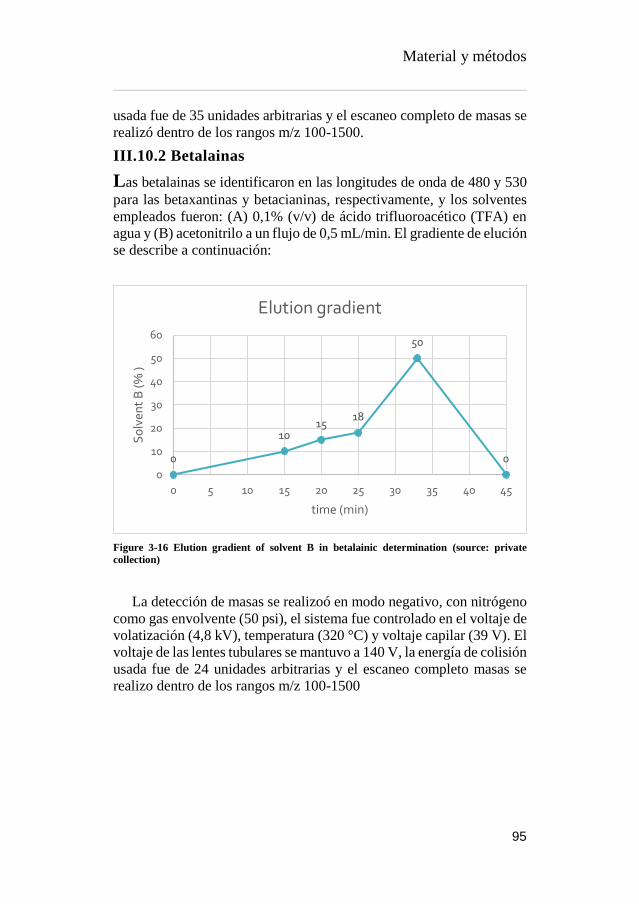
Material y métodos
95
usada fue de 35 unidades arbitrarias y el escaneo completo de masas se
realizó dentro de los rangos m/z 100-1500.
III.10.2 Betalainas
Las betalainas se identificaron en las longitudes de onda de 480 y 530
para las betaxantinas y betacianinas, respectivamente, y los solventes
empleados fueron: (A) 0,1% (v/v) de ácido trifluoroacético (TFA) en
agua y (B) acetonitrilo a un flujo de 0,5 mL/min. El gradiente de elución
se describe a continuación:
Figure 3-16 Elution gradient of solvent B in betalainic determination (source: private
collection)
La detección de masas se realizoó en modo negativo, con nitrógeno
como gas envolvente (50 psi), el sistema fue controlado en el voltaje de
volatización (4,8 kV), temperatura (320 °C) y voltaje capilar (39 V). El
voltaje de las lentes tubulares se mantuvo a 140 V, la energía de colisión
usada fue de 24 unidades arbitrarias y el escaneo completo masas se
realizo dentro de los rangos m/z 100-1500
0
1015
18
50
0
0
10
20
30
40
50
60
0 5 10 15 20 25 30 35 40 45
So
lven
t B
(%
)
time (min)
Elution gradient

Material y métodos
96
III.11 DETERMINACIÓN Y MODELIZACIÓN DE
ISOTERMAS DE SORCIÓN
La determinación de las isotermas de adsorción/desorción, se realizó
mediante el método estático gravimétrico, el cual consiste en la
determinación de la humedad en equilibrio de la muestra en un
medioambiente de humedad relativa conocida, lo cual se logra
manteniendo las muestras analizadas dentro de una cámara termo-
controlada cerrada herméticamente con disoluciones salina saturada de
actividad de agua (aw) conocida.
Las sales empleadas utilizadas fueron seleccionadas para cubrir una aw
en el rango de 0,1 a 0,9 (LiCl, MgCl2, K2CO3, Mg(NO3)2, NaCl, KCl y
BaCl2).
Las muestras de adsorción se sometieron a secado a 60 °C en estufa de
vacío para evitar reaciones de caramelización de los azúcares
intrínsecos, mientras que para las isotermas de desorción se utilizaron
las muestras frescas. La determinación de humedad en equilibrio se
llevó a cabo midiendo iterativamente los pesos de las muestras, hasta
que estas reportaron peso constante en equilibrio a lo largo del tiempo.
Una vez obtenido el peso constante, se calcula la humedad en
equilibrio de los triplicados de las muestras mediante la fórmula:
𝑋𝑏𝑠 = (𝑀𝑎𝑠𝑎 𝑑𝑒 𝑎𝑔𝑢𝑎 𝑒𝑛 𝑒𝑙 𝑒𝑞𝑢𝑖𝑙𝑖𝑏𝑟𝑖𝑜
𝑀𝑎𝑠𝑎 𝑠𝑒𝑐𝑎 𝑑𝑒 𝑙𝑎 𝑚𝑢𝑒𝑠𝑡𝑟𝑎)
Equation 3-8 Moisture equilibrium of the sorption isotherms
Los resultados de Xbs se grafican y modelan. Posteriormente, a través
de un software estadístico empleando las expresiones de los modelos
de mayor uso y citados en la Tabla 3-2.

Material y métodos
97
Table 3-2 Common correlation models of sorption Isotherms (Mayor et al., 2013)
Material y métodos
98
III.12 REFERENCES
Ahn, J.H., Jang, Y.-S., Lee, S.Y., 2016. Production of succinic acid by
metabolically engineered microorganisms. Curr. Opin.
Biotechnol. 42, 54–66.
https://doi.org/10.1016/j.copbio.2016.02.034
Al-Farsi, M.A., Lee, C.Y., 2008. Optimization of phenolics and dietary
fibre extraction from date seeds. Food Chem. 108, 977–985.
https://doi.org/10.1016/j.foodchem.2007.12.009
Albano, C., Negro, C., Tommasi, N., Gerardi, C., Mita, G., Miceli, A.,
De Bellis, L., Blando, F., 2015. Betalains, Phenols and
Antioxidant Capacity in Cactus Pear [Opuntia ficus-indica (L.)
Mill.] Fruits from Apulia (South Italy) Genotypes. Antioxidants
4, 269–280. https://doi.org/10.3390/antiox4020269
Albuquerque, T.G., Santos, F., Sanches-Silva, A., Beatriz Oliveira, M.,
Bento, A.C., Costa, H.S., 2016. Nutritional and phytochemical
composition of Annona cherimola Mill. fruits and by-products:
Potential health benefits. Food Chem. 193, 187–195.
https://doi.org/10.1016/j.foodchem.2014.06.044
Allai, L., Druart, X., Öztürk, M., BenMoula, A., Nasser, B., El Amiri,
B., 2016. Protective effects of Opuntia ficus-indica extract on ram
sperm quality, lipid peroxidation and DNA fragmentation during
liquid storage. Anim. Reprod. Sci. 175, 1–9.
https://doi.org/10.1016/j.anireprosci.2016.09.013
Almeida, M.M.B., de Sousa, P.H.M., Arriaga, Â.M.C., do Prado, G.M.,
Magalhães, C.E. de C., Maia, G.A., de Lemos, T.L.G., 2011.
Bioactive compounds and antioxidant activity of fresh exotic
fruits from northeastern Brazil. Food Res. Int. 44, 2155–2159.
https://doi.org/10.1016/j.foodres.2011.03.051
Alvarez, B., 2007. Análisis de factibilidad del cultivo de la tuna en la
localidad de Icaño , Departamento La Paz, Dirección provincial
de programación del desarrollo, Ministerio de Producción y
Desarrollo. Gobierno de la Provincia de Catamarca. Catamarca,
Argentina.
Alzate T, L.M., González, D., Hincapié, S., Cardona S, B.L., Londoño-
Londoño, J., Jiménez-Cartagena, C., 2016. The profile of

Material y métodos
99
bioactive substances in ten vegetable and fruit by-products from
a food supply chain in Colombia. Sustain. Prod. Consum.
https://doi.org/10.1016/j.spc.2016.07.005
Ammar, I., Ben Salem, M., Harrabi, B., Mzid, M., Bardaa, S., Sahnoun,
Z., Attia, H., Ennouri, M., 2018. Anti-inflammatory activity and
phenolic composition of prickly pear (Opuntia ficus-indica)
flowers. Ind. Crops Prod. 112, 313–319.
https://doi.org/10.1016/j.indcrop.2017.12.028
Angulo-Bejarano, P.I., Martínez-Cruz, O., Paredes-López, O., 2014.
Phytochemical Content, Nutraceutical Potential and
Biotechnological Applications of an Ancient Mexican Plant:
Nopal (Opuntia ficus-indica). Curr. Nutr. Food Sci. 10, 196–217.
https://doi.org/10.2174/157340131003140828121015
Arlt, U., 2010. NATCOL | Foods Colours Legislation.
Aruwa, C.E., Amoo, S.O., Kudanga, T., 2018. Opuntia (Cactaceae)
plant compounds, biological activities and prospects – A
comprehensive review. Food Res. Int. 112, 328–344.
https://doi.org/10.1016/j.foodres.2018.06.047
Association of Official Analytical Chemists, 2016. Official Methods of
Analysis of AOAC International, 20th ed. The Association,
Gaithersburg, MD, USA.
Attokaran, M., 2011. Natural Food Flavors and Colorants Natural Food
Flavors and Colorants, 1st ed. Blackwell Publishing LTD, Iowa.
Augustin, M.A., Sanguansri, L., 2015. Challenges and Solutions to
Incorporation of Nutraceuticals in Foods. Annu. Rev. Food Sci.
Technol. 6, 463–477. https://doi.org/10.1146/annurev-food-
022814-015507
Ayala-Zavala, J.F., Vega-Vega, V., Rosas-Domínguez, C., Palafox-
Carlos, H., Villa-Rodriguez, J.A., Siddiqui, M.W., Dávila-Aviña,
J.E., González-Aguilar, G.A., 2011. Agro-industrial potential of
exotic fruit byproducts as a source of food additives. Food Res.
Int. 44, 1866–1874.
https://doi.org/10.1016/j.foodres.2011.02.021
Azeredo, H.M.C., 2009. Betalains: Properties, sources, applications,
and stability - A review. Int. J. Food Sci. Technol. 44, 2365–2376.
https://doi.org/10.1111/j.1365-2621.2007.01668.x
Balouiri, M., Sadiki, M., Ibnsouda, S.K., 2016. Methods for in vitro
evaluating antimicrobial activity: A review. J. Pharm. Anal. 6,
71–79. https://doi.org/10.1016/j.jpha.2015.11.005
Barros, L., Calhelha, R.C., Queiroz, M.J.R.P., Santos-Buelga, C.,
Santos, E.A., Regis, W.C.B., Ferreira, I.C.F.R., 2015. The

Material y métodos
100
powerful in vitro bioactivity of Euterpe oleracea Mart. seeds and
related phenolic compounds. Ind. Crops Prod. 76, 318–322.
https://doi.org/10.1016/j.indcrop.2015.05.086
Barros, L., Pereira, C., Ferreira, I.C.F.R., 2013. Optimized Analysis of
Organic Acids in Edible Mushrooms from Portugal by Ultra Fast
Liquid Chromatography and Photodiode Array Detection. Food
Anal. Methods 6. https://doi.org/10.1007/s12161-012-9443-1
Barrows, J.N., Lipman, A.L., Bailey, C.J., Cianci, S., 2003. Color
Additives: FDA’s Regulatory Process and Historical
Perspectives. Food Saf. Mag. URL
https://www.fda.gov/forindustry/coloradditives/regulatoryproces
shistoricalperspectives/ (accessed 9.3.18).
Baydar, N.G., Özkan, G., Sağdiç, O., 2004. Total phenolic contents and
antibacterial activities of grape (Vitis vinifera L.) extracts. Food
Control 15, 335–339. https://doi.org/10.1016/S0956-
7135(03)00083-5
Belščak-Cvitanović, A., Durgo, K., Huđek, A., Bačun-Družina, V.,
Komes, D., 2018. Metabolism and Health Effects of Polyphenols,
in: Galanakis, C.M. (Ed.), Polyphenols: Properties, Recovery, and
Applications. Woodhead Publishing, pp. 1–44.
https://doi.org/https://doi.org/10.1016/B978-0-12-813572-
3.00001-4
Belwal, T., Dhyani, P., Bhatt, I.D., Rawal, R.S., Pande, V., 2016.
Optimization extraction conditions for improving phenolic
content and antioxidant activity in Berberis asiatica fruits using
response surface methodology (RSM). Food Chem. 207, 115–
124. https://doi.org/10.1016/j.foodchem.2016.03.081
Bessada, S.M.F., Barreira, J.C.M., Barros, L., Ferreira, I.C.F.R.,
Oliveira, M.B.P.P., 2016. Phenolic profile and antioxidant
activity of Coleostephus myconis (L.) Rchb.f.: An underexploited
and highly disseminated species. Ind. Crops Prod. 89, 45–51.
https://doi.org/10.1016/j.indcrop.2016.04.065
Betancourt, C., Cejudo-Bastante, M.J., Heredia, F.J., Hurtado, N.,
2017. Pigment composition and antioxidant capacity of
betacyanins and betaxanthins fractions of Opuntia dillenii (Ker
Gawl) Haw cactus fruit. Food Res. Int. 101, 173–179.
https://doi.org/10.1016/j.foodres.2017.09.007
Bhatia, S., Bera, T., Dahiya, R., Bera, T., Bhatia, S., Bera, T., 2015.
Classical and Nonclassical Techniques for Secondary Metabolite
Production in Plant Cell Culture, in: Modern Applications of Plant
Biotechnology in Pharmaceutical Sciences. Elsevier, pp. 231–
Material y métodos
101
291. https://doi.org/10.1016/B978-0-12-802221-4.00007-8
Brand-Williams, W., Cuvelier, M.E., Berset, C., 1995. Use of a free
radical method to evaluate antioxidant activity. LWT - Food Sci.
Technol. 28, 25–30. https://doi.org/10.1016/S0023-
6438(95)80008-5
Bravo-Hollis, H., 1978. Las cactaceas de Mexico. Universidad
Nacional Autonoma de Mexico, Mexio.
https://doi.org/10.15713/ins.mmj.3
Brundtland, G.H., 1987. Our common future.
Cai, Y., Sun, M., Corke, H., 2003. Antioxidant activity of betalains
from plants of the Amaranthaceae. J. Agric. Food Chem. 51,
2288–2294. https://doi.org/10.1021/jf030045u
Calderón-Oliver, M., Escalona-Buendía, H.B., Medina-Campos, O.N.,
Pedraza-Chaverri, J., Pedroza-Islas, R., Ponce-Alquicira, E.,
2016. Optimization of the antioxidant and antimicrobial response
of the combined effect of nisin and avocado byproducts. LWT -
Food Sci. Technol. 65, 46–52.
https://doi.org/10.1016/j.lwt.2015.07.048
Cardoso-Ugarte, G.A., Sosa-Morales, M.E., Ballard, T., Liceaga, A.,
San Martín-González, M.F., 2014. Microwave-assisted extraction
of betalains from red beet (Beta vulgaris). LWT - Food Sci.
Technol. 59, 276–282. https://doi.org/10.1016/j.lwt.2014.05.025
Carocho, M., Ferreira, I.C.F.R., 2013. A review on antioxidants,
prooxidants and related controversy: Natural and synthetic
compounds, screening and analysis methodologies and future
perspectives. Food Chem. Toxicol. 51, 15–25.
https://doi.org/10.1016/j.fct.2012.09.021
Carr, M., 2013. The water relations and irrigation requirements of
avocado (Persea americana Mill.): a review. Exp. Agric. 49, 256–
278. https://doi.org/10.1017/S0014479712001317
Cassano, A., Conidi, C., Timpone, R., D’Avella, M., Drioli, E., 2007.
A membrane-based process for the clarification and the
concentration of the cactus pear juice. J. Food Eng. 80, 914–921.
https://doi.org/10.1016/j.jfoodeng.2006.08.005
Castellanos-Santiago, E., Yahia, E.M., 2008. Identification and
quantification of betalains from the fruits of 10 Mexican prickly
pear cultivars by high-performance liquid chromatography and
electrospray ionization mass spectrometry. J. Agric. Food Chem.
56, 5758–5764. https://doi.org/10.1021/jf800362t
Castellar, R., Obón, J.M., Alacid, M., Fernández-López, J.A., 2003.
Color Properties and Stability of Betacyanins from Opuntia

Material y métodos
102
Fruits. J. Agric. Food Chem. 51, 2772–2776.
https://doi.org/10.1021/jf021045h
Cejudo Bastante, M.J., Chaalal, M., Louaileche, H., Parrado, J.,
Heredia, F.J., 2014. Betalain Profile, Phenolic Content, and Color
Characterization of Different Parts and Varieties of Opuntia ficus-
indica. J. Agric. Food Chem. 62, 8491–8499.
https://doi.org/10.1021/jf502465g
Chahdoura, H., Barreira, J.C.M., Barros, L., Santos-Buelga, C.,
Ferreira, I.C.F.R., Achour, L., 2014. Phytochemical
characterization and antioxidant activity of Opuntia microdasys
(Lehm.) Pfeiff flowers in different stages of maturity. J. Funct.
Foods 9, 27–37. https://doi.org/10.1016/j.jff.2014.04.011
Chahdoura, H., Barreira, J.C.M., Fernández-Ruiz, V., Morales, P.,
Calhelha, R.C., Flamini, G., Soković, M., Ferreira, I.C.F.R.,
Achour, L., 2016. Bioactivity, proximate, mineral and volatile
profiles along the flowering stages of Opuntia microdasys
(Lehm.): defining potential applications. Food Funct. 7, 1458–67.
https://doi.org/10.1039/c5fo01536g
Chan, L.G., Cohen, J.L., Maria, J., Nobrega, L., Bell, D.M., 2018.
Conversion of Agricultural Streams and Food-Processing By-
Products to Value-Added Compounds using Filamentous Fungi.
Annu. Rev. Food Sci. Technol. 9, 1–21.
https://doi.org/10.1146/annurev-food-030117-012626
Chougui, N., Djerroud, N., Naraoui, F., Hadjal, S., Aliane, K., Zeroual,
B., Larbat, R., 2015. Physicochemical properties and storage
stability of margarine containing Opuntia ficus-indica peel extract
as antioxidant. Food Chem. 173, 382–90.
https://doi.org/10.1016/j.foodchem.2014.10.025
Chougui, N., Tamendjari, A., Hamidj, W., Hallal, S., Barras, A.,
Richard, T., Larbat, R., 2013. Oil composition and
characterisation of phenolic compounds of Opuntia ficus-indica
seeds. Food Chem. 139, 796–803.
https://doi.org/10.1016/j.foodchem.2013.01.054
Clifford, M.N., Johnston, K.L., Knight, S., Kuhnert, N., 2003.
Hierarchical Scheme for LC-MSn Identification of Chlorogenic
Acids. J. Agric. Food Chem. 51, 2900–2911.
https://doi.org/10.1021/jf026187q
Clifford, M.N., Knight, S., Kuhnert, N., 2005. Discriminating between
the six isomers of dicaffeoylquinic acid by LC-MSn. J. Agric.
Food Chem. 53, 3821–3832. https://doi.org/10.1021/jf050046h
Cowan, A.K., Cripps, R.F., Richings, E.W., Taylor, N.J., 2001. Fruit

Material y métodos
103
size: Towards an understanding of the metabolic control of fruit
growth using avocado as a model system. Physiol. Plant. 111,
127–136. https://doi.org/10.1034/j.1399-3054.2001.1110201.x
Cowan, A.K., Wolstenholme, B.N., 2003. AVOCADOS, in:
Encyclopedia of Food Sciences and Nutrition. Elsevier, pp. 348–
353. https://doi.org/10.1016/B0-12-227055-X/00073-0
Crane, J.H., Balerdi, C.F., Maguire, I., 2016. Avocado Growing in the
Florida Home Landscape.
Da-Costa-Rocha, I., Bonnlaender, B., Sievers, H., Pischel, I., Heinrich,
M., 2014. Hibiscus sabdariffa L. – A phytochemical and
pharmacological review. Food Chem. 165, 424–443.
https://doi.org/10.1016/j.foodchem.2014.05.002
da Silva, R.P.F.F., Rocha-Santos, T.A.P., Duarte, A.C., 2016.
Supercritical fluid extraction of bioactive compounds. TrAC -
Trends Anal. Chem. 76, 40–51.
https://doi.org/10.1016/j.trac.2015.11.013
Dabas, D., Shegog, R.M., Ziegler, G.R., Lambert, J.D., 2013. Avocado
(Persea americana) seed as a source of bioactive phytochemicals.
Curr. Pharm. Des. 19, 6133–40.
https://doi.org/10.2174/1381612811319340007
Day, L., Seymour, R.B., Pitts, K.F., Konczak, I., Lundin, L., 2009.
Incorporation of functional ingredients into foods. Trends Food
Sci. Technol. 20, 388–395.
https://doi.org/10.1016/J.TIFS.2008.05.002
DeFelice, S.L., 2002. FIM Rationale And Proposed Guidelines For The
Nutraceutical Research & Education Act - NREA. New York.
Delgado-Vargas, F., Paredes-Lopez, O., 2003. Natural Colorants for
Food and Nutraceutical Uses, Trends in Food Science &
Technology. https://doi.org/10.1016/S0924-2244(03)00076-1
Destro dos Santos, C., Külzer Scherer, R., Schilling Cassini, A.,
Ferreira Marczak, L.D., Tessaro, I.C., 2016. Clarification of red
beet stalks extract by microfiltration combined with
ultrafiltration. J. Food Eng. 185, 35–41.
https://doi.org/10.1016/j.jfoodeng.2016.03.031
Devalaraja, S., Jain, S., Yadav, H., 2011. Exotic fruits as therapeutic
complements for diabetes, obesity and metabolic syndrome. Food
Res. Int. 44, 1856–1865.
https://doi.org/10.1016/j.foodres.2011.04.008
Devasagayam, T.P.A., Boloor, K.K., Ramasarma, T., 2003. Methods
for estimating lipid peroxidation: An analysis of merits and
demerits. Indian J. Biochem. Biophys. 40, 300–308.

Material y métodos
104
Dias, M.I., Sousa, M.J., Alves, R.C., Ferreira, I.C.F.R., 2016. Exploring
plant tissue culture to improve the production of phenolic
compounds: A review. Ind. Crops Prod. 82, 9–22.
https://doi.org/10.1016/j.indcrop.2015.12.016
Donkin, R.A., 1977. Spanish Red: An Ethnogeographical Study of
Cochineal and the Opuntia Cactus. Trans. Am. Philos. Soc. 67, 1–
84. https://doi.org/10.2307/1006195
Esatbeyoglu, T., Wagner, A.E., Schini-Kerth, V.B., Rimbach, G.,
2015a. Betanin--a food colorant with biological activity. Mol.
Nutr. Food Res. 59, 36–47.
https://doi.org/10.1002/mnfr.201400484
Esatbeyoglu, T., Wagner, A.E., Schini-Kerth, V.B., Rimbach, G.,
2015b. Betanin-A food colorant with biological activity. Mol.
Nutr. Food Res. 59, 36–47.
https://doi.org/10.1002/mnfr.201400484
Espinel-Ingroff, A., 2001. Comparison of the E-test with the NCCLS
M38-P method for antifungal susceptibility testing of common
and emerging pathogenic filamentous fungi. J. Clin. Microbiol.
39, 1360–1367. https://doi.org/10.1128/JCM.39.4.1360-
1367.2001
Esquivel, P., 2016. Handbook on Natural Pigments in Food and
Beverages Industrial Applications for Improving Food Colorl, in:
Carle, R., Schweiggert, R.M. (Eds.), . Woodhead Publishing, San
Pedro, Costa Rica, pp. 81–99.
FAO, 2016. FAOSTAT. URL
http://www.fao.org/faostat/en/?#data/QC/visualize (accessed
5.3.18).
FAO, 2004. AVOCADO: Post-Harvest Operation. Rome.
FAO, IFAD, WFP, 2015. The State of Food Insecurity in the World.
Meeting the 2015 international hunger targets: taking stock of
uneven progress. Rome.
Farag, M.A., Maamoun, A.A., Ehrlich, A., Fahmy, S., Wesjohann,
L.A., 2017. Assessment of sensory metabolites distribution in 3
cactus Opuntia ficus-indica fruit cultivars using UV
fingerprinting and GC/MS profiling techniques. LWT - Food Sci.
Technol. 80, 145–154. https://doi.org/10.1016/j.lwt.2017.02.014
Fathordoobady, F., Mirhosseini, H., Selamat, J., Manap, M.Y.A., 2016.
Effect of solvent type and ratio on betacyanins and antioxidant
activity of extracts from Hylocereus polyrhizus flesh and peel by
supercritical fluid extraction and solvent extraction. Food Chem.
202, 70–80. https://doi.org/10.1016/j.foodchem.2016.01.121

Material y métodos
105
FDA, 2017. SCOGS (Select Committee on GRAS Substances). URL
http://www.accessdata.fda.gov/scripts/fdcc/?set=SCOGS
(accessed 3.14.17).
FDA, 2007. Guidance for Industry and Other Stakeholders
Toxicological Principles for the Safety Assessment of Food
Ingredients, Human Studies.
Fernández-López, J. a., Almela, L., 2001. Application of high-
performance liquid chromatography to the characterization of the
betalain pigments in prickly pear fruits. J. Chromatogr. A 913,
415–420. https://doi.org/10.1016/S0021-9673(00)01224-3
Fernandez-Rojas, B., Ortiz-Moreno, A., Hernandez-Navarro, D., 2010.
Phenolic Content and its Antioxidant Activity of Persimmon
(Dyospiros kaki L.) Cultivated in Mexico. J. Biotechnol. 150.
https://doi.org/10.1016/j.jbiotec.2010.09.289
Ferreira, I.C.F.R., Martins, N., Barros, L., 2017. Phenolic Compounds
and Its Bioavailability: In Vitro Bioactive Compounds or Health
Promoters? Adv. Food Nutr. Res. 82, 1–44.
https://doi.org/10.1016/bs.afnr.2016.12.004
Ferreres, F., Grosso, C., Gil-Izquierdo, A., Valentão, P., Mota, A.T.,
Andrade, P.B., 2017. Optimization of the recovery of high-value
compounds from pitaya fruit by-products using microwave-
assisted extraction. Food Chem. 230, 463–474.
https://doi.org/10.1016/j.foodchem.2017.03.061
Flores-Valdez, C., Luna-Esquivel, J., Ramirez-Moreno, P., 1995.
Manejo postcosecha de la tuna para el mercado nacional e
internacional y Procesamiento y transformación industrial.
Chapingo, Estado de México.
Galanakis, C.M., 2015. Chapter 3 – The universal recovery strategy,
Food Waste Recovery. Elsevier Inc.
https://doi.org/10.1016/B978-0-12-800351-0.00003-1
Galanakis, C.M., 2012. Recovery of high added-value components
from food wastes: Conventional, emerging technologies and
commercialized applications. Trends Food Sci. Technol. 26, 68–
87. https://doi.org/10.1016/j.tifs.2012.03.003
Gandía-herrero, F., Cabanes, J., Escribano, J., García-carmona, F.,
Jimenez-atienzar, M., Jiménez-atiénzar, M., 2013. Encapsulation
of the most antioxidant betalains in edible matrices as powders of
different colors Encapsulation of the most antioxidant betalains in
edible matrices as powders of different colors.
https://doi.org/10.1021/jf400337g
Gandía-Herrero, F., Escribano, J., García-Carmona, F., 2010. Structural
Material y métodos
106
implications on color, fluorescence, and antiradical activity in
betalains. Planta 232, 449–460. https://doi.org/10.1007/s00425-
010-1191-0
Garcia-Castello, E.M., Rodriguez-Lopez, A.D., Mayor, L., Ballesteros,
R., Conidi, C., Cassano, A., 2015. Optimization of conventional
and ultrasound assisted extraction of flavonoids from grapefruit
(Citrus paradisi L.) solid wastes. LWT - Food Sci. Technol. 64,
1114–1122. https://doi.org/10.1016/j.lwt.2015.07.024
García-Cruz, L., Dueñas, M., Santos-Buelgas, C., Valle-Guadarrama,
S., Salinas-Moreno, Y., 2017. Betalains and phenolic compounds
profiling and antioxidant capacity of pitaya (Stenocereus spp.)
fruit from two species (S. Pruinosus and S. stellatus). Food Chem.
234, 111–118. https://doi.org/10.1016/j.foodchem.2017.04.174
Gebhardt, D., 2018. The Economics of Natural Color Pigments. URL
https://sensientfoodcolors.com/en-us/research-
development/economics-natural-color-pigments/
Ghasemzadeh, A., Ghasemzadeh, N., 2011. Flavonoids and phenolic
acids: Role and biochemical activity in plants and human. J. Med.
Plants Res. 5, 6697–6703. https://doi.org/10.5897/JMPR11.363
Gomes-Correa, R.C., de Souza, A.H.P., Calhelha, R.C., Barros, L.,
Glamoclija, J., Sokovic, M., Peralta, R.M., Bracht, A., Ferreira,
I.C.F.R., 2015. Bioactive formulations prepared from fruiting
bodies and submerged culture mycelia of the Brazilian edible
mushroom Pleurotus ostreatoroseus Singer. Food Funct. 6, 2155–
2164. https://doi.org/10.1039/C5FO00465A
Gong, M., Bassi, A., 2016. Carotenoids from microalgae: A review of
recent developments. Biotechnol. Adv. 34, 1396–1412.
https://doi.org/10.1016/j.biotechadv.2016.10.005
Griffith, M.P., 2004. The origins of an important cactus crop, Opuntia
ficus-indica (Cactaceae): New molecular evidence. Am. J. Bot.
91, 1915–1921. https://doi.org/10.3732/ajb.91.11.1915
Guimarães, R., Barros, L., Dueñas, M., Calhelha, R.C., Carvalho,
A.M., Santos-Buelga, C., Queiroz, M.J.R.P., Ferreira, I.C.F.R.,
2013. Nutrients, phytochemicals and bioactivity of wild Roman
chamomile: A comparison between the herb and its preparations.
Food Chem. 136, 718–725.
https://doi.org/10.1016/j.foodchem.2012.08.025
Guldiken, B., Toydemir, G., Nur Memis, K., Okur, S., Boyacioglu, D.,
Capanoglu, E., 2016. Home-processed red beetroot (Beta vulgaris
L.) products: Changes in antioxidant properties and
bioaccessibility. Int. J. Mol. Sci. 17.

Material y métodos
107
https://doi.org/10.3390/ijms17060858
GVR, 2016. Natural Antioxidants Market Analysis By Product
(Vitamin C, Vitamin E, Polyphenols, Carotenoids) And Segment
Forecasts To 2022. URL
https://www.grandviewresearch.com/industry-analysis/natural-
antioxidants-market
Heleno, S.A., Barros, L., Sousa, M.J., Martins, A., Ferreira, I.C.F.R.,
2010. Tocopherols composition of Portuguese wild mushrooms
with antioxidant capacity. Food Chem. 119, 1443–1450.
https://doi.org/10.1016/j.foodchem.2009.09.025
Henry, B.S., 1996. Natural food colours, in: Natural Food Colorants.
Springer US, Boston, MA, pp. 40–79.
https://doi.org/10.1007/978-1-4615-2155-6_2
Herbach, K.M., Stintzing, F.C., Carle, R., 2006. Betalain stability and
degradation - Structural and chromatic aspects. J. Food Sci. 71,
41–50. https://doi.org/10.1111/j.1750-3841.2006.00022.x
Herbach, K.M., Stintzing, F.C., Carle, R., 2005. Identification of heat-
induced degradation products from purified betanin, phyllocactin
and hylocerenin by high-performance liquid chromatography/
electrospray ionization mass spectrometry. Rapid Commun. Mass
Spectrom. 19, 2603–2616. https://doi.org/10.1002/rcm.2103
Hiwasa-Tanase, K., Ezure, H., 2014. Climateric and non-climateric
ripening, in: Nath, P., Bouzayen, M., Mattoo, A.K., Pech, J.C.
(Eds.), Fruit Ripening: Physiology, Signalling and Genomics.
CABI International, pp. 151–177.
https://doi.org/10.1017/CBO9781107415324.004
Hogervorst-Cvejić, J., Atanacković-Krstonošić, M., Bursać, M., Miljić,
U., 2017. Polyphenols, in: Nutraceutical and Functional Food
Components. Academic Press, pp. 203–258.
https://doi.org/10.1016/B978-0-12-805257-0.00007-7
Hollman, P.C., Bijsman, M.N., van Gameren, Y., Cnossen, E.P., de
Vries, J.H., Katan, M.B., 1999. The sugar moiety is a major
determinant of the absorption of dietary flavonoid glycosides in
man. Free Radic. Res. 31, 569–573.
https://doi.org/10.1080/10715769900301141
Hurtado-Fernández, E., Fernandez-Gutierrez, A., Carrasco-Pancorbo,
A., 2018a. Avocado fruit, in: Rodrigues, S., de Oliveira, E., Sousa,
E. (Eds.), Exotic Fruits. Academic Press, London, pp. 37–46.
Hurtado-Fernández, E., Pacchiarotta, T., Longueira-Suárez, E.,
Mayboroda, O.A., Fernández-Gutiérrez, A., Carrasco-Pancorbo,
A., 2013. Evaluation of gas chromatography-atmospheric
Material y métodos
108
pressure chemical ionization-mass spectrometry as an alternative
to gas chromatography-electron ionization-mass spectrometry:
Avocado fruit as example. J. Chromatogr. A 1313, 228–244.
https://doi.org/10.1016/j.chroma.2013.08.084
Hurtado-Fernández, E., Pacchiarotta, T., Mayboroda, O.A., Fernández-
Gutiérrez, A., Carrasco-Pancorbo, A., 2014. Quantitative
characterization of important metabolites of avocado fruit by gas
chromatography coupled to different detectors (APCI-TOF MS
and FID). Food Res. Int. 62, 801–811.
https://doi.org/10.1016/j.foodres.2014.04.038
Hussain, Z., Thu, H.E., Amjad, M.W., Hussain, F., Ahmed, T.A., Khan,
S., 2017. Exploring recent developments to improve antioxidant,
anti-inflammatory and antimicrobial efficacy of curcumin: A
review of new trends and future perspectives. Mater. Sci. Eng. C.
https://doi.org/10.1016/j.msec.2017.03.226
IFIC, FDA, 2010. Overview of Food Ingredients, Additives and Colors.
URL
https://www.fda.gov/food/ingredientspackaginglabeling/foodadd
itivesingredients/ucm094211.htm
Inglese, P., Giugliuzza, G., Liguori, G., 2004. Cactus pear fruit
production: from knowledge to development, in: Esparza-Frausto,
G., Valdez-Cepeda, R.D., Méndez- Gallego, S.J. (Eds.), El
Nopal.Tópicos de Actualidad. Universidad Autónoma de
Chapingo. Colegio de Postgraduados, México, pp. 89–108.
IUPAC, IUPAC-IUB, 1976. Nomenclature of cyclitols. Biochem. J.
153, 23–31. https://doi.org/10.1351/pac197437010283
Jain, G., Gould, K.S., 2015. Are betalain pigments the functional
homologues of anthocyanins in plants? Environ. Exp. Bot. 119,
48–53. https://doi.org/10.1016/J.ENVEXPBOT.2015.06.002
Jain, T., Grover, K., Kaur, G., 2016. Effect of processing on nutrients
and fatty acid composition of garden cress (Lepidium sativum)
seeds. Food Chem. 213, 806–812.
https://doi.org/10.1016/j.foodchem.2016.07.034
Jimenez-Garcia, S.N., Vazquez-Cruz, M.A., Garcia-Mier, L.,
Contreras-Medina, L.M., Guevara-González, R.G., Garcia-Trejo,
J.F., Feregrino-Perez, A.A., 2018. Phytochemical and
Pharmacological Properties of Secondary Metabolites in Berries,
in: Therapeutic Foods. Elsevier, pp. 397–427.
https://doi.org/10.1016/B978-0-12-811517-6.00013-1
Kaderides, K., Goula, A.M., 2017. Development and characterization
of a new encapsulating agent from orange juice by-products. Food

Material y métodos
109
Res. Int. 0–1. https://doi.org/10.1016/j.foodres.2017.07.057
Kanner, J., Harel, S., Granit, R., 2001. Betalains - A new class of
dietary cationized antioxidants. J. Agric. Food Chem. 49, 5178–
5185. https://doi.org/10.1021/jf010456f
Karamac, M., Estrella, I., Herna, T., Dykes, G. a., Kosińska, A.,
Karamać, M., Estrella, I., Hernández, T., Bartolomé, B., Dykes,
G. a., 2012. Phenolic compound profiles and antioxidant capacity
of Persea americana Mill. peels and seeds of two varieties. J.
Agric. Food Chem. 60, 4613–4619.
https://doi.org/10.1021/jf300090p
Katsube, T., Tsurunaga, Y., Sugiyama, M., Furuno, T., Yamasaki, Y.,
2009. Effect of air-drying temperature on antioxidant capacity and
stability of polyphenolic compounds in mulberry (Morus alba L.)
leaves. Food Chem. 113, 964–969.
https://doi.org/10.1016/j.foodchem.2008.08.041
Kaur Kala, H., Mehta, R., Tandey, R., Sen, K.K., Mandal, V., 2016.
Ten years of research on phenolics (2005–2015): A status report.
Pacific Sci. Rev. A Nat. Sci. Eng.
https://doi.org/10.1016/j.psra.2016.07.002
Knight, R.J., Campbell, C.W., 1999. Ecological adaptation and the
evolution of modern avocado cultivars. Rev. Chapingo Ser.
Hortic. 49–54.
Kosińska, A., Karamać, M., Estrella, I., Hernández, T., Bartolomé, B.,
Dykes, G.A., 2012. Phenolic compound profiles and antioxidant
capacity of persea americana mill. peels and seeds of two
varieties. J. Agric. Food Chem. 60, 4613–4619.
https://doi.org/10.1021/jf300090p
Kris-Etherton, P.M., Hecker, K.D., Bonanome, A., Coval, S.M.,
Binkoski, A.E., Hilpert, K.F., Griel, A.E., Etherton, T.D., 2002.
Bioactive compounds in foods: their role in the prevention of
cardiovascular disease and cancer. Am. J. Med. 113, 71–88.
https://doi.org/10.1016/S0002-9343(01)00995-0
Kuhnle, G.G., 2012. Nutritional biomarkers for objective dietary
assessment. J. Sci. Food Agric. 92, 1145–1149.
https://doi.org/10.1002/jsfa.5631
Kuti, J.O., 2004. Antioxidant compounds from four Opuntia cactus
pear fruit varieties. Food Chem. 85, 527–533.
https://doi.org/10.1016/S0308-8146(03)00184-5
Kyriacou, M.C., Emmanouilidou, M.G., Soteriou, G.A., 2016.
Asynchronous ripening behavior of cactus pear (Opuntia ficus-
indica) cultivars with respect to physicochemical and
Material y métodos
110
physiological attributes. Food Chem. 211, 598–607.
https://doi.org/10.1016/j.foodchem.2016.05.113
Laqui-Vilca, C., Aguilar-Tuesta, S., Mamani-Navarro, W., Montaño-
Bustamante, J., Condezo-Hoyos, L., 2018. Ultrasound-assisted
optimal extraction and thermal stability of betalains from colored
quinoa (Chenopodium quinoa Willd) hulls. Ind. Crops Prod. 111,
606–614. https://doi.org/10.1016/j.indcrop.2017.11.034
Lea, A., 2008. Analysis of polyphenol antioxidants in fortified foods
and supplements. Food Fortif. Suppl. 175–194.
https://doi.org/10.1533/9781845694265.2.175
Leardi, R., 2013. Experimental Design, 1st ed, Data Handling in
Science and Technology. Copyright © 2013 Elsevier B.V. All
rights reserved. https://doi.org/10.1016/B978-0-444-59528-
7.00002-8
Li, J., Chen, Z., Di, D., 2012. Preparative separation and purification of
Rebaudioside A from Stevia rebaudiana Bertoni crude extracts by
mixed bed of macroporous adsorption resins. Food Chem. 132,
268–276. https://doi.org/10.1016/j.foodchem.2011.10.077
M. Librán, C., Mayor, L., M. Garcia-Castello, E., Vidal-Brotons, D.,
2013. Polyphenol extraction from grape wastes: Solvent and pH
effect. Agric. Sci. 04, 56–62.
https://doi.org/10.4236/as.2013.49B010
Madrau, M. a., Piscopo, A., Sanguinetti, A.M., Del Caro, A., Poiana,
M., Romeo, F. V., Piga, A., 2009. Effect of drying temperature on
polyphenolic content and antioxidant activity of apricots. Eur.
Food Res. Technol. 228, 441–448.
https://doi.org/10.1007/s00217-008-0951-6
Madunić, J., Madunić, I.V., Gajski, G., Popić, J., Garaj-Vrhovac, V.,
2018. Apigenin: A dietary flavonoid with diverse anticancer
properties. Cancer Lett. 413, 11–22.
https://doi.org/10.1016/j.canlet.2017.10.041
Manach, C., Scalbert, A., Morand, C., Rémésy, C., Jiménez, L., 2004.
Bioavailability, Polyphenols: Food sources and. Am. J. Clin.
Nutr. 79, 727–747. https://doi.org/10.1038/nature05488
Mandal, S.C., Mandal, V., Das, A.K., 2015. Classification of Extraction
Methods, in: Essentials of Botanical Extraction. Academic Press,
pp. 83–136. https://doi.org/10.1016/B978-0-12-802325-9.00006-
9
Marais, J.P.J., Deavours, B., Dixon, R.A., Ferreira, D., 2006. The
science of flavonoids, The Science of Flavonoids. Springer, Ohio.
https://doi.org/10.1007/978-0-387-28822-2

Material y métodos
111
Marco, G.J., 1968. A Rapid Method for Evaluation of Antioxidants. J.
Am. oil Chem. Soc. 45, 594–598.
https://doi.org/10.1007/BF02668958
Maria, J., Andrade, D.M., Fasolo, D., 2014. Polyphenol Antioxidants
from Natural Sources and Contribution to Health Promotion,
Polyphenols in Human Health and Disease. Elsevier Inc.
https://doi.org/10.1016/B978-0-12-398456-2.00020-7
Martins, N., Roriz, C.L., Morales, P., Barros, L., Ferreira, I.C.F.R.,
2016. Food colorants: Challenges, opportunities and current
desires of agro-industries to ensure consumer expectations and
regulatory practices. Trends Food Sci. Technol. 52, 1–15.
https://doi.org/10.1016/j.tifs.2016.03.009
Mata, A., Ferreira, J.P., Semedo, C., Serra, T., Duarte, C.M.M., Bronze,
M.R., 2016. Contribution to the characterization of Opuntia spp.
juices by LC–DAD–ESI-MS/MS. Food Chem. 210, 558–565.
https://doi.org/10.1016/j.foodchem.2016.04.033
Mayor, L., Calvo, A., Moreira, R., Fito, P., Garcia-Castello, E., 2013.
Water sorption isotherms of globe artichoke leaves. Agric. Sci.
04, 63–69. https://doi.org/10.4236/as.2013.49B011
McGuire, S., 2011. U.S. Department of Agriculture and U.S.
Department of Health and Human Services, Dietary Guidelines
for Americans, 2010. 7th Edition, Washington, DC: U.S.
Government Printing Office, January 2011. Adv. Nutr. 2, 293–
294. https://doi.org/10.3945/an.111.000430
Medina-Meza, I.G., Barbosa-Cánovas, G. V., 2015. Assisted extraction
of bioactive compounds from plum and grape peels by ultrasonics
and pulsed electric fields. J. Food Eng. 166, 268–275.
https://doi.org/10.1016/j.jfoodeng.2015.06.012
Melgar, B., Dias, M.I., Ciric, A., Sokovic, M., Garcia-Castello, E.M.,
Rodriguez-Lopez, A.D., Barros, L., Ferreira, I., 2017a. By-
product recovery of Opuntia spp. peels: Betalainic and phenolic
profiles and bioactive properties.
https://doi.org/10.1016/j.indcrop.2017.06.011
Melgar, B., Pereira, E., Oliveira, M.B.P.P., Garcia-Castello, E.M.,
Rodriguez-Lopez, A.D., Sokovic, M., Barros, L., Ferreira,
I.C.F.R., 2017b. Extensive profiling of three varieties of Opuntia
spp. fruit for innovative food ingredients. Food Res. Int. 101, 259–
265. https://doi.org/10.1016/j.foodres.2017.09.024
Mena, P., Tassotti, M., Andreu, L., Nuncio-Jáuregui, N., Legua, P., Del
Rio, D., Hernández, F., 2018. Phytochemical characterization of
different prickly pear (Opuntia ficus-indica (L.) Mill.) cultivars

Material y métodos
112
and botanical parts: UHPLC-ESI-MSnmetabolomics profiles and
their chemometric analysis. Food Res. Int. 108, 301–308.
https://doi.org/10.1016/j.foodres.2018.03.062
Mirabella, N., Castellani, V., Sala, S., 2014. Current options for the
valorization of food manufacturing waste: a review. J. Clean.
Prod. 65, 28–41.
https://doi.org/10.1016/J.JCLEPRO.2013.10.051
Mishra, A., Sharma, A.K., Kumar, S., Saxena, A.K., Pandey, A.K.,
2013. Bauhinia variegata leaf extracts exhibit considerable
antibacterial, antioxidant, and anticancer activities. Biomed Res.
Int. 2013. https://doi.org/10.1155/2013/915436
Mishra, A.K., Mishra, A., Kehri, H.K., Sharma, B., Pandey, A.K.,
2009. Inhibitory activity of Indian spice plant Cinnamomum
zeylanicum extracts against Alternaria solani and Curvularia
lunata, the pathogenic dematiaceous moulds. Ann. Clin.
Microbiol. Antimicrob. 8, 9. https://doi.org/10.1186/1476-0711-
8-9
Monks, A., Scudiero, D., Skehan, P., Shoemaker, R., Paull, K., Vistica,
D., Hose, C., Langley, J., Cronise, P., Vaigro-wolff, A., Gray-
goodrich, M., Campbell, H., Boyd, M., 1991. Feasibility of a
High-Flux Anticancer Drug Screen Using a Diverse Panel of
Cultured. J. Natl. Cancer Inst. 83, 757–766.
Morales, P., Barros, L., Ramírez-Moreno, E., Santos-Buelga, C.,
Ferreira, I.C.F.R., 2015. Xoconostle fruit (Opuntia matudae
Scheinvar cv. Rosa) by-products as potential functional
ingredients. Food Chem. 185, 289–97.
https://doi.org/10.1016/j.foodchem.2015.04.012
Morales, P., Barros, L., Ramírez-Moreno, E., Santos-Buelga, C.,
Ferreira, I.C.F.R., 2014. Exploring xoconostle by-products as
sources of bioactive compounds. Food Res. Int. 65, 437–444.
https://doi.org/10.1016/j.foodres.2014.05.067
Morales, P., Ramírez-Moreno, E., Sanchez-Mata, M.D.C., Carvalho,
A.M., Ferreira, I.C.F.R., 2012. Nutritional and antioxidant
properties of pulp and seeds of two xoconostle cultivars (Opuntia
joconostle F.A.C. Weber ex Diguet and Opuntia matudae
Scheinvar) of high consumption in Mexico. Food Res. Int. 46.
https://doi.org/10.1016/j.foodres.2011.12.031
Moreno, J., Peinado, R., 2012. Polyphenols, in: Moreno, J., Peinado, R.
(Eds.), Enological Chemistry. Academic Press, pp. 53–76.
https://doi.org/10.1016/B978-0-12-388438-1.00005-4
Naik, S.N., Goud, V. V., Rout, P.K., Dalai, A.K., 2010. Production of

Material y métodos
113
first and second generation biofuels: A comprehensive review.
Renew. Sustain. Energy Rev. 14, 578–597.
https://doi.org/10.1016/j.rser.2009.10.003
Nicoletti, M., 2012. Nutraceuticals and botanicals: overview and
perspectives. Int. J. Food Sci. Nutr. 63, 2–6.
https://doi.org/10.3109/09637486.2011.628012
NIH, 2017. Vitamin E — Fact sheet for Consumer. URL
https://ods.od.nih.gov/factsheets/VitaminE-Consumer/ (accessed
3.14.17).
Nobel, P.S., 1998. Los incomparables agaves y cactos. Trillas, México.
Novoa, A., Le Roux, J.J., Robertson, M.P., Wilson, J.R.U., Richardson,
D.M., 2015. Introduced and invasive cactus species: A global
review. AoB Plants 7. https://doi.org/10.1093/aobpla/plu078
Ochoa, M. judith, Barbera, G., 2017. History and economic and agro-
ecological importance, in: Crop Ecology, Cultivation and Uses of
Catus Pears. FAO, ICARDA, pp. 1–11.
Osborn, S., 2015. 12 – Labelling relating to natural ingredients and
additives, in: Advances in Food and Beverage Labelling. pp. 207–
221. https://doi.org/10.1533/9781782420934.3.207
Paciulli, M., Medina-Meza, I.G., Chiavaro, E., Barbosa-Cánovas, G.V.,
2016. Impact of thermal and high pressure processing on quality
parameters of beetroot (Beta vulgaris L.). LWT - Food Sci.
Technol. 68, 98–104. https://doi.org/10.1016/j.lwt.2015.12.029
Patel, D., Shukla, S., Gupta, S., 2007. Apigenin and cancer
chemoprevention: Progress, potential and promise (Review). Int.
J. Oncol. 30, 233–245. https://doi.org/10.3892/ijo.30.1.233
Pedreño, M.A., Escribano, J., 2000. Studying the oxidation and the
antiradical activity of betalain from beetroot. J. Biol. Educ. 35,
49–51. https://doi.org/10.1080/00219266.2000.9655736
Peláez-Cid, a. a., Velázquez-Ugalde, I., Herrera-González, a. M.,
García-Serrano, J., 2013. Textile dyes removal from aqueous
solution using Opuntia ficus-indica fruit waste as adsorbent and
its characterization. J. Environ. Manage. 130, 90–97.
https://doi.org/10.1016/j.jenvman.2013.08.059
Pereira, C., Barros, L., Carvalho, A.M., Ferreira, I.C.F.R., 2011.
Nutritional composition and bioactive properties of commonly
consumed wild greens: Potential sources for new trends in modern
diets. Food Res. Int. 44, 2634–2640.
https://doi.org/10.1016/j.foodres.2011.05.012
Pereira, E., Barros, L., Calhelha, R.C., Duenas, M., Carvalho, A.M.,
Santos-Buelga, C., Ferreira, I.C.F.R., 2014. Bioactivity and

Material y métodos
114
phytochemical characterization of Arenaria montana L. Food
Funct. 5, 1848–1855. https://doi.org/10.1039/c4fo00210e
Pereira, E., Barros, L., Martins, A., Ferreira, I.C.F.R., 2012. Towards
chemical and nutritional inventory of Portuguese wild edible
mushrooms in different habitats. Food Chem. 130, 394–403.
https://doi.org/10.1016/j.foodchem.2011.07.057
Piattelli, M., Minale, L., Prota, G., 1964. Isolation, structure and
absolute configuration of indicaxanthin. Tetrahedron 20, 2325–
2329. https://doi.org/10.1016/S0040-4020(01)97621-5
Pimienta, E., 1990. El nopal tunero. Libros de tiempos de ciencia ;5.
Pimiento Barrios, E., Munoz-Urias, A., 1999. Domesticacion de
nopales tuneros (Opuntis spp.) y descripcion de las principales
variedades cultivadas. FAO, Rome (Italy).
Pinela, J., Prieto, M.A., Barreiro, M.F., Carvalho, A.M., Oliveira,
M.B.P.P., Vázquez, J.A., Ferreira, I.C.F.R., 2016. Optimization
of microwave-assisted extraction of hydrophilic and lipophilic
antioxidants from a surplus tomato crop by response surface
methodology. Food Bioprod. Process. 98, 283–298.
https://doi.org/10.1016/j.fbp.2016.02.002
Primorac, T., Požar, M., Sokolić, F., Zoranić, L., Urbic, T., 2018. A
simple two dimensional model of methanol. J. Mol. Liq. 262, 46–
57. https://doi.org/10.1016/j.molliq.2018.04.055
Quideau, S., Deffieux, D., Douat-Casassus, C., Pouysegu, L., 2011.
Plant polyphenols: chemical properties, biological activities, and
synthesis. Angew. Chem. Int. Ed. Engl. 50, 586–621.
https://doi.org/10.1002/anie.201000044
Rached, W., Calhelha, R.C., Fernandes, Â., Carvalho, A.M.,
Bennaceur, M., Marouf, A., Barros, L., Santos-Buelga, C.,
Ferreira, I.C.F.R., 2016. Phytochemical characterization and
bioactive properties of Osyris quadripartita Salzm. ex Decne.
leaves from Algeria. RSC Adv. 6, 72768–72776.
https://doi.org/10.1039/C6RA11787B
Ramos-Jerz, M.D.R., Villanueva, S., Jerz, G., Winterhalter, P., Deters,
A.M., 2013. Persea americana Mill. Seed: Fractionation,
Characterization, and Effects on Human Keratinocytes and
Fibroblasts. Evidence-based Complement. Altern. Med. eCAM
2013, 391247. https://doi.org/10.1155/2013/391247
Rao, G., 2010. Optimization of ultrasound-assisted extraction of
cyanidin 3-rutinoside from litchi (Lichi chinensis Sonn.) fruit
pericarp. Anal. Methods 2, 1166.
https://doi.org/10.1039/c0ay00203h

Material y métodos
115
Ravichandran, K., Saw, N.M.M.T., Mohdaly, A.A.A., Gabr, A.M.M.,
Kastell, A., Riedel, H., Cai, Z., Knorr, D., Smetanska, I., 2013.
Impact of processing of red beet on betalain content and
antioxidant activity. Food Res. Int. 50, 670–675.
https://doi.org/10.1016/j.foodres.2011.07.002
Raymond Chia, T.W., Dykes, G. a, 2010. Antimicrobial activity of
crude epicarp and seed extracts from mature avocado fruit (Persea
americana) of three cultivars. Pharm. Biol. 48, 753–756.
https://doi.org/10.3109/13880200903273922
Reis, F.S., Barreira, J.C.M., Calhelha, R.C., van Griensven, L.J.I.D.,
Ćirić, A., Glamočlija, J., Soković, M., Ferreira, I.C.F.R., 2014.
Chemical characterization of the medicinal mushroom Phellinus
linteus (Berkeley & Curtis) Teng and contribution of different
fractions to its bioactivity. LWT - Food Sci. Technol. 58, 478–
485. https://doi.org/10.1016/j.lwt.2014.04.013
Reyes-Agüero, J.A., Aguirre, J.R., Hernández, H., 2005. Systematyc
notes and a Detailed description of Opuntia ficus-indica (L) Mill.
(CACTACEAE). Agrociencia 39, 395–408.
Reynolds, S.G., Arias, E., 2003. Opuntia Spp. -A Strategic fodder and
efficient tool for combat desertification in the Wana Region,
Cactus (Opuntia spp.) as forage. FAO Plant Production and
Protection Paper 169.
Roberfroid, M.B., 2000. Concepts and strategy of functional food
science: the European perspective. Am. J. Clin. Nutr. 71, 1660S–
4S; discussion 1674S–5S.
https://doi.org/10.1093/ajcn/71.6.1660S
Rodrigo, D., Martínez-l, A., 2015. Antimicrobial activity of cauli fl
ower (Brassica oleracea var. Botrytis) by-product against Listeria
monocytogenes 50, 435–440.
https://doi.org/10.1016/j.foodcont.2014.09.031
Rodríguez-Carpena, J.G., Morcuende, D., Estévez, M., 2011. Avocado
by-products as inhibitors of color deterioration and lipid and
protein oxidation in raw porcine patties subjected to chilled
storage. Meat Sci. 89, 166–173.
https://doi.org/10.1016/j.meatsci.2011.04.013
Rohwer, J.G., 1993. Lauraceae, in: The Families and Genera of
Vascular Plants Free Preview, Volume II, Flowering Plants ·
Dicotyledons. Springer, Berlin, pp. 366–391.
https://doi.org/10.1007/978-3-662-02899-5
Roriz, C.L., Barros, L., Prieto, M.A., Morales, P., Ferreira, I.C.F.R.,
2017. Floral parts of Gomphrena globosa L. as a novel alternative
Material y métodos
116
source of betacyanins: Optimization of the extraction using
response surface methodology. Food Chem. 229, 223–234.
https://doi.org/10.1016/j.foodchem.2017.02.073
Saavedra, J., Córdova, A., Navarro, R., Díaz-Calderón, P., Fuentealba,
C., Astudillo-Castro, C., Toledo, L., Enrione, J., Galvez, L., 2017.
Industrial avocado waste: Functional compounds preservation by
convective drying process. J. Food Eng. 198, 81–90.
https://doi.org/10.1016/j.jfoodeng.2016.11.018
Sáenz, C., 2006. Utilización agroindustrial del nopal. Boletín Serv.
Agrícolas La Fao 162, 165.
SAGARPA, 2011. Monografía de cultivos. Aguacate. México city.
Sansano, S., Rivas, A., Pina-Pérez, M.C., Martinez, A., Rodrigo, D.,
2017. Stevia rebaudiana Bertoni effect on the hemolytic potential
of Listeria monocytogenes, International Journal of Food
Microbiology. https://doi.org/10.1016/j.ijfoodmicro.2017.03.006
SARAVACOS, G.D., 2014. Mass Transfer Properties of Foods 346–
399. https://doi.org/10.1201/9781420028805-11
Sawicki, T., Wiczkowski, W., 2018. The effects of boiling and
fermentation on betalain profiles and antioxidant capacities of red
beetroot products. Food Chem. 259, 292–303.
https://doi.org/10.1016/j.foodchem.2018.03.143
Shahidi, F., Ambigaipalan, P., 2015. Phenolics and polyphenolics in
foods, beverages and spices: Antioxidant activity and health
effects - A review. J. Funct. Foods.
https://doi.org/10.1016/j.jff.2015.06.018
Sharma, K., Mahato, N., Cho, M.H., Lee, Y.R., 2017. Converting citrus
wastes into value-added products: Economic and environmently
friendly approaches. Nutrition 34, 29–46.
https://doi.org/10.1016/j.nut.2016.09.006
Šiler, B., Živković, S., Banjanac, T., Cvetković, J., Nestorović
Živković, J., Ćirić, A., Soković, M., Mišić, D., 2014. Centauries
as underestimated food additives: Antioxidant and antimicrobial
potential. Food Chem. 147, 367–376.
https://doi.org/10.1016/j.foodchem.2013.10.007
Silva, P., Ferreira, S., Nunes, F.M., 2016. Elderberry (Sambucus nigra
L.) by-products a source of anthocyanins and antioxidant
polyphenols. Ind. Crops Prod.
https://doi.org/10.1016/j.indcrop.2016.10.018
Singleton, V.L., Rossi, J.A., 1965. Colorimetry of Total Phenolics with
Phosphomolybdic-Phosphotungstic Acid Reagents. Am. J. Enol.
Vitic. 16, 144–158. https://doi.org/10.12691/ijebb-2-1-5
Material y métodos
117
Song, F.L., Gan, R.Y., Zhang, Y., Xiao, Q., Kuang, L., Li, H. Bin, 2010.
Total phenolic contents and antioxidant capacities of selected
chinese medicinal plants. Int. J. Mol. Sci. 11, 2362–2372.
https://doi.org/10.3390/ijms11062362
Spórna-Kucab, A., Ignatova, S., Garrard, I., Wybraniec, S., 2013.
Versatile solvent systems for the separation of betalains from
processed Beta vulgaris L. juice using counter-current
chromatography. J. Chromatogr. B Anal. Technol. Biomed. Life
Sci. 941, 54–61. https://doi.org/10.1016/j.jchromb.2013.10.001
Stintzing, F., Schieber, A., Carle, R., 2000. Cactus pear-a promising
component to functional food. Obs. Kartof. 85, 40–47.
Stintzing, F.C., Herbach, K.M., Mosshammer, M.R., Carle, R., Yi, W.,
Sellappan, S., Akoh, C.C., Bunch, R., Felker, P., 2005. Color,
betalain pattern, and antioxidant properties of cactus pear
(Opuntia spp.) clones. J. Agric. Food Chem. 53, 442–451.
https://doi.org/10.1021/jf048751y
Stintzing, F.C., Schieber, A., Carle, R., 2002. Betacyanins in fruits from
red-purple pitaya, Hylocereus polyrhizus (Weber) Britton &
Rose. Food Chem. 77, 101–106. https://doi.org/10.1016/S0308-
8146(01)00374-0
Strack, D., Vogt, T., Schliemann, W., 2003. Recent advances in
betalain research. Phytochemistry 62, 247–269.
https://doi.org/10.1016/S0031-9422(02)00564-2
Sudzuki, F., Muñoz, C., Berger, H., 1993. El cultivo de la tuna (cactus
pear)., 1 ed. ed. Departamento de Reproducción Agrícola.
Universidad de Chile, Santiago., Santiago, Chile.
Svenson, J., Smallfield, B.M., Joyce, N.I., Sansom, C.E., Perry, N.B.,
2008. Betalains in red and yellow varieties of the andean tuber
crop ulluco (Ullucus tuberosus). J. Agric. Food Chem. 56, 7730–
7737. https://doi.org/10.1021/jf8012053
Sýs, M., Švecová, B., Švancara, I., Metelka, R., 2017. Determination
of vitamin E in margarines and edible oils using square wave
anodic stripping voltammetry with a glassy carbon paste
electrode. Food Chem. 229, 621–627.
https://doi.org/10.1016/j.foodchem.2017.02.068
Szot, D., Skopińska, A., Wybraniec, S., 2015. Decomposition of 17-
decarboxy-betanin in selected aqueous-organic solutions induced
by Cu ( II ) cations. PhD Interdiscip. J. Politech. Gdansk Univ. 1,
217–223.
Takshak, S., Agrawal, S.B., 2015. Defence strategies adopted by the
medicinal plant Coleus forskohlii against supplemental

Material y métodos
118
ultraviolet-B radiation: Augmentation of secondary metabolites
and antioxidants. Plant Physiol. Biochem. 97, 124–138.
https://doi.org/10.1016/j.plaphy.2015.09.018
Thirugnanasambandham, K., Sivakumar, V., 2017. Microwave assisted
extraction process of betalain from dragon fruit and its antioxidant
activities. J. Saudi Soc. Agric. Sci. 16, 41–48.
https://doi.org/10.1016/j.jssas.2015.02.001
Timilsena, Y.P., Wang, B., Adhikari, R., Adhikari, B., 2017. Advances
in microencapsulation of polyunsaturated fatty acids (PUFAs)-
rich plant oils using complex coacervation: A review. Food
Hydrocoll. 69, 369–381.
https://doi.org/10.1016/j.foodhyd.2017.03.007
Timpanaro, G., Urso, A., Spampinato, D., Foti, V., 2015. Cactus pear
market in Italy: Competitiveness and perspectives. Acta Hortic.
1067.
Torres, M.D., Chenlo, F., Moreira, R., 2018. Structural features and
water sorption isotherms of carrageenans: A prediction model for
hybrid carrageenans. Carbohydr. Polym. 180, 72–80.
https://doi.org/10.1016/J.CARBPOL.2017.10.010
Tretter, L., Patocs, A., Chinopoulos, C., 2016. Succinate, an
intermediate in metabolism, signal transduction, ROS, hypoxia,
and tumorigenesis. Biochim. Biophys. Acta - Bioenerg. 1857,
1086–1101. https://doi.org/10.1016/j.bbabio.2016.03.012
Tsuchiya, H., Iinuma, M., 2000. Reduction of membrane fluidity by
antibacterial sophoraflavanone G isolated from Sophora exigua.
Phytomedicine 7, 161–165. https://doi.org/10.1016/S0944-
7113(00)80089-6
UN DESA, 2015. World Population Prospects: The 2015 Revision.
New York.
Unilever, 2010. The Unilever Sustainable Living Plan.
Vieira, V., Barros, L., Martins, A., Ferreira, I., 2016a. Nutritional and
Biochemical Profiling of Leucopaxillus candidus (Bres.) Singer
Wild Mushroom. Molecules 21, 99.
https://doi.org/10.3390/molecules21010099
Vieira, V., Fernandes, Â., Barros, L., Glamočlija, J., Ćirić, A.,
Stojković, D., Martins, A., Soković, M., Ferreira, I.C.F.R., 2016b.
Wild Morchella conica Pers. from different origins: A
comparative study of nutritional and bioactive properties. J. Sci.
Food Agric. 96, 90–98. https://doi.org/10.1002/jsfa.7063
Vinatoru, M., 2001. An overview of the ultrasonically assisted
extraction of bioactive principles from herbs. Ultrason.
Material y métodos
119
Sonochem. 8, 303–313. https://doi.org/10.1016/S1350-
4177(01)00071-2
Wang, W., Bostic, T.R., Gu, L., 2010. Antioxidant capacities,
procyanidins and pigments in avocados of different strains and
cultivars. Food Chem. 122, 1193–1198.
https://doi.org/10.1016/j.foodchem.2010.03.114
Watada, A.E., Herner, R.C., Kader, A.A., Romani, R.J., Staby, G.L.,
1984. Terminology for the description of developmental stages of
horticultural crops. HortScience.
Weinstein, M.P., 2012. Methods for Dilution Antimicrobial
Susceptibility Tests for Bacteria That Grow Aerobically ;
Approved Standard — Ninth Edition, Methods for Dilution
Antimicrobial Susceptibility Tests for Bacteria That Grow
Aerobically; Approved Standar- Ninth Edition.
https://doi.org/10.4103/0976-237X.91790
WHO, 2014. GLOBAL STATUS REPORT on noncommunicable
diseases 2014. "Attaining the nine global noncommunicable
diseases targets; a shared responsibility"
Wrolstad, R.E., Culver, C.A., 2012. Alternatives to those artificial
FD&C food colorants. Annu. Rev. Food Sci. Technol. 3, 59–77.
https://doi.org/10.1146/annurev-food-022811-101118
Wybraniec, S., Starzak, K., Szneler, E., Pietrzkowski, Z., 2016.
Separation of chlorinated diastereomers of decarboxy-
betacyanins in myeloperoxidase catalyzed chlorinated Beta
vulgaris L. extract. J. Chromatogr. B Anal. Technol. Biomed. Life
Sci. 1036–1037, 20–32.
https://doi.org/10.1016/j.jchromb.2016.09.040
Yahia, E.M., Woolf, A.B., 2011. Avocado (Persea americana Mill.),
in: Postharvest Biology and Technology of Tropical and
Subtropical Fruits: Volume 2: Açai to Citrus. Woodhead
Publishing, pp. 125–185.
https://doi.org/10.1533/9780857092762.125
Yamashita, C., Chung, M.M.S., dos Santos, C., Mayer, C.R.M.,
Moraes, I.C.F., Branco, I.G., 2017. Microencapsulation of an
anthocyanin-rich blackberry (Rubus spp.) by-product extract by
freeze-drying. LWT - Food Sci. Technol. 84, 256–262.
https://doi.org/10.1016/j.lwt.2017.05.063
Yeddes, N., Chérif, J., Guyot, S., Sotin, H., Ayadi, M., 2013.
Comparative Study of Antioxidant Power, Polyphenols,
Flavonoids and Betacyanins of the Peel and Pulp of Three
Tunisian Opuntia Forms. Antioxidants 2, 37–51.

Material y métodos
120
https://doi.org/10.3390/antiox2020037
Zhishen, J., Mengcheng, T., Jianming, W., 1999. The determination of
flavonoid contents in mulberry and their scavenging effects on
superoxide radicals. Food Chem. https://doi.org/10.1016/S0308-
8146(98)00102-2

Material y métodos
121
Capítulo 4 CARACTERIZACIÓN DE LOS
FRUTOS Y SUBPRODUCTOS
DE PERSEA AMERICANA


Resultados
124
IV.1 ANTECEDENTES
Previamente a realizar el trabajo que dio pauta a la segunda publicación
incluida en esta Tesis Doctoral y que se centra en los subproductos de
Persea americana var Hass, se realizaron 2 estudios que ayudaron a
entender con mayor profundidad, los resultados incluidos en dicha
publicación.
Por una parte y al igual que en el capítulo 4, se realizó una
caracterización morfológica, nutricional y un cribado
espectrofotométrico de los compuestos funcionales de todas las
fracciones del fruto de aguacate (fruto, piel y semilla). Posteriormente,
por otro lado, se evaluó el efecto que ejerce el secado convencional en
las biomoléculas activas que poseen los subproductos del aguacate.
IV.1.1 Características de los frutos de Persea americana
Para poder entender mejor el impacto que pueden representar el flujo
de residuos de los frutos de aguacate en los procesos industriales, en la
Figura 4-1, hacemos la representación de los porcentajes de cada una
de las fracciones dentro de Persea americana var Hass, y claramente
podemos observar como alrededor del 30% del peso total de fruto, lo
constituyen los subproductos. Con casi 16% del total del fruto
encontramos a las semillas y con alrededor del 14% a las pieles, las
cuales, más adelante en este capítulo caracterizamos a profundidad para
poder sugerir posibles alternativas de aprovechamiento de estos
subproductos.

Resultados
125
Figure 4-4-1 Avocado fruit fraction percentages (source: private collection)
En la Tabla 4-1 exponemos los valores morfológicos, los
porcentajes de macronutrientes, las concentraciones de los compuestos
fitoquímicos y la capacidad antioxidante de las tres fracciones del
aguacate.
Las características más remarcables obtenidas en esta tabla son: el
alto contenido de grasas (13,14%) en el fruto. En este punto es
importante remarcar que la mayoría de estos lípidos presentes en la
pulpa pertenecen a grasa mono y poliinsaturadas, las cuales poseen
propiedades buenas para la salud (Hurtado-Fernández et al., 2018b). En
las pieles podemos observar un alto porcentaje en fibra (18,83%), que
nos ayuda a entender mejor la composición estructural de esta fracción,
mientras que en las semillas uno de los macronutrientes en mayor
proporción, son los carbohidratos totales (40,33%), que al igual que en
las pieles las fibras dan estructura, en la semilla, los carbohidratos
complejos lignificados aportan fuerza a esta fracción (Yahia and Woolf,
2011).

Resultados
126
Table 4-1 Nutritional and morphological characteristics of avocado fractions
Pulp Peel Kernel
Weight (g) 90,23 ± 13,04 17,83 ± 1,64 20,28 ± 6,31
Water activity 0,99 ± 0, 003 0,98 ± 0,006 0,98 ± 0,013
pH 7,45 - -
Titratable acidity
(g citric acid
/100g)
0,15 ± 0,01 0,2 ± 0,05 0,27 ± 0,01
Moisture(%) 74,8 ± 0,27 69,9 ± 0,82 54,1 ± 0,77
Ashes (%) 3,58 ± 0,56 3,18 ± 0,39 2,13 ± 0,93
Fats (%) 13,14 ± 0,84 1,75 ± 0,67 0,71 ± 0,06
Fiber (%) 5,47 ± 1,17 18,83 ± 1,05 4,69 ± 0,77
Protein (%) 3,68 4,03 2,71
Carbohydates (by
diference) (%)
4,76 21,11 40,33
Total phenolic
content (mg galic
acid/g D.B.)
3,21 ± 0,34 25,65 ± 6,36 21,81 ± 3,13
Scavenging
activity (μmol
Trolox/ g D.B)
7,10 ± 1,19 203,87 ± 82,8 119,65 ± 14,15
Aunque los datos representativos de esta tabla los encontramos al
final de ella donde podemos observar claramente y en concordancia con
la bibliografía científica (Calderón-Oliver et al., 2016; Dabas et al.,
2013; Karamac et al., 2012) como los subproductos de este fruto poseen
un contenido total de polifenoles mucho mayor que la parte comestible
del fruto, siendo la piel el compuesto con mayor contenido (25,65 mg
equivalente a ácido gálico/ g de esta fracción en peso seco) en
compuestos fenólico. Consecuentemente, la capacidad antioxidante
que se encuentra fuertemente ligada a estas biomoléculas activas,
muestra de la misma forma a la piel seguida de las semillas, como las
fracciones con mejor poder antioxidante in-vitro. Posteriormente en
este capítulo, se desarrollaran más ampliamente las implicaciones que
esto representa.
IV.1.2 Efecto del secado sobre los compuestos fenólicos
Una vez realizadas las pruebas de cribado espectrofotométrico en los
compuestos fitoquímicos de las fracciones del aguacate, resultó

Resultados
127
pertinente el estudio del efecto que causa el secado convencional a las
biomoléculas activas, concretamente a los polifenoles totales en los
subproductos de este fruto.
Para poder observar dicho efecto, se realizó un experimental en
donde se secaron los subproductos (pieles y semillas) en una estufa de
convección a 40 °C, hasta que estos mostraron la máxima sequedad
total alcanzando peso constante a lo largo del tiempo. Acto seguido se
realizaron las extracciones de las moléculas bioactivas con metanol
80% y se evaluó su contenido total en polifenoles (CTP) a través del
método de Folin-Ciocalteu, comparándolas frente a los subproductos
frescos. Además, de esta comparación, los extractos se almacenaron a
distintas temperaturas (24, 3 y -40 °C) por 3 meses para observar mes
a mes si existía degradación a lo largo del tiempo. En este experimento
se utilizaron las pieles frecas como control.
Figure 4-4-2 Effect of drying and storage on phenolic compounds of avocado peels (source:
private collection)
0
10
20
30
40
control 0 días 30 días 60 días 90 días
mg
áci
do
gál
ico
/g d
e m
ues
tra
B.S
. Extractos de pieles secas de Persea americana
almacenada a 24 °C almacenada a 3 °C almacenada a -40 °C

Resultados
128
Figure 4-4-3 Effect of drying and storage on phenolic compounds of avocado kernels (source:
private collection)
En las Figuras 4-2 y 4-3 observamos el mismo patrón de
comportamiento tanto en la piel como en las semillas, donde
prácticamente el total de compuestos fenólicos disminuye alrededor de
8 y 17 veces en las semillas y en las pieles, respectivamente. Esta
disminución ha sido reportada anteriormente en otros frutos debido a la
termolabilidad de estos compuestos (Katsube, Tsurunaga, Sugiyama,
Furuno, & Yamasaki, 2009; Madrau et al., 2009). Podemos observar
claramente como los compuestos de la piel fueron afectados con mayor
intensidad, probablemente por el tipo de moléculas específicas
contenidas en ellas, aunque para tener una propuesta más clara, se
deberían de estudiar con mayor profundidad el efecto de cada una las
moléculas y no de maneral general, como se realizó en este trabajo.
Por último, podemos observar claramente cómo ni el tiempo ni las
temperaturas de almacenaje tuvieron un efecto significativo en las
muestras a lo largo del experiental realizado.
0.0
5.0
10.0
15.0
20.0
control 0 días 30 días 60 días 90 díasmg
áci
do
gál
ico
/g d
e m
ues
tra
B.S
. Extractos de semillas secas de Persea americana
almacenada a 24 °C almacenada a 3 °C almacenada a -40 °C

Resultados
129
IV.2 CARACTERIZACIÓN DE LOS COMPUESTOS
BIOACTIVOS DE LOS SUBPRODUCTOS DE PERSEA
AMERICANA MILL: UNA RICA FUENTE DE
ANTIOXIDANTES INHERENTES.
En este capítulo, una adaptación al formato de la tesis doctoral del
artículo titulado “Bioactive characterization of persea americana Mill.
by-products: A rich source of inherent antioxidants”, publicado en la
revista Industrial Crops and Products. En él, los subproductos (piel y
semilla) de los frutos de aguacate (Persea americana mill) fueron
caracterizados en términos funcionales, y la evaluadación de sus
propiedades antioxidantes y antimicrobianas. Además, una
caracterización completa del perfil polifenolico (ácidos fenólicos y
flavonoides) fue llevada a cabo mediante técnicas cromatográficas. Los
datos bibliográficos del artículo se destacan a continuación:
Melgar, B., Dias, M. I., Ciric, A., Sokovic, M., Garcia-Castello, E.
M., Rodriguez-Lopez, A. D.,Barros, L., Ferreira, I. C. R. F. (2018).
Bioactive characterization of persea americana mill. by-products: A
rich source of inherent antioxidants.
Industrial Crops and Products, 111, 212-218.

Resultados
130
Bioactive characterization of Persea americana Mill. by-products: A
rich source of inherent antioxidants.
Bruno Melgara,b, Maria Inês Diasa, Ana Ciricc, Marina Sokovicc,
Esperanza M. Garcia-Castellob, Antonio D. Rodriguez-Lopezd,
Lillian Barrosa, Isabel C.F.R. Ferreiraa,*
aCentro de Investigação de Montanha (CIMO), ESA, Instituto
Politécnico de Bragança, Campus de Santa Apolónia, 5300-253
Bragança, Portugal. bInstitute of Food Engineering for Development, Universitat
Politècnica de València, Camino de Vera, s/n CP, 46022 Valencia,
Spain. cUniversity of Belgrade, Department of Plant Physiology, Institute for
Biological Research “Siniša Stanković”, Bulevar Despota Stefana
142, 11000 Belgrade, Serbia. dInstitute for Industrial, Radiophysical and Environmental Safety
(ISIRYM), Universitat Politècnica de València, Camino de Vera, s/n
CP, 46022 Valencia, Spain.
*Corresponding author. Tel.+351 273 303219; fax +351 273 325405.
E-mail address: [email protected] (I.C.F.R. Ferreira)

Resultados
131
1Abstract
Avocado (Persea americana Mill.) is a worldwide consumed fruit,
with great interest for cosmetic and pharmaceutical industries;
however, 30% of avocado fruits are bio-wastes (peels and kernels),
converting them into a potential source of bioactive compounds, such
as phenolic compounds. Therefore, the hydroethanolic extracts of peels
and kernels of Persea america Mill. var. Hass were analysed regarding
their individual phenolic profile by HPLC-DAD/ESI-MS and
correlated with their antioxidant, antimicrobial and cytotoxic activities.
Avocado by-products presented a very distinct phenolic profile,
presenting higher concentration in peels (227.9 mg/g of extract), mainly
in (epi)catechin derivatives (175 mg/g of extract), followed by
chlorogenic derivatives (42,9 mg/g of extract). In this study hydrophilic
and lipophilic antioxidant assays were performed together for first time
in P. americana by-products and although kernels showed great
antioxidant potential (IC50: 220 µg/mL DPPH; 276 µg/mL β-carotene),
peels presented the highest antioxidant activity (IC50: 149 µg/mL
DPPH; 152 µg/mL β-carotene), mainly due to the presence of phenolic
compounds, and an overall better performance in the antibacterial
assays. Further studies needs to be conducted to better understand the
correlation between the presence of phenolic compounds and
bioactivities, however, the main objective is to implement these
biocompounds in different products and industries, due to results
obtained, P. americana peels could be a great alternative to synthetic
antioxidants.
Keywords: Hass avocado; polyphenols; flavonoids; antimicrobial
activity; antioxidant capacity; HPLC-DAD/ESI-MS.

Resultados
132
IV.2.1 INTRODUCTION
The world population is increasingly higher each year, which can reach
9.7 billion by the year 2050, leading to many concerns about food
security issues, especially food inequity in undeveloped countries (UN
DESA, 2015). The numbers are alarming, demonstrating great
differences between countries, in this sense the Food and Agriculture
Organisation report 795 millions of undernourishment people in the
world (FAO et al., 2015) and the World Health Organisation published
data where more than 1,9 billion adults are overweight (WHO, 2014).
Currently, some food crops are being exploited with purposes other
than human feed, like the ones used to produce biofuels (Naik et al.,
2010), broadening the food scarcity. Fortunately, food industries and
academia have started to play special attention to food by-products in
order of their revalorization, keeping a better control of waste
management and finding alternatives to the usage of food crops in other
applications (Ayala-Zavala et al., 2011; M. Librán et al., 2013; Sharma
et al., 2017).
Avocado (Persea americana Mill.) is a dicotyledoneous plant from
the Lauraceae family, native from south central Mexico, but with
global consumption. It is manly consumed as a fresh fruit, although,
food, cosmetic and pharmaceutical industries process its pulp in order
to increase commercialisation and give a higher added value to avocado
(FAO, 2004). There are several varieties within the P. americana
species such as Bacon, Hass, Fuerte, Gwen, among many others, being
Hass one of the most commercialise varieties. Hass avocados has
around 14% and 16% of total fruit weight in its peels and kernel,
respectively, accounting around 30% of its weight on by-products
(Calderón-Oliver et al., 2016).
The peels and pulps of many fruits, including avocado, contain a
large amount of antioxidants that are responsible for the plant defence
system, against different types of stresses, such as temperature and light
(Ghasemzadeh and Ghasemzadeh, 2011; Manach et al., 2004). They
are also known to have effects against some human degenerative
diseases (Ghasemzadeh and Ghasemzadeh, 2011; Kaur Kala et al.,
2016; Šiler et al., 2014).
Resultados
133
The recovery of bioactive compounds from different waste
materials has been the main focus of many scientific studies, since the
agro-industries valorise these by-products, generating a large amount
of phytochemicals, that can be applied as functional food compounds
or as food ingredients, (e.g. colorants, emulsifiers, thickeners,
antioxidants, among others) (Azeredo, 2009; Dias et al., 2016; Gong
and Bassi, 2016).
Although numerous methodologies can be found in literature for
bioactive compounds recovery like ultrafiltration, supercritical fluid
extraction, ultrasound extraction, resin adsorption, encapsulation and
spray drying among others (da Silva et al., 2016; Destro dos Santos et
al., 2016; Kaderides and Goula, 2017; Li et al., 2012; Medina-Meza
and Barbosa-Cánovas, 2015; Yamashita et al., 2017), five distinct
recovery stages can principally be observed: macroscopic pretreatment,
macro and micromolecules separation, extraction, purification and
nutraceuticals formation (Galanakis, 2012).
Due to the high volume of by-products generated from the avocado
industries and the possibility of extracting functional biomolecules, the
main focus of this study was to analyse the waste materials (peels and
kernels) of Persea america Mill. var. Hass, concerning their individual
phenolic profile, and also evaluate their antioxidant, antimicrobial and
cytotoxic potential, in order to acquire better understanding of their
intrinsic compounds and functions, which would allow to find an
adequate use of these by-products.

Resultados
134
IV.2.2 MATERIAL AND METHODS
IV.2.2.1 Samples preparation
Ripen avocado (Persea americana Mill. var. Hass) were purchased
from a local market in Bragança, Portugal and stored at 4 °C until
further used. Within 24 hours, fruits were cut, peeled and separated in
three fractions (peel, kernel and pulp). Pulp was discarded, peels were
carefully cleaned and kernels were frozen. Afterwards, kernels and
peels were lyophilized (LabConco, Frezone -105 °C, 4.5 L Cascade
Benchtop Freeze Dry System, Kansas, MO, USA), grounded (~20
mesh), and stored in a cool and dry place until further use.
IV.2.2.2 Extraction procedure
Hydroethanolic extraction (ethanol: water, 80:20 v/v) was performed
with 1 g of lyophilized peels and kernels, by magnetic stirring with 25
mL of aqueous ethanol (25 ºC at 150 rpm) for 1 h and subsequently
filtered through the Whatman no. 4 paper. Supernatants were collected
and tissues were re-extracted one more time with the same conditions.
The obtained extracts were combined, and the ethanol was evaporated
(rotary evaporator Büchi R-210, Flawil, Switzerland), the remaining
aqueous phase was frozen and subsequently lyophilized.
The obtained dry extracts were re-dissolved in order to prepare stock
solutions in (i) aqueous ethanol solution 80%, for antioxidant activity
evaluation (final concentration 40 mg/mL) and for phenolic
characterization (final concentration 5 mg/mL); (ii) water (final
concentration 8 mg/mL) for cytotoxicity evaluation, and (iii) 5%
DMSO in distilled water (final concentration 10 mg/mL) for
antimicrobial properties. Stock solutions were further diluted to
different concentration for the evaluation of distinct in vitro bioactivity
assays.
IV.2.2.3. Phenolic compounds
For phenolic profile characterization, LC-DAD–ESI/MS analyses
were performed using a Dionex Ultimate 3000 UPLC instrument
(ThermoScientific, San Jose, CA, USA), coupled to a diode-array
detector (using several wavelengths, 280 nm, 330 nm and 370 nm) and
to a mass detector (Linear Ion Trap LTQ XL mass spectrometer,

Resultados
135
equipped with an ESI source, ThermoFinnigan, San Jose, CA, USA).
The elution gradient, chromatographic and mass spectrometer
conditions were performed according to the previously described by
Bessada et al., (2016), Identification was performed by comparing their
fragmentation pattern, retention times and UV–vis spectra with
authentic standards, when available, or by comparing the obtained
information with available data from literature. The quantification was
performed using calibration curves of phenolic standards (chlorogenic
acid, epiccatechin, catechin and ρ-coumaric acid). When a standard was
not available to quantify a phenolic compound, a similar compound of
the same phenolic group was used (quercetin 3-O rutinoside and
glucoside, kaempferol 3-O rutinoside and isorhamnetin 3-O glucoside).
Phenolic compounds quantification results are present in mg/g of
extract.
IV.2.2.4. Bioactive properties evaluation
IV.2.2.4.1 Antioxidant activity assays
Stock solution from P. americana by-products were successively
diluted and submitted to different in vitro assays (DPPH, Reducing
Power, β-carotene bleaching and TBARS) to evaluate the antioxidant
activity of the samples, following the previously described procedure
by Vieira et al., (2016a). The results were expressed as IC50 values
(sample concentration providing 50% of antioxidant activity or 0.5 of
absorbance in the reducing power assay) for antioxidant activity.
Trolox was used as positive control.
IV.2.2.4.2. Antimicrobial activity assays
Antibacterial activity was assayed using the following, four Gram-
positive bacteria: Staphylococcus aureus (ATCC 6538), Bacillus
cereus (clinical isolate), Micrococcus flavus (ATCC10240), and
Listeria monocytogenes (NCTC7973) and four Gram-negative
bacteria: Escherichia coli (ATCC 35210), Pseudomonas aeruginosa
(ATCC 27853), Salmonella typhimurium (ATCC 13311), and
Enterobacter cloacae (ATCC 35030). For the antifungal assays, seven
microfungi were used: Aspergillus fumigatus (ATCC1022), Aspergillus
ochraceus (ATCC12066), Aspergillus versicolor (ATCC11730),
Aspergillus niger (ATCC6275), Penicillium funiculosum (ATCC
36839), Penicillium ochrochloron (ATCC9112), Penicillium

Resultados
136
verrucosum var. cyclopium (food isolate) and Trichoderma viride (IAM
5061).
The minimum inhibitory (MIC), minimum bactericidal
concentrations (MBC) and minimum fungicidal concentrations (MFC)
were determined by methodologies, described by (Gomes-Correa et al.,
2015). The minimum inhibitory (MIC) and minimum bactericidal
(MBC) concentrations were determined by the microdilution method.
Each fresh overnight culture of bacteria was adjusted
spectrophotometrically to a concentration of 1 × 10⁵ CFU mL−1. The
requested CFU mL−1 corresponded to a bacterial suspension
determined in a spectrophotometer at 625 nm (OD625). Dilutions of
inocula were cultured on solid medium to verify the absence of
contamination and check the validity of each inoculum. Different
solvent dilutions of the ethanolic extract were added to the wells
containing 100 μL of Tryptic Soy Broth (TSB) and afterwards, 10 μL
of inoculum was added to all wells.
The microplates were incubated for 24 h at 37 °C. The MIC of the
samples was detected following the addition of 40 μL of
iodonitrotetrazolium chloride (INT) (0.2 mg mL−1) and incubation at
37 °C for 30 min. The lowest concentration that produced a significant
inhibition (around 50%) of the growth of the bacteria in comparison
with the positive control was identified as the MIC. The minimum
inhibitory concentrations (MICs) obtained from the susceptibility
testing of various bacteria to tested extracts were determined also by a
colorimetric microbial viability assay based on the reduction of the INT
color and compared with a positive control for each bacterial strain.
MBC was determined by serial sub-cultivation of 10 μL into
microplates containing 100 μL of TSB. The lowest concentration that
showed no growth after this sub-culturing was read as the MBC.
The fungal spores were washed from the surface of agar plates with
sterile 0.85% saline containing 0.1% Tween 80 (v/v). The spore
suspension was adjusted with sterile saline to a concentration of
approximately 1.0 × 10⁵ in a final volume of 100 µL per well. The
inocula were stored at 4 °C for further use. Dilutions of each inoculum
were cultured on solid MA to verify the absence of contamination and
to check the validity of the inoculum. Minimum inhibitory

Resultados
137
concentration (MIC) determination was performed by a serial dilution
technique using 96-well microtitre plates.
The investigated extract was dissolved in a 5% solution of DMSO
and added to broth malt medium with a fungal inoculum. The
microplates were incubated for 72 h at 28 °C. The lowest concentrations
without visible growth (as assessed using a binocular microscope) were
defined as the MICs. The minimum fungicidal concentrations (MFCs)
were determined by serial sub-cultivation of 2 µL in microtitre plates
containing 100 µL of malt broth per well and further incubation for 72
h at 28 °C. The lowest concentration with no visible growth was defined
as the MFC, indicating 99.5% killing of the original inoculum.
Standard drugs, namely streptomycin and ampicillin, bifonazole and
ketoconazole were used as positive controls, while 5% DMSO was used
as the negative control. Samples were tested in duplicate and
experiments were repeated three times.
Bacterial and fungal organisms were obtained from the Mycological
Laboratory, Department of Plant Physiology, Institute for Biological
Research “Sinisa Stanković”, University of Belgrade, Serbia and the
results were expressed in mg/mL.
IV.2.2.5. Statistical analysis
All the extractions and assays were performed in triplicate and results
were expressed as mean values and standard deviation (SD). Results
were analysed using a Student´s t-test, in order to determine the
significant difference between the two samples, with p = 0.05. The
treatment was carried out using IBM SPSS Statistics for Windows,
version 23.0. (IBM Corp., Armonk, New York, USA).
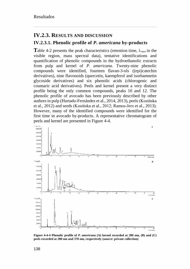
Resultados
138
IV.2.3. RESULTS AND DISCUSSION
IV.2.3.1. Phenolic profile of P. americana by-products
Table 4-2 presents the peak characteristics (retention time, max in the
visible region, mass spectral data), tentative identifications and
quantification of phenolic compounds in the hydroethanolic extracts
from pulp and kernel of P. americana. Twenty-nine phenolic
compounds were identified, fourteen flavan-3-ols ((epi)catechin
derivatives), nine flavonoids (quercetin, kaempferol and isorhamnetin
glycoside derivatives) and six phenolic acids (chlorogenic and
coumaric acid derivatives). Peels and kernel present a very distinct
profile being the only common compounds, peaks 10 and 12. The
phenolic profile of avocado has been previously described by other
authors in pulp (Hurtado-Fernández et al., 2014, 2013), peels (Kosińska
et al., 2012) and seeds (Kosińska et al., 2012; Ramos-Jerz et al., 2013).
However, many of the identified compounds were identified for the
first time in avocado by-products. A representative chromatogram of
peels and kernel are presented in Figure 4-4.
Figure 4-4-4 Phenolic profile of P. americana (A) kernel recorded at 280 nm, (B) and (C)
peels recorded at 280 nm and 370 nm, respectively (source: private collection)

Resultados
139
The main family of phenolic compounds found in avocado peels and
kernels were (epi)catechin derivatives. Peaks 7 and 10 were positively
identified as (+)-catechin and (-)-epicatechin, respectively, by
comparison with commercial standards taking into account also their
retention time, mass and UV-vis characteristics. Peak 7 (catechin) was
the major compound found in kernel sample, while epicatechin (peak
10) was the major compound in peel samples. Peaks 3, 9 and 21
presented a pseudomolecular ion at m/z 577 and MS2 fragments at m/z
451 (-126 mu), 425 (-152 mu) and 407 (-152-18 mu) and also m/z 289
and 287, coherent with the loss of two (epi)catechin units, being for that
manner tentatively identified as B-type (epi)catechin dimers. Similarly,
peaks 11 and 12 ([M-H]- at m/z 865), peaks 13 and 14 ([M-H]- at m/z
1153), peaks 15 and 16 ([M-H]- at m/z 1441) and peaks 18, 19 and 20 (
[M-H]- at m/z 1729) were assigned as B-type (epi)catechin trimers,
tetramers, pentamers and hexamers (Barros et al., 2015; Peláez-Cid et
al., 2013; Rached et al., 2016).
The second major family of compounds found in avocado peels
samples was flavonoids, mainly quercetin derivatives. Peak 24 was
identified as quercetin-3-O-glucoside by comparison of its UV
spectrum (max 352 nm) and retention time with a commercial standard.
Peaks 17, 22, 23, 25, 26 and 28 presented a pseudomolecular ion [M-
H]- at m/z 625, 595, 477, 463, 609 and 579, respectively, and an unique
MS2 fragment at m/z 301, being tentatively identified as quercetin-
dihexoside, quercetin-pentoside-hexoside, quercetin-glucuronide,
quercetin-hexoside, quercetin-rhamnoside-hexoside and quercetin-
rhamnoside-pentoside, respectively. Peaks 27 ([M-H]- at m/z 461) and
28 ([M-H]- at m/z 491) presented a unique MS2 fragments at m/z 285
and 315, respectively, corresponding to the loss of a glucuronide unit (-
176 mu), being tentatively identified as kaempferol- and isorhamnetin-
glucuronide, respectively.
Regarding the phenolic acids, caffeoylquinic acids and p-coumaroyl
quinic acid isomers were the only compounds found in both samples,
being the majority of them found in kernel samples, being peaks 8 and
6 the only phenolic acids found in peels samples. Peaks 1, 2, 6 and 8
were identified as caffeoylquinic acid derivatives according to their UV
spectra and pseudomolecular ions, being previously found by Kosińska
et al., (2012b) and Ramos-Jerz et al., (2013) in peels and seeds of P.
americana. Peak assignments of the different caffeoylquinic acids and

Resultados
140
p-coumaroyl quinic acid isomers were made using the recommended
IUPAC numbering system (IUPAC, 1976) as also the hierarchical keys
previously developed by Clifford et al., (2005, 2003).
By comparison its UV spectrum (max 326 nm) and retention time
with a commercial standard, peak 8 was identified as 5-O-
caffeoylquinic acid. Peaks 1 and 2 ([M-H]- at m/z 353) were identified
as cis 3-O-caffeoylquinic acid and trans 3-O-caffeoylquinic acid,
respectively, according to their elution order and also yielding a base
peak at m/z 191 and the ion at m/z 179 with an intensity >70% base
peak, characteristic of 3-acylchlorogenic acids (Clifford et al., 2005,
2003). After UV irradiation (366 nm, 24 h) of hydroxycinnamic acids
in our laboratory, it was possible to observe that the hydroxycinnamoyl
cis derivatives elute before the corresponding trans ones and therefore
the assignment of cis and trans forms for peaks 1 and 2. Compound 6
was tentatively identified as 4-O-caffeoylquinic acid ([M-H]- at m/z
353) according to the fragmentation pattern yielding a base peak at m/z
173 [quinic acid-H-H2O]-, accompanied by a secondary fragment ion at
m/z 179 (~75% abundance), thus being distinct from the other two
isomers (Clifford et al., 2005, 2003).
Taking into account the same findings, peaks 4 and 5 were
tentatively identified as cis and trans 3-p-coumarouylquinic acids.
(Karamac et al., 2012) have previously identified these compounds in
avocado peels.
IV.2.3.2. Antioxidant capacity of P. americana by-products
Polyphenolic content in plants and fruits have been extensively studied
due to the good impact these substances has shown as a potential health
benefits arising from their biological activity as hepatoprotective,
antiinflammatory, antiviral, antimicrobial and antioxidant activities
(Carocho and Ferreira, 2013; Dias et al., 2016; Kaur Kala et al., 2016;
Shahidi and Ambigaipalan, 2015). From the 29 different compounds
found on P. americana by-products, 23 of them were found in peels
(227.9 mg/g of extract) and only 8 in kernels (72.5 mg/g of extract),
this represent around 3-fold higher polyphenols content in peels, these
results obtained proved to be consistent with the other reports
(Kosińska et al., 2012; Rodríguez-Carpena et al., 2011).

Table 4-2 Retention time (rt), waveleghts of maximum absorption in visible region (λmax), mass spectral data, tentative identification and quantification (mg/g
of extract) of phenolic compounds in the P. americana by-products (peels and kernels)
Peak
Tentative
identification
Rt
(min)
λmax
(nm)
[M-H]-
(m/z)
MS2
(m/z)
Peel
Kernel
1 cis 3-O-Caffeoylquinic acid1 4.7 325 353 191(100),179(76),161(5),135(12) n/d 5.93 ± 0.04
2 trans 3-O-Caffeoylquinic acid1 4.89 325 353 191(100),179(75),161(4),135(10) n/d 13.6 ± 0.1
3 B-type (epi)catechin dimer2 5.22 280 577 451(21),425(100),407(26),
287(6), 289(5) n/d 7.4 ± 0.1
4 cis 3-p-Coumarouylquinic acid3 6.4 311 377 191(90),173(5),163(100),155(3),
137(5), 119(4) n/d 2.6 ± 0.1
5 cis 3-p-Coumarouylquinic acid3 6.8 311 377 191(89),173(5),163(100),155(3),
137(5), 119(4) n/d 1.70 ± 0.02
6 4-O-Caffeoylquinic acid1 7.09 325 353 191(61),179(75),173(100),
161(5),135(3) 20.2 ± 0.1 n/d
7 Catechin4 7.18 280 287 245(100),203(5),187(3),
161(3),137(3) n/d 20 ± 1
8 5-O-Caffeoylquinic acid1 7.42 325 353 191(100),179(5),161(3),135(3) 22.7 ± 0.1 n/d
9 B-type (epi)catechin dimer2 7.87 280 577 451(19),425(100),407(21),
289(9), 287(7), 34.1 ± 0.2 n/d

10 Epicatechin2 9.73 280 289 245(100),203(5),187(3),
161(3),137(3) 46.5 ± 0.2* 15.8 ± 0.3
11 B-type (epi)catechin trimer2 10.27 280 865
739(92),713(59),695(100),577(69),
575(49),425(14),407(10),
289(6),287(12)
7.81 ± 0.01 n/d
12 B-type (epi)catechin trimer2 11.11 280 865
739(89),713(51),695(100),577(8),
575(53),425(13),407(12),
289(5),287(11)
18.4 ± 0.4* 5.86 ± 0.05
13 B-type (epi)catechin tetramer2 11.83 280 1153 865(), 739(16),713(20),695(4),577(10),
575(21),425(10),407(3),289(9),287(12) 6.74 ± 0.04 n/d
14 B-type (epi)catechin tetramer2 12.19 280 1153
865(68),
739(27),713(20),695(2),577(22),
575(40),425(5),407(5),289(5),287(10)
14.5 ± 0.3 n/d
15 B-type (epi)catechin pentamer2 13.07 280 1441 1153(27),865(92),577(47),289(25),
287(10) 7.8 ± 0.1 n/d
16 B-type (epi)catechin pentamer2 13.45 280 1441 1153(41),865(29),577(61),289(34),
287(5) 8.8 ± 0.1 n/d
17 Quercetin-dihexoside5 14.11 352 625 301(100) 1.439 ± 0.001 n/d

18 B-type (epi)catechin hexamer2 14.59 279 1729 1441(14),1153(34),865(14),
577(46),289(20),287(66) 8.89 ± 0.11 n/d
19 B-type (epi)catechin hexamer2 14.88 279 1729 1441(16),1153(19),865(28),
577(17),289(13),287(41) 7.55 ± 0.01 n/d
20 B-type (epi)catechin hexamer2 15.65 280 1729 1441(12),1153(10),865(15),
577(37),289(10),287(16) 6.8 ± 0.1 n/d
21 B-type (epi)catechin dimer2 16.06 280 577 451(19),425(100),407(10),
289(6),287(3) 7.13 ± 0.04 n/d
22 Quercetin-pentoside-hexoside5 16.08 352 595 301(100) 1.521 ± 0.002 n/d
23 Quercetin-glucoronide5 18.38 352 477 301(100) 1.244 ± 0.001 n/d
24 Quercetin-3-O-glucoside5 18.5 352 463 301(100) 1.175 ± 0.002 n/d
25 Quercetin-hexoside5 19.2 345 463 301(100) 1.123 ± 0.004 n/d
26 Quercetin-rhamnoside-hexoside5 20.74 348 609 301(100) 1.21 ± 0.01 n/d
27 Kaempferol-glucuronide6 22.22 352 461 285(100) tr n/d
28 Quercetin-rhamnoside-pentoside5 22.5 350 579 301(100) 1.015 ± 0.002 n/d
29 Isorhametin-glucuronide5 23.79 352 491 315(100) 1.09 ± 0.01 n/d
Total phenolic compounds 227.9 ± 0.4* 72.5 ± 0.1
n/d- not detected; tr, traces. Standard calibration curves used: 1 – Chlorogenic acid (𝑦= 168823 𝑥 – 161172, 𝑅² = 0.9999); 2 – epicatechin (𝑦= 10314 𝑥 + 147331, 𝑅² =
0.9994); 3 – p-coumaric acid (𝑦= 301950 𝑥 + 6966.7, 𝑅² = 0.9999); 4 – catechin (𝑦= 84950 𝑥 – 23200, 𝑅² = 1); 5 – quercetin-3-O-glucoside (𝑦= 34843 𝑥 – 160173, 𝑅² =
0.9998); 6 – kaempferol -3-O-rutinoside (𝑦= 11117 𝑥 + 30861, 𝑅² = 0.9999). *Statistical differences (<0.001) were observed when t-student test was applied.

Resultados
144
It is also important here to stress, the higher phenolic content in P.
americana by-products compared with the edible pulp (Wang et al.,
2010). This greater polyphenolic content has also displayed higher
antioxidant capacity, and as expected P. americana peels proof to excel
the kernels activity in around 1.5-fold higher in all the antioxidant tests
performed and shown in Table 4-3, this result proved to be in
concordance with Calderón-Oliver et al., 2016, Kosińska et al., 2012a
and Saavedra et al., 2017). Although the differences in the total
phenolic content between peels and kernels is 3 times higher, the
antioxidant activity observed it is only around half (1.5) greater. This
could be explained due to the different degrees of polymerization of the
polyphenolic compounds of P. american peels. As it was mention
before, Epicatechin was the most abundant compound in both by-
products, peels are also majoritarian in different sort of B-type
epicatechin dimer, trimer, tetramers and flavonol glycosides.
On one hand Hollman et al., (1999) reports higher antioxidant
activity on aglycones compared with glycosides, while Mishra et al.,
(2013) point out the difference between dimers, trimers, tetramers,
heptamers and hexamers specificity on their properties as an
antioxidants. These and other interaction could explain the differences
obtained in the antioxidant activity between P. americana by-products,
but in order to obtain better comprehension on this phenomena, more
analysis have to be performed.
Table 4-3 Antioxidant activity of P. americana by-products (peels and kernels)
IC50 (µg/mL) Peel Kernel t-Students test
p-value
DPPH scavenging
activity 149 ± 5 220 ± 3 <0.001
Reducing power 32 ± 1 51 ± 1 <0.001
β-carotene bleaching
inhibition 152 ± 1 276 ± 15 <0.001
TBARS 11.7 ± 0.1 18.1 ± 0.1 <0.001
In each row different letters mean significant differences (p < 0.05). IC50 values
correspond to the sample concentration achieving 50% of antioxidant activity or 0.5
of absorbance in reducing power assay. IC50 values for the positive control trolox:
62.98 μg/mL (DDPH), 45.71 μg/mL (reducing power), 10.25 μg/mL (β-carotene
bleaching inhibition) and 10.83 μg/mL (TBARS inhibition).
Resultados
145
IV.2.3.3. Antimicrobial activity of P. americana by-
products
Flavonoids are known to be synthesized by plants in response to
microbial infections, thus, it is not surprising this sort of compounds
exhibit antimicrobial activity in many in vitro assays. Many flavonoid
rich plant and fruits from different species have been reported with
antimicrobial activity (Balouiri et al., 2016; Melgar et al., 2017;
Rodrigo and Martínez-l, 2015; Sansano et al., 2017; Šiler et al., 2014;
Vieira et al., 2016b).
P. americana by-products have shown great antibacterial and
moderate antifungal activity against the strains tasted Table 4-4. The
bactericidal effect of the samples proved to outstand from the controls
employed in 7 out of 8 different gram positive and negative strains,
although, in this particular assay extracts from kernels displayed better
MCB in 6 out of 8 strains, same power in B. cereus strain, and only
worst power in E. cloacae strain. The results here exposed shown better
performance when compared with the results reported by (Calderón-
Oliver et al., 2016; Raymond Chia and Dykes, 2010).
Contrary to bactericidal effect, fungicidal effect only was shown in
2 strains but only with kernel extracts, from which, the better fungicidal
effect was against Trichoderma viride. Comparing the fungistatic
effect, both by-product extract were effective against all 8 strains, but
only kernel extract did not shown affectivity against P. funiculosum.
Both peels and kernels extract performed better at fungistastic level in
3 strains (A. ochraceus, T. viride and P. ochrochloron) when compared
with ketoconazole antifungal commercial drug and kernel extract
display better fungistastic effect in strain vs bifonazole (stronger
control used).
Antibacterial flavonoids might be having multiple cellular targets,
rather than one specific site of action. Tsuchiya and Iinuma, (2000)
suggested an alteration of membrane fluidity in hydrophilic and
hydrophobic regions in this way flavonoids might reduce the fluidity of
outer and inner layers of membranes. Mishra et al., (2009) propose that
one of the molecular actions of flavonoids is to form complex with
proteins through nonspecific forces such as hydrogen bonding and
hydrophobic effects, as well as by covalent bond formation, this

Resultados
146
interactions have the ability to inactivate microbial adhesions,
enzymes, cell envelope transport proteins, and so forth. In this sense,
our understanding on the antimicrobial effect of polyphenols agrees
with the previous mechanism of action mentioned.

Table 4-4 Antibacterial and antifungal activity of P. americana by-products (peels and kernels)
Antibacterial activity (mg/mL) Peel Kernel C1 C2
MIC MBC MIC MBC MIC MBC MIC MBC
Gram positive bacteria
Bacillus cereus 0.015 0.030 0.020 0.030 0.10 0.20 0.25 0.40
Listeria monocytogenes 0.030 0.075 0.030 0.070 0.20 0.30 0.40 0.50
Micrococcus flavus 0.030 0.075 0.050 0.070 0.20 0.30 0.25 0.40
Staphylococcus aureus 0.030 0.075 0.030 0.070 0.04 0.10 0.25 0.45
Gram negative
Enterobacter cloacae 0.015 0.030 0.050 0.070 0.20 0.30 0.25 0.50
Escherichia coli 0.30 0.45 0.15 0.30 0.20 0.30 0.40 0.50
Pseudomonas aeruginosa 0.030 0.075 0.030 0.070 0.20 0.30 0.75 1.20
Salmonella typhimurium 0.10 0.15 0.030 0.07 0.25 0.50 0.40 0.75
Antifungal activity (mg/mL) Peel Kernel C3 C4
MIC MFC MIC MFC MIC MFC MIC MFC
Aspergillus fumigatus 0.3 - 0.3 - 0.25 0.50 0.15 0.20
Aspergillus versicolor 0.3 - 0.3 - 0.20 0.50 0.10 0.20
Aspergillus ochraceus 0.3 - 0.2 0.3 1.50 2.00 0.15 0.20
Aspergillus niger 0.3 - 0.3 - 0.20 0.50 0.15 0.20
Trichoderma viride 0.3 - 0.02 0.03 1.00 1.00 0.15 0.20
Penicillium funiculosum 0.3 - - - 0.20 0.50 0.20 0.25
Penicillium ochrochloron 0.3 - 0.3 - 2.50 3.50 0.20 0.25
Penicillium verrucosum var. cyclopium 0.3 - 0.3 - 0.20 0.30 0.10 0.20

Resultados
148
IV.2.4. CONCLUSION
One of the main goals of this research was to revalorize P. americana
by-products by characterizing their main bioactive properties. With the
assays performed we were able to detect new phenolic compounds in
avocado by-products, quantify them and test their hydrophilic and
lipophilic antioxidant capacity which shown to be superior to the
capacity reported by other authors in the edible pulp.
In the same manner, previous reports about antimicrobial activity
were revised and confronted with the data obtained, the results obtained
exhibit greater capacity against certain bacterial and fungal strains and
wider study of strains tested. All the data recollected is just the begging
of a series of experiments that have to be design in order to implement
these functional molecules in different products and industries.
The deeper analysis of bioactive compounds performed on P.
americana by-products, will allow to continue searching the optimum
process of extraction, purification and implementation as food
ingredients.
Acknowledgements
The authors are grateful to the Foundation for Science and Technology
(FCT, Portugal) and FEDER under Program PT2020 for financial
support to CIMO (UID/AGR/00690/2013) and L. Barros contract. B.
Melgar thanks CONACyT for his grant (No. 329930). The authors are
also grateful to the Serbian Ministry of Education, Science and
Technological Development, grant number 173032 for financial
support.

Resultados
149
References
Ahn, J.H., Jang, Y.-S., Lee, S.Y., 2016. Production of succinic acid by
metabolically engineered microorganisms. Curr. Opin.
Biotechnol. 42, 54–66.
https://doi.org/10.1016/j.copbio.2016.02.034
Al-Farsi, M.A., Lee, C.Y., 2008. Optimization of phenolics and dietary
fibre extraction from date seeds. Food Chem. 108, 977–985.
https://doi.org/10.1016/j.foodchem.2007.12.009
Albano, C., Negro, C., Tommasi, N., Gerardi, C., Mita, G., Miceli, A.,
De Bellis, L., Blando, F., 2015. Betalains, Phenols and
Antioxidant Capacity in Cactus Pear [Opuntia ficus-indica (L.)
Mill.] Fruits from Apulia (South Italy) Genotypes. Antioxidants
4, 269–280. https://doi.org/10.3390/antiox4020269
Albuquerque, T.G., Santos, F., Sanches-Silva, A., Beatriz Oliveira, M.,
Bento, A.C., Costa, H.S., 2016. Nutritional and phytochemical
composition of Annona cherimola Mill. fruits and by-products:
Potential health benefits. Food Chem. 193, 187–195.
https://doi.org/10.1016/j.foodchem.2014.06.044
Allai, L., Druart, X., Öztürk, M., BenMoula, A., Nasser, B., El Amiri,
B., 2016. Protective effects of Opuntia ficus-indica extract on ram
sperm quality, lipid peroxidation and DNA fragmentation during
liquid storage. Anim. Reprod. Sci. 175, 1–9.
https://doi.org/10.1016/j.anireprosci.2016.09.013
Almeida, M.M.B., de Sousa, P.H.M., Arriaga, Â.M.C., do Prado, G.M.,
Magalhães, C.E. de C., Maia, G.A., de Lemos, T.L.G., 2011.
Bioactive compounds and antioxidant activity of fresh exotic
fruits from northeastern Brazil. Food Res. Int. 44, 2155–2159.
https://doi.org/10.1016/j.foodres.2011.03.051
Alvarez, B., 2007. Análisis de factibilidad del cultivo de la tuna en la
localidad de Icaño , Departamento La Paz, Dirección provincial
de programación del desarrollo, Ministerio de Producción y
Desarrollo. Gobierno de la Provincia de Catamarca. Catamarca,
Argentina.
Alzate T, L.M., González, D., Hincapié, S., Cardona S, B.L., Londoño-
Londoño, J., Jiménez-Cartagena, C., 2016. The profile of
bioactive substances in ten vegetable and fruit by-products from
a food supply chain in Colombia. Sustain. Prod. Consum.
https://doi.org/10.1016/j.spc.2016.07.005
Resultados
150
Ammar, I., Ben Salem, M., Harrabi, B., Mzid, M., Bardaa, S., Sahnoun,
Z., Attia, H., Ennouri, M., 2018. Anti-inflammatory activity and
phenolic composition of prickly pear (Opuntia ficus-indica)
flowers. Ind. Crops Prod. 112, 313–319.
https://doi.org/10.1016/j.indcrop.2017.12.028
Angulo-Bejarano, P.I., Martínez-Cruz, O., Paredes-López, O., 2014.
Phytochemical Content, Nutraceutical Potential and
Biotechnological Applications of an Ancient Mexican Plant:
Nopal (Opuntia ficus-indica). Curr. Nutr. Food Sci. 10, 196–217.
https://doi.org/10.2174/157340131003140828121015
Arlt, U., 2010. NATCOL | Foods Colours Legislation.
Aruwa, C.E., Amoo, S.O., Kudanga, T., 2018. Opuntia (Cactaceae)
plant compounds, biological activities and prospects – A
comprehensive review. Food Res. Int. 112, 328–344.
https://doi.org/10.1016/j.foodres.2018.06.047
Association of Official Analytical Chemists, 2016. Official Methods of
Analysis of AOAC International, 20th ed. The Association,
Gaithersburg, MD, USA.
Attokaran, M., 2011. Natural Food Flavors and Colorants Natural Food
Flavors and Colorants, 1st ed. Blackwell Publishing LTD, Iowa.
Augustin, M.A., Sanguansri, L., 2015. Challenges and Solutions to
Incorporation of Nutraceuticals in Foods. Annu. Rev. Food Sci.
Technol. 6, 463–477. https://doi.org/10.1146/annurev-food-
022814-015507
Ayala-Zavala, J.F., Vega-Vega, V., Rosas-Domínguez, C., Palafox-
Carlos, H., Villa-Rodriguez, J.A., Siddiqui, M.W., Dávila-Aviña,
J.E., González-Aguilar, G.A., 2011. Agro-industrial potential of
exotic fruit byproducts as a source of food additives. Food Res.
Int. 44, 1866–1874.
https://doi.org/10.1016/j.foodres.2011.02.021
Azeredo, H.M.C., 2009. Betalains: Properties, sources, applications,
and stability - A review. Int. J. Food Sci. Technol. 44, 2365–2376.
https://doi.org/10.1111/j.1365-2621.2007.01668.x
Balouiri, M., Sadiki, M., Ibnsouda, S.K., 2016. Methods for in vitro
evaluating antimicrobial activity: A review. J. Pharm. Anal. 6,
71–79. https://doi.org/10.1016/j.jpha.2015.11.005
Barros, L., Calhelha, R.C., Queiroz, M.J.R.P., Santos-Buelga, C.,
Santos, E.A., Regis, W.C.B., Ferreira, I.C.F.R., 2015. The
Resultados
151
powerful in vitro bioactivity of Euterpe oleracea Mart. seeds and
related phenolic compounds. Ind. Crops Prod. 76, 318–322.
https://doi.org/10.1016/j.indcrop.2015.05.086
Barros, L., Pereira, C., Ferreira, I.C.F.R., 2013. Optimized Analysis of
Organic Acids in Edible Mushrooms from Portugal by Ultra Fast
Liquid Chromatography and Photodiode Array Detection. Food
Anal. Methods 6. https://doi.org/10.1007/s12161-012-9443-1
Barrows, J.N., Lipman, A.L., Bailey, C.J., Cianci, S., 2003. Color
Additives: FDA’s Regulatory Process and Historical
Perspectives. Food Saf. Mag. URL
https://www.fda.gov/forindustry/coloradditives/regulatoryproces
shistoricalperspectives/ (accessed 9.3.18).
Baydar, N.G., Özkan, G., Sağdiç, O., 2004. Total phenolic contents and
antibacterial activities of grape (Vitis vinifera L.) extracts. Food
Control 15, 335–339. https://doi.org/10.1016/S0956-
7135(03)00083-5
Belščak-Cvitanović, A., Durgo, K., Huđek, A., Bačun-Družina, V.,
Komes, D., 2018. Metabolism and Health Effects of Polyphenols,
in: Galanakis, C.M. (Ed.), Polyphenols: Properties, Recovery, and
Applications. Woodhead Publishing, pp. 1–44.
https://doi.org/https://doi.org/10.1016/B978-0-12-813572-
3.00001-4
Belwal, T., Dhyani, P., Bhatt, I.D., Rawal, R.S., Pande, V., 2016.
Optimization extraction conditions for improving phenolic
content and antioxidant activity in Berberis asiatica fruits using
response surface methodology (RSM). Food Chem. 207, 115–
124. https://doi.org/10.1016/j.foodchem.2016.03.081
Bessada, S.M.F., Barreira, J.C.M., Barros, L., Ferreira, I.C.F.R.,
Oliveira, M.B.P.P., 2016. Phenolic profile and antioxidant
activity of Coleostephus myconis (L.) Rchb.f.: An underexploited
and highly disseminated species. Ind. Crops Prod. 89, 45–51.
https://doi.org/10.1016/j.indcrop.2016.04.065
Betancourt, C., Cejudo-Bastante, M.J., Heredia, F.J., Hurtado, N.,
2017. Pigment composition and antioxidant capacity of
betacyanins and betaxanthins fractions of Opuntia dillenii (Ker
Gawl) Haw cactus fruit. Food Res. Int. 101, 173–179.
https://doi.org/10.1016/j.foodres.2017.09.007
Bhatia, S., Bera, T., Dahiya, R., Bera, T., Bhatia, S., Bera, T., 2015.
Classical and Nonclassical Techniques for Secondary Metabolite
Resultados
152
Production in Plant Cell Culture, in: Modern Applications of Plant
Biotechnology in Pharmaceutical Sciences. Elsevier, pp. 231–
291. https://doi.org/10.1016/B978-0-12-802221-4.00007-8
Brand-Williams, W., Cuvelier, M.E., Berset, C., 1995. Use of a free
radical method to evaluate antioxidant activity. LWT - Food Sci.
Technol. 28, 25–30. https://doi.org/10.1016/S0023-
6438(95)80008-5
Bravo-Hollis, H., 1978. Las cactaceas de Mexico. Universidad
Nacional Autonoma de Mexico, Mexio.
https://doi.org/10.15713/ins.mmj.3
Brundtland, G.H., 1987. Our common future.
Cai, Y., Sun, M., Corke, H., 2003. Antioxidant activity of betalains
from plants of the Amaranthaceae. J. Agric. Food Chem. 51,
2288–2294. https://doi.org/10.1021/jf030045u
Calderón-Oliver, M., Escalona-Buendía, H.B., Medina-Campos, O.N.,
Pedraza-Chaverri, J., Pedroza-Islas, R., Ponce-Alquicira, E.,
2016. Optimization of the antioxidant and antimicrobial response
of the combined effect of nisin and avocado by-products. LWT -
Food Sci. Technol. 65, 46–52.
https://doi.org/10.1016/j.lwt.2015.07.048
Cardoso-Ugarte, G.A., Sosa-Morales, M.E., Ballard, T., Liceaga, A.,
San Martín-González, M.F., 2014. Microwave-assisted extraction
of betalains from red beet (Beta vulgaris). LWT - Food Sci.
Technol. 59, 276–282. https://doi.org/10.1016/j.lwt.2014.05.025
Carocho, M., Ferreira, I.C.F.R., 2013. A review on antioxidants,
prooxidants and related controversy: Natural and synthetic
compounds, screening and analysis methodologies and future
perspectives. Food Chem. Toxicol. 51, 15–25.
https://doi.org/10.1016/j.fct.2012.09.021
Carr, M., 2013. The water relations and irrigation requirements of
avocado (Persea americana Mill.): a review. Exp. Agric. 49, 256–
278. https://doi.org/10.1017/S0014479712001317
Cassano, A., Conidi, C., Timpone, R., D’Avella, M., Drioli, E., 2007.
A membrane-based process for the clarification and the
concentration of the cactus pear juice. J. Food Eng. 80, 914–921.
https://doi.org/10.1016/j.jfoodeng.2006.08.005
Castellanos-Santiago, E., Yahia, E.M., 2008. Identification and
quantification of betalains from the fruits of 10 Mexican prickly

Resultados
153
pear cultivars by high-performance liquid chromatography and
electrospray ionization mass spectrometry. J. Agric. Food Chem.
56, 5758–5764. https://doi.org/10.1021/jf800362t
Castellar, R., Obón, J.M., Alacid, M., Fernández-López, J.A., 2003.
Color Properties and Stability of Betacyanins from Opuntia
Fruits. J. Agric. Food Chem. 51, 2772–2776.
https://doi.org/10.1021/jf021045h
Cejudo Bastante, M.J., Chaalal, M., Louaileche, H., Parrado, J.,
Heredia, F.J., 2014. Betalain Profile, Phenolic Content, and Color
Characterization of Different Parts and Varieties of Opuntia ficus-
indica. J. Agric. Food Chem. 62, 8491–8499.
https://doi.org/10.1021/jf502465g
Chahdoura, H., Barreira, J.C.M., Barros, L., Santos-Buelga, C.,
Ferreira, I.C.F.R., Achour, L., 2014. Phytochemical
characterization and antioxidant activity of Opuntia microdasys
(Lehm.) Pfeiff flowers in different stages of maturity. J. Funct.
Foods 9, 27–37. https://doi.org/10.1016/j.jff.2014.04.011
Chahdoura, H., Barreira, J.C.M., Fernández-Ruiz, V., Morales, P.,
Calhelha, R.C., Flamini, G., Soković, M., Ferreira, I.C.F.R.,
Achour, L., 2016. Bioactivity, proximate, mineral and volatile
profiles along the flowering stages of Opuntia microdasys
(Lehm.): defining potential applications. Food Funct. 7, 1458–67.
https://doi.org/10.1039/c5fo01536g
Chan, L.G., Cohen, J.L., Maria, J., Nobrega, L., Bell, D.M., 2018.
Conversion of Agricultural Streams and Food-Processing By-
Products to Value-Added Compounds using Filamentous Fungi.
Annu. Rev. Food Sci. Technol. 9, 1–21.
https://doi.org/10.1146/annurev-food-030117-012626
Chougui, N., Djerroud, N., Naraoui, F., Hadjal, S., Aliane, K., Zeroual,
B., Larbat, R., 2015. Physicochemical properties and storage
stability of margarine containing Opuntia ficus-indica peel extract
as antioxidant. Food Chem. 173, 382–90.
https://doi.org/10.1016/j.foodchem.2014.10.025
Chougui, N., Tamendjari, A., Hamidj, W., Hallal, S., Barras, A.,
Richard, T., Larbat, R., 2013. Oil composition and
characterisation of phenolic compounds of Opuntia ficus-indica
seeds. Food Chem. 139, 796–803.
https://doi.org/10.1016/j.foodchem.2013.01.054
Clifford, M.N., Johnston, K.L., Knight, S., Kuhnert, N., 2003.

Resultados
154
Hierarchical Scheme for LC-MSn Identification of Chlorogenic
Acids. J. Agric. Food Chem. 51, 2900–2911.
https://doi.org/10.1021/jf026187q
Clifford, M.N., Knight, S., Kuhnert, N., 2005. Discriminating between
the six isomers of dicaffeoylquinic acid by LC-MSn. J. Agric.
Food Chem. 53, 3821–3832. https://doi.org/10.1021/jf050046h
Cowan, A.K., Cripps, R.F., Richings, E.W., Taylor, N.J., 2001. Fruit
size: Towards an understanding of the metabolic control of fruit
growth using avocado as a model system. Physiol. Plant. 111,
127–136. https://doi.org/10.1034/j.1399-3054.2001.1110201.x
Cowan, A.K., Wolstenholme, B.N., 2003. AVOCADOS, in:
Encyclopedia of Food Sciences and Nutrition. Elsevier, pp. 348–
353. https://doi.org/10.1016/B0-12-227055-X/00073-0
Crane, J.H., Balerdi, C.F., Maguire, I., 2016. Avocado Growing in the
Florida Home Landscape.
Da-Costa-Rocha, I., Bonnlaender, B., Sievers, H., Pischel, I., Heinrich,
M., 2014. Hibiscus sabdariffa L. – A phytochemical and
pharmacological review. Food Chem. 165, 424–443.
https://doi.org/10.1016/j.foodchem.2014.05.002
Da-Silva, R.P.F.F., Rocha-Santos, T.A.P., Duarte, A.C., 2016.
Supercritical fluid extraction of bioactive compounds. TrAC -
Trends Anal. Chem. 76, 40–51.
https://doi.org/10.1016/j.trac.2015.11.013
Dabas, D., Shegog, R.M., Ziegler, G.R., Lambert, J.D., 2013. Avocado
(Persea americana) seed as a source of bioactive phytochemicals.
Current Pharmaceutical Design, 19 (34), 6133–40.
https://doi.org/10.2174/1381612811319340007
Day, L., Seymour, R.B., Pitts, K.F., Konczak, I., Lundin, L., 2009.
Incorporation of functional ingredients into foods. Trends Food
Sci. Technol. 20, 388–395.
https://doi.org/10.1016/J.TIFS.2008.05.002
DeFelice, S.L., 2002. FIM Rationale And Proposed Guidelines For The
Nutraceutical Research & Education Act - NREA. New York.
Delgado-Vargas, F., Paredes-Lopez, O., 2003. Natural Colorants for
Food and Nutraceutical Uses, Trends in Food Science &
Technology. https://doi.org/10.1016/S0924-2244(03)00076-1
Destro dos Santos, C., Külzer Scherer, R., Schilling Cassini, A.,
Ferreira Marczak, L.D., Tessaro, I.C., 2016. Clarification of red

Resultados
155
beet stalks extract by microfiltration combined with
ultrafiltration. J. Food Eng. 185, 35–41.
https://doi.org/10.1016/j.jfoodeng.2016.03.031
Devalaraja, S., Jain, S., Yadav, H., 2011. Exotic fruits as therapeutic
complements for diabetes, obesity and metabolic syndrome. Food
Res. Int. 44, 1856–1865.
https://doi.org/10.1016/j.foodres.2011.04.008
Devasagayam, T.P.A., Boloor, K.K., Ramasarma, T., 2003. Methods
for estimating lipid peroxidation: An analysis of merits and
demerits. Indian J. Biochem. Biophys. 40, 300–308.
Dias, M.I., Sousa, M.J., Alves, R.C., Ferreira, I.C.F.R., 2016. Exploring
plant tissue culture to improve the production of phenolic
compounds: A review. Ind. Crops Prod. 82, 9–22.
https://doi.org/10.1016/j.indcrop.2015.12.016
Donkin, R.A., 1977. Spanish Red: An Ethnogeographical Study of
Cochineal and the Opuntia Cactus. Trans. Am. Philos. Soc. 67, 1–
84. https://doi.org/10.2307/1006195
Esatbeyoglu, T., Wagner, A.E., Schini-Kerth, V.B., Rimbach, G.,
2015a. Betanin--a food colorant with biological activity. Mol.
Nutr. Food Res. 59, 36–47.
https://doi.org/10.1002/mnfr.201400484
Esatbeyoglu, T., Wagner, A.E., Schini-Kerth, V.B., Rimbach, G.,
2015b. Betanin-A food colorant with biological activity. Mol.
Nutr. Food Res. 59, 36–47.
https://doi.org/10.1002/mnfr.201400484
Espinel-Ingroff, A., 2001. Comparison of the E-test with the NCCLS
M38-P method for antifungal susceptibility testing of common
and emerging pathogenic filamentous fungi. J. Clin. Microbiol.
39, 1360–1367. https://doi.org/10.1128/JCM.39.4.1360-
1367.2001
Esquivel, P., 2016. Handbook on Natural Pigments in Food and
Beverages Industrial Applications for Improving Food Colorl, in:
Carle, R., Schweiggert, R.M. (Eds.). Woodhead Publishing, San
Pedro, Costa Rica, pp. 81–99.
FAO, 2016. FAOSTAT. URL
http://www.fao.org/faostat/en/?#data/QC/visualize (accessed
5.3.18).
FAO, 2004. AVOCADO: Post-Harvest Operation. Rome.

Resultados
156
FAO, IFAD, WFP, 2015. The State of Food Insecurity in the World.
Meeting the 2015 international hunger targets: taking stock of
uneven progress. Rome.
Farag, M.A., Maamoun, A.A., Ehrlich, A., Fahmy, S., Wesjohann,
L.A., 2017. Assessment of sensory metabolites distribution in 3
cactus Opuntia ficus-indica fruit cultivars using UV
fingerprinting and GC/MS profiling techniques. LWT - Food Sci.
Technol. 80, 145–154. https://doi.org/10.1016/j.lwt.2017.02.014
Fathordoobady, F., Mirhosseini, H., Selamat, J., Manap, M.Y.A., 2016.
Effect of solvent type and ratio on betacyanins and antioxidant
activity of extracts from Hylocereus polyrhizus flesh and peel by
supercritical fluid extraction and solvent extraction. Food Chem.
202, 70–80. https://doi.org/10.1016/j.foodchem.2016.01.121
FDA, 2017. SCOGS (Select Committee on GRAS Substances). URL
http://www.accessdata.fda.gov/scripts/fdcc/?set=SCOGS
(accessed 3.14.17).
FDA, 2007. Guidance for Industry and Other Stakeholders
Toxicological Principles for the Safety Assessment of Food
Ingredients, Human Studies.
Fernández-López, J. a., Almela, L., 2001. Application of high-
performance liquid chromatography to the characterization of the
betalain pigments in prickly pear fruits. J. Chromatogr. A 913,
415–420. https://doi.org/10.1016/S0021-9673(00)01224-3
Fernandez-Rojas, B., Ortiz-Moreno, A., Hernandez-Navarro, D., 2010.
Phenolic Content and its Antioxidant Activity of Persimmon
(Dyospiros kaki L.) Cultivated in Mexico. J. Biotechnol. 150.
https://doi.org/10.1016/j.jbiotec.2010.09.289
Ferreira, I.C.F.R., Martins, N., Barros, L., 2017. Phenolic Compounds
and Its Bioavailability: In Vitro Bioactive Compounds or Health
Promoters? Adv. Food Nutr. Res. 82, 1–44.
https://doi.org/10.1016/bs.afnr.2016.12.004
Ferreres, F., Grosso, C., Gil-Izquierdo, A., Valentão, P., Mota, A.T.,
Andrade, P.B., 2017. Optimization of the recovery of high-value
compounds from pitaya fruit by-products using microwave-
assisted extraction. Food Chem. 230, 463–474.
https://doi.org/10.1016/j.foodchem.2017.03.061
Flores-Valdez, C., Luna-Esquivel, J., Ramirez-Moreno, P., 1995.
Manejo postcosecha de la tuna para el mercado nacional e
Resultados
157
internacional y Procesamiento y transformación industrial ...
Chapingo, Estado de México.
Galanakis, C.M., 2015. Chapter 3 – The universal recovery strategy,
Food Waste Recovery. Elsevier Inc.
https://doi.org/10.1016/B978-0-12-800351-0.00003-1
Galanakis, C.M., 2012. Recovery of high added-value components
from food wastes: Conventional, emerging technologies and
commercialized applications. Trends Food Sci. Technol. 26, 68–
87. https://doi.org/10.1016/j.tifs.2012.03.003
Gandía-Herrero, F., Cabanes, J., Escribano, J., García-Carmona, F.,
Jimenez-Atienzar, M., 2013. Encapsulation of the most
antioxidant betalains in edible matrices as powders of different
colors Encapsulation of the most antioxidant betalains in edible
matrices as powders of different colors.
https://doi.org/10.1021/jf400337g
Gandía-Herrero, F., Escribano, J., García-Carmona, F., 2010. Structural
implications on color, fluorescence, and antiradical activity in
betalains. Planta 232, 449–460. https://doi.org/10.1007/s00425-
010-1191-0
Garcia-Castello, E.M., Rodriguez-Lopez, A.D., Mayor, L., Ballesteros,
R., Conidi, C., Cassano, A., 2015. Optimization of conventional
and ultrasound assisted extraction of flavonoids from grapefruit
(Citrus paradisi L.) solid wastes. LWT - Food Sci. Technol. 64,
1114–1122. https://doi.org/10.1016/j.lwt.2015.07.024
García-Cruz, L., Dueñas, M., Santos-Buelgas, C., Valle-Guadarrama,
S., Salinas-Moreno, Y., 2017. Betalains and phenolic compounds
profiling and antioxidant capacity of pitaya (Stenocereus spp.)
fruit from two species (S. Pruinosus and S. stellatus). Food Chem.
234, 111–118. https://doi.org/10.1016/j.foodchem.2017.04.174
Gebhardt, D., 2018. The Economics of Natural Color Pigments. URL
https://sensientfoodcolors.com/en-us/research-
development/economics-natural-color-pigments/
Ghasemzadeh, A., Ghasemzadeh, N., 2011. Flavonoids and phenolic
acids: Role and biochemical activity in plants and human. J. Med.
Plants Res. 5, 6697–6703. https://doi.org/10.5897/JMPR11.363
Gomes-Correa, R.C., de Souza, A.H.P., Calhelha, R.C., Barros, L.,
Glamoclija, J., Sokovic, M., Peralta, R.M., Bracht, A., Ferreira,
I.C.F.R., 2015. Bioactive formulations prepared from fruiting
Resultados
158
bodies and submerged culture mycelia of the Brazilian edible
mushroom Pleurotus ostreatoroseus Singer. Food Funct. 6, 2155–
2164. https://doi.org/10.1039/C5FO00465A
Gong, M., Bassi, A., 2016. Carotenoids from microalgae: A review of
recent developments. Biotechnol. Adv. 34, 1396–1412.
https://doi.org/10.1016/j.biotechadv.2016.10.005
Griffith, M.P., 2004. The origins of an important cactus crop, Opuntia
ficus-indica (Cactaceae): New molecular evidence. Am. J. Bot.
91, 1915–1921. https://doi.org/10.3732/ajb.91.11.1915
Guimarães, R., Barros, L., Dueñas, M., Calhelha, R.C., Carvalho,
A.M., Santos-Buelga, C., Queiroz, M.J.R.P., Ferreira, I.C.F.R.,
2013. Nutrients, phytochemicals and bioactivity of wild Roman
chamomile: A comparison between the herb and its preparations.
Food Chem. 136, 718–725.
https://doi.org/10.1016/j.foodchem.2012.08.025
Guldiken, B., Toydemir, G., Nur Memis, K., Okur, S., Boyacioglu, D.,
Capanoglu, E., 2016. Home-processed red beetroot (Beta vulgaris
L.) products: Changes in antioxidant properties and
bioaccessibility. Int. J. Mol. Sci. 17.
https://doi.org/10.3390/ijms17060858
GVR, 2016. Natural Antioxidants Market Analysis By Product
(Vitamin C, Vitamin E, Polyphenols, Carotenoids) And Segment
Forecasts To 2022. URL
https://www.grandviewresearch.com/industry-analysis/natural-
antioxidants-market
Heleno, S.A., Barros, L., Sousa, M.J., Martins, A., Ferreira, I.C.F.R.,
2010. Tocopherols composition of Portuguese wild mushrooms
with antioxidant capacity. Food Chem. 119, 1443–1450.
https://doi.org/10.1016/j.foodchem.2009.09.025
Henry, B.S., 1996. Natural food colours, in: Natural Food Colorants.
Springer US, Boston, MA, pp. 40–79.
https://doi.org/10.1007/978-1-4615-2155-6_2
Herbach, K.M., Stintzing, F.C., Carle, R., 2006. Betalain stability and
degradation - Structural and chromatic aspects. J. Food Sci. 71,
41–50. https://doi.org/10.1111/j.1750-3841.2006.00022.x
Herbach, K.M., Stintzing, F.C., Carle, R., 2005. Identification of heat-
induced degradation products from purified betanin, phyllocactin
and hylocerenin by high-performance liquid chromatography/
Resultados
159
electrospray ionization mass spectrometry. Rapid Commun. Mass
Spectrom. 19, 2603–2616. https://doi.org/10.1002/rcm.2103
Hiwasa-Tanase, K., Ezure, H., 2014. Climateric and non-climateric
ripening, in: Nath, P., Bouzayen, M., Mattoo, A.K., Pech, J.C.
(Eds.), Fruit Ripening: Physiology, Signalling and Genomics.
CABI International, pp. 151–177.
https://doi.org/10.1017/CBO9781107415324.004
Hogervorst-Cvejić, J., Atanacković-Krstonošić, M., Bursać, M., Miljić,
U., 2017. Polyphenols, in: Nutraceutical and Functional Food
Components. Academic Press, pp. 203–258.
https://doi.org/10.1016/B978-0-12-805257-0.00007-7
Hollman, P.C., Bijsman, M.N., van Gameren, Y., Cnossen, E.P., de
Vries, J.H., Katan, M.B., 1999. The sugar moiety is a major
determinant of the absorption of dietary flavonoid glycosides in
man. Free Radic. Res. 31, 569–573.
https://doi.org/10.1080/10715769900301141
Hurtado-Fernández, E., Fernandez-Gutierrez, A., Carrasco-Pancorbo,
A., 2018a. Avocado fruit, in: Rodrigues, S., de Oliveira, E., Sousa,
E. (Eds.), Exotic Fruits. Academic Press, London, pp. 37–46.
Hurtado-Fernández, E., Pacchiarotta, T., Longueira-Suárez, E.,
Mayboroda, O.A., Fernández-Gutiérrez, A., Carrasco-Pancorbo,
A., 2013. Evaluation of gas chromatography-atmospheric
pressure chemical ionization-mass spectrometry as an alternative
to gas chromatography-electron ionization-mass spectrometry:
Avocado fruit as example. J. Chromatogr. A 1313, 228–244.
https://doi.org/10.1016/j.chroma.2013.08.084
Hurtado-Fernández, E., Pacchiarotta, T., Mayboroda, O.A., Fernández-
Gutiérrez, A., Carrasco-Pancorbo, A., 2014. Quantitative
characterization of important metabolites of avocado fruit by gas
chromatography coupled to different detectors (APCI-TOF MS
and FID). Food Res. Int. 62, 801–811.
https://doi.org/10.1016/j.foodres.2014.04.038
Hussain, Z., Thu, H.E., Amjad, M.W., Hussain, F., Ahmed, T.A., Khan,
S., 2017. Exploring recent developments to improve antioxidant,
anti-inflammatory and antimicrobial efficacy of curcumin: A
review of new trends and future perspectives. Mater. Sci. Eng. C.
https://doi.org/10.1016/j.msec.2017.03.226
IFIC, FDA, 2010. Overview of Food Ingredients, Additives and Colors.
URL
Resultados
160
https://www.fda.gov/food/ingredientspackaginglabeling/foodadd
itivesingredients/ucm094211.htm
Inglese, P., Giugliuzza, G., Liguori, G., 2004. Cactus pear fruit
production: from knowledge to development, in: Esparza-Frausto,
G., Valdez-Cepeda, R.D., Méndez- Gallego, S.J. (Eds.), El
Nopal.Tópicos de Actualidad. Universidad Autónoma de
Chapingo. Colegio de Postgraduados, México, pp. 89–108.
IUPAC, IUPAC-IUB, 1976. Nomenclature of cyclitols. Biochem. J.
153, 23–31. https://doi.org/10.1351/pac197437010283
Jain, G., Gould, K.S., 2015. Are betalain pigments the functional
homologues of anthocyanins in plants? Environ. Exp. Bot. 119,
48–53. https://doi.org/10.1016/J.ENVEXPBOT.2015.06.002
Jain, T., Grover, K., Kaur, G., 2016. Effect of processing on nutrients
and fatty acid composition of garden cress (Lepidium sativum)
seeds. Food Chem. 213, 806–812.
https://doi.org/10.1016/j.foodchem.2016.07.034
Jimenez-Garcia, S.N., Vazquez-Cruz, M.A., Garcia-Mier, L.,
Contreras-Medina, L.M., Guevara-González, R.G., Garcia-Trejo,
J.F., Feregrino-Perez, A.A., 2018. Phytochemical and
Pharmacological Properties of Secondary Metabolites in Berries,
in: Therapeutic Foods. Elsevier, pp. 397–427.
https://doi.org/10.1016/B978-0-12-811517-6.00013-1
Kaderides, K., Goula, A.M., 2017. Development and characterization
of a new encapsulating agent from orange juice by-products. Food
Res. Int. 0–1. https://doi.org/10.1016/j.foodres.2017.07.057
Kanner, J., Harel, S., Granit, R., 2001. Betalains - A new class of
dietary cationized antioxidants. J. Agric. Food Chem. 49, 5178–
5185. https://doi.org/10.1021/jf010456f
Karamac, M., Estrella, I., Herna, T., Dykes, G. a., Kosińska, A.,
Karamać, M., Estrella, I., Hernández, T., Bartolomé, B., Dykes,
G. a., 2012. Phenolic compound profiles and antioxidant capacity
of Persea americana Mill. peels and seeds of two varieties. J.
Agric. Food Chem. 60, 4613–4619.
https://doi.org/10.1021/jf300090p
Katsube, T., Tsurunaga, Y., Sugiyama, M., Furuno, T., Yamasaki, Y.,
2009. Effect of air-drying temperature on antioxidant capacity and
stability of polyphenolic compounds in mulberry (Morus alba L.)
leaves. Food Chem. 113, 964–969.

Resultados
161
https://doi.org/10.1016/j.foodchem.2008.08.041
Kaur Kala, H., Mehta, R., Tandey, R., Sen, K.K., Mandal, V., 2016.
Ten years of research on phenolics (2005–2015): A status report.
Pacific Sci. Rev. A Nat. Sci. Eng.
https://doi.org/10.1016/j.psra.2016.07.002
Knight, R.J., Campbell, C.W., 1999. Ecological adaptation and the
evolution of modern avocado cultivars. Rev. Chapingo Ser.
Hortic. 49–54.
Kosińska, A., Karamać, M., Estrella, I., Hernández, T., Bartolomé, B.,
Dykes, G.A., 2012. Phenolic compound profiles and antioxidant
capacity of Persea americana Mill. peels and seeds of two
varieties. J. Agric. Food Chem. 60, 4613–4619.
https://doi.org/10.1021/jf300090p
Kris-Etherton, P.M., Hecker, K.D., Bonanome, A., Coval, S.M.,
Binkoski, A.E., Hilpert, K.F., Griel, A.E., Etherton, T.D., 2002.
Bioactive compounds in foods: their role in the prevention of
cardiovascular disease and cancer. Am. J. Med. 113, 71–88.
https://doi.org/10.1016/S0002-9343(01)00995-0
Kuhnle, G.G., 2012. Nutritional biomarkers for objective dietary
assessment. J. Sci. Food Agric. 92, 1145–1149.
https://doi.org/10.1002/jsfa.5631
Kuti, J.O., 2004. Antioxidant compounds from four Opuntia cactus
pear fruit varieties. Food Chem. 85, 527–533.
https://doi.org/10.1016/S0308-8146(03)00184-5
Kyriacou, M.C., Emmanouilidou, M.G., Soteriou, G.A., 2016.
Asynchronous ripening behavior of cactus pear (Opuntia ficus-
indica) cultivars with respect to physicochemical and
physiological attributes. Food Chem. 211, 598–607.
https://doi.org/10.1016/j.foodchem.2016.05.113
Laqui-Vilca, C., Aguilar-Tuesta, S., Mamani-Navarro, W., Montaño-
Bustamante, J., Condezo-Hoyos, L., 2018. Ultrasound-assisted
optimal extraction and thermal stability of betalains from colored
quinoa (Chenopodium quinoa Willd) hulls. Ind. Crops Prod. 111,
606–614. https://doi.org/10.1016/j.indcrop.2017.11.034
Lea, A., 2008. Analysis of polyphenol antioxidants in fortified foods
and supplements. Food Fortif. Suppl. 175–194.
https://doi.org/10.1533/9781845694265.2.175
Leardi, R., 2013. Experimental Design, 1st ed, Data Handling in

Resultados
162
Science and Technology. Copyright © 2013 Elsevier B.V. All
rights reserved. https://doi.org/10.1016/B978-0-444-59528-
7.00002-8
Li, J., Chen, Z., Di, D., 2012. Preparative separation and purification of
Rebaudioside A from Stevia rebaudiana Bertoni crude extracts by
mixed bed of macroporous adsorption resins. Food Chem. 132,
268–276. https://doi.org/10.1016/j.foodchem.2011.10.077
Librán, C. M., Mayor, L., Garcia-Castello, E. M., Vidal-Brotons, D.,
2013. Polyphenol extraction from grape wastes: Solvent and pH
effect. Agric. Sci. 04, 56–62.
https://doi.org/10.4236/as.2013.49B010
Madrau, M. a., Piscopo, A., Sanguinetti, A.M., Del Caro, A., Poiana,
M., Romeo, F. V., Piga, A., 2009. Effect of drying temperature on
polyphenolic content and antioxidant activity of apricots. Eur.
Food Res. Technol. 228, 441–448.
https://doi.org/10.1007/s00217-008-0951-6
Madunić, J., Madunić, I.V., Gajski, G., Popić, J., Garaj-Vrhovac, V.,
2018. Apigenin: A dietary flavonoid with diverse anticancer
properties. Cancer Lett. 413, 11–22.
https://doi.org/10.1016/j.canlet.2017.10.041
Manach, C., Scalbert, A., Morand, C., Rémésy, C., Jiménez, L., 2004.
Bioavailability, Polyphenols: Food sources and. Am. J. Clin.
Nutr. 79, 727–747. https://doi.org/10.1038/nature05488
Mandal, S.C., Mandal, V., Das, A.K., 2015. Classification of Extraction
Methods, in: Essentials of Botanical Extraction. Academic Press,
pp. 83–136. https://doi.org/10.1016/B978-0-12-802325-9.00006-
9
Marais, J.P.J., Deavours, B., Dixon, R.A., Ferreira, D., 2006. The
science of flavonoids, The Science of Flavonoids. Springer, Ohio.
https://doi.org/10.1007/978-0-387-28822-2
Marco, G.J., 1968. A Rapid Method for Evaluation of Antioxidants. J.
Am. oil Chem. Soc. 45, 594–598.
https://doi.org/10.1007/BF02668958
Maria, J., Andrade, D.M., Fasolo, D., 2014. Polyphenol Antioxidants
from Natural Sources and Contribution to Health Promotion,
Polyphenols in Human Health and Disease. Elsevier Inc.
https://doi.org/10.1016/B978-0-12-398456-2.00020-7
Martins, N., Roriz, C.L., Morales, P., Barros, L., Ferreira, I.C.F.R.,

Resultados
163
2016. Food colorants: Challenges, opportunities and current
desires of agro-industries to ensure consumer expectations and
regulatory practices. Trends Food Sci. Technol. 52, 1–15.
https://doi.org/10.1016/j.tifs.2016.03.009
Mata, A., Ferreira, J.P., Semedo, C., Serra, T., Duarte, C.M.M., Bronze,
M.R., 2016. Contribution to the characterization of Opuntia spp.
juices by LC–DAD–ESI-MS/MS. Food Chem. 210, 558–565.
https://doi.org/10.1016/j.foodchem.2016.04.033
Mayor, L., Calvo, A., Moreira, R., Fito, P., Garcia-Castello, E., 2013.
Water sorption isotherms of globe artichoke leaves. Agric. Sci.
04, 63–69. https://doi.org/10.4236/as.2013.49B011
McGuire, S., 2011. U.S. Department of Agriculture and U.S.
Department of Health and Human Services, Dietary Guidelines
for Americans, 2010. 7th Edition, Washington, DC: U.S.
Government Printing Office, January 2011. Adv. Nutr. 2, 293–
294. https://doi.org/10.3945/an.111.000430
Medina-Meza, I.G., Barbosa-Cánovas, G. V., 2015. Assisted extraction
of bioactive compounds from plum and grape peels by ultrasonics
and pulsed electric fields. J. Food Eng. 166, 268–275.
https://doi.org/10.1016/j.jfoodeng.2015.06.012
Melgar, B., Dias, M.I., Ciric, A., Sokovic, M., Garcia-Castello, E.M.,
Rodriguez-Lopez, A.D., Barros, L., Ferreira, I., 2017a. By-
product recovery of Opuntia spp. peels: Betalainic and phenolic
profiles and bioactive properties.
https://doi.org/10.1016/j.indcrop.2017.06.011
Melgar, B., Pereira, E., Oliveira, M.B.P.P., Garcia-Castello, E.M.,
Rodriguez-Lopez, A.D., Sokovic, M., Barros, L., Ferreira,
I.C.F.R., 2017b. Extensive profiling of three varieties of Opuntia
spp. fruit for innovative food ingredients. Food Res. Int. 101, 259–
265. https://doi.org/10.1016/j.foodres.2017.09.024
Mena, P., Tassotti, M., Andreu, L., Nuncio-Jáuregui, N., Legua, P., Del
Rio, D., Hernández, F., 2018. Phytochemical characterization of
different prickly pear (Opuntia ficus-indica (L.) Mill.) cultivars
and botanical parts: UHPLC-ESI-MSnmetabolomics profiles and
their chemometric analysis. Food Res. Int. 108, 301–308.
https://doi.org/10.1016/j.foodres.2018.03.062
Mirabella, N., Castellani, V., Sala, S., 2014. Current options for the
valorization of food manufacturing waste: a review. J. Clean.
Prod. 65, 28–41.
Resultados
164
https://doi.org/10.1016/J.JCLEPRO.2013.10.051
Mishra, A., Sharma, A.K., Kumar, S., Saxena, A.K., Pandey, A.K.,
2013. Bauhinia variegata leaf extracts exhibit considerable
antibacterial, antioxidant, and anticancer activities. Biomed Res.
Int. 2013. https://doi.org/10.1155/2013/915436
Mishra, A.K., Mishra, A., Kehri, H.K., Sharma, B., Pandey, A.K.,
2009. Inhibitory activity of Indian spice plant Cinnamomum
zeylanicum extracts against Alternaria solani and Curvularia
lunata, the pathogenic dematiaceous moulds. Ann. Clin.
Microbiol. Antimicrob. 8, 9. https://doi.org/10.1186/1476-0711-
8-9
Monks, A., Scudiero, D., Skehan, P., Shoemaker, R., Paull, K., Vistica,
D., Hose, C., Langley, J., Cronise, P., Vaigro-wolff, A., Gray-
goodrich, M., Campbell, H., Boyd, M., 1991. Feasibility of a
High-Flux Anticancer Drug Screen Using a Diverse Panel of
Cultured. J. Natl. Cancer Inst. 83, 757–766.
Morales, P., Barros, L., Ramírez-Moreno, E., Santos-Buelga, C.,
Ferreira, I.C.F.R., 2015. Xoconostle fruit (Opuntia matudae
Scheinvar cv. Rosa) by-products as potential functional
ingredients. Food Chem. 185, 289–97.
https://doi.org/10.1016/j.foodchem.2015.04.012
Morales, P., Barros, L., Ramírez-Moreno, E., Santos-Buelga, C.,
Ferreira, I.C.F.R., 2014. Exploring xoconostle by-products as
sources of bioactive compounds. Food Res. Int. 65, 437–444.
https://doi.org/10.1016/j.foodres.2014.05.067
Morales, P., Ramírez-Moreno, E., Sanchez-Mata, M.D.C., Carvalho,
A.M., Ferreira, I.C.F.R., 2012. Nutritional and antioxidant
properties of pulp and seeds of two xoconostle cultivars (Opuntia
joconostle F.A.C. Weber ex Diguet and Opuntia matudae
Scheinvar) of high consumption in Mexico. Food Res. Int. 46.
https://doi.org/10.1016/j.foodres.2011.12.031
Moreno, J., Peinado, R., 2012. Polyphenols, in: Moreno, J., Peinado, R.
(Eds.), Enological Chemistry. Academic Press, pp. 53–76.
https://doi.org/10.1016/B978-0-12-388438-1.00005-4
Naik, S.N., Goud, V. V., Rout, P.K., Dalai, A.K., 2010. Production of
first and second generation biofuels: A comprehensive review.
Renew. Sustain. Energy Rev. 14, 578–597.
https://doi.org/10.1016/j.rser.2009.10.003

Resultados
165
Nicoletti, M., 2012. Nutraceuticals and botanicals: overview and
perspectives. Int. J. Food Sci. Nutr. 63, 2–6.
https://doi.org/10.3109/09637486.2011.628012
NIH, 2017. Vitamin E — Fact sheet for Consumer. URL
https://ods.od.nih.gov/factsheets/VitaminE-Consumer/ (accessed
3.14.17).
Nobel, P.S., 1998. Los incomparables agaves y cactos. Trillas, México.
Novoa, A., Le Roux, J.J., Robertson, M.P., Wilson, J.R.U., Richardson,
D.M., 2015. Introduced and invasive cactus species: A global
review. AoB Plants 7. https://doi.org/10.1093/aobpla/plu078
Ochoa, M. judith, Barbera, G., 2017. History and economic and agro-
ecological importance, in: Crop Ecology, Cultivation and Uses of
Catus Pears. FAO, ICARDA, pp. 1–11.
Osborn, S., 2015. 12 – Labelling relating to natural ingredients and
additives, in: Advances in Food and Beverage Labelling. pp. 207–
221. https://doi.org/10.1533/9781782420934.3.207
Paciulli, M., Medina-Meza, I.G., Chiavaro, E., Barbosa-Cánovas, G.V.,
2016. Impact of thermal and high pressure processing on quality
parameters of beetroot (Beta vulgaris L.). LWT - Food Sci.
Technol. 68, 98–104. https://doi.org/10.1016/j.lwt.2015.12.029
Patel, D., Shukla, S., Gupta, S., 2007. Apigenin and cancer
chemoprevention: Progress, potential and promise (Review). Int.
J. Oncol. 30, 233–245. https://doi.org/10.3892/ijo.30.1.233
Pedreño, M.A., Escribano, J., 2000. Studying the oxidation and the
antiradical activity of betalain from beetroot. J. Biol. Educ. 35,
49–51. https://doi.org/10.1080/00219266.2000.9655736
Peláez-Cid, A. A., Velázquez-Ugalde, I., Herrera-González, a. M.,
García-Serrano, J., 2013. Textile dyes removal from aqueous
solution using Opuntia ficus-indica fruit waste as adsorbent and
its characterization. J. Environ. Manage. 130, 90–97.
https://doi.org/10.1016/j.jenvman.2013.08.059
Pereira, C., Barros, L., Carvalho, A.M., Ferreira, I.C.F.R., 2011.
Nutritional composition and bioactive properties of commonly
consumed wild greens: Potential sources for new trends in modern
diets. Food Res. Int. 44, 2634–2640.
https://doi.org/10.1016/j.foodres.2011.05.012
Pereira, E., Barros, L., Calhelha, R.C., Duenas, M., Carvalho, A.M.,
Santos-Buelga, C., Ferreira, I.C.F.R., 2014. Bioactivity and
Resultados
166
phytochemical characterization of Arenaria montana L. Food
Funct. 5, 1848–1855. https://doi.org/10.1039/c4fo00210e
Pereira, E., Barros, L., Martins, A., Ferreira, I.C.F.R., 2012. Towards
chemical and nutritional inventory of Portuguese wild edible
mushrooms in different habitats. Food Chem. 130, 394–403.
https://doi.org/10.1016/j.foodchem.2011.07.057
Piattelli, M., Minale, L., Prota, G., 1964. Isolation, structure and
absolute configuration of indicaxanthin. Tetrahedron 20, 2325–
2329. https://doi.org/10.1016/S0040-4020(01)97621-5
Pimienta, E., 1990. El nopal tunero. Libros de tiempos de ciencia ;5.
Pimiento Barrios, E., Munoz-Urias, A., 1999. Domesticacion de
nopales tuneros (Opuntis spp.) y descripcion de las principales
variedades cultivadas. FAO, Rome (Italy).
Pinela, J., Prieto, M.A., Barreiro, M.F., Carvalho, A.M., Oliveira,
M.B.P.P., Vázquez, J.A., Ferreira, I.C.F.R., 2016. Optimization
of microwave-assisted extraction of hydrophilic and lipophilic
antioxidants from a surplus tomato crop by response surface
methodology. Food Bioprod. Process. 98, 283–298.
https://doi.org/10.1016/j.fbp.2016.02.002
Primorac, T., Požar, M., Sokolić, F., Zoranić, L., Urbic, T., 2018. A
simple two dimensional model of methanol. J. Mol. Liq. 262, 46–
57. https://doi.org/10.1016/j.molliq.2018.04.055
Quideau, S., Deffieux, D., Douat-Casassus, C., Pouysegu, L., 2011.
Plant polyphenols: chemical properties, biological activities, and
synthesis. Angew. Chem. Int. Ed. Engl. 50, 586–621.
https://doi.org/10.1002/anie.201000044
Rached, W., Calhelha, R.C., Fernandes, Â., Carvalho, A.M.,
Bennaceur, M., Marouf, A., Barros, L., Santos-Buelga, C.,
Ferreira, I.C.F.R., 2016. Phytochemical characterization and
bioactive properties of Osyris quadripartita Salzm. ex Decne.
leaves from Algeria. RSC Adv. 6, 72768–72776.
https://doi.org/10.1039/C6RA11787B
Ramos-Jerz, M.D.R., Villanueva, S., Jerz, G., Winterhalter, P., Deters,
A.M., 2013. Persea americana Mill. Seed: Fractionation,
Characterization, and Effects on Human Keratinocytes and
Fibroblasts. Evidence-based Complement. Altern. Med. eCAM
2013, 391247. https://doi.org/10.1155/2013/391247
Rao, G., 2010. Optimization of ultrasound-assisted extraction of

Resultados
167
cyanidin 3-rutinoside from litchi (Lichi chinensis Sonn.) fruit
pericarp. Anal. Methods 2, 1166.
https://doi.org/10.1039/c0ay00203h
Ravichandran, K., Saw, N.M.M.T., Mohdaly, A.A.A., Gabr, A.M.M.,
Kastell, A., Riedel, H., Cai, Z., Knorr, D., Smetanska, I., 2013.
Impact of processing of red beet on betalain content and
antioxidant activity. Food Res. Int. 50, 670–675.
https://doi.org/10.1016/j.foodres.2011.07.002
Raymond Chia, T.W., Dykes, G. A, 2010. Antimicrobial activity of
crude epicarp and seed extracts from mature avocado fruit (Persea
americana) of three cultivars. Pharm. Biol. 48, 753–756.
https://doi.org/10.3109/13880200903273922
Reis, F.S., Barreira, J.C.M., Calhelha, R.C., van Griensven, L.J.I.D.,
Ćirić, A., Glamočlija, J., Soković, M., Ferreira, I.C.F.R., 2014.
Chemical characterization of the medicinal mushroom Phellinus
linteus (Berkeley & Curtis) Teng and contribution of different
fractions to its bioactivity. LWT - Food Sci. Technol. 58, 478–
485. https://doi.org/10.1016/j.lwt.2014.04.013
Reyes-Agüero, J.A., Aguirre, J.R., Hernández, H., 2005. Systematyc
notes and a Detailed description of Opuntia ficus-indica (L) Mill.
(CACTACEAE). Agrociencia 39, 395–408.
Reynolds, S.G., Arias, E., 2003. Opuntia Spp. -A Strategic fodder and
efficient tool for combat desertification in the Wana Region,
Cactus (Opuntia spp.) as forage. FAO Plant Production and
Protection Paper 169.
Roberfroid, M.B., 2000. Concepts and strategy of functional food
science: the European perspective. Am. J. Clin. Nutr. 71, 1660S–
4S; discussion 1674S–5S.
https://doi.org/10.1093/ajcn/71.6.1660S
Rodrigo, D., Martínez-l, A., 2015. Antimicrobial activity of cauli fl
ower (Brassica oleracea var . Botrytis) by-product against
Listeria monocytogenes 50, 435–440.
https://doi.org/10.1016/j.foodcont.2014.09.031
Rodríguez-Carpena, J.G., Morcuende, D., Estévez, M., 2011. Avocado
by-products as inhibitors of color deterioration and lipid and
protein oxidation in raw porcine patties subjected to chilled
storage. Meat Sci. 89, 166–173.
https://doi.org/10.1016/j.meatsci.2011.04.013
Resultados
168
Rohwer, J.G., 1993. Lauraceae, in: The Families and Genera of
Vascular Plants Free Preview, Volume II, Flowering Plants ·
Dicotyledons. Springer, Berlin, pp. 366–391.
https://doi.org/10.1007/978-3-662-02899-5
Roriz, C.L., Barros, L., Prieto, M.A., Morales, P., Ferreira, I.C.F.R.,
2017. Floral parts of Gomphrena globosa L. as a novel alternative
source of betacyanins: Optimization of the extraction using
response surface methodology. Food Chem. 229, 223–234.
https://doi.org/10.1016/j.foodchem.2017.02.073
Saavedra, J., Córdova, A., Navarro, R., Díaz-Calderón, P., Fuentealba,
C., Astudillo-Castro, C., Toledo, L., Enrione, J., Galvez, L., 2017.
Industrial avocado waste: Functional compounds preservation by
convective drying process. J. Food Eng. 198, 81–90.
https://doi.org/10.1016/j.jfoodeng.2016.11.018
Sáenz, C., 2006. Utilización agroindustrial del nopal. Boletín Serv.
Agrícolas La Fao 162, 165.
SAGARPA, 2011. Monografía de cultivos. Aguacate. México city.
Sansano, S., Rivas, A., Pina-Pérez, M.C., Martinez, A., Rodrigo, D.,
2017. Stevia rebaudiana Bertoni effect on the hemolytic potential
of Listeria monocytogenes, International Journal of Food
Microbiology. https://doi.org/10.1016/j.ijfoodmicro.2017.03.006
SARAVACOS, G.D., 2014. Mass Transfer Properties of Foods 346–
399. https://doi.org/10.1201/9781420028805-11
Sawicki, T., Wiczkowski, W., 2018. The effects of boiling and
fermentation on betalain profiles and antioxidant capacities of red
beetroot products. Food Chem. 259, 292–303.
https://doi.org/10.1016/j.foodchem.2018.03.143
Shahidi, F., Ambigaipalan, P., 2015. Phenolics and polyphenolics in
foods, beverages and spices: Antioxidant activity and health
effects - A review. J. Funct. Foods.
https://doi.org/10.1016/j.jff.2015.06.018
Sharma, K., Mahato, N., Cho, M.H., Lee, Y.R., 2017. Converting citrus
wastes into value-added products: Economic and environmently
friendly approaches. Nutrition 34, 29–46.
https://doi.org/10.1016/j.nut.2016.09.006
Šiler, B., Živković, S., Banjanac, T., Cvetković, J., Nestorović
Živković, J., Ćirić, A., Soković, M., Mišić, D., 2014. Centauries
as underestimated food additives: Antioxidant and antimicrobial

Resultados
169
potential. Food Chem. 147, 367–376.
https://doi.org/10.1016/j.foodchem.2013.10.007
Silva, P., Ferreira, S., Nunes, F.M., 2016. Elderberry (Sambucus nigra
L.) by-products a source of anthocyanins and antioxidant
polyphenols. Ind. Crops Prod.
https://doi.org/10.1016/j.indcrop.2016.10.018
Singleton, V.L., Rossi, J.A., 1965. Colorimetry of Total Phenolics with
Phosphomolybdic-Phosphotungstic Acid Reagents. Am. J. Enol.
Vitic. 16, 144–158. https://doi.org/10.12691/ijebb-2-1-5
Song, F.L., Gan, R.Y., Zhang, Y., Xiao, Q., Kuang, L., Li, H. Bin, 2010.
Total phenolic contents and antioxidant capacities of selected
chinese medicinal plants. Int. J. Mol. Sci. 11, 2362–2372.
https://doi.org/10.3390/ijms11062362
Spórna-Kucab, A., Ignatova, S., Garrard, I., Wybraniec, S., 2013.
Versatile solvent systems for the separation of betalains from
processed Beta vulgaris L. juice using counter-current
chromatography. J. Chromatogr. B Anal. Technol. Biomed. Life
Sci. 941, 54–61. https://doi.org/10.1016/j.jchromb.2013.10.001
Stintzing, F., Schieber, A., Carle, R., 2000. Cactus pear-a promising
component to functional food. Obs. Kartof. 85, 40–47.
Stintzing, F.C., Herbach, K.M., Mosshammer, M.R., Carle, R., Yi, W.,
Sellappan, S., Akoh, C.C., Bunch, R., Felker, P., 2005. Color,
betalain pattern, and antioxidant properties of cactus pear
(Opuntia spp.) clones. J. Agric. Food Chem. 53, 442–451.
https://doi.org/10.1021/jf048751y
Stintzing, F.C., Schieber, A., Carle, R., 2002. Betacyanins in fruits from
red-purple pitaya, Hylocereus polyrhizus (Weber) Britton &
Rose. Food Chem. 77, 101–106. https://doi.org/10.1016/S0308-
8146(01)00374-0
Strack, D., Vogt, T., Schliemann, W., 2003. Recent advances in
betalain research. Phytochemistry 62, 247–269.
https://doi.org/10.1016/S0031-9422(02)00564-2
Sudzuki, F., Muñoz, C., Berger, H., 1993. El cultivo de la tuna (cactus
pear)., 1 ed. ed. Departamento de Reproducción Agrícola.
Universidad de Chile, Santiago., Santiago, Chile.
Svenson, J., Smallfield, B.M., Joyce, N.I., Sansom, C.E., Perry, N.B.,
2008. Betalains in red and yellow varieties of the andean tuber
crop ulluco (Ullucus tuberosus). J. Agric. Food Chem. 56, 7730–

Resultados
170
7737. https://doi.org/10.1021/jf8012053
Sýs, M., Švecová, B., Švancara, I., Metelka, R., 2017. Determination
of vitamin E in margarines and edible oils using square wave
anodic stripping voltammetry with a glassy carbon paste
electrode. Food Chem. 229, 621–627.
https://doi.org/10.1016/j.foodchem.2017.02.068
Szot, D., Skopińska, A., Wybraniec, S., 2015. Decomposition of 17-
decarboxy-betanin in selected aqueous-organic solutions induced
by Cu ( II ) cations. PhD Interdiscip. J. Politech. Gdansk Univ. 1,
217–223.
Takshak, S., Agrawal, S.B., 2015. Defence strategies adopted by the
medicinal plant Coleus forskohlii against supplemental
ultraviolet-B radiation: Augmentation of secondary metabolites
and antioxidants. Plant Physiol. Biochem. 97, 124–138.
https://doi.org/10.1016/j.plaphy.2015.09.018
Thirugnanasambandham, K., Sivakumar, V., 2017. Microwave assisted
extraction process of betalain from dragon fruit and its antioxidant
activities. J. Saudi Soc. Agric. Sci. 16, 41–48.
https://doi.org/10.1016/j.jssas.2015.02.001
Timilsena, Y.P., Wang, B., Adhikari, R., Adhikari, B., 2017. Advances
in microencapsulation of polyunsaturated fatty acids (PUFAs)-
rich plant oils using complex coacervation: A review. Food
Hydrocoll. 69, 369–381.
https://doi.org/10.1016/j.foodhyd.2017.03.007
Timpanaro, G., Urso, A., Spampinato, D., Foti, V., 2015. Cactus pear
market in Italy: Competitiveness and perspectives. Acta Hortic.
1067.
Torres, M.D., Chenlo, F., Moreira, R., 2018. Structural features and
water sorption isotherms of carrageenans: A prediction model for
hybrid carrageenans. Carbohydr. Polym. 180, 72–80.
https://doi.org/10.1016/J.CARBPOL.2017.10.010
Tretter, L., Patocs, A., Chinopoulos, C., 2016. Succinate, an
intermediate in metabolism, signal transduction, ROS, hypoxia,
and tumorigenesis. Biochim. Biophys. Acta - Bioenerg. 1857,
1086–1101. https://doi.org/10.1016/j.bbabio.2016.03.012
Tsuchiya, H., Iinuma, M., 2000. Reduction of membrane fluidity by
antibacterial sophoraflavanone G isolated from Sophora exigua.
Phytomedicine 7, 161–165. https://doi.org/10.1016/S0944-

Resultados
171
7113(00)80089-6
UN DESA, 2015. World Population Prospects: The 2015 Revision.
New York.
Unilever, 2010. The Unilever Sustainable Living Plan.
Vieira, V., Barros, L., Martins, A., Ferreira, I., 2016a. Nutritional and
Biochemical Profiling of Leucopaxillus candidus (Bres.) Singer
Wild Mushroom. Molecules 21, 99.
https://doi.org/10.3390/molecules21010099
Vieira, V., Fernandes, Â., Barros, L., Glamočlija, J., Ćirić, A.,
Stojković, D., Martins, A., Soković, M., Ferreira, I.C.F.R., 2016b.
Wild Morchella conica Pers. from different origins: A
comparative study of nutritional and bioactive properties. J. Sci.
Food Agric. 96, 90–98. https://doi.org/10.1002/jsfa.7063
Vinatoru, M., 2001. An overview of the ultrasonically assisted
extraction of bioactive principles from herbs. Ultrason.
Sonochem. 8, 303–313. https://doi.org/10.1016/S1350-
4177(01)00071-2
Wang, W., Bostic, T.R., Gu, L., 2010. Antioxidant capacities,
procyanidins and pigments in avocados of different strains and
cultivars. Food Chem. 122, 1193–1198.
https://doi.org/10.1016/j.foodchem.2010.03.114
Watada, A.E., Herner, R.C., Kader, A.A., Romani, R.J., Staby, G.L.,
1984. Terminology for the description of developmental stages of
horticultural crops. HortScience.
Weinstein, M.P., 2012. Methods for Dilution Antimicrobial
Susceptibility Tests for Bacteria That Grow Aerobically ;
Approved Standard — Ninth Edition, Methods for Dilution
Antimicrobial Susceptibility Tests for Bacteria That Grow
Aerobically; Approved Standar- Ninth Edition.
https://doi.org/10.4103/0976-237X.91790
WHO, 2014. GLOBAL STATUS REPORT on noncommunicable
diseases 2014. Attaining the nine global noncommunicable
diseases targets; a shared responsibility
Wrolstad, R.E., Culver, C.A., 2012. Alternatives to those artificial
FD&C food colorants. Annu. Rev. Food Sci. Technol. 3, 59–77.
https://doi.org/10.1146/annurev-food-022811-101118
Wybraniec, S., Starzak, K., Szneler, E., Pietrzkowski, Z., 2016.
Separation of chlorinated diastereomers of decarboxy-
Resultados
172
betacyanins in myeloperoxidase catalyzed chlorinated Beta
vulgaris L. extract. J. Chromatogr. B Anal. Technol. Biomed. Life
Sci. 1036–1037, 20–32.
https://doi.org/10.1016/j.jchromb.2016.09.040
Yahia, E.M., Woolf, A.B., 2011. Avocado (Persea americana Mill.),
in: Postharvest Biology and Technology of Tropical and
Subtropical Fruits: Volume 2: Açai to Citrus. Woodhead
Publishing, pp. 125–185.
https://doi.org/10.1533/9780857092762.125
Yamashita, C., Chung, M.M.S., dos Santos, C., Mayer, C.R.M.,
Moraes, I.C.F., Branco, I.G., 2017. Microencapsulation of an
anthocyanin-rich blackberry (Rubus spp.) by-product extract by
freeze-drying. LWT - Food Sci. Technol. 84, 256–262.
https://doi.org/10.1016/j.lwt.2017.05.063
Yeddes, N., Chérif, J., Guyot, S., Sotin, H., Ayadi, M., 2013.
Comparative Study of Antioxidant Power, Polyphenols,
Flavonoids and Betacyanins of the Peel and Pulp of Three
Tunisian Opuntia Forms. Antioxidants 2, 37–51.
https://doi.org/10.3390/antiox2020037
Zhishen, J., Mengcheng, T., Jianming, W., 1999. The determination of
flavonoid contents in mulberry and their scavenging effects on
superoxide radicals. Food Chem. https://doi.org/10.1016/S0308-
8146(98)00102-2


Capítulo 5 CARACTERIZACIÓN DE LOS
FRUTOS DE LAS ESPECIES
OPUNTIA SPP


177
V.1 ANTECEDENTES
De la misma manera que en el capítulo 4, antes de realizar el trabajo
experimental de los subproductos de Opuntia spp. del cual se publicó
el segundo artículo incluido mas adelante en esta Tesis Doctoral, se
realizaron algunos análisis que permitieron de una manera más rápida
cribar información que permitió seleccionar metodologías óptimas para
los posteriores análisis y que de detallan acontinuación.
La información reportada en este apartado consiste básicamente en
la descripción rápida de detección de betalaínas.
V.1.1 Metodología rápida de detección de betalaínas
Con la finalidad de seleccionar rápidamente especies de frutos de
Opuntia con concentraciones importantes de betalaínas, se realizó la
determinación de ellas mediante el método espectrofotométrico citado
en el apartado III.8.3 de la sección de material y métodos.
Para la detección de estas moléculas, se realizaron extracciones de
las pieles usando etanol al 20% como solvente de extracción,
agitándolas en un agitador orbital de movimiento alternativo a 190 rpm
por 20 min, posteriormente se centrifugaron a 10000g y los
sobrenadante se filtraron a través de filtros de nylon de 0,45 μm.
Posteriormente, se realizó un barrido espectrofotométrico desde los 400
hasta las 600 nm y a partir de la Ecuación 3-4 descrita por Cassano et
al. (2007) se calcularon los valores estimados de las concentraciones de
betacianinas y betaxantinas.
En las Figuras 5-1 y 5-2 se muestran los barridos obtenidos de OFI var
Gialla y OFI var Sanguigna respectivamente. Estos barridos resultan de
mucha ayuda al ser un método visual por medio del cual se pueden
identificar la existencia o ausencia de betacianinas y betaxantinas de
una manera muy rápida. Aunque los valores son más del tipo cualitativo
que cuantitativo, esta sencilla técnica nos permite seleccionar las
variedades de mayor interés a estudiar, antes de realizar extracciones
con mayor grado de complejidad y análisis mas específicos.
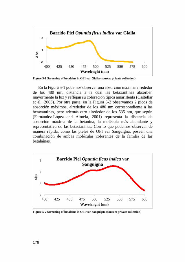
178
Figure 5-1 Screening of betalains in OFI var Gialla (source: private collection)
En la Figura 5-1 podemos observar una absorción máxima alrededor
de los 480 nm, distancia a la cual las betaxantinas absorben
mayormente la luz y reflejan su coloración típica amarillenta (Castellar
et al., 2003). Por otra parte, en la Figura 5-2 observamos 2 picos de
absorción máximos, alrededor de los 480 nm correspondiente a las
betaxantinas, pero además otro alrededor de los 535 nm, que según
(Fernández-López and Almela, 2001) representa la distancia de
absorción máxima de la betanina, la molécula más abundante y
representativa de las betacianinas. Con lo que podemos observar de
manera rápida, como las pieles de OFI var Sanguigna, poseen una
combinación de ambas moléculas colorantes de la familia de las
betalaínas.
Figure 5-2 Screening of betalains in OFI var Sanguigna (source: private collection)
0
1
2
400 425 450 475 500 525 550 575 600
Ab
s
Wavelenght (nm)
Barrido Piel Opuntia ficus indica var Gialla
0
1
2
3
400 425 450 475 500 525 550 575 600
Ab
s
Wavelenght (nm)
Barrido Piel Opuntia ficus indica var
Sanguigna

179
Se debe de tener en cuenta que los picos a 480 nm (representativo
de indicaxantina) y a 535 nm (representativo de betanina), no muestran
en ningún caso exclusividad los dos compuestos representativos, sino,
un cúmulo de moléculas con estructuras muy similares. Además, se
deben de tener muy en cuenta los efectos hipsocrómicos y batcrómicos
que presenta la glucosilación, metilación o cualquier otro tipo de
anexión de las agliconas (Esquivel, 2016). De acuerdo con lo anterior
mencionado, esta metodología es recomendada solo como técnica de
cribado, ya que si se precisa de mayor información, entonces se debe
de recurrir a técnicas analíticas especializadas como se hace más
adelante en este capítulo.
Otro de los usos de esta técnica de cribado, lo demostramos con la
Figura 5-3, en donde comparamos de manera rápida el efecto del pH en
los solventes de extracción. Como podemos observar en la figura,
mientras el pH sea más básico, mejor será la respuesta y los
rendimientos de recuperación del contenido de betalaínas. De la misma
forma, podemos probar distintas condiciones de extracción de una
manera muy rápida que en pruebas posteriores se podrá refinar. Un
ejemplo más complejo de extracción de betalaínas se presenta más
adelante en el capítulo 7.
Figure 5-3 Effect of pH on betalain extraction (source: private collection)
0
0.2
0.4
0.6
0.8
1
1.2
1.4
400 425 450 475 500 525 550 575 600
Ab
s
Wavelenght (nm)
Extracciónes piel OFI var Sanguigna a
diferentes valores de pHPH 6,3
PH 2,3

180
V.2 CARACTERIZACIÓN EXTENSIVA DE TRES
VARIEDADES DE FRUTOS DE OPUNTIA SPP. PARA LA
INNOVACIÓN DE INGREDIENTES ALIMENTARIOS.
El presente capítulo consiste en una adaptación al formato de la tesis
doctoral del artículo titulado “Extensive profiling of three varieties of
Opuntia spp. fruit for innovative food ingredients”, publicado en la
revista Food Research International. En él, tres especies frutales del
genero Opuntia (2 especies de Opuntia ficus-indica y Opuntia
engelmannii) fueron caracterizados en términos nutricionales y
funcionales, y evaluadas sus propiedades antimicrobianas, además, una
caracterización completa del perfil betalainico fue descrito mediante
técnicas cromatográficas. Los datos bibliográficos del artículo se
destacan a continuación:
Melgar, B., Pereira, E., Oliveira, M. B. P. P., Garcia-Castello, E. M.,
Rodriguez-Lopez, A. D., Sokovic, M., Barros, L., Ferreira, I. C. F. R.
(2017).
Extensive profiling of three varieties of Opuntia spp. fruit for
innovative food ingredients.
Food Research International, 101, 259-265.

181

Resultados
182
Extensive profiling of three varieties of Opuntia spp. fruit for
innovative food ingredients
Bruno Melgara,b, Eliana Pereiraa, M. Beatriz P.P. Oliveirac,
Esperanza M. Garcia-Castellob, Antonio D. Rodriguez-Lopezd,
Marina Sokovice, Lillian Barrosa, Isabel C.F.R. Ferreiraa,*
aCentro de Investigação de Montanha (CIMO), ESA, Instituto
Politécnico de Bragança, Campus de Santa Apolónia, 5300-253
Bragança, Portugal bInstitute of Food Engineering for Development, Universitat
Politècnica de València, Camino de Vera, s/n CP, 46022 Valencia,
Spain.
cREQUIMTE/LAQV, Science Chemical Department, Faculty of
Pharmacy of University of Porto, Rua Jorge Viterbo Ferreira, 228,
4050-313 Porto, Portugal. dInstitute for Industrial, Radiophysical and Environmental Safety
(ISIRYM), Universitat Politècnica de València, Camino de Vera, s/n
CP, 46022 Valencia, Spain. eUniversity of Belgrade, Department of Plant Physiology, Institute for
Biological Research “Siniša Stanković”, Bulevar Despota Stefana
142, 11000 Belgrade, Serbia.
*Corresponding author. Tel. +351 273 303219; fax +351 273 325405.
E-mail address: [email protected] (I.C.F.R. Ferreira)

Resultados
183
Abstract
Consumer interest in the use of natural ingredients is creating a
growing trend in the food industry, leading to research into the
development of natural products such as colorants, antimicrobials and
antioxidant compounds. This work involves an extensive chemical and
microbiological characterization of Opuntia ficus-indica var.
Sanguigna, Gialla and Opuntia engelmannii fruits. These species have
proved to be a good source of hydrophilic (sugars, organic acids and
betalains) and lipophilic (tocopherols and fatty acids) compounds.
While Opuntia engelmannii exhibited higher content of betacyanins,
Opuntia ficus-indica varieties Sanguigna and Gialla displayed greater
sugar and organic acid content. The Sanguigna variety also showed the
highest α-tocopherol content, enhancing the bioactivity of this variety,
which can be observed in its antimicrobial potential, tested in the
studied strains too. Results revealed that Opuntia spp. could be used as
a nutraceutical and/or food additive, maintaining and promoting health
and life quality.
Keywords: Opuntia ficus-indica; Opuntia engelmannii; nutritional
properties; betalains; antimicrobial activity

Resultados
184
V.2.1. INTRODUCTION
Consumers, in today’s food market, are becoming increasingly
conscious of high-quality healthy foods, leading to a demand for the
exclusion of synthetic food additives in groceries. These consumers
expect to see more familiar ingredients in their food products following
the food authorities warnings on the reduction of daily intake levels and
the addition of these additives to the Redbook 2000 (FDA, 2007). This
provides scientific information about the toxicological effects of the
consumption of additives, but often leads to misunderstanding by
consumers.
Scientific research is pursuing better alternatives to synthetic food
additives, including functional properties. Some researchers like
Almeida et al., (2011), directed their investigations towards the
revalorization of exotic fruit juices and extracts. These substances
contain biomolecules that could be applied as unpurified extracts or
isolated molecules, or as a possible substitute for synthetic additives.
Thus, by employing these new natural ingredients, consumers will be
able to take advantage of these molecules in their role of improving
overall well-being, as well as their contribution to the prevention of
some diseases (Devalaraja, Jain, & Yadav, 2011). Additionally, food
industries would be able to publish clearer labelling that could have a
beneficial impact on their sales (Osborn, 2015).
The prickly pear (Opuntia spp.) is an important crop to study due to
its adaptability to difficult growing conditions (arid and semiarid
zones). Although this species is native to Mexico, it has spread and been
cultivated across the world (Novoa et al., 2015). The genus Opuntia is
reported to have almost 300 different varieties (Reynolds and Arias,
2003), between domesticated and wild species. Opuntia ficus-indica is
one of the five most cultivated species for fruit production (Griffith,
2004), but there are also other wild species such as Opuntia
engelmannii that could be potentially used in the extraction of natural
ingredients, such as colorants.
The wide range of colour in prickly pears is due to the presence of
betalains, water-soluble, nitrogen-containing pigments present in a
limited number of families of the plant order Caryophyllales (Strack et
al., 2003). There are two types of betalains, red/violet betacyanins and
Resultados
185
yellow/orange betaxanthins (Esquivel, 2016), creating an interesting
palette of natural colouring agents. There is growing interest in
betalains, partially due to their good stability between the pH values of
3 and 7 (Herbach et al., 2006), and their ability to protect against the
potential effects of oxidative stress (Azeredo, 2009; Strack et al., 2003).
Although, the antixodant properties of betalains could be related to
other bioactive molecules such as tocopherols, organic acids, reducing
sugars and polyunsaturated fatty acids (PUFA), rather than the effect of
just one type of molecule (Pereira et al., 2014).
Therefore, the aim of this research was to carry out an extensive
physical, chemical and microbiological characterization of Opuntia
ficus-indica var. Sanguigna (OS) and Gialla (OG) and Opuntia
engelmannii (OE) fruits, as possible fruit to be used in the food industry
as natural ingredients.

Resultados
186
V.2.2. MATERIAL AND METHODS
V.2.2.1. Sample preparation
Cactus pear fruits (Opuntia ficus-indica var. Sanguigna -OS and Gialla
-OG) were collected in July-August 2016 in Sicily, Italy and were
purchased from a local market in Bragança, Portugal. Fruit from these
species were separated according to the maturation colour orange-red
(pulp and peel) and red-violet, obtaining two different samples. Wild
prickly pear fruit (Opuntia engelmannii- OE) were collected in
Bragança, Portugal (GPS coordinates: 41.797344, -6.772735) in early
September 2016. Dr. Carlos Aguiar of the School of Agriculture,
Polytechnic Institute of Bragança (Trás-os-Montes, Portugal),
confirmed the botanical identifications and voucher specimens were
deposited.
Within 24 hours, the fruit were washed with distilled water in order to
remove glochids and then air-dried. Afterwards, all the fruits were
peeled and the resulting pulp was lyophilized (LabConco, Frezone -105
°C, 4.5 L Cascade Benchtop Freeze Dry System, Kansas, MO, USA)
and stored in a cool, dry place until use.
V.2.2.2. Morphological parameters
Fruit size: the length and width of the entire fruit and pulp were
measured with a calliper. The whole fruit, pulp and peel where weighed
separately. For colour detection, fresh and lyophilized pulp and peel
were measured with a colorimeter (model CR-400; Konica Minolta
Sensing Inc., Japan), previously calibrated using the standard white
plate. Using illuminant C and a diaphragm opening of 8 mm, the CIE
L* a* b* colour space values were registered with a computerized
system using the colour data software “Spectra Magic Nx” (version
CM-S100W 2.03.0006).
V.2.2.3. Chemical characterisation
V.2.2.3.1 Proximal nutritional composition
Chemical and nutritional parameters (protein, fat, moisture, ash,
carbohydrates and energy) were determined only for the edible part of
the fruit (pulp). Samples were analysed according to the AOAC
Resultados
187
procedures (AOAC, 2016) Crude protein content (N × 6.25) was
determined by the macro-Kjeldahl method (AOAC, 991.02). Crude fat
(AOAC, 989.05) was estimated by extracting a known weight of
powdered sample with petroleum ether, using a Soxhlet apparatus.
Moisture content was determined by the weight difference before and
after the drying process (oven at 100 ºC); the ash content (AOAC,
935.42) was determined by incineration at 550 ± 15 °C. Total
carbohydrates (including fibre) were calculated by difference [total
carbohydrates (g/100 g) = 100 − (g fat + g protein + g ash)]. Total
energy was calculated according to the following equation: Energy
(kcal/100 g) = 4 × (g proteins + g carbohydrates) + 9 × (g fat).
V.2.2.3.2. Hydrophilic compounds
Soluble sugars. Sugars were determined in defatted samples by high
performance liquid chromatography coupled to a refraction index
detector (HPLC-RI, Knauer, Smartline system 1000, Berlin, Germany)
following a procedure described by Pereira, Barros, Carvalho, &
Ferreira (2011).
Organic acids. Organic acids were determined by an optimised
procedure previously described by Barros, Pereira, & Ferreira (2013)
and the analysis was performed using a Shimadzu 20A series UFLC
(Shimadzu Coperation, Kyoto, Japan) coupled to a diode array detector
(DAD), using 215 and 245 nm (for ascorbic acid) as the preferred
wavelengths. Results were expressed as g and mg per 100 g of pulp
fresh weight (FW), for sugars and organic acids, respectively.
The sugars and organic acids were identified by comparing their
retention times with standard compounds, and quantification was
conducted by comparison with dose–response curves constructed from
authentic standards. For sugar determination, melezitose was used as
the internal standard. Results were expressed as g and mg per 100 g of
pulp fresh weight (FW), for sugars and organic acids, respectively.
Betalains. The profile of these compounds was determined by LC-
DAD-ESI/MSn (Dionex Ultimate 3000 UPLC, Thermo Scientific, San
Jose, CA, USA). The lyophilized pulps were re-dissolved in water at a
concentration of 150 mg/mL. Chromatographic separation was

Resultados
188
achieved with a Waters Spherisorb S3 ODS-2 C18 (3 μm, 4.6 mm ×
150 mm, Waters, Milford, MA, USA) column working at 35 °C. The
solvents used were: (A) 0.1% trifluoroacetic acid (TFA) in water, and
(B) acetonitrile. The gradient elution followed these parameters: from
0% to 10% B for 15 min, from 10% to 15% B for 5 min, from 15 to
18% B for 5 min, from 18 to 50% B for 8 min, and from 50 to 0% B for
12 min.
The resulting total run time was 45 minutes, using a flow rate of 0.5
mL/min. Double detection was carried out in the DAD using 480 nm
(for betaxanthins) and 530 nm (for betacyanins), as the preferred
wavelengths, and in a mass spectrometer (MS). MS detection was
performed using positive mode, with a Linear Ion Trap LTQ XL mass
spectrometer (ThermoFinnigan, San Jose, CA, USA) equipped with an
ESI source. Nitrogen served as the sheath gas (50 psi); the system was
operated with a spray voltage of 4.8 kV, a source temperature of 320
°C, and a capillary voltage of 39 V. The tube lens offset was kept at a
voltage of 140 V. The full scan covered the mass range from m/z 100
to 1500. The collision energy used was 24 (arbitrary units). Data
acquisition was carried out with the Xcalibur® data system
(ThermoFinnigan, San Jose, CA, USA).
Identification of the betalain compounds (betacyanins and
betaxanthins) was performed by comparing the obtained information
with available data reported in the literature, providing a tentative
identification. For quantitative analysis, a calibration curve using an
isolated compound gomphrenin III (isolated from Gomphrena globosa
L.) was constructed based on the UV signal (y = 14670x - 19725, R² =
0.9997). The results for betacyanins were expressed as mg per 100 g of
pulp fresh weight (FW), and the results for betaxanthins were expressed
as a relative percentage (%) of the areas recorded at 480 nm.
V.2.2.3.3. Lipophilic compounds
Fatty acids. Fatty acid determination was achieved via the
transesterification procedure described previously by Guimarães et al.
(2013). The analysis was performed in gas chromatography (GC DANI
1000; Contone, Switzerland) equipment with flame ionization
detection. Results were expressed as relative percentages of each fatty
acid.

Resultados
189
Tocopherols. The four isoforms of tocopherols (vitamin E) were
determined according to the previously described procedure (Heleno et
al., 2010). Analysis was performed using a HPLC system (Knauer,
Smartline system 1000, Berlin, Germany), coupled to a fluorescence
detector (FP-2020; Jasco, Easton, MD, USA). Quantification was based
on the fluorescence signal response of each standard, using the internal
standard (tocol) method and using calibration curves obtained from
commercial standards of each isoform. The results were expressed in
µg per 100 g of pulp fresh weight (FW).
V.2.2.4. Antimicrobial effect of fruit pulp
Antibacterial activity was assayed according to Reis et al., (2014) using
the following Gram-positive bacteria: Staphylococcus aureus (ATCC
6538), Bacillus cereus (clinical isolate), Micrococcus flavus
(ATCC10240), and Listeria monocytogenes (NCTC7973) and Gram-
negative bacteria: Escherichia coli (ATCC 35210), Pseudomonas
aeruginosa (ATCC 27853), Salmonella typhimurium (ATCC 13311),
and Enterobacter cloacae (ATCC 35030).
While for antifungal assays, the following microfungi were used:
Aspergillus fumigatus (ATCC1022), Aspergillus ochraceus
(ATCC12066), Aspergillus versicolor (ATCC11730), Aspergillus
niger (ATCC6275), Penicillium funiculosum (ATCC 36839),
Penicillium ochrochloron (ATCC9112), Penicillium verrucosum var.
cyclopium (food isolate), and Trichoderma viride (IAM 5061).
The minimum inhibitory (MIC), minimum bactericidal (MBC) and
minimum fungicidal concentrations (MFC) were determined by
methodologies described previously (Reis et al., 2014; Chahdoura et
al., 2016). Bacterial and fungal organisms were obtained from the
Mycological Laboratory, Department of Plant Physiology, Institute for
Biological Research “Sinisa Stanković”, University of Belgrade,
Serbia.
Resultados
190
V.2.2.5. Statistical analysis
All the extractions and assays were performed in triplicate. Results
were expressed as mean values and standard deviations (SD), analysed
using a one-way analysis of variance (ANOVA) followed by Tukey’s
HSD Test with p = 0.05. The treatment was carried out using IBM SPSS
Statistics for Windows, version 23.0. (IBM Corp., Armonk, New York,
USA).
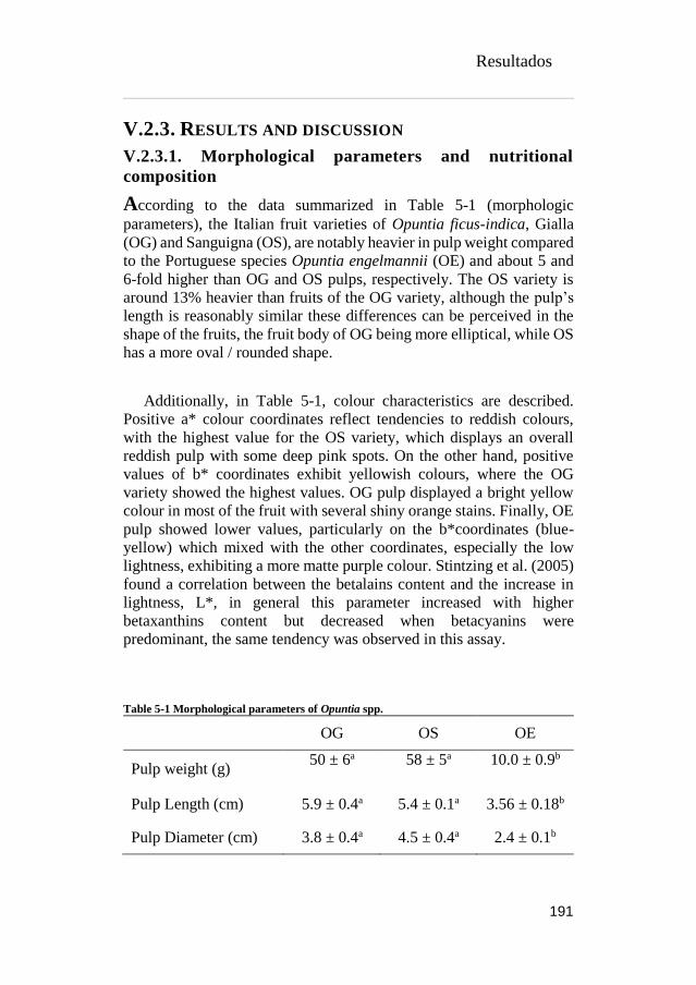
Resultados
191
V.2.3. RESULTS AND DISCUSSION
V.2.3.1. Morphological parameters and nutritional
composition
According to the data summarized in Table 5-1 (morphologic
parameters), the Italian fruit varieties of Opuntia ficus-indica, Gialla
(OG) and Sanguigna (OS), are notably heavier in pulp weight compared
to the Portuguese species Opuntia engelmannii (OE) and about 5 and
6-fold higher than OG and OS pulps, respectively. The OS variety is
around 13% heavier than fruits of the OG variety, although the pulp’s
length is reasonably similar these differences can be perceived in the
shape of the fruits, the fruit body of OG being more elliptical, while OS
has a more oval / rounded shape.
Additionally, in Table 5-1, colour characteristics are described.
Positive a* colour coordinates reflect tendencies to reddish colours,
with the highest value for the OS variety, which displays an overall
reddish pulp with some deep pink spots. On the other hand, positive
values of b* coordinates exhibit yellowish colours, where the OG
variety showed the highest values. OG pulp displayed a bright yellow
colour in most of the fruit with several shiny orange stains. Finally, OE
pulp showed lower values, particularly on the b*coordinates (blue-
yellow) which mixed with the other coordinates, especially the low
lightness, exhibiting a more matte purple colour. Stintzing et al. (2005)
found a correlation between the betalains content and the increase in
lightness, L*, in general this parameter increased with higher
betaxanthins content but decreased when betacyanins were
predominant, the same tendency was observed in this assay.
Table 5-1 Morphological parameters of Opuntia spp.
OG OS OE
Pulp weight (g) 50 ± 6a 58 ± 5a 10.0 ± 0.9b
Pulp Length (cm) 5.9 ± 0.4a 5.4 ± 0.1a 3.56 ± 0.18b
Pulp Diameter (cm) 3.8 ± 0.4a 4.5 ± 0.4a 2.4 ± 0.1b

Resultados
192
Color coordinates
a* 12 ± 1c 36.8 ± 0.4a 16 ± 0.8b
b* 49 ± 3a 19.8 ± 0.6b 4.9 ± 0.7c
L* 48.9 ± 0.9a 33 ± 1b 25 ± 1c
C* 50 ± 2a 41.8 ± 0.6b 16.8 ± 0.5c
h° 1.33 ± 0.03a 0.49 ± 0.01b 0.30 ± 0.05c
OG, Opuntia ficus-indica var Gialla; OS, Opuntia ficus-indica var
Sanguigna; OE, Opuntia engelmannii. In each row different letters
mean significant differences (p < 0.05).
The macronutrient composition of Opuntia fruiting bodies is presented
in Table 5-2. Opuntia ficus-indica var. Gialla and Sanguigna do not
show significant differences in most of the macronutrients, excluding
proteins and ash content, which had higher concentrations. Higher
percentages in proteins could be due to a greater concentration of
pigments in the cell vacuoles, betalainic colorants being water-soluble
nitrogen-containing pigments (Azeredo, 2009). Despite the differences
shown between proteins and ash, these macronutrients along with
moisture, fat and carbohydrates are in accordance with results reported
by other authors like Angulo-Bejarano, Martínez-Cruz, & Paredes-
López (2014).
Table 5-2 Nutritional value and hydrophilic compounds of the studied Opuntia spp.
OG OS OE
Moisture (%) 83.38 ±
0.07a
81 ± 2a 65 ± 2b
Fat (g/100 g FW) 0.03 ±
0.001b
0.06 ±
0.004b
0.38 ± 0.03a
Proteins (g/100 g FW) 0.52 ± 0.01c 0.84 ± 0.03b 1.62 ± 0.06a
Ash (g/100 g FW) 0.34 ±
0.004c
0.42 ± 0.02b 0.75 ± 0.02a
Carbohydrates (g/100
g FW)
15.68 ±
0.04b
18 ± 2b 33 ± 2a

Resultados
193
Energy (Kcal/100 g
FW)
65.1 ± 0.1b 77 ± 7b 140 ± 6a
Sugars (g/100 g FW)
Fructose 4.13 ± 0.02b 4.97 ± 0.07a 0.53 ± 0.01c
Glucose 5.53 ± 0.05b 6.30 ± 0.03a 0.83 ± 0.01c
Sucrose 0.13 ±
0.005c
0.15 ±
0.001b
0.31 ± 0.01a
Total sugars 9.79 ± 0.07b 11.43 ±
0.09a
1.68 ± 0.02c
Organic acids (mg/100
g FW)
Oxalic 26.7 ± 0.3b 29.5 ± 0.6a tr
Quinic 28 ± 1* 35.6 ± 0.3* nd
Malic tr tr tr
Ascorbic 2.3 ± 0.1b 2.0 ± 0.1b 19.7 ± 0.1a
Citric 49.3 ± 0.7a 46.9 ± 0.1b 0.8 ± 0.01c
Succinic 184 ± 1* 131 ± 2* nd
Fumaric 8.0 ± 0.1* 6.3 ± 0.2* nd
Total organic acids 298.1 ± 0.8a 251 ± 2b 20.5 ± 0.1c
FW, pulp fresh weight; nd, not detected; tr, traces. OG, Opuntia
ficus-indica var Gialla; OS, Opuntia ficus-indica var Sanguigna; OE,
Opuntia engelmannii. In each row different letters mean significant
differences (p < 0.05). *Statistical differences (<0.001) were
observed when t-student test was applied.
Conversely, OE was statistically different from OG and OS,
moisture in OE was around 17 percent inferior, while protein, ash and
carbohydrates were 2-fold higher in OE and fat content was up to 10-
fold higher. Once again, high protein content could be related to the
greater pigment composition, as will be discussed later on in the
betalainic profile of Opuntia spp.

Resultados
194
In addition, fat, ash and carbohydrate content could be superior, and
moisture inferior due to a higher ratio of seed/pulp compared to OG or
OS. Chougui et al. (2013) have worked on the oil composition and
characterization of Opuntia seeds and have shown that a higher ratio of
seed/pulp increases the oil yield and the fat content. Likewise Jain,
Grover, & Kaur (2016) reported that seeds are mainly composed of
carbohydrates with a considerable amount of minerals and proteins, this
information supports our hypothesis on the greater amount of
macronutrients found in OE samples. To the best of our knowledge, no
reports are available on the nutritional composition of Opuntia
engelmannii species.
V.2.3.2. Hydrophilic and lipophilic compounds
The free sugar content of Opuntia spp. is displayed in Table 5-2, OE
has the lowest total sugar content, followed by OG and OS in ascending
order. It is worth highlighting that OE is around 10-fold lower in total
sugar concentration compared to Opuntia ficus-indica fruit, this higher
concentration is mainly due to the presence of glucose and fructose, the
most characteristic sugars in Opuntia ficus-indica (Kyriacou et al.,
2016). Although all species were collected in the same period, OE
species were probably not at their optimum ripeness. Despite the fact
that OE has higher content in carbohydrates and lower total sugar
content compared to OG and OS, this could indicate that the majority
of the carbohydrates could be fibres or longer polysaccharide chains
like mucilage (Da-Costa-Rocha et al., 2014).
Table 5-3 Chemical composition in lipophilic compounds, tocopherols and fatty acids of
Opuntia spp.
OG OS OE
Fatty acids (%)
C6:0 0.32 ± 0.08a 0.21 ± 0.02b 0.09 ± 0.01c
C8:0 0.22 ± 0.06a 0.07 ± 0.02b
0.044 ±
0.001b
C10:0 0.6 ± 0.2a 0.18 ± 0.02b
0.097 ±
0.001b
C12:0 0.7 ± 0.2a 0.23 ± 0.02b 0.087 ± 0.001c
C14:0 1.4 ± 0.3a 0.56 ± 0.03b 0.234 ± 0.001c

Resultados
195
C15:0 0.47 ± 0.01a 0.16 ± 0.01b 0.055 ± 0.001c
C16:0 16 ± 1a 13.9 ± 0.1b 7.92 ± 0.03c
C16:1 0.31 ± 0.02b 0.504 ± 0.001a
0.298 ±
0.001b
C17:0 0.39 ± 0.04a 0.171 ± 0.001b 0.106 ± 0.001c
C18:0 6 ± 1a 3.95 ± 0.01b 3.58 ± 0.01b
C18:1n9 11.7 ± 0.3a 9.82 ± 0.03c 10.23 ± 0.01b
C18:2n6 59 ± 1c 68.3 ± 0.2b 75.73 ± 0.02a
C18:3n3 1.9 ± 0.9a 1.26 ± 0.05ab 0.71 ± 0.04b
C20:0 0.5 ± 0.1a 0.28 ± 0.01b
0.305 ±
0.001b
C20:1 0.116 ±
0.001 nd 0.144 ± 0.001
C22:0 0.636 ±
0.07a 0.38 ± 0.02b 0.242 ± 0.001c
C24:0 nd nd 0.115 ± 0.001
SFA 27 ± 2a 20.1 ± 0.2b 12.75 ± 0.05c
MUFA 12.1 ± 0.3a 10.33 ± 0.03c 10.69 ± 0.01b
PUFA 61 ± 1c 69.6 ± 0.2b 76.45 ± 0.06a
Tocopherols (μg/100g FW)
α-tocopherol 53 ± 1b 99 ± 3a 15.1 ± 0.3c
β-tocopherol 2.97 ± 0.05* 5.2 ± 0.2* nd
γ-tocopherol 2.8 ± 0.4c 53 ± 1a 27 ± 0.9b
Total tocopherols 58.9 ± 0.4b 158 ± 1a 43.0 ± 0.6c
OG, Opuntia ficus-indica var Gialla; OS, Opuntia ficus-indica var
Sanguigna; OE, Opuntia engelmannii.
FW, pulp fresh wight; nd, not detected; SFA, saturated fatty acids;
MUFA, monounsaturated fatty acids; PUFA, polyunsaturated fatty
acids. In each row different letters mean significant differences (p <
0.05). *Statistical differences (<0.001) were observed when t-student
test was applied.

Resultados
196
The total organic acid content of OE is 12 to 15-fold lower compared
to OS and OG, respectively (Table 5-2). The main difference in the total
organic acids analysed in Opuntia spp., is due to the succinic acid
content, this acid is by far the most abundant in Opuntia ficus-indica
fruits, while it was not detected in OE. Farag, Maamoun, Ehrlich,
Fahmy, & Wesjohann (2017), also identified succinic acid as the more
abundant organic acid in Italian Opuntia ficus-indica varieties. Succinic
acid is an important metabolite involved in several signalling processes,
not only in the mitochondria were it is generated, but in the cytoplasm
as well as the extracellular space and it is also involved in the
elimination of reactive oxygen species (Tretter et al., 2016). Succinic
acid is generally recognized as safe (FDA, 2017) and has different
applications in the pharmaceutical and food industries, although it is
primarily used as an acidity regulator (Ahn et al., 2016). Organic acid
profiles are not reported for Opuntia spp. in the literature, although,
extensive assays have been performed on ascorbic acid for Opuntia
ficus-indica. Variable values are found ranging from 3.5 to 45 mg /100
g edible pulp (Kuti, 2004; Stintzing et al., 2005). OG and OS showed
lower levels of vitamin C, compared with reported data, while OE was
almost 10-fold higher than the previously mentioned varieties.
The betalain profiles of OG, OS and OE are shown in Figure 5-4.
Data regarding retention time, λmax in the visible region, molecular ion
and main fragment ions observed in MS2, obtained by HPLC-DAD-
ESI/MS analysis regarding betalains identification and quantification
are presented in Table 5-4. Compounds 1-3 were identified as
betaxanthin derivatives, and compounds 4-7 as betacyanin derivatives.
All of the identified compounds were previously described in Opuntia
spp. (Cejudo Bastante et al., 2014; Mata et al., 2016). Betaxanthins
were found in a higher percentage in OG, followed by OS and
were not identified in OE. Nevertheless, betacyanidin content was
lower in OG (11.32 mg/100 g of FW), followed by OS (215 mg/100 g
FW) and the largest amounts were present in OE (total content of 283
mg/100 g of FW), which presented 25-fold higher quantities then OG
and 1.3-fold higher then OS. The hierarchical order observed agrees
with the data found by other researchers, were the amount of betalains
increased from yellow to red and to purple varieties (Farag et al., 2017).
Saturated fatty acids (SFA), monounsaturated fatty acids (MUFA)
and polyunsaturated fatty acids (PUFA) are presented in Table 5-3.

Resultados
197
Samples were richer in PUFA with over 60% of the total fatty acids,
where OE was notably higher than OS and OG. SFA were the second
most abundant group followed by MUFA. In all the Opuntia species,
linoleic palmitic and oleic acids were the main fatty acids present,
respectively.
Although a plethora of information on the seed oils of Opuntia spp
are available, few publications have analysed the fatty acid content in
the fruit, Farag et al., (2017) also report linoleic, palmitic and oleic acid
as the major fatty acids present in the edible portion of Opuntia ficus-
indica. The essential fatty acids have given rise to great interest due to
the health potential of PUFA. According to Timilsena, Wang, Adhikari,
& Adhikari, (2017), PUFA plays a vital role in maintaining health in
humans by minimizing the risk of cardiovascular and
neurodegenerative disease, arthritis, diabetes and certain types of
cancer. Therefore, it is important to stress the possibility of finding
richer PUFA matrices in other fruit, vegetables and seeds.
As shown in Table 5-3, OS was the variety that displayed the highest
tocopherol content, α-tocopherol being the main isoform present,
followed by γ- and β-tocopherol. The lowest content in tocopherols was
found in OE samples, where only α- and γ- isoforms were present, the
latter isoform being the one that contributes most to the total content.
Regarding OG, α-tocopherol was around 18-fold higher compared with
β- and γ-tocopherol. As far as we know, there are no studies related to
the tocopherol content in Opuntia ficus-indica, only Farag et al. (2017)
describe the relative percentage of α-tocopherol in 3 varieties of
Opuntia ficus-indica, but do not give concrete amounts with which a
comparison could be performed.
The concentration of tocopherols in the same genus, but different
species was assayed by Morales et al. (2012), with total tocopherol
content ranging between 140 and 220 μg/100 g FW in Opuntia
joconostle and matudae. Only the values obtained by OS are in
concordance with those revealed by Morales et al. (2012), while
samples OG and OE only displayed 59 and 43 μg/100 g, respectively.
Vitamin E is the major lipid-soluble antioxidant in the cell antioxidant
defence system, nonetheless, the human body is not able to synthesize
these substances using its own metabolic pathways, therefore it has to

Resultados
198
Figure 5-4 HPLC chromatograms of (A) OG betaxanthin profile recorded at 484 nm and
(B) OE betacyanin profile recorded at 535 nm (source: private collection)
be obtained from the diet (Sýs et al., 2017). Vitamin E functions as a
chain-breaking antioxidant, inhibiting the propagation of lipid
peroxidation, preventing lipoproteins and cell membranes from
oxidative damage by acting as singlet oxygen quencher and stabilizing
chloroplast membranes (Takshak and Agrawal, 2015). Although OS
has the highest content in total tocopherols, the amount present in 100
g of fruit only represents around 1% of the Reference Daily Intake
(NIH, 2017).
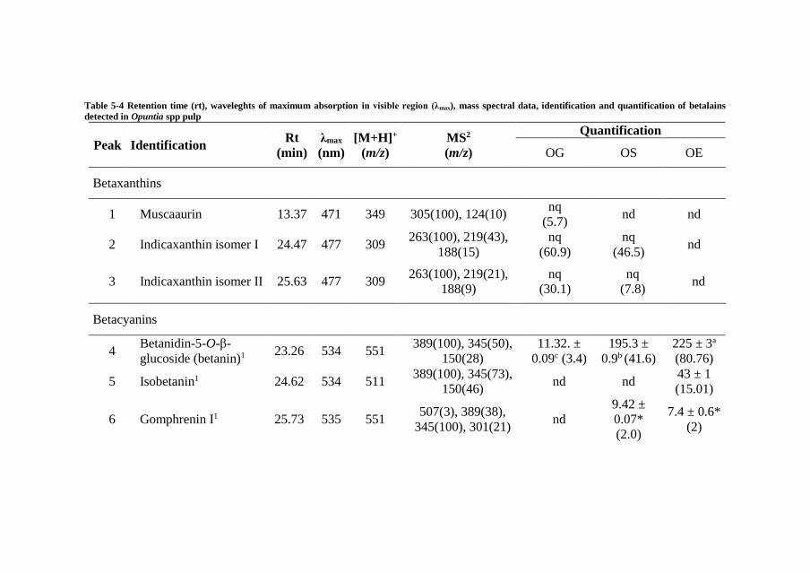
Table 5-4 Retention time (rt), waveleghts of maximum absorption in visible region (λmax), mass spectral data, identification and quantification of betalains
detected in Opuntia spp pulp
Peak Identification Rt
(min)
λmax
(nm)
[M+H]+
(m/z)
MS2
(m/z)
Quantification
OG OS OE
Betaxanthins
1 Muscaaurin 13.37 471 349 305(100), 124(10) nq
(5.7) nd nd
2 Indicaxanthin isomer I 24.47 477 309 263(100), 219(43),
188(15)
nq
(60.9)
nq
(46.5) nd
3 Indicaxanthin isomer II 25.63 477 309 263(100), 219(21),
188(9)
nq
(30.1)
nq
(7.8) nd
Betacyanins
4 Betanidin-5-O-β-
glucoside (betanin)1 23.26 534 551
389(100), 345(50),
150(28)
11.32. ±
0.09c (3.4)
195.3 ±
0.9b (41.6)
225 ± 3a
(80.76)
5 Isobetanin1 24.62 534 511 389(100), 345(73),
150(46) nd nd
43 ± 1
(15.01)
6 Gomphrenin I1 25.73 535 551 507(3), 389(38),
345(100), 301(21) nd
9.42 ±
0.07*
(2.0)
7.4 ± 0.6*
(2)

Resultados
200
7 Betanidin1 28.16 523 389 343(97), 150(91) nd
9.89 ±
0.03*
(2.1)
7.5 ± 0.1*
(2.13)
OG, Opuntia ficus-indica var Gialla; OS, Opuntia ficus-indica var Sanguigna; OE, Opuntia engelmannii.
Amounts in samples outside brackets in mg/100 g pulp FW, amounts inside brackets in relative percentages. FW, Fresh
weight; nq, not quantified; nd, not detected. Calibration curve: 1gomphrenin III (y = 14670x - 19725). In each row
different letters mean significant differences (p < 0.05). *Statistical differences (<0.001) were observed when t-student
test was applied.

Resultados
201
V.2.3.3. Antimicrobial properties
Results showing pathogenic bacteria growth inhibition are presented
in Table 5-5. The three varieties of Opuntia spp. showed different levels
of antibacterial activity. The Opuntia variety OS and OE displayed
activity against all the tested bacterial strains, being more active than
the commercial controls ampicillin and streptomycin, for all the MIC
values, with the exception of Staphylococcus aureus. When comparing
the bactericidal (MBC) potential of OS with the commercial antibiotic
streptomycin, only 5 out of the 8 tested strains had better performance
(Bacillus cereus, Micrococcus flavus, Escherichia coli, Enterobacter
cloacae and Salmonella typhimurium), but in the other three strains, the
difference shown was very small. It is important to stress the strong
effect of OS samples against Micrococcus flavus, this sample displayed
4-fold stronger inhibition compared to the best antibiotic tested as a
positive control (streptomycin).
On the other hand, OE showed higher potential than the commercial
antibiotic ampicillin against all 8 strains assayed. When this sample was
compared to streptomycin, only 2 strains (Staphylococcus aureus and
Listeria monocytogenes) stood out as the species with the highest
resistance against the OE sample, otherwise for the remaining 6 strains
OE showed a similar or better performance than the mentioned
antibiotic.
The sample with the least potential was OG, which only had an
effect on 5 out of 8 strains tested. Nevertheless, for 4 of the strains, the
OG sample showed the same or better potential compared to the
positive antibiotic controls.
The fungi positive controls used in this assay were ketoconazole and
bifonazole (Table 5-5), the latter showing overall a stronger effect
against the pathogenic fungal strains. Samples of OS, OG and OE
exhibit fungistatic and fungicidal effects against all 8 strains tested. The
minimum inhibitory concentrations (MIC) were similar or better than
the concentration of ketoconazole control (except in Trichoderma
viride, Aspergillus ochraceus and Penicillium ochrochloron), but none
of the samples exhibited better performance than bifonazole.

Table 5-5 Antibacterial and antifungal activity of Opuntia samples
Bacteria Samples Control
OS OG OE C1 C2
MIC MBC MIC MBC MIC MBC MIC MBC MIC MBC
Bacillus cereus 0.075 0.15 0.10 0.15 0.10 0.15 0.10 0.20 0.25 0.40
Micrococcus flavus 0.050 0.07 - - 0.20 0.30 0.20 0.30 0.25 0.40
Staphylococcus aureus 0.15 0.30 0.075 0.15 0.15 0.45 0.04 0.10 0.25 0.45
Listeria monocytogenes 0.15 0.45 0.10 0.15 0.15 0.45 0.20 0.30 0.40 0.50
Escherichia coli 0.10 0.15 - - 0.20 0.30 0.20 0.30 0.40 0.50
Enterobacter cloacae 0.10 0.15 - - 0.075 0.30 0.20 0.30 0.25 0.50
Pseudomonas
aeruginosa
0.10 0.30 0.15 0.30 0.10 0.30 0.20 0.30 0.75 1.20
Salmonella
typhimurium
0.10 0.45 0.20 0.45 0.30 0.45 0.25 0.50 0.40 0.75

Resultados
203
Fungi Samples Control
OS OG OE C3 C4
MIC MFC MIC MFC MIC MFC MIC MFC MIC MFC
Aspergillus fumigatus 0.30 0.60 0.30 0.60 0.30 0.45 0.25 0.50 0.15 0.20
Aspergillus versicolor 0.30 0.45 0.30 0.45 0.15 0.30 0.20 0.50 0.10 0.20
Aspergillus ochraceus 0.20 0.45 0.30 0.45 0.20 0.30 1.50 2.00 0.15 0.20
Aspergillus niger 0.30 0.60 0.30 0.60 0.30 0.45 0.20 0.50 0.15 0.20
Trichoderma viride 0.10 0.20 0.075 0.15 0.0375 0.075 1.00 1.00 0.15 0.20
Penicillium funiculosum 0.30 0.45 0.30 0.45 0.30 0.45 0.20 0.50 0.20 0.25
Penicillium
ochrochloron
0.50 0.75 0.075 0.15 0.30 0.45 2.50 3.50 0.20 0.25
Penicillium verrucosum
var. cyclopium
0.30 0.60 0.30 0.60 0.30 0.60 0.20 0.30 0.10 0.20
OG, Opuntia ficus-indica var Gialla; OS, Opuntia ficus-indica var Sanguigna; OE, Opuntia engelmannii. C1,
Streptomycin; C2, Ampicillin; C3, Ketoconazole; C4, Bifonazole; MIC, minimum inhibitory concentration; MBC,
minimum bactericidal concentrations; MBC, minimum fungicidal concentrations; N.A., No activity

Resultados
204
The minimum fungicidal concentrations (MFC) of the samples
against bifonazole were always inferior, except for the T. viride strain
against all the tested samples and in the P. ochrochloron strain against
OG. The fungicidal power of OE against T. viride presented 3-fold
lower concentrations (higher potential) than the fungicidal power
displayed by bifonazole. Chahdoura et al., (2016), tested the
antimicrobial activity from Opuntia microdasys flowers and reported
lower effects on their samples compared to OS, OG and OE extracts.
Polyphenols and other biofunctional molecules, such as betalains,
have shown the capacity to induce cellular damage in pathogenical
microorganisms (Azeredo, 2009; Sansano et al., 2017). Hussain et al.
(2017), presented results were curcuminoids distort the cellular
architecture, causing cellular edema, followed by lysis and extrusion of
cytoplasmic content, this mechanism could also explain the
polyphenolic and betalainic antimicrobial effect of OG, OS and OE
samples.

Resultados
205
V.2.4. CONCLUSIONS
Overall, fruit from Opuntia ficus indica var Gialla and Sanguigna and
Opuntia engelmannii, revealed interesting antimicrobial properties,
which might be used to functionalize products in different areas such
as cosmetics, pharmaceuticals or food industries, as natural ingredients
or as bioactives. An option is the use of betalains as natural colorants,
in which the different hues provided from betaxanthins and
betacyanins, could be employed, in isolation or mixed in order to obtain
a broader range of colours with healthy properties. Opuntia samples
have also shown strong antimicrobial activity as well as biomolecules
with bioactive potential, which could act positively on the health and
well-being of consumers.
Acknowledgements
The authors are grateful to the Foundation for Science and Technology
(FCT, Portugal) and FEDER under Programme PT2020 for financial
support to CIMO (UID/AGR/00690/2013) and L. Barros contract. B.
Melgar (No. 329930) also thanks CONACyT for his grant. The authors
are grateful for a grant from the Serbian Ministry of Education,
Sciences and Technological Development (no. 173032). The authors
would also like to thank Dr. Carlos Aguiar for the botanical
identification of these species.

Resultados
206
References
Ahn, J.H., Jang, Y.-S., Lee, S.Y., 2016. Production of succinic acid by
metabolically engineered microorganisms. Curr. Opin.
Biotechnol. 42, 54–66.
https://doi.org/10.1016/j.copbio.2016.02.034
Al-Farsi, M.A., Lee, C.Y., 2008. Optimization of phenolics and dietary
fibre extraction from date seeds. Food Chem. 108, 977–985.
https://doi.org/10.1016/j.foodchem.2007.12.009
Albano, C., Negro, C., Tommasi, N., Gerardi, C., Mita, G., Miceli, A.,
De Bellis, L., Blando, F., 2015. Betalains, Phenols and
Antioxidant Capacity in Cactus Pear [Opuntia ficus-indica (L.)
Mill.] Fruits from Apulia (South Italy) Genotypes. Antioxidants
4, 269–280. https://doi.org/10.3390/antiox4020269
Albuquerque, T.G., Santos, F., Sanches-Silva, A., Beatriz Oliveira, M.,
Bento, A.C., Costa, H.S., 2016. Nutritional and phytochemical
composition of Annona cherimola Mill. fruits and by-products:
Potential health benefits. Food Chem. 193, 187–195.
https://doi.org/10.1016/j.foodchem.2014.06.044
Allai, L., Druart, X., Öztürk, M., BenMoula, A., Nasser, B., El Amiri,
B., 2016. Protective effects of Opuntia ficus-indica extract on ram
sperm quality, lipid peroxidation and DNA fragmentation during
liquid storage. Anim. Reprod. Sci. 175, 1–9.
https://doi.org/10.1016/j.anireprosci.2016.09.013
Almeida, M.M.B., de Sousa, P.H.M., Arriaga, Â.M.C., do Prado, G.M.,
Magalhães, C.E. de C., Maia, G.A., de Lemos, T.L.G., 2011.
Bioactive compounds and antioxidant activity of fresh exotic
fruits from northeastern Brazil. Food Res. Int. 44, 2155–2159.
https://doi.org/10.1016/j.foodres.2011.03.051
Alvarez, B., 2007. Análisis de factibilidad del cultivo de la tuna en la
localidad de Icaño , Departamento La Paz, Dirección provincial
de programación del desarrollo, Ministerio de Producción y
Desarrollo. Gobierno de la Provincia de Catamarca. Catamarca,
Argentina.
Alzate T, L.M., González, D., Hincapié, S., Cardona S, B.L., Londoño-
Londoño, J., Jiménez-Cartagena, C., 2016. The profile of
bioactive substances in ten vegetable and fruit by-products from
a food supply chain in Colombia. Sustain. Prod. Consum.
https://doi.org/10.1016/j.spc.2016.07.005

Resultados
207
Ammar, I., Ben Salem, M., Harrabi, B., Mzid, M., Bardaa, S., Sahnoun,
Z., Attia, H., Ennouri, M., 2018. Anti-inflammatory activity and
phenolic composition of prickly pear (Opuntia ficus-indica)
flowers. Ind. Crops Prod. 112, 313–319.
https://doi.org/10.1016/j.indcrop.2017.12.028
Angulo-Bejarano, P.I., Martínez-Cruz, O., Paredes-López, O., 2014.
Phytochemical Content, Nutraceutical Potential and
Biotechnological Applications of an Ancient Mexican Plant:
Nopal (Opuntia ficus-indica). Curr. Nutr. Food Sci. 10, 196–217.
https://doi.org/10.2174/157340131003140828121015
Arlt, U., 2010. NATCOL | Foods Colours Legislation.
Aruwa, C.E., Amoo, S.O., Kudanga, T., 2018. Opuntia (Cactaceae)
plant compounds, biological activities and prospects – A
comprehensive review. Food Res. Int. 112, 328–344.
https://doi.org/10.1016/j.foodres.2018.06.047
Association of Official Analytical Chemists, 2016. Official Methods of
Analysis of AOAC International, 20th ed. The Association,
Gaithersburg, MD, USA.
Attokaran, M., 2011. Natural Food Flavors and Colorants Natural Food
Flavors and Colorants, 1st ed. Blackwell Publishing LTD, Iowa.
Augustin, M.A., Sanguansri, L., 2015. Challenges and Solutions to
Incorporation of Nutraceuticals in Foods. Annu. Rev. Food Sci.
Technol. 6, 463–477. https://doi.org/10.1146/annurev-food-
022814-015507
Ayala-Zavala, J.F., Vega-Vega, V., Rosas-Domínguez, C., Palafox-
Carlos, H., Villa-Rodriguez, J.A., Siddiqui, M.W., Dávila-Aviña,
J.E., González-Aguilar, G.A., 2011. Agro-industrial potential of
exotic fruit byproducts as a source of food additives. Food Res.
Int. 44, 1866–1874.
https://doi.org/10.1016/j.foodres.2011.02.021
Azeredo, H.M.C., 2009. Betalains: Properties, sources, applications,
and stability - A review. Int. J. Food Sci. Technol. 44, 2365–2376.
https://doi.org/10.1111/j.1365-2621.2007.01668.x
Balouiri, M., Sadiki, M., Ibnsouda, S.K., 2016. Methods for in vitro
evaluating antimicrobial activity: A review. J. Pharm. Anal. 6,
71–79. https://doi.org/10.1016/j.jpha.2015.11.005
Barros, L., Calhelha, R.C., Queiroz, M.J.R.P., Santos-Buelga, C.,
Santos, E.A., Regis, W.C.B., Ferreira, I.C.F.R., 2015. The
Resultados
208
powerful in vitro bioactivity of Euterpe oleracea Mart. seeds and
related phenolic compounds. Ind. Crops Prod. 76, 318–322.
https://doi.org/10.1016/j.indcrop.2015.05.086
Barros, L., Pereira, C., Ferreira, I.C.F.R., 2013. Optimized Analysis of
Organic Acids in Edible Mushrooms from Portugal by Ultra Fast
Liquid Chromatography and Photodiode Array Detection. Food
Anal. Methods 6. https://doi.org/10.1007/s12161-012-9443-1
Barrows, J.N., Lipman, A.L., Bailey, C.J., Cianci, S., 2003. Color
Additives: FDA’s Regulatory Process and Historical
Perspectives. Food Saf. Mag. URL
https://www.fda.gov/forindustry/coloradditives/regulatoryproces
shistoricalperspectives/ (accessed 9.3.18).
Baydar, N.G., Özkan, G., Sağdiç, O., 2004. Total phenolic contents and
antibacterial activities of grape (Vitis vinifera L.) extracts. Food
Control 15, 335–339. https://doi.org/10.1016/S0956-
7135(03)00083-5
Belščak-Cvitanović, A., Durgo, K., Huđek, A., Bačun-Družina, V.,
Komes, D., 2018. Metabolism and Health Effects of Polyphenols,
in: Galanakis, C.M. (Ed.), Polyphenols: Properties, Recovery, and
Applications. Woodhead Publishing, pp. 1–44.
https://doi.org/https://doi.org/10.1016/B978-0-12-813572-
3.00001-4
Belwal, T., Dhyani, P., Bhatt, I.D., Rawal, R.S., Pande, V., 2016.
Optimization extraction conditions for improving phenolic
content and antioxidant activity in Berberis asiatica fruits using
response surface methodology (RSM). Food Chem. 207, 115–
124. https://doi.org/10.1016/j.foodchem.2016.03.081
Bessada, S.M.F., Barreira, J.C.M., Barros, L., Ferreira, I.C.F.R.,
Oliveira, M.B.P.P., 2016. Phenolic profile and antioxidant
activity of Coleostephus myconis (L.) Rchb.f.: An underexploited
and highly disseminated species. Ind. Crops Prod. 89, 45–51.
https://doi.org/10.1016/j.indcrop.2016.04.065
Betancourt, C., Cejudo-Bastante, M.J., Heredia, F.J., Hurtado, N.,
2017. Pigment composition and antioxidant capacity of
betacyanins and betaxanthins fractions of Opuntia dillenii (Ker
Gawl) Haw cactus fruit. Food Res. Int. 101, 173–179.
https://doi.org/10.1016/j.foodres.2017.09.007
Bhatia, S., Bera, T., Dahiya, R., Bera, T., Bhatia, S., Bera, T., 2015.
Classical and Nonclassical Techniques for Secondary Metabolite

Resultados
209
Production in Plant Cell Culture, in: Modern Applications of Plant
Biotechnology in Pharmaceutical Sciences. Elsevier, pp. 231–
291. https://doi.org/10.1016/B978-0-12-802221-4.00007-8
Brand-Williams, W., Cuvelier, M.E., Berset, C., 1995. Use of a free
radical method to evaluate antioxidant activity. LWT - Food Sci.
Technol. 28, 25–30. https://doi.org/10.1016/S0023-
6438(95)80008-5
Bravo-Hollis, H., 1978. Las cactaceas de Mexico. Universidad
Nacional Autonoma de Mexico, Mexio.
https://doi.org/10.15713/ins.mmj.3
Brundtland, G.H., 1987. Our common future.
Cai, Y., Sun, M., Corke, H., 2003. Antioxidant activity of betalains
from plants of the Amaranthaceae. J. Agric. Food Chem. 51,
2288–2294. https://doi.org/10.1021/jf030045u
Calderón-Oliver, M., Escalona-Buendía, H.B., Medina-Campos, O.N.,
Pedraza-Chaverri, J., Pedroza-Islas, R., Ponce-Alquicira, E.,
2016. Optimization of the antioxidant and antimicrobial response
of the combined effect of nisin and avocado byproducts. LWT -
Food Sci. Technol. 65, 46–52.
https://doi.org/10.1016/j.lwt.2015.07.048
Cardoso-Ugarte, G.A., Sosa-Morales, M.E., Ballard, T., Liceaga, A.,
San Martín-González, M.F., 2014. Microwave-assisted extraction
of betalains from red beet (Beta vulgaris). LWT - Food Sci.
Technol. 59, 276–282. https://doi.org/10.1016/j.lwt.2014.05.025
Carocho, M., Ferreira, I.C.F.R., 2013. A review on antioxidants,
prooxidants and related controversy: Natural and synthetic
compounds, screening and analysis methodologies and future
perspectives. Food Chem. Toxicol. 51, 15–25.
https://doi.org/10.1016/j.fct.2012.09.021
Carr, M., 2013. The water relations and irrigation requirements of
avocado (Persea americana Mill.): a review. Exp. Agric. 49, 256–
278. https://doi.org/10.1017/S0014479712001317
Cassano, A., Conidi, C., Timpone, R., D’Avella, M., Drioli, E., 2007.
A membrane-based process for the clarification and the
concentration of the cactus pear juice. J. Food Eng. 80, 914–921.
https://doi.org/10.1016/j.jfoodeng.2006.08.005
Castellanos-Santiago, E., Yahia, E.M., 2008. Identification and
quantification of betalains from the fruits of 10 Mexican prickly

Resultados
210
pear cultivars by high-performance liquid chromatography and
electrospray ionization mass spectrometry. J. Agric. Food Chem.
56, 5758–5764. https://doi.org/10.1021/jf800362t
Castellar, R., Obón, J.M., Alacid, M., Fernández-López, J.A., 2003.
Color Properties and Stability of Betacyanins from Opuntia
Fruits. J. Agric. Food Chem. 51, 2772–2776.
https://doi.org/10.1021/jf021045h
Cejudo Bastante, M.J., Chaalal, M., Louaileche, H., Parrado, J.,
Heredia, F.J., 2014. Betalain Profile, Phenolic Content, and Color
Characterization of Different Parts and Varieties of Opuntia ficus-
indica. J. Agric. Food Chem. 62, 8491–8499.
https://doi.org/10.1021/jf502465g
Chahdoura, H., Barreira, J.C.M., Barros, L., Santos-Buelga, C.,
Ferreira, I.C.F.R., Achour, L., 2014. Phytochemical
characterization and antioxidant activity of Opuntia microdasys
(Lehm.) Pfeiff flowers in different stages of maturity. J. Funct.
Foods 9, 27–37. https://doi.org/10.1016/j.jff.2014.04.011
Chahdoura, H., Barreira, J.C.M., Fernández-Ruiz, V., Morales, P.,
Calhelha, R.C., Flamini, G., Soković, M., Ferreira, I.C.F.R.,
Achour, L., 2016. Bioactivity, proximate, mineral and volatile
profiles along the flowering stages of Opuntia microdasys
(Lehm.): defining potential applications. Food Funct. 7, 1458–67.
https://doi.org/10.1039/c5fo01536g
Chan, L.G., Cohen, J.L., Maria, J., Nobrega, L., Bell, D.M., 2018.
Conversion of Agricultural Streams and Food-Processing By-
Products to Value-Added Compounds using Filamentous Fungi.
Annu. Rev. Food Sci. Technol. 9, 1–21.
https://doi.org/10.1146/annurev-food-030117-012626
Chougui, N., Djerroud, N., Naraoui, F., Hadjal, S., Aliane, K., Zeroual,
B., Larbat, R., 2015. Physicochemical properties and storage
stability of margarine containing Opuntia ficus-indica peel extract
as antioxidant. Food Chem. 173, 382–90.
https://doi.org/10.1016/j.foodchem.2014.10.025
Chougui, N., Tamendjari, A., Hamidj, W., Hallal, S., Barras, A.,
Richard, T., Larbat, R., 2013. Oil composition and
characterisation of phenolic compounds of Opuntia ficus-indica
seeds. Food Chem. 139, 796–803.
https://doi.org/10.1016/j.foodchem.2013.01.054
Clifford, M.N., Johnston, K.L., Knight, S., Kuhnert, N., 2003.
Resultados
211
Hierarchical Scheme for LC-MSn Identification of Chlorogenic
Acids. J. Agric. Food Chem. 51, 2900–2911.
https://doi.org/10.1021/jf026187q
Clifford, M.N., Knight, S., Kuhnert, N., 2005. Discriminating between
the six isomers of dicaffeoylquinic acid by LC-MSn. J. Agric.
Food Chem. 53, 3821–3832. https://doi.org/10.1021/jf050046h
Cowan, A.K., Cripps, R.F., Richings, E.W., Taylor, N.J., 2001. Fruit
size: Towards an understanding of the metabolic control of fruit
growth using avocado as a model system. Physiol. Plant. 111,
127–136. https://doi.org/10.1034/j.1399-3054.2001.1110201.x
Cowan, A.K., Wolstenholme, B.N., 2003. AVOCADOS, in:
Encyclopedia of Food Sciences and Nutrition. Elsevier, pp. 348–
353. https://doi.org/10.1016/B0-12-227055-X/00073-0
Crane, J.H., Balerdi, C.F., Maguire, I., 2016. Avocado Growing in the
Florida Home Landscape.
Da-Costa-Rocha, I., Bonnlaender, B., Sievers, H., Pischel, I., Heinrich,
M., 2014. Hibiscus sabdariffa L. A phytochemical and
pharmacological review. Food Chem. 165, 424–443.
https://doi.org/10.1016/j.foodchem.2014.05.002
da Silva, R.P.F.F., Rocha-Santos, T.A.P., Duarte, A.C., 2016.
Supercritical fluid extraction of bioactive compounds. TrAC -
Trends Anal. Chem. 76, 40–51.
https://doi.org/10.1016/j.trac.2015.11.013
Dabas, D., Shegog, R.M., Ziegler, G.R., Lambert, J.D., 2013. Avocado
(Persea americana) seed as a source of bioactive phytochemicals.
Curr. Pharm. Des. 19, 6133–40.
https://doi.org/10.2174/1381612811319340007
Day, L., Seymour, R.B., Pitts, K.F., Konczak, I., Lundin, L., 2009.
Incorporation of functional ingredients into foods. Trends Food
Sci. Technol. 20, 388–395.
https://doi.org/10.1016/J.TIFS.2008.05.002
DeFelice, S.L., 2002. FIM Rationale And Proposed Guidelines For The
Nutraceutical Research & Education Act - NREA. New York.
Delgado-Vargas, F., Paredes-Lopez, O., 2003. Natural Colorants for
Food and Nutraceutical Uses, Trends in Food Science &
Technology. https://doi.org/10.1016/S0924-2244(03)00076-1
Destro dos Santos, C., Külzer Scherer, R., Schilling Cassini, A.,
Ferreira Marczak, L.D., Tessaro, I.C., 2016. Clarification of red

Resultados
212
beet stalks extract by microfiltration combined with
ultrafiltration. J. Food Eng. 185, 35–41.
https://doi.org/10.1016/j.jfoodeng.2016.03.031
Devalaraja, S., Jain, S., Yadav, H., 2011. Exotic fruits as therapeutic
complements for diabetes, obesity and metabolic syndrome. Food
Res. Int. 44, 1856–1865.
https://doi.org/10.1016/j.foodres.2011.04.008
Devasagayam, T.P.A., Boloor, K.K., Ramasarma, T., 2003. Methods
for estimating lipid peroxidation: An analysis of merits and
demerits. Indian J. Biochem. Biophys. 40, 300–308.
Dias, M.I., Sousa, M.J., Alves, R.C., Ferreira, I.C.F.R., 2016. Exploring
plant tissue culture to improve the production of phenolic
compounds: A review. Ind. Crops Prod. 82, 9–22.
https://doi.org/10.1016/j.indcrop.2015.12.016
Donkin, R.A., 1977. Spanish Red: An Ethnogeographical Study of
Cochineal and the Opuntia Cactus. Trans. Am. Philos. Soc. 67, 1–
84. https://doi.org/10.2307/1006195
Esatbeyoglu, T., Wagner, A.E., Schini-Kerth, V.B., Rimbach, G.,
2015a. Betanin--a food colorant with biological activity. Mol.
Nutr. Food Res. 59, 36–47.
https://doi.org/10.1002/mnfr.201400484
Esatbeyoglu, T., Wagner, A.E., Schini-Kerth, V.B., Rimbach, G.,
2015b. Betanin-A food colorant with biological activity. Mol.
Nutr. Food Res. 59, 36–47.
https://doi.org/10.1002/mnfr.201400484
Espinel-Ingroff, A., 2001. Comparison of the E-test with the NCCLS
M38-P method for antifungal susceptibility testing of common
and emerging pathogenic filamentous fungi. J. Clin. Microbiol.
39, 1360–1367. https://doi.org/10.1128/JCM.39.4.1360-
1367.2001
Esquivel, P., 2016. Handbook on Natural Pigments in Food and
Beverages Industrial Applications for Improving Food Colorl, in:
Carle, R., Schweiggert, R.M. (Eds.), . Woodhead Publishing, San
Pedro, Costa Rica, pp. 81–99.
FAO, 2016. FAOSTAT. URL
http://www.fao.org/faostat/en/?#data/QC/visualize (accessed
5.3.18).
FAO, 2004. AVOCADO: Post-Harvest Operation. Rome.

Resultados
213
FAO, IFAD, WFP, 2015. The State of Food Insecurity in the World.
Meeting the 2015 international hunger targets: taking stock of
uneven progress. Rome.
Farag, M.A., Maamoun, A.A., Ehrlich, A., Fahmy, S., Wesjohann,
L.A., 2017. Assessment of sensory metabolites distribution in 3
cactus Opuntia ficus-indica fruit cultivars using UV
fingerprinting and GC/MS profiling techniques. LWT - Food Sci.
Technol. 80, 145–154. https://doi.org/10.1016/j.lwt.2017.02.014
Fathordoobady, F., Mirhosseini, H., Selamat, J., Manap, M.Y.A., 2016.
Effect of solvent type and ratio on betacyanins and antioxidant
activity of extracts from Hylocereus polyrhizus flesh and peel by
supercritical fluid extraction and solvent extraction. Food Chem.
202, 70–80. https://doi.org/10.1016/j.foodchem.2016.01.121
FDA, 2017. SCOGS (Select Committee on GRAS Substances). URL
http://www.accessdata.fda.gov/scripts/fdcc/?set=SCOGS
(accessed 3.14.17).
FDA, 2007. Guidance for Industry and Other Stakeholders
Toxicological Principles for the Safety Assessment of Food
Ingredients, Human Studies.
Fernández-López, J. a., Almela, L., 2001. Application of high-
performance liquid chromatography to the characterization of the
betalain pigments in prickly pear fruits. J. Chromatogr. A 913,
415–420. https://doi.org/10.1016/S0021-9673(00)01224-3
Fernandez-Rojas, B., Ortiz-Moreno, A., Hernandez-Navarro, D., 2010.
Phenolic Content and its Antioxidant Activity of Persimmon
(Dyospiros kaki L.) Cultivated in Mexico. J. Biotechnol. 150.
https://doi.org/10.1016/j.jbiotec.2010.09.289
Ferreira, I.C.F.R., Martins, N., Barros, L., 2017. Phenolic Compounds
and Its Bioavailability: In Vitro Bioactive Compounds or Health
Promoters? Adv. Food Nutr. Res. 82, 1–44.
https://doi.org/10.1016/bs.afnr.2016.12.004
Ferreres, F., Grosso, C., Gil-Izquierdo, A., Valentão, P., Mota, A.T.,
Andrade, P.B., 2017. Optimization of the recovery of high-value
compounds from pitaya fruit by-products using microwave-
assisted extraction. Food Chem. 230, 463–474.
https://doi.org/10.1016/j.foodchem.2017.03.061
Flores-Valdez, C., Luna-Esquivel, J., Ramirez-Moreno, P., 1995.
Manejo postcosecha de la tuna para el mercado nacional e

Resultados
214
internacional y Procesamiento y transformación industrial ...
Chapingo, Estado de México.
Galanakis, C.M., 2015. Chapter 3 – The universal recovery strategy,
Food Waste Recovery. Elsevier Inc.
https://doi.org/10.1016/B978-0-12-800351-0.00003-1
Galanakis, C.M., 2012. Recovery of high added-value components
from food wastes: Conventional, emerging technologies and
commercialized applications. Trends Food Sci. Technol. 26, 68–
87. https://doi.org/10.1016/j.tifs.2012.03.003
Gandía-herrero, F., Cabanes, J., Escribano, J., García-carmona, F.,
Jimenez-atienzar, M., Jiménez-atiénzar, M., 2013. Encapsulation
of the most antioxidant betalains in edible matrices as powders of
different colors Encapsulation of the most antioxidant betalains in
edible matrices as powders of different colors.
https://doi.org/10.1021/jf400337g
Gandía-Herrero, F., Escribano, J., García-Carmona, F., 2010. Structural
implications on color, fluorescence, and antiradical activity in
betalains. Planta 232, 449–460. https://doi.org/10.1007/s00425-
010-1191-0
Garcia-Castello, E.M., Rodriguez-Lopez, A.D., Mayor, L., Ballesteros,
R., Conidi, C., Cassano, A., 2015. Optimization of conventional
and ultrasound assisted extraction of flavonoids from grapefruit
(Citrus paradisi L.) solid wastes. LWT - Food Sci. Technol. 64,
1114–1122. https://doi.org/10.1016/j.lwt.2015.07.024
García-Cruz, L., Dueñas, M., Santos-Buelgas, C., Valle-Guadarrama,
S., Salinas-Moreno, Y., 2017. Betalains and phenolic compounds
profiling and antioxidant capacity of pitaya (Stenocereus spp.)
fruit from two species (S. Pruinosus and S. stellatus). Food Chem.
234, 111–118. https://doi.org/10.1016/j.foodchem.2017.04.174
Gebhardt, D., 2018. The Economics of Natural Color Pigments. URL
https://sensientfoodcolors.com/en-us/research-
development/economics-natural-color-pigments/
Ghasemzadeh, A., Ghasemzadeh, N., 2011. Flavonoids and phenolic
acids: Role and biochemical activity in plants and human. J. Med.
Plants Res. 5, 6697–6703. https://doi.org/10.5897/JMPR11.363
Gomes-Correa, R.C., de Souza, A.H.P., Calhelha, R.C., Barros, L.,
Glamoclija, J., Sokovic, M., Peralta, R.M., Bracht, A., Ferreira,
I.C.F.R., 2015. Bioactive formulations prepared from fruiting
Resultados
215
bodies and submerged culture mycelia of the Brazilian edible
mushroom Pleurotus ostreatoroseus Singer. Food Funct. 6, 2155–
2164. https://doi.org/10.1039/C5FO00465A
Gong, M., Bassi, A., 2016. Carotenoids from microalgae: A review of
recent developments. Biotechnol. Adv. 34, 1396–1412.
https://doi.org/10.1016/j.biotechadv.2016.10.005
Griffith, M.P., 2004. The origins of an important cactus crop, Opuntia
ficus-indica (Cactaceae): New molecular evidence. Am. J. Bot.
91, 1915–1921. https://doi.org/10.3732/ajb.91.11.1915
Guimarães, R., Barros, L., Dueñas, M., Calhelha, R.C., Carvalho,
A.M., Santos-Buelga, C., Queiroz, M.J.R.P., Ferreira, I.C.F.R.,
2013. Nutrients, phytochemicals and bioactivity of wild Roman
chamomile: A comparison between the herb and its preparations.
Food Chem. 136, 718–725.
https://doi.org/10.1016/j.foodchem.2012.08.025
Guldiken, B., Toydemir, G., Nur Memis, K., Okur, S., Boyacioglu, D.,
Capanoglu, E., 2016. Home-processed red beetroot (Beta vulgaris
L.) products: Changes in antioxidant properties and
bioaccessibility. Int. J. Mol. Sci. 17.
https://doi.org/10.3390/ijms17060858
GVR, 2016. Natural Antioxidants Market Analysis By Product
(Vitamin C, Vitamin E, Polyphenols, Carotenoids) And Segment
Forecasts To 2022. URL
https://www.grandviewresearch.com/industry-analysis/natural-
antioxidants-market
Heleno, S.A., Barros, L., Sousa, M.J., Martins, A., Ferreira, I.C.F.R.,
2010. Tocopherols composition of Portuguese wild mushrooms
with antioxidant capacity. Food Chem. 119, 1443–1450.
https://doi.org/10.1016/j.foodchem.2009.09.025
Henry, B.S., 1996. Natural food colours, in: Natural Food Colorants.
Springer US, Boston, MA, pp. 40–79.
https://doi.org/10.1007/978-1-4615-2155-6_2
Herbach, K.M., Stintzing, F.C., Carle, R., 2006. Betalain stability and
degradation - Structural and chromatic aspects. J. Food Sci. 71,
41–50. https://doi.org/10.1111/j.1750-3841.2006.00022.x
Herbach, K.M., Stintzing, F.C., Carle, R., 2005. Identification of heat-
induced degradation products from purified betanin, phyllocactin
and hylocerenin by high-performance liquid chromatography/

Resultados
216
electrospray ionization mass spectrometry. Rapid Commun. Mass
Spectrom. 19, 2603–2616. https://doi.org/10.1002/rcm.2103
Hiwasa-Tanase, K., Ezure, H., 2014. Climateric and non-climateric
ripening, in: Nath, P., Bouzayen, M., Mattoo, A.K., Pech, J.C.
(Eds.), Fruit Ripening: Physiology, Signalling and Genomics.
CABI International, pp. 151–177.
https://doi.org/10.1017/CBO9781107415324.004
Hogervorst-Cvejić, J., Atanacković-Krstonošić, M., Bursać, M., Miljić,
U., 2017. Polyphenols, in: Nutraceutical and Functional Food
Components. Academic Press, pp. 203–258.
https://doi.org/10.1016/B978-0-12-805257-0.00007-7
Hollman, P.C., Bijsman, M.N., van Gameren, Y., Cnossen, E.P., de
Vries, J.H., Katan, M.B., 1999. The sugar moiety is a major
determinant of the absorption of dietary flavonoid glycosides in
man. Free Radic. Res. 31, 569–573.
https://doi.org/10.1080/10715769900301141
Hurtado-Fernández, E., Fernandez-Gutierrez, A., Carrasco-Pancorbo,
A., 2018a. Avocado fruit, in: Rodrigues, S., de Oliveira, E., Sousa,
E. (Eds.), Exotic Fruits. Academic Press, London, pp. 37–46.
Hurtado-Fernández, E., Pacchiarotta, T., Longueira-Suárez, E.,
Mayboroda, O.A., Fernández-Gutiérrez, A., Carrasco-Pancorbo,
A., 2013. Evaluation of gas chromatography-atmospheric
pressure chemical ionization-mass spectrometry as an alternative
to gas chromatography-electron ionization-mass spectrometry:
Avocado fruit as example. J. Chromatogr. A 1313, 228–244.
https://doi.org/10.1016/j.chroma.2013.08.084
Hurtado-Fernández, E., Pacchiarotta, T., Mayboroda, O.A., Fernández-
Gutiérrez, A., Carrasco-Pancorbo, A., 2014. Quantitative
characterization of important metabolites of avocado fruit by gas
chromatography coupled to different detectors (APCI-TOF MS
and FID). Food Res. Int. 62, 801–811.
https://doi.org/10.1016/j.foodres.2014.04.038
Hussain, Z., Thu, H.E., Amjad, M.W., Hussain, F., Ahmed, T.A., Khan,
S., 2017. Exploring recent developments to improve antioxidant,
anti-inflammatory and antimicrobial efficacy of curcumin: A
review of new trends and future perspectives. Mater. Sci. Eng. C.
https://doi.org/10.1016/j.msec.2017.03.226
IFIC, FDA, 2010. Overview of Food Ingredients, Additives and Colors.
URL

Resultados
217
https://www.fda.gov/food/ingredientspackaginglabeling/foodadd
itivesingredients/ucm094211.htm
Inglese, P., Giugliuzza, G., Liguori, G., 2004. Cactus pear fruit
production: from knowledge to development, in: Esparza-Frausto,
G., Valdez-Cepeda, R.D., Méndez- Gallego, S.J. (Eds.), El
Nopal.Tópicos de Actualidad. Universidad Autónoma de
Chapingo. Colegio de Postgraduados, México, pp. 89–108.
IUPAC, IUPAC-IUB, 1976. Nomenclature of cyclitols. Biochem. J.
153, 23–31. https://doi.org/10.1351/pac197437010283
Jain, G., Gould, K.S., 2015. Are betalain pigments the functional
homologues of anthocyanins in plants? Environ. Exp. Bot. 119,
48–53. https://doi.org/10.1016/J.ENVEXPBOT.2015.06.002
Jain, T., Grover, K., Kaur, G., 2016. Effect of processing on nutrients
and fatty acid composition of garden cress (Lepidium sativum)
seeds. Food Chem. 213, 806–812.
https://doi.org/10.1016/j.foodchem.2016.07.034
Jimenez-Garcia, S.N., Vazquez-Cruz, M.A., Garcia-Mier, L.,
Contreras-Medina, L.M., Guevara-González, R.G., Garcia-Trejo,
J.F., Feregrino-Perez, A.A., 2018. Phytochemical and
Pharmacological Properties of Secondary Metabolites in Berries,
in: Therapeutic Foods. Elsevier, pp. 397–427.
https://doi.org/10.1016/B978-0-12-811517-6.00013-1
Kaderides, K., Goula, A.M., 2017. Development and characterization
of a new encapsulating agent from orange juice by-products. Food
Res. Int. 0–1. https://doi.org/10.1016/j.foodres.2017.07.057
Kanner, J., Harel, S., Granit, R., 2001. Betalains - A new class of
dietary cationized antioxidants. J. Agric. Food Chem. 49, 5178–
5185. https://doi.org/10.1021/jf010456f
Karamac, M., Estrella, I., Herna, T., Dykes, G. a., Kosińska, A.,
Karamać, M., Estrella, I., Hernández, T., Bartolomé, B., Dykes,
G. a., 2012. Phenolic compound profiles and antioxidant capacity
of Persea americana Mill. peels and seeds of two varieties. J.
Agric. Food Chem. 60, 4613–4619.
https://doi.org/10.1021/jf300090p
Katsube, T., Tsurunaga, Y., Sugiyama, M., Furuno, T., Yamasaki, Y.,
2009. Effect of air-drying temperature on antioxidant capacity and
stability of polyphenolic compounds in mulberry (Morus alba L.)
leaves. Food Chem. 113, 964–969.
Resultados
218
https://doi.org/10.1016/j.foodchem.2008.08.041
Kaur Kala, H., Mehta, R., Tandey, R., Sen, K.K., Mandal, V., 2016.
Ten years of research on phenolics (2005–2015): A status report.
Pacific Sci. Rev. A Nat. Sci. Eng.
https://doi.org/10.1016/j.psra.2016.07.002
Knight, R.J., Campbell, C.W., 1999. Ecological adaptation and the
evolution of modern avocado cultivars. Rev. Chapingo Ser.
Hortic. 49–54.
Kosińska, A., Karamać, M., Estrella, I., Hernández, T., Bartolomé, B.,
Dykes, G.A., 2012. Phenolic compound profiles and antioxidant
capacity of Persea americana Mill. peels and seeds of two
varieties. J. Agric. Food Chem. 60, 4613–4619.
https://doi.org/10.1021/jf300090p
Kris-Etherton, P.M., Hecker, K.D., Bonanome, A., Coval, S.M.,
Binkoski, A.E., Hilpert, K.F., Griel, A.E., Etherton, T.D., 2002.
Bioactive compounds in foods: their role in the prevention of
cardiovascular disease and cancer. Am. J. Med. 113, 71–88.
https://doi.org/10.1016/S0002-9343(01)00995-0
Kuhnle, G.G., 2012. Nutritional biomarkers for objective dietary
assessment. J. Sci. Food Agric. 92, 1145–1149.
https://doi.org/10.1002/jsfa.5631
Kuti, J.O., 2004. Antioxidant compounds from four Opuntia cactus
pear fruit varieties. Food Chem. 85, 527–533.
https://doi.org/10.1016/S0308-8146(03)00184-5
Kyriacou, M.C., Emmanouilidou, M.G., Soteriou, G.A., 2016.
Asynchronous ripening behavior of cactus pear (Opuntia ficus-
indica) cultivars with respect to physicochemical and
physiological attributes. Food Chem. 211, 598–607.
https://doi.org/10.1016/j.foodchem.2016.05.113
Laqui-Vilca, C., Aguilar-Tuesta, S., Mamani-Navarro, W., Montaño-
Bustamante, J., Condezo-Hoyos, L., 2018. Ultrasound-assisted
optimal extraction and thermal stability of betalains from colored
quinoa (Chenopodium quinoa Willd) hulls. Ind. Crops Prod. 111,
606–614. https://doi.org/10.1016/j.indcrop.2017.11.034
Lea, A., 2008. Analysis of polyphenol antioxidants in fortified foods
and supplements. Food Fortif. Suppl. 175–194.
https://doi.org/10.1533/9781845694265.2.175
Leardi, R., 2013. Experimental Design, 1st ed, Data Handling in

Resultados
219
Science and Technology. Copyright © 2013 Elsevier B.V. All
rights reserved. https://doi.org/10.1016/B978-0-444-59528-
7.00002-8
Li, J., Chen, Z., Di, D., 2012. Preparative separation and purification of
Rebaudioside A from Stevia rebaudiana Bertoni crude extracts by
mixed bed of macroporous adsorption resins. Food Chem. 132,
268–276. https://doi.org/10.1016/j.foodchem.2011.10.077
Librán, C. M., Mayor, L., Garcia-Castello, E. M., Vidal-Brotons, D.,
2013. Polyphenol extraction from grape wastes: Solvent and pH
effect. Agric. Sci. 04, 56–62.
https://doi.org/10.4236/as.2013.49B010
Madrau, M. a., Piscopo, A., Sanguinetti, A.M., Del Caro, A., Poiana,
M., Romeo, F. V., Piga, A., 2009. Effect of drying temperature on
polyphenolic content and antioxidant activity of apricots. Eur.
Food Res. Technol. 228, 441–448.
https://doi.org/10.1007/s00217-008-0951-6
Madunić, J., Madunić, I.V., Gajski, G., Popić, J., Garaj-Vrhovac, V.,
2018. Apigenin: A dietary flavonoid with diverse anticancer
properties. Cancer Lett. 413, 11–22.
https://doi.org/10.1016/j.canlet.2017.10.041
Manach, C., Scalbert, A., Morand, C., Rémésy, C., Jiménez, L., 2004.
Bioavailability, Polyphenols: Food sources and. Am. J. Clin.
Nutr. 79, 727–747. https://doi.org/10.1038/nature05488
Mandal, S.C., Mandal, V., Das, A.K., 2015. Classification of Extraction
Methods, in: Essentials of Botanical Extraction. Academic Press,
pp. 83–136. https://doi.org/10.1016/B978-0-12-802325-9.00006-
9
Marais, J.P.J., Deavours, B., Dixon, R.A., Ferreira, D., 2006. The
science of flavonoids, The Science of Flavonoids. Springer, Ohio.
https://doi.org/10.1007/978-0-387-28822-2
Marco, G.J., 1968. A Rapid Method for Evaluation of Antioxidants. J.
Am. oil Chem. Soc. 45, 594–598.
https://doi.org/10.1007/BF02668958
Maria, J., Andrade, D.M., Fasolo, D., 2014. Polyphenol Antioxidants
from Natural Sources and Contribution to Health Promotion,
Polyphenols in Human Health and Disease. Elsevier Inc.
https://doi.org/10.1016/B978-0-12-398456-2.00020-7
Martins, N., Roriz, C.L., Morales, P., Barros, L., Ferreira, I.C.F.R.,
Resultados
220
2016. Food colorants: Challenges, opportunities and current
desires of agro-industries to ensure consumer expectations and
regulatory practices. Trends Food Sci. Technol. 52, 1–15.
https://doi.org/10.1016/j.tifs.2016.03.009
Mata, A., Ferreira, J.P., Semedo, C., Serra, T., Duarte, C.M.M., Bronze,
M.R., 2016. Contribution to the characterization of Opuntia spp.
juices by LC–DAD–ESI-MS/MS. Food Chem. 210, 558–565.
https://doi.org/10.1016/j.foodchem.2016.04.033
Mayor, L., Calvo, A., Moreira, R., Fito, P., Garcia-Castello, E., 2013.
Water sorption isotherms of globe artichoke leaves. Agric. Sci.
04, 63–69. https://doi.org/10.4236/as.2013.49B011
McGuire, S., 2011. U.S. Department of Agriculture and U.S.
Department of Health and Human Services, Dietary Guidelines
for Americans, 2010. 7th Edition, Washington, DC: U.S.
Government Printing Office, January 2011. Adv. Nutr. 2, 293–
294. https://doi.org/10.3945/an.111.000430
Medina-Meza, I.G., Barbosa-Cánovas, G. V., 2015. Assisted extraction
of bioactive compounds from plum and grape peels by ultrasonics
and pulsed electric fields. J. Food Eng. 166, 268–275.
https://doi.org/10.1016/j.jfoodeng.2015.06.012
Melgar, B., Dias, M.I., Ciric, A., Sokovic, M., Garcia-Castello, E.M.,
Rodriguez-Lopez, A.D., Barros, L., Ferreira, I., 2017a. By-
product recovery of Opuntia spp. peels: Betalainic and phenolic
profiles and bioactive properties.
https://doi.org/10.1016/j.indcrop.2017.06.011
Melgar, B., Pereira, E., Oliveira, M.B.P.P., Garcia-Castello, E.M.,
Rodriguez-Lopez, A.D., Sokovic, M., Barros, L., Ferreira,
I.C.F.R., 2017b. Extensive profiling of three varieties of Opuntia
spp. fruit for innovative food ingredients. Food Res. Int. 101, 259–
265. https://doi.org/10.1016/j.foodres.2017.09.024
Mena, P., Tassotti, M., Andreu, L., Nuncio-Jáuregui, N., Legua, P., Del
Rio, D., Hernández, F., 2018. Phytochemical characterization of
different prickly pear (Opuntia ficus-indica (L.) Mill.) cultivars
and botanical parts: UHPLC-ESI-MSn metabolomics profiles and
their chemometric analysis. Food Res. Int. 108, 301–308.
https://doi.org/10.1016/j.foodres.2018.03.062
Mirabella, N., Castellani, V., Sala, S., 2014. Current options for the
valorization of food manufacturing waste: a review. J. Clean.
Prod. 65, 28–41.
Resultados
221
https://doi.org/10.1016/J.JCLEPRO.2013.10.051
Mishra, A., Sharma, A.K., Kumar, S., Saxena, A.K., Pandey, A.K.,
2013. Bauhinia variegata leaf extracts exhibit considerable
antibacterial, antioxidant, and anticancer activities. Biomed Res.
Int. 2013. https://doi.org/10.1155/2013/915436
Mishra, A.K., Mishra, A., Kehri, H.K., Sharma, B., Pandey, A.K.,
2009. Inhibitory activity of Indian spice plant Cinnamomum
zeylanicum extracts against Alternaria solani and Curvularia
lunata, the pathogenic dematiaceous moulds. Ann. Clin.
Microbiol. Antimicrob. 8, 9. https://doi.org/10.1186/1476-0711-
8-9
Monks, A., Scudiero, D., Skehan, P., Shoemaker, R., Paull, K., Vistica,
D., Hose, C., Langley, J., Cronise, P., Vaigro-wolff, A., Gray-
goodrich, M., Campbell, H., Boyd, M., 1991. Feasibility of a
High-Flux Anticancer Drug Screen Using a Diverse Panel of
Cultured. J. Natl. Cancer Inst. 83, 757–766.
Morales, P., Barros, L., Ramírez-Moreno, E., Santos-Buelga, C.,
Ferreira, I.C.F.R., 2015. Xoconostle fruit (Opuntia matudae
Scheinvar cv. Rosa) by-products as potential functional
ingredients. Food Chem. 185, 289–97.
https://doi.org/10.1016/j.foodchem.2015.04.012
Morales, P., Barros, L., Ramírez-Moreno, E., Santos-Buelga, C.,
Ferreira, I.C.F.R., 2014. Exploring xoconostle by-products as
sources of bioactive compounds. Food Res. Int. 65, 437–444.
https://doi.org/10.1016/j.foodres.2014.05.067
Morales, P., Ramírez-Moreno, E., Sanchez-Mata, M.D.C., Carvalho,
A.M., Ferreira, I.C.F.R., 2012. Nutritional and antioxidant
properties of pulp and seeds of two xoconostle cultivars (Opuntia
joconostle F.A.C. Weber ex Diguet and Opuntia matudae
Scheinvar) of high consumption in Mexico. Food Res. Int. 46.
https://doi.org/10.1016/j.foodres.2011.12.031
Moreno, J., Peinado, R., 2012. Polyphenols, in: Moreno, J., Peinado, R.
(Eds.), Enological Chemistry. Academic Press, pp. 53–76.
https://doi.org/10.1016/B978-0-12-388438-1.00005-4
Naik, S.N., Goud, V. V., Rout, P.K., Dalai, A.K., 2010. Production of
first and second generation biofuels: A comprehensive review.
Renew. Sustain. Energy Rev. 14, 578–597.
https://doi.org/10.1016/j.rser.2009.10.003

Resultados
222
Nicoletti, M., 2012. Nutraceuticals and botanicals: overview and
perspectives. Int. J. Food Sci. Nutr. 63, 2–6.
https://doi.org/10.3109/09637486.2011.628012
NIH, 2017. Vitamin E — Fact sheet for Consumer. URL
https://ods.od.nih.gov/factsheets/VitaminE-Consumer/ (accessed
3.14.17).
Nobel, P.S., 1998. Los incomparables agaves y cactos. Trillas, México.
Novoa, A., Le Roux, J.J., Robertson, M.P., Wilson, J.R.U., Richardson,
D.M., 2015. Introduced and invasive cactus species: A global
review. AoB Plants 7. https://doi.org/10.1093/aobpla/plu078
Ochoa, M. judith, Barbera, G., 2017. History and economic and agro-
ecological importance, in: Crop Ecology, Cultivation and Uses of
Catus Pears. FAO, ICARDA, pp. 1–11.
Osborn, S., 2015. 12 – Labelling relating to natural ingredients and
additives, in: Advances in Food and Beverage Labelling. pp. 207–
221. https://doi.org/10.1533/9781782420934.3.207
Paciulli, M., Medina-Meza, I.G., Chiavaro, E., Barbosa-Cánovas, G.V.,
2016. Impact of thermal and high pressure processing on quality
parameters of beetroot (Beta vulgaris L.). LWT - Food Sci.
Technol. 68, 98–104. https://doi.org/10.1016/j.lwt.2015.12.029
Patel, D., Shukla, S., Gupta, S., 2007. Apigenin and cancer
chemoprevention: Progress, potential and promise (Review). Int.
J. Oncol. 30, 233–245. https://doi.org/10.3892/ijo.30.1.233
Pedreño, M.A., Escribano, J., 2000. Studying the oxidation and the
antiradical activity of betalain from beetroot. J. Biol. Educ. 35,
49–51. https://doi.org/10.1080/00219266.2000.9655736
Peláez-Cid, a. a., Velázquez-Ugalde, I., Herrera-González, a. M.,
García-Serrano, J., 2013. Textile dyes removal from aqueous
solution using Opuntia ficus-indica fruit waste as adsorbent and
its characterization. J. Environ. Manage. 130, 90–97.
https://doi.org/10.1016/j.jenvman.2013.08.059
Pereira, C., Barros, L., Carvalho, A.M., Ferreira, I.C.F.R., 2011.
Nutritional composition and bioactive properties of commonly
consumed wild greens: Potential sources for new trends in modern
diets. Food Res. Int. 44, 2634–2640.
https://doi.org/10.1016/j.foodres.2011.05.012
Pereira, E., Barros, L., Calhelha, R.C., Duenas, M., Carvalho, A.M.,
Santos-Buelga, C., Ferreira, I.C.F.R., 2014. Bioactivity and

Resultados
223
phytochemical characterization of Arenaria montana L. Food
Funct. 5, 1848–1855. https://doi.org/10.1039/c4fo00210e
Pereira, E., Barros, L., Martins, A., Ferreira, I.C.F.R., 2012. Towards
chemical and nutritional inventory of Portuguese wild edible
mushrooms in different habitats. Food Chem. 130, 394–403.
https://doi.org/10.1016/j.foodchem.2011.07.057
Piattelli, M., Minale, L., Prota, G., 1964. Isolation, structure and
absolute configuration of indicaxanthin. Tetrahedron 20, 2325–
2329. https://doi.org/10.1016/S0040-4020(01)97621-5
Pimienta, E., 1990. El nopal tunero. Libros de tiempos de ciencia ;5.
Pimiento Barrios, E., Munoz-Urias, A., 1999. Domesticacion de
nopales tuneros (Opuntis spp.) y descripcion de las principales
variedades cultivadas. FAO, Rome (Italy).
Pinela, J., Prieto, M.A., Barreiro, M.F., Carvalho, A.M., Oliveira,
M.B.P.P., Vázquez, J.A., Ferreira, I.C.F.R., 2016. Optimization
of microwave-assisted extraction of hydrophilic and lipophilic
antioxidants from a surplus tomato crop by response surface
methodology. Food Bioprod. Process. 98, 283–298.
https://doi.org/10.1016/j.fbp.2016.02.002
Primorac, T., Požar, M., Sokolić, F., Zoranić, L., Urbic, T., 2018. A
simple two dimensional model of methanol. J. Mol. Liq. 262, 46–
57. https://doi.org/10.1016/j.molliq.2018.04.055
Quideau, S., Deffieux, D., Douat-Casassus, C., Pouysegu, L., 2011.
Plant polyphenols: chemical properties, biological activities, and
synthesis. Angew. Chem. Int. Ed. Engl. 50, 586–621.
https://doi.org/10.1002/anie.201000044
Rached, W., Calhelha, R.C., Fernandes, Â., Carvalho, A.M.,
Bennaceur, M., Marouf, A., Barros, L., Santos-Buelga, C.,
Ferreira, I.C.F.R., 2016. Phytochemical characterization and
bioactive properties of Osyris quadripartita Salzm. ex Decne.
leaves from Algeria. RSC Adv. 6, 72768–72776.
https://doi.org/10.1039/C6RA11787B
Ramos-Jerz, M.D.R., Villanueva, S., Jerz, G., Winterhalter, P., Deters,
A.M., 2013. Persea americana Mill. Seed: Fractionation,
Characterization, and Effects on Human Keratinocytes and
Fibroblasts. Evidence-based Complement. Altern. Med. eCAM
2013, 391247. https://doi.org/10.1155/2013/391247
Rao, G., 2010. Optimization of ultrasound-assisted extraction of

Resultados
224
cyanidin 3-rutinoside from litchi (Lichi chinensis Sonn.) fruit
pericarp. Anal. Methods 2, 1166.
https://doi.org/10.1039/c0ay00203h
Ravichandran, K., Saw, N.M.M.T., Mohdaly, A.A.A., Gabr, A.M.M.,
Kastell, A., Riedel, H., Cai, Z., Knorr, D., Smetanska, I., 2013.
Impact of processing of red beet on betalain content and
antioxidant activity. Food Res. Int. 50, 670–675.
https://doi.org/10.1016/j.foodres.2011.07.002
Raymond Chia, T.W., Dykes, G. a, 2010. Antimicrobial activity of
crude epicarp and seed extracts from mature avocado fruit (Persea
americana) of three cultivars. Pharm. Biol. 48, 753–756.
https://doi.org/10.3109/13880200903273922
Reis, F.S., Barreira, J.C.M., Calhelha, R.C., van Griensven, L.J.I.D.,
Ćirić, A., Glamočlija, J., Soković, M., Ferreira, I.C.F.R., 2014.
Chemical characterization of the medicinal mushroom Phellinus
linteus (Berkeley & Curtis) Teng and contribution of different
fractions to its bioactivity. LWT - Food Sci. Technol. 58, 478–
485. https://doi.org/10.1016/j.lwt.2014.04.013
Reyes-Agüero, J.A., Aguirre, J.R., Hernández, H., 2005. Systematyc
notes and a Detailed description of Opuntia ficus-indica (L) Mill.
(CACTACEAE). Agrociencia 39, 395–408.
Reynolds, S.G., Arias, E., 2003. Opuntia Spp. -A Strategic fodder and
efficient tool for combat desertification in the Wana Region,
Cactus (Opuntia spp.) as forage. FAO Plant Production and
Protection Paper 169.
Roberfroid, M.B., 2000. Concepts and strategy of functional food
science: the European perspective. Am. J. Clin. Nutr. 71, 1660S–
4S; discussion 1674S–5S.
https://doi.org/10.1093/ajcn/71.6.1660S
Rodrigo, D., Martínez-l, A., 2015. Antimicrobial activity of cauli fl
ower (Brassica oleracea var . Botrytis) by-product against
Listeria monocytogenes 50, 435–440.
https://doi.org/10.1016/j.foodcont.2014.09.031
Rodríguez-Carpena, J.G., Morcuende, D., Estévez, M., 2011. Avocado
by-products as inhibitors of color deterioration and lipid and
protein oxidation in raw porcine patties subjected to chilled
storage. Meat Sci. 89, 166–173.
https://doi.org/10.1016/j.meatsci.2011.04.013

Resultados
225
Rohwer, J.G., 1993. Lauraceae, in: The Families and Genera of
Vascular Plants Free Preview, Volume II, Flowering Plants ·
Dicotyledons. Springer, Berlin, pp. 366–391.
https://doi.org/10.1007/978-3-662-02899-5
Roriz, C.L., Barros, L., Prieto, M.A., Morales, P., Ferreira, I.C.F.R.,
2017. Floral parts of Gomphrena globosa L. as a novel alternative
source of betacyanins: Optimization of the extraction using
response surface methodology. Food Chem. 229, 223–234.
https://doi.org/10.1016/j.foodchem.2017.02.073
Saavedra, J., Córdova, A., Navarro, R., Díaz-Calderón, P., Fuentealba,
C., Astudillo-Castro, C., Toledo, L., Enrione, J., Galvez, L., 2017.
Industrial avocado waste: Functional compounds preservation by
convective drying process. J. Food Eng. 198, 81–90.
https://doi.org/10.1016/j.jfoodeng.2016.11.018
Sáenz, C., 2006. Utilización agroindustrial del nopal. Boletín Serv.
Agrícolas La Fao 162, 165.
SAGARPA, 2011. Monografía de cultivos. Aguacate. México city.
Sansano, S., Rivas, A., Pina-Pérez, M.C., Martinez, A., Rodrigo, D.,
2017. Stevia rebaudiana Bertoni effect on the hemolytic potential
of Listeria monocytogenes, International Journal of Food
Microbiology. https://doi.org/10.1016/j.ijfoodmicro.2017.03.006
SARAVACOS, G.D., 2014. Mass Transfer Properties of Foods 346–
399. https://doi.org/10.1201/9781420028805-11
Sawicki, T., Wiczkowski, W., 2018. The effects of boiling and
fermentation on betalain profiles and antioxidant capacities of red
beetroot products. Food Chem. 259, 292–303.
https://doi.org/10.1016/j.foodchem.2018.03.143
Shahidi, F., Ambigaipalan, P., 2015. Phenolics and polyphenolics in
foods, beverages and spices: Antioxidant activity and health
effects - A review. J. Funct. Foods.
https://doi.org/10.1016/j.jff.2015.06.018
Sharma, K., Mahato, N., Cho, M.H., Lee, Y.R., 2017. Converting citrus
wastes into value-added products: Economic and environmently
friendly approaches. Nutrition 34, 29–46.
https://doi.org/10.1016/j.nut.2016.09.006
Šiler, B., Živković, S., Banjanac, T., Cvetković, J., Nestorović
Živković, J., Ćirić, A., Soković, M., Mišić, D., 2014. Centauries
as underestimated food additives: Antioxidant and antimicrobial

Resultados
226
potential. Food Chem. 147, 367–376.
https://doi.org/10.1016/j.foodchem.2013.10.007
Silva, P., Ferreira, S., Nunes, F.M., 2016. Elderberry (Sambucus nigra
L.) by-products a source of anthocyanins and antioxidant
polyphenols. Ind. Crops Prod.
https://doi.org/10.1016/j.indcrop.2016.10.018
Singleton, V.L., Rossi, J.A., 1965. Colorimetry of Total Phenolics with
Phosphomolybdic-Phosphotungstic Acid Reagents. Am. J. Enol.
Vitic. 16, 144–158. https://doi.org/10.12691/ijebb-2-1-5
Song, F.L., Gan, R.Y., Zhang, Y., Xiao, Q., Kuang, L., Li, H. Bin, 2010.
Total phenolic contents and antioxidant capacities of selected
chinese medicinal plants. Int. J. Mol. Sci. 11, 2362–2372.
https://doi.org/10.3390/ijms11062362
Spórna-Kucab, A., Ignatova, S., Garrard, I., Wybraniec, S., 2013.
Versatile solvent systems for the separation of betalains from
processed Beta vulgaris L. juice using counter-current
chromatography. J. Chromatogr. B Anal. Technol. Biomed. Life
Sci. 941, 54–61. https://doi.org/10.1016/j.jchromb.2013.10.001
Stintzing, F., Schieber, A., Carle, R., 2000. Cactus pear-a promising
component to functional food. Obs. Kartof. 85, 40–47.
Stintzing, F.C., Herbach, K.M., Mosshammer, M.R., Carle, R., Yi, W.,
Sellappan, S., Akoh, C.C., Bunch, R., Felker, P., 2005. Color,
betalain pattern, and antioxidant properties of cactus pear
(Opuntia spp.) clones. J. Agric. Food Chem. 53, 442–451.
https://doi.org/10.1021/jf048751y
Stintzing, F.C., Schieber, A., Carle, R., 2002. Betacyanins in fruits from
red-purple pitaya, Hylocereus polyrhizus (Weber) Britton &
Rose. Food Chem. 77, 101–106. https://doi.org/10.1016/S0308-
8146(01)00374-0
Strack, D., Vogt, T., Schliemann, W., 2003. Recent advances in
betalain research. Phytochemistry 62, 247–269.
https://doi.org/10.1016/S0031-9422(02)00564-2
Sudzuki, F., Muñoz, C., Berger, H., 1993. El cultivo de la tuna (cactus
pear)., 1 ed. ed. Departamento de Reproducción Agrícola.
Universidad de Chile, Santiago., Santiago, Chile.
Svenson, J., Smallfield, B.M., Joyce, N.I., Sansom, C.E., Perry, N.B.,
2008. Betalains in red and yellow varieties of the andean tuber
crop ulluco (Ullucus tuberosus). J. Agric. Food Chem. 56, 7730–

Resultados
227
7737. https://doi.org/10.1021/jf8012053
Sýs, M., Švecová, B., Švancara, I., Metelka, R., 2017. Determination
of vitamin E in margarines and edible oils using square wave
anodic stripping voltammetry with a glassy carbon paste
electrode. Food Chem. 229, 621–627.
https://doi.org/10.1016/j.foodchem.2017.02.068
Szot, D., Skopińska, A., Wybraniec, S., 2015. Decomposition of 17-
decarboxy-betanin in selected aqueous-organic solutions induced
by Cu ( II ) cations. PhD Interdiscip. J. Politech. Gdansk Univ. 1,
217–223.
Takshak, S., Agrawal, S.B., 2015. Defence strategies adopted by the
medicinal plant Coleus forskohlii against supplemental
ultraviolet-B radiation: Augmentation of secondary metabolites
and antioxidants. Plant Physiol. Biochem. 97, 124–138.
https://doi.org/10.1016/j.plaphy.2015.09.018
Thirugnanasambandham, K., Sivakumar, V., 2017. Microwave assisted
extraction process of betalain from dragon fruit and its antioxidant
activities. J. Saudi Soc. Agric. Sci. 16, 41–48.
https://doi.org/10.1016/j.jssas.2015.02.001
Timilsena, Y.P., Wang, B., Adhikari, R., Adhikari, B., 2017. Advances
in microencapsulation of polyunsaturated fatty acids (PUFAs)-
rich plant oils using complex coacervation: A review. Food
Hydrocoll. 69, 369–381.
https://doi.org/10.1016/j.foodhyd.2017.03.007
Timpanaro, G., Urso, A., Spampinato, D., Foti, V., 2015. Cactus pear
market in Italy: Competitiveness and perspectives. Acta Hortic.
1067.
Torres, M.D., Chenlo, F., Moreira, R., 2018. Structural features and
water sorption isotherms of carrageenans: A prediction model for
hybrid carrageenans. Carbohydr. Polym. 180, 72–80.
https://doi.org/10.1016/J.CARBPOL.2017.10.010
Tretter, L., Patocs, A., Chinopoulos, C., 2016. Succinate, an
intermediate in metabolism, signal transduction, ROS, hypoxia,
and tumorigenesis. Biochim. Biophys. Acta - Bioenerg. 1857,
1086–1101. https://doi.org/10.1016/j.bbabio.2016.03.012
Tsuchiya, H., Iinuma, M., 2000. Reduction of membrane fluidity by
antibacterial sophoraflavanone G isolated from Sophora exigua.
Phytomedicine 7, 161–165. https://doi.org/10.1016/S0944-

Resultados
228
7113(00)80089-6
UN DESA, 2015. World Population Prospects: The 2015 Revision.
New York.
Unilever, 2010. The Unilever Sustainable Living Plan.
Vieira, V., Barros, L., Martins, A., Ferreira, I., 2016a. Nutritional and
Biochemical Profiling of Leucopaxillus candidus (Bres.) Singer
Wild Mushroom. Molecules 21, 99.
https://doi.org/10.3390/molecules21010099
Vieira, V., Fernandes, Â., Barros, L., Glamočlija, J., Ćirić, A.,
Stojković, D., Martins, A., Soković, M., Ferreira, I.C.F.R., 2016b.
Wild Morchella conica Pers. from different origins: A
comparative study of nutritional and bioactive properties. J. Sci.
Food Agric. 96, 90–98. https://doi.org/10.1002/jsfa.7063
Vinatoru, M., 2001. An overview of the ultrasonically assisted
extraction of bioactive principles from herbs. Ultrason.
Sonochem. 8, 303–313. https://doi.org/10.1016/S1350-
4177(01)00071-2
Wang, W., Bostic, T.R., Gu, L., 2010. Antioxidant capacities,
procyanidins and pigments in avocados of different strains and
cultivars. Food Chem. 122, 1193–1198.
https://doi.org/10.1016/j.foodchem.2010.03.114
Watada, A.E., Herner, R.C., Kader, A.A., Romani, R.J., Staby, G.L.,
1984. Terminology for the description of developmental stages of
horticultural crops. HortScience.
Weinstein, M.P., 2012. Methods for Dilution Antimicrobial
Susceptibility Tests for Bacteria That Grow Aerobically ;
Approved Standard — Ninth Edition, Methods for Dilution
Antimicrobial Susceptibility Tests for Bacteria That Grow
Aerobically; Approved Standar- Ninth Edition.
https://doi.org/10.4103/0976-237X.91790
WHO, 2014. GLOBAL STATUS REPORT on noncommunicable
diseases 2014. "Attaining the nine global noncommunicable
diseases targets; a shared responsibility"
Wrolstad, R.E., Culver, C.A., 2012. Alternatives to those artificial
FD&C food colorants. Annu. Rev. Food Sci. Technol. 3, 59–77.
https://doi.org/10.1146/annurev-food-022811-101118
Wybraniec, S., Starzak, K., Szneler, E., Pietrzkowski, Z., 2016.
Separation of chlorinated diastereomers of decarboxy-

Resultados
229
betacyanins in myeloperoxidase catalyzed chlorinated Beta
vulgaris L. extract. J. Chromatogr. B Anal. Technol. Biomed. Life
Sci. 1036–1037, 20–32.
https://doi.org/10.1016/j.jchromb.2016.09.040
Yahia, E.M., Woolf, A.B., 2011. Avocado (Persea americana Mill.),
in: Postharvest Biology and Technology of Tropical and
Subtropical Fruits: Volume 2: Açai to Citrus. Woodhead
Publishing, pp. 125–185.
https://doi.org/10.1533/9780857092762.125
Yamashita, C., Chung, M.M.S., dos Santos, C., Mayer, C.R.M.,
Moraes, I.C.F., Branco, I.G., 2017. Microencapsulation of an
anthocyanin-rich blackberry (Rubus spp.) by-product extract by
freeze-drying. LWT - Food Sci. Technol. 84, 256–262.
https://doi.org/10.1016/j.lwt.2017.05.063
Yeddes, N., Chérif, J., Guyot, S., Sotin, H., Ayadi, M., 2013.
Comparative Study of Antioxidant Power, Polyphenols,
Flavonoids and Betacyanins of the Peel and Pulp of Three
Tunisian Opuntia Forms. Antioxidants 2, 37–51.
https://doi.org/10.3390/antiox2020037
Zhishen, J., Mengcheng, T., Jianming, W., 1999. The determination of
flavonoid contents in mulberry and their scavenging effects on
superoxide radicals. Food Chem. https://doi.org/10.1016/S0308-
8146(98)00102-2


Capítulo 6 CARACTERIZACIÓN DE LOS
SUBPRODUCTOS DE
OPUNTIA SPP.


Resultados
VI.1 ANTECEDENTES
En este apartado nos centramos en otras pruebas importantes que se
realizaron para obtener un mejor panorama de los subproductos de los
higos chumbos. A continuación se describe el efecto del secado en los
compuestos fenólicos de las pieles de Opuntia ficus-indica var
Sanguigna y el análisis del modelado de las isotermas de sorción de las
pieles de Opuntia.
Es importante resaltar también los porcentajes de subproductos
presentes en los frutos de Opuntia, y debido a que más adelante en este
capítulo se analizan 3 variedades distintas, nos resulta conveniente
mostrar la imagen 6-1 en donde se exhiben la relación piel/pulpa de las
variedades OFI var. Gialla, OFI var. Sanguigna y Opuntia engelmannii.
Figure 6-1 Fraction percentage of Opuntia fruits (source: private collection)
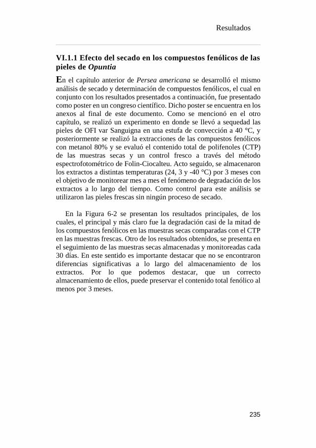
Resultados
235
VI.1.1 Efecto del secado en los compuestos fenólicos de las
pieles de Opuntia
En el capítulo anterior de Persea americana se desarrolló el mismo
análisis de secado y determinación de compuestos fenólicos, el cual en
conjunto con los resultados presentados a continuación, fue presentado
como poster en un congreso científico. Dicho poster se encuentra en los
anexos al final de este documento. Como se mencionó en el otro
capítulo, se realizó un experimento en donde se llevó a sequedad las
pieles de OFI var Sanguigna en una estufa de convección a 40 °C, y
posteriormente se realizó la extracciones de las compuestos fenólicos
con metanol 80% y se evaluó el contenido total de polifenoles (CTP)
de las muestras secas y un control fresco a través del método
espectrofotométrico de Folin-Ciocalteu. Acto seguido, se almacenaron
los extractos a distintas temperaturas (24, 3 y -40 °C) por 3 meses con
el objetivo de monitorear mes a mes el fenómeno de degradación de los
extractos a lo largo del tiempo. Como control para este análisis se
utilizaron las pieles frescas sin ningún proceso de secado.
En la Figura 6-2 se presentan los resultados principales, de los
cuales, el principal y más claro fue la degradación casi de la mitad de
los compuestos fenólicos en las muestras secas comparadas con el CTP
en las muestras frescas. Otro de los resultados obtenidos, se presenta en
el seguimiento de las muestras secas almacenadas y monitoreadas cada
30 días. En este sentido es importante destacar que no se encontraron
diferencias significativas a lo largo del almacenamiento de los
extractos. Por lo que podemos destacar, que un correcto
almacenamiento de ellos, puede preservar el contenido total fenólico al
menos por 3 meses.

Resultados
236
Figure 6-2 Effect of drying and storage in OFI var Sanguigna (source: private collection)
En este caso específico del estudio de degradación de compuestos
fenólicos en OFI var. Sanguigna, se quiso ir un paso más allá
analizando a 2 temperaturas distintas (40 y 60 °C) los perfiles
cromatográficos de dos flavonoides (isorhamnetina y quercetina -3-O-
rutinosidos) específicos cuantificados a través de uso de patones
específicos (R2 = 0.998 en ambos casos), y se muestra en la Figura 6-3
la termolabilidad de estos compuestos dentro de la matriz de OFI var
Sanguigna.
Figure 6-3 Effect of drying temperatures on flavonoids (source: private collection)
La isorhamnetina-3-O-rutinosido se ve extremamente afectado por
el proceso de secado, reduciendo su contenido en 9 y hasta 87 veces a
las temperaturas de secado de 40 y 60 °C respectivamente, mientras que
0
10
20
30
40
Control 0 días 30 días 60 días 90 díasmg
áci
do
gál
ico
/g d
e m
ues
tra
B.S
.Extractos de pieles secas de OFI var Sanguigna
almacenada a 24 °C almacenada a 3 °C almacenada a -40 °C
0.000
0.500
1.000
1.500
Control sinsecado
40 °C60 °C
1.305
0.151
0.015
0.157
0.0520.047
mg
de
Fla
von
oid
e/g
de
mu
estr
a B
.S.
Flavonoides en Pieles secas de OFI Sanguigna
Isorhamnetin Quercetin

Resultados
237
la querecetin-3-O-rutinosido reduce 3 veces su contenido en ambas
temperaturas. Aunque anteriormente Madrau et al. (2009) describiera
que el decremento de los polifenoles es inversamente proporcional al
aumento de las temperaturas, los resultados mostrados no fueron tan
violentos como los presentados en este análisis, donde creemos que la
diferencia que ha minimizado tanto el contenido fenólico, fue además
de la temperatura la prolongada exposición a ella, ya que para
asegurarnos de sequedad total, se extendió el proceso de secado a 10
días.
VI.1.2 Modelización de isotermas de sorción de las pieles
de Opuntia
Aunado al análisis de secado realizado anteriormente, se determinaron
experimentalmente las isotermas de adsorción y desorción de las pieles
de Opuntia ficus-indica var. Sanguigna a 20 °C. Posteriormente se
analizaron los valores de humedad de equilibrio de las distintas sales
con aw conocida mediante los modelos mencionado en la Tabla 3-2.
Figure 6-4 Sorption isotherms of OFI Sanguigna at 20 °C (source: private collection)

Resultados
238
En la Figura 6-4 se puede observar la representación gráfica de
ambas isotermas y la representación del modelo descrito por Peleg, que
fue el que se acoplo de mejor manera a los datos experimentales. A
primera vista podemos observar el fenómeno de desplazamiento entre
las isotermas de adsorción y desorción, quedando la última por encima
de la primera. A este fenómeno se le conoce como histéresis y es
consecuencia de fenómenos irreversibles de reordenamiento
conformacional y estructural, los cuales pueden alterar la accesibilidad
a sitios polares energéticamente favorables, dificultando la movilidad
del agua (Saravacos, 2014). Consecuentemente, podemos mencionar
dos cosas importantes: el fenómeno de histéresis es muy común en las
matices alimentarias, y estas graficas además nos muestran como las
pieles secas no llegan a re-adsorber la misma cantidad de agua
contenida en las pieles frescas.
La comprensión de las características de absorción de agua entre los
subproductos y la atmósfera es esencial para la selección de las
condiciones de proceso adecuadas debido a las características
fisicoquímicas, la seguridad microbiana u otras características que
dependen de la actividad del agua (Torres et al., 2018).
De esta forma se observa el punto crítico de estas isotermas en una
actividad de agua de alrededor de 0,55, lo que nos indica que para un
óptimo almacenamiento de las pieles a la temperatura de 20 °C,
debemos de secarlas por debajo de esta aw.
Con toda la información obtenida anteriormente, determinamos que:
1) el porcentaje de subproductos de las tres variedades de Opuntia, es
muy considerable para su revalorización; 2) es necesario reducir la
actividad de agua por debajo de 0,55 para su optimo almacenamiento;
3) el secado por convección no es nada recomendable como método de
secado previo a las extracciones de las biomoléculas activas, debido a
la disminución de CTP. Por lo tanto, en los diseños experimentales
publicados en esta tesis, recurrimos a técnicas de liofilización como
alternativa de secado.
Resultados
239
VI.2 VALORIZACIÓN DE LOS SUBPRODUCTOS DE
OPUNTIA SPP.: DETERMINACIÓN DE LOS PERFILES
FENOLICOS, BETALAINICOS Y SUS PROPIEDADES
BIOACTIVAS.
A continuación se presenta una adaptación al formato de la tesis
doctoral del artículo titulado “By-product recovery of Opuntia spp.
peels: Betalainic and phenolic profiles and bioactive properties”,
publicado en la revista Industrial Crops and Products. En él, los
subproductos (piel) de los frutos de higos chumbos de tres especies
(Opuntia ficus-indica var. Gialla y var. Sanguignea y Opuntia
engelmannii) fueron caracterizados en términos funcionales, y la
evaluadación sus propiedades antioxidantes, antimicrobianas y
citotoxicas fue evaluada, además, una caracterización completa del
perfil polifenolico (ácidos fenólicos y flavonoides) y betalainico fue
descrito mediante técnicas cromatográficas. Los datos bibliográficos
del artículo se destacan a continuación:
Melgar, B., Dias, M. I., Ciric, A., Sokovic, M., Garcia-Castello, E.
M., Rodriguez-Lopez, A. D., Barros, L., Ferreira, I. C. F. R. (2017).
By-product recovery of Opuntia spp. peels: Betalainic and phenolic
profiles and bioactive properties.
Industrial Crops and Products, 107, 353-359.
Resultados
240
By-product recovery of Opuntia spp. peels: betalainic and
phenolic profiles and bioactive properties
Bruno Melgara,c, Maria Inês Diasa, Ana Ciricb, Marina Sokovicb,
Esperanza M. Garcia-Castelloc, Antonio D. Rodriguez-Lopezd,
Lillian Barrosa, Isabel C.F.R. Ferreiraa,*
aCentro de Investigação de Montanha (CIMO), ESA, Instituto
Politécnico de Bragança, Campus de Santa Apolónia, 5300-253
Bragança, Portugal. bUniversity of Belgrade, Department of Plant Physiology, Institute for
Biological Research “Siniša Stanković”, Bulevar Despota Stefana
142, 11000 Belgrade, Serbia. cInstitute of Food Engineering for Development, Universitat
Politècnica de València, Camino de Vera, s/n CP, 46022 Valencia,
Spain. dInstitute for Industrial, Radiophysical and Environmental Safety
(ISIRYM), Universitat Politècnica de València, Camino de Vera, s/n
CP, 46022 Valencia, Spain.
*Corresponding author. Tel.+351 273 303219; fax +351 273 325405.
E-mail address: [email protected] (I.C.F.R. Ferreira)
Resultados
241
1Abstract
Opuntia spp. are a tropical and subtropical plant that provides both
edible green steams and fruits; however, the processing of this fruits
results in the accumulation of enormous amount of by-products that can
be a source of bioactive and pigmented compounds. Herein, three
cactus pear from the species Opuntia ficus-indica var. Sanguigna (OS)
and Gialla (OG) and Opuntia engelmannii (OE) were fully
characterized regarding their phenolic and betalain composition and
correlated with their antioxidant and antimicrobial activities. The
hydroethanolic extracts of OE gave the highest amount of phenolic
compounds (isorhamentin-O-(deoxyhexosyl-hexoside) and betacianins
(betanin); however, no betaxanthins were identified in this sample. This
sample also revealed the lowest IC50 values in all the antioxidant
activity assays. Regarding antimicrobial activity, the hydroethanolic
extracts of all species revealed to be more active than ampicillin. The
pivotal objective of this work was to focus on exploring by-product
biocompounds and possible outputs, thus, we could suggest the use of
these natural colorants with intrinsic antioxidant and antimicrobial
activity, which would grant industries to produce cleaner label products
with functional benefits.
Keywords: Opuntia ficus-indica; Opuntia engelmannii; by-product;
betalains; phenolic compounds; antimicrobial activity.

Resultados
242
VI.2.1. INTRODUCTION
Opuntia spp., including their several varieties, belong to the
dicotyledonous angiosperm and are the largest genus within Cactaceae
family. Opuntia species are tropical and subtropical plants, able to grow
in arid and semi-arid environments, with easy geographic adaptation
due to their crassulacean acid metabolism (CAM), which enables the
plant to survive to extreme heat, low temperatures (-40 °C) and drought
because of their highly efficiency in the use of water (Nobel, 1998;
Sudzuki et al., 1993). This plant provides both edible green steams and
fruits.
Cactus fruit is also known as prickly pear, cactus fig, Indian fig,
cactus pear, Barbary fig, and is available across the 5 continents from
early summer until late autumn. Although Opuntia species are native
of Mexico, where the steams and fruits are consumed fresh in the local
diet, fruits, are also processed and commercialized in numerous
countries as jams, sweats, ice cream and alcoholic and not alcoholic
beverages (Martins et al., 2016).
The processing of the fruits results in the accumulation of several
quantities of by-products, namely prickly pear peels. Proper utilization
of this by-product could reduce waste disposal problems and serve as a
potential new source of bioactive compounds and pigments. According
to several authors (Belwal et al., 2016; Garcia-Castello et al., 2015;
Pinela et al., 2016; Rao, 2010), one of the best ways to use this kind of
by-products could be the application of an appropriate green solid-
liquid extraction technology in order to obtain bioactive compounds
with different properties and health benefits.
Several studies marked fruits and fruits by-products as a rich source
of natural molecules such as polyphenols (i.e. flavonoids), vitamins,
colorants like betalains and carotenoids (Albuquerque et al., 2016;
Alzate T et al., 2016; Ayala-Zavala et al., 2011; Fernandez-Rojas et al.,
2010; Silva et al., 2016), which could have high potential interest in
human health, medicine and production of new added-value products.
Scientists have lately reported multiple properties regarding to
phenolic compounds as antioxidants endowed with anticancer, anti-

Resultados
243
inflammatory, antimicrobial and antidiabetic activities (Dias et al.,
2016; Kaur Kala et al., 2016), among the most cited properties
attributed to polyphenols due to their protective effect against damage
of the human body caused by reactive oxygen, nitrogen and sulfur
species (ROS, RNS and RSS, respectively) (Carocho and Ferreira,
2013; Shahidi and Ambigaipalan, 2015).
Additionally, to phenolic compounds, other relevant components in
the cactus pear are the betalains pigments, which are present in most of
the Caryophyllales family including the Cactaceae and replace the
anthocyanins in flowers and fruits of plants within this family.
Betalains are water soluble nitrogen-containing pigments situated in the
vacuoles of the plant tissue responsible for the red-violet (betacyanins)
and yellow-orange (betaxanthins) colors, showing a stable appearance
in the range of pH 3 to 7 (Stintzing et al., 2002). Therefore, betalains
could be used as a natural colorant alternative to synthetic dyes used in
a broad range of food products.
The present study intends to contribute to the characterization of the
bioactive compounds profile, antioxidant and antimicrobial activities of
by-products, such as peels, of the cactus pear from the species Opuntia
ficus-indica var. Sanguigna (OS) and Gialla (OG) and Opuntia
engelmannii (OE). The results of this study might be useful to
maximize the potential of the fruits by-products for their colorant and
functional added value

Resultados
244
VI.2.2. MATERIAL AND METHODS
VI.2.2.1. Samples preparation
Cactus pear fruits (Opuntia ficus-indica var. Sanguigna -OS and Gialla
-OG) were collected in July-August 2016 in Sicily, Italy and purchased
from a local market in Bragança, Portugal. Fruits from this specie were
separated regarding the maturation colour orange-red (Opuntia ficus-
indica var. Gialla) and red-violet (Opuntia ficus-indica var.
Sanguigna), obtaining two different samples. Prickly pear wild fruits
(Opuntia engelmannii- OE) were collected in Bragança, Portugal (GPS
location: 41.797344, -6.772735) on early September 2016. Dr. Carlos
Aguiar of the School of Agriculture, Polytechnic Institute of Bragança
(Trás-os-Montes, Portugal), confirmed the botanical identifications and
voucher specimens were deposited in a herbarium.
Within 24 hours, fruits were washed with distilled water in order to
remove glochids and then further air-dried. Afterwards, all the fruits
were peeled and the resulting peel was lyophilized (LabConco, Frezone
-105 °C, 4.5 L Cascade Benchtop Freeze Dry System, Kansas, MO,
USA) and stored in a cool and dry place until use.
VI.2.2.2. Extraction procedure
The hydroethanolic (ethanol: water, 80:20 v/v) extract was obtained
from the lyophilized peels. The sample (1 g) was extracted twice by
stirring with 25 mL of hydro-alcoholic solution (25 ºC at 150 rpm) for
1 h and subsequently filtered through the Whatman no. 4 paper. The
obtained extracts were frozen, lyophilized and re-dissolved in: (i)
hydroethanolic solution (ethanol: water, 80:20 v/v) for phenolic
characterization (final concentration 5 mg/mL) and antioxidant activity
evaluation (final concentration 40 mg/mL); or (ii) water for cytotoxicity
evaluation (final concentration 8 mg/mL) and betalain characterization
(final concentration 30 mg/mL); or (iii) 5% DMSO in distilled water
(final concentration 10 mg/mL) for antimicrobial assays. The final
solutions were further diluted to different concentrations to be
submitted for distinct in vitro bioactivity evaluation assays.

Resultados
245
VI.2.2.3. Phenolic compounds
The lyophilized hydroethanolic extracts were analysed for their
content in phenolic compounds, re-dissolve in ethanol:water (80:20,
v/v) to a final concentration of 5 mg/mL. LC-DAD–ESI/MSn analyses
were performed using a Dionex Ultimate 3000 UPLC instrument
(Thermo Scientific, San Jose, CA, USA), coupled to a diode-array
detector (using several wavelengths, 280, 330 and 370 nm) and to a
mass detector (Linear Ion Trap LTQ XL mass spectrometer, equipped
with an ESI source, ThermoFinnigan, San Jose, CA, USA). The elution
gradient, chromatographic and mass spectrometer conditions were
performed according to (Bessada et al., 2016). Identification was
performed by comparing their fragmentation pattern, retention times
and UV–vis spectra with authentic standards, when available, or by
comparing the obtained information with available data from literature.
When a standard was not available to quantify a phenolic compound a
calibration curve of similar compound from the same phenolic group
was used.
VI.2.2.4. Betalain compounds
The profile in betalain compounds was determined by LC-DAD-
ESI/MSn (Dionex Ultimate 3000 UPLC, Thermo Scientific, San Jose,
CA, USA). Chromatographic separation was achieved with a Waters
Spherisorb S3 ODS-2 C18 (3 μm, 4.6 mm × 150 mm, Waters, Milford,
MA, USA) column working at 35 °C. The solvents used were: (A) 0.1%
trifluoroacetic acid (TFA) in water, (B) acetonitrile. The gradient
elution followed these parameters: from 0% to 10% B for 15 min, from
10% to 15% B for 5 min, from 15 to 18% B for 5 min, from 18 to 50%
B for 8 min, and from 50 to 0% B for 12 min. The resulting total run
time was 45 minutes, using a flow rate of 0.5 mL/min. Double detection
was carried out in the DAD using 480 nm (betaxanthins) and 535 nm
(betacyanins), as the preferred wavelength, and in a mass spectrometer
(MS).
MS detection was performed using positive mode, with a Linear Ion
Trap LTQ XL mass spectrometer (ThermoFinnigan, San Jose, CA,
USA) equipped with an ESI source. Nitrogen served as the sheath gas
(50 psi); the system was operated with a spray voltage of 4.8 kV, a
source temperature of 320 °C, a capillary voltage of 39 V. The tube lens
offset was kept at a voltage of 140 V. The full scan covered the mass

Resultados
246
range from m/z 100 to 1500, collision energy used was 24 (arbitrary
units) and data acquisition was carried out with Xcalibur® data system
(ThermoFinnigan, San Jose, CA, USA).
The identification of the betalain compounds (betacyanins and
betaxanthins) were performed by comparing the obtained information
with available data reported in the literature giving a tentative
identification. For quantitative analysis, a calibration curve using an
isolated compound gomphrenin III (isolated from Gomphrena globosa
L.) was constructed based on the UV signal (y = 14670x - 19725, R² =
0.9997) (Roriz et al., 2017). The results of betacyanins were expressed
as mg per 100 g of pulp fresh weight (FW), and the results of
betaxanthins were expressed as relative percentage (%) of their areas
recorded at 480 nm.
VI.2.2.5. Bioactive properties evaluation
VI.2.2.5.1. Antioxidant activity assays
Successive dilutions were made from the stock solution and submitted
to in vitro assays already described by (Heleno et al., 2010) to evaluate
the antioxidant activity of the samples. DPPH radical-scavenging
activity was evaluated using an ELX800 microplate reader (Bio-Tek
Instruments, Inc.; Winooski, VT, USA) and calculated in percentage
after 1 h of reaction in the dark.
The reducing power (RP) assay evaluated the capacity of the
extracts to reduce Fe3+ to Fe2+, measuring the absorbance at 690 nm.
In β-carotene bleaching assay, the presence of antioxidants in the
samples and their capacity to neutralize the linoleate free radicals,
avoids β-carotene bleaching, which can be evaluated measuring the
absorbance at 470 nm. Therefore, β-carotene bleaching inhibition was
calculated using the following formula: (Absorbance after 2 h of
assay/initial absorbance) × 100. The results were expressed as IC50
values (sample concentration providing 50% of antioxidant activity or
0.5 of absorbance in the reducing power assay) for antioxidant activity.
Trolox was used as positive control.

Resultados
247
VI.2.2.5.2. Antimicrobial activity assays
The minimum inhibitory (MIC), minimum bactericidal concentrations
(MBC) and minimum fungicidal concentrations (MFC) were
determined by methodologies, described by (Vieira et al., 2016b).
Antibacterial activity was assayed using the following, Gram-
positive bacteria: Staphylococcus aureus (ATCC 6538), Bacillus
cereus (clinical isolate), Micrococcus flavus (ATCC10240), and
Listeria monocytogenes (NCTC7973) and Gram-negative bacteria:
Escherichia coli (ATCC 35210), Pseudomonas aeruginosa (ATCC
27853), Salmonella typhimurium (ATCC 13311), and Enterobacter
cloacae (ATCC 35030) were used. Whilst for antifungal assays, the
following microfungi were used: Aspergillus fumigatus (ATCC1022),
Aspergillus ochraceus (ATCC12066), Aspergillus versicolor
(ATCC11730), Aspergillus niger (ATCC6275), Penicillium
funiculosum (ATCC 36839), Penicillium ochrochloron (ATCC9112),
Penicillium verrucosum var. cyclopium (food isolate), and
Trichoderma viride (IAM 5061). Bacterial and fungal organisms were
obtained from the Mycological Laboratory, Department of Plant
Physiology, Institute for Biological Research “Sinisa Stanković”,
University of Belgrade, Serbia. Standard drugs, namely streptomycin,
ampicillin, bifonazole and ketoconazole were used as positive controls.
VI.2.2.5.3. Cytotoxicity assays
Both, antitumor activity and hepatotoxicity was evaluated by the
Sulphorhodamine B (SRB) assay that has been previously described by
(Gomes-Correa et al., 2015; Pereira et al., 2014). Four human tumour
cell lines were assayed: HeLa (cervical carcinoma), HepG2
(hepatocellular carcinoma), MCF-7 (breast adenocarcinoma) and NCI-
H460 (non-small cell lung cancer), while PLP2 cells were used for
hepatotoxicity. The five cell cultures were treated for 48 h with the
different diluted sample solutions and assayed according to the SRB
colorimetric methodology. Ellipticine was used as positive control and
results were expressed in GI50 values in μg/mL (sample concentration
that inhibited 50% of the net cell growth).
Resultados
248
VI.2.2.6. Statistical analysis
All the extractions and assays were performed in triplicate. Results
were expressed as mean values and standard deviation (SD), being
analysed using one-way analysis of variance (ANOVA) followed by
Tukey’s HSD Test with p = 0.05. In the case of only existing two
samples, the results were analysed using a Student´s t-test, in order to
determine the significant difference between the two samples, with p =
0.05. The treatment was carried out using IBM SPSS Statistics for
Windows, version 23.0. (IBM Corp., Armonk, New York, USA).

Resultados
249
VI.2.3. RESULTS AND DISCUSSION
VI.2.3.1. Phenolic and betalain composition
Tables 6-1 and 6-2 present the peak characteristics (retention time,
wavelength of maximum absorption and mass spectral data), tentative
identification and quantification of the phenolic compounds and
betalains, respectively, present in the hydroethanolic extracts of
Opuntia ficus-indica var Gialla (OG), Opuntia ficus-indica var
Sanguigna (OS) and Opuntia engelmanniii (OE) peels (Figure 6-8).
Twelve different phenolic compounds (Table 6-1) were found, two
phenolic acids, piscid acid (peaks 1) and eucomic acid (peaks 2), and
ten flavonoids (isorhamnetin, quercetin and kaempferol derivatives,
peaks 3 to 12). All of the compounds have been previously described
by other authors in Opuntia spp samples(Chahdoura et al., 2014;
Chougui et al., 2015; Mata et al., 2016; Morales et al., 2015, 2014)
Overall, OE sample presented higher concentration of phenolic
compounds, being peak 11 (isorhamnetin-O-(deoxyhexosyl-hexoside))
the major compound found (5.99 mg/g).
Regarding betalain fractions (Table 6-2), seven compounds were
identified; two betaxanthins, indicaxanthin isomer I and II (peaks 1 and
2), previously described by Castellanos-Santiago & Yahia (2008) in
Opuntia spp. cultivars and also by Svenson et al., (2008) in Ullucus
tuberosus fruits. Betalain molecules displays wide range of hues,
betacyanins have reddish-violet colour while betaxanthins have a
general yellow-orange colour (Albano et al., 2015; Esquivel, 2016;
Gandía-herrero et al., 2013; Gandía-Herrero et al., 2010), the latter
being only identified in OG and OS samples. Five betacyanins were
identified, betanidin-5-O-β-sophoroside, betanidin-5-O-β-glucoside
(betanin), isobetanin, gomphrenin I and betanidin (peaks 3, 4, 5, 6 and
7, respectively), having been previously identified by other authors
(Castellanos-Santiago and Yahia, 2008; Morales et al., 2015, 2014;
Stintzing et al., 2002; Svenson et al., 2008).

Resultados
250
Figure 6-5 HPLC chromatogram of OG phenolic profile recorded at 370 nm (A),
betaxanthins profile of OS recorded at 484 nm (B) and betacyanins profile of OE recorded
at 535 nm (C). (source: private collection)
On the contrary of betaxanthins, betacianins fraction was found in
higher quantities in OE samples, being peak 4 (betanin) the main
compound found (14.9 mg/g extract), This characteristic can be easily

Resultados
251
predicted due to the strong violet hue present in OE and also because
betanin (betanidin-5-O-β-glucoside) is the most widespread betacyanin
among plants (Esatbeyoglu et al., 2015b). In this matter, betanin
concentration in the analysed samples was in concordance with the
previous statement commented. OE displayed differences of around 15
and 5-fold higher than OG and OS, respectively. The second highest
concentration of betacyanins was conferred by isobetanin in OE and
OS extracts. Only OE showed other 3 different betacyanins (betanidin,
betanidin-5-O-β-sophoroside and gomphrenin I). Although, OG has
this name from the Italian word “yellow”, betacyanin molecule betanin,
was also present but only around 10% of the total betalains detected,
this pigment can be spotted as a reddish-orange hue intrinsic in OG
peels.
VI.2.3.2. Antioxidant activity
The results on antioxidant activity of the hydroethanolic extracts are
shown in Table 6-3. There is a clear pattern in the antioxidant activity
of each Opuntia spp., at first glance, OE exhibits the best IC50 values,
followed by OS and lastly OG, nonetheless, the difference within OE
vs OS and OG is around 2-fold higher, while the difference among OS
and OG is barely perceptible. At this point, it is worth noting the high
IC50 values presented by OS in the β-carotene bleaching inhibition our
hypothesis on this value could be due to an interference with the sample
colour.
Independently of the IC50 values that OS shown in the and β-
carotene assay, all the antioxidant activities of all samples, can be
inversely correlated with the concentration of the betacyanins and
phenolic compounds previously described in Table 6-2 and Table 6-1,
respectively. The relationship between betacyanins (Bc) or phenolic
compounds (PC) exhibited high correlation pattern with DPPH radical
scavenging activity (R2=-0.99 for both Bc and PC), reducing power
(R2=-0.97 also for Bc and PC) and β-carotene bleaching inhibition
(R2=-0.75 for Bc and R2=-0.76 for PC).

Table 6-1 Retention time (Rt), wavelengths of maximum absorption in visible region (λmax), mass spectral data, tentative identification and quantification of
phenolic compounds in Opuntia peels
Peak Tentative
identification
Rt
(min)
λmax
(nm)
[M-H]-
(m/z)
MS2
(m/z) OG OS OE
t-
Studen
t
p-value
1 Piscidic acid1 4.65 222 255 193(34),179(6),
165(100),149(3) 1.061 ± 0.04 nd nd -
2 Eucomic acid1 6.71 223 239 179(100),149(62) 1.40 ± 0.01 2.2 ± 0.2 nd <0.001
3 Isorhamnetin-O-(di-deoxyhexosyl-hexoside)2 17.09 354 769 315(100) 0.22 ± 0.01 0.56 ± 0.06 nd <0.001
4 Isorhamnetin-O-(di-deoxyhexosyl-hexoside) 2 17.4 353 769 315(100) tr 0.21 ± 0.03 nd -
5 Isorhamentin-O-(deoxyhexosyl-pentosyl-hexoside)
2 17.7 354 755 315(100) 0.77 ± 0.02 1.3 ± 0.1 nd <0.001
6 Quercetin-3-O-rutinoside2 17.98 354 609 301(100) nd nd tr -
7 Isorhamentin-O-(deoxyhexosyl-pentosyl-
hexoside)2 18.14 354 755 315(100) tr tr nd -
8 Isorhamentin-O-(pentosyl-hexoside)2 19.58 354 609 315(100) 0.100 ± 0.001 0.67 ± 0.06 nd <0.001

9 Kaempferol-3-O-rutinoside2 21.31 348 593 285(100) nd nd tr -
10 Isorhamentin-O-(deoxyhexosyl-hexoside)2 21.67 354 623 315(100) tr tr 0.48 ± 0.01 -
11 Isorhamentin-O-(deoxyhexosyl-hexoside)2 22.25 354 623 315(100) 0.77 ± 0.01c 1.7 ± 0.1b 5.99 ± 0.04a -
12 Isorhametin-3-O-glucoside2 23.08 354 447 315(100) nd nd tr -
Total phenolic compounds 3.26±0.04c 3.7±0.3b 6.5±0.1a -
Phenolic compounds were expressed in mg/g extract; nd- not detected; Calibration standards: 1- p-hydroxibenzoic acid (𝑦= 208604𝑥 + 173056,
𝑅² = 0.9995); 2-quercetin-3-O-glucoside (𝑦= 34843𝑥 – 160173, 𝑅² = 0.9998). OG, Opuntia ficus-indica var Gialla; OS, Opuntia ficus-indica
var Sanguigna; OE, Opuntia engelmannii; nd, not detected; tr, traces.
In each row different letters mean significant differences (p < 0.05). *Statistical differences (<0.001) were observed
when t-student test was applied.
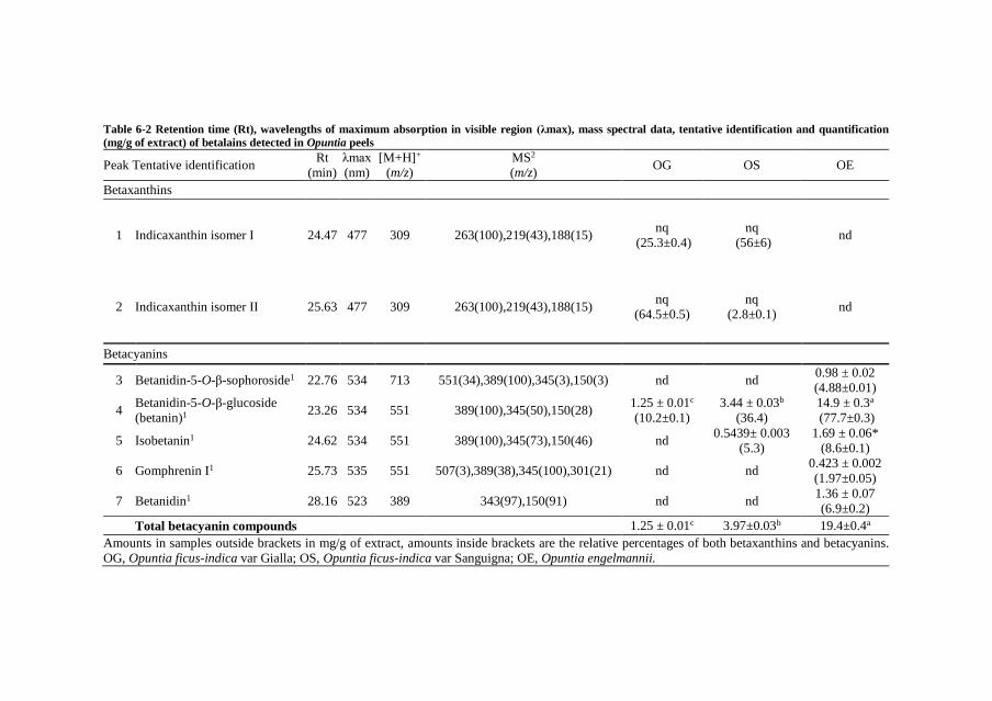
Table 6-2 Retention time (Rt), wavelengths of maximum absorption in visible region (λmax), mass spectral data, tentative identification and quantification
(mg/g of extract) of betalains detected in Opuntia peels
Peak Tentative identification Rt
(min)
λmax
(nm)
[M+H]+
(m/z)
MS2
(m/z) OG OS OE
Betaxanthins
1 Indicaxanthin isomer I 24.47 477 309 263(100),219(43),188(15) nq
(25.3±0.4)
nq
(56±6) nd
2 Indicaxanthin isomer II 25.63 477 309 263(100),219(43),188(15) nq
(64.5±0.5)
nq
(2.8±0.1) nd
Betacyanins
3 Betanidin-5-O-β-sophoroside1 22.76 534 713 551(34),389(100),345(3),150(3) nd nd 0.98 ± 0.02
(4.88±0.01)
4 Betanidin-5-O-β-glucoside
(betanin)1 23.26 534 551 389(100),345(50),150(28)
1.25 ± 0.01c
(10.2±0.1)
3.44 ± 0.03b
(36.4)
14.9 ± 0.3a
(77.7±0.3)
5 Isobetanin1 24.62 534 551 389(100),345(73),150(46) nd 0.5439± 0.003
(5.3)
1.69 ± 0.06*
(8.6±0.1)
6 Gomphrenin I1 25.73 535 551 507(3),389(38),345(100),301(21) nd nd 0.423 ± 0.002
(1.97±0.05)
7 Betanidin1 28.16 523 389 343(97),150(91) nd nd 1.36 ± 0.07
(6.9±0.2)
Total betacyanin compounds 1.25 ± 0.01c 3.97±0.03b 19.4±0.4a
Amounts in samples outside brackets in mg/g of extract, amounts inside brackets are the relative percentages of both betaxanthins and betacyanins.
OG, Opuntia ficus-indica var Gialla; OS, Opuntia ficus-indica var Sanguigna; OE, Opuntia engelmannii.

nq, not quantified; nd, not detected. Calibration curve: 1gomphrenin III (y=14670x–19725, R2=0.9997). In each row different letters mean significant
differences (p < 0.05). *Statistical differences (<0.001) were observed when t-student test was applied.
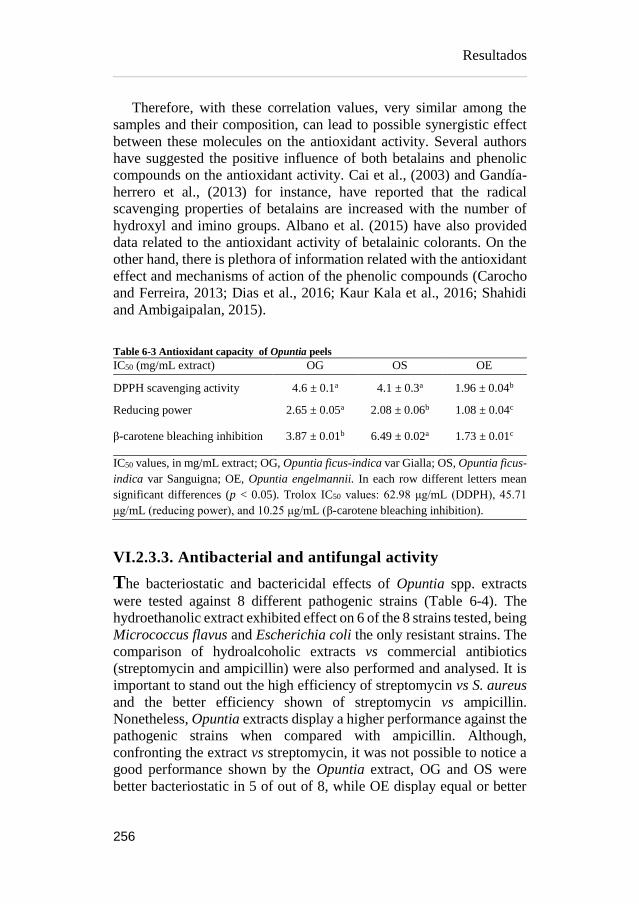
Resultados
256
Therefore, with these correlation values, very similar among the
samples and their composition, can lead to possible synergistic effect
between these molecules on the antioxidant activity. Several authors
have suggested the positive influence of both betalains and phenolic
compounds on the antioxidant activity. Cai et al., (2003) and Gandía-
herrero et al., (2013) for instance, have reported that the radical
scavenging properties of betalains are increased with the number of
hydroxyl and imino groups. Albano et al. (2015) have also provided
data related to the antioxidant activity of betalainic colorants. On the
other hand, there is plethora of information related with the antioxidant
effect and mechanisms of action of the phenolic compounds (Carocho
and Ferreira, 2013; Dias et al., 2016; Kaur Kala et al., 2016; Shahidi
and Ambigaipalan, 2015).
Table 6-3 Antioxidant capacity of Opuntia peels
IC50 (mg/mL extract) OG OS OE
DPPH scavenging activity 4.6 ± 0.1a 4.1 ± 0.3a 1.96 ± 0.04b
Reducing power 2.65 ± 0.05a 2.08 ± 0.06b 1.08 ± 0.04c
β-carotene bleaching inhibition 3.87 ± 0.01b 6.49 ± 0.02a 1.73 ± 0.01c
IC50 values, in mg/mL extract; OG, Opuntia ficus-indica var Gialla; OS, Opuntia ficus-
indica var Sanguigna; OE, Opuntia engelmannii. In each row different letters mean
significant differences (p < 0.05). Trolox IC50 values: 62.98 μg/mL (DDPH), 45.71
μg/mL (reducing power), and 10.25 μg/mL (β-carotene bleaching inhibition).
VI.2.3.3. Antibacterial and antifungal activity
The bacteriostatic and bactericidal effects of Opuntia spp. extracts
were tested against 8 different pathogenic strains (Table 6-4). The
hydroethanolic extract exhibited effect on 6 of the 8 strains tested, being
Micrococcus flavus and Escherichia coli the only resistant strains. The
comparison of hydroalcoholic extracts vs commercial antibiotics
(streptomycin and ampicillin) were also performed and analysed. It is
important to stand out the high efficiency of streptomycin vs S. aureus
and the better efficiency shown of streptomycin vs ampicillin.
Nonetheless, Opuntia extracts display a higher performance against the
pathogenic strains when compared with ampicillin. Although,
confronting the extract vs streptomycin, it was not possible to notice a
good performance shown by the Opuntia extract, OG and OS were
better bacteriostatic in 5 of out of 8, while OE display equal or better

Resultados
257
strength in 3 out of 8 strain tasted. The bactericidal activity of the
extracts was equal or higher in 3 out of 8 strains. OG exhibited the best
performance against Bacillus cereus, while OE had stronger activity vs
Enterobacter cloacae.
Regarding antifungal activity, the results obtained proved to be
consistent and reproducible with the hydroethanolic extracts (Table 6-
4). The results revealed an equal or higher performance in 4 (OS
extract) and 3 (OG and OE extract) strains out of the 8 strains tested.
All three samples had higher activity on Penicillium ochrochloron
compared with the antifungal controls used. However, the fungicidal
activity could not improve compared with fungistatic activity mention
before. The Opuntia peel extracts showed equal or higher activity
regarding ketoconazole or bifonazole drugs. A higher activity
performance of the extract was attained on Trichoderma viride and
Penicillum ochrochloron strains.
Studies from different authors attributed the inhibitory effect of
plant extracts against microbial pathogens to their phenolic
composition (Balouiri et al., 2016; Song et al., 2010). The inhibitory
effect of these phenolics could be explained by adsorption to cell
membranes, interaction with enzymes or deprivation of substrate and
metal ions (Baydar et al., 2004).

Table 6-4 Antibacterial and antifungal activity (mg/mL of extract) of Opuntia peels
Bacteria OS OG OE C1 C2
MIC MBC MIC MBC MIC MBC MIC MBC MIC MBC
Bacillus cereus 0.075 0.15 0.05 0.075 0.10 0.15 0.10 0.20 0.25 0.40
Micrococcus flavus N.A. N.A. N.A. N.A. N.A. N.A. 0.20 0.30 0.25 0.40
Staphylococcus aureus 0.15 0.45 0.15 0.30 0.30 0.45 0.04 0.10 0.25 0.45
Listeria monocytogenes 0.15 0.60 0.15 0.45 0.30 0.45 0.20 0.30 0.40 0.50
Escherichia coli N.A. N.A. N.A. N.A. N.A. N.A. 0.20 0.30 0.40 0.50
Enterobacter cloacae 0.075 0.30 0.075 0.30 0.10 0.15 0.20 0.30 0.25 0.50
Pseudomonas aeruginosa 0.15 0.45 0.15 0.45 0.30 0.45 0.20 0.30 0.75 1.20
Salmonella typhimurium 0.15 0.30 0.15 0.45 0.15 0.30 0.25 0.50 0.40 0.75
Mold OS OG OE C3 C4
MIC MFC MIC MFC MIC MFC MIC MFC MIC MFC
Aspergillus fumigatus 0.30 0.45 0.30 0.60 0.30 0.60 0.25 0.50 0.15 0.20
Aspergillus versicolor 0.30 0.45 0.30 0.45 0.30 0.45 0.20 0.50 0.10 0.20
Aspergillus ochraceus 0.10 0.15 0.15 0.30 0.15 0.30 1.50 2.00 0.15 0.20
Aspergillus niger 0.30 0.45 0.30 0.45 0.30 0.45 0.20 0.50 0.15 0.20
Trichoderma viride 0.15 0.30 0.075 0.15 0.10 0.15 1.00 1.00 0.15 0.20
Penicillium funiculosum 0.15 0.30 0.30 0.45 0.30 0.45 0.20 0.50 0.20 0.25
Penicillium ochrochloron 0.075 0.15 0.075 0.15 0.10 0.15 2.50 3.50 0.20 0.25
Penicillium verrucosum var. cyclopium 0.30 0.45 0.30 0.45 0.30 0.60 0.20 0.30 0.10 0.20
C1, Streptomycin; C2, Ampicillin; C3, Ketoconazole; C4, Bifonazole; MIC, minimum inhibitory concentration; MBC, minimum bactericidal
concentrations; MBC, minimum fungicidal concentrations; N.A., No activity.
OG, Opuntia ficus-indica var Gialla; OS, Opuntia ficus-indica var Sanguigna; OE, Opuntia engelmannii.
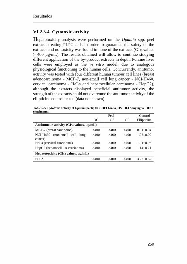
Resultados
259
VI.2.3.4. Cytotoxic activity
Hepatotoxicity analysis were performed on the Opuntia spp. peel
extracts treating PLP2 cells in order to guarantee the safety of the
extracts and no toxicity was found in none of the extracts (GI50 values
> 400 µg/mL). The results obtained will allow to continue studying
different application of the by-product extracts in depth. Porcine liver
cells were employed as the in vitro model, due to analogous
physiological functioning to the human cells. Concurrently, antitumor
activity was tested with four different human tumour cell lines (breast
adenocarcinoma - MCF-7, non-small cell lung cancer - NCI-H460,
cervical carcinoma - HeLa and hepatocellular carcinoma - HepG2),
although the extracts displayed beneficial antitumor activity, the
strength of the extracts could not overcome the antitumor activity of the
ellipticine control tested (data not shown).
Table 6-5 Cytotoxic activity of Opuntia peels; OG: OFI Gialla, OS: OFI Sanguigna, OE: o.
engelmannii
Peel Control
OG OS OE Ellipticine
Antitumour activity (GI50 values. µg/mL)
MCF-7 (breast carcinoma) >400 >400 >400 0.91±0.04
NCI-H460 (non-small cell lung
cancer)
>400 >400 >400 1.03±0.09
HeLa (cervical carcinoma) >400 >400 >400 1.91±0.06
HepG2 (hepatocellular carcinoma) >400 >400 >400 1.14±0.21
Hepatotoxicity (GI50 values. µg/mL)
PLP2 >400 >400 >400 3.22±0.67

Resultados
260
VI.2.4. CONCLUSION
Overall, Opuntia spp. chemical characterization was able to identify
the phenolic and betalainic profile inherent to the by-products (peels)
of Opuntia ficus-indica and Opuntia engelmanni. This recompilation of
data will bring deeper comprehension on the specific compounds
related to the antioxidant and antimicrobial capacity of the extracts
analysed and the absence of toxicity. The mentioned benefits make
them of huge interest as a new source of functional ingredients. The
pivotal objective of this work was focused on exploring by-product
biocompounds and possible outputs, thus, we could suggest their use as
natural colorants with intrinsic antioxidant and antimicrobial activity,
which would grant industries to produce cleaner label products with
functional benefits.
Aknowledgement
The authors are grateful to the Foundation for Science and Technology
(FCT, Portugal) and FEDER under Program PT2020 for financial
support to CIMO (UID/AGR/00690/2013) and L. Barros contract. B.
Melgar thanks CONACyT for his grant (No. 329930). The authors
would also like to thank Dr. Carlos Aguiar for the botanical
identification of these species. The authors are also grateful to the
Serbian Ministry of Education, Science and Technological
Development, grant number 173032 for financial support.

Resultados
261
References
Ahn, J.H., Jang, Y.-S., Lee, S.Y., 2016. Production of succinic acid by
metabolically engineered microorganisms. Curr. Opin.
Biotechnol. 42, 54–66.
https://doi.org/10.1016/j.copbio.2016.02.034
Al-Farsi, M.A., Lee, C.Y., 2008. Optimization of phenolics and dietary
fibre extraction from date seeds. Food Chem. 108, 977–985.
https://doi.org/10.1016/j.foodchem.2007.12.009
Albano, C., Negro, C., Tommasi, N., Gerardi, C., Mita, G., Miceli, A.,
De Bellis, L., Blando, F., 2015. Betalains, Phenols and
Antioxidant Capacity in Cactus Pear [Opuntia ficus-indica (L.)
Mill.] Fruits from Apulia (South Italy) Genotypes. Antioxidants
4, 269–280. https://doi.org/10.3390/antiox4020269
Albuquerque, T.G., Santos, F., Sanches-Silva, A., Beatriz Oliveira, M.,
Bento, A.C., Costa, H.S., 2016. Nutritional and phytochemical
composition of Annona cherimola Mill. fruits and by-products:
Potential health benefits. Food Chem. 193, 187–195.
https://doi.org/10.1016/j.foodchem.2014.06.044
Allai, L., Druart, X., Öztürk, M., BenMoula, A., Nasser, B., El Amiri,
B., 2016. Protective effects of Opuntia ficus-indica extract on ram
sperm quality, lipid peroxidation and DNA fragmentation during
liquid storage. Anim. Reprod. Sci. 175, 1–9.
https://doi.org/10.1016/j.anireprosci.2016.09.013
Almeida, M.M.B., de Sousa, P.H.M., Arriaga, Â.M.C., do Prado, G.M.,
Magalhães, C.E. de C., Maia, G.A., de Lemos, T.L.G., 2011.
Bioactive compounds and antioxidant activity of fresh exotic
fruits from northeastern Brazil. Food Res. Int. 44, 2155–2159.
https://doi.org/10.1016/j.foodres.2011.03.051
Alvarez, B., 2007. Análisis de factibilidad del cultivo de la tuna en la
localidad de Icaño , Departamento La Paz, Dirección provincial
de programación del desarrollo, Ministerio de Producción y
Desarrollo. Gobierno de la Provincia de Catamarca. Catamarca,
Argentina.
Alzate T, L.M., González, D., Hincapié, S., Cardona S, B.L., Londoño-
Londoño, J., Jiménez-Cartagena, C., 2016. The profile of
bioactive substances in ten vegetable and fruit by-products from
a food supply chain in Colombia. Sustain. Prod. Consum.
https://doi.org/10.1016/j.spc.2016.07.005

Resultados
262
Ammar, I., Ben Salem, M., Harrabi, B., Mzid, M., Bardaa, S., Sahnoun,
Z., Attia, H., Ennouri, M., 2018. Anti-inflammatory activity and
phenolic composition of prickly pear (Opuntia ficus-indica)
flowers. Ind. Crops Prod. 112, 313–319.
https://doi.org/10.1016/j.indcrop.2017.12.028
Angulo-Bejarano, P.I., Martínez-Cruz, O., Paredes-López, O., 2014.
Phytochemical Content, Nutraceutical Potential and
Biotechnological Applications of an Ancient Mexican Plant:
Nopal (Opuntia ficus-indica). Curr. Nutr. Food Sci. 10, 196–217.
https://doi.org/10.2174/157340131003140828121015
Arlt, U., 2010. NATCOL | Foods Colours Legislation.
Aruwa, C.E., Amoo, S.O., Kudanga, T., 2018. Opuntia (Cactaceae)
plant compounds, biological activities and prospects – A
comprehensive review. Food Res. Int. 112, 328–344.
https://doi.org/10.1016/j.foodres.2018.06.047
Association of Official Analytical Chemists, 2016. Official Methods of
Analysis of AOAC International, 20th ed. The Association,
Gaithersburg, MD, USA.
Attokaran, M., 2011. Natural Food Flavors and Colorants Natural Food
Flavors and Colorants, 1st ed. Blackwell Publishing LTD, Iowa.
Augustin, M.A., Sanguansri, L., 2015. Challenges and Solutions to
Incorporation of Nutraceuticals in Foods. Annu. Rev. Food Sci.
Technol. 6, 463–477. https://doi.org/10.1146/annurev-food-
022814-015507
Ayala-Zavala, J.F., Vega-Vega, V., Rosas-Domínguez, C., Palafox-
Carlos, H., Villa-Rodriguez, J.A., Siddiqui, M.W., Dávila-Aviña,
J.E., González-Aguilar, G.A., 2011. Agro-industrial potential of
exotic fruit byproducts as a source of food additives. Food Res.
Int. 44, 1866–1874.
https://doi.org/10.1016/j.foodres.2011.02.021
Azeredo, H.M.C., 2009. Betalains: Properties, sources, applications,
and stability - A review. Int. J. Food Sci. Technol. 44, 2365–2376.
https://doi.org/10.1111/j.1365-2621.2007.01668.x
Balouiri, M., Sadiki, M., Ibnsouda, S.K., 2016. Methods for in vitro
evaluating antimicrobial activity: A review. J. Pharm. Anal. 6,
71–79. https://doi.org/10.1016/j.jpha.2015.11.005

Resultados
263
Barros, L., Calhelha, R.C., Queiroz, M.J.R.P., Santos-Buelga, C.,
Santos, E.A., Regis, W.C.B., Ferreira, I.C.F.R., 2015. The
powerful in vitro bioactivity of Euterpe oleracea Mart. seeds and
related phenolic compounds. Ind. Crops Prod. 76, 318–322.
https://doi.org/10.1016/j.indcrop.2015.05.086
Barros, L., Pereira, C., Ferreira, I.C.F.R., 2013. Optimized Analysis of
Organic Acids in Edible Mushrooms from Portugal by Ultra Fast
Liquid Chromatography and Photodiode Array Detection. Food
Anal. Methods 6. https://doi.org/10.1007/s12161-012-9443-1
Barrows, J.N., Lipman, A.L., Bailey, C.J., Cianci, S., 2003. Color
Additives: FDA’s Regulatory Process and Historical
Perspectives. Food Saf. Mag. URL
https://www.fda.gov/forindustry/coloradditives/regulatoryproces
shistoricalperspectives/ (accessed 9.3.18).
Baydar, N.G., Özkan, G., Sağdiç, O., 2004. Total phenolic contents and
antibacterial activities of grape (Vitis vinifera L.) extracts. Food
Control 15, 335–339. https://doi.org/10.1016/S0956-
7135(03)00083-5
Belščak-Cvitanović, A., Durgo, K., Huđek, A., Bačun-Družina, V.,
Komes, D., 2018. Metabolism and Health Effects of Polyphenols,
in: Galanakis, C.M. (Ed.), Polyphenols: Properties, Recovery, and
Applications. Woodhead Publishing, pp. 1–44.
https://doi.org/https://doi.org/10.1016/B978-0-12-813572-
3.00001-4
Belwal, T., Dhyani, P., Bhatt, I.D., Rawal, R.S., Pande, V., 2016.
Optimization extraction conditions for improving phenolic
content and antioxidant activity in Berberis asiatica fruits using
response surface methodology (RSM). Food Chem. 207, 115–
124. https://doi.org/10.1016/j.foodchem.2016.03.081
Bessada, S.M.F., Barreira, J.C.M., Barros, L., Ferreira, I.C.F.R.,
Oliveira, M.B.P.P., 2016. Phenolic profile and antioxidant
activity of Coleostephus myconis (L.) Rchb.f.: An underexploited
and highly disseminated species. Ind. Crops Prod. 89, 45–51.
https://doi.org/10.1016/j.indcrop.2016.04.065
Betancourt, C., Cejudo-Bastante, M.J., Heredia, F.J., Hurtado, N.,
2017. Pigment composition and antioxidant capacity of
betacyanins and betaxanthins fractions of Opuntia dillenii (Ker
Gawl) Haw cactus fruit. Food Res. Int. 101, 173–179.
https://doi.org/10.1016/j.foodres.2017.09.007
Resultados
264
Bhatia, S., Bera, T., Dahiya, R., Bera, T., Bhatia, S., Bera, T., 2015.
Classical and Nonclassical Techniques for Secondary Metabolite
Production in Plant Cell Culture, in: Modern Applications of Plant
Biotechnology in Pharmaceutical Sciences. Elsevier, pp. 231–
291. https://doi.org/10.1016/B978-0-12-802221-4.00007-8
Brand-Williams, W., Cuvelier, M.E., Berset, C., 1995. Use of a free
radical method to evaluate antioxidant activity. LWT - Food Sci.
Technol. 28, 25–30. https://doi.org/10.1016/S0023-
6438(95)80008-5
Bravo-Hollis, H., 1978. Las cactaceas de Mexico. Universidad
Nacional Autonoma de Mexico, Mexio.
https://doi.org/10.15713/ins.mmj.3
Brundtland, G.H., 1987. Our common future.
Cai, Y., Sun, M., Corke, H., 2003. Antioxidant activity of betalains
from plants of the Amaranthaceae. J. Agric. Food Chem. 51,
2288–2294. https://doi.org/10.1021/jf030045u
Calderón-Oliver, M., Escalona-Buendía, H.B., Medina-Campos, O.N.,
Pedraza-Chaverri, J., Pedroza-Islas, R., Ponce-Alquicira, E.,
2016. Optimization of the antioxidant and antimicrobial response
of the combined effect of nisin and avocado byproducts. LWT -
Food Sci. Technol. 65, 46–52.
https://doi.org/10.1016/j.lwt.2015.07.048
Cardoso-Ugarte, G.A., Sosa-Morales, M.E., Ballard, T., Liceaga, A.,
San Martín-González, M.F., 2014. Microwave-assisted extraction
of betalains from red beet (Beta vulgaris). LWT - Food Sci.
Technol. 59, 276–282. https://doi.org/10.1016/j.lwt.2014.05.025
Carocho, M., Ferreira, I.C.F.R., 2013. A review on antioxidants,
prooxidants and related controversy: Natural and synthetic
compounds, screening and analysis methodologies and future
perspectives. Food Chem. Toxicol. 51, 15–25.
https://doi.org/10.1016/j.fct.2012.09.021
Carr, M., 2013. The water relations and irrigation requirements of
avocado (Persea americana Mill.): a review. Exp. Agric. 49, 256–
278. https://doi.org/10.1017/S0014479712001317
Cassano, A., Conidi, C., Timpone, R., D’Avella, M., Drioli, E., 2007.
A membrane-based process for the clarification and the
concentration of the cactus pear juice. J. Food Eng. 80, 914–921.
https://doi.org/10.1016/j.jfoodeng.2006.08.005

Resultados
265
Castellanos-Santiago, E., Yahia, E.M., 2008. Identification and
quantification of betalains from the fruits of 10 Mexican prickly
pear cultivars by high-performance liquid chromatography and
electrospray ionization mass spectrometry. J. Agric. Food Chem.
56, 5758–5764. https://doi.org/10.1021/jf800362t
Castellar, R., Obón, J.M., Alacid, M., Fernández-López, J.A., 2003.
Color Properties and Stability of Betacyanins from Opuntia
Fruits. J. Agric. Food Chem. 51, 2772–2776.
https://doi.org/10.1021/jf021045h
Cejudo Bastante, M.J., Chaalal, M., Louaileche, H., Parrado, J.,
Heredia, F.J., 2014. Betalain Profile, Phenolic Content, and Color
Characterization of Different Parts and Varieties of Opuntia ficus-
indica. J. Agric. Food Chem. 62, 8491–8499.
https://doi.org/10.1021/jf502465g
Chahdoura, H., Barreira, J.C.M., Barros, L., Santos-Buelga, C.,
Ferreira, I.C.F.R., Achour, L., 2014. Phytochemical
characterization and antioxidant activity of Opuntia microdasys
(Lehm.) Pfeiff flowers in different stages of maturity. J. Funct.
Foods 9, 27–37. https://doi.org/10.1016/j.jff.2014.04.011
Chahdoura, H., Barreira, J.C.M., Fernández-Ruiz, V., Morales, P.,
Calhelha, R.C., Flamini, G., Soković, M., Ferreira, I.C.F.R.,
Achour, L., 2016. Bioactivity, proximate, mineral and volatile
profiles along the flowering stages of Opuntia microdasys
(Lehm.): defining potential applications. Food Funct. 7, 1458–67.
https://doi.org/10.1039/c5fo01536g
Chan, L.G., Cohen, J.L., Maria, J., Nobrega, L., Bell, D.M., 2018.
Conversion of Agricultural Streams and Food-Processing By-
Products to Value-Added Compounds using Filamentous Fungi.
Annu. Rev. Food Sci. Technol. 9, 1–21.
https://doi.org/10.1146/annurev-food-030117-012626
Chougui, N., Djerroud, N., Naraoui, F., Hadjal, S., Aliane, K., Zeroual,
B., Larbat, R., 2015. Physicochemical properties and storage
stability of margarine containing Opuntia ficus-indica peel extract
as antioxidant. Food Chem. 173, 382–90.
https://doi.org/10.1016/j.foodchem.2014.10.025
Chougui, N., Tamendjari, A., Hamidj, W., Hallal, S., Barras, A.,
Richard, T., Larbat, R., 2013. Oil composition and
characterisation of phenolic compounds of Opuntia ficus-indica
seeds. Food Chem. 139, 796–803.
Resultados
266
https://doi.org/10.1016/j.foodchem.2013.01.054
Clifford, M.N., Johnston, K.L., Knight, S., Kuhnert, N., 2003.
Hierarchical Scheme for LC-MSn Identification of Chlorogenic
Acids. J. Agric. Food Chem. 51, 2900–2911.
https://doi.org/10.1021/jf026187q
Clifford, M.N., Knight, S., Kuhnert, N., 2005. Discriminating between
the six isomers of dicaffeoylquinic acid by LC-MSn. J. Agric.
Food Chem. 53, 3821–3832. https://doi.org/10.1021/jf050046h
Cowan, A.K., Cripps, R.F., Richings, E.W., Taylor, N.J., 2001. Fruit
size: Towards an understanding of the metabolic control of fruit
growth using avocado as a model system. Physiol. Plant. 111,
127–136. https://doi.org/10.1034/j.1399-3054.2001.1110201.x
Cowan, A.K., Wolstenholme, B.N., 2003. AVOCADOS, in:
Encyclopedia of Food Sciences and Nutrition. Elsevier, pp. 348–
353. https://doi.org/10.1016/B0-12-227055-X/00073-0
Crane, J.H., Balerdi, C.F., Maguire, I., 2016. Avocado Growing in the
Florida Home Landscape.
Da-Costa-Rocha, I., Bonnlaender, B., Sievers, H., Pischel, I., Heinrich,
M., 2014. Hibiscus sabdariffa L. – A phytochemical and
pharmacological review. Food Chem. 165, 424–443.
https://doi.org/10.1016/j.foodchem.2014.05.002
da Silva, R.P.F.F., Rocha-Santos, T.A.P., Duarte, A.C., 2016.
Supercritical fluid extraction of bioactive compounds. TrAC -
Trends Anal. Chem. 76, 40–51.
https://doi.org/10.1016/j.trac.2015.11.013
Dabas, D., Shegog, R.M., Ziegler, G.R., Lambert, J.D., 2013. Avocado
(Persea americana) seed as a source of bioactive phytochemicals.
Curr. Pharm. Des. 19, 6133–40.
https://doi.org/10.2174/1381612811319340007
Day, L., Seymour, R.B., Pitts, K.F., Konczak, I., Lundin, L., 2009.
Incorporation of functional ingredients into foods. Trends Food
Sci. Technol. 20, 388–395.
https://doi.org/10.1016/J.TIFS.2008.05.002
DeFelice, S.L., 2002. FIM Rationale And Proposed Guidelines For The
Nutraceutical Research & Education Act - NREA. New York.
Delgado-Vargas, F., Paredes-Lopez, O., 2003. Natural Colorants for
Food and Nutraceutical Uses, Trends in Food Science &
Technology. https://doi.org/10.1016/S0924-2244(03)00076-1

Resultados
267
Destro dos Santos, C., Külzer Scherer, R., Schilling Cassini, A.,
Ferreira Marczak, L.D., Tessaro, I.C., 2016. Clarification of red
beet stalks extract by microfiltration combined with
ultrafiltration. J. Food Eng. 185, 35–41.
https://doi.org/10.1016/j.jfoodeng.2016.03.031
Devalaraja, S., Jain, S., Yadav, H., 2011. Exotic fruits as therapeutic
complements for diabetes, obesity and metabolic syndrome. Food
Res. Int. 44, 1856–1865.
https://doi.org/10.1016/j.foodres.2011.04.008
Devasagayam, T.P.A., Boloor, K.K., Ramasarma, T., 2003. Methods
for estimating lipid peroxidation: An analysis of merits and
demerits. Indian J. Biochem. Biophys. 40, 300–308.
Dias, M.I., Sousa, M.J., Alves, R.C., Ferreira, I.C.F.R., 2016. Exploring
plant tissue culture to improve the production of phenolic
compounds: A review. Ind. Crops Prod. 82, 9–22.
https://doi.org/10.1016/j.indcrop.2015.12.016
Donkin, R.A., 1977. Spanish Red: An Ethnogeographical Study of
Cochineal and the Opuntia Cactus. Trans. Am. Philos. Soc. 67, 1–
84. https://doi.org/10.2307/1006195
Esatbeyoglu, T., Wagner, A.E., Schini-Kerth, V.B., Rimbach, G.,
2015a. Betanin a food colorant with biological activity. Mol. Nutr.
Food Res. 59, 36–47. https://doi.org/10.1002/mnfr.201400484
Espinel-Ingroff, A., 2001. Comparison of the E-test with the NCCLS
M38-P method for antifungal susceptibility testing of common
and emerging pathogenic filamentous fungi. J. Clin. Microbiol.
39, 1360–1367. https://doi.org/10.1128/JCM.39.4.1360-
1367.2001
Esquivel, P., 2016. Handbook on Natural Pigments in Food and
Beverages Industrial Applications for Improving Food Colorl, in:
Carle, R., Schweiggert, R.M. (Eds.). Woodhead Publishing, San
Pedro, Costa Rica, pp. 81–99.
FAO, 2016. FAOSTAT. URL
http://www.fao.org/faostat/en/?#data/QC/visualize (accessed
5.3.18).
FAO, 2004. AVOCADO: Post-Harvest Operation. Rome.
FAO, IFAD, WFP, 2015. The State of Food Insecurity in the World.
Meeting the 2015 international hunger targets: taking stock of
uneven progress. Rome.

Resultados
268
Farag, M.A., Maamoun, A.A., Ehrlich, A., Fahmy, S., Wesjohann,
L.A., 2017. Assessment of sensory metabolites distribution in 3
cactus Opuntia ficus-indica fruit cultivars using UV
fingerprinting and GC/MS profiling techniques. LWT - Food Sci.
Technol. 80, 145–154. https://doi.org/10.1016/j.lwt.2017.02.014
Fathordoobady, F., Mirhosseini, H., Selamat, J., Manap, M.Y.A., 2016.
Effect of solvent type and ratio on betacyanins and antioxidant
activity of extracts from Hylocereus polyrhizus flesh and peel by
supercritical fluid extraction and solvent extraction. Food Chem.
202, 70–80. https://doi.org/10.1016/j.foodchem.2016.01.121
FDA, 2017. SCOGS (Select Committee on GRAS Substances). URL
http://www.accessdata.fda.gov/scripts/fdcc/?set=SCOGS
(accessed 3.14.17).
FDA, 2007. Guidance for Industry and Other Stakeholders
Toxicological Principles for the Safety Assessment of Food
Ingredients, Human Studies.
Fernández-López, J. a., Almela, L., 2001. Application of high-
performance liquid chromatography to the characterization of the
betalain pigments in prickly pear fruits. J. Chromatogr. A 913,
415–420. https://doi.org/10.1016/S0021-9673(00)01224-3
Fernandez-Rojas, B., Ortiz-Moreno, A., Hernandez-Navarro, D., 2010.
Phenolic Content and its Antioxidant Activity of Persimmon
(Dyospiros kaki L.) Cultivated in Mexico. J. Biotechnol. 150.
https://doi.org/10.1016/j.jbiotec.2010.09.289
Ferreira, I.C.F.R., Martins, N., Barros, L., 2017. Phenolic Compounds
and Its Bioavailability: In Vitro Bioactive Compounds or Health
Promoters? Adv. Food Nutr. Res. 82, 1–44.
https://doi.org/10.1016/bs.afnr.2016.12.004
Ferreres, F., Grosso, C., Gil-Izquierdo, A., Valentão, P., Mota, A.T.,
Andrade, P.B., 2017. Optimization of the recovery of high-value
compounds from pitaya fruit by-products using microwave-
assisted extraction. Food Chem. 230, 463–474.
https://doi.org/10.1016/j.foodchem.2017.03.061
Flores-Valdez, C., Luna-Esquivel, J., Ramirez-Moreno, P., 1995.
Manejo postcosecha de la tuna para el mercado nacional e
internacional y Procesamiento y transformación industrial ...
Chapingo, Estado de México.
Resultados
269
Galanakis, C.M., 2015. Chapter 3 – The universal recovery strategy,
Food Waste Recovery. Elsevier Inc.
https://doi.org/10.1016/B978-0-12-800351-0.00003-1
Galanakis, C.M., 2012. Recovery of high added-value components
from food wastes: Conventional, emerging technologies and
commercialized applications. Trends Food Sci. Technol. 26, 68–
87. https://doi.org/10.1016/j.tifs.2012.03.003
Gandía-Herrero, F., Cabanes, J., Escribano, J., García-Carmona, F.,
Jimenez-Atienzar, M., Jiménez-Atiénzar, M., 2013.
Encapsulation of the most antioxidant betalains in edible matrices
as powders of different colors Encapsulation of the most
antioxidant betalains in edible matrices as powders of different
colors. https://doi.org/10.1021/jf400337g
Gandía-Herrero, F., Escribano, J., García-Carmona, F., 2010. Structural
implications on color, fluorescence, and antiradical activity in
betalains. Planta 232, 449–460. https://doi.org/10.1007/s00425-
010-1191-0
Garcia-Castello, E.M., Rodriguez-Lopez, A.D., Mayor, L., Ballesteros,
R., Conidi, C., Cassano, A., 2015. Optimization of conventional
and ultrasound assisted extraction of flavonoids from grapefruit
(Citrus paradisi L.) solid wastes. LWT - Food Sci. Technol. 64,
1114–1122. https://doi.org/10.1016/j.lwt.2015.07.024
García-Cruz, L., Dueñas, M., Santos-Buelgas, C., Valle-Guadarrama,
S., Salinas-Moreno, Y., 2017. Betalains and phenolic compounds
profiling and antioxidant capacity of pitaya (Stenocereus spp.)
fruit from two species (S. Pruinosus and S. stellatus). Food Chem.
234, 111–118. https://doi.org/10.1016/j.foodchem.2017.04.174
Gebhardt, D., 2018. The Economics of Natural Color Pigments. URL
https://sensientfoodcolors.com/en-us/research-
development/economics-natural-color-pigments/
Ghasemzadeh, A., Ghasemzadeh, N., 2011. Flavonoids and phenolic
acids: Role and biochemical activity in plants and human. J. Med.
Plants Res. 5, 6697–6703. https://doi.org/10.5897/JMPR11.363
Gomes-Correa, R.C., de Souza, A.H.P., Calhelha, R.C., Barros, L.,
Glamoclija, J., Sokovic, M., Peralta, R.M., Bracht, A., Ferreira,
I.C.F.R., 2015. Bioactive formulations prepared from fruiting
bodies and submerged culture mycelia of the Brazilian edible
mushroom Pleurotus ostreatoroseus Singer. Food Funct. 6, 2155–
2164. https://doi.org/10.1039/C5FO00465A

Resultados
270
Gong, M., Bassi, A., 2016. Carotenoids from microalgae: A review of
recent developments. Biotechnol. Adv. 34, 1396–1412.
https://doi.org/10.1016/j.biotechadv.2016.10.005
Griffith, M.P., 2004. The origins of an important cactus crop, Opuntia
ficus-indica (Cactaceae): New molecular evidence. Am. J. Bot.
91, 1915–1921. https://doi.org/10.3732/ajb.91.11.1915
Guimarães, R., Barros, L., Dueñas, M., Calhelha, R.C., Carvalho,
A.M., Santos-Buelga, C., Queiroz, M.J.R.P., Ferreira, I.C.F.R.,
2013. Nutrients, phytochemicals and bioactivity of wild Roman
chamomile: A comparison between the herb and its preparations.
Food Chem. 136, 718–725.
https://doi.org/10.1016/j.foodchem.2012.08.025
Guldiken, B., Toydemir, G., Nur Memis, K., Okur, S., Boyacioglu, D.,
Capanoglu, E., 2016. Home-processed red beetroot (Beta vulgaris
L.) products: Changes in antioxidant properties and
bioaccessibility. Int. J. Mol. Sci. 17.
https://doi.org/10.3390/ijms17060858
GVR, 2016. Natural Antioxidants Market Analysis By Product
(Vitamin C, Vitamin E, Polyphenols, Carotenoids) And Segment
Forecasts To 2022. URL
https://www.grandviewresearch.com/industry-analysis/natural-
antioxidants-market
Heleno, S.A., Barros, L., Sousa, M.J., Martins, A., Ferreira, I.C.F.R.,
2010. Tocopherols composition of Portuguese wild mushrooms
with antioxidant capacity. Food Chem. 119, 1443–1450.
https://doi.org/10.1016/j.foodchem.2009.09.025
Henry, B.S., 1996. Natural food colours, in: Natural Food Colorants.
Springer US, Boston, MA, pp. 40–79.
https://doi.org/10.1007/978-1-4615-2155-6_2
Herbach, K.M., Stintzing, F.C., Carle, R., 2006. Betalain stability and
degradation - Structural and chromatic aspects. J. Food Sci. 71,
41–50. https://doi.org/10.1111/j.1750-3841.2006.00022.x
Herbach, K.M., Stintzing, F.C., Carle, R., 2005. Identification of heat-
induced degradation products from purified betanin, phyllocactin
and hylocerenin by high-performance liquid chromatography/
electrospray ionization mass spectrometry. Rapid Commun. Mass
Spectrom. 19, 2603–2616. https://doi.org/10.1002/rcm.2103

Resultados
271
Hiwasa-Tanase, K., Ezure, H., 2014. Climateric and non-climateric
ripening, in: Nath, P., Bouzayen, M., Mattoo, A.K., Pech, J.C.
(Eds.), Fruit Ripening: Physiology, Signalling and Genomics.
CABI International, pp. 151–177.
https://doi.org/10.1017/CBO9781107415324.004
Hogervorst-Cvejić, J., Atanacković-Krstonošić, M., Bursać, M., Miljić,
U., 2017. Polyphenols, in: Nutraceutical and Functional Food
Components. Academic Press, pp. 203–258.
https://doi.org/10.1016/B978-0-12-805257-0.00007-7
Hollman, P.C., Bijsman, M.N., van Gameren, Y., Cnossen, E.P., de
Vries, J.H., Katan, M.B., 1999. The sugar moiety is a major
determinant of the absorption of dietary flavonoid glycosides in
man. Free Radic. Res. 31, 569–573.
https://doi.org/10.1080/10715769900301141
Hurtado-Fernández, E., Fernandez-Gutierrez, A., Carrasco-Pancorbo,
A., 2018a. Avocado fruit, in: Rodrigues, S., de Oliveira, E., Sousa,
E. (Eds.), Exotic Fruits. Academic Press, London, pp. 37–46.
Hurtado-Fernández, E., Pacchiarotta, T., Longueira-Suárez, E.,
Mayboroda, O.A., Fernández-Gutiérrez, A., Carrasco-Pancorbo,
A., 2013. Evaluation of gas chromatography-atmospheric
pressure chemical ionization-mass spectrometry as an alternative
to gas chromatography-electron ionization-mass spectrometry:
Avocado fruit as example. J. Chromatogr. A 1313, 228–244.
https://doi.org/10.1016/j.chroma.2013.08.084
Hurtado-Fernández, E., Pacchiarotta, T., Mayboroda, O.A., Fernández-
Gutiérrez, A., Carrasco-Pancorbo, A., 2014. Quantitative
characterization of important metabolites of avocado fruit by gas
chromatography coupled to different detectors (APCI-TOF MS
and FID). Food Res. Int. 62, 801–811.
https://doi.org/10.1016/j.foodres.2014.04.038
Hussain, Z., Thu, H.E., Amjad, M.W., Hussain, F., Ahmed, T.A., Khan,
S., 2017. Exploring recent developments to improve antioxidant,
anti-inflammatory and antimicrobial efficacy of curcumin: A
review of new trends and future perspectives. Mater. Sci. Eng. C.
https://doi.org/10.1016/j.msec.2017.03.226
IFIC, FDA, 2010. Overview of Food Ingredients, Additives and Colors.
URL
https://www.fda.gov/food/ingredientspackaginglabeling/foodadd
itivesingredients/ucm094211.htm

Resultados
272
Inglese, P., Giugliuzza, G., Liguori, G., 2004. Cactus pear fruit
production: from knowledge to development, in: Esparza-Frausto,
G., Valdez-Cepeda, R.D., Méndez- Gallego, S.J. (Eds.), El
Nopal.Tópicos de Actualidad. Universidad Autónoma de
Chapingo. Colegio de Postgraduados, México, pp. 89–108.
IUPAC, IUPAC-IUB, 1976. Nomenclature of cyclitols. Biochem. J.
153, 23–31. https://doi.org/10.1351/pac197437010283
Jain, G., Gould, K.S., 2015. Are betalain pigments the functional
homologues of anthocyanins in plants? Environ. Exp. Bot. 119,
48–53. https://doi.org/10.1016/J.ENVEXPBOT.2015.06.002
Jain, T., Grover, K., Kaur, G., 2016. Effect of processing on nutrients
and fatty acid composition of garden cress (Lepidium sativum)
seeds. Food Chem. 213, 806–812.
https://doi.org/10.1016/j.foodchem.2016.07.034
Jimenez-Garcia, S.N., Vazquez-Cruz, M.A., Garcia-Mier, L.,
Contreras-Medina, L.M., Guevara-González, R.G., Garcia-Trejo,
J.F., Feregrino-Perez, A.A., 2018. Phytochemical and
Pharmacological Properties of Secondary Metabolites in Berries,
in: Therapeutic Foods. Elsevier, pp. 397–427.
https://doi.org/10.1016/B978-0-12-811517-6.00013-1
Kaderides, K., Goula, A.M., 2017. Development and characterization
of a new encapsulating agent from orange juice by-products. Food
Res. Int. 0–1. https://doi.org/10.1016/j.foodres.2017.07.057
Kanner, J., Harel, S., Granit, R., 2001. Betalains - A new class of
dietary cationized antioxidants. J. Agric. Food Chem. 49, 5178–
5185. https://doi.org/10.1021/jf010456f
Karamac, M., Estrella, I., Herna, T., Dykes, G. a., Kosińska, A.,
Karamać, M., Estrella, I., Hernández, T., Bartolomé, B., Dykes,
G. a., 2012. Phenolic compound profiles and antioxidant capacity
of Persea americana Mill. peels and seeds of two varieties. J.
Agric. Food Chem. 60, 4613–4619.
https://doi.org/10.1021/jf300090p
Katsube, T., Tsurunaga, Y., Sugiyama, M., Furuno, T., Yamasaki, Y.,
2009. Effect of air-drying temperature on antioxidant capacity and
stability of polyphenolic compounds in mulberry (Morus alba L.)
leaves. Food Chem. 113, 964–969.
https://doi.org/10.1016/j.foodchem.2008.08.041
Resultados
273
Kaur Kala, H., Mehta, R., Tandey, R., Sen, K.K., Mandal, V., 2016.
Ten years of research on phenolics (2005–2015): A status report.
Pacific Sci. Rev. A Nat. Sci. Eng.
https://doi.org/10.1016/j.psra.2016.07.002
Knight, R.J., Campbell, C.W., 1999. Ecological adaptation and the
evolution of modern avocado cultivars. Rev. Chapingo Ser.
Hortic. 49–54.
Kosińska, A., Karamać, M., Estrella, I., Hernández, T., Bartolomé, B.,
Dykes, G.A., 2012. Phenolic compound profiles and antioxidant
capacity of Persea americana Mill. peels and seeds of two
varieties. J. Agric. Food Chem. 60, 4613–4619.
https://doi.org/10.1021/jf300090p
Kris-Etherton, P.M., Hecker, K.D., Bonanome, A., Coval, S.M.,
Binkoski, A.E., Hilpert, K.F., Griel, A.E., Etherton, T.D., 2002.
Bioactive compounds in foods: their role in the prevention of
cardiovascular disease and cancer. Am. J. Med. 113, 71–88.
https://doi.org/10.1016/S0002-9343(01)00995-0
Kuhnle, G.G., 2012. Nutritional biomarkers for objective dietary
assessment. J. Sci. Food Agric. 92, 1145–1149.
https://doi.org/10.1002/jsfa.5631
Kuti, J.O., 2004. Antioxidant compounds from four Opuntia cactus
pear fruit varieties. Food Chem. 85, 527–533.
https://doi.org/10.1016/S0308-8146(03)00184-5
Kyriacou, M.C., Emmanouilidou, M.G., Soteriou, G.A., 2016.
Asynchronous ripening behavior of cactus pear (Opuntia ficus-
indica) cultivars with respect to physicochemical and
physiological attributes. Food Chem. 211, 598–607.
https://doi.org/10.1016/j.foodchem.2016.05.113
Laqui-Vilca, C., Aguilar-Tuesta, S., Mamani-Navarro, W., Montaño-
Bustamante, J., Condezo-Hoyos, L., 2018. Ultrasound-assisted
optimal extraction and thermal stability of betalains from colored
quinoa (Chenopodium quinoa Willd) hulls. Ind. Crops Prod. 111,
606–614. https://doi.org/10.1016/j.indcrop.2017.11.034
Lea, A., 2008. Analysis of polyphenol antioxidants in fortified foods
and supplements. Food Fortif. Suppl. 175–194.
https://doi.org/10.1533/9781845694265.2.175
Leardi, R., 2013. Experimental Design, 1st ed, Data Handling in
Science and Technology. Copyright © 2013 Elsevier B.V. All

Resultados
274
rights reserved. https://doi.org/10.1016/B978-0-444-59528-
7.00002-8
Li, J., Chen, Z., Di, D., 2012. Preparative separation and purification of
Rebaudioside A from Stevia rebaudiana Bertoni crude extracts by
mixed bed of macroporous adsorption resins. Food Chem. 132,
268–276. https://doi.org/10.1016/j.foodchem.2011.10.077
Librán, C.M., Mayor, L., Garcia-Castello, E.M., Vidal-Brotons, D.,
2013. Polyphenol extraction from grape wastes: Solvent and pH
effect. Agric. Sci. 04, 56–62.
https://doi.org/10.4236/as.2013.49B010
Madrau, M. a., Piscopo, A., Sanguinetti, A.M., Del Caro, A., Poiana,
M., Romeo, F. V., Piga, A., 2009. Effect of drying temperature on
polyphenolic content and antioxidant activity of apricots. Eur.
Food Res. Technol. 228, 441–448.
https://doi.org/10.1007/s00217-008-0951-6
Madunić, J., Madunić, I.V., Gajski, G., Popić, J., Garaj-Vrhovac, V.,
2018. Apigenin: A dietary flavonoid with diverse anticancer
properties. Cancer Lett. 413, 11–22.
https://doi.org/10.1016/j.canlet.2017.10.041
Manach, C., Scalbert, A., Morand, C., Rémésy, C., Jiménez, L., 2004.
Bioavailability, Polyphenols: Food sources and. Am. J. Clin.
Nutr. 79, 727–747. https://doi.org/10.1038/nature05488
Mandal, S.C., Mandal, V., Das, A.K., 2015. Classification of Extraction
Methods, in: Essentials of Botanical Extraction. Academic Press,
pp. 83–136. https://doi.org/10.1016/B978-0-12-802325-9.00006-
9
Marais, J.P.J., Deavours, B., Dixon, R.A., Ferreira, D., 2006. The
science of flavonoids, The Science of Flavonoids. Springer, Ohio.
https://doi.org/10.1007/978-0-387-28822-2
Marco, G.J., 1968. A Rapid Method for Evaluation of Antioxidants. J.
Am. oil Chem. Soc. 45, 594–598.
https://doi.org/10.1007/BF02668958
Maria, J., Andrade, D.M., Fasolo, D., 2014. Polyphenol Antioxidants
from Natural Sources and Contribution to Health Promotion,
Polyphenols in Human Health and Disease. Elsevier Inc.
https://doi.org/10.1016/B978-0-12-398456-2.00020-7
Martins, N., Roriz, C.L., Morales, P., Barros, L., Ferreira, I.C.F.R.,
2016. Food colorants: Challenges, opportunities and current
Resultados
275
desires of agro-industries to ensure consumer expectations and
regulatory practices. Trends Food Sci. Technol. 52, 1–15.
https://doi.org/10.1016/j.tifs.2016.03.009
Mata, A., Ferreira, J.P., Semedo, C., Serra, T., Duarte, C.M.M., Bronze,
M.R., 2016. Contribution to the characterization of Opuntia spp.
juices by LC–DAD–ESI-MS/MS. Food Chem. 210, 558–565.
https://doi.org/10.1016/j.foodchem.2016.04.033
Mayor, L., Calvo, A., Moreira, R., Fito, P., Garcia-Castello, E.M.,
2013. Water sorption isotherms of globe artichoke leaves. Agric.
Sci. 04, 63–69. https://doi.org/10.4236/as.2013.49B011
McGuire, S., 2011. U.S. Department of Agriculture and U.S.
Department of Health and Human Services, Dietary Guidelines
for Americans, 2010. 7th Edition, Washington, DC: U.S.
Government Printing Office, January 2011. Adv. Nutr. 2, 293–
294. https://doi.org/10.3945/an.111.000430
Medina-Meza, I.G., Barbosa-Cánovas, G. V., 2015. Assisted extraction
of bioactive compounds from plum and grape peels by ultrasonics
and pulsed electric fields. J. Food Eng. 166, 268–275.
https://doi.org/10.1016/j.jfoodeng.2015.06.012
Melgar, B., Dias, M.I., Ciric, A., Sokovic, M., Garcia-Castello, E.M.,
Rodriguez-Lopez, A.D., Barros, L., Ferreira, I., 2017a. By-
product recovery of Opuntia spp. peels: Betalainic and phenolic
profiles and bioactive properties.
https://doi.org/10.1016/j.indcrop.2017.06.011
Melgar, B., Pereira, E., Oliveira, M.B.P.P., Garcia-Castello, E.M.,
Rodriguez-Lopez, A.D., Sokovic, M., Barros, L., Ferreira,
I.C.F.R., 2017b. Extensive profiling of three varieties of Opuntia
spp. fruit for innovative food ingredients. Food Res. Int. 101, 259–
265. https://doi.org/10.1016/j.foodres.2017.09.024
Mena, P., Tassotti, M., Andreu, L., Nuncio-Jáuregui, N., Legua, P., Del
Rio, D., Hernández, F., 2018. Phytochemical characterization of
different prickly pear (Opuntia ficus-indica (L.) Mill.) cultivars
and botanical parts: UHPLC-ESI-MSn metabolomics profiles and
their chemometric analysis. Food Res. Int. 108, 301–308.
https://doi.org/10.1016/j.foodres.2018.03.062
Mirabella, N., Castellani, V., Sala, S., 2014. Current options for the
valorization of food manufacturing waste: a review. J. Clean.
Prod. 65, 28–41.
https://doi.org/10.1016/J.JCLEPRO.2013.10.051

Resultados
276
Mishra, A., Sharma, A.K., Kumar, S., Saxena, A.K., Pandey, A.K.,
2013. Bauhinia variegata leaf extracts exhibit considerable
antibacterial, antioxidant, and anticancer activities. Biomed Res.
Int. 2013. https://doi.org/10.1155/2013/915436
Mishra, A.K., Mishra, A., Kehri, H.K., Sharma, B., Pandey, A.K.,
2009. Inhibitory activity of Indian spice plant Cinnamomum
zeylanicum extracts against Alternaria solani and Curvularia
lunata, the pathogenic dematiaceous moulds. Ann. Clin.
Microbiol. Antimicrob. 8, 9. https://doi.org/10.1186/1476-0711-
8-9
Monks, A., Scudiero, D., Skehan, P., Shoemaker, R., Paull, K., Vistica,
D., Hose, C., Langley, J., Cronise, P., Vaigro-wolff, A., Gray-
goodrich, M., Campbell, H., Boyd, M., 1991. Feasibility of a
High-Flux Anticancer Drug Screen Using a Diverse Panel of
Cultured. J. Natl. Cancer Inst. 83, 757–766.
Morales, P., Barros, L., Ramírez-Moreno, E., Santos-Buelga, C.,
Ferreira, I.C.F.R., 2015. Xoconostle fruit (Opuntia matudae
Scheinvar cv. Rosa) by-products as potential functional
ingredients. Food Chem. 185, 289–97.
https://doi.org/10.1016/j.foodchem.2015.04.012
Morales, P., Barros, L., Ramírez-Moreno, E., Santos-Buelga, C.,
Ferreira, I.C.F.R., 2014. Exploring xoconostle by-products as
sources of bioactive compounds. Food Res. Int. 65, 437–444.
https://doi.org/10.1016/j.foodres.2014.05.067
Morales, P., Ramírez-Moreno, E., Sanchez-Mata, M.D.C., Carvalho,
A.M., Ferreira, I.C.F.R., 2012. Nutritional and antioxidant
properties of pulp and seeds of two xoconostle cultivars (Opuntia
joconostle F.A.C. Weber ex Diguet and Opuntia matudae
Scheinvar) of high consumption in Mexico. Food Res. Int. 46.
https://doi.org/10.1016/j.foodres.2011.12.031
Moreno, J., Peinado, R., 2012. Polyphenols, in: Moreno, J., Peinado, R.
(Eds.), Enological Chemistry. Academic Press, pp. 53–76.
https://doi.org/10.1016/B978-0-12-388438-1.00005-4
Naik, S.N., Goud, V. V., Rout, P.K., Dalai, A.K., 2010. Production of
first and second generation biofuels: A comprehensive review.
Renew. Sustain. Energy Rev. 14, 578–597.
https://doi.org/10.1016/j.rser.2009.10.003
Nicoletti, M., 2012. Nutraceuticals and botanicals: overview and
perspectives. Int. J. Food Sci. Nutr. 63, 2–6.

Resultados
277
https://doi.org/10.3109/09637486.2011.628012
NIH, 2017. Vitamin E — Fact sheet for Consumer. URL
https://ods.od.nih.gov/factsheets/VitaminE-Consumer/ (accessed
3.14.17).
Nobel, P.S., 1998. Los incomparables agaves y cactos. Trillas, México.
Novoa, A., Le Roux, J.J., Robertson, M.P., Wilson, J.R.U., Richardson,
D.M., 2015. Introduced and invasive cactus species: A global
review. AoB Plants 7. https://doi.org/10.1093/aobpla/plu078
Ochoa, M. judith, Barbera, G., 2017. History and economic and agro-
ecological importance, in: Crop Ecology, Cultivation and Uses of
Catus Pears. FAO, ICARDA, pp. 1–11.
Osborn, S., 2015. 12 – Labelling relating to natural ingredients and
additives, in: Advances in Food and Beverage Labelling. pp. 207–
221. https://doi.org/10.1533/9781782420934.3.207
Paciulli, M., Medina-Meza, I.G., Chiavaro, E., Barbosa-Cánovas, G.V.,
2016. Impact of thermal and high pressure processing on quality
parameters of beetroot (Beta vulgaris L.). LWT - Food Sci.
Technol. 68, 98–104. https://doi.org/10.1016/j.lwt.2015.12.029
Patel, D., Shukla, S., Gupta, S., 2007. Apigenin and cancer
chemoprevention: Progress, potential and promise (Review). Int.
J. Oncol. 30, 233–245. https://doi.org/10.3892/ijo.30.1.233
Pedreño, M.A., Escribano, J., 2000. Studying the oxidation and the
antiradical activity of betalain from beetroot. J. Biol. Educ. 35,
49–51. https://doi.org/10.1080/00219266.2000.9655736
Peláez-Cid, a. a., Velázquez-Ugalde, I., Herrera-González, a. M.,
García-Serrano, J., 2013. Textile dyes removal from aqueous
solution using Opuntia ficus-indica fruit waste as adsorbent and
its characterization. J. Environ. Manage. 130, 90–97.
https://doi.org/10.1016/j.jenvman.2013.08.059
Pereira, C., Barros, L., Carvalho, A.M., Ferreira, I.C.F.R., 2011.
Nutritional composition and bioactive properties of commonly
consumed wild greens: Potential sources for new trends in modern
diets. Food Res. Int. 44, 2634–2640.
https://doi.org/10.1016/j.foodres.2011.05.012
Pereira, E., Barros, L., Calhelha, R.C., Duenas, M., Carvalho, A.M.,
Santos-Buelga, C., Ferreira, I.C.F.R., 2014. Bioactivity and
phytochemical characterization of Arenaria montana L. Food
Funct. 5, 1848–1855. https://doi.org/10.1039/c4fo00210e
Resultados
278
Pereira, E., Barros, L., Martins, A., Ferreira, I.C.F.R., 2012. Towards
chemical and nutritional inventory of Portuguese wild edible
mushrooms in different habitats. Food Chem. 130, 394–403.
https://doi.org/10.1016/j.foodchem.2011.07.057
Piattelli, M., Minale, L., Prota, G., 1964. Isolation, structure and
absolute configuration of indicaxanthin. Tetrahedron 20, 2325–
2329. https://doi.org/10.1016/S0040-4020(01)97621-5
Pimienta, E., 1990. El nopal tunero. Libros de tiempos de ciencia.
Pimiento Barrios, E., Munoz-Urias, A., 1999. Domesticacion de
nopales tuneros (Opuntis spp.) y descripcion de las principales
variedades cultivadas. FAO, Rome (Italy).
Pinela, J., Prieto, M.A., Barreiro, M.F., Carvalho, A.M., Oliveira,
M.B.P.P., Vázquez, J.A., Ferreira, I.C.F.R., 2016. Optimization
of microwave-assisted extraction of hydrophilic and lipophilic
antioxidants from a surplus tomato crop by response surface
methodology. Food Bioprod. Process. 98, 283–298.
https://doi.org/10.1016/j.fbp.2016.02.002
Primorac, T., Požar, M., Sokolić, F., Zoranić, L., Urbic, T., 2018. A
simple two dimensional model of methanol. J. Mol. Liq. 262, 46–
57. https://doi.org/10.1016/j.molliq.2018.04.055
Quideau, S., Deffieux, D., Douat-Casassus, C., Pouysegu, L., 2011.
Plant polyphenols: chemical properties, biological activities, and
synthesis. Angew. Chem. Int. Ed. Engl. 50, 586–621.
https://doi.org/10.1002/anie.201000044
Rached, W., Calhelha, R.C., Fernandes, Â., Carvalho, A.M.,
Bennaceur, M., Marouf, A., Barros, L., Santos-Buelga, C.,
Ferreira, I.C.F.R., 2016. Phytochemical characterization and
bioactive properties of Osyris quadripartita Salzm. ex Decne.
leaves from Algeria. RSC Adv. 6, 72768–72776.
https://doi.org/10.1039/C6RA11787B
Ramos-Jerz, M.D.R., Villanueva, S., Jerz, G., Winterhalter, P., Deters,
A.M., 2013. Persea americana Mill. Seed: Fractionation,
Characterization, and Effects on Human Keratinocytes and
Fibroblasts. Evidence-based Complement. Altern. Med. eCAM
2013, 391247. https://doi.org/10.1155/2013/391247
Rao, G., 2010. Optimization of ultrasound-assisted extraction of
cyanidin 3-rutinoside from litchi (Lichi chinensis Sonn.) fruit
pericarp. Anal. Methods 2, 1166.
Resultados
279
https://doi.org/10.1039/c0ay00203h
Ravichandran, K., Saw, N.M.M.T., Mohdaly, A.A.A., Gabr, A.M.M.,
Kastell, A., Riedel, H., Cai, Z., Knorr, D., Smetanska, I., 2013.
Impact of processing of red beet on betalain content and
antioxidant activity. Food Res. Int. 50, 670–675.
https://doi.org/10.1016/j.foodres.2011.07.002
Raymond Chia, T.W., Dykes, G. a, 2010. Antimicrobial activity of
crude epicarp and seed extracts from mature avocado fruit (Persea
americana) of three cultivars. Pharm. Biol. 48, 753–756.
https://doi.org/10.3109/13880200903273922
Reis, F.S., Barreira, J.C.M., Calhelha, R.C., van Griensven, L.J.I.D.,
Ćirić, A., Glamočlija, J., Soković, M., Ferreira, I.C.F.R., 2014.
Chemical characterization of the medicinal mushroom Phellinus
linteus (Berkeley & Curtis) Teng and contribution of different
fractions to its bioactivity. LWT - Food Sci. Technol. 58, 478–
485. https://doi.org/10.1016/j.lwt.2014.04.013
Reyes-Agüero, J.A., Aguirre, J.R., Hernández, H., 2005. Systematyc
notes and a Detailed description of Opuntia ficus-indica (L) Mill.
(CACTACEAE). Agrociencia 39, 395–408.
Reynolds, S.G., Arias, E., 2003. Opuntia Spp. -A Strategic fodder and
efficient tool for combat desertification in the Wana Region,
Cactus (Opuntia spp.) as forage. FAO Plant Production and
Protection Paper 169.
Roberfroid, M.B., 2000. Concepts and strategy of functional food
science: the European perspective. Am. J. Clin. Nutr. 71, 1660S–
4S; discussion 1674S–5S.
https://doi.org/10.1093/ajcn/71.6.1660S
Rodrigo, D., Martínez-l, A., 2015. Antimicrobial activity of cauli fl
ower (Brassica oleracea var . Botrytis) by-product against
Listeria monocytogenes 50, 435–440.
https://doi.org/10.1016/j.foodcont.2014.09.031
Rodríguez-Carpena, J.G., Morcuende, D., Estévez, M., 2011. Avocado
by-products as inhibitors of color deterioration and lipid and
protein oxidation in raw porcine patties subjected to chilled
storage. Meat Sci. 89, 166–173.
https://doi.org/10.1016/j.meatsci.2011.04.013
Rohwer, J.G., 1993. Lauraceae, in: The Families and Genera of
Vascular Plants Free Preview, Volume II, Flowering Plants ·

Resultados
280
Dicotyledons. Springer, Berlin, pp. 366–391.
https://doi.org/10.1007/978-3-662-02899-5
Roriz, C.L., Barros, L., Prieto, M.A., Morales, P., Ferreira, I.C.F.R.,
2017. Floral parts of Gomphrena globosa L. as a novel alternative
source of betacyanins: Optimization of the extraction using
response surface methodology. Food Chem. 229, 223–234.
https://doi.org/10.1016/j.foodchem.2017.02.073
Saavedra, J., Córdova, A., Navarro, R., Díaz-Calderón, P., Fuentealba,
C., Astudillo-Castro, C., Toledo, L., Enrione, J., Galvez, L., 2017.
Industrial avocado waste: Functional compounds preservation by
convective drying process. J. Food Eng. 198, 81–90.
https://doi.org/10.1016/j.jfoodeng.2016.11.018
Sáenz, C., 2006. Utilización agroindustrial del nopal. Boletín Serv.
Agrícolas La Fao 162, 165.
SAGARPA, 2011. Monografía de cultivos. Aguacate. México city.
Sansano, S., Rivas, A., Pina-Pérez, M.C., Martinez, A., Rodrigo, D.,
2017. Stevia rebaudiana Bertoni effect on the hemolytic potential
of Listeria monocytogenes, International Journal of Food
Microbiology. https://doi.org/10.1016/j.ijfoodmicro.2017.03.006
SARAVACOS, G.D., 2014. Mass Transfer Properties of Foods 346–
399. https://doi.org/10.1201/9781420028805-11
Sawicki, T., Wiczkowski, W., 2018. The effects of boiling and
fermentation on betalain profiles and antioxidant capacities of red
beetroot products. Food Chem. 259, 292–303.
https://doi.org/10.1016/j.foodchem.2018.03.143
Shahidi, F., Ambigaipalan, P., 2015. Phenolics and polyphenolics in
foods, beverages and spices: Antioxidant activity and health
effects - A review. J. Funct. Foods.
https://doi.org/10.1016/j.jff.2015.06.018
Sharma, K., Mahato, N., Cho, M.H., Lee, Y.R., 2017. Converting citrus
wastes into value-added products: Economic and environmently
friendly approaches. Nutrition 34, 29–46.
https://doi.org/10.1016/j.nut.2016.09.006
Šiler, B., Živković, S., Banjanac, T., Cvetković, J., Nestorović
Živković, J., Ćirić, A., Soković, M., Mišić, D., 2014. Centauries
as underestimated food additives: Antioxidant and antimicrobial
potential. Food Chem. 147, 367–376.
https://doi.org/10.1016/j.foodchem.2013.10.007

Resultados
281
Silva, P., Ferreira, S., Nunes, F.M., 2016. Elderberry (Sambucus nigra
L.) by-products a source of anthocyanins and antioxidant
polyphenols. Ind. Crops Prod.
https://doi.org/10.1016/j.indcrop.2016.10.018
Singleton, V.L., Rossi, J.A., 1965. Colorimetry of Total Phenolics with
Phosphomolybdic-Phosphotungstic Acid Reagents. Am. J. Enol.
Vitic. 16, 144–158. https://doi.org/10.12691/ijebb-2-1-5
Song, F.L., Gan, R.Y., Zhang, Y., Xiao, Q., Kuang, L., Li, H. Bin, 2010.
Total phenolic contents and antioxidant capacities of selected
chinese medicinal plants. Int. J. Mol. Sci. 11, 2362–2372.
https://doi.org/10.3390/ijms11062362
Spórna-Kucab, A., Ignatova, S., Garrard, I., Wybraniec, S., 2013.
Versatile solvent systems for the separation of betalains from
processed Beta vulgaris L. juice using counter-current
chromatography. J. Chromatogr. B Anal. Technol. Biomed. Life
Sci. 941, 54–61. https://doi.org/10.1016/j.jchromb.2013.10.001
Stintzing, F., Schieber, A., Carle, R., 2000. Cactus pear-a promising
component to functional food. Obs. Kartof. 85, 40–47.
Stintzing, F.C., Herbach, K.M., Mosshammer, M.R., Carle, R., Yi, W.,
Sellappan, S., Akoh, C.C., Bunch, R., Felker, P., 2005. Color,
betalain pattern, and antioxidant properties of cactus pear
(Opuntia spp.) clones. J. Agric. Food Chem. 53, 442–451.
https://doi.org/10.1021/jf048751y
Stintzing, F.C., Schieber, A., Carle, R., 2002. Betacyanins in fruits from
red-purple pitaya, Hylocereus polyrhizus (Weber) Britton &
Rose. Food Chem. 77, 101–106. https://doi.org/10.1016/S0308-
8146(01)00374-0
Strack, D., Vogt, T., Schliemann, W., 2003. Recent advances in
betalain research. Phytochemistry 62, 247–269.
https://doi.org/10.1016/S0031-9422(02)00564-2
Sudzuki, F., Muñoz, C., Berger, H., 1993. El cultivo de la tuna (cactus
pear)., 1 ed. ed. Departamento de Reproducción Agrícola.
Universidad de Chile, Santiago., Santiago, Chile.
Svenson, J., Smallfield, B.M., Joyce, N.I., Sansom, C.E., Perry, N.B.,
2008. Betalains in red and yellow varieties of the andean tuber
crop ulluco (Ullucus tuberosus). J. Agric. Food Chem. 56, 7730–
7737. https://doi.org/10.1021/jf8012053

Resultados
282
Sýs, M., Švecová, B., Švancara, I., Metelka, R., 2017. Determination
of vitamin E in margarines and edible oils using square wave
anodic stripping voltammetry with a glassy carbon paste
electrode. Food Chem. 229, 621–627.
https://doi.org/10.1016/j.foodchem.2017.02.068
Szot, D., Skopińska, A., Wybraniec, S., 2015. Decomposition of 17-
decarboxy-betanin in selected aqueous-organic solutions induced
by Cu ( II ) cations. PhD Interdiscip. J. Politech. Gdansk Univ. 1,
217–223.
Takshak, S., Agrawal, S.B., 2015. Defence strategies adopted by the
medicinal plant Coleus forskohlii against supplemental
ultraviolet-B radiation: Augmentation of secondary metabolites
and antioxidants. Plant Physiol. Biochem. 97, 124–138.
https://doi.org/10.1016/j.plaphy.2015.09.018
Thirugnanasambandham, K., Sivakumar, V., 2017. Microwave assisted
extraction process of betalain from dragon fruit and its antioxidant
activities. J. Saudi Soc. Agric. Sci. 16, 41–48.
https://doi.org/10.1016/j.jssas.2015.02.001
Timilsena, Y.P., Wang, B., Adhikari, R., Adhikari, B., 2017. Advances
in microencapsulation of polyunsaturated fatty acids (PUFAs)-
rich plant oils using complex coacervation: A review. Food
Hydrocoll. 69, 369–381.
https://doi.org/10.1016/j.foodhyd.2017.03.007
Timpanaro, G., Urso, A., Spampinato, D., Foti, V., 2015. Cactus pear
market in Italy: Competitiveness and perspectives. Acta Hortic.
1067.
Torres, M.D., Chenlo, F., Moreira, R., 2018. Structural features and
water sorption isotherms of carrageenans: A prediction model for
hybrid carrageenans. Carbohydr. Polym. 180, 72–80.
https://doi.org/10.1016/J.CARBPOL.2017.10.010
Tretter, L., Patocs, A., Chinopoulos, C., 2016. Succinate, an
intermediate in metabolism, signal transduction, ROS, hypoxia,
and tumorigenesis. Biochim. Biophys. Acta - Bioenerg. 1857,
1086–1101. https://doi.org/10.1016/j.bbabio.2016.03.012
Tsuchiya, H., Iinuma, M., 2000. Reduction of membrane fluidity by
antibacterial sophoraflavanone G isolated from Sophora exigua.
Phytomedicine 7, 161–165. https://doi.org/10.1016/S0944-
7113(00)80089-6

Resultados
283
UN DESA, 2015. World Population Prospects: The 2015 Revision.
New York.
Unilever, 2010. The Unilever Sustainable Living Plan.
Vieira, V., Barros, L., Martins, A., Ferreira, I., 2016a. Nutritional and
Biochemical Profiling of Leucopaxillus candidus (Bres.) Singer
Wild Mushroom. Molecules 21, 99.
https://doi.org/10.3390/molecules21010099
Vieira, V., Fernandes, Â., Barros, L., Glamočlija, J., Ćirić, A.,
Stojković, D., Martins, A., Soković, M., Ferreira, I.C.F.R., 2016b.
Wild Morchella conica Pers. from different origins: A
comparative study of nutritional and bioactive properties. J. Sci.
Food Agric. 96, 90–98. https://doi.org/10.1002/jsfa.7063
Vinatoru, M., 2001. An overview of the ultrasonically assisted
extraction of bioactive principles from herbs. Ultrason.
Sonochem. 8, 303–313. https://doi.org/10.1016/S1350-
4177(01)00071-2
Wang, W., Bostic, T.R., Gu, L., 2010. Antioxidant capacities,
procyanidins and pigments in avocados of different strains and
cultivars. Food Chem. 122, 1193–1198.
https://doi.org/10.1016/j.foodchem.2010.03.114
Watada, A.E., Herner, R.C., Kader, A.A., Romani, R.J., Staby, G.L.,
1984. Terminology for the description of developmental stages of
horticultural crops. HortScience.
Weinstein, M.P., 2012. Methods for Dilution Antimicrobial
Susceptibility Tests for Bacteria That Grow Aerobically ;
Approved Standard — Ninth Edition, Methods for Dilution
Antimicrobial Susceptibility Tests for Bacteria That Grow
Aerobically; Approved Standar- Ninth Edition.
https://doi.org/10.4103/0976-237X.91790
WHO, 2014. GLOBAL STATUS REPORT on noncommunicable
diseases 2014. "Attaining the nine global noncommunicable
diseases targets; a shared responsibility"
Wrolstad, R.E., Culver, C.A., 2012. Alternatives to those artificial
FD&C food colorants. Annu. Rev. Food Sci. Technol. 3, 59–77.
https://doi.org/10.1146/annurev-food-022811-101118
Wybraniec, S., Starzak, K., Szneler, E., Pietrzkowski, Z., 2016.
Separation of chlorinated diastereomers of decarboxy-
betacyanins in myeloperoxidase catalyzed chlorinated Beta

Resultados
284
vulgaris L. extract. J. Chromatogr. B Anal. Technol. Biomed. Life
Sci. 1036–1037, 20–32.
https://doi.org/10.1016/j.jchromb.2016.09.040
Yahia, E.M., Woolf, A.B., 2011. Avocado ( Persea americana Mill.),
in: Postharvest Biology and Technology of Tropical and
Subtropical Fruits: Volume 2: Açai to Citrus. Woodhead
Publishing, pp. 125–185.
https://doi.org/10.1533/9780857092762.125
Yamashita, C., Chung, M.M.S., dos Santos, C., Mayer, C.R.M.,
Moraes, I.C.F., Branco, I.G., 2017. Microencapsulation of an
anthocyanin-rich blackberry (Rubus spp.) by-product extract by
freeze-drying. LWT - Food Sci. Technol. 84, 256–262.
https://doi.org/10.1016/j.lwt.2017.05.063
Yeddes, N., Chérif, J., Guyot, S., Sotin, H., Ayadi, M., 2013.
Comparative Study of Antioxidant Power, Polyphenols,
Flavonoids and Betacyanins of the Peel and Pulp of Three
Tunisian Opuntia Forms. Antioxidants 2, 37–51.
https://doi.org/10.3390/antiox2020037
Zhishen, J., Mengcheng, T., Jianming, W., 1999. The determination of
flavonoid contents in mulberry and their scavenging effects on
superoxide radicals. Food Chem. https://doi.org/10.1016/S0308-
8146(98)00102-2


Capítulo 7 OPTIMIZACIÓN DE LOS
EXTRACTOS DE LAS PIELES
DE OPUNTIA SPP.


289
VII.1. Optimización de los extractos obtenidos de las
pieles de Opuntia spp. a través de las técnicas UAE y
MAE: a la vanguardia en uso de subproductos.
En el capítulo actual se presenta una adaptación al formato de la tesis
doctoral del artículo titulado “Extraction recovery optimization through
UAE and MAE of Opuntia peels biocompounds: Avant-garde use of
the byproducts”, que actualmente se encuentra en proceso de revisión
de nuestro equipo de investigaciónpara ser publicado. En él, se realiza
un análisis multivariable de las extraciones de los subproductos (piel)
de los frutos de higos chumbos (Opuntia spp) usando y comparando las
tecnologías de extracción asistidas por microondas y ultrasonidos. Para
ello, fue necesario emplear un diseño experimental realizado mediante
las herramientas estadísticas de metodología de respuesta de superfice
(RSM) acoplado a un diseño central compuesto (CCD), con la finalidad
de obtener modelizaciones graficas y matemáticas que permitan
optimizar las respuestas estudiadas. Los datos bibliográficos del
artículo se destacan a continuación:
Melgar, B., Dias, M. I., Barros, L., Ferreira, I. C. F. R. Rodriguez-
Lopez, A. D., Garcia-Castello, E. M. (2018)
Extraction recovery optimization through UAE and MAE of Opuntia
peels biocompounds: Avant-garde use of the byproducts,.
En proceso de revisión interna para ser publicado.


Resultados
291
Extraction recovery optimization through UAE and MAE of
Opuntia peels biocompounds: avant-garde use of the byproducts.
Bruno Melgara, Maria Inês Diasb, Lillian Barrosb, Isabel C.F.R.
Ferreirab, , Antonio D. Rodriguez-Lopezc, Esperanza M. Garcia-
Castelloa*
a. Institute of Food Engineering for Development, Universitat
Politècnica de València, Camino de Vera, s/n CP, 46022 Valencia,
Spain. bCentro de Investigação de Montanha (CIMO), ESA, Instituto
Politécnico de Bragança, Campus de Santa Apolónia, 5300-253
Bragança, Portugal cInstitute for Industrial, Radiophysical and Environmental Safety
(ISIRYM), Universitat Politècnica de València, Camino de Vera, s/n
CP, 46022 Valencia, Spain.
*Corresponding author. Tel.+34 687896131. E-mail address:

Resultados
292
Abstract
Utrasound-assisted extraction (UAE) and microwave-assisted
extraction (MAE) of bioactive compounds, namely, betalains, phenolic
acids and flavonoids from coloured Opuntia engelmannii cultivar (cv.)
Valencia peels were optimized by response surface methodology.
Thirty-one randomized extraction runs were performed for each of the
technologies employed in order to build effective models with
maximum (bioactive molecules content and yield) and minimum
(antioxidant activity) responses. A 5-level, 4-factor central composite
design was used in both UAE and MAE to obtain target responses as a
function of extraction time (t: 0,5-2,5 min for UAE and 2,5-12,5 min
for MAE), solid to liquid ratio (S/L: 5-45 g/L), methanol concentration,
(MetOH: 0-100%) and temperature (T: 5-35 °C for UAE and 25-105
°C for MAE). Specific response optimization for each technology was
analysed and discussed, along with a general optimization from all the
responses together was also gather. The optimum values for each factor
were t= 2,5 and 1,4 min, S/L= 5 and 5 g/L, MetOH=34,6 and 0,0 % of
methanol and T= 30 and 36,6 °C; yielding 201,6 and 132,9 mg/g of
betalains, 13,9 and 8,0 mg/g of phenolic acids, 2,4 and 1,5 mg/g of
flavonoids, 71,8 and 79,1% of extraction yields and IC50 antioxidant
values of 2,9 and 3,6, for UAE and MAE, respectively. Opuntia peels
are an important by-product with a good source of bioactive
compounds, which extractions were optimized to obtain functional
ingredients for the food, pharmaceutical, or cosmetic industries.
Keywords: Opuntia; by-products; betalains; natural pigments;
extraction; optimization; RSM.

Resultados
293
VII.1.1. INTRODUCTION
Actual market trends, diversification of ingredients, and a growing
consciousness in the consumers for healthier and fair trade products,
has made an important transformation in the food industries looking
beyond than flavour/nutrition balance in their products, extending their
awareness on usage of organic products and natural ingredients as part
of their new formulations (Gebhardt, 2018; GVR, 2016). Therefore,
exponential growth in research and development of new alternatives to
synthetic ingredients (such as colourants or bioactive compounds) has
been seen in recent years (do Prado et al., 2018). Incorporation of
additives can be listed by their important impact on final products, such
as 1) maintain or improve safety and freshness, 2) improve or maintain
nutritional value and/or 3) improve taste, texture, and appearance (IFIC
and FDA, 2010). Furthermore, transversal interest in food security and
environmental protection has also encouraged the development in by-
products in by-products recovery and utilization.
Opuntia spp., grows in arid and semiarid environments and belongs
to the Cactaceae family. Prickly pear is a meaningful source of the
natural pigments betalains and also has been recognized with health-
promoting effects and is considered an interesting source of
pharmacologically active phytochemicals (i.e. polyphenols), whose
involvement in antimicrobial and antioxidant processes has been
demonstrated previously (Aruwa et al., 2018). Despite the potential of
these natural biomolecules and their further incorporation into
industrial sectors, the development of more efficient processes for their
recovery remains challenging and is a current hot topic.
Several studies have been carried out to improve the extraction of
these compounds from different plant materials throughout
conventional, ultrasound or microwave assisted extraction (Cardoso-
Ugarte et al., 2014; Garcia-Castello et al., 2015; Laqui-Vilca et al.,
2018; Thirugnanasambandham and Sivakumar, 2017), but a more
efficient and robust analysis in Opuntia spp. peels are needed to achieve
superior quality phytochemical at lower processing costs and in an
environmentally friendly manner.
Resultados
294
Ultrasound and Microwave Assisted Extractions (UAE and MAE,
respectively) are emerging technologies increasingly used in extraction
industries. UAE is a process that uses acoustic energy (a mechanical
energy i.e. it is not absorbed by molecules but is being transmitted
throughout the medium) and solvents to extract target compounds from
various plant matrices. Ultrasound is transmitted through a medium via
pressure waves by inducing vibrational motion of the molecules which
alternately compress and stretch the molecular structure of the medium
due to a time-varying pressure. While, microwaves, heat up the
molecules of any object by a dual mechanism of ionic conduction and
dipole rotation, both techniques end up by disrupting the cell walls and
releasing the compounds of interest to the extracting solvent (Mandal
et al., 2015).
The Conventional optimization technique of changing one variable
at a time to study the effects of variables on the responses analysed is
an exhausting and expensive task, especially for multivariable systems.
Thus, in order to obtain significant models of different variables
performing a minimum number of experiments, statistical design of
experiments becomes necessary. Central composite design (CCD)
along with response surface methodology (RSM) is an efficient and
flexible tool, which provides sufficient data on the modelling of
multivariable systems, minimizing experimental errors and reducing
significantly the number of experiments needed (Leardi, 2013).
To our knowledge, there is scarce information about the recovery of
betalains and phenolic compounds from Opuntia spp. peels using UAE
and MAE techniques. Therefore, to achieve by-product utilization as
natural sources of phytochemical compounds with application in
specific industries, the main objective of this study was to optimize
UAE and MAE extraction, using RSM methodology in order to identify
which of the employed techniques could be potentially established in
order to maximize molecules recovery from Opuntia by-products.

Resultados
295
VII.1.2. MATERIAL AND METHODS
VII.1.2.1. Samples preparation
Cactus pear fruits (Opuntia engelmannii) were manually collected in
October 2017 in Valencia, Spain (GPS coordinates: 39°28’34.7”N
0°20’00.4”W). Within 24 hours, fruits were washed with distilled water
to remove glochids and then further air-dried. Afterwards, all the fruits
were peeled and the resulting peel was lyophilized (Telstar Lioalfa 6,
Azbil corp., Tokyo, Japan), grounded and sieved using a fine mesh
sieve and stored in a bag under vacuum in a cool and dry place until
use.
VII.1.2.2. Experimental design
In this study, central composite design (CCD) was used for
optimization of UAE and MAE of prickly pear peels. The design
consisted of 31 randomized runs with seven replicates at the central
point. Fixed variables in the designed experiment were defined as X1:
time (0,5–2,5 min), X2: solid to liquid ratio (5–45 g/L), X3: solvent
concentration (0–100 % Methanol/water) and X4: temperature (3-35
°C), and each one was tested at five different levels. While for
Microwave Assisted Extraction (MAE), the independent variables were
the same as in UAE, changing the ranges in time (2,5-12,5 min) and
temperature (25-105 °C).
The dependent variable investigated were Response 1: betacyanin
content (mg/g), Response 2: phenolic acids (mg/g), Response 3:
flavonoids (mg/g), Response 4: extraction yield (mg/g), Response 5:
antioxidant activity IC50 (mg/mL) and Response 6: colour (a*
coordinate).
The experimental data were fitted to the second-order polynomial
model to obtain the regression coefficients (b) using Statgraphics
Centurion XVI software (StatPoint Technologies, Inc. Warrenton, VA,
USA). The generalized second-order polynomial model used in the
response surface analysis as follows:

Resultados
296
𝑌 = 𝑏0 + ∑ 𝑏𝑖𝑋𝑖
𝑘
𝑖=1
+ ∑ 𝑏𝑖𝑖𝑋𝑖2
𝑘
𝑖=1
+ ∑ 𝑏𝑖𝑗𝑋𝑖𝑋𝑗
𝑘
𝑗=𝑖+1
Equation 7-1 Second-order polynomial regression coefficients
Where Y is the dependent variable (response variable) to be
modelled, b0 is a constant coefficient (intercept); bi, bii and bij are the
coefficients of the linear, quadratic, and interactive terms, respectively;
k is the number of tested variables (k = 4); Xi and Xj are the independent
variables.
Parametric estimation responses were collected in the form of Total
Betacyanins Content (TBC), Total Phenolic Content (TPC), Total
Flavonoid Content (TFC), Power Reduction antioxidant activity (PR),
Yield and Colour parameters.
VII.1.2.3. Extraction procedure
VII.1.2.3.1. Ultrasound-assisted extraction (UAE)
Ultrasound-assisted extraction of bioactive compounds from Opuntia
engelmannii was performed using an ultrasonic bath system (ATM40-
3LCD, Labbox, Barcelona, Spain) connected to a cooled thermal
regulation system, consisting of a cooling bath (Frigiterm-TFT-30, JP
Selecta, Barcelona, Spain) joined to an immersion thermostat (Tectron
bio, JP Selecta, Barcelona, Spain) and linked throughout water
recirculation with a peristaltic pump (Pumpdrive 5006, Heidolph,
Schwabach, Germany) in order to precisely control the temperature
selected. The necessary amount of lyophilized peel with the designed
volume of methanol concentration in the required ratio with a constant
volume of 20 mL were placed in an Erlenmeyer flask inside of the
ultrasonic bath that was equipped with digital control system for
sonication time and frequency (40 ± 2 kHz), and was agitated with an
overhead stirrer (RZR 2021, Heidolph, Schwabach, Germany) at the
speed of 200 rpm.
Prior to extractions, every methanol concentration employed were
taken to pH 7 (S20 SevenEasy pH, Mettler Toledo, Ohio, USA) using

Resultados
297
McIlvaine buffer solutions. Following the extraction, samples were
centrifuged (5804 R centrifuge, Eppendorf, Hamburg, Germany) at
11000 rpm for 7 minutes at 10 °C, filtered through Buchner funnel with
fritted disc, pore No. 2 (40-90 μm), dried at 40 °C using a vacuum
rotary evaporator (Hei-VAP Value, Heidolph, Schwabach, Germany)
and lyophilized (Telstar Lioalfa 6, Azbil corp., Tokyo, Japan) to obtain
the powder extract. The extracts were stored in sealed vaccum bags in
a dry place prior to consequent analysis.
VII.1.2.3.2. Microwave-assisted extraction (MAE)
Samples were prepared according to the experimental design, using the
same lyophilized peel and solvents used in UAE assays. Extractions
were performed in a microwave digestion system (Mars 6, CEM, North
Carolina, USA) using the MARSxpress plus vessels. The microwave
was equipped with a digital control system for irradiation (iWave), time
and microwave power (the latter was set at 400 W). After conditions
selected were reached, samples were cooled down to room temperature
and centrifuged, filtered, dried, lyophilized and stored as in the UAE
extractions.
VII.1.2.4. Colorimetric determination and extraction yield
A Minolta spectrophotometer (CM-3600d, Konica Minolta Sensing,
Inc., Tokyo, Japan) was used to measure re-suspended extracts
(methanol 80%, 7,5 mg/ml) colour. Using the reflectance mode and the
LAV lens with diffuse illumination 65/10 ° viewing angle and specular
component excluded. The CIE L* values, Cartesian coordinates a* and
b* and cylindrical coordinates C* and h° were reported throughout
Spectra Magic software (version 3.6, CyberChrome Inc., Tokyo,
Japan). In this context each coordinate represents, L∗: lightness, a∗:
chromaticity on a green (−) to red (+) axis, b∗: chromaticity on a blue
(−) to yellow (+) axis, C*: Chroma or relative saturation and h°: the hue
angle in the CIELab colour wheel. In the other hand, “Yield” parameter
was obtained from separating a portion (5 mL) of the filtered extraction
liquid obtained from UAE and MAE and placed inside of an oven
where both parts of the mixture (methanol/water) solvent were
evaporated. Afterwards, the dried sample was cooled down and the
residue was calculated by difference.
Resultados
298
VII.1.2.5. Antioxidant activity evaluation
A stock solution (30 mg/mL) was made from each extract with
methanol 80% and successive dilutions were performed (0,9-15
mg/mL) and submitted to the reducing power (RP) assays to evaluate
the antioxidant activity of the samples. The reducing power RP assay
evaluated the capacity of the extracts to reduce Fe3+ to Fe2+,
measuring the absorbance at 690 nm. Briefly, different concentration
of extracts 0,5 mL was combined with 0,5 mL of sodium phosphate
tampon (pH 6,6) and 0,5 mL of potassium ferrocyanide (1% p/v). The
mixture was incubated at 50 °C for 20 min, the reaction was stopped
with 0,5 mL of trichloroacetic acid (10% v/v). From the latter mixture,
0,8 mL was taken and diluted with 0,8 mL of distilled water and then,
160 μL of iron chloride (0,1% p/v) was added and the absorbance was
measured in the spectrophotometer. The results were expressed as IC50
values (sample concentration providing 50% of antioxidant activity) for
antioxidant activity, Trolox was used as positive control.
VII.1.2.6. LC-DAD/MS-MS characterization of extracts
VII.1.2.6.1. Phenolic profiling
The profile of these compounds was determined by LC-DAD-ESI/MS
(Dionex Ultimate 3000 UPLC, Thermo Scientific, San Jose, CA,
USA). The lyophilized extracts were analysed for their phenolic content
compounds, re-dissolving in methanol:water (20:80, v/v) to a final
concentration of 30 mg/mL and filtered through a 0,22-μm disposable
LC filter disk. Chromatographic separation was achieved with a Waters
Spherisorb S3 ODS-2 C18 (3 μm, 4,6 mm × 150 mm, Waters, Milford,
MA, USA) column working at 35 °C. The solvents used were: (A) 0,1%
trifluoroacetic acid (TFA) in water, and (B) acetonitrile. The gradient
elution followed these parameters: isocratic 15% for 5 min, from 15%
to 20% B for 5 min, from 20% to 25% B for 10 min, from 25 to 35% B
for 10 min, from 35 to 50% B for 10 min, and from 50 to 15% B for
20 min. The resulting total run time was 60 min, using a flow rate of
0,5 mL/min. Detection was carried out in the DAD using several
wavelengths (280, 330 and 370 nm), and in a mass spectrometer (MS).
MS detection was performed using negative mode, with a Linear Ion
Trap LTQ XL mass spectrometer (ThermoFinnigan, San Jose, CA,
USA) equipped with an ESI source. Nitrogen served as the sheath gas
(50 psi); the system was operated with a spray voltage of 5 kV, a source
temperature of 325 °C, and a capillary voltage of -20 V. The tube lens

Resultados
299
offset was kept at a voltage of -66 V. The full scan covered the mass
range from m/z 100 to 1500. The collision energy used was 35
(arbitrary units). Data acquisition was carried out with the Xcalibur®
data system (ThermoFinnigan, San Jose, CA, USA).
The phenolic compounds were identified by comparing their
retention times, UV–vis and mass spectra with those obtained from
standard compounds, when available. Otherwise, compounds were
tentatively identified comparing the obtained information with
available data reported in the literature. For quantitative analysis, a
calibration curve for each available phenolic standard was constructed
based on the UV signal. For the identified phenolic compounds for
which a commercial standard was not available, the quantification was
performed through the calibration curve of the most similar available
standard.
VII.1.2.6.2. Betalainic profiling
The lyophilized extracts were re-dissolved in methanol:water
(20:80, v/v) at a concentration of 30 mg/mL and filtered through a 0,22-
μm disposable LC filter disk. LC-DAD–ESI/MS analyses were
performed using a Dionex Ultimate 3000 UPLC instrument (Thermo
Scientific, San Jose, CA, USA), coupled to a diode-array
detector (using different wavelengths, 480 and 530 nm) and to a mass
detector Linear Ion Trap LTQ XL mass spectrometer, equipped with an
ESI source, ThermoFinnigan, San Jose, CA, USA). The elution
gradient, chromatographic and mass spectrometer conditions were
performed according to (Melgar et al., 2017a). Identification was
performed by comparing their fragmentation pattern, retention times
and UV–vis spectra with data reported in the literature. For quantitative
analysis, a calibration curve was built with an isolated compound
gomphrenin III (isolated from Gomphrena globosa L.) (y=14670
x−19725, R2=0,9997) (Roriz et al., 2017).
VII.1.2.7. Statistical analysis
The analysis of variance (ANOVA) was carried out to determine
individual linear, quadratic and interaction regression coefficient using
Statgraphics Centurion XVI software (StatPoint Technologies, Inc.
Warrenton, VA, USA), The significance of all the terms of the

Resultados
300
polynomial equation was analysed statistically by computing the F
value at p < 0,05. Statgraphics software was used to optimize the
conditions of extraction throughout response surface methodology
(RSM) with their respective 3D graphs.
Resultados
301
VII.1.3. RESULTS AND DISCUSSION
VII.1.3.1. Tentative identification of bioactive compounds.
Table 7-1. present the peak characteristics (Abbreviation used,
retention time, the wavelength of maximum absorption, molecular ion,
and main fragment ions observed in MS/MS) and tentative
identification of the bioactive compounds present in the
hydromethanolic extracts of Opuntia engelmannii (cv) Valencia peels
(Figure 7-1)
Betalain molecules betacyanins display a wide range of hues
between reddish-violet colour (Esquivel, 2016; Melgar et al., 2017a;
Szot et al., 2015). In the betalain fractions (Table 7-1), seven
betacyanins were identified (Bc1 to Bc7). Out of the seven betacyanins
identified, six common Opuntia spp isomers were found, betanin,
isobetanin, 17-decarboxy-betanin, betanidin and isobetanidin (peaks
Bc1, Bc2, Bc5, Bc6, and Bc7, respectively), having been previously
identified by other authors (Betancourt et al., 2017; Mata et al., 2016;
Mena et al., 2018). While gomphrenin I (Bc3) have been also
previously described in our past works (Melgar et al., 2017b).
Although (Iso)phyllocactin (peak Bc4), have been already described
in Cactaceae (Fathordoobady et al., 2016; García-Cruz et al., 2017;
Herbach et al., 2005) and in red beets (Spórna-Kucab et al., 2013;
Wybraniec et al., 2016), as far as we know, this is the first attempt at
tentatively identifying it by HPLC-DAD-ESI-MS in Opuntia
engelmannii.
Regarding polyphenolic fraction, twelve different compounds
(Table 7-1) were found, four phenolic acids (Ph1 to Ph4), and eight
flavonoids (Fv1 to Fv8). The main family of flavonoid compounds
found in Opuntia engelmanni cv Valencia peels were seven
isorhamnetin and only one apigenin derivatives. Peaks Fv1,2,4-8, were
positively identified by comparison with commercial standards taking
into account also their retention time, mass and UV–vis characteristics.
Peaks presented pseudomolecular ions at m/z 931, and MS2 fragments
at m/z 769 (−162 mu), and 315 (−454 mu) and also pseudomolecular
ions at m/z 785, 769, 623, and MS2 fragments at m/z 315 (-470, -454, -
308), coherent with the loss of hexose or rutinose derivatives, being for

Table 7-1 Abbreviation, Retention times (Rt), waveleghts of maximum absorption (λmax), mass spectral data and tentative identification bioactive compounds
in the Opuntia engelmannii cv Valencia peels.
Peak Rt (min) λmax
(nm)
[M-H]-
(m/z)
MS2 (m/z) Tentative identification
Phenolic acids
Ph1 4,19 278 255 193(32),179(7),165(100),149(5) Piscidic acid
Ph2 6,47 321 367 193(100),191(12),173(13),149(2
3)
3-O-Feruloylquinic acid
Ph3 7,68 285 179 161(100),143(79),119(32) cis-Caffeic acid
Ph4 8,84 283 355 193(100) Ferulic acid hexoside
Flavonoids
Fv1 10,48 331 931 769(31),315(100) Isorhamnetin-O-hexoside-O-(dihexosyl-
hexoside)
Fv2 12,33 338 785 315(100) Isorhamnetin-O-hexoside-
(deoxyhexosyl-hexosyde)
Fv3 16,17 345 431 269(100) Apigenin-O-hexoside
Fv4 16,61 346 931 315(100) Isorhamentin-dirutinoside
Fv5 16,85 332 931 315(100) Isorhamentin-dirutinoside
Fv6 17,31 331 769 315(100) Isorhamnetin-O-(di-deoxyhexosyl-
hexoside)

Fv7 18,22 325 785 315(100) Isorhamnetin-O-hexoside-
(deoxyhexosyl-hexosyde)
Fv8 19,04 337 623 315(100) Isorhamentin-O-(deoxyhexosyl-
hexoside)
Betalains
Bc1 22,2 534 551 389(100),345(50),150(28) Betanin
Bc2 23,67 534 511 389(100), 345(73), 150(46) Isobetanin
Bc3 23,8 535 551 507(3), 389(38), 345(100),
301(21)
Gomphrenin I
Bc4 25,2 409 637 551(20),389(54),345(100),1
50(62)
(Iso)phyllocactin
Bc5 25,43 505 507 345(100),301(63) 17-Decarboxy-betanin
Bc6 26,1 523 389 343(97), 150(91) Betanidin
Bc7 28,04 534 389 389 (100), 345(73),150(46) Isobetanidin

Resultados
304
Figure 7-1 HPLC chromatogram of Opuntia engelmannii cv. Valencia, (A) phenolic profile
recorded at 370 nm, (B) flavonoid profile recorded at 280 nm and (C) betacyanins profile
recorded at 535 (source: private collection)
that manner tentatively identified as isorhamnetin.
Taking into account the number of compounds identified in prickly
pear peels, flavonoids were the most relevant class of phytochemicals,
Although, the more abundant out of this 12 compounds was piscidic
acid (Figure 7-1). This phenolic acid was previously identified in
Opuntia ficus-indica in one of our past works (Melgar et al., 2017a).
Resultados
305
On the other hand, the main family of phenolic compounds found in
Opuntia engelmanni peels were ferulic acid derivatives.
All of the compounds have been previously described by other
authors in Opuntia spp samples (Chougui et al., 2015; Mena et al.,
2018; Yeddes et al., 2013), except for peak Fv3 apigenin-O-hexoside,
which, to our knowledge, this is the first time this compound has been
identified for Opuntia spp. peels. although, apigenin has been founded
in Opuntia ficus-indica flowers and cladodes before (Allai et al., 2016;
Ammar et al., 2018), this flavone is more abundant in fruits or
vegetables, such as, parsley, onion, celery, orange, chamomile, among
others (Madunić et al., 2018; Patel et al., 2007).
VII.1.3..2. Model fitting and technologies used.
To improve the extraction amounts of the different responses,
optimization of the process is required. Thus, in order to find out the
relationship between the influence factors and the characteristics of the
extraction by ultrasound assistance (UAE), we monitored four
independent variables (Table 7-2),
For the process of optimization, 31 samples (runs) were prepared for
UAE-RSM and 31 runs for MAE-RSM, based on the Central
Composite Design (CCD), which the independent and dependent
variables are summarized in Table 7-2.
Particular interest was to maximize responses, in order to obtain
overall better amounts of “yield” (Res4), natural colourants
“betacyanins” (Res1) and bioactive compounds “phenolic acids” and
“flavonoids” (Res2 and Res3), which are of particular interest for food,
cosmetic and pharmaceutical industries. In the specific case of
“antioxidant activity” (Res5) the calculation was performed through
power reduction analysis and reported as IC50, consequently, the main
interest was to minimize this value. Briefly, as less amount of extract
was needed to reduce 50% of the radical tested, better was the
performance and higher interest on those extracts. The other parameter
analysed was the a* coordinate of the “colour” extracts (Res 6), this
result indicates the qualitative value for the green-red (-a* to +a*
respectively) colouration of the diluted extract.
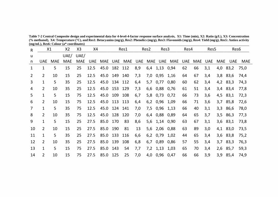
Table 7-2 Central Composite design and experimental data for 4-level-4-factor response surface analysis, X1: Time (min), X2: Ratio (g/L), X3: Concentration
(% methanol), X4: Temperature (°C), and Res1: Betacyanins (mg/g), Res2: Phenolics (mg/g), Res3: Flavonoids (mg/g), Res4: Yield (mg/g), Res5: Antiox activity
(mg/mL), Res6: Colour (a* coordinates)
R u n
X1 X2 X3 X4 Res1 Res2 Res3 Res4 Res5 Res6
UAE MAE UAE/MAE
UAE/MAE UAE MAE UAE MAE UAE MAE UAE MAE UAE MAE UAE MAE UAE MAE
1 1 5 15 25 12.5 45.0 182 112 8,9 6,4 1,13 0,94 62 66 3,1 4,0 83,2 75,0
2 2 10 15 25 12.5 45.0 149 140 7,3 7,0 0,95 1,16 64 67 3,4 3,8 83,6 74,4
3 1 5 35 25 12.5 45.0 134 112 6,4 5,7 0,77 0,80 60 62 3,4 4,2 83,3 74,3
4 2 10 35 25 12.5 45.0 153 129 7,3 6,6 0,88 0,76 61 51 3,4 3,4 83,4 77,8
5 1 5 15 75 12.5 45.0 109 108 6,7 5,8 0,73 0,72 66 73 3,6 4,5 83,1 72,3
6 2 10 15 75 12.5 45.0 113 113 6,4 6,2 0,96 1,09 66 71 3,6 3,7 85,8 72,6
7 1 5 35 75 12.5 45.0 124 141 7,0 7,5 0,96 1,13 66 40 3,1 3,3 86,6 78,0
8 2 10 35 75 12.5 45.0 128 120 7,0 6,4 0,88 0,89 64 65 3,7 3,5 86,3 77,3
9 1 5 15 25 27.5 85.0 170 83 8,6 5,6 1,14 0,90 63 67 3,1 3,6 83,1 73,8
10 2 10 15 25 27.5 85.0 190 81 13 5,6 2,06 0,88 63 89 3,0 4,1 83,0 73,5
11 1 5 35 25 27.5 85.0 133 116 6,6 6,2 0,79 1,02 44 65 3,4 3,6 83,8 75,2
12 2 10 35 25 27.5 85.0 139 108 6,8 6,7 0,89 0,86 57 55 3,4 3,7 83,3 76,3
13 1 5 15 75 27.5 85.0 143 54 7,7 7,2 1,13 1,03 65 70 3,4 2,6 85,7 59,3
14 2 10 15 75 27.5 85.0 125 25 7,0 4,0 0,96 0,47 66 66 3,9 3,9 85,4 74,9
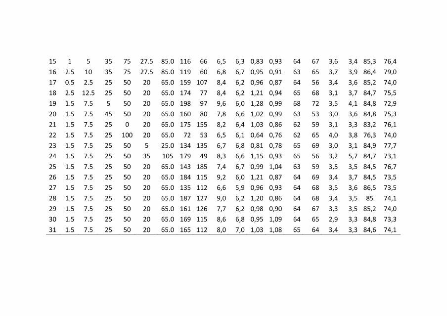
15 1 5 35 75 27.5 85.0 116 66 6,5 6,3 0,83 0,93 64 67 3,6 3,4 85,3 76,4
16 2.5 10 35 75 27.5 85.0 119 60 6,8 6,7 0,95 0,91 63 65 3,7 3,9 86,4 79,0
17 0.5 2.5 25 50 20 65.0 159 107 8,4 6,2 0,96 0,87 64 56 3,4 3,6 85,2 74,0
18 2.5 12.5 25 50 20 65.0 174 77 8,4 6,2 1,21 0,94 65 68 3,1 3,7 84,7 75,5
19 1.5 7.5 5 50 20 65.0 198 97 9,6 6,0 1,28 0,99 68 72 3,5 4,1 84,8 72,9
20 1.5 7.5 45 50 20 65.0 160 80 7,8 6,6 1,02 0,99 63 53 3,0 3,6 84,8 75,3
21 1.5 7.5 25 0 20 65.0 175 155 8,2 6,4 1,03 0,86 62 59 3,1 3,3 83,2 76,1
22 1.5 7.5 25 100 20 65.0 72 53 6,5 6,1 0,64 0,76 62 65 4,0 3,8 76,3 74,0
23 1.5 7.5 25 50 5 25.0 134 135 6,7 6,8 0,81 0,78 65 69 3,0 3,1 84,9 77,7
24 1.5 7.5 25 50 35 105 179 49 8,3 6,6 1,15 0,93 65 56 3,2 5,7 84,7 73,1
25 1.5 7.5 25 50 20 65.0 143 185 7,4 6,7 0,99 1,04 63 59 3,5 3,5 84,5 76,7
26 1.5 7.5 25 50 20 65.0 184 115 9,2 6,0 1,21 0,87 64 69 3,4 3,7 84,5 73,5
27 1.5 7.5 25 50 20 65.0 135 112 6,6 5,9 0,96 0,93 64 68 3,5 3,6 86,5 73,5
28 1.5 7.5 25 50 20 65.0 187 127 9,0 6,2 1,20 0,86 64 68 3,4 3,5 85 74,1
29 1.5 7.5 25 50 20 65.0 161 126 7,7 6,2 0,98 0,90 64 67 3,3 3,5 85,2 74,0
30 1.5 7.5 25 50 20 65.0 169 115 8,6 6,8 0,95 1,09 64 65 2,9 3,3 84,8 73,3
31 1.5 7.5 25 50 20 65.0 165 112 8,0 7,0 1,03 1,08 65 64 3,4 3,3 84,6 74,1
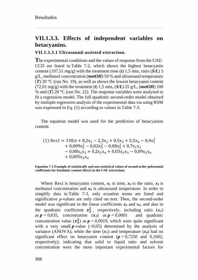
Resultados
308
VII.1.3.3. Effects of independent variables on
betacyanins.
VII.1.3.3.1 Ultrasound-assisted extraction.
The experimental conditions and the values of response from the UAE-
CCD are listed in Table 7-2, which shows the highest betacyanin
content (197,51 mg/g) with the treatment time (t) 1,5 min, ratio (S/L) 5
g/L, methanol concentration (metOH) 50 % and ultrasound temperature
(T) 20 °C (run No. 19), as well as shows the lowest betacyanin content
(72,01 mg/g) with the treatment (t) 1,5 min, (S/L) 25 g/L, (metOH) 100
% and (T) 20 °C (run No. 22). The response variables were analysed to
fit a regression model. The full quadratic second-order model obtained
by multiple regression analysis of the experimental data via using RSM
was expressed in Eq. (1) according to values in Table 7-3.
The equation model was used for the prediction of betacyanins
content.
(1) 𝑅𝑒𝑠1 = 138,6 + 8,2x1 − 2,3x2 + 0,5x3 + 5,5x4 − 8,4x12
+ 0,009x22 − 0,02x3
2 − 0,08x42 + 0,7x1x2
− 0,08x1x3 + 0,2x1x4 + 0,03x2x3 − 0,09x2x4
+ 0,005x3x4
Equation 7-2 Example of statistically and non-statistical values of second-order polynomial
coefficients for betalainic content (Res1) in the UAE extractions.
Where Res1 is betacyanin content, x1 is time, x2 is the ratio, x3 is
methanol concentration and x4 is ultrasound temperature. In order to
simplify data in Table 7-3, only ecuation terms are listed and
significative p-values are only cited on text. Then, the second-order
model was significant in the linear coefficients x2 and x3, and also in
the quadratic coefficient 𝒙𝟑𝟐 , respectively, including ratio (x2)
as p = 0,035, concentration (x3) as p = 0,0001 and quadratic
concentration value (𝒙𝟑𝟐) as p = 0,0019, which were quite significant
with a very small p-value (<0,05) determined by the analysis of
variance (ANOVA), while the time (x1) and temperature (x4) had no
significant effect on betacyanin content (p = 0,7250 and 0,1602,
respectively); indicating that solid to liquid ratio and solvent
concentration were the more important experimental factors for

Resultados
309
ultrasound-assisted extraction. Furthermore, Durbin-Watson
coefficient 2,19 with p = 0,6713 was greater than p = 0,05 indicating
that the residual distribution does not follow any type of autocorrelation
from the regression analysis.
From the ANOVA analysis, in the factorial optimization response
section, we are able to stand out the importance of the two factors ratio
and concentration. The low sample to liquid solvent ratio was explained
by (Vinatoru, 2001), which mention, increasing solvent volume with
respect to sample provides more surface area for the acoustic wave to
form cavitation bubbles and thus increases mass transfer between the
solvent and sample. On the other hand, low methanolic concentration
could be explained due to betalains polarity, these water-soluble
pigments are normally sequestered in the vacuole of the cell (Esquivel,
2016; Strack et al., 2003), so after the breaking up the cell walls through
ultrasonic waves, these molecules are more likely to dissolve in more
polar solvents like water.
VII.1.3.3.2. Microwave-assisted extraction
Optimal extraction conditions for maximum total betacyanins were
recognized by constructing 3D response surface curves with underlying
contour plots (Annex 7-1) and determined by interpolation of
experimental values. MAE process variables significantly affected
(p < 0.05) b3 and b4 as a function of methanol concentration and
extraction temperature showed marked decrease, with the increase of
methanol concentration and temperature as shown in Figure 7-2. On
one hand, the effect of methanol on betalains extraction has been
expressed before in section VII.4.3, while on the other hand, according
to (Sawicki and Wiczkowski, 2018), betalains are susceptible to a wide
range of activities, and thermal treatment could lead to wide ranges of
losses, from 6-81% depending on the process applied or temperature
used (Guldiken et al., 2016; Paciulli et al., 2016; Ravichandran et al.,
2013), and although (Ferreres et al., 2017; Ravichandran et al., 2013)
reports higher betalainic content using MAE, extension of this
technique and temperature may also contribute to their degradation.

Table 7-3 Statistical analysis (ANOVA) of the central composite design including response terms for building the predictive models and optimal response values
for the parametric response criteria. Numbers in linear, quadratic and interaction marked with (*) means statistically significant. Terms included: MSE:
Minimum Square Error; RMSE Root Minimum Square Error; MAPE: Mean Absolute Percentage Error, DW: Durbin-Watson statistic. Responses are
summarized as Res1: Betacyanins (mg/g), Res2: Phenolics (mg/g), Res3: Flavonoids (mg/g), Res4: Yield (mg/g), Res5: Antiox activity (mg/mL), Res6: Colour
(a* coordinates). Fixed variables are summarized as x1= time (min) ; x2= ratio (g/L); x3= methanol concentration (%); x4= temperature (°C).
UAE extraction MAE extraction
Res1 Res2 Res3 Res4 Res5 Res6 Res1 Res2 Res3 Res4 Res5 Res6
Intercept b0 138,65 6,26 0,43 67,40 3,15 45,16 -37,68 5,74 0,03 61,48 9,38 65,60
Linear b1 8,21 -0,23 0,12 2,21 -0,04 0,02 23,64 0,50 0,16 2,66 -0,42 *-2,814
b2 *-2,278 *-0,085 *-0,009 *-0,304 0,00 -0,04 1,98 -0,11 -0,02 *-0,720 -0,05 0,21
b3 *0,501 *0,017 *0,006 *0,067 *-0,008 *-0,069 *1,514 0,03 0,01 -0,01 -0,01 *-0,542
b4 5,60 0,32 *0,049 -0,19 0,01 0,06 *1,739 -0,02 0,01 0,19 -0,11 *-0,515 Quadratic b11 -8,47 0,01 0,03 -0,76 0,00 -0,27 -0,88 -0,01 0,00 -0,07 0,00 0,08
b22 0,01 0,00 0,00 0,00 0,00 0,00 *-0,064 0,00 0,00 0,00 0,00 0,00
b33 *-0,021 0,00 0,00 0,00 0,00 *0,001 0,00 0,00 0,00 0,00 0,00 *0,003
b44 -0,08 0,00 0,00 0,00 0,00 0,00 -0,01 0,00 0,00 0,00 *0,001 *0,003 Interaction b12 0,76 0,00 -0,01 0,09 0,00 0,02 -0,06 0,01 0,00 -0,03 0,00 -0,01
b13 -0,09 -0,02 0,00 -0,08 0,00 0,00 -0,08 *-0,005 0,00 0,02 0,00 0,02
b14 0,27 0,08 0,01 0,16 -0,01 0,00 -0,09 0,00 0,00 -0,01 0,01 0,02
b23 0,03 *0,003 *0,000 0,01 0,00 0,00 0,01 0,00 0,00 0,00 0,00 0,00
b24 -0,09 -0,01 *-0,001 -0,02 0,00 0,00 0,02 0,00 0,00 0,01 0,00 0,00
b34 0,01 0,00 0,00 0,01 0,00 0,00 *-0,021 0,00 0,00 0,00 0,00 *0,003
Statistical information of the fitting analysis Observations 31 31 31 31 31 31 31 31 31 31 31 31 MSE 18,65 0,99 0,16 2,99 0,23 0,85 13,99 0,58 0,13 8,54 0,52 2,65 RMSE 4,32 0,99 0,40 1,73 0,48 0,92 3,74 0,76 0,36 2,92 0,72 1,63 MAPE 10,42 0,55 0,09 1,45 0,13 0,46 8,29 0,34 0,08 4,72 0,27 1,43

DW 2,19 1,76 1,78 *1,01 1,48 1,62 1,94 1,70 1,53 2,05 2,19 1,68 Factorial Optimization Response max max max max min max max max max max min max Optimum value 227,6 17,8 3,1 73,1 2,3 87,1 144,6 8,6 1,5 84,0 1,8 73,7 x1 1,2 2,5 2,5 2,5 2,2 2,1 8,8 2,5 12,5 11,9 12,5 12,4 x2 5,0 5,0 5,0 5,3 5,0 44,3 20,3 34,0 5,0 5,0 44,3 16,2 x3 17,7 0,1 0,0 30,2 0,0 56,3 54,8 100,0 25,1 46,8 0,0 100,0 x4 33,9 34,1 33,8 34,7 35,0 5,0 25,0 103,6 25,0 25,0 25,0 105,0
General Optimization ¡ max max max max min max max max max max min max Optimum response 201,6 13,9 2,4 71,8 2,9 85,7 132,9 8,0 1,5 79,1 3,6 78,7
Deseability 0,985 0,871 Optimum value x1= 2,5 x2= 5 x3= 34,6 x4= 30,0 x1= 12,4 x2= 5,0 x3= 0,0 x4= 36,6

Resultados
312
(1) 𝑅𝑒𝑠1 = −37,7 + 1,5x3 + 1,7x4 − 0,06x22 − 0,02x3x4
Equation 7-3 Statistically values of second-order polynomial coefficients for betalainic
content (Res1) in the MAE extractions.
In Table 7-3 has been also exposed 2 different optimization values,
1) the factorial optimization response, which determine the optimal
value of a single response in case of being interested only in
maximizing the colorant extraction in a single process, in this case
betalainic content of 144,6 mg/g correspondent with factorial values of
x1= 8,8 min; x2= 20,3 g/L; x3= 54,8 % of methanol and x4= 25 °C the
general optimization response, which determines the optimal value for
all the responses in the same batch, in this case, the max value for
betalains was 132,9 mg/g lower than the one obtained in the factorial
optimization response, but at the same contemplating maximization
from the other responses, in order to achieve this optimization factorial
values, factors are meant to be x1= 12,4 min; x2= 5 g/L; x3= 0 methanol
% and x4= 36,6 °C.
VII.1.3.4. Effects of independent variables on
phenolic acids and flavonoids.
VII.1.3.4.1. Ultrasound-assisted extraction
Based on Table 7-3, Total Phenolic Content (TPC) and Total Flavonoid
Content (TFC) under ultrasonic extraction were significantly (p < 0.05)
affected by the linear effect of ratio and solvent concentration for TPC
and TFC, being temperature extraction significant only for the
flavonoid content, and time non-significant for any of both. Significant
interaction values were also present on ratio-concentration for both
TPC and TFC, although, the last mentioned response, was also
significant on ratio-temperature interaction Remaining factors did not
show any significant (p < 0.05) effect on TPC or TFC. Removing all
non-significant terms, the polynomial Equation (2) and (3) for TPC
(Res2) and TFC (Res3) respectively, comes as:
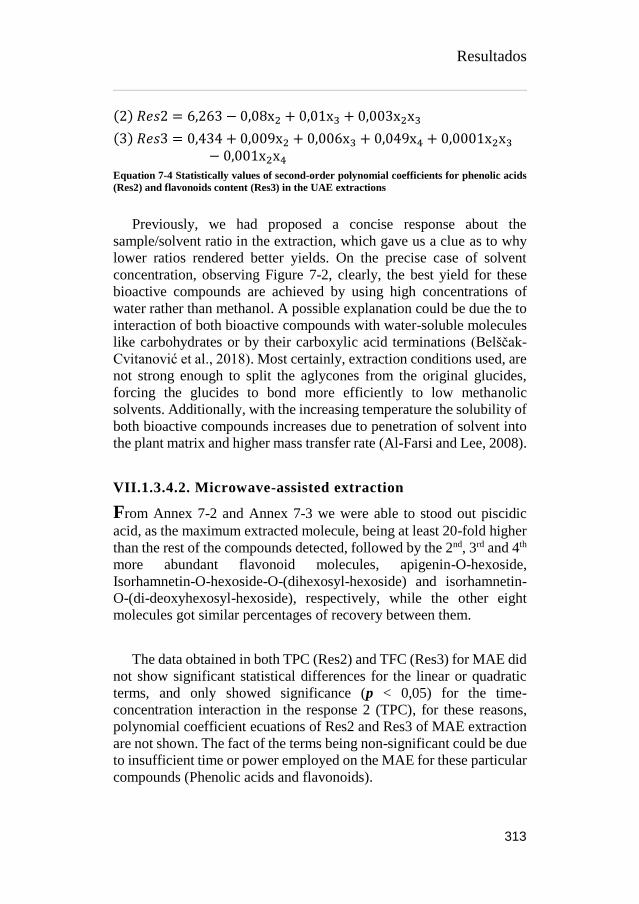
Resultados
313
(2) 𝑅𝑒𝑠2 = 6,263 − 0,08x2 + 0,01x3 + 0,003x2x3
(3) 𝑅𝑒𝑠3 = 0,434 + 0,009x2 + 0,006x3 + 0,049x4 + 0,0001x2x3
− 0,001x2x4
Equation 7-4 Statistically values of second-order polynomial coefficients for phenolic acids
(Res2) and flavonoids content (Res3) in the UAE extractions
Previously, we had proposed a concise response about the
sample/solvent ratio in the extraction, which gave us a clue as to why
lower ratios rendered better yields. On the precise case of solvent
concentration, observing Figure 7-2, clearly, the best yield for these
bioactive compounds are achieved by using high concentrations of
water rather than methanol. A possible explanation could be due the to
interaction of both bioactive compounds with water-soluble molecules
like carbohydrates or by their carboxylic acid terminations (Belščak-
Cvitanović et al., 2018). Most certainly, extraction conditions used, are
not strong enough to split the aglycones from the original glucides,
forcing the glucides to bond more efficiently to low methanolic
solvents. Additionally, with the increasing temperature the solubility of
both bioactive compounds increases due to penetration of solvent into
the plant matrix and higher mass transfer rate (Al-Farsi and Lee, 2008).
VII.1.3.4.2. Microwave-assisted extraction
From Annex 7-2 and Annex 7-3 we were able to stood out piscidic
acid, as the maximum extracted molecule, being at least 20-fold higher
than the rest of the compounds detected, followed by the 2nd, 3rd and 4th
more abundant flavonoid molecules, apigenin-O-hexoside,
Isorhamnetin-O-hexoside-O-(dihexosyl-hexoside) and isorhamnetin-
O-(di-deoxyhexosyl-hexoside), respectively, while the other eight
molecules got similar percentages of recovery between them.
The data obtained in both TPC (Res2) and TFC (Res3) for MAE did
not show significant statistical differences for the linear or quadratic
terms, and only showed significance (p < 0,05) for the time-
concentration interaction in the response 2 (TPC), for these reasons,
polynomial coefficient ecuations of Res2 and Res3 of MAE extraction
are not shown. The fact of the terms being non-significant could be due
to insufficient time or power employed on the MAE for these particular
compounds (Phenolic acids and flavonoids).

Resultados
314
VII.1.3.5. Effects of independent variables on
antioxidant activity.
VII.1.3.5.1. Ultrasound-assisted extraction and microwave-
assisted extraction
Our main goal was to minimize the IC50 value in Response 5
(antioxidant effect). Briefly, lower concentration of antioxidant
molecules that reduce 50 % of the oxidation process, means better
phytochemicals quality.
Betalains and polyphenolics have shown a wide range physiological
properties, such as antimicrobial, anti-inflammatory, cardioprotective
and antioxidant among others (Melgar et al., 2017a; Strack et al., 2003;
Yeddes et al., 2013). Fom Table 7-3 and Figure 7-2 we were able to
identify solvent concentration (b3), as the only linear effect with
statistical significance in UAE and MAE, both cases exhibit better
antioxidant effect on highly aqueous concentration, which is in
concordance with data analysed, previously, we exposed in this essay
how betalains, phenolic acids and flavonoids concentrations were
higher on low methanolic solvents, providing a clear synergistic effect
from the bioactive compounds on the antioxidant effect tested.
VII.1.3.6. Effects of independent variables on yield
and colour.
VII.1.3.6.1. Extraction yield on UAE and MAE
From Figure 7-2, we can clearly stand out, first, how ratio effect (x2)
affect negatively while this increases in both types of extractions,
second, in UAE, solvent concentration effect (x3) also played an
important role, which is confirmed with the statistical significance in
Table 7-3, in this particular case, observing higher yields with higher
methanolic concentration, achieving an optimum value for x3 in
Response 4 of 30,2 % of methanol.
The effect of ratio on yielding of phytochemical compounds was
mentioned before on section VII.4.3, so no further comments on this
matter will be mention in this section. On the other hand, solvent
concentration yielding could be explained as a polarity effect, we

Resultados
315
pointed out how our interest phytochemicals have been better extracted
with low methanolic concentration, perhaps, this little increase on
methanol helps also to extract various unidentified polar compounds,
and due to its amphiphilic properties, also some non-polar ones
(Primorac et al., 2018).
VII.1.3.7. Colour effect on ultrasonic and microwave
extractions
The colour properties of Opuntia peels, ranging in colour from pink to
violet, were measured by colourimetric analysis. Statistics from Table
7-3, stands out solvent concentration (x3) in both extraction
technologies employed, and time (x1) and temperature (x4) only on
microwave extractions. Even though, the statistical analysis focused
only on coordinate a*, to better understand the total colour perspective,
table Annex 7-8, exhibits the complementary parameters L*, b* and
chroma. From all this data brighter (chroma) and lighter colour
intensity (L*) but reddish tone (a*) can be noted in every single run of
UAE compared to MAE, and the possible explanation for this effect
could be due to small oxidation of sugar attached to betalains caused
by employment of higher temperatures on the microwave extraction
technique.
VII.1.3.8. Comparison of UAE and MAE RSM
Ultrasound and microwave are both common extraction technologies
used for the recovery of bioactive compounds, on the bibliography,
plethora of information about different plants and by-products can be
found, mainly oriented to improve specific conditions, although these
technologies have different mechanism of action, both are used cited
on extraction procedures, microwave assisted extraction essentially
heat up the molecules by dual mechanism of ionic conduction and
dipole rotation, while ultrasound assisted extraction relies on the uses
of acoustic energy inducing vibrational motion (cavitation) of the
molecules (Mandal et al., 2015).
On this assay, we have already described the effects of every single
factor and the influence of the different levels on the responses
analysed, now as a brief summary, a comparison between these two
technologies and their results is discussed.

Resultados
316
Figure 7-2 Principal effect graphs of UAE and MAE, representing responses values, acquire
from the independent variables employed on the CCD-RMS (source: private collection)

Resultados
317
All the phytochemical compounds were identified using both
technologies, varying the percentages and the concentrations of these
molecules. Considering maximums obtained in each different
extraction (Table 7-2), all the maximum amounts of bioactive
compounds (betalains, phenolic acids and flavonoids) was achieved by
ultrasonic assisted extraction.
In betalains for instance, almost 22% more was achieved, 197,51
mg/g of UAE vs 151,11 mg/g of MAE; for phenolic acids and
flavonoids this differences went up to about 43%, 12,99 mg/g of UAE
vs 7,46 mg/g of MAE and 2,06 mg/g of UAE vs 1,16 mg/g of MAE,
respectively. Differences in the means of specific compounds did not
exceed the 2 % for betacyanins, 5,3% for flavonoids and 5 % for
phenolic acids, in this last one mentioned, piscidic acid (Ph1) for
instance changed from 93,3% on UAE to 98,3% on MAE,
compensating those differences on cis-caffeic acid, where 4,1 % was
achieved on UAE compared to the 0,3 % of the MAE. As far as we
know, there has not been performed any other comparison of this type
in which we could compare our results.
In the antioxidant activity effect (Table 7-2), although lower IC50
response was obtained on the MAE 2,6 mg/mL vs 2,9 from the UAE,
the majority of data in the 31 runs was always lower for the UAE
extracts. Two possible explanations for this unexpected result could be
due to a colorimetric interference while performing the assay or an
operational error, either way, UAE has shown also an overall better
antioxidant performance, which is in concordance with the maximum
amounts of biomolecules obtained also by UAE. Table 7-3, also shows
optimum responses values of 2,9 mg/mL for UAE and 3,6 mg/mL for
MAE, which agrees with the aforementioned.
On the other hand, the percentage of yield (Table 7-2) was in the
opposite way, where MAE exhibits the best results almost in every run.

Resultados
318
VII.1.4. CONCLUSION
The present work has described the use of ultrasound-assisted
extraction and microwave-assisted extraction techniques in order to
extract bioactive compounds of interest, namely betacyanins, phenolic
acids, flavonoid, as well as their antioxidant profiling. In this essay, the
interaction of four different factors (time, solid to solvent ratio,
methanol concentration and temperature) were investigated using the
RSM model for predicting variables. From the data obtained, we are
able to stand out:
I. The identification through HPLC-DAD-ESI-MS/MS of 7
different betacyanins, 4 phenolic acids and 8 flavonoids, from
the Opuntia engelmaannii peels, reporting their respective
tentative names, retention times and identification patterns.
II. By using the CCD-RSM modelling, principal effect and 3D
graphs, the recognition of each independent variable impact on
the studied responses was highlighted, indicating the factorial
optimisation responses and proposing how to build the
equation models for the prediction of the different responses.
III. Compiling all of the previous information, a comparison of the
different techniques of extraction was mentioned, based on the
singular responses of each UAE and MAE, the most suitable
technique for this particular by product and conditions was
proposed.
Finally, a general response optimization of both types of
extractions was also performed statistically, involving all the responses
analysed at the same time. With a deseability response of 0,985, the
optimum values for each factor in UAE was x1= 2,5 min, x2= 5 g/L,
x3=34,6 % of methanol and x4= 30 °C. In the same manner, with a
deseability response of 0,871, the optimum values for each factor in
MAE was x1= 1,4 min, x2= 5 g/L, x3=0,0 % of methanol and x4= 36,6
°C. These conditions will produce 201,6 and 132,9 mg/g of betalains,
13,9 and 8,0 mg/g of phenolic acids, 2,4 and 1,5 mg/g of flavonoids,
71,8 and 79,1 % of yields and IC50 antioxidant values of 2,9 and 3,6,
for UAE and MAE respectively.
Concluding, the present study suggests the use of ultrasound-
assisted extraction in order to maximize recovery of bioactive
Resultados
319
compounds such as betalains, phenolic acids and flavonoids under the
conditions previously mentioned, which also provide better antioxidant
activity.
The main recommendation is to work at temperatures lower than 37 °C,
with the minimum solid to solvent ratio used (5 g/L) and low
concentrations of methanol (<35%).
Acknowledgements
The authors are grateful to CONACyT for supporting B. Melgar with
his doctoral grant (No. 329930). The authors specially thanks to Maria
Luisa Ruiz and the “Laboratorio Agroalimentario de la Comunitat
Valenciana” for allow us to use the microwave for the extractions.

Resultados
320
References
Al-Farsi, M.A., Lee, C.Y., 2008. Optimization of phenolics and dietary
fibre extraction from date seeds. Food Chem. 108, 977–985.
https://doi.org/10.1016/j.foodchem.2007.12.009
Allai, L., Druart, X., Öztürk, M., BenMoula, A., Nasser, B., El Amiri,
B., 2016. Protective effects of Opuntia ficus-indica extract on ram
sperm quality, lipid peroxidation and DNA fragmentation during
liquid storage. Anim. Reprod. Sci. 175, 1–9.
https://doi.org/10.1016/j.anireprosci.2016.09.013
Ammar, I., Ben Salem, M., Harrabi, B., Mzid, M., Bardaa, S., Sahnoun,
Z., Attia, H., Ennouri, M., 2018. Anti-inflammatory activity and
phenolic composition of prickly pear (Opuntia ficus-indica)
flowers. Ind. Crops Prod. 112, 313–319.
https://doi.org/10.1016/j.indcrop.2017.12.028
Aruwa, C.E., Amoo, S.O., Kudanga, T., 2018. Opuntia (Cactaceae)
plant compounds, biological activities and prospects – A
comprehensive review. Food Res. Int. 112, 328–344.
https://doi.org/10.1016/j.foodres.2018.06.047
Belščak-Cvitanović, A., Durgo, K., Huđek, A., Bačun-Družina, V.,
Komes, D., 2018. Metabolism and Health Effects of Polyphenols,
in: Galanakis, C.M. (Ed.), Polyphenols: Properties, Recovery, and
Applications. Woodhead Publishing, pp. 1–44.
https://doi.org/https://doi.org/10.1016/B978-0-12-813572-
3.00001-4
Betancourt, C., Cejudo-Bastante, M.J., Heredia, F.J., Hurtado, N.,
2017. Pigment composition and antioxidant capacity of
betacyanins and betaxanthins fractions of Opuntia dillenii (Ker
Gawl) Haw cactus fruit. Food Res. Int. 101, 173–179.
https://doi.org/10.1016/j.foodres.2017.09.007
Cardoso-Ugarte, G.A., Sosa-Morales, M.E., Ballard, T., Liceaga, A.,
San Martín-González, M.F., 2014. Microwave-assisted extraction
of betalains from red beet (Beta vulgaris). LWT - Food Sci.
Technol. 59, 276–282. https://doi.org/10.1016/j.lwt.2014.05.025
Chougui, N., Djerroud, N., Naraoui, F., Hadjal, S., Aliane, K., Zeroual,
B., Larbat, R., 2015. Physicochemical properties and storage
stability of margarine containing Opuntia ficus-indica peel extract
as antioxidant. Food Chem. 173, 382–90.
https://doi.org/10.1016/j.foodchem.2014.10.025
Resultados
321
do Prado, D. Z., Capoville, B. L., Delgado, C. H. O., Heliodoro, J. C.
A., Pivetta, M. R., Pereira, M. S., … Fleuri, L. F. (2018).
Nutraceutical food: composition, biosynthesis, therapeutic
properties, and applications. Alternative and Replacement Foods,
95–140. https://doi.org/10.1016/B978-0-12-811446-9.00004-6
Esquivel, P., 2016. Handbook on Natural Pigments in Food and
Beverages Industrial Applications for Improving Food Colorl, in:
Carle, R., Schweiggert, R.M. (Eds.), . Woodhead Publishing, San
Pedro, Costa Rica, pp. 81–99.
Fathordoobady, F., Mirhosseini, H., Selamat, J., Manap, M.Y.A., 2016.
Effect of solvent type and ratio on betacyanins and antioxidant
activity of extracts from Hylocereus polyrhizus flesh and peel by
supercritical fluid extraction and solvent extraction. Food Chem.
202, 70–80. https://doi.org/10.1016/j.foodchem.2016.01.121
Ferreres, F., Grosso, C., Gil-Izquierdo, A., Valentão, P., Mota, A.T.,
Andrade, P.B., 2017. Optimization of the recovery of high-value
compounds from pitaya fruit by-products using microwave-
assisted extraction. Food Chem. 230, 463–474.
https://doi.org/10.1016/j.foodchem.2017.03.061
Garcia-Castello, E. M., Rodriguez-Lopez, A. D., Mayor, L.,
Ballesteros, R., Conidi, C., & Cassano, A. (2015). Optimization
of conventional and ultrasound assisted extraction of flavonoids
from grapefruit (Citrus paradisi L.) solid wastes. LWT - Food
Science and Technology, 64(2), 1114–1122.
https://doi.org/10.1016/j.lwt.2015.07.024
García-Cruz, L., Dueñas, M., Santos-Buelgas, C., Valle-Guadarrama,
S., Salinas-Moreno, Y., 2017. Betalains and phenolic compounds
profiling and antioxidant capacity of pitaya (Stenocereus spp.)
fruit from two species (S. Pruinosus and S. stellatus). Food Chem.
234, 111–118. https://doi.org/10.1016/j.foodchem.2017.04.174
Gebhardt, D., 2018. The Economics of Natural Color Pigments. URL
https://sensientfoodcolors.com/en-us/research-
development/economics-natural-color-pigments/
Guldiken, B., Toydemir, G., Nur Memis, K., Okur, S., Boyacioglu, D.,
Capanoglu, E., 2016. Home-processed red beetroot (Beta vulgaris
L.) products: Changes in antioxidant properties and
bioaccessibility. Int. J. Mol. Sci. 17.
https://doi.org/10.3390/ijms17060858
GVR, 2016. Natural Antioxidants Market Analysis By Product

Resultados
322
(Vitamin C, Vitamin E, Polyphenols, Carotenoids) And Segment
Forecasts To 2022. URL
https://www.grandviewresearch.com/industry-analysis/natural-
antioxidants-market
Herbach, K.M., Stintzing, F.C., Carle, R., 2005. Identification of heat-
induced degradation products from purified betanin, phyllocactin
and hylocerenin by high-performance liquid chromatography/
electrospray ionization mass spectrometry. Rapid Commun. Mass
Spectrom. 19, 2603–2616. https://doi.org/10.1002/rcm.2103
IFIC, FDA, 2010. Overview of Food Ingredients, Additives and Colors.
URL
https://www.fda.gov/food/ingredientspackaginglabeling/foodadd
itivesingredients/ucm094211.htm
Laqui-Vilca, C., Aguilar-Tuesta, S., Mamani-Navarro, W., Montaño-
Bustamante, J., Condezo-Hoyos, L., 2018. Ultrasound-assisted
optimal extraction and thermal stability of betalains from colored
quinoa (Chenopodium quinoa Willd) hulls. Ind. Crops Prod. 111,
606–614. https://doi.org/10.1016/j.indcrop.2017.11.034
Leardi, R., 2013. Experimental Design, 1st ed, Data Handling in
Science and Technology. Copyright © 2013 Elsevier B.V. All
rights reserved. https://doi.org/10.1016/B978-0-444-59528-
7.00002-8
Madunić, J., Madunić, I.V., Gajski, G., Popić, J., Garaj-Vrhovac, V.,
2018. Apigenin: A dietary flavonoid with diverse anticancer
properties. Cancer Lett. 413, 11–22.
https://doi.org/10.1016/j.canlet.2017.10.041
Mandal, S.C., Mandal, V., Das, A.K., 2015. Classification of Extraction
Methods, in: Essentials of Botanical Extraction. Academic Press,
pp. 83–136. https://doi.org/10.1016/B978-0-12-802325-9.00006-
9
Mata, A., Ferreira, J.P., Semedo, C., Serra, T., Duarte, C.M.M., Bronze,
M.R., 2016. Contribution to the characterization of Opuntia spp.
juices by LC–DAD–ESI-MS/MS. Food Chem. 210, 558–565.
https://doi.org/10.1016/j.foodchem.2016.04.033
Melgar, B., Dias, M.I., Ciric, A., Sokovic, M., Garcia-Castello, E.M.,
Rodriguez-Lopez, A.D., Barros, L., Ferreira, I., 2017a. By-
product recovery of Opuntia spp. peels: Betalainic and phenolic
profiles and bioactive properties.
https://doi.org/10.1016/j.indcrop.2017.06.011

Resultados
323
Melgar, B., Pereira, E., Oliveira, M.B.P.P., Garcia-Castello, E.M.,
Rodriguez-Lopez, A.D., Sokovic, M., Barros, L., Ferreira,
I.C.F.R., 2017b. Extensive profiling of three varieties of Opuntia
spp. fruit for innovative food ingredients. Food Res. Int. 101, 259–
265. https://doi.org/10.1016/j.foodres.2017.09.024
Mena, P., Tassotti, M., Andreu, L., Nuncio-Jáuregui, N., Legua, P., Del
Rio, D., Hernández, F., 2018. Phytochemical characterization of
different prickly pear (Opuntia ficus-indica (L.) Mill.) cultivars
and botanical parts: UHPLC-ESI-MSn metabolomics profiles and
their chemometric analysis. Food Res. Int. 108, 301–308.
https://doi.org/10.1016/j.foodres.2018.03.062
Paciulli, M., Medina-Meza, I.G., Chiavaro, E., Barbosa-Cánovas, G.V.,
2016. Impact of thermal and high pressure processing on quality
parameters of beetroot (Beta vulgaris L.). LWT - Food Sci.
Technol. 68, 98–104. https://doi.org/10.1016/j.lwt.2015.12.029
Patel, D., Shukla, S., Gupta, S., 2007. Apigenin and cancer
chemoprevention: Progress, potential and promise (Review). Int.
J. Oncol. 30, 233–245. https://doi.org/10.3892/ijo.30.1.233
Primorac, T., Požar, M., Sokolić, F., Zoranić, L., Urbic, T., 2018. A
simple two dimensional model of methanol. J. Mol. Liq. 262, 46–
57. https://doi.org/10.1016/j.molliq.2018.04.055
Ravichandran, K., Saw, N.M.M.T., Mohdaly, A.A.A., Gabr, A.M.M.,
Kastell, A., Riedel, H., Cai, Z., Knorr, D., Smetanska, I., 2013.
Impact of processing of red beet on betalain content and
antioxidant activity. Food Res. Int. 50, 670–675.
https://doi.org/10.1016/j.foodres.2011.07.002
Roriz, C.L., Barros, L., Prieto, M.A., Morales, P., Ferreira, I.C.F.R.,
2017. Floral parts of Gomphrena globosa L. as a novel alternative
source of betacyanins: Optimization of the extraction using
response surface methodology. Food Chem. 229, 223–234.
https://doi.org/10.1016/j.foodchem.2017.02.073
Sawicki, T., Wiczkowski, W., 2018. The effects of boiling and
fermentation on betalain profiles and antioxidant capacities of red
beetroot products. Food Chem. 259, 292–303.
https://doi.org/10.1016/j.foodchem.2018.03.143
Spórna-Kucab, A., Ignatova, S., Garrard, I., Wybraniec, S., 2013.
Versatile solvent systems for the separation of betalains from
processed Beta vulgaris L. juice using counter-current
chromatography. J. Chromatogr. B Anal. Technol. Biomed. Life

Resultados
324
Sci. 941, 54–61. https://doi.org/10.1016/j.jchromb.2013.10.001
Strack, D., Vogt, T., Schliemann, W., 2003. Recent advances in
betalain research. Phytochemistry 62, 247–269.
https://doi.org/10.1016/S0031-9422(02)00564-2
Szot, D., Skopińska, A., Wybraniec, S., 2015. Decomposition of 17-
decarboxy-betanin in selected aqueous-organic solutions induced
by Cu ( II ) cations. PhD Interdiscip. J. Politech. Gdansk Univ. 1,
217–223.
Thirugnanasambandham, K., Sivakumar, V., 2017. Microwave assisted
extraction process of betalain from dragon fruit and its antioxidant
activities. J. Saudi Soc. Agric. Sci. 16, 41–48.
https://doi.org/10.1016/j.jssas.2015.02.001
Vinatoru, M., 2001. An overview of the ultrasonically assisted
extraction of bioactive principles from herbs. Ultrason.
Sonochem. 8, 303–313. https://doi.org/10.1016/S1350-
4177(01)00071-2
Wybraniec, S., Starzak, K., Szneler, E., Pietrzkowski, Z., 2016.
Separation of chlorinated diastereomers of decarboxy-
betacyanins in myeloperoxidase catalyzed chlorinated Beta
vulgaris L. extract. J. Chromatogr. B Anal. Technol. Biomed. Life
Sci. 1036–1037, 20–32.
https://doi.org/10.1016/j.jchromb.2016.09.040
Yeddes, N., Chérif, J., Guyot, S., Sotin, H., Ayadi, M., 2013.
Comparative Study of Antioxidant Power, Polyphenols,
Flavonoids and Betacyanins of the Peel and Pulp of Three
Tunisian Opuntia Forms. Antioxidants 2, 37–51.
https://doi.org/10.3390/antiox2020037

Annex 7-1 Amount in mg/g of the specific betacyanins identified in every single run and the sum of all to obtained the total betacyanin content
Run Peak 1 Peak 2 Peak 3 Peak 4 Peak 5 Peak 6 Peak 7 Total
Betanin Isobetanin Gomphrenin I (Iso)phyllocactin 17 decarboxy-
betanin
Betanidin Isobetanidin mg/g
UAE MAE UAE MAE UAE MAE UAE MAE UAE MAE UAE MAE UAE MAE UAE MAE
1 144,0 90,4 21,6 12,6 4,8 1,7 2,2 0,4 2,0 0,6 3,3 1,2 3,7 5,4 181,5 112,3
2 118,7 111,9 18,0 16,0 4,1 3,0 1,7 1,2 1,6 0,9 2,3 1,5 2,5 5,7 148,8 140,1
3 107,6 95,2 16,2 13,0 3,3 1,8 1,6 1,0 1,3 0,7 1,8 0,7 2,6 0,0 134,3 112,5
4 123,7 104,8 17,7 15,1 3,9 2,3 1,9 0,6 1,6 0,2 2,3 2,0 1,9 4,2 152,9 129,3
5 91,0 91,5 11,1 12,2 2,8 2,0 1,0 1,0 0,9 0,8 0,9 0,3 1,5 0,0 109,3 107,9
6 92,2 91,7 12,4 14,2 2,9 2,2 1,4 0,3 1,0 0,6 1,2 0,9 1,3 3,5 112,5 113,5
7 100,4 117,1 13,4 17,2 3,2 2,5 1,4 0,4 1,1 0,5 1,3 0,3 2,7 3,3 123,6 141,3
8 103,6 96,1 13,8 15,6 3,2 2,1 1,5 0,9 1,1 0,6 1,8 2,0 2,8 2,5 127,8 119,7
9 135,2 67,6 20,8 10,0 4,5 1,0 1,8 0,2 1,8 0,4 2,8 0,1 3,5 3,4 170,3 82,7
10 150,2 65,3 22,7 9,6 5,2 1,0 2,2 0,2 2,3 0,4 3,2 0,8 3,7 3,1 189,5 80,5
11 107,5 92,4 16,6 16,4 3,3 1,6 1,2 0,1 1,2 0,6 1,8 1,0 1,6 4,1 133,2 116,4
12 111,5 83,0 16,5 14,4 3,7 1,7 1,5 0,2 1,4 0,1 2,2 3,7 1,7 4,4 138,6 107,6
13 116,1 43,1 16,0 7,5 4,1 1,0 1,8 0,2 1,5 0,1 2,1 0,1 1,8 1,5 143,2 53,5
14 102,6 18,8 13,4 3,3 3,3 0,3 1,2 0,1 1,1 0,2 1,3 0,3 1,8 2,0 124,7 25,1
15 94,8 52,1 12,7 10,6 2,9 1,2 1,1 0,7 0,9 0,4 1,6 1,2 1,6 0,0 115,7 66,3

16 97,1 44,2 13,4 10,0 3,0 1,2 1,4 0,2 1,0 0,2 1,1 0,7 2,4 3,1 119,3 59,6
17 126,3 88,7 19,1 14,8 3,9 1,8 1,8 0,5 1,3 0,2 2,2 0,6 4,4 0,0 159,1 106,6
18 136,3 60,9 21,3 10,8 4,6 1,5 2,1 0,1 1,8 0,2 3,3 2,3 4,5 1,3 173,9 77,1
19 156,1 78,6 23,6 11,5 5,1 1,4 2,3 0,4 2,0 0,4 3,7 0,5 4,6 4,0 197,5 96,8
20 126,6 55,9 18,8 16,5 4,4 2,2 2,3 0,9 1,6 0,7 2,6 1,5 3,5 2,2 159,7 79,8
21 139,0 127,0 21,4 18,2 4,6 3,4 2,1 1,2 2,4 0,9 2,8 1,3 2,2 3,2 174,5 155,1
22 58,6 16,7 7,2 30,8 1,8 5,0 0,8 0,1 0,6 0,6 0,9 0,0 2,1 0,0 72,0 53,2
23 108,0 113,3 15,2 15,8 3,4 2,5 1,7 1,6 1,3 1,2 2,0 0,8 2,1 0,0 133,8 135,1
24 140,6 25,8 21,8 16,5 5,1 4,8 2,5 0,9 1,7 0,3 3,4 1,2 4,1 0,0 179,2 49,4
25 116,3 92,1 16,1 12,5 3,7 1,5 1,4 0,5 1,2 0,2 1,7 0,7 2,6 0,0 143,0 107,6
26 144,9 95,1 23,2 14,9 5,3 2,1 2,3 1,3 1,9 0,9 3,1 0,5 3,6 0,0 184,3 114,8
27 106,8 93,0 16,9 14,4 3,6 1,8 1,5 0,8 1,3 0,8 2,2 1,0 2,3 0,0 134,7 111,8
28 149,9 102,8 22,8 16,1 5,1 2,4 2,4 0,2 1,8 0,7 2,8 0,8 2,7 4,1 187,4 127,1
29 128,8 101,6 19,7 15,9 4,3 1,8 2,0 0,3 1,6 0,7 2,2 1,3 2,6 4,6 161,2 126,3
30 134,7 94,1 19,4 14,4 4,8 1,6 2,3 0,3 1,7 0,5 2,5 0,3 3,6 3,4 169,0 114,7
31 131,6 90,4 19,6 12,6 4,4 1,7 2,1 0,4 1,7 0,6 2,5 1,2 3,2 5,4 165,0 112,3

Annex 7-2 Amount in mg/g of the specific phenolic acids identified in every single run and the sum of all to obtained the total phenolic content
Run Peak 1 Peak 2 Peak 3 Peak 4 Total
Piscidic acid
3-O-Feruloylquinic
acid cis-Caffeic acid
Ferulic acid
hexoside mg/g
UAE MAE UAE MAE UAE MAE UAE MAE UAE MAE
1 8,46 6,30 n/d 0,03 0,38 0,02 0,07 0,07 8,91 6,41
2 6,85 6,89 n/d 0,02 0,36 0,02 0,06 0,08 7,26 7,01
3 6,00 5,57 0,08 0,02 0,25 0,01 0,05 0,07 6,39 5,66
4 6,86 6,51 n/d 0,02 0,37 0,03 0,06 0,07 7,29 6,62
5 6,34 5,71 0,09 0,02 0,21 0,00 0,05 0,06 6,69 5,80
6 6,03 6,12 0,08 0,02 0,24 0,01 0,05 0,08 6,40 6,23
7 6,59 7,32 n/d 0,03 0,34 0,02 0,06 0,09 6,98 7,46
8 6,62 6,33 n/d 0,02 0,35 0,01 0,06 0,06 7,04 6,42
9 8,11 5,52 n/d 0,01 0,43 0,01 0,07 0,07 8,61 5,60
10 12,13 5,50 0,71 0,01 0,06 0,01 0,09 0,07 12,99 5,59
11 6,23 6,07 0,07 0,03 0,24 0,02 0,05 0,08 6,60 6,20
12 6,44 6,54 n/d 0,01 0,32 0,04 0,05 0,07 6,81 6,65
13 7,28 7,14 0,00 0,01 0,36 0,02 0,07 0,06 7,71 7,23
14 6,60 3,95 n/d 0,01 0,34 0,01 0,06 0,04 7,00 4,01
15 6,13 6,22 0,09 0,03 0,25 0,00 0,06 0,07 6,52 6,32

16 6,33 6,59 n/d 0,04 0,36 0,02 0,06 0,07 6,75 6,73
17 7,93 6,08 0,10 0,02 0,28 0,01 0,06 0,06 8,37 6,18
18 7,92 6,12 n/d 0,02 0,40 0,01 0,07 0,07 8,39 6,22
19 9,05 5,94 n/d 0,01 0,45 0,01 0,08 0,07 9,58 6,03
20 7,33 6,48 n/d 0,01 0,35 0,02 0,06 0,07 7,75 6,57
21 7,78 6,34 n/d 0,01 0,38 0,03 0,06 0,06 8,22 6,43
22 6,21 5,99 n/d 0,04 0,28 0,01 0,05 0,06 6,53 6,10
23 6,28 6,70 0,12 0,01 0,21 0,01 0,05 0,06 6,66 6,78
24 7,83 6,48 n/d 0,02 0,41 0,02 0,06 0,07 8,31 6,60
25 7,00 6,61 0,10 0,04 0,28 0,02 0,06 0,07 7,44 6,74
26 8,61 5,93 n/d 0,00 0,49 0,01 0,07 0,07 9,17 6,02
27 6,17 5,76 n/d 0,02 0,34 0,02 0,05 0,08 6,56 5,87
28 8,41 6,07 0,19 0,03 0,30 0,04 0,07 0,06 8,97 6,20
29 7,19 6,06 0,12 0,02 0,29 0,01 0,06 0,08 7,66 6,18
30 8,00 6,68 0,13 0,02 0,35 0,02 0,07 0,09 8,55 6,81
31 7,57 6,84 n/d 0,02 0,35 0,01 0,06 0,08 7,98 6,95
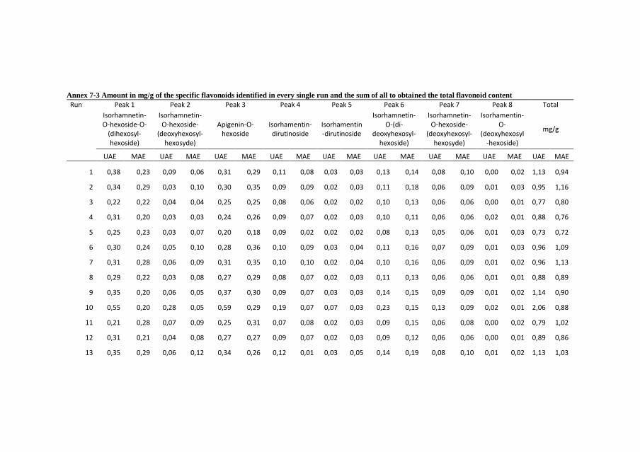
Annex 7-3 Amount in mg/g of the specific flavonoids identified in every single run and the sum of all to obtained the total flavonoid content
Run Peak 1 Peak 2 Peak 3 Peak 4 Peak 5 Peak 6 Peak 7 Peak 8 Total
Isorhamnetin-O-hexoside-O-
(dihexosyl-hexoside)
Isorhamnetin-O-hexoside-
(deoxyhexosyl-hexosyde)
Apigenin-O-hexoside
Isorhamentin-dirutinoside
Isorhamentin-dirutinoside
Isorhamnetin-O-(di-
deoxyhexosyl-hexoside)
Isorhamnetin-O-hexoside-
(deoxyhexosyl-hexosyde)
Isorhamentin-O-
(deoxyhexosyl-hexoside)
mg/g
UAE MAE UAE MAE UAE MAE UAE MAE UAE MAE UAE MAE UAE MAE UAE MAE UAE MAE
1 0,38 0,23 0,09 0,06 0,31 0,29 0,11 0,08 0,03 0,03 0,13 0,14 0,08 0,10 0,00 0,02 1,13 0,94
2 0,34 0,29 0,03 0,10 0,30 0,35 0,09 0,09 0,02 0,03 0,11 0,18 0,06 0,09 0,01 0,03 0,95 1,16
3 0,22 0,22 0,04 0,04 0,25 0,25 0,08 0,06 0,02 0,02 0,10 0,13 0,06 0,06 0,00 0,01 0,77 0,80
4 0,31 0,20 0,03 0,03 0,24 0,26 0,09 0,07 0,02 0,03 0,10 0,11 0,06 0,06 0,02 0,01 0,88 0,76
5 0,25 0,23 0,03 0,07 0,20 0,18 0,09 0,02 0,02 0,02 0,08 0,13 0,05 0,06 0,01 0,03 0,73 0,72
6 0,30 0,24 0,05 0,10 0,28 0,36 0,10 0,09 0,03 0,04 0,11 0,16 0,07 0,09 0,01 0,03 0,96 1,09
7 0,31 0,28 0,06 0,09 0,31 0,35 0,10 0,10 0,02 0,04 0,10 0,16 0,06 0,09 0,01 0,02 0,96 1,13
8 0,29 0,22 0,03 0,08 0,27 0,29 0,08 0,07 0,02 0,03 0,11 0,13 0,06 0,06 0,01 0,01 0,88 0,89
9 0,35 0,20 0,06 0,05 0,37 0,30 0,09 0,07 0,03 0,03 0,14 0,15 0,09 0,09 0,01 0,02 1,14 0,90
10 0,55 0,20 0,28 0,05 0,59 0,29 0,19 0,07 0,07 0,03 0,23 0,15 0,13 0,09 0,02 0,01 2,06 0,88
11 0,21 0,28 0,07 0,09 0,25 0,31 0,07 0,08 0,02 0,03 0,09 0,15 0,06 0,08 0,00 0,02 0,79 1,02
12 0,31 0,21 0,04 0,08 0,27 0,27 0,09 0,07 0,02 0,03 0,09 0,12 0,06 0,06 0,00 0,01 0,89 0,86
13 0,35 0,29 0,06 0,12 0,34 0,26 0,12 0,01 0,03 0,05 0,14 0,19 0,08 0,10 0,01 0,02 1,13 1,03

14 0,30 0,13 0,05 0,02 0,30 0,15 0,09 0,05 0,02 0,01 0,11 0,07 0,07 0,04 0,02 0,00 0,96 0,47
15 0,29 0,24 0,03 0,06 0,24 0,31 0,08 0,09 0,02 0,03 0,10 0,14 0,06 0,07 0,01 0,02 0,83 0,93
16 0,32 0,23 0,05 0,06 0,29 0,30 0,10 0,07 0,02 0,03 0,10 0,14 0,06 0,08 0,01 0,02 0,95 0,91
17 0,23 0,24 0,10 0,06 0,31 0,27 0,08 0,06 0,03 0,03 0,13 0,13 0,06 0,06 0,01 0,02 0,96 0,87
18 0,37 0,22 0,06 0,09 0,37 0,31 0,12 0,07 0,03 0,04 0,15 0,13 0,09 0,07 0,01 0,01 1,21 0,94
19 0,40 0,25 0,05 0,06 0,41 0,33 0,13 0,08 0,04 0,02 0,15 0,15 0,09 0,08 0,01 0,01 1,28 0,99
20 0,34 0,26 0,04 0,11 0,30 0,29 0,13 0,08 0,02 0,03 0,12 0,13 0,07 0,06 0,01 0,02 1,02 0,99
21 0,32 0,21 0,05 0,06 0,35 0,29 0,08 0,07 0,03 0,03 0,13 0,12 0,07 0,07 0,01 0,01 1,03 0,86
22 0,21 0,20 0,03 0,04 0,19 0,25 0,04 0,05 0,03 0,02 0,09 0,12 0,04 0,06 0,01 0,01 0,64 0,76
23 0,27 0,13 0,04 0,07 0,24 0,27 0,08 0,07 0,02 0,03 0,09 0,13 0,05 0,06 0,01 0,01 0,81 0,78
24 0,37 0,26 0,05 0,06 0,35 0,30 0,12 0,07 0,04 0,03 0,13 0,13 0,08 0,06 0,01 0,02 1,15 0,93
25 0,32 0,28 0,05 0,08 0,28 0,33 0,11 0,07 0,02 0,03 0,11 0,16 0,08 0,08 0,02 0,02 0,99 1,04
26 0,38 0,20 0,06 0,04 0,38 0,30 0,11 0,08 0,03 0,03 0,14 0,14 0,09 0,06 0,01 0,02 1,21 0,87
27 0,30 0,20 0,05 0,08 0,31 0,30 0,09 0,07 0,02 0,03 0,11 0,15 0,07 0,08 0,01 0,02 0,96 0,93
28 0,41 0,24 0,05 0,10 0,36 0,23 0,12 0,01 0,03 0,03 0,13 0,14 0,08 0,08 0,01 0,03 1,20 0,86
29 0,31 0,20 0,06 0,07 0,30 0,29 0,10 0,08 0,02 0,03 0,12 0,14 0,07 0,07 0,01 0,02 0,98 0,90
30 0,24 0,27 0,10 0,07 0,29 0,36 0,10 0,09 0,03 0,02 0,12 0,17 0,06 0,08 0,01 0,04 0,95 1,09
31 0,30 0,29 0,07 0,07 0,32 0,34 0,10 0,10 0,04 0,03 0,13 0,16 0,07 0,08 0,01 0,02 1,03 1,08

Annex 7-4 Total betacyanin content 3d response surface plots of UAE and MAE for the parametric responses. For representation purposes, the 2 variable
which are not confronted, were positioned at the centre of their experimental domain.
Time Ratio Concentration Temperature
Tim
eR
ati
oC
on
ce
ntr
ati
on
Te
mp
era
ture
Ultrasound Assisted Extraction
Microwave Assisted Extraction
Betalains response surface graphs
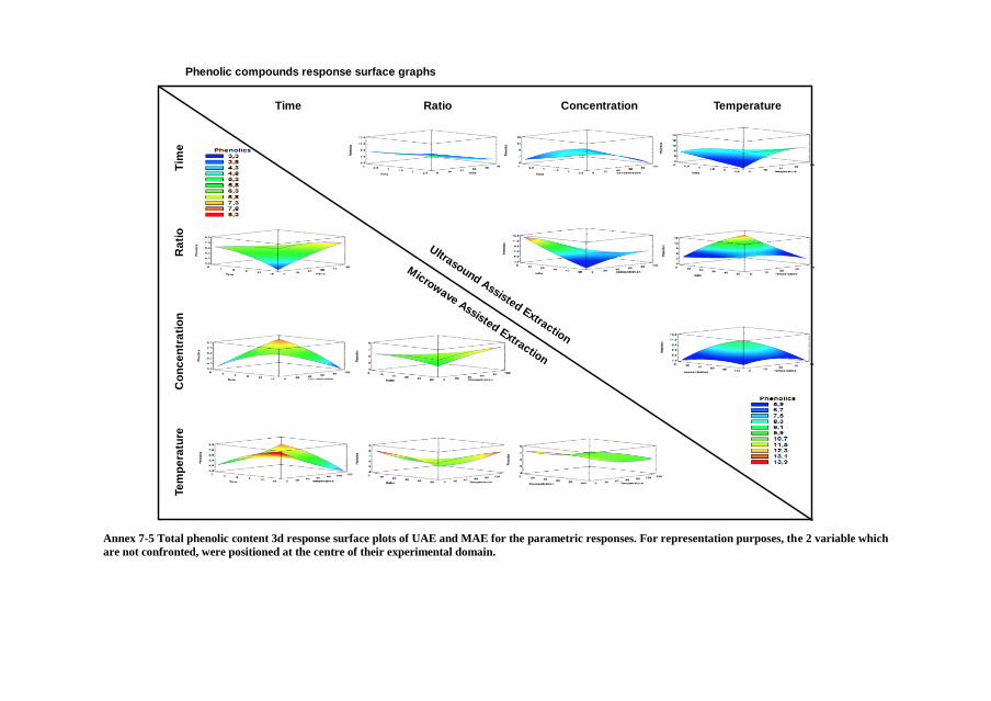
Annex 7-5 Total phenolic content 3d response surface plots of UAE and MAE for the parametric responses. For representation purposes, the 2 variable which
are not confronted, were positioned at the centre of their experimental domain.
Time Ratio Concentration Temperature
Tim
eR
ati
oC
on
ce
ntr
ati
on
Te
mp
era
ture
Microwave Assisted Extraction
Phenolic compounds response surface graphs
Ultrasound Assisted Extraction

Annex 7-6 Total flavonoid content 3d response surface plots of UAE and MAE for the parametric responses. For representation purposes, the 2 variable which
are not confronted, were positioned at the centre of their experimental domain.
Time Ratio Concentration Temperature
Tim
eR
ati
oC
on
ce
ntr
ati
on
Te
mp
era
ture
Microwave Assisted Extraction
Flavonoids response surface graphs
Ultrasound Assisted Extraction

Annex 7-7 Antioxidant activity 3d response surface plots of UAE and MAE for the parametric responses. For representation purposes, the 2 variable which
are not confronted, were positioned at the centre of their experimental domain.
Time Ratio Concentration Temperature
Tim
eR
ati
oC
on
ce
ntr
ati
on
Te
mp
era
ture
Microwave Assisted Extraction
Antioxidant activity response surface graphs
Ultrasound Assisted Extraction
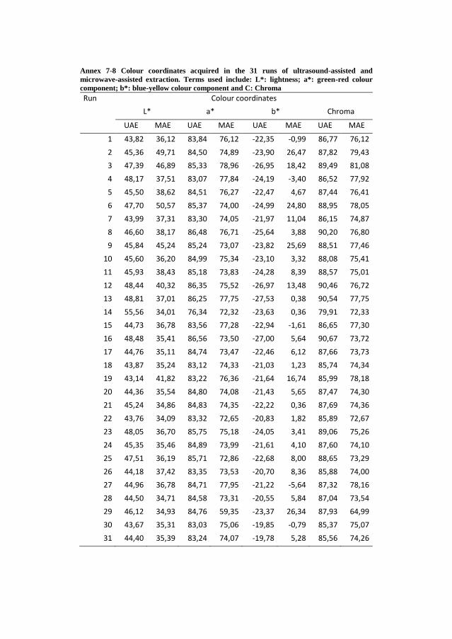
Annex 7-8 Colour coordinates acquired in the 31 runs of ultrasound-assisted and
microwave-assisted extraction. Terms used include: L*: lightness; a*: green-red colour
component; b*: blue-yellow colour component and C: Chroma
Run Colour coordinates
L* a* b* Chroma
UAE MAE UAE MAE UAE MAE UAE MAE
1 43,82 36,12 83,84 76,12 -22,35 -0,99 86,77 76,12
2 45,36 49,71 84,50 74,89 -23,90 26,47 87,82 79,43
3 47,39 46,89 85,33 78,96 -26,95 18,42 89,49 81,08
4 48,17 37,51 83,07 77,84 -24,19 -3,40 86,52 77,92
5 45,50 38,62 84,51 76,27 -22,47 4,67 87,44 76,41
6 47,70 50,57 85,37 74,00 -24,99 24,80 88,95 78,05
7 43,99 37,31 83,30 74,05 -21,97 11,04 86,15 74,87
8 46,60 38,17 86,48 76,71 -25,64 3,88 90,20 76,80
9 45,84 45,24 85,24 73,07 -23,82 25,69 88,51 77,46
10 45,60 36,20 84,99 75,34 -23,10 3,32 88,08 75,41
11 45,93 38,43 85,18 73,83 -24,28 8,39 88,57 75,01
12 48,44 40,32 86,35 75,52 -26,97 13,48 90,46 76,72
13 48,81 37,01 86,25 77,75 -27,53 0,38 90,54 77,75
14 55,56 34,01 76,34 72,32 -23,63 0,36 79,91 72,33
15 44,73 36,78 83,56 77,28 -22,94 -1,61 86,65 77,30
16 48,48 35,41 86,56 73,50 -27,00 5,64 90,67 73,72
17 44,76 35,11 84,74 73,47 -22,46 6,12 87,66 73,73
18 43,87 35,24 83,12 74,33 -21,03 1,23 85,74 74,34
19 43,14 41,82 83,22 76,36 -21,64 16,74 85,99 78,18
20 44,36 35,54 84,80 74,08 -21,43 5,65 87,47 74,30
21 45,24 34,86 84,83 74,35 -22,22 0,36 87,69 74,36
22 43,76 34,09 83,32 72,65 -20,83 1,82 85,89 72,67
23 48,05 36,70 85,75 75,18 -24,05 3,41 89,06 75,26
24 45,35 35,46 84,89 73,99 -21,61 4,10 87,60 74,10
25 47,51 36,19 85,71 72,86 -22,68 8,00 88,65 73,29
26 44,18 37,42 83,35 73,53 -20,70 8,36 85,88 74,00
27 44,96 36,78 84,71 77,95 -21,22 -5,64 87,32 78,16
28 44,50 34,71 84,58 73,31 -20,55 5,84 87,04 73,54
29 46,12 34,93 84,76 59,35 -23,37 26,34 87,93 64,99
30 43,67 35,31 83,03 75,06 -19,85 -0,79 85,37 75,07
31 44,40 35,39 83,24 74,07 -19,78 5,28 85,56 74,26


Capítulo 8 CONCLUSIONES


Conclusiones
340
Las principales conclusiones obtenidas de los resultados en estas tesis
son:
Frutos higos chumbos (Opuntia)
1) La diferencia entre especies y variedades de la misma familia
Opuntia son muy claras en términos morfológicos, en donde los pesos
y las medidas axiales de los frutos varían drásticamente, encontrando
diferencias de hasta 6 veces el peso y 2 veces el diámetro de Opuntia
engelmannii comparado con Opuntia ficus-indica var Sanguigna.
Incluso pudimos identificar diferencias nutricionales significativas
dentro de la misma especie, como es el caso de las concentraciones
proteicas entre OFI Sanguigna y OFI Gialla, en donde la primera
variedad contiene hasta 40% más de contenido proteico y lo cual
atribuimos al aumento en pigmentos betalainicos los cuales contienen
estructuras nitrogenadas, y es debido a estos pigmentos que
observamos la diferencia más clara que les atribuye sus características
distintivas entre cada variedad, las diferentes coloraciones en los frutos.
2) Los perfiles en compuestos lipofílicos e hidrofílicos muestran
también diferencias muy marcadas, sobre todo entre especies, el total
de ácidos orgánicos presentes en Opuntia engelmannii (OE),
comparados con los frutos de OFI var. Sanguigna y var. Gialla, son
hasta 10 veces menores, valores que se repiten en las concentraciones
de azúcares libres, en donde observamos como los valores de fructosa
en OE (0,53 g/100 g PS) son extremamente bajos comparados con OFI
var Gialla y Sanguigna (4,13 y 4,97 g/100 g PS respectivamente).
Debido a que los valores de carbohidratos totales son inversos y OE
llega a duplicar a los valores de las variedades de OFI, propusimos que
mientras los frutos de OFI son favorecidos en cuestiones organolépticas
debido al alto contenido de azúcares libres, su contraparte OE, contiene
arreglos de fibras y carbohidratos complejos como el mucilago, el cual
tiene propiedades potenciales para su uso como aditivo en las industrias
de alimentación, farmacia y cosmética.
3) Se identificaron 7 betalaínas, de las cuales 3 pertenecen al grupo
de las betaxantinas y 4 al de las betacianinas. Las moléculas
mayoritarias de cada grupo son también las más abundantes en la
naturaleza según la bibliografía científica, por parte de las betacianinas
encontramos la betanina e indicaxantina como la más abundante
betaxantina. Su distribución dentro de las especies estudiadas es muy

Conclusones
341
heterogénea, aunque los perfiles betalainicos de cada variedad nos
describe muy bien sus características visuales, en OE se encuentran
solo betacianinas con lo que obtenemos una tonalidad violeta muy
intensa. OFI Sanguigna presenta una mayoría y alto contenido en
betacianinas, aunque también contiene betaxantinas, lo que ayuda a
modificar la tonalidad del fruto a un rojo más vibrante. Por ultimo OFI
Gialla predomina en betaxantinas aunque también tiene un bajo
contenido en betanin, lo cual proporciona las motas rojas que se
combinan para dar tonalidades más naranjas.
4) Los frutos de todas las variedades estudiadas, presentaron
potencial antimicrobiológico. Todos los frutos mostraron poder
antifúngico contra las 9 cepas probadas, mientras que el poder
bactericida testado en 8 cepas fue efectivo en los 3 frutos, con
excepción en 3 cepas, donde OFI Gialla no mostró efecto alguno
(Micrococcus aureus, Escherichia coli y Enterobacter cloacae). El
poder inhibidor, bactericida y fungicida de los frutos fue comparado
contra antibióticos comerciales y en muchos de los casos, las
concentraciones necesarias de inhibición o aniquilación de
microorganismos fueron menores en los frutos y por tanto mejores.
Subproductos Aguacate (Persea americana)
5) De acuerdo a los estudios realizados preliminarmente en los frutos
del aguacate, se ha determinado que la capacidad antioxidante de los
subproductos, son mucho mejores que la de los fruto, lo que resalta la
importancia del aprovechamiento de estos residuos, cuando
comparamos ambos subproductos, y como era de esperar, observamos
una mejor capacidad en las pieles comparadas con las semillas tanto en
las metodologías experimentales hidrofílicas como en las lipofílicas.
Subproductos Aguacate (Persea americana)
6) Los subproductos de Persea americana var Hass, presentaron en
conjunto 29 compuestos fenólicos, 14 flavan-3-oles (derivados de
epicatequinas), 9 flavonoides (derivados de Quercetina, Kaempferol e
isorhamnetina) y 6 ácidos fenólicos (derivados de ácidos cloragénico y
cumárico). De estos 23 compuestos, se encontraron 18 en las pieles y 8
en las semillas, de los cuales solo 2 compuestos coincidieron en ambos
Conclusiones
342
subproductos (Epicatequina y un trímero de epicatequina de tipo B).
Las semillas son ricas en ácidos fenólicos, contienen solo una molécula
flavan-3-oles y carecen de flavonoides, mientras que las pieles
contienen mayoritariamente flavan-3-oles, flavonoides y solo 2 ácidos
fenólicos derivados del ácido clorogénico. Debido a la mayor cantidad
de compuestos fenólicos, las pieles contienen hasta 3 veces más de
estos compuestos comparados con las semillas (227,9 y 72,5 mg/g
extracto respectivamente).
7) Es importante remarcar que aunque en el contenido total de
compuestos fenólicos en las pieles triplica la cantidad total obtenida en
las semillas, la actividad antioxidante es ligeramente menor, las pieles
son 1,5 veces mejores en este sentido que las semillas, lo que atribuimos
a la complejidad de las estructuras de los flavan-3-oles, ya que de
acuerdo a otras investigaciones en la bibliografía, la especificidad de
los antioxidantes disminuye conforme aumentan los números de
moléculas agregadas, en este caso, en los dímeros, trímeros, tetrámeros,
y así sucesivamente.
8) Debido a los compuestos fenólicos son metabolitos secundarios
generados en el metabolismo vegetal como respuesta a diferentes tipos
de estrés, entre ellos el combatir infecciones, en este trabajo hemos
observado en las 8 cepas testadas alta eficiencia de los extractos de los
subproductos para inhibir y destruir tanto bacterias Gram positivas
como negativas, destacando ambas capacidades en bacterias Gram
negativas como Enterobacter cloacae, Pseudomonas aeruginosa y
Salmonella typhimurium, incluso mejor que los antibióticos usados
como control positivo. Por otro lado, su capacidad antifúngica testada
de los subproductos contra 8 cepas patógenas comunes, mostró una
eficiencia media-buena como inhibidor, y solo las semillas exhibieron
una capacidad excelente antifúngica ante 2 cepas (Aspergillus
ochraceus y Trichoderma viride), superando por mucho a los
antimicóticos comerciales.
Subproductos higos chumbos (Opuntia spp.)
9) El perfil fenólico de los extractos de los subproductos de Opuntia
obtenido, corresponde a 12 compuestos fenólicos, de los cuales 10 de
ellos son flavonoides 8 derivados de Isorhamnetina, 1 derivado de
Quercetina y 1 derivado de Kaempferol, además de 2 ácidos fenólicos
(ácido piscidico y eucomino). El extracto de OE es el que se encuentra

Conclusones
343
en mayor concentración, casi el doble comparado con los extractos de
OFI Gialla y Sanguigna. Aunque en OE solo se detectaron 5 de los 12
compuestos, solo 2 de ellos se pudieron cuantificar mientras que los
otros tres, solo se pudieron detectar trazas, pero aun así, el contenido en
Isorhamnetina-O-(deoxihexosil-hexosido) (5,99 mg/g extracto),
dispara el contenido total de fenoles totales.
10) Se detectaron 7 betalaínas en total en los tres extractos, 2 isómeros
de indicaxantina pertenecientes a las betaxantinas y 5 betacianinas. Al
igual que en los frutos, betanin e indicaxantina, fueron los compuestos
mayoritarios de cada grupo y estos se encontraron distribuidos de
distintas manera en los tres subproductos. Las betaxantinas fueron solo
detectadas en las variedades de OFI, mientras que OE predomina en el
contenido de betacianinas hasta 5 y 15 veces más que los extractos de
OFI Sanguigna y OFI Gialla respectivamente.
11) La capacidad antioxidante de los extractos de los subproductos
analizados, se corresponde inversamente al contenido de compuestos y
betalaínas. De esta forma que apreciamos que OE es 2,3 y 2 más veces
eficaz que OG y OS respectivamente, en los análisis de capacidad
antioxidante de compuestos hidrofílicos. En el caso de la capacidad
antioxidante de los compuestos lipofílicos, solo el análisis de β-
caroteno se pudo realizar despreciando los valores obtenidos en OS, lo
que atribuimos a que los valores fueron erróneos debido a una posible
interferencia del color en este análisis espectrofotométrico especifico,
lo que comprobamos al obtener datos totalmente erróneos en el análisis
TBARS, el cual fue descartado de la publicación.
12) La capacidad antioxidante, al igual que el poder antimicrobiano, se
atribuye en este caso a un posible efecto sinérgico entre los compuestos
fenólicos y las betalaínas. Estrictamente hablando del poder
antibacteriano, observamos una eficiencia media alta, ya que dentro de
las 8 cepas patógenas analizadas, en 2 de ellas los 3 extractos no
mostraron efecto alguno, aunque por otro lado, se comprobó que en
otras tres cepas (Bacillus cereus, Enterobacter cloacae y Salmonella
typhimurium), estos poseen una mejor eficacia cuando se les compara
con el control comercial utilizado. En cualquiera de los casos, ningún
extracto demostró ser mejor que los otros en términos generales, debido

Conclusiones
344
a que cada uno de ellos actúa de forma distinta con respecto a las cepa
bacteriana analizada.
A diferencia del poder bactericida, el poder fungicida muestra una alta
eficacia en las tres variedades. De las 8 cepas estudias, en 2 de ellas
(Penicillium ochrochloron y Trichoderma viride para OG y OE y
Penicillium ochrochloron y Aspergillus ochraceus en OS) los tres
extractos de los subproductos de Opuntia, probaron ser mejores que el
fungicida comercial utilizado como control positivo. En este apartado
si se observa un mejor efecto protector frente a patógenos fúngicos en
OS seguido por OE y por último en OG.
Los efectos de los extractos se evaluaron in-vitro en cultivos celulares
PLP2 para poder garantizar la seguridad de cada uno de los extractos y
ninguno de estos mostro toxicidad a estas células testadas. Además, se
comprobó en las 4 líneas celulares tumorales su efecto antitumoral, y a
pesar de demostrar esta capacidad benéfica, las concentraciones
necesarias para observar dicho efecto fueron mayores que las utilizadas
en el control positivo de elipticina. Por lo que podemos corroborar la
seguridad de los extractos además de su capacidad antitumoral.
Optimización de extraciones
13) El perfil fenólico de los extractos de las pieles de Opuntia
engelmannii obtenidos mediante extracciones asistidas por microondas
y ultrasonidos, corresponde a 12 compuestos fenólicos, 8 de ellos con
estructuras flavonoides derivados casi en totalidad de isorhamnetina,
exceptuando un solo derivado de Apigenina, compuesto que ha sido
identificado por primera vez en las pieles de frutos de Opuntia.
Además, se detectaron 4 ácidos fenólicos (ácido piscidico, ácido
cafeico y 2 derivados del ácido ferúlico). Los ácidos fenólicos fueron
los que se encontraron en mayor concentración y esto se debe sobre
todo a que el ácido piscidico fue el compuesto mayoritario en todas las
extracciones con concentraciones muy por encima del resto de los
compuestos fenólicos.
14) En los subproductos de Opuntia engelmannii, se identificaron 7
betalaínas correspondientes todas al grupo de las betacianinas, 6 de

Conclusones
345
ellas previamente documentadas en nuestros y otros trabajos, y una de
ellas, (iso)-phyllocactina, a pesar de haber sido descrita antes en la
familia Cactaceae, es la primera vez que se describe en esta variedad.
Las betacianinas mayoritarias fueron betanina, seguidas de su isómero,
isobetanina, gomphrenina, betanidina, isobetanidina,
(iso)phyllocactina y por ultimo 17 decarboxi-betanina.
15) Mediante el uso del diseño experimental de respuesta de superficie
acompañado del diseño central compuesto, pudimos determinar dentro
de los rangos estudiados, las variables significativas de acuerdo a la
tecnología de extracción empleada y a la respuesta analizada. Por un
lado, siendo el ratio solido-líquido y la concentración de solvente las
variables que más influencia tienen sobre las respuestas usando tanto la
extracción asistida por microondas como por ultrasonidos. Mientras
que por otro lado, las variables temperatura y tiempo de extracción solo
afectaron a las extracciones realizadas en el microondas,
concretamente, la temperatura afecto significativamente a las
extracciones de betalaínas, y la variables temperatura y tiempo
afectaron también significativamente al parámetro de color.
16) También, a través del uso de estos diseños experimentales hemos
sido capaces de obtener los coeficientes polinomiales de segundo
orden, a partir de los cuales podemos hacer tanto la representación
gráfica de las extracciones, como poder inferir los valores óptimos de
cada una de las respuesta analizadas. Adicionalmente, determinamos la
optimización general del cumulo de repuestas, mediante el cual
obtuvimos un mayor factor de deseabilidad (0,985) y mejores
concentraciones en las respuestas de las extracciones asistidas mediante
microondas y por lo cual nos declinamos a su elección como
metodología de extracción recomendada, ya que utilizando los valores
de 2,5 minutos, 5 g/L, 34,6 % de metanol en agua y una temperatura de
30 °C, obtuvimos los mejores valores de cada respuesta, en este caso:
201,6 mg de betalaínas/ g de piel seca, 13,9 mg de ácidos fenólicos/g,
2,4 mg de flavonoides/g, 85,7 % de rendimiento de extracción y un
valor de 2,9 mg de extracto/ mL para poder obtener el valor IC50


Capítulo 9 ANEXOS


Anexos
349
Anexo 1
Poster 4ta edición del “ISEKI Food conference”
(Julio 2016) Viena, Austria. ISBN 978-3-900932-34-3
Effect of drying temperatures and stored conditions
on bioactive compound of prickly pear (Opuntia
spp.) peels.
Bruno Melgar1*, Esperanza M. Garcia-Castello1, Luis Mayor?
and Antonio Rodriguez-Lopez3.
1. Institute of Food Engineering for Development (IIAD), Universitat
Politècnica de València,
Spain.
2. Department2, College or Institute2, University2, Country2
3. Dpt. Of Chemical and Nuclear Engineering, Universitat Politècnica
de València, Spain.
* Corresponding author: [email protected];
Prickly pear has become an important fruit crop in many semi-arid
lands of the world. The cactus pear is often marketed as a raw peeled
fruit, juice, marmalades and other different products. These
transformations produce considerable amounts of by-products (peels
and seeds).
Peels represent a large proportion of the whole fruit and
constitute an important source of bioactive compounds, especially
polyphenols, flavonoids and betalains which are the characteristic
colorants of this fruit. Polyphenols are present in a higher concentration
on the peels compared to the edible portion of the fruit, which makes it
a great source of extraction for this type of bioactive compound.
These phenolic compounds have been recognized as a potential
nutraceutical target for the food and pharmaceutical industries. Due to

Anexos
350
their wide range of bioactivities, there is a growing interest in
developing of methods for their recovery and for the optimal storage of
the by-products.
Storing these by-products to prevent the microbial spoilage and
to preserve them in optimal conditions until their use could be
expensive and might be considered irrational. Therefore, the purpose is
to look for alternatives to reduce the cost of storage without sacrificing
the bioactive compounds.
The aim of the current investigation was to evaluate changes on
the phenolic compounds of the dried peels of prickly pear at different
temperatures (40, 60 and 70 °C) and to follow their behavior once they
are dried and stored at room temperature (15, 60 and 90 days) by
HPLC-DAD and spectrophotometric methods.
For the different drying temperatures we detected a decrease of
up to 58% (70 °C) of the phenolic compounds, which also has negative
influence on the antioxidant capacity. On the other hand, stored dry peel
did not show any important changes through time.
Keywords: Food by-products, Bioactive compounds, Prickly pear,
Drying.

Anexos
351

Anexos
352
Anexo 2
Presentación oral en el “6to Simposio de becarios CONACyT en
Europa” disponible en https://goo.gl/Xbz3aW
Extraction of natural colorants obtained from
prickly pear fruit by-products.
Bruno Melgar Castañedaa,c, Esperanza M. Garcia-Castelloa, Antonio
D. Rodriguez-Lopezb.
aInstituto Universitario de Ingeniería de Alimentos para el Desarrollo,
Universitat Politècnica de València, Valencia, Spain bInstitute for Industrial, Radiophysical and Environmental Safety
(ISIRYM), Universitat Politècnica de València, Valencia, Spain cSpeaker ([email protected])
Color perception has been crucial on the selection and ingestion of
food products. Since the beginning of the mankind, color parameters
were related primarily to food quality and safety.
Food industry has employed several synthetic colorants obtained
from mineral or chemical resources, the advantage of those colorants
are their high stability in the food matrixes and the low concentrations
needed to give the desired color.
In the other hand, due to the constant research and improvement on
the food safety field, some drawback has emerged, arising the
awareness of the consumers, who constantly demands to the food
industries for minimal processed food and synthetic additives free,
opening new research lines focused on the replacement of synthetic for
natural obtained colorants.
In addition, food chain productions generate humongous quantities
of food by-products, which according to their origin and composition
might been used in different applications, from simple fodder until
more refined process for recovery of different biomolecules with
Anexos
353
functional properties, called thickeners, acidity regulators, foaming or
antifoaming agents, antioxidants, colorants, among others.
The research ongoing, works in the utilization of food by-products
from Opuntia spp. fruits for the obtention of natural colorants with
antioxidant properties, which could be used as an alternative to
synthetic colorants helping at the same time on scavenging of free
radical that could prevent many diseases caused by oxidative stress.

Anexos
354
Anexo 3
En el capitulo VI en la sección 1.2 se menciona la elaboración de curvas
de adsorción y desorción de Opuntia ficus-indica var. Sanguignea así
como el uso de 7 modelos distintos, los cuales por simplificación de
resultados, se expone en esa sección solo el modelo que major se adapto
a los datos experimentales, pero acontinuación se presentan la
comparativa del resto de los modelos y las tablas con las regresiones
correspondientes.
Figure 8-1 Desorption isotherms of OFI Sanguigna at 20 °C (source: private collection)
0
0.1
0.2
0.3
0.4
0.5
0.6
0.7
0 0.2 0.4 0.6 0.8 1
Xb
s (K
g w
/kg
ss)
Actividad de agua
Piel Desorción 20ºC
Experimental
BET
GAB
HALSEY
HENDERSON
OSWIN
PELEG
SMITM

Anexos
355
Figure 8-2 Adsorption isotherms of OFI Sanguigna at 20 °C (source: private collection)
0
0.1
0.2
0.3
0.4
0.5
0.6
0.7
0.8
0.9
0 0.2 0.4 0.6 0.8 1
Xb
s (K
g w
/kg
ss)
Actividad de agua
Piel Adsorción 20ºC
Experimental
BET
GAB
HALSEY
HENDERSON
OSWIN
PELEG
SMITH

Anexos
356
Figure 8-3 Adsorption isotherms regressions of OFI Sanguigna at 20 °C (source: private collection)

Anexos
357
Figure 8-4 Dessorption isotherms regressions of OFI Sanguigna at 20 °C (source: private collection)


