diversidad microbiana de los sedimentos anaerobios …€¦ · buenas charlas. a emiliano por darme...
TRANSCRIPT

DIVERSIDAD MICROBIANA
DE LOS SEDIMENTOS ANAEROBIOS DE RÍO TINTO
Irene Sánchez-Andrea

UNIVERSIDAD AUTÓNOMA DE MADRID
FACULTAD DE BIOLÓGICAS
DEPARTAMENTO DE BIOLOGÍA MOLECULAR
TESIS DOCTORAL
DIVERSIDAD MICROBIANA DE LOS SEDIMENTOS ANAEROBIOS DE RÍO TINTO
Irene Sánchez-Andrea
Madrid, 2012
Dr. José Luis Sanz y Dr. Ricardo Amils, catedráticos del Departamento de Biología Molecular de la Universidad Autónoma de Madrid,
CERTIFICAN:
Que los trabajos de investigación desarrollados en la memoria de tesis doctoral: “DIVERSIDAD MICROBIANA DE LOS SEDIMENTOS ANAEROBIOS DE RÍO TINTO” son aptos para ser presentados por Irene Sánchez-Andrea ante el Tribunal que en su día consigne, para aspirar al Grado de Doctor en Microbiología por la Universidad Autónoma de Madrid.
VºBº Director Tesis
Dr. José Luis Sanz
VºBº Director Tesis
Dr. Ricardo Amils
5
AGRADECIMIENTOS
Son tan largos cuatro años..., tanta gente a la que estar agradecida…
En primer lugar, a mi jefesito, el doctor José Luis Sanz Martín por aconsejarme pero no
imponer, por hacerme reír y pensar, y ser a parte de un excelente director, un amigo.
Igualmente quisiera agradecer al doctor Ricardo Amils, por confiar en mí desde el
principio, abrirme las puertas de su laboratorio y por su constante ayuda.
Son muchos los que han pasado por el laboratorio estos cuatro años, y a todos les
debo una parte de esta tesis por los momentos compartidos. Gracias a todos por
amenizar tantas horas perdidas frente a geles, microscopios y termocicladores. Nicolas
Raho, gracias por estar ahí desde el principio hasta el final, ser un amigo y compañero y
haber estado en lo bueno y en lo malo. A Carlotta Vizzioli por sacar siempre una sonrisa
con tu genial sentido del humor. A Monica Conthe por haber inundado el laboratorio
con tu frescura, por tu ayuda, tu “perfect english”, tu amistad y apoyo. Y Carla que nos
descubre el mundo de las manualidades con su originalidad. A Ana por pringarse con
todo y con todos. A Bia Missagia porque compartimos una de las mejores épocas jamás
vividas en el labo, por la PCR y el meneaito. A Patri y a Santi, por los desayunos y las
buenas charlas. A Emiliano por darme animos cuando empecé con esto de la ciencia y
confiar siempre en mí. A mis estudiantes, directos e indirectos, porque con todos ellos
he tenido una excelente relación y os he echado de menos tras vuestra partida (David,
Maria, Carlota la maja!) y a todos los que habéis pasado por el labo y volado a tiempo.
A toda la gente del pasillo, profesores y estudiantes por estar ahí (Concha, Irma,
Pascual, Mirna, etc). A Nuria Rodríguez por ser la guía andante en el Tinto. Miguel y
Nacho por esos paseos en coche tan graciosos que me hacían llegar al labo con una
sonrisa de oreja a oreja. A Raquel Simarro por esos momentos DGGE tan memorables,
¡ojala hubieras estado en mi labo trabajando!. A Jose del CBM por su incansable ayuda
maquetando geles y banditas y geles y banditas y vuelta a empezar. Y a gente que aún
estando lejos han ayudado mucho, Patxi, por ser mi maestro mis primeros meses de
tesis.
Al doctor Rudolf Amann y la doctora Katrin Knittel, del Max-Planck-Institute fuer Marine
Mikrobiologie, por acogerme, darme la oportunidad de formar parte de ese gran grupo
en ese gran instituto y brindarme sus inestimables opiniones y consejos. Y a todos los
amigos de Alemania, a Mar y Ana que latinizaron mi estancia y a Sara, Chia-I, Sven,
Ingrid, Christian porque me hicisteis sentir como en casa.
Al doctor Alfons J. M. Stams por la amable acogida, permitirme trabajar en
Laboratorium voor Microbiologie de la Wageningen Universiteit, en Holanda, por
dejarme su casa y ayudarme siempre que lo necesitaba con sus perfectos y audaces
consejos. A toda la gente de su laboratorio por ser mi segunda familia Teun, Rozelyn,
Derya, Ana, Michael, Peer, Jose, Juanan, Thomas y un largo etc.. y por su puesto a mis

6
niñas, a Albita por ser mi hermanita holandesa, a Marta y Miriam por ser un gran apoyo
durante esos 6 meses.
Y lo profesional está bien, pero lo que me ha hecho tener animos para seguir con esta
ardua tesis han sido mis amigos! Amigos de la universidad, colegio y barrio, gracias por
estar ahí. Esos ambientologos que me han dado tantos ánimos, gracias a todos!! y
permitirme un especial énfasis para Olguita, Sonsi, Pepiño, Lu, Diego, Edu, Alfredo e
Irene (integrados como ningunos), a la nueva Enara, ¡¡bienvenida a este mundo!! y a
Dieguin, sé que no es un Nobel, pero las cosas de palacio van despacio ;). Mis amigos
del barrio, mis pequeños demonios (Apu, Sumo, Nando, Noce..), los pilukeros (Rober,
Jaime, Nuri, Uta, mis compis de piso, Angel, etc..) y por supuesto mis niñas del barrio:
Eva, Lurdes, Marina, Maria, MariaIsabel y Cris, os quiero con locura, gracias por ser
como sois y por seguir a mi lado desde hace más de 10 años.
A mi familia, a mis padres por creer siempre en mí, apoyarme y darme un entorno
familiar alegre y lleno de amor. A mi hermana por ser mi amiga y compañera. A Joaquín
por hacerse indispensable, y por supuesto, por ser el padre de la criaturilla. Adriana, mi
pequeña, gracias por haber venido a este mundo e inundarlo con tus sonrisitas y
carcajadas. Algún día tu tita te hará una friki como es debido. Agradezco hasta a mi
gata, la Pulgui, que me ha hecho una gran compañía todas esas horas ante el
ordenador
Y finalmente, a Rafa, porque llegaste a mi vida para quedarte…
A todos, gracias.

7
ÍNDICE
RESUMEN 9
INTRODUCCIÓN 15
OBJETIVOS 37
METODOLOGÍA GENERAL 41
LISTADO DE PUBLICACIONES 55
CHAPTER 1: MICROBIAL DIVERSITY OF ANAEROBIC SEDIMENTS OF RÍO TINTO: A NATURAL ACID AND HIGH HEAVY METALS CONTENT ENVIRONMENT 59
CHAPTER 2: QUANTIFICATION OF TINTO RIVER SEDIMENT MICROBIAL COMMUNITIES: THE IMPORTANCE OF SULFATE-REDUCING BACTERIA AND THEIR ROLE IN ATTENUATING ACID MINE DRAINAGE 77
CHAPTER 3: SCREENING OF ANAEROBIC ACTIVITIES IN SEDIMENTS OF AN ACIDIC ENVIRONMENT: TINTO RIVER 93
CHAPTER 4: MICROBIAL DIVERSITY OF ANAEROBIC ZONES OF TINTO RIVER: A CULTURE-DEPENDENT AND NON-DEPENDENT APPROACH 111
CHAPTER 5: ENRICHMENT AND ISOLATION OF ACIDOPHILIC SULFATE-REDUCING BACTERIA FROM TINTO RIVER SEDIMENTS 135
CHAPTER 6: BIOREMEDIATION OF ACID MINE DRAINAGE COUPLED WITH DOMESTIC WASTEWATER TREATMENT 149
DISCUSIÓN GENERAL 163
CONCLUSIONES GENERALES 185

8

RESUMEN


RESUMEN
11
El río Tinto (Huelva, España) es un ambiente natural, semejante a los denominados drenajes
ácidos de mina, producido por la bio-oxidación de sulfuros metálicos procedentes de la Faja
Pirítica Ibérica. Hasta ahora, y a pesar de su interés ecológico, sus sedimentos habían sido
escasamente estudiados. En esta tesis, presentamos un amplio estudio de la microbiota
anaerobia de los sedimentos del río Tinto combinando técnicas dependientes e
independientes de cultivo siguiendo el llamado ciclo completo del rRNA.
Usando técnicas de biología molecular, ha sido desarrollado un modelo geomicrobiológico de
los distintos ciclos microbianos que operan en el sistema. Para ello, se aplicaron técnicas
independientes de cultivo basados en la subunidad pequeña del RNA ribosomal (SSU RNA),
como electroforesis en gel de gradiente desnaturalizante (DGGE), secuenciación del gen de
16S rRNA (clonaje) y la variación de la hibridación in situ con sondas fluorescente con
deposición catalizada (CARD-FISH). Microorganismos relacionados con el ciclo del hierro (At.
ferrooxidans, Sulfobacillus spp., Ferroplasma spp., etc.), azufre (Desulfurella spp.,
Desulfosporosinus spp., Thermodesulfobium spp., etc.), carbono (Acidiphilium spp., Bacillus
spp., Clostridium spp., Acidobacterium spp., etc.) y nitrógeno (Alcaligenes spp.,
Pseudochrobactrum spp., etc.) fueron identificados y su distribución correlacionada con los
parámetros fisicoquímicos de los sedimentos. En aquellos puntos donde el pH y el potencial
redox son mas cercanos a la columna de agua (pH 2,5 y +400 mV), los organismos
predominantes fueron identificados como bacterias reductoras de hierro: Acidithiobacillus spp.
y Acidiphilium spp., probablemente relacionado con la alta solubilidad del hierro a bajo pH. En
cambio, a mayor pH (4,2-6,2) y condiciones redox mas reductoras (50, -210 mV) dominaban
bacterias sulfatoreductoras de los generos Syntrophobacter, Desulfosporosinus y Desulfurella.
El diseño de una sonda específica para los reductores de azufre pertenecientes al género
Desulfurella fue primordial para el trabajo.
Adicionalmente, se han usado cultivos de enriquecimiento específicos para validar este modelo
y probar la existencia de las potenciales actividades anaerobias que habían sido inferidas en los
sedimentos ácidos del río Tinto. Se obtuvieron resultados positivos para cultivos de
metanógenas, sulfatorreductoras, desnitrficantes, reductoras de hierro y productoras de
hidrógeno. Algunos microorganismos han podido ser aislados como sulfatorreductoras
acidófilas (dos posibles nuevas especies y un posible nuevo género relacionados con
Desulfosporosinus/Desulfitobacterium), fermentadoras (un posible nuevo género, próximo a
Paludibacter) y productoras de hidrógeno.
Finalmente, se ha ensayado la capacidad de biorremediar aguas ácidas de mina, usando los
sedimentos del río Tinto como fuente de bacterias sulfatorreductoras, y aguas residuales
urbanas como fuente de carbón económica. Se obtuvo un efluente libre de metales pesados.
ABSTRACT
Tinto river (Huelva, Spain) is a natural acidic rock drainage (ARD) environment
produced by the bio-oxidation of metallic sulfides from the Iberian Pyritic Belt. So far, and
despite their ecological interest, the underlying sediments were studied only very sparsely and
no complete studies were undertaken. In this thesis, an extensive survey of the Tinto River
anaerobic sediment microbiota is presented combining culture independent and dependent
methods following the “full-cycle rRNA approach”.
RESUMEN
12
A geomicrobiological model of the different microbial cycles operating in the sediments has
been developed through molecular biological methods. Culture independent methods
targeting the small subunit ribosomal RNA (SSU rRNA) such as denaturing gradient gel
electrophoresis (DGGE), 16S rRNA gene sequencing (cloning) and catalyzed reporter
deposition fluorescence in situ hybridization (CARD-FISH) were used. Microorganisms involved
in the iron (Acidithiobacillus ferrooxidans, Sulfobacillus spp., etc.), sulfur (Desulfurella spp.,
Desulfosporosinus spp., Thermodesulfobium spp., etc.), carbon (Acidiphilium spp., Bacillus spp.,
Clostridium spp., Acidobacterium spp., etc.) and nitrogen (Alcaligenes spp., Pseudochrobactrum
spp., etc.) cycles were identified and their distribution correlated with physicochemical
parameters of the sediments. Where the pH and redox potential are closer to those of the
water column (pH 2.5 and +400 mV), the most abundant organisms were identified as iron-
reducing bacteria: Acidithiobacillus spp. and Acidiphilium spp., probably related to the higher
iron solubility at low pH. At higher pH (4.2-6.2) and more reducing redox potential (50, -210
mV) and therefore, lower solubility of iron, members of the sulfate-reducing genera
Syntrophobacter, Desulfosporosinus and Desulfurella were dominant. The design of a specific
probe (DSU655) targeting sulfur-reducing organisms belonging to Desulfurella genus was
conclusive to the work.
Additionally, we used targeted enrichment incubations to validate this model and prove the
existence of the potential anaerobic activities detected in the acidic sediments of Tinto River.
Methanogenic, sulfate-reducing, denitrifying, iron-reducing and hydrogen-producing
enrichments yield positive results. Classical techniques for bacterial isolation were applied and
some microorganisms were isolated such as acidophilic sulfate-reducing bacteria (two new
species and a new genus, related to the Desulfosporosinus/Desulfitobacterium cluster),
fermenters (a new genus, closed related to Paludibacter) and hydrogen-producers.
Finally, a biotechnological application using Tinto river sediments was tested. Bioremediation
of acid rock drainage (ARD) with sulfate-reducing bacteria using domestic wastewater (DW) as
a cost-effective carbon-source showed a complete cleaning of the ARD water obtaining an
effluent with neutral pH and no metal content.
SUMMARY OF CHAPTERS
In Chapter 1 an extensive survey of the Tinto River sediment microbiota using two culture
independent approaches: denaturing gel gradient electrophoresis (DGGE) and cloning of 16S
rRNA genes in two physic-chemically contrasting sampling sites (SN and JL dams) is presented.
The taxonomic affiliation of the Bacteria showed a high degree of biodiversity, falling into five
different phyla: Proteobacteria, Firmicutes, Bacteroidetes, Acidobacteria and Actinobacteria,
while all the Archaea were affiliated to the Thermoplasmatales group. Microorganisms involved
in the iron (At. ferrooxidans, Sulfobacillus spp., Ferroplasma spp., etc.), sulfur (Desulfurella spp.,
Desulfosporosinus spp., Thermodesulfobium spp., etc.) and carbon (Acidiphilium spp., Bacillus
spp., Clostridium spp., Acidobacterium spp., etc.) cycles were identified and their distribution
correlated with physicochemical parameters of the sediments. Ferric iron was the main
electron acceptor for the oxidation of organic matter in the most acid and oxidizing layers, so
acidophilic facultative Fe(III)-reducing bacteria appeared extensively in the clones libraries.
With increasing pH, the solubility of iron decreases and sulfate-reducing bacteria become
dominant, the ecological role of methanogens being scarce. Considering the identified
microorganisms – which according to the rarefaction curves and Good´s coverage values cover

RESUMEN
13
almost all of the diversity - and their corresponding metabolism, a model of the iron, sulfur and
carbon cycles in an ARD-related sediments is proposed.
In Chapter 2, the abundance of diverse microbial populations inhabiting both physic-chemically
contrasting sampling sites (SN and JL dams) was quantified. Depth profiles of total cell
numbers differed greatly between the two sites, yet were consistent in decreasing sharply at
greater depths. Although catalyzed reporter deposition fluorescence in situ hybridization
(CARD-FISH) with domain-specific probes showed that Bacteria (>98%) dominated over
Archaea (<2%) at both sites, important differences were detected at the class and genus level
reflecting differences in pH, redox potential and heavy metal concentrations. At SN, where the
pH and redox potential are similar to those of the water column (pH 2.5 and +400 mV), the
most abundant organisms were identified as iron-reducing bacteria: Acidithiobacillus spp. and
Acidiphilium spp., probably related to the higher iron solubility at low pH. At the JL dam,
characterized by banded sediment with higher pHs (4.2-6.2), more reducing redox potential
(50, -210 mV) and a lower solubility of iron, members of sulfate-reducing genera
Syntrophobacter, Desulfosporosinus and Desulfurella were dominant. The latter was quantified
with a newly designed CARD-FISH probe (DSU655). In layers where sulfate-reducing bacteria
were abundant, pH was higher and redox potential, dissolved metals and iron were lower.
These results suggest that the attenuation of ARD characteristics is biologically driven by
sulfate-reducers and the consequent precipitation of metals and iron as sulfides.
In Chapter 3, a combination of molecular biological methods and targeted enrichment
incubations was used to validate the formerly proposed model and prove the existence of the
inferred potential anaerobic activities in the acidic sediments of Tinto River. Methanogenic,
sulfate-reducing, denitrifying and hydrogen-producing enrichments were all positive at pH
between 5 and 7. Methanogenic enrichments revealed the presence of methanogenic archaea
belonging to the genera Methanosarcina and Methanobrevibacter. Enrichments for sulfate-
reducing microorganisms were dominated by Desulfotomaculum spp. Denitrifying enrichments
showed a broad diversity of bacteria belonging to the genera Paenibacillus, Bacillus,
Sedimentibacter, Lysinobacillus, Delftia, Alcaligenes, Clostridium and Desulfitobacterium.
Hydrogen-producing enrichments were dominated by Clostridium spp. These enrichments
confirm the presence of anaerobic activities in the acidic sediments of the Tinto river that are
normally assumed to take place exclusively at neutral pH.
In Chapter 4, we complete the overview of the microbial diversity of the sediments -increasing
the cloning effort and the studied sites- and combine it with culture-dependent methods -
enrichments for iron-reduction, methanogenesis, denitrification and sulfate-reduction- to pool
all the latter studies in a comprehensive common discussion at a global scale. By cloning, it was
possible to phylogenetically identify down to the genus level most of the bacterial sequences.
61 genera were identified, falling in thirteen different phyla: Synergistetes,
Cyanobacteria/Chloroplast, TM7, Chloroflexi, Nitrospira, Verrucomicrobia, Fibrobacter,
Firmicutes, Actinobacteria, Acidobacteria, Proteobacteria, Bacteroidetes and Chloroflexi. Positive
enrichments were obtained for the tested activities in most of the sample sites showing the
diversity of metabolisms present along the river. Enrichments at different pH showed that
activities such as methanogenesis, denitrification and sulfate-reduction were favored at pHs
near 5 while iron-reduction was promoted at lower pHs (~4.5). In the sulfate-reducing
enrichment organism belonging to genera Clostridium and Desulfosporosinus (Firmicutes) were
RESUMEN
14
identified. Bacteria sequences from iron-reduction enrichments clustered in the family
Bradyrhizobiacea (Alphaproteobacteria) and Clostridium genus. Sequences retrieved from
denitrifying enrichments clustered in genera Alicyclobacillus and Desulfurella. Archaeal
sequences of the methanogenic enrichment shared high similarity with Methanosaeta
thermophile. Remarkably Methanosaeta concilli was identified in the sediment used as inocula.
In Chapter 5, sulfate reduction was studied with three sediment samples from Tinto River basin
at pHs 4 to 6 with succinate, glycerol, methanol, lactate or hydrogen as electron donors. Stable
enrichments of SRB were obtained at a pH as low as 4 with glycerol, methanol and hydrogen
as substrates, at pH 4.5 with lactate and at pH 5.5 with succinate. Cloning and sequencing of
the 16S rRNA genes showed that fermentative bacteria (Paludibacter spp., Pseudomonas spp.,
Oscillibacter spp., Variovorax spp.) and SRB (Thermodesulfobium spp, Desulfosporosinus spp.,
Desulfitobacterium spp., Desulfotomaculum spp.) were co-enriched. By repeated serial dilutions
and streaking on agar plates, 4 strains of SRB were isolated. For three of the isolates, the
highest 16S rRNA gene similarity with characterized species is 96%. Two of them are closely
related to Desulfosporosinus acidophilus and one is closely related to Desulfosporosinus orientis.
One isolate that has just 93% rRNA gene sequence similarity with the Desulfosporosinus/
Desulfitobacterium cluster, might represent a novel species within a novel genus.
In Chapter 6, domestic wastewater (DW) was tested as a cost-effective carbon-source for the
bioremediation of acid rock drainage (ARD) with sulfate-reducing bacteria. Sediments from
Tinto were used as inoculum. Three anaerobic bioreactors with different microbial supports
were operated, fed with a 1:10 mixture of ARD:DW. Around 50% of the organic matter present
in the DW co-precipitated with the metals from the AMD previous to feeding the reactor.
Therefore, the reactors had to be supplemented with an extra carbon-source (acetate) to
achieve high S elimination. Elevated removal efficiencies of COD (>88 %), sulfate (>75 %), FeTot
(>85 %) and other dissolved metals (>99 % except for Mn) were achieved. Bacterial
communities were examined through denaturing gradient gel electrophoresis and scanning
electron microscopy. Higher biodiversity was found in the bioreactors compared to that of the
inoculum. Dominant species belong to two metabolic groups: fermentative (Clostridium spp.,
Paludibacter spp. and Pelotomaculum spp.) and sulfate-reducing bacteria (Desulfomonile spp.,
Desulfovibrio spp., Desulfosporosinus spp., Desulfurella spp. and Desulfotomaculum spp).
INTRODUCCIÓN

INTRODUCCIÓN
17
GEOQUÍMICA DEL RÍO TINTO
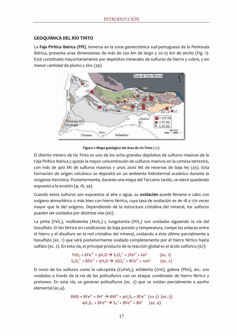
La Faja Pirítica Ibérica (FPI), inmersa en la zona geotectónica sud-portuguesa de la Península
Ibérica, presenta unas dimensiones de más de 250 km de largo y 20-75 km de ancho (Fig. 1).
Está constituida mayoritariamente por depósitos minerales de sulfuros de hierro y cobre, y en
menor cantidad de plomo y zinc (39).
Figura 1: Mapa geológico del área de río Tinto (72).
El distrito minero de río Tinto es uno de los ocho grandes depósitos de sulfuros masivos de la
Faja Pirítica Ibérica y quizás la mayor concentración de sulfuros masivos en la corteza terrestre,
con más de 400 Mt de sulfuros masivos y unos 2000 Mt de reservas de baja ley (45). Esta
formación de origen volcánico se depositó en un ambiente hidrotermal oceánico durante la
orogenia Hercínica. Posteriormente, durante una etapa del Terciario tardío, se elevó quedando
expuesta a la erosión (9, 18, 39).
Cuando estos sulfuros son expuestos al aire o agua, su oxidación puede llevarse a cabo con
oxígeno atmosférico o más bien con hierro férrico, cuya tasa de oxidación es de 18 a 170 veces
mayor que la del oxígeno. Dependiendo de la estructura cristalina del mineral, los sulfuros
pueden ser oxidados por distintas vías (60).
La pirita (FeS2), molibdenita (MoS2) y tungstenita (WS2) son oxidadas siguiendo la vía del
tiosulfato. El ión férrico en condiciones de baja presión y temperatura, rompe los enlaces entre
el hierro y el disulfuro en la red cristalina del mineral, oxidando a éste último parcialmente a
tiosulfato (ec. 1) que será posteriormente oxidado completamente por el hierro férrico hasta
sulfato (ec. 2). En esta vía, el principal producto de la reacción global es el ácido sulfúrico (67).
FeS2 + 6Fe3+ + 3H2O S2O32- + 7Fe2+ + 6H+ (ec. 1)
S2O32- + 8Fe3+ + 5H2O 2SO4
2- + 8Fe2+ + 10H+ (ec. 2)
El resto de los sulfuros como la calcopirita (CuFeS2), esfalerita (ZnS), galena (PbS), etc. son
oxidados a través de la vía de los polisulfuros con un ataque combinado de hierro férrico y
protones. En esta vía, se generan polisulfuros (ec. 3) que se oxidan parcialmente a azufre
elemental (ec.4).
8MS + 8Fe3+ + 8H+ 8M2+ + 4H2Sn + 8Fe2+ (n≥ 2) (ec. 3)
4H2Sn + 8Fe3+ S8o + 8Fe2+ + 8H+ (ec. 4)

INTRODUCCIÓN
18
Para la oxidación completa de estos sulfuros, se requiere el concurso de microorganismos
oxidadores de azufre (Acidithiobacillus ferrooxidans, At. thiooxidans, etc.), capaces de oxidar el
azufre elemental a ácido sulfúrico según la reacción 5:
S8º + 12O2 + 8H2O 8SO4− + 16H+ (ec. 5)
En resumen, la completa oxidación de los sulfuros por la vía del tiosulfato está bajo control
estrictamente electroquímico mientras que por la vía de los polisulfuros se requiere la acción
de oxidadores de azufre para oxidar el azufre completamente hasta sulfato. Sin embargo, en
ambas vías, los microorganismos acidófilos quimiolitótrofos juegan un papel clave.
Leptospirillum spp. o Acidithiobacillus spp. contribuyen al proceso aumentando hasta 5
ordenes de magnitud la tasa de oxidación del ión ferroso (ec. 6), manteniendo así una alta
concentración de ión férrico, el agente oxidante responsable del proceso (60).
4Fe2+ + O2 + 2H+ 2Fe3+
+ 2H2O (ec. 6)
El río Tinto nace en Peña de Hierro, en las cercanías del Pico del Padre Caro, en el corazón de la
FPI, a una altura de 701 metros sobre el nivel del mar recorriendo 92 km antes de desembocar
en el Océano Atlántico, en la ciudad de Huelva. La cuenca del río Tinto atraviesa la provincia de
Huelva de noreste a sudoeste cubriendo una extensión de 1.676 km2 con un suave desnivel del
6%. De norte a sur, el río atraviesa los municipios onubenses de Nerva, Minas de Río Tinto, El
Campillo, Zalamea la Real, Berrocal, Valverde del Camino, Paterna del Campo, Niebla, La Palma
del Condado, Villarrasa, Bonares, Trigueros, Lucena del Puerto, San Juan del Puerto, Moguer,
Palos de la Frontera y Huelva.
Los productos de la oxidación de los minerales que sirven de lecho al río (hierro férrico y ácido
sulfúrico) son los responsables de sus peculiares características. En primer lugar, su nombre -
Tinto - refleja el intenso color rojo de sus aguas (Fig. 2), causado por la elevada concentración
de hierro férrico en solución (~2 g L-1).
Figura 2: Imagen del río Tinto y el intenso color rojo de sus aguas.
INTRODUCCIÓN
19
El ión férrico también otorga un potencial redox altamente oxidante (~400 mV). El ácido
sulfúrico producido en la oxidación es responsable de la alta concentración de sulfatos en sus
aguas (~6 g L-1) y de la extrema y constante acidez (pH~2,3) que está, a su vez, tamponada por
la reacción de hidrólisis del férrico (ec. 7). El ión férrico en presencia de agua precipita como
hidróxido férrico y libera protones, lo que mantiene constante el pH del agua al compensar con
su equilibrio químico las fluctuaciones en el caudal del río (42).
Fe3+ +3H2O Fe(OH)3 +3H+ (ec. 7)
Cuando estas aguas entran en contacto con las vetas de sulfuros metálicos, el bajo pH y el ión
férrico facilitan la solubilidad de los metales, lo que explica las altas concentraciones de
metales pesados en disolución (Cu: ~0,1 g L-1; Zn: ~0,2 g L-1, etc.) (33).
Las condiciones geoquímicas del río definen tres zonas principales (63). En primer lugar la
zona norte (desde Peña de Hierro a La Palma del Condado), consistente en un complejo
volcánico paleozoico caracterizado por altas concentraciones de férrico en solución y un pH
constante de 2.3. En segundo lugar, la zona de transición (desde La Palma del Condado a San
Juan del Puerto) con un substrato mixto compuesto por rocas paleozoicas, arcillas terciarias y
terrazas fluviales cuaternarias que muestran un ligero incremento del pH hasta 3. Por último, el
estuario (desde San Juan del Puerto al océano Atlántico), la sección con influencia de mareal,
caracterizada por su menor contenido en hierro y su mezcla con aguas salinas de mayor pH (9).
De acuerdo con los parámetros bioclimáticos, el área norte se corresponde con un clima
subhúmedo meso-termomediterráneo, mientras que la zona de transición y el estuario podrían
ser incluidas en un clima termomediterráneo subhúmedo/seco (10, 59).
La FPI ha sido sujeta a actividades mineras durante miles de años (66). Comenzando en la
Edad de Bronce, los Íberos (año 3.000 a. C.) - que denominaban al río Tinto Iberus -, tartesos,
fenicios, romanos – que lo llamaban Urium - y los musulmanes, han explotado sus recursos
minerales. En el siglo XIX, comenzó la explotación minera a gran escala, principalmente llevada
a cabo por empresas del Reino Unido como Rio Tinto Company Limited. Por su antigua
tradición minera, se consideraba que el río Tinto era un ambiente producto de la
contaminación (38). Sin embargo, se ha probado que las condiciones ácidas del Tinto son más
antiguas que las explotaciones mineras al datar las formaciones de hierro de los depósitos
sedimentarios de jarosita, coquimbita y copiapita (precipitados necesariamente en condiciones
de acidez) en más de 2 millones de años (18, 19).
Por sus peculiares características, el río Tinto ha sido objeto de diversos campos de
investigación. Por un lado, tiene tanto interés biogeoquímico -ahondando en la naturaleza de
las comunidades microbianas asociadas con las aguas ácidas de mina (Acid Mine Drainage,
AMD)- como puramente microbiológico, ya que los ambientes ácidos ofrecen una oportunidad
única para estudiar la complejidad de hábitats biológicos en condiciones extremas. Además, el
río Tinto posee un gran potencial biotecnológico ya que los organismos que lo pueblan son
usados en biominería (biolixiviación), desulfuración de carbones, etc. Por último, el río Tinto
tiene un alto interés astrobiológico: ha sido propuesto como un análogo de Marte ya que
ambos ambientes parecen poseer una mineralogía común. Las misiones MER (Mars Exploration
Rover) de la NASA (8, 18) han descubierto unidades litológicas con alto contenido en hierro y
sulfatos sobre la superficie de Marte.
INTRODUCCIÓN
20
BIODIVERSIDAD DEL RÍO TINTO
Como consecuencia del gran interés suscitado por el río Tinto, se conoce mucho sobre la
microbiota que habita sus aguas. El uso combinado de métodos convencionales y de ecología
molecular ha llevado a la identificación de los organismos más representativos de la columna
de agua de la cuenca del Tinto (1-3, 20, 21, 23, 24, 26, 41, 42, 62, 76).
Aproximadamente el 80% de la biomasa procariótica en la columna de agua corresponde a tan
sólo tres bacterias: A. ferrooxidans, L. ferrooxidans y Acidiphilium spp., todas ellas relacionadas
con el ciclo del hierro. A. ferrooxidans es una bacteria acidófila anaerobia facultativa y
quimiolitótrofa altamente versátil: es capaz de oxidar hierro y azufre en condiciones aerobias y
de reducir hierro oxidando azufre en condiciones anaerobias (44). L. ferrooxidans es una
bacteria acidófila aerobia estricta y quimiolitótrofa que oxida hierro y parece capaz de fijar N2
(21). Por último, las especies del género Acidiphilium son heterótrofas, capaces de usar el
oxígeno como aceptor de electrones o bien, el ión férrico en condiciones de microaerobiosis
(16). Otros organismos oxidadores de hierro, como las arqueas Ferroplasma spp. o
Thermoplasma spp., o reductores de hierro, como Ferrimicrobium spp., también han sido
identificados pero su baja abundancia sugiere que juegan un papel minoritario en la ecología
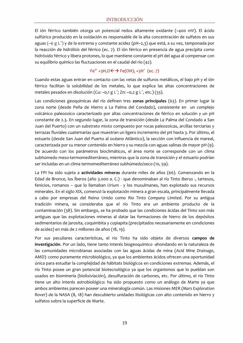
del Tinto (23, 25). Aunando toda esta información, se planteó un modelo de funcionamiento de
los organismos en la columna de agua (Fig. 3).
Figura 3: Modelo de la ecología microbiana del río Tinto. Metabolismo del hierro: el ion ferroso sería oxidado por bacterias como At. ferrooxidans, L. ferrooxidans, Ferrimicrobium spp., Acidimicrobium spp. y por la arquea Ferroplasma spp. El ion férrico sería reducido en condiciones de anaerobiosis por Acidimicrobium spp., Ferrimicrobium spp. y Acidiphilium spp., utilizando compuestos reducidos de carbono ((CH2O)n) como fuente de energía o por At. ferrooxidans oxidando compuestos reducidos del azufre. Respecto al ciclo del azufre, bacterias quimiolitoautótrofas como At. ferrooxidans, At. thiooxidans y At. caldus, serían responsables de oxidar azufre a sulfato. Además, en ausencia de oxígeno At. ferrooxidans llevaría a cabo la oxidación de azufre acoplada a la reducción de hierro como se explicó anteriormente. En cuanto a la regeneración del sulfato podría llevarse a cabo por sulfatorreductoras (26).
A lo largo del río aparecen unas estructuras filamentosas macroscópicas de color marrón (Fig.
4), denominadas serpentinas o streamers, compuestas por los tres grupos de bacterias
mayoritarias de la columna de agua (Acidithiobacillus, Leptospirillum y Acidiphilium) inmersas
en una matriz de exopolisacáridos y partículas minerales. Minoritariamente aparecen algunos

INTRODUCCIÓN
21
microorganismos de metabolismo anaerobio, lo que sugiere que podrían ser originados en los
sedimentos y elevarse hasta la superficie al esponjarse y hacerse menos densos (20).
Figura 4: Serpentinas suspendidas en las aguas del río (20).
A pesar de la baja diversidad procariota, y de la creencia generalizada de que los metales
pesados inhiben el crecimiento y diversidad de los eucariotas (28), en el río Tinto aparece una
inesperada diversidad eucariótica (1-3, 76) que incluye algas, ciliados, flagelados, amebas,
hongos y rotíferos. Coloridas biopelículas que cubren grandes superficies, comunidades
filamentosas y algas macroscópicas son características comunes en las aguas del Tinto.
La mayoría de las especies eucariotas son fotosintéticas siendo los principales contribuyentes a
la biomasa (>65%) en este hostil río (42). Estas algas son mayoritariamente clorofitas (géneros
Chlamydomonas, Chlorella, Euglena, Dunaliella, etc.) y aparecen formando largos filamentos
verdes (Fig. 5) (1-3). En las zonas con características más extremas, predomina el género de
rodofita Cyanidium con la clorofita Dunaliella, uno de los organismos más extremos descritos
hasta ahora (22). También aparecen, especialmente durante los meses secos, algas
filamentosas de los géneros Zygnemopsis y Klebsormidium. Diatomeas penadas del género
Pinnularia (y en ocasiones Nitzchia y Cyclotella.) aparecen en el río formando largas biopelículas
marrones.
Figura 5: Biopelículas de eucariotas en río Tinto. A) Euglenas y diatomeas, B) Cyanidium y diatomeas, C) Euglena, Chlamydomonas y hongos (3).
Los protistas heterótrofos también están ampliamente distribuidos a lo largo del río.
Flagelados mixotrofos del género Bodo y Ochromonas y ciliados del género Oxytrichia han sido
identificados. Frecuentemente aparecen amebas, tanto del tipo lobosea como acantameba,
que se alimentan de las diatomeas. También han sido identificados heliozoos del género
Actiniphyris. Entre los descomponedores, los hongos son los más abundantes y aparecen tanto
levaduras como hongos filamentosos (41, 42). Los hongos parecen tener un papel fundamental
en la formación de las biopelículas y aparecen asociados a las principales bacterias de la
columna de agua (70). El único animal encontrado en el río es una especie de rotífero bdeloide

INTRODUCCIÓN
22
relacionado con el género Rotifera (76) que parece subsistir gracias a su alta tolerancia al
estrés ácido y a la ausencia de competidores más eficientes.
Los sedimentos del Tinto poseen un gran interés ecológico (permiten cerrar los ciclos
biogeoquímicos de los ambientes ácidos de mina), biotecnológico (organismos con
aplicaciones a bio-pilas, recuperación de metales, inmovilización de radionucleidos, …) y
astrobiológico (la atmósfera de Marte carece de oxígeno por lo que los metabolismos que
pudieran encontrarse serían anaerobios). A pesar de ello y por las limitaciones metodológicas
de su estudio, hasta ahora había un conocimiento muy limitado de su microbiota. En río Tinto,
sólo había sido descrita la presencia de arqueas metanogénicas (68) y alguna especie sulfato-
reductora (20), pero sin llegar a valorar su diversidad y funcionamiento de manera profunda.
Por otro lado, en perforaciones rocosas, el denominado proyecto MARTE (7), se detectó la
existencia de actividades anaerobias a altas profundidades y la presencia de gas metano, oxido
nítrico e hidrógeno.
Estos antecedentes sugerían la posibilidad de existencia y diversidad de actividades anaerobias
en el subsuelo del río Tinto y abrían la cuestión de cómo sería la ecología microbiana de los
sedimentos, qué organismos estarían presentes, su abundancia y cuáles serían las rutas
metabólicas presentes en los sedimentos de este hostil ecosistema. Este ha sido el ambicioso
objetivo de la presente tesis.
POTENCIALES PROCESOS MICROBIANOS EN LOS SEDIMENTOS DEL RÍO TINTO.
Los parámetros ambientales de un ecosistema, tales como temperatura, concentración de
oxígeno, flujos de nutrientes, pH, características geológicas, etc., influyen en propiedades de
los sedimentos tales como el tamaño de partícula, el contenido de materia orgánica, la tasa de
sedimentación, etc. En el río Tinto encontramos sedimentos con distinta granulometría. A lo
largo de su cuenca mayoritariamente se producen acumulaciones de partículas minerales de
cierto tamaño - arenas y grava - con alta porosidad y sin estructura real de sedimento. En ellas,
el principal mecanismo de transporte será la convección y sus características biogeoquímicas
estarán fuertemente influenciadas por las de la columna de agua. Sin embargo, existen ciertas
zonas con presas o con menor pendiente donde se reduce la velocidad lineal de la masa de
agua permitiendo una acumulación estratificada de sedimentos con menor tamaño de
partícula, principalmente limos. En ellos el principal mecanismo de transporte será la difusión,
lo que crea mayor variedad de posibles nichos ecológicos con características que pueden
diferir de las del agua superficial. En los sedimentos esta variabilidad se refleja en una
diversidad de comunidades microbianas y procesos biogeoquímicos (31, 40, 61).
Los procesos biológicos que se dan en un sistema dependen de la energética de las reacciones,
favoreciéndose aquellos que obtienen mayor energía por mol de substrato. La fuente de
energía para el mantenimiento celular puede ser química (quimiótrofos) o lumínica
(fotótrofos). Los organismos fotótrofos convierten la energía lumínica en energía metabólica y
poder reductor pero en los sedimentos del río Tinto, debido a la ausencia de luz, predominarán
los metabolismos basados en la quimiotrofía (quimio-organotrofía o quimio-litotrofía). A su
vez, la fuente de carbono puede ser inorgánica (autótrofos) u orgánica (heterótrofos).
Hay dos tipos de metabolismos quimiótrofos principales: la fermentación y la respiración. En la
fermentación no hay un aceptor externo de electrones por lo que no requieren de una cadena

INTRODUCCIÓN
23
transportadora y no hay cambio global del estado de oxidación. Como consecuencia, el
rendimiento energético es bajo (2-3 ATP por mol hexosa). Hay distintos tipos de fermentación:
alcohólica, láctica (homo o heteroláctica), propiónica, ácido-mixta, acetona-butanol que
generan como productos distintos tipos de ácidos grasos volátiles, alcoholes e hidrógeno.
En los procesos de respiración, el dador primario de electrones cede los electrones al NAD+
reduciéndolo a NADH,H+ desde el que fluyen a través de la cadena transportadora de
electrones que irá traslocándolos -a través de citocromos y quinonas- hasta el aceptor final de
electrones, generando en el proceso un gradiente de protones a ambos lados de la membrana
usado para realizar un trabajo: químico (almacenar energía en forma de ATP), cinético
(movimiento celular), de transporte, etc. De un modo resumido, la energía obtenida en este
proceso será mayor cuanto mayor sea la diferencia de potenciales de reducción de los
elementos que intervengan (Fig. 6), es decir, la energía libre de Gibbs (∆G0) (ec. 8) es
proporcional a la diferencia del potencial de reducción entre el aceptor de electrones
(oxidante, elemento que se reduce) y el dador de electrones (reductor, elemento que se
oxida).
∆Gº´ = -n·F·∆ε = -n·F·∆ε = -n·F·(εREDUCCIÓNº - εOXIDACIÓNº) (ec. 8)
siendo F la constante de Faraday, cuyo valor es de 23 kcal mol-1.
Figura 6: Torre de potenciales. Los pares redox se expresan como compuesto oxidado/reducido (ej. O2/H2O). Cuanto mayor, más positivo sea el potencial de reducción de un par redox, más tendencia tendrá a reducirse, esto
es a actuar como aceptor de electrones.
De modo que, ante cierta disponibilidad de diferentes dadores y aceptores de electrones, la
secuencia de aceptores preferenciales de electrones (Fig. 6) sería: oxígeno (respiración
aerobia), hierro (reducción de hierro), nitrato (desnitrificación), sulfato (sulfato-reducción) y
dióxido de carbono (metanogénesis y, posteriormente, acetogénesis). Puesto que la energía
libre liberada depende no sólo de los potenciales estándar, si no también de las
concentraciones de dador y aceptor de electrones (ec. 9), el orden puede alterarse. Esto es
INTRODUCCIÓN
24
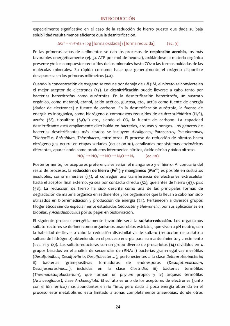
especialmente significativo en el caso de la reducción de hierro puesto que dada su baja
solubilidad resulta menos eficiente que la desnitrificación.
∆Gº´ = -n·F·∆ε + log [forma oxidada] / [forma reducida] (ec. 9)
En las primeras capas de sedimentos se dan los procesos de respiración aerobia, los más
favorables energéticamente (ej. 34 ATP por mol de hexosa), oxidándose la materia orgánica
presente y/o los compuestos reducidos de los minerales hasta CO2 o las formas oxidadas de las
moléculas minerales. Su rápido consumo hace que generalmente el oxígeno disponible
desaparezca en los primeros milímetros (40).
Cuando la concentración de oxígeno se reduce por debajo de 2-8 μM, el nitrato se convierte en
el mejor aceptor de electrones (13). La desnitrificación puede llevarse a cabo tanto por
bacterias heterótrofas como autótrofas. En la desnitrificación heterótrofa, un sustrato
orgánico, como metanol, etanol, ácido acético, glucosa, etc., actúa como fuente de energía
(dador de electrones) y fuente de carbono. En la desnitrificación autótrofa, la fuente de
energía es inorgánica, como hidrógeno o compuestos reducidos de azufre: sulfhídrico (H2S),
azufre (Sº), tiosulfato (S2O32-) etc., siendo el CO2 la fuente de carbono. La capacidad
desnitrificante está ampliamente distribuida en bacterias, arqueas y hongos. Los géneros de
bacterias desnitrificantes más citados se incluyen: Alcaligenes, Paracoccus, Pseudomonas,
Thiobacillus, Rhizobium, Thiosphaera, entre otros. El proceso de reducción de nitratos hasta
nitrógeno gas ocurre en etapas seriadas (ecuación 10), catalizadas por sistemas enzimáticos
diferentes, apareciendo como productos intermedios nitritos, óxido nítrico y óxido nitroso.
NO3- → NO2
- → NO → N2O → N2 (ec. 10)
Posteriormente, los aceptores preferenciales serían el manganeso y el hierro. Al contrario del
resto de procesos, la reducción de hierro (Fe3+) y manganeso (Mn4+) es posible en sustratos
insolubles, como minerales (13), al conseguir una transferencia de electrones extracelular
hasta el aceptor final externo, ya sea por contacto directo (52), quelantes de hierro (43), pilis
(58). La reducción de hierro ha sido descrita como una de las principales formas de
degradación de materia orgánica en sedimentos y los organismos que la llevan a cabo han sido
utilizados en biorremediación y producción de energía (74). Pertenecen a diversos grupos
filogenéticos siendo especialmente estudiados Geobacter y Shewanella, por sus aplicaciones en
biopilas, y Acidithiobacillus por su papel en biolixiviación.
El siguiente proceso energéticamente favorable sería la sulfato-reducción. Los organismos
sulfatorrectores se definen como organismos anaerobios estrictos, que viven a pH neutro, con
la habilidad de llevar a cabo la reducción disasimilativa de sulfato (reducción de sulfato a
sulfuro de hidrógeno) obteniendo en el proceso energía para su mantenimiento y crecimiento
(ecs. 11 y 12)). Las sulfatoreductoras son un grupo diverso de procariotas (14) divididos en 4
grupos basados en el análisis de secuencias de rRNA: i) bacterias gram-negativas mesófilas
(Desulfobulbus, Desulfovibrio, Desulfobacter…), pertenecientes a la clase Deltaproteobacteria;
ii) bacterias gram-positivas formadoras de endoesporas (Desulfotomaculum,
Desulfosporosinus…), incluidas en la clase Clostridia; iii) bacterias termófilas
(Thermodesulfobacterium), que forman un phylum propio; y iv) arqueas termófilas
(Archaeoglobus), clase Archaeoglobi. El sulfato es uno de los aceptores de electrones (junto
con el ión férrico) más abundantes en río Tinto, pero dada la poca energía obtenida en el
proceso este metabolismo está limitado a zonas completamente anaerobias, donde otros

INTRODUCCIÓN
25
aceptores más favorables hayan sido consumidos. Sin embargo, se estima que puede llegar a
contribuir a la mineralización del 50% de la materia orgánica en los sedimentos (35).
4 H2 + SO42- + 2H+ ---> H2S + 4 H2O ΔG0 = -153,5 kJ mol-1 (ec. 11)
CH3-COO- + SO42- + H+ ---> H2S + HCO3
- ΔG0 = -47,1 kJ mol-1 (ec. 12)
Por último, el paso final en la mineralización de la materia orgánica en condiciones anaerobias
es la metanogénesis. Ciertos grupos de arqueas utilizan el CO2 como aceptor final de
electrones reduciéndolo a metano con hidrógeno. Se denomina metanogénesis
hidrogenotrófica (ec. 13) y es llevada a cabo por géneros como Methanobacterium,
Methanococcus o Methanospirillum. Tan sólo dos géneros, Methanosarcina y Methanosaeta,
utilizan el acetato para generar metano en la llamada metanogénesis acetoclástica (ec. 14).
CO2 + 4 H2 → CH4 + 2H2O ∆Gº´ = - 135 kJ mol-1 (ec. 13) CH3COOH → CH4 + CO2 ∆Gº´ = -28.4 kJ mol-1 (ec. 14)
A pesar de que la metanogénesis hidrogenotrófica es más eficiente (ec. 13), energéticamente
hablando, que la acetoclástica (ec. 14), el 98% del CO2 utilizado se emplea para obtener energía
y tan sólo el 2% para formar biomasa. A las presiones parciales en las que normalmente se
encuentra el hidrógeno en el interior celular, la variación de energía libre se sitúa entorno a 14
kJ mol-1 metano generado, lo que permite obtener tan sólo un mol de ATP por mol de metano
producido. Ello hace que las arqueas metanogénicas tengan tiempos de generación muy
largos, siendo muy sensibles a las variaciones medioambientales. Además, en ambientes con
elevadas concentraciones de sulfato, como son los sedimentos marinos o el caso del río Tinto,
las bacterias sulfato-reductoras se ven favorecidas sobre las arqueas metanogénicas (ecs. 11 y
12 vs. 13 y 14), siendo las primeras los principales mineralizadores terminales de la materia
orgánica.
Existen aceptores de electrones alternativos a los aquí citados, tales como los pares SeO42-
/SeO32- (E: +0,48), AsO4
3-/AsO33- (E: +0,16), o compuesto clorados alifáticos y aromáticos, como
el par Cl-benzoato/benzoato (E: +0,3), pero su importancia ecológica es, en general, escasa.
ECOLOGÍA MOLECULAR APLICADA AL ESTUDIO DE SEDIMENTOS
Las técnicas de microbiología clásica requieren del aislamiento de cultivos puros. El
desconocimiento de las propiedades fisiológicas de un organismo o el hecho de que en la
naturaleza muchos organismos aparecen en simbiosis o sinergia con otros - creando
condiciones prácticamente imposibles de reproducir en el laboratorio - dificultan o
imposibilitan su aislamiento, lo que conlleva grandes sesgos en la identificación de la
microbiota de un ecosistema. Además, posteriormente son necesarios múltiples y largos
ensayos morfológicos, metabólicos, bioquímicos, genéticos, etc., para la identificación de los
aislados. Esto se plasma en un bajo porcentaje de organismos cultivados descritos respecto al
total que debe existir, estimado en menos de 1% para diversos ambientes (6). En particular, los
ambientes anaerobios han presentado considerables dificultades en este aspecto, debido a las
bajas tasas de crecimiento y a los estrictos requerimientos de cultivo. Por ello, en el caso de los
sedimentos, este porcentaje se reduce hasta 0,25%.
A finales de los 80´ se desarrollaron una serie de técnicas de biología molecular que supusieron
una revolución en el campo de la ecología microbiana (6, 55, 75). Estas técnicas se basaron
principalmente en dos grandes avances: i) el descubrimiento de que las relaciones
INTRODUCCIÓN
26
filogenéticas podían ser inferidas por las secuencias de genes como el 16S rRNA (75) y ii) la
amplificación selectiva de ácidos nucleicos de muestras ambientales a través de la reacción en
cadena de la polimerasa (Polymerase Chain Reaction: PCR) (64). Así surgió la ecología molecular
(4), siendo posible la caracterización de la diversidad microbiana de un ecosistema a nivel DNA
sin necesidad de enriquecimientos o aislados.
Actualmente la taxonomía microbiana está basada en la información contenida en los ácidos
nucleicos (ej. 16S rRNA). La elección del rRNA para la identificación de los organismos y el
estudio de su filogenia (55, 56) se fundamenta en su universalidad y abundancia en todos los
seres vivos. Al ser parte de la maquinaria para sintetizar proteínas, el RNA ribosomal es una
molécula muy preservada. Además, posee un tamaño que lo hace estadísticamente adecuado
y fácil de secuenciar con las técnicas actuales. Pero su más preciada característica es su papel
como registro evolutivo: asumiendo que los cambios se producen al azar, y que aumentan con
el tiempo de manera linear, las diferencias en la secuencia reflejan la distancia evolutiva,
permitiéndonos estudiar las divergencias evolutivas entre los distintos organismos. Además,
posee zonas con diferentes grados de conservación, regiones muy conservadas y regiones
altamente variables, que permiten distinta especificidad desde dominio hasta especie.
Adicionalmente, esta molécula no se ve afectada por transferencia horizontal de genes.
A pesar de las innegables ventajas de las técnicas de biología molecular, éstas no permiten
obtener información sobre el metabolismo de los taxones identificados. Por lo tanto, es
recomendable combinar técnicas de microbiología clásica con técnicas moleculares para
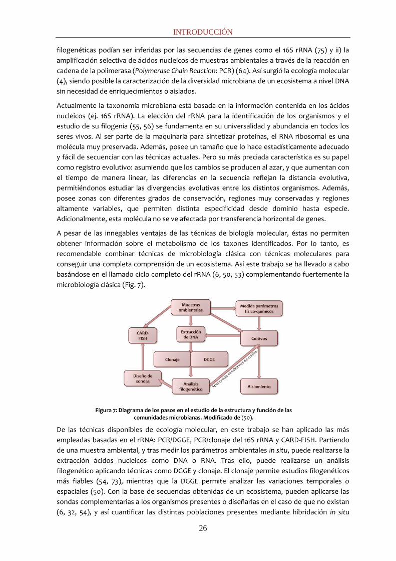
conseguir una completa comprensión de un ecosistema. Así este trabajo se ha llevado a cabo
basándose en el llamado ciclo completo del rRNA (6, 50, 53) complementando fuertemente la
microbiología clásica (Fig. 7).
Figura 7: Diagrama de los pasos en el estudio de la estructura y función de las comunidades microbianas. Modificado de (50).
De las técnicas disponibles de ecología molecular, en este trabajo se han aplicado las más
empleadas basadas en el rRNA: PCR/DGGE, PCR/clonaje del 16S rRNA y CARD-FISH. Partiendo
de una muestra ambiental, y tras medir los parámetros ambientales in situ, puede realizarse la
extracción ácidos nucleicos como DNA o RNA. Tras ello, puede realizarse un análisis
filogenético aplicando técnicas como DGGE y clonaje. El clonaje permite estudios filogenéticos
más fiables (54, 73), mientras que la DGGE permite analizar las variaciones temporales o
espaciales (50). Con la base de secuencias obtenidas de un ecosistema, pueden aplicarse las
sondas complementarias a los organismos presentes o diseñarlas en el caso de que no existan
(6, 32, 54), y así cuantificar las distintas poblaciones presentes mediante hibridación in situ

INTRODUCCIÓN
27
(FISH/CARD-FISH). Finalmente, con la información recabada sobre los posibles metabolismos
presentes, pueden seleccionarse las condiciones para cultivos de enriquecimiento selectivos
de ciertas actividades y, si cabe, proceder al aislamiento de microorganismos. A continuación
se explicarán brevemente las técnicas seleccionadas.
Extracción de DNA
Tras la toma de muestras, la extracción de DNA de los organismos presentes en el ecosistema
es un paso previo necesario a la amplificación por PCR. Hay dos procedimientos generales: i)
extracción in situ, en el que las células se lisan en la misma matriz de la muestra ambiental, y ii)
extracción ex situ, donde las células se separan previamente. El primer método es el usado
comúnmente por su sencillez, rapidez y cantidad de DNA extraído. Sin embargo, en el caso de
sedimentos de un ambiente ácido con alta concentración de metales pesados, este método no
es adecuado debido a la co-extracción de ácidos húmicos y metales pesados junto con el DNA
(inhibidores de PCR). Adicionalmente, el contenido de células por peso de sedimento es menor
en comparación con otros ecosistemas, requiriéndose por tanto técnicas de concentración y
purificación previas a la extracción.
Reacción en cadena de la polimerasa.
De la compleja mezcla de DNAs que pueden ser extraídas de una muestra ambiental, se
pueden amplificar secuencias específicas mediante PCR. La reacción en cadena de la
polimerasa se usa para amplificar un determinado segmento de DNA situado entre dos
regiones de secuencia conocida. Para iniciar la síntesis de DNA, que será catalizada por la DNA
polimerasa, se usan dos cebadores o primers: oligonucleótidos complementarios a las
secuencias que flanquean el segmento de DNA que se desea amplificar. Primero, para separar
las cadenas complementarias de la doble hélice de DNA, se desnaturaliza el DNA molde
mediante calor, en una mezcla de reacción con los dos cebadores y los cuatro
deoxinucleótidos (dNTPs: dATP, dTTP, dGTP, dCTP). Posteriormente, la mezcla de reacción se
enfría hasta una temperatura que permite a los cebadores formar puentes de hidrógeno con
sus secuencias diana, tras lo cual la DNA polimerasa comienza a sintetizar DNA a partir de ellos.
Este ciclo de desnaturalización, unión de cebadores y síntesis de DNA se repite varias veces, y
puesto que los productos de un ciclo sirven de molde para el siguiente, en cada ciclo se dobla
la cantidad de DNA. La mezcla de fragmentos amplificados de DNA pude ser usada en
posteriores análisis para clonaje y DGGE. Cabe señalar que puede darse la amplificación
preferente de algunas secuencias. Además el número de copias de un determinado gen no es
el mismo en todos los organismos (por ejemplo, Mycobacterium tuberculosis tiene 1 copia, E.
coli presenta 7 copias y Bacillus cerus 12 copias para el gen del 16S rRNA). Por ambas razones
esta técnica no puede ser usada de modo cuantitativo para determinar la abundancia natural
de los genes amplificados.
Los protocolos originales para la PCR (46, 47, 65) usaban un fragmento de la DNA polimerasa I
de E. coli para catalizar la síntesis, pero esta enzima se inactiva a las temperaturas requeridas
para desnaturalizar el DNA (95ºC), de modo que cada ciclo requería la adicción de nueva
enzima. Este problema fue resuelto con la introducción (64) de una DNA polimerasa
termoestable, purificada de la bacteria termófila Thermus aquaticus (Taq DNA polimerasa) (15).
Esta enzima no se inactiva al subir la temperatura para desnaturalizar el DNA, por lo que no
necesita ser reemplazada en cada ciclo. Actualmente, también se usa otra enzima
termoestable: la polimerasa Vent de Thermococcus litoralis.

INTRODUCCIÓN
28
Clonaje del gen del 16S rRNA
El clonaje del gen del 16S rRNA para procariotas es una de las técnicas moleculares más
utilizada para el estudio de una población microbiana, ya que proporciona de manera fiable
información sobre la biodiversidad de un determinado sistema y la relación filogenética de los
organismos que lo componen (56).
Generalmente, utilizando cebadores universales del dominio Bacteria o Archaea, se procede a
la amplificación del gen completo del 16S rRNA contenido en el DNA extraído de una muestra
(Fig. 8). Para separar la mezcla de los genes del 16S rRNA de los distintos organismos
presentes, éstos son unidos o ligados a un vector de clonación, generalmente un plásmido,
que es introducido en un organismo huésped, generalmente células competentes de E. coli.
Los plásmidos usados contienen un gen de resistencia a la ampicilina y el gen LacZ en la zona
de inserción del gen a clonar, de tal manera que una vez cultivadas las colonias en placas de
agar sólo crecerán las que hayan insertado eficientemente el plásmido. Además, se podrá
hacer una selección cromática puesto que la inserción del gen inhibe la expresión del gen lacZ
produciendo colonias blancas con el inserto y colonias azules sin inserto. La amplificación o
extracción de los plásmidos y la posterior secuenciación del 16S rRNA dará como resultado la
construcción de una librería de los genes 16S rRNA de los organismos que componen el
sistema a estudiar. Las secuencias obtenidas pueden ser utilizadas para realizar estudios
filogenéticos, para el diseño de sondas necesarias en otras técnicas de ecología molecular
como CARD-FISH o para el diseño de cebadores específicos para amplificación de poblaciones
concretas (6, 54). La mayor ventaja de esta metodología es que las secuencias obtenidas
contienen el gen completo, lo que proporciona alta precisión en los estudios filogenéticos (71).
Figura 8: Protocolo de clonaje para el estudio de una comunidad microbiana. Tras la toma de muestra y la extracción de DNA, se lleva a cabo la amplificación del gen completo del 16S rRNA. Distintas copias se insertan en distintos plásmidos (ligación) y después se introducen en células competentes de E. coli DH5α (transformación). Tras la selección de las colonias transformadas (blancas) y su crecimiento, se lleva a cabo la extracción del plásmido acorde a sus patrones de restricción. Finalmente tras la secuenciación del gen clonado, se obtiene la afiliación filogenética de las secuencias obtenidas.

INTRODUCCIÓN
29
Electroforesis en gel con gradiente desnaturalizante.
La técnica fue inicialmente desarrollada para el estudio de mutaciones que producen
patologías en los genes humanos (12). Su primera aplicación en ecología microbiana fue para el
análisis de biopelículas y tapetes microbianos (48). Actualmente es la técnica de huella
genética más utilizada en Ecología microbiana, ampliamente aplicada para estudiar los cambios
espaciales o temporales en las comunidades en un ecosistema (50).
La DGGE permite la separación de fragmentos de doble hélice de DNA de la misma longitud
pero distinta secuencia (49). La técnica se fundamenta en la diferente estabilidad del enlace GC
(tres puentes de hidrógeno por enlace) respecto al enlace AT (2 puentes de hidrógeno). Una
mezcla de fragmentos de DNA de diferente secuencia se somete a electroforesis a través de
un gel de acrilamida con un gradiente químico creciente de sustancias desnaturalizantes (urea-
formamida). En general, los fragmentos de DNA ricos en GC serán más estables y mantienen la
estructura de doble hélice a mayores concentraciones de desnaturalizantes, hasta que llegan a
un punto crítico en el que el DNA se desnaturaliza. Para evitar la separación completa de las
dos hebras de DNA, se suele añadir una cola de oligonucleótidos rica en G+C en el extremo del
cebador. Ésta cola mantiene unidas las dos hebras complementarias, de tal forma que al
liberarse del medio que las mantenía desnaturalizadas, puedan volver a unirse. Este cambio de
estructura en el DNA obstruye el movimiento a través del gel: los fragmentos de DNA de doble
hélice migran mejor en el gel de acrilamida mientras que los fragmentos desnaturalizados
reducen su velocidad y se paran. De este modo, los fragmentos de DNA de diferente secuencia
se van separando en el gel de acrilamida. El resultado de un gel de DGGE es un patrón de
bandas donde teóricamente cada banda corresponde a un microorganismo diferente (49). La
obtención de un patrón de bandas (Fig. 9) es útil para seguir la evolución espacial y temporal
de las poblaciones de microorganismos y su respuesta a cambios físico-químicos y
nutricionales. Las bandas pueden amplificarse y secuenciarse y generar información
taxonómica y filogenética, aunque de menor precisión que con el clonaje (71) dado el menor
tamaño de las secuencias (max. 500-600 pb) (27).
Figura 9: Protocolo de DGGE. Tras la toma de muestra y la extracción de DNA, se lleva a cabo la amplificación de una región del gen 16S rRNA. Los fragmentos generados son sometidos a electroforesis obteniendo un patrón de bandas. Adicionalmente, estas bandas pueden extraerse y tras elución de su DNA, amplificarlo y secuenciarlo.
INTRODUCCIÓN
30
Hibridación in situ fluorescente
La técnica de FISH (Fluorescence In Situ Hybridization) permite la identificación in situ de
organismos no cultivados en su ambiente natural, así como la determinación de su abundancia,
distribución espacial y morfología celular. Esta técnica fue descrita por primera vez por DeLong
y col. en 1989 (17). Se trata de una hibridación DNA-RNA en la una sonda de DNA se une
específicamente con una secuencia firma del rRNA. Las dianas del 16S rRNA – idóneo por su
alto número de copias y su accesibilidad (77) - son secuencias de nucleótidos características de
un determinado grupo filogenético, que permanecen invariables dentro de los organismos de
un mismo grupo y que no se encuentran en otros. Tras localizarlos, se construye una sonda
específica: una pequeña molécula de DNA de unos 18 nucleótidos, marcada en el extremo 5’
con un fluorocromo (para su posterior visualización) complementaria a esta región firma del
rRNA 16S.
La sensibilidad de esta técnica puede ser problemática si las células no están activas, puesto
que la intensidad de la señal depende del número de ribosomas y por tanto de la actividad de
las células, o si son demasiado pequeñas y la señal de hibridación está por debajo del limite de
detección. En ocasiones, como muestras de sedimentos con autofluorescencia o una gran
señal de fondo, la señal de hibridación puede ser difícil de diferenciar del fondo. Para resolver
este problema, se han buscado diversos modos de aumentar la intensidad de la señal de
fluorescencia a través de fluorocromos más potentes, uso de cloranfenicol, etc.
Recientemente se ha desarrollado una modificación del FISH que incrementa notablemente la
señal emitida por las moléculas hibridadas, el CARD-FISH, o Catalyzed Reporter Deposition (57,
69). La modificación consiste en que la sonda de DNA no va unida directamente al
fluorocromo, sino que la sonda va marcada con la peroxidasa de rábano (HorseRadish
Peroxidase: HRP). En una etapa posterior de amplificación (Fig. 10), se introduce el
fluorocromo unido a tiramidas (compuestos fenólicos). La HRP cataliza la unión de las
tiramidas a los residuos de tirosina de las proteínas cercanas a la HRP, aumentando
notablemente el número de fluorocromos que emiten por ribosoma de la bacteria hibridada:
pasa de un fluorocromo por ribosoma a n fluorocromos por ribosoma. Este aumento de la
señal de fluorescencia permite mejorar la detección de las células. Al iluminar la muestra a la
longitud de onda de excitación del fluorocromo empleado, éste emite a una longitud mayor y
se hacen visibles las células marcadas con la sonda. Éstas células sólo corresponderán al grupo
filogenético diana, que puede ser desde un dominio - como Bacteria - hasta una especie o cepa
concreta. Para visualizar y cuantificar el número total de células presentes en la muestra, se
usan moléculas fluorescentes como DAPI (4’, 6-Diamidino-2-Fenilindol) o SybrGreenI, que se
unen inespecíficamente a la doble hélice de DNA.

INTRODUCCIÓN
31
Figura 10: Protocolo de CARD-FISH. La muestra ambiental debe ser fijada in situ para preservar el contenido ribosomal. Para permitir el acceso de la sonda con la peroxidasa, las células se permeabilizan. Durante la hibridación, la sonda unida covalentemente a la peroxidasa del rábano (HRP) se une a su región diana del 16S rRNA. En el paso de amplificación, esta enzima cataliza la deposición de las tiramidas unidas a los fluorocromos. Posteriormente, pude teñirse el total de células con colorantes universales y así cuantificar los distintos grupos presentes. Adaptada de (5).
Cultivos de enriquecimiento y aislamiento.
El aislamiento y estudio de cultivos puros supone el complemento ideal del ciclo completo del
rRNA. Aunque las técnicas moleculares permiten una rápida caracterización de la diversidad de
sistemas complejos, para una completa visión del ecosistema se requieren cultivos de
enriquecimiento que prueben la existencia y actividad de los microorganismos que llevan a
cabo los metabolismos detectados por técnicas moleculares. Adicionalmente, el aislamiento
de los distintos organismos que pueblan un ecosistema es esencial. Los aislados son
imprescindibles para estudiar su metabolismo y evaluar tanto su papel en el ecosistema como
su potencial biotecnológico.
Los ambientes ácidos están poco caracterizados debido a las peculiaridades fisiológicas de los
microorganismos asociados a los mismos (29). En la columna de agua del Tinto, se han
estudiado y demostrado metabolismos como la oxidación de compuestos de azufre y hierro, y
la reducción de hierro. Muchos de los organismos responsables han sido aislados en distintos
ecosistemas ácidos relacionados. Entre ellos, se han aislado gran variedad de quimiolitótrofos
oxidadores de hierro y/o azufre como Acidithiobacillus ferrooxidans, Leptospirillum spp. y
Acidithiobacillus thiooxidans (36), Acidithiobacillus caldus (30), Sulfobacillus spp. y miembros de
la familia de arqueas Ferroplasmaceae. En ambientes ácidos también existe gran variedad de
heterótrofos acidófilos como las especies del género Acidiphilium, o heterótrofos facultativos
como Acidimicrobium spp. y Ferrimicrobium spp. (34). Estas zonas aerobias de los ambientes
ácidos de mina, y los organismos que llevan a cabo la oxidación de los sulfuros metálicos, han
sido estudiadas exhaustivamente. Por un lado, su control permitiría reducir el impacto de la
contaminación minera, y por otro, poseen gran interés biotecnológico siendo aplicados para
biolixiviación de metales.
En cambio, hasta ahora, las zonas anaerobias habían suscitado menos atención y los estudios
habían sido limitados principalmente a reducción de hierro (11). Pero en los últimos años se ha

INTRODUCCIÓN
32
constatado que existen metabolismos, como la reducción de sulfato, capaces de llevarse a
cabo a bajos pH y que pueden ser utilizados para la precipitación y recuperación selectiva de
los metales pesados, así como bioremediación de zonas contaminadas con radionucleidos (37,
51). También se ha probado la existencia de metanógenas en un ambiente tan hóstil como río
Tinto (68). Todo ello, plantea la posibilidad de una mayor diversidad de metabolismos que la
inicialmente pensada, y nos muestra la amplitud de nuestro desconocimiento sobre estos
ecosistemas haciendo necesario un estudio más exhaustivo de diversas actividades
anaerobias.
REFERENCIAS
1. Aguilera, A., S. C. Manrubia, F. Gomez, N. Rodriguez, and R. Amils. 2006. Eukaryotic community distribution and its relationship to water physicochemical parameters in an extreme acidic environment, Río Tinto (Southwestern Spain). Appl. Environ. Microbiol. 72:5325-5330.
2. Aguilera, A., V. Souza-Egipsy, F. Gomez, and R. Amils. 2007. Development and structure of eukaryotic biofilms in an extreme acidic environment, Río Tinto (SW, Spain). Microb. Ecol. 53:294-305.
3. Aguilera, A., E. Zettler, F. Gómez, L. Amaral-Zettler, N. Rodríguez, and R. Amils. 2007. Distribution and seasonal variability in the benthic eukaryotic community of Río Tinto (SW, Spain), an acidic, high metal extreme environment. Syst. Appl. Microbiol. 30:531-546.
4. Akkermans, A. D. L., M. S. Mirza, H. J. M. Harmsen, H. J. Blok, P. R. Herron, A. Sessitsch, and W. M. Akkermans. 1994. Molecular ecology of microbes: A review of promises, pitfalls and true progress. FEMS Microbiol. Rev. 15:185-194.
5. Amann, R., and B. M. Fuchs. 2008. Single-cell identification in microbial communities by improved fluorescence in situ hybridization techniques. Nature Reviews Microbiology. 6:339-348.
6. Amann, R. I., W. Ludwig, and K. H. Schleifer. 1995. Phylogenetic identification and in situ detection of individual microbial cells without cultivation. Microbiol. Mol. Biol. Rev. 59:143-169.
7. Amils, R., D. Fernández-Remolar, F. Gómez, E. González-Toril, N. Rodríguez, C. Briones, O. Prieto-Ballesteros, J. L. Sanz, E. Díaz, and T. O. Stevens. 2008. Subsurface Geomicrobiology of the Iberian Pyritic Belt. In P. Dion and C. Shekhar Nautiyal (Ed. ), Soil Biology (Vol. 13): Microbiology of Extreme Soils. Springer, Berlin. 205-223.
8. Amils, R., E. González-Toril, D. Fernández-Remolar, F. Gómez, Á. Aguilera, N. Rodríguez, M. Malki, A. García-Moyano, A. G. Fairén, and V. de La Fuente. 2007. Extreme environments as Mars terrestrial analogs: the Río Tinto case. Planet. Space Sci. 55:370-381.
9. Amils, R., E. González-Toril, F. Gómez, D. Fernández-Remolar, N. Rodríguez, M. Malki, J. Zuluaga, A. Aguilera, and L. Amaral-Zettler. 2005. Importance of chemolithotrophy for early life on earth: the Tinto River (Iberian Pyritic Belt) case. In J.Seckbach (ed.), Origins. Kluwer Academic Publishers. Dordrecht, Netherlands. Origins. 463-480.
10. Asensi, A., and B. Díez Garretas. 1987. Andalucía occidental. In: Peinado M, Rivas-Martínez S. De Alcalá de Henares, Colección Aula Abierta. La Vegetación De España. 3:197-230.
11. Blothe, M., D. M. Akob, J. E. Kostka, K. Goschel, H. L. Drake, and K. Kusel. 2008. pH gradient induced heterogeneity of Fe (III)-reducing microorganisms in coal mining-associated lake sediments. Appl. Environ. Microbiol. 74:1019-1029.
INTRODUCCIÓN
33
12. Børresen, A. L., E. Hovig, and A. Brøgger. 1988. Detection of base mutations in genomic DNA using denaturing gradient gel electrophoresis (DGGE) followed by transfer and hybridization with gene-specific probes. Mutation Research/Fundamental and Molecular Mechanisms of Mutagenesis. 202:77-83.
13. Burdige, D. J. 2006. Geochemistry of marine sediments. Princeton University Press Princeton.
14. Castro, H. F., N. H. Williams, and A. Ogram. 2000. Phylogeny of sulfate-reducing bacteria1. FEMS Microbiol. Ecol. 31:1-9.
15. Chien, A., D. B. Edgar, and J. M. Trela. 1976. Deoxyribonucleic acid polymerase from the extreme thermophile Thermus aquaticus. J. Bacteriol. 127:1550-1557.
16. Coupland, K., and D. B. Johnson. 2008. Evidence that the potential for dissimilatory ferric iron reduction is widespread among acidophilic heterotrophic bacteria. FEMS Microbiol. Lett. 279:30-35.
17. DeLong, E. F., G. S. Wickham, and N. R. Pace. 1989. Phylogenetic stains: ribosomal RNA-based probes for the identification of single cells. Science. 243:1360-1363.
18. Fernández-Remolar, D. C., R. V. Morris, J. E. Gruener, R. Amils, and A. H. Knoll. 2005. The Río Tinto Basin, Spain: Mineralogy, sedimentary geobiology, and implications for interpretation of outcrop rocks at Meridiani Planum, Mars. Earth Planet. Sci. Lett. 240:149-167.
19. Fernández-Remolar, D. C., O. Prieto-Ballesteros, N. Rodríguez, F. Gómez, R. Amils, J. Gómez-Elvira, and C. R. Stoker. 2008. Underground habitats in the Río Tinto Basin: a model for subsurface life habitats on Mars. Astrobiology. 8:1023-1047.
20. García-Moyano, A., E. González-Toril, A. Aguilera, and R. Amils. 2007. Prokaryotic community composition and ecology of floating macroscopic filaments from an extreme acidic environment, Río Tinto (SW, Spain). Syst. Appl. Microbiol. 30:601-614.
21. García-Moyano, A., E. González-Toril, M. Moreno-Paz, V. Parro, and R. Amils. 2008. Evaluation of Leptospirillum spp. in the Río Tinto, a model of interest to biohydrometallurgy. Hydrometallurgy. 94:155-161.
22. Gimmler, H., and U. Weis. 1992. Dunaliella acidophila–life at pH 1.0 In “Dunaliella Physiology, Biochemistry and Biotechnology” (Avron, M and Ben-Amotz, A, Eds), CRC Press, Boca Raton. 99-134.
23. González-Toril, E., A. Aguilera, N. Rodriguez, D. Fernández-Remolar, F. Gómez, E. Diaz, A. García-Moyano, J. Sanz, and R. Amils. 2010. Microbial ecology of Río Tinto, a natural extreme acidic environment of biohydrometallurgical interest. Hydrometallurgy. 104:329-333.
24. González-Toril, E., F. Gómez, M. Malki, and R. Amils. 2006. The Isolation and Study of Acidophilic Microorganisms. Extremophiles – Methods in Microbiology. Rainey, F. A. , and Oren, A. (Eds). London, UK: Elsevier/Academic Press. 35:471-510.
25. Gonzalez-Toril, E., F. Gómez, N. Rodríguez, D. Fernández-Remolar, J. Zuluaga, I. Marín, and R. Amils. 2003. Geomicrobiology of the Tinto River, a model of interest for biohydrometallurgy. Hydrometallurgy. 71:301-309.
26. Gonzalez-Toril, E., E. Llobet-Brossa, E. O. Casamayor, R. Amann, and R. Amils. 2003. Microbial ecology of an extreme acidic environment, the Tinto River. Appl. Environ. Microbiol. 69:4853-4865.
27. Green, S. J. A guide to denaturing gradient gel electrophoresis. Online Publication. Http://ddgehelp. Blogspot. Com/.

INTRODUCCIÓN
34
28. Gross, W. 2000. Ecophysiology of algae living in highly acidic environments. Hydrobiologia. 433:31-37.
29. Hallberg, K. B., and D. Barrie Johnson. 2001. Biodiversity of acidophilic prokaryotes. Adv. Appl. Microbiol. 49:37-84.
30. Hallberg, K. B., and E. B. Lindström. 1994. Characterization of Thiobacillus caldus sp. nov., a moderately thermophilic acidophile. Microbiology. 140:3451-3456.
31. Holmer, M., C. Duarte, H. Boschker, and C. Barrón. 2004. Carbon cycling and bacterial carbon sources in pristine and impacted Mediterranean seagrass sediments. Aquat. Microb. Ecol. 36:227-237.
32. Hugenholtz, P., G. W. Tyson, and L. L. Blackall. 2002. Design and evaluation of 16S rRNA-targeted oligonucleotide probes for fluorescence in situ hybridization. Methods in molecular Biology, Clifton then Totowa. 179:29-42.
33. Johnson, D. B. 1998. Biodiversity and ecology of acidophilic microorganisms. FEMS Microbiol. Ecol. 27:307-317.
34. Johnson, D. B., and F. F. Roberto. 1997. Heterotrophic acidophiles and their roles in the bioleaching of sulfide materials. In: Biomining: Theory, Microbes and Industrial Processes (D. E. Rawlings, Ed). 259-279. Springer, Berlin.
35. Jorgensen, B., and J. Postgate. 1982. Ecology of the Bacteria of the Sulphur Cycle with Special Reference to Anoxic-Oxic Interface Environments. Philosophical Transactions of the Royal Society of London.B, Biological Sciences. 298:543-561.
36. Kelly, D., and A. Harrison. 1989. Genus Thiobacillus. Bergey's Manual of Systematic Bacteriology. 1:1842-1858.
37. Koschorreck, M. 2008. Microbial sulphate reduction at a low pH. FEMS Microbiol. Ecol. 64:329-342.
38. Leblanc, M., J. Morales, J. Borrego, and F. Elbaz-Poulichet. 2000. 4,500-year-old mining pollution in southwestern Spain: long-term implications for modern mining pollution. Economy Geology. 95:655-662.
39. Leistel, J., E. Marcoux, D. Thiéblemont, C. Quesada, A. Sánchez, G. Almodóvar, E. Pascual, and R. Sáez. 1997. The volcanic-hosted massive sulphide deposits of the Iberian Pyrite Belt Review and preface to the Thematic Issue. Miner Depos. 33:2-30.
40. Llobet-Brossa, E., R. Rosselló-Mora, and R. Amann. 1998. Microbial community composition of Wadden Sea sediments as revealed by fluorescence in situ hybridization. Appl. Environ. Microbiol. 64:2691-2696.
41. López-Archilla, A. I., A. E. González, M. C. Terrón, and R. Amils. 2005. Diversity and ecological relationships of the fungal populations of an acidic river of Southwestern Spain: the Tinto River. Can. J. Microbiol. 50:923-934.
42. Lopez-Archilla, A. I., I. Marin, and R. Amils. 2001. Microbial community composition and ecology of an acidic aquatic environment: the Tinto River, Spain. Microb. Ecol. 41:20-35.
43. Lovely, D., R. Mahadevan, and K. Nevin. 2008. Systems biology approach to bioremediation with extracellular electron transfer. Microbial Biodegradation-Genomics and Molecular Biology. 4:
44. Malki, M., E. González-Toril, J. Sanz, F. Gómez, N. Rodríguez, and R. Amils. 2006. Importance of the iron cycle in biohydrometallurgy. Hydrometallurgy. 83:223-228.
45. Mellado Sánchez, D. 2006. Geología y estructura de la Mina de Río Tinto (Faja Pirítica Ibérica, España). Geogaceta. 231-234.

INTRODUCCIÓN
35
46. Mullis, K. B., and F. A. Faloona. 1987. Specific synthesis of DNA in vitro via a polymerase-catalyzed chain reaction. Meth. Enzymol. 155:335-350.
47. Mullis, K., F. Faloona, S. Scharf, R. Saiki, G. Horn, and H. Erlich. 1986. Specific enzymatic amplification of DNA in vitro: the polymerase chain reaction, p. 263-273. In Anonymous Cold Spring Harb Symp Quant Biol.
48. Muyzer, G., E. C. De Waal, and A. G. Uitterlinden. 1993. Profiling of complex microbial populations by denaturing gradient gel electrophoresis analysis of polymerase chain reaction-amplified genes coding for 16S rRNA. Appl. Environ. Microbiol. 59:695-700.
49. Muyzer, G., S. Hottenträger, A. Teske, and C. Wawer. 1996. Denaturing gradient gel electrophoresis of PCR-amplified 16S rDNA -a new molecular approach to analyse the genetic diversity of mixed microbial communities. In A. D. L. Akkermans, J. D. Van Elsas, and F. J. De Bruijn (Ed.), Molecular Microbial Ecology Manual, 2nd Ed. Kluwer Academic Publishers, Dordrecht, Netherlands. 3:1-23.
50. Muyzer, G., and K. Smalla. 1998. Application of denaturing gradient gel electrophoresis (DGGE) and temperature gradient gel electrophoresis (TGGE) in microbial ecology. Antonie Van Leeuwenhoek. 73:127-141.
51. Muyzer, G., and A. J. M. Stams. 2008. The ecology and biotechnology of sulphate-reducing bacteria. Nature Reviews Microbiology. 6:441-454.
52. Nealson, K. H., and D. Saffarini. 1994. Iron and manganese in anaerobic respiration: environmental significance, physiology, and regulation. Annual Reviews in Microbiology. 48:311-343.
53. Olsen, G. J., D. J. Lane, S. J. Giovannoni, N. R. Pace, and D. A. Stahl. 1986. Microbial ecology and evolution: a ribosomal RNA approach. Annual Reviews in Microbiology. 40:337-365.
54. Pace, N. R. 1996. New perspective on the natural microbial world: molecular microbial ecology. ASM News. 62:463-470.
55. Pace, N., D. Stahl, D. Lane, and G. Olsen. 1986. The analysis of natural microbial populations by ribosomal RNA sequences. Adv. Microb. Ecol. 9:1-55.
56. Pace, N. R., G. J. Olsen, and C. R. Woese. 1986. Ribosomal RNA phylogeny and the primary lines of evolutionary descent. Cell. 45:325-326.
57. Pernthaler, A., J. Pernthaler, and R. Amann. 2002. Fluorescence in situ hybridization and catalyzed reporter deposition for the identification of marine bacteria. Appl. Environ. Microbiol. 68:3094-3101.
58. Reguera, G., K. D. McCarthy, T. Mehta, J. S. Nicoll, M. T. Tuominen, and D. R. Lovley. 2005. Extracellular electron transfer via microbial nanowires. Nature. 435:1098-1101.
59. Rivas Martínez, S., T. E. Díaz González, F. Fernández González, J. Izco, J. Loidi Arregui, M. Fernandes Lousã, and A. Penas Merino. 2002. Vascular plant communities of Spain and Portugal: addenda to the syntaxonomical checklist of 2001. Intinera Geobot. 15(1):5-432.
60. Rohwerder, T., T. Gehrke, K. Kinzler, and W. Sand. 2003. Bioleaching review part A. Progress in bioleaching: fundamentals and mechanisms of bacterial metal sulfide oxidation. Appl. Microbiol. Biotechnol. 63:239-248.
61. Roselló-Mora, R. 1999. Sobre el concepto de especie en Microbiología. Actualidad SEM, Sociedad Española De Microbiología. 27:18-22.
62. Rowe, O. F., J. Sánchez‐España, K. B. Hallberg, and D. B. Johnson. 2007. Microbial
communities and geochemical dynamics in an extremely acidic, metal‐rich stream at an
INTRODUCCIÓN
36
abandoned sulfide mine (Huelva, Spain) underpinned by two functional primary production systems. Environ. Microbiol. 9:1761-1771.
63. Rufo, L., N. Rodríguez, R. Amils, V. de la Fuente, and R. Jiménez-Ballesta. 2007. Surface geochemistry of soils associated to the Tinto River (Huelva, Spain). Sci. Total Environ. 378:223-227.
64. Saiki, R. K., D. H. Gelfand, S. Stoffel, S. J. Scharf, R. Higuchi, G. T. Horn, K. B. Mullis, and H. A. Erlich. 1988. Primer-directed enzymatic amplification of DNA with a thermostable DNA polymerase. Science. 239:487-491.
65. Saiki, R. K., S. Scharf, F. Faloona, K. B. Mullis, G. T. Horn, H. A. Erlich, and N. Arnheim. 1985. Enzymatic amplification of beta-globin genomic sequences and restriction site analysis for diagnosis of sickle cell anemia. Science. 230:1350-1354.
66. Salkield, L. U., and M. J. Cahalan. 1987. A technical history of the Río Tinto mines. Some notes on exploitation from pre-Phoenician times to the 1950's. Institution of Mining and Metallurgy (IMM), London, U. K.
67. Sand, W., T. Gehrke, P. G. Jozsa, and A. Schippers. 2001. (Bio)chemistry of bacterial leaching-direct vs. indirect bioleaching. Hydrometallurgy. 59:159-175.
68. Sanz, J. L., N. Rodríguez, E. E. Díaz, and R. Amils. 2011. Methanogenesis in the sediments of Rio Tinto, an extreme acidic river. Environ. Microbiol. 13:2336-2341.
69. Sekar, R., A. Pernthaler, J. Pernthaler, F. Warnecke, T. Posch, and R. Amann. 2003. An improved protocol for quantification of freshwater Actinobacteria by fluorescence in situ hybridization. Appl. Environ. Microbiol. 69:2928-2935.
70. Souza-Egipsy, V., E. González-Toril, E. Zettler, L. A. Amaral-Zettler, A. Aguilera, and R. Amils. 2008. Prokaryotic community structure in algal photosynthetic biofilms from extreme acidic streams in Río Tinto (Huelva, Spain). Int. Microbiol. 11:251-260.
71. Stackebrandt, E., and F. Rainey. 1995. Partial and complete 16S rDNA sequences, their use in generation of 16S rDNA phylogenetic trees and their implications in molecular ecological studies. In A. D. L. Akkermans, J. D. Van Elsas, and F. J. De Bruijn (Ed.), Molecular Microbial Ecology Manual. Kluwer Academic Publishers, Dordrecht. 1-17.
72. Tornos, F. 2008. La Geología y Metalogenia de la Faja Pirítica Ibérica. Macla, 10, 13. 23:10-13.
73. Ward, D. M., R. Weller, and M. M. Bateson. 1990. 16S rRNA sequences reveal uncultured inhabitants of a well-studied thermal community. FEMS Microbiol. Lett. 75:105-115.
74. Weber, K. A., L. A. Achenbach, and J. D. Coates. 2006. Microorganisms pumping iron: anaerobic microbial iron oxidation and reduction. Nature Reviews Microbiology. 4:752-764.
75. Woese, C. R. 1987. Bacterial evolution. Microbiol. Rev. 51:221.
76. Zettler, L. A. A., F. Gómez, E. Zettler, B. G. Keenan, R. Amils, and M. L. Sogin. 2002. Microbiology: eukaryotic diversity in Spain's River of Fire. Nature. 417:137.
77. Zwirglmaier, K. 2005. Fluorescence in situ hybridisation (FISH)–the next generation. FEMS Microbiol. Lett. 246:151-158.

OBJETIVOS

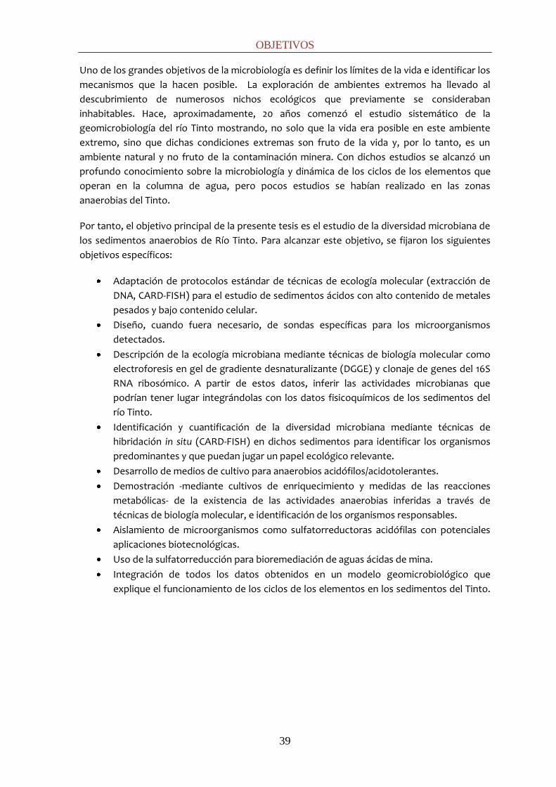
OBJETIVOS
39
Uno de los grandes objetivos de la microbiología es definir los límites de la vida e identificar los
mecanismos que la hacen posible. La exploración de ambientes extremos ha llevado al
descubrimiento de numerosos nichos ecológicos que previamente se consideraban
inhabitables. Hace, aproximadamente, 20 años comenzó el estudio sistemático de la
geomicrobiología del río Tinto mostrando, no solo que la vida era posible en este ambiente
extremo, sino que dichas condiciones extremas son fruto de la vida y, por lo tanto, es un
ambiente natural y no fruto de la contaminación minera. Con dichos estudios se alcanzó un
profundo conocimiento sobre la microbiología y dinámica de los ciclos de los elementos que
operan en la columna de agua, pero pocos estudios se habían realizado en las zonas
anaerobias del Tinto.
Por tanto, el objetivo principal de la presente tesis es el estudio de la diversidad microbiana de
los sedimentos anaerobios de Río Tinto. Para alcanzar este objetivo, se fijaron los siguientes
objetivos específicos:
Adaptación de protocolos estándar de técnicas de ecología molecular (extracción de
DNA, CARD-FISH) para el estudio de sedimentos ácidos con alto contenido de metales
pesados y bajo contenido celular.
Diseño, cuando fuera necesario, de sondas específicas para los microorganismos
detectados.
Descripción de la ecología microbiana mediante técnicas de biología molecular como
electroforesis en gel de gradiente desnaturalizante (DGGE) y clonaje de genes del 16S
RNA ribosómico. A partir de estos datos, inferir las actividades microbianas que
podrían tener lugar integrándolas con los datos fisicoquímicos de los sedimentos del
río Tinto.
Identificación y cuantificación de la diversidad microbiana mediante técnicas de
hibridación in situ (CARD-FISH) en dichos sedimentos para identificar los organismos
predominantes y que puedan jugar un papel ecológico relevante.
Desarrollo de medios de cultivo para anaerobios acidófilos/acidotolerantes.
Demostración -mediante cultivos de enriquecimiento y medidas de las reacciones
metabólicas- de la existencia de las actividades anaerobias inferidas a través de
técnicas de biología molecular, e identificación de los organismos responsables.
Aislamiento de microorganismos como sulfatorreductoras acidófilas con potenciales
aplicaciones biotecnológicas.
Uso de la sulfatorreducción para bioremediación de aguas ácidas de mina.
Integración de todos los datos obtenidos en un modelo geomicrobiológico que
explique el funcionamiento de los ciclos de los elementos en los sedimentos del Tinto.


METODOLOGÍA GENERAL


METODOLOGÍA GENERAL
43
Los materiales y métodos utilizados han sido descritos y referenciados en cada uno de los
capítulos de los que se compone la presente tesis. Sin embargo, debido al formato de las
revistas que obligan a una definición breve de los métodos, resulta conveniente una
explicación más detallada de aquellos procedimientos que no son rutinarios o han sido
puesto a punto exclusivamente en el transcurso de la presente tesis y que por lo tanto, no
están debidamente descritos en ninguna publicación previa.
EXTRACCIÓN DE DNA PARA SEDIMENTOS CON METALES PESADOS
Los sedimentos de río Tinto presentan baja densidad celular y un alto contenido en metales
pesados que inhiben posteriores procesos de amplificación de DNA. Por ello, para poder
obtener una cantidad suficiente de DNA amplificable, fue necesario la aplicación de un
tratamiento previo a la extracción (Fig. 1) detallado a continuación.
Figura 1: Tratamiento de muestras previo a la extracción de DNA
- Se resuspendió una porción de muestra (~6 gramos de sedimento) en 10 mL de
NaCl al 0,9% o PBS (1x) en tubos de 15 mL.
- Se agitó en noria durante 24 horas a 4ºC, combinándolo con pulsos de vortex (15
min, 3 veces).
- Se centrifugó a 500 x g durante 1 min para retirar las partículas de mayor tamaño.
- Se transfirió cuidadosamente el sobrenadante a otro tubo y se centrifugó de nuevo
a 8000 x g durante 20 min.
- Se desechó el sobrenadante.
- Se lavó dos veces el pellet con EDTA (0,5 M, pH 8) con objeto de quelar los metales
pesados presentes en la muestra.
- Se lavó el pellet con 10 mL de NaCl al 0,9% o PBS (1x) eliminando el exceso de EDTA.
- Se reservó el pellet para la extracción de DNA mediante el kit de extracción
FastDNA® SPIN® Kit (For Soil) (Qbiogene, USA) siguiendo el protocolo indicado por el
fabricante a excepción de la adicción de 100 μL de TE (1x) junto con el PPS para
proteger el DNA de los posibles metales pesados remanentes.
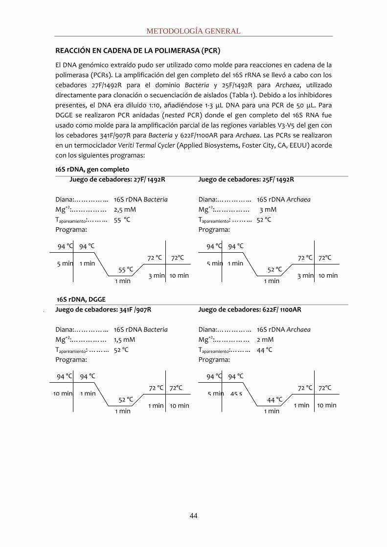
METODOLOGÍA GENERAL
44
REACCIÓN EN CADENA DE LA POLIMERASA (PCR)
El DNA genómico extraído pudo ser utilizado como molde para reacciones en cadena de la
polimerasa (PCRs). La amplificación del gen completo del 16S rRNA se llevó a cabo con los
cebadores 27F/1492R para el dominio Bacteria y 25F/1492R para Archaea, utilizado
directamente para clonación o secuenciación de aislados (Tabla 1). Debido a los inhibidores
presentes, el DNA era diluido 1:10, añadiéndose 1-3 μL DNA para una PCR de 50 μL. Para
DGGE se realizaron PCR anidadas (nested PCR) donde el gen completo del 16S RNA fue
usado como molde para la amplificación parcial de las regiones variables V3-V5 del gen con
los cebadores 341F/907R para Bacteria y 622F/1100AR para Archaea. Las PCRs se realizaron
en un termociclador Veriti Termal Cycler (Applied Biosystems, Foster City, CA, EEUU) acorde
con los siguientes programas:
16S rDNA, gen completo
Juego de cebadores: 27F/ 1492R
Diana:…………... 16S rDNA Bacteria
Mg+2:…………… 2,5 mM
Tapareamiento:……... 55 ºC
Programa:
16S rDNA, DGGE
Juego de cebadores: 341F /907R
Diana:…………... 16S rDNA Bacteria
Mg+2:…………… 1,5 mM
Tapareamiento: ……... 52 ºC
Programa:
Juego de cebadores: 25F/ 1492R
Diana:…………... 16S rDNA Archaea
Mg+2:…………… 3 mM
Tapareamiento: ……... 52 ºC
Programa:
Juego de cebadores: 622F/ 1100AR
Diana:…………... 16S rDNA Archaea
Mg+2:…………… 2 mM
Tapareamiento:……... 44 ºC
Programa:
94 ºC 94 ºC
5 min 45 s 72 ºC 72ºC
1 min 10 min 44 ºC
1 min
94 ºC 94 ºC
5 min 1 min 72 ºC 72ºC
3 min 10 min 52 ºC
1 min
94 ºC 94 ºC
10 min 1 min 72 ºC 72ºC
1 min 10 min 52 ºC
1 min
94 ºC 94 ºC
5 min 1 min 72 ºC 72ºC
3 min 10 min 55 ºC
1 min
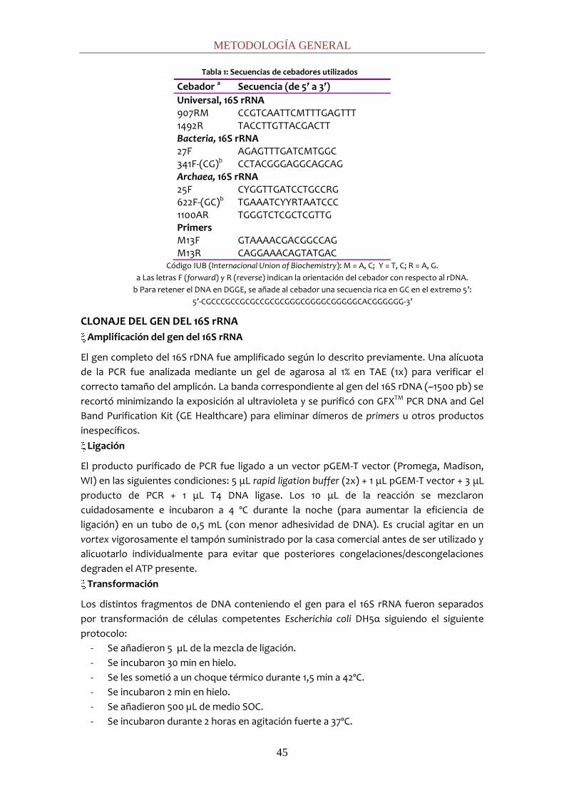
METODOLOGÍA GENERAL
45
Tabla 1: Secuencias de cebadores utilizados
Cebador a Secuencia (de 5’ a 3’) Universal, 16S rRNA 907RM CCGTCAATTCMTTTGAGTTT 1492R TACCTTGTTACGACTT Bacteria, 16S rRNA 27F AGAGTTTGATCMTGGC 341F-(CG)b CCTACGGGAGGCAGCAG Archaea, 16S rRNA 25F CYGGTTGATCCTGCCRG 622F-(GC)b TGAAATCYYRTAATCCC 1100AR TGGGTCTCGCTCGTTG Primers M13F GTAAAACGACGGCCAG M13R CAGGAAACAGTATGAC
Código IUB (Internacional Union of Biochemistry): M = A, C; Y = T, C; R = A, G.
a Las letras F (forward) y R (reverse) indican la orientación del cebador con respecto al rDNA.
b Para retener el DNA en DGGE, se añade al cebador una secuencia rica en GC en el extremo 5’:
5’-CGCCCGCCGCGCCGCGCGGGCGGGGCGGGGGCACGGGGGG-3’
CLONAJE DEL GEN DEL 16S rRNA
Amplificación del gen del 16S rRNA
El gen completo del 16S rDNA fue amplificado según lo descrito previamente. Una alícuota
de la PCR fue analizada mediante un gel de agarosa al 1% en TAE (1x) para verificar el
correcto tamaño del amplicón. La banda correspondiente al gen del 16S rDNA (~1500 pb) se
recortó minimizando la exposición al ultravioleta y se purificó con GFXTM PCR DNA and Gel
Band Purification Kit (GE Healthcare) para eliminar dímeros de primers u otros productos
inespecíficos.
Ligación
El producto purificado de PCR fue ligado a un vector pGEM-T vector (Promega, Madison,
WI) en las siguientes condiciones: 5 μL rapid ligation buffer (2x) + 1 μL pGEM-T vector + 3 μL
producto de PCR + 1 μL T4 DNA ligase. Los 10 μL de la reacción se mezclaron
cuidadosamente e incubaron a 4 ºC durante la noche (para aumentar la eficiencia de
ligación) en un tubo de 0,5 mL (con menor adhesividad de DNA). Es crucial agitar en un
vortex vigorosamente el tampón suministrado por la casa comercial antes de ser utilizado y
alicuotarlo individualmente para evitar que posteriores congelaciones/descongelaciones
degraden el ATP presente.
Transformación
Los distintos fragmentos de DNA conteniendo el gen para el 16S rRNA fueron separados
por transformación de células competentes Escherichia coli DH5α siguiendo el siguiente
protocolo:
- Se añadieron 5 µL de la mezcla de ligación.
- Se incubaron 30 min en hielo.
- Se les sometió a un choque térmico durante 1,5 min a 42ºC.
- Se incubaron 2 min en hielo.
- Se añadieron 500 µL de medio SOC.
- Se incubaron durante 2 horas en agitación fuerte a 37ºC.

METODOLOGÍA GENERAL
46
- Se sembraron distintas cantidades (25, 50 y 75 µL) en las placas de LB/Amp/X-gal.
- Tras una noche de crecimiento a 37ºC, los clones recombinantes fueron identificados
como colonias blancas y transferidos a nuevas placas para asegurar la correcta
asignación positiva de los clones.
Cultivo
Los clones que resultaron positivos fueron crecidos en 1,5 mL de medio Terrific Broth
(Sigma-Aldrich) en agitación a 37oC de 12-16 h en placas de 96 pocillos.
Lisis alcalina
El DNA de los plásmidos recombinantes fue extraído mediante lisis alcalina (1).
- Se centrifugaron las placas a 3750 rpm durante 20 min. Se volcaron para eliminar
sobrenadante.
- Se añadió al pellet 300 μL de TE (1x) y se resuspendió con vortex.
- Se centrifugó (3750 rpm) durante 20 min. Se eliminó el sobrenadante.
- Se añadió al pellet 100 μL de la solución 1 (fría) y se resuspendió con vortex.
SOLUCIÓN 1: 50 mM glucosa; 25 mM EDTA (pH 8, 0,5 M); 25 mM Tris-Cl (pH 8, 1 M).
Para preparar 100 mL se deben mezclar 4 mL EDTA (pH 8, 0,5 M)+ 1 g glucosa + 2,5 mL
Tris-Cl (pH 8, 1 M)+ 93 mL agua milli-Q y esterilizar a 0,5 atm durante 30 min.
- Se añadió al pellet 200 μL de la solución 2. Se tapó la placa e invertió 2-3 veces sin
agitar.
SOLUCIÓN 2: 0,2 N NaOH; 1% SDS. Es necesario preparar en el momento mezclando
800 μL de un stock NaOH (5 N) con 2000 μL de SDS (10%) y 400 μL de agua milli-Q.
Esterilizar por filtración con filtros de 0,22 μm (MilliPore).
- Se incubó en hielo a 4ºC durante 5 min.
- Se añadió 150 μL de la solución 3. Se tapó e invirtió 2-3 veces hasta que se enturbió.
SOLUCIÓN 3: Mezclar 60 mL de NaAc (5 M) con 11,5 mL HAc glacial y 28,5 mL agua
milli-Q. Esterilizar por filtración con filtros de 0,22 μm (MilliPore).
- Se incubó en hielo 10 min.
- Se centrifugó a 3750 rpm durante 45 min a 4ºC.
- Se transfirió el sobrenadante a una placa nueva.
- Se precipitó el DNA con 1 mL de isopropanol (previamente a –20ºC) overnight a -
20ºC.
- Se centrifugó a 3750 rpm durante 45 min a 4 ºC.
- Se eliminó el sobrenadante suavemente con pipeta y se añadieron, sin resuspender,
150 μL de etanol al 70% (previamente enfriado a –20ºC).
- Se centrifugó a 3750 rpm durante 30 min a 4 °C.
- Se eliminó el sobrenadante cuidadosamente.
- Se secó completamente el pellet al aire durante 1 h o en un speed-vac durante 10
min.
- Se resuspendió el pellet en 30 μL de agua milli-Q con RNAsa (50 μg mL-1). Por placa,
se añade 30 μL de RNAsa (10 mg mL-1) a 3000 μL de agua milli-Q.
- Se incubó 30 min a 37ºC.
- Se conservó a -20 °C.
El tamaño correcto de los insertos fue verificado mediante electroforesis convencional en
geles de agarosa a 0,8 % p/v en TAE (1x) cargando 3 μL del DNA plasmídico (Fig. 2). Los

METODOLOGÍA GENERAL
47
falsos positivos se distinguieron por su menor peso molecular al no tener inserto el 16S
rRNA (1500 pb).
Figura 2: Comprobación del tamaño de los insertos en los plásmidos. El plásmido utilizado contiene 3015 pares de
bases (según la información facilitada por el fabricante) que, unidos a los 1465 pb del inserto suman casi 4500 pb. Si
comprobamos la extracción del DNA plasmídico mediante electroforesis convencional, el plásmido unido al inserto
da como resultado una banda del tamaño de (i). Aquellos plásmidos en los que no se ha insertado nada generan
una banda tipo (ii), y en los que se ha insertado algo, pero de menor tamaño, como por ejemplo dímeros de
cebadores, la banda resultante es del tipo (iii).
Digestión del DNA plasmídico
Si el número de clones a analizar es muy alto, el coste de secuenciación puede ser elevado,
de modo que, mediante agrupamiento por digestión, se redujo el número de clones a
secuenciar. Para ello, el DNA plasmídico de los clones positivos se digirió enzimáticamente
(Amplified Ribosomal DNA Restriction Analysis, ARDRA) usando la endonucleasa BfuCI (1 U,
4 h, 37 ºC) mezclando 10 μL agua milli-Q + 1,5 μL tampón Bfu (10x) + 0,36 μL enzima BfuCI +
0,15 μL BSA (100x) + 3 μL DNA plasmídico. Los fragmentos resultantes fueron analizados en
un gel de agarosa al 3% p/v mediante electroforesis convencional a 40 mA durante 2 h (Fig.
3). Los clones fueron agrupados según los distintos patrones de restricción obtenidos. Se
secuenciaron dos representantes de cada grupo utilizando las pareja de cebadores
M13F/M13R, secuencias plasmidicas que flanquean la zona de inserción del gen del 16S
rRNA. En caso necesario se utilizaron cebadores internos cuya diana se encuentra en el 16S
rDNA (341F para Bacteria, 622F para Archaea).
Figura 3: Patrones de restricción obtenidos con la enzima Bfu CI.
Tamaño correcto: clon positivo (i)
Tamaño incorrecto: clon negativo (ii)
Tamaño incorrecto: clon negativo (iii)
4500 pb
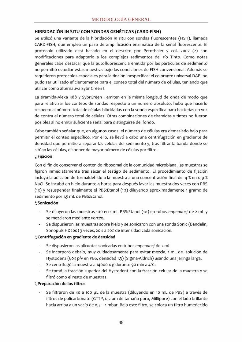
METODOLOGÍA GENERAL
48
HIBRIDACIÓN IN SITU CON SONDAS GENÉTICAS (CARD-FISH)
Se utilizó una variante de la hibridación in situ con sondas fluorescentes (FISH), llamada
CARD-FISH, que emplea un paso de amplificación enzimática de la señal fluorescente. El
protocolo utilizado está basado en el descrito por Pernthaler y col. 2002 (2) con
modificaciones para adaptarlo a los complejos sedimentos del río Tinto. Como notas
generales cabe destacar que la autofluorescencia emitida por las partículas de sedimento
no permitió estudiar estas muestras bajo las condiciones de FISH convencional. Además se
requirieron protocolos especiales para la tinción inespecífica: el colorante universal DAPI no
pudo ser utilizado eficientemente para el conteo total del número de células, teniendo que
utilizar como alternativa Sybr Green I.
La tiramida-Alexa 488 y SybrGreen I emiten en la misma longitud de onda de modo que
para relativizar los conteos de sondas respecto a un numero absoluto, hubo que hacerlo
respecto al número total de células hibridadas con la sonda específica para bacterias en vez
de contra el número total de células. Otras combinaciones de tiramidas y tintes no fueron
posibles al no emitir suficiente señal para distinguirse del fondo.
Cabe también señalar que, en algunos casos, el número de células era demasiado bajo para
permitir el conteo específico. Por ello, se llevó a cabo una centrifugación en gradiente de
densidad que permitiera separar las células del sedimento y, tras filtrar la banda donde se
sitúan las células, disponer de mayor número de células por filtro.
Fijación
Con el fin de conservar el contenido ribosomal de la comunidad microbiana, las muestras se
fijaron inmediatamente tras sacar el testigo de sedimento. El procedimiento de fijación
incluyó la adicción de formaldehído a la muestra a una concentración final del 4 % en 0,9 %
NaCl. Se incubó en hielo durante 4 horas para después lavar las muestra dos veces con PBS
(1x) y resuspender finalmente el PBS:Etanol (1:1) diluyendo aproximadamente 1 gramo de
sedimento por 1,5 mL de PBS:Etanol.
Sonicación
- Se diluyeron las muestras 1:10 en 1 mL PBS:Etanol (1:1) en tubos eppendorf de 2 mL y
se mezclaron mediante vortex.
- Se dispusieron las muestras sobre hielo y se sonicaron con una sonda Sonic (Bandelin,
Sonopuls HD200) 3 veces, 20 s a 20% de intensidad cada sonicación.
Centrifugación en gradiente de densidad
- Se dispusieron las alicuotas sonicadas en tubos eppendorf de 2 mL.
- Se incorporó debajo, muy cuidadosamente para evitar mezcla, 1 mL de solución de
Hystodenz (60% p/v en PBS, densidad 1,3) (Sigma-Aldrich) usando una jeringa larga.
- Se centrifugó la muestra a 14000 x g durante 90 min a 4ºC.
- Se tomó la fracción superior del Hystodent con la fracción celular de la muestra y se
filtró como el resto de muestras.
Preparación de los filtros
- Se filtraron de 40 a 100 μL de la muestra (diluyendo en 10 mL de PBS) a través de
filtros de policarbonato (GTTP, 0,2 µm de tamaño poro, Millipore) con el lado brillante
hacia arriba a un vacío de 0,5 – 1 mbar. Bajo este filtro, se coloca un filtro humedecido

METODOLOGÍA GENERAL
49
en PBS de Celulosa-Nitrato (HAMP, 0,45 µm de tamaño poro, Millipore) para
homogeneizar la presión y propiciar la homogeneidad de la muestra-
- Se dejaron secar los filtros al aire en placas Petri. Es importante teñir la muestra y
comprobar que la dilución sea la adecuada, como regla general debería haber unas
50-100 células por campo o deberían estar cubiertos unos 2/3 del filtro con
sedimento.
Tincion inespecífica
- DAPI: las secciones se colocaron boca-arriba sobre los porta-objetos y se embebieron
en 8 µL de la mezcla de DAPI/Citifluor (1 µg mL-1). Se dejaron al menos 30 min a -20ºC
para intensificar la señal del DAPI.
- SybrGreen I: Las secciones se colocaron boca-arriba sobre los porta-objetos y se
embebieron en 8 µL de la mezcla de SybrGreen/Moviol Medium (1:40) preparado
según lo descrito en Lunau and col., 2005 (3). Se dejaron al menos 30 min a -20ºC para
intensificar la señal.
Inclusión de los filtros en agarosa
- Se precalentó agarosa LMP (low melting point) 0,1 % en microondas hasta ebullición, y
se atemperó hasta ~50°C.
- Se dispuso en un spray y se roció el filtro 2-3 veces a 50 cm de distancia.
- Se dejaron secar los filtros con las placas abiertas durante 20-30 min a 37°C hasta que
la agarosa estuvo seca.
Inactivación de las peroxidasas endógenas
- Se incubaron los filtros boca-arriba en H2O2/metanol [0,15% H2O2/metanol = 50 L H2O2
(30%) en 10 mL metanol] durante 30 min a temperatura ambiente en placa Petri
sellada con parafilm o en campana de extracción para evitar el riesgo de inhalación
por el extenso periodo de exposición.
- Se lavaron los filtros en agua durante 1 min a temperatura ambiente.
- Se lavaron los filtros en etanol absoluto durante 1 min a temperatura ambiente.
- Se dejaron secar completamente al aire.
- Se cortaron con bisturí y se rotularon con lápiz blando (HB, 2B).
Permeabilización
con lisozima (recomendada para mayoría de las bacterias)
- Se preparó una solución fresca de lisozima [10 mg mL-1 en EDTA (0,05 M, pH 8); Tris-
HCl (0,1 M, pH 8) = 8 mL agua milli-Q + 1 mL Tris-HCl (0,1 M, pH 8) + 1 mL EDTA (0,05 M,
pH 8) + 100 mg lisozima].
- Se incubaron los filtros boca-abajo durante 1 h.
- Se lavaron los filtros con agua milli-Q durante 1 min a temperatura ambiente.
con acromopeptidasa (recomendada para Firmicutes)
- Se incubaron los filtros durante 30 min a 37ºC boca-abajo en acromopeptidasa (60 U
mL-1) = 9860 L agua milli-Q + 20 L NaCl (5 M) + 100 L Tris-HCl (1 M, pH 8) + 20 L
stock acromopeptidasa (30000 U mL-1).
con Proteinasa-K (recomendada para arqueas):

METODOLOGÍA GENERAL
50
- Se preparó una solución de trabajo de Proteinasa-K [15 µg mL-1 en EDTA (0,05 M, pH
8); Tris-HCl (0,1 M, pH 8) = 8 mL agua milli-Q + 1 mL Tris-HCl (0,1 M, pH 8) + 1 mL EDTA
(0,05 M, pH 8)+ 7,5 µL stock Proteinasa (20 mg mL-1)].
- Se incubaron los filtros durante 3 min a 37 ºC.
- Se inactivó la Proteinasa-K en 10 mL de HCl (0,1 M) durante 1 min a temperatura
ambiente.
- Se lavaron los filtros en agua milli-Q y se dejaron secar completamente al aire.
Hibridación
- Se mezcló el tampón de hibridación (Tabla 2) con la solución de trabajo de la sonda
(50 ng µL-1) en un ratio de 300:1 v/v en un tubo eppendorf de 2 mL. - Tabla 2: Tampón de Hibridación para sondas marcadas con HRP
Reactivos Volumen 10 ml 5 M NaCl 1800 μL 1 M Tris-HCl pH 8 200 μL Sulfato de Dextrano 1 g Formamida (x%) 0 μl - 0% /6000 µL - 60% Agua milli-Q 6990 μL - 0% / 990 μL - 60% Agente bloqueante* 1000 μL SDS (10%) 10 μL
- *Agente bloqueante: 10% en tampón de ácido maleico.
- Tampón de ácido maleico: 100 mM de ácido maleico; 150 mM NaCl (pH 5)
- Se dispusieron las secciones de filtro pegadas a los laterales del eppendorf con la cara
con muestra hacia el interior para evitar desprendimiento de la misma.
- Se hibridaron las muestras estáticamente a 46ºC durante 2 h.
Lavado
- Se preparó y precalentó el tampón de lavado a 48ºC.
- Se lavaron los filtros durante 10 min a 48ºC.
- Se incubaron las secciones en 50 mM de PBS (1x) durante 15 min a temperatura
ambiente para equilibrar la sonda con la peroxidasa.
Amplificación
- Se preparó una solución stock fresca de H2O2 (100x) [0,15 % H2O2/PBS (1x) = 1 µL H2O2
30% en 200 µL PBS (1x)].
- Se mezclaron 1 mL del tampón de amplificación (Tabla 3) con 10 µL de la solución
anterior de H2O2 (100x) y 1 µL de la tiramida marcada con el cromóforo fluorescente
Alexa 488 (1 mg fluorocromo mL-1) en un tubo de 2 mL donde se introdujeron
cuidadosamente las secciones de filtro.
- Se incubaron durante 45 min a 46ºC en oscuridad.
- Se transfirieron sucesivamente las secciones a 50 mL de agua milli-Q y a etanol 96%.
- Se secaron las secciones al aire.
- Se tiñeron inespecíficamente con DAPI según lo descrito anteriormente. - Tabla 3: Tampón de Hibridación para sondas marcadas con HRP
Reactivos Volumen 50 ml 5 M NaCl 12 mL PBS (10x) 4 mL Agente bloqueante 10%* 0,4 mL Sulfato de Dextrano 4 g

METODOLOGÍA GENERAL
51
Análisis y cuantificación de células por microscopía
Las muestras hibridadas fueron observadas con un microscopio de epifluorescencia y
cuantificadas manualmente. Los cálculos porcentuales se realizaron frente al número de
células hibridadas con la sonda específica de bacteria en vez de contra el número de células
totales. Se contaron alrededor de 1000 células hibridadas por muestra cuando era posible,
sino era el caso, al menos 200 campos.
Para estimar número total de células por gramo de sedimento presentes en la muestra
original a partir del número de células contadas por campo se consideraron los siguientes
parámetros y sus valores:
Área del filtro: 1,93·108 µm2.
Área de la rejilla contada (microscopio): 1,5·104 µm2.
Correspondiendo el peso y volumen de muestra a la relación 1 g: 1,5 mL PBS:etanol.
Y se aplicó la siguiente fórmula.
N = mediacélulas* factor dilución*área filtro
árearejilla*
volumenmuestra
volumen filtradopesomuestra
Diseño de sondas
Los altos porcentajes de hibridación obtenidos para la sonda específica de
Deltaproteobacteria, la abundancia de clones pertenecientes a Desulfurella obtenidos
durante este trabajo unido a la ausencia de una sonda específica de Desulfurella
previamente diseñada, hizo necesario el diseño de una nueva sonda para este género. Para
diseñar una nueva sonda es preciso seleccionar una secuencia de oligonucleótidos
completamente específica, es decir, complementaria a una región de las secuencias diana y
que se diferencie en, al menos, un nucleótido (mismatch) de la misma región del resto de
secuencias (organismos) no diana. La sonda específica para el género Desulfurella fue
diseñada usando la función PROBE_DESIGN del paquete informático ARB. Para ello,
- Se seleccionaron como secuencias diana aquellas secuencias de Desulfurella
obtenidas en los clonajes y DGGE y todas las secuencias de la base de datos de ARB
incluidas en el género.
- Se comprobó y mejoró el alineamiento de las secuencias diana manualmente en la
ventana ALIGN de ARB.
- Con las secuencias diana marcadas, se abrió la ventana PROBE DESIGN.
- Se seleccionó el PT_SERVER actualizado y se definieron los parámetros: “minimun
percentage of group hits” a 100, “máximum number of nongroup hits” a 0 y la
longitud de la secuencia se dejó, por defecto, en 18 nucleótidos.
- Se presionó el botón GO. Los resultados aparecen en la ventana PD RESULT (Fig. 4),
mostrando las potenciales sondas y sus parámetros asociados como el largo de la
secuencia, la temperatura de disociación, el contenido G+C y la localización respecto
a Escherichia coli.
Figura 4: Ventana PD RESULT del paquete informático ARB.
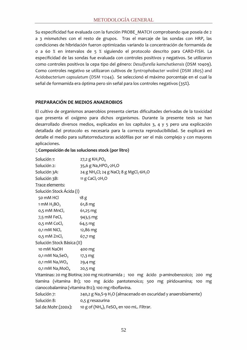
METODOLOGÍA GENERAL
52
Su especificidad fue evaluada con la función PROBE_MATCH comprobando que poseía de 2
a 3 mismatches con el resto de grupos. Tras el marcaje de las sondas con HRP, las
condiciones de hibridación fueron optimizadas variando la concentración de formamida de
0 a 60 % en intervalos de 5 % siguiendo el protocolo descrito para CARD-FISH. La
especificidad de las sondas fue evaluada con controles positivos y negativos. Se utilizaron
como controles positivos la cepa tipo del género: Desulfurella kamchatkensis (DSM 10409).
Como controles negativo se utilizaron cultivos de Syntrophobacter wolinii (DSM 2805) and
Acidobacterium capsulatum (DSM 11244). Se seleccionó el máximo porcentaje en el cual la
señal de formamida era óptima pero sin señal para los controles negativos (35%).
PREPARACIÓN DE MEDIOS ANAEROBIOS
El cultivo de organismos anaerobios presenta ciertas dificultades derivadas de la toxicidad
que presenta el oxígeno para dichos organismos. Durante la presente tesis se han
desarrollado diversos medios, explicados en los capítulos 3, 4 y 5 pero una explicación
detallada del protocolo es necesaria para la correcta reproducibilidad. Se explicará en
detalle el medio para sulfatorreductoras acidófilas por ser el más complejo y con mayores
aplicaciones.
Composición de las soluciones stock (por litro)
Solución 1: 27,2 g KH2PO4
Solución 2: 35,6 g Na2HPO4·2H2O
Solución 3A: 24 g NH4Cl; 24 g NaCl; 8 g MgCl2·6H2O
Solución 3B: 11 g CaCl2·2H2O
Trace elements:
Solución Stock Ácida (I)
50 mM HCl 18 g
1 mM H3BO3 61,8 mg
0,5 mM MnCl2 61,25 mg
7,5 mM FeCl2 943,5 mg
0,5 mM CoCl2 64,5 mg
0,1 mM NiCl2 12,86 mg
0,5 mM ZnCl2 67,7 mg
Solución Stock Básica (II)
10 mM NaOH 400 mg
0,1 mM Na2SeO3 17,3 mg
0,1 mM Na2WO4 29,4 mg
0,1 mM Na2MoO4 20,5 mg
Vitaminas: 20 mg Biotina; 200 mg nicotinamida ; 100 mg ácido p-aminobenzoic0; 200 mg
tiamina (vitamina B1); 100 mg ácido pantotenoico; 500 mg piridoxamina; 100 mg
cianocobalamina (vitamina B12); 100 mg riboflavina.
Solución 7: 240,2 g Na2S·9 H2O (almacenado en oscuridad y anaerobiamente)
Solución 8: 0,5 g resazurina
Sal de Mohr (200x): 10 g of (NH4)2 FeSO4 en 100 mL. Filtrar.
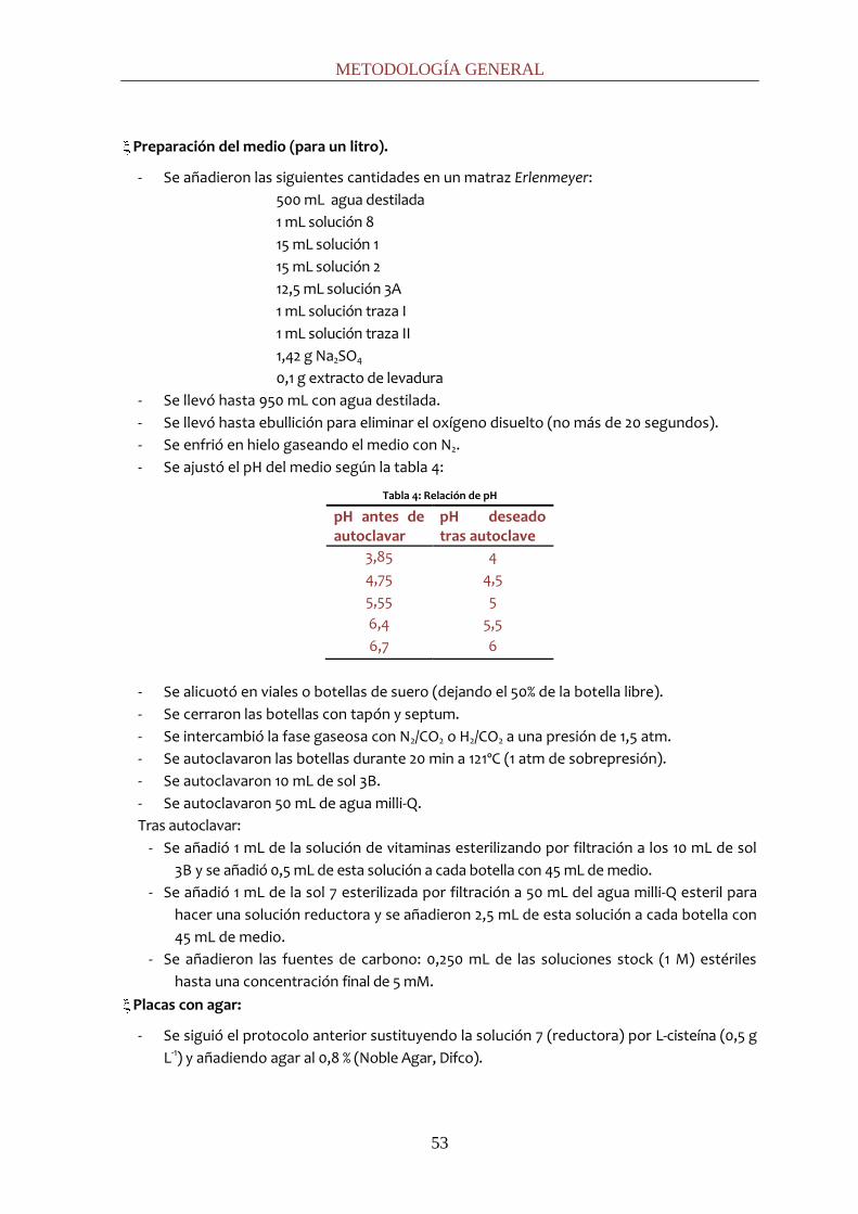
METODOLOGÍA GENERAL
53
Preparación del medio (para un litro).
- Se añadieron las siguientes cantidades en un matraz Erlenmeyer:
500 mL agua destilada
1 mL solución 8
15 mL solución 1
15 mL solución 2
12,5 mL solución 3A
1 mL solución traza I
1 mL solución traza II
1,42 g Na2SO4
0,1 g extracto de levadura
- Se llevó hasta 950 mL con agua destilada.
- Se llevó hasta ebullición para eliminar el oxígeno disuelto (no más de 20 segundos).
- Se enfrió en hielo gaseando el medio con N2.
- Se ajustó el pH del medio según la tabla 4:
Tabla 4: Relación de pH
pH antes de autoclavar
pH deseado tras autoclave
3,85 4
4,75 4,5
5,55 5
6,4 5,5
6,7 6
- Se alicuotó en viales o botellas de suero (dejando el 50% de la botella libre).
- Se cerraron las botellas con tapón y septum.
- Se intercambió la fase gaseosa con N2/CO2 o H2/CO2 a una presión de 1,5 atm.
- Se autoclavaron las botellas durante 20 min a 121ºC (1 atm de sobrepresión).
- Se autoclavaron 10 mL de sol 3B.
- Se autoclavaron 50 mL de agua milli-Q.
Tras autoclavar:
- Se añadió 1 mL de la solución de vitaminas esterilizando por filtración a los 10 mL de sol
3B y se añadió 0,5 mL de esta solución a cada botella con 45 mL de medio.
- Se añadió 1 mL de la sol 7 esterilizada por filtración a 50 mL del agua milli-Q esteril para
hacer una solución reductora y se añadieron 2,5 mL de esta solución a cada botella con
45 mL de medio.
- Se añadieron las fuentes de carbono: 0,250 mL de las soluciones stock (1 M) estériles
hasta una concentración final de 5 mM.
Placas con agar:
- Se siguió el protocolo anterior sustituyendo la solución 7 (reductora) por L-cisteína (0,5 g
L-1) y añadiendo agar al 0,8 % (Noble Agar, Difco).
METODOLOGÍA GENERAL
54
REFERENCIAS
1. Birnboim, H., and J. Doly. 1979. A rapid alkaline extraction method for screening recombinant plasmid DNA. Nucleic Acids Res. 7:1513-1523.
2. Pernthaler, A., J. Pernthaler, and R. Amann. 2002. Fluorescence in situ hybridization and catalyzed reporter deposition for the identification of marine bacteria. Appl. Environ. Microbiol. 68:3094-3101.
3. Lunau, M., A. Lemke, K. Walther, W. Martens-Habbena, and M. Simon. 2005. An improved method for counting bacteria from sediments and turbid environments by epifluorescence microscopy. Environ. Microbiol. 7:961-968
LISTADO DE PUBLICACIONES


LISTADO DE PUBLICACIONES
57
Los capítulos que integran este proyecto de tesis doctoral fueron redactados íntegramente en
inglés acorde con los requisitos de publicación en revistas de ámbito internacional. A
continuación se enumeran los artículos producidos en el transcurso de la presente tesis: título,
autores y estado de publicación.
Sánchez-Andrea I., N. Rodriguez, R. Amils, and J. L. Sanz.
Microbial Diversity in Anaerobic Sediments at Rio Tinto, a Naturally Acidic Environment with a
High Heavy Metal Content.
Applied and Environmental Microbiology (2011) 77:6085-6093.
Sánchez-Andrea I., K. Knittel, R. Amann, R. Amils, and J. L. Sanz.
Quantification of Tinto River Sediments Microbial Communities: the Importance of Sulfate-
Reducing Bacteria and their Role in Attenuating Acid Mine Drainage. Applied and
Environmental Microbiology (2012) 78 (13): doi: 10.1128/AEM.00848-12
Sánchez-Andrea I., P. Rojas, R. Amils, and J. L. Sanz.
Screening of Anaerobic Activities in Sediments of an Acidic Environment: Tinto River
Aceptado en Extremophiles el 25-Abril-2012: EXT-12-Feb-0043.R1
Sánchez-Andrea I., A.J.M. Stams, R. Amils and J.L. Sanz.
Enrichment and isolation of acidophilic sulfate-reducing bacteria from Tinto River sediments
Environmental Microbiology reports (Subscrito Junio 2012. En revisión)
Sánchez-Andrea I., D.Triana and J.L. Sanz
Bioremediation of acid mine drainage coupled with domestic wastewater treatment.
Water Science and Technology (Subscrito Abril 2012. En revisión)
Sánchez-Andrea I., N.Rodriguez, R. Amils, and J. L. Sanz
Microbial diversity of anaerobic zones of Tinto river combining culture dependent and non-
dependent methods.
En preparación.
Amils, R., E. Gonzalez-Toril, A. Aguilera, N. Rodriguez, D. Fernandez-Remolar, F. Gomez, A.
Garcia-Moyano, M. Malki, M. Oggerin, I. Sánchez-Andrea, and J. L. Sanz.
From Río Tinto to Mars: the terrestrial and extraterrestrial ecology of acidophiles.
Advances in Applied Microbiology (2011) 77:41-70.


CHAPTER 1:
Microbial Diversity of
Anaerobic Sediments of
Río Tinto: a Natural Acid and
High Heavy Metals Content
Environment


MICROBIAL DIVERSITY OF TINTO RIVER SEDIMENTS
61
INTRODUCTION
Pyrite (FeS2) is the most abundant sulfide mineral in Earth's crust. When it is exposed to air and
water, its oxidation can be carried out with either atmospheric oxygen or ferric iron, according
to the equations 1 to 3.
FeS2 + 3.5O2 + H2O → Fe2+ + 2SO42- + 2H+ (eq. 1)
Fe2+ + H+ + 0.25O2 → Fe3+ + 0.5H2O (eq. 2)
FeS2 + 14Fe3+ + 8H2O → 15Fe2+ + 2SO42- + 16H+ (eq. 3)
Ferric iron has been shown to oxidize pyrite 18 to 170 times more rapidly than O2, producing
waters with a low pH and a high iron content. However, this step is limited by the rate of
ferrous iron oxidation (equation 2), which is greatly increased (by up to 5 orders of magnitude)
at low pH through the action of Fe-oxidizing chemolithoautotrophs (38), such as the bacteria
Acidithiobacillus spp. and Leptospirillum spp. Furthermore, low pH facilitates metal
solubilization, particularly that of cationic metals; therefore, acidic water tends to be highly
metalliferous (24). Pyrite and ferrous iron oxidation are both processes that occur naturally
and, indeed, natural acidic drainage has been identified in many locations (44). Mining in these
areas is known to dramatically increase the ferrous iron oxidation rates (39).
The Tinto River is a natural acidic drainage environment. It rises in Peña de Hierro, in the core
of the Iberian Pyritic Belt (IPB), and reaches the Atlantic Ocean at Huelva, Spain. The IPB is one
of the largest massive sulfidic deposits on Earth (250 km-long) and formed as a hydrothermal
deposit during the Paleozoic accretion of the Iberian Peninsula (32). Although the IPB has been
subjected to mining activities for thousands of years (45), a recent analysis has shown that the
iron oxides present in its sedimentary deposits are the result of the metabolic activity of
acidophilic prokaryotes (14). The Tinto River is an unusual extreme ecosystem due to its size
(100 km long), constant acidic pH (mean pH, 2.3) and high concentration of heavy metals, iron
and sulfate in its waters, characteristics that make Tinto River Basin comparable to Acidic Mine
Drainage (AMD) systems.
The Tinto River has been the focus of an increasing amount of research including
biogeochemistry (exploring the nature of the microbial communities associated with AMD),
biotechnology (bioleaching processes), microbiology (acidic environments offer an almost
unique opportunity to reveal habitat biological complexity) and Mars-analogue studies
(jarosite is a common mineral on both Mars and Earth). Consequently, much is already known
about the microbiota inhabiting the water column (1, 15, 18, 43, 52). Microbial ecology studies
have shown that ca. 80% of the prokaryotic diversity in the water is explained by the presence
of three bacteria Acidithiobacillus ferrooxidans, Leptospirillum ferrooxidans and Acidiphilium
spp., all of which are all involved in the iron cycle. Some other species related to the iron cycle
have also been identified (i.e. Ferroplasma acidiphilum or Ferrimicrobium acidiphilum), but their
low number suggests that they play a minor role in this cycle (18). Despite the low prokaryotic
diversity, an unexpected degree of eukaryotic diversity has been found, and is the principal
contributor of biomass in this hostile river (1, 52).
Despite ecological interest in the underlying sediments, they have been studied only very
sparsely and studies have mainly focused on methanogens (46). So far, no complete studies of
the anaerobic microbial diversity were undertaken. In this paper we present an extensive
survey of the Tinto River anaerobic sediment microbiota, using culture-independent

CHAPTER 1
62
approaches targeting the small-subunit (SSU) rRNA such as PCR-denaturing gradient gel
electrophoresis (DGGE) and cloning of 16S rRNA genes. Furthermore, a model of the iron,
sulfur and carbon cycles is proposed to elucidate the microbial ecology of AMD-related
sediments.
MATERIALS AND METHODS
Field site description and sampling
The acidic sediments studied are located in Tinto River basin (Huelva), in southwestern Spain.
Samples were collected from two sampling sites, SN dam (Universal Transverse Mercator
[UTM], E715378-N4178002) and JL dam (UTM: E715073-N4174390), in March 2008 and March
2009. Sediment cores (inner diameter, 7 cm; length, 45 cm) were taken with a sampler
(Eijkelkamp Agrisearch equipment, Giesbeek, The Netherlands). The redox potential (E) and
pH of the drill core samples were measured in situ with E and pH probes connected to a
Thermo Orion 290A potentiometer placed into the fresh sediment just after extracting the
core. The cores were sliced according to both physicochemical gradients and visual aspects,
and the slices were kept separately until processing in the laboratory.
Nucleic acid extraction from sediments
Total DNA was extracted from 6 g of each slice of sediment using FastDNA® SPIN® Kit (for
Soil) (Qbiogene, USA). Adaptation of the commercial protocol was carried out to optimise it
for sediments with high concentrations of heavy metals and low biomass content. Samples
were first resuspended in 10 mL of phosphate-buffered saline (PBS) and sonicated to detach
cells from the solid phase. The sediment was centrifuged at 500 x g for 1 min in order to
remove larger particles; the supernatant was carefully removed and centrifuged again at 8000
x g for 20 min. After the supernatant was discarded, the remaining pellet was resuspended in
10 mL 0.5 M EDTA, pH 8 and incubated overnight at 4ºC to dissolve humic substances and
remove heavy metals. After incubation, sediment was washed with PBS and DNA extraction
procedures were applied to the pellet.
Denaturing gradient gel electrophoresis (DGGE) from sediment samples
DGGE analyses were carried out in samples from March 2008 to get a first insight of the
microbial diversity of the sediments. The V3 to V5 variable regions of the 16S rRNA gene were
amplified with the primers set 341F (GC)-907R (annealing temperature (Ta=52ºC) for Bacteria
and 622F (GC)-1100R (Ta =42ºC) for Archaea. Primers 341F (GC) and 622F (GC) included a GC
clamp: 5’-CGC CCG CCG CGC CCC GCG CCC GTC CCG CCC CCG CCC-3’ (37). The amplification
reaction was performed according to the Taq DNA Polymerase protocol (Promega Madison,
Wis). The PCR conditions were as follow: 10 min of initial denaturation at 94ºC and 30 cycles at
94ºC for 1 min and annealing at 52/42ºC for 1 min and 72ºC for 2 min, followed by 10 min of final
primer extension. DGGE analysis was carried out using a D-Code Universal Detection System
instrument (Bio-Rad) and a model 475 gradient former according to the manufacturer’s
instructions (Bio-Rad). Polyacrylamide (6%; 37.5:1 acrylamide-bisacrylamide) gels with a 30 to
60% urea-formamide denaturant gradient (100% urea-formamide contains 7 M urea and 40%
deionized formamide) were used in 1x TAE (Tris-acetate-EDTA) buffer, pH 7.4, at 200 V for 4 h
at 60°C. Gels were stained with ethidium bromide and visualized under UV illumination. About
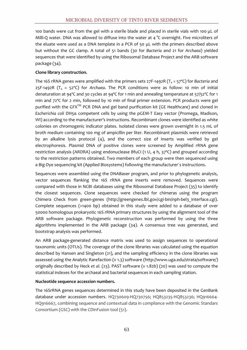
MICROBIAL DIVERSITY OF TINTO RIVER SEDIMENTS
63
100 bands were cut from the gel with a sterile blade and placed in sterile vials with 100 μL of
Milli-Q water. DNA was allowed to diffuse into the water at 4 °C overnight. Five microliters of
the eluate were used as a DNA template in a PCR of 50 μL with the primers described above
but without the GC clamp. A total of 51 bands (30 for Bacteria and 21 for Archaea) yielded
sequences that were identified by using the Ribosomal Database Project and the ARB software
package (34).
Clone library construction.
The 16S rRNA genes were amplified with the primers sets 27F-1492R (Ta = 57ºC) for Bacteria and
25F-1492R (Ta = 52ºC) for Archaea. The PCR conditions were as follow: 10 min of initial
denaturation at 94ºC and 30 cycles at 94ºC for 1 min and annealing temperature at 57/52ºC for 1
min and 72ºC for 2 min, followed by 10 min of final primer extension. PCR products were gel
purified with the GFXTM PCR DNA and gel band purification kit (GE Healthcare) and cloned in
Escherichia coli DH5α competent cells by using the pGEM-T Easy vector (Promega, Madison,
WI) according to the manufacturer’s instructions. Recombinant clones were identified as white
colonies on chromogenic indicator plates. Isolated clones were grown overnight in 1.5 mL of
broth medium containing 100 mg of ampicillin per liter. Recombinant plasmids were retrieved
by an alkaline lysis protocol (4), and the correct size of inserts was verified by gel
electrophoresis. Plasmid DNA of positive clones were screened by Amplified rRNA gene
restriction analysis (ARDRA) using endonuclease BfuCI (1 U, 4 h, 37ºC) and grouped according
to the restriction patterns obtained. Two members of each group were then sequenced using
a Big-Dye sequencing kit (Applied Biosystems) following the manufacturer´s instructions.
Sequences were assembled using the DNABaser program, and prior to phylogenetic analysis,
vector sequences flanking the 16S rRNA gene inserts were removed. Sequences were
compared with those in NCBI databases using the Ribosomal Database Project (35) to identify
the closest sequences. Clone sequences were checked for chimeras using the program
Chimera Check from green-genes (http://greengenes.lbl.gov/cgi-bin/nph-bel3_interface.cgi).
Complete sequences (>1400 bp) obtained in this study were added to a database of over
50000 homologous prokaryotic 16S rRNA primary structures by using the alignment tool of the
ARB software package. Phylogenetic reconstruction was performed by using the three
algorithms implemented in the ARB package (34). A consensus tree was generated, and
bootstrap analysis was performed.
An ARB package-generated distance matrix was used to assign sequences to operational
taxonomic units (OTUs). The coverage of the clone libraries was calculated using the equation
described by Hansen and Singleton (21), and the sampling efficiency in the clone libraries was
assessed using the Analytic Rarefaction (v 1.3) software (http://www.uga.edu/strata/software/)
originally described by Heck et al. (23). PAST software (v 1.82b) (20) was used to compute the
statistical indexes for the archaeal and bacterial sequences in each sampling station.
Nucleotide sequence accession numbers.
The 16SrRNA genes sequences determined in this study have been deposited in the GenBank
database under accession numbers. HQ730609-HQ730756; HQ853235-HQ853236; HQ916664-
HQ916667, combining sequence and contextual data in compliance with the Genomic Standars
Consortium (GSC) with the CDinFusion tool (51).

CHAPTER 1
64
RESULTS
Physicochemical parameters of the sampling site
Significant differences between both sample sites (JL and SN) were observed (Table 1).
Table 1: Physicochemical parameters of both sample sites over two years.
year 2008 year 2009
Sample Depth (cm)
pH Redox (mV)
Depth (cm)
pH Redox (mV)
JL1 8 3.8 168 7 3.8 200 JL2 11 3.9 69 12 4.6 -13 JL3 14 4 113 16 4 53 JL4 26 4.4 8 22 4.1 12 JL5 30 5.3 -120 26 4.4 1 JL6 33 4.9 -27 33 5.7 -132 JL7 38 5.4 -71 40 4.7 -30 SN1 21-26 3.9 200 25 2.3 255 SN2 40-45 4 -13 43 3.63 47
JL cores were banded, showed local variations from oxidizing zones to reducing zones with a
redox range from 200 to -120 and pH range from 3.9 to 5.7. Meanwhile, the SN sampling site
comprised only two layers and showed a more oxidized zone in the superficial layer defined by
the occurrence of brownish Fe(III) (hydr)oxides, a positive redox potential (+200mV), a low pH
of 2 to 4, and a deeper layer with less oxidant or even reducing redox potential.
DGGE fingerprint analyses
Both sites were analysed at different depths to evaluate the level of diversity and to elucidate
the possible correlation between the distribution of microorganisms with the physicochemical
parameters. The bacterial fingerprint (Fig. 1A) showed a relatively high number of bands
compared to the low number found in previous water column studies (18).
Fig. 1: DGGE fingerprints of 16S rRNA obtained with domain-specific primers for Bacteria (A) and Archaea (B). Lane names are shown according to depth and sample site.
The pattern of bands differed between the two sampling sites and, on a global perspective,
between depths. Bacteria sequences clustered in the phyla Proteobacteria (classes
Gammaproteobacteria and Deltaproteobacteria), Bacteroidetes, Acidobacteria and Firmicutes.
As seen in Fig. 2, there is a correlation between layers and the phylogenetic affiliation of the
sequences.

MICROBIAL DIVERSITY OF TINTO RIVER SEDIMENTS
65
Fig. 2: Phylogenetic affiliation of bacterial DGGE sequences obtained from the Tinto River samples. The phylogenetic tree was generated using parsimony with the ARB program. The bar indicates a 10% estimated sequences divergence. The sequences from Tinto River are indicated in bold type, and designations after the organism names or identifiers are GenBank accession numbers
In the surface layers of JL dam (JL1 and JL2), sequences belong to the phylum Acidobacteria,
which includes acidophilic bacteria typically found in soils. In the next layer (JL3) sequences
belong to the phylum Firmicutes, clustering in the order Clostridiales (Sporacetigenium spp.)
and Bacillales (Alicyclobacillus spp). In the deepest layers (JL5, JL6, JL7), extremely anaerobic
organisms were found, including sulfate-reducing bacteria, such as Desulfurella and
Desulfosporosinus spp., and Syntrophobacter spp, propionate-degrading syntrophic bacteria
typically from neutral pH environments, as well as Paludibacter spp., a fermenting
Bacteroidetes. In the surface layer of SN dam (SN1), sequences were identified as belonging to
the family Acidithiobacillaceae (Gammaproteobacteria class). In the deeper layer (SN2),
organisms were related to the spore-forming sulfate-reducing bacteria Desulfosporosinus spp.
The archaeal fingerprints (Fig.1B) had a lower number of bands than the Bacteria and their
phylogenetic distribution was less varied; in fact, all the Archaea sequences cluster in the
phylum Euryarchaeota, in the order Thermoplasmatales, independently of the sample site and
depth (Fig. 3).
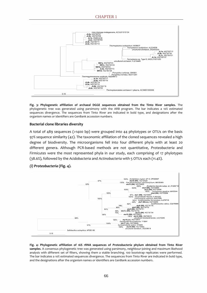
CHAPTER 1
66
Fig. 3: Phylogenetic affiliation of archaeal DGGE sequences obtained from the Tinto River samples. The phylogenetic tree was generated using parsimony with the ARB program. The bar indicates a 10% estimated sequences divergence. The sequences from Tinto River are indicated in bold type, and designations after the organism names or identifiers are GenBank accession numbers.
Bacterial clone libraries diversity
A total of 489 sequences (>1400 bp) were grouped into 44 phylotypes or OTUs on the basis
97% sequence similarity (42). The taxonomic affiliation of the cloned sequences revealed a high
degree of biodiversity. The microorganisms fell into four different phyla with at least 20
different genera. Although PCR-based methods are not quantitative, Proteobacteria and
Firmicutes were the most represented phyla in our study, each comprising of 17 phylotypes
(38.6%), followed by the Acidobacteria and Actinobacteria with 5 OTUs each (11.4%).
(i) Proteobacteria (Fig. 4).
Fig. 4: Phylogenetic affiliation of 16S rRNA sequences of Proteobacteria phylum obtained from Tinto River samples. A consensus phylogenetic tree was generated using parsimony, neighbour-joining and maximum likehood analysis with different set of filters, showing them a stable branching. 100 bootstrap replicates were performed. The bar indicates a 10% estimated sequences divergence. The sequences from Tinto River are indicated in bold type, and the designations after the organism names or identifiers are GenBank accession numbers.
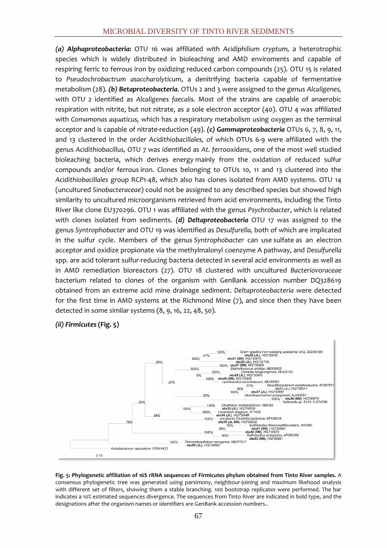
MICROBIAL DIVERSITY OF TINTO RIVER SEDIMENTS
67
(a) Alphaproteobacteria: OTU 16 was affiliated with Acidiphilium cryptum, a heterotrophic
species which is widely distributed in bioleaching and AMD enviroments and capable of
respiring ferric to ferrous iron by oxidizing reduced carbon compounds (25). OTU 15 is related
to Pseudochrobactrum asaccharolyticum, a denitrifying bacteria capable of fermentative
metabolism (28). (b) Betaproteobacteria. OTUs 2 and 3 were assigned to the genus Alcaligenes,
with OTU 2 identified as Alcaligenes faecalis. Most of the strains are capable of anaerobic
respiration with nitrite, but not nitrate, as a sole electron acceptor (40). OTU 4 was affiliated
with Comamonas aquaticus, which has a respiratory metabolism using oxygen as the terminal
acceptor and is capable of nitrate-reduction (49). (c) Gammaproteobacteria OTUs 6, 7, 8, 9, 11,
and 13 clustered in the order Acidithiobacillales, of which OTUs 6-9 were affiliated with the
genus Acidithiobacillus, OTU 7 was identified as At. ferrooxidans, one of the most well studied
bioleaching bacteria, which derives energy mainly from the oxidation of reduced sulfur
compounds and/or ferrous iron. Clones belonging to OTUs 10, 11 and 13 clustered into the
Acidithiobacillales group RCP1-48, which also has clones isolated from AMD systems. OTU 14
(uncultured Sinobacteraceae) could not be assigned to any described species but showed high
similarity to uncultured microorganisms retrieved from acid environments, including the Tinto
River like clone EU370296. OTU 1 was affiliated with the genus Psychrobacter, which is related
with clones isolated from sediments. (d) Deltaproteobacteria OTU 17 was assigned to the
genus Syntrophobacter and OTU 19 was identified as Desulfurella, both of which are implicated
in the sulfur cycle. Members of the genus Syntrophobacter can use sulfate as an electron
acceptor and oxidize propionate via the methylmalonyl coenzyme A pathway, and Desulfurella
spp. are acid tolerant sulfur-reducing bacteria detected in several acid environments as well as
in AMD remediation bioreactors (27). OTU 18 clustered with uncultured Bacteriovoraceae
bacterium related to clones of the organism with GenBank accession number DQ328619
obtained from an extreme acid mine drainage sediment. Deltaproteobacteria were detected
for the first time in AMD systems at the Richmond Mine (7), and since then they have been
detected in some similar systems (8, 9, 16, 22, 48, 50).
(ii) Firmicutes (Fig. 5)
Fig. 5: Phylogenetic affiliation of 16S rRNA sequences of Firmicutes phylum obtained from Tinto River samples. A consensus phylogenetic tree was generated using parsimony, neighbour-joining and maximum likehood analysis with different set of filters, showing them a stable branching. 100 bootstrap replicates were performed. The bar indicates a 10% estimated sequences divergence. The sequences from Tinto River are indicated in bold type, and the designations after the organism names or identifiers are GenBank accession numbers..

CHAPTER 1
68
The sequenced clones of the Firmicutes were distributed evenly over the two relevant
environmental subgroups, classes Bacilli and Clostridia. All the OTUs could be identified at least
at the genus level as Staphylococcus (OTU 27), Lysinibacillus (OTU 28), Cohnella (OTU 29),
Alicyclobacillus (OTUs 30 to 32), Clostridium (OTU 33 to 35), Veillonella (OTU 36),
Desulfosporosinus (OTU 37), Desulfitobacterium (OTU 39-40), Sulfobacillus (OTU 41 to 43), and
Thermodesulfobium narugense (OTU 50). Several of these microorganisms are involved in the
sulfur cycle. Sulfobacillus spp. are iron- and sulfur- oxidizers and grow over a broad range of
temperature (20 to 60°C). Desulfosporosinus spp. are obligately anaerobic, sulfate-reducing
bacteria with the ability to sporulate, making them more resistant to acidic conditions (15). The
majority of these clone sequences were 97% similar to Desulfosporosinus orientis, an organism
that has previously been detected in other acidic environments, including acidic mining-
impacted lake sediments (31). Desulfitobacterium spp. are very versatile microorganism that
can use a wide variety of electron acceptors, such as nitrate, sulfite, metals, humic acids, and
halogenated organic compounds (48). Thermodesulfobium is a moderate thermophilic
autotroph that is able to grow on H2/CO2 by sulfate- respiration (36).
(iii) Acidobacteria: Seven OTUs were associated with the Acidobacteria. OTUs 20 to 25
clustered in Acidobacterium group and OTU 26 was related to the genus Edaphobacter.
Although most of the clones were situated at considerable distances from the cultured
members of this phylum, OTU 24 was identified as Acidobacterium capsulatum, a bacterium
that formed Fe(II) under anoxic conditions at pHs ranging from 3 to 5 in association with the
anaerobic respiration of glucose (12).
(iv) Actinobacteria: All clones comprising OTUs 45 to 49 clustered into the family
Acidimicrobiaceae, and although they could not be assigned to described species, they showed
high similarity to uncultured microorganisms retrieved from AMD and a low similarity (90%) to
Acidimicrobium ferrooxidans, a moderately thermophilic and acidophilic bacterium with a
versatile metabolism (11): it oxidizes ferrous-iron or reduces ferric-iron as At. ferrooxidans.
Archaeal clone (Euryarchaeota) library diversity
After chimeras and low-quality sequences were removed, a total of 78 archaeal clones
distributed in 7 OTUs were retrieved. The clones retrieved from both sampling stations were
phylogenetically homogeneous and belonged to the order Thermoplasmatales. Although SN
clones could not be identified as belonging to any cultured species, JL clones belonged to the
genus Thermoplasma, a moderate thermoacidophilic facultatively anaerobic archaeon which
respires sulfur with organic carbon as an electron donor.
Relative distribution of the major phylogenetic groups in both sample sites.
The distribution of the major phylogenetic groups differed between sample sites (Fig. 6). Just
2 of 44 OTUs were shared between them. In JL dam, a total of 249 sequences (>1400 bp) were
grouped into 18 OTUs. In SN dam, a total of 240 sequences were grouped into 28 OTUs. In JL
dam, the group of Bacteria detected with the most numerous organisms was the phylum
Firmicutes (56.6%), followed by phylum Acidobacteria (27.3%) and the class Deltaproteobacteria
(11.6%). Organisms of the Actinobacteria phylum (4%) and Gammaproteobacteria class (0.4%)
were less abundant. In SN dam, the phylum Proteobacteria (72.1%) was the most represented,
mainly falling into the Alphaproteobacteria (39.6%) and Gammaproteobacteria (30.4%), followed

MICROBIAL DIVERSITY OF TINTO RIVER SEDIMENTS
69
by Actinobacteria (20.4%). Organisms of the phyla Firmicutes (5.3%) and Acidobacteria (1.7%)
were present in low percentages.
Fig. 6: Phylogenetic composition of the bacterial 16S rRNA gene libraries of the two sample sites JL dam (A) and SN dam (B).
Diversity indexes, coverage and rarefaction analyses of the 16S rRNA gene libraries.
Rarefaction curves (Fig. 7) and Good’s coverage values and diversity indexes (Table 2) were
calculated separately for Archaea and Bacteria in each sample site (SN and JL).
Fig. 7: Rarefaction curves obtained for the sequences of the 16S rRNA gene libraries for Bacteria and Archaea of both sampling sites.
Good’s value, an estimation of the proportion of the population represented by the retrieved
sequences (19), indicates high coverage in the archaeal and bacterial libraries of both sample
sites, in agreement with the rarefaction curves. Dominance (D) values are similar in both
samples sites but differ between the two domains. Whereas in Bacteria this value is close to 0,
indicating that no OTUs predominate in the community, in Archaea, D values are higher,
indicating the predominance of some OTUs in the sequence data set. In accordance, the
Shannon-Wiever index value was lower.
Gammaproteobacteria
0,4% Deltaproteobacteria
11,6%
Acidobacte
ria 27,3%
Firmicutes 56,6%
Nitros
pira 00%
Actinobacteria
4,0%
JL
Alphaproteobacteria
39,6%
Betaproteobacteria
1,7%
Gammaproteobacteri
a 30,4%
Deltaproteobacteria
0,4%
Acidobacte
ria 1,7%
Firmicutes
5,8%
Actinobacteria
20,4%
SN
0
5
10
15
20
25
30
0 50 100 150 200 250 300
Nu
mb
er
of
ide
nti
ty c
lust
ers
Number of 16S rRNA gene clones
JL Bacteria
SN Bacteria
JL Archaea
SN Archaea
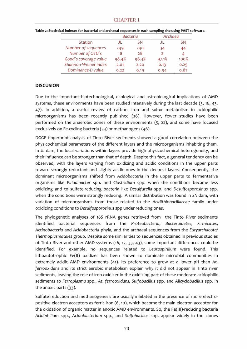
CHAPTER 1
70
Table 2: Statistical indexes for bacterial and archaeal sequences in each sampling site using PAST software.
Bacteria Archaea
Station JL SN JL SN Number of sequences 249 240 34 44
Number of OTU´s 18 28 2 4 Good´s coverage value 98.4% 96.3% 97.1% 100% Shannon-Weiner index 2.01 2.20 0.13 0.25
Dominance-D value 0.22 0.19 0.94 0.87
DISCUSION
Due to the important biotechnological, ecological and astrobiological implications of AMD
systems, these environments have been studied intensively during the last decade (3, 16, 43,
47). In addition, a useful review of carbon, iron and sulfur metabolism in acidophilic
microorganisms has been recently published (26). However, fewer studies have been
performed on the anaerobic zones of these environments (5, 22), and some have focused
exclusively on Fe-cycling bacteria (33) or methanogens (46).
DGGE fingerprint analysis of Tinto River sediments showed a good correlation between the
physicochemical parameters of the different layers and the microorganisms inhabiting them.
In JL dam, the local variations within layers provide high physicochemical heterogeneity, and
their influence can be stronger than that of depth. Despite this fact, a general tendency can be
observed, with the layers varying from oxidizing and acidic conditions in the upper parts
toward strongly reductant and slighty acidic ones in the deepest layers. Consequently, the
dominant microorganisms shifted from Acidobacteria in the upper parts to fermentative
organisms like Paludibacter spp. and Clostridium spp. when the conditions became less
oxidizing and to sulfate-reducing bacteria like Desulfurella spp. and Desulfosporosinus spp.
when the conditions were strongly reducing. A similar distribution was found in SN dam, with
variation of microorganisms from those related to the Acidithiobacillaceae family under
oxidizing conditions to Desulfosporosinus spp under reducing ones.
The phylogenetic analyses of 16S rRNA genes retrieved from the Tinto River sediments
identified bacterial sequences from the Proteobacteria, Bacteroidetes, Firmicutes,
Actinobacteria and Acidobacteria phyla, and the archaeal sequences from the Euryarchaeota/
Thermoplasmatales group. Despite some similarities to sequences obtained in previous studies
of Tinto River and other AMD systems (16, 17, 33, 43), some important differences could be
identified. For example, no sequences related to Leptospirillum were found. This
lithoautotrophic Fe(II) oxidizer has been shown to dominate microbial communities in
extremely acidic AMD environments (41). Its preference to grow at a lower pH than At.
ferrooxidans and its strict aerobic metabolism explain why it did not appear in Tinto river
sediments, leaving the role of iron-oxidizer in the oxidizing part of these moderate acidophilic
sediments to Ferroplasma spp., At. ferrooxidans, Sulfobacillus spp. and Alicyclobacillus spp. in
the anoxic parts (33).
Sulfate reduction and methanogenesis are usually inhibited in the presence of more electro-
positive electron acceptors as ferric iron (6, 10), which become the main electron acceptor for
the oxidation of organic matter in anoxic AMD environments. So, the Fe(III)-reducing bacteria
Acidiphilium spp., Acidobacterium spp., and Sulfobacillus spp. appear widely in the clones
MICROBIAL DIVERSITY OF TINTO RIVER SEDIMENTS
71
libraries. Some 27.3% of the bacteria were identified as Acidobacteria and were specifically
related to A. capsulatum. With increasing pH, the solubility of iron decreases and sulfate
reduction seems to play a more important role. Almost 20% of the sequences retrieved in JL
dam belonged to sulfate-reducing bacterium-related microorganisms: Syntrophobacter spp.,
Desulfosporosinus spp., Desulfurella spp., Desulfitobacterium spp. and Thermodesulfobium spp.
Their prevalence should not be surprising, because in presence of high concentration of
sulfate, as the case of Tinto River, sulfate reducers dominate over methane producers when
competing with each other over limited resources. Methanogens likely do not play a significant
role in the ecology of the sediments. In fact, we failed to identify them by molecular
approaches. However, their presence must not be ruled out: methane production was
observed in situ and in the laboratory (data not shown). In addition, the occurrence of
methanogens in JL dam sediments has recently been described (46). At SN dam, with a
comparatively low pH, Fe(III) reducers Acidiphilium spp., Sulfobacillus spp. and At. ferrooxidans
predominated.
From our data, other anaerobic metabolisms apart from iron and sulfate reduction can be
inferred. Several sequences related to potential nitrate or nitrite reducers such as Alcaligenes
faecalis, Pseudochrobactrum spp., Pseudomonas spp., and Bacillus spp. were found. The
concentrations of both nitrate and nitrite in the pore water of the sediments were below the
detection limit (1 ppb), so nitrogen must be a limiting factor for bacterial growth in this
environment and any input will be removed quickly. In previous studies of the boreholes in the
Tinto River (2), the fluid extracted from those underground habitats contained dissolved
gases, such as H2, N2O and CH4. Although the chemical origin of H2 was proposed to be by a
water/rock interaction, the identification of H2-producing bacteria, such as Syntrophobacter
spp. or Clostridium spp., introduces the possibility of a biological production of this gas.
In an extreme acidic, oxidizing, and cold bulk environment, we have found zones with slightly
acidic pH and reducing redox potential. In accordance with this finding, sequences
corresponding to microorganisms with forms of metabolism (such as sulfate and nitrate/nitrite
reduction) that do not normally take place under the physicochemical conditions prevailing in
the Tinto River were retrieved. Protection from acidity (29) and even the removal of oxygen by
active respiration (30) have been described. In a similar way, the exothermic microbial
oxidation of pyrite, which releases heat under both aerobic and anaerobic conditions, could be
the source of microenvironments with relatively high temperatures, explaining the presence of
moderate thermophilic organisms (Alicyclobacillus spp., Cohnella spp., Desulfurella spp.,
Thermodesulfobium spp., Sulfobacillus spp., and Thermoplasma spp.) found in the sediments.
Geomicrobiologial model of the sediments of Tinto River
In this study, Tinto sediments were analysed by combining physicochemical data with
molecular ecology studies. Rarefaction curves and Good´s coverage values showed that almost
all of the diversity was covered with our data set. So, through integration of geochemical and
biological information, a model of the geomicrobiological processes that allow organisms to
thrive in this extreme environment is suggested (Fig. 8).

CHAPTER 1
72
Fig. 8: Geomicrobiological model of Tinto River sediments, an AMD-related environment. Iron, sulfur and carbon cycles are associated to the microorganisms involved in them.
In contrast to previous studies on the water column, the molecular ecology of Tinto River
sediments is not based exclusively on the iron cycle. In fact, it seems that the sulfur cycle and
the removal of organic matter are predominant in this particular system. On the basis of the
mineralogical composition of the Iberian Pyritic Belt, the main chemolithotrophic substrate
available would be pyrite (FeS2), and that is the basis for the development of a complete
community of microorganisms.
Iron in its reduced form, Fe(II), would be the main energy source of the autotrophic,
facultatively anaerobic bacterium At. ferrooxidans, as well as members of the versatile genera
Sulfobacillus spp. and Alicyclobacillus spp. and the archaeon Ferroplasma spp. As AMD solutions
are iron rich because of the high solubility of iron at low pH, Fe(III) concentrations can exceed
oxygen concentrations by several orders of magnitude even in surface layers, so Fe(III) can be
widely used as electron acceptor in microbial metabolism (12, 13). Thus, Fe(III) can be reduced,
coupled to the oxidation of organic matter by heterotrophic acidophiles such as Acidiphilium
spp., Sulfobacillus spp., Acidobacterium spp. or coupled to the oxidation of S0 under anoxic
conditions by At. ferrooxidans and Sulfobacillus spp. These processes imply the regeneration of
the ferrous iron as an energy source, completing the iron cycle with the consortia of
microorganism thriving in these sediments.
The second crucial metabolites in this system are sulfur compounds. Sulfide is oxidized to
sulfate by the chemolitotrophic bacterium At. ferrooxidans under both aerobic and anaerobic
conditions and is coupled to Fe(III) reduction in the latter case. Sulfate reduction is carried out
by bacteria such as Desulfosporosinus spp., Desulfurella spp., Thermodesulfobium spp., and
Syntrophobacter spp. The presence of sulfate-reducing bacteria in the Tinto River system is
evidence of microbial sulfate reduction at low pH, completing the sulfur cycle in Tinto
sediments.
MICROBIAL DIVERSITY OF TINTO RIVER SEDIMENTS
73
Regarding the carbon cycle, both fermentative and respiring types of metabolisms are
present. There is a fermentative pathway that starts with organisms such as Paludibacter,
Veillonella, Staphylococcus, and Clostridium spp. This creates a complex mix of intermediates,
and some of them, e.g., propionate, can be used by the syntrophic bacteria Syntrophobacter
spp., which generates hydrogen and acetate. These compounds act as substrates for the
metabolism of sulfate-reducing bacteria and methanogens. Ferric iron can be respired by
heterotrophic bacteria of the genera Acidiphilium, Sulfobacillus, and Acidobacterium, which
oxidize organic matter, using it as electron acceptor. Although their metabolism is not
predominant, there are enough data to support the presence of methanogens (33, 46) that
would complete the anaerobic degradation.
Our study implies that organic matter degradation must also be coupled to nitrate reduction
mediated by organisms such as Alcaligenes faecalis, Pseudochrobactrum spp., Pseudomonas
spp., and Bacillus spp. In fact, nitrous oxides have been measured in bioaugmented cultures of
Tinto River sediments (data not shown).
In conclusion, the identification of microorganisms in this study and a detailed examination of
their corresponding metabolism have allowed us to compile a model of the iron, sulfur and
carbon cycles and their relation to microbial ecology in AMD-related sediments.
REFERENCES
1. Aguilera, A., S. C. Manrubia, F. Gomez, N. Rodriguez, and R. Amils. 2006. Eukaryotic community distribution and its relationship to water physicochemical parameters in an extreme acidic environment, Rio Tinto (Southwestern Spain). Appl. Environ. Microbiol. 72:5325-5330.
2. Amils, R., D. Fernández-Remolar, F. Gómez, E. González-Toril, N. Rodríguez, C. Briones, O. Prieto-Ballesteros, J. L. Sanz, E. Díaz, and T. O. Stevens. 2008. Subsurface Geomicrobiology of the Iberian Pyritic Belt. Subsurface Geomicrobiology of the Iberian Pyritic Belt, p. 205-223. In P. Dion and C. Shekhar Nautiyal (Ed.), Soil Biology (Vol. 13): Microbiology of Extreme Soils. Springer, Berlin, Germany.
3. Baker, B. J., and J. F. Banfield. 2003. Microbial communities in acid mine drainage. FEMS Microbiol. Ecol. 44:139-152.
4. Birnboim, H., and J. Doly. 1979. A rapid alkaline extraction method for screening recombinant plasmid DNA. Nucleic Acids Res. 7:1513-1523.
5. Blothe, M., D. M. Akob, J. E. Kostka, K. Goschel, H. L. Drake, and K. Kusel. 2008. pH gradient-induced heterogeneity of Fe (III)-reducing microorganisms in coal mining-associated lake sediments. Appl. Environ. Microbiol. 74:1019-1029.
6. Bodegom, P. M., J. Scholten, and A. J. M. Stams. 2004. Direct inhibition of methanogenesis by ferric iron. FEMS Microbiol. Ecol. 49:261-268.
7. Bond, P. L., S. P. Smriga, and J. F. Banfield. 2000. Phylogeny of microorganisms populating a thick, subaerial, predominantly lithotrophic biofilm at an extreme acid mine drainage site. Appl. Environ. Microbiol. 66:3842-3849.
8. Brofft, J. E., J. V. McArthur, and L. J. Shimkets. 2002. Recovery of novel bacterial diversity from a forested wetland impacted by reject coal. Environ. Microbiol. 4:764-769.

CHAPTER 1
74
9. Bruneel, O., R. Duran, K. Koffi, C. Casiot, A. Fourçans, F. Elbaz-Poulichet, and J. C. Personne. 2005. Microbial diversity in a pyrite-rich tailings impoundment (Carnoulès, France). Geomicrobiol. J. 22:249-257.
10. Canfield, D. E., R. Raiswell, and S. Bottrell. 1992. The reactivity of sedimentary iron minerals toward sulfide. Am. J. Sci. 292:659-683.
11. Clark, D. A., and P. R. Norris. 1996. Acidimicrobium ferrooxidans gen. nov., sp. nov.: mixed-culture ferrous iron oxidation with Sulfobacillus species. Microbiology. 142:785-790.
12. Coupland, K., and D. B. Johnson. 2008. Evidence that the potential for dissimilatory ferric iron reduction is widespread among acidophilic heterotrophic bacteria. FEMS Microbiol. Lett. 279:30-35.
13. Druschel, G. K., B. J. Baker, T. M. Gihring, and J. F. Banfield. 2004. Acid mine drainage biogeochemistry at Iron Mountain, California. Geochem. T. 5:13-32.
14. Fernández-Remolar, D. C., O. Prieto-Ballesteros, N. Rodríguez, F. Gómez, R. Amils, J. Gómez-Elvira, and C. R. Stoker. 2008. Underground habitats in the Río Tinto Basin: a model for subsurface life habitats on Mars. Astrobiology. 8:1023-1047.
15. García-Moyano, A., E. González-Toril, A. Aguilera, and R. Amils. 2007. Prokaryotic community composition and ecology of floating macroscopic filaments from an extreme acidic environment, Río Tinto (SW, Spain). Syst. Appl. Microbiol. 30:601-614.
16. Gonzalez-Toril, E., A. Aguilera, V. Souza-Egipsy, E. Lopez Pamo, J. Sanchez Espana, and R. Amils. 2011. Geomicrobiology of La Zarza-Perrunal Acid Mine Effluent (Iberian Pyritic Belt, Spain). Appl. Environ. Microbiol. 77:2685-2694.
17. Gonzalez-Toril, E., F. Gómez, N. Rodríguez, D. Fernández-Remolar, J. Zuluaga, I. Marín, and R. Amils. 2003. Geomicrobiology of the Tinto River, a model of interest for biohydrometallurgy. Hydrometallurgy. 71:301-309.
18. Gonzalez-Toril, E., E. Llobet-Brossa, E. O. Casamayor, R. Amann, and R. Amils. 2003. Microbial ecology of an extreme acidic environment, the Tinto River. Appl. Environ. Microbiol. 69:4853-4865.
19. Good, I. J. 1953. The population frequencies of species and the estimation of population parameters. Biometrika. 40:237-264.
20. Hammer, Ø., D. A. T. Harper, and P. D. Ryan. 2001. PAST: paleontological statistics software package for education and data analysis. Palaeontol. Electron. 4:1-9.
21. Hansen, L. P., and K. J. Singleton. 1982. Generalized instrumental variables estimation of nonlinear rational expectations models. J. Econometric. 50:1269-1286.
22. Hao, C., H. Zhang, R. Haas, Z. Bai, and B. Zhang. 2007. A novel community of acidophiles in an acid mine drainage sediment. World J Microbiol Biotechnol. 23:15-21.
23. Heck Jr, K. L., G. van Belle, and D. Simberloff. 1975. Explicit calculation of the rarefaction diversity measurement and the determination of sufficient sample size. Ecology. 56:1459-1461.
24. Johnson, D. B. 1998. Biodiversity and ecology of acidophilic microorganisms. FEMS Microbiol. Ecol. 27:307-317.
25. Johnson, D. B., and T. A. M. Bridge. 2002. Reduction of ferric iron by acidophilic heterotrophic bacteria: evidence for constitutive and inducible enzyme systems in Acidiphilium spp. J. Appl. Microbiol. 92:315-321.
26. Johnson, D. B., and K. B. Hallberg. 2008. Carbon, iron and sulfur metabolism in acidophilic micro-organisms. Adv. Microb. Physiol. 54:201-255.
MICROBIAL DIVERSITY OF TINTO RIVER SEDIMENTS
75
27. Kaksonen, A., J. Plumb, W. Robertson, M. Riekkola-Vanhanen, P. Franzmann, and J. Puhakka. 2006. The performance, kinetics and microbiology of sulfidogenic fluidized-bed treatment of acidic metal-and sulfate-containing wastewater. Hydrometallurgy. 83:204-213.
28. Kampfer, P., R. Rossello-Mora, H. C. Scholz, C. Welinder-Olsson, E. Falsen, and H. J. Busse. 2006. Description of Pseudochrobactrum gen. nov., with the two species Pseudochrobactrum asaccharolyticum sp. nov. and Pseudochrobactrum saccharolyticum sp. nov. Int. J. Syst. Evol. Microbiol. 56:1823-1829.
29. Kock, D., and A. Schippers. 2008. Quantitative microbial community analysis of three different sulfidic mine tailing dumps generating acid mine drainage. Appl. Environ. Microbiol. 74:5211-5219.
30. Krekeler, D., A. Teske, and H. Cypionka. 1998. Strategies of sulfate-reducing bacteria to escape oxygen stress in a cyanobacterial mat. FEMS Microbiol. Ecol. 25:89-96.
31. Küsel, K., U. Roth, T. Trinkwalter, and S. Peiffer. 2001. Effect of pH on the anaerobic microbial cycling of sulfur in mining-impacted freshwater lake sediments. Environ. Exp. Bot. 46:213-223.
32. Leistel, J., E. Marcoux, D. Thiéblemont, C. Quesada, A. Sánchez, G. Almodóvar, E. Pascual, and R. Sáez. 1997. The volcanic-hosted massive sulphide deposits of the Iberian Pyrite Belt Review and preface to the Thematic Issue. Miner. Deposita. 33:2-30.
33. Lu, S., S. Gischkat, M. Reiche, D. M. Akob, K. B. Hallberg, and K. Kusel. 2010. Ecophysiology of Fe-cycling bacteria in acidic sediments. Appl. Environ. Microbiol. 76:8174-8183.
34. Ludwig, W., O. Strunk, R. Westram, L. Richter, and H. Meier. 2004. ARB: a software environment for sequence data. Nucleic Acids Res. 32:1363-1371.
35. Maidak, B. L., J. R. Cole, T. G. Lilburn, C. T. Parker Jr, P. R. Saxman, R. J. Farris, G. M. Garrity, G. J. Olsen, T. M. Schmidt, and J. M. Tiedje. 2001. The RDP-II (ribosomal database project). Nucleic Acids Res. 29:173-174.
36. Mori, K., H. Kim, T. Kakegawa, and S. Hanada. 2003. A novel lineage of sulfate-reducing microorganisms: Thermodesulfobiaceae fam. nov., Thermodesulfobium narugense, gen. nov., sp. nov., a new thermophilic isolate from a hot spring. Extremophiles. 7:283-290.
37. Muyzer, G., and N. B. Ramsing. 1995. Molecular methods to study the organization of microbial communities. Water Sci. Technol. 32:1-9.
38. Nordstrom, D. K., and C. N. Alpers. 1999. Geochemistry of acid mine waters. The Environmental Geochemistry of Mineral Deposits. Part A: Processes, Techniques, and Health Issues. Society of Economic Geologists, Littleton, CO: S. 133-160.
39. Nordstrom, D. K., and C. N. Alpers. 1999. Negative pH, efflorescent mineralogy, and consequences for environmental restoration at the Iron Mountain Superfund site, California. Proc. Natl. Acad. Sci. U. S. A. 96:3455-3462.
40. Otte, S., N. G. Grobben, L. A. Robertson, M. S. Jetten, and J. G. Kuenen. 1996. Nitrous oxide production by Alcaligenes faecalis under transient and dynamic aerobic and anaerobic conditions. Appl. Environ. Microbiol. 62:2421-2426.
41. Rawlings, D. E., H. Tributsch, and G. S. Hansford. 1999. Reasons why ‘Leptospirillum’-like species rather than Thiobacillus ferrooxidans are the dominant iron-oxidizing bacteria in many commercial processes for the biooxidation of pyrite and related ores. Microbiology. 145:5-13.
42. Rosselló-Mora, R., and R. Amann. 2001. The species concept for prokaryotes. FEMS Microbiol. Rev. 25:39-67.
CHAPTER 1
76
43. Rowe, O. F., J. Sánchez-España, K. B. Hallberg, and D. B. Johnson. 2007. Microbial communities and geochemical dynamics in an extremely acidic, metal-rich stream at an abandoned sulfide mine (Huelva, Spain) underpinned by two functional primary production systems. Environ. Microbiol. 9:1761-1771.
44. Runnells, D. D., T. A. Shepherd, and E. E. Angino. 1992. Metals in water. Determining natural background concentrations in mineralized areas. Environ. Sci. Technol. 26:2316-2323.
45. Salkield, L. U., and M. J. Cahalan. 1987. A technical history of the Rio Tinto mines. Some notes on exploitation from pre-Phoenician times to the 1950’s. Institution of Mining and Metallurgy, London, United Kingdom.
46. Sanz, J. L., N. Rodriguez, E. E. Díaz, and R. Amils. 2011. Methanogenesis in the sediments of Rio Tinto, an extreme acidic river. Environ. Microbiol. 13:2336-2341
47. Tan, G. L., W. S. Shu, K. B. Hallberg, F. Li, C. Y. Lan, and L. N. Huang. 2007. Cultivation-dependent and cultivation-independent characterization of the microbial community in acid mine drainage associated with acidic Pb/Zn mine tailings at Lechang, Guangdong, China. FEMS Microbiol. Ecol. 59:118-126.
48. Villemur, R., M. Lanthier, R. Beaudet, and F. Lépine. 2006. The Desulfitobacterium genus. FEMS Microbiol. Rev. 30:706-733.
49. Wauters, G., T. De Baere, A. Willems, E. Falsen, and M. Vaneechoutte. 2003. Description of Comamonas aquatica comb. nov. and Comamonas kerstersii sp. nov. for two subgroups of Comamonas terrigena and emended description of Comamonas terrigena. Int. J. Syst. Evol. Microbiol. 53:859-862.
50. Winch, S., H. J. Mills, J. E. Kostka, D. Fortin, and D. R. S. Lean. 2009. Identification of sulphate-reducing bacteria in methylmercury-contaminated mine tailings by analysis of SSU rRNA genes. FEMS Microbiol. Ecol. 68:94-107.
51. Yilmaz, P., R. Kottmann, D. Field, R. Knight, J. R. Cole, L. Amaral-Zettler, J. A. Gilbert, I. Karsch-Mizrachi, A. Johnston, and G. Cochrane. 2010. The “Minimum Information about an Environmental Sequence” (MIENS) specification. Nature Precedings http://dx.doi.org/10.1038/npre.2010.5252.1.
52. Zettler, L. A. A., F. Gómez, E. Zettler, B. G. Keenan, R. Amils, and M. L. Sogin. 2002. Microbiology: eukaryotic diversity in Spain's River of Fire. Nature. 417:137.

CHAPTER 2:
Quantification of Tinto River
sediment microbial
communities: the importance
of Sulfate-Reducing
Bacteria and their role in
attenuating acid mine drainage


MICROBIAL ABUNDANCE OF TINTO RIVER SEDIMENTS
79
INTRODUCTION
The Tinto River is a natural acidic rock drainage (ARD) environment located in the Huelva
region, in southwestern Spain. On its way from Peña de Hierro to the Atlantic Ocean, the
Tinto River emerges from the core of the Iberian Pyritic Belt (IPB), one of the largest metal
sulfides (pyrite, chalcopyrite, etc.) deposit on Earth. When exposed to air and water, the
microbiologically enhanced oxidation of sulfides produces waters with low pH (pH 2.3) and
high ferric iron (~2 g L-1) and sulfate (~6 g L-1) content along the river course. Low pH and
ferric iron facilitate metal solubilization; therefore, when these waters come into contact
with metallic sulfides, they become highly metalliferous (Cu: ~0.1 g L-1; Zn: ~0.2 g L-1, etc.) (20).
ARD results in contamination of aquifer with secondary effects like the death of fish, rodents,
livestock, and reduced crop yields (36). These unpleasant consequences have led to efforts
to remediate ARD environments (21). Besides the environmental concerns, there are
biological, geochemical, and biotechnological reasons to analyze microbial populations in
acidic environments and to understand their ecology. In ARD, acidophilic, chemolithotrophic,
aerobic Bacteria and Archaea dissolve metallic sulfides by oxidizing the iron and sulfur
components. Products resulting from these oxidation processes can be used as electron
acceptors in dissimilatory reduction of ferric iron and sulfate under anaerobic conditions
linked to the carbon and nitrogen cycles. So far, only ARD aquifers have been studied
intensively (7, 8, 19, 22, 23); in contrast, little is known about the ARD sediments (17, 41).
At Tinto River, knowledge of the diversity and abundance of benthic microbial populations is
limited to a methanogen population study (43) and a description of the microbial ecology of
two contrasting sampling sites (SN and JL dams) (42). In the latter study, an extensive survey
of the Tinto River sediment microbiota was performed using two culture independent
approaches: denaturing gradient gel electrophoresis and 16S rRNA gene sequencing. The
taxonomic affiliation of the identified Bacteria showed a high degree of diversity, falling into
5 different phyla: Proteobacteria, Firmicutes, Bacteroidetes, Acidobacteria, and Actinobacteria;
meanwhile, all the Archaea were affiliated with the order Thermoplasmatales.
Microorganisms involved in the iron (Acidithiobacillus ferrooxidans, Sulfobacillus spp. and
Ferroplasma spp.), sulfur (Desulfurella spp., Desulfosporosinus spp. and Thermodesulfobium
spp.) and carbon (Acidiphilium spp., Bacillus spp., Clostridium spp. and Acidobacterium spp.)
cycles were identified. However, the abundance of these groups of organisms was unknown.
In this study, the microbial communities of the Tinto River sediments were quantified for the
first time, and analyzed in relation to the physico-chemical properties of the sediments with
the objective of identifying the dominant and active organisms thriving in the sediments and
inferring the main microbial activities taking place.
MATERIALS AND METHODS
Field site description and sampling
The selected sediments are located in the Tinto River basin (Huelva). Samples were collected
from two sampling sites (Fig. 1), SN dam (37.72173N, 6.557465W) and JL dam (37.691207N,
6.560587W) at different positions of the river. SN dam is a spring feeding the main stream of

CHAPTER 2
80
the river, while JL is located in the main course of the river. Sediment cores (inner diameter, 7
cm; length, 45 cm) were taken with a sampler (Eijkelkamp Agrisearch equipment, The
Netherlands). The cores were sliced at 5 cm intervals. The redox potential (E) and pH of the
drill core samples were measured in situ with E and pH probes, connected to a Thermo Orion
290A potentiometer placed into the fresh sediment just after extraction of the core.
Sediment samples for hybridization were fixed immediately in 4% (w/v) paraformaldehyde
(PFA) in phosphate-buffered saline (PBS: 145 mM NaCl, 1.4 mM NaH2PO4, 8 mM Na2HPO4 pH
7.6) at 4°C for 4 h, washed twice with PBS then stored in 50% (v/v) ethanol-PBS at -20°C until
further use.
Fig. 1: Map of Tinto River in Huelva (Spain) with the location of the studied dams.
Chemical analysis
According to physico-chemical parameters and visual appearance, the JL dam core was
composed of two main layers with black and brown colors, whereas the SN core looked
homogeneous (Fig. 2a,e). The black (10-15 cm) and brown (25-30 cm) layers from JL dam and
two layers between 10-15 cm and 25-30 cm from SN dam were excised and immediately
stored in 15 mL tubes at 4ºC until further processing in the laboratory (less than one week).
Sediments were centrifuged at 13000 x g for 5 min and the supernatant was analyzed for the
presence of heavy metals by Inductively Coupled Plasma Mass Spectrometry (ICP-MS) and
organic anions/Volatile Fatty Acids (VFA) by Ionic Chromatography (IC) using an 861
Advanced Compact IC instrument. Water samples of both dams were also taken, kept at 4ºC
and analyzed by Total Reflection X-ray Fluorescence (TXRF) for S, P, Mn, Fe, Cu, Zn, As and
Ca; by ICP-MS for the rest of the elements and by thermo-chemical oxidation (Thermostar
LT200 of Hachlange) for total organic carbon (TOC).

MICROBIAL ABUNDANCE OF TINTO RIVER SEDIMENTS
81
Fig. 2: Depth profiles of pH and redox JL (a) and SN (e), Sybr Green I and Domain-specific probes counts JL (b) and SN (f), phylum-specific probes counts JL (c) and SN (g) and genera-specific probes counts JL (d) and SN (h).
Quantification of microorganisms
(i) Sybr green I direct counts.
Aliquots of fixed samples were sonicated to detach bacteria from the particles with a
sterilized Sonic probe (Bandelin, Sonopuls HD200) for 20 sec, 3 times at 20% intensity.
Samples were kept on ice for 4 min to precipitate large sediment particles that could clog the
filter and then the supernatant was gently filtered through a black 0.2 m polycarbonate
membrane (Nuclepore, 25 mm diameter) (<0.2 bar). The filters were stained with SybrGreen I
in Moviol Medium as described elsewhere (30). Samples were examined with an
epifluorescence microscope (Axioplan 2, Carl Zeiss, Jena, Germany). A minimum of 1000
SybrGreen I-stained cells or at least 100 independent microscopic fields were counted.
(ii) CARD-FISH.
Samples were filtered following the same procedure as described above for the direct
counts, except that white filters (0.2 m pore size; Millipore GTTP) were used. In situ
hybridizations with horseradish peroxidase (HRP)-labeled probes (biomers.net, Ulm,
Germany) followed by catalyzed reporter deposition of fluorescently labeled tyramide were
carried out as described elsewhere (38). Permeabilization was done with lysozyme (10 mg
mL-1) for 90 min at 37°C, followed by achromopeptidase (60 U mL-1) for gram-positive cells
(44) and proteinase-K for Archaea (15 g mL-1) (46). Probe sequences and formamide
concentrations required for specific hybridization are given in Table 1.
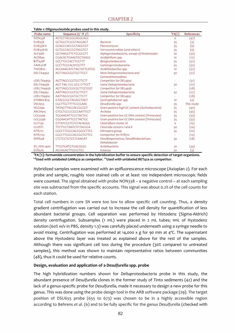
CHAPTER 2
82
Table 1: Oligonucleotide probes used in this study.
Probe name Sequence (5’ 3’) Specificity aFA[%] References
NON338 ACTCCTACGGGAGGCAGC - 0 (47) EUB338 GCTGCCTCCCGTAGGAGT Bacteria 35 (3) EUB338-II GCAGCCACCCGTAGGTGT Planctomyces 35 (9) EUB338-III GCTGCCACCCGTAGGTGT Verrucomicrobiae (and others) 35 (9) ALF968 GGTAAGGTTCTGCGCGTT Alphaproteobacteria, except of Rickettsiales 20 (35) ACD840 CGACACTGAAGTGCTAAGC Acidiphilium spp. 10 (12)
GCCTTCCCACTTCGTTT Betaproteobacteria 35 (32) GCCTTCCCACATCGTTT Gammaproteobacteria 35 (32)
THIO820 ACCAAACATCTAGTATTCATCG Acidithiobacillus spp. 10 (37) DELTA495a AGTTAGCCGGTGCTTCCT Most Deltaproteobacteria and
Gemmatimonadetes 30 (27)
cDELTA495a AGTTAGCCGGTGCTTCTT Competitor for DEL495a - (31) DELTA495b AGT TAG CCG GCG CTTCCT Some Deltaproteobacteria 30 (27) cDELTA495b AGTTAGCCGGCGCTTC(T/G)T Competitor for DEL495b - (28) DELTA495c AATTAGCCGGTGCTTCCT Some Deltaproteobacteria 30 (27) cDELTA495c AATTAGCCGGTGCTTCTT Competitor for DEL495c - (28) SYNBAC824 GTACCCGCTACACCTAGT Syntrophobacter spp 10 (4) DSU655 CGCTTGCTTTTCCCGAAC Desulfurella spp. 35 This study HGC69a TATAGTTACCACCGCCGT Gram-positive high GC content (Actinobacteria) 25 (40) ARCH915 GTGCTCCCCCGCCAATTCCT Archaea 35 (45) LGC354a TGGAAGATTCCCTACTGC Gram-positive low GC DNA content (Firmicutes) 35 (33) LGC354b CGGAAGATTCCCTACTGC Gram-positive low GC DNA content (Firmicutes) 35 (33) CLIT135 GTTATCCGTGTGTACAGGG Clostridium cluster XI 0 (15) CLOST1 TTCTTCCTAATCTCTACGCA Clostridia clusters I and II 30 (24) NTR712 CGCCTTCGCCACCGGCCTTCC Nitrospira group 35 (10) NTR712c CGCCTTCGCCACCGGTGTTCC Competitor for NTR712 - (10) DSP648 CTCTCCTGTCCTCAAGAT Desulfosporosinus, Desulfitobacterium,
Dehalobacter 30 (18)
SS_HOL1400 TTCGTGATGTGACGGGC Acidobacteria 20 (34) EUK516 ACCAGACTTGCCCTCC Eukarya 30 (3) aFA [%]: formamide concentration in the hybridization buffer to ensure specific detection of target organisms.
Used with unlabeled GAM42a as competitor. Used with unlabeled BET42a as competitor.
Hybridized samples were examined with an epifluorescence microscope (Axioplan 2). For each
probe and sample, roughly 1000 stained cells or at least 100 independent microscopic fields
were counted. The signal obtained with probe NON338 – a negative control – at each sampling
site was subtracted from the specific accounts. This signal was about 0.2% of the cell counts for
each station.
Total cell numbers in core SN were too low to allow specific cell counting. Thus, a density
gradient centrifugation was carried out to increase the cell density for quantification of less
abundant bacterial groups. Cell separation was performed by Histodenz (Sigma-Aldrich)
density centrifugation. Subsamples (1 mL) were placed in 2 mL tubes; 1mL of Hystodenz
solution (60% w/v in PBS, density 1.3) was carefully placed underneath using a syringe needle to
avoid mixing. Centrifugation was performed at 14,000 x g for 90 min at 4ºC. The supernatant
above the Hystodenz layer was treated as explained above for the rest of the samples.
Although there was significant cell loss during the procedure (30% compared to untreated
samples), this method was shown to maintain representative ratios between communities
(48), thus it could be used for relative counts.
Design, evaluation and application of a Desulfurella spp. probe
The high hybridization numbers shown for Deltaproteobacteria probe in this study, the
abundant presence of Desulfurella clones in the former study of Tinto sediments (42) and the
lack of a genus-specific probe for Desulfurella, made it necessary to design a new probe for this
genus. This was done using the probe design tool in the ARB software package (29). The target
position of DSU655 probe (655 to 673) was chosen to be in a highly accessible region
according to Behrens et al. (6) and to be fully specific for the genus Desulfurella (checked with
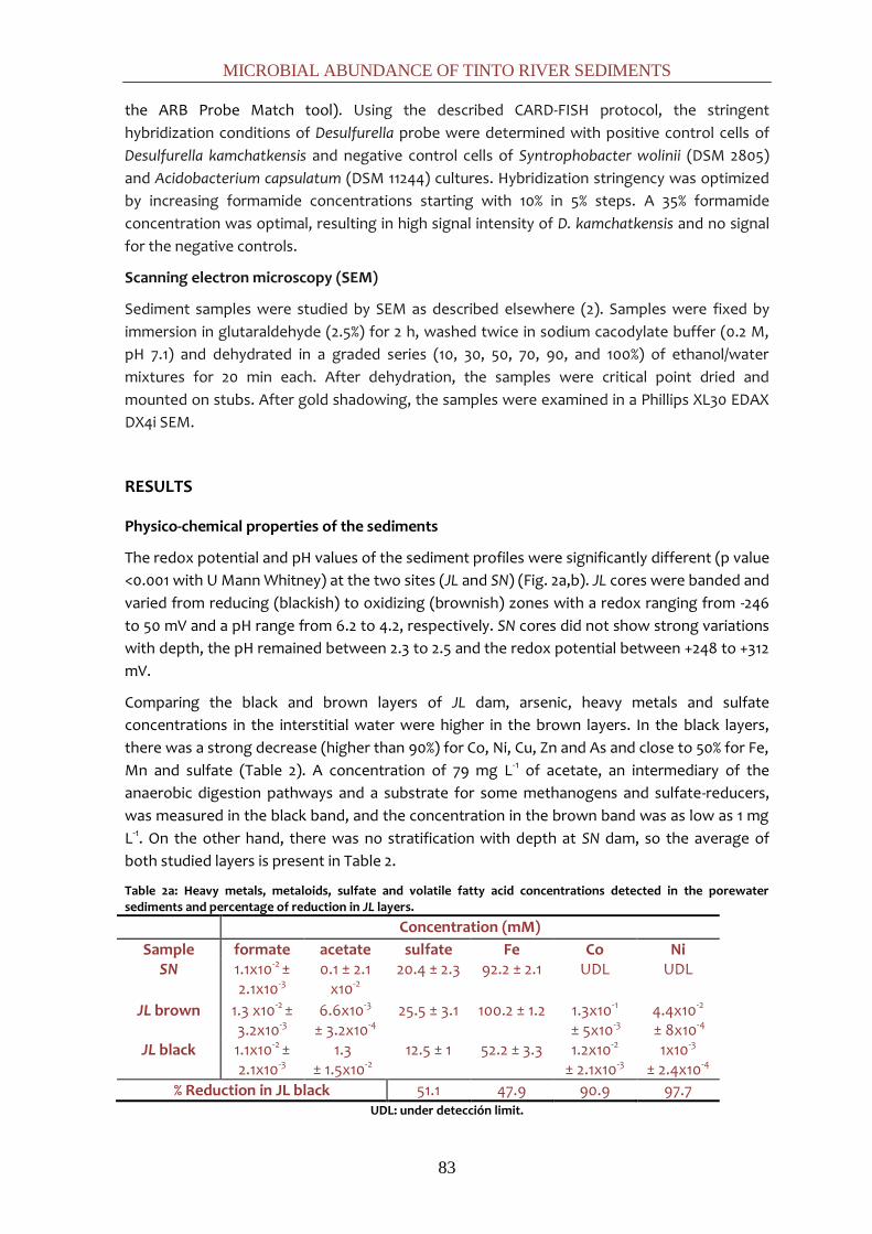
MICROBIAL ABUNDANCE OF TINTO RIVER SEDIMENTS
83
the ARB Probe Match tool). Using the described CARD-FISH protocol, the stringent
hybridization conditions of Desulfurella probe were determined with positive control cells of
Desulfurella kamchatkensis and negative control cells of Syntrophobacter wolinii (DSM 2805)
and Acidobacterium capsulatum (DSM 11244) cultures. Hybridization stringency was optimized
by increasing formamide concentrations starting with 10% in 5% steps. A 35% formamide
concentration was optimal, resulting in high signal intensity of D. kamchatkensis and no signal
for the negative controls.
Scanning electron microscopy (SEM)
Sediment samples were studied by SEM as described elsewhere (2). Samples were fixed by
immersion in glutaraldehyde (2.5%) for 2 h, washed twice in sodium cacodylate buffer (0.2 M,
pH 7.1) and dehydrated in a graded series (10, 30, 50, 70, 90, and 100%) of ethanol/water
mixtures for 20 min each. After dehydration, the samples were critical point dried and
mounted on stubs. After gold shadowing, the samples were examined in a Phillips XL30 EDAX
DX4i SEM.
RESULTS
Physico-chemical properties of the sediments
The redox potential and pH values of the sediment profiles were significantly different (p value
<0.001 with U Mann Whitney) at the two sites (JL and SN) (Fig. 2a,b). JL cores were banded and
varied from reducing (blackish) to oxidizing (brownish) zones with a redox ranging from -246
to 50 mV and a pH range from 6.2 to 4.2, respectively. SN cores did not show strong variations
with depth, the pH remained between 2.3 to 2.5 and the redox potential between +248 to +312
mV.
Comparing the black and brown layers of JL dam, arsenic, heavy metals and sulfate
concentrations in the interstitial water were higher in the brown layers. In the black layers,
there was a strong decrease (higher than 90%) for Co, Ni, Cu, Zn and As and close to 50% for Fe,
Mn and sulfate (Table 2). A concentration of 79 mg L-1 of acetate, an intermediary of the
anaerobic digestion pathways and a substrate for some methanogens and sulfate-reducers,
was measured in the black band, and the concentration in the brown band was as low as 1 mg
L-1. On the other hand, there was no stratification with depth at SN dam, so the average of
both studied layers is present in Table 2.
Table 2a: Heavy metals, metaloids, sulfate and volatile fatty acid concentrations detected in the porewater sediments and percentage of reduction in JL layers.
Concentration (mM)
Sample formate acetate sulfate Fe Co Ni SN
1.1x10⁻² ± 2.1x10⁻³
0.1 ± 2.1 x10⁻²
20.4 ± 2.3 92.2 ± 2.1 UDL UDL
JL brown 1.3 x10⁻² ± 3.2x10⁻³
6.6x10⁻³
± 3.2x10⁻⁴ 25.5 ± 3.1 100.2 ± 1.2 1.3x10⁻¹
± 5x10⁻³ 4.4x10⁻²
± 8x10⁻⁴ JL black 1.1x10⁻² ±
2.1x10⁻³ 1.3
± 1.5x10⁻² 12.5 ± 1 52.2 ± 3.3 1.2x10⁻²
± 2.1x10⁻³ 1x10⁻³
± 2.4x10⁻⁴ % Reduction in JL black 51.1 47.9 90.9 97.7
UDL: under detección limit.

CHAPTER 2
84
Table 2b: Heavy metals, metaloids, sulfate and volatile fatty acid concentrations detected in the porewater sediments and percentage of reduction in JL layers.
Concentration (mM)
Sample Cu Zn As Al Mn SN
1.0 ± 0.2 11.2 ± 0.4 1.1x10⁻²
± 9x10⁻⁴ 43 ± 3.5 2.9 ± 0.2
JL brown 7.9x10⁻³
± 3x10⁻⁴ 1.9 ± 0.1 1.4x10⁻²
± 2.5x10⁻³ 33± 1.4 4.1 ± 1.4
JL black 3.1x10⁻⁴
± 1x10⁻⁵ 6.1x10⁻³
± 4.1x10⁻⁴ 1.3x10⁻³
± 4.5x10⁻³ 3.3x10⁻²
± 4x10⁻³ 2.1 ± 0.1
% Reduction 96.0 99.7 90.6 99.9 48.3 UDL: under detección limit.
Compared to JL heavy metal concentrations varied strongly, Co and Ni were under the
detection limit while the concentrations of Zn and Cu were much higher.
Chemistries of the overlaying waters at both dams were similar in pH and some metals (Table
3), but differed for K, Mn, Cd, P, S and Fe. S and Fe are the elements that the oxidation of
pyrite (the main substrate in the IPB) put into solution in ferric iron and sulfate forms. Both
elements were less abundant at JL dam, probably due to a dilution along the river of the
concentrated ARD waters present at the SN dam.
Table 3: Element concentration, Total Organic Carbon (TOC), redox potential and pH values of SN and JL overlaid waters.
pH E (mV)
TOC (mg L-1)
Concentration (mM)
Mg Al Mn Fe Cu Zn As Cd S SN 2.5 400 9 ± 1.69 8.25
± 0.03 14.54 ± 0.7
UDL 41.30 ± 2.4
0.43 ± 0.04
1.19 ± 0.3
0.07 ± 2.1x10⁻³
2.67x10⁻³
± 5x10⁻⁴ 85.83 ± 0.4
JL 2.8 365 20 ± 4 12.34 ± 0.07
13.60 ± 0.4
26.07 ± 1.3
16.34 ± 1.7
1.94 ± 0.07
1.41 ± 0.05
0.04 ± 3.7x10⁻³
UDL 54.32 ± 0.6
UDL: under detección limit
Total cell counts with the Sybr Green I method
In comparison to DAPI staining, the use of the highly sensitive Sybr Green I dye in combination
with the Moviol embedding medium that lowered photo-bleaching and the use of black filters,
which have less background, significantly improved the signal to noise ratio. This enabled
reliable counting of total cell numbers.
The average of Sybr Green I stained cells of JL core (Fig. 2b) was 5.5x10⁸ cells g-1 wet weight of
sediment whereas in SN dam sediments (Fig. 2f) the average was 1.4x10⁷, which is 40-fold
lower. A common feature among depth profiles of both sites is that cell number in both dams
is higher in the 15-20 cm layer. After that, depth profiles followed a general trend observed in
many sediments of a drastic decrease with depth (25), from 1.4x10⁹ cells g-1 wet weight in the
15-20 cm layer to 1.3x10⁸ in the 25-30 cm layer in JL dam (10-fold lower) and from 2.4x10⁷ to
3.7x10⁶ in SN dam (6-fold lower).
Scanning electron microscopy (SEM)
SEM analysis of sediments revealed high densities of cells attached to the sediment particles in
the black layer of site JL (Fig. 3c). The colonization of sediment from site SN and the brownish
layer of JL was scarce (Fig. 3a,b). This corroborated the direct counts obtained with the Sybr
Green I method.
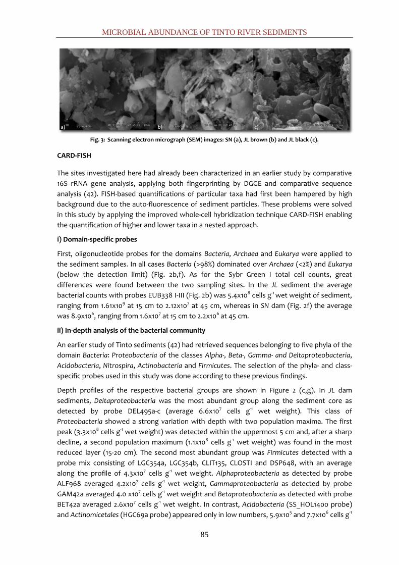
MICROBIAL ABUNDANCE OF TINTO RIVER SEDIMENTS
85
Fig. 3: Scanning electron micrograph (SEM) images: SN (a), JL brown (b) and JL black (c).
CARD-FISH
The sites investigated here had already been characterized in an earlier study by comparative
16S rRNA gene analysis, applying both fingerprinting by DGGE and comparative sequence
analysis (42). FISH-based quantifications of particular taxa had first been hampered by high
background due to the auto-fluorescence of sediment particles. These problems were solved
in this study by applying the improved whole-cell hybridization technique CARD-FISH enabling
the quantification of higher and lower taxa in a nested approach.
i) Domain-specific probes
First, oligonucleotide probes for the domains Bacteria, Archaea and Eukarya were applied to
the sediment samples. In all cases Bacteria (>98%) dominated over Archaea (<2%) and Eukarya
(below the detection limit) (Fig. 2b,f). As for the Sybr Green I total cell counts, great
differences were found between the two sampling sites. In the JL sediment the average
bacterial counts with probes EUB338 I-III (Fig. 2b) was 5.4x10⁸ cells g-1 wet weight of sediment,
ranging from 1.61x10⁹ at 15 cm to 2.12x10⁷ at 45 cm, whereas in SN dam (Fig. 2f) the average
was 8.9x10⁶, ranging from 1.6x10⁷ at 15 cm to 2.2x10⁶ at 45 cm.
ii) In-depth analysis of the bacterial community
An earlier study of Tinto sediments (42) had retrieved sequences belonging to five phyla of the
domain Bacteria: Proteobacteria of the classes Alpha-, Beta-, Gamma- and Deltaproteobacteria,
Acidobacteria, Nitrospira, Actinobacteria and Firmicutes. The selection of the phyla- and class-
specific probes used in this study was done according to these previous findings.
Depth profiles of the respective bacterial groups are shown in Figure 2 (c,g). In JL dam
sediments, Deltaproteobacteria was the most abundant group along the sediment core as
detected by probe DEL495a-c (average 6.6x10⁷ cells g-1 wet weight). This class of
Proteobacteria showed a strong variation with depth with two population maxima. The first
peak (3.3x10⁸ cells g-1 wet weight) was detected within the uppermost 5 cm and, after a sharp
decline, a second population maximum (1.1x10⁸ cells g-1 wet weight) was found in the most
reduced layer (15-20 cm). The second most abundant group was Firmicutes detected with a
probe mix consisting of LGC354a, LGC354b, CLIT135, CLOSTI and DSP648, with an average
along the profile of 4.3x10⁷ cells g-1 wet weight. Alphaproteobacteria as detected by probe
ALF968 averaged 4.2x10⁷ cells g-1 wet weight, Gammaproteobacteria as detected by probe
GAM42a averaged 4.0 x10⁷ cells g-1 wet weight and Betaproteobacteria as detected with probe
BET42a averaged 2.6x10⁷ cells g-1 wet weight. In contrast, Acidobacteria (SS_HOL1400 probe)
and Actinomicetales (HGC69a probe) appeared only in low numbers, 5.9x10⁵ and 7.7x10⁶ cells g-1

CHAPTER 2
86
wet weight respectively. Signals for probe NTR712 (Nitrospira group) were below the detection
limit.
The depth profiles of SN dam sediments did not show the variation found in JL. In this core,
numbers decrease homogeneously with depth. Gammaproteobacteria dominated (average
1.7x10⁶ cells g-1 wet weight) followed by Alphaproteobacteria with an average along the profile
of 1.1x10⁶ cells g-1 wet weight. The rest of the groups were close to the detection limit, around
105 cells g-1 wet weight, in decreasing order: Actinomycetales, Deltaproteobacteria, Firmicutes,
Betaproteobacteria and Acidobacteria.
iii) Genus-specificprobes
All genus-specific probes applied in this study were targeting genera for which sequences had
been frequently retrieved in an earlier study (42). This included the deltaproteobacterial
genera Syntrophobacter (probe Synbac824) and Desulfurella (probe DSU655), the genus
Desulfosporosinus (probe DSP648) within the phylum Firmicutes, the alphaproteobacterial
genus Acidiphilium (probe ACD638) and the gammaproteobactrial genus Acidithiobacillus
(Thio820).
The genus-specific probing confirmed the great differences between the two sampling sites
(Fig.2 d,h). In JL, the most abundant genus detected was Syntrophobacter. Cells were rod-
shaped and had a size of about 2 by 1 μm. The average along the profile was 4.7x10⁷ cells g-1
wet weight, but just in the first layer the signal counts were 3.6x10⁸, representing 36% of the
total Bacteria counts. Syntrophobacter spp. are propionate-degrading syntrophic bacteria that
use sulfate as electron acceptor. They typically occur in environments with neutral pH. The
other abundant genera were Desulfurella (3.1x10⁷ cells g-1 wet weight, mostly rods of a size of 2
by 1 μm), and Desulfosporosinus (2.8x10⁷ cells g-1 wet weight, curved rods of a size of 2-4 by 0.7
μm). The latter genus is known to encompass spore-forming sulfate reducing bacteria. Both
genera showed higher numbers at those layers with higher pH and reducing redox potential
(5-10 and 15-20 cm) (Fig.2 a,d). At SN, Acidithiobacillus spp. dominated with an average of
1.5x10⁶ cells g-1 wet weight along the profile. The hybridized cells were coccoid rods with a size
of 1 by 0.7 μm. Acidithiobacillus spp. derive energy from the oxidation of reduced sulfur
compound and/or ferrous iron, or the reduction of ferric iron under anoxic conditions. Finally,
Acidiphilium spp. were also present with average numbers of 4.2x10⁵ cells g-1 wet weight (rods
of a size of 2-3 by 1 μm).
DISCUSSION
We used a nested-probe approach for the quantification of the microbial community
composition in sediments of Tinto River. The generally low abundance of Archaea agreed with
similar results found in the sediments of mine tailing dumps (23). In a previous study of Río
Tinto sediments (42), the archaeal diversity had been restricted to two genera: Ferroplasma
spp. and Thermoplasma spp. Although Sanz et al. (43) had described the presence of
methanogens in the sediments of the Tinto basin, our data suggest that Archaea play a minor
role in the ecology of these sediments. Both Ferroplasma and Thermoplasma are usually found
in bioleaching operations at lower pH (around 2) and at much higher iron and sulfate
concentrations than those prevailing in Río Tinto sediments (13). Although algae, ciliates,
flagellates, amoebae, and fungi have been reported in the water column and the biofilms of

MICROBIAL ABUNDANCE OF TINTO RIVER SEDIMENTS
87
the Río Tinto (1, 5), no Eukarya probe signal was detected by CARD-FISH, therefore, Bacteria
seem to fully dominate the anoxic sediment layers of Tinto River.
When genus-specific probes were applied, a pronounced variability was detected between the
two sampling sites. In SN sediments, where parameters such as pH and redox potential were
similar to water column values, Bacteria related to the iron cycle prevailed, likely reflecting the
higher availability of iron at low pH and oxidative redox potential. This included the genera
Acidiphilium (ACD638 probe) and Acidithiobacillus (Thio820 probe), which both are capable of
iron-reduction under anoxic conditions. No members of Leptospirillum genus, Nitrospira-like
bacteria (Ntr712 probe), were detected. The common iron oxidizers in ARD environments are
members of the aerobic genus Leptospirillum and the facultative anaerobe bacterium
Acidithiobacillus ferrooxidans, which can oxidize iron and sulfur in aerobic conditions and
reduce iron in anoxia. Our results confirm the dominance of the versatile Acidithiobacillus
ferrooxidans in the sediments over the strictly aerobic Leptospirillum spp. that usually
dominates in extremely acidic oxic ARD environments (7, 13).
At JL dam, where the iron solubility is lower due to higher pH and reducing redox potential,
microbes of the sulfur cycle were most abundant. Here, sulfate-reducing bacteria (SRB) of the
genera Desulfosporosinus (DSP 648 probe) and Syntrophobacter (Syn824 probe) as well as
sulfur reducers of the genus Desulfurella (DSU655 probe) predominated, showing the
importance of sulfur cycling in these sediments. Syntrophobacter-affiliated 16S rRNA gene
sequences (42) (HQ730668.1, HQ730666.1) have been retrieved in several acidic environments
before (11, 17, 26), suggesting that not yet cultured relatives of Syntrophobacter might be well
adapted to acidic conditions in contrast to the general neutral pH environments where they
thrive.
The analyses of both brown and black layers in the JL sediments showed a significant
reduction in the last ones for dissolved metals, iron and sulfate in the black layers, which run
parallel to the SRB abundance. Desulfurella and Desulfosporosinus genera abundance peaked
perfectly with those black layers where the pH values and acetate concentration increased and
the redox potential, the sulfate and the dissolved metals decrease. Our results suggest that
the attenuation of ARD characteristics in the reduced black layers is biologically driven by SRB
(Fig.4).
Fig. 4: ARD bioremediation by SBR model.

CHAPTER 2
88
The dissimilative reduction of sulfate, driven by identified sulfate- and sulfur-reducing bacteria
leads to a consumption of protons thus, increasing alkalinity, and to sulfide formation. In
addition, there is a sulfide-mediated metal precipitation: the sulfide reacts with the dissolved
iron and other heavy metals which can form black amorphous precipitates (FeS) and metallic
sulfides (MS). These results agree with previous studies where remediation in AMD associated
with SRB metabolism was observed (14, 16, 21, 39).
In summary, the CARD-FISH quantifications performed in this study underline the great
differences between microbial communities of the two Río Tinto sediments studied that had
been first indicated by DGGE and comparative sequence analysis. These differences correlate
with their physico-chemical characteristics, which would correspond to different hydrology at
both sites. In JL dam, the location, thickness and values of pH and redox potential of the
oxidized and reduced zones vary temporally, but stratification has been routinely observed. In
contrast, sediments in SN have been more homogeneous. Different interpretations can be put
forward. On the one hand, the two sampling sites have different hydrology. Site SN is a stream
located close to the source of the river with a constant water flow containing high
concentration of heavy metals, mainly iron. Therefore, the sediments characteristics are close
to those of the water column strongly buffered by ferric iron. In contrast, the site JL is located
5 km downstream, with an increasing flow due to the addition of neutral tributaries which
lower the iron concentration, albeit with a strong seasonal variability. One of the tributaries
upstream JL contains the waters from the municipal wastewater treatment plant of Nerva.
This significantly increases the total organic content (TOC) of the waters. The combination of
both factors could explain the differences detected in both types of sediments. At JL dam,
with seasonal variability, higher TOC and lower iron concentration, microbial reduction
processes such as sulfate reduction increase the pH. This effect would take place firstly in
microniches and then expands to macroscopic conditions. That would explain the banded
sediment at JL dam, which would correspond to the fluctuating conditions of the input water.
Within them, SRB communities reduce the dissolved sulfates to sulfides that precipitate iron
and heavy metals, performing a local natural bioremediation of Tinto River.
REFERENCES
1. Aguilera, A., S. C. Manrubia, F. Gomez, N. Rodriguez, and R. Amils. 2006. Eukaryotic community distribution and its relationship to water physicochemical parameters in an extreme acidic environment, Rio Tinto (Southwestern Spain). Appl. Environ. Microbiol. 72:5325-5330.
2. Alphenaar, P. A., N. Groeneveld, and A. C. Van Aelst. 1994. Scanning electron microscopical method for internal structure analysis of anaerobic granular sludge. Micron. 25:129-133.
3. Amann, R. I., B. J. Binder, R. J. Olson, S. W. Chisholm, R. Devereux, and D. A. Stahl. 1990. Combination of 16S rRNA-targeted oligonucleotide probes with flow cytometry for analyzing mixed microbial populations. Appl. Environ. Microbiol. 56:1919-1925.
4. Ariesyady, H. D., T. Ito, K. Yoshiguchi, and S. Okabe. 2007. Phylogenetic and functional diversity of propionate-oxidizing bacteria in an anaerobic digester sludge. Appl. Microbiol. Biotechnol. 75:673-683.
5. Baker, B. J., G. W. Tyson, L. Goosherst, and J. F. Banfield. 2009. Insights into the diversity of eukaryotes in acid mine drainage biofilm communities. Appl. Environ. Microbiol. 75:2192-2199.
MICROBIAL ABUNDANCE OF TINTO RIVER SEDIMENTS
89
6. Behrens, S., C. Rühland, J. Inácio, H. Huber, A. Fonseca, I. Spencer-Martins, B. M. Fuchs, and R. Amann. 2003. In situ accessibility of small-subunit rRNA of members of the domains Bacteria, Archaea, and Eucarya to Cy3-labeled oligonucleotide probes. Appl. Environ. Microbiol. 69:1748-1758.
7. Bond, P. L., G. K. Druschel, and J. F. Banfield. 2000. Comparison of acid mine drainage microbial communities in physically and geochemically distinct ecosystems. Appl. Environ. Microbiol. 66:4962-4971.
8. Bruneel, O., R. Duran, C. Casiot, F. Elbaz-Poulichet, and J. C. Personné. 2006. Diversity of microorganisms in Fe-As-rich acid mine drainage waters of Carnoules, France. Appl. Environ. Microbiol. 72:551-556.
9. Daims, H., A. Brühl, R. Amann, K. H. Schleifer, and M. Wagner. 1999. The domain-specific probe EUB 338 is insufficient for the detection of all bacteria: development and evaluation of a more comprehensive probe set. Syst. Appl. Microbiol. 22:434-444.
10. Daims, H., J. L. Nielsen, P. H. Nielsen, K. H. Schleifer, and M. Wagner. 2001. In situ characterization of Nitrospira-like nitrite-oxidizing bacteria active in wastewater treatment plants. Appl. Environ. Microbiol. 67:5273-5284.
11. Dedysh, S. N., T. A. Pankratov, S. E. Belova, I. S. Kulichevskaya, and W. Liesack. 2006. Phylogenetic analysis and in situ identification of Bacteria community composition in an acidic Sphagnum peat bog. Appl. Environ. Microbiol. 72:2110-2117.
12. Demanèche, S., H. Sanguin, J. Poté, E. Navarro, D. Bernillon, P. Mavingui, W. Wildi, T. M. Vogel, and P. Simonet. 2008. Antibiotic-resistant soil bacteria in transgenic plant fields. Proceedings of the National Academy of Sciences. 105:3957-3962.
13. Edwards, K. J., T. M. Gihring, and J. F. Banfield. 1999. Seasonal variations in microbial populations and environmental conditions in an extreme acid mine drainage environment. Appl. Environ. Microbiol. 65:3627-3632.
14. Fortin, D., J. P. Rioux, and M. Roy. 2002. Geochemistry of iron and sulfur in the zone of microbial sulfate reduction in mine tailings. Water, Air, & Soil Pollution: Focus. 2:37-56.
15. Franks, A. H., H. J. M. Harmsen, G. C. Raangs, G. J. Jansen, F. Schut, and G. W. Welling. 1998. Variations of bacterial populations in human feces measured by fluorescent in situ hybridization with group-specific 16S rRNA-targeted oligonucleotide probes. Appl. Environ. Microbiol. 64:3336-3345.
16. Friese, K., K. Wendt-Potthoff, D. W. Zachmann, A. Fauville, B. Mayer, and J. Veizer. 1998. Biogeochemistry of iron and sulfur in sediments of an acidic mining lake in Lusatia, Germany. Water Air Soil Poll. 108:231-247.
17. Gonzalez-Toril, E., A. Aguilera, V. Souza-Egipsy, E. Lopez Pamo, J. Sanchez Espana, and R. Amils. 2011. Geomicrobiology of La Zarza-Perrunal Acid Mine Effluent (Iberian Pyritic Belt, Spain). Appl. Environ. Microbiol. 77:2685-2694.
18. González-Toril, E., F. Gómez, M. Malki, and R. Amils. 2006. The Isolation and Study of Acidophilic Microorganisms. Extremophiles – Methods in Microbiology. Rainey, F. A. , and Oren, A. (Eds). London, UK: Elsevier/Academic Press. 35:471-510.
19. Gonzalez-Toril, E., E. Llobet-Brossa, E. O. Casamayor, R. Amann, and R. Amils. 2003. Microbial ecology of an extreme acidic environment, the Tinto River. Appl. Environ. Microbiol. 69:4853-4865.
20. Johnson, D. B. 1998. Biodiversity and ecology of acidophilic microorganisms. FEMS Microbiol. Ecol. 27:307-317.
21. Johnson, D. B., and K. B. Hallberg. 2005. Acid mine drainage remediation options: a review.
CHAPTER 2
90
Sci. Total Environ. 338:3-14.
22. Johnson, D. B., and K. B. Hallberg. 2008. Carbon, iron and sulfur metabolism in acidophilic micro-organisms. Adv. Microb. Physiol. 54:201-255.
23. Kock, D., and A. Schippers. 2008. Quantitative microbial community analysis of three different sulfidic mine tailing dumps generating acid mine drainage. Appl. Environ. Microbiol. 74:5211-5219.
24. Kusel, K., H. C. Pinkart, H. L. Drake, and R. Devereux. 1999. Acetogenic and sulfate-reducing bacteria inhabiting the rhizoplane and deep cortex cells of the sea grass Halodule wrightii. Appl. Environ. Microbiol. 65:5117-5123.
25. Llobet-Brossa, E., R. Rosselló-Mora, and R. Amann. 1998. Microbial community composition of Wadden Sea sediments as revealed by fluorescence in situ hybridization. Appl. Environ. Microbiol. 64:2691-2696.
26. Loy, A., K. Kusel, A. Lehner, H. L. Drake, and M. Wagner. 2004. Microarray and functional gene analyses of sulfate-reducing prokaryotes in low-sulfate, acidic fens reveal cooccurrence of recognized genera and novel lineages. Appl. Environ. Microbiol. 70:6998-7009.
27. Loy, A., A. Lehner, N. Lee, J. Adamczyk, H. Meier, J. Ernst, K. H. Schleifer, and M. Wagner. 2002. Oligonucleotide microarray for 16S rRNA gene-based detection of all recognized lineages of sulfate-reducing prokaryotes in the environment. Appl. Environ. Microbiol. 68:5064-5081.
28. Lücker, S., D. Steger, K. U. Kjeldsen, B. J. MacGregor, M. Wagner, and A. Loy. 2007. Improved 16S rRNA-targeted probe set for analysis of sulfate-reducing bacteria by fluorescence in situ hybridization. J. Microbiol. Methods. 69:523-528.
29. Ludwig, W., O. Strunk, R. Westram, L. Richter, and H. Meier. 2004. ARB: a software environment for sequence data. Nucleic Acids Res. 32:1363-1371.
30. Lunau, M., A. Lemke, K. Walther, W. Martens-Habbena, and M. Simon. 2005. An improved method for counting bacteria from sediments and turbid environments by epifluorescence microscopy. Environ. Microbiol. 7:961-968.
31. Macalady, J. L., E. H. Lyon, B. Koffman, L. K. Albertson, K. Meyer, S. Galdenzi, and S. Mariani. 2006. Dominant microbial populations in limestone-corroding stream biofilms, Frasassi cave system, Italy. Appl. Environ. Microbiol. 72:5596-5609.
32. Manz, W., R. Amann, W. Ludwig, M. Wagner, and K. H. Schleifer. 1992. Phylogenetic oligodeoxynucleotide probes for the major subclasses of proteobacteria: problems and solutions. Syst. Appl. Microbiol. 15:593-600.
33. Meier, H., R. Amann, W. Ludwig, and K. H. Schleifer. 1999. Specific oligonucleotide probes for in situ detection of a major group of gram-positive bacteria with low DNA G+C content. Syst. Appl. Microbiol. 22:186-196.
34. Meisinger, D. B., J. Zimmermann, W. Ludwig, K. H. Schleifer, G. Wanner, M. Schmid, P. C. Bennett, A. S. Engel, and N. M. Lee. 2007. In situ detection of novel Acidobacteria in microbial mats from a chemolithoautotrophically based cave ecosystem (Lower Kane Cave, WY, USA). Environ. Microbiol. 9:1523-1534.
35. Neef, A. 1997. Anwendung der in situ Einzelzell-Identifizierung von Bakterien zur Populationsanalyse in komplexen mikrobiellen Biozönosen. Technische Universität München, Munich, Germany.
36. Nordstrom, D. K., and C. N. Alpers. 1999. Negative pH, efflorescent mineralogy, and consequences for environmental restoration at the Iron Mountain Superfund site, California. Proc. Natl. Acad. Sci. U. S. A. 96:3455-3462.
MICROBIAL ABUNDANCE OF TINTO RIVER SEDIMENTS
91
37. Peccia, J., E. A. Marchand, J. Silverstein, and M. Hernandez. 2000. Development and application of small-subunit rRNA probes for assessment of selected Thiobacillus species and members of the genus Acidiphilium. Appl. Environ. Microbiol. 66:3065-3072.
38. Pernthaler, A., J. Pernthaler, and R. Amann. 2002. Fluorescence in situ hybridization and catalyzed reporter deposition for the identification of marine bacteria. Appl. Environ. Microbiol. 68:3094-3101.
39. Praharaj, T., and D. Fortin. 2004. Indicators of microbial sulfate reduction in acidic sulfide-rich mine tailings. Geomicrobiol. J. 21:457-467.
40. Roller, C., M. Wagner, R. Amann, W. Ludwig, and K. H. Schleifer. 1994. In situ probing of Gram-positive bacteria with high DNA G+C content using 23S rRNA-targeted oligonucleotides. Microbiology 140:2849-2858.
41. Rowe, O. F., J. Sánchez‐España, K. B. Hallberg, and D. B. Johnson. 2007. Microbial communities and geochemical dynamics in an extremely acidic, metal-rich stream at an abandoned sulfide mine (Huelva, Spain) underpinned by two functional primary production systems. Environ. Microbiol. 9:1761-1771.
42. Sánchez-Andrea, I., N. Rodriguez, R. Amils, and J. L. Sanz. 2011. Microbial Diversity in Anaerobic Sediments at Rio Tinto, a Naturally Acidic Environment with a High Heavy Metal Content. Appl. Environ. Microbiol. 77:6085-6093.
43. Sanz, J. L., N. Rodríguez, E. E. Díaz, and R. Amils. 2011. Methanogenesis in the sediments of Rio Tinto, an extreme acidic river. Environ. Microbiol. 13:2336-2341.
44. Sekar, R., A. Pernthaler, J. Pernthaler, F. Warnecke, T. Posch, and R. Amann. 2003. An improved protocol for quantification of freshwater Actinobacteria by fluorescence in situ hybridization. Appl. Environ. Microbiol. 69:2928-2935.
45. Stahl, D. A., and R. Amann. 1991. Development and application of nucleic acid probes. In E. Stackebrandt and M. Goodfellow (Ed.), Nucleic Acid Techniques in Bacterial Systematics. John Wiley & Sons, Ltd., Chichester, United Kingdom. 205-248.
46. Teira, E., T. Reinthaler, A. Pernthaler, J. Pernthaler, and G. J. Herndl. 2004. Combining catalyzed reporter deposition-fluorescence in situ hybridization and microautoradiography to detect substrate utilization by bacteria and archaea in the deep ocean. Appl. Environ. Microbiol. 70:4411-4414.
47. Wallner, G., R. Amann, and W. Beisker. 1993. Optimizing fluorescent in situ hybridization with rRNA‐targeted oligonucleotide probes for flow cytometric identification of microorganisms. Cytometry. 14:136-143.
48. Whiteley, A. S., R. I. Griffiths, and M. J. Bailey. 2003. Analysis of the microbial functional diversity within water-stressed soil communities by flow cytometric analysis and CTC+ cell sorting. J. Microbiol. Methods. 54:257-267.

CHAPTER 3:
Screening of Anaerobic
Activities in Sediments of an
Acidic Environment:
Tinto River


SCREENING OF ANAEROBIC ACTIVITIES
95
INTRODUCTION
The exploration of extreme environments has led to the discovery of microbial activities in
habitats that only a few years ago were considered uninhabitable. As a consequence, interest
in their ecology has grown. The Tinto river is a natural acidic rock drainage (ARD) environment
located in the Iberian Pyritic Belt (IPB), in south-western Spain. The bio-oxidation of metal-
sulfides in this system results in waters of low pH (pH 2.3), high concentrations of sulfate (6 g
L-1), ferric iron (2.3 g L-1), and other heavy metals (Cu: 0.1 g L-1; Zn: 0.23 g L-1, etc.). Apart from
the ecological interest of extremophiles; the Tinto River is also of interest in the areas of
biogeochemistry (exploring the nature of the microbial communities associated with ARD),
biotechnology (bioleaching processes), microbiology (acidic environments offer an almost
unique opportunity to reveal habitat biological complexity) and Mars-analogue studies
(common mineralogy).
Recent geomicrobiological studies of the Tinto river have shown a high level of microbial
diversity in the water column, both in prokaryotic and eukaryotic organisms. Eighty percent of
the prokaryotic biomass in the water column is composed of three genera of bacteria,
Leptospirillum, Acidithiobacillus and Acidiphilium (20), all of which are involved in the iron cycle.
Macroscopic brownish-filamentous structures, streamers, are present along the river,
composed mostly of those three bacterial genera enmeshed in a matrix of exopolysaccharides
and mineral particles (19). A wide variety of eukaryotes, which include algae, ciliates,
flagellates, amoebae, fungi and rotifers have been reported. Eukaryotic algae, which
contribute to over 60% of the river biomass (32), are mainly chlorophytes but also filamentous
algae forming green-phototrophic filaments (1-3). Heterotrophic protists -mixotrophic
flagellates, ciliates or amoebas- are also widely distributed along the river. Among
decomposers, fungi are the most abundant and both unicellular and filamentous forms are
present (31, 32). The only animal found in the river is a species of bdelloid rotifer related to the
genus Rotifera (60).
Most of the interest in these ecosystems has focused on the aerobic iron- and sulfur-oxidizing
microorganisms due to their bio-mining applications. Despite their ecological interest, the
characterization of the anoxic sediments from acidic environments, such as those of the Tinto
river, had been neglected, with few exceptions (9, 22). Our group has recently started an in
depth Tinto River sediments study to understand how the element cycles work under
anaerobic conditions and to find potential biotechnological applications for the
microorganisms thriving in them. First, an extensive survey of the Tinto River sediment
microbiota based on comparative sequence analyses of 16S rRNA genes was performed at two
contrasting sampling sites (SN and JL dams) using two culture independent approaches: DGGE
and cloning of 16S rRNA genes (46). Microorganisms involved in the iron (At. ferrooxidans,
Sulfobacillus spp., Ferroplasma spp., etc.), sulfur (Desulfurella spp., Desulfosporosinus spp.,
Thermodesulfobium spp., etc.) and carbon (Acidiphilium spp., Bacillus spp., Clostridium spp.,
Acidobacterium spp., etc.) cycles were identified and their distribution correlated with
physicochemical parameters of the sediments. Hybridization with domain-specific probes
showed that Bacteria (>98%) dominated over Archaea (<2%) (45). Specific probes showed large
differences between dams. At SN where the pH and redox potential are similar to that of the
water column (pH 2.5 and +300 mV), the most abundant organisms were identified as iron-
reducing microorganisms: Acidithiobacillus spp. and Acidiphilium spp., given the higher
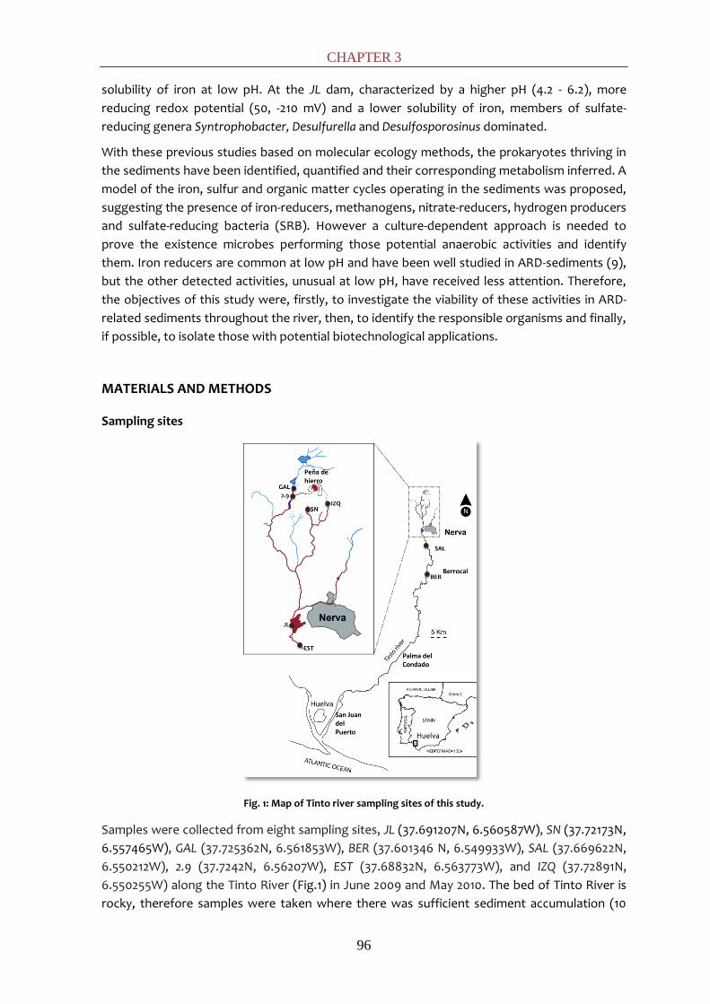
CHAPTER 3
96
solubility of iron at low pH. At the JL dam, characterized by a higher pH (4.2 - 6.2), more
reducing redox potential (50, -210 mV) and a lower solubility of iron, members of sulfate-
reducing genera Syntrophobacter, Desulfurella and Desulfosporosinus dominated.
With these previous studies based on molecular ecology methods, the prokaryotes thriving in
the sediments have been identified, quantified and their corresponding metabolism inferred. A
model of the iron, sulfur and organic matter cycles operating in the sediments was proposed,
suggesting the presence of iron-reducers, methanogens, nitrate-reducers, hydrogen producers
and sulfate-reducing bacteria (SRB). However a culture-dependent approach is needed to
prove the existence microbes performing those potential anaerobic activities and identify
them. Iron reducers are common at low pH and have been well studied in ARD-sediments (9),
but the other detected activities, unusual at low pH, have received less attention. Therefore,
the objectives of this study were, firstly, to investigate the viability of these activities in ARD-
related sediments throughout the river, then, to identify the responsible organisms and finally,
if possible, to isolate those with potential biotechnological applications.
MATERIALS AND METHODS
Sampling sites
Fig. 1: Map of Tinto river sampling sites of this study.
Samples were collected from eight sampling sites, JL (37.691207N, 6.560587W), SN (37.72173N,
6.557465W), GAL (37.725362N, 6.561853W), BER (37.601346 N, 6.549933W), SAL (37.669622N,
6.550212W), 2.9 (37.7242N, 6.56207W), EST (37.68832N, 6.563773W), and IZQ (37.72891N,
6.550255W) along the Tinto River (Fig.1) in June 2009 and May 2010. The bed of Tinto River is
rocky, therefore samples were taken where there was sufficient sediment accumulation (10

SCREENING OF ANAEROBIC ACTIVITIES
97
cm). The redox potential (E) and pH of the drill core samples were measured in situ with E and
pH probes, connected to a Thermo Orion 290A potentiometer placed into the fresh sediment
just after extraction. In general all sampling sites presented a pH range between 2 and 3 and
redox between 200 and 300 mV with the exception of JL at which pH was 5 and redox
potential between -50 and 80 mV.
Enrichment cultures
Enrichments were prepared anaerobically using an anaerobic chamber. Approximately 4 g of
sediment were added to 50 mL glass serum bottles with 20 mL of media and capped with butyl
rubber stoppers. The headspace was then degassed with a N2/CO2 (80:20) mixture.
Enrichments were incubated at 30ºC in the dark. The growth media used were selective for
various microbial activities. For methanogenesis, the basal medium previously described (47)
was used at pH 6. To stimulate specific methanogenic groups, six different carbon sources
were used: a) H2+CO2 (80:20); b) volatile fatty acids (VFAs) (0.5 g L-1 of each sodium salts of
acetate, propionate and butyrate); c) mixture (M) (g L-1) (0.7 sodium formate, 0.5 sodium
acetate, 0.3 sodium propionate, 0.3 sodium butyrate, 0.5 methanol, 2 sucrose and 0.7 sodium
lactate); d) 2 g L-1 sodium acetate; e) 1.5 g L-1 sodium formate; and f) 1 g L-1 methanol. For sulfate
reduction, the medium contained (g L-1): 2 sodium lactate, 2 KHCO3, 2 yeast extract, 2
MgSO4.7H2O, 1.5 Na2SO4, 0.5 K2HPO4, 0.1 CaCl2, 0.48 (NH4)2Fe(SO4)2·6H2O, and 1 mL of
micronutrients solution (47). Enrichments for sulfate reducing bacteria were conducted at pH
5 and 7. Enrichments for both autotroph and heterotroph denitrification were conducted. Both
the heterotrophic (3635) and the autotrophic (4544) denitrifying media were prepared as
described previously. In both media, final pH 5 and 7 were assayed. The medium used for
hydrogen production was (g L-1): 0.3 NH4Cl, 0.3 K2HPO4, 0.1 MgSO4, 0.5 NaHCO3, 0.2 sucrose, 0.1
meat extract, 0.5 yeast extract, and 1 mL of micronutrients solution (47). Cysteine was used as
reducing agent (0.5%) in all media.
Analytical Measurement
Activity in methanogenic and denitrifying enrichments was determined by monitoring CH4, and
N2O production in serum vial headspaces. For CH4 determinations, 100 µL subsamples of
headspace were injected into a Varian Star 3400CX gas chromatograph equipped with a
split/splitness injector and a flame ionization detector (FID). The GC was fitted with a SPB1000
(25 m x 0.32 mm x 0.25 μm) column manufactured by Supelco with Carbowax 20M
polyethilenglicol as stationary phase. The temperatures for the column, injector and detector
were 60ºC, 200ºC and 250ºC respectively. Nitrogen was used as carrier gas. The production of
N2O was measured using a HP 5890 Series Chromatograph equipped with an on-column
injector and an electron capture detector (ECD). A Porapack 80-100 (1 m x 1/8” mm x 2 mm)
column manufactured by Sugerlabor was used. The temperatures for the column, injector and
detector were 40ºC, 300ºC and 300ºC respectively. Nitrogen was used as carrier gas.
Measurements were made by injecting 50 µL of headspace into the GC. Denitrifying activity
was also determined measuring nitrate and nitrite concentrations by ion chromatography,
using an Omega Metrohm 790 Personal IC, with a Metrosep A Supp 5 250/4.0 (Omega) column.
Sulfate-reducing activity was determined visually by the formation of black precipitates from
the reaction of S2- produced by sulfate reduction and Fe2+ added in the media. The hydrogen-
production was measured periodically using a hydrogen detector (MDA Scientific Midas Gas
Detector, Honeywell).
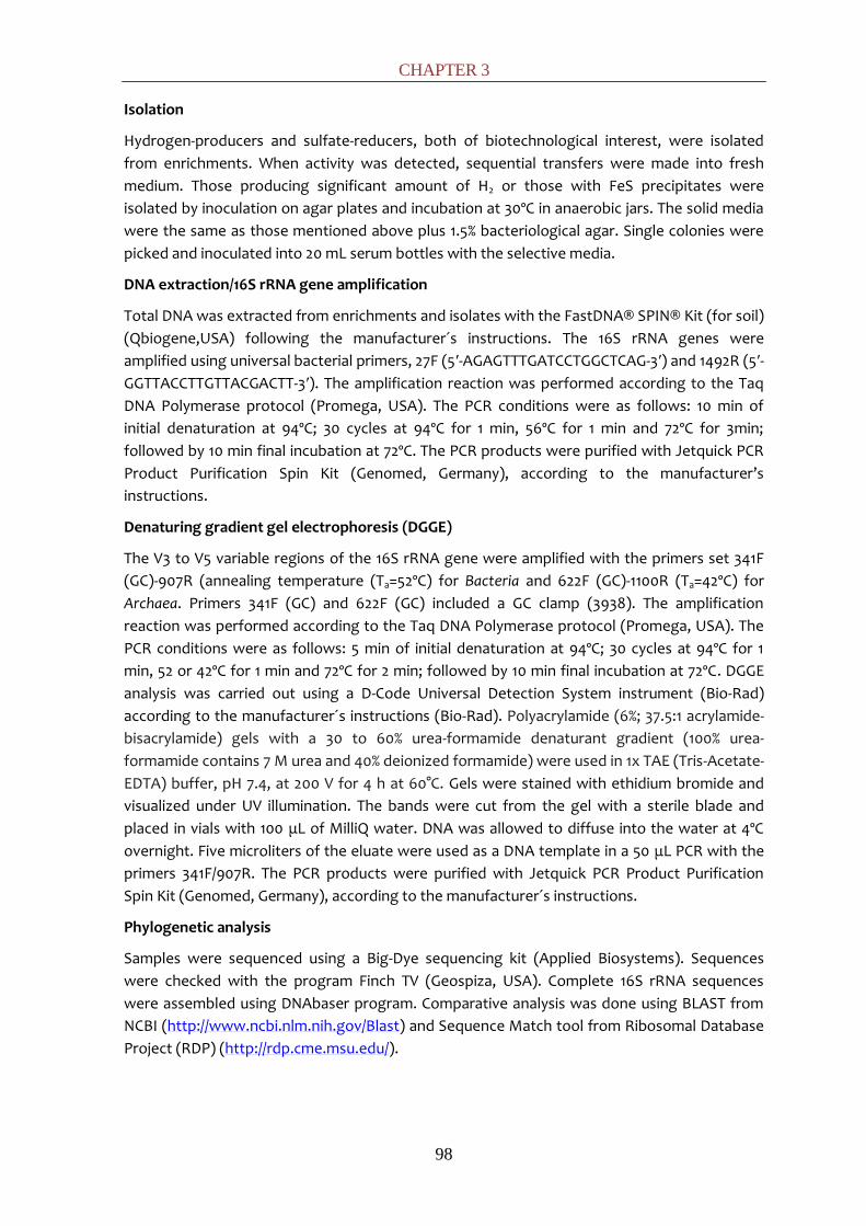
CHAPTER 3
98
Isolation
Hydrogen-producers and sulfate-reducers, both of biotechnological interest, were isolated
from enrichments. When activity was detected, sequential transfers were made into fresh
medium. Those producing significant amount of H2 or those with FeS precipitates were
isolated by inoculation on agar plates and incubation at 30ºC in anaerobic jars. The solid media
were the same as those mentioned above plus 1.5% bacteriological agar. Single colonies were
picked and inoculated into 20 mL serum bottles with the selective media.
DNA extraction/16S rRNA gene amplification
Total DNA was extracted from enrichments and isolates with the FastDNA® SPIN® Kit (for soil)
(Qbiogene,USA) following the manufacturer´s instructions. The 16S rRNA genes were
amplified using universal bacterial primers, 27F (5′-AGAGTTTGATCCTGGCTCAG-3′) and 1492R (5′-
GGTTACCTTGTTACGACTT-3′). The amplification reaction was performed according to the Taq
DNA Polymerase protocol (Promega, USA). The PCR conditions were as follows: 10 min of
initial denaturation at 94ºC; 30 cycles at 94ºC for 1 min, 56ºC for 1 min and 72ºC for 3min;
followed by 10 min final incubation at 72ºC. The PCR products were purified with Jetquick PCR
Product Purification Spin Kit (Genomed, Germany), according to the manufacturer’s
instructions.
Denaturing gradient gel electrophoresis (DGGE)
The V3 to V5 variable regions of the 16S rRNA gene were amplified with the primers set 341F
(GC)-907R (annealing temperature (Ta=52ºC) for Bacteria and 622F (GC)-1100R (Ta=42ºC) for
Archaea. Primers 341F (GC) and 622F (GC) included a GC clamp (3938). The amplification
reaction was performed according to the Taq DNA Polymerase protocol (Promega, USA). The
PCR conditions were as follows: 5 min of initial denaturation at 94ºC; 30 cycles at 94ºC for 1
min, 52 or 42ºC for 1 min and 72ºC for 2 min; followed by 10 min final incubation at 72ºC. DGGE
analysis was carried out using a D-Code Universal Detection System instrument (Bio-Rad)
according to the manufacturer´s instructions (Bio-Rad). Polyacrylamide (6%; 37.5:1 acrylamide-
bisacrylamide) gels with a 30 to 60% urea-formamide denaturant gradient (100% urea-
formamide contains 7 M urea and 40% deionized formamide) were used in 1x TAE (Tris-Acetate-
EDTA) buffer, pH 7.4, at 200 V for 4 h at 60°C. Gels were stained with ethidium bromide and
visualized under UV illumination. The bands were cut from the gel with a sterile blade and
placed in vials with 100 μL of MilliQ water. DNA was allowed to diffuse into the water at 4ºC
overnight. Five microliters of the eluate were used as a DNA template in a 50 μL PCR with the
primers 341F/907R. The PCR products were purified with Jetquick PCR Product Purification
Spin Kit (Genomed, Germany), according to the manufacturer´s instructions.
Phylogenetic analysis
Samples were sequenced using a Big-Dye sequencing kit (Applied Biosystems). Sequences
were checked with the program Finch TV (Geospiza, USA). Complete 16S rRNA sequences
were assembled using DNAbaser program. Comparative analysis was done using BLAST from
NCBI (http://www.ncbi.nlm.nih.gov/Blast) and Sequence Match tool from Ribosomal Database
Project (RDP) (http://rdp.cme.msu.edu/).
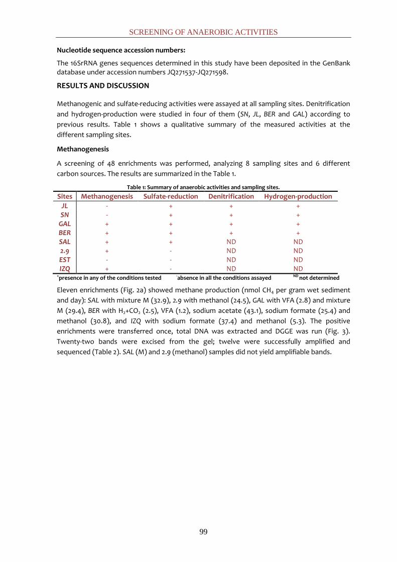
SCREENING OF ANAEROBIC ACTIVITIES
99
Nucleotide sequence accession numbers:
The 16SrRNA genes sequences determined in this study have been deposited in the GenBank database under accession numbers JQ271537-JQ271598.
RESULTS AND DISCUSSION
Methanogenic and sulfate-reducing activities were assayed at all sampling sites. Denitrification
and hydrogen-production were studied in four of them (SN, JL, BER and GAL) according to
previous results. Table 1 shows a qualitative summary of the measured activities at the
different sampling sites.
Methanogenesis
A screening of 48 enrichments was performed, analyzing 8 sampling sites and 6 different
carbon sources. The results are summarized in the Table 1.
Table 1: Summary of anaerobic activities and sampling sites.
Sites Methanogenesis Sulfate-reduction Denitrification Hydrogen-production JL - + + + SN - + + +
GAL + + + + BER + + + + SAL + + ND ND 2.9 + - ND ND EST - - ND ND IZQ + - ND ND
+presence in any of the conditions tested -absence in all the conditions assayed ND not determined
Eleven enrichments (Fig. 2a) showed methane production (nmol CH4 per gram wet sediment
and day): SAL with mixture M (32.9), 2.9 with methanol (24.5), GAL with VFA (2.8) and mixture
M (29.4), BER with H2+CO2 (2.5), VFA (1.2), sodium acetate (43.1), sodium formate (25.4) and
methanol (30.8), and IZQ with sodium formate (37.4) and methanol (5.3). The positive
enrichments were transferred once, total DNA was extracted and DGGE was run (Fig. 3).
Twenty-two bands were excised from the gel; twelve were successfully amplified and
sequenced (Table 2). SAL (M) and 2.9 (methanol) samples did not yield amplifiable bands.
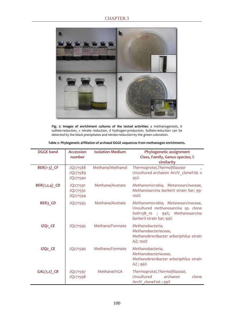
CHAPTER 3
100
Fig. 2: Images of enrichment cultures of the tested activities: a methanogenesis, b sulfate-reduction, c nitrate reduction, d hydrogen-production. Sulfate-reduction can be detected by the black precipitates and nitrate-reduction by the green coloration.
Table 2: Phylogenetic affiliation of archaeal DGGE sequences from methanogen enrichments.
DGGE band Accession number
Isolation Medium Phylogenetic assignment Class, Family, Genus species; %
similarity BER(1-3)_CF JQ271588
JQ271589 JQ271590
Methane/Methanol Thermoprotei,Thermofiliaceae , Uncultured archaeon ArcIV_cloneF06 < 95%
BER(1,2,4)_CD
JQ271591 JQ271592 JQ271594
Methane/Acetate Methanomicrobia, Metanosarcinaceae, Methanosarcina barkerii strain Sar; 99-100%
BER3_CD JQ271593 Methane/Acetate Methanomicrobia, Metanosarcinaceae, Uncultured methanosarcina sp. clone Soil113B_10 ; 94%; Methanosarcina barkerii strain Sar; 94%
IZQ1_CE JQ271595 Methane/Formate Methanobacteria, Methanobacteriaceae, Methanobrevibacter arboriphilus strain AZ; 100%
IZQ2_CE JQ271596 Methane/Formate Methanobacteria, Methanobacteriaceae, Methanobrevibacter arboriphilus strain AZ ; 96%
GAL(1,2)_CB JQ271597 JQ271598
Methane/VGA Thermoprotei,Thermofiliaceae, Uncultured archaeon clone ArcIV_cloneF06 ; 99%

SCREENING OF ANAEROBIC ACTIVITIES
101
Fig. 3: DGGE fingerprints of 16S rRNA obtained with domain-specific primers for Archaea from methanogenic enrichments. Sampling locations are displayed on the top of the lanes followed by the electron donor nomenclature used in the sequences: methanol (f), acetate (d) and formate (e). Letters within lances correspond to the sequence nomenclature shown in Table 2.
Sequences were affiliated to both archaeon phyla described: Crenarchaeota and Euryarchaeota.
Regarding the Crenarchaeota, some sequences retrieved from GAL and BER sites were affiliated
to the family Thermofiliaceae, which comprise known neutrophilic and hyperthermophilic
archaea (11). Despite these features, uncultured relatives of our Thermofiliaceae-related
sequences have been detected in acidic sediments (FN870267) (33). Related sequences have
also appeared linked to methanogenic habitats (AJ576215) (25), (AB243802) (43) and
(EU155990) (12).
It was possible to identify phylogenetically down to the genus level some of the Euryarchaeota
sequences. The nearest cultivated relatives to the DGGE bands at the sampling site IZQ
incubated with formate was Methanobrevibacter arboriphilus, being the formate a common M.
arboriphilus substrate (5, 42). This is to our knowledge the first report of this neutrophilic
methanogen in an acidic environment. On the other hand, the nearest cultivated relatives to
sequences of sampling site BER incubated with acetate was Methanosarcina barkerii. The
presence of Methanosarcina and the absence of Methanosaeta, the other methanogenic
acetoclastic genera, can be attributed to: (i) the high acetate concentration used in the
enrichment medium, which is much higher than the Ks of Methanosarcina for acetate, which
outcompete Methanosaeta (Ks and µmax 5x10⁻³ mol L-1and 2.5x10⁻² h-1 for Methanosarcina
versus 0.5x10⁻³ mol L-1 and 3x10⁻³ h-1 for Methanosaeta) (ii) the ability of Methanosarcina to
tolerate low pH conditions (34), consistent with previous studies (18, 23, 52). Remarkably, Ms.
barkeri was identified in a methanol-spiked microcosm inoculated with Tinto River sediments
(48).
Methanogenesis is usually believed to be inhibited by low pH, however, several studies have
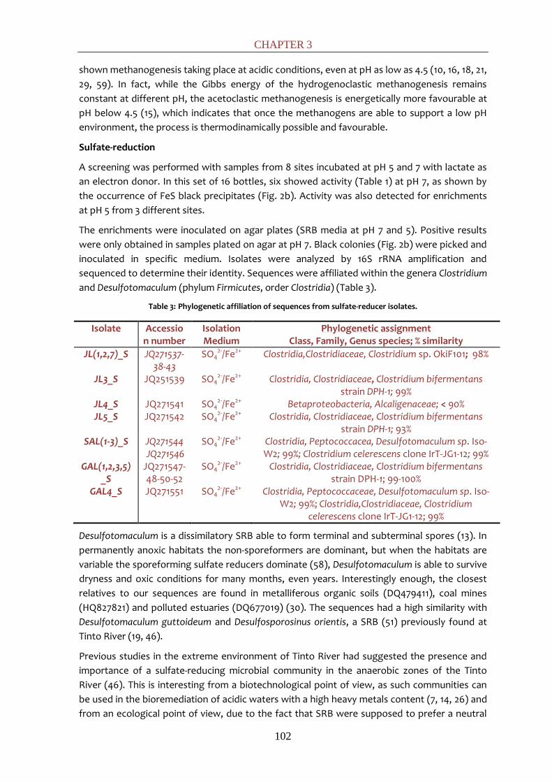
CHAPTER 3
102
shown methanogenesis taking place at acidic conditions, even at pH as low as 4.5 (10, 16, 18, 21,
29, 59). In fact, while the Gibbs energy of the hydrogenoclastic methanogenesis remains
constant at different pH, the acetoclastic methanogenesis is energetically more favourable at
pH below 4.5 (15), which indicates that once the methanogens are able to support a low pH
environment, the process is thermodinamically possible and favourable.
Sulfate-reduction
A screening was performed with samples from 8 sites incubated at pH 5 and 7 with lactate as
an electron donor. In this set of 16 bottles, six showed activity (Table 1) at pH 7, as shown by
the occurrence of FeS black precipitates (Fig. 2b). Activity was also detected for enrichments
at pH 5 from 3 different sites.
The enrichments were inoculated on agar plates (SRB media at pH 7 and 5). Positive results
were only obtained in samples plated on agar at pH 7. Black colonies (Fig. 2b) were picked and
inoculated in specific medium. Isolates were analyzed by 16S rRNA amplification and
sequenced to determine their identity. Sequences were affiliated within the genera Clostridium
and Desulfotomaculum (phylum Firmicutes, order Clostridia) (Table 3).
Table 3: Phylogenetic affiliation of sequences from sulfate-reducer isolates.
Isolate Accession number
Isolation Medium
Phylogenetic assignment Class, Family, Genus species; % similarity
JL(1,2,7)_S JQ271537-38-43
SO42-/Fe2+ Clostridia,Clostridiaceae, Clostridium sp. OkiF101; 98%
JL3_S JQ251539 SO42-/Fe2+ Clostridia, Clostridiaceae, Clostridium bifermentans
strain DPH-1; 99% JL4_S JQ271541 SO4
2-/Fe2+ Betaproteobacteria, Alcaligenaceae; < 90% JL5_S JQ271542 SO4
2-/Fe2+ Clostridia, Clostridiaceae, Clostridium bifermentans strain DPH-1; 93%
SAL(1-3)_S JQ271544 JQ271546
SO42-/Fe2+ Clostridia, Peptococcacea, Desulfotomaculum sp. Iso-
W2; 99%; Clostridium celerescens clone IrT-JG1-12; 99% GAL(1,2,3,5)
_S JQ271547-48-50-52
SO42-/Fe2+ Clostridia, Clostridiaceae, Clostridium bifermentans
strain DPH-1; 99-100% GAL4_S JQ271551 SO4
2-/Fe2+ Clostridia, Peptococcaceae, Desulfotomaculum sp. Iso-W2; 99%; Clostridia,Clostridiaceae, Clostridium
celerescens clone IrT-JG1-12; 99%
Desulfotomaculum is a dissimilatory SRB able to form terminal and subterminal spores (13). In
permanently anoxic habitats the non-sporeformers are dominant, but when the habitats are
variable the sporeforming sulfate reducers dominate (58), Desulfotomaculum is able to survive
dryness and oxic conditions for many months, even years. Interestingly enough, the closest
relatives to our sequences are found in metalliferous organic soils (DQ479411), coal mines
(HQ827821) and polluted estuaries (DQ677019) (30). The sequences had a high similarity with
Desulfotomaculum guttoideum and Desulfosporosinus orientis, a SRB (51) previously found at
Tinto River (19, 46).
Previous studies in the extreme environment of Tinto River had suggested the presence and
importance of a sulfate-reducing microbial community in the anaerobic zones of the Tinto
River (46). This is interesting from a biotechnological point of view, as such communities can
be used in the bioremediation of acidic waters with a high heavy metals content (7, 14, 26) and
from an ecological point of view, due to the fact that SRB were supposed to prefer a neutral

SCREENING OF ANAEROBIC ACTIVITIES
103
environment. In the last decades, considerable evidence has revealed that sulfate-reduction at
pH values below 5 is possible (28) and our results confirm the effective adaptation of SRB at
low pHs by showing that active acidotolerant sulfate-reducers thrive in Tinto River sediments.
Denitrification
Biological denitrification, both assimilative and dissimilative, is the biological process for
removing nitrate. Typically in most environments, nitrate is used as an electron acceptor by
facultative heterotrophic bacteria comprising many diverse genera (Pseudomonas, Alcaligenes,
Bacillus, etc.) under anoxic conditions, where organic matter provides electron donors to
reduce nitrate to dinitrogen gas (37). In contrast to the great diversity of heterotrophic
denitrifiers, there are just a few autotrophic denitrifiers, able to use hydrogen, ferrous iron, or
reduced sulfur compounds as electron donors.
As previously mentioned, autotrophic and heterotopic denitrification at pH 5 and 7 were
assayed at selected sampling sites (SN, JL, BER and GAL). In this set of 16 enrichments,
denitrification activity was followed by nitrate (NO3-), reduction and nitrite (NO2
-) and nitrous
oxide (N2O) production. Eleven of the enrichments showed activity with the exception of all
GAL enrichments and SN enrichments at pH 5 in autotrophic medium. Positive activity
correlated with the appearance of green colour in the bottles (Fig. 2c). An increment in the pH
was detected in the enrichments with an average of 0.02 units/day. Nitrate was consumed
during the assay with an average of 1 mmol per mg bacterial wet weight and day. Nitrite was
detected, but in a 7-fold lower concentration in the autotrophic samples in comparison with
the heterotrophic ones. Nitrous oxide accumulated at 0.01 mmol per mg bacterial wet weight
and day. A larger accumulation of nitrous oxide, 5-fold, was detected in the enrichments
growing at pH 5 versus those growing at pH 7.
The optimum pH range for complete reduction of nitrate to nitrogen gas (N2) is considered to
be between 6 and 8. In acid soils, below this optimal pH range, it has been reported that final
steps of the denitrification are inhibited. In these environments, the proportion of
intermediate products such as nitrite (8, 40) and nitrous oxide (50) increase with respect to
dinitrogen gas. A study with enrichment cultures suggested that denitrification occured in ARD
streams, and might reduce acidity (6), as confirmed in this assay.
Microbial populations were analyzed by DGGE to study the diversity and taxonomic position of
the responsible microbes. From the eleven positive enrichments, just eight yielded amplifiable
DNA (Fig. 4).
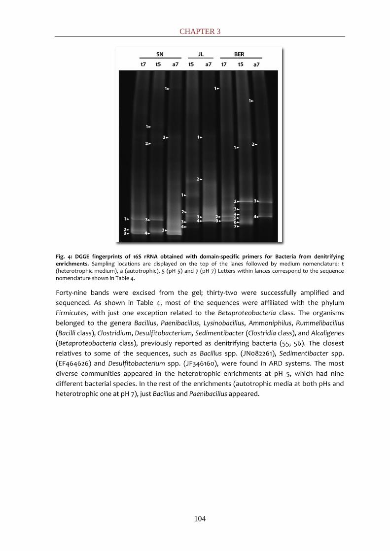
CHAPTER 3
104
Fig. 4: DGGE fingerprints of 16S rRNA obtained with domain-specific primers for Bacteria from denitrifying enrichments. Sampling locations are displayed on the top of the lanes followed by medium nomenclature: t (heterotrophic medium), a (autotrophic), 5 (pH 5) and 7 (pH 7) Letters within lances correspond to the sequence nomenclature shown in Table 4.
Forty-nine bands were excised from the gel; thirty-two were successfully amplified and
sequenced. As shown in Table 4, most of the sequences were affiliated with the phylum
Firmicutes, with just one exception related to the Betaproteobacteria class. The organisms
belonged to the genera Bacillus, Paenibacillus, Lysinobacillus, Ammoniphilus, Rummelibacillus
(Bacilli class), Clostridium, Desulfitobacterium, Sedimentibacter (Clostridia class), and Alcaligenes
(Betaproteobacteria class), previously reported as denitrifying bacteria (55, 56). The closest
relatives to some of the sequences, such as Bacillus spp. (JN082261), Sedimentibacter spp.
(EF464626) and Desulfitobacterium spp. (JF346160), were found in ARD systems. The most
diverse communities appeared in the heterotrophic enrichments at pH 5, which had nine
different bacterial species. In the rest of the enrichments (autotrophic media at both pHs and
heterotrophic one at pH 7), just Bacillus and Paenibacillus appeared.

SCREENING OF ANAEROBIC ACTIVITIES
105
Table 4: Phylogenetic affiliation of bacterial DGGE sequences from denitrifying enrichments
In previous studies of the boreholes in the Tinto River, activity in the enrichments of
thiosulfate-oxidizers using nitrate as an electron acceptor was detected, but the organisms
responsible for these activities were neither isolated nor characterized (17). Our study is the
first to confirm the presence, activity and diversity of a denitrifying community in Tinto River
sediments.
Hydrogen-production
Because hydrogen is released as a consequence of different anaerobic metabolisms, four of
the selected sampling sites (JL, SN, BER and GAL) were assayed for hydrogen production.
Enrichments showed significant production of H2 (300-400 mL g-1 Chemical Oxygen Demand
consumed) in all the cases. Twelve colonies per sample were isolated from agar plates (Fig. 2d)
and analyzed by 16S rRNA gene amplification (Table 5).
Band Accession number
Isolation Medium
Phylogenetic assignment Class, Family, Genus species; % similarity
JL1_Na5 JQ271565 NO3-/S2O3
2- Bacilli, Paenibacillaceae, Paenibacillus sp. enrichment culture clone 9; 99%
JL(1-4)_Na7 JQ271566 JQ271569
NO3-/S2O3
2- Bacilli, Bacillaceae, Bacillus sp. cp64; 100%
JL1_Nt5 JQ271570 NO3-/Y.E. Clostridia, Clostridiaceae, Sedimentibacter sp.
enrichment culture clone B4120; 96%
JL2_Nt5 JQ271571 NO3-/Y.E. Bacilli, Bacillaceae, Lysinobacillus fusiformis
strain B116; 98%
JL3_Nt5 JQ271572 NO3-/Y.E. Bacilli, Paenibacillaceae, Ammoniphilus sp. CC-
RT-E; 98%
JL1_Nt7 JQ271573 NO3-/Y.E. Bacilli, Bacillaceae, Bacillus sp. HZ-B; 99%
SN(1-3)_Na7 JQ271574-76 NO3-/S2O3
2- Bacilli, Bacillaceae, Bacillus sp. HZ-B; 99%
SN1_Nt5 JQ271577 NO3-/Y.E. Betaproteocbacteria, Comamonadaceae ,
Delftia tsuruhatensis strain WYLW2-1; 91% ; Betaproteocbacteria, Alcaligenaceae,
Alcaligenes sp; 91%
SN1_Nt7 JQ271578 NO3-/Y.E. Bacilli, Bacillaceae, Bacillus sp. HZ-B; 99%
BER(1-4)_Na7 JQ271553 JQ271556
NO3-/S2O3
2- Clostridia, Clostridiaceae, Clostridium jejuense strain HY-35-12; 96%
BER1_Nt5 JQ271557 NO3-/Y.E. Clostridia, Clostridiaceae, Clostridium jejuense
strain HY-35-12; 97%
BER(2,3,4)_Nt5 JQ271558 JQ271560, JQ271561
NO3-/Y.E. Clostridia, Peptococaceae, Desulfitobacterium
sp. enrichment culture clone CEB3; 97%
BER(1-3)_Nt7 JQ271562 JQ271564
NO3-/Y.E. Bacilli, Paenibacillaceae, Paenibacillus sp.
enrichment culture clone S16; 88%
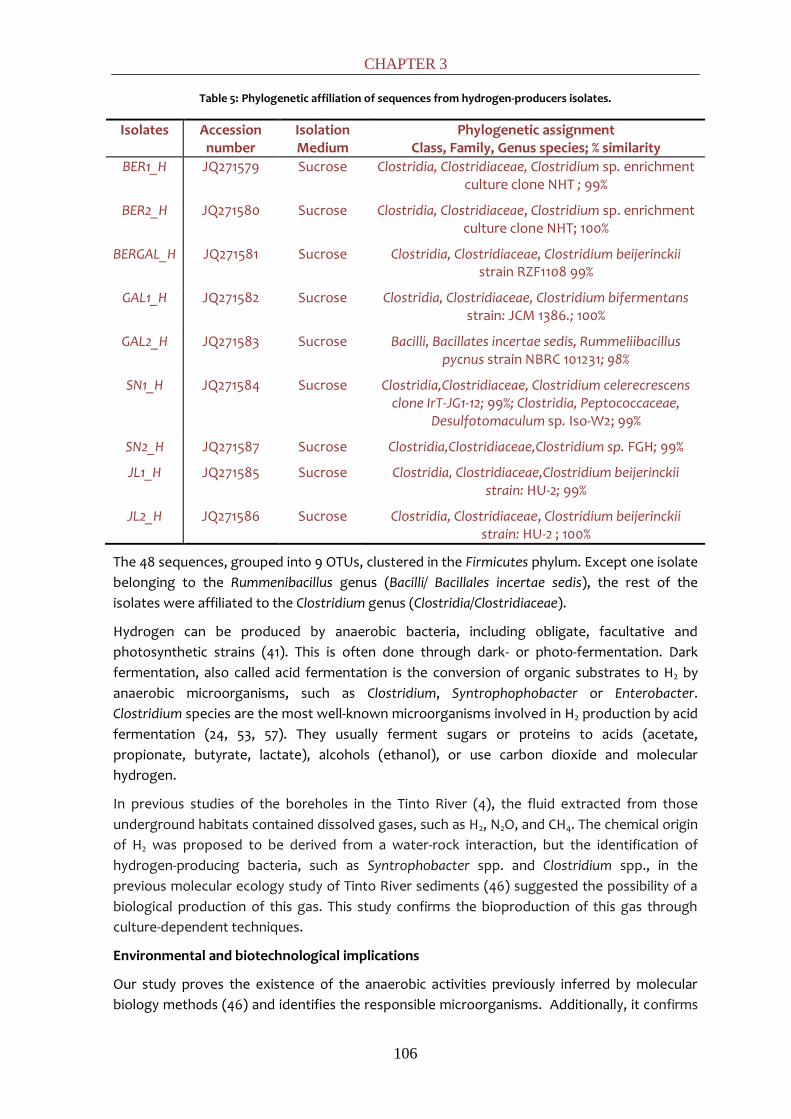
CHAPTER 3
106
Table 5: Phylogenetic affiliation of sequences from hydrogen-producers isolates.
Isolates Accession number
Isolation Medium
Phylogenetic assignment Class, Family, Genus species; % similarity
BER1_H JQ271579 Sucrose Clostridia, Clostridiaceae, Clostridium sp. enrichment culture clone NHT ; 99%
BER2_H JQ271580 Sucrose Clostridia, Clostridiaceae, Clostridium sp. enrichment culture clone NHT; 100%
BERGAL_H JQ271581 Sucrose Clostridia, Clostridiaceae, Clostridium beijerinckii strain RZF1108 99%
GAL1_H JQ271582 Sucrose Clostridia, Clostridiaceae, Clostridium bifermentans strain: JCM 1386.; 100%
GAL2_H JQ271583 Sucrose Bacilli, Bacillates incertae sedis, Rummeliibacillus pycnus strain NBRC 101231; 98%
SN1_H
JQ271584 Sucrose Clostridia,Clostridiaceae, Clostridium celerecrescens clone IrT-JG1-12; 99%; Clostridia, Peptococcaceae,
Desulfotomaculum sp. Iso-W2; 99%
SN2_H JQ271587 Sucrose Clostridia,Clostridiaceae,Clostridium sp. FGH; 99%
JL1_H JQ271585 Sucrose Clostridia, Clostridiaceae,Clostridium beijerinckii strain: HU-2; 99%
JL2_H JQ271586 Sucrose Clostridia, Clostridiaceae, Clostridium beijerinckii strain: HU-2 ; 100%
The 48 sequences, grouped into 9 OTUs, clustered in the Firmicutes phylum. Except one isolate
belonging to the Rummenibacillus genus (Bacilli/ Bacillales incertae sedis), the rest of the
isolates were affiliated to the Clostridium genus (Clostridia/Clostridiaceae).
Hydrogen can be produced by anaerobic bacteria, including obligate, facultative and
photosynthetic strains (41). This is often done through dark- or photo-fermentation. Dark
fermentation, also called acid fermentation is the conversion of organic substrates to H2 by
anaerobic microorganisms, such as Clostridium, Syntrophophobacter or Enterobacter.
Clostridium species are the most well-known microorganisms involved in H2 production by acid
fermentation (24, 53, 57). They usually ferment sugars or proteins to acids (acetate,
propionate, butyrate, lactate), alcohols (ethanol), or use carbon dioxide and molecular
hydrogen.
In previous studies of the boreholes in the Tinto River (4), the fluid extracted from those
underground habitats contained dissolved gases, such as H2, N2O, and CH4. The chemical origin
of H2 was proposed to be derived from a water-rock interaction, but the identification of
hydrogen-producing bacteria, such as Syntrophobacter spp. and Clostridium spp., in the
previous molecular ecology study of Tinto River sediments (46) suggested the possibility of a
biological production of this gas. This study confirms the bioproduction of this gas through
culture-dependent techniques.
Environmental and biotechnological implications
Our study proves the existence of the anaerobic activities previously inferred by molecular
biology methods (46) and identifies the responsible microorganisms. Additionally, it confirms

SCREENING OF ANAEROBIC ACTIVITIES
107
the bioproduction of H2, CH4 and N2O, some of the gases detected in a borehole performed in
the deposits rock matrix of Tinto River (4).
Together with the ecological implications of the results shown in this study, many potential
biotechnological applications arise. Methanogenesis is an important process in wastewater
treatment, and methanogens with a tolerance for acidic or metalliferous conditions could be
very useful in remediating acidic metal polluted waste streams. Biological processes using
renewable sources for hydrogen-production are more economical and environmental friendly
than the physical-chemical processes presently used (36, 49), so interest in biological
hydrogen-production is increasing. Additionally, SRB can be used for remediation of acidic
water with high heavy metals content or radionuclides (26, 27, 39, 54).
REFERENCES
1. Aguilera, A., S. C. Manrubia, F. Gomez, N. Rodriguez, and R. Amils. 2006. Eukaryotic community distribution and its relationship to water physicochemical parameters in an extreme acidic environment, Rio Tinto (Southwestern Spain). Appl. Environ. Microbiol. 72:5325-5330.
2. Aguilera, A., V. Souza-Egipsy, F. Gomez, and R. Amils. 2007. Development and structure of eukaryotic biofilms in an extreme acidic environment, Río Tinto (SW, Spain). Microb. Ecol. 53:294-305.
3. Aguilera, A., E. Zettler, F. Gómez, L. Amaral-Zettler, N. Rodríguez, and R. Amils. 2007. Distribution and seasonal variability in the benthic eukaryotic community of Río Tinto (SW, Spain), an acidic, high metal extreme environment. Syst. Appl. Microbiol. 30:531-546.
4. Amils, R., D. Fernández-Remolar, F. Gómez, E. González-Toril, N. Rodríguez, C. Briones, O. Prieto-Ballesteros, J. L. Sanz, E. Díaz, and T. O. Stevens. 2008. Subsurface Geomicrobiology of the Iberian Pyritic Belt. In P. Dion and C. Shekhar Nautiyal (Ed.), Soil Biology (Vol. 13): Microbiology of Extreme Soils. Springer, Berlin. 205-223.
5. Asakawa, S., H. Morii, M. Akagawa-Matsushita, Y. Koga, and K. Hayano. 1993. Characterization of Methanobrevibacter arboriphilicus SA isolated from a paddy field soil and DNA-DNA hybridization among M. arboriphilicus strains. Int. J. Syst. Bacteriol. 43:683-686.
6. Baeseman, J. L., R. L. Smith, and J. Silverstein. 2006. Denitrification potential in stream sediments impacted by acid mine drainage: effects of pH, various electron donors, and iron. Microb. Ecol. 51:232-241.
7. Bijmans, M. F. M., E. de Vries, C. H. Yang, C. J. N. Buisman, P. N. L. Lens, and M. Dopson. 2010. Sulfate reduction at pH 4.0 for treatment of process and wastewaters. Biotechnol. Prog. 26:1029-1039.
8. Blösl, M., and R. Conrad. 1992. Influence of an increased pH on the composition of the nitrate-reducing microbial populations in an anaerobically incubated acidic forest soil. Syst. Appl. Microbiol. 15:624-627.
9. Blothe, M., D. M. Akob, J. E. Kostka, K. Goschel, H. L. Drake, and K. Kusel. 2008. pH gradient-induced heterogeneity of Fe (III)-reducing microorganisms in coal mining-associated lake sediments. Appl. Environ. Microbiol. 74:1019-1029.
10. Bräuer, S. L., J. B. Yavitt, and S. H. Zinder. 2004. Methanogenesis in McLean Bog, an acidic peat bog in upstate New York: stimulation by H2/CO2 in the presence of rifampicin, or by low concentrations of acetate. Geomicrobiol. J. 21:433-443.
11. Burggraf, S., H. Huber, and K. Stetter. 1997. Reclassification of the crenarchaeal orders and
CHAPTER 3
108
families in accordance with 16S rRNA sequence data. Int. J. Syst. Bacteriol. 47:657-660.
12. Cadillo-Quiroz, H., E. Yashiro, J. B. Yavitt, and S. H. Zinder. 2008. Characterization of the archaeal community in a minerotrophic fen and terminal restriction fragment length polymorphism-directed isolation of a novel hydrogenotrophic methanogen. Appl. Environ. Microbiol. 74:2059-2068.
13. Campbell, L. L., and J. R. Postgate. 1965. Classification of the spore-forming sulfate-reducing bacteria. Microbiology and Molecular Biology Reviews. 29:359-362.
14. Church, C. D., R. T. Wilkin, C. N. Alpers, R. O. Rye, and R. B. McCleskey. 2007. Microbial sulfate reduction and metal attenuation in pH 4 acid mine water. Geochem. Trans. 8:10.
15. Dolfing, J., A. Xu, and I. M. Head. 2010. Anomalous energy yields in thermodynamic calculations: importance of accounting for pH-dependent organic acid speciation. The ISME Journal. 4:463-464.
16. Dunfield, P., R. Dumont, and T. R. Moore. 1993. Methane production and consumption in temperate and subarctic peat soils: response to temperature and pH. Soil Biol. Biochem. 25:321-326.
17. Fernández-Remolar, D. C., O. Prieto-Ballesteros, N. Rodríguez, F. Gómez, R. Amils, J. Gómez-Elvira, and C. R. Stoker. 2008. Underground habitats in the Río Tinto Basin: a model for subsurface life habitats on Mars. Astrobiology. 8:1023-1047.
18. Florencio, L., A. Nozhevnikova, A. Van Langerak, A. J. M. Stams, J. A. Field, and G. Lettinga. 1993. Acidophilic degradation of methanol by a methanogenic enrichment culture. FEMS Microbiol. Lett. 109:1-6.
19. García-Moyano, A., E. González-Toril, A. Aguilera, and R. Amils. 2007. Prokaryotic community composition and ecology of floating macroscopic filaments from an extreme acidic environment, Río Tinto (SW, Spain). Syst. Appl. Microbiol. 30:601-614.
20. Gonzalez-Toril, E., E. Llobet-Brossa, E. O. Casamayor, R. Amann, and R. Amils. 2003. Microbial ecology of an extreme acidic environment, the Tinto River. Appl. Environ. Microbiol. 69:4853-4865.
21. Goodwin, S., and J. G. Zeikus. 1987. Ecophysiological adaptations of anaerobic bacteria to low pH: analysis of anaerobic digestion in acidic bog sediments. Appl. Environ. Microbiol. 53:57-64.
22. Hao, C., H. Zhang, R. Haas, Z. Bai, and B. Zhang. 2007. A novel community of acidophiles in an acid mine drainage sediment. World J Microbiol Biotechnol. 23:15-21.
23. Horn, M. A., C. Matthies, K. Kusel, A. Schramm, and H. L. Drake. 2003. Hydrogenotrophic methanogenesis by moderately acid-tolerant methanogens of a methane-emitting acidic peat. Appl. Environ. Microbiol. 69:74-83.
24. Hu, B., and S. Chen. 2007. Pretreatment of methanogenic granules for immobilized hydrogen fermentation. Int J Hydrogen Energy. 32:3266-3273.
25. Huang, L. N., Y. Q. Chen, H. Zhou, S. Luo, C. Y. Lan, and L. H. Qu. 2003. Characterization of methanogenic Archaea in the leachate of a closed municipal solid waste landfill. FEMS Microbiol. Ecol. 46:171-177.
26. Johnson, D. B., and K. B. Hallberg. 2005. Acid mine drainage remediation options: a review. Sci. Total Environ. 338:3-14.
27. Klonowska, A., M. Clark, S. Thieman, B. Giles, J. Wall, and M. Fields. 2008. Hexavalent chromium reduction in Desulfovibrio vulgaris Hildenborough causes transitory inhibition of sulfate reduction and cell growth. Appl. Microbiol. Biotechnol. 78:1007-1016.

SCREENING OF ANAEROBIC ACTIVITIES
109
28. Koschorreck, M. 2008. Microbial sulphate reduction at a low pH. FEMS Microbiol. Ecol. 64:329-342.
29. Koschorreck, M., K. Wendt-Potthoff, B. Scharf, and H. H. Richnow. 2008. Methanogenesis in the sediment of the acidic Lake Caviahue in Argentina. J. Volcanol. Geotherm. Res. 178:197-204.
30. Lin, B., C. Hyacinthe, S. Bonneville, M. Braster, P. Van Cappellen, and W. F. M. Röling. 2007. Phylogenetic and physiological diversity of dissimilatory ferric iron reducers in sediments of the polluted Scheldt estuary, Northwest Europe. Environ. Microbiol. 9:1956-1968.
31. López-Archilla, A. I., A. E. González, M. C. Terrón, and R. Amils. 2005. Diversity and ecological relationships of the fungal populations of an acidic river of Southwestern Spain: the Tinto River. Can. J. Microbiol. 50:923-934.
32. Lopez-Archilla, A. I., I. Marin, and R. Amils. 2001. Microbial community composition and ecology of an acidic aquatic environment: the Tinto River, Spain. Microb. Ecol. 41:20-35.
33. Lu, S., S. Gischkat, M. Reiche, D. M. Akob, K. B. Hallberg, and K. Kusel. 2010. Ecophysiology of Fe-cycling bacteria in acidic sediments. Appl. Environ. Microbiol. 76:8174-8183.
34. Maestrojuán, G. M., and D. R. Boone. 1991. Characterization of Methanosarcina barkeri MST and 227, Methanosarcina mazei S-6T, and Methanosarcina vacuolata Z-761T. Int. J. Syst. Bacteriol. 41:267-274.
35. Manaia, C. M., and M. S. Costa. 1991. Characterization of halotolerant Thermus isolates from shallow marine hot springs on S. Miguel, Azores. J. Gen. Microbiol. 137:2643-2648.
36. Manish, S., and R. Banerjee. 2008. Comparison of biohydrogen production processes. Int J Hydrogen Energy. 33:279-286.
37. Mateju, V., S. Cizinska, J. Krejci, and T. Janoch. 1992. Biological water denitrification-A review. Enzyme Microb. Tech. 14:170-183.
38. Muyzer, G., and N. B. Ramsing. 1995. Molecular methods to study the organization of microbial communities. Water Sci. Technol. 32:1-9.
39. Muyzer, G., and A. J. M. Stams. 2008. The ecology and biotechnology of sulphate-reducing bacteria. Nat Rev Microbiol. 6:441-454.
40. Nagele, W., and R. Conrad. 1990. Influence of soil pH on the nitrate-reducing microbial populations and their potential to reduce nitrate to NO and N2O. FEMS Microbiol. Lett. 74:49-57.
41. Nandi, R., and S. Sengupta. 1998. Microbial production of hydrogen: an overview. Crit. Rev. Microbiol. 24:61-84.
42. Rea, S., J. P. Bowman, S. Popovski, C. Pimm, and A. D. G. Wright. 2007. Methanobrevibacter millerae sp. nov. and Methanobrevibacter olleyae sp. nov., methanogens from the ovine and bovine rumen that can utilize formate for growth. Int. J. Syst. Evol. Microbiol. 57:450-456.
43. Sakai, S., H. Imachi, Y. Sekiguchi, A. Ohashi, H. Harada, and Y. Kamagata. 2007. Isolation of key methanogens for global methane emission from rice paddy fields: a novel isolate affiliated with the clone cluster rice cluster I. Appl. Environ. Microbiol. 73:4326-4331.
44. Sánchez, I., N. Fernández, R. Amils, and J. L. Sanz. 2008. Assessment of the addition of "Thiobacillus denitrificans" and "Thiomicrospira denitrificans" to chemolithoautotrophic denitrifying bioreactors. Int Microbiol. 11:179-184.
45. Sánchez-Andrea, I., K. Knittel, R. Amann, R. Amils, and J. L. Sanz. 2012. Quantification of Tinto River Sediments Microbial Communities: the Importance of Sulfate-Reducing Bacteria and their Role in Attenuating Acid Mine Drainage. Appl. Environ. Microbiol. doi:
CHAPTER 3
110
10.1128/AEM.00848-12
46. Sánchez-Andrea, I., N. Rodriguez, R. Amils, and J. L. Sanz. 2011. Microbial Diversity in Anaerobic Sediments at Rio Tinto, a Naturally Acidic Environment with a High Heavy Metal Content. Appl. Environ. Microbiol. 77:6085-6093.
47. Sanz, J. L., N. Rodriguez, and R. Amils. 1997. Effect of chlorinated aliphatic hydrocarbons on the acetoclastic methanogenic activity of granular sludge. Appl. Microbiol. Biotechnol. 47:324-328.
48. Sanz, J. L., N. Rodríguez, E. E. Díaz, and R. Amils. 2011. Methanogenesis in the sediments of Rio Tinto, an extreme acidic river. Environ. Microbiol. 13:2336-2341.
49. Show, K., D. Lee, and J. Chang. 2011. Bioreactor and process design for biohydrogen production. Biores. Technol.102:8524-8533.
50. Simek, M., L. Jisova, and D. W. Hopkins. 2002. What is the so-called optimum pH for denitrification in soil. Soil Biol. Biochem. 34:1227-1234.
51. Stahl, D. A., and R. Amann. 1991. Development and application of nucleic acid probes. In E. Stackebrandt and M. Goodfellow (Ed. ), Nucleic Acid Techniques in Bacterial Systematics. John Wiley & Sons, Ltd., Chichester, United Kingdom. 205-248.
52. Staley, B. F., de los Reyes III, F.L., and M. A. Barlaz. 2011. Effect of spatial differences in microbial activity, pH, and substrate levels on methanogenesis initiation in refuse. Appl. Environ. Microbiol. 77:2381-2391.
53. Ueno, Y., S. Haruta, M. Ishii, and Y. Igarashi. 2001. Microbial community in anaerobic hydrogen-producing microflora enriched from sludge compost. Appl. Microbiol. Biotechnol. 57:555-562.
54. Valls, M., and V. Lorenzo. 2002. Exploiting the genetic and biochemical capacities of bacteria for the remediation of heavy metal pollution. FEMS Microbiol. Rev. 26:327-338.
55. Verbaendert, I., P. De Vos, N. Boon, and K. Heylen. 2011. Denitrification in Gram-positive bacteria: an underexplored trait. Biochem. Soc. Trans. 39:254-258.
56. Villemur, R., M. Lanthier, R. Beaudet, and F. Lépine. 2006. The Desulfitobacterium genus. FEMS Microbiol. Rev. 30:706-733.
57. Wang, X., D. Hoefel, C. P. Saint, P. T. Monis, and B. Jin. 2007. The isolation and microbial community analysis of hydrogen producing bacteria from activated sludge. J. Appl. Microbiol. 103:1415-1423.
58. Widdel, F. 1992. The genus Desulfotomaculum. The Prokaryotes. 2:1792-1799.
59. Williams, R. T., and R. L. Crawford. 1985. Methanogenic bacteria, including an acid-tolerant strain, from peatlands. Appl. Environ. Microbiol. 50:1542-1544.
60. Zettler, L. A. A., F. Gómez, E. Zettler, B. G. Keenan, R. Amils, and M. L. Sogin. 2002. Microbiology: eukaryotic diversity in Spain's River of Fire. Nature. 417:137.

CHAPTER 4:
Microbial diversity of
anaerobic zones of Tinto River: a
culture-dependent and non-dependent
approach

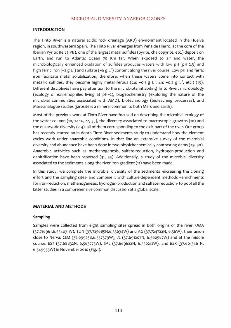
MICROBIAL DIVERSITY ANAEROBIC ZONES
113
INTRODUCTION
The Tinto River is a natural acidic rock drainage (ARD) environment located in the Huelva
region, in southwestern Spain. The Tinto River emerges from Peña de Hierro, at the core of the
Iberian Pyritic Belt (IPB), one of the largest metal sulfides (pyrite, chalcopyrite, etc.) deposit on
Earth, and run to Atlantic Ocean 70 Km far. When exposed to air and water, the
microbiologically enhanced oxidation of sulfides produces waters with low pH (pH 2.3) and
high ferric iron (~2 g L-1) and sulfate (~6 g L-1) content along the river course. Low pH and ferric
iron facilitate metal solubilization; therefore, when these waters come into contact with
metallic sulfides, they become highly metalliferous (Cu: ~0.1 g L-1; Zn: ~0.2 g L-1, etc.) (19).
Different disciplines have pay attention to the microbiota inhabiting Tinto River: microbiology
(ecology of extremophiles living at pH~2), biogeochemistry (exploring the nature of the
microbial communities associated with AMD), biotechnology (bioleaching processes), and
Mars-analogue studies (jarosite is a mineral common to both Mars and Earth).
Most of the previous work at Tinto River have focused on describing the microbial ecology of
the water column (10, 12-14, 22, 35), the diversity associated to macroscopic growths (10) and
the eukaryotic diversity (2-4), all of them corresponding to the oxic part of the river. Our group
has recently started an in depth Tinto River sediments study to understand how the element
cycles work under anaerobic conditions. In that line an extensive survey of the microbial
diversity and abundance have been done in two physichochemically contrasting dams (29, 30).
Anaerobic activities such as methanogenesis, sulfate-reduction, hydrogen-production and
denitrification have been reported (31, 33). Additionally, a study of the microbial diversity
associated to the sediments along the river iron gradient (11) have been made.
In this study, we complete the microbial diversity of the sediments -increasing the cloning
effort and the sampling sites- and combine it with culture-dependent methods –enrichments
for iron-reduction, methanogenesis, hydrogen-production and sulfate-reduction- to pool all the
latter studies in a comprehensive common discussion at a global scale.
MATERIAL AND METHODS
Sampling
Samples were collected from eight sampling sites spread in both origins of the river: UMA
(37.716961,6.554031W), TUN (37.725685N,6.55634W) and AG (37.724722N, 6.56W); their union
close to Nerva: CEM (37.699238,6.557379W), JL (37.691207N, 6.560587W) and at the middle
course: EST (37.68832N, 6.563773W), SAL (37.669622N, 6.550212W), and BER (37.601346 N,
6.549933W) in November 2010 (Fig.1).
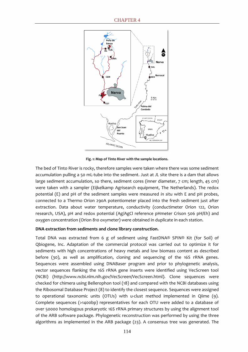
CHAPTER 4
114
Fig. 1: Map of Tinto River with the sample locations.
The bed of Tinto River is rocky, therefore samples were taken where there was some sediment
accumulation pulling a 50 mL-tube into the sediment. Just at JL site there is a dam that allows
large sediment accumulation, so there, sediment cores (inner diameter, 7 cm; length, 45 cm)
were taken with a sampler (Eijkelkamp Agrisearch equipment, The Netherlands). The redox
potential (E) and pH of the sediment samples were measured in situ with E and pH probes,
connected to a Thermo Orion 290A potentiometer placed into the fresh sediment just after
extraction. Data about water temperature, conductivity (conductimeter Orion 122, Orion
research, USA), pH and redox potential (Ag/AgCl reference pHmeter Crison 506 pH/Eh) and
oxygen concentration (Orion 810 oxymeter) were obtained in duplicate in each station.
DNA extraction from sediments and clone library construction.
Total DNA was extracted from 6 g of sediment using FastDNA® SPIN® Kit (for Soil) of
Qbiogene, Inc. Adaptation of the commercial protocol was carried out to optimize it for
sediments with high concentrations of heavy metals and low biomass content as described
before (30), as well as amplification, cloning and sequencing of the 16S rRNA genes.
Sequences were assembled using DNABaser program and prior to phylogenetic analysis,
vector sequences flanking the 16S rRNA gene inserts were identified using VecScreen tool
(NCBI) (http://www.ncbi.nlm.nih.gov/VecScreen/VecScreen.html). Clone sequences were
checked for chimera using Bellerophon tool (18) and compared with the NCBI databases using
the Ribosomal Database Project (8) to identify the closest sequence. Sequences were assigned
to operational taxonomic units (OTUs) with u-clust method implemented in Qiime (9).
Complete sequences (>1400bp) representatives for each OTU were added to a database of
over 50000 homologous prokaryotic 16S rRNA primary structures by using the alignment tool
of the ARB software package. Phylogenetic reconstruction was performed by using the three
algorithms as implemented in the ARB package (23). A consensus tree was generated. The

MICROBIAL DIVERSITY ANAEROBIC ZONES
115
coverage of the clone libraries was calculated using the equation described by Singleton et al.
(17). The PAST software v1.82b (16) was used to compute the statistical indexes and the
sampling efficiency in clone libraries of each sampling station. The 16SrRNA genes sequences
determined in this study have been deposited in the GenBank database under accession
nu - .
Enrichment cultures
Enrichments were prepared in an anaerobic chamber. For all the sampling sites, approximately
3 g of sediment were added to 22 mL glass serum bottles with 20 mL of media and capped with
butyl rubber stoppers. The headspace was then degassed with a N2/CO2 (80:20) mixture.
Enrichments were incubated at 30ºC in the dark. The media used were selective for various
microbial activities. For methanogenesis, the basal medium described by Sanz et al (32) was
used at pH 6 supplemented with a mixture of (g L-1): 0.7 sodium formate, 0.5 sodium acetate,
0.3 sodium propionate, 0.3 sodium butyrate, 0.5 methanol, 2 sucrose and 0.7 sodium lactate.
For sulfate reduction, the medium used was described previously (31) and used buffered at pH
7 and non-buffered. Enrichments for denitrification were conducted preparing the media as
described elsewhere (24). The media used for iron-reduction was (g L-1): 13.7 Fe(III)citrate, 2.5
NaHCO3, 1.5 NH4Cl, 0.1 KCl, 0.1 yeast extract, 1 sucrose, 3.16 Na2S2O3.5H2O , and 1 mL of
micronutrients solution (32). L-Cysteine was used as reducing agent (0.05%) in all media.
Banded JL dam sediments were analyzed deeply; therefore some methodological differences
regarding the rest of the sediment enrichments were made. Approximately 50 g of sediment
were added to 122 mL-glass serum bottles with 100 mL of media and capped with butyl rubber
stoppers. Two kinds of denitrifying enrichments were conducted separately. Both the
heterotrophic (24) and the autotrophic (28) denitrifying media were prepared as described
previously. The sulfate-reducing media added contained no buffers to allow the media to
equilibrate with the natural pH of the sediment. It was tested with lactate (2 g L-1), hydrogen
(gas phase H2:CO2 80:20) and nothing (native) as electron donors. Regarding the
methanogenic enrichments, they were performed with the mixture described above and also
with hydrogen under a H2/CO2 (80:20) atmosphere and acetate (0.5 g L-1) as electron donors.
Analytical Measurement
Activity in methanogenic and denitrifying enrichments was determined by monitoring CH4, and
N2O production in serum vial headspaces. For CH4 determinations, 100 µL subsamples of
headspace were injected into a Varian Star 3400CX gas chromatograph equipped with a
split/splitness injector and a flame ionization detector (FID). The GC was fitted with a SPB1000
(25 m x 0.32 mm x 0.25 μm) column manufactured by Supelco with Carbowax 20M
polyethilenglicol as stationary phase. The temperatures for the column, injector and detector
were 60, 200 and 250ºC respectively. Nitrogen was used as carrier gas. The production of N2O
was measured using a HP 5890 Series Chromatograph equipped with an on-column injector
and an electron capture detector (ECD). A Porapack 80-100 (1 m x 1/8” mm x 2 mm) column
manufactured by Sugerlabor was used. The temperatures for the column, injector and
detector were 40, 300 and 300ºC respectively. Nitrogen was used as carrier gas.
Measurements were made by injecting 50 µl of headspace into the GC. Denitrifying activity was
also determined measuring nitrate and nitrite concentrations by ion chromatography, using an
Omega Metrohm 790 Personal IC, with a Metrosep A Supp 5 250/4.0 (Omega) column. Sulfate-
reducing activity was determined visually by the formation of black precipitates from the

CHAPTER 4
116
reaction of H2S produced by sulfate reduction and Fe2+ added in the media.
Sulfate concentration was determined by ion chromatography with a suppressed conductivity
detector (790 Personal IC with an A Supp 5 250/4.0 column Omega Metrohom, Switzerland).
Measurements for pH were obtained with an Orion 2-Star portable pH meter (USA). Iron
species (ferric, ferrous and total iron) were determined by reflectrometry (Iron test method
1.16983.0001, Reflectoquant, Merck, Germany).
Denaturing gradient gel electrophoresis (DGGE) from sediment samples
DGGE analyses were carried out in JL enrichments to get an insight of the responsible organism
for each activity. DNA was extracted as explained before for the environmental sediment
samples. The V3 to V5 variable regions of the 16S rRNA gene were amplified with the primers
set 341F (GC)-907R (annealing temperature (Ta=52ºC) for Bacteria and 622F (GC)-1100R (Ta
=42ºC) for Archaea. Primers 341F (GC) and 622F (GC) included a GC clamp: 5’-CGC CCG CCG CGC
CCC GCG CCC GTC CCG CCC CCG CCC-3’ (25). The amplification reaction was performed
according to the Taq DNA Polymerase protocol (Promega Madison, Wis). The PCR conditions
were as follow: 10 min of initial denaturation at 94ºC and 30 cycles at 94ºC for 1 min and
annealing at 52/42ºC for 1 min and 72ºC for 2 min, followed by 10 min of final primer extension.
DGGE analysis was carried out using a D-Code Universal Detection System instrument (Bio-Rad)
and a model 475 gradient former according to the manufacturer’s instructions (Bio-Rad).
Polyacrylamide (6%; 37.5:1 acrylamide-bisacrylamide) gels with a 30 to 60% urea-formamide
denaturant gradient (100% urea-formamide contains 7 M urea and 40% deionized formamide)
were used in 1x TAE (Tris-acetate-EDTA) buffer, pH 7.4, at 200 V for 4 h at 60°C. Gels were
stained with ethidium bromide and visualized under UV illumination. About 100 bands were cut
from the gel with a sterile blade and placed in sterile vials with 100 μL of Milli-Q water. DNA
was allowed to diffuse into the water at 4 °C overnight. Five microliters of the eluate were
used as a DNA template in a PCR of 50 μL with the primers described above but without the GC
clamp.
Scanning electron microscopy (SEM)
Sediment samples were studied by SEM as described elsewhere (5). Samples were fixed by
immersion in glutaraldehyde (2.5%) for 2 h, washed twice in sodium cacodylate buffer (0.2 M,
pH 7.1) and dehydrated in a graded series (10, 30, 50, 70, 90, and 100%) of ethanol/water
mixtures for 20 min each. After dehydration, the samples were critical point dried and
mounted on stubs. After gold shadowing, the samples were examined in a Phillips XL30 EDAX
DX4i SEM.
RESULTS
Physicochemical parameters of the sampling sites
Table 1a and 1b show that in general all sampling sites present a pH range between 1.6 and 2.8.
The redox potential (E) ranged between 351.9 and 482.8 mV, positive and high, held to the
presence of a strong oxidant, Fe3+. Both, pH and E, are quite constant along the river due to
the buffering effect of the Fe3+ present in Tinto waters (equation 1).
Fe3+ + 3 H2O <--> Fe(OH)3 + 3H+ (eq. 1)

MICROBIAL DIVERSITY ANAEROBIC ZONES
117
Table 1a: Water physichochemical parameters measured in most of the samples.
Sample pH E (mV)
Cond (mS cm-2)
SO42- Fe Mn
(mg L-1) Co Ni Cu Zn As
BER 2.4 482.8 2.8 719.1 280.5 8.2 0.2 0.2 21.8 21.4 0.5 EST 2.2 456.9 8.0 2558.0 1212.6 33.9 1.1 0.5 117 93.9 4.3 CEM 2.4 351.9 9.4 4394.2 1049.7 57.9 3.8 1.1 223 182 2.4 SAL 2.3 418.2 8.2 2754.1 1510.4 37.0 ND 0.6 110 105 5.8 UMA 1.6 459.7 24.8 6861.3 8945.9 10.8 ND ND 43.9 72.3 15 TUN 2.4 308.9 5.7 1229.8 685.3 13.3 ND 0.0 10.4 40.8 2.0 AG 2.4 459.7 6.1 1815.0 1189.8 18.5 ND 0.1 14.8 64.8 2.0 JL 2.6 356.0 9.3 1738.2 912.6 26.1 ND 0.1 123 92.2 3.1
Conductivity values were the most variable parameter and have a direct relation with metal
concentration, especially with iron (the most abundant). Iron concentration decreased along
the river, the addition of neutral tributaries has a direct dilution effect lowering the
concentrations of metals, and an indirect effect on ferric oxides precipitation that releases
protons maintaining a constant pH. The highest values are found at UMA (8.9 g L-1 of iron and a
conductivity of 24.7 mS cm-2) and the lowest at BER (0.3 g L-1 of iron and a conductivity of 2.8
mS cm-2). The measured sulfate concentrations positively correlated with the iron
concentration along the river, logical taking into account that both are the products of the
metallic sulfides oxidation.
Table 1b: Water physichochemical concentration (mg L-1) parameters measured in most of the samples.
Sample acetate propionate formiate nitrite nitrate sulfate
BER 0.044 0 0.025 0.256 0.252 558.02 EST 0.177 0 0.025 0.284 0.397 2719.17 CEM 0.796 0.164 0 0.27 0.621 3473.5 SAL 0.044 0 0.027 0.438 0.349 2182.51 UMA 0 3.288 0 0.467 0 7160.53 TUN 0.068 0.1 0 0.12 0 562.4 AG 0.043 0 0.028 0.098 0 733.236 JL 0.469 0 0.102 0.103 0.182 1341.1
Volatile fatty acids and nitrogen species like nitrite and nitrate were detected (Table 1b). It is
the first time such data is reported in the Tinto water column. This has important ecological
implications showing that substrates and intermediates for denitrification and carbon cycle-
related metabolism are present.
With the exception of JL, where real sediment stratification exists, the values of water layer
are closed to the values of its underlying sediments. At JL sediments, the pH ranged from 3.6
to 5.5 and the redox potential (E) between -141 and 128 mV showing strong variations with
depth (Table 2). According to physico-chemical parameters and visual appearance, the JL dam
core was composed of two main layers. The reducing black layers (15-25 cm and 40-45 cm) with
mean pH of 5.5 and the oxidant brown layers (5-10 and 30-35 cm) with mean pH of 4.5 were
excised and immediately stored in 15 mL-tubes at 4ºC until further processing in the laboratory
(less than one week) for enrichments.

CHAPTER 4
118
Table 2: Physicochemical parameters measured in JL sediment layers.
Depth (cm) pH E (mV)
5 3.6 128
10 3.78 106
15 4.5 -60
20 5.5 -118 25 4.7 -47
30 4.3 45
35 4.2 26
40 4.7 -60 45 5 -141
Bacterial clone library diversity. Diversity indexes, coverage, and rarefaction analysis of the
16S rRNA gene libraries.
Former studies of the Tinto Sediments (11, 14, 30) coincided in their low archaeal diversity,
falling all the sequences retrieved in the Thermoplasma class. Therefore, archaeal community
was not further investigated in this study. Positive amplifications were obtained with bacterial
specific primers in all sediments samples. A total of 864 clones were obtained. After
elimination of chimerical and bad quality sequences, 714 sequences were analyzed.
All data analysis was performed for the total set of sequences and in each clone library
separately. Globally, the 714 total sequences clustered in 176 OTUs, based on 97% sequence
similarity (27). Specifically per sample, the number of OTUs varied from 46 till 92 (Table 2).
Between the 176 OTUs, 29 OTUs appeared to be common at different sampling sites and
others exclusive for one. There were some remarkable OTUs, which appear widely in the clone
libraries. For instance, clones related to the actinobacterial genus Ferrimicrobium were
detected in UMA, TUN, AG and CEM (OTU 10). This genus corresponds to heterotrophic, iron-
oxidizing and extremely acidophilic actinobacteria (20).
The common iron oxidizers in ARD environments are members of the aerobic
chemolithotrophic bacterial genus Leptospirillum (Nitrospira), which appear in different
locations (OTU 45 in CEM and AG and OTU 94 in TUN and EST) and the facultative anaerobe
gammaproteobacterial Acidithiobacillus ferrooxidans, which can oxidize iron and sulfur in
aerobic conditions and reduce iron in anoxia and appeared in most of the samples (OTU 120 in
TUN, CEM, SAL and BER; OTU 135 in TUN, CEM and UMA and OTU 171 in TUN, BER, CEM, EST and
SAL). In the Tinto River, together with the two former genera mentioned above, the
alphaproteobacterial Acidiphilium also appear at different locations (OTU 24 in EST and CEM;
OTU 147 in UMA, EST, CEM and SAL). This heterotrophic bacterium is able to oxidize organic
compounds using ferric iron as electron acceptor, even in the presence of oxygen, playing a
key role in the mineralization of organic matter, toxic at low pH for the acidophiles.
Within the phylum Firmicutes, several clones clustering within Clostridiales were retrieved from
JL black, EST and BER (OTU 170) and the sulfate-reducing Desulfosporosinus spp. were found in
JL black and TUN (OTU 1). Deltaproteobacterial clones belonging to the sulfur-reducer
Desulfurella genus were identified exclusively in both layers of JL dam (OTU 17).
Moreover, sequences related to the genus Dechloromonas were obtained at BER, AG, SAL, UMA
and CEM (OTUs 57 and 112). They are metabolic versatile organisms that can oxidize aromatic
compounds such as toluene, benzoate, and chlorobenzoate or Fe(II) and AHDS (a reduced
humic compound) aerobically and anaerobically using nitrate or even perchlorate or chlorate

MICROBIAL DIVERSITY ANAEROBIC ZONES
119
as a suitable electron acceptor (1, 7). Therefore, they have a great potential for bioremediation
processes.
Table 3: Statistical indexes in each sample
Sample BER EST CEM SAL UMA TUN AG
JL black
JL brown
TOTAL
Nº clones 96 96 96 96 96 96 96 96 96 864 Nº sequences 79 81 85 92 89 78 46 85 79 714
Nº OTUs 39 24 30 21 39 44 31 37 12 175 Good´s
coverage value (%)
25.6 54.2 60 61.9 35.9 34.1 25.8 32.4 58.3 44.9
Shannon-Wiener index
3.18 2.8 3.01 2.68 3.28 3.50 3.23 3.11 1.65 4.32
Dominance (D) value
0.07 0.08 0.06 0.08 0.05 0.04 0.05 0.07 0.3 0.02
Rarefaction curves, Good’s coverage values and diversity indexes were calculated separately
for each sample site. Good’s value (Table 3), an estimation of the proportion of the population
represented by the retrieved sequences (15), indicates low coverage of the bacterial libraries,
in agreement with the rarefaction curves (Fig. 2).
Fig. 2: Rarefaction curves for each sample.
Dominance (D) values are very low in all samples sites and in the global analysis, close to 0 (no
OTUs predominance in the community), with exception of JL Brown (D=0.3) indicating the
predominance of some OTUs in JL brown sequence data set (Desulfurella clones). In
accordance with this, the Shannon-Wiever index value was lower in JL Brown than in the rest
of samples. Higher diversity indexes were obtained compared with previous studies in Tinto
River (30).
It was possible to identify phylogenetically down to the genus level most of the Bacteria
sequences. 61 genera were identified, falling in twelve different phyla: Cloacibacillus
(Synergistetes); Bacillariophyta (Cyanobacteria/Chloroplast); TM7_incertae_sedis (TM7); family
Anaerolineaceae (Chloroflexi); Leptospirillum (Nitrospira), Prosthecobacter (Verrucomicrobia);
Fibrobacter (Fibrobacter); Alkalibacterium, Bacillus, Paenibacillus, Oceanobacillus,
Staphylococcus, Tumebacillus, Alicyclobacillus (Bacilli class, Firmicutes); Turicibacter
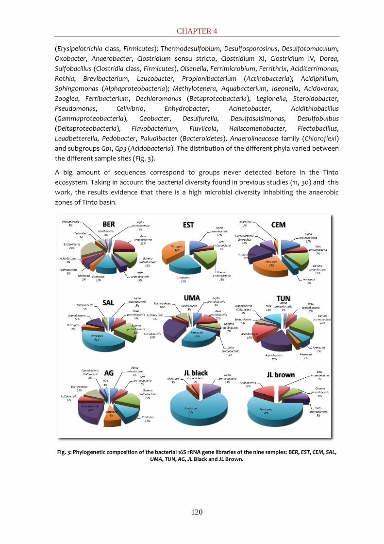
CHAPTER 4
120
(Erysipelotrichia class, Firmicutes); Thermodesulfobium, Desulfosporosinus, Desulfotomaculum,
Oxobacter, Anaerobacter, Clostridium sensu stricto, Clostridium XI, Clostridium IV, Dorea,
Sulfobacillus (Clostridia class, Firmicutes), Olsenella, Ferrimicrobium, Ferrithrix, Aciditerrimonas,
Rothia, Brevibacterium, Leucobacter, Propionibacterium (Actinobacteria); Acidiphilium,
Sphingomonas (Alphaproteobacteria); Methylotenera, Aquabacterium, Ideonella, Acidovorax,
Zooglea, Ferribacterium, Dechloromonas (Betaproteobacteria), Legionella, Steroidobacter,
Pseudomonas, Cellvibrio, Enhydrobacter, Acinetobacter, Acidithiobacillus
(Gammaproteobacteria), Geobacter, Desulfurella, Desulfosalsimonas, Desulfobulbus
(Deltaproteobacteria), Flavobacterium, Fluviicola, Haliscomenobacter, Flectobacillus,
Leadbetterella, Pedobacter, Paludibacter (Bacteroidetes), Anaerolineaceae family (Chloroflexi)
and subgroups Gp1, Gp3 (Acidobacteria). The distribution of the different phyla varied between
the different sample sites (Fig. 3).
A big amount of sequences correspond to groups never detected before in the Tinto
ecosystem. Taking in account the bacterial diversity found in previous studies (11, 30) and this
work, the results evidence that there is a high microbial diversity inhabiting the anaerobic
zones of Tinto basin.
Fig. 3: Phylogenetic composition of the bacterial 16S rRNA gene libraries of the nine samples: BER, EST, CEM, SAL, UMA, TUN, AG, JL Black and JL Brown.
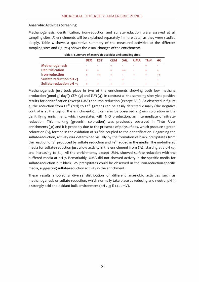
MICROBIAL DIVERSITY ANAEROBIC ZONES
121
Anaerobic Activities Screening
Methanogenesis, denitrification, iron-reduction and sulfate-reduction were assayed at all
sampling sites. JL enrichments will be explained separately in more detail as they were studied
deeply. Table 4 shows a qualitative summary of the measured activities at the different
sampling sites and Figure 4 shows the visual changes of the enrichments.
Table 4: Summary of anaerobic activities and sampling sites.
BER EST CEM SAL UMA TUN AG
Methanogenesis - - + - - + - Denitrification + + + ++ - + + Iron-reduction + ++ + - + + ++ Sulfate-reduction pH <5 - - - + - - - Sulfate-reduction pH ~7 + + + + - + +
Methanogenesis just took place in two of the enrichments showing both low methane
production (pmol g-1 day-1): CEM (9) and TUN (4). In contrast all the sampling sites yield positive
results for denitrification (except UMA) and iron-reduction (except SAL). As observed in figure
4, the reduction from Fe3+ (red) to Fe2+ (green) can be easily detected visually (the negative
control is at the top of the enrichments). It can also be observed a green coloration in the
denitrifying enrichment, which correlates with N2O production, an intermediate of nitrate-
reduction. This marking (greenish coloration) was previously observed in Tinto River
enrichments (31) and it is probably due to the presence of polysulfides, which produce a green
coloration (6), formed in the oxidation of sulfide coupled to the denitrification. Regarding the
sulfate-reduction, activity was determined visually by the formation of black precipitates from
the reaction of S2- produced by sulfate reduction and Fe2+ added in the media. The un-buffered
media for sulfate-reduction just allow activity in the enrichment from SAL, starting at a pH 4.5
and increasing to 6.5. All the enrichments, except UMA, showed sulfate-reduction with the
buffered media at pH 7. Remarkably, UMA did not showed activity in the specific media for
sulfate-reduction but black FeS precipitates could be observed in the iron-reduction-specific
media, suggesting sulfate-reduction activity in the enrichment.
These results showed a diverse distribution of different anaerobic activities such as
methanogenesis or sulfate-reduction, which normally take place at reducing and neutral pH in
a strongly acid and oxidant bulk environment (pH 2.3; E +400mV).

CHAPTER 4
122
Fig. 4: Screening of anaerobic activities at the different sampling sites.
MICROBIAL DIVERSITY ANAEROBIC ZONES
123
Anaerobic Activities at different pH.
(i) Activities rates
Methanogenesis was investigated in both layers of JL using hydrogen, acetate and a complex
mixture of VFA as electron donors. The methane production was rather slow in general
averaging 1.5 pmol CH4 per gram wet sediment and day except in JL black biostimulate with
the mixture M that produce 13 pmol CH4 per gram wet sediment and day. The visual
observation of the methanogenic enrichments (Fig. 5) evidenced the simultaneous occurrence
of sulfate-reduction in most of the bottles by the presence of FeS (black) and Fe2S (metallic)
precipitates.
Denitrifying enrichments were performed with sediments from reducing (black) and oxidant
(brown) layers using heterotrophic and autotrophic media. Denitrification activity was
followed by nitrate (NO3-), reduction and nitrite (NO2
-) and nitrous oxide (N2O) production. All
the enrichments showed activity at the end of the experiment (61 days) but heterotrophic
ones were faster showing activity at day 15. Nitrate was consumed during the assay with an
average (μmol g-1 day-1) of 2.6 for black layer-enrichments and 1 for brown layer-enrichments
showing a better develop at higher pH. The nitrous oxide production measured (μmol g-1 day-1)
was also higher in black (1) than in brown (0.7) layer. Comparing the nitrous oxide production
with the nitrate reduction rates, it was seen that a 37% of the reduced nitrate is accumulated in
nitrous oxide with the black layer inoculum while the 69% is accumulated with the brown layer
inoculum. The optimum pH range for complete reduction of nitrate to nitrogen gas (N2) is
considered to be between 6 and 8; below this optimal pH range, it has been reported that final
steps of the denitrification are inhibited and the proportion of intermediate products, as
nitrous oxide, increases with respect to dinitrogen gas (34). This result is in accordance with
previous ones performed with Tinto river sediments (31). Positive activity correlated with the
appearance of green colour in the bottles even in a quantitative way.
Iron-reduction activity was studied for both layers. The reduction of ferric iron results higher at
lower pH, reaching till 75% of the added iron at day 15 in brown-layer enrichment while in black-
layer enrichment took 61 days to achieve 50% reduction. In figure 5 it can be observed that
after iron-reduction took place, sulfate-reduction started, reasonable taking in account
suggesting that high Fe concentrations would inhibit sulfate-reduction.
Finally, sulfate-reduction carried-out by both layers was investigated with un-buffered media
using hydrogen and lactate as electron donors and a control with the natural donors of the
sample. The results showed clearly that sulfate-reduction takes place better at pH around 5
(black layer) than at pH around 4 (brown layer). In just 3 days the sulfate-activity could be
observed in all the enrichments from black layers. At day 61, enrichments of brown layer with
hydrogen and natural turned positive while enrichment with lactate remained negative,
confirming the inhibition caused for organic acids at pHs below its pKa. This result showed the
sulfate-reduction inhibition with the organic acid lactate at pH 4 while there was no inhibition
with hydrogen at pH as low as 3.6.
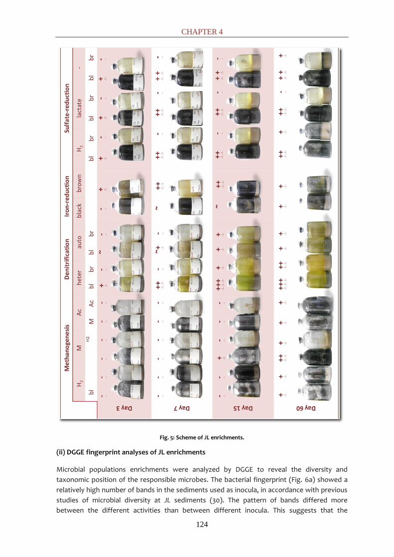
CHAPTER 4
124
Fig. 5: Scheme of JL enrichments.
(ii) DGGE fingerprint analyses of JL enrichments
Microbial populations enrichments were analyzed by DGGE to reveal the diversity and
taxonomic position of the responsible microbes. The bacterial fingerprint (Fig. 6a) showed a
relatively high number of bands in the sediments used as inocula, in accordance with previous
studies of microbial diversity at JL sediments (30). The pattern of bands differed more
between the different activities than between different inocula. This suggests that the
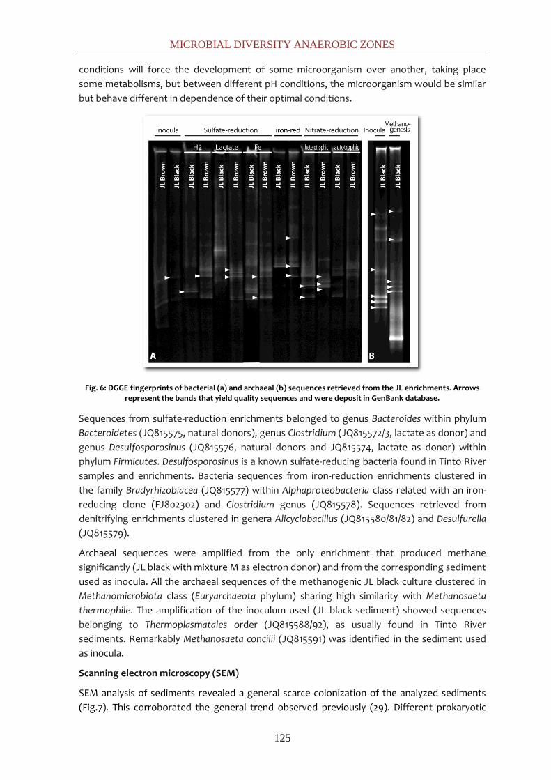
MICROBIAL DIVERSITY ANAEROBIC ZONES
125
conditions will force the development of some microorganism over another, taking place
some metabolisms, but between different pH conditions, the microorganism would be similar
but behave different in dependence of their optimal conditions.
Fig. 6: DGGE fingerprints of bacterial (a) and archaeal (b) sequences retrieved from the JL enrichments. Arrows represent the bands that yield quality sequences and were deposit in GenBank database.
Sequences from sulfate-reduction enrichments belonged to genus Bacteroides within phylum
Bacteroidetes (JQ815575, natural donors), genus Clostridium (JQ815572/3, lactate as donor) and
genus Desulfosporosinus (JQ815576, natural donors and JQ815574, lactate as donor) within
phylum Firmicutes. Desulfosporosinus is a known sulfate-reducing bacteria found in Tinto River
samples and enrichments. Bacteria sequences from iron-reduction enrichments clustered in
the family Bradyrhizobiacea (JQ815577) within Alphaproteobacteria class related with an iron-
reducing clone (FJ802302) and Clostridium genus (JQ815578). Sequences retrieved from
denitrifying enrichments clustered in genera Alicyclobacillus (JQ815580/81/82) and Desulfurella
(JQ815579).
Archaeal sequences were amplified from the only enrichment that produced methane
significantly (JL black with mixture M as electron donor) and from the corresponding sediment
used as inocula. All the archaeal sequences of the methanogenic JL black culture clustered in
Methanomicrobiota class (Euryarchaeota phylum) sharing high similarity with Methanosaeta
thermophile. The amplification of the inoculum used (JL black sediment) showed sequences
belonging to Thermoplasmatales order (JQ815588/92), as usually found in Tinto River
sediments. Remarkably Methanosaeta concilii (JQ815591) was identified in the sediment used
as inocula.
Scanning electron microscopy (SEM)
SEM analysis of sediments revealed a general scarce colonization of the analyzed sediments
(Fig.7). This corroborated the general trend observed previously (29). Different prokaryotic

CHAPTER 4
126
morphologies could be observed as well as diatoms and fungus structures.
Fig. 7: SEM images of the sediments. DISCUSSION
The Tinto River looks quite homogeneous looking at parameters as pH (2.3±0.4) or redox
potential (419±61) showing acid and oxidant conditions along the course. But due to the
hydrology, e.g. addition of neutral tributaries, the river presents a metal gradient along its
course, specially notary for iron (g L-1) ranging from 8.9 at the upper part till 0.3 in the middle
course (with the consequent conductivity variation), but also important in outstanding for
most of the metals: Cu (88.7±69), Co (3.8±3.8), Mn (27.7±16), and metalloids: As (5±4.6).
Fig. 8: Statistical analysis performed with FastUniFrac attending to different physicho-chemical parameters such as pH (a), conductivity (b) and arsenic concentration (c).

MICROBIAL DIVERSITY ANAEROBIC ZONES
127
Playing attention to the samples distribution with specific characteristics, statistical analysis
shows that some parameters influence on microbial distribution. When analyzing the
distribution according to the pH values, it can be seen in Figure 8a that AG, SEM, TUN and BER,
which clustered together, share common pH values. In the same way conductivity bring
together SAL, EST and JL, which shared medium values (Fig. 8b). Remarkably arsenic also
showed a weighting effect on the samples distribution that clustered AG, TUN and CEM (Fig.
8c).
The microbial diversity of Tinto River water is limited to a few organisms (14). Microbial
consortia in AMD waterflows are generally dominated by acidophilic, autotrophic iron- or
sulfur-oxidizing prokaryotes such as members of Acidithiobacillus and Leptospirillum genera
and heterotrophic acidophiles as Acidiphilium (21). But studies in two dams previously studied
showed an extensive biodiversity (30, 31), what it is definitely confirmed with the results of this
study in anaerobic zones through the river.
The superficial sediments layers would act as transition from the oxic water overlayed
characteristics to the anaerobic deeper sediments. Therefore in this zone, we can find
organisms as At. ferrooxidans and L. ferrooxidans, specially adapted to the high ionic strength
and oxidative stress (26) present in Tinto waters but also a complex community which links
iron, sulfur, nitrogen and carbon cycles with intricate relationships. The development of
anaerobic conditions will favor sequentially different anaerobic activities as correspond to
their redox chemistry. First of all, the reduction of the ferric iron would occur, what will turn
the redox potential less oxidant allowing the occurrence of nitrate reduction. Denitrification, in
general, increases the pH favoring less extreme conditions in the sediments (31). Next,
metabolism like the sulfate-reduction would have place increasing the pH and reducing the
redox potential (29). And lately, with a very low energy yield, the methanogenesis would occur
(33).
In the cloning of environmental sediment samples and supported by the DGGE sequences
retrieved from enrichments, we have found sequences belonging to organism able to develop
the different anaerobic activities studied and detected. Microorganisms involved in the
reduction of iron (A. ferrooxidans, Sulfobacillus, Geobacter, Acidiphilium, Acidobacteria…) were
identified as well as sulfate-reducers (Desulfurella, Desulfosporosinus, Desulfotomaculum,
Desulfobulbus and Thermodesulfobium). Nitrate-reducers were widely identified:
Dechloromonas, Propionibacterium, Pseudomonas, Bacillus… Methanogens, just identified by
DGGE, correspond to Methanosaeta spp. Besides the activities tested, another activities, as
fermentation, can be inferred from the clone libraries (Clostridium and Bacillus and related
species). The results of this study clearly highlight the importance of this compartment as a
cluster where an active iron, sulfur, nitrogen, carbon and metals cycles are operating.
REFERENCES
1. Achenbach, L. A., U. Michaelidou, R. A. Bruce, J. Fryman, and J. D. Coates. 2001. Dechloromonas agitata gen. nov., sp. nov. and Dechlorosoma suillum gen. nov., sp. nov., two novel environmentally dominant (per)chlorate-reducing bacteria and their phylogenetic position. Int. J. Syst. Evol. Microbiol. 51:527-533.
2. Aguilera, A., S. C. Manrubia, F. Gomez, N. Rodriguez, and R. Amils. 2006. Eukaryotic community distribution and its relationship to water physicochemical parameters in an

CHAPTER 4
128
extreme acidic environment, Rio Tinto (Southwestern Spain). Appl. Environ. Microbiol. 72:5325-5330.
3. Aguilera, A., V. Souza-Egipsy, F. Gomez, and R. Amils. 2007. Development and structure of eukaryotic biofilms in an extreme acidic environment, Río Tinto (SW, Spain). Microb. Ecol. 53:294-305.
4. Aguilera, A., E. Zettler, F. Gómez, L. Amaral-Zettler, N. Rodríguez, and R. Amils. 2007. Distribution and seasonal variability in the benthic eukaryotic community of Río Tinto (SW, Spain), an acidic, high metal extreme environment. Syst. Appl. Microbiol. 30:531-546.
5. Alphenaar, P. A., N. Groeneveld, and A. C. Van Aelst. 1994. Scanning electron microscopical method for internal structure analysis of anaerobic granular sludge. Micron. 25:129-133.
6. Banda, R. M. H., I. G. Dance, T. D. Bailey, D. C. Craig, and M. L. Scudder. 1989. Cadmium polysulfide complexes,[Cd (Sx)(Sy)] 2-: syntheses, crystal and molecular structures, and cadmium-113 NMR studies. Inorg. Chem. 28:1862-1871.
7. Chakraborty, R., S. M. O'Connor, E. Chan, and J. D. Coates. 2005. Anaerobic degradation of benzene, toluene, ethylbenzene, and xylene compounds by Dechloromonas strain RCB. Appl. Environ. Microbiol. 71:8649-8655.
8. Cole, J., Q. Wang, E. Cardenas, J. Fish, B. Chai, R. Farris, A. Kulam-Syed-Mohideen, D. McGarrell, T. Marsh, and G. Garrity. 2009. The Ribosomal Database Project: improved alignments and new tools for rRNA analysis. Nucleic Acids Res. 37:141-145.
9. Edgar, R. C. 2010. Search and clustering orders of magnitude faster than BLAST. Bioinformatics. 26:2460-2461.
10. García-Moyano, A., E. González-Toril, A. Aguilera, and R. Amils. 2007. Prokaryotic community composition and ecology of floating macroscopic filaments from an extreme acidic environment, Río Tinto (SW, Spain). Syst. Appl. Microbiol. 30:601-614.
11. García-Moyano, A., E. González-Toril, A. Aguilera, and R. Amils. 2012. Comparative microbial ecology study of the sediments and the water column of Río Tinto, an extreme acidic environment. FEMS Microbiol. Ecol. . doi: DOI: 10.1111/j.1574-6941.2012.01346.x.
12. García-Moyano, A., E. González-Toril, M. Moreno-Paz, V. Parro, and R. Amils. 2008. Evaluation of Leptospirillum spp. in the Río Tinto, a model of interest to biohydrometallurgy. Hydrometallurgy. 94:155-161.
13. Garrido, P., E. González-Toril, A. García-Moyano, M. Moreno-Paz, R. Amils, and V. Parro. 2008. An oligonucleotide prokaryotic acidophile microarray: its validation and its use to monitor seasonal variations in extreme acidic environments with total environmental RNA. Environ. Microbiol. 10:836-850.
14. Gonzalez-Toril, E., F. Gómez, N. Rodríguez, D. Fernández-Remolar, J. Zuluaga, I. Marín, and R. Amils. 2003. Geomicrobiology of the Tinto River, a model of interest for biohydrometallurgy. Hydrometallurgy. 71:301-309.
15. Good, I. J. 1953. The population frequencies of species and the estimation of population parameters. Biometrika. 40:237-264.
16. Hammer, Ø., D. A. T. Harper, and P. D. Ryan. 2001. PAST: paleontological statistics software package for education and data analysis. Palaeontol. Electron. 4:1-9.
17. Hansen, L. P., and K. J. Singleton. 1982. Generalized instrumental variables estimation of nonlinear rational expectations models. J. Econometric. 50:1269-1286.
18. Huber, T., G. Faulkner, and P. Hugenholtz. 2004. Bellerophon: a program to detect chimeric sequences in multiple sequence alignments. Bioinformatics. 20:2317-2319.

MICROBIAL DIVERSITY ANAEROBIC ZONES
129
19. Johnson, D. B. 1998. Biodiversity and ecology of acidophilic microorganisms. FEMS Microbiol. Ecol. 27:307-317.
20. Johnson, D. B., P. Bacelar-Nicolau, N. Okibe, A. Thomas, and K. B. Hallberg. 2009. Ferrimicrobium acidiphilum gen. nov., sp. nov. and Ferrithrix thermotolerans gen. nov., sp. nov.: heterotrophic, iron-oxidizing, extremely acidophilic actinobacteria. Int. J. Syst. Evol. Microbiol. 59:1082-1089.
21. Johnson, D. B., and K. B. Hallberg. 2003. The microbiology of acidic mine waters. Res. Microbiol. 154:466-473.
22. Lopez-Archilla, A. I., I. Marin, and R. Amils. 2001. Microbial community composition and ecology of an acidic aquatic environment: the Tinto River, Spain. Microb. Ecol. 41:20-35.
23. Ludwig, W., O. Strunk, R. Westram, L. Richter, and H. Meier. 2004. ARB: a software environment for sequence data. Nucleic Acids Res. 32:1363-1371.
24. Manaia, C. M., and M. S. Costa. 1991. Characterization of halotolerant Thermus isolates from shallow marine hot springs on S. Miguel, Azores. J. Gen. Microbiol. 137:2643-2648.
25. Muyzer, G., and N. B. Ramsing. 1995. Molecular methods to study the organization of microbial communities. Water Sci. Technol. 32:1-9.
26. Parro, V., M. Moreno‐Paz, and E. González‐Toril. 2007. Analysis of environmental transcriptomes by DNA microarrays. Environ. Microbiol. 9:453-464.
27. Rosselló-Mora, R., and R. Amann. 2001. The species concept for prokaryotes. FEMS Microbiol. Rev. 25:39-67.
28. Sánchez, I., N. Fernández, R. Amils, and J. L. Sanz. 2008. Assessment of the addition of "Thiobacillus denitrificans" and "Thiomicrospira denitrificans" to chemolithoautotrophic denitrifying bioreactors. Int Microbiol. 11:179-184.
29. Sánchez-Andrea, I., K. Knittel, R. Amann, R. Amils, and J. L. Sanz. 2012. Quantification of Tinto River sediment microbial communities: the importance of Sulfate-Reducing Bacteria and their role in attenuating acid mine drainage. Appl. Environ. Microbiol. . doi: doi: 10.1128/AEM.00848-12.
30. Sánchez-Andrea, I., N. Rodriguez, R. Amils, and J. L. Sanz. 2011. Microbial Diversity in Anaerobic Sediments at Rio Tinto, a Naturally Acidic Environment with a High Heavy Metal Content. Appl. Environ. Microbiol. 77:6085-6093.
31. Sánchez-Andrea, I., P. Rojas, R. Amils, and J. L. Sanz. 2012. Screening of Anaerobic Activities in Sediments of an Acidic Environment: Tinto River. Extremophiles. . doi: Acceptted.
32. Sanz, J. L., N. Rodriguez, and R. Amils. 1997. Effect of chlorinated aliphatic hydrocarbons on the acetoclastic methanogenic activity of granular sludge. Appl. Microbiol. Biotechnol. 47:324-328.
33. Sanz, J. L., N. Rodríguez, E. E. Díaz, and R. Amils. 2011. Methanogenesis in the sediments of Rio Tinto, an extreme acidic river. Environ. Microbiol. 13:2336-2341.
34. Simek, M., L. Jisova, and D. W. Hopkins. 2002. What is the so-called optimum pH for denitrification in soil. Soil Biol. Biochem. 34:1227-1234.
35. Souza-Egipsy, V., E. González-Toril, E. Zettler, L. A. Amaral-Zettler, A. Aguilera, and R. Amils. 2008. Prokaryotic community structure in algal photosynthetic biofilms from extreme acidic streams in Río Tinto (Huelva, Spain). Int. Microbiol. 11:251-260.

CHAPTER 4
130
OT
U ID
AG
BE
R
CE
M
ES
T
JLb
lac
k JL
bro
wn
S
AL
TU
N
UM
A Consensus Lineage
1 4 1 Firmicutes;Clostridia;Clostridiales;Peptococcaceae;Desulfosporosinus
2 1 Firmicutes;Clostridia;Clostridiales;Clostridiaceae;Oxobacter
3 1 Actinobacteria;Actinobacteria;Acidimicrobiales
4 1 1 Proteobacteria;Betaproteobacteria;Methylophilales;Methylophilaceae
5 1 Bacteroidetes;Flavobacteria;Flavobacteriales;Cryomorphaceae;Fluviicola
6 1 TM7;TM7_genera_incertae_sedis
7 1 Proteobacteria;Alphaproteobacteria;Rhodospirillales;Acetobacteraceae;Acidiphilium
8 2 Firmicutes;Clostridia;Clostridiales;Peptococcaceae;Desulfosporosinus
9 3 Bacteroidetes
10 3 9 1 5 Actinobacteria;Actinobacteria;Acidimicrobiales;Acidimicrobiaceae;Ferrimicrobium
11 3 Firmicutes;Bacilli;Bacillales;Bacillaceae;Oceanobacillus
12 3 Firmicutes;Bacilli;Bacillales;Bacillaceae;Oceanobacillus
13 2 Proteobacteria;Betaproteobacteria;Burkholderiales;Comamonadaceae;Acidovorax
14 1 Firmicutes;Clostridia;Clostridiales;Incertae Sedis XVII;Sulfobacillus
15 1 Nitrospira;Nitrospira;Nitrospirales;Nitrospiraceae;Leptospirillum
16 1 Firmicutes;Clostridia;Clostridiales;Ruminococcaceae
17 1 41 Proteobacteria;Deltaproteobacteria;Desulfurellales;Desulfurellaceae;Desulfurella
18 2 Proteobacteria;Betaproteobacteria;Rhodocyclales;Rhodocyclaceae
19 1 1 Proteobacteria;Alphaproteobacteria;Rhodospirillales;Acetobacteraceae;Acidiphilium
20 1 Proteobacteria;Betaproteobacteria
21 1 Proteobacteria;Gammaproteobacteria;Chromatiales
22 8 Actinobacteria;Actinobacteria;Actinomycetales
23 1 Bacteroidetes;Sphingobacteria;Sphingobacteriales;Cytophagaceae;Flectobacillus
24 1 2 Proteobacteria;Alphaproteobacteria;Rhodospirillales;Acetobacteraceae;Acidiphilium
25 1 Proteobacteria;Deltaproteobacteria;Desulfobacterales;Desulfobacteraceae
26 1 Proteobacteria;Betaproteobacteria;Burkholderiales;Comamonadaceae
27 1 Proteobacteria;Gammaproteobacteria
28 1 Proteobacteria;Deltaproteobacteria
29 2 Acidobacteria;Acidobacteria_Gp1;Gp1
30 2 Firmicutes;Clostridia;Clostridiales;Peptococcaceae
31 1 Actinobacteria;Actinobacteria;Acidimicrobiales
32 1 Proteobacteria;Gammaprot.;Xanthomonadales;Sinobacteraceae;Steroidobacter
33 1 Chloroflexi;Anaerolineae;Anaerolineales;Anaerolineaceae
34 1 Actinobacteria;Actinobacteria;Acidimicrobiales;Acidimicrobiaceae;Ferrithrix
35 1 8 Actinobacteria;Actinobacteria;Acidimicrobiales;Acidimicrobiaceae
36 1 Firmicutes;Clostridia;Clostridiales;Ruminococcaceae

MICROBIAL DIVERSITY ANAEROBIC ZONES
131
OT
U ID
AG
BE
R
CE
M
ES
T
JLb
lac
k JL
bro
wn
S
AL
TU
N
UM
A Consensus Lineage
37 1 Proteobacteria;Gammaprot.;Pseudomonadales;Pseudomonadaceae;Pseudomonas
38 1 Firmicutes;Bacilli;Bacillales;Bacillaceae;Bacillus
39 1 Firmicutes;Bacilli;Bacillales;Bacillaceae;Oceanobacillus
40 1 Firmicutes;Clostridia;Clostridiales;Peptostreptococcaceae;Sporacetigenium
41 1 Bacteroidetes;Sphingobacteria;Sphingobacteriales;Cytophagaceae;Leadbetterella
42 9 Proteobacteria;Gammaprot.;Acidithiobacillales;Acidithiobacillaceae;Acidithiobacillus
43 1 Firmicutes;Clostridia;Clostridiales;Clostridiaceae;Clostridium
44 1 Actinobacteria;Actinobacteria;Actinomycetales;Propionibacteriaceae
45 8 29 3 2 Nitrospira;Nitrospira;Nitrospirales;Nitrospiraceae;Leptospirillum
46 1 Bacteroidetes;Sphingobacteria;Sphingobacteriales;Sphingobacteriaceae;Pedobacter
47 2 Bacteroidetes;Bacteroidia;Bacteroidales;Porphyromonadaceae;Paludibacter
48 1 Acidobacteria;Acidobacteria_Gp1;Gp1
49 2 TM7;TM7_genera_incertae_sedis
50 5 Firmicutes;Clostridia;Clostridiales;Peptostreptococcaceae;Sporacetigenium
51 1 Proteobacteria;Gammaproteobacteria;Pseudomonadales;Pseudomonadaceae;Cellvibrio
52 3 Proteobacteria;Deltaproteobacteria;Desulfobacterales;Desulfobulbaceae;Desulfobulbus
53 1 Fibrobacteres;Fibrobacteria;Fibrobacterales;Fibrobacteraceae;Fibrobacter
54 1 Actinobacteria;Actinobacteria;Acidimicrobiales
55 1 Firmicutes;Clostridia;Clostridiales;Lachnospiraceae;Dorea
56 1 Firmicutes;Bacilli;Bacillales;Alicyclobacillaceae;Alicyclobacillus
57 1 11 1 1 1 2 10 Proteobacteria;Betaproteobacteria;Rhodocyclales;Rhodocyclaceae;Dechloromonas
58 1 Firmicutes;Clostridia;Clostridiales;Clostridiaceae;Clostridium
59 1 Firmicutes;Clostridia;Clostridiales;Clostridiaceae;Clostridium
60 1 Nitrospira;Nitrospira;Nitrospirales;Nitrospiraceae;Leptospirillum
61 13 Firmicutes;Clostridia;Clostridiales;Peptostreptococcaceae;Sporacetigenium
62 2 Acidobacteria;Acidobacteria_Gp1;Gp1
63 3 1 Cyanobacteria;Cyanobacteria;Chloroplast;Bacillariophyta
64 1 Bacteria
65 1 Proteobacteria;Gammaproteobacteria;Legionellales;Legionellaceae;Legionella
66 3 Firmicutes;Clostridia;Thermoanaerobacterales;Thermodesulfobiaceae;Thermodesulfobium
67 1 Actinobacteria;Actinobacteria;Acidimicrobiales;Iamiaceae;Iamia
68 1 Proteobacteria;Deltaproteobacteria;Desulfuromonadales;Geobacteraceae;Geobacter
69 1 Proteobacteria;Betaprot.;Burkholderiales;Burkholderiales_incertae_sedis;Aquabacterium
70 1 Firmicutes;Clostridia;Clostridiales;Incertae Sedis XVII;Sulfobacillus
71 1 Bacteroidetes
72 1 Firmicutes;Clostridia;Clostridiales;Incertae Sedis XVII;Sulfobacillus
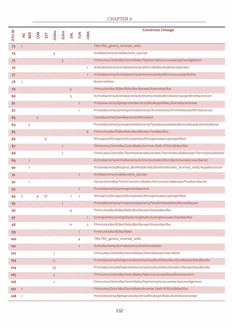
CHAPTER 4
132
OT
U ID
AG
BE
R
CE
M
ES
T
JLb
lac
k JL
bro
wn
S
AL
TU
N
UM
A Consensus Lineage
73 1 TM7;TM7_genera_incertae_sedis
74 3 Acidobacteria;Acidobacteria_Gp1;Gp1
75 3 Firmicutes;Clostridia;Clostridiales;Peptostreptococcaceae;Sporacetigenium
76 1 Actinobacteria;Actinobacteria;Acidimicrobiales;Acidimicrobiaceae
77 1 Actinobacteria;Actinobacteria;Actinomycetales;Micrococcaceae;Rothia
78 1 Bacteroidetes
79 5 Firmicutes;Bacilli;Bacillales;Bacillaceae;Oceanobacillus
80 3 Actinobacteria;Actinobacteria;Actinomycetales;Brevibacteriaceae;Brevibacterium
81 1 Proteobacteria;Alphaproteobacteria;Rhodospirillales;Acetobacteraceae
82 1 Proteobacteria;Gammaproteobacteria;Chromatiales;Chromatiaceae;Nitrosococcus
83 2 Cyanobacteria;Cyanobacteria;Chloroplast
84 5 Proteobacteria;Gammaproteobacteria;Pseudomonadales;Moraxellaceae;Acinetobacter
85 6 Firmicutes;Bacilli;Bacillales;Bacillaceae;Tumebacillus
86 6 Nitrospira;Nitrospira;Nitrospirales;Nitrospiraceae;Leptospirillum
87 1 Firmicutes;Clostridia;Clostridiales;Incertae Sedis XVII;Sulfobacillus
88 1 Firmicutes;Clostridia;Thermoanaerobacterales;Thermodesulfobiaceae;Thermodesulfobium
89 1 Actinobacteria;Actinobacteria;Actinomycetales;Microbacteriaceae;Leucobacter
90 1 Proteobacteria;Betaprot.;Burkholderiales;Burkholderiales_incertae_sedis;Aquabacterium
91 1 Acidobacteria;Acidobacteria_Gp1;Gp1
92 1 Verrucomicrobia;*e;Verrucomicrobiales;Verrucomicrobiaceae;Prosthecobacter
93 1 Proteobacteria;Gammaproteobacteria
94 2 4 27 1 1 Nitrospira;Nitrospira;Nitrospirales;Nitrospiraceae;Leptospirillum
95 1 Proteobacteria;Gammaproteobacteria;Pseudomonadales;Moraxellaceae
96 4 Firmicutes;Bacilli;Bacillales;Bacillaceae;Oceanobacillus
97 1 Synergistetes;Synergistia;Synergistales;Synergistaceae;Cloacibacillus
98 11 2 Firmicutes;Bacilli;Bacillales;Bacillaceae;Oceanobacillus
99 1 Firmicutes;Bacilli;Bacillales
100 4 TM7;TM7_genera_incertae_sedis
101 1 Actinobacteria;Actinobacteria;Acidimicrobiales
102 1 Firmicutes;Clostridia;Clostridiales;Clostridiaceae;Clostridium
103 5 Proteobacteria;Deltaproteobacteria;Desulfurellales;Desulfurellaceae;Desulfurella
104 25 Proteobacteria;Deltaproteobacteria;Desulfurellales;Desulfurellaceae;Desulfurella
105 2 Firmicutes;Clostridia;Clostridiales;Peptococcaceae;Desulfotomaculum
106 1 Firmicutes;Clostridia;Clostridiales;Peptostreptococcaceae;Sporacetigenium
107 1 Firmicutes;Clostridia;Clostridiales;Incertae Sedis XVII;Sulfobacillus
108 1 Proteobacteria;Alphaproteobacteria;Rhodospirillales;Acetobacteraceae

MICROBIAL DIVERSITY ANAEROBIC ZONES
133
OT
U ID
AG
BE
R
CE
M
ES
T
JLb
lac
k JL
bro
wn
S
AL
TU
N
UM
A Consensus Lineage
109 1 Bacteroidetes;Bacteroidia;Bacteroidales;Porphyromonadaceae
110 1 Bacteroidetes;Sphingobacteria;Sphingobacteriales;Saprospiraceae;Haliscomenobacter
111 1 1 Acidobacteria;Acidobacteria_Gp3;Gp3
112 1 2 Proteobacteria;Betaproteobacteria;Rhodocyclales;Rhodocyclaceae;Dechloromonas
113 1 Bacteroidetes;Bacteroidia;Bacteroidales;Porphyromonadaceae;Paludibacter
114 25 Firmicutes;Bacilli;Bacillales;Staphylococcaceae;Staphylococcus
115 1 Proteobacteria;Betaprot.;Burkholderiales;Burkholderiales_incertae_sedis;Ideonella
116 1 Proteobacteria;Alphaproteobacteria;Rhodospirillales;Acetobacteraceae;Acidiphilium
117 2 Proteobacteria;Betaproteobacteria;Rhodocyclales;Rhodocyclaceae;Ferribacterium
118 1 Firmicutes;Clostridia;Clostridiales;Peptococcaceae
119 1 Acidobacteria;Acidobacteria_Gp1;Gp1
120 4 6 1 7 1 Proteobacteria;Gammaprot.;Acidithiobacillales;Acidithiobacillaceae;Acidithiobacillus
121 1 1 Firmicutes;Erysipelotrichi;Erysipelotrichales;Erysipelotrichaceae;Turicibacter
122 1 Firmicutes;Clostridia;Clostridiales;Clostridiaceae
123 8 Acidobacteria;Acidobacteria_Gp1;Gp1
124 2 Proteobacteria;Gammaprot.;Acidithiobacillales;Acidithiobacillaceae;Acidithiobacillus
125 2 Actinobacteria;Actinobacteria;Acidimicrobiales
126 1 14 Proteobacteria;Gammaproteobacteria;Xanthomonadales;Xanthomonadaceae
127 5 Actinobacteria;Actinobacteria;Acidimicrobiales;Acidimicrobiaceae
128 5 Firmicutes;Bacilli;Bacillales;Bacillaceae;Bacillus
129 2 1 Firmicutes;Clostridia;Clostridiales;Clostridiaceae;Clostridium
130 1 Proteobacteria;Gammaprot.;Pseudomonadales;Pseudomonadaceae;Pseudomonas
131 9 Firmicutes;Bacilli;Bacillales;Bacillaceae;Tumebacillus
132 3 Actinobacteria;Actinobacteria;Acidimicrobiales;Acidimicrobiaceae
133 1 Cyanobacteria;Cyanobacteria;Chloroplast
134 1 Bacteroidetes
135 3 4 1 Proteobacteria;Gammaprot.;Acidithiobacillales;Acidithiobacillaceae;Acidithiobacillus
136 3 Actinobacteria;Actinobacteria
137 1 Bacteroidetes;Sphingobacteria;Sphingobacteriales;Chitinophagaceae;Ferruginibacter
138 1 Firmicutes;Bacilli;Bacillales
139 1 1 2 Actinobacteria;Actinobacteria;Acidimicrobiales
140 1 Firmicutes;Clostridia;Clostridiales;Peptococcaceae
141 2 Actinobacteria;Actinobacteria;Acidimicrobiales;Acidimicrobiaceae;Ferrimicrobium
142 1 Proteobacteria;Alphaprot.;Sphingomonadales;Sphingomonadaceae;Sphingomonas
143 4 Bacteria
144 2 Bacteroidetes;Flavobacteria;Flavobacteriales;Flavobacteriaceae;Flavobacterium

CHAPTER 4
134
OT
U ID
AG
BE
R
CE
M
ES
T
JLb
lac
k JL
bro
wn
S
AL
TU
N
UM
A Consensus Lineage
145 1 Proteobacteria;Gammaproteobacteria;Pseudomonadales;Moraxellaceae;Enhydrobacter
146 2 Proteobacteria;Betaproteobacteria
147 1 10 1 12 Proteobacteria;Alphaproteobacteria;Rhodospirillales;Acetobacteraceae;Acidiphilium
148 4 Acidobacteria;Acidobacteria_Gp1;Gp1
149 1 Nitrospira;Nitrospira;Nitrospirales;Nitrospiraceae;Leptospirillum
150 1 Proteobacteria;Alphaproteobacteria;Rhodospirillales;Acetobacteraceae
151 1 Proteobacteria;Betaproteobacteria;Rhodocyclales;Rhodocyclaceae;Zoogloea
152 2 Firmicutes;Clostridia;Clostridiales;Incertae Sedis XVII;Sulfobacillus
153 2 2 Firmicutes;Clostridia;Clostridiales;Incertae Sedis XVII;Sulfobacillus
154 1 Actinobacteria;Actinobacteria;Acidimicrobiales
155 1 Verrucomicrobia;*e;Verrucomicrobiales;Verrucomicrobiaceae;Prosthecobacter
156 4 Bacteroidetes
157 3 Chloroflexi;Anaerolineae;Anaerolineales;Anaerolineaceae
158 1 Bacteria
159 1 1 Firmicutes;Clostridia;Clostridiales;Peptostreptococcaceae;Peptostreptococcus
160 1 Bacteroidetes;Sphingobacteria;Sphingobacteriales;Chitinophagaceae
161 1 Acidobacteria;Acidobacteria_Gp1;Gp1
162 1 Firmicutes;Bacilli;Lactobacillales;Carnobacteriaceae;Alkalibacterium
163 5 Acidobacteria;Acidobacteria_Gp1;Gp1
164 1 Firmicutes;Bacilli;Bacillales;Paenibacillaceae;Paenibacillus
165 1 Firmicutes;Clostridia;Clostridiales;Clostridiaceae;Anaerobacter
166 3 Firmicutes;Clostridia;Clostridiales;Peptococcaceae;Desulfosporosinus
167 1 Firmicutes;Bacilli;Bacillales;Bacillaceae;Bacillus
168 1 1 Bacteroidetes;Flavobacteria;Flavobacteriales;Flavobacteriaceae;Flavobacterium
169 1 Bacteroidetes
170 2 4 16 1 Firmicutes;Clostridia;Clostridiales;Peptostreptococcaceae
171 2 3 10 6 14 4 Proteobacteria;Gammaprot.;Acidithiobacillales;Acidithiobacillaceae;Acidithiobacillus
172 1 1 1 Actinobacteria;Actinobacteria;Actinomycetales;Propionibacteriaceae;Propionibacterium
173 1 Actinobacteria;Actinobacteria;Coriobacteriales;Coriobacteriaceae
174 1 TM7;TM7_genera_incertae_sedis
175 1 Firmicutes;Bacilli;Bacillales;Alicyclobacillaceae;Alicyclobacillus
176 2 1 Proteobacteria;Gammaproteobacteria
CHAPTER 5:
Enrichment and isolation of acidophilic
sulfate-reducing bacteria from
Tinto River sediments


ACIDOPHILIC SULFATE-REDUCING BACTERIA
137
INTRODUCTION
Sulfur is one of the most abundant elements on the Earth. It occurs in different oxidation
states and chemical forms in the environment. Under aerobic conditions, sulfate is the
thermodynamically stable form. Sulfate is formed by biological oxidation of sulfide in ores and
sediments and anthropogenically by burning processes. Sulfate causes low direct
environmental risk, but in the environment it can be reduced to sulfide (H2S) by anaerobic
microorganisms. H2S is toxic, odorous and causes corrosion problems (27).
Sulfate reduction is mediated by a group of microorganisms, both bacteria and archaea, often
termed sulfate-reducing bacteria (SRB), though the terms sulfate-reducing prokaryotes (SRP)
and sulfate-reducing microorganisms (SRM) are also used. These microorganisms are
characterized by their ability of dissimilatory sulfate reduction. This is a process by which the
reduction of sulfate (SO42-) to sulfide (S2-) with organic electron donors or H2 is coupled to
energy conservation and growth. SRB play an important role in biocorrosion of concrete
structures e.g. in wastewater systems (26) and ferrous metal installations such as pipes in the
petroleum industry (5). Besides these unwanted properties of SRB, recent studies (15, 20, 34)
address the bioremediation potential of SRB for the removal and recovery of sulfur, toxic
heavy metals and radionuclides from waste streams. Generally, SRB prefer pH values between
6 and 8 for growth (37) but research has indicated that sulfate reduction at a pH<5 is possible
(17). Thus far, only a few SRB that grow at low pH have been isolated (1, 28).
Previous studies in the extreme acidic environment of Tinto River (Huelva, Spain) indicated the
presence and activity of a sulfate-reducing microbial community in Tinto River sediments with
a pH of 3.8-5.4 (24, 25). The Tinto River (Huelva, Spain) is a natural acidic rock drainage (ARD)
environment. The biological oxidation of metallic sulfides from the Iberian Pyritic Belt
produces waters with low pH (pH 2.3) and high ferric iron (~2 g L-1) and sulfate (~6 g L-1)
concentrations. Low pH and ferric iron facilitate metal solubilization (Cu: ~0.1 g L-1; Zn: ~0.2 g L-
1, etc.) (14). Such metals might be immobilized or recovered by biological sulfate reduction.
Using Tinto River sediments as inocula, a screening for sulfate-reduction was performed,
studying the effect of different pH, inocula origins and electron donors, with the final aim to
isolate SBR able to grow at low pH.
MATERIALS AND METHODS
Source of organisms: field site description and sampling
The acid sediments studied are located in Tinto River basin (Huelva), in southwestern Spain.
Samples were collected from three sampling sites, JL dam (37.691207N, 6.560587W), SN dam
(37.72173N, 6.557465W) and SAL (37.675125N, 6.551628W) in September 2011. At JL and SN
dams, sediment cores (7 cm inner diameter and 45 cm length) were taken with a sampler
(Eijkelkamp Agrisearch equipment, Giesbeek, The Netherlands). At SAL, the sample was taken
from the accumulated sediment directly to a 50-mL tube. The redox potential (E) and pH of the
drill core samples were measured in situ with E and pH probes, connected to a Thermo Orion
290A potentiometer placed into the fresh sediment just after extraction. JL, SN and SAL have a
pH of 5.5, 3.2 and 2.3 and a redox potential of -118, 200 and 300 mV, respectively.

CHAPTER 5
138
Media
(i) Liquid medium: A O2-free, N2-bubbled basal medium was prepared as described before (30)
but bicarbonate-buffer was eliminated to allow pH modifications. The basal medium contained
the following (g L-1): 0.53 Na2HPO4.2H20, 0.41KH2PO4, 0.3 NH4Cl, 0.11 CaCl2
.2H2O, 0.1 MgCl2.6H20,
0.3 NaCl and 0.48 Na2S.9H2O; as well as the acid and alkaline trace elements (1 mL L-1 each) and
vitamins (0.2 mL L-1) also described. The basal medium was supplemented with 0.1 g L-1 BBL
yeast extract (Becton Dickinson, Cockeysville, Md.) and 1.42 g L-1 Na2SO4. The pH was adjusted
before autoclaving according to the final desired pH. All compounds were heat-sterilized
except for the vitamins and the solution of Na2S, which were filter-sterilized. Organic
substrates (glycerol, lactate, methanol and succinate) were added from 1 M anaerobic sterile
stock solutions to give final concentrations of 5 mM. Routinely, cultures were incubated
statically at 30ºC in 120-mL serum vials with 50 mL of medium, sealed with butyl rubber
stoppers (Rubber BV, Hilversum) and a gas phase of 1.5 atm N2/CO2 (80:20, v/v). For the
cultivation of hydrogen-consumers, H2/CO2 (80:20, v/v) was used and 2 mM of acetate was
supplemented as carbon source as recommended elsewhere (37). (ii) Solid media: To prepare
agar plates, agar (0.8% agar noble; Difco) was added to the medium described above. Slight
modifications to distinguish SRB from other bacteria were performed: Mohr´s Salt (0.1%) was
added and L-cysteine (0.05%) was used as reducing solution instead of Na2S. Plates were
prepared in an anaerobic chamber and left to dry overnight.
Experimental set-up
Screening: A screening for sulfate reduction was performed with Tinto river sediments making
variations in medium conditions. Five mL of sediment samples from three different origins (JL,
SN and SAL) were diluted in basal media (1:10). The range of pH tested was from 4 till 6.5 with
0.5 increments. Five different electron donors were used: succinate, glycerol, methanol (just
for JL), lactate and hydrogen. In this set of 78 bottles the sulfate-reducing activity was
followed by measuring pH, sulfate conversion to sulfide and organic compound
transformations. According to the results, ten enrichments were selected for molecular
characterization of the 16S rRNA gene sequence and for further enrichment and isolation of
SRB. Selected enrichments from the screening were periodically transferred to fresh medium
with the same pH and electron donors.
Isolation: Taking into account some difficulties to grow SRB on plates at low pH, the serial
dilutions approach was first applied using a 10% inoculum size into fresh media. The highest
dilution that showed growth after 3-4 weeks of incubation was used for further isolation.
Serial dilution was performed three times. Finally, pure cultures were obtained by plating.
Aliquots of 0.1 mL of diluted samples were spread on agar plates in an anaerobic chamber. The
streaking procedure was repeated three times to obtain pure cultures. Plates were incubated
upside down in a gas tight jar with the anaerobic chamber atmosphere (96:4 N2/H2) and
containing an O2 scavenger (Anaerocult mini, Merck) at 30°C. Finally single colonies were
picked up and inoculated into serum bottles. To check purity, isolates were inoculated into the
corresponding medium with 1 g L-1 BBL yeast extract (Becton Dickinson, Cockeysville, Md.).
After incubation, the cultures were examined microscopically for purity and the 16S rRNA
genes of the pure cultures were sequenced.
ACIDOPHILIC SULFATE-REDUCING BACTERIA
139
Clone library construction for 16S rRNA gene phylogenetic analysis.
Total DNA was extracted from 10 mL of each culture using FastDNA® SPIN® Kit (for Soil)
(Qbiogene, USA) according to the manufacturer´s instructions. The 16S rRNA genes were
amplified with the primers set 27F-1492R (Ta = 57ºC) for Bacteria with GoTaq (Promega,
Madison, WI) according to the manufacturer´s instructions. PCR products were purified with
DNA Clean and Concentrator 25™ from Zymo Research and cloned in Escherichia coli DH5α
competent cells by using the pGEM-T vector (Promega, Madison, WI) according to the
manufacturer’s instructions. Recombinant clones were identified as white colonies on
chromogenic indicator plates. Isolated clones were transferred to 100 μL MilliQ water and
thermic lyses was performed at 94ºC for 10 min. Eluate (2 μL per 50 μL final volume) was used
as template in a plasmid DNA PCR using SP6/T7 primers (Ta = 45ºC). Plasmid DNA of positive
clones was screened by Amplified Ribosomal DNA Restriction Analysis (ARDRA) using
endonuclease BfuCI (1 U, 4 h, 37ºC) and grouped according to the restriction patterns
obtained. Two members of each group were then sequenced. Sequences were assembled
using DNABaser programme and prior to phylogenetic analysis, vector sequences flanking the
16S rRNA gene inserts were identified using VecScreen tool (NCBI)
(http://www.ncbi.nlm.nih.gov/VecScreen/VecScreen.htmL). Clone sequences were checked for
chimera using Bellerophon tool (13) and compared with the NCBI databases using the
Ribosomal Database Project (4) to identify the closest sequence. The 16SrRNA genes
sequences determined in this study have been deposited in the GenBank database under
accession numbers - - for
the pure and highly purified cultures.
Other methods
Sulfate was measured using a Dionex 1000 ion chromatograph. Organic compounds were
quantified with an LKB high-performance liquid chromatograph (HPLC) as described previously
(30). Sulfide was measured photometrically with methylene blue method (3). Phase contrast
photographs were made with a Leica DM2000 microscope. Gram staining was done according
to standard procedures (6). Statistic analysis was performed with FastUniFrac (10).
RESULTS
Screening
Sulfate reduction hardly occurred in the enrichments with the SAL sample where mainly
fermentative growth was observed. Enrichments from this sample were not further analyzed.
For an easier comprehension, the data set of sulfate and organic acids measurements in JL and
SN enrichments is summarized in figure 1, the results of each enrichment are shown in a semi-
quantitative way with the origin of the inoculum, the electron donor and pH of incubation.
Sulfate reduction occurred over a wide range of pH; a depletion of almost 4 mM of sulfate had
occurred at day 15 in most enrichments in the whole range of pH (from 4 till 6.5). The sulfate
reduction rates (SRR) of the enrichments averaged (mM day-1): 0.12 at pH 4, 0.15 at pH 4.5, 0.16
at pH 5, 0.15 at pH 5.5, 0.14 at pH 6 and 0.1 at pH 6.5. Higher sulfate reduction rates were found
at pH 5 than at circumneutral pH, suggesting an acidophilic nature of the microbiota inhabiting
the enrichments.
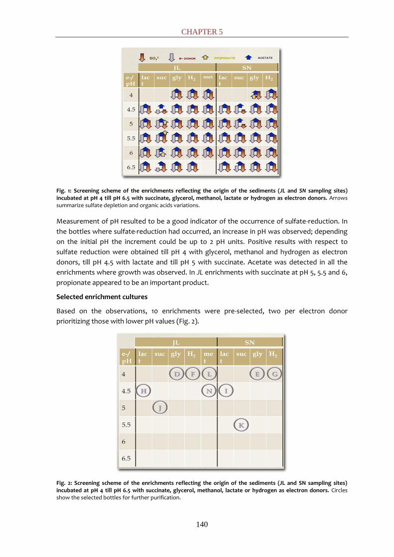
CHAPTER 5
140
Fig. 1: Screening scheme of the enrichments reflecting the origin of the sediments (JL and SN sampling sites) incubated at pH 4 till pH 6.5 with succinate, glycerol, methanol, lactate or hydrogen as electron donors. Arrows summarize sulfate depletion and organic acids variations.
Measurement of pH resulted to be a good indicator of the occurrence of sulfate-reduction. In
the bottles where sulfate-reduction had occurred, an increase in pH was observed; depending
on the initial pH the increment could be up to 2 pH units. Positive results with respect to
sulfate reduction were obtained till pH 4 with glycerol, methanol and hydrogen as electron
donors, till pH 4.5 with lactate and till pH 5 with succinate. Acetate was detected in all the
enrichments where growth was observed. In JL enrichments with succinate at pH 5, 5.5 and 6,
propionate appeared to be an important product.
Selected enrichment cultures
Based on the observations, 10 enrichments were pre-selected, two per electron donor
prioritizing those with lower pH values (Fig. 2).
Fig. 2: Screening scheme of the enrichments reflecting the origin of the sediments (JL and SN sampling sites) incubated at pH 4 till pH 6.5 with succinate, glycerol, methanol, lactate or hydrogen as electron donors. Circles show the selected bottles for further purification.
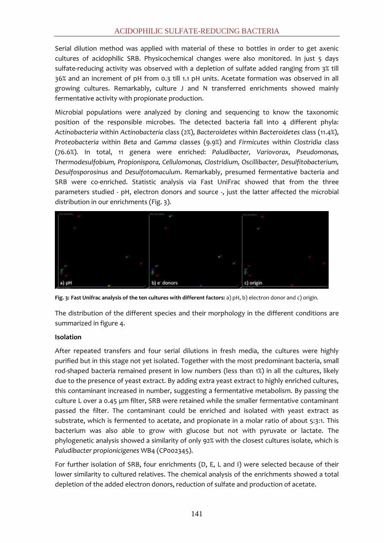
ACIDOPHILIC SULFATE-REDUCING BACTERIA
141
Serial dilution method was applied with material of these 10 bottles in order to get axenic
cultures of acidophilic SRB. Physicochemical changes were also monitored. In just 5 days
sulfate-reducing activity was observed with a depletion of sulfate added ranging from 3% till
36% and an increment of pH from 0.3 till 1.1 pH units. Acetate formation was observed in all
growing cultures. Remarkably, culture J and N transferred enrichments showed mainly
fermentative activity with propionate production.
Microbial populations were analyzed by cloning and sequencing to know the taxonomic
position of the responsible microbes. The detected bacteria fall into 4 different phyla:
Actinobacteria within Actinobacteria class (2%), Bacteroidetes within Bacteroidetes class (11.4%),
Proteobacteria within Beta and Gamma classes (9.9%) and Firmicutes within Clostridia class
(76.6%). In total, 11 genera were enriched: Paludibacter, Variovorax, Pseudomonas,
Thermodesulfobium, Propionispora, Cellulomonas, Clostridium, Oscillibacter, Desulfitobacterium,
Desulfosporosinus and Desulfotomaculum. Remarkably, presumed fermentative bacteria and
SRB were co-enriched. Statistic analysis via Fast UniFrac showed that from the three
parameters studied - pH, electron donors and source -, just the latter affected the microbial
distribution in our enrichments (Fig. 3).
Fig. 3: Fast Unifrac analysis of the ten cultures with different factors: a) pH, b) electron donor and c) origin.
The distribution of the different species and their morphology in the different conditions are
summarized in figure 4.
Isolation
After repeated transfers and four serial dilutions in fresh media, the cultures were highly
purified but in this stage not yet isolated. Together with the most predominant bacteria, small
rod-shaped bacteria remained present in low numbers (less than 1%) in all the cultures, likely
due to the presence of yeast extract. By adding extra yeast extract to highly enriched cultures,
this contaminant increased in number, suggesting a fermentative metabolism. By passing the
culture L over a 0.45 μm filter, SRB were retained while the smaller fermentative contaminant
passed the filter. The contaminant could be enriched and isolated with yeast extract as
substrate, which is fermented to acetate, and propionate in a molar ratio of about 5:3:1. This
bacterium was also able to grow with glucose but not with pyruvate or lactate. The
phylogenetic analysis showed a similarity of only 92% with the closest cultures isolate, which is
Paludibacter propionicigenes WB4 (CP002345).
For further isolation of SRB, four enrichments (D, E, L and I) were selected because of their
lower similarity to cultured relatives. The chemical analysis of the enrichments showed a total
depletion of the added electron donors, reduction of sulfate and production of acetate.
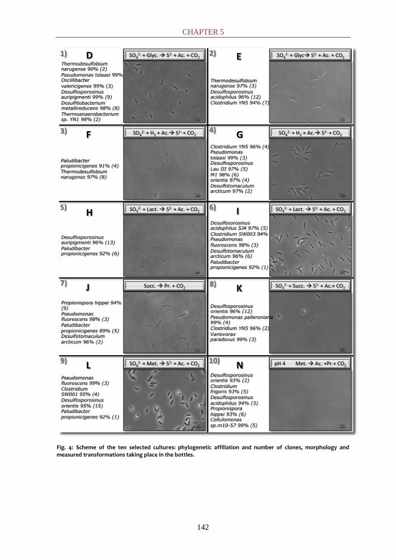
CHAPTER 5
142
Fig. 4: Scheme of the ten selected cultures: phylogenetic affiliation and number of clones, morphology and measured transformations taking place in the bottles.

ACIDOPHILIC SULFATE-REDUCING BACTERIA
143
The enrichments reduced sulfate and oxidized the added electron donors (glycerol, lactate,
and methanol) as indicated in Table 1.
Table 1: Stoichiometry of sulfate and electron donor utilization by the enrichments.
Electron acceptor
Consumption
Electron donor
consumption
Acetate production
Enrichment mM meq redox mM meq redox mM meq redox
D (glycerol) 3.3 26.4
5.2 31.2 5.3 31.8
E (glycerol) 3.5 28 4.6 27.6 5.4 32.4
I (lactate) 2.7 21.6 5.4 21.6 6.1 24.4
L (methanol) 3.6 28.8 5.5 30.6* 0.6 2.4 *From 1 mol methanol, 0.15 is converted till acetate via reaction 5 and 0.85 is oxidized via sulfate reduction (reaction 1 and 4).
According to the equations shown in Table 2, 1 mol glycerol and lactate will be partially
oxidized to 1 mol acetate with the concomitant reduction of 0.75 and 0.5 mol sulfate,
respectively. In both cases, higher acetate production than theoretically expected was
observed. It can be due to an acetate formation via acetogenesis (eq. 6, Table 2) from CO2 and
the H+ liberate from electron donor oxidation. Methanol metabolism can follow two ways. On
the one hand, 1 mol methanol can be totally oxidized till carbon dioxide reducing 0.75 mol of
sulfate (reaction 4) and on the other hand, 1 mol of methanol can be partially oxidized till 0.5
mol acetate with the reduction of 0.25 mol of sulfate (reaction 5). The ratio between both
reactions will determine the final stoichiometry.
Table 2: Metabolic reactions in the cultures.
(1) Sulfate SO42- + 8H+ + 8e- S2- + 4H2O
(2) Glycerol C3H8O3 + H2O C2H4O2 + CO2 + 6H+ + 6e-
(3) Lactate C3H6O3 + H2O C2H4O2 + CO2 + 4H+ + 4e-
(4) Methanol A CH4O +H2O CO2 + 6H+ + 6e-
(5) Methanol B 4CH4O +2CO2 3C2H4O2 + 2H2O
(6) Acetogenesis 2CO2 + 8H+ + 8e- C2H4O2 + 2H2O
To ensure separation of the enrichments from the fermenting bacteria, inoculation on agar
plates was done. The D, E and L cultures grew on agar plates, but culture I did not. The SRB in
culture I could be isolated by performing another 2 extra dilution series. As common features,
the sulfate-reducing isolates consisted of gram-positive, rod-shaped and spore-forming
bacteria with ellipsoidal spores causing swelling of the cells, but differ in sizes or spore-
position. From culture D rod-shaped bacteria of 1.1 by 3-5 μm with subterminal spores were
isolated, cells from culture E were 0.7 by 2 μm with subterminal-spores, bacteria in culture I
were straight rods 0.7 by 4 μm with terminal spores and culture L were 1.1 by 2-3 μm and had
central spores.
Sequencing of the 16S rRNA genes was performed with these 4 cultures. The phylogenetic tree
based on 16S rRNA genes is shown in figure 5.
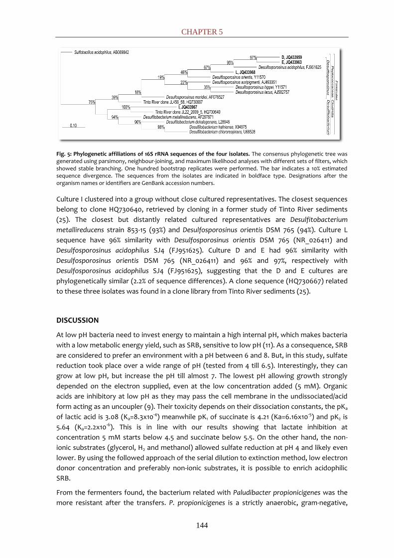
CHAPTER 5
144
Fig. 5: Phylogenetic affiliations of 16S rRNA sequences of the four isolates. The consensus phylogenetic tree was generated using parsimony, neighbour-joining, and maximum likelihood analyses with different sets of filters, which showed stable branching. One hundred bootstrap replicates were performed. The bar indicates a 10% estimated sequence divergence. The sequences from the isolates are indicated in boldface type. Designations after the organism names or identifiers are GenBank accession numbers.
Culture I clustered into a group without close cultured representatives. The closest sequences
belong to clone HQ730640, retrieved by cloning in a former study of Tinto River sediments
(25). The closest but distantly related cultured representatives are Desulfitobacterium
metallireducens strain 853-15 (93%) and Desulfosporosinus orientis DSM 765 (94%). Culture L
sequence have 96% similarity with Desulfosporosinus orientis DSM 765 (NR_026411) and
Desulfosporosinus acidophilus SJ4 (FJ951625). Culture D and E had 96% similarity with
Desulfosporosinus orientis DSM 765 (NR_026411) and 96% and 97%, respectively with
Desulfosporosinus acidophilus SJ4 (FJ951625), suggesting that the D and E cultures are
phylogenetically similar (2.2% of sequence differences). A clone sequence (HQ730667) related
to these three isolates was found in a clone library from Tinto River sediments (25).
DISCUSSION
At low pH bacteria need to invest energy to maintain a high internal pH, which makes bacteria
with a low metabolic energy yield, such as SRB, sensitive to low pH (11). As a consequence, SRB
are considered to prefer an environment with a pH between 6 and 8. But, in this study, sulfate
reduction took place over a wide range of pH (tested from 4 till 6.5). Interestingly, they can
grow at low pH, but increase the pH till almost 7. The lowest pH allowing growth strongly
depended on the electron supplied, even at the low concentration added (5 mM). Organic
acids are inhibitory at low pH as they may pass the cell membrane in the undissociated/acid
form acting as an uncoupler (9). Their toxicity depends on their dissociation constants, the pKa
of lactic acid is 3.08 (Ka=8.3x10⁻⁴) meanwhile pK1 of succinate is 4.21 (Ka=6.16x10⁻⁵) and pK2 is
5.64 (Ka=2.2x10⁻⁶). This is in line with our results showing that lactate inhibition at
concentration 5 mM starts below 4.5 and succinate below 5.5. On the other hand, the non-
ionic substrates (glycerol, H2 and methanol) allowed sulfate reduction at pH 4 and likely even
lower. By using the followed approach of the serial dilution to extinction method, low electron
donor concentration and preferably non-ionic substrates, it is possible to enrich acidophilic
SRB.
From the fermenters found, the bacterium related with Paludibacter propionicigenes was the
more resistant after the transfers. P. propionicigenes is a strictly anaerobic, gram-negative,

ACIDOPHILIC SULFATE-REDUCING BACTERIA
145
propionate-producing bacterium (33), what may explain the propionate production detected
in several cultures. Related sequences have been found before in Tinto River by DGGE and
cloning of environmental DNA (25) and others acidic environments (DQ205192) suggesting
that such types of bacteria can adapt well to acidic conditions. Currently, the physiological
properties of this bacterium are studied further.
Thus far, just a few acidophilic/tolerant SRB have been isolated or obtained in stable
enrichment cultures (1, 2, 12, 16, 18, 28, 32). In this study, 4 novel acidophilic SRB strains were
isolated. For three isolated SRB the highest similarity with characterized species is only 96%;
two of them to Desulfosporosinus acidophilus and one to Desulfosporosinus orientis. The fourth
one has just 93% 16S rRNA gene sequence similarity with the clade
Desulfitobacterium/Desulfosporosinus. The first three strains could be new Desulfosporosinus
species, while the fourth one could be a new Desulfitobacterium species or according to the
16S rRNA sequence, even a new genus.
Members of the genera Desulfitobacterium and Desulfosporosinus represent a clade of strictly
anaerobic, rod-shaped, and sporeforming bacteria, affiliated with the family Peptococcaceae,
order Clostridiales, phylum Firmicutes. Representatives of the genus Desulfosporosinus can be
easily distinguished by their ability to reduce sulfate, autotrophically with H2 and
heterotrophically with lactate. By contrast, the key feature of members of the genus
Desulfitobacterium is their ability to use a wide spectrum of electron acceptors, including
chlorinated aliphatic and aromatic compounds for reductive dehalogenation. Strains of both
genera are able to utilize sulfite and heavy metals as electron acceptors (29). In permanently
anaerobic environments, sporeforming species are apparently less competitive than non
sporeforming ones (38) but in contrast, sporeforming are able to survive dryness and oxic
conditions for months what make them dominant in adverse conditions. Desulfosporosinus
members have previously been detected in other acidic environments, including acidic mining-
impacted lake sediments (18) and Tinto River (7, 25) where Desulfitobacterium was also found
(25).
Desulfosporosinus genus belongs to the SRB group that degrade organic compound
incompletely to acetate (1, 28), indeed, acetate accumulation was observed in the
enrichments. Desulfosporosinus spp. are known to switch under sulfate limitation to the
fermentation of lactate and pyruvate (29), to reductive acetogenesis from formate, methanol
or methyl groups of aromatic compounds (22) or to dissimilatory iron (III) reduction (23). This
bacterium was proposed to be important for the carbon flow in terrestrial ecosystems (19, 21)
and on globally relevant processes such as the decrease in emission of the greenhouse gas
methane (8). There is some evidence that syntrophic relationships occur between
Desulfitobacterium spp. and sulfate-reducing bacteria such as Desulfosporosinus spp., from
which the Desulfitobacterium cells acquire their electrons by interspecies hydrogen transfer.
Their versatility makes desulfitobacteria excellent candidates for the development of
anaerobic bioremediation processes. In fact, desulfitobacteria have been detected in
bioprocesses treating sites contaminated with halogenated organic compounds but also with
high concentrations of toxic metals (35).
Acidotrophic SRB may become important for the bioremediation of acid mine drainage (AMD)
waters, which are highly acidic (pH<4) and have a high dissolved metals content. The
dissimilative reduction of sulfate leads to a consumption of protons which increase the pH, and
CHAPTER 5
146
to sulfide formation which reacts with heavy metals such as Fe, Zn, Cu, Cd, Ni, and Pb, leading
to the precipitation of insoluble metal sulfide phases and consequently, removal of metals
from solution (15). Additionally, some metals can be enzymatically reduced such as Cr(VI),
Mn(IV), Fe(III) or U(VI) (31). Remarkably, dissolved uranium, U (VI), may be removed by
reduction to U(IV) creating the insoluble mineral uraninite (36). Further studies are needed to
get insight into sulfate reduction and metal reduction by acidophilic SRB.
REFERENCES
1. Alazard, D., M. Joseph, F. Battaglia-Brunet, J. L. Cayol, and B. Ollivier. 2010. Desulfosporosinus acidiphilus sp. nov.: a moderately acidophilic sulfate-reducing bacterium isolated from acid mining drainage sediments. Extremophiles. 14:305-312.
2. Church, C. D., R. T. Wilkin, C. N. Alpers, R. O. Rye, and R. B. McCleskey. 2007. Microbial sulfate reduction and metal attenuation in pH 4 acid mine water. Geochem. Trans. 8:10.
3. Cline, J. D. 1969. Spectrophotometric determination of hydrogen sulfide in natural waters. Limnol. Oceanogr. 14:454-458.
4. Cole, J., Q. Wang, E. Cardenas, J. Fish, B. Chai, R. Farris, A. Kulam-Syed-Mohideen, D. McGarrell, T. Marsh, and G. Garrity. 2009. The Ribosomal Database Project: improved alignments and new tools for rRNA analysis. Nucleic Acids Res. 37:141-145.
5. Dinh, H. T., J. Kuever, M. Mußmann, A. W. Hassel, M. Stratmann, and F. Widdel. 2004. Iron corrosion by novel anaerobic microorganisms. Nature. 427:829-832.
6. Doetsch, R. 1981. Determinative methods of light microscopy. In P.Gerhardt, R.D.E. Murray, R.N. Costilow, E.W.Nester, W.A.Wood, N.R.Krieg, and G.B. Philips (Ed.), Manual of Methods for General Bacteriology. American Society for Microbiology, Washington, D.C. 21-33.
7. García-Moyano, A., E. González-Toril, A. Aguilera, and R. Amils. 2007. Prokaryotic community composition and ecology of floating macroscopic filaments from an extreme acidic environment, Río Tinto (SW, Spain). Syst. Appl. Microbiol. 30:601-614.
8. Gauci, V., E. Matthews, N. Dise, B. Walter, D. Koch, G. Granberg, and M. Vile. 2004. Sulfur pollution suppression of the wetland methane source in the 20th and 21st centuries. Proc. Natl. Acad. Sci. U. S. A. 101:12583-12587.
9. Ghose, T. K., and T. Wikén. 1955. Inhibition of Bacterial Sulphate‐Reduction in Presence of Short Chain Fatty Acids. Physiol. Plantarum. 8:116-135.
10. Hamady, M., C. Lozupone, and R. Knight. 2009. Fast UniFrac: facilitating high-throughput phylogenetic analyses of microbial communities including analysis of pyrosequencing and PhyloChip data. The ISME Journal. 4:17-27.
11. Hamilton, W. A. 1998. Sulfate‐reducing bacteria: Physiology determines their environmental impact. Geomicrobiol. J. 15:19-28.
12. Hard, B. C., S. Friedrich, and W. Babel. 1997. Bioremediation of acid mine water using facultatively methylotrophic metal-tolerant sulfate-reducing bacteria. Microbiol. Res. 152:65-73.
13. Huber, T., G. Faulkner, and P. Hugenholtz. 2004. Bellerophon: a program to detect chimeric sequences in multiple sequence alignments. Bioinformatics. 20:2317-2319.
14. Johnson, D. B. 1998. Biodiversity and ecology of acidophilic microorganisms. FEMS Microbiol. Ecol. 27:307-317.
15. Johnson, D. B., and K. B. Hallberg. 2005. Acid mine drainage remediation options: a review. Sci. Total Environ. 338:3-14.
ACIDOPHILIC SULFATE-REDUCING BACTERIA
147
16. Kimura, S., K. B. Hallberg, and D. B. Johnson. 2006. Sulfidogenesis in low pH (3.8–4.2) media by a mixed population of acidophilic bacteria. Biodegradation. 17:57-65.
17. Koschorreck, M. 2008. Microbial sulphate reduction at a low pH. FEMS Microbiol. Ecol. 64:329-342.
18. Kusel, K., U. Roth, T. Trinkwalter, and S. Peiffer. 2001. Effect of pH on the anaerobic microbial cycling of sulfur in mining-impacted freshwater lake sediments. Environ. Exp. Bot. 46:213-223.
19. Loy, A., K. Kusel, A. Lehner, H. L. Drake, and M. Wagner. 2004. Microarray and functional gene analyses of sulfate-reducing prokaryotes in low-sulfate, acidic fens reveal cooccurrence of recognized genera and novel lineages. Appl. Environ. Microbiol. 70:6998-7009.
20. Muyzer, G., and A. J. M. Stams. 2008. The ecology and biotechnology of sulphate-reducing bacteria. Nature Reviews Microbiology. 6:441-454.
21. Pester, M., N. Bittner, P. Deevong, M. Wagner, and A. Loy. 2010. A ‘rare biosphere’ microorganism contributes to sulfate reduction in a peatland. The ISME Journal. 4:1591-1602.
22. Rabus, R., T. A. Hansen, and F. Widdel. 2006. Dissimilatory sulfate-and sulfur-reducing prokaryotes. The Prokaryotes. 2:659-768.
23. Ramamoorthy, S., H. Sass, H. Langner, P. Schumann, R. Kroppenstedt, S. Spring, J. Overmann, and R. Rosenzweig. 2006. Desulfosporosinus lacus sp. nov., a sulfate-reducing bacterium isolated from pristine freshwater lake sediments. Int. J. Syst. Evol. Microbiol. 56:2729-2736.
24. Sánchez-Andrea, I., K. Knittel, R. Amann, R. Amils, and J. L. Sanz. 2012. Quantification of Tinto River Sediments Microbial Communities: the Importance of Sulfate-Reducing Bacteria and their Role in Attenuating Acid Mine Drainage. Appl. Environ. Microbiol. doi:10.1128/AEM.00848-12
25. Sánchez-Andrea, I., N. Rodriguez, R. Amils, and J. L. Sanz. 2011. Microbial Diversity in Anaerobic Sediments at Rio Tinto, a Naturally Acidic Environment with a High Heavy Metal Content. Appl. Environ. Microbiol. 77:6085-6093.
26. Satoh, H., M. Odagiri, T. Ito, and S. Okabe. 2009. Microbial community structures and in situ sulfate-reducing and sulfur-oxidizing activities in biofilms developed on mortar specimens in a corroded sewer system. Water Res. 43:4729-4739.
27. Sawyer, C. N., P. L. McCarty, and G. F. Parkin. 2003. Chemistry for environmental engineering and science. McGraw-Hill New York.
28. Senko, J. M., G. Zhang, J. T. McDonough, M. A. Bruns, and W. D. Burgos. 2009. Metal reduction at low pH by a Desulfosporosinus species: implications for the biological treatment of acidic mine drainage. Geomicrobiol. J. 26:71-82.
29. Spring, S., and F. Rosenzweig. 2006. The genera Desulfitobacterium and Desulfosporosinus: taxonomy. The Prokaryotes: A Handbook on the Biology of Bacteria. 771-786.
30. Stams, A. J. M., J. B. Van Dijk, C. Dijkema, and C. M. Plugge. 1993. Growth of syntrophic propionate-oxidizing bacteria with fumarate in the absence of methanogenic bacteria. Appl. Environ. Microbiol. 59:1114-1119.
31. Tebo, B. M., and A. Y. Obraztsova. 1998. Sulfate‐reducing bacterium grows with Cr (VI), U (VI), Mn (IV), and Fe (III) as electron acceptors. FEMS Microbiol. Lett. 162:193-198.
32. Tuttle, J. H., P. R. Dugan, C. B. Macmillan, and C. I. Randles. 1969. Microbial dissimilatory sulfur cycle in acid mine water. J. Bacteriol. 97:594-602.
33. Ueki, A., H. Akasaka, D. Suzuki, and K. Ueki. 2006. Paludibacter propionicigenes gen. nov.,

CHAPTER 5
148
sp. nov., a novel strictly anaerobic, Gram-negative, propionate-producing bacterium isolated from plant residue in irrigated rice-field soil in Japan. Int. J. Syst. Evol. Microbiol. 56:39-44.
34. Valls, M., and V. Lorenzo. 2002. Exploiting the genetic and biochemical capacities of bacteria for the remediation of heavy metal pollution. FEMS Microbiol. Rev. 26:327-338.
35. Villemur, R., M. Lanthier, R. Beaudet, and F. Lépine. 2006. The Desulfitobacterium genus. FEMS Microbiol. Rev. 30:706-733.
36. Wall, J. D., and L. R. Krumholz. 2006. Uranium reduction. Annu. Rev. Microbiol. 60:149-166.
37. Widdel, F. 1988. Microbiology and ecology of sulfate-and sulfur-reducing bacteria. Biology of Anaerobic Microorganisms. 469-585.
38. Widdel, F. 1992. The genus Desulfotomaculum. The Prokaryotes. 2:1792-1799.
CHAPTER 6:
Bioremediation of acid mine
drainage coupled with
domestic wastewater
treatment

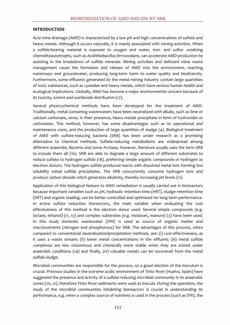
BIOREMEDIATION OF AMD AND DW BY SRB
151
INTRODUCTION
Acid mine drainage (AMD) is characterized by a low pH and high concentrations of sulfate and
heavy metals. Although it occurs naturally, it is mainly associated with mining activities. When
a sulfide-bearing material is exposed to oxygen and water, iron- and sulfur- oxidizing
chemolitoautotrophs, such as Acidithiobacillus ferrooxidans, can accelerate AMD production by
assisting in the breakdown of sulfide minerals. Mining activities and deficient mine waste
management cause the formation and release of AMD into the environment, reaching
waterways and groundwater, producing long-term harm to water quality and biodiversity.
Furthermore, some effluents generated by the metal mining industry contain large quantities
of toxic substances, such as cyanides and heavy metals, which have serious human health and
ecological implications. Globally, AMD has become a major environmental concern because of
its toxicity, extent and worldwide distribution (17).
Several physicochemical methods have been developed for the treatment of AMD.
Traditionally, metal containing wastewaters have been neutralized with alkalis, such as lime or
calcium carbonate, since, in their presence, heavy metals precipitate in form of hydroxides or
carbonates. This method, however, has some disadvantages such as its operational and
maintenance costs, and the production of large quantities of sludge (4). Biological treatment
of AMD with sulfate-reducing bacteria (SRB) has been under research as a promising
alternative to chemical methods. Sulfate-reducing metabolisms are widespread among
different anaerobic Bacteria and some Archaea, however, literature usually uses the term SRB
to include them all (16). SRB are able to degrade a large amount of different substrates to
reduce sulfate to hydrogen sulfide (18), preferring simple organic compounds or hydrogen as
electron donors. The hydrogen sulfide produced reacts with dissolved metal ions forming low
solubility metal sulfide precipitates. The SRB concurrently consume hydrogen ions and
produce carbon dioxide which generates alkalinity, thereby increasing pH levels (11).
Application of this biological feature to AMD remediation is usually carried out in bioreactors
because important variables such as pH, hydraulic retention time (HRT), sludge retention time
(SRT) and organic loading, can be better controlled and optimized for long term performance.
In active sulfate reduction bioreactors, the main variable when evaluating the cost
effectiveness of this method is the electron donor used. Several simple compounds (e.g.
lactate, ethanol) (11, 13) and complex substrates (e.g. molasses, manure) (3) have been used.
In this study domestic wastewater (DW) is used as source of organic matter and
macronutrients (nitrogen and phosphorous) for SRB. The advantages of this process, when
compared to conventional neutralization/precipitation methods, are: (i) cost-effectiveness, as
it uses a waste stream; (ii) lower metal concentrations in the effluent; (iii) metal sulfide
complexes are less voluminous and chemically more stable when they are stored under
anaerobic conditions (14) and finally, (iv) valuable metals can be recovered from the metal
sulfide sludge.
Microbial communities are responsible for the process, so a good election of the inoculum is
crucial. Previous studies in the extreme acidic environment of Tinto River (Huelva, Spain) have
suggested the presence and activity of a sulfate-reducing microbial community in its anaerobic
zones (20, 21), therefore Tinto River sediments were used as inocula. During the operation, the
study of the microbial communities inhabiting bioreactors is crucial in understanding its
performance, e.g. when a complex source of nutrients is used in the process (such as DW), the

CHAPTER 6
152
efficiency depends on a consortium rather than SRB alone (14). Another key factor in sulfate-
reducing bioreactors is the microbial support to avoid biomass loss (17).
The aim of this study was to assess the feasibility of coupling AMD bioremediation with the
treatment of DW through a sulfate reduction process in anaerobic bioreactors. Three different
bacterial supports were evaluated by following the efficiency of the process (sulfate reduction,
metal precipitation and pH attenuation) and examined through SEM. Influence of HRT, influent
pH and extra addition of electron donors were also tested. Bacterial diversity and shifts were
examined at different times during the study through Denaturing Gradient Gel Electrophoresis
(DGGE) analysis.
MATERIALS AND METHODS
Site sampling and inocula preparation
Sediments from the Tinto River basin (Huelva, southwestern Spain) were used as inoculum.
Samples were collected from the JL dam (37.691207N, 6.560587W) in November 2010.
Sediment cores (inner diameter, 7 cm; length, 45 cm) were taken with a sampler (Eijkelkamp
Agrisearch Equipment, The Netherlands). The redox potential (E) and pH of the drill core
samples were measured in situ with E and pH probes connected to a Thermo Orion 290A
potentiometer placed in the fresh sediment just after extraction of the core. The cores were
sliced according to both physicochemical parameters and visual aspect; blackish slices (pH
between 4.5 and 5.5 and redox potential between 0 and -130) were kept separate until
processing in the laboratory (less than 1 week).
An enrichment culture was grown in 1 L- anaerobic bottle containing 100 g of sediment and 500
mL of the following sterilized medium (in g L-1): 3.5 sodium lactate, 2 yeast extract, 2
MgSO4·7H2O, 1.5 Na2SO4, 0.5 K2HPO4·3H2O, 0.5 L-Cysteine, 0.4 Fe(NH4)2(SO4)2·6H2O, 0.1 CaCl2,
plus 1.0 mL of a mixture of trace elements; pH was adjusted to 5.5. The trace element solution
was prepared as follows (in mg L-1): 500 EDTA, 39.0 ZnSO4·7H2O, 50.6 MnCl2·4H2O, 11.0
(NH4)6Mo7O24·4H2O, 15.7 CuSO4·5H2O, 16.1 CoCl2·6 H2O. The culture was incubated statically at
30 ºC. After one month of incubation, an anaerobic bottle of 1 L containing 500 mL of medium
and 150 g of granular activated carbon (Chemviron F-400, Aguas de Levante, Spain) was
inoculated with 10% inoculum from the aforementioned enrichment.
Bacterial supports
SRB supports for the three reactors were prepared as follows. For reactor 1 (R1) granular
activated carbon (AC) from the second enriched culture described above was placed in the
reactor. For reactor 2 (R2), sediment from the first enriched culture described was mixed in a
1:1 proportion with melted agar (1% at 41 ºC). The gel formed was cut with a sieve of a 2 mm
diameter. For reactor 3, (R3) 500 ml of the supernatant from the second enrichment culture
was concentrated to 100 ml through centrifugation, and mixed with 100 ml of melted agar (2%
and 41ºC). The gel obtained was cut with a sieve of a 2 mm diameter. The bacterial supports
were incubated in batch mode for 3 weeks in the AMD-DW medium, which was replaced
weekly.
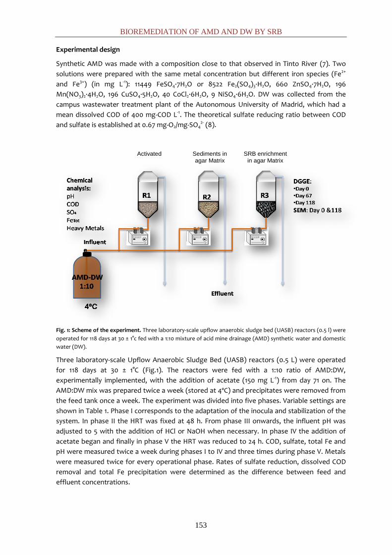
BIOREMEDIATION OF AMD AND DW BY SRB
153
Experimental design
Synthetic AMD was made with a composition close to that observed in Tinto River (7). Two
solutions were prepared with the same metal concentration but different iron species (Fe2+
and Fe3+) (in mg L-1): 11449 FeSO4·7H2O or 8522 Fe2(SO4)3·H2O, 660 ZnSO4·7H2O, 196
Mn(NO3)2·4H2O, 196 CuSO4·5H2O, 40 CoCl2·6H2O, 9 NiSO4·6H2O. DW was collected from the
campus wastewater treatment plant of the Autonomous University of Madrid, which had a
mean dissolved COD of 400 mg-COD L-1. The theoretical sulfate reducing ratio between COD
and sulfate is established at 0.67 mg-O2/mg-SO42- (8).
Fig. 1: Scheme of the experiment. Three laboratory-scale upflow anaerobic sludge bed (UASB) reactors (0.5 l) were
operated for 118 days at 30 ± 1°c fed with a 1:10 mixture of acid mine drainage (AMD) synthetic water and domestic
water (DW).
Three laboratory-scale Upflow Anaerobic Sludge Bed (UASB) reactors (0.5 L) were operated
for 118 days at 30 ± 1°C (Fig.1). The reactors were fed with a 1:10 ratio of AMD:DW,
experimentally implemented, with the addition of acetate (150 mg L-1) from day 71 on. The
AMD:DW mix was prepared twice a week (stored at 4ºC) and precipitates were removed from
the feed tank once a week. The experiment was divided into five phases. Variable settings are
shown in Table 1. Phase I corresponds to the adaptation of the inocula and stabilization of the
system. In phase II the HRT was fixed at 48 h. From phase III onwards, the influent pH was
adjusted to 5 with the addition of HCl or NaOH when necessary. In phase IV the addition of
acetate began and finally in phase V the HRT was reduced to 24 h. COD, sulfate, total Fe and
pH were measured twice a week during phases I to IV and three times during phase V. Metals
were measured twice for every operational phase. Rates of sulfate reduction, dissolved COD
removal and total Fe precipitation were determined as the difference between feed and
effluent concentrations.
Activated
Carbon
Sediments in agar Matrix
SRB enrichment in agar Matrix
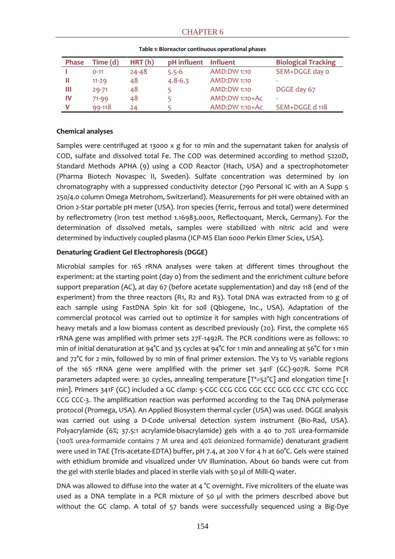
CHAPTER 6
154
Table 1: Bioreactor continuous operational phases
Phase Time (d) HRT (h) pH influent Influent Biological Tracking I 0-11 24-48 5.5-6 AMD:DW 1:10 SEM+DGGE day 0 II 11-29 48 4.8-6.3 AMD:DW 1:10 - III 29-71 48 5 AMD:DW 1:10 DGGE day 67 IV 71-99 48 5 AMD:DW 1:10+Ac - V 99-118 24 5 AMD:DW 1:10+Ac SEM+DGGE d 118
Chemical analyses
Samples were centrifuged at 13000 x g for 10 min and the supernatant taken for analysis of
COD, sulfate and dissolved total Fe. The COD was determined according to method 5220D,
Standard Methods APHA (9) using a COD Reactor (Hach, USA) and a spectrophotometer
(Pharma Biotech Novaspec II, Sweden). Sulfate concentration was determined by ion
chromatography with a suppressed conductivity detector (790 Personal IC with an A Supp 5
250/4.0 column Omega Metrohom, Switzerland). Measurements for pH were obtained with an
Orion 2-Star portable pH meter (USA). Iron species (ferric, ferrous and total) were determined
by reflectrometry (Iron test method 1.16983.0001, Reflectoquant, Merck, Germany). For the
determination of dissolved metals, samples were stabilized with nitric acid and were
determined by inductively coupled plasma (ICP-MS Elan 6000 Perkin Elmer Sciex, USA).
Denaturing Gradient Gel Electrophoresis (DGGE)
Microbial samples for 16S rRNA analyses were taken at different times throughout the
experiment: at the starting point (day 0) from the sediment and the enrichment culture before
support preparation (AC), at day 67 (before acetate supplementation) and day 118 (end of the
experiment) from the three reactors (R1, R2 and R3). Total DNA was extracted from 10 g of
each sample using FastDNA Spin kit for soil (Qbiogene, Inc., USA). Adaptation of the
commercial protocol was carried out to optimize it for samples with high concentrations of
heavy metals and a low biomass content as described previously (20). First, the complete 16S
rRNA gene was amplified with primer sets 27F-1492R. The PCR conditions were as follows: 10
min of initial denaturation at 94°C and 35 cycles at 94°C for 1 min and annealing at 56°C for 1 min
and 72°C for 2 min, followed by 10 min of final primer extension. The V3 to V5 variable regions
of the 16S rRNA gene were amplified with the primer set 341F (GC)-907R. Some PCR
parameters adapted were: 30 cycles, annealing temperature [Tª=52°C] and elongation time [1
min]. Primers 341F (GC) included a GC clamp: 5-CGC CCG CCG CGC CCC GCG CCC GTC CCG CCC
CCG CCC-3. The amplification reaction was performed according to the Taq DNA polymerase
protocol (Promega, USA). An Applied Biosystem thermal cycler (USA) was used. DGGE analysis
was carried out using a D-Code universal detection system instrument (Bio-Rad, USA).
Polyacrylamide (6%; 37.5:1 acrylamide-bisacrylamide) gels with a 40 to 70% urea-formamide
(100% urea-formamide contains 7 M urea and 40% deionized formamide) denaturant gradient
were used in TAE (Tris-acetate-EDTA) buffer, pH 7.4, at 200 V for 4 h at 60°C. Gels were stained
with ethidium bromide and visualized under UV illumination. About 60 bands were cut from
the gel with sterile blades and placed in sterile vials with 50 µl of Milli-Q water.
DNA was allowed to diffuse into the water at 4 °C overnight. Five microliters of the eluate was
used as a DNA template in a PCR mixture of 50 µl with the primers described above but
without the GC clamp. A total of 57 bands were successfully sequenced using a Big-Dye

BIOREMEDIATION OF AMD AND DW BY SRB
155
sequencing kit (Applied Biosystems) following the manufacturer’s instructions. Sequencing
chromatograms were checked with the program Finch TV v.1.4.0 (Geospiza). A comparative
analysis of all the sequences was done using the BLAST routine from NCBI employing the
GenBank database (http://www.ncbi.nlm.nih.gov/Blast) and the tool Sequence Match from the
Ribosomal Database Project (RDP) at Michigan State University (http://rdp.cme.msu.edu/).
Sequences were deposited in GenBank under accession numbers: JQ517512-JQ517538.
Scanning electron microscopy (SEM)
Samples were taken at the starting point (day 0) and at the end of the experiment (day 118)
and fixed by immersion in glutaraldehyde (2.5%) for 2 h, then washed twice in sodium
cacodylate buffer (0.2 M, pH 7.1). Samples were dehydrated in a graded series (10, 30, 50, 70,
90, and 100%) of ethanol/water mixtures, leaving them 20 min in each mixture. After
dehydration, samples were critical point dried and mounted on stubs. After gold shadowing,
samples were examined in a Phillips XL30 EDAX DX4i SEM (Netherlands).
RESULTS AND DISCUSSION
Influent design
The iron species (Fe2+ or Fe3+) of the synthetic AMD and the ratio between DW and AMD were
investigated in batch assays. Since SRB lower their activity at pH<5 (2), the election of Fe2+ over
Fe3+ depended on the final pH of the AMD:DW mixture, which turned out to be 5.6 with Fe2+
and over 2.9 with Fe3+. After mixing AMD and DW, flocks appeared after several minutes due
to the coagulant properties of metal cations, which cause the co-precipitation of suspended
and colloidal organic matter. This fact causes organic matter loss, so the theoretical calculated
AMD:DW ratio (1:7) was increased up to 1:10. Additionally, analyses of metal concentration
showed a reduction of metal concentration due to coagulation (%): Fe 51.5, Mn 31.4, Co 38.7, Ni
49.2, Cu 68.2 and Zn 34.1, which indicates that the mere mixture of DW and AMD started the
remediation process chemically.
Water quality
R1 and R2 showed similar evolution patterns for COD, sulfate and total Fe removal throughout
the experiment (Fig. 2). However, the efficiencies were slightly higher for R1 than R2 during
phase I to III (COD, sulfate and Fe removal: 62%, 39%, 51% for R1 and 46%, 25% and 41% for R2).
Significant fluctuations in the removal efficiencies were observed during these phases,
possibly due to the variations of the DW influent. As observed, the mean COD removal
efficiencies were 62% and 46% for R1 and R2, even with electron acceptor excess, which might
reflect the non-degradable nature of some compounds of the domestic water. Supporting this
is the fact that after acetate supplement reduction efficiencies increased notably for the three
parameters (88, 75 and 85%), and became more stable. According with these results, a fraction
of the DW organic matter (24±3%) is scarcely biodegradable, limiting sulfate reduction. For this
reason, sulfate reduction efficiency seems to be coupled to the COD/SO42- ratio (1:1) more than
the theoretical ratio 0.67:1.
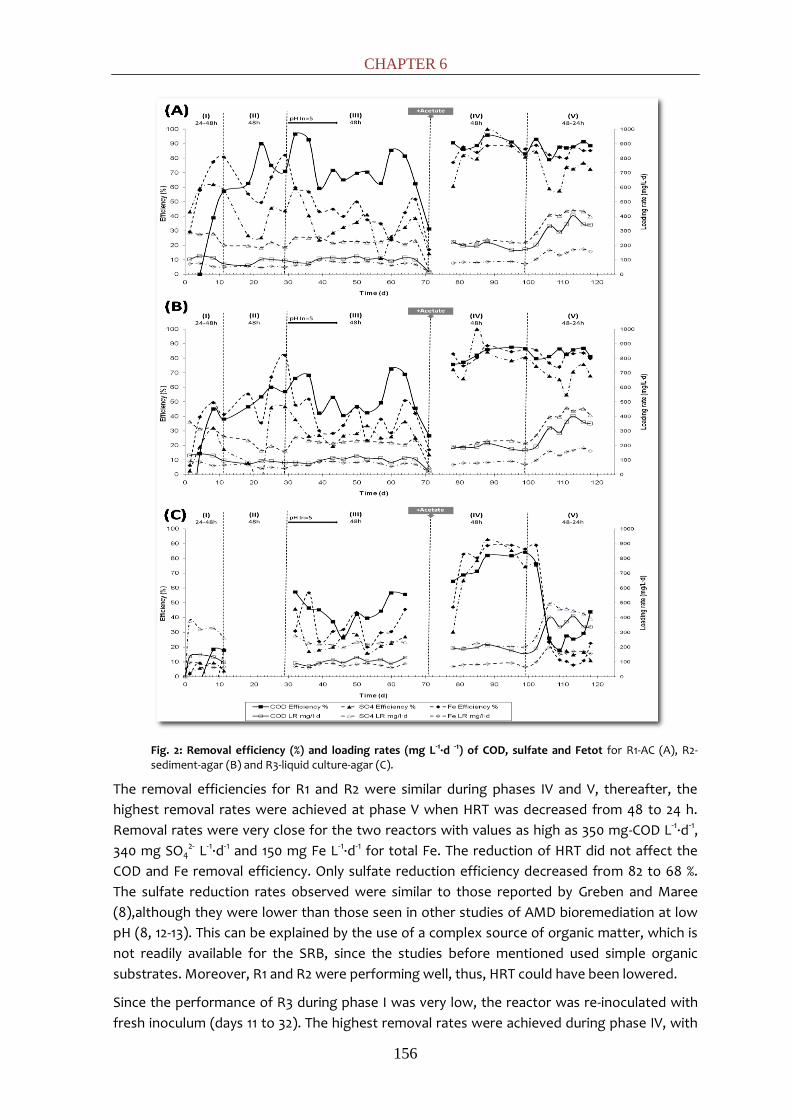
CHAPTER 6
156
Fig. 2: Removal efficiency (%) and loading rates (mg L-1·d -1) of COD, sulfate and Fetot for R1-AC (A), R2-sediment-agar (B) and R3-liquid culture-agar (C).
The removal efficiencies for R1 and R2 were similar during phases IV and V, thereafter, the
highest removal rates were achieved at phase V when HRT was decreased from 48 to 24 h.
Removal rates were very close for the two reactors with values as high as 350 mg-COD L-1·d-1,
340 mg SO42- L-1·d-1 and 150 mg Fe L-1·d-1 for total Fe. The reduction of HRT did not affect the
COD and Fe removal efficiency. Only sulfate reduction efficiency decreased from 82 to 68 %.
The sulfate reduction rates observed were similar to those reported by Greben and Maree
(8),although they were lower than those seen in other studies of AMD bioremediation at low
pH (8, 12-13). This can be explained by the use of a complex source of organic matter, which is
not readily available for the SRB, since the studies before mentioned used simple organic
substrates. Moreover, R1 and R2 were performing well, thus, HRT could have been lowered.
Since the performance of R3 during phase I was very low, the reactor was re-inoculated with
fresh inoculum (days 11 to 32). The highest removal rates were achieved during phase IV, with

BIOREMEDIATION OF AMD AND DW BY SRB
157
removal efficiencies that were slightly lower than the ones for R1 and R2. However, at phase V,
R3 efficiencies dropped drastically. The overloading of the reactor, which is confirmed by a
decreased of the pH, or diffusion limitation of the nutrients inside the cell, suggested by SEM
observations, could be the reasons.
Throughout the experiment, an increase of the pH effluent was observed in the three reactors.
This increase is caused by the inorganic carbon formed during anaerobic respiration (12).
Effluent pH stayed at approximately 6.0 for the three reactors through phases I to III. After the
addition of acetate, pH values increased up to 6.3 and were more stable, except for R3 at
phase V when pH dropped to 5.7.
Between days 64 to 78 the accumulation of precipitates at the reactor inlets caused an
obstruction in the pipes. This is a common problem in systems in which sulfate reduction and
metal precipitation occur in the same reactor (17). The separation of these processes in two
different reactors may reduce clogging problems (2, 5, 15).
Metal removal
Metal removal efficiencies were elevated (above 99%) for Co, Ni, Cu and Zn and Fe for R1 and
R2, releasing an effluent with low metal concentrations. The performance of R3 was also very
satisfactory for metal removal (higher than 98%) with the exception of Fe (Table 2). Mn
removal was low for the three reactors, even reaching negative values, which can be explained
by an accumulation in the reactor during previous phases. Poor performance of Mn removal is
explained by its chemistry: Mn has to be oxidized from Mn2+ to Mn4+ before being removed as
MnO2 (10).
Table 2: Average metal concentration (mg L-1) and removal efficiency (% in brackets) for phases IV and V for the three reactors (R1, R2 and R3).
Average metal concentration (mg L-1) and removal efficiency (%) for each metal
Fe Mn Co Ni Cu Zn R1 13.2
±10 85 2.5
±0.8 7.7 5x10⁻⁴
±5x10⁻⁴ 99.9 1.3x10⁻³
±8x10⁻⁴ 99.1 1.4x10⁻²
±1x10⁻² 99.1 4.9x10⁻²
±2x10⁻³ 99.5
R2 12.7 ±9.2
85.2 3.4 ±0.4
-13 5x10⁻⁴ ±5x10⁻⁴
99.9 8x10⁻⁴
±4x10⁻⁴ 99.4 8.5x10⁻³
±8x10⁻³ 99.5 5.2x10⁻²
±2x10⁻³ 99.4
R3 42.1 ±41
49 3.4 ±0.8
-13 2x10⁻⁴
±2x10⁻⁴ 99.6 3x10⁻³
±3x10⁻³ 98.0 7x10⁻³
±5x10⁻³ 99.5 0.11
±0.07 98.9
DGGE
The microbial community and its variation throughout the experiment were analyzed by DGGE.
Comparing the band pattern from time 0 and the end of phases III and IV (days 67 and 118), an
increase in the microbial diversity in the samples from the reactor is observed (Fig. 3) possibly
due to a diversification of the community to adapt to a complex source of nutrients, like DW,
and the colonization of carriers by bacterial species present in the DW.

CHAPTER 6
158
Fig. 3: DGGE fingerprints from the inocula (Tinto Sediment and enrichment culture with Activated Carbon), and the three reactors’ supports at day 67 (end phase III) and day 118 (end phase IV).
The sequences obtained (Table 3) fell into two main metabolic bacterial groups: (i)
fermentative bacteria, which would metabolize complex substrates: Clostridium spp.,
Paludibacter spp. and Pelotomaculum spp. (syntrophic bacterium) and (ii) SRB, which would
use the intermediate molecules produced by fermenters or the added acetate: Desulfomonile
spp., Desulfovibrio spp., Desulfotomaculum spp. and Desulfosporosinus spp. (Clostridia) and
Desulfurella spp. (Deltaproteobacteria).
Several bacterial sequences identified in this study have a high similarity to those identified in a
recent study of Tinto River sediments (20). For instance, bands E2, E3, F1, H1, H2, H3 share a
high similarity with HQ730741 (uncultured Paludibacter sp. detected by DGGE) and band A5
shares a high similarity with HQ730647 (uncultured Peptostreptococcaceae identified by
cloning). These organisms were found in those sediment strata with reducing redox potential
and less acidic pH (5.3-5.4). Other bands (E4, E5, E6, E7) are related with HM745405
(Desulfosporosinus spp.), a sequence retrieved from a close area of the Iberian Pyritic Belt (6).
Bands F4 and H8, identified in R2 and R3 at day 118, share a high similarity with FJ873799
(Desulfovibrio desulfuricans strain BSR-22). This organism has been reported as the
predominant SRB species in a sulfate reducing bioreactor working at pH 4-4.5 (1). Furthermore,
other identified bacteria, for instance bands D4 and D5 seem to be related with AY167450
(uncultured Desulfomonile) found in an acidic environment.

BIOREMEDIATION OF AMD AND DW BY SRB
159
Table 3: DGGE sequenced bands and their closest related named gene sequences in the NCBI database
Lan
e
Band Closest related species NCBI
Accession
number
Identities
(%)
Tin
to
Se
dim
A5 (JQ517512) Uncultured Peptostreptococcaceae clone
JL22_2009
or Clostridium sp. K39
HQ730647
AB610575
94
94
R1
T1
C1 (JQ517513) Clostridium acetobutylicum (NCP 262) X78073 94
C2 (JQ517514)&
C3 (JQ517515)&
C4 (JQ517516)
Clostridium diolis
AB610548 97-96
100
R1
T2
D4 (JQ517517)&
D5 (JQ517518)
Uncultured Desulfomonile (Fen soil)
or Desulfomonile tiedjei
AY167450
AM086646
95-92
92-89
D6 (JQ517519) Uncultured sulphate-reducing bacterium
(Benzene-degrading enrichment culture)
or Pelotomaculum schinkii
EU523095
X91170
97
97
R2
T1
E2 (JQ517520)&
E3 (JQ517521)
Uncultured Paludibacter sp. DGGE gel band
JL5a
or Paludibacter propionicigenes
HQ730741
CP002345
97
90
E4 (JQ517522)&
E5 (JQ517523)&
E6 (JQ517524)&
E7 (JQ517525)
Uncultured bacterium (AMD, Iberian Pyritic
Belt)
or Desulfosporosinus sp. ML1/10-BSR14
HM745405
AJ306703
97
97
E8 (JQ517526) Desulfomonile tiedjei
AM086646 88
R2
T2
F1 (JQ517527) Uncultured Paludibacter sp. DGGE gel band
JL5a
HQ730741 96
F4 (JQ517528) Desulfovibrio desulfuricans strain BSR-22 FJ873799 98
R3
T1
G2 (JQ517529) Desulfosporosinus sp. ML1/10-SRB3 AJ306705 93
G7 (JQ517530) Desulfotomaculum sp. ECP-C5 AF529223 92
R3
T2
H1 (JQ517531)&
H2 (JQ517532)
Uncultured Paludibacter sp. DGGE gel band
JL5a
or Paludibacter propionicigenes
HQ730741
CP002345
99
89
H3 (JQ517533) Uncultured Paludibacter sp. DGGE gel band
JL5a
or Paludibacter propionicigenes
HQ730741
CP002345
100
91
H5 (JQ517534) Delftia sp. R-41380 FR682925 97
H6 (JQ517535) 16S rDNA sequence from human fecal sample
or Desulfovibrio piger ATCC 29098
FP083728
NR_041778
94
93
H7 (JQ517536) Desulfovibrio sp. DSM12803 AJ251630 93
H8 (JQ517537) Desulfovibrio desulfuricans strain BSR-22 FJ873799 91
H9 (JQ517538) Desulfovibrio desulfuricans strain Ser-1 EU980605 95
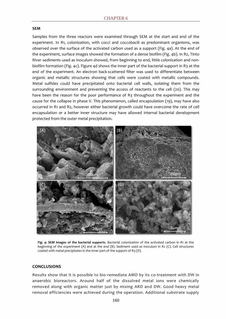
CHAPTER 6
160
SEM
Samples from the three reactors were examined through SEM at the start and end of the
experiment. In R1, colonization, with cocci and coccobacili as predominant organisms, was
observed over the surface of the activated carbon used as a support (Fig. 4a). At the end of
the experiment, surface images showed the formation of a dense biofilm (Fig. 4b). In R2, Tinto
River sediments used as inoculum showed, from beginning to end, little colonization and non-
biofilm formation (Fig. 4c). Figure 4d shows the inner part of the bacterial support in R3 at the
end of the experiment. An electron back-scattered filter was used to differentiate between
organic and metallic structures showing that cells were coated with metallic compounds.
Metal sulfides could have precipitated onto bacterial cell walls, isolating them from the
surrounding environment and preventing the access of reactants to the cell (20). This may
have been the reason for the poor performance of R3 throughout the experiment and the
cause for the collapse in phase V. This phenomenon, called encapsulation (19), may have also
occurred in R1 and R2, however either bacterial growth could have overcome the rate of cell
encapsulation or a better inner structure may have allowed internal bacterial development
protected from the outer metal precipitation.
Fig. 4: SEM images of the bacterial supports. Bacterial colonization of the activated carbon in R1 at the beginning of the experiment (A) and at the end (B). Sediment used as inoculum in R2 (C). Cell structures coated with metal precipitates in the inner part of the support of R3 (D).
CONCLUSIONS
Results show that it is possible to bio-remediate AMD by its co-treatment with DW in
anaerobic bioreactors. Around half of the dissolved metal ions were chemically
removed along with organic matter just by mixing ARD and DW. Good heavy metal
removal efficiencies were achieved during the operation. Additional substrate supply

BIOREMEDIATION OF AMD AND DW BY SRB
161
made elevated pollutant removal efficiencies possible (COD>88 %; sulfate>75 %; FeTot >85
%; and dissolved metals >99 % except Mn) leading to a good effluent quality. No significant
differences were found between R1 and R2, but the agar matrix used for R3 was the least
efficient, collapsing in the last phase of the experiment. Two groups of bacterial metabolism
were found by DGGE: fermenters and SRB. Clogging problems and the encapsulation
phenomenon can be important problems when sulfate reduction and metal precipitation occur
in the same reactor.
REFERENCES
1. Bijmans, M. F. M., E. de Vries, C. H. Yang, C. J. N. Buisman, P. N. L. Lens, and M. Dopson. 2010. Sulfate reduction at pH 4.0 for treatment of process and wastewaters. Biotechnol. Prog. 26:1029-1037.
2. Cao, J., G. Zhang, Z. Mao, Z. Fang, and C. Yang. 2009. Precipitation of valuable metals from bioleaching solution by biogenic sulfides. Miner. Eng. 22:289-295.
3. Choudhary, R. P., and A. S. Sheoran. 2011. Comparative study of cellulose waste versus organic waste as substrate in a sulfate reducing bioreactor. Bioresource Technology 102:4319-4324.
4. Egiebor, N. O., and B. Oni. 2007. Acid rock drainage formation and treatment: a review. Asia‐Pacific Journal of Chemical Engineering 2:47-62.
5. Foucher, S., F. Battaglia-Brunet, I. Ignatiadis, and D. Morin. 2001. Treatment by sulfate-reducing bacteria of Chessy acid-mine drainage and metals recovery. Chem. Eng. Sci. 56:1639-1645.
6. Gonzalez-Toril, E., A. Aguilera, V. Souza-Egipsy, E. Lopez Pamo, J. Sanchez Espana, and R. Amils. 2011. Geomicrobiology of La Zarza-Perrunal Acid Mine Effluent (Iberian Pyritic Belt, Spain). Appl. Environ. Microbiol. 77:2685-2694.
7. Gonzalez-Toril, E., E. Llobet-Brossa, E. Casamayor, R. Amann, and R. Amils. 2003. Microbial ecology of an extreme acidic environment, the Tinto River. Appl. Environ. Microbiol. 69:4853.
8. Greben, H., and J. Maree. 2005. Removal of Sulphate, Metals, and Acidity from a Nickel and Copper Mine Effluent in a Laboratory Scale Bioreactor. Mine. Water. Env. 24:194-198.
9. Greenberg, A. (ed.). 2005. Standard Methods for the Examination of Water and Wastewater, 19th ed. American Public Health Association, American Water Works Association and Water Environment Federation, Washington, DC. USA.
10. Hallberg, K. B., and D. B. Johnson. 2005. Biological manganese removal from acid mine drainage in constructed wetlands and prototype bioreactors. Sci. Total Environ. 338:115-124.
11. Kaksonen, A., M. L. Riekkola-Vanhanen, and J. Puhakka. 2003. Optimization of metal sulphide precipitation in fluidized-bed treatment of acidic wastewater. Water Res. 37:255-266.
12. Kaksonen, A. H., P. D. Franzmann, and J. A. Puhakka. 2003. Performance and ethanol oxidation kinetics of a sulfate-reducing fluidized-bed reactor treating acidic metal-containing wastewater. Biodegradation 14:207-217.
13. Kolmert, Å., and D. B. Johnson. 2001. Remediation of acidic waste waters using immobilised, acidophilic sulfate reducing bacteria. J Chem Technol Biotechnol 76:836-843.
14. Kuyucak, N. 2002. Role of microorganisms in mining: generation of acid rock drainage and its mitigation and treatment. European Journal of Mineral Processing and Environmental Protection 2:179-196.

CHAPTER 6
162
15. Luptakova, A., and M. Kusnierova. 2005. Bioremediation of acid mine drainage contaminated by SRB. Hydrometallurgy 77:97-102.
16. Muyzer, G., and A. J. M. Stams. 2008. The ecology and biotechnology of sulphate-reducing bacteria. Nat. Rev. Microbiol. 6:441-454.
17. Neculita, C.-M., G. J. Zagury, and B. Bussière. 2007. Passive Treatment of Acid Mine Drainage in Bioreactors using Sulfate-Reducing Bacteria. J. Environ. Qual. 36:1-16.
18. Odom, J. M., R. Singleton, and J. R. Postgate. 1993. The sulfate-reducing bacteria: contemporary perspectives. Springer-Verlag, New York. USA.
19. Remoundaki, E., P. Kousi, C. Joulian, F. Battaglia-Brunet, A. Hatzikioseyian, and M. Tsezos. 2008. Characterization, morphology and composition of biofilm and precipitates from a sulphate-reducing fixed-bed reactor. J. Hazard. Mater. 153:514-524.
20. Sánchez-Andrea, I., N. Rodríguez, R. Amils, and J. L. Sanz. 2011. Microbial diversity in anaerobic sediments at Río Tinto, a naturally acidic environment with a high heavy metal content. Appl. Environ. Microbiol. 77:6085-6093.
21. Sánchez-Andrea, I., K. Knittel, R. Amann, R. Amils, and J. L. Sanz. 2012. Quantification of Tinto River Sediments Microbial Communities: the Importance of Sulfate-Reducing Bacteria and their Role in Attenuating Acid Mine Drainage. Appl. Environ. Microbiol. doi: 10.1128/AEM.00848-12
DISCUSIÓN GENERAL

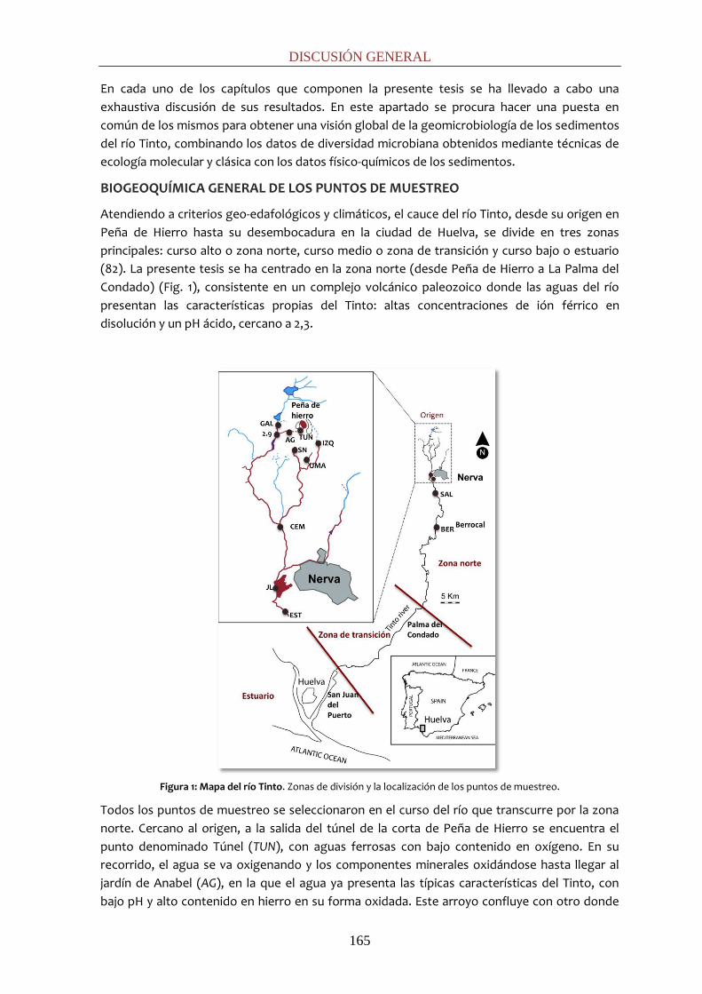
DISCUSIÓN GENERAL
165
En cada uno de los capítulos que componen la presente tesis se ha llevado a cabo una
exhaustiva discusión de sus resultados. En este apartado se procura hacer una puesta en
común de los mismos para obtener una visión global de la geomicrobiología de los sedimentos
del río Tinto, combinando los datos de diversidad microbiana obtenidos mediante técnicas de
ecología molecular y clásica con los datos físico-químicos de los sedimentos.
BIOGEOQUÍMICA GENERAL DE LOS PUNTOS DE MUESTREO
Atendiendo a criterios geo-edafológicos y climáticos, el cauce del río Tinto, desde su origen en
Peña de Hierro hasta su desembocadura en la ciudad de Huelva, se divide en tres zonas
principales: curso alto o zona norte, curso medio o zona de transición y curso bajo o estuario
(82). La presente tesis se ha centrado en la zona norte (desde Peña de Hierro a La Palma del
Condado) (Fig. 1), consistente en un complejo volcánico paleozoico donde las aguas del río
presentan las características propias del Tinto: altas concentraciones de ión férrico en
disolución y un pH ácido, cercano a 2,3.
Figura 1: Mapa del río Tinto. Zonas de división y la localización de los puntos de muestreo.
Todos los puntos de muestreo se seleccionaron en el curso del río que transcurre por la zona
norte. Cercano al origen, a la salida del túnel de la corta de Peña de Hierro se encuentra el
punto denominado Túnel (TUN), con aguas ferrosas con bajo contenido en oxígeno. En su
recorrido, el agua se va oxigenando y los componentes minerales oxidándose hasta llegar al
jardín de Anabel (AG), en la que el agua ya presenta las típicas características del Tinto, con
bajo pH y alto contenido en hierro en su forma oxidada. Este arroyo confluye con otro donde

DISCUSIÓN GENERAL
166
se sitúa Campo de Galdierias (GAL), y aguas abajo se encuentra una represa bautizada como
2.9 (2.9). Por otro lado, al este de Peña de Hierro confluyen tres arroyos donde se sitúan la
presa SN, UMA, e izquierda (IZQ). Ambos grupos de arroyos confluyen aguas abajo uniéndose
en un punto cercano a cementerio (CEM). Unos metros más adelante, cercano al municipio de
Nerva, se sitúa una presa cuyo punto de muestreo es conocido como JL, donde vierte el
efluente de la depuradora municipal de Nerva y pocos metros después se encuentra el punto
de muestro estación (EST). A partir de esta zona, se muestrearon dos estaciones: Salinas (SAL),
denominado así por la construcción de balsas de evaporación destinadas a la recuperación del
cobre en las terrazas del río y Berrocal (BER), próximo a la localidad del mismo nombre.
El río Tinto se muestra considerablemente homogéneo atendiendo a parámetros como pH y
potencial redox, mostrando condiciones ácidas y oxidantes a lo largo de su curso gracias al
efecto tampón que ejerce el férrico (61). Sin embargo, la adición de afluentes neutros genera
un gradiente decreciente en la concentración de metales (especialmente hierro) y,
consecuentemente, también un gradiente en la conductividad, que se pone de manifiesto - en
la zona muestreada - en una disminución gradual desde los orígenes hasta el punto de
muestreo BER. Destacan ciertos puntos de muestreo como TUN, cercano al origen, donde las
condiciones anóxicas todavía no han generado las características extremas que se presentarán
aguas abajo mostrando bajo potencial redox, bajo contenido en sulfatos y baja conductividad;
UMA, donde aparecen los menores valores de pH y los mayores de potencial redox,
conductividad y arsénico; o CEM, con los mayores valores de Ni, Cu y Zn, posiblemente debido
a la influencia de un gran deposito cercano de gossan.
El lecho del río Tinto es rocoso por lo que la mayoría de los puntos de muestreo presentan
acumulaciones carentes de estructura real de sedimento: consisten en partículas minerales de
cierto tamaño como arenas y grava con alta porosidad. Sus características biogeoquímicas
están fuertemente influenciadas por las de la columna de agua y por tanto lo están sus
comunidades microbianas. Así, en estas acumulaciones encontramos comunidades
pertenecientes principalmente a hábitats óxicos aunque algunos microorganimos pertenezcan
a ambientes anóxicos. Por ejemplo, en distintas localizaciones encontramos oxidadores de
hierro comunes de la columna de agua como la actinobacteria acidófila heterótrofa
Ferrimicrobium, el quimiolitótrofo aerobio estricto Leptospirillum, o el anaerobio facultativo
Acidithiobacillus ferrooxidans, así como diversos géneros de heterótrofos aerobios estrictos
como Pseudomonas, Pedobacter, Flectobacillus, etc. También encontramos fermentadores
como Clostridium spp. y, en menor medida, anaerobios estrictos como bacterias sulfato-
reductoras (BSR) de los géneros Desulfosporosinus o Desulfobulbus.
Sin embargo, existen ciertas zonas con pequeñas presas – la lámina de agua raramente alcanza
el metro de profundidad - que permiten la acumulación estratificada de sedimentos con menor
tamaño de partícula, principalmente limos y arcillas. A estas zonas pertenecen los puntos JL y
SN (Fig. 2). En ellos, la menor porosidad y la estratificación de los sedimentos permite cierta
diferenciación de sus características físico-químicas respecto a las de la columna de agua,
encontrándose verdaderas condiciones de anaerobiosis.
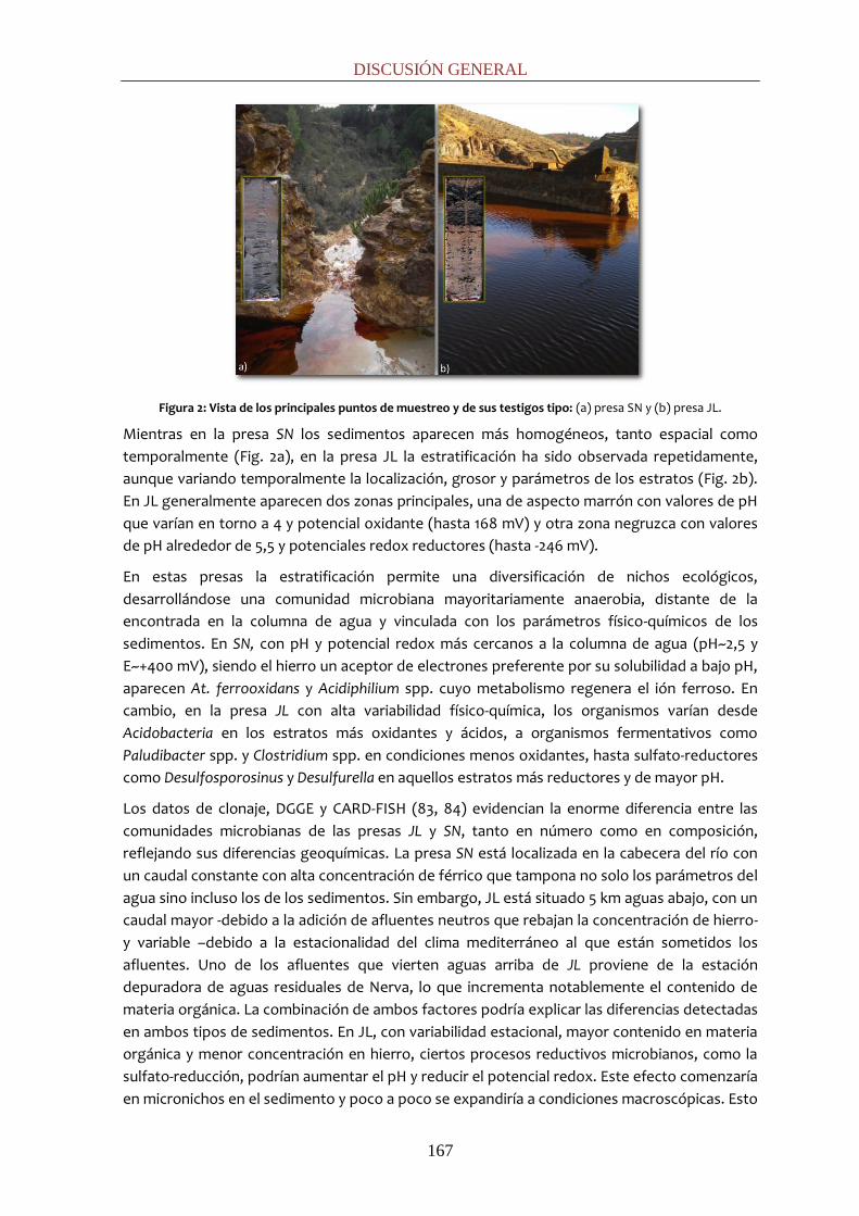
DISCUSIÓN GENERAL
167
Figura 2: Vista de los principales puntos de muestreo y de sus testigos tipo: (a) presa SN y (b) presa JL.
Mientras en la presa SN los sedimentos aparecen más homogéneos, tanto espacial como
temporalmente (Fig. 2a), en la presa JL la estratificación ha sido observada repetidamente,
aunque variando temporalmente la localización, grosor y parámetros de los estratos (Fig. 2b).
En JL generalmente aparecen dos zonas principales, una de aspecto marrón con valores de pH
que varían en torno a 4 y potencial oxidante (hasta 168 mV) y otra zona negruzca con valores
de pH alrededor de 5,5 y potenciales redox reductores (hasta -246 mV).
En estas presas la estratificación permite una diversificación de nichos ecológicos,
desarrollándose una comunidad microbiana mayoritariamente anaerobia, distante de la
encontrada en la columna de agua y vinculada con los parámetros físico-químicos de los
sedimentos. En SN, con pH y potencial redox más cercanos a la columna de agua (pH~2,5 y
E~+400 mV), siendo el hierro un aceptor de electrones preferente por su solubilidad a bajo pH,
aparecen At. ferrooxidans y Acidiphilium spp. cuyo metabolismo regenera el ión ferroso. En
cambio, en la presa JL con alta variabilidad físico-química, los organismos varían desde
Acidobacteria en los estratos más oxidantes y ácidos, a organismos fermentativos como
Paludibacter spp. y Clostridium spp. en condiciones menos oxidantes, hasta sulfato-reductores
como Desulfosporosinus y Desulfurella en aquellos estratos más reductores y de mayor pH.
Los datos de clonaje, DGGE y CARD-FISH (83, 84) evidencian la enorme diferencia entre las
comunidades microbianas de las presas JL y SN, tanto en número como en composición,
reflejando sus diferencias geoquímicas. La presa SN está localizada en la cabecera del río con
un caudal constante con alta concentración de férrico que tampona no solo los parámetros del
agua sino incluso los de los sedimentos. Sin embargo, JL está situado 5 km aguas abajo, con un
caudal mayor -debido a la adición de afluentes neutros que rebajan la concentración de hierro-
y variable –debido a la estacionalidad del clima mediterráneo al que están sometidos los
afluentes. Uno de los afluentes que vierten aguas arriba de JL proviene de la estación
depuradora de aguas residuales de Nerva, lo que incrementa notablemente el contenido de
materia orgánica. La combinación de ambos factores podría explicar las diferencias detectadas
en ambos tipos de sedimentos. En JL, con variabilidad estacional, mayor contenido en materia
orgánica y menor concentración en hierro, ciertos procesos reductivos microbianos, como la
sulfato-reducción, podrían aumentar el pH y reducir el potencial redox. Este efecto comenzaría
en micronichos en el sedimento y poco a poco se expandiría a condiciones macroscópicas. Esto

DISCUSIÓN GENERAL
168
explicaría el sedimento bandeado en JL, que podría estar relacionado con fluctuaciones del
agua superficial.
La diversidad encontrada en los sedimentos del río Tinto ha demostrado ser mucho mayor que
la encontrada en la columna de agua en estudios previos (29, 32). Mediante técnicas
moleculares, como clonaje y DGGE, a lo largo de este trabajo se han identificado directamente
de sedimentos secuencias pertenecientes a 12 phyla bacterianos distintos: Proteobacteria
(clases Alpha-, Beta-, Gamma- y Delta-proteobacteria), Firmicutes, Bacteroidetes, Acidobacteria,
Actinobacteria, Synergistetes, Cyanobacteria/Chloroplast, TM7, Chloroflexi, Nitrospira,
Verrucomicrobia, Fibrobacter. Las secuencias han podido ser afiliadas filogenéticamente a 72
géneros diferentes. Respecto a arqueas, prácticamente todas las secuencias correspondían a
los géneros Ferroplasma y Thermoplasma (orden Thermoplasmatales, phylum Euryarchaeota); a
excepción de una secuencia de Methanosaeta concilii (Euryarchaeota) y la familia
Thermofiliaceae (Crenarchaeota).
Dada la diversidad encontrada y la complejidad de los posibles metabolismos que interactúan
en este ecosistema, es necesaria una discusión global de los resultados repasando los distintos
ciclos geoquímicos encontrados. Debido al substrato geológico por el que transcurre la zona
norte del río Tinto, el hierro y el azufre - en sus distintas formas moleculares - están presentes
en altas concentraciones en el ecosistema. Sin embargo, y en contraste con lo que pudiera
suceder en las aguas, donde dominan los metabolismos implicados en el ciclo biogeoquímicos
del hierro, en los sedimentos también se detecta una amplia actividad relacionada con los
ciclos del N y del C.
CICLO DEL HIERRO
El hierro suele ser un elemento limitante en la mayoría de los ecosistemas ya que su baja
solubilidad a pH neutro hace que sea poco biodisponible. Dado el substrato geológico que
ofrece la FPI, y a la mayor solubilidad del hierro a bajo pH, el hierro se encuentra biodisponible
a altas concentraciones (~2,3 g L-1) en el ecosistema del Tinto.
La estabilidad del hierro ferroso a bajos pHs facilita su oxidación biológica al no competir con la
oxidación química (94), permitiendo la abundante presencia y actividad de microorganismos
acidófilos oxidadores de hierro. Nótese que a continuación se discute la capacidad de
oxidación de hierro de los organismos, sin detrimento de que por su versatilidad puedan
reducirlo u oxidar otros compuestos. La oxidación desasimilativa del hierro puede llevarse a
cabo tanto por organismos autótrofos como heterótrofos. En las aguas del río Tinto otros
estudios han demostrado el predominio absoluto de los oxidadores acidófilos extremos
quimiolitótrofos como At. ferrooxidans o Leptospirillum spp. y la presencia del heterótrofo
Ferroplasma spp. (30, 32, 61). Sin embargo, en este trabajo, técnicas moleculares como
clonación han mostrado una mayor diversidad de oxidadores de hierro en los sedimentos del
Tinto. Además de especies autótrofas (At. ferrooxidans, L. ferriphilum o L. ferrooxidans,),
aparecen gran variedad de autótrofos facultativos (Sulfobacillus spp., Alicyclobacillus spp.), o
heterótrofos obligados (Ferrimicrobium spp., Ferrithrix spp.). Esta gradación de comunidades
oxidadoras corresponde a sus necesidades fisiológicas y requerimientos ecológicos. En los
drenajes ácidos de mina, el oxidador aerobio obligado Leptospirillum domina las comunidades
microbianas de las zonas óxicas con condiciones más extremas (78) debido a su preferencia
por bajos pH, su mayor tolerancia a altas concentraciones de férrico y su mayor afinidad por el
ión ferroso (Km=0,025 mM) que At. ferrooxidans (Km=1,34 mM). Sin embargo, a menores

DISCUSIÓN GENERAL
169
potenciales redox, el anaerobio facultativo At. ferrooxidans posee mayor actividad específica
oxidadora (44) compitiendo eficientemente. En los sedimentos óxicos del río, donde cierta
acumulación de materia orgánica es posible, se incrementan los organismos heterótrofos.
Ferrimicrobium y Ferrithrix, actinobacterias acidófilas extremas y heterótrofas obligadas (42),
aparecerán en las zonas de menores pH mientras que Alicyclobacillus tiene preferencia por
zonas de pH algo más elevado (63). También aparece Sulfobacillus spp., capaces de crecer de
forma autótrofa, mixótrofa y heterótrofa (108). Finalmente, en los sedimentos anóxicos -
donde hay limitación de oxígeno pero no de materia orgánica - los quimiolitótrofos y
hetótrofos comparten nicho ecológico pero se limitan a aquellos con un metabolismo más
versátil como Sulfobacillus spp., Alicyclobacillus spp. o At. ferrooxidans, que además es capaz
de oxidar H2 (21). Aunque estos organismos podrían utilizar el posible oxígeno que difundiera a
los sedimentos, su persistencia parece atribuible a su versatilidad metabólica; al ser capaces de
reducir hierro en ausencia de oxígeno, actuarían más probablemente como reductores que
como oxidadores de hierro en estos sedimentos anóxicos.
Por último, la oxidación de hierro también podría aparecer acoplada a desnitrificación en
condiciones anóxicas (93). Diversos estudios han detectado esta capacidad en cepas de
Pseudomonas, bacterias púrpuras (25), y Dechloromonas (2, 14), todas ellas presentes en los
sedimentos del Tinto. Además esta actividad ha sido detectada en cultivos de enriquecimiento
con sedimentos del Tinto donde se apreciaba la oxidación de ferroso a férrico en condiciones
de anaerobiosis.
La reducción desasimilativa del hierro está muy extendida entre los acidófilos, pudiendo ser
llevada a cabo por una gran variedad de microorganismos. Numerosas secuencias
pertenecientes a organismos hierro-reductores han sido detectadas por técnicas moleculares
(DGGE y clonación) en los sedimentos del río Tinto; además, la reducción de hierro ha sido
confirmada mediante medida de actividad en cultivos de enriquecimiento específicos, siendo
mayor a menores pHs. Algunos de los reductores corresponden a los versátiles oxidadores de
hierro antes mencionados. Entre ellos, At. ferrooxidans – capaz de reducir ión férrico usando
azufre elemental (74) o hidrógeno (71) como dadores inorgánicos de electrones - parece tener
un papel principal al aparecer de forma profusa en las librerías genéticas (84) y mostrar altos
porcentajes de hibridación (17% respecto al número total de bacterias) en puntos como SN
(83). Otros oxidadores de hierro, tales como Ferrimicrobium spp., Ferrithrix spp. (42),
Sulfobacillus spp. (108), y Ferroplasma (20) son también capaces de reducir hierro usando
compuestos orgánicos como dadores de electrones. Alicyclobacillus spp. son capaces de
reducir hierro aunque no siempre vinculado al crecimiento (63).
Aparte de los anaerobios facultativos mencionados en el párrafo anterior, capaces de oxidar o
reducir hierro, existen numerosos reductores de hierro especializados. En ausencia de
oxígeno, el ión férrico es el aceptor preferencial en los sedimentos para la mineralización de la
materia orgánica (13) dado que procesos como la sulfato-reducción y la metanogénesis se
inhiben en su presencia (9). El heterótrofo Acidiphilium cryptum es capaz de reducir hierro
oxidando una diversidad de compuestos orgánicos de bajo peso molecular, azúcares o
hidrógeno (43), incluso en presencia de oxígeno (57). Este organismo, identificado en los
sedimentos del Tinto, parece jugar un papel relevante en determinados emplazamientos,
como SN donde representa el 5% del total de bacterias presentes en el mismo (83). Diversas
acidobacterias también son capaces de reducir hierro (63), específicamente, Acidobacterium
capsulatum que forma Fe(II) en condiciones anaerobias a pHs de 3 a 5 respirando glucosa (17).

DISCUSIÓN GENERAL
170
También aparecen Geobacter, anaerobio estricto capaz de usar ácidos grasos de cadena corta,
alcoholes y compuestos monoaromáticos como dadores de electrones (62) para la reducción
de hierro; Ferribacterium, capaz de usar acetato u otros acidos orgánicos (18) y Aciditerrimonas
ferrireducens, termoacidófila anaerobia (39). Por último, multitud de BSR, tales como
Desulfosporosinus spp., pueden actuar como Fe (III)-reductoras (77). Estos procesos implican la
regeneración del ión ferroso en los sedimentos del río Tinto completando el ciclo del hierro
con los organismos encontrados.
Los metabolismos basados en el hierro están íntimamente relacionados con las
transformaciones minerales. Aparte de la conocida acción de los oxidadores de hierro en la
lixiviación de los sulfuros metálicos, los fenómenos de reducción también parecen estar
implicados en otros procesos. Por ejemplo, se asocia la acción de BSR como Desulfosporosinus
sp. GBSRB4.2 con la transformación de schwertmanita [Fe8O8(OH)6SO4·nH2O] a goetita (α-
FeOOH) al aumentar el pH y la concentración de Fe(II) (7) o la acción reductiva de A. cryptum
JF-5 sobre el hierro contenido en minerales como goetita, jarosita, etc. favoreciendo su
disolución (12).
CICLO DEL AZUFRE
En las zonas anaerobias de ecosistemas con gran cantidad de pirita y otros sulfuros metálicos
se acumulan distintas formas reducidas del azufre (tiosulfato, tetrationato, azufre elemental,
sulfuro, etc.) que pueden ser usadas como substratos por distintos organismos en procesos
oxidativos. Algunos de los organismos identificados como oxidadores de hierro también son
capaces de oxidar sulfuros (At. ferrooxidans o Sulfobacillus spp.). Adicionalmente, diversas
especies de Acidiphilium (A. acidophilum, A. cryptum, etc.) pueden oxidar azufre elemental (34,
81). Este grupo de microorganismos podrían oxidar las especies reducidas de azufre con el
oxígeno que difundiera a los sedimentos. Por último, se han identificado secuencias de
cromatiales o bacterias rojas del azufre, que son organismos anaerobios o microaerófilos
capaces de realizar la fotosíntesis. Éstos podrían usar el sulfuro de hidrógeno como agente
reductor produciendo gránulos de azufre que pueden ser posteriormente oxidados
totalmente hasta sulfatos.
Pese a que la forma mayoritaria y estable del azufre es el sulfato, hasta la fecha no son muchos
los acidófilos conocidos capaces de catalizar la reducción desasimilativa del sulfato a sulfuro.
Generalmente las BSR prefieren pHs entre 6 y 8 (103) pero, desde hace unos años, se ha visto
que este metabolismo está presente en diversos ambientes ácidos (53), entre ellos río Tinto.
Mediante técnicas moleculares, en la presente tesis se han identificado diversos géneros de
BSR (Desulfosporosinus, Desulfotomaculum, Desulfobulbus, Desulfosalsimonas,
Desulfitobacterium y Thermodesulfobium) y reductores de azufre elemental de los géneros
Thermoplasma, Acidithiobacillus y Desulfurella en los sedimentos del Tinto (83-85).
La arquea termoacidófila anaerobia facultativa Thermoplasma (87) y la bacteria
termoacidotolerante anaerobia Desulfurella (46), capaces de respirar azufre elemental, habían
sido previamente detectadas en diversos ambientes ácidos. En recientes investigaciones se ha
visto que varias especies de Acidithiobacillus, entre ellas A. ferrooxidans, son capaces de
respirar azufre oxidando hidrógeno a pHs tan bajos como 2,5 (40). Entre las BSR,
Syntrophobacter puede usar sulfato como aceptor de electrones oxidando propionato por la
via methylmalonil CoA (36); el termófilo moderado autótrofo Thermodesulfobium puede usar
hidrógeno (67); Desulfosalsimonas es una SRB halófila capaz de usar lactato, propionato,
DISCUSIÓN GENERAL
171
acetato y butirato usando sulfato o tiosulfato como aceptores de electrones (52, 90);
Desulfobulbus (105) usa propionato, lactato y alcoholes oxidándolos incompletamente a
acetato, o bien, H2 suplementado con acetato como fuente de carbono. Desulfosporosinus spp.
pueden también fermentar distintos substratos (91), realizar acetogénesis a partir de formiato
o metanol (75) o reducir hierro disasimilativamente (77). Desulfosporosinus spp. y
Desulfotomaculum spp., oxidadores incompletos de diversos compuestos orgánicos a acetato,
son anaerobios obligados con la habilidad de esporular, lo que les hace más resistentes a
condiciones adversas pudiendo sobrevivir a condiciones oxidantes y de sequedad durante
meses (104).
Mediante técnicas de hibridación in situ se ha visto que los géneros Desulfosporosinus,
Desulfurella y Syntrophobacter son muy abundantes (11, 6 y 5% del número total de bacterias)
en ciertos estratos de los sedimentos (83), especialmente en aquellos de mayor pH (4,2-6,2) y
condiciones redox mas reductoras (50, -210 mV), llegando sólo Syntrophobacter a representar
el 37% del total en el estrato superficial. El número de BSR se correlaciona con la variación de
los parámetros físico-químicos de los sedimentos. Donde las BSR son más abundantes, como
consecuencia de su metabolismo, se reduce la concentración de sulfato y aumenta el pH. El
sulfuro de hidrógeno producido reacciona con el hierro y otros metales precipitándolos en
forma de sulfuros. De este modo, se observa una atenuación natural, llevada a cabo por las
BSR, de las características extremas de ambientes ácidos (bajo pH y alto contenido en hierro,
sulfato y metales pesados en las aguas).
Adicionalmente, se han usado cultivos de enriquecimiento específicos para probar no solo su
presencia y abundancia, sino también la actividad sulfato-reductora en los sedimentos. En una
primera aproximación se realizaron enriquecimientos a pH 5 y 7 obteniendo resultados
positivos a ambos pHs (85). Sin embargo, el aislamiento sólo fue posible a pH 7, perteneciendo
todos los aislados al género Desulfotomaculum spp., una conocida BSR neutrófila (104). Estos
resultados están en la línea de otros trabajos, en los que partiendo de inóculos de ambientes
ácidos se aislaron BSR neutrófilas (58, 60, 95). Desde hace tiempo, hay un interés creciente en
el aislamiento de BSR realmente acidófilas, pero con pocos resultados positivos. Otros autores
consiguieron enriquecimientos que reducían sulfatos a bajo pH pero resultaron ser co-cultivos
con Acidocella o Desulfitobacterium (15, 51). En algunos casos, se consiguieron aislar cepas
acidófilas pero nunca fueron caracterizadas (35, 45). De modo que, tras todo el interés
despertado por este metabolismo y el esfuerzo invertido, a día de hoy contamos con pocas
cepas de verdaderas BSR acidófilas caracterizadas (4, 88). Por todo ello, se realizó un screening
exhaustivo con sedimentos de río Tinto, obteniéndose cultivos que crecían a pH tan bajos
como 4 con glicerol, metanol e hidrógeno, 4,5 con lactato y 5,5 con succinato. Además
pudieron aislarse varias cepas, identificando dos nuevas especies del género Desulfosporosinus
y un nuevo género cercano al grupo Desufosporosinus/Desulfitobacterium que crece a un pH
óptimo de 5. Con este trabajo, se prueba la existencia y actividad de BSR en los sedimentos del
río Tinto, además del carácter acidófilo y no sólo acidotolerante de los aislados.
Dado el carácter acidófilo de las comunidades presentes en el río y en vista de la atenuación
natural de las características extremas de los ambientes ácidos de mina, observada en aquellos
estratos donde eran abundantes las BSR, se planteó un posible uso tecnológico. Se acopló el
tratamiento de aguas de drenaje ácido (con bajo pH y alta concentración de metales pesados,
hierro y sulfatos) con el tratamiento de aguas residuales (alto contenido orgánico). El carácter
acidófilo de las comunidades microbianas de los sedimentos del río Tinto fue clave, puesto que

DISCUSIÓN GENERAL
172
con un inoculo de carácter neutrófilo hubiera sido necesario una neutralización del influente
previa al tratamiento. Sin embargo, la comunidad sulfato-reductora intrínseca de los
sedimentos del río Tinto fue capaz de llevar a cabo el tratamiento acoplado de ambos vertidos
obteniendo en una sola etapa un efluente de calidad, a pH neutro y con bajo contenido en
materia orgánica, sulfatos, hierro y metales pesados, mostrando la gran capacidad de
biorremediación de las BSR de río Tinto.
CICLO DEL NITROGENO
Los ciclos del hierro y del azufre han sido profusamente estudiados en el río Tinto y
ecosistemas similares, pero no se ha prestado ninguna atención al ciclo del nitrógeno en tales
ambientes ácidos extremos, quedando su análisis excluido de revisiones globales (44). Sin
embargo, durante la presente tesis se han detectado secuencias relacionadas con organismos
que podrían estar involucrados en el ciclo del nitrógeno, se han medido actividades
desnitrificantes en cultivos de enriquecimiento y se han detectado nitratos y nitritos en las
aguas del Tinto. Por ello, se plantea el ciclo del nitrógeno como un ciclo activo e importante en
el ecosistema.
Por técnicas moleculares se han identificado organismos con capacidad fijadora de nitrógeno
tales como el quimiorganótrofo aerobio Ideonella azotifigens (70) o Anaerobacter
polyendosporus, fermentador anaerobio obligado esporulante con capacidad de reducir nitrito
a amonio y de fijar N2 (23, 64). Además ha sido descrita la capacidad fijadora de diversas cepas
de Leptospirillum spp. aisladas de río Tinto (30).
La capacidad desnitrificante está ampliamente extendida entre los organismos procariotas. El
nitrato sirve de aceptor de electrones a multitud de bacterias heterótrofas y, en ocasiones,
también autótrofas capaces de oxidar hierro ferroso, hidrógeno o compuestos reducidos de
azufre. Por medio de clonaciones se han identificado géneros que contienen especies
denitrificantes como Alcaligenes, Pseudochrobactrum, Pseudomonas, Bacillus, Dechloromonas,
Propionibacterium, Acidovorax, Delftia, Commamonas, Aquabacterium, Clostridium, Geobacter,
Anaerobacter, etc. presentando distintas tolerancias a condiciones óxicas.
Algunos de ellos son respiradores aerobios con capacidad de usar nitrato como aceptor de
electrones alternativo, tales como Acidovorax, adaptados a ambientes oligotróficos y
frecuentes en suelos contaminados con uranio (3, 50), Delftia (102), Steroidobacter denitrificans
(26), Comamonas aquaticus (101), Aquabacterium que usa un amplio rango de carbohidratos
(47), Dechloromonas (2, 14) y Alicyclobacillus, oxidadores aerobios de hierro y/o azufre capaces
de usar nitrato como aceptor (41). También aparecen diversos respiradores de carácter
anaerobio como Alcaligenes faecalis, capaz de respiración anaerobia con nitrito pero no con
nitrato (73), Geobacter, que pueden oxidar acetato (62), o Aquabacterium, microaerofilos que
oxidan un amplio rango de ácidos orgánicos pudiendo usar el nitrato como aceptor (47). Por
ultimo, se han identificado fermentadores como Brevibacterium (16), Pseudochrobactrum
asaccharolyticum (49), Anaerobacter (23, 64) y Propionibacterium, de lento metabolismo que
producen lactato, propionato y acetato a partir de glucosa y capaces de desnitrificar (79).
La actividad desnitrificante en los sedimentos de río Tinto se confirmó mediante
enriquecimientos específicos a distintos pHs (4,5 - 7), obteniendo resultados positivos en
condiciones tanto heterótrofas como autótrofas. Esta actividad es dependiente de pH:
mientras la reducción de nitratos se incrementa a pH más altos, la proporción de oxido nitroso
DISCUSIÓN GENERAL
173
acumulado es mayor a menores pH. Esto confirma diversas investigaciones previas que
sugieren que el rango óptimo de pH para la desnitrificación se sitúa entre 6 y 8; por debajo de
éste, los pasos finales de la desnitrificación hasta nitrógeno molecular se inhiben,
acumulándose los productos intermedios como nitritos (8, 69) u óxidos nitrosos (89).
Mediante análisis por DGGE de dichos cultivos de enriquecimiento, se obtuvieron secuencias
de organismos pertenecientes a géneros como Bacillus, Paenibacillus, Lysinobacillus,
Ammoniphilus, Rummelibacillus (clase Bacilli); Clostridium, Desulfitobacterium, Sedimentibacter
(clase Clostridia), y Alcaligenes (clase Betaproteobacteria), todos previamente vinculados a
actividades desnitrificantes (98, 99). El análisis de diversidad de los microorganismos
presentes en los cultivos confirmó la presencia y actividad de organismos previamente
detectados mediante clonación y aportó evidencias de la participación de nuevos géneros con
metabolismo desnitrificante en la cuenca del Tinto.
CICLO DEL CARBONO
En los estudios previos sobre las zonas aerobias de ecosistemas tipo drenajes ácidos de minas,
el ciclo del carbono aparecía principalmente vinculado a, y dependiente de, los otros ciclos
biogeoquímicos. La heterotrofía parecía tener un papel ecológico de mera detoxificación para
los quimiolitótrofos, sensibles a altas concentraciones de materia orgánica (6). En cambio,
durante la presente tesis, en los sedimentos del río Tinto se han obtenido secuencias y medido
actividades vinculadas a diversos metabolismos del ciclo del carbono tales como oxidación
heterótrofa, fermentación, metanogénesis e incluso acetogénesis, lo que evidencia un activo e
importante ciclo del carbono operando en los sedimentos del río Tinto.
Atendiendo al modo de asimilación del carbono, los organismos se podrían dividir en
autótrofos –aquellos que fijan CO2 por mecanismos como el ciclo de Calvin (At. ferrooxidans o
Leptospirillum spp.) - o heterótrofos, que asimilan el carbono contenido en compuestos
orgánicos. En esta sección nos centraremos en discutir aquellos metabolismos centrados
exclusivamente en el carbono, excluyendo aquellos heterótrofos que obtienen energía
mediante respiración anaerobia utilizando compuestos de azufre, hierro o nitrógeno, los
cuales se discuten en sus correspondientes secciones.
Algunas secuencias obtenidas corresponden a organismos heterótrofos cuya forma principal
de obtención de energía es la oxidación aerobia de la materia orgánica. Entre ellos hay
bacterias metilótrofas (oxidadoras de metanol) pertenecientes al género Methylotenera (48),
además de multitud de organismos capaces de oxidar compuestos orgánicos más complejos
(Acidiphilium, Aquabacterium, Pedobacter, Flectobacillus, Sphingomonas, Variovorax,
Pseudomonas, etc.). Uno de los principales heterótrofos encontrados en la cuenca del Tinto,
tanto en la columna de agua (32) como en sedimentos, es Acidiphilium, bacterias acidófilas
extremas que oxidan compuestos orgánicos en condiciones aerobias o microaerófilas (43),
llegando a representar un 5,6% de media respecto al número total de bacterias (83). En los
sedimentos microaerófilos aparecen multitud de respiradores aerobios como Aquabacterium
spp. que crecen en condiciones de microaerofília oxidando un amplio rango de ácidos
orgánicos (47); Pedobacter, quimiorganotrofos con metabolismo oxidativo (92);
Haliscomenobacter, bacterias filamentosas presentes en fangos activos que promueven la
floculación (68); Flectobacillus aerobios obligados (59) al igual que Acinetobacter; Rothia o
Fluviicola (72). U otros como Leucobacter con varias cepas resistentes al cromo (66);
Sphingomonas, quimioheterótrofos aerobios obligados (28), Dellvibrio, bacterias celulolíticas

DISCUSIÓN GENERAL
174
(65), Pseudomonas, bacilos aerobios no esporulantes (31) y Cohnella, aerobias formadoras de
esporas. También fueron encontradas secuencias relacionadas con organismos facultativos
como Staphylococcus y Variovorax paradoxus, un heterótrofo o autótrofo facultativo (oxidador
de hidrógeno) capaz de usar una amplia variedad de compuestos orgánicos como fuente de
carbono (106).
También se han identificado diversos organismos fermentadores como las bacterias del ácido
láctico microaerotolerantes Olsenella (55), Brevibacterium, quimioorganotrofos aerobios
estrictos, que oxidan y fermentan azúcares (16); Propionibacterium, fermentadores que, a
partir de glucosa, producen acetato, lactato y propionato (19); Prosthecobacter, heterótrofos
oligotróficos o Fibrobacter, bacterias celulolíticas que producen formato, acetato y succinato.
También fermentadores anaerobios estrictos como Turicibacter (10), Anaerobacter spp., (23)
fermentadores esporulantes de carbohidratos que producen etanol, acetato, lactato, butirato,
butanol, hidrógeno y CO2 (64); Dorea, quimiorganótrofos que producen etanol, formato,
acetato, hidrógeno y CO2 como producto del metabolismo de la glucosa; Veillonella,
fermentadores de lactato que producen propionato, acetato, hidrógeno y CO2 pudiendo usar
también usar ácidos pirúvico, oxalacético, málico, fumárico, y succínico (80); Propionispora
hippie, formador de esporas productor de propionato (1) o Oscillibacter valericigenes,
productor de valerato (38). Muchos de estos fermentadores han aparecido en librerías
genéticas amplificando DNA ambiental o DNA de cultivos de enriquecimientos de BSR a bajo
pH donde, además, se han detectado algunos de sus productos de fermentación como
butirato, propionato e hidrógeno. Esto confirma su presencia en los sedimentos de río Tinto y
su actividad en las condiciones extremas que éstos presentan.
Uno de los fermentadores encontrado con mayor frecuencia mediante clonación, además de
representar al menos el 1% de las bacterias totales en los sedimentos del Tinto, ha sido
Clostridium spp. (83, 84). A pesar de ser bacterias anaerobias obligadas, no todos tienen la
misma sensibilidad al oxígeno. Llevan a cabo la fermentación oscura, o fermentación ácida, en
la que los sustratos orgánicos como azúcares o proteínas son fermentados a diversos ácidos
orgánicos (butirato, acetato, propionato o lactato), alcoholes (etanol), CO2 e hidrógeno (37,
97, 100). Por su capacidad de producción de hidrógeno fueron los organismos dominantes en
los cultivos específicos de enriquecimiento para detectar tal actividad, habiéndose aislado
varias cepas pertenecientes a este género. Estos aislados han sido caracterizados y está
estudiándose su utilización en la producción biológica de hidrógeno (76).
Otro fermentador que parece jugar un papel importante en ambientes ácidos es un organismo
lejanamente relacionado con Paludibacter propionicigenes, una bacteria anaerobia estricta
productora de propionato (96). Numerosas secuencias relacionadas con este organismo
fueron obtenidas por DGGE en estratos reductores de los sedimentos, así como en diversas
clonaciones (84). Este fermentador fue detectado como el contaminante mayoritario en
cultivos de enriquecimiento para BSR acidófilas, a partir de los cuales fue aislado por filtración
selectiva. El nuevo aislado posee tan solo un 92% de similitud con P. propionicigenes, por lo que
podría suponer un nuevo género dentro del phylum Bacteroidetes. Las secuencias cercanas al
nuevo aislado han sido obtenidas en ambientes ácidos de minas o con alta concentración de
metales pesados, lo que sugiere la naturaleza tolerante del aislado ante tales condiciones
extremas.

DISCUSIÓN GENERAL
175
Los productos de fermentación como el propionato y el butirato pueden ser oxidados a
acetato, CO2 y H2. Esta oxidación es llevada a cabo por un grupo denominado “organismos
acetógenos productores obligados de hidrógeno (OHPA)”, mediante un proceso conocido
como acetogénesis. Aunque la energía de este proceso es muy baja, su acoplamiento con
metanógenas consumidoras de hidrógeno (que reducen la presión parcial de hidrógeno) la
convierte en favorable. Esta relación se denomina sintrófica y la llevan a cabo bacterias del
género Syntrophobacter. En los sedimentos del río Tinto, Syntrophobacter llega a representar
el 37% del total en ciertos estratos aunque no está claro si actúa como bacteria sintrófica o
BSR. También se han obtenido secuencias pertenecientes a Oxobacter spp., anaerobios
obligados acetogénicos. La única especie, Oxobacter pfennigii, es un anaerobio esporulante
que cataliza la transformación de piruvato a acetato y CO2 (56).
El acetato e hidrógeno producido tras los procesos anteriores son típicos substratos de la
metanogénesis. Aunque se considera una actividad anaerobia que se inhibe a bajos pHs,
diversos estudios han detectado metanogénesis en condiciones ácidas incluso a pHs tan bajos
como 4,5 (11, 24, 27, 33, 54, 107). Entre ellos, por su proximidad con este trabajo, destaca un
estudio realizado usando sedimentos de la presa JL y GAL como inóculos (86).
Esto suscitó cuestiones sobre su presencia y extensión en un ecosistema tan ácido como el río
Tinto, por lo que se realizó un screening con sedimentos de 8 puntos distribuidos a lo largo del
río con 6 diferentes fuentes de carbono. Mediante la medida de la producción de metano, se
constató la ubicuidad de las metanógenas en dicho ecosistema y su actividad en cultivos de
enriquecimientos con pH alrededor de 5. La secuenciación del DNA de dichos enriquecimientos
culminó con la identificación de arqueas como Methanobrevibacter arboriphilus,
Methanosarcina barkerii o Methanosaeta thermophile. Además, por DGGE directa de DNA
ambiental de un estrato reductor de los sedimentos de la presa JL, se ha obtenido una
secuencia de arquea metanógena perteneciente a Methanosaeta concilii (JQ815591)
coincidiendo con el estudio previo (86). M. concilii, que parece ser la arquea predominante en
condiciones naturales, es una arquea metanogénica consumidora de acetato. El acetato, que
ha sido medido en numerosas ocasiones en el agua intersticial de los sedimentos en
concentraciones hasta 79 mg L-1, es un producto de diversas fermentaciones y de oxidaciones
parciales llevadas a cabo por BSR, metabolismos ambos presentes y abundantes en los
sedimentos, por lo que la abundancia natural del substrato de M. concilii podría favorecer su
prevalencia frente a otras metanógenas. Posiblemente las arqueas no jueguen un papel
cuantitativamente importante en la degradación anaerobia de la material orgánica en el río
Tinto, pero su presencia y actividad en sedimentos tan adversos tiene importantes
connotaciones ecológicas –al ampliar el rango de tolerancia de dichas arqueas- y
astrobiológicas –las detecciones de metano en la atmósfera de Marte podrían tener un origen
biológico.
MODELO GEOMICROBIOLÓGICO DE LOS SEDIMENTOS DEL RÍO TINTO.
En esta tesis se ha abordado el estudio de los sedimentos del río Tinto combinando trabajos de
ecología molecular microbiana y de microbiología tradicional e interpretándolos en función de
los datos físico-químicos de los sedimentos. La integración de esta información biogeoquímica
deriva en un modelo geomicrobiológico que integra los distintos ciclos de los elementos que
operan en los sedimentos del río Tinto (Fe, N, C, y S) así como sus interacciones (Fig. 3).

DISCUSIÓN GENERAL
176
Figura 3: Modelo geomicrobiológico de los sedimentos del río Tinto. Los proceso implicados se señalan con flechas azules (ciclo del azufre), rojas (ciclo del hierro), amarillas (ciclo del carbono) y verdes (ciclo del nitrógeno).

DISCUSIÓN GENERAL
177
A diferencia de los estudios previos relativos a la columna de agua del río, la ecología
microbiana de los sedimentos no está basada fundamentalmente en el ciclo del hierro. De
hecho, parece que el ciclo del azufre y la utilización de compuestos orgánicos dominan este
subsistema del Tinto, con diversas interacciones con el ciclo del nitrógeno y hierro. Partiendo
de los substratos que proporciona el lecho de la Faja Píritica Ibérica, principalmente pirita
(FeS2), los compuestos reducidos de hierro y azufre serán el substrato disponible para los
quimiolitótrofos, siendo la base para el desarrollo de una compleja comunidad.
El hierro en su forma reducida, Fe(II), sería la principal fuente de energía en condiciones
aerobias o microaerófilas para bacterias como At. ferrooxidans, Sulfobacillus spp.,
Alicyclobacillus spp., Ferrimicrobium spp y arqueas como Ferroplasma spp; y en condiciones de
anaerobias para Pseudomonas spp. o Alicyclobacillus spp. acoplado a la reducción de nitratos.
Los sistemas ácidos son ricos en hierro debido a su alta solubilidad a bajo pH, excediendo las
concentraciones de Fe(III) a las de oxígeno en dos órdenes de magnitud, de modo que el
Fe(III) puede ser usado como aceptor de electrones por multitud de microorganismos (17, 22).
Así, el Fe(III) puede ser reducido acoplado a la oxidación de materia orgánica por heterótrofos
acidófilos como A. capsulatum , Acidiphilium spp., Sulfobacillus spp., Ferrimicrobium spp., etc. o
acoplado a la oxidación de azufre por At. ferrooxidans y Sulfobacillus spp. Estos procesos
implican la regeneración del ferroso completándose el ciclo del hierro con los organismos
identificados en los sedimentos del río Tinto.
El segundo grupo de metabolitos cruciales en este sistema son los compuestos del azufre. El
sulfuro puede ser oxidado a sulfato en condiciones aerobias o anaerobias (acoplado a la
reducción de hierro mencionada) por At. ferrooxidans y Sulfobacillus spp. El sulfato puede ser
reducido desasimilativamente por bacterias como Desulfosporosinus spp., Thermodesulfobium
spp., Syntrophobacter spp., Desulfotomaculum spp., etc. Un metabolito intermedio como el
azufre elemental, también puede ser reducido completamente a sulfuro por BSR como
Desulfurella, At. ferrooxidans o arqueas como Thermoplasma spp. La presencia y actividad
demostrada de SBR en el sistema evidencia la existencia de la sulfato-reducción en condiciones
ácidas, cerrando el ciclo del azufre en sedimentos extremos como los del río Tinto.
Respecto al ciclo de nitrógeno, ciertos organismos de los géneros Leptospirillum, Ideonella o
Anaerobacter podrían fijar nitrógeno atmosférico. Además, diversos organismos - tales como
miembros de los géneros: Alcaligenes, Pseudochrobactrum, Pseudomonas, Desulfitobacterium,
Delftia, etc.- llevan a cabo la desnitrificación acoplada a la oxidación de azufre, de hierro
ferroso o de materia orgánica
Y por último, el ciclo del carbono, con el que interactúan un número considerable de los
organismos implicados en los otros tres ciclos. Entre los organismos heterótrofos
encontramos metabolismos tanto respiratorios (aerobios y anaerobios) como fermentadores.
La oxidación de la materia orgánica podría llevarse a cabo en condiciones aerobias o
microaerobias por Acidiphilium, Pedobacter, Variovorax y una gran variedad de respiradores, o
anaerobias acoplada a la reducción del hierro, del sulfato o del nitrato antes mencionados.
Adicionalmente, se dan procesos fermentativos llevados a cabo por representantes de
géneros como Clostridium, Bacillus, Propionibacterium, Veillonella, Paludibacter, Staphylococcus,
etc. Estas fermentaciones crean una compleja mezcla de intermediarios como ácidos grasos
volátiles, alcoholes e hidrógeno. Muchos de estos productos pueden ser oxidados hasta
acetato por BSR o por bacterias sintróficas como Syntrophobacter spp., asociado, en este

DISCUSIÓN GENERAL
178
último caso, a la producción de hidrógeno. Estos compuestos son los sustratos principales de
otros organismos, tanto respiradores aerobios o anaerobios, como de las arqueas
metanogénicas Methanosaeta, Methanosarcina o Methanobrevibacter.
En estudios previos en perforaciones del subsuelo en el entorno de río Tinto (5), se había
detectado la presencia de gases como H2, NO2 y CH4 en el fluido subterráneo extraído,
proponiéndose el origen químico del H2 derivado de la interacción roca-agua. La identificación
de organismos productores de hidrógeno como Syntrophobacter spp. y Clostridium spp.,
productores de metano como Methanosaeta, Methanosarcina y Methanobrevibacter y diversos
desnitrificantes sugiere la posibilidad de producción biológica de estos gases.
En conclusión, la puesta en común de los datos recopilados en la presente tesis permite una
compresión profunda de los distintos ciclos geoquímicos que operan en los sedimentos del río
Tinto, así como sus complejas interacciones. Además, las actividades inferidas mediante
técnicas de ecología molecular han sido demostradas con técnicas de microbiología clásica y
medidas cuantitativas, validando el modelo propuesto.
REFERENCIAS
1. Abou-Zeid, D. M., H. Biebl, C. Spröer, and R. J. Müller. 2004. Propionispora hippei sp. nov., a novel Gram-negative, spore-forming anaerobe that produces propionic acid. Int. J. Syst. Evol. Microbiol. 54:951-954.
2. Achenbach, L. A., U. Michaelidou, R. A. Bruce, J. Fryman, and J. D. Coates. 2001. Dechloromonas agitata gen. nov., sp. nov. and Dechlorosoma suillum gen. nov., sp. nov., two novel environmentally dominant (per)chlorate-reducing bacteria and their phylogenetic position. Int. J. Syst. Evol. Microbiol. 51:527-533.
3. Akob, D. M., H. J. Mills, and J. E. Kostka. 2007. Metabolically active microbial communities in uranium‐contaminated subsurface sediments. FEMS Microbiol. Ecol. 59:95-107.
4. Alazard, D., M. Joseph, F. Battaglia-Brunet, J. L. Cayol, and B. Ollivier. 2010. Desulfosporosinus acidiphilus sp. nov.: a moderately acidophilic sulfate-reducing bacterium isolated from acid mining drainage sediments. Extremophiles. 14:305-312.
5. Amils, R., D. Fernández-Remolar, F. Gómez, E. González-Toril, N. Rodríguez, C. Briones, O. Prieto-Ballesteros, J. L. Sanz, E. Díaz, and T. O. Stevens. 2008. Subsurface Geomicrobiology of the Iberian Pyritic Belt. In P. Dion and C. Shekhar Nautiyal (Ed.), Soil Biology (Vol.13): Microbiology of Extreme Soils. Springer, Berlin. 205-223.
6. Baker, B. J., and J. F. Banfield. 2003. Microbial communities in acid mine drainage. FEMS Microbiol. Ecol. 44:139-152.
7. Bertel, D., J. Peck, T. J. Quick, and J. M. Senko. 2012. Iron Transformations Induced by an Acid-Tolerant Desulfosporosinus Species. Appl. Environ. Microbiol. 78:81-88.
8. Blösl, M., and R. Conrad. 1992. Influence of an increased pH on the composition of the nitrate-reducing microbial populations in an anaerobically incubated acidic forest soil. Syst. Appl. Microbiol. 15:624-627.
9. Bodegom, P. M., J. Scholten, and A. J. M. Stams. 2004. Direct inhibition of methanogenesis by ferric iron. FEMS Microbiol. Ecol. 49:261-268.
10. Bosshard, P. P., R. Zbinden, and M. Altwegg. 2002. Turicibacter sanguinis gen. nov., sp. nov., a novel anaerobic, Gram-positive bacterium. Int. J. Syst. Evol. Microbiol. 52:1263-1266.

DISCUSIÓN GENERAL
179
11. Bräuer, S. L., J. B. Yavitt, and S. H. Zinder. 2004. Methanogenesis in McLean Bog, an acidic peat bog in upstate New York: stimulation by H2/CO2 in the presence of rifampicin, or by low concentrations of acetate. Geomicrobiol. J. 21:433-443.
12. Bridge, T. A. M., and D. B. Johnson. 2000. Reductive dissolution of ferric iron minerals by Acidiphilium SJH. Geomicrobiol. J. 17:193-206.
13. Canfield, D. E., R. Raiswell, and S. Bottrell. 1992. The reactivity of sedimentary iron minerals toward sulfide. Am. J. Sci. 292:659-683.
14. Chakraborty, R., S. M. O'Connor, E. Chan, and J. D. Coates. 2005. Anaerobic degradation of benzene, toluene, ethylbenzene, and xylene compounds by Dechloromonas strain RCB. Appl. Environ. Microbiol. 71:8649-8655.
15. Church, C. D., R. T. Wilkin, C. N. Alpers, R. O. Rye, and R. B. McCleskey. 2007. Microbial sulfate reduction and metal attenuation in pH 4 acid mine water. Geochem. Trans. 8:10.
16. Collins, M. D. 1992. The genus Brevibacterium. The Prokaryotes. 2:1351-1354.
17. Coupland, K., and D. B. Johnson. 2008. Evidence that the potential for dissimilatory ferric iron reduction is widespread among acidophilic heterotrophic bacteria. FEMS Microbiol. Lett. 279:30-35.
18. Cummings, D. E., F. Caccavo Jr, S. Spring, and R. F. Rosenzweig. 1999. Ferribacterium limneticum, gen. nov., sp. nov., an Fe (III)-reducing microorganism isolated from mining-impacted freshwater lake sediments. Arch. Microbiol. 171:183-188.
19. Cummins, C., J. Johnson, A. Balows, H. Truper, M. Dworkin, W. Harder, and K. Schleifer. 1992. The genus Propionibacterium. The Prokaryotes. 834-849.
20. Dopson, M., C. Baker-Austin, A. Hind, J. P. Bowman, and P. L. Bond. 2004. Characterization of Ferroplasma isolates and Ferroplasma acidarmanus sp. nov., extreme acidophiles from acid mine drainage and industrial bioleaching environments. Appl. Environ. Microbiol. 70:2079-2088.
21. Drobner, E., H. Huber, and K. O. Stetter. 1990. Thiobacillus ferrooxidans, a facultative hydrogen oxidizer. Appl. Environ. Microbiol. 56:2922-2923.
22. Druschel, G. K., B. J. Baker, T. M. Gihring, and J. F. Banfield. 2004. Acid mine drainage biogeochemistry at Iron Mountain, California. Geochem. T. 5:13-32.
23. Duda, V., A. Lebedinsky, M. Mushegjan, and L. Mitjushina. 1987. A new anaerobic bacterium, forming up to five endospores per cell -Anaerobacter polyendosporus gen. et spec. nov. Arch. Microbiol. 148:121-127.
24. Dunfield, P., R. Dumont, and T. R. Moore. 1993. Methane production and consumption in temperate and subarctic peat soils: response to temperature and pH. Soil Biol. Biochem. 25:321-326.
25. Ehrenreich, A., and F. Widdel. 1994. Anaerobic oxidation of ferrous iron by purple bacteria, a new type of phototrophic metabolism. Appl. Environ. Microbiol. 60:4517-4526.
26. Fahrbach, M., J. Kuever, M. Remesch, B. E. Huber, P. Kämpfer, W. Dott, and J. Hollender. 2008. Steroidobacter denitrificans gen. nov. sp. nov., a steroidal hormone-degrading gammaproteobacterium. Int. J. Syst. Evol. Microbiol. 58:2215-2223.
27. Florencio, L., A. Nozhevnikova, A. Van Langerak, A. J. M. Stams, J. A. Field, and G. Lettinga. 1993. Acidophilic degradation of methanol by a methanogenic enrichment culture. FEMS Microbiol. Lett. 109:1-6.
28. Fredrickson, J., D. Balkwill, G. Drake, M. Romine, D. Ringelberg, and D. White. 1995. Aromatic-degrading Sphingomonas isolates from the deep subsurface. Appl. Environ.

DISCUSIÓN GENERAL
180
Microbiol. 61:1917-1922.
29. García-Moyano, A., E. González-Toril, A. Aguilera, and R. Amils. 2012. Comparative microbial ecology study of the sediments and the water column of Río Tinto, an extreme acidic environment. FEMS Microbiol. Ecol. DOI: 10.1111/j.1574-6941.2012.01346.x.
30. García-Moyano, A., E. González-Toril, M. Moreno-Paz, V. Parro, and R. Amils. 2008. Evaluation of Leptospirillum spp. in the Río Tinto, a model of interest to biohydrometallurgy. Hydrometallurgy. 94:155-161.
31. Gilardi, G. 1971. Characterization of Pseudomonas species isolated from clinical specimens. Appl. Environ. Microbiol. 21:414.
32. Gonzalez-Toril, E., E. Llobet-Brossa, E. O. Casamayor, R. Amann, and R. Amils. 2003. Microbial ecology of an extreme acidic environment, the Tinto River. Appl. Environ. Microbiol. 69:4853-4865.
33. Goodwin, S., and J. G. Zeikus. 1987. Ecophysiological adaptations of anaerobic bacteria to low pH: analysis of anaerobic digestion in acidic bog sediments. Appl. Environ. Microbiol. 53:57-64.
34. Hallberg, K. B., H. E. C. Thomson, I. Boeselt, and D. B. Johnson. 2001. Aerobic and anaerobic sulfur metabolism by acidophilic bacteria. p. 423-431. Biohydrometallurgy: Fundamentals, Technology and Sustainable Developmentvol. In V.S.T. Ciminelli and O. Garcia, Jr. (ed.), 11A. Elsevier, Amsterdan.
35. Hard, B. C., S. Friedrich, and W. Babel. 1997. Bioremediation of acid mine water using facultatively methylotrophic metal-tolerant sulfate-reducing bacteria. Microbiol. Res. 152:65-73.
36. Harmsen, H. J. M., B. L. M. Van Kuijk, C. M. Plugge, A. D. L. Akkermans, W. M. De Vos, and A. J. M. Stams. 1998. Syntrophobacter fumaroxidans sp. nov., a syntrophic propionate-degrading sulfate-reducing bacterium. Int. J. Syst. Bacteriol. 48:1383-1387.
37. Hu, B., and S. Chen. 2007. Pretreatment of methanogenic granules for immobilized hydrogen fermentation. Int J Hydrogen Energy. 32:3266-3273.
38. Iino, T., K. Mori, K. Tanaka, K. Suzuki, and S. Harayama. 2007. Oscillibacter valericigenes gen. nov., sp. nov., a valerate-producing anaerobic bacterium isolated from the alimentary canal of a Japanese corbicula clam. Int. J. Syst. Evol. Microbiol. 57:1840-1845.
39. Itoh, T., K. Yamanoi, T. Kudo, M. Ohkuma, and T. Takashina. 2011. Aciditerrimonas ferrireducens gen. nov., sp. nov., an iron-reducing thermoacidophilic actinobacterium isolated from a solfataric field. Int. J. Syst. Evol. Microbiol. 61:1281-1285.
40. Jameson, E., O. Rowe, K. Hallberg, and D. Johnson. 2010. Sulfidogenesis and selective precipitation of metals at low pH mediated by Acidithiobacillus spp. and acidophilic sulfate-reducing bacteria. Hydrometallurgy. 104:488-493.
41. Jiang, C., Y. Liu, Y. Liu, X. Guo, and S. J. Liu. 2009. Isolation and characterization of ferrous-and sulfur-oxidizing bacteria from Tengchong solfataric region, China. Journal of Environmental Sciences. 21:1247-1252.
42. Johnson, D. B., P. Bacelar-Nicolau, N. Okibe, A. Thomas, and K. B. Hallberg. 2009. Ferrimicrobium acidiphilum gen. nov., sp. nov. and Ferrithrix thermotolerans gen. nov., sp. nov.: heterotrophic, iron-oxidizing, extremely acidophilic actinobacteria. Int. J. Syst. Evol. Microbiol. 59:1082-1089.
43. Johnson, D. B., and T. A. M. Bridge. 2002. Reduction of ferric iron by acidophilic heterotrophic bacteria: evidence for constitutive and inducible enzyme systems in Acidiphilium spp. J. Appl. Microbiol. 92:315-321.

DISCUSIÓN GENERAL
181
44. Johnson, D. B., and K. B. Hallberg. 2008. Carbon, iron and sulfur metabolism in acidophilic micro-organisms. Adv. Microb. Physiol. 54:201-255.
45. Johnson, D. B., E. Jameson, O. Rowe, K. Wakeman, and K. B. Hallberg. 2009. Sulfidogenesis at low pH by acidophilic bacteria and its potential for the selective recovery of transition metals from mine waters. Advanced Materials Research. 71:693-696.
46. Kaksonen, A., J. Plumb, W. Robertson, M. Riekkola-Vanhanen, P. Franzmann, and J. Puhakka. 2006. The performance, kinetics and microbiology of sulfidogenic fluidized-bed treatment of acidic metal-and sulfate-containing wastewater. Hydrometallurgy. 83:204-213.
47. Kalmbach, S., W. Manz, J. Wecke, and U. Szewzyk. 1999. Aquabacterium gen. nov., with description of Aquabacterium citratiphilum sp. nov., Aquabacterium parvum sp. nov. and Aquabacterium commune sp. nov., three in situ dominant bacterial species from the Berlin drinking water system. Int. J. Syst. Bacteriol. 49:769-777.
48. Kalyuzhnaya, M. G., S. Bowerman, J. C. Lara, M. E. Lidstrom, and L. Chistoserdova. 2006. Methylotenera mobilis gen. nov., sp. nov., an obligately methylamine-utilizing bacterium within the family Methylophilaceae. Int. J. Syst. Evol. Microbiol. 56:2819-2823.
49. Kampfer, P., R. Rossello-Mora, H. C. Scholz, C. Welinder-Olsson, E. Falsen, and H. J. Busse. 2006. Description of Pseudochrobactrum gen. nov., with the two species Pseudochrobactrum asaccharolyticum sp. nov. and Pseudochrobactrum saccharolyticum sp. nov. Int. J. Syst. Evol. Microbiol. 56:1823-1829.
50. Khan, S. T., Y. Horiba, M. Yamamoto, and A. Hiraishi. 2002. Members of the family Comamonadaceae as primary poly (3-hydroxybutyrate-co-3-hydroxyvalerate)-degrading denitrifiers in activated sludge as revealed by a polyphasic approach. Appl. Environ. Microbiol. 68:3206-3214.
51. Kimura, S., K. B. Hallberg, and D. B. Johnson. 2006. Sulfidogenesis in low pH (3.8–4.2) media by a mixed population of acidophilic bacteria. Biodegradation. 17:57-65.
52. Kjeldsen, K. U., T. F. Jakobsen, J. Glastrup, and K. Ingvorsen. 2010. Desulfosalsimonas propionicica gen. nov., sp. nov., a halophilic, sulfate-reducing member of the family Desulfobacteraceae isolated from a salt-lake sediment. Int. J. Syst. Evol. Microbiol. 60:1060-1065.
53. Koschorreck, M. 2008. Microbial sulphate reduction at a low pH. FEMS Microbiol. Ecol. 64:329-342.
54. Koschorreck, M., K. Wendt-Potthoff, B. Scharf, and H. H. Richnow. 2008. Methanogenesis in the sediment of the acidic Lake Caviahue in Argentina. J. Volcanol. Geotherm. Res. 178:197-204.
55. Kraatz, M., R. J. Wallace, and L. Svensson. 2011. Olsenella umbonata sp. nov., a microaerotolerant anaerobic lactic acid bacterium from the sheep rumen and pig jejunum, and emended descriptions of Olsenella, Olsenella uli and Olsenella profusa. Int. J. Syst. Evol. Microbiol. 61:795-803.
56. Krumholz, L. R., and M. P. Bryant. 1985. Clostridium pfennigii sp. nov. uses methoxyl groups of monobenzenoids and produces butyrate. Int. J. Syst. Evol. Microbiol. 35:454-456.
57. Küsel, K., U. Roth, and H. L. Drake. 2002. Microbial reduction of Fe (III) in the presence of oxygen under low pH conditions. Environ. Microbiol. 4:414-421.
58. Kusel, K., U. Roth, T. Trinkwalter, and S. Peiffer. 2001. Effect of pH on the anaerobic microbial cycling of sulfur in mining-impacted freshwater lake sediments. Environ. Exp. Bot. 46:213-223.
59. Larkin, J. M., P. M. Williams, and R. Taylor. 1977. Taxonomy of the genus Microcyclus

DISCUSIÓN GENERAL
182
Ørskov 1928: reintroduction and emendation of the genus Spirosoma Migula 1894 and proposal of a new genus, Flectobacillus. Int. J. Syst. Bacteriol. 27:147-156.
60. Lee, Y. J., C. S. Romanek, and J. Wiegel. 2009. Desulfosporosinus youngiae sp. nov., a spore-forming, sulfate-reducing bacterium isolated from a constructed wetland treating acid mine drainage. Int. J. Syst. Evol. Microbiol. 59:2743-2746.
61. Lopez-Archilla, A. I., I. Marin, and R. Amils. 2001. Microbial community composition and ecology of an acidic aquatic environment: the Tinto River, Spain. Microb. Ecol. 41:20-35.
62. Lovley, D., S. J. Giovannoni, D. C. White, J. E. Champine, E. Phillips, Y. A. Gorby, and S. Goodwin. 1993. Geobacter metallireducens gen. nov. sp. nov., a microorganism capable of coupling the complete oxidation of organic compounds to the reduction of iron and other metals. Arch. Microbiol. 159:336-344.
63. Lu, S., S. Gischkat, M. Reiche, D. M. Akob, K. B. Hallberg, and K. Kusel. 2010. Ecophysiology of Fe-cycling bacteria in acidic sediments. Appl. Environ. Microbiol. 76:8174-8183.
64. Madigan, M. T. 2005. Brock Biology of Microorganisms, 11th edn. International Microbiology. 8:149-152.
65. Mergaert, J., D. Lednická, J. Goris, M. C. Cnockaert, P. De Vos, and J. Swings. 2003. Taxonomic study of Cellvibrio strains and description of Cellvibrio ostraviensis sp. nov., Cellvibrio fibrivorans sp. nov. and Cellvibrio gandavensis sp. nov. Int. J. Syst. Evol. Microbiol. 53:465-471.
66. Morais, P. V., C. Paulo, R. Francisco, R. Branco, A. Paula Chung, and M. S. Da Costa. 2006. Leucobacter luti sp. nov., and Leucobacter alluvii sp. nov., two new species of the genus Leucobacter isolated under chromium stress. Syst. Appl. Microbiol. 29:414-421.
67. Mori, K., H. Kim, T. Kakegawa, and S. Hanada. 2003. A novel lineage of sulfate-reducing microorganisms: Thermodesulfobiaceae fam. nov., Thermodesulfobium narugense, gen. nov., sp. nov., a new thermophilic isolate from a hot spring. Extremophiles. 7:283-290.
68. Mulder, E. G., and M. H. Deinema. 1992. The genus Haliscomenobacter. The Prokaryotes. 3688-3690.
69. Nagele, W., and R. Conrad. 1990. Influence of soil pH on the nitrate-reducing microbial populations and their potential to reduce nitrate to NO and N2O. FEMS Microbiol. Lett. 74:49-57.
70. Noar, J. D., and D. H. Buckley. 2009. Ideonella azotifigens sp. nov., an aerobic diazotroph of the Betaproteobacteria isolated from grass rhizosphere soil, and emended description of the genus Ideonella. Int. J. Syst. Evol. Microbiol. 59:1941-1946.
71. Ohmura, N., K. Sasaki, N. Matsumoto, and H. Saiki. 2002. Anaerobic respiration using Fe3+ , S0, and H2 in the chemolithoautotrophic bacterium Acidithiobacillus ferrooxidans. J. Bacteriol. 184:2081-2087.
72. O'Sullivan, L. A., J. Rinna, G. Humphreys, A. J. Weightman, and J. C. Fry. 2005. Fluviicola taffensis gen. nov., sp. nov., a novel freshwater bacterium of the family Cryomorphaceae in the phylum ‘Bacteroidetes’. Int. J. Syst. Evol. Microbiol. 55:2189-2194.
73. Otte, S., N. G. Grobben, L. A. Robertson, M. S. Jetten, and J. G. Kuenen. 1996. Nitrous oxide production by Alcaligenes faecalis under transient and dynamic aerobic and anaerobic conditions. Appl. Environ. Microbiol. 62:2421-2426.
74. Pronk, J., J. De Bruyn, P. Bos, and J. Kuenen. 1992. Anaerobic growth of Thiobacillus ferrooxidans. Appl. Environ. Microbiol. 58:2227-2230.

DISCUSIÓN GENERAL
183
75. Rabus, R., T. A. Hansen, and F. Widdel. 2006. Dissimilatory sulfate-and sulfur-reducing prokaryotes. The Prokaryotes. 2:659-768.
76. Rajhi, H., M. Conthe, D. Puyol, E. díaz, and J. L. Sanz. 2012. Dark fermentation: Isolation and characterization of hydrogen-producing strains from different sludges. Bioresource Technology. Submitted BITE-S-12-03266-1.
77. Ramamoorthy, S., H. Sass, H. Langner, P. Schumann, R. Kroppenstedt, S. Spring, J. Overmann, and R. Rosenzweig. 2006. Desulfosporosinus lacus sp. nov., a sulfate-reducing bacterium isolated from pristine freshwater lake sediments. Int. J. Syst. Evol. Microbiol. 56:2729-2736.
78. Rawlings, D. E., H. Tributsch, and G. S. Hansford. 1999. Reasons why ‘Leptospirillum’-like species rather than Thiobacillus ferrooxidans are the dominant iron-oxidizing bacteria in many commercial processes for the biooxidation of pyrite and related ores. Microbiology. 145:5-13.
79. Rehberger, T. G. 2001. Waste treatment with a combination of denitrifying Propionibacterium acidipropionici and protease-producing Bacillus. US Patent 6,221,650. .
80. Rogosa, M. 1964. The genus Veillonella I. J. Bacteriol. 87:162-170.
81. Rohwerder, T., C. Janosch, and W. Sand. 2007. Elemental Sulfur Oxidation in Acidiphilium spp. Advanced Materials Research. 20:583-583.
82. Rufo, L., N. Rodríguez, R. Amils, V. de la Fuente, and R. Jiménez-Ballesta. 2007. Surface geochemistry of soils associated to the Tinto River (Huelva, Spain). Sci. Total Environ. 378:223-227.
83. Sánchez-Andrea, I., K. Knittel, R. Amann, R. Amils, and J. L. Sanz. 2012. Quantification of Tinto River sediment microbial communities: the importance of Sulfate-Reducing Bacteria and their role in attenuating acid mine drainage. Appl. Environ. Microbiol. 78 (13).
84. Sánchez-Andrea, I., N. Rodriguez, R. Amils, and J. L. Sanz. 2011. Microbial Diversity in Anaerobic Sediments at Rio Tinto, a Naturally Acidic Environment with a High Heavy Metal Content. Appl. Environ. Microbiol. 77:6085-6093.
85. Sánchez-Andrea, I., P. Rojas, R. Amils, and J. L. Sanz. 2012. Screening of Anaerobic Activities in Sediments of an Acidic Environment: Tinto River. Extremophiles. Acceptted.
86. Sanz, J. L., N. Rodríguez, E. E. Díaz, and R. Amils. 2011. Methanogenesis in the sediments of Rio Tinto, an extreme acidic river. Environ. Microbiol. 13:2336-2341.
87. Segerer, A., T. A. Langworthy, and K. O. Stetter. 1988. Thermoplasma acidophilum and Thermoplasma volcanium sp. nov. from Solfatara Fields. Syst. Appl. Microbiol. 10:161-171.
88. Senko, J. M., G. Zhang, J. T. McDonough, M. A. Bruns, and W. D. Burgos. 2009. Metal reduction at low pH by a Desulfosporosinus species: implications for the biological treatment of acidic mine drainage. Geomicrobiol. J. 26:71-82.
89. Simek, M., L. Jisova, and D. W. Hopkins. 2002. What is the so-called optimum pH for denitrification in soil. Soil Biol. Biochem. 34:1227-1234.
90. Sorokin, D. Y., E. E. Zacharova, N. V. Pimenov, T. P. Tourova, A. N. Panteleeva, and G. Muyzer. 2012. Sulfidogenesis in hypersaline chloride–sulfate lakes of Kulunda Steppe (Altai, Russia). FEMS Microbiol. Ecol. 79:445-453.
91. Spring, S., and F. Rosenzweig. 2006. The genera Desulfitobacterium and Desulfosporosinus: taxonomy. The Prokaryotes: A Handbook on the Biology of Bacteria. 771-786.
92. Steyn, P., P. Segers, M. Vancanneyt, P. Sandra, K. Kersters, and J. Joubert. 1998. Classification of heparinolytic bacteria into a new genus, Pedobacter, comprising four species: Pedobacter heparinus comb. nov., Pedobacter piscium comb. nov., Pedobacter africanus sp. nov.
DISCUSIÓN GENERAL
184
and Pedobacter saltans sp. nov. Proposal of the family Sphingobacteriaceae fam. nov. Int. J. Syst. Bacteriol. 48:165-177.
93. Straub, K. L., M. Benz, B. Schink, and F. Widdel. 1996. Anaerobic, nitrate-dependent microbial oxidation of ferrous iron. Appl. Environ. Microbiol. 62:1458-1460.
94. Stumm, W., and J. J. Morgan . 1970. Aquatic Chemistry; An Introduction Emphasizing Chemical Equilibria in Natural Waters. Wiley, New York.
95. Tuttle, J. H., P. R. Dugan, C. B. Macmillan, and C. I. Randles. 1969. Microbial dissimilatory sulfur cycle in acid mine water. J. Bacteriol. 97:594-602.
96. Ueki, A., H. Akasaka, D. Suzuki, and K. Ueki. 2006. Paludibacter propionicigenes gen. nov., sp. nov., a novel strictly anaerobic, Gram-negative, propionate-producing bacterium isolated from plant residue in irrigated rice-field soil in Japan. Int. J. Syst. Evol. Microbiol. 56:39-44.
97. Ueno, Y., S. Haruta, M. Ishii, and Y. Igarashi. 2001. Microbial community in anaerobic hydrogen-producing microflora enriched from sludge compost. Appl. Microbiol. Biotechnol. 57:555-562.
98. Verbaendert, I., P. De Vos, N. Boon, and K. Heylen. 2011. Denitrification in Gram-positive bacteria: an underexplored trait. Biochem. Soc. Trans. 39:254-258.
99. Villemur, R., M. Lanthier, R. Beaudet, and F. Lépine. 2006. The Desulfitobacterium genus. FEMS Microbiol. Rev. 30:706-733.
100. Wang, X., D. Hoefel, C. P. Saint, P. T. Monis, and B. Jin. 2007. The isolation and microbial community analysis of hydrogen producing bacteria from activated sludge. J. Appl. Microbiol. 103:1415-1423.
101. Wauters, G., T. De Baere, A. Willems, E. Falsen, and M. Vaneechoutte. 2003. Description of Comamonas aquatica comb. nov. and Comamonas kerstersii sp. nov. for two subgroups of Comamonas terrigena and emended description of Comamonas terrigena. Int. J. Syst. Evol. Microbiol. 53:859-862.
102. Wen, A., M. Fegan, C. Hayward, S. Chakraborty, and L. I. Sly. 1999. Phylogenetic relationships among members of the Comamonadaceae, and description of Delftia acidovorans (den Dooren de Jong 1926 and Tamaoka et al. 1987) gen. nov., comb. nov. Int. J. Syst. Bacteriol. 49:567-576.
103. Widdel, F. 1988. Microbiology and ecology of sulfate-and sulfur-reducing bacteria. Biology of Anaerobic Microorganisms. 469-585.
104. Widdel, F. 1992. The genus Desulfotomaculum. The Prokaryotes. 2:1792-1799.
105. Widdel, F., and N. Pfennig. 1982. Studies on dissimilatory sulfate-reducing bacteria that decompose fatty acids II. Incomplete oxidation of propionate by Desulfobulbus propionicus gen. nov., sp. nov. Arch. Microbiol. 131:360-365.
106. Willems, A., J. De Ley, M. Gillis, and K. Kersters. 1991. Comamonadaceae, a New Family Encompassing the Acidovorans rRNA Complex, Including Variovorax paradoxus gen. nov., comb. nov., for Alcaligenes paradoxus Int. J. Syst. Bacteriol. 41:445-450.
107. Williams, R. T., and R. L. Crawford. 1985. Methanogenic bacteria, including an acid-tolerant strain, from peatlands. Appl. Environ. Microbiol. 50:1542-1544.
108. Zhuravleva, A. E., I. Tsaplina, A. Ismailov, L. Zakharchuk, and G. Karavaiko. 2007. Metabolism peculiarities of bacteria of the genus Sulfobacillus. Advanced Materials Research. 20:469-472.

CONCLUSIONES GENERALES

CONCLUSIONES GENERALES
187
1. Los datos de clonaje y DGGE muestran que la biodiversidad procariótica en los
sedimentos, especialmente la diversidad bacteriana, es significativamente mayor que
la encontrada en estudios previos en la columna de agua. Frente a la uniformidad
físico-química en dicha columna, la protección que confiere la estructura de los
sedimentos permite el desarrollo de diferentes nichos ecológicos que albergan
consorcios microbianos más diversos.
2. La densidad de microorganismos en los sedimentos del río Tinto varía sustancialmente
entre distintos puntos de muestreo, moviéndose en el rango 107 - 108 células por gramo
de peso húmedo de sedimento. Además también varía con la profundidad,
disminuyendo hasta 10 veces su valor a lo largo de los primeros 50 cm de profundidad.
3. Los datos de CARD-FISH muestran que los microorganismos pertenecientes al dominio
Bacteria (>98%) dominan sobre Archaea (<2%), no habiéndose detectado organismos
activos pertenecientes al dominio Eukarya.
4. Se ha diseñado una sonda específica para el género Desulfurella (DSU655). Dicha sonda
ha permitido cuantificar - mediante CARD-FISH - la abundancia de Desulfurella spp. en
los sedimentos del río Tinto.
5. En los sedimentos ácidos del río aparecen ciertas actividades anaerobias que tienen
lugar preferentemente a pHs neutros, tales como metanogénesis, sulfato-reducción,
producción de hidrógeno y desnitrificación, lo que evidencia el amplio rango de
tolerancia de los microorganismos a las condiciones ambientales. Estas actividades
confirman la producción biológica de gases como H2, CH4 y N2O en el subsuelo del río
Tinto.
6. Mediante técnicas moleculares, se han identificado microorganismos relacionados con
el ciclo del hierro (At. ferrooxidans, Sulfobacillus spp., Ferroplasma spp., etc.), azufre
(Desulfurella spp., Desulfosporosinus spp., Thermodesulfobium spp., etc.), carbono
(Acidiphilium spp., Bacillus spp., Clostridium spp., Acidobacterium spp., etc.) y nitrógeno
(Alcaligenes spp., Pseudochrobactrum spp., etc.), y su distribución se ha correlacionado
con los parámetros fisicoquímicos de los sedimentos.
7. La distribución de los organismos en los sedimentos está íntimamente relacionada con
las características físico-químicas de los mismos (pH, potencial redox, sulfato y
concentraciones de metales pesados); las comunidades varían entre distintos puntos
de muestreo e incluso entre distintas profundidades en el mismo punto. Cuando los
parámetros son más próximos a los de la columna de agua (pH 2,5 y +400 mV), los
organismos más abundantes corresponden a reductores de hierro (Acidithiobacillus,
Acidiphilium , etc.) mientras que cuando aparecen mayores pHs (4,2-6,2) y potenciales
redox más reductores (50, -246 mV), los organismos relacionados con reducción de
sulfato parecen predominar (Desulfosporosinus, Desulfurella, Syntrophobacter, etc.).
8. Los datos obtenidos mediante cultivos de enriquecimiento vinculan la sulfato-
reducción con representantes de los géneros Desulfotomaculum, Thermodesulfobium,
Desulfitobacterium y especialmente, Desulfosporosinus; la desnitrificación con diversos
organismos pertenecientes a géneros como Paenibacillus, Bacillus, Sedimentibacter,
Lysinobacillus, Delftia, Alcaligenes, Clostridium y Desulfitobacterium; las especies del
CONCLUSIONES GENERALES
188
género Clostridium parecen ser los principales productores de hidrógeno en este
ecosistema y por último, aunque parece tener escasa relevancia ecológica, la
metanogénesis aparece vinculada a la presencia de arqueas de los géneros
Methanosarcina, Methanosaeta y Methanobrevibacter.
9. Se han aislado bacterias productoras de hidrógeno, sulfato-reductoras y
fermentadoras. Entre las sulfato-reductoras, dos cepas son próximas (96%) a
Desulfosporosinus acidophilus, una a Desulfosporosinus orientis (96%), y otra al grupo
Desulfitobacterium/Desulfosporosinus (93%), por lo que puede tratarse de dos nuevas
especies y un nuevo genero dentro de la familia Peptococcaceae (Firmicutes). Un
aislado con metabolismo fermentativo presenta un porcentaje de semejanza de tan
sólo un 92% con Paludibacter propionicigenes, por lo que podría suponer un nuevo
género dentro del phylum Bacteroidetes.
10. Nuestros resultados sugieren que se produce una atenuación natural de las
características extremas de las aguas ácidas de mina (pH ácido y alta concentración de
metales en disolución) mediada por bacterias sulfatorreductoras acidófilas, con la
consecuente precipitación de metales en forma de sulfuros. Este proceso pudo ser
usado para biorremediación: el tratamiento conjunto de aguas ácidas con aguas
residuales urbanas produjo un efluente de calidad con mínimo coste económico y
ambiental.
11. La identificación de diversos microorganismos, el examen detallado de sus
metabolismos correspondientes, su cuantificación y su cultivo han permitido
desarrollar y validar un modelo de funcionamiento de los ciclos del azufre, hierro,
carbón y nitrógeno en los sedimentos del río Tinto en relación a la ecología microbiana
de los mismos.

CONCLUSIONES GENERALES
189
GENERAL CONCLUSIONS
1. Cloning and DGGE data show that prokaryotic diversity in the sediments, especially
bacterial diversity, is significantly greater than that found in previous studies in the
water column. In contrast to the homogeneity of the water column, the protection
that the sediment’s structure confers, allows for the development of diverse ecological
niches that host more diverse microbial consortia.
2. The density of microorganisms in sediments of the Tinto river varies substantially
between different sampling points, ranging from 107 to 108 cells per gram of wet
weight of sediment. In addition, the density also varies with depth, decreasing up to 10
fold over 50 cm of depth.
3. CARD-FISH data show that microorganisms belonging to Bacteria (>98%) dominated
over Archaea (<2%) while no active cells of the domain Eukarya were detected.
4. A specific probe for the genus Desulfurella (DSU655) has been designed. This probe has
allowed for the quantification –by CARD-FISH- of the abundance of Desulfurella spp. in
the Tinto river sediments.
5. In the acidic sediments of Tinto river, certain anaerobic activities that take place
preferentially at neutral pH, such as methanogenesis, sulfate-reduction, hydrogen-
production and denitrification, occur, which shows the great tolerance of
microorganisms to extreme conditions. The detected and quantified activities confirm
the bioproduction of H2, CH4 and N2O in the subsurface of Tinto river.
6. Microorganisms involved in the iron (Acidithiobacillus ferrooxidans, Sulfobacillus spp.,
etc.), sulfur (Desulfurella spp., Desulfosporosinus spp., Thermodesulfobium spp., etc.),
carbon (Acidiphilium spp., Bacillus spp., Clostridium spp., Acidobacterium spp., etc.) and
nitrogen (Alcaligenes spp., Pseudochrobactrum spp., etc.) cycles were identified. The
detection of these microorganisms, a detailed examination of their metabolism, and
their quantification and cultivation have allowed for the compilation of a model of the
iron, sulfur, nitrogen and carbon cycles and their relation to microbial ecology in
extreme acidic sediments.
7. The distribution of organisms in the sediments is closely related to their physico-
chemical characteristics (pH, redox potential, sulfate and concentrations of heavy
metals); the communities vary between different sampling sites and even between
different depths at the same site. When the parameters are closest to those of the
water column (pH 2.5 and + 400 mV), the most abundant organisms are iron reducers
(Acidithiobacillus, Acidiphilium , etc.) while when higher pHs (4.2-6.2) and more
reducing redox potentials (50, - 210 mV) appear, sulfate-related organisms seem to
predominate (Desulfosporosinus, Desulfurella and Syntrophobacter).
8. Data from enrichments suggests that the sulfate-reduction is linked to representatives
from the Desulfotomaculum, Thermodesulfobium, Desulfitobacterium and, especially,
Desulfosporosinus genera. Denitrifying organisms showed a broad diversity, scattered
among the genera Paenibacillus, Bacillus, Sedimentibacter, Lysinobacillus, Delftia,
Alcaligenes, Clostridium and Desulfitobacterium. Hydrogen-producing enrichments were
dominated by Clostridium spp. Methanogenesis, although of scarce ecological
CONCLUSIONES GENERALES
190
relevance, appeared to be related with the archaeal genera Methanosarcina,
Methanosaeta and Methanobrevibacter.
9. Strains of hydrogen-producers, sulfate-reducers and fermenters, have been isolated
from Tinto river sediments. Among the sulfate-reducers, two strains share 96% of
similarity to Desulfosporosinus acidophilus, another 96% to Desulfosporosinus orientis
and another one has just 93% 16S rRNA gene sequence similarity with the clade
Desulfitobacterium/Desulfosporosinus; the might represent two new species and a new
genus in the family Peptococcaceae (Firmicutes) Another isolate with fermentative
metabolism presents a percentage of similarity of just 92% to the closest phylogenetic
isolate: Paludibacter propionicigenes, so it might represent a new genus in the
Bacteroidetes phylum.
10. Our results suggest that a natural attenuation of ARD characteristics (low pH and high
heavy metal content) is biologically driven by sulfate-reducers and the consequent
precipitation of metals as sulfides. The application of this biotechnological potential
was possible coupling the bioremediation of Acid Mine Drainage waters with domestic
waste treatment producing a quality effluent with a minimal economic and ecological
cost.
11. The identification of different microorganisms in this study, a detailed examination of
their corresponding metabolism, their quantification and their cultivation have allowed
us to compile and validate a model of the iron, sulfur, nitrogen and carbon cycles and
their relation to microbial ecology in Tinto river sediments.
ANEXO: Trabajo en proceso


ANEXO
193
Caracterización del aislado fermentador
Uno de los estudios de la presente tesis (capítulo 5) se obtuvo un co-cultivo de una fermentadora y una BSR.
Tras filtrar el co-cultivo a través de un filtro con un diámetro de poro de 0,45 µm, las bacterias SRB quedaron retenidas mientras que las fermentadoras pasaron al filtrado. Tras 3 diluciones seriadas y 3 transferencias sucesivas en placas de agar, el estudio culminó con la obtención de un aislado de un organismo fermentador con 92% de similitud con el aislado más próximo que corresponde a Paludibacter propionicigenes. Este aislado podría representar un nuevo género dentro del phylum Bacteroidetes, por lo que se ha procedido a su caracterización.
Análisis de pH, temperatura, salinidad y substratos:
Para conocer los rangos y los óptimos de crecimiento del aislado para distintos parámetros, se creció el aislado bajo los siguientes parámetros: pH (3-13), temperatura (4-80 °C), salinidad (0-3 %) y 10 mM de concentración de sustratos (almidón soluble , sacarosa, lactosa, xilosa, arabinosa, celobiosa, manosa, galactosa, glucosa, maltosa, L-sorbosa, acetato, arsenato, piruvato, butirato, malato, propionato, lactato, benzoato, succinato, citrato, nitrato, metanol, etanol, 2-propanol, manitol, glicerol, SO4
-2, Fe3+). Los ensayos se realizaron por triplicado en el medio anaerobio anteriormente descrito en el capítulo 5 suplementado con 10 mM de extracto de levadura (excepto en los ensayos de substratos). Se inoculó 1 mL de cultivo en botellas anaerobias de 50 mL, dejando transcurrir 15 días.
El rango de temperatura de crecimiento del aislado es de 22-38,5 °C (Figura 1). La temperatura
óptima fue de 37 °C observándose una disminución de pH paralela al aumento de DO, típico en
los cultivos fermentadores debido a la liberación de ácidos.
Figura 6: Imagen del cocultivo tomada mediante microscopía electrónica. Puede apreciarse la diferencia de tamaños entre la BSR (,4 µmx1µm), resaltada en amarillo y la fermentadora (1 µmx0,3µm),
resaltada en blanco

ANEXO
194
Por lo que se refiere al pH, el óptimo se sitúa en un valor de 5,75, con un rango de crecimiento
entre 4,9-7 (Figura 2).
Respecto al rango de salinidad en el que se desarrolla el cultivo, éste tolera valores entre 0-1,6% de NaCl (Figura 3).
0 0,05
0,1 0,15
0,2 0,25
0,3 0,35
0,4
3 4 5 6 7 8 9 10 11 12 13
DO
pH inicial
D.O
Figura 1. Optimización de la temperatura. Se puede apreciar un valor máximo a los 37°C, en cuanto al rango de crecimiento, se sitúa entre valores de 22-38,5 °C.
Figura 2. Optimización del pH. Se muestra un óptimo valor cuando el cultivo alcanza un pH de 5,75, permitiendo el crecimiento en un rango de 4,9-7.
0
0,05
0,1
0,15
0,2
0,25
0,3
0,35
0,4
3
3,5
4
4,5
5
5,5
6
6,5
4 9 14 19 24 29 34 39 44 49 54 59 64 69 74 79
pH
Temperatura
pH DO

ANEXO
195
Respecto a la utilización de substratos, mostró crecimiento con almidón soluble, maltosa, glucosa, fructosa, xilosa, arabinosa, celobiosa, manosa, galactosa. Como productos de fermentación se midieron acetato y propionato en todos los casos en una relación media de 1,8:1 aunque variaba en función del substrato (Fig. 4).
Los ensayos de utilización de substratos se complementaron con el test API20. Resultó positivo para la reacción/enzima β-galactosidasa, arginina dihidrolasa, lisina descarboxilasa, ornitina, ureasa y para las fermentaciones de glucosa, ramnosa, melibiosa, arabinosa.
Con objeto de probar la pureza del aislado, los cultivos crecidos con distintos sustratos se sometieron a DGGE. El patrón de bandas resulto ser siempre el mismo, confirmando que se trata de un cultivo homogéneo (Figura 5).
0
0,1
0,2
0,3
0,4
0,5
0,6
0
0,05
0,1
0,15
0,2
0,25
0,3
0,35
0,4
0 0,5 1 1,5 2 2,5 3
Δp
H/d
ía
NaCl (%)
DO ΔpH/dia
0
2
4
6
8
10
Co
nce
ntr
ació
n (
mM
)
Crecimiento con diferentes sustratos
acetate(mM) propionate(mM)
Figura 3. Optimización de la salinidad.
Figura 4. Producción de ácidos en función del substrato utilizado.

ANEXO
196
Por lo que se refiere a sus características bioquímicas, morfológicas y desarrollo, el organismo aislado es estrictamente anaerobio, Gram negativo, no formador de esporas y con un aspecto de varillas cortas de 1,1 micras de longitud por 0,4 micras de diámetro.
Presenta resultados negativos para la peroxidasa, catalasa, reducción de nitrato y de hierro. En condiciones óptimas y a una concentración inicial de sustrato de 10mM, el cultivo mostraba crecimiento a las 72 horas y alcanzaba valores de 2,8 x10 9 cel mL-1 al cabo de una semana.
Conclusiones: El aislado muestra crecimiento en un rango de temperaturas entre 22-38,5 °C con óptimo a 37°C; un rango de pH entre 4,9-7 con valor óptimo en 5,75; y un rango de salinidad de 0-1,6%. El aislado es anaerobio estricto, con metabolismo fermentador capaz de utilizar para su crecimiento almidón soluble, maltosa, glucosa, fructosa, xilosa, arabinosa, celobiosa, manosa, galactosa, aunque presentando un mayor crecimiento con glucosa y galactosa. Como principales productos de su metabolismo se generan, con independencia del sustrato utilizado, acetato y propionato, lo que apunta a una fermentación propiónica y/o ácido mixta.
Figura 5. Patrones de bandas obtenidos mediante DGGE. As: almidónsoluble, Mts: maltosa, Frct: fructosa, Gluc: glucosa, Xil: xilosa, Arab: arabinosa, Celb: celobiosa, Man: manosa, Galc: galactosa