alteración de los factores de hipercoagulabilidad en...
TRANSCRIPT

Alteración de los factores de hipercoagulabilidad en pacientes con enfermedad de Chagas crónica:
¿Pueden ser considerados marcadores de respuesta terapéutica?
María Jesús Pinazo Delgado
ADVERTIMENT. La consulta d’aquesta tesi queda condicionada a l’acceptació de les següents condicions d'ús: La difusió d’aquesta tesi per mitjà del servei TDX (www.tdx.cat) i a través del Dipòsit Digital de la UB (diposit.ub.edu) ha estat autoritzada pels titulars dels drets de propietat intel·lectual únicament per a usos privats emmarcats en activitats d’investigació i docència. No s’autoritza la seva reproducció amb finalitats de lucre ni la seva difusió i posada a disposició des d’un lloc aliè al servei TDX ni al Dipòsit Digital de la UB. No s’autoritza la presentació del seu contingut en una finestra o marc aliè a TDX o al Dipòsit Digital de la UB (framing). Aquesta reserva de drets afecta tant al resum de presentació de la tesi com als seus continguts. En la utilització o cita de parts de la tesi és obligat indicar el nom de la persona autora. ADVERTENCIA. La consulta de esta tesis queda condicionada a la aceptación de las siguientes condiciones de uso: La difusión de esta tesis por medio del servicio TDR (www.tdx.cat) y a través del Repositorio Digital de la UB (diposit.ub.edu) ha sido autorizada por los titulares de los derechos de propiedad intelectual únicamente para usos privados enmarcados en actividades de investigación y docencia. No se autoriza su reproducción con finalidades de lucro ni su difusión y puesta a disposición desde un sitio ajeno al servicio TDR o al Repositorio Digital de la UB. No se autoriza la presentación de su contenido en una ventana o marco ajeno a TDR o al Repositorio Digital de la UB (framing). Esta reserva de derechos afecta tanto al resumen de presentación de la tesis como a sus contenidos. En la utilización o cita de partes de la tesis es obligado indicar el nombre de la persona autora. WARNING. On having consulted this thesis you’re accepting the following use conditions: Spreading this thesis by the TDX (www.tdx.cat) service and by the UB Digital Repository (diposit.ub.edu) has been authorized by the titular of the intellectual property rights only for private uses placed in investigation and teaching activities. Reproduction with lucrative aims is not authorized nor its spreading and availability from a site foreign to the TDX service or to the UB Digital Repository. Introducing its content in a window or frame foreign to the TDX service or to the UB Digital Repository is not authorized (framing). Those rights affect to the presentation summary of the thesis as well as to its contents. In the using or citation of parts of the thesis it’s obliged to indicate the name of the author.


Tesis doctoral
Alteración de los factores de hipercoagulabilidad
en pacientes con enfermedad de Chagas crónica:
¿Pueden ser considerados marcadores de
respuesta terapéutica?
Altered hypercoagulability factors in patients
with chronic Chagas disease:
Can they be considered as markers of therapeutic response?
María Jesús Pinazo Delgado Instituto de Salud Global de Barcelona (ISGlobal)
Hospital Clinic- Universitat de Barcelona Barcelona, España (Spain)


[…]Sábete, Sancho, que no es un hombre más que otro, si no hace más que otro[…]
“El ingenioso hidalgo Don Quijote de la Mancha” Miguel de Cervantes Saavedra


Papá, esta tesis doctoral está dedicada a ti.


Tesis depositada por María-Jesús Pinazo Delgado, licenciada en Medicina y Cirugía, para
optar al grado de Doctora en Medicina por la Universidad de Barcelona, bajo la dirección
del Dr. Joaquim Gascón y el Dr. Joan Carles Reverter.
Título: Alteración de los factores de hipercoagulabilidad en pacientes con enfermedad de Chagas crónica: ¿Pueden ser considerados marcadores de respuesta terapéutica?
Línea de investigación: Salud Internacional
Programa de Doctorado de Medicina de la Universitat de Barcelona
La presente Tesis doctoral, presentada en formato de compendio de publicaciones,
cumple todos los requisitos de la Universidad de Barcelona para ser defendida ante el
Tribunal de Evaluación correspondiente, como certifican los directores de la Tesis.
Dr. Joaquim Gascón Dr. Joan Carles Reverter
Barcelona, 08 Enero 2016

Índice
Abreviaciones y acrónimos ....................................................................................................... 13
Resumen ..................................................................................................................................... 15
Summary .................................................................................................................................... 21
I. Introducción ........................................................................................................................ 25
I.a. Enfermedad de Chagas .................................................................................................... 27
La enfermedad de Chagas: una enfermedad globalizada ................................................ 30
Limitaciones actuales para el manejo de una enfermedad olvidada. ............................. 31
I.b. Coagulación y enfermedades infecciosas ....................................................................... 33
Interacción del agente infeccioso-sistema de coagulación ............................................. 34
Alteración de la hemostasia en la infección por T. cruzi .................................................. 37
I.c. Biomarcadores de respuesta terapéutica en la infección por T. cruzi ........................... 39
II. Hipótesis ............................................................................................................................. 43
III. Objetivos ............................................................................................................................. 47
III.a. Subproyecto 1 ................................................................................................................. 49
III.b. Subproyecto 2 ................................................................................................................ 49
III.b.1. Subestudio 2.1 .......................................................................................................... 49
III.b.2.Subestudio 2.2 .......................................................................................................... 49
IV. Material y métodos ............................................................................................................ 51
IV.a. Población de estudio ..................................................................................................... 53
IV.b. Metodología de los estudios ......................................................................................... 54
V. Resultados .......................................................................................................................... 57
ARTÍCULO 1 ............................................................................................................................. 59
ARTÍCULO 2 ............................................................................................................................ 79
ARTÍCULO 3 ............................................................................................................................ 89
VI. Resumen de resultados y discusión ................................................................................. 105
VII. Conclusiones .................................................................................................................... 119
VII.a. Subproyecto 1 .............................................................................................................. 121
VII.b. Subproyecto 2 ............................................................................................................. 122
Bibliografía ............................................................................................................................... 125
Agradecimientos………………………………………………………………………………………………………………….133

13
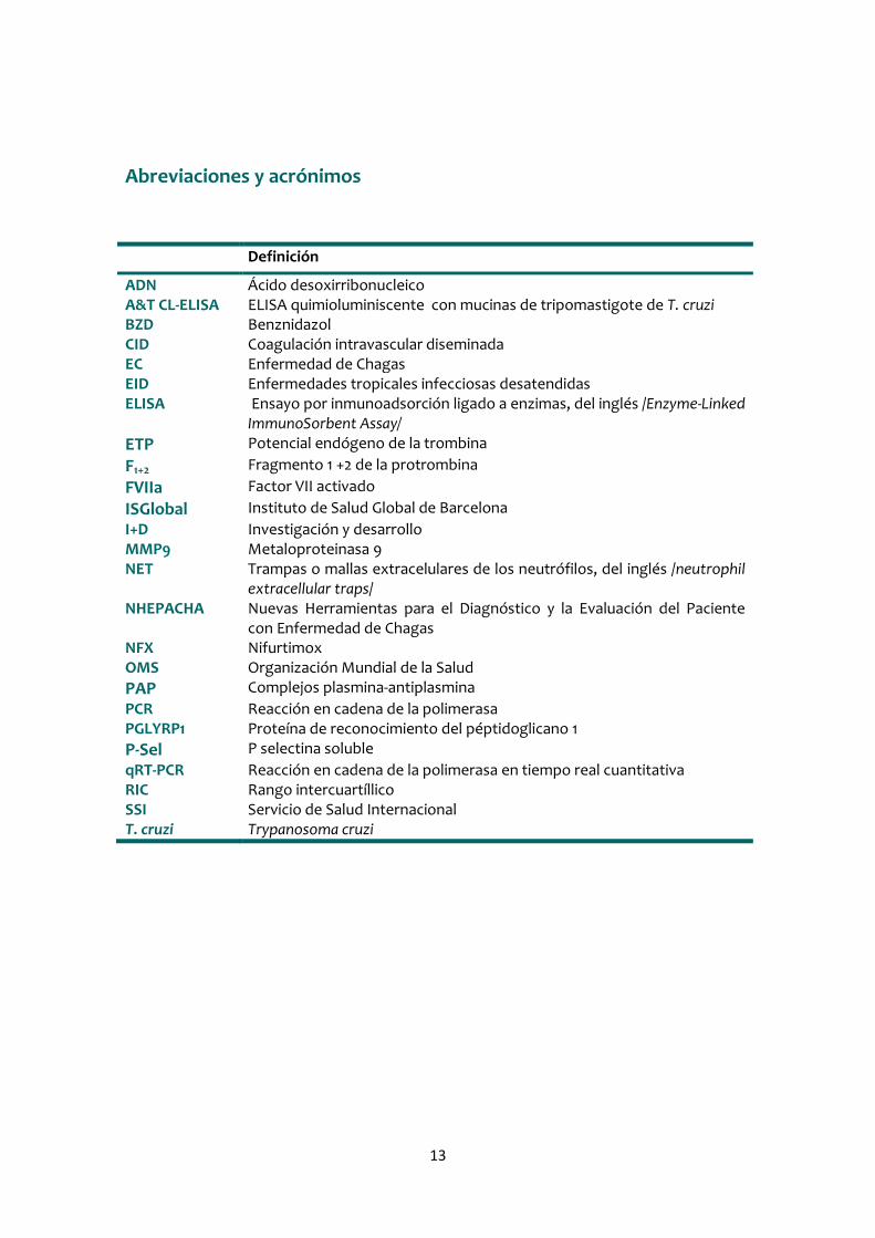
Abreviaciones y acrónimos Definición
ADN Ácido desoxirribonucleico A&T CL-ELISA ELISA quimioluminiscente con mucinas de tripomastigote de T. cruzi BZD Benznidazol CID Coagulación intravascular diseminada EC Enfermedad de Chagas EID Enfermedades tropicales infecciosas desatendidas ELISA Ensayo por inmunoadsorción ligado a enzimas, del inglés /Enzyme-Linked
ImmunoSorbent Assay/ ETP Potencial endógeno de la trombina F1+2 Fragmento 1 +2 de la protrombina FVIIa Factor VII activado ISGlobal Instituto de Salud Global de Barcelona I+D Investigación y desarrollo MMP9 Metaloproteinasa 9 NET Trampas o mallas extracelulares de los neutrófilos, del inglés /neutrophil
extracellular traps/ NHEPACHA Nuevas Herramientas para el Diagnóstico y la Evaluación del Paciente
con Enfermedad de Chagas NFX Nifurtimox OMS Organización Mundial de la Salud PAP Complejos plasmina-antiplasmina PCR Reacción en cadena de la polimerasa PGLYRP1 Proteína de reconocimiento del péptidoglicano 1 P-Sel P selectina soluble qRT-PCR Reacción en cadena de la polimerasa en tiempo real cuantitativa RIC Rango intercuartíllico SSI Servicio de Salud Internacional T. cruzi Trypanosoma cruzi


15
Resumen
Antecedentes y justificación
La eficacia del tratamiento de la infección crónica por Trypanosoma cruzi (T.
cruzi) no está bien definida debido a la carencia de marcadores precoces de
respuesta terapéutica. En este escenario, no existía un consenso en la definición de
las características exigidas a un potencial biomarcador de respuesta terapéutica
precoz al tratamiento de la infección por T. cruzi/enfermedad de Chagas (EC)
crónica.
Por otro lado, existen pacientes con infección crónica por T.cruzi y sin
miocardiopatía que han presentado fenómenos tromboembólicos, que podrían ser
explicados por la alteración de algunos factores de hipercoagulabilidad.
Con las dos premisas anteriores, se plantea un trabajo de tesis doctoral cuya
hipótesis es que existe un estado de hipercoagulabilidad en pacientes con infección
crónica por T.cruzi que se normaliza precozmente tras el tratamiento de la
enfermedad con benznidazol (BZD).
Para confirmar esta hipótesis, se establecen como objetivos por un lado,
definir las características que debe de cumplir un biomarcador de respuesta
terapéutica precoz; y por otro, detectar si existe un estado de hipercoagulbilidad en
pacientes con infección crónica por T.cruzi, cuáles son los factores concretos que
16
definen el estado de hipercoagulabilidad y evaluar su utilidad como marcadores de
respuesta terapéutica.
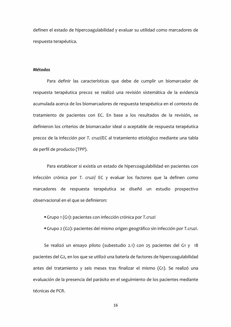
Métodos
Para definir las características que debe de cumplir un biomarcador de
respuesta terapéutica precoz se realizó una revisión sistemática de la evidencia
acumulada acerca de los biomarcadores de respuesta terapéutica en el contexto de
tratamiento de pacientes con EC. En base a los resultados de la revisión, se
definieron los criterios de biomarcador ideal o aceptable de respuesta terapéutica
precoz de la infección por T. cruzi/EC al tratamiento etiológico mediante una tabla
de perfil de producto (TPP).
Para establecer si existía un estado de hipercoagulabilidad en pacientes con
infección crónica por T. cruzi/ EC y evaluar los factores que la definen como
marcadores de respuesta terapéutica se diseñó un estudio prospectivo
observacional en el que se definieron:
Grupo 1 (G1): pacientes con infección crónica por T.cruzi
Grupo 2 (G2): pacientes del mismo origen geográfico sin infección por T.cruzi.
Se realizó un ensayo piloto (subestudio 2.1) con 25 pacientes del G1 y 18
pacientes del G2, en los que se utilizó una batería de factores de hipercoagulabilidad
antes del tratamiento y seis meses tras finalizar el mismo (G1). Se realizó una
evaluación de la presencia del parásito en el seguimiento de los pacientes mediante
técnicas de PCR.

17
En un segundo tiempo se aumentó el número de pacientes de cada grupo (56
en G1 y 43 en G2) (subestudio 2.2). Utilizando la misma metodología se realizó
determinación de los factores de hipercoagulabilidad de forma semestral durante
36 meses desde la finalización del tratamiento. Adicionalmente, se realizó
evaluación serológica de los pacientes mediante ELISA quimioluminiscente con
mucinas de tripomastigote de T. cruzi (A&T CL-ELISA), y detección de ácidos
nucleicos del parásito circulante mediante técnicas cuantitativas de reacción en
cadena de la polimerasa en tiempo real (qRT-PCR).
Resultados clave
En cuanto a los resultados del subproyecto 1, en la revisión sistemática se
detectaron varios grupos de moléculas que demostraron capacidad de detectar
respuesta al tratamiento, en diferentes estadios de desarrollo, y ninguna por sí sola
tiene la capacidad de evaluar la respuesta a corto plazo al tratamiento etiológico en
pacientes con infección por T. cruzi/EC en diferentes situaciones clínicas. Por el
momento, destacan para el uso en el seguimiento de pacientes después del
tratamiento las técnicas de amplificación de ácidos nucleicos del parásito.
Como resultado del análisis de los diferentes biomarcadores y en el contexto
de un trabajo colaborativo con un grupo de expertos en Chagas, se definió un perfil
de producto ideal/ aceptable para los biomarcadores de respuesta terapéutica en la
infección crónica por T. cruzi, aplicable en diferentes escenarios clínicos y
epidemiológicos.

18
Los resultados del subproyecto 2 mostraron inicialmente diferencias en los
valores de ETP (p 0.0001) y el F1+2 (p<0.0001) en los individuos de G1 y G2, siendo los
valores de los integrantes de G1 superiores al rango de la normalidad. Ambos
factores se normalizaron seis meses después del tratamiento en pacientes del G1
(ETP p<0.0008 y F1+2 p<0.004). Este hecho se confirmó con una mayor potencia en
con los resultados del subestudio 2.2, en el que se evidenció una alteración de los
valores de F1+2 (70% de pacientes de G1) y ETP (50% de pacientes de G1) por encima
de rangos normales, y diferentes estadísticamente de los valores de estos factores
en los pacientes del G2. Adicionalmente, después del tratamiento, el 76% de los
pacientes con alteración del F1+2, el 98% de los pacientes con alteración del ETP
normalizaron los valores en nueve (rango intercuartílico (RIC) 8) y seis meses (RIC 3)
de mediana respectivamente. Los valores se mantuvieron en rango normal sin
cambios estadísticamente significativos durante todo el periodo de seguimiento.
En cuanto a los valores de A&T CL-ELISA, si bien no se negativizaron durante
el seguimiento, se observó una reducción de los mismos a partir del mes 12, que fue
progresiva hasta el final del seguimiento y estadísticamente significativa desde el
mes 18 (p = 0,0052).
Conclusiones y recomendaciones
- Existen diversos biomarcadores en diferentes fases de estudio, que han
demostrado ser útiles para evaluar la respuesta al tratamiento en pacientes con
infección por T. cruzi en diferentes estadios, entre los que destacan las técnicas de
amplificación de ácidos nucleicos.

19
- La definición de los criterios de un biomarcador ideal o aceptable para la
evaluación del tratamiento específico de la infección por T. cruzi es indispensable
para estandarizar el desarrollo de estas moléculas y su uso en ensayos clínicos con
nuevos medicamentos.
- Existe un estado de hipercoagulabilidad en un porcentaje de pacientes con
infección por T. cruzi definido por la alteración del F1+2, del ETP.
- Los factores F1+2, ETP, y en menor medida el PAP, tienen un importante valor
como indicadores de respuesta precoz al tratamiento.
- De cara al futuro, se perfila el uso de una batería de biomarcadores que incluyan
moléculas dependientes del parásito, del hospedador y técnicas de amplificación de
ácidos nucleicos, para una evaluación precisa y temprana de la respuesta
terapéutica a la infección por T. cruzi/EC.

21
Summary
Background and rationale
The efficacy of specific treatment in people with chronic T. cruzi-infection has
not been established up to now, due to the lack of early markers of therapeutic
response. In this scenario, there was no consensus on the characteristics that a
potential early therapeutic response biomarker must or should fulfill in order to
evaluate specific treatment in people with T. cruzi-infection / Chagas disease (CD).
There are patients with chronic T. cruzi infection without cardiomyopathy
that presented thromboembolic events, which could be explained by the alteration
of several hypercoagulability factors.
Given the above two premises, the doctoral thesis hypothesis is that there is
a hypercoagulable state in patients chronically infected with T. cruzi which is
normalized soon after treatment of the disease with benznidazole (BZD).
In order to confirm the thesis hypothesis, the first objective was to define
the characteristics that a biomarker for early therapeutic response must fulfill
(ideally or acceptably); and the second objective was to identify if there is a
hypercoagulable state in patients chronically infected with T. cruzi, which specific
factors define the hypercoagulable state and to evaluate their usefulness as
markers of therapeutic response.
Methods
To define the characteristics required for a biomarker to asses early response
to treatment, a systematic review of the accumulated evidence on biomarkers of
22
therapeutic response in the context of treatment of patients with T. cruzi-
infection/CD was performed. On the basis of the review results, a Target Product
Profile (TPP) was developed to define an “ideal” or “acceptable” biomarker for
early therapeutic response of T. cruzi infection / CD to etiological treatment.
To establish whether there was a hypercoagulable state in patients with
chronic T. cruzi / EC and to assess the hypercoagulability factors that define this
state, and the usefulness as a biomarkers of therapeutic response, an observational,
prospective study was designed. In the study, there were two patients groups:
Group 1 (G1): patients with chronic T. cruzi infection
Group 2 (G2): patients from the same geographical origin without T. cruzi
infection.
A pilot trial (substudy 2.1) with 25 patients and 18 patients G1 and G2 respectively
was performed. A battery of tests for hypercoagulable factors before treatment in
both groups, and six months after treatment in G1 was done. To evaluate the
presence of the parasite, PCR techniques were included during the follow-up.
After substudy 2.1 results, the number of patients in each group was increased
(56 in G1 and 43 in G2) in order to increase the statistical power of the results. Using
the same methodology, test for hypercoagulability factors were performed every
six months for 36 months from the end of treatment. Additionally, serological
evaluation of patients was performed by ELISA with chemiluminescent
trypomastigote mucins of T. cruzi (A & T CL-ELISA), and detection of parasite nucleic
acids by quantitative techniques of real time polymerase chain reaction (qRT-PCR).
Key results
23
Subproject 1 systematic review showed that several groups of molecules
demonstrated an ability to detect response to treatment, but none of them had the
enough power to assess, by themselves, if a response to etiological treatment was
achieved in patients infected with T. cruzi / EC in different stages of the disease. To
date, only nucleic acid amplification techniques have shown their usefulness to
evaluate treatment failure and monitoring patients after specific treatment.
On the basis of review results, a Target Product Profile (TPP) was developed
to define an “ideal” or “acceptable” biomarker for early therapeutic response of T.
cruzi infection / CD to etiological treatment in the context of ongoing collaborative
work with a group of experts in Chagas, applicable in different epidemiological and
clinical scenarios.
The results of the subproject 2 initially showed differences in ETP (p 0.0001)
and F1 + 2 (p <0.0001) comparing individuals of G1 and G2, and G1 F1+2 and ETP
values were over the normal ranges. Both factors values normalized six months
after treatment in G1 patients of G1 (ETP p <0.0008 and F1 + 2 p <0.004). These
results were confirmed with the substudy 2.2 results. An alteration of F1 + 2 values
(70% G1 patients) and ETP values (50% G1 patients) were over normal range., and
statistically different from G2 patients values. Additionally, 76% of patients with
impaired F1+2, and 98% of patients with impaired ETP normalized values in nine
(interquartile range (IQR) 8) and six months (IQR 3) medium respectively after
treatment. After achieving normal ranges, values remained with no statistically
significant changes during the follow-up period.

24
A & T CL-ELISA levels remained positive during the whole follow-up, but a
progressive reduction was observed until the follow-up end, being statistically
significant from month 18 (p = 0.0052) on.
Conclusions and recommendations
- There are different biomarkers in different development phases that are
potentially useful in assessing response to treatment in patients with T. cruzi-
infection in different stages, especially nucleic acid amplification techniques.
- The criteria for an “ideal” or “acceptable” biomarker for the evaluation of the
specific treatment of T. cruzi infection biomarker is imperative to standardize the
development of these molecules and their use in clinical trials with new drugs.
- There is a hypercoagulable state in a percentage of patients with T. cruzi-infection,
defined by altering F1 + 2 and ETP.
- The F1 + 2, ETP (and PAP) factors have significant value as indicators of early
response to treatment.
- The use of a battery of biomarkers that include parasite molecules, host molecules
and nucleic acid amplification techniques, could be the future strategy for an
accurate assessment of therapeutic response to T. cruzi-infection /CD.

25
I. Introducción


27
I.a. Enfermedad de Chagas
La enfermedad de Chagas (EC), descrita por Carlos Chagas en 1909, es una de
las 17 enfermedades tropicales infecciosas desatendidas (EID), y afecta
principalmente a poblaciones que viven en condiciones socioeconómicas pobres y
con difícil acceso a servicios básicos y de salud. (1)
El agente etiológico responsable de esta enfermedad es Trypanosoma cruzi
(T. cruzi), (2) parásito flagelado hematófago endémico de 21 países en América.
Según datos de la Organización Mundial de la Salud (OMS), 100 millones de
personas están en riesgo de adquirir la infección, de las cuales unos 6 millones la
presentan actualmente.(3) Con una incidencia anual de 56.000 casos nuevos, la
enfermedad de Chagas se considera la principal causa de muerte en 12.000 personas
cada año.
En área endémica, la infección por T. cruzi se adquiere principalmente a
través de la vía vectorial. (2) La transmisión vertical es la segunda en frecuencia,
siendo la vía transfusional la tercera más común.(4) Ocasionalmente se han descrito
infecciones a través de manipulación en laboratorio de sangre contaminada con el
parásito, y trasplante de órganos. (5,6) Recientemente se han descrito varios brotes
de transmisión oral. (7, 8)
T. cruzi tiene un ciclo de vida complejo con varias etapas de desarrollo, tanto
en los mamíferos hospedadores como en los insectos vectores.(9) Los insectos
vectores toman tripomastigotes circulantes de sangre infectada de mamíferos

28
como zarigüeyas, gatos, perros, especies de murinos y humanos. Dentro de los
vectores, los tripomastigotes se transforman en epimastigotes flagelados con la
capacidad de multiplicación extracelular y migración al intestino posterior del
vector, convirtiéndose en tripomastigotes metacíclicos. Estos tripomastigotes
metacíclicos, con capacidad infecciosa, se pueden transmitir a los humanos y otros
mamíferos a través de las deyecciones del vector, en las cuáles se encuentran. Si las
heces son depositadas por parte del insecto en alguna zona del futuro hospedador
donde exista la posibilidad de penetración (solución de continuidad en la piel,
contacto con mucosas externas o mucosa gástrica), invaden células locales. Dentro
de las células locales que invaden, pierden el flagelo y se convierten en formas
amastigotes que se multiplican intracelularmente, rompiendo la célula huésped, y se
diseminan por vía hematógena como tripomastigote. (9) El paso del parásito por la
sangre periférica se llama parasitemia, y permite que el parásito pueda llegar a
órganos distantes y tejidos en los que pueden continuar en proliferación, causando
como resultado daño a los tejidos en los cuáles anida.
Tras la transmisión del parásito, en el 95% de las personas no existen
síntomas o estos pasan desapercibidos al ser síntomas inespecíficos. (2) Si no existe
un diagnóstico y tratamiento precoz, la infección se cronifica en la mayor parte de
los casos sin la aparición de síntomas durante décadas.(2) Sin embargo, y
generalmente de forma insidiosa, entre el 25-30% de las personas que padecen la
infección crónica por T. cruzi desarrollarán a lo largo de su vida miocardiopatía
chagásica, y de estas, el 50% se manifestará en formas severas (arritmias severas,
miocardiopatía dilatada y/o enfermedades tromboembólicas).(2,10) Las alteraciones
digestivas de la enfermedad, debidas a las alteraciones en los plexos nerviosos del

29
tracto digestivo, también son características de la forma crónica. Se observan en el
15-20% de las personas afectadas,(2) y clínicamente se manifiestan como
alteraciones de la motilidad, secreción y absorción de esófago, intestino delgado y
colon, llegando en ocasiones a provocar megaesófago o megacolon graves.(11,12)
En el caso de los pacientes inmunodeprimidos, el curso de la enfermedad puede ser
más severo, debido a la posibilidad de replicación masiva del parásito, y suelen
presentar por ello parasitemias más elevadas. (13)
El diagnóstico de la infección por T. cruzi en fase aguda o en pacientes
inmunodeprimidos que están en fase crónica, consiste en el aislamiento del parásito
en sangre mediante el examen directo o el cultivo. Las pruebas parasitológicas en la
fase crónica son de baja sensibilidad, por lo que el diagnóstico está basado en
pruebas serológicas, que son altamente sensibles, aunque no suficientemente
específicas debido a la presencia de reacciones cruzadas. Actualmente se considera
que para el diagnóstico serológico de T. cruzi se requieren dos pruebas positivas que
determinen diferentes grupos antigénicos. Hasta ahora, los antígenos utilizados son
de formas no infectivas de T. cruzi (epimastigote).(14)
En la actualidad, se dispone de dos fármacos para el tratamiento etiológico
de la infección por T. cruzi: benznidazol (BZD) y nifurtimox (NFX). Ambos
medicamentos presentan un perfil de toxicidad elevada,(15, 16) y los estudios de
eficacia, diseñados para la evaluación de estos fármacos, han mostrado resultados
muy variables dependiendo de la fase de la enfermedad, la dosis empleada, la edad
y la procedencia geográfica de los pacientes.

30
La enfermedad de Chagas: una enfermedad globalizada
Históricamente, la migración ha sido un factor clave en la diseminación de la
EC.(17) Este hecho continúa siendo vigente: los procesos migratorios desde las
zonas rurales a las zonas urbanas en áreas endémicas, en primer lugar, y más tarde
de América Latina a Norteamérica, Europa y el resto del mundo, han cambiado la
epidemiología de la infección por T. cruzi. (18) Se estima que en Europa hay entre
68.000 y 123.000 personas con la infección por T. cruzi, y la mayoría de ellos viven en
España. Sin embargo, hasta el año 2009 se ha reportado sólo 4.290 casos. (19, 20)
En estas regiones, donde la EC es una enfermedad emergente además de olvidada,
la transmisión vertical es la vía de transmisión más frecuentes, y la transmisión vía
transfusional también se ha documentado. (20, 21)
Por estas razones, ante la presencia de la infección en un nuevo escenario y
la posibilidad de transmisión en el mismo, la EC se considera actualmente un
problema de salud pública en áreas en las que previamente no existía.(17, 18)
En este contexto y considerando la EC como una enfermedad emergente
globalizada, se estima que la carga económica global de la EC es de unos 7 - 19 mil
millones de dólares al año, similar o superior a la de otras enfermedades como la
infección por rotavirus, o el cáncer de cuello uterino. (22) Sin embargo, la cantidad
de fondos para la investigación de la enfermedad de Chagas es de alrededor de 1%
de la financiación global de investigación y desarrollo (I + D). (23)

31
Limitaciones actuales para el manejo de una enfermedad olvidada.
Existe escaso conocimiento en cuanto a algunos aspectos fundamentales de
la infección por T. cruzi, como la interacción huésped-parásito y los mecanismos por
los cuáles la infección progresa a enfermedad. Este hecho, sumado a la falta de
interés de los decisores en salud e investigación de los países principalmente
afectados, ha dado como resultado que existan paradigmas antiguos en base a los
cuáles se ha limitado el acceso a tratamiento de las personas que padecen la EC.
La persistencia de las malas condiciones sociales, la transmisión vectorial y el
dominio de la teoría autoinmune, originaron durante décadas un descuido en el
tratamiento de pacientes crónicos y en la investigación de nuevas herramientas de
diagnóstico y seguimiento dirigidas al control de la progresión de esta enfermedad.
(24)
En cuanto al manejo de la EC, sobre todo en pacientes que se encuentran en
el estadio crónico, existen actualmente dos áreas en las que se hacen más evidentes
las lagunas en el conocimiento, ambas relacionadas con el tratamiento etiológico de
la infección. Este hecho repercute directamente en el acceso equitativo a la salud de
la población afectada, tanto en áreas endémicas como no endémicas.
De un lado, no ha habido desarrollo de nuevos medicamentos para el
tratamiento de la EC desde los años 70. Los fármacos actualmente usados para el
tratamiento de la infección por T. cruzi (BZD y NFX) tienen una alta incidencia de
reacciones adversas,(25-27) que si bien la mayoría son leves y pueden controlarse
32
satisfactoriamente con tratamiento sintomático, algunas de ellas progresan, y en
consecuencia los pacientes no finalizan la dosificación completa. (25,26) En este
sentido, en la última década se han diseñado ensayos clínicos con nuevos
medicamentos, y con nuevos regímenes de dosis y tiempo de prescripción en el
caso de los fármacos antiguos.(28, 29) La gran limitación para la evaluación de estos
nuevos medicamentos y/o dosificación de los antiguos, es el déficit de un parámetro
objetivable de respuesta precoz al fármaco por parte de los individuos que lo
reciben. Actualmente, la OMS establece como patrón de oro de respuesta al
tratamiento con BZD o NFX la seronegativización o seroconvesión, lo que ocurre
transcurridos entre ocho y diez años en personas con infección crónica por T. cruzi.
(30, 31)
Por otro lado, no existía un consenso en la definición de cuáles son las
características que un biomarcador de respuesta terapéutica precoz debe de
cumplir, en un contexto ambiguo que es la propia definición de respuesta
terapéutica.(30, 31)

33
I.b. Coagulación y enfermedades infecciosas
La hemostasia es el proceso biológico que previene el sangrado después de
un daño vascular, ya sea arterial o venoso. Implica la función de dos principales
componentes que actúan de forma coordinada: las plaquetas y el sistema de
coagulación.
El esquema tradicional de activación de la coagulación, basado en dos vías
separadas (vía intrínseca y extrínseca) que confluyen en una vía común y ésta en la
formación de trombina, actualmente no se considera ajustado a la realidad. En la
actualidad se considera un modelo de activación más complejo (32,33) que consta
de una fase inicial, que implica a los elementos celulares con capacidad de expresar
factor tisular (como los monocitos), y los factores VII, X, V y II. Esta fase inicial lleva a
la formación de una pequeña cantidad de trombina, la cual inicia la activación
‘explosiva’ de la coagulación (fase de amplificación) mediante la activación de las
plaquetas, que se convierten en el soporte necesario para la acción de los factores
IX, VIII, X, V y II dando lugar a la formación de gran cantidad de trombina.
Finalmente este proceso acaba en una fase en la que se impone el efecto de los
mecanismos inhibidores de la hemostasia. Los elementos celulares se acumulan en
el coágulo en formación y al mismo tiempo se incrementa en la misma zona la
concentración de factores procoagulantes, entre ellos la trombina recién formada, y
de algunos factores anticoagulantes o de la fibrinolisis. En el coágulo en formación,
los elementos celulares, especialmente las plaquetas, interaccionan con la fibrina
activándose y aportando los fosfolípidos necesarios para sustentar la actividad de
34
los factores de la coagulación. (34) Las plaquetas requieren del correcto
funcionamiento de sus glicoproteínas de membrana IIb-IIIa y Ib-alfa para dar
soporte a la generación de trombina, (35) al contrario de lo que sucede en la fase de
suspensión en la que únicamente se precisa de la acción glicoproteína IIb-IIIa. (36)
En los últimos años se ha demostrado la gran importancia de las micropartículas
circulantes en la iniciación y la amplificación de la coagulación. (37, 38) Su origen
está en los monocitos y en las células endoteliales que una vez activados o
lesionados pueden liberar fragmentos de membranas (microvesículas) de tamaño
inferior a 1-1,5 μm que llevan en su superficie proteínas de membrana de las células
de origen. En la hemostasia, el factor tisular es la principal de estas proteínas
expresadas por las micropartículas, siendo el presente en las micropartículas
circulantes la principal fuente del mismo en la formación de un coágulo normal. A
este esquema se ha venido a sumar más recientemente la activación de la
coagulación por la vía de la acción de los neutrófilos, como se verá en el siguiente
apartado.
Interacción del agente infeccioso-sistema de coagulación
La trombosis se considera una desviación de la hemostasia fisiológica, y es
actualmente la principal causa de mortalidad a nivel mundial (> 2.000 personas/
día).(39) Pese a esto, recientes hallazgos sugieren que bajo ciertas circunstancias la
microtrombosis es un proceso fisiológico, que constituye un mecanismo efector
intrínseco de la inmunidad innata, y a este proceso se le denomina
“inmunotrombosis”. La inmunotrombosis se define como la respuesta inmunitaria
inducida por la formación de un trombo venoso, especialmente en la
35
microvasculatura. (40-42) Se considera un proceso crucial de la inmunidad
intravascular. (43)
Los agentes infecciosos desencadenan a menudo los procesos de
inmunotrombosis, mediante los cuales, se reconoce al patógeno en cuestión y a las
células dañadas por el mismo, y se inhibe la difusión del microorganismo mediante
su eliminación.(43) Diferentes parásitos estimulan diferentes mecanismos de
respuesta inmune, que pueden implicar tanto las respuestas humorales y celulares.
La eficacia de la respuesta inmune depende del agente infeccioso y la fase de la
infección en la que se encuentre. La resistencia contra la infección podría ser
regulada por las características genéticas del huésped, pero la persistencia de la
infección es producida por una suma de factores en función de ambos, el parásito y
el huésped.(44)
La coagulación tiene un papel fundamental en la respuesta a la exposición a
patógenos infecciosos.(38,45) La fibrina puede ejercer acción antimicrobiana
directa.(46) La fibrina y/o su precursor, el fibrinógeno, se unen y activan las células
inmunitarias innatas, tales como neutrófilos, en los lugares donde se produce la
infección.(47,48) Este hecho sugiere que además de la función hemostática, la
coagulación juega un papel directo en la respuesta del hospedador al agente
infeccioso.
En el lugar donde se produce el daño, se forman microtrombos que actúan
como matrices antimicrobianas, mediando la respuesta inmunológica del
hospedador frente a agente infeccioso.(49) La inmunotrombosis se activa y

36
mantiene gracias a la acumulación local de células de respuesta inmune innata,
especialmente monocitos y neutrófilos . (50)
En este punto, es importante destacar la descripción durante los últimos
años de fibras o redes extracelulares generadas por los neutrófilos llamadas
neutrophil extracellular traps (NET), las cuales están compuestas de un esqueleto
de ADN sobre el que se encuentran diversos componentes citoplásmicos –entre
ellos diversas enzimas– y nucleares. Las NET son una barrera física que en muchos
casos evita la diseminación de los microorganismos, e incluso facilita su muerte al
favorecer una alta concentración local de moléculas antimicrobianas, incluyendo
mieloperoxidasas, elastasas, lactoferina, metaloproteinasa 9 (MMP9) y la proteína
de reconcimiento del péptidoglicano 1 (PGLYRP1).(51-54) Por otro lado, su
estructura fibrosa limita el daño al tejido donde se generan, al restringir el radio de
acción de las moléculas que son liberadas por el neutrófilo.
Los NETS inducen adicionalmente una intensa respuesta procoagulante, de
un lado, atrapando las plaquetas activadas, y de otro, mediante la actividad
proteolítica de la neutrófilo elastasa.
No obstante, una alteración de la immunotrombosis puede constituir un
evento clave en el desarrollo de trastornos trombóticos, incluyendo infarto de
miocardio, accidente cerebrovascular y la coagulación intravascular diseminada
(CID).
En resumen, la presencia de microorganismos dentro de los vasos
sanguíneos es una gran amenaza para el organismo huésped, ya que la circulación
de patógenos puede infectar simultáneamente varios órganos que son cruciales

37
para la supervivencia. La immunotrombosis en este escenario es un mecanismo de
defensa antimicrobiana intravascular que evita la difusión e invasión del agente
infeccioso en los tejidos diana. La implicación de mediadores celulares y moleculares
de la inmunotrombosis (por ejemplo, neutrófilos y factor tisular respectivamente)
conlleva el riesgo de que un desequilibrio en la activación de los mismos puede dar
lugar a trastornos trombóticos y activación aberrante de la coagulación, por
ejemplo, por una exposición mantenida y no controlada al agente infeccioso.
En este punto, el conocimiento en profundidad de los procesos
fisiopatogénicos de los diferentes agentes infecciosos y la forma en la que los
mismos pueden alterar el equilibrio de la coagulación, facilitará el desarrollo de
nuevas herramientas para su diagnóstico y tratamiento precoz de la enfermedad en
sí, previniendo eventos trombóticos.
Alteración de la hemostasia en la infección por T. cruzi
La enfermedad tromboembólica asociada a la enfermedad de Chagas es bien
conocida,(55) e históricamente se ha relacionado con la cardiopatía chagásica y la
presencia de dilatación de las cavidades cardíacas, aneurismas ventriculares y
trombosis intracavitaria,(56, 57) ya que estos condicionarían factores reológicos
que favorecen la formación de trombos intraluminales con capacidad embolígena.
Sin embargo, en los últimos años se ha postulado que otros factores podrían influir
en la génesis de la enfermedad tromboembólica, ya que se han descritos eventos en
personas con infección por T. cruzi pero sin miocardiopatía chagásica u otros
factores de riesgo vascular.(58,59)

38
Adicionalmente a lo expuesto en el epígrafe anterior, el proceso inflamatorio
asociado a la infección puede causar vasculitis generalizada con aumento de los
niveles de factores endoteliales mediante la expresión de citocinas
proinflamatorias.(60) Al adquirir la infección por T. cruzi, existe una respuesta
inflamatoria inicial, en la cual macrófagos, linfocitos y células porlimorfonucleares
son reclutadas como resultado de la ruptura de células infectadas y hay una gran
cantidad de sustancias proinflamatorias secretadas.(61) En respuesta a la
inflamación se producen, además, fenómenos de adhesión plaquetaria. El aumento
de la viscosidad sanguínea, las microembolias plaquetarias y la activación
leucocitaria pueden producir disminución del flujo circulatorio provocando
enlentecimiento del flujo sanguíneo y con ello, infarto de los tejidos en los que se
encuentran. Por todo lo anterior, pacientes en un estado de inflamación crónica,
como el que ocurre en la infección crónica por T. cruzi, presentan un mayor riesgo
de padecer fenómenos trombóticos, tanto por el estado inflamatorio crónico al que
se ven sometidos, como por los fenómenos de inmunotrombosis que desencadena
la inflamación crónica descritos anteriormente.
En estudios experimentales de infección por T. cruzi en modelos murinos se
observan cambios en la viscosidad sanguínea secundarios a la respuesta
inmunológica del huésped,(55) tanto por la presencia del parásito como por el daño
que éste produce en el endotelio (55) mediante la secreción de neuraminidasas que
afectan al ácido siálico de las células infectadas.(56) Sin embargo, estudios en seres
humanos muestran resultados controvertidos en cuanto a la existencia de un
estado de hipercoagulabilidad en personas con infección por T. cruzi, y si existe, si
este estado puede ser calificado como factor de riesgo vascular.(62-64)

39
En estudios preliminares a esta tesis doctoral se describieron alteraciones de
algunos marcadores biológicos de hipercoagulabilidad (F1+2 de la protrombina,
complejos trombina-antitrombina, fibrinógeno / fibrina productos de degradación y
el dímero D) en pacientes de infección por T. cruzi, incluso en las etapas tempranas
de la enfermedad.(63,64) Resultados contrarios fueron obtenidos en el estudio de
Carod-Artal y cols, en el que no se halló diferencia entre los factores trombofílicos
estudiados en pacientes con y sin infección por T. cruzi (proteína S, antitrombina,
proteína C activada, factor V Leiden, anticoagulante lúpico y anticuerpos
anticardiolipina) en individuos infectados por T. cruzi y no infectadas, y considera
que el estado procoagulante no es un factor de riesgo isquémico en estos
pacientes.(62)
En este punto, en el estado de conocimiento actual de los factores que
pueden producir un desequilibrio en el sistema de coagulación, y su relación con la
respuesta inmunológica innata que se desencadena ante la presencia de un agente
infeccioso, la hipótesis planteada para el desarrollo de esta tesis doctoral toma
mayor sentido.
I.c. Biomarcadores de respuesta terapéutica en la infección por T. cruzi
Un biomarcador se define como un signo que se puede medir con precisión
y que es reproducible, y refleja el estado puntual de un proceso de la enfermedad.
Los biomarcadores se correlacionan, ya sea inversa o directamente con la
progresión de la enfermedad en sí. Un marcador o biomarcador subrogado se
define como el signo que se utiliza en los ensayos terapéuticos para medir de forma

40
directa una función biológica concreta, cómo responde clínicamente el paciente, su
supervivencia y/o como predictor de la respuesta al tratamiento en sí. (65,66)
En el caso de la EC crónica, la identificación de biomarcadores precoces de
respuesta terapéutica es indispensable para mejorar la atención integral de los
pacientes.
Como se indicaba en el apartado I.a, la carencia de marcadores de eficacia
terapéutica limita el tratamiento en dos aspectos fundamentales. En primer lugar,
en el contexto del manejo del paciente, dado que actualmente no existen
herramientas mediante las cuáles podamos predecir la progresión de la enfermedad
o evaluar la respuesta óptima al tratamiento a corto plazo. La eficacia de un
fármaco no puede ser actualmente evaluada en un corto período de tiempo
después del tratamiento en pacientes con infección crónica por T. cruzi, ya que las
técnicas de biología molecular no dan hasta el momento una respuesta absoluta a si
existió eliminación del parásito y cuál es el significado real de esta eliminación en
caso de haberse dado.(67-70)
En segundo lugar, el desarrollo o identificación de biomarcadores de
respuesta terapéutica en el contexto de la EC es indispensable como herramienta
para el diseño e implementación de ensayos clínicos con fármacos más seguros y
eficaces.
Es evidente que existe la necesidad de desarrollo de marcadores de
progresión y pronóstico de la EC, sobre todo en su fase crónica. Debido a ello,
durante las últimas décadas se han identificado biomarcadores que están
actualmente en diferentes puntos del proceso de validación.
41
En este contexto, los biomarcadores detectados en la EC pueden ser
moléculas del parásito o del hospedador. En relación a las moléculas parasitarias, se
han identificado fundamentalmente proteínas (antígenos) y se han desarrollado
técnicas de amplificación de ácidos nucleicos. Con respecto a los biomarcadores del
hospedador, existen dos grandes grupos de biomarcadores: por un lado, los
relacionados con la respuesta inmunológica del hospedador (citocinas y marcadores
celulares de superficie); por otro lado, los relacionados con el proceso inflamatorio y
las alteraciones metabólicas que produce una inflamación crónica en el organismo.
Las referencias bibliográficas de cada uno de los biomarcadores constan en el
primer artículo que compone esta tesis.
Pese a que existe una amplia propuesta de moléculas, no se ha llegado, hasta
el momento, a una definición consensuada de las características que debe de
cumplir un biomarcador de respuesta terapéutica en pacientes con infección
crónica por T. cruzi. Este hecho ha sido la causa de que muchos de los ensayos
llevados a cabo con las moléculas seleccionadas no hayan sido homogéneos ni
respondan, en muchos de los casos, a unos criterios básicos.
Durante el presente trabajo de tesis, esta limitación se hizo evidente tras
completar el subestudio piloto (2.1), ya que inicialmente la investigación consistía en
probar algunos de los factores de hipercoagulabilidad como biomarcadores de
respuesta terapéutica en pacientes con infección crónica por T. cruzi o EC crónica.
En este punto, y en el marco del trabajo colaborativo en una red de
investigación internacional en nuevas herramientas de manejo para la enfermedad
de Chagas (Red NHEPACHA), se planteó el primero de los subproyectos de que
consta este trabajo de tesis, sin el cual no se hubiera podido avanzar en las

42
conclusiones del subestudio 2.2. Se realizó una revisión sistemática de la evidencia
actual con el fin de evaluar moléculas como posibles biomarcadores de respuesta
terapéutica y sus características de una etapa específica de la enfermedad, y se
estableció la definición de biomarcador de respuesta terapéutica al tratamiento
específico de pacientes con infección crónica por T. cruzi.

43
II. Hipótesis


45
Hipótesis
La hipótesis de este proyecto es que existe un estado de hipercoagulabilidad
en pacientes con infección crónica por T. cruzi que se normaliza de forma precoz
tras el tratamiento etiológico de la enfermedad con benznidazol.
Si esta hipótesis se confirma, tales marcadores de hipercoagulabilidad
podrían ser propuestos como biomarcadores de respuesta precoz al tratamiento
antiparasitario en pacientes con enfermedad de Chagas crónica.
Para dar respuesta a esta hipótesis, se diseñaron dos subproyectos. El
primero de ellos consistió en realizar una revisión sistemática para definir las cuáles
son los biomarcadores de respuesta al tratamiento etiológico de pacientes con
enfermedad de Chagas crónica que se han desarrollado hasta el momento, y definir
las características que debe de cumplir un biomarcador para ser utilizado con este
fin.
El segundo subproyecto consiste en la detección de marcadores de
hipercoagulabilidad en una cohorte prospectiva de pacientes, comparado con
individuos sanos procedentes de áreas endémicas para la enfermedad. Este
segundo subproyecto se dividió en dos partes: a) un estudio piloto con seguimiento
a corto plazo (seis meses) de las personas con infección por T. cruzi que habían
realizado tratamiento; y b) una cohorte que ampliaba el estudio piloto en cuanto a
número de participantes y tiempo de seguimiento (36 meses).


47
III. Objetivos

49
III.a. Subproyecto 1
1.- Evaluar los biomarcadores que existen de respuesta terapéutica a benznidazol en
pacientes con enfermedad de Chagas crónica.
2.- Establecer un perfil de producto ideal/ aceptable como marcador de respuesta
terapéutica en la infección crónica por T. cruzi, aplicable en diferentes escenarios
clínicos y epidemiológicos.
III.b. Subproyecto 2
III.b.1. Subestudio 2.1
1.- Determinar si existe un estado de hipercoagulabilidad en pacientes con infección
crónica por T. cruzi, y definir cuáles son los parámetros de coagulación que lo
determinan.
2.- Determinar si existe normalización de los parámetros que marcan este estado a
los seis meses de finalizar el tratamiento con benznidazol.
III.b.2.Subestudio 2.2
1.- Determinar si existe un estado de hipercoagulabilidad en pacientes con infección
crónica por T. cruzi, y definir cuáles son los parámetros de coagulación que lo
definen.
2.- Determinar si existe normalización de los parámetros que marcan este estado
entre los seis y doce meses tras finalizar el tratamiento con benznidazol.

50
3.- Determinar si la normalización precoz de estos parámetros se mantiene en el
tiempo y establecer su utilidad como marcadores precoces de respuesta
terapéutica.
4.- Establecer la utilidad del ELISA quimioluminiscente ELISA con mucinas de
tripomastigote de T. cruzi (A&T CL-ELISA) como marcador de evolución de la
enfermedad para valorar respuesta a tratamiento etiológico, y ver la coherencia
entre la evolución de los niveles de los títulos de este ELISA con los niveles de los
parámetros de hipercoagulabilidad estudiados a lo largo del tiempo después del
tratamiento, y con los análisis de qRT-PCR como parámetro de respuesta al
tratamiento sostenida en tiempo.

51
IV. Material y métodos


53
IV. Material y métodos
Los trabajos incluidos en esta tesis doctoral han sido coordinados desde el
Servicio de Salud Internacional (SSI) del Hospital Clínic de Barcelona. El SSI tiene una
amplia experiencia en el manejo de patología importada. Es un centro de referencia
nacional y con reconocimiento a nivel internacional en el manejo e investigación de
enfermedades desatendidas. La investigación del SSI se canaliza a través del
Instituto de Salud Global de Barcelona (ISGlobal), dónde se ha creado la Iniciativa
sobre la enfermedad de Chagas (http://www.isglobal.org/web/guest/chagas).
El primero de los subproyectos se ha realizado en el contexto de una red de
investigación internacional mediante la cual se establecen trabajos colaborativos
para el diseño y la validación de nuevas herramientas para el diagnóstico y manejo
de la enfermedad de Chagas (Red NHEPACHA).
IV.a. Población de estudio
El subproyecto 1 se basó en una revisión sistemática de la literatura.
En el caso del subproyecto 2, la población de estudio fueron pacientes
procedentes de áreas en que la infección por T. cruzi es endémica, que acudieron al
SSI del Hospital Clínic de Barcelona para cualquier tipo de consulta en el periodo de
reclutamiento de cada uno de los subestudios 2.1 y 2.2, y que no presentaron ningún
criterio de exclusión. Se excluyeron pacientes que presentaban comorbilidad
importante: diagnóstico previo de cardiopatía de otra etiología (isquémica,
alcohólica o hipertensiva), enfermedades inflamatorias o inmunológicas sistémicas

54
o infecciones activas por otro agente causal. Se excluyeron pacientes con infección
por T. cruzi que habían recibido previamente tratamiento antiparasitario para la
infección por T. cruzi (BZD y/o NFX).
Se definieron dos grupos:
Grupo 1 (G1): pacientes con infección crónica por T. cruzi/ enfermedad de
Chagas en cualquiera de sus fases (crónica indeterminada o crónica sintomática).
Grupo 2 (G2): pacientes sin infección por T. cruzi de zona endémica para esta
enfermedad.
En el subestudio 2.1, la población de estudio fue de un total de 25 pacientes del
G1 y 18 pacientes del G2. En base a los resultados del subestudio 2.1, se aumentó el
tamaño de muestra a 56 pacientes en G1 y de 43 pacientes en G2.
Todos estos pacientes estuvieron de acuerdo en participar en los estudios, y
firmaron un consentimiento informado específico tras la explicación en profundidad
de los objetivos y procedimientos de los estudios a los que aceptaban entrar.
IV.b. Metodología de los estudios
La tesis está estructurada en base a dos subproyectos.
El subproyecto 1 es una revisión sistemática de la evidencia acumulada
acerca de los biomarcadores de respuesta terapéutica en el contexto de
tratamiento de pacientes con enfermedad de Chagas. Para ello, se definieron tres
áreas de análisis en las que se dividieron las publicaciones incluidas: biomarcadores
inmunológicos, biomarcadores bioquímicos y técnicas de amplificación de ácidos

55
nucleicos. En base a los resultados de la misma, se definieron los criterios de
biomarcador ideal o aceptable de respuesta terapéutica precoz de la infección por
T. cruzi al tratamiento etiológico.
El subproyecto 2 fue un estudio prospectivo observacional en el que se
definieron dos grupos: Grupo 1 (G1), pacientes con infección crónica por T. cruzi/
enfermedad de Chagas en cualquiera de sus fases (crónica indeterminada o crónica
sintomática); Grupo 2 (G2), pacientes sin infección por T. cruzi de zona endémica
para esta enfermedad. Una vez establecidos los grupos, a los pacientes del G1 se les
ofreció tratamiento con BZD (cinco mg/ kg/ día por 60 días). Durante el tratamiento
se realizó seguimiento quincenal clínico y analítico (hemograma completo, y
bioquímica con perfil hepático y renal) de estos pacientes para el monitoreo y
control de efectos adversos secundarios a este fármaco.
Este estudio se realizó en dos tiempos:
El subestudio 2.1 fue un ensayo piloto con 25 pacientes del G1 y 18 pacientes
del G2. Se realizó una evaluación de los pacientes del G1 a los seis meses post-
tratamiento, en la que se incluyeron pruebas serológicas convencionales y PCR para
T. cruzi y la determinación de los factores de hemostasia e hipercoagulabilidad.
En base a los resultados del proyecto piloto, se realizó el subestudio 2.2,
mediante el cual se aumentó el tamaño de muestra y se alargó el periodo de
seguimiento de los pacientes, obteniéndose una muestra final de 56 pacientes en
G1 y de 43 pacientes en G2. En los pacientes del G1 se realizó un seguimiento
semestral de 36 meses post-tratamiento con la finalidad de establecer si las
modificaciones de los factores de hipercoagulabilidad observadas eran mantenidas

56
en el tiempo. Durante el seguimiento de la cohorte completa, además de las
pruebas realizadas al grupo de pacientes del proyecto piloto, se realizó ELISA
quimioluminiscente con mucinas de tripomastigote de T. cruzi (A&T CL-ELISA).
La metodología específica de cada uno de los subproyectos está detallada en
los artículos publicados e incorporados a la presente tesis.

57
V. Resultados


59
ARTÍCULO 1
Biological markers for evaluating therapeutic
efficacy in Chagas disease, a systematic review
María-Jesús Pinazo, Maria Carmen Thomas, Jacqueline Bua, Alina Perrone, Alejandro-Gabriel Schijman, Rodolfo-Jorge Viotti, Janine-M Ramsey, Isabela Ribeiro, Sergio Sosa-Estani, Manuel-Carlos López, Joaquim Gascon
Expert Rev Anti Infect Ther. 2014; 12(4), 479–496
Factor de impacto 2014: 3.461


Biological markers forevaluating therapeuticefficacy in Chagas disease,a systematic reviewExpert Rev. Anti Infect. Ther. 12(4), 479–496 (2014)
Maria-Jesus Pinazo*,M Carmen Thomas,Jacqueline Bua,Alina Perrone,Alejandro-GabrielSchijman,Rodolfo-Jorge Viotti,Janine-M Ramsey,Isabela Ribeiro,Sergio Sosa-Estani,Manuel-Carlos Lopezand Joaquim Gascon
*Author for correspondence:
Tel.: +34 932 275 400
Fax: +34 932 279 853
For a full list of author affiliations,
please see page 496.
The most neglected aspects of Chagas disease (CD) have been patient care and treatment.Despite recent progress in the development of potentially improved drugs, there is noconsensus among different research groups on the lack of therapeutic response markers toevaluate efficacy of newly proposed drugs early after treatment. A systematic review ofcurrent evidence regarding molecules which are potential biomarkers for therapeutic responsehas been conducted using quality assessment and target responses as primary criteria. Thereview provides a panorama of the cumulative evidence and specific needs for developmentof a battery of complementary biomarkers which together fulfill ideal or acceptable criteria toevaluate early responses to treatment for chronic CD. There are several marker candidateswhich together may fulfill acceptable criteria to indicate the efficacy of a trypanocidaltreatment. Data from ongoing studies are considered essential to improve assessment ofexisting markers and to identify those for early follow-up of treated patients.
KEYWORDS: biological marker • biomarker • Chagas disease • cure marker • humoral and cellular immune response
• PCR • treatment • Trypanosoma cruzi
A lack of appropriate clinical and biomarkertools limits the direct measurement of treat-ment impact for any infectious disease. This isa common limitation for many of the 17 inter-nationally recognized neglected diseases. Onestriking example is Chagas disease (CD), anendemic zoonosis caused by the protozoanparasite Trypanosoma cruzi, which affects 7–8million people currently, not only in LatinAmerica where the disease is autochthonous [1]
but also is worldwide due to populationmigrations [2,3].
Despite the burden of CD morbidity andlower cost of timely diagnosed and treatedpatients [4], only two drugs, benznidazole andnifurtimox, are currently available for treat-ment. Both drugs have variable efficacydepending on the disease stage, drug dose,patient age and geographical origin. T. cruziinfection treatment is currently strongly rec-ommended in both acute and chronic stagesof the infection [5–7]. The association ofT. cruzi infection and disease progression, and
therefore its clinical cure, remains unclearmainly because physiopathological changesdevelop slowly and symptoms may appear sev-eral years after infection. Disease symptomsmay appear when there is an imbalancebetween the host immune response and para-site proliferation in tissues. Along with tissuedamage caused by the presence and persistenceof the parasite [8], there are inflammatory pro-cesses and cross-reactivity with host mole-cules [9]. Currently, the association of parasitepersistence and symptom development is arecurring controversy.
In the chronic stage, drug therapy efficacy isvariable and is difficult to compare since moststudies use different treatment regimens andresponse assessment methods (variable assays,frequency and duration of follow-up) [10], inaddition to the lack of class I studies [11,12].Even in successful treatment, the gold standardfor evaluating efficacy (seroconversion usingconventional serological tests) may take yearsto decades to assess [13,14]. Hence, long-term
informahealthcare.com 10.1586/14787210.2014.899150 � 2014 Informa UK Ltd ISSN 1478-7210 479
Review
Exp
ert R
evie
w o
f A
nti-
infe
ctiv
e T
hera
py D
ownl
oade
d fr
om in
form
ahea
lthca
re.c
om b
y 20
0.58
.76.
155
on 0
3/12
/14
For
pers
onal
use
onl
y.

treatment of chronic CD cases has been neglected in two pri-mary areas:
• A lack of interest since the 1970s to develop new drugs spe-cifically targeting intracellular parasites in general and forT. cruzi in particular. Fortunately, in the last 5 years, newdrugs or new schemes of current drugs have been proposedas potential alternatives and are being tested in differentphases of development for safety and efficacy against T. cruziin humans.
• A lack of consensus regarding therapeutic response markersfor early assessment of antitrypanocidal drug efficacy for dis-ease management and of clinical trials with new drugs forT. cruzi treatment.
Several prognosis and progression markers for T. cruzi infec-tion have been proposed during the last 20 years. However,only a few of these have been evaluated using appropriatelydesigned studies for therapeutic response. In order to assessmolecules as potential biomarkers for therapeutic response andtheir disease stage-specific characteristics, a systematic review ofcurrent evidence was conducted focusing on the quality of bio-marker studies in both aspects. Only studies assessing biologicalmarkers (immunological, biochemical and molecular bio-markers or nucleic acid-based biomarkers) during treatmentfollow-up of chronic T. cruzi patients have been included. Onthe basis of review of current evidence, a target product profile(TPP) was developed for an ‘ideal’ and/or an ‘acceptable’ bio-marker for anti-T. cruzi treatment response in different epide-miological and clinical scenarios.
MethodsThe reviewed publications addressing CD-specific treatmentresponse markers were organized into three areas of analysis:
immunological markers, biochemical biomarkers and nucleicacid amplification strategies.
Acquisition of evidence
The literature was reviewed based on electronic searches in TheCochrane Central Register of Controlled Trials on TheCochrane Library, NLM GATEWAY (PubMed/MEDLINE,Clinicaltrials.gov, Bookshelf and Meeting Abstracts), WHOLIS,BVS (BIREME and LILACS), SCIELO (1990–2012). Theindexing terms used in the searches are presented in TABLE 1.
A secondary search was performed using the first or lastarticle’s author or the marker under analysis as keywords AND‘CD’ OR ‘Chagas’ AND ‘treatment’, AND ‘human’, AND‘patient’, OR ‘patients’.
We considered reports only of original research, mainly but notexclusively intervention trials, specificity of diagnostic methods andobservational studies, with scope targeted at biomarkers of treat-ment response for patients with chronic CD. Articles published inSpanish, English and Portuguese from 1990 through 31 December2012 were reviewed according to the following inclusion criteria:
• Studies concerning development, standardization and/or vali-dation of:
– Biochemical biomarkers– Immunological biomarkers– Nucleic acid amplification strategies
• The aim of the study was to:
– Monitor antiparasitic treatment– Test the response to antiparasitic treatment or to testing
antiparasitic treatment outcomes
• Studies performed in humans
Table 1. Keywords used for literature searches using the NLM gateway.
Category MeSH term MeSH subheading
Response to treatment by biochemical biomarkers CD OR Trypanosoma cruzi AND biological markers AND treatment
AND cure markers AND treatment
AND biomarker AND treatment
Response to treatment by immunological biomarkers CD OR T. cruzi AND biological markers
AND biomarkers
AND immunological markers
AND biological markers AND treatment
AND biomarkers AND treatment
AND immunological markers AND treatment
AND biological markers AND immunology
AND biomarkers AND immunology
AND immunological markers AND immunology
PCR for the evaluation of treatment CD AND polymerase chain reaction
CD: Chagas disease.
Review Pinazo, Thomas, Bua et al.
480 Expert Rev. Anti Infect. Ther. 12(4), (2014)
Exp
ert R
evie
w o
f A
nti-
infe
ctiv
e T
hera
py D
ownl
oade
d fr
om in
form
ahea
lthca
re.c
om b
y 20
0.58
.76.
155
on 0
3/12
/14
For
pers
onal
use
onl
y.

Articles that had the following characteristics were excluded:
• Studies previous to 1990• Studies performed in experimental models or PCR studiesusing vector samples
• Studies not specifically designed to evaluate treatment impactby specific biomarkers and/or PCR techniques
• Studies that evaluated response to antiparasitic treatment withimage techniques (x-ray, echocardiography, scintygraphy, etc.)
• Nucleic acid amplification studies, if the purpose of the tech-nique was genotyping, DNA cloning or genomics and retro-transcription–PCR assays for gene expression.
Systematizing results
A matrix was constructed with the following information cate-gories for each biomarker and article: which test was evaluated,distribution by sex, age of study population, geographical area,times of study patients follow-up, sample size, missing data,study design, stages of the disease included in the study, treat-ment (drug, dose, length of treatment), reference test per-formed, values of the reference standardized test, dispersionvalues of the standardized test, sensitivity and specificity of thebiomarker, biomarker efficacy evaluation and study biases(TABLE 2). The main characteristics and limitations of each bio-marker are highlighted.
Results of the searchesThe results of the searches are summarized in FIGURE 1 and areview of the titles, abstracts and in some cases the full textswere examined to select relevant papers for the review. A moredetailed review and data collection from each study were con-ducted after reviewing title and/or abstracts, in order to evalu-ate its relevance.
Evidence synthesisThe results of the searching have been summarized in TABLE 2.
Analysis of the results by type of molecules & technique
Immunological molecules
Four categories of 25 markers have been used to measure thera-peutic efficacy for CD. The first group includes four markersthat detected specific antibodies for host antigens. The secondgroup (14 markers) involves methods to detect antibodies gen-erated against parasite antigens. The third group includes thosethat measure the cytokine level and/or cytokine pattern in apatient’s serum (three markers), and a fourth group includesfour markers to quantify cellular immune response populationsor populations expressing specific cytokines.
Host antigens
Several human antigens have been proposed as biomarkers oftreatment response in CD. Increased levels of sP-selectin andsoluble vascular cell adhesion molecule ‘1’ (sVCAM-1) havebeen observed in 41 asymptomatic chronic CD pediatricpatients. Before treatment, 83 and 71% of these exceeded the
cut-off control value for sP-selectin and sVCAM-1, respectively.There was a significantly greater decrease in the titers ofsP-selectin (66.7%) and sVCAM-1 (41.0%) in those childrenwho received benznidazole therapy compared with a controlgroup receiving placebo [15].
Levels of anti-R3 antibodies, a peptide encoded in thehuman autoantigen ‘Cha’, increased with the progression ofclinical manifestations of chronic CD. Anti-R3 antibody titersdecreased in 19 patients treated with antiparasitic drugs (benz-nidazole or nifurtimox), despite the fact that all had highertiters than those observed in healthy donors [16].
The production of anti-M2 muscarinic receptor autoantibod-ies (anti-M2R Ab) and IFN-g profiles was characterized in 30T. cruzi-infected children in the early stage of chronic CD,before and after trypanocidal benznidazole chemotherapy [17].Before treatment, anti-M2 receptor autoantibodies weredetected in 56% of T. cruzi-infected patients and none of the19 uninfected control subjects. Infected children also exhibiteda significantly higher serum IFN-g level than that observed inhealthy controls. At 6 months post-treatment with benznida-zole, there was a significant decrease in anti-M2R Ab andIFN-g levels in all patients, throughout follow-up, with a29.7–88.1% decrease in anti-M2R Ab and 10–100% decreaseof IFN-g .
Parasite antigens
A complement-mediated lysis test (CoML) using living trypo-mastigotes was compared with conventional serological meth-ods at different times following treatment [18]. Seroconversionof the CoML occurred in 8 out of 21 patients (38%) between6 and 24 months following treatment, in 4 out of 21 patients(19%) between 24 and 36 months and in one patient within4 years post-treatment. The use of the CoML test has, how-ever, several limitations, in particular the need for living infec-tive trypomastigotes. A possible substitute for the CoML test,an ELISA technique based on a low-molecular weight-recombi-nant protein of T. cruzi, rTc24 was also developed [19]. Allpatients with active infection (positive CoML) recognizedrTc24 using ELISA and western blot, while 80% of seroposi-tive patients with negative CoML were seronegative to rTc24.There was a decrease in anti-rTc24 antibodies in 38% ofpatients using ELISA between 6 and 24 months post-treatmentand in 19% of patients at 36 months post-treatment.
Three groups of T. cruzi-infected patients, untreated cases,patients with treatment failure and successfully treated patients,were tested for antiparasite antibodies using an immunofloures-cence assay of fixed trypomastigotes (referred as ISIFA) [20].A successfully treated patient was defined as a case with unde-tectable parasitemia using xenodiagnosis at 6 years post-treat-ment. ISIFA was able to differentiate successfully treated casesfrom untreated or those with treatment failure [21,22]. Treatmentefficacy was monitored by using disappearance of antibodies byserological methods (complement fixation, indirect immunoflu-orescence, indirect hemagglutination and ELISA using totalT. cruzi protein as antigens). Only 8% of 113 patients in the
Biological markers for evaluating therapeutic efficacy in CD Review
informahealthcare.com 481
Exp
ert R
evie
w o
f A
nti-
infe
ctiv
e T
hera
py D
ownl
oade
d fr
om in
form
ahea
lthca
re.c
om b
y 20
0.58
.76.
155
on 0
3/12
/14
For
pers
onal
use
onl
y.
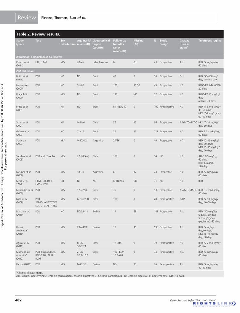
Table 2. Review results.
Study(year)
Test Sexdistribution
Age (rank/mean–SD)
Geographicalregion(country)
Follow-up(months:rank/mean–SD)
Missing(%)
N Studydesign
Chagasdiseasestage†
Treatment regime
Biochemical and metabolic biomarkers
Pinazo et al.
(2011)
ETP, F 1+2 YES 20–45 Latin America 6 23 43 Prospective ALL BZD, 5 mg/kg/day,
60 days
PCR techniques
Britto et al.
(1995)
PCR NO ND Brasil 48 0 34 Prospective C/ I BZD, 50–600 mg/
day, 45–180 days
Lauria-pires
(2000)
PCR NO 31–60 Brasil 120 15.50 45 Prospective ND BZD/NFX, ND, 60/30/
20 days
Braga MS
(2000)
PCR YES ND Brasil 120 ND 17 Prospective ND BZD/NFX,10 mg/kg/
day,
at least 30 days
Britto et al.
(2001)
PCR ND ND Brasil 84–420/240 0 100 Retrospective ND BZD, 5–6 mg/kg/day,
30–60 days;
NFX, 7–8 mg/kg/day,
60–90 days
Solari et al.
(2001)
PCR ND 0–10/6 Chile 36 15 66 Prospective ASYMTOMATIC NFX, 7–10 mg/kg/
day, 60 days
Galvao et al.
(2003)
PCR NO 7 a 12 Brazil 36 13 127 Prospective ND BZD 7.5 mg/kg/day,
60 days
Schijman
(2003)
PCR YES 0–17/4.2 Argentina 24/36 0 40 Prospective ND BZD,10–16 mg/kg/
day, 60 days;
NFX,10–15 mg/kg /
day, 60 days
Sanchez et al.
(2005)
PCR and FC-ALTA YES 22 (MEAN) Chile 120 0 54 ND ND ALLO 8.5 mg/kg,
60 days;
ITRA 6 mg/kg,
120 days
Lacunza et al.
(2006)
PCR YES 18–30 Argentina 6 17 23 Prospective ND BZD, 5 mg/kg/day,
60 days
Meira et al.
2006
HEMOCULTURE,
LMCo, PCR
ND ND ND 6–48/27.7 ND 31 ND ND BZD
Fernandes et al.
(2009)
PCR YES 17–42/30 Brasil 36 0 130 Prospective ASYMTOMATIC BZD, 10 mg/kg/day,
60 days
Lana et al.
(2009)
PCR,
SEMIQUANTITATIVE
ELISA, FC-ALTA IgG
YES 6–37/27–8 Brasil 108 0 28 Retrospective C/D/I BZD, 5–10 mg/kg/
day, 40–60 days
Murcia et al.
(2010)
PCR NO ND/33–11 Bolivia 14 68 181 Prospective ALL BZD, 300 mg/day
(adults), 60 days
5–7 mg/kg/day
(pediatrics), 60 days
Perez-
ayala et al.
(2010)
PCR YES 29–44/36 Bolivia 12 41 195 Prospective ALL BZD, 5 mg/kg/
day,60 days;
NFX, 8–10 mg/kg/
day, 90 days
Aguiar et al.
(2012)
PCR YES 8–56/
36–7.24
Brasil 12–348 0 39 Retrospective ND BZD, 5–7 mg/kg/day,
60 day
Machado de
assis et al.
(2012)
PCR, Hemoculture,
REC-ELISA, TESA-
BLOT
YES 2–60/
32,9–10,9
Brasil 120–432/
16.9–6.8
0 94 Retrospective ALL BZD, 5 mg/kg/day,
60 days
Ramos (2012) PCR YES 0–72/35 Bolivia ND 25 76 Retrospective ALL BZD, 5 mg/kg/day,
40–60 days
†Chagas disease stage.ALL: Acute, indeterminate, chronic cardiological, chronic digestive; C: Chronic cardiological; D: Chronic digestive; I: Indeterminate; ND: No data.
Review Pinazo, Thomas, Bua et al.
482 Expert Rev. Anti Infect. Ther. 12(4), (2014)
Exp
ert R
evie
w o
f A
nti-
infe
ctiv
e T
hera
py D
ownl
oade
d fr
om in
form
ahea
lthca
re.c
om b
y 20
0.58
.76.
155
on 0
3/12
/14
For
pers
onal
use
onl
y.

Standardizedreferencetest
Standardizedreferencetest valuesdefined
Dispersionmeasuresof the test
S(%)
E(%)
Reviewbias
Verificationbias
Spectrumbias
Representationbias
Detectionbias
Patients withbasal value ofthe testaltered (%)
Patients withnormal testvalue aftertreatment (%)
Ref.
ND ND ND ND ND No Yes Yes No No ETP: 73,3 F 1
+2: 80
ETP: 100 F
1 + 2: 73.3
[69]
Serology Yes No ND ND No Yes No No No ND 71.8 [71]
Serology No No ND ND No No No No No ND 3.15
Serology No No ND ND No No Yes No No ND 0
Serology No No ND ND No No No No No ND 65 [72]
Serology Yes No ND ND No No No No No 100 100 [85]
Serology Yes ND ND ND No No No No No 85.9 60.4 [81]
Serology Yes No ND ND No Yes No No No 77.5 100
Serology Yes No ND ND No No No No No ND 14.8 [76]
Serology Yes No ND ND No No No No No 100 85.7 [78]
Serology No No ND ND ND ND ND ND ND 77.4 50 [27]
Serology Yes No ND ND No ND No No No 100 11.3 [73]
Serology Yes No ND ND No No No No No ND 14.8 [74]
Serology Yes No ND ND ND ND No No No 68 90 [79]
Serology No No ND ND No Yes No No No 63 100 [80]
Serology No No ND ND Yes Yes No No No ND 41.4 [83]
Serology No No ND ND No ND No No No ND 47.2 [84]
Serology No No ND ND No ND No No Yes 65.8 100
Biological markers for evaluating therapeutic efficacy in CD Review
informahealthcare.com 483
Exp
ert R
evie
w o
f A
nti-
infe
ctiv
e T
hera
py D
ownl
oade
d fr
om in
form
ahea
lthca
re.c
om b
y 20
0.58
.76.
155
on 0
3/12
/14
For
pers
onal
use
onl
y.
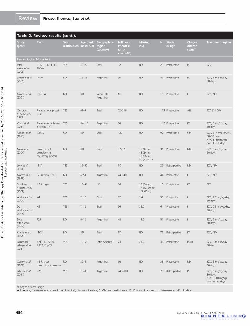
Table 2. Review results (cont.).
Study(year)
Test Sexdistribution
Age (rank/mean–SD)
Geographicalregion(country)
Follow-up(months:rank/mean–SD)
Missing(%)
N Studydesign
Chagasdiseasestage†
Treatment regime
Immunological biomarkers
Vitelli
avelar et al.
(2008)
IL-12, IL-10, IL-13,
TNF-aYES 43–70 Brasil 12 ND 29 Prospective I/C BZD
Laucella et al.
(2009)
INF-g NO 23–55 Argentina 36 ND 43 Prospective I/C BZD, 5 mg/kg/day,
30 days
Girones et al.
(2001)
R3-CHA NO ND Venezuela,
Argentina
ND ND 19 Prospective I BZD, NFX
Cancado Jr
et al. (2002,
1999)
Parasite total protein
(STc)
YES 69–9 Brasil 72–216 ND 113 Prospective ALL BZD (18 GR)
Viotti et al.
(2011)
Parasite-recombinant
proteins (14)
YES 8–41.4 Argentina 36 ND 142 Prospective I/C BZD, 5 mg/kg/day,
30 days
Galvao et al.
(1993)
CoML NO ND Brasil 120 ND 82 Prospective ND BZD, 5–7 mg/kg/DIA,
30–60 days;
NFX, 8–10 mg/kg/
day, 30–60 days
Meira et al.
(2004)
recombinant
complement
regulatory protein
NO ND Brasil 37–12 13 (12 m),
48 (24 m),
32 (36 m),
80 (> 37 m)
31 Prospective ND BZD, 5 mg/kg/day,
60 days
Levy et al.
(1996)
ISIFA YES 25–50 Brasil ND ND 26 Retrospective ND BZD, NFX
Moretti et al.
(1998)
IV Fraction, EXO NO 4–53 Argentina 24–240 ND 44 Prospective I BZD, NFX
Sanchez-
negrete et al.
(2008)
13 Antigen YES 19–41 ND 36 28 (36 m),
17 (42–60 m),
1.5 (66 m)
18 Prospective I/C BZD
Andrade et al.
(2004)
AT YES 7–12 Brasil 72 9.4 53 Prospective I BZD, 7.5 mg/kg/day,
60 days
De
Andrade et al.
(1996)
AT YES 7–12 Brasil 36 25.0 64 Prospective I BZD, 7.5 mg/kg/day,
60 days
Sosa
estani et al.
(1998)
F29 NO 6–12 Argentina 48 13.7 51 Prospective I BZD, 5 mg/kg/day,
60 days
Krautz et al.
(1995)
rTc24 NO ND Brasil ND ND 72 Retrospective I/C BZD, NFX
Fernandez-
villegas et al.
(2011)
KMP11, HSP70,
PAR2, Tgp63
YES 18–68 Latin America 24 24.0 46 Prospective I/C/D BZD, 5 mg/kg/day,
60 days
Cooley et al.
(2008)
16 T. cruzi
recombinant proteins
NO 29–61 Argentina 36 ND 38 Prospective ND BZD, 5 mg/kg/day,
60 days
Fabbro et al.
(2011)
P2b YES 29–35 Argentina 240–300 ND 78 Retrospective I/C BZD, 5 mg/kg/day,
30 days;
NFX, 8–10 mg/kg/
day, 45–60 days
†Chagas disease stage.ALL: Acute, indeterminate, chronic cardiological, chronic digestive; C: Chronic cardiological; D: Chronic digestive; I: Indeterminate; ND: No data.
Review Pinazo, Thomas, Bua et al.
484 Expert Rev. Anti Infect. Ther. 12(4), (2014)
Exp
ert R
evie
w o
f A
nti-
infe
ctiv
e T
hera
py D
ownl
oade
d fr
om in
form
ahea
lthca
re.c
om b
y 20
0.58
.76.
155
on 0
3/12
/14
For
pers
onal
use
onl
y.

Standardizedreferencetest
Standardizedreferencetest valuesdefined
Dispersionmeasuresof the test
S(%)
E(%)
Reviewbias
Verificationbias
Spectrumbias
Representationbias
Detectionbias
Patients withbasal value ofthe testaltered (%)
Patients withnormal testvalue aftertreatment (%)
Ref.
ND NO ND ND ND No No No No No ND ND [36]
Serology Yes ND ND ND No No No No No 71.0 IFN-g decrease:
34.6% (12 m),
46.8% (24 m),
71.8% (36 m)
[41]
Serology (R3
and Shed Acute
Phase Antigen
Ag, total extract)
Yes ND 92.4 100 No No No No No 92.4 100.0 [16]
ELISA, IFI, HAI,
Xenidiagnosis.
Hemoculture
ND ND ND ND No No No No No 100.0 8.0 [21,22]
HAI, IFI Yes ND ND ND No No Yes No No ND 40.0 [33]
IFI,
Hemoculture
Yes ND ND ND No No No No No ND 38% (6 m),
38% (24 m),
19% 36 (m)
[18]
CoML,
Hemoculture,
PCR, serology
Yes ND ND ND No No No No No 100.0 29.7% (12 m),
37.5% (24 m),
28.6% (36 m),
66.6% (48 m)
[27]
CoML, MbIFA,
XENODIAGNOSIS
Yes Yes 80–98.6 98 No No Yes No No ND ISIFA: 84% [20]
IFI, HAI,
SEROLOGY
Yes ND ND ND No Yes Yes Yes No ND F IV: 36%
EXO: 44%
[26]
HAI, CMA
ELISA, IFI
Yes Yes 72.2 ND No Yes Yes No No 72.2 66.6% [31]
ND No ND ND ND No No No No No 100.0 88.7% [24]
HAI, ELISA, IFI Yes Yes ND ND No No No No No 100.0 57.8% [23]
HAI, ELISA,
SEROLOGY
Yes Yes ND ND No No No No No 100.0 35.7% (6 m),
62.1% (48 m)
[25]
GST ELISA Yes ND ND ND Yes No No No No ND ND [19]
ELISA Yes ND KMP11:90
HSP70: 90
PFR2: 75
Tgp63: 30
KMP11:85
HSP70: 90
PFR2: 92
Tgp63: 70
No No No No No 100.0 KMP11: 67%
HSP70: 50%
PFR2:34%
K11-HP70-
PFR2-Tgp63: 80%
[34]
ELISA, IFI, HAI No ND ND 100 No No No Yes No ND ND [42]
Xenodiagnosis No ND ND ND No Yes Yes No Yes ND 75.4% in I and 45%
in C (23 years)
[35]
Biological markers for evaluating therapeutic efficacy in CD Review
informahealthcare.com 485
Exp
ert R
evie
w o
f A
nti-
infe
ctiv
e T
hera
py D
ownl
oade
d fr
om in
form
ahea
lthca
re.c
om b
y 20
0.58
.76.
155
on 0
3/12
/14
For
pers
onal
use
onl
y.
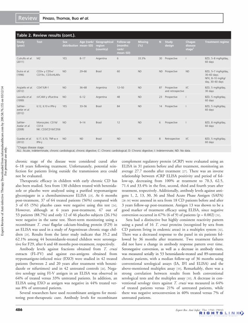
chronic stage of the disease were considered cured after6–18 years following treatment. Unfortunately, potential rein-fection for patients living outside the transmission area couldnot be evaluated.
Therapeutic efficacy in children with early chronic CD hasalso been studied. Sera from 130 children treated with benznida-zole or placebo were analyzed using a purified trypomastigoteglyconjugate in a chemiluminescent ELISA [23]. At 6 monthspost-treatment, 37 of 64 treated patients (56%) compared with3 of 65 (5%) placebo cases were negative using this test [24].However, although at 6 years post-treatment, 47 out of53 patients (88.7%) and only 12 of 46 placebo subjects (26.1%)were negative in the same test. Short-term monitoring using arecombinant T. cruzi flagelar calcium-binding protein (F29) inan ELISA was used in a study of Argentinean chronic stage chil-dren [25]. Results from the latter study indicate that 35.2 and62.1% among 44 benznidazole-treated children were seronega-tive for F29, after 6 and 48 months post-treatment, respectively.
Antibody levels against fractions obtained from T. cruziextracts (FI–FV) and against exo-antigens obtained fromtrypomastigote-infected mice (EXO) were studied in 42 treatedpatients (between 2 and 20 years after treatment with benzni-dazole or nifurtimox) and in 42 untreated controls [26]. Nega-tive serology using FI-V antigen in an ELISA was observed in64% of treated versus 33% untreated patients. In addition, anELISA using EXO as antigen was negative in 44% treated ver-sus 8% of untreated patients.
Several researchers have used recombinant antigens for moni-toring post-therapeutic cure. Antibody levels for recombinant
complement regulatory protein (rCRP) were evaluated using anELISA in 31 patients before and after treatment, monitoring anaverage 27.7 months after treatment [27]. There was an inverserelationship between rCRP ELISA positivity and period of fol-low-up, decreasing from 100% at treatment to 70.3, 62.5,71.4 and 33.4% in the first, second, third and fourth years aftertreatment, respectively. Additionally, antibody levels against anti-gens 1, 2, 13, 30, 36 and Shed Acute Phase Antigen (SAPA)[28–30] were assessed in sera from 18 CD patients before and after3 years follow-up post-treatment. Antigen 13 was shown to be agood marker of treatment efficacy using ELISA, since negativeconversion occurred in 67% (6 of 9) of patients (p = 0.002) [31].
Sera had a distinctive but highly consistent reactivity patternusing a panel of 16 T. cruzi proteins (recognized by sera fromCD patients living in endemic areas) in a multiplex system [32].There was a decreased response to the panel in six patients fol-lowed by 36 months after treatment. Two treatment failuresdid not have a change in antibody response pattern over time.Seronegative conversion, as well as a decrease in antibody titers,was measured serially in 53 benznidazole-treated and 89-untreatedchronic patients, with a median follow-up of 36 months usingconventional serological assays (IA, IFI and ELISA) and theabove-mentioned multiplex assay [33]. Remarkably, there was astrong correlation between results from both conventionalserological tests and the multiplex assay [33]. A decrease in con-ventional serology titers against T. cruzi was measured in 64%of treated patients versus 21% of untreated patients, whilethere was negative seroconversion in 40% treated versus 7% ofuntreated patients.
Table 2. Review results (cont.).
Study(year)
Test Sexdistribution
Age (rank/mean–SD)
Geographicalregion(country)
Follow-up(months:rank/mean–SD)
Missing(%)
N Studydesign
Chagasdiseasestage†
Treatment regime
Cutrullis et al.
(2011)
M2 YES 8–17 Argentina 6 33.3% 30 Prospective I BZD, 5–8 mg/kg/day,
60 days
Dutra et al.
(1996)
CD3+ y CD5+/
CD19+, CD3+HLAR+
NO 29–66 Brasil 60 ND ND Prospective ND BZD, 5–7 mg/kg/day,
30–60 days;
NFX, 8–10 mg/kg/
day, 30–60 days
Arguello et al.
(2012)
CD4+/LIR-1 NO 36–68 Argentina 12–50 ND 87 Prospective
and retrospective
I/C BZD, 5 mg/kg/day,
30 days
Laucella et al.
(1999)
sVCAM y sPLectina NO 6–12 Argentina 48 ND 23 Prospective I BZD, 5 mg/kg/day,
60 days
Sathler-
avelar et al.
(2012)
IL12, IL10 e IFN-g YES 33–56 Brasil 84 ND 14 Prospective I BZD, 5 mg/kg/day,
60 days
Sathler-
Avelar et al.
(2008)
Monocytes: CD16/
CD14
NK: CD3/CD16/CD56
NO 9–14 Brasil 12 ND 6 Prospective I BZD, 8 mg/kg/day,
60 days
Guedes et al.
(2012)
IL17, IL10, TNF-a e
IFN-gNO ND Brasil ND ND 8 Retrospective I/C BZD, 5 mg/kg/day,
60 days
†Chagas disease stage.ALL: Acute, indeterminate, chronic cardiological, chronic digestive; C: Chronic cardiological; D: Chronic digestive; I: Indeterminate; ND: No data.
Review Pinazo, Thomas, Bua et al.
486 Expert Rev. Anti Infect. Ther. 12(4), (2014)
Exp
ert R
evie
w o
f A
nti-
infe
ctiv
e T
hera
py D
ownl
oade
d fr
om in
form
ahea
lthca
re.c
om b
y 20
0.58
.76.
155
on 0
3/12
/14
For
pers
onal
use
onl
y.

Standardizedreferencetest
Standardizedreferencetest valuesdefined
Dispersionmeasuresof the test
S(%)
E(%)
Reviewbias
Verificationbias
Spectrumbias
Representationbias
Detectionbias
Patients withbasal value ofthe testaltered (%)
Patients withnormal testvalue aftertreatment (%)
Ref.
ELISA, HAI Yes YES ND 56.70 No No No No No 56.7 antiMR2:
29.7–88.1%
IFN-g : 10–100% (6 m)
[17]
Hemoculture,
CoML ELISA
Yes ND ND ND No No Yes No No ND ND [39]
ND ND ND ND ND No No Yes No No 60.0 60.0% [43]
ND ND ND ND No No No No No sPselectin: 83
sVCAM: 71
sPselectin: 66.7%
sVCAM: 41%
[15]
Cytokine
measure in
stimulation
culture
ND ND ND sPselectin:
83
sVCAM:
71
No Yes No No No ND ND [37]
ND ND ND ND ND No No No No No ND ND [40]
ND ND ND ND ND No Yes Yes No No ND ND [38]
A serological test using the KMP11, HSP70, PFR2 andTgp63 recombinant proteins was evaluated in 35 treated patientsbefore and after benznidazole administration [34]. A statisticallysignificant decrease in reactivity against KMP11 occurred6 months post-treatment in 26 out of 35 of patients (74%), at9 months post-treatment against PFR2 in 26 out of 35 (74%)and against HSP70 in 25 out of 35 (71%) CD patients. Whenthe response against only two of these antigens was evaluated,the decrease in specific antibody titer occurred in 80% ofpatients at 9 months post-treatment and continued during the2-year post-treatment follow-up period. The overall decrease intiters against KMP11, HSP70 and PFR2 24 months post-treatment was 67, 50 and 34% of patients, respectively.
Antibody levels against T. cruzi ribosomal acidic proteinP2b (Tc P P2b) were analyzed in: 30 asymptomatic CDpatients having received specific treatment with clinical follow-up for more than 20 years (group A); 37 asymptomatic CDpatients not having been treated (group B); and 11 untreatedchronic CD patients (group C) [35]. Antibody levels againstTcP2b were significantly lower only in patients from group A.
Cell markers: cytokines
Active parasite-specific B and T cell responses and specific cyto-kine profiles in patient sera are associated with development ofCD pathology and have been proposed as immune markers fordisease progression and for treatment assessment and follow-up.Circulating leukocytes from asymptomatic T. cruzi-infectedpatients (without stimuli) secreted a predominant regulatorycytokine profile, whereas symptomatic cardiac patients had a
predominant inflammatory cytokine pattern [36]. This cytokineprofile reverts after treatment, with asymptomatic patients shift-ing to predominant inflammatory profiles, and symptomaticcardiac patients upregulate regulatory cytokine production.A similar profile is observed following in vitro stimulation ofleukocytes with T. cruzi trypomastigotes. Untreated asymptom-atic patients have a type-1 regulated cytokine profile (innateimmune compartment) and a predominantly type-2 adaptiveprofile in ex vivo analysis. Following treatment, they have adownregulated cytokine profile in both innate and adaptiveimmune compartments [37].
Patients with moderate and severe cardiomyopathy producehigh levels of TNF-a and IFN-g and low levels of IL-10 andIL-17 compared with mild cardiomyopathy or cardiomyopathy-free patients [38]. Treated patients with mild or free cardiomyopa-thy produced high levels of IFN-g compared with untreatedpatients with mild or free cardiomyopathy. Deficient suppressoractivity controlling myocardial inflammation by regulatory T cellsmay cause the altered immune response observed in patients withmoderate and severe cardiomyopathy.
Cellular surface markers
The phenotype of T and B lymphocytes from peripheral bloodmononuclear cells (PBMCs) was analyzed in untreated, treated(uncured and cured) CD patients and healthy donors. Thepatients were considered cured when hemoculture and theabove-mentioned CoML tests were negative [39]. Untreatedpatients had a lower proportion of CD3+ T lymphocytes and ahigher proportion of CD5+ B cells than healthy donors, while
Biological markers for evaluating therapeutic efficacy in CD Review
informahealthcare.com 487
Exp
ert R
evie
w o
f A
nti-
infe
ctiv
e T
hera
py D
ownl
oade
d fr
om in
form
ahea
lthca
re.c
om b
y 20
0.58
.76.
155
on 0
3/12
/14
For
pers
onal
use
onl
y.

References identified in NLM searches
Immunological biomarkers: 278Biochemical biomarkers: 768
Nucleic acid amplification techniques: 332
Articles fulfilled the inclusion criteria
Immunological biomarkers: 25Biochemical biomarkers: 1
Nucleic acid amplification techniques: 17
Diagnostic studies: 72
Epidemiological studies: 8
Clinical studies: 58
Genotypification studies: 37
PCR studies: 539
Studies performed in animalmodels: 17
Studies of other diseases: 28
Studies without evaluationpost-treatment: 2
Epidemiological studies: 56
Clinical studies: 47
Genotypification studies: 43
Biochemistry and molecularbiology studies: 28
Chagas congenitaltransmission studies: 24
Reviews: 20
Immunological studies: 17
Treatment in humansevaluating other biomarker: 1
Diagnostic studies: 73
Articles using PCR inresponse to treatment: 23
Biochemical and methabolicbiomaker studies: 33
Vaccine studies: 3
Studies performed in animalmodels: 164
Studies without evaluationpost-treatment: 53
Immunological biomarkersNucleic acid amplification
techniques Biochemical biomarkers
Exclusion
Criteria
Acute congenital Chagas: 1Do not use serology as gold
standard: 5
Figure 1. Flow of inclusion of studies on biological markers for evaluating.
Review Pinazo, Thomas, Bua et al.
488 Expert Rev. Anti Infect. Ther. 12(4), (2014)
Exp
ert R
evie
w o
f A
nti-
infe
ctiv
e T
hera
py D
ownl
oade
d fr
om in
form
ahea
lthca
re.c
om b
y 20
0.58
.76.
155
on 0
3/12
/14
For
pers
onal
use
onl
y.

treated patients (uncured and cured) had intermediate values.Treated or untreated patients had approximately 2.5-fold moreCD3+/HLA-DR+ cells than those detected in uninfected indi-viduals. The mean proliferative response in vitro of PBMCfrom cured patients to parasite-related stimuli was equivalent tothe low levels detected in healthy donors.
Analysis of the immune response changes following etiologi-cal treatment of CD with benznidazole was analyzed in chil-dren at an early indeterminate stage [40]. Treated patients had ahigher activation status of circulating monocytes, inversely asso-ciated with the level of IL-12+CD14+ cells. Moreover, benzni-dazole treatment triggered a high proportion of circulatingCD3-/CD16+/CD56- NK cells associated with a type 1-modu-lated cytokine pattern. The benznidazole treatment induced sub-stantial T and B cell activation associated with an overall IL-10-modulated type 1 cytokine profile.
The proportion of PBMC-expressing IFN-g was measuredin 67 indeterminate patients (treated or untreated) and in8 treated patients with abnormal electrocardiographic find-ings [41]. Following treatment, there was either nil or a three-fold decrease of IFN-g expression compared with pretreatmentlevels, from 9 of 26, 15 of 32 and 23 of 32 patients, at 12,24 and 36 months after treatment, respectively. The antibodyresponse of these patients to a pool of recombinant T. cruziproteins using a multiplex system also decreased over time [42].The increase in cells not expressing IFN-g is associated with anearly increase in IFN-g-producing T cells with effector/effectormemory cell phenotype (result observed in 7 of 19 patientsanalyzed).
The proportion of total CD4+LIR+ T cells decreases signifi-cantly in 60% of patients (6 of 10) with indeterminate stageCD, after benznidazole treatment [43]. The decrease is observedas early as 2–6 months after treatment and is sustained at leastfor 2 years.
In summary, cellular markers are not direct markers of treat-ment as they do not pretend to detect parasite presence. How-ever, they may be used to asses the influence of the treatment onthe clinical improvement in a particular CD patient group, bycomparing the presence or expression level of the marker, inother words, in symptomatic versus asymptomatic patients. Mostof the data on immunological biomarkers were published beforethe development of nucleic acid amplification techniques.
Biochemical & metabolic molecules
CD is a chronic infection, which stimulates a continuousinflammatory immune response. Molecules that are inflamma-tory mediators of metabolic processes are altered in chronicstates of inflammation and/or infection such as chronic CD.Even though many biochemical biomarkers are easily accessibleand easy to use at a reasonable cost, we found that few ofthem have been considered as potential treatment responsebiomarkers, until recently.
Certain biomarkers of cardiac damage, troponin I and T andnatriuretic peptides, have been analyzed to determine progres-sion of the primary complication of CD and are also being
proposed as diagnostic tools for T. cruzi infection progres-sion [44]. Atrial natriuretic peptide, brain natriuretic peptide(BNP) and N-terminal proBNP have been assessed to discrimi-nate myocardial involvement in early stages of CD [45–50].Results from these studies are contradictory, indicating thatserum BNP levels are similar in T. cruzi-infected and unin-fected individuals [51]. Other natriuretic peptides (NT-proBNP [52–54], BNP [55–57] and atrial natriuretic peptide [46,49])and troponin I and T [58] have been proposed to differentiallydiagnosis late stages of CD.
Clinical trials have assessed the serum enzymes glutamic oxa-loacetic transaminase, glutamic–pyruvic transaminase, alkalinephosphatase, acid maltase and alpha-hydroxybutyric dehydroge-nase (alpha-HBDH or LDH1) for early detection of myocar-dial involvement [59]. Patients with chagasic cardiomyopathyhave low leptin [54], adipokynes [60] and angiotensin-convertingenzyme levels [57], which suggest their potential use as progres-sion markers. Low selenium levels have also been suggested as aprogression marker for chronic digestive and cardiac CD [61].Using animal models, caveolin-3 (Cav-3) [62], myocardial andperipheral protein-3-nitrotyrosine (3NT) and its protein car-bonyl formation [63], the higher level of catalase, glutathioneperoxidase, glutathione reductase and reduced function of glu-tathione and Mn(2+) superoxide dismutase have been reportedin T. cruzi-infected individuals [64].
Hypercoagulability markers such as prothrombin fragments1+2 (F 1+2), thrombin–antithrombin complex (ATM com-plex), fibrinogen/fibrin degradation products, D-dimer [65] andlipid bodies [66] in addition to apolipoprotein A1 (ApoA1) [67]
have been measured in T. cruzi patients. Despite the importantnumber of biomarkers studied from this latter group, responseto treatment has not been evaluated except in two of these in asingle study. These unpublished studies demonstrated the use-fulness of ApoA1 and fibronectin [68].
Hemostatic biomarkers such as endogenous thrombinpotential (ETP) and 1+2 prothrombin fragments 1+2 (F 1+2) have altered levels in T. cruzi-infected patients comparedwith controls (73 and 80% of patients, respectively), whichdecreased significantly 6 months after treatment (100 and73%, respectively) [69]. Evaluation of ETP and F 1+2 wasconducted in a nonendemic area on all stages of CD, therebycontrolling for possible reinfection. Nevertheless, since theresults are unpublished from a preliminary phase of a largerstudy, the small sample size, short follow-up period aftertreatment, large number of cases lost to follow-up (due tohigh mobility of the study population) and lack of a stan-dardized test to compare proposed biomarkers argue for cau-tion in data interpretation.
In summary, biochemical and metabolic molecules could beuseful surrogates for response to treatment of T. cruzi infectiondue to their easy analysis and low cost. However, only four ofthese (ETP, F 1+2, potentially ApoA1 and fibronectin) havebeen evaluated after treatment of chronic CD patients, whichimplies that further studies will be required to assess their spec-ificity and validate their use in diagnosis or prognosis.
Biological markers for evaluating therapeutic efficacy in CD Review
informahealthcare.com 489
Exp
ert R
evie
w o
f A
nti-
infe
ctiv
e T
hera
py D
ownl
oade
d fr
om in
form
ahea
lthca
re.c
om b
y 20
0.58
.76.
155
on 0
3/12
/14
For
pers
onal
use
onl
y.

Nucleic acid amplification techniques
Amplification of T. cruzi DNA has been tested and is beingevaluated as a reliable marker of therapeutic response in clinicaltrials for efficacy of trypanocidal drugs in T. cruzi-infectedpatients. When parasite DNA amplification was compared withother parasitological diagnostic methods, such as hemocultureand xenodiagnosis, the sensitivity obtained clearly favors thePCR technique for patient treatment follow-up [70–74]. PCRalone or combined with DNA hybridizations was used to eval-uate efficacy in patients treated with itraconazole or allopurinol,having a significantly higher sensitivity compared withxenodiagnosis [75–77].
Although T. cruzi DNA amplification in the blood ofchronic T. cruzi-infected and treated patients proved to be inmost cases a useful tool to demonstrate failure of treatment,
different research groups reported variable success in completenegativization. Unsuccessful specific elimination of T. cruzi wasreported in 85–89% of patients positive for PCR after trypano-cidal treatment [70,73,74] although in other studies, a high per-centage (>70%) of patients given trypanocidal treatmentconverted to negative when tested by PCR [71,78–82]. After anaverage follow-up of 20–35 years after trypanocidal treatment,30–60% of T. cruzi-infected patients continued to be positiveusing DNA amplification [72,83,84].
Pharmacological treatment does not correlate with parasiteelimination or sustained elimination of parasite DNA, whichmay be related to many factors such as CD stage, patientpathology at the time of treatment, drug and treatment proto-cols, parasite strain and load, DNA amplification techniqueand/or the number of patients studied. The most sensitive
Table 3. Target product profile for an ‘ideal’ or/and an ‘acceptable’ biomarker criteria for anti-T. cruzitreatment response.
Acceptable Ideal
Indications and usage Chronic CD in the symptomatic and
asymptomatic form
Acute and chronic CD in all the stages
Samples
(Sample collection, conservation)
Peripheral blood (cubital puncture)
Collection in stabilizing buffer for conservation
and transportation at room temperature or at 4˚C
Peripheral blood (digital puncture)
Urine
Umbilical cord blood for congenital CD
Collection in stabilizing buffer for
conservation and transportation
at room temperature
Number of samples and volume Three samples
• One pretreatment
• Two post-treatment
Maximum volume: 5 ml in adults;
2 ml in children, 1 ml neonates and newborns
Two samples
• One pretreatment
• One post-treatment
Maximum volume: 2 ml in adults,
1 ml in children, 0.5 ml umbilical cord
blood in newborns
Storage conditions technology
• Equipment required
• High technology required
• Human resources
Storage at room temperature, 4˚C or -20˚C
• Laboratory equipment
• No high technology required
• Specialized human resources
(second/third-level center)
Storage at room temperature or 4˚C
• Point of care
• No high technology required
• Nonspecialized human resources
(primary care center)
Time to processing 48–72 h <24 h
Methods Qualitative
Semi quantitative
Qualitative
Quantitative
Sensitivity and specificity ‡95%, ‡95% 100%, ‡98%
Time of response 12–24 months 3 months
Percentage of expression of altered BMK
values (nontreated T. cruzi-infected patients)
50% 100%
Percentage of response in treated
T. cruzi-infected patients
70% 100%
Precautions None None
Costs Low Low
Availability In endemic countries In all countries
CD: Chagas disease
Review Pinazo, Thomas, Bua et al.
490 Expert Rev. Anti Infect. Ther. 12(4), (2014)
Exp
ert R
evie
w o
f A
nti-
infe
ctiv
e T
hera
py D
ownl
oade
d fr
om in
form
ahea
lthca
re.c
om b
y 20
0.58
.76.
155
on 0
3/12
/14
For
pers
onal
use
onl
y.
method, unfortunately not used in all studies, is to test severalsamples from each patient at different intervals, which increasesthe sensitivity of the PCR to detect T. cruzi DNA in bloodsamples. Nevertheless, while PCR-negative conversion could beachieved in many studies and in a variable proportion oftreated patients, a significant seroconversion has not beenrecorded [71,73,74,78–80,83,85]. It is important to note that obtaininga negative PCR result does not guarantee parasitological cure,since parasitemia may fluctuate, at least in the chronic phase ofthe infection and parasitemia may be below the PCR detectionlevel, especially after a long period of follow-up after trypanoci-dal treatment [79,80,86]. In order to avoid false negatives andmeasure test confidence, repeated PCR of new patient samplesis necessary over time.
T. cruzi nucleic acid amplification techniques offer themost sensitive parasitological diagnostic method for infection.Early detection of parasite susceptibility to drugs, and there-fore, the use of PCR as a method to promptly detect failurefor lack of adherence or parasite resistance to chemotherapy,far out-weighs serology as a tool for patient follow-up aftertrypanocidal treatment. Since T. cruzi-specific antibodiescould persist for many years after the etiological treatment inchronic chagasic patients, parasite DNA amplification pro-vides a more rapid, sensitive and cost-effective test to avoidvery demanding long patient follow-up, which is always verydifficult to accomplish.
Recommendations based on the review: what to expectfrom early markers of therapeutic response – adefinition based on the TPP modelA biomarker is defined as a sign that can be measured accu-rately and reproducibly to reflect the status of a disease process.Effective markers quantitatively correlate (either directly orinversely) with disease progression. A surrogate marker could bedefined as a sign that is used in therapeutic trials as a substitutefor a clinically meaningful endpoint, that is a direct measure ofhow a patient feels, functions or survives and is expected to pre-dict the effect of the therapy [87,88]. One of the major reasonsfor identifying early biomarkers of cure/progression of chronicCD is to improve patient management and has available toolsto evaluate clinical trials. Currently, the efficacy of a drug can-not be evaluated in a short period of time after treatment, sinceparasitological clearance cannot be measured except if it corre-lates with a significant decrease in titers from conventional serol-ogy, which may take many years if at all.
Early biomarkers and some surrogate markers of therapeuticresponse for chronic CD should be molecules that fulfill spe-cific quality criteria. ‘Acceptable’ and ‘ideal’ characteristics thatare necessary for biomarkers used to evaluate response to treat-ment in patients with chronic CD are proposed in TABLE 3
according to TPP models used for development and evaluationof drugs. There are two primary and essential technical aspectsto fulfill for biomarkers: the level of biomarker expressionbefore treatment and the elapsed time in which the markerbegins to decrease. There is no consensus regarding the
definition of an ‘early’ therapeutic response for chronic CD,and there is dearth of evidence to define the optimum timingfor marker assessment. Evidence suggests that the parasite geno-type will have a direct influence on the treatment efficacy, buta biomarker will assess the patient’s response to treatment andhence the biomarker effectiveness would not be influenced byT. cruzi genotype. The ideal biomarker must be expressed athigh levels in chronic T. cruzi patients before treatment, butevidence for proportional change after treatment will dependon normal population variation and statistical difference foreach individual marker.
Future strategies in order to validate early biomarkersof response to treatmentThere are several key questions in order to drive future studiesregarding specific response biomarkers to treatment with benz-nidazole and/or nifurtimox in chronic T. cruzi-infectedpatients. Validation and use criteria have also been included inthe TPP model (TABLE 3). It is important to highlight the hetero-geneity of data from the markers studied by different groups,which do not evaluate the same parameters. There are, how-ever, several markers that do have the ability to detect treat-ment response and can be classified in two groups:
• Parasite biomarkers
There are two main classes of recombinant proteins that areeffective at different ages and stages of the disease:
• a 16 protein group [42]
• a combination of four recombinant proteins [34]: KMP11,HSP70, PAR2 and Tgp63.
Other parasite biomarkers include the CoML marker puri-fied from trypomastigotes [18]; and in pediatrics, the AT anti-gen, which is treatment response indicators still needing to betested in adults [24] and the F29 protein, recently tested inadults [25,89]. In this field, the disaccharide Gala(1,3)Galb as animmunodominant glycotope of a synthetic glycoarray contain-ing nonreducing a-galactopyranosyl moieties related to mucinO-glycans, evaluated by a chemiluminescent enzyme-linkedimmunosorbent assay, has showed its usefulness in the diagno-sis of the infection in chronic stages, but it has not been testedto assess response to treatment [90].
• DNA amplification techniques
Recently, Nagarkatti et al. (2014) used short RNA ligandscalled aptamers to detect biomarkers of T. cruzi infection inthe plasma of infected mice [91]. Aptamers were generatedagainst T. cruzi excreted/secreted antigens (TESA) purifiedfrom in vitro culture supernatants of infected host cells andused as specific ligands in enzyme-linked aptamer assays. TESAmolecules could be detected in the blood of infected mice dur-ing both the acute and the chronic phases of the disease.Although the identity of the TESA biomarkers is currently notknown, these molecules represent novel markers of T. cruziinfection. Their detection in clinical samples is currently beingassessed (personal communication). These assays also have great
Biological markers for evaluating therapeutic efficacy in CD Review
informahealthcare.com 491
Exp
ert R
evie
w o
f A
nti-
infe
ctiv
e T
hera
py D
ownl
oade
d fr
om in
form
ahea
lthca
re.c
om b
y 20
0.58
.76.
155
on 0
3/12
/14
For
pers
onal
use
onl
y.
potential in drug development applications and/or to help eval-uate treatment efficacy and possibly parasitological cure inhuman clinical trials.
• Host response/damage biomarkers
Biochemical biomarkers such as F 1+2, ETP [69], apolipopro-tein 1 and fibronectin [68] (Ndao et al., personal communica-tion) detect early treatment response at different stages ofchronic CD in adults. Muscarinic receptor antigen M2 is effec-tive to detect treatment response, but it has only been studiedin patients under 18 years old. Cytokines and surface markersthat characterize host cellular responses need to be furtherassessed although the ELISPOT for IFN-g is standardized [41].
Expert commentary & five-year viewBased on current published data, there are certain biomarkersthat have shown their effectiveness assessing responses to spe-cific treatment with benznidazole and nifurtimox in differentstages of CD. Nucleic acid amplification techniques have dem-onstrated their effectiveness to assess therapeutic failure. Immu-nological and biochemical biomarkers have not been fullydeveloped as tools to monitor treatment response, even if theyare considered interesting research paths. There is heterogeneityin methodologies and scarce data evaluating specificity and sen-sitivity of assays using these biomarkers.
In our opinion, the availability of suitable biomarkers wouldopen the door for new drugs with better tolerance profiles andgreater efficacy in clinical trials. Standardized methods for eval-uation of diagnostic tools (specificity, sensitivity, precision andreproducibility) are currently needed to improve biomarkerassessment and develop new markers and to motivate researchon CD diagnosis and treatment.
The present article reviews the landscape of existing evidencein biomarkers of chronic CD, and specific needs, to develop abattery of complementary biomarkers which together fulfillideal or acceptable criteria to evaluate response to treatment forchronic CD. Currently, there is no published data to support
the use of a single biomarker to monitor treatment efficacy.DNA amplification techniques and other marker candidatesshow promise and are currently being tested in different popu-lation groups. New studies are necessary to improve assessmentof existing markers and to identify those that could be usefulfor early follow-up of treated patients.
Acknowledgements
We are grateful to A Fernandez–Villegas from IPBLN for her collabora-
tion in the searching of manuscripts on immunology biomarkers, and A
Debrabant for his contribution regarding aptamers data.
Financial & competing interests disclosure
The NHEPACHA network is supported by DNDi (Drugs for Neglected
Tropical Diseases initiative). Members of the NHEPACHA network.
Apart from the authors of this article, the working group of NHEPACHA
comprises BA de Noya (Instituto Medicina Tropical - Universidad Central
de Venezuela (IMT-UCV); T Araujo-Jorge Fundacao Oswaldo
Cruz – Instituto Oswaldo Cruz(FIOCRUZ), Brasil; M Grijalva, Centro de
Investigacion de Enfermedades Infecciosas de la Pontificia Universidad Catol-
ica del Ecuador (CIEI-PUCE); F Guhl Universidad de los Andes
(UA-CIMPAT), Colombia; Faustino Torrico, Universidad Mayor de San
Simon (UMSS), Bolivia; Marcelo Abril, Fundacion Mundo Sano, Argentina.
The CRESIB and IPBLN research members were partially supported
by the Tropical Disease Cooperative Research Network (RICET - grants
RD12/0018/0010 and RD12/0018/0021). MCT and MCL were also
supported by Plan Nacional de I+D+I (MINECO-Spain) grants
BFU2010-1670 and SAF2012-35777 and FEDER. MJP and JG
received research funds from the Agencia de Gestio d’Ajuts Universitaris i
de Recerca (AGAUR; grant 2009SGR385) and from Fundacion Mundo
Sano. J.B. and S.S.E. are members of the Research Career of CONICET,
Argentina.
The authors have no other relevant affiliations or financial involvement
with any organization or entity with a financial interest in or financial
conflict with the subject matter or materials discussed in the manuscript
apart from those disclosed.
No writing assistance was utilized in the production of this manuscript.
Key issues
• There is a lack of biomarkers for early therapeutic response to antitrypanocidal drugs.
• Accurate and early biomarkers that assess the effectiveness of new drugs or which are useful for management of patients with Chagas
CD are needed.
• Certain biomarkers have shown their effectiveness to assess treatment response in different CD stages including DNA amplification
techniques.
• There is heterogeneity of treatment response and therapeutic failure biomarkers available.
• Data suggest that no current biomarker may be sensitive enough to be used as a single tool to monitor the efficacy of a
trypanocidal treatment.
• Early surrogate markers of therapeutic response in chronic CD should be defined following quality criteria. Using the target product
profile model, acceptable and ideal characteristics for a biomarker are proposed to evaluate response to treatment in patients with
chronic CD.
Review Pinazo, Thomas, Bua et al.
492 Expert Rev. Anti Infect. Ther. 12(4), (2014)
Exp
ert R
evie
w o
f A
nti-
infe
ctiv
e T
hera
py D
ownl
oade
d fr
om in
form
ahea
lthca
re.c
om b
y 20
0.58
.76.
155
on 0
3/12
/14
For
pers
onal
use
onl
y.

References
Papers of special note have been highlighted as:
• of interest
•• of considerable interest
1. World Health Organization (WHO).
Chagas disease (American trypanosomiasis).
2013;2013(340)
•• Over a 10 years follow-up period, a
complement-mediated lysis test using
living trypomastigotes implemented in
T. cruzi-infected patients after specific
treatment reverted negative, even in cases
in which conventional serology remaining
positive.
2. Gascon J, Bern C, Pinazo MJ. Chagas
disease in Spain, the United States and
other non-endemic countries. Acta Trop
2010;115(1-2):22-7
• In a 6-year follow-up, the
chemiluminescent enzyme-linked
immunosorbent assay with a purified
trypomastigote mucin antigen showed
efficacy evaluating success with
benznidazole treatment in adolescents in
64.7% (95% confidence interval
[CI] = 50.2–78.7) and 84.7% (95%
CI = 66.8–92.9) respectively, by
intention-to-treat and by per protocol
analysis.
3. Schmunis GA, Yadon ZE. Chagas disease:
a Latin American health problem becoming
a world health problem. Acta Trop 2010;
115(1-2):14-21
•• A double-blind, randomized, clinical field
trial was designed to test the efficacy and
tolerance of benznidazole. In this study,
ELISA using a T. cruzi flagellarcalcium-binding protein (F29) was used
additionally to immunofluorescence and
xenodiagnosis, showing its utility.
4. Lee BY, Bacon KM, Bottazzi ME, Hotez PJ.
Global economic burden of Chagas disease:
a computational simulation model. Lancet
Infect Dis 2013;13(4):342-8
•• The recombinant antigens KMP11,
PFR2, Tgp63 and HSP70 were
recognized by Chagas disease patients’
sera at any clinical stage of the disease.
Three among these antigens showed a
drop in reactivity after specific treatment.
5. Bern C, Montgomery SP, Herwaldt BL,
et al. Evaluation and treatment of Chagas
disease in the United States: a systematic
review. Jama 2007;298(18):2171-81
•• Peripheral interferon (IFN)-g-
producing T cells specific for T. cruzideclined 12 months after benznidazole
treatment as well as a pool of
recombinant T. cruzi proteins and an
increase in naive and early differentiated
memory-like CD8(+) T cells, in adult
patients in the chronic stage of the
disease.
6. World Health Organization (WHO).
Control of Chagas’ disease. 2002;1-109
•• A set of 16 recombinant proteins among
more than 400 tested were identified and
included into a multiplex bead array
format that detected 100% of
>100 confirmed positive, demonstrating
its utility in chronic Chagas disease
diagnosis, showing a strong response in
undetected and discordant sera and for
monitoring drug treatment efficacy.
7. Viotti R, De Noya BA, Araujo-Jorge T,
et al. Towards a paradigm shift in the
treatment of chronic Chagas disease.
Antimicrob Agents Chemother 2013;58(2):
635-9
•• The prothrombin fragment 1+2 (F 1+2)
(p < 0.0001) and the endogenous
thrombin potential (ETP) (p < 0.0001)
showed pathological levels in T. cruzipatients compared with people without
the infection. Normalization of both of
them was observed a 6 months after
specific treatment.
8. Schijman AG, Vigliano CA, Viotti RJ, et al.
Trypanosoma cruzi DNA in cardiac lesions
of Argentinean patients with end-stage
chronic chagas heart disease. Am J Trop
Med Hyg 2004;70(2):210-20
•• PCR was used to assess the rate of
specific chemotherapy failure in a cohort
of T. cruzi-seropositive children, showingusefulness for revealing therapeutic failure
of T. cruzi infection on a short-term
basis. Untreated patients had a 1.6-fold
higher chance of remaining positive by
PCR than those in the Bz group
(p < 0.05).
9. Kierszenbaum F. Where do we stand on the
autoimmunity hypothesis of Chagas disease?
Trends Parasitol 2005;21(11):513-16
10. Rodriques Coura J, De Castro SL. A critical
review on Chagas disease chemotherapy.
Mem Inst Oswaldo Cruz 2002;97(1):3-24
11. Marin-Neto JA, Rassi A Jr, Morillo CA,
et al. Rationale and design of a randomized
placebo-controlled trial assessing the effects
of etiologic treatment in Chagas’cardiomyopathy: the BENznidazole
Evaluation For Interrupting
Trypanosomiasis (BENEFIT). Am Heart J
2008;156(1):37-43
12. Viotti R, Vigliano C, Lococo B, et al. Side
effects of benznidazole as treatment in
chronic Chagas disease: fears and realities.
Expert Rev Anti Infect Ther 2009;7(2):
157-63
13. Viotti R, Vigliano C, Lococo B, et al.
Long-term cardiac outcomes of treating
chronic Chagas disease with benznidazole
versus no treatment: a nonrandomized trial.
Ann Intern Med 2006;144(10):724-34
14. Fabbro DL, Streiger ML, Arias ED, et al.
Trypanocide treatment among adults with
chronic Chagas disease living in Santa Fe
city (Argentina), over a mean follow-up of
21 years: parasitological, serological and
clinical evolution. Rev Soc Bras Med Trop
2007;40(1):1-10
15. Laucella SA, Segura EL, Riarte A, Sosa ES.
Soluble platelet selectin (sP-selectin) and
soluble vascular cell adhesion molecule-1
(sVCAM-1) decrease during therapy with
benznidazole in children with indeterminate
form of Chagas’ disease. Clin Exp Immunol
1999;118(3):423-7
16. Girones N, Rodriguez CI, Basso B, et al.
Antibodies to an epitope from the Cha
human autoantigen are markers of Chagas’disease. Clin Diagn Lab Immunol 2001;
8(6):1039-43
17. Cutrullis RA, Moscatelli GF, Moroni S,
et al. Benzonidazole therapy modulates
interferon-gamma and M2 muscarinic
receptor autoantibody responses in
Trypanosoma cruzi-infected children. PLoS
One 2011;6(10):e27133
18. Galvao LM, Nunes RM, Cancado JR, et al.
Lytic antibody titre as a means of assessing
cure after treatment of Chagas disease:
a 10 years follow-up study. Trans R Soc
Trop Med Hyg 1993;87(2):220-3
19. Krautz GM, Galvao LM, Cancado JR, et al.
Use of a 24-kilodalton Trypanosoma cruzi
recombinant protein to monitor cure of
human Chagas’ disease. J Clin Microbiol
1995;33(8):2086-90
20. Levy AM, Boainain E, Kloetzel JK. In situ
indirect fluorescent antibody: a new specific
test to detect ongoing chagasic infections.
J Clin Lab Anal 1996;10(2):98-103
21. Cancado JR. Criteria of Chagas disease
cure. Mem Inst Oswaldo Cruz
1999;94 Suppl 1:331-5
22. Cancado JR. Long term evaluation of
etiological treatment of chagas disease with
benznidazole. Rev Inst Med Trop Sao Paulo
2002;44(1):29-37
23. De Andrade AL, Zicker F, De Oliveira RM,
et al. Randomised trial of efficacy of
benznidazole in treatment of early
Trypanosoma cruzi infection. Lancet 1996;
348(9039):1407-13
Biological markers for evaluating therapeutic efficacy in CD Review
informahealthcare.com 493
Exp
ert R
evie
w o
f A
nti-
infe
ctiv
e T
hera
py D
ownl
oade
d fr
om in
form
ahea
lthca
re.c
om b
y 20
0.58
.76.
155
on 0
3/12
/14
For
pers
onal
use
onl
y.

24. Andrade AL, Martelli CM, Oliveira RM,
et al. Short report: benznidazole efficacy
among Trypanosoma cruzi-infected
adolescents after a six-year follow-up. Am J
Trop Med Hyg 2004;71(5):594-7
25. Sosa Estani S, Segura EL, Ruiz AM, et al.
Efficacy of chemotherapy with benznidazole
in children in the indeterminate phase of
Chagas’ disease. Am J Trop Med Hyg
1998;59(4):526-9
26. Moretti E, Cervetta L, Basso B, et al.
[Chronic Chagas’ disease: effects oftreatment nn the levels of antibodies to
crude and partially purified Trypanosoma
cruzi antigens]. Bol Chil Parasitol 1998;
53(1-2):3-9
27. Meira WS, Galvao LM, Gontijo ED, et al.
Use of the Trypanosoma cruzi recombinant
complement regulatory protein to evaluate
therapeutic efficacy following treatment of
chronic chagasic patients. J Clin Microbiol
2004;42(2):707-12
28. Da Silveira JF, Umezawa ES, Luquetti AO.
Chagas disease: recombinant Trypanosoma
cruzi antigens for serological diagnosis.
Trends Parasitol 2001;17(6):286-91
29. Ibanez CF, Affranchino JL, Macina RA,
et al. Multiple Trypanosoma cruzi antigens
containing tandemly repeated amino acid
sequence motifs. Mol Biochem Parasitol
1988;30(1):27-33
30. Vergara U, Lorca M, Veloso C, et al. Assay
for detection of Trypanosoma cruzi
antibodies in human sera based on reaction
with synthetic peptides. J Clin Microbiol
1991;29(9):2034-7
31. Sanchez Negrette O, Sanchez Valdez FJ,
Lacunza CD, et al. Serological evaluation of
specific-antibody levels in patients treated
for chronic Chagas’ disease. Clin Vaccine
Immunol 2008;15(2):297-302
32. Cooley G, Etheridge RD, Boehlke C, et al.
High throughput selection of effective
serodiagnostics for Trypanosoma cruzi
infection. PLoS Negl Trop Dis 2008;2(10):
e316
33. Viotti R, Vigliano C, Alvarez MG, et al.
Impact of aetiological treatment on
conventional and multiplex serology in
chronic Chagas disease. PLoS Negl Trop
Dis 2011;5(9):e1314
34. Fernandez-Villegas A, Pinazo MJ,
Maranon C, et al. Short-term follow-up of
chagasic patients after benzonidazole
treatment using multiple serological
markers. BMC Infect Dis 2011;11:206
35. Fabbro DL, Olivera V, Bizai ML, et al.
Humoral immune response against P2beta
from Trypanosoma cruzi in persons with
chronic Chagas disease: its relationship with
treatment against parasites and myocardial
damage. Am J Trop Med Hyg 2011;84(4):
575-80
36. Vitelli-Avelar DM, Sathler-Avelar R,
Teixeira-Carvalho A, et al. Strategy to assess
the overall cytokine profile of circulating
leukocytes and its association with distinct
clinical forms of human Chagas disease.
Scand J Immunol 2008;68(5):516-25
37. Sathler-Avelar R, Vitelli-Avelar DM,
Eloi-Santos SM, et al. Blood leukocytes
from benznidazole-treated indeterminate
chagas disease patients display an overall
type-1-modulated cytokine profile upon
short-term in vitro stimulation with
Trypanosoma cruzi antigens. BMC Infect
Dis 2012;12:123
38. Guedes PM, Gutierrez FR, Silva GK, et al.
Deficient regulatory T cell activity and low
frequency of IL-17-producing T cells
correlate with the extent of cardiomyopathy
in human Chagas’ disease. PLoS Negl Trop
Dis 2012;6(4):e1630
39. Dutra WO, Da Luz ZM, Cancado JR,
et al. Influence of parasite presence on the
immunologic profile of peripheral blood
mononuclear cells from chagasic patients
after specific drug therapy. Parasite
Immunol 1996;18(11):579-85
40. Sathler-Avelar R, Vitelli-Avelar DM,
Massara RL, et al. Etiological treatment
during early chronic indeterminate Chagas
disease incites an activated status on innate
and adaptive immunity associated with a
type 1-modulated cytokine pattern.
Microbes Infect 2008;10(2):103-13
41. Laucella SA, Mazliah DP, Bertocchi G,
et al. Changes in Trypanosoma
cruzi-specific immune responses after
treatment: surrogate markers of treatment
efficacy. Clin Infect Dis 2009;49(11):
1675-84
42. Cooley G, Etheridge RD, Boehlke C, et al.
High throughput selection of effective
serodiagnostics for Trypanosoma cruzi
infection. PLoS Negl Trop Dis 2008;2(10):
e316
43. Arguello RJ, Albareda MC, Alvarez MG,
et al. Inhibitory receptors are expressed by
Trypanosoma cruzi-specific effector T cells
and in hearts of subjects with chronic
Chagas disease. PLoS ONE 2012;7(5):
e35966
44. Lima-Costa MF, Cesar CC, Peixoto SV,
Ribeiro AL. Plasma B-type natriuretic
peptide as a predictor of mortality in
community-dwelling older adults with
Chagas disease: 10-year follow-up of the
Bambui Cohort Study of Aging. Am J
Epidemiol 2010;172(2):190-6
45. Moreira Mda C, Heringer-Walther S,
Wessel N, et al. Prognostic value of
natriuretic peptides in Chagas’ disease:a 3-year follow-up investigation. Cardiology
2008;110(4):217-25
46. Puyo AM, Scaglione J, Auger S, et al. Atrial
natriuretic factor as marker of myocardial
compromise in Chagas’ disease. Regul Pept2002;105(2):139-43
47. Puyo AM, Scaglione J, Auger S, et al.
Natriuretic peptides as prognostic and
diagnostic markers in Chagas’ disease. RegulPept 2005;128(3):203-10
48. Garcia-Alvarez A, Sitges M, Pinazo MJ,
et al. Chagas cardiomyopathy: the potential
of diastolic dysfunction and brain natriuretic
peptide in the early identification of cardiac
damage. PLoS Neg Trop Dis 2010;4(9):pii:
e826
49. Heringer-Walther S, Moreira MC,
Wessel N, et al. Brain natriuretic peptide
predicts survival in Chagas’ disease more
effectively than atrial natriuretic peptide.
Heart 2005;91(3):385-7
50. Ribeiro AL, Reis AM, Teixeira MM,
Rocha MO. Brain natriuretic peptide in
Chagas’ disease: further insights. Lancet2003;362(9380):333
51. Melo RB, Parente GB, Victor EG.
Measurement of human brain natriuretic
peptide in patients with Chagas’ disease.Arq Bras Cardiol 2005;84(2):137-40
52. Barbosa MM, Nunes Mdo C, Ribeiro AL,
et al. N-terminal proBNP levels in patients
with Chagas disease: a marker of systolic
and diastolic dysfunction of the left
ventricle. Eur J Echocardiogr 2007;8(3):
204-12
53. Mady C, Fernandes F, Arteaga E, et al.
Serum NT pro-BNP: relation to systolic
and diastolic function in cardiomyopathies
and pericardiopathies. Arq Bras Cardiol
2008;91(1):46-54
54. Fernandes F, Dantas S, Ianni BM, et al.
Leptin levels in different forms of Chagas’disease. Braz J Med Biol Res 2007;40(12):
1631-6
55. Ribeiro AL, Dos Reis AM, Barros MV,
et al. Brain natriuretic peptide and left
ventricular dysfunction in Chagas’ disease.Lancet 2002;360(9331):461-2
56. Talvani A, Rocha MO, Cogan J, et al.
Brain natriuretic peptide and left ventricular
dysfunction in chagasic cardiomyopathy.
Mem Inst Oswaldo Cruz 2004;99(6):645-9
Review Pinazo, Thomas, Bua et al.
494 Expert Rev. Anti Infect. Ther. 12(4), (2014)
Exp
ert R
evie
w o
f A
nti-
infe
ctiv
e T
hera
py D
ownl
oade
d fr
om in
form
ahea
lthca
re.c
om b
y 20
0.58
.76.
155
on 0
3/12
/14
For
pers
onal
use
onl
y.

57. Wang Y, Moreira Mda C,
Heringer-Walther S, et al. Plasma
ACE2 activity is an independent prognostic
marker in Chagas’ disease and equally
potent as BNP. J Card Fail 2010;16(2):
157-63
58. Machado MN, Suzuki FA, Mouco OC,
et al. Positive troponin T in a chagasic
patient with sustained ventricular
tachycardia and no obstructive lesions on
cine coronary angiography. Arq Bras
Cardiol 2005;84(2):182-4
59. Alarcon-Corredor OM, Carrasco-Guerra H,
Ramirez De Fernandez M, Leon W. Serum
enzyme pattern and local enzyme gradients
in chronic chagasic patients. Acta Cient
Venez 2002;53(3):210-17
60. Combs TP, Nagajyothi Mukherjee S, et al.
The adipocyte as an important target cell
for Trypanosoma cruzi infection. J Biol
Chem 2005;280(25):24085-94
61. Rivera MT, De Souza AP, Moreno AH,
et al. Progressive Chagas’ cardiomyopathy is
associated with low selenium levels. Am J
Trop Med Hyg 2002;66(6):706-12
62. Adesse D, Lisanti MP, Spray DC, et al.
Trypanosoma cruzi infection results in the
reduced expression of caveolin-3 in the
heart. Cell Cycle 2010;9(8):1639-46
63. Dhiman M, Nakayasu ES, Madaiah YH,
et al. Enhanced nitrosative stress during
Trypanosoma cruzi infection causes
nitrotyrosine modification of host proteins:
implications in Chagas’ disease. Am J
Pathol 2008;173(3):728-40
64. Wen JJ, Vyatkina G, Garg N. Oxidative
damage during chagasic cardiomyopathy
development: role of mitochondrial oxidant
release and inefficient antioxidant defense.
Free Radic Biol Med 2004;37(11):1821-33
65. Herrera RN, Diaz E, Perez Aguilar R, et al.
Prothrombotic state in early stages of
chronic Chagas’ disease. Its association with
thrombotic risk factors. Arch Cardiol Mex
2005;75 Suppl 3:S3-38-48
66. Melo RC, Fabrino DL, Dias FF,
Parreira GG. Lipid bodies: structural
markers of inflammatory macrophages in
innate immunity. Inflamm Res 2006;55(8):
342-8
67. Ndao M, Spithill TW, Caffrey R, et al.
Identification of novel diagnostic serum
biomarkers for Chagas’ disease inasymptomatic subjects by mass
spectrometric profiling. J Clin Microbiol
2010;48(4):1139-49
68. Ndao M. Biomarker discovery in serum/
plasma using surface enhanced laser
desorption ionization time of flight (SELDI-
TOF) mass spectrometry. Methods Mol
Biol 2012;818:67-79
69. Pinazo MJ, Tassies D, Munoz J, et al.
Hypercoagulability biomarkers in
Trypanosoma cruzi -infected patients.
Thromb Haemost 2011;106(4):617-23
70. Britto C, Cardoso A, Silveira C, et al.
Polymerase chain reaction (PCR) as a
laboratory tool for the evaluation of the
parasitological cure in Chagas disease after
specific treatment. Medicina (B Aires) 1999;
59(Suppl 2):176-8
71. Britto C, Cardoso MA, Vanni CM, et al.
Polymerase chain reaction detection of
Trypanosoma cruzi in human blood samples
as a tool for diagnosis and treatment
evaluation. Parasitology 1995;110(3):241-7
72. Britto C, Silveira C, Cardoso MA, et al.
Parasite persistence in treated chagasic
patients revealed by xenodiagnosis and
polymerase chain reaction. Mem Inst
Oswaldo Cruz 2001;96(6):823-6
73. Fernandes CD, Tiecher FM, Balbinot MM,
et al. Efficacy of benznidazol treatment for
asymptomatic chagasic patients from state of
Rio Grande do Sul evaluated during a three
years follow-up. Mem Inst Oswaldo Cruz
2009;104(1):27-32
74. Lana M, Lopes LA, Martins HR, et al.
Clinical and laboratory status of patients
with chronic Chagas disease living in a
vector-controlled area in Minas Gerais,
Brazil, before and nine years after
aetiological treatment. Mem Inst Oswaldo
Cruz 2009;104(8):1139-47
75. Apt W, Arribada A, Zulantay I, et al.
Itraconazole or allopurinol in the treatment
of chronic American trypanosomiasis: the
results of clinical and parasitological
examinations 11 years post-treatment. Ann
Trop Med Parasitol 2005;99(8):733-41
76. Sanchez G, Coronado X, Zulantay I, et al.
Monitoring the efficacy of specific treatment
in chronic Chagas disease by polymerase
chain reaction and flow cytometry analysis.
Parasite 2005;12(4):353-7
77. Zulantay I, Honores P, Solari A, et al. Use
of polymerase chain reaction (PCR) and
hybridization assays to detect Trypanosoma
cruzi in chronic chagasic patients treated
with itraconazole or allopurinol. Diagn
Microbiol Infect Dis 2004;48(4):253-7
78. Lacunza CDN, Sanchez O, Mora MC,
et al. Use of the polymerase chain reaction
(PCR) for early evaluation etiological
treatment in young adults, chronically
infected with Trypanosoma cruzi. Rev Patol
Trop 2006;35:227-32
79. Murcia L, Carrilero B, Munoz MJ, et al.
Usefulness of PCR for monitoring
benznidazole response in patients with
chronic Chagas’ disease: a prospective studyin a non-disease-endemic country. J
Antimicrob Chemother 2010;65(8):1759-64
80. Perez-Ayala A, Perez-Molina JA, Norman F,
et al. Chagas disease in Latin American
migrants: a Spanish challenge. Clin
Microbiol Infect 2011;17(7):1108-13
81. Galvao LM, Chiari E, Macedo AM, et al.R
assay for monitoring Trypanosoma cruzi
parasitemia in childhood after specific
chemotherapy. J Clin Microbiol 2003;
41(11):5066-70
82. Sosa-Estani S, Cura E, Velazquez E, et al.
Etiological treatment of young women
infected with Trypanosoma cruzi, and
prevention of congenital transmission. Rev
Soc Bras Med Trop 2009;42(5):484-7
83. Aguiar C, Batista AM, Pavan TB, et al.
Serological profiles and evaluation of
parasitaemia by PCR and blood culture in
individuals chronically infected by
Trypanosoma cruzi treated with
benzonidazole. Trop Med Int Health 2012;
17(3):368-73
84. Machado-De-Assis GF, Silva AR,
Do Bem VA, et al. Posttherapeutic cure
criteria in Chagas’ disease: conventionalserology followed by supplementary
serological, parasitological, and molecular
tests. Clin Vaccine Immunol 2012;19(8):
1283-91
85. Solari A, Ortiz S, Soto A, et al. Treatment
of Trypanosoma cruzi-infected children with
nifurtimox: a 3 year follow-up by PCR.
J Antimicrob Chemother 2001;48(4):515-19
86. Gomes YM, Lorena VM, Luquetti AO.
Diagnosis of Chagas disease: what has been
achieved? What remains to be done with
regard to diagnosis and follow up studies?
Mem Inst Oswaldo Cruz 2009;
104(Suppl 1):115-21
87. Temple R. Are surrogate markers adequate
to assess cardiovascular disease drugs? Jama
1999;282(8):790-5
88. Katz R. Biomarkers and surrogate markers:
an FDA perspective. NeuroRx 2004;1(2):
189-95
89. Fabbro D, Velazquez E, Bizai ML, et al.
Evaluation of the ELISA-F29 test as an
early marker of therapeutic efficacy in adults
with chronic Chagas disease. Rev Inst Med
Trop Sao Paulo 2013;55(3
90. Ashmus RA, Schocker NS,
Cordero-Mendoza Y, et al. Potential use of
synthetic alpha-galactosyl-containing
glycotopes of the parasite Trypanosoma
Biological markers for evaluating therapeutic efficacy in CD Review
informahealthcare.com 495
Exp
ert R
evie
w o
f A
nti-
infe
ctiv
e T
hera
py D
ownl
oade
d fr
om in
form
ahea
lthca
re.c
om b
y 20
0.58
.76.
155
on 0
3/12
/14
For
pers
onal
use
onl
y.

cruzi as diagnostic antigens for Chagas
disease. Org Biomol Chem 2013;11(34):
5579-83
91. Nagarkatti R, Fortes De Araujo F, Gupta C,
et al. Aptamer Based, Non-PCR
Non-Serological, Detection of Chagas
Disease Biomarkers in Trypanosoma cruzi
Infected Mice. PLoS Negl Trop Dis 2014;
8(1):e2650
Affiliations
Maria-Jesus PinazoBarcelona Centre for International Health Research (CRESIB, HospitalClınic-Universitat de Barcelona), 132 Rosello st 4˚,08036, Barcelona, Spain
M Carmen ThomasInstituto de Parasitologıa y Biomedicina Lopez Neyra, Consejo Superior deInvestigaciones Cientıficas (IPBLN-CSIC). Parque Tecnologico de Ciencias de la Salud.Conocimiento av. 18100 Granada, Spain
Jacqueline BuaInstituto Nacional de Parasitologia Dr. Mario Fatala Chaben. ANLIS Carlos GMALBRAN. 68 Paseo Colon 1063, Buenos Aires, ArgentinaandConsejo Nacional de Investigaciones Cientıficas y Tecnicas (CONICET),1917 Rivadavia av 1033 Buenos Aires, Argentina
Alina PerroneInstituto Nacional de Parasitologia Dr. Mario Fatala Chaben. ANLIS Carlos GMALBRAN. 68 Paseo Colon 1063, Buenos Aires, ArgentinaandConsejo Nacional de Investigaciones Cientıficas y Tecnicas (CONICET),1917 Rivadavia av 1033 Buenos Aires, Argentina
Alejandro-Gabriel SchijmanLaboratorio de Biologia Molecular de la Enfermedad de Chagas INGEBI – CONICET,2490 Vuelta de Obligado, Buenos Aires, Argentina
Rodolfo-Jorge ViottiCardiology Department, Hospital Eva Peron, 3200 Ricardo Balbın av. San Martın,Buenos Aires, Argentina
Janine-M RamseyCentro Regional de Investigacion en Salud Publica, Instituto Nacional de SaludPublica, 19 Poniente esq 4 Sur, Tapachula, Chiapas 30700, Mexico
Isabela RibeiroDrugs for Neglected Diseases Initiative, 15 Ch. Louis-Dunant 1202 Geneva,Switzerland
Sergio Sosa-EstaniInstituto Nacional de Parasitologia Dr. Mario Fatala Chaben. ANLIS Carlos GMALBRAN. 68 Paseo Colon 1063, Buenos Aires, ArgentinaandConsejo Nacional de Investigaciones Cientıficas y Tecnicas (CONICET),1917 Rivadavia av 1033 Buenos Aires, Argentina
Manuel-Carlos LopezInstituto de Parasitologıa y Biomedicina Lopez Neyra, Consejo Superior deInvestigaciones Cientıficas (IPBLN-CSIC). Parque Tecnologico de Ciencias de la Salud.Conocimiento av. 18100 Granada, Spain
Joaquim GasconBarcelona Centre for International Health Research (CRESIB, HospitalClınic-Universitat de Barcelona), 132 Rosello st 4˚,08036, Barcelona, Spain
Review Pinazo, Thomas, Bua et al.
496 Expert Rev. Anti Infect. Ther. 12(4), (2014)
Exp
ert R
evie
w o
f A
nti-
infe
ctiv
e T
hera
py D
ownl
oade
d fr
om in
form
ahea
lthca
re.c
om b
y 20
0.58
.76.
155
on 0
3/12
/14
For
pers
onal
use
onl
y.
79
ARTÍCULO 2
Hypercoagulability biomarkers in Trypanosoma cruzi-infected patients
María-Jesús Pinazo, Dolors Tassies, Jose Muñoz, Roser Fisa, Elizabeth de Jesús Posada, Juan Monteagudo, Edgar Ayala, Montserrat Gállego, Joan-Carles Reverter, Joaquim Gascon
Thromb. Haemost. 2011; 106: 617–623
Factor de impacto 2011: 5.044

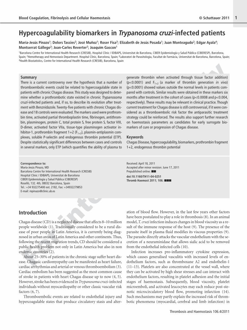
1© Schattauer 2011
Thrombosis and Haemostasis 106.4/2011
Blood Coagulation, Fibrinolysis and Cellular Haemostasis
Hypercoagulability biomarkers in Trypanosoma cruzi-infected patientsMaría-Jesús Pinazo1; Dolors Tassies2; José Muñoz1; Roser Fisa3; Elizabeth de Jesús Posada1; Juan Monteagudo2; Edgar Ayala4;Montserrat Gállego3; Joan-Carles Reverter2; Joaquim Gascon1
1Barcelona Centre for International Health Research (CRESIB), Hospital Clínic / IDIBAPS, Universitat de Barcelona, CIBER Epidemiología y Salud Pública (CIBERESP), Barcelona,Spain; 2Hemotherapy and Hemostasis Department. Hospital Clínic, Barcelona, Spain; 3Laboratori de Parasitologia, Facultat de Farmàcia, Universitat de Barcelona, Barcelona, Spain;4Health Biostatistics, Centre for International Health Research (CRESIB), Barcelona, Spain
SummaryThere is a current controversy over the hypothesis that a number ofthromboembolic events could be related to hypercoagulable state inpatients with chronic Chagas disease.This study was designed to deter-mine whether a prothrombotic state existed in chronic Trypanosomacruzi-infected patients and, if so, to describe its evolution after treat-ment with Benznidazole.Twenty-five patients with chronic Chagas dis-ease and 18 controls were evaluated.The markers used were prothrom-bin time, activated partial thromboplastin time, fibrinogen, antithrom-bin, plasminogen, protein C, total protein S, free protein S, factor VIII,D-dimer, activated factor VIIa, tissue-type plasminogen activator in-hibitor-1, prothrombin fragment 1+2 (F1+2), plasmin-antiplasmin com-plexes, soluble P-selectin and endogenous thrombin potential (ETP).Despite statistically significant differences between cases and controlsin several markers, only ETP (which quantifies the ability of plasma to
Correspondence to:María-Jesús Pinazo, MDBarcelona Centre for International Health Research (CRESIB)Hospital Clínic / IDIBAPS, Universitat de BarcelonaCIBER Epidemiología y Salud Pública (CIBERESP)Roselló, 132. 4th, 08032 Barcelona, SpainTel.: +34 932275400 ext. 2182, Fax: +34932279853E-mail: [email protected]
generate thrombin when activated through tissue factor addition)(p<0.0001) and F1+2 (a marker of thrombin generation in vivo)(p<0.0001) showed values outside the normal levels in patients com-pared with controls. Similar results were obtained in these markers sixmonths after treatment in the cohort of cases (p<0.0008 and p<0.004,respectively). These results may be relevant in clinical practice. Thoughcurrent treatment for Chagas disease is still controversial, if it were con-sidered as a thromboembolic risk factor the antiparasitic treatmentstrategy could be reinforced. The results also support further researchon haemostasis parameters as candidates for early surrogate bio-markers of cure or progression of Chagas disease.
KeywordsChagas Disease, hypercoagulability, biomarkers, prothrombin fragment1+2, endogenous thrombin potential
Received: April 18, 2011Accepted after minor revision: June 17, 2011Prepublished online: ■■
doi:10.1160/TH11-04-0251Thromb Haemost 2011; 106: ■■■
Introduction
Chagas disease (CD) is a neglected disease that affects 8–10 millionpeople worldwide (1). Traditionally considered to be a rural dis-ease of poor people in Latin America, it is currently being diag-nosed in urban areas of Latin America and other continents. Thus,following the recent migration trends, CD should be considered apublic health problem not only in Latin America but also in nonendemic countries (2).
About 25–30% of patients in the chronic stage suffer heart dis-ease. Chagasic cardiomyopathy can be manifested as heart failure,cardiac arrhythmias and arterial or venous thromboembolism (3).Cardiac embolism has been suggested as the most common causeof stroke in patients with heart Chagas disease up to now (4, 5).However, stroke has been evidenced in Trypanosoma cruzi-infectedindividuals without myocardiopathy or other classic vascular riskfactors (6, 7).
Thromboembolic events are related to endothelial injury andhypercoagulable states that produce circulatory stasis and alter-
ation of blood flow. However, in the last few years other factorshave been postulated to play a role in thrombosis (8). In an animalmodel, T. cruzi infection induces changes in blood viscosity as a re-sult of the immune response of the host (9). The presence of theparasite itself in plasma fluid modifies its viscous properties (9).The parasite directly attacks the vascular endothelium with the se-cretion of a neuraminidase that allows sialic acid to be removedfrom the endothelial infected cells (10).
Infection increases pro-inflammatory cytokine expression,which causes generalised vasculitis with increased levels of en-dothelium factors, such as thromboxane A2 and endothelin-1(11–13). Platelets are also concentrated at the vessel wall, wherethey can be activated by high shear stresses and can interact withendothelium factors, resulting in platelet adhesion and the initialstages of haemostasis. Subsequently, blood viscosity, plateletmicroemboli, and activated leucocytes may each reduce post-ste-notic microcirculatory blood flow, promoting infarction (14).Such mechanisms may partly explain the increased risk of throm-botic phenomena (myocardial, cerebral and limb infarction) in

Thrombosis and Haemostasis 106.4/2011 © Schattauer 2011
2 Pinazo et al. Hypercoagulability in T. cruzi infection
CD patients.There is currently controversy concerning the role of throm-
bophilia in thromboembolic events in CD patients. High levels ofsome hypercoagulability biomarkers, such as prothrombin F1+2,ATM-complex, fibrinogen/fibrin degradation products andD-dimer have been described as high in T. cruzi infection patients,even in early stages of the disease (15) and have been considered asthrombotic risk factors (16). However, one study shows that thereare no differences between thrombophilic factors (protein S, anti-thrombin, activated protein C resistance, factor V Leiden, lupusanticoagulant and anticardiolipin antibodies) in T. cruzi-infectedand noninfected individuals and considers that the procoagulantstate is not an ischaemic risk factor in these patients (17).
One of the main issues that complicate the management of CDis the lack of early markers of disease progression and severity aswell as markers of cure after antiparasitic treatment. Currently, thedecrease in serum IgG anti-T. cruzi titers, which occurs more than10 years after treatment is completed in chronically infected pa-tients, is the only accepted criterion of cure (18).
The objective of our study was to determine whether a pro-thrombotic state exists in T. cruzi-infected patients and, if so, to de-scribe its evolution after treatment with Benznidazole.
Materials and methods
Design and setting
This is a descriptive study of 43 individuals (25 cases and 18 con-trols) coming from Latin American countries where CD is en-demic and attending the Centre for International Health in theHospital Clínic, a university hospital.
Recruitment and participants
Forty-three persons from T. cruzi endemic areas who were over 18years old and living in Barcelona were invited to participate in thestudy. Exclusion criteria were pregnancy, non-chagasic cardio-pathy, inflammatory or immunological diseases (active infectionsof other etiology, inflammatory intestinal diseases and other auto-immune diseases such a rheumatoid arthritis, systemic lupuserythematosus) and chronic systemic diseases such as high bloodpressure and diabetes.
Procedures (including ethics)
After signing the informed consent form, the subjects were askedfor clinical and epidemiological data, including age, sex, area oforigin, history of rural environments and mud houses, previouscontact with the vector, and their history of blood donation or
transfusion. Past history – including toxic habits and other vascu-lar risk factors – and previous vascular events were recorded.
Serological tests for T. cruzi and human immunodeficiencyvirus (HIV) infection were conducted on all participants. Haema-tology, biochemistry (including renal and liver function), and aspecific determination of haemostasis factors were also perform-ed. For haemostasis studies, blood was collected in citrate-contain-ing tubes (Becton Dickinson, San José, CA, USA), samples werecentrifuged and platelet-poor plasma aliquots were frozen at–80ºC until assayed. PT and aPTT were determined in an auto-mated BCS XP analyzer (Siemens, Marburg, Germany) using stan-dard reagents (Thromborel and Actin FS; Siemens). Fibrinogenwas measured by the Clauss technique. Coagulation factorVIII wasdetermined using a chromogenic assay (Chromogenix IL, Milano,Italy). Protein C activity was quantified by a colorimetric assay(Chromogenix). Free and total protein S were quantified byenzyme-linked immunosorbent assay (ELISA) (Stago, Asnières,France). Antithrombin and plasminogen activity were measuredusing chromogenic assays (Siemens). The F1+2 and PAP ((pleasespell out first time used)) were quantified by ELISA (Siemens).D-dimer was measured with a turbidimetric method (Siemens).FVIIa and PAI-1 antigen were determined by ELISA (AmericanDiagnostica, Greenwich, CT, USA). Plasma levels of P-Sel were alsomeasured by ELISA (R&D Systems, Abingdon, UK). ETP, calcu-lated as percentage ETP value of the control group, was measuredusing a chromogenic method in a continuous thrombin gener-ation assay and the ETP Curves software (Siemens). Factor VLeiden and prothrombin gene G20210A mutation were deter-mined by real-time polymerase chain reaction (PCR) (Roche,Mannheim, Germany). Lupus anticoagulant was detected follow-ing the guidelines of the International Society on Thrombosis andHaemostasis, and anticardiolipin antibodies were measured byELISA (Chesire Diagnostics, Chester, UK). Normal values of thehaemostasis factors are given in Table 1.
For laboratory diagnosis of T. cruzi infection, three serumELISA tests were performed: a commercial ELISA with recom-binant antigens (BioELISA Chagas, Biokit S.A., Lliçà d’Amunt,Barcelona, Spain), an in-house ELISA (whole T. cruzi epimasti-gotes antigen)(19) and a conventional ELISA (Orthoclinical Diag-nostics, Johnson & Johnson Company, Rochester, NY, USA) (20).Participants were considered infected if the results from at leasttwo serological methods were positive (18). Blood nested-PCR(21) and RT-PCR (22) were performed on seropositive partici-pants. Nested-PCR was carried out at the beginning of the study.This was later replaced by a RT-PCR that amplifies the same DNAregion as the nested-PCR but is safer and easier to perform, whilegiving similar results (22).
T. cruzi-infected patients were studied using a protocol that in-cluded a 12-lead electrocardiogram, chest X-ray and echocardio-gram. Other tests were made according to the individual symp-toms.
Specific treatment with Benznidazole (5 mg/kg/ day for 60days) was offered to T. cruzi-infected patients regardless of theirclinical stage, followed by fortnightly clinical and analytical follow-up during the treatment.

© Schattauer 2011 Thrombosis and Haemostasis 106.4/2011
3Pinazo et al. Hypercoagulability in T. cruzi infection
Six months after treatment finished T. cruzi-infected patientswere tested, including serology, PCR for T. cruzi and haemostasisfactors.
Statistical analysis
Fisher’s and chi-square tests were used to compare qualitative vari-ables. A variable t-test was used to compare normally distributedcontinuous variables and a Wilcoxon test for non-normally dis-tributed variables. Crude and multivariate logistic regressionmodels were estimated to identify factors associated with outcome.Multivariate analyses were performed by a forward stepwise pro-cedure, using p<0.05 and p>0.10 from the likelihood ratio test toenter and remove criteria, respectively. In case of co-linearity oftwo variables, the model took into account only one of them.
Results from estimated models were expressed as odds ratio(OR) and 95% confidence interval (CI). The analyses were per-formed using Stata 10 (Stata Corp., College Station, TX, USA) (23).
Results
We recruited 43 patients: 25 T. cruzi-infected individuals (cases)and 18 non-infected individuals (controls), all of them 20–45 yearsold. The follow-up was completed in 15 of the 25 patients whowere treated with benznidazole ( Fig. 1), and none of themtravelled to their countries or other endemic areas during the fol-low-up. Demographic and atherothrombotic risk factors areshown in Table 2. Twenty-two cases were in an indeterminatestate of the disease, and the other three had mild to moderate car-diac involvement (two cases of Kuschnir group II and one case ofKuschnir group I). None of the cases or controls had a past historyof ischaemic events (central nervous system and heart included) oratrial fibrillation.
Statistically significant differences in hypercoagulability biom-arkers between untreated cases and controls were observed for F1+2
(p<0.001), PAP (p=0.002), P-Sel (p=0.001), ETP (p=0.001),D-dimer (p=0.049) and FVIIa (p=0.03). The results of all the vari-ables are shown in ((Table 3??)). In spite of these differences, themedians of PAP, P-Sel, D-dimer and FVIIa were sometimes withinthe normal ranges. Results of F1+2 and ETP show that 84% and64%, respectively, of the patients had abnormal values before treat-ment.
In the cohort of follow-up cases (N=15), a statistically signifi-cant decrease was observed for fibrinogen (p=0.004), F1+2
(p=0.003), PAP (p=0.004), P-Sel (p=0.006), and ETP (p=0.0008),when baseline results were compared with the results obtained sixmonths after treatment ( Fig. 2). However, fibrinogen, PAP andP-Sel baseline data were within the normal ranges in most cases.
In order to evaluate the possibility of correlation between somehaemostasis parameters, we studied the correlation between all ofthem with the values before the treatment (cases and controls), and
six months after treatment in cases. The analysis did not show highcorrelation values, and the values over 0.650–0.600 (free protein Sand total protein S in baseline values; F1+2– Antithrombin, Fibri-nogen-PAP, ETP-aPTT, TP- Free and total protein S of six monthsafter values), were not relevant.
RT-PCR was positive in three patients before treatment and be-came negative in two of them after treatment. RT-PCR was positivesix months after treatment in two cases. One of them was fromSantibáñez, a rural area of Cochabamba (Bolivia): in this case, the
Table 1: Demographic and clinical data of the recruited patients.
Infection by T. cruzi Control
Sex
Men 3 4
Women 22 14
Country of origin
Bolivia 23 10
Ecuador 1 1
Brazil 1 1
Colombia 0 3
Peru 0 1
Argentina 0 2
Smoking 0 1
Alcohol consumption 0 2
Atherothrombotic risk factors
High blood pressure 0 0
Diabetes mellitus 0 0
Hyperlipidaemia 1 2
Figure 1: Recruitment and follow-up.

Thrombosis and Haemostasis 106.4/2011 © Schattauer 2011
4 Pinazo et al. Hypercoagulability in T. cruzi infection
PCR before treatment was negative and RT-PCR six months aftertreatment was positive (CT32). The other patient was from SantaTerezinha (Goias, Brazil), and showed qualitative PCR positive be-fore treatment and RT-PCR positive after treatment (CT39). Inthese two individuals all the haemostatic parameters studied re-mained altered, included F1+2.
Discussion
A hundred years after its discovery, CD remains a complex diseasewith many unknown aspects. The cardiovascular profile of pa-tients with CD is changing: their life-expectancy is increasing and,due to vector control programs, re-infection is less likely. However,for those who migrate some habits such as diet and physical exer-cise have also changed, leading to a possible increase in chronic sys-temic diseases such as high blood pressure and diabetes in thispopulation.
In T. cruzi-infected patients, ischaemic neurological events werefirst associated with the presence of thromboembolic phenomenarelated to cardiac events such as arrhythmias, heart failure or an-eurisms. However, stroke has been described in T. cruzi-infectedpatients without cardiomyopathy. Parasympathetic nervous sys-tem dysfunction has been proposed as a pathogenesis of these is-chaemic white matter lesions in patients with CD and no cardio-logic risk factors (24).
On the other hand, it is well established that infections can pro-duce endothelial dysfunction and activation of inflammatory andatherogenic phenomena (25, 26). T. cruzi infection, like otherchronic inflammatory diseases (27–29), has itself been proposed asa thromboembolic risk factor (9, 15).
Among the haemostasis parameters studied, ETP, F1+2, PAP,P-Sel, D-dimer, and FVIIa showed significant differences betweencases and controls, but only ETP and F1+2 showed values clearlyoutside the normal levels. F1+2 is one of the main markers ofthrombin generation in vivo. An increase in F1+2 values indicates ahypercoagulable state that may be due to endothelial injuries inseveral pathologic settings (30). The role of F1+2 as a hypercoagula-bility biomarker has been well established in ischaemic cardiomyo-pathy, even in subclinical states (31). ETP is a functional test thatquantifies the ability of plasma to generate thrombin when acti-vated through tissue factor addition. Thus, ETP and F1+2 identifytwo different sides of potential hypercoagulability: F1+2 valuesmeasure indirectly the real amount of thrombin generated in vivoand ETP indicates the potential amount of thrombin that can beformed when blood coagulation is activated. In addition, these fac-tors are proved to have stability over time. In a control group of 12healthy Caucasian volunteers, mean differences in the quantitativehaemostasis variables between two determinations performed be-tween 30 and 45 days apart ranged from –3.0% to 3.3% (p=NS inall of them)(personal communication, data from Dr. Reverter).
In CD patients, both ways of measuring thrombin generationshowed a marked increase in comparison with controls, sustaining
Table 2: Descriptive and baseline univariate analysis between averages of haemostasis parameters in pre-treatment cases (25) and controls(18).
Variable Controls (18) Cases (25) Pa Normal range(units)Median (C25; C75) Median (C25; C75)
Protein C 103.50 (91.00; 109.00) 100.00 (96.00; 108.00) 0.640 60–140 (%)
Total Protein S 83.50 (81.00; 88.00) 82.00 (79.00; 90.00) 0.711 60–140 (%)
Free Protein S 83.00 (81.00; 88.00) 81.00 (77.00; 84.00) 0.128 60–140 (%)
Factor VIII 112.00 (92.00; 132.00) 106.00 (94.00; 123.00) 0.980 60–140 (%)
Antithrombin 104.00 (98.00; 109.00) 106.00 (95.00; 109.00) 0.666 60–140 (%)
aPTT 29.00 (27.00; 31.00) 30.00 (28.00; 31.00) 0.494 25–35 (sec)
Fibrinogen 3.35 ( 3.10; 3.70) 3.60 ( 3.20; 3.90) 0.261 1.5–4.5 (g/l)
D-dimer 201.50 (118.00; 255.00) 229.00 (204.00; 289.00) 0.049 50–400 (µg/l)
F 1+2 0.75 ( 0.61; 0.97) 1.80 ( 1.37; 2.71) 0.000 0.40–1.1 (nM)
ETP 415.35 (388.90; 436.00) 478.80 (413.00; 510.60) 0.001 351–473 (me)
PAI-1 21.40 (15.00; 34.20) 24.10 (19.50; 34.50) 0.205 4.0–43.0 (ng/ml)
aPTT, activated partial thromboplastin time; F1+2, prothrombin fragment 1+2; PAP, plasmin-antiplasmin complexes; PT, prothrombin time; P-Sel, P-selectin; ETP, en-dogenous thrombin potential; PAI-1, plasminogen activator inhibitor-1. a Wilcoxon rank-sum test.
PAP 249.25 (169.60; 366.60) 341.20 (267.00; 591.40) 0.002 80–470 (µg/l)
Plasminogen 110.00 (102.00; 113.00) 109.00 (103.00; 115.00) 0.853 60–140 (%)
Factor VIIa 2.44 ( 2.20; 3.49) 3.49 ( 2.89; 3.98) 0.027 1.5–4.1 (ng/ml)
PT 1.02 (0.96; 1.06) 1.01 (0.96; 1.05) 0.203 0.85–1.15 (ratio, no units)
P-Sel 31.85 (20.90; 41.20) 50.10 (40.00; 64.90) 0.001 3–90 (ng/ml)
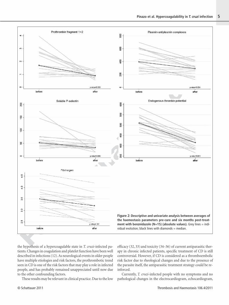
© Schattauer 2011 Thrombosis and Haemostasis 106.4/2011
5Pinazo et al. Hypercoagulability in T. cruzi infection
the hypothesis of a hypercoagulable state in T. cruzi-infected pa-tients. Changes in coagulation and platelet function have been welldescribed in infections (12). As neurological events in older peoplehave multiple etiologies and risk factors, the prothrombotic trendseen in CD is one of the risk factors that may play a role in infectedpeople, and has probably remained unappreciated until now dueto the other confounding factors.
These results may be relevant in clinical practice. Due to the low
efficacy (32, 33) and toxicity (34–36) of current antiparasitic ther-apy in chronic infected patients, specific treatment of CD is stillcontroversial. However, if CD is considered as a thromboembolicrisk factor due to rheological changes and due to the presence ofthe parasite itself, the antiparasitic treatment strategy could be re-inforced.
Currently, T. cruzi-infected people with no symptoms and nopathological changes in the electrocardiogram, echocardiogram,
Figure 2: Descriptive and univariate analysis between averages ofthe haemostasis parameters pre-cure and six months post-treat-ment with benznidazole (N=15) (absolute values). Grey lines = indi-vidual evolution; black lines with diamonds = median.
6 Pinazo et al. Hypercoagulability in T. cruzi infection
Thrombosis and Haemostasis 106.4/2011 © Schattauer 2011
chest radiograph, and gastrointestinal images are considered freeof disease and classified in the indeterminate form of the disease.The present results, along with changes in these biological markers,also challenge the notion of the indeterminate form of the disease.
Six months after treatment, a significant decrease in the bloodlevels of several hypercoagulability biomarkers was observed.Some questions remain on this subject: a low percentage of cureafter benznidazole treatment has been described in chronically in-fected patients (34), but decreasing parasitaemia has been postu-lated to be related to less progression of the disease (37). The de-crease in hypercoagulability after benznidazole therapy could beexplained by the eradication of T. cruzi or only by a significativedrop in the parasite burden (38). Interestingly we observed themaintenance of the F1+2 levels in the two patients with treatmentfailure proved by RT-PCR.
One of the major reasons for seeking biomarkers of cure/pro-gression of CD is to improve the management of these patients.Currently we do not know the results of the treatment until severalyears after it has finished, when conventional serology shows a sig-nificant decrease in titres. The PCR can only detect failures of treat-ment in some patients, but a negative result cannot prove eradi-cation of the parasite.
The present study has three main limitations: 1) The smallnumber of patients included, as it was a preliminary phase aimedat testing a wide panel of haemostasis parameters to discriminatespotentially useful biomarkers; 2) The short period of follow-upafter treatment; and 3) the high number of cases lost to follow-up,due to high mobility of the population studied. Currently this co-hort is still under supervision for a larger follow-up and new pa-tients have been included in order to strengthen this initial result.
Conclusions
In conclusion, there is a need for new drugs against T. cruzi. Earlybiomarkers of cure or progression of the disease are also a crucial
tool for future clinical trials with new drugs. Other strategies forobtaining early biomarkers of cure of CD have been proposed, andseveral groups have obtained encouraging results, but all the strat-egies proposed have limitations (38) . Due to the complexity of theT. cruzi infection, there will probably not be a single marker thatcan be used alone. The results obtained in this cohort support theneed for continuing research on haemostasis parameters as candi-dates for early biomarkers of cure of CD.
AcknowledgmentsThe authors would like to thank Fundación Mundo Sano Españafor support of our research on Chagas Disease. This material isbased upon work supported by grant 2009SGR385 from the De-partment d'Universitats, recerca I Societat de la Informació de laGenralitat de Catalunya, Spain.
Conflict of interestNone declared.
What is known about this topic?● Thromboembolic events has been observed in patients with T. cruzi
infection● Stroke has been described in T. cruzi-infected patients without
myocardiopathy.● Thromboembolic events could be related to hypercoagulable state
in patients with Chagas disease.
What does this paper add?● T. cruzi-infected patients present higher levels of several prothrom-
botic factors.● High levels of ETP and F1+2 among T. cruzi-infected patients could
be modified after treatment with Benznidazole.● If Chagas disease is considered a thromboembolic risk factor, the
antiparasitic strategy (still controversial in adults with chronic Cha-gas disease) is further indicated.
AbbreviationsCD: Chagas’ disease; T. cruzi: Trypanosoma cruzi; F 1+2: prothrombinfragment 1+2; ATM-complex: antithrombin-enzyme complex; PAP:plasmin-antiplasmin complexes; FVIIa: activated factor VII; PAI-1: tis-sue-type plasminogen activator inhibitor-1; P-Sel: soluble P-selectin;ETP: endogenous thrombin potential; aPTT: activated partial thrombo-plastin time; PT: prothrombin time; RT-PCR: real time-PCR.
References1. Pan American Health Organization. Estimación cuantitativa de la enfermedad de
Chagas en las Americas. Organización Panamericana de la Salud, Montevideo,Uruguay, 2006.
2. Gascon J, Bern C, Pinazo MJ. Chagas disease in Spain, the United States and othernon-endemic countries. Acta Trop 2010; 115: 22–27.
3. Punukollu G, Gowda RM, Khan IA, et al. Clinical aspects of the Chagas' heart dis-ease. Int J Cardiol 2007; 115: 279–283.
4. Carod-Artal FJ, Vargas AP, Horan TA, et al. Chagasic cardiomyopathy is indepen-dently associated with ischemic stroke in Chagas disease. Stroke 2005; 36:965–970.
5. Carod-Artal FJ. Chagas cardiomyopathy and ischemic stroke. Expert Rev Car-diovasc Ther 2006; 4: 119–130.
6. Andrade ZA, Sadigurski M. Trombo-embolismo command post chagásicos semcardiac insuficiência. Gaz Med Bay 1971; 71: 50–64.
7. Carod-Artal FJ. Stroke: a neglected complication of American trypanosomiasis(Chagas' disease). Trans R Soc Trop Med Hyg 2007; 101: 1075–1080.
8. Lowe GD. Virchow's triad revisited: abnormal flow. Pathophysiol HaemostThromb 2004; 33: 455–457.
9. Berra HH, Piaggio E, Revelli SS, et al. Blood viscosity changes in experimentallyTrypanosoma cruzi-infected rats. Clin Hemorheol Microcirc 2005; 32: 175–182.
10. Salomone OA. Chagas’ Cardiomyopathy and Thrombosis: the Beginning and theEnd of a Dangerous Affair. Rev Esp Cardiol 2003; 56: 333–334.
11. Carod-Artal FJ. Enfermedad de Chagas e Ictus. Neurología 2006; 21: 135–149.12. Schuetz P, Christ-Crain M, Morgenthaler NG, et al. Circulating precursor levels of
endothelin-1 and adrenomedullin, two endothelium-derived, counteracting sub-stances, in sepsis. Endothelium 2007; 14: 345–351.
13. Schuetz P, Stolz D, Mueller B, et al. Endothelin-1 precursor peptides correlate withseverity of disease and outcome in patients with community acquired pneu-monia. BMC Infect Dis 2008; 28: 8–22.
7Pinazo et al. Hypercoagulability in T. cruzi infection
© Schattauer 2011 Thrombosis and Haemostasis 106.4/2011
14. Kreutz RP, Tantry US, Bliden KP, et al. Inflammatory changes during the‘commoncold’ are associated with platelet activation and increased reactivity of platelets toagonists. Blood Coagulation and Fibrinolysis 2007; 18: 713–718.
15. Herrera RN, Diaz E, Pérez R, et al. The prothrombotic state in early stages ofchronic Chagas' disease. Rev Esp Cardiol 2003; 56: 377–382.
16. Herrera RN, Diaz E, Perez Aguilar R, et al. Prothrombotic state in early stages ofchronic Chagas' disease. Its association with thrombotic risk factors.Arch CardiolMex 2005; 75: 38–48.
17. Carod-Artal FJ, Fernandes da Silva TV,Vilela Nunes S, et al. Thrombophilia is nota predictive factor of stroke in Chagas Disease. Neurology 2004; 62: A240–241.
18. World Health Organization (WHO). Chagas disease. Thirteenth Programme Re-port UNDP/TDR. Geneva, Switzerland 1997.
19. Riera C, Vergés M, López-Chejade P, et al. Desarrollo y evaluación de una técnicaELISA con antígeno crudo de Trypanosoma cruzi para el diagnóstico de la enfer-medad de Chagas. Enf Emerg 2009; 11: 22–29.
20. Tobler LH, Contestable P, Pitina L, et al. Evaluation of a new enzyme-linked im-munosorbent assay for detection of Chagas antibody in US blood donors. Trans-fusion 2007; 47: 90–96.
21. Marcon GE, Andrade PD, de Albuquerque DM, et al. Use of a nested polymerasechain reaction (N-PCR) to detect Trypanosoma cruzi in blood samples fromchronic chagasic patients and patients with doubtful serologies. Diagn MicrobiolInfect Dis 2002; 43: 39–43.
22. Piron M, Fisa R, Casamitjana N, et al. Development of a real-time PCR assay forTrypanosoma cruzi detection in blood samples. Acta Trop 2007; 103: 195–200.
23. StataCorp. 2007. Stata Statistical Software: Release 10. College Station, TX: Stata-Corp LP.
24. Py M, Pedrosa R, Silveira J, et al. Neurological Manifestations in Chagas Diseasewithout Cardiac Dysfunction: Correlation between Dysfunction of the Parasym-pathetic Nervous System and White Matter Lesions in the Brain. J Neuroimaging2009; 19: 332–336.
25. Tanowitz HB, Burns ER, Sinha AK, et al. Enhanced platelet adherence and aggre-gation in Chagas' disease: a potential pathogenic mechanism for cardiomyopathy.Am J Trop Med Hyg 1990; 43: 274–281.
26. Schnittler HJ, Preissner KT. Vascular endothelium and infectious diseases: trickand treat. Thromb Haemost 2005; 94: 238–239.
27. Jong E, Louw S, van Gorp EC, et al. The effect of initiating combined antirretrovi-ral therapy on endotelial cell activation and coagulation markers in South AfricanHIV-infected individuals. Thromb Haemost 2010; 104: 1228–1234.
28. Fota-Markowska H, Modrzewska R, Krzowska-Firych J, et al. Concentration ofprothrombin fragment 1 + 2 (f1 + 2) in patients with liver cirrhosis and chronichepatitis C infection. Ann Univ Mariae Curie Sklodowska Med 2004; 59: 4–7.
29. Consolazio A, Borgia MC, Ferro D, et al. Increased thrombin generation andcirculating levels of tumour necrosis factor-alpha in patients with chronic Heli-cobacter pylori-positive gastritis. Aliment Pharmacol Ther 2004; 1: 289–294.
30. Manucci PM. Mechanisms, markers and management of coagulation activation.Thrombosis 1993; 44: 851–870.
31. Fuster V, Badimon L, Badimon JJ, et al. The pathogenesis of coronary artery dis-ease and the acute coronary syndromes. N Eng J Med 1992; 326: 242–250,310–318.
32. Cançado JR. Long term evaluation of Chagas disease with Benznidazole. Rev InstMed Trop Sao Paulo 2002; 44: 29–37.
33. Urbina JA, Docampo R. Specific chemotherapy of Chagas disease: controversiesand advances. Trends Parasitol 2003; 19: 495–501.
34. Rodrigues Coura J, de Castro SL. A critical review on Chagas disease chemother-apy. Mem Inst Oswaldo Cruz 2002; 97: 3–24.
35. Jackson Y, Chappuis F. Tolerance of Nifurtimox for the treatment of Chagas dis-ease in adults. Enf Emerg 2009; 11: 49.
36. Pinazo MJ, Muñoz J, Posada E, et al. Tolerance of benznidazole in treatment ofChagas' disease in adults. Antimicrob Agents Chemother 2011; 54: 4896–4899.
37. Viotti R, Vigliano C, Lococo B, et al. Long-term cardiac outcomes of treatingchronic Chagas disease with benznidazole versus no treatment: a nonrandomizedtrial. Ann Intern Med 2006; 144: 724–734.
38. Laucella SA, Mazliah DP, Bertocchi G, et al. Changes in Trypanosoma cruzi-spe-cific immune responses after treatment: surrogate markers of treatment efficacy.Clin Infect Dis 2009; 49: 1675–1684.


89
ARTÍCULO 3
Altered hypercoagulability factors in patients with
chronic Chagas disease: potential biomarkers of therapeutic response.
María-Jesús Pinazo, Elizabeth de Jesús Posada, Luis Izquierdo, Dolors Tassies, Alexandre F Marques, Elisa de Lazzari, Edelweiss Aldasoro, Jose Muñoz, Alba Abrás, Silvia Tebar, Montserrat Gállego, Igor Correia de Almeida, Joan-Carles Reverter, Joaquim Gascon
PLoS Negl Trop Dis. 2016;10(1):e0004269
Factor de impacto 2015: 4.446

90

RESEARCH ARTICLE
Altered Hypercoagulability Factors inPatients with Chronic Chagas Disease:Potential Biomarkers of TherapeuticResponseMaria-Jesus Pinazo1*, Elizabeth de Jesus Posada1, Luis Izquierdo1, Dolors Tassies2,Alexandre-Ferreira Marques3,4, Elisa de Lazzari5, Edelweiss Aldasoro1, Jose Muñoz1,Alba Abras6, Silvia Tebar6, Montserrat Gallego6,7, Igor Correia de Almeida3, Joan-Carles Reverter2, Joaquim Gascon1
1 ISGlobal, Barcelona Centre for International Health Research (CRESIB), Hospital Clínic- Universitat deBarcelona, Barcelona, Spain, 2 Hemotherapy and Hemostasis Department, Hospital Clínic de Barcelona,Barcelona, Spain, 3 Border Biomedical Research Center, Department of Biological Sciences, University ofTexas at El Paso, El Paso, Texas, United States of America, 4 Universidade Federal de Minas Gerais,Departamento de Parasitologia, Belo Horizonte, Minas Gerais, Brazil, 5 Health Biostatistics, ISGlobal,Barcelona Centre for International Health Research (CRESIB), Hospital Clínic- Universitat de Barcelona,Barcelona, Spain, 6 Laboratori de Parasitologia, Facultat de Farmàcia, Universitat de Barcelona, Barcelona,Spain, 7 Barcelona Institute for Global Health, Hospital Clínic—Universitat de Barcelona, Barcelona, Spain
AbstractThromboembolic events were described in patients with Chagas disease without cardiomy-
opathy. We aim to confirm if there is a hypercoagulable state in these patients and to deter-
mine if there is an early normalization of hemostasis factors after antiparasitic treatment.
Ninety-nine individuals from Chagas disease-endemic areas were classified in two groups:
G1, with T.cruzi infection (n = 56); G2, healthy individuals (n = 43). Twenty-four hemostasis
factors were measured at baseline. G1 patients treated with benznidazole were followed for
36 months, recording clinical parameters and performance of conventional serology, chemi-
luminescent enzyme-linked immunosorbent assay (trypomastigote-derived glycosylpho-
sphatidylinositol-anchored mucins), quantitative polymerase chain reaction, and
hemostasis tests every 6-month visits. Prothrombin fragment 1+2 (F1+2) and endogenous
thrombin potential (ETP) were abnormally expressed in 77% and 50% of infected patients
at baseline but returned to and remained at normal levels shortly after treatment in 76% and
96% of cases, respectively. Plasmin-antiplasmin complexes (PAP) were altered before
treatment in 32% of G1 patients but normalized in 94% of cases several months after treat-
ment. None of the patients with normal F1+2 values during follow-up had a positive qRT-
PCR result, but 3/24 patients (13%) with normal ETP values did. In a percentage of chronic
T. cruzi infected patients treated with benznidazole, altered coagulation markers returned
into normal levels. F1+2, ETP and PAP could be useful markers for assessing sustained
response to benznidazole.
PLOS Neglected Tropical Diseases | DOI:10.1371/journal.pntd.0004269 January 4, 2016 1 / 14
OPEN ACCESS
Citation: Pinazo M-J, Posada EdJ, Izquierdo L,Tassies D, Marques A-F, de Lazzari E, et al. (2016)Altered Hypercoagulability Factors in Patients withChronic Chagas Disease: Potential Biomarkers ofTherapeutic Response. PLoS Negl Trop Dis 10(1):e0004269. doi:10.1371/journal.pntd.0004269
Editor: Rodrigo Correa-Oliveira, FIOCRUZ - Minas,BRAZIL
Received: May 11, 2015
Accepted: November 6, 2015
Published: January 4, 2016
Copyright: © 2016 Pinazo et al. This is an openaccess article distributed under the terms of theCreative Commons Attribution License, which permitsunrestricted use, distribution, and reproduction in anymedium, provided the original author and source arecredited.
Data Availability Statement: Consent formsinformed participants that their data would beanonymized. Release of data would compromisepatient privacy and would be a breach of protocolapproved by the IRB committee. However, qualifiedresearchers will be able to access the data byrequesting it from Sergi Sanz ([email protected]), the person in charge of research data at theGlobal Health Institute.
Funding: This work was supported by FundaciónMundo Sano. ISGlobal Research group receivesfunds from the Agència de Gestió d’Ajuts

Author Summary
The manuscript describes the results of a study whose aim was to assess the tendency tocoagulate in people suffering from a parasitic infection frequent in Latin America namedT. cruzi infection or Chagas disease, by the study of several coagulation factors. Accordingto the state of the art in this topic, specific treatment for Chagas disease is recommendedin recent (acute) and late (chronic) stages of the infection. The effectiveness of currentavailable drugs in the chronic stage of infection is still a topic of debate due to inconsistentresults across studies and a lack of early measurable parameters of response to specifictreatment. Another aim of this study was to determine if the presence of an upregulatedprocoagulative activity in plasma in people suffering T. cruzi infection could be used aspotential marker that indicates therapeutic response in people at chronic stage of the dis-ease. The results of this study suggest that measurements of alterations of procoagulativeactivity may be useful to indicate specific treatment for T. cruzi chronically infectedpatients and new data concerning early response to treatment biomarkers.
IntroductionChagas disease (CD) is one of 17 neglected tropical diseases recognized by the World HealthOrganization. Caused by the protozoan parasite Trypanosoma cruzi, it mainly affects peoplewith poor socioeconomic status and limited health care access in endemic and nonendemiccountries. [1, 2]
Thrombosis is considered as a pathological deviation of haemostasis, and it is characterizedby intravascular thrombus formation and vessel occlusion. Perturbation of hemostasis is animportant factor in the pathogenesis of thromboembolic events, which can be caused by bloodflow dysregulation, endothelial injury, and coagulation system alterations.
Recently, is has been described that under certain circumstances thrombosis is a physiologi-cal process that constitutes an intrinsic effector mechanism of innate immunity, and the pro-cess has been defined as “immunothrombosis”. [3] It is activated after the recognition ofpathogens and damaged cells, and inhibits pathogen dissemination and survival. Immuno-thrombosis can therefore be regarded as a newly identified, crucial element of intravascularimmunity, which is a part of the immune system that encompasses a wide range of host strate-gies to detect and protect against pathogens in the vasculature. Dysregulation of immuno-thrombosis is likely to constitute a key event in the development of thrombotic disorders. [3]
Infectious disease can cause a hypercoagulable state through the upregulation of tissue fac-tor in monocytes, the generation of procoagulant microparticles, the activation of the coagula-tion intrinsic pathway, platelet activation, and NETs (Neutrophil Extracellular Traps) release.[3] Different infectious agents may cause different responses but a final degree of hypercoagu-lability can be a common trait as one of the biological endpoints. Additionally, patients withchronic inflammation may also present platelet adhesion events, which are considered inflam-matory processes and can be observed in patients with chronic T. cruzi infection, even in theasymptomatic stages. [4] Infection itself can cause vasculitis, increasing proinflammatory cyto-kine levels and perpetuating the risk of thrombotic events. [5] In the case of the Chagas’ diseasethe effect of hemostasis in the bradikinin formation, through the effect of factor XII activationin the Kallikrein-Kinin system, can modify the type 1 immune response and then modulate theantiparasite immunity as suggested in a mice model of subcutaneous infection by T.cruzi. [6]
Thromboembolic events and dilated cardiomyopathy, ventricular aneurysms, and intracavitarythrombosis are associated with CD. [7, 8] Rheological factors can induce intraluminal thrombus
Hypercoagulability Markers in Chronic Chagas Disease
PLOS Neglected Tropical Diseases | DOI:10.1371/journal.pntd.0004269 January 4, 2016 2 / 14
Universitaris i de Recerca (AGAUR) [grant number2009SGR385], and from the Tropical DiseaseCooperative Research Network (RICET) [grantnumber RD12/0018/0010]. ICdA was supported byNIH grants R01AI070655-A5, R01AI070655-A5S1,and 5G12MD007592; AFM was supported by theConselho Nacional de Desenvolvimento Científico eTecnológico (CNPq, Brazil). The funders had no rolein study design, data collection and analysis, decisionto publish, or preparation of the manuscript.
Competing Interests: The authors have declaredthat no competing interests exist.

formation with the risk of embolism. [9] Alterations of molecular markers of coagulation systemactivation have been described in T. cruzi infection individuals with or without clinical thrombo-sis. [9–12] Other factors, such as injury to vessel walls by parasites or changes in blood viscositydue to host immune response, may influence in the development of thromboembolic events in T.cruzi-infected individuals without Chagas cardiomyopathy or other vascular risk factors. [13]Based on studies performed in humans with chronic T. cruzi infection, there are controversialresults regarding the existence of a prothrombotic status in T. cruzi-infected patients. [13,14]There is an study in which a of higher prothrombotic status in the CD group was not found, butthe control group were individuals without T. cruzi infection and heart failure. [14] In previousstudies performed in murine models, several abnormalities of the heart microcirculation of indi-viduals with chronic CD were pointed out, but they did not find evidence of thrombi and neitherthromboembolism. [15, 16] Higher levels of the hypercoagulability markers prothrombin frag-ment 1+2 (F1+2), thrombin-antithrombin complexes (TAT), fibrinogen/fibrin degradation prod-ucts, plasminogen activator inhibitor type 1 (PAI-1), and D-dimer have been reported in T. cruzi–infected patients compared with healthy individuals. [10, 11] A pilot study performed by ourgroup showed that endogenous thrombin potential (ETP) and F1+2 levels were outside normalranges in 73% and 80% of T. cruzi–infected patients without advanced heart disease, respectively.[12] We demonstrated a 100% and 73% decrease in these levels six months after treatment withbenznidazole. Thus, if they prove to remain stable in time, hypercoagulability factors could beused as biomarkers of therapeutic response in CD. Besides, although whether or not chronic Cha-gas disease is an independent vascular risk factor remains to be confirmed. [17,18]
While specific treatment is recommended in both acute and chronic stages of infection[19,20], there are only two drugs (i.e., benznidazole and nifurtimox) available for the treatmentof CD. The mechanism of action of benznidazole relates to the nitro-reduction of componentsof the parasite, the binding of metabolites of the nuclear DNA and k-DNA of T. cruzi and thelipids and proteins of the parasite. [21] In adults, benznidazole has a high rate of adverseeffects, which can be classified into three groups: (i) hypersensitivity, including dermatitis withcutaneous eruptions (usually appearing between days 7 and 10), myalgias, arthralgias, andlymphadenopathy; (ii) polyneuropathy, paresthesias, and polineuritis usually during the 4thweek of treatment); and (iii) bone marrow disorders, such as thrombopenic purpura andagranulocytosis (usually after the second week of treatment). [22]Furthermore, the effective-ness of these drugs in the chronic stage of infection is still a topic of debate due to inconsistentstudies’ results [23–25] and a lack of early biomarkers of response to specific T. cruzi treatmentwith benznidazole. [26]
Following on from our pilot study [12], here we increased the sample size and extended fol-low-up to further investigate the value of hypercoagulability factors as biomarkers of treatmentresponse in CD. We also added current treatment response parameters measured by conven-tional serology, serology for lytic anti-α-galactosyl (anti-α-Gal) antibodies against T.cruzi [27–29], and quantitative reverse transcription polymerase chain reaction (qRT-PCR). [30]
The aims of the study were to investigate alterations of hypercoagulability factors in patientschronically infected with T. cruzi and determine whether there is an early and sustainableimprovement of the hypercoagulability factors after antiparasitic treatment.
Methods
Ethics StatementWritten informed consent was obtained from participants before being recruited (all of themwere adults). Approval for the protocols and for the informed consent was obtained from theHospital Clínic of Barcelona Ethics Review Committee.
Hypercoagulability Markers in Chronic Chagas Disease
PLOS Neglected Tropical Diseases | DOI:10.1371/journal.pntd.0004269 January 4, 2016 3 / 14
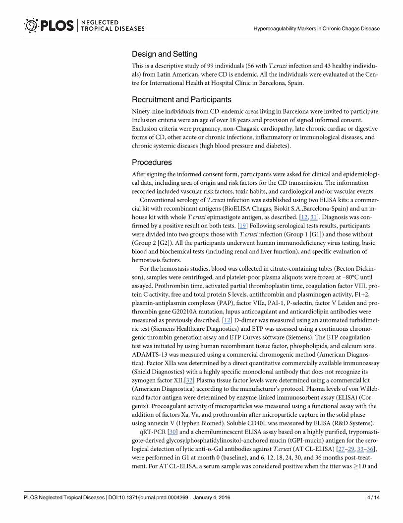
Design and SettingThis is a descriptive study of 99 individuals (56 with T.cruzi infection and 43 healthy individu-als) from Latin American, where CD is endemic. All the individuals were evaluated at the Cen-tre for International Health at Hospital Clínic in Barcelona, Spain.
Recruitment and ParticipantsNinety-nine individuals from CD-endemic areas living in Barcelona were invited to participate.Inclusion criteria were an age of over 18 years and provision of signed informed consent.Exclusion criteria were pregnancy, non-Chagasic cardiopathy, late chronic cardiac or digestiveforms of CD, other acute or chronic infections, inflammatory or immunological diseases, andchronic systemic diseases (high blood pressure and diabetes).
ProceduresAfter signing the informed consent form, participants were asked for clinical and epidemiologi-cal data, including area of origin and risk factors for the CD transmission. The informationrecorded included vascular risk factors, toxic habits, and cardiological and/or vascular events.
Conventional serology of T.cruzi infection was established using two ELISA kits: a commer-cial kit with recombinant antigens (BioELISA Chagas, Biokit S.A.,Barcelona-Spain) and an in-house kit with whole T.cruzi epimastigote antigen, as described. [12, 31]. Diagnosis was con-firmed by a positive result on both tests. [19] Following serological tests results, participantswere divided into two groups: those with T.cruzi infection (Group 1 [G1]) and those without(Group 2 [G2]). All the participants underwent human immunodeficiency virus testing, basicblood and biochemical tests (including renal and liver function), and specific evaluation ofhemostasis factors.
For the hemostasis studies, blood was collected in citrate-containing tubes (Becton Dickin-son), samples were centrifuged, and platelet-poor plasma aliquots were frozen at –80°C untilassayed. Prothrombin time, activated partial thromboplastin time, coagulation factor VIII, pro-tein C activity, free and total protein S levels, antithrombin and plasminogen activity, F1+2,plasmin-antiplasmin complexes (PAP), factor VIIa, PAI-1, P-selectin, factor V Leiden and pro-thrombin gene G20210A mutation, lupus anticoagulant and anticardiolipin antibodies weremeasured as previously described. [12] D-dimer was measured using an automated turbidimet-ric test (Siemens Healthcare Diagnostics) and ETP was assessed using a continuous chromo-genic thrombin generation assay and ETP Curves software (Siemens). The ETP coagulationtest was initiated by using human recombinant tissue factor, phospholipids, and calcium ions.ADAMTS-13 was measured using a commercial chromogenic method (American Diagnos-tica). Factor XIIa was determined by a direct quantitative commercially available immunoassay(Shield Diagnostics) with a highly specific monoclonal antibody that does not recognize itszymogen factor XII.[32] Plasma tissue factor levels were determined using a commercial kit(American Diagnostica) according to the manufacturer’s protocol. Plasma levels of vonWilleb-rand factor antigen were determined by enzyme-linked immunosorbent assay (ELISA) (Cor-genix). Procoagulant activity of microparticles was measured using a functional assay with theaddition of factors Xa, Va, and prothrombin after microparticle capture in the solid phaseusing annexin V (Hyphen Biomed). Soluble CD40L was measured by ELISA (R&D Systems).
qRT-PCR [30] and a chemiluminescent ELISA assay based on a highly purified, trypomasti-gote-derived glycosylphosphatidylinositol-anchored mucin (tGPI-mucin) antigen for the sero-logical detection of lytic anti-α-Gal antibodies against T.cruzi (AT CL-ELISA) [27–29, 33–36],were performed in G1 at month 0 (baseline), and 6, 12, 18, 24, 30, and 36 months post-treat-ment. For AT CL-ELISA, a serum sample was considered positive when the titer was�1.0 and
Hypercoagulability Markers in Chronic Chagas Disease
PLOS Neglected Tropical Diseases | DOI:10.1371/journal.pntd.0004269 January 4, 2016 4 / 14
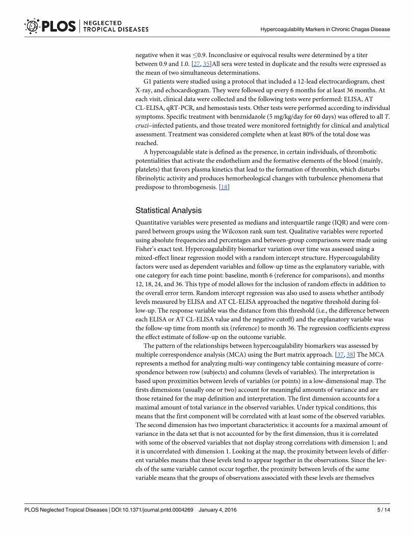
negative when it was�0.9. Inconclusive or equivocal results were determined by a titerbetween 0.9 and 1.0. [27, 35]All sera were tested in duplicate and the results were expressed asthe mean of two simultaneous determinations.
G1 patients were studied using a protocol that included a 12-lead electrocardiogram, chestX-ray, and echocardiogram. They were followed up every 6 months for at least 36 months. Ateach visit, clinical data were collected and the following tests were performed: ELISA, ATCL-ELISA, qRT-PCR, and hemostasis tests. Other tests were performed according to individualsymptoms. Specific treatment with benznidazole (5 mg/kg/day for 60 days) was offered to all T.cruzi–infected patients, and those treated were monitored fortnightly for clinical and analyticalassessment. Treatment was considered complete when at least 80% of the total dose wasreached.
A hypercoagulable state is defined as the presence, in certain individuals, of thromboticpotentialities that activate the endothelium and the formative elements of the blood (mainly,platelets) that favors plasma kinetics that lead to the formation of thrombin, which disturbsfibrinolytic activity and produces hemorheological changes with turbulence phenomena thatpredispose to thrombogenesis. [18]
Statistical AnalysisQuantitative variables were presented as medians and interquartile range (IQR) and were com-pared between groups using the Wilcoxon rank sum test. Qualitative variables were reportedusing absolute frequencies and percentages and between-group comparisons were made usingFisher’s exact test. Hypercoagulability biomarker variation over time was assessed using amixed-effect linear regression model with a random intercept structure. Hypercoagulabilityfactors were used as dependent variables and follow-up time as the explanatory variable, withone category for each time point: baseline, month 6 (reference for comparisons), and months12, 18, 24, and 36. This type of model allows for the inclusion of random effects in addition tothe overall error term. Random intercept regression was also used to assess whether antibodylevels measured by ELISA and AT CL-ELISA approached the negative threshold during fol-low-up. The response variable was the distance from this threshold (i.e., the difference betweeneach ELISA or AT CL-ELISA value and the negative cutoff) and the explanatory variable wasthe follow-up time from month six (reference) to month 36. The regression coefficients expressthe effect estimate of follow-up on the outcome variable.
The pattern of the relationships between hypercoagulability biomarkers was assessed bymultiple correspondence analysis (MCA) using the Burt matrix approach. [37, 38] The MCArepresents a method for analyzing multi-way contingency table containing measure of corre-spondence between row (subjects) and columns (levels of variables). The interpretation isbased upon proximities between levels of variables (or points) in a low-dimensional map. Thefirsts dimensions (usually one or two) account for meaningful amounts of variance and arethose retained for the map definition and interpretation. The first dimension accounts for amaximal amount of total variance in the observed variables. Under typical conditions, thismeans that the first component will be correlated with at least some of the observed variables.The second dimension has two important characteristics: it accounts for a maximal amount ofvariance in the data set that is not accounted for by the first dimension, thus it is correlatedwith some of the observed variables that not display strong correlations with dimension 1; andit is uncorrelated with dimension 1. Looking at the map, the proximity between levels of differ-ent variables means that these levels tend to appear together in the observations. Since the lev-els of the same variable cannot occur together, the proximity between levels of the samevariable means that the groups of observations associated with these levels are themselves
Hypercoagulability Markers in Chronic Chagas Disease
PLOS Neglected Tropical Diseases | DOI:10.1371/journal.pntd.0004269 January 4, 2016 5 / 14
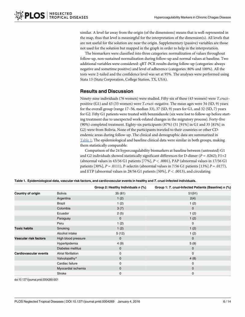
similar. A level far away from the origin (of the dimensions) means that is well-represented inthe map, thus that level is meaningful for the interpretation of the dimension(s). All levels thatare not useful for the solution are near the origin. Supplementary (passive) variables are thosenot used for the solution but mapped in the graph in order to help in the interpretation.
The biomarkers were classified into three categories: normalization of values throughoutfollow-up, non-sustained normalization during follow-up and normal values at baseline. Twoadditional variables were considered: qRT-PCR results during follow-up (categories: alwaysnegative and sometime positive) and level of adherence (categories: 80% and 100%). All thetests were 2-tailed and the confidence level was set at 95%. The analyses were performed usingStata 13 (Stata Corporation, College Station, TX, USA).
Results and DiscussionNinety-nine individuals (76 women) were studied. Fifty-six of these (43 women) were T.cruzi–positive (G1) and 43 (33 women) were T.cruzi–negative. The mean ages were 34 (SD, 9) yearsfor the overall group (range 17–56, median 33), 37 (SD, 9) years for G1, and 32 (SD, 7) yearsfor G2. Fifty G1 patients were treated with benznidazole (six were lost to follow-up before start-ing treatment due to unexpected work-related changes in the migratory process). Forty-five(90%) completed treatment. Eighty-six participants (87%) (51 [91%] in G1 and 35 [81%] inG2) were from Bolivia. None of the participants traveled to their countries or other CD-endemic areas during follow-up. The clinical and demographic data are summarized inTable 1. The epidemiological and baseline clinical data were similar in both groups, makingthem statistically comparable.
Comparison of the 24 hypercoagulability biomarkers at baseline between (untreated) G1and G2 individuals showed statistically significant differences for D-dimer (P = .0262); F1+2(abnormal values in 43/56 G1 patients [77%], P< .0001), PAP (abnormal values in 17/56 G1patients [30%], P = .0111), P-selectin (abnormal values in 7/56 G1 patients [13%] P = .0177),and ETP (abnormal values in 28/56 G1 patients [50%], P< .0013), and circulating
Table 1. Epidemiological data, vascular risk factors, and cardiovascular events in healthy and T. cruzi-infected individuals.
Group 2: Healthy Individuals n (%) Group 1: T. cruzi-Infected Patients (Baseline) n (%)
Country of origin Bolivia 35 (81) 51(91)
Argentina 1 (2) 2(4)
Brazil 1 (2) 1 (2)
Colombia 3 (7) 0
Ecuador 2 (5) 1 (2)
Paraguay 0 1 (2)
Peru 1 (2) 0
Toxic habits Smoking 1 (2) 1 (2)
Alcohol intake 5 (12) 1 (2)
Vascular risk factors High blood pressure 0 0
Hyperlipidemia 4 (9) 5 (9)
Diabetes mellitus 0 0
Cardiovascular events Atrial fibrillation 0 0
Valvulopathy* 0 4 (8)
Cardiac failure 0 0
Myocardial ischemia 0 0
Stroke 0 0
doi:10.1371/journal.pntd.0004269.t001
Hypercoagulability Markers in Chronic Chagas Disease
PLOS Neglected Tropical Diseases | DOI:10.1371/journal.pntd.0004269 January 4, 2016 6 / 14

microparticles (P = .0112) (Table 2). D-dimer levels were normal in all the individuals in G1and G2, and microparticles were within the normal range in a high percentage of patients (86%in G1 and 93% in G2, P = .3402). Our findings showed that a high percentage of patients withchronic T.cruzi infection have a hypercoagulable state regardless the clinical stage of disease,thus confirming the observations of previous studies. [11–13]
Thirty-three (76%) of the 43 patients with abnormal baseline F1+2 values achieved normallevels after a median follow-up of 9 month (IQR, 8). All but one of the 28 patients with abnor-mal ETP values before treatment showed normal values at 6 months (IQR, 3). These valueswere maintained throughout follow-up (30 months; IQR, 28) in 15 patients (60%). Fifteen ofthe 17 patients with abnormal baseline PAP values showed normal values 7 months (IQR, 7)after treatment and nine of these (60%) maintained these values throughout follow-up (28months; IQR,11). However, PAP values at 12 and 48 months seemed to be higher than those at6 months, but the confidence interval indicates a lack of precision for both time point effectestimates (Table 3). Thus, once normalized, F1+2 and ETP levels did not increase again signifi-cantly after treatment. Fig 1 shows a graphic representation of these results.
Table 2. Descriptive analysis and comparisons of hemostasis parameters between pretreatment Group 1 (N = 56) and Group 2 (N = 43).
VARIABLE Group 1 Median (IQR) [n] Group 2 Median (IQR) [n] P Value a Normal range (units)
D-dimer 228.5 (119.0) [56] 201.0 (125.0) [43] 0.0262 50–400 (μg/L)
Prothrombin fragment 1+2 1.8 (1.3) [56] 0.8 (0.4) [43] < 0.0001 0.40–1.1 (nM)
PAI-1 24.6 (14.5) [56] 21.3 (14.5) [43] 0.0680 4.0–43.0 (ng/mL)
Factor VIIa 3.5 (1.4) [56] 2.9 (1.8) [43] 0.4312 1.5–4.1 (ng/mL)
PAP complexes 360.9 (275.8) [56] 258.1 (225.8) [43] 0.0006 80–470 (μg/L)
P-selectin 41.8 (40.0) [56] 32.1 (21.7) [43] 0.0200 3–90 (μg/mL)
ETP 475.2 (99.2) [56] 412.4 (75.8) [43] <0.0001 351–473 (mEq)
Prothrombin time 98.5 (5.0) [56] 98.0 (5.0) [43] 0.4701 0.85–1.15 (ratio)/ 80–100 (%)
Attp 30.0 (3.0) [56] 30.0 (4.0) [43] 0.4849 25–35 (sec)
Fibrinogen 3.5 (0.8) [56] 3.4 (0.8) [43] 0.8540 1.5–4.5 (g/L)
Antithrombin 104.5 (16.5) [56] 101.0 (18.0) [43] 0.4518 60–140 (%)
Plasminogen 108.5 (17.5) [56] 107.0 (16.0) [43] 0.1467 60–140 (%)
Protein C 103.0 (30.5) [56] 104.0 (27.0) [43] 0.7082 60–140 (%)
Total protein S 87.0 (14.5) [56] 88.0 (12.0) [43] 0.8156 60–140 (%)
Free protein S 85.5 (11.5) [56] 88.0 (13.0) [43] 0.1125 60–140 (%)
FVIII 112.5 (51.0) [56] 103.0 (36.0) [43] 0.1391 60–140 (%)
FvWAg 136.0 (52.0) [56] 116.0 (46.0) [43] 0.0758 65–150 (U/dL)
Microparticles 21.1 (11.0) [56] 17.7 (13.5) [43] 0.0112 8–30 (nM)
CD40L 98.2 (39.3) [56] 89.0 (42.1) [43] 0.6952 30–145 (pg/mL)
Tissue factor 116.5 (50.1) [56] 124.2 (57.3) [43] 0.3737 80–280 (ng/mL)
ADAMT13 103.8 (41.9) [56] 97.9 (60.1) [43] 0.8905 50–120 (ng/mL)
Factor XIIa 3.7 (3.9) [56] 3.1 (4.6) [43] 0.3178 1.0–4.4 (ng/mL)
Factor V Leiden b No mutation 55 (98%) 42 (98%) 1.0000 c Mutations/no mutations
Heterocygote 1 (2%) 1 (2%)
G20210Ab No mutation 56 (100%) 43 (100%) Mutations/no mutations
a Wilcoxon rank sum test P valueb Absolute frequency (column percentage)c Fisher’s exact test
doi:10.1371/journal.pntd.0004269.t002
Hypercoagulability Markers in Chronic Chagas Disease
PLOS Neglected Tropical Diseases | DOI:10.1371/journal.pntd.0004269 January 4, 2016 7 / 14
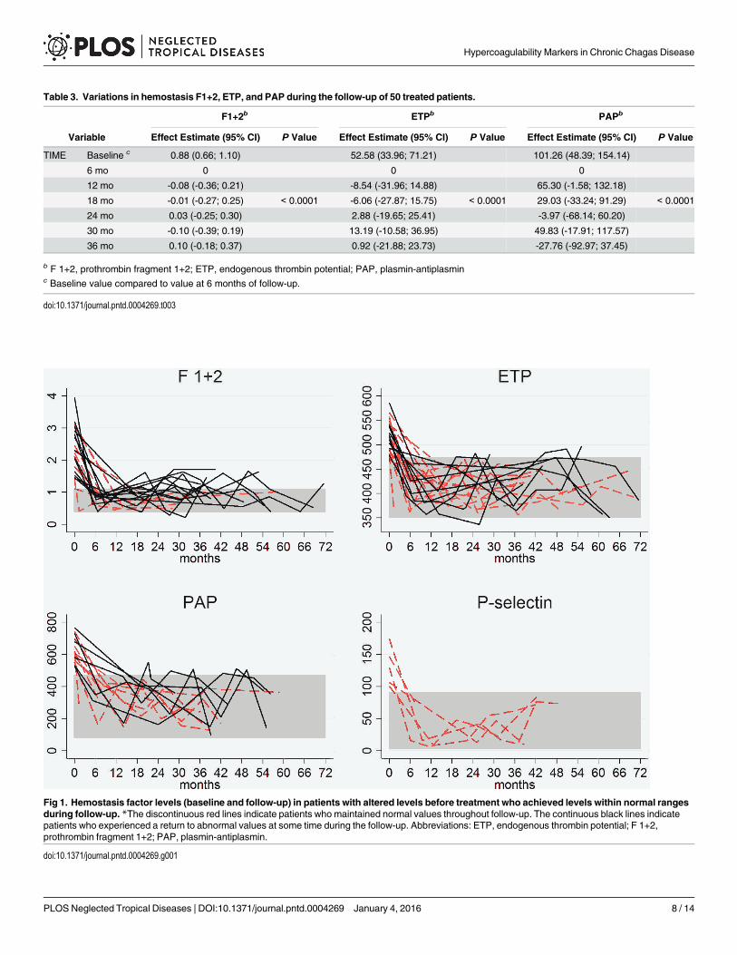
Table 3. Variations in hemostasis F1+2, ETP, and PAP during the follow-up of 50 treated patients.
F1+2b ETPb PAPb
Variable Effect Estimate (95% CI) P Value Effect Estimate (95% CI) P Value Effect Estimate (95% CI) P Value
TIME Baseline c 0.88 (0.66; 1.10) 52.58 (33.96; 71.21) 101.26 (48.39; 154.14)
6 mo 0 0 0
12 mo -0.08 (-0.36; 0.21) -8.54 (-31.96; 14.88) 65.30 (-1.58; 132.18)
18 mo -0.01 (-0.27; 0.25) < 0.0001 -6.06 (-27.87; 15.75) < 0.0001 29.03 (-33.24; 91.29) < 0.0001
24 mo 0.03 (-0.25; 0.30) 2.88 (-19.65; 25.41) -3.97 (-68.14; 60.20)
30 mo -0.10 (-0.39; 0.19) 13.19 (-10.58; 36.95) 49.83 (-17.91; 117.57)
36 mo 0.10 (-0.18; 0.37) 0.92 (-21.88; 23.73) -27.76 (-92.97; 37.45)
b F 1+2, prothrombin fragment 1+2; ETP, endogenous thrombin potential; PAP, plasmin-antiplasminc Baseline value compared to value at 6 months of follow-up.
doi:10.1371/journal.pntd.0004269.t003
Fig 1. Hemostasis factor levels (baseline and follow-up) in patients with altered levels before treatment who achieved levels within normal rangesduring follow-up. *The discontinuous red lines indicate patients who maintained normal values throughout follow-up. The continuous black lines indicatepatients who experienced a return to abnormal values at some time during the follow-up. Abbreviations: ETP, endogenous thrombin potential; F 1+2,prothrombin fragment 1+2; PAP, plasmin-antiplasmin.
doi:10.1371/journal.pntd.0004269.g001
Hypercoagulability Markers in Chronic Chagas Disease
PLOS Neglected Tropical Diseases | DOI:10.1371/journal.pntd.0004269 January 4, 2016 8 / 14

F1+2 values are an indirect measure of the amount of thrombin generated in vivo (mainlydue to endothelial injury, even in subclinical states) [39], and ETP levels indicate the potentialamount of thrombin that can be formed when blood coagulation is activated through the addi-tion of tissue factor. PAP complexes are markers of fibrinolysis. Upon activation, plasmin,which is primarily responsible for a controlled and regulated dissolution of the fibrin polymersinto soluble fragments, is immediately inactivated by antiplasmin, forming PAP complexes.[40] Therefore, it is conceivable that the increase formation of PAP complexes stems fromexcessive formation of fibrin in the blood stream of untreated T. cruzi infected patients. SolubleP-selectin is considered a biomarker of in vivo platelet activation. P-selectin is contained in theα-granules of platelets; following platelet activation, the soluble form is expressed on the plate-let surface and then shed by cleavage. P-selectin has been shown to act as a link between throm-bosis and inflammation. [41] Additionally, the four biomarkers-F1+2, ETP, PAP complexes,and P-selectin-reflect are highly stable over time.
A hypercoagulable state is a term that pretends to denominate a condition in which there isan increased tendency toward blood clotting. There is not a universally accepted definition forthis state based in biomarkers values, but an increase in several of them suggests the possibilityof an increase in the person's chances of developing blood clots. The increases in F1+2, PAPand ETP are congruent with this idea: F1+2 and PAP indicate the actual amount of thrombinand plasmin formed, as markers in procoagulant and fibrinolysis pathways, respectively; andETP indicates the potential amount of thrombin that can be formed considering globally allthe activators, inhibitors and substrates of the hemostasis present in the plasma. The increaseobserved in these biomarkers is good enough to be an argument to point out a hypercoagulablestate in patients with Chagas’ disease.
Sixteen (33%) of the 56 G1 patients had a positive qRT-PCR result at baseline, but only fourof these had a positive result after treatment (treatment failure rate of 25% in this subgroup).Five of the 34 patients with a negative baseline qRT-PCR result showed a positive result duringfollow-up. None of the patients with normal F1+2 values during follow-up had a positiveqRT-PCR result, but 3(13%) of the 24 patients with normal ETP values during follow-up did.Of the patients with altered levels of F1+2, ETP, or PAP complexes at baseline, a positiveqRT-PCR result during follow-up was not significantly associated with changes observed inlytic anti-α-Gal antibodies, F1+2, ETP, and/or PAP levels.
A positive qRT-PCR result after treatment in patients who achieved normalization of F1+2,ETP, and/or PAP could mean that a decrease in parasite load is sufficient to modify the hyper-coagulable state or that benznidazole, which acts on the redox system, could modify these bio-markers without eliminating the parasites. This would limit the use of these factors asbiomarkers for parasite elimination, although they could be valuable indicators of treatmentresponse and add support to the theory that, by reverting the hypercoagulable state, benznida-zole may also prevent clinical thrombotic events.
Conventional ELISA results were positive in all the patients in G1. Although, as expected,antibodies remained positive throughout follow-up, a slight decrease was detected by the com-mercial and in-house methods during this period. A statistically significant relevant decrease,was only observed with the in-house test from month 18 onwards (P = .0006).
Lytic anti-α-Gal antibodies were positive in 52 (96%) of the 54 patients tested before treat-ment, and in all patients AT CL-ELISA remained within positive levels to the end of the fol-low-up (Fig 2). Besides, there was no correlation between lytic anti-α-Gal antibody assay andthe hemostasis factors evaluated. In relation to previous studies’ results, early decreases in lyticanti-α-Gal antibodies were expected to be observed. On the contrary, a decrease in levels wasevident at month 12 and this was significant since month 18 and forward (P = .0052). [28, 34]
Hypercoagulability Markers in Chronic Chagas Disease
PLOS Neglected Tropical Diseases | DOI:10.1371/journal.pntd.0004269 January 4, 2016 9 / 14

Adherence to treatment was high, with only five patients not achieving 80% of the totaldose. All five patients showed abnormal F1+2 values throughout follow-up and 3 (60%) hadabnormal ETP and PAP values. One of the five patients had a positive qRT-PCR result duringfollow-up, and all five maintained the same positive ELISA and AT CL-ELISA results through-out follow-up. A large cohort of adolescents with T cruzi infection treated with benznidazoleshowed seronegativity in lytic anti-α-Gal antibodies, as measured by AT CL-ELISA, in 58%and 85% of the patients 36 and 72 months after treatment, respectively. [28, 34] The differencesbetween those studies and ours may be due to the nature of the cohorts (adolescents vs. adults)and the stage of the disease. Nevertheless, both studies showed a similar trend towards a reduc-tion in lytic anti-α-Gal antibodies following treatment with benznidazole.
We studied the relationship between normalization of hypercoagulability markers F1+2,PAP, and ETP and qRT-PCR results by multiple correspondence analyses (MCA). Due to thelow rate of positive qRT-PCR results, this variable was used as a supplementary variable jointlywith treatment adherence. The MCA results (Fig 3) showed an association between completenormalization of PAP and ETP levels and non-sustained and marginally abnormal values in F1+2. These factors had the highest contribution and correlation in the positive part of the second
Fig 2. Variations in AT CL-ELISA levels.Months of follow-up predictive margins with 95% CI. Cutoff AT CL ELISA = 0.
doi:10.1371/journal.pntd.0004269.g002
Hypercoagulability Markers in Chronic Chagas Disease
PLOS Neglected Tropical Diseases | DOI:10.1371/journal.pntd.0004269 January 4, 2016 10 / 14

dimension, while normal baseline ETP and PAP values had the highest contribution and corre-lation in the negative part. F1+2 normalization clearly characterized the positive part of thefirst dimension, while non-sustained normalization of PAP and ETP values clearly character-ized the negative part. In other words, the sustained normalization observed post-treatment inPAP and ETP, could, despite the non-sustained normalization of F1+2 values, reflect responseto antiparasitic treatment due to the strong correlation between these three variables.
The projection of qRT-PCR results and adherence to treatment in the solution space pro-vided little additional information. Consistently negative qRT-PCR results throughout follow-up appear to be related to 100% treatment adherence.
In a recent study, the authors found that the serum samples of 37 individuals with chronicChagas disease showed an upregulation of specific fragments of apolipoprotein A-1 (Apo A1)and one fibronectin fragment, that returned to normal levels in 43% of them three years after atreatment with nifurtimox. [38] Apo A1 and fibronectin fragment were altered in all the 37patients with T.cruzi infection before treatment, but the number of patients treated with thatnormalized levels was lower than in our series (60% and 96% of patients who normalized F1+2and ETP values).
This study has some limitations. Although the sample size was calculated to obtain sufficientstatistical power to answer the hypothesis, a larger sample may have detected differences thatwould be expected to appear earlier (e.g., before 12 months). The lost to follow-up samples also
Fig 3. Multiple correspondence analysis coordinate plot. Biomarkers: N, sustained normalization ofvalues throughout follow-up; nsn: non-sustained normalization throughout follow-up; nbas, normal value atbaseline. Supplementary variables: qRT-PCR during follow-up: neg (negative); pos (positive); Level ofadherence: 80%; 100%. Abbreviations: ETP, endogenous thrombin potential; F1+2, prothrombin fragment1+2; PCR, polymerase chain reaction.
doi:10.1371/journal.pntd.0004269.g003
Hypercoagulability Markers in Chronic Chagas Disease
PLOS Neglected Tropical Diseases | DOI:10.1371/journal.pntd.0004269 January 4, 2016 11 / 14

affected the estimates. Even within Spain, it is difficult to follow individuals with high migra-tory mobility for long periods. In addition, the fact that only 30% of patients had a positivebaseline qRT-PCR result was a constraint for assessing the effect of treatment.
In conclusion, patients with chronic T.cruzi infection have a potential hypercoagulablestate, regardless of cardiological and/or digestive involvement. The hypercoagulability markersF1+2 and ETP were abnormally expressed in a high percentage of patients with chronic T.cruziinfection before treatment (77% and 50%, respectively) but returned to and remained at nor-mal levels shortly after treatment in 76% and 96% of patients, respectively. Baseline PAP valueswere altered in just 30% of patients before treatment, but normalized several months aftertreatment in 88% of these. These three hypercoagulability biomarkers could be useful forassessing short-term response to treatment. However, the fact that normal values were seen insome infected patients, including some with positive post-treatment qRT-PCR results, reducestheir usefulness as universal biomarkers. The decrease in hypercoagulability factor levels couldbe explained by a decrease in parasitemia or by other benznidazole effect.
Author ContributionsConceived and designed the experiments: MJP JM LI JCR JG. Performed the experiments: MJPEdJP LI DT AFM EA JM AA STMG ICdA JCR JG. Analyzed the data: MJP EdL ICdA JCR JG.Contributed reagents/materials/analysis tools: MJP LI DT AFM AA STMG ICdA JCR JG.Wrote the paper: MJP EdJP LI DT AFM EA JM AA STMG ICdA JCR JG.
References1. Schmunis GA, Yadon ZE. Chagas disease: a Latin American health problem becoming a world health
problem. Acta Trop 2010; 115(1–2): 14–21. doi: 10.1016/j.actatropica.2009.11.003 PMID: 19932071
2. Gascon J, Bern C, Pinazo MJ. Chagas disease in Spain, the United States and other non-endemiccountries. Acta Trop 2010; 115(1–2): 22–27. doi: 10.1016/j.actatropica.2009.07.019 PMID: 19646412
3. Engelmann B, Massberg S. Thrombosis as an intravascular effector of innate immunity. Nat Rev Immu-nol 2013; 13(1): 34–45. doi: 10.1038/nri3345 PMID: 23222502
4. Schuetz P, Christ-Crain M, Morgenthaler NG, Struck J, Bergman A, Müller B. Circulating precursor lev-els of endothelin-1 and adrenomedullin, two endothelium-derived, counteracting substances, in sepsis.Endothelium 2007; 14(6): 345–351. PMID: 18080871
5. Kreutz RP, Tantry US, Bliden KP, Gurbel PA. Inflammatory changes during the 'common cold' areassociated with platelet activation and increased reactivity of platelets to agonists. Blood Coagul Fibri-nolysis 2007; 18(8): 713–718. PMID: 17982310
6. Monteiro AC, Schmitz V, Svensjo E, Gazzinelli RT, Almeida IC, Todorov A, et al. Cooperative activationof TLR2 and bradykinin B2 receptor is required for induction of type 1 immunity in a mouse model ofsubcutaneous infection by Trypanosoma cruzi. J Immunol. 2006; 177(9):6325–6335. PMID: 17056563
7. Alejandro Salomone O. Chagas cardiomyopathy and thrombosis: the beginning and the end of a dan-gerous affair. Rev Esp Cardiol 2003; 56(4): 333–334. PMID: 12689565
8. Carod-Artal FJ, Vargas AP, Horan TA, Nunes LG. Chagasic cardiomyopathy is independently associ-ated with ischemic stroke in Chagas disease. Stroke 2005; 36(5): 965–970. PMID: 15845889
9. Berra HH, Piaggio E, Revelli SS, Luquita A. Blood viscosity changes in experimentally Trypanosomacruzi-infected rats. Clin Hemorheol Microcirc 2005; 32(3): 175–182. PMID: 15851836
10. Herrera RN, Diaz de Amaya EI, Perez Aguilar RC, Joo Turoni C, Marañón R, Berman SG, et al. Inflam-matory and prothrombotic activation with conserved endothelial function in patients with chronic,asymptomatic Chagas disease. Clin Appl Thromb Hemost 2011; 17(5): 502–507. doi: 10.1177/1076029610375814 PMID: 20699256
11. Herrera RN, Diaz E, Perez Aguilar R, Bianchi J, Berman S, Luciardi HL. Prothrombotic state in earlystages of chronic Chagas' disease. Its association with thrombotic risk factors. Arch Cardiol Mex 2005;75(3): 38–48.
12. Pinazo MJ, Tassies D, Muñoz J, Fisa R, Posada E de J, Monteagudo J, et al. Hypercoagulability bio-markers in Trypanosoma cruzi -infected patients. Thromb Haemost 2011; 106(4): 617–623. doi: 10.1160/TH11-04-0251 PMID: 21866301
Hypercoagulability Markers in Chronic Chagas Disease
PLOS Neglected Tropical Diseases | DOI:10.1371/journal.pntd.0004269 January 4, 2016 12 / 14

13. Carod-Artal FJ, Vargas AP, Falcao T. Stroke in asymptomatic Trypanosoma cruzi-infected patients.Cerebrovasc Dis 2011; 31(1): 24–28. doi: 10.1159/000320248 PMID: 20980750
14. Melo LM, Souza GE, Valim LR, Moreira LF, Damico EA, Rocha TR, et al. Study of pro-thrombotic andpro-inflammatory factors in Chagas cardiomyopathy. Arq Bras Cardiol 2010; 95(5):655–662. PMID:21109919
15. Higuchi MdeL, De Morais CF, Sambiase NV, Pereira-Barretto AC, Bellotti G, Pileggi F. Histopatholog-ical criteria of myocarditis—a study based on normal heart, chagasic heart and dilated cardiomyopathy.Jpn Circ J 1990; 54(4):391–400. PMID: 2398619
16. Higuchi ML, Fukasawa S, De Brito T, Parzianello LC, Bellotti G, Ramires JA. Different microcirculatoryand interstitial matrix patterns in idiopathic dilated cardiomyopathy andChagas' disease: a three dimen-sional confocal microscopy study.Heart. 1999 Sep; 82(3):279–285. PMID: 10455076
17. Carod-Artal FJ, FdST, Vilela Nunes S et al. Thrombophilia is not a predictive factor of stroke in ChagasDisease. Neurology 2004; 62: A240–241.
18. Herrera RN, Díaz E, Pérez R, Chaín S, Sant-Yacumo R, Rodríguez E, et al. The prothrombotic state inearly stages of chronic Chagas' disease. Rev Esp Cardiol 2003; 56: 377–382. PMID: 12689572
19. World Health Organization (WHO). Control of Chagas’ disease; 2002.
20. Bern C, Montgomery SP, Herwaldt BL, Rassi A Jr, Marin-Neto JA, Dantas RO, et al. Evaluation andtreatment of chagas disease in the United States: a systematic review. Jama 2007; 298(18): 2171–2181. PMID: 18000201
21. Urbina JA. Specific treatment of Chagas disease: current status and new developments. Curr OpinInfect Dis 2001; 14(6):733–41. PMID: 11964893
22. Sosa Estani S., Armenti A., Araujo G., Viotti R., Lococo B., Ruiz Vera B., et al. Treatment of Chagas’di-sease with benznidazole and thioctic acid. Medicina (B. Aires) 2004; 64:1–6.
23. Marin-Neto JA, Rassi A Jr., Morillo CA, Avezum A, Connolly SJ, Sosa-Estani S, et al. Rationale anddesign of a randomized placebo-controlled trial assessing the effects of etiologic treatment in Chagas'cardiomyopathy: the BENznidazole Evaluation For Interrupting Trypanosomiasis (BENEFIT). Am HeartJ 2008; 156(1): 37–43. doi: 10.1016/j.ahj.2008.04.001 PMID: 18585495
24. Rodriques Coura J, de Castro SL. A critical review on Chagas disease chemotherapy. Mem InstOswaldo Cruz 2002; 97(1): 3–24. PMID: 11992141
25. Viotti R, Vigliano C, Lococo B, Bertochi G, Petti M, Álvarez MG, et al. Long-term cardiac outcomes oftreating chronic Chagas disease with benznidazole versus no treatment: a nonrandomized trial. AnnIntern Med 2006; 144(10): 724–734. PMID: 16702588
26. Requena-Mendez A, Lopez MC, Angheben A, Izquierdo L, Ribeiro I, Pinazo MJ, et al. Evaluating Cha-gas disease progression and cure through blood-derived biomarkers: a systematic review. Expert RevAnti Infect Ther 2013; 11(9): 957–976. doi: 10.1586/14787210.2013.824718 PMID: 24053276
27. Almeida IC, Covas DT, Soussumi LM, Travassos LR. A highly sensitive and specific chemiluminescentenzyme-linked immunosorbent assay for diagnosis of active Trypanosoma cruzi infection. Transfusion1997; 37(8): 850–857. PMID: 9280332
28. de Andrade AL, Zicker F, de Oliveira RM, Almeida Silva S, Luquetti A, Travassos LR. Randomised trialof efficacy of benznidazole in treatment of early Trypanosoma cruzi infection. Lancet 1996; 348(9039):1407–1413. PMID: 8937280
29. Krettli AU. The utility of anti-trypomastigote lytic antibodies for determining cure of Trypanosoma cruziinfections in treated patients: an overview and perspectives. Mem Inst Oswaldo Cruz 2009; 104 (1):142–151.
30. Piron M, Fisa R, Casamitjana N, López-Chejade P, Puig L, Vergés M, et al. Development of a real-timePCR assay for Trypanosoma cruzi detection in blood samples. Acta Trop 2007; 103(3): 195–200.PMID: 17662227
31. Riera C, Verges M, López-Chejade P, et al. Desarrollo y evaluación de una técnica ELISA con antígenocrudo de Trypanosoma cruzi para el diagnóstico de la enfermedad de Chagas. Enfermedades emer-gentes 2009; 11: 19–22.
32. Ford RP, Esnouf MP, Burgess AI, Sarphie AF. An enzyme linked immunosorbent assay (ELISA) for themeasurement of activated factor XII (Hageman factor) in human plasma. J Immunoassay 1996;17:119–131. PMID: 8744282
33. Almeida IC, Ferguson MA, Schenkman S, Travassos LR. Lytic anti-alpha-galactosyl antibodies frompatients with chronic Chagas' disease recognize novel O-linked oligosaccharides on mucin-like glyco-syl-phosphatidylinositol-anchored glycoproteins of Trypanosoma cruzi. Biochem J 1994; 304 (3): 793–802.
Hypercoagulability Markers in Chronic Chagas Disease
PLOS Neglected Tropical Diseases | DOI:10.1371/journal.pntd.0004269 January 4, 2016 13 / 14

34. Andrade AL, Martelli CM, Oliveira RM, Silva SA, Aires AI, Soussumi LM, et al. Short report: benznida-zole efficacy among Trypanosoma cruzi-infected adolescents after a six-year follow-up. Am J Trop MedHyg 2004; 71(5): 594–597. PMID: 15569790
35. Izquierdo L, Marques AF, Gállego M, Sanz S, Tebar S, Riera C, et al. Evaluation of a chemiluminescentenzyme-linked immunosorbent assay for the diagnosis of Trypanosoma cruzi infection in a nonendemicsetting. Mem Inst Oswaldo Cruz 2013; 108(7):928–931. doi: 10.1590/0074-0276130112 PMID:24271047
36. Almeida IC, Krautz GM, Krettli AU, Travassos LR. Glycoconjugates of Trypanosoma cruzi: a 74 kD anti-gen of trypomastigotes specifically reacts with lytic anti-alpha-galactosyl antibodies from patients withchronic Chagas disease. J Clin Lab Anal 1993; 7(6): 307–316. PMID: 8277354
37. Greenacre MJ. Theory and Applications of Correspondence Analysis. London: Academic Press.1984.
38. Greenacre MJ, Blasius J. From simple to multiple correspondence analysis. In Multiple Correspon-dence Analysis and Related Methods, ed. Raton Boca, 2006. FL: Chapman & Hall/CRC.
39. Manucci PM. Mechanisms, markers and management of coagulation activation. Thrombosis 1993; 44:851–870.
40. Schaller J, Gerber SS. The plasmin-antiplasmin system: structural and functional aspects. Cell Mol LifeSci 2011; 68(5): 785–801. doi: 10.1007/s00018-010-0566-5 PMID: 21136135
41. Ferroni P, Martini F, Riondino S, La Farina F, Magnapera A, Ciatti F, et al. Soluble P-selectin as amarker of in vivo platelet activation. Clin Chim Acta 2009; 399(1–2): 88–91. doi: 10.1016/j.cca.2008.09.018 PMID: 18835553
Hypercoagulability Markers in Chronic Chagas Disease
PLOS Neglected Tropical Diseases | DOI:10.1371/journal.pntd.0004269 January 4, 2016 14 / 14
105
VI. Resumen de resultados y discusión


107
Los estudios presentados en esta tesis contribuyen a profundizar en el
conocimiento acerca de la fisiopatología de la infección por T. cruzi, y a mejorar de
forma directa el manejo de la personas que la padecen, mediante a) la definición de
biomarcadores de respuesta terapéutica; y b) la identificación de algunos de los
factores de hipercoagulabilidad como biomarcadores de respuesta terapéutica
precoz.
Subproyecto 1:
Evaluación de los biomacadores de respuesta terapéutica
existentes y definición de perfil de producto
La primera de las observaciones de la revisión sistemática realizada en el
contexto del primero de los subproyectos es la heterogeneicidad de datos que se
extraen de los diferentes estudios y de los parámetros de evaluación de los
biomarcadores seleccionados. Este hecho dificultó la comparación de la eficacia de
los mismos. No obstante, existen varios grupos de moléculas que demostraron
capacidad de detectar respuesta al tratamiento, y que en nuestro trabajo dividimos
en dos:
a) Biomarcadores del parásito
Se detectaron dos tipos principales de proteínas recombinantes que
demostraron ser útiles como marcadores de respuesta terapéutica en diferentes

108
estadios de la enfermedad y en poblaciones de diferentes edades, que son las
siguientes:
- Grupo de 16 proteínas recombinantes. (71)
- Grupo de cuatro proteínas recombinantes: KMP11,HSP70, PFR2 y Tgp63.
(72)
En la revisión destacaron otras moléculas que han demostrado utilidad
práctica en estadios concretos de la enfermedad, como la CoML (purificado de
tripomastigote)(73) o el antígeno F29,(74,75) o en población pediátrica, como el
antígeno AT. (76)
En el grupo de biomarcadores o moléculas dependientes del parásito se
incluyen las técnicas mediante las cuáles se detectan los ácidos nucleicos del mismo.
Hasta el momento, la reacción en cadena de la polimerasa (PCR) es la que ofrece
mayor sensibilidad en el diagnóstico parasitológico de la infección. Las técnicas de
PCR se usan para evaluar la presencia de fallo terapéutico o falta de adherencia al
tratamiento de forma precoz, mediante la detección del ADN del parásito en el
hospedador, por lo que se establece como una de las técnicas de elección para el
seguimiento después del tratamiento específico en todos los estadios de la
infección/ enfermedad. (77) No obstante, el uso de estas técnicas no está exento de
limitaciones: la sensibilidad del test en fases crónicas de la enfermedad no es del
100%, por lo que un resultado negativo no excluye que el parásito permanezca en el
organismo después de que se haya completado el tratamiento. Por otro lado, es
necesario un equipo específico para la realización de estas técnicas, y personal
109
altamente cualificado, lo que supone una limitación en su uso, sobre todo en áreas
en que la infección por T. cruzi es endémica.
b) Biomarcadores del hospedador
En relación a la respuesta a la infección por parte del hospedador, se
detectan dos grupos de marcadores bien definidos: aquellos que surgen como
respuesta inmunológica al parásito, ya sea celular o humoral; y aquellos que dan
noticia del daño que el parásito produce en el organismo del hospedador.
En cuanto a los marcadores de respuesta inmunológica, se han detectado
diferentes moléculas para el diagnóstico de la infección o alguno de sus estadios,
pero solo el receptor muscarínico del antígeno M2 ha sido evaluado como marcador
de respuesta terapéutica en poblaciones de edad inferior a los 18 años.(78) Otros
marcadores inmunológicos como citokinas, interferón gamma, o marcadores de
superficie celular del hospedador se encuentran en un estado de desarrollo inicial, y
no han sido hasta el momento evaluados como marcadores de respuesta
terapéutica en el contexto de la infección crónica por T. cruzi.
En cuanto a los marcadores de daño en el hospedador, se han estudiado
varias moléculas en este contexto, pero solo cuatro entre ellas (ETP, F1+2, ApoA1 y
fibronectina) (artículo 2 de la presente tesis, 79) han sido evaluadas como
marcadores de respuesta terapéutica en diferentes estadios de la infección. No
obstante, en el momento en el que se realizó la revisión sistemática, todas ellas
requerían ser evaluadas en profundidad para una mejor definición de su uso como

110
herramienta de diagnóstico y/o pronóstico. El objetivo del subestudio 2.2 de la
presente tesis doctoral fue la evaluación en profundidad de ETP y F1+2.
El segundo de los objetivos del primer subproyecto fue, en base a la revisión
sistemática de los estudios publicados acerca de biomarcadores de respuesta
terapéutica, establecer un perfil de producto ideal/ aceptable como marcador de
respuesta terapéutica en la infección crónica por T. cruzi, aplicable en diferentes
escenarios clínicos y epidemiológicos, hasta el momento inexistente.
Para ello, en el artículo 1 se incluye una tabla en la que se define cuáles son
los criterios de calidad ideales y/ o aceptables que debe de cumplir una molécula
para ser considerado como biomarcador de respuesta terapéutica al tratamiento
específico de la infección por T. cruzi.
En este punto, es importante destacar, que entre los criterios exigidos a las
moléculas evaluadas, destacan dos aspectos esenciales:
- El porcentaje de personas con infección por T. cruzi/ EC que expresan el
marcador antes del inicio del tratamiento específico: idealmente se
debería de expresar en el 100% de las personas antes del tratamiento,
pero se consideró aceptable su expresión en al menos un 50% de las
personas con la infección.
- El tiempo en el que el biomarcador se modifica hacia rangos de
normalidad una vez completado el tratamiento: se consideró ideal que la
normalización después del tratamiento ocurriera en un plazo de tres
meses después del tratamiento, pero se consideró aceptable que
111
ocurriera en 12-24 meses (en relación con los actuales ocho-diez años que
demora la serología convencional en modificarse).
Asimismo, es importante destacar que si bien se espera que el genotipo
parasitario tenga una influencia directa en la respuesta al tratamiento, la expresión
y la respuesta del biomarcador no debería verse de influenciada por el mismo.
Con los resultados previamente expuestos se dio respuesta a los objetivos
definidos para el subproyecto 1.
Una vez definidos los criterios ideales/ aceptables que debe de cumplir un
biomarcador de respuesta al tratamiento etiológico de la infección por T. cruzi,
pudo ser completado el segundo de los subestudios, cuyos principales resultados y
discusión se describen a continuación.
Subproyecto 2:
Desarrollo de factores de hipercoagulabilidad como marcadores de
respuesta terapéutica en la enfermedad de Chagas crónica.
Como resultados de este subproyecto se incluyen los datos obtenidos de los
subestudios 2.1 y 2.2.
El primero de los subestudios (proyecto piloto) fue publicado en Thrombosis
and Haemostasis, 2011.
112
Se reclutaron 43 pacientes, 25 del G1 y 18 del G2, con un rango etario entre 20
y 45 años. De los pacientes del G1, se completó seguimiento en 15. No se hallaron
diferencias entre pacientes de ambos grupos en factores de riesgo
aterotrombótico, y ninguno de ellos presentó antecedentes de ictus y/o fibrilación
auricular. Tres de los pacientes del G1 presentaban enfermedad de Chagas en fase
crónica cardiológica, ninguno de ellos avanzada (moderada o estadio Kuschnir II en
dos de los tres pacientes). Se hallaron diferencias estadísticamente significativas en
algunos de los factores de hipercoagulabilidad, entre los pacientes del G1 antes del
tratamiento con benznidazol y los pacientes del G2. Estos factores fueron F1+2
(p<0.001), PAP (p=0.002), P-Sel (p=0.001), ETP (p=0.001), D-dímero (p=0.049) y
FVIIa (p=0.03). Pese a las diferencias encontradas, las medianas del PAP, P-Sel, D-
dimer y FVIIa se hallaron en ambos grupos dentro del rango de normalidad, por lo
que no podemos concluir que en pacientes con infección con T. cruzi/ EC exista una
alteración de estos factores. Los resultados del F1+2 y del ETP mostraron sin
embargo que en el 84% y en el 64% de los pacientes del G1 presentaban valores
alterados respectivamente, existiendo una diferencia de estos en relación a los
hallado en el G2, tal y como se ha descrito. En el análisis a los seis meses después del
tratamiento de los pacientes del G1 (N=15), se halló una disminución
estadísticamente significativa de los niveles de fibrinógeno (p=0.004), F1+2
(p=0.003), PAP (p=0.004), P-Sel (p=0.006), y ETP (p=0.0008). Sin embargo, pese a la
diferencia estadísticamente significativa del descenso observado, solo el F1+2 y el
ETP presentaban una mediana de valores alterado antes de realizar el tratamiento, y
se normalizaron seis meses después del mismo.

113
En consecuencia, si evaluamos el F1+2 y el ETP en base a los parámetros
definidos en el subproyecto 1, ambos cumplen los criterios de biomarcador
aceptable de respuesta terapéutica en cuanto a las características principales que se
le exigen a los mismos: expresión en un alto porcentaje de las personas no tratadas
con infección por T. cruzi y normalización de los mismos a los seis meses desde el
final del tratamiento (siendo el criterio de aceptabilidad entre los 12- 24 meses).
La RT-PCR fue positiva en tres pacientes antes de iniciar tratamiento,
negativizándose en dos de ellos seis meses después del tratamiento. En dos
pacientes se observó una RT-PCR positiva 6 meses después de realizar tratamiento
(uno de ellos había presentado PCR positiva antes del tratamiento). En estos dos
pacientes los parámetros hemostáticos también se mantenían alterados seis meses
después del tratamiento.
Con estos datos se responde al segundo de los objetivos definidos para el
subestudio 2.1, pero queda por analizar si los valores fuera de rango del F1+2 y del
ETP definen un estado de hipercoagulabilidad en aquellas personas en las que se
hallan alterados, lo que pasamos a analizar después de exponer los datos del
subestudio 2.2.
En base a los resultados del subestudio 2.1, y una vez definidos los criterios
de biomarcador ideal/ aceptable de respuesta terapéutica al tratamiento de la
infección por T. cruzi, se completó el diseño del subestudio 2.2. Para la realización de
este estudio, se aumentó el tamaño de muestra para dar una mayor potencia a los
resultados, y se realizó un seguimiento más prolongado en el tiempo con la finalidad
114
de evaluar si la respuesta terapéutica con la normalización de los mismos era
sostenida en el tiempo.
En el subestudio 2.2. participaron 99 pacientes, 56 fueron incluidas en el G1
(con infección por T. cruzi) y 43 personas en el G2 (no presentaban la infección). La
edad, el origen y las características clínicas del grupo ampliado de participantes
fueron similares al del subestudios 2.1. En este subestudio, con un period0 de
seguimiento de 36 meses, es importante destacar que ninguno de los participantes
considerado en el análisis viajó a su país de origen durante el periodo del estudio,
evitando así la posibilidad de reinfección después del tratamiento. Entre los
pacientes del G1, 50 de los 56 realizaron tratamiento con BZD, de los cuales 46 lo
completaron (90%).
En el subestudio 2.2 se evidencia una alteración de los valores de F1+2 (70%
de pacientes de G1) y ETP (50% de pacientes de G1), por encima de rangos normales,
y diferentes estadísticamente de los valores de estos factores en los pacientes del
G2, en los que todos ellos se mantuvieron dentro de rangos normales. Los hallazgos
del subestudio 2.2 refuerzan los resultados del subestudio 2.1 y de otros estudios
publicados, si consideramos que la alteración de estos factores define un estado de
hipercoagulabilidad.(63, 64, 80)
Después del tratamiento, el 76% de los pacientes con alteración del F1+2, el
98% de los pacientes con alteración del ETP normalizaron los valores en nueve
(rango intercuartílico (RIC) 8) y seis meses (RIC 3) de mediana respectivamente. Los
valores se mantuvieron en rango normal o con mínimas alteraciones durante todo el
periodo de seguimiento, sin cambios estadísticamente significativos.

115
En G1 se detectaron nueve pacientes con qRT-PCR (reacción en cadena de la
polimerasa en tiempo real cuantitativa) en algún punto del seguimiento después del
tratamiento (cuatro de ellos con qRT-PCR positiva antes del tratamiento), lo que
indica una tasa de fracaso del tratamiento del 18%. Es importante destacar que
ninguno de los pacientes con valores de F1+2 normales durante el seguimiento
positivizó a qRT-PCR durante el seguimiento. Tres (13%) pacientes con valores
normales ETP durante el seguimiento positivizaron la qRT-PCR.
La lectura de los resultados del subestudio 2.2, en base a los criterios de
evaluación de estos factores como marcadores de respuesta terapéutica, indica que
ambos son aceptables como biomarcadores de respuesta terapéutica en pacientes
con infección por T. cruzi, respondiendo así a los objetivos 2 y 3 de este subestudio.
No obstante, y en base al análisis de la coherencia de normalización de estos
factores en relación a los resultados de qRT-PCR, un resultado positivo qRT-PCR
después del tratamiento en pacientes que alcanzaron la normalización de F1+2 y/o
ETP, podría significar que una disminución de la parasitemia sería suficiente para
modificar el estado de hipercoagulabilidad. Por otro lado, se podría interpretar que
BZD, que actúa sobre el sistema oxidación-reducción, podría modificar estos
biomarcadores sin eliminar la parasitemia por completo. En este caso, esto limitaría
el uso de estos factores como biomarcadores que indiquen la eliminación del
parásito, aunque siguen teniendo un importante valor como indicadores de
respuesta al tratamiento.

116
En este punto retomamos la definición del estado de hipercoagulabilidad de
las personas con infección por T. cruzi y si este puede ser definido por la alteración
del F1+2 y del ETP.
El F1+2 es una medida indirecta de la cantidad real de trombina generada in
vivo (principalmente debido a la lesión endotelial, incluso en estados subclínicos)
(81), y los niveles de ETP indican la cantidad potencial de trombina que se puede
formar cuando la coagulación sanguínea se active a través de la adición de factor
tisular.(81) La alteración de ambos en rangos por encima de la normalidad se
expresaría, desde el punto de vista clínico, como un estado de hipercoagulabilidad.
Otros factores en los que se hallaron alteraciones, pero que sin embargo no
cumplen con los criterios de biomarcador ideal/ aceptable para la evaluación de
respuesta terapéutica en la infección por T. cruzi fueron el PAP y la P-Sel. Los
complejos PAP son marcadores de la activación de la fibrinólisis. Tras la activación,
la plasmina, que es la principal responsable de la disolución controlada y regulada
de los polímeros de fibrina en fragmentos solubles, se inactiva inmediatamente por
antiplasmina, formando complejos inactivos de PAP.(82) Por lo tanto es de esperar
un aumento en los complejos de PAP, que indica de modo indirecto la cantidad de
plasmina formada in vivo, en pacientes con infección por T. cruzi, en los que se
observa un aumento de la trombina que es activador suyo. La P-selectina soluble se
considera un biomarcador de la activación plaquetaria in vivo, y se ha demostrado
que actúa como un enlace entre la trombosis y la inflamación.(83) Adicionalmente,
estos cuatro factores (F1+2, ETP, complejos PAP, y P-Sel) son altamente estables en
el tiempo en los individuos.

117
En el subestudio 2.2 se definió como objetivo 4 establecer la utilidad A&T CL-
ELISA como marcador de evolución de la enfermedad, y ver si existía coherencia
entre la evolución de los niveles de los títulos de este ELISA con los niveles de los
factores de hipercoagulabilidad estudiados a lo largo del tiempo después del
tratamiento. Los resultados de A&T CL-ELISA fueron positivos en 52 (96%) de los 54
pacientes evaluados antes del tratamiento, y en todos los pacientes se mantuvieron
dentro de los niveles positivos al final del seguimiento. Sin embargo, se observó una
disminución en los niveles de A&T CL-ELISA a partir del mes 12, que fue progresiva
hasta el final del seguimiento y estadísticamente significativa desde el mes 18 (p =
0,0052). Resultados similares se obtuvieron en las pruebas de ELISA convencional,
con la diferencia de que el resultado esperado en la reducción de títulos serológicos
en los ensayos convencionales tras 36 meses de seguimiento, era que no hubiera
variación o que el descenso de títulos fuera mínimo. En el caso de los A&T CL-ELISA,
y en base a lo recogido en la literatura, en una cohorte de adolescentes con
infección por T. cruzi tratados con BZD los anticuerpos líticos anti-α-Gal, medidos
por A&T CL-ELISA, mostraron seronegativización en el 58% y el 85% de los pacientes
a los 36 y 72 meses después del tratamiento, respectivamente. (84,85) Las
diferencias entre estos estudios y los nuestros pueden deberse a la naturaleza de las
cohortes (adolescentes frente a adultos) y la etapa de la infección. Sin embargo,
ambos estudios mostraron una tendencia similar hacia una reducción de los
anticuerpos anti-α-Gal líticas medidos por A&T CL-ELISA después del tratamiento
con BZD.
De los pacientes con niveles alterados de F1+2, ETP, o complejos PAP al inicio
del estudio, un resultado positivo qRT-PCR durante el seguimiento no se asoció
118
significativamente con los cambios observados en A&T CL-ELISA, F1+2, ETP y / o
PAP.
La adherencia al tratamiento de los pacientes de la cohorte fue elevada (sólo
cinco pacientes no superaron el 80% de la dosis total de BZD prescrita). Los cinco
pacientes mostraron valores anormales de F1+2 durante todo el seguimiento y tres
(60%) tuvieron valores de ETP y PAP anormales. Uno de los cinco pacientes tuvo un
resultado qRT-PCR positivo durante el seguimiento, y los cinco mantuvieron los
valores de ELISA y A&T CL-ELISA en rango similar durante todo el seguimiento.
Por último, y en base a los resultados del subestudio 2.2, se analizó la relación
entre la normalización de los factores de hipercoagulabilidad (F1+2, ETP y PAP), los
resultados de qRT-PCR y la adherencia al tratamiento mediante un análisis de
correspondencias múltiples. El análisis muestra que la normalización sostenida
observada después del tratamiento en el PAP y ETP, podría, a pesar de la
normalización intermitente de los valores de F1+2, reflejar la respuesta al
tratamiento antiparasitario, debido a la fuerte correlación entre estas tres variables.
El modelo también indica que resultados consistentemente negativos de qRT-PCR
durante todo el seguimiento parecen estar relacionados con pacientes que
completaron el 100% del tratamiento.

119
VII. Conclusiones

121
VII.a. Subproyecto 1
1.- Existen diferentes biomarcadores que han demostrado ser útiles para evaluar la
respuesta al tratamiento antiparasitario en pacientes con infección por T. cruzi en
diferentes estadios.
2.- Hasta el momento, las técnicas de amplificación de ácidos nucleicos son las de
elección en la evaluación de fallo terapéutico del tratamiento específico de la
infección por T. cruzi.
3.- Los biomarcadores bioquímicos e inmunológicos de respuesta terapéutica se
encuentran en diferentes fases de desarrollo, y para estandarizar su uso se requiere
de nuevos estudios que obtengan resultados más robustos.
4.- Los biomarcadores de respuesta al tratamiento en pacientes con infección
crónica por T. cruzi han sido evaluados en base a parámetros heterogéneos, lo que
dificulta la comparación de eficacia entre ellos.
5.- La definición de los criterios de un biomarcador ideal o aceptable para la
evaluación del tratamiento específico de la infección por T. cruzi es indispensable
para estandarizar el desarrollo de estas moléculas, y poder comparar la eficacia de
las mismas en este uso.
6.- De cara al futuro, se perfila el uso de una batería de biomarcadores que incluyan
moléculas descritas en cada uno de los grupos (dependientes del parásito, del
hospedador y de técnicas de amplificación de ácidos nucleicos) para una evaluación
precisa de la respuesta terapéutica a la infección por T. cruzi.

122
VII.b. Subproyecto 2
7.- En los pacientes con infección por T. cruzi incluidos en este subproyecto, el F1+2 y
el ETP estaban elevados en rango patológico antes del inicio del tratamiento
antiparasitario con BZD en un porcentaje superior al 50% de los individuos.
8.- Existe un estado de hipercoagulabilidad en un porcentaje de pacientes con
infección por T. cruzi definido por la alteración del F1+2, del ETP, y en menor
frecuencia del PAP.
9.- Los factores F1+2, ETP, y en menor medida el PAP, tienen un importante valor
como indicadores de respuesta al tratamiento, pese a que tienen, sobre todo el ETP,
un valor limitado como biomarcadores que indiquen la eliminación del parásito.
10.- Se observa una normalización estadísticamente significativa del F1+2 y del ETP
después de la administración de tratamiento con BZD para la infección por T. cruzi
en un tiempo inferior a 12 meses.
11.- El F1+2 y ETP cumplen con los criterios que definen un biomarcador de respuesta
al tratamiento específico anti- T. cruzi como aceptable.
12.- Los anticuerpos líticos anti-α-Gal medidos por A&T CL-ELISA después del
tratamiento con BZD mostraron una reducción tras 36 meses de seguimiento, sin
observarse negativización en los mismos.


125
Bibliografía
1. Hotez PJ, Molyneux DH, Fenwick A, et
al. Control of neglected tropical
diseases. N Engl J Med. 2007; 357:
1018–1027.
2. Prata A. Clinical and epidemiological
aspects of Chagas disease. Lancet
Infect Dis. 2001 Sep; 1(2): 92-100.
3. World Health Organization. Weekly
epidemiological record - Chagas
diseases in Latin America: an
epidemiological update based on 2010
estimates. 5–13 (2015).
4. Alonso-Vega C, Billot C, Torrico F.
Achievements and challenges upon
theimplementation of a program for
national control of congenital Chagas
inBolivia: results 2004–2009. PLoS
Negl Trop Dis. 2013; 7: e2304.
5. Leiby DA, Herron RM, Read EJ, Lenes
BA, Stumpf RJ. Trypanosoma cruzi in
Los Angeles and Miami blood donors:
impact of evolving donor
demographics on seroprevalence and
implications for transfusion
transmission. Transfusion. 2002; 42:
549–555.
6. Rodriguez-Guardado A, González ML,
Rodriguez M, Flores-Chavez M, Boga
JA, Gascon J. Trypanosoma cruzi
infection in a Spanish liver transplant
recipient. Clin Microbiol Infect. 2015
Jul;21(7): 687. e1-3
7. Domingues CS, Hardoim DJ, Souza CS,
et al. Oral Outbreak of Chagas Disease
in Santa Catarina, Brazil: Experimental
Evaluation of a Patient's Strain. PLoS
One. 2015 Oct 15; 10(10): e0122566.
8. de Noya BA, Díaz-Bello Z, Colmenares
C, et al. Update on oral Chagas disease
outbreaks in Venezuela:
epidemiological, clinical and
diagnostic approaches. Mem Inst
Oswaldo Cruz. 2015 May; 110(3): 377-
386.
9. Chagas, C. Nova tripanozomiase
humana. Estudos sobre a morfologia e
o ciclo evolutivo do Schizotrypanum
cruzi n.gen.,n.sp., agente etiológico de
nova entidade mórbida do homem.
Mem Inst Oswaldo Cruz.1909; 1: 159–
218.
10. Punukollu G, Gowda RM, Khan IA,
Navarro VS, Vasavada BC. Clinical
aspects of the Chagas' heart disease.
Int J Cardiol. 2007 Feb 14; 115(3): 279-
283.
11. Meneghelli UG. Chagasic enteropathy.
Rev Soc Bras Med Trop. 2004 May-
Jun; 37(3): 252-260.
12. de Rezende JM. Chagas' disease of
the gastrointestinal tract:
126
megaesophagus, megacolon and
denervation. Med Monatsschr. 1969
Aug; 23(8): 344-350.
13. Sartori AM, Neto JE, Nunes EV, et al.
Trypanosoma cruzi parasitemia in
chronic Chagas disease: comparison
between human immunodeficiency
virus (HIV)-positive and HIV-negative
patients. J Infect Dis. 2002 Sep 15;
186(6): 872-875.
14. Umezawa ES, Luquetti AO, Levitus G,
et al. Serodiagnosis of chronic and
acute Chagas' disease with
Trypanosoma cruzi recombinant
proteins: results of a collaborative
study in six Latin American countries.
J Clin Microbiol. 2004 Jan; 42(1): 449-
452.
15. Pinazo MJ, Munoz J, Posada E, et al.
Tolerance of benznidazole in
treatment of Chagas' disease in
adults. Antimicrob Agents Chemother.
2010 Nov; 54(11): 4896-4899.
16. Jackson Y, Alirol E, Getaz L, et al.
Tolerance and safety of nifurtimox in
patients with chronic Chagas disease.
Clin Infect Dis. 2010 Nov 15;51(10):e69-
75.
17. Schmunis GA, Yadon ZE. Chagas
disease: a Latin American health
problem becoming a world health
problem. Acta Trop. 2010 Jul-Aug;
115(1-2): 14-21.
18. Gascon J, Bern C, Pinazo MJ. Chagas
disease in Spain, the United States
and other non-endemic countries.
Acta Trop. 2010 Jul-Aug; 115(1-2): 22-27.
19. Basile L, Jansa JM, Carlier Y, et al.
Chagas disease in European countries:
the challenge of a surveillance system.
Eurosurveillance. 2011; 16: 14–23.
20. Munoz J, Coll O, Juncosa T, et al.
Prevalence and vertical transmission
of Trypanosoma cruzi infection among
pregnant Latin American women
attending 2 maternity clinics in
Barcelona, Spain. Clin Infect Dis. 2009
Jun 15; 48(12): 1736-1740.
21. Piron M, Vergés M, Muñoz J, et al.
Seroprevalence of Trypanosoma cruzi
infection in at-risk blood donors in
Catalonia (Spain). Transfusion. 2008
Sep; 48(9): 1862-1868.
22. Lee BY, Bacon KM, Bottazzi ME, et al.
Global economic burden of Chagas
disease: A computational simulation
model. Lancet Infect Dis. 2013; 13:
342–348.
23. Mathers CD, Ezzati M, Lopez A D.
Measuring the Burden of Neglected
Tropical Diseases : The Global Burden
of Disease Framework. PLoS Negl
Trop. Dis. 2007 Nov 7; 1(2): e114.
127
24. Kierszenbaum F. Where do we stand
on the autoimmunity hypothesis of
Chagas disease? Trends Parasitol.
2005; 21: 513–516.
25. Sperandio da Silva GM, Mediano MF,
Alvarenga Americano do Brasil PE, et
al. A clinical adverse drug reaction
prediction model for patients with
chagas disease treated with
benznidazole. Antimicrob Agents
Chemother. 2014 Nov; 58(11): 6371-
6377.
26. Pinazo MJ, Guerrero L, Posada E, et al.
Benznidazole-related adverse drug
reactions and their relationship to
serum drug concentrations in patients
with chronic chagas disease.
Antimicrob Agents Chemother. 2013
Jan; 57(1): 390-395.
27. Hasslocher-Moreno AM, do Brasil PE,
de Sousa AS, et al. Safety of
benznidazole use in the treatment of
chronic Chagas' disease. J Antimicrob
Chemother. 2012 May; 67(5): 1261-
1266.
28. Molina I, Gómez i Prat J, Salvador F, et
al. Randomized trial of posaconazole
and benznidazole for chronic Chagas'
disease. N Engl J Med. 2014; 370(20):
1899-1908.
29. Torrico F. E1224 results of proof of
concept clinical trial in patients with
chronic Chagas disease. In:
Proceedings of 62nd Annual Meeting
of American society of Tropical
Medicine and Hygiene; November 13-
17; 2013; Washington DC.
30. Viotti R, Vigliano C, Lococo B, et al.
Long-term cardiac outcomes of
treating chronic Chagas disease with
benznidazole versus no treatment: a
nonrandomized trial. Ann Intern Med.
2006; 144(10): 724-734.
31. Fabbro DL, Streiger ML, Arias ED, et al.
Trypanocide treatment among adults
with chronic Chagas disease living in
Santa Fe city (Argentina), over a mean
follow-up of 21 years: parasitological,
serological and clinical evolution. Rev
Soc Bras Med Trop. 2007; 40(1): 1-10.
32. Hoffman M, Monroe DM, Oliver JA, et
al. Factors IX and X play distinc roles in
tissue factor-dependent initiation of
coagulation. Blood. 1995; 86: 1794-
1801.
33. Cawthron KM, van't Veer C, Lock JB,
et al. Blood coagulation in hemophilia
A and hemophilia C. Blood. 1998; 91:
4581-4592.
34. Kumar R, Béguin S, Hemker C. The
effect of fibrin clots and clot-bound
thrombin on the development of
platlet procoagulant activity. Thromb
Haemost. 1995; 74: 962-968.
128
35. Béguin S, Kumar R, Keularts I, et al.
Fibrin-depend platelet procoagulant
activity requires GPIb receptors and
von Willebrand factor. Blood. 1999;
93: 564-570.
36. Reverter JC, Béguin S, Kessels H, et al.
Inhibition of platelet-mediated, tissue
factor-induced, thrombin generation
by the mouse/human chimeric 7E3
antibody: Potential implications for
the effect of c7E3 Fab treatment on
acute thrombosis and 'clinical
restenosis'. J Clin Invest. 1996; 98:
863-874.
37. Chou J, Mackman N, Merrill-Skoloff G,
et al. Hematopoietic cell-derived
microparticle tissue factor contributes
to fibrin formation during thrombus
propagation. Blood. 2004; 104(10):
3190-3197.
38. Falati S, Liu Q, Gross P, Merrill-Skoloff
G, et al. Accumulation of tissue factor
into developing thrombi in vivo is
dependent upon microparticle P-
selectin glycoprotein ligand 1 and
platelet P-selectin. J Exp Med. 2003;
197(11): 1585-1598.
39. Roger VL, Go AS, Lloyd-Jones DM, et
al. Heart disease and stroke statistics-
2011 update: a report from the
American Heart Association.
Circulation 2011; 123: e18–e209.
40. Loof TG, Mörgelin M, Johansson L, et
al. Coagulation, an ancestral serine
protease cascade, exerts a novel
function in early immune defense.
Blood. 2011; 118: 2589–2598.
41. Loof TG, Schmidt O, Herwald H,
Theopold U. Coagulation systems of
invertebrates and vertebrates and
their roles in innate immunity: the
same side of two coins? J Innate
Immun. 2011; 3: 34–40.
42. Dushay MS. Insect hemolymph
clotting. Cell Mol Life Sci. 2009; 66:
2643–2650.
43. Hickey MJ, Kubes P. Intravascular
immunity: the host–pathogen
encounter in blood vessels. Nature
Rev Immunol. 2009; 9:364–375.
44. Truyens C, Carlier Y. Protective Host
Response to Parasite and Its
Limitations, in: Telleria J., Tibayrenc M.
(Ed.), American trypanosomiais.
Chagas disease. One hundred years of
research. 2010. Elsevier, London-
Burlington, 601-642.
45. van der Poll T, de BoerJD, Levi M. The
effect of inflammation on coagulation
and vice versa. Curr Opin Infect Dis.
2011; 24: 273–278.
46. Zinsser HH, Pryde AW. Experimental
study of physical factors, including
fibrin formation, influencing the

129
spread of fluids and small particles
within and from the peritoneal cavity
of the dog. Ann Surg. 1952; 136: 818–
827.
47. Degen JL, Bugge TH, Goguen JD.
Fibrin and fibrinolysis in infection and
host defense. J Thromb Haemost.
2007; 5 (Suppl. 1): 24–31.
48. Delvaeye M, Conway EM. Coagulation
and innate immune responses: can we
view them separately? Blood. 2009;
114: 2367–2374.
49. Massberg S, Grahl L, von Bruehl ML,
et al. Reciprocal coupling of
coagulation and innate immunity via
neutrophil serine proteases. Nature
Med. 2010; 16: 887–896.
50. Hippenstiel S, Suttorp N. Interaction
of pathogens with the endothelium.
Thromb Haemost. 2003; 89: 18–24.
51. Brinkmann V, Reichard U, Goosmann
C, et al. Neutrophil extracellular traps
kill bacteria. Science. 2004; 303: 1532–
1535.
52. Jaillon S, Peri G, Delneste Y, et al. The
humoral pattern recognition receptor
PTX3 is stored in neutrophil granules
and localizes in extracellular traps. J
Exp Med. 2007; 204: 793–804.
53. Cho JH, Fraser IP, Fukase K, et al.
Human peptidoglycan recognition
protein S is an effector of neutrophil-
mediated innate immunity. Blood.
2005; 106: 2551–2558.
54. Dziarski R, Platt KA, Gelius E, Steiner
H, Gupta D. Defect in neutrophil killing
and increased susceptibility to
infection with nonpathogenic Gram-
positive bacteria in peptidoglycan
recognition protein-S (PGRP-S)-
deficient mice. Blood. 2003; 102: 689–
697.
55. Berra HH, Piaggio E, Revelli SS,
Luquita A. Blood viscosity changes in
experimentally Trypanosoma cruzi-
infected rats. Clin Hemorheol
Microcirc. 2005; 32(3): 175-182.
56. Salomone OA. Chagas’
Cardiomyopathy and Thrombosis: the
Beginning and the End of a Dangerous
Affair. Rev Esp Cardiol. 2003; 56: 333-
334.
57. Carod-Artal FJ, Vargas AP, Horan TA,
Nunes LG. Chagasic cardiomyopathy is
independently associated with
ischemic stroke in Chagas disease.
Stroke. 2005 May; 36(5): 965-970.
58. Andrade ZA SM. Trombo-embolismo
command post chagásicos sem
cardiac insuficiência. Gaz Med Bay.
1971; 71: 50-64.
59. Carod-Artal FJ, Vargas AP, Falcao T.
Stroke in asymptomatic Trypanosoma

130
cruzi-infected patients. Cerebrovasc
Dis. 2011; 31(1): 24-28.
60. Schuetz P, Christ-Crain M,
Morgenthaler NG, et al. Circulating
precursor levels of endothelin-1 and
adrenomedullin, two endothelium-
derived, counteracting substances, in
sepsis. Endothelium. 2007 Nov-Dec;
14(6): 345-351.
61. Almeida IC, Camargo MM, Procópio
DO, et al. Highly purified
glycosylphosphatidylinositols from
Trypanosoma cruzi are potent
proinflammatory agents. EMBO J.
2000 Apr 3; 19(7): 1476-1485.
62. Carod-Artal FJ Fernandes da Silva TV,
Vilela Nunes S, et al. Thrombophilia is
not a predictive factor of stroke in
Chagas Disease. Neurology. 2004; 62:
A240-241.
63. Herrera RN, Diaz E, Pérez R, et al. The
prothrombotic state in early stages of
chronic Chagas' disease. Rev Esp
Cardiol. 2003; 56: 377-382.
64. Herrera RN, Diaz E, Perez Aguilar R, et
al. Prothrombotic state in early stages
of chronic Chagas' disease. Its
association with thrombotic risk
factors. Arch Cardiol Mex. 2005; 75:
38-48.
65. Temple R. Are surrogate markers
adequate to assess cardiovascular
disease drugs? Jama. 1999; 282(8):
790-795.
66. Katz R. Biomarkers and surrogate
markers: an FDA perspective.
NeuroRx. 2004; 1(2): 189-195.
67. Britto C, Cardoso MA, Vanni CM, et al.
Polymerase chain reaction detection
of Trypanosoma cruzi in human blood
samples as a tool for diagnosis and
treatment evaluation. Parasitology.
1995; 110(3): 241-247.
68. Lacunza CDN, Sánchez O, Mora MC, et
al. Use of the polymerase chain
reaction (PCR) for early evaluation
etiological treatment in young adults,
chronically infected with Trypanosoma
cruzi. Rev Patol Trop. 2006; 35: 227-
232.
69. Murcia L, Carrilero B, Munoz MJ, et al.
Usefulness of PCR for monitoring
benznidazole response in patients
with chronic Chagas’ disease: a
prospective study in a non-disease-
endemic country. J Antimicrob
Chemother. 2010; 65(8): 1759-1764.
70. Perez-Ayala A, Perez-Molina JA,
Norman F, et al. Chagas disease in
Latin American migrants: a Spanish
challenge. Clin Microbiol Infect 2011;
17(7): 1108-1113.
71. Cooley G, Etheridge RD, Boehlke C, et
al. High throughput selection of
131
effective serodiagnostics for
Trypanosoma cruzi infection. PLoS
Negl Trop Dis. 2008; 2(10): e316.
72. Fernandez-Villegas A, Pinazo MJ,
Maranon C, et al. Short-term follow-up
of chagasic patients after
benzonidazole treatment using
multiple serological markers. BMC
Infect Dis. 2011; 11: 206.
73. Galvao LM, Nunes RM, Cancado JR, et
al. Lytic antibody titre as a means of
assessing cure after treatment of
Chagas disease: a 10 years follow-up
study. Trans R Soc Trop Med Hyg.
1993; 87(2): 220-223.
74. Sosa Estani S, Segura EL, Ruiz AM, et
al. Efficacy of chemotherapy with
benznidazole in children in the
indeterminate phase of Chagas’
disease. Am J Trop Med Hyg. 1998;
59(4): 526-529.
75. Fabbro D, Velazquez E, Bizai ML, et al.
Evaluation of the ELISA-F29 test as an
early marker of therapeutic efficacy in
adults with chronic Chagas disease.
Rev Inst Med Trop Sao Paulo. 2013;
55(3)
76. Andrade AL, Martelli CM, Oliveira RM,
et al. Short report: benznidazole
efficacy among Trypanosoma cruzi-
infected adolescents after a six-year
follow-up. Am J Trop Med Hyg. 2004;
71(5): 594-597.
77. Schijman AG, Vigliano CA, Viotti RJ, et
al. Trypanosoma cruzi DNA in cardiac
lesions of Argentinean patients with
end-stage chronic chagas heart
disease. Am J Trop Med Hyg. 2004;
70(2): 210-220.
78. Cutrullis RA, Moscatelli GF, Moroni S,
et al. Benzonidazole therapy
modulates interferon-gamma and M2
muscarinic receptor autoantibody
responses in Trypanosoma cruzi-
infected children. PLoS One. 2011;
6(10): e27133
79. Ndao M, Spithill TW, Caffrey R, et al.
Identification of novel diagnostic
serum biomarkers for Chagas’ disease
in asymptomatic subjects by mass
spectrometric profiling. J Clin
Microbiol. 2010; 48(4): 1139-1149.
80. Carod-Artal FJ, Vargas AP, Falcao T.
Stroke in asymptomatic Trypanosoma
cruzi-infected patients. Cerebrovasc
Dis. 2011; 31(1): 24-28.
81. Manucci PM. Mechanisms, markers
and management of coagulation
activation. Thrombosis. 1993; 44: 851-
870.
82. Schaller J, Gerber SS. The plasmin-
antiplasmin system: structural and
132
functional aspects. Cell Mol Life Sci.
2011; 68(5): 785-801.
83. Ferroni P, Martini F, Riondino S, et al.
Soluble P-selectin as a marker of in
vivo platelet activation. Clin Chim
Acta. 2009; 399(1-2): 88-91.
84. de Andrade AL, Zicker F, de Oliveira
RM, Almeida Silva S, Luquetti A,
Travassos LR. Randomised trial of
efficacy of benznidazole in treatment
of early Trypanosoma cruzi infection.
Lancet. 1996; 348(9039): 1407-1413.
85. Andrade AL, Martelli CM, Oliveira RM,
et al. Short report: benznidazole
efficacy among Trypanosoma cruzi-
infected adolescents after a six-year
follow-up. Am J Trop Med Hyg. 2004;
71(5): 594-597.

133
Agradecimientos Los agradecimientos de este trabajo van dirigidos a todas aquellas personas
que lo han inspirado en algún momento. Al releerlos, una vez redactados, me siento
muy afortunada.
En primer lugar, nuestros pacientes, personas detrás del escritorio y sobre
las camillas de la consulta y del hospital, que dan sentido a todo nuestro accionar en
las diferentes direcciones en las que nos movemos, desde la investigación y desde la
atención clínica. Porque desde su lugar, en ocasiones con una palabra o con algún
gesto, hacen que algo se mueva dentro de nosotros. Las respuestas de este trabajo
y de otros muchos, así como mi admiración y respeto van dirigidas a ellos y a su
entorno.
A Quim y a Joan Carles, por su paciencia, dirección y buenos consejos a lo
largo de todo proceso de elaboración de esta tesis. Un agradecimiento especial a
Quim, como amigo, jefe y guía en mi camino profesional y a veces personal, y junto
con Ana, por los ratos compartidos aquí y allí.
A Ester y a Alfonso, que junto con Rosa me hicieron conocer las
enfermedades infecciosas desde un punto de vista muy humano, y enamorarme de
ellas. Gracias por vuestra amistad, sabiduría y soporte en todo momento.
A Faustino, que me enseñó, entre otras muchas cosas que Bolivia no era
España, y desde ese lugar pude comprender y aprender qué es la enfermedad de
Chagas en toda su dimensión.

134
A Ginés, uno de las personas más sabias con las que he tenido el privilegio de
compartir momentos. Porque él me enseñó la grandeza de la sencillez, y a entender
que nuestro legado son los actos, no el nombre detrás de ellos.
A David, Inés, Emili, Azu, Edel, Antonia y Ana, por compartir a lo largo de
todos estos años inquietudes entorno a nuestros pacientes, y por el aprendizaje
conjunto con ellos. A Jose, por abrirme las puertas de este mundo, en el sentido
más estricto de la palabra. A Eliza, por pasar de ser mi compañera a ser mi hermana.
A todo el equipo de Salud Internacional, de Hematología y de Microbiología
del Hospital Clínic de Barcelona, a Luis I y al equipo de la Facultad de Farmacia de la
UB. Sin su trabajo y colaboración esta tesis doctoral no hubiera sido posible. A
Montse G, por su acompañamiento en este trabajo y su amistad.
A Raquel, con quien tuve la suerte de iniciar mi incursión laboral en otros
países (Mozambique, 2006). Gracias a ella y a toda su familia. Cabalmente, en el final
de este trabajo su ayuda y soporte han sido fundamentales.
A Jimy, Helmut, Rosma, Nilce, Yurli, Jimena, Gimena, Dunia, Albert y el resto
de los compañeros de la Plataforma de Bolivia y de CEADES, por el trabajo en
equipo y por estar en momentos personales tan importantes. A Cris, por todo,
incluyendo el propóleo y la echinácea, y los bailoteos y el soplo de aire español en la
etapa de Bolivia. Es hermoso encontrar familia en la distancia.
A todos los chicos de la MTV por los momentos compartidos desde hace
tantos años.
A mis pilares de toda la vida, MariaJo, Mónica, Rosa, Yolanda y Helena. No
encuentro las palabras para hablar de cada una de vosotras, pero eso nunca ha sido
necesario.

135
A Karloz, Ada y la banda de El Pueblito, con quien nos encanta tocar todas las
canciones. Un agradecimiento especial a Ada por la portada de este trabajo. A
Carlos, Alfonso y Egui, la familia ampliada. A Majo, por su apoyo incondicional.
A Ilsen , por sus indicaciones y apoyo en la última fase de este trabajo, y por
las cuecas y las chelas.
A Javier, el hermano que me regaló la vida, que me ha intentado enseñar en
varias ocasiones lo importante que es valorarse uno mismo, no siempre con éxito. A
Irene, por compartir conmigo su visión liviana de la vida y mostrarte su lado
divertido, y por ser Irene.
A las tres T. A la primera de las T (+), por su humor y su estela, por su
presencia permanente en esta su tan sentida ausencia. A la segunda T, la principal
de mis referencias y el espejo en el que me miro cada día para saber si tengo suerte
y mis pasos me llevan a parecerme un poquito a ella, mi ancla a la tierra. Y a la
tercera T, la más pequeña de la saga, que vino a completar la trinidad de las T. La T
con la que se cierra el círculo de forma perfecta. Las tres T han inspirado el trabajo
hacia los demás, dándome fuerza y ánimo, y mostrándome el camino a seguir, cada
una a su manera.
A Leo, mi compañero de viaje, que comparte conmigo el amor por las
personas y los lugares de las dos orillas. Muchas gracias por la paciencia y por
enseñarme a ver más profundo en el interior de las personas.
Finalmente, esta tesis doctoral está dedicada a mi padre, a mi entrenador en
la pista y en la vida, ejemplo a seguir, trabajador incansable, y mano de hierro en
guante de seda. Junto con mi madre, ambos tejieron unas grandes alas que mi

136
hermana terminó de soplar hacia otros mundos. Con él he compartido desde niña
todas mis inquietudes, y muy en especial el proceso del trabajo de esta tesis.
Cochabamba, 27 de diciembre de 2015
